EP4338673A1 - Bestimmung einer weichgewebebezogenen eigenschaft aus röntgenbildgebungsdaten - Google Patents
Bestimmung einer weichgewebebezogenen eigenschaft aus röntgenbildgebungsdaten Download PDFInfo
- Publication number
- EP4338673A1 EP4338673A1 EP22195311.0A EP22195311A EP4338673A1 EP 4338673 A1 EP4338673 A1 EP 4338673A1 EP 22195311 A EP22195311 A EP 22195311A EP 4338673 A1 EP4338673 A1 EP 4338673A1
- Authority
- EP
- European Patent Office
- Prior art keywords
- bone
- constellation
- soft tissue
- pose estimation
- bones
- Prior art date
- Legal status (The legal status is an assumption and is not a legal conclusion. Google has not performed a legal analysis and makes no representation as to the accuracy of the status listed.)
- Withdrawn
Links
- 210000004872 soft tissue Anatomy 0.000 title claims abstract description 70
- 238000003384 imaging method Methods 0.000 title claims abstract description 44
- 210000000988 bone and bone Anatomy 0.000 claims abstract description 131
- 238000005094 computer simulation Methods 0.000 claims abstract description 43
- 238000000034 method Methods 0.000 claims description 21
- 230000005856 abnormality Effects 0.000 claims description 18
- 230000003902 lesion Effects 0.000 claims description 16
- 238000004590 computer program Methods 0.000 claims description 15
- 238000013519 translation Methods 0.000 claims description 10
- 239000007943 implant Substances 0.000 claims description 7
- 208000014674 injury Diseases 0.000 description 13
- 238000001514 detection method Methods 0.000 description 10
- 238000003745 diagnosis Methods 0.000 description 10
- 210000000544 articulatio talocruralis Anatomy 0.000 description 8
- 208000027418 Wounds and injury Diseases 0.000 description 7
- 230000006378 damage Effects 0.000 description 7
- 238000004458 analytical method Methods 0.000 description 6
- 210000003041 ligament Anatomy 0.000 description 6
- 201000008482 osteoarthritis Diseases 0.000 description 6
- 230000008733 trauma Effects 0.000 description 6
- 210000000845 cartilage Anatomy 0.000 description 5
- 210000000459 calcaneus Anatomy 0.000 description 4
- 238000013527 convolutional neural network Methods 0.000 description 4
- 210000002683 foot Anatomy 0.000 description 4
- 238000012545 processing Methods 0.000 description 4
- 230000011218 segmentation Effects 0.000 description 4
- 210000002303 tibia Anatomy 0.000 description 4
- 238000012549 training Methods 0.000 description 4
- 230000007850 degeneration Effects 0.000 description 3
- 230000001419 dependent effect Effects 0.000 description 3
- 238000002059 diagnostic imaging Methods 0.000 description 3
- 210000002082 fibula Anatomy 0.000 description 3
- 210000003205 muscle Anatomy 0.000 description 3
- 210000004233 talus Anatomy 0.000 description 3
- 210000002435 tendon Anatomy 0.000 description 3
- 230000009466 transformation Effects 0.000 description 3
- 208000010392 Bone Fractures Diseases 0.000 description 2
- 206010010356 Congenital anomaly Diseases 0.000 description 2
- 210000003423 ankle Anatomy 0.000 description 2
- 238000013459 approach Methods 0.000 description 2
- 238000004891 communication Methods 0.000 description 2
- 238000010586 diagram Methods 0.000 description 2
- 210000003195 fascia Anatomy 0.000 description 2
- 238000005070 sampling Methods 0.000 description 2
- 230000003936 working memory Effects 0.000 description 2
- 208000011092 Hand injury Diseases 0.000 description 1
- 208000001132 Osteoporosis Diseases 0.000 description 1
- 208000026137 Soft tissue injury Diseases 0.000 description 1
- 210000001015 abdomen Anatomy 0.000 description 1
- 230000006978 adaptation Effects 0.000 description 1
- 210000003484 anatomy Anatomy 0.000 description 1
- 230000005540 biological transmission Effects 0.000 description 1
- 230000000295 complement effect Effects 0.000 description 1
- 238000012937 correction Methods 0.000 description 1
- 230000000916 dilatatory effect Effects 0.000 description 1
- 230000006870 function Effects 0.000 description 1
- 230000004927 fusion Effects 0.000 description 1
- 210000003127 knee Anatomy 0.000 description 1
- 210000002414 leg Anatomy 0.000 description 1
- 238000007477 logistic regression Methods 0.000 description 1
- 230000003287 optical effect Effects 0.000 description 1
- 238000007781 pre-processing Methods 0.000 description 1
- 238000001303 quality assessment method Methods 0.000 description 1
- 210000003625 skull Anatomy 0.000 description 1
- 239000007787 solid Substances 0.000 description 1
- 230000002195 synergetic effect Effects 0.000 description 1
- 238000003325 tomography Methods 0.000 description 1
- 238000000844 transformation Methods 0.000 description 1
Images



Classifications
-
- A—HUMAN NECESSITIES
- A61—MEDICAL OR VETERINARY SCIENCE; HYGIENE
- A61B—DIAGNOSIS; SURGERY; IDENTIFICATION
- A61B6/00—Apparatus or devices for radiation diagnosis; Apparatus or devices for radiation diagnosis combined with radiation therapy equipment
- A61B6/52—Devices using data or image processing specially adapted for radiation diagnosis
- A61B6/5211—Devices using data or image processing specially adapted for radiation diagnosis involving processing of medical diagnostic data
- A61B6/5217—Devices using data or image processing specially adapted for radiation diagnosis involving processing of medical diagnostic data extracting a diagnostic or physiological parameter from medical diagnostic data
-
- A—HUMAN NECESSITIES
- A61—MEDICAL OR VETERINARY SCIENCE; HYGIENE
- A61B—DIAGNOSIS; SURGERY; IDENTIFICATION
- A61B6/00—Apparatus or devices for radiation diagnosis; Apparatus or devices for radiation diagnosis combined with radiation therapy equipment
- A61B6/50—Apparatus or devices for radiation diagnosis; Apparatus or devices for radiation diagnosis combined with radiation therapy equipment specially adapted for specific body parts; specially adapted for specific clinical applications
- A61B6/505—Apparatus or devices for radiation diagnosis; Apparatus or devices for radiation diagnosis combined with radiation therapy equipment specially adapted for specific body parts; specially adapted for specific clinical applications for diagnosis of bone
Definitions
- the present invention relates to medical imaging, and in particular to an apparatus and a computer-implemented method for determining a soft tissue-related property of a bone constellation from X-ray imaging data, and a computer program element.
- MRT magnetic resonance tomography
- an apparatus for determining a soft tissue-related property of a bone constellation from X-ray imaging data comprises at least one data processor configured to: (i) obtain the X-ray imaging data of a region of interest including the bone constellation; and (ii) determine, by utilizing a 3D pose estimation computer model into which the X-ray imaging data is fed, a pose estimation of the bone constellation, and at least one geometric parameter defined between bones of the bone constellation, to determine the soft tissue-related property of the bone constellation.
- a processor i.e. a computer
- bone constellation-related properties especially at least one soft tissue-related property, that are not directly visible in an X-ray image.
- This automatic analysis significantly saves costs because no MRI is necessary.
- automatic analysis requires less welltrained personnel, and the result of the analysis may be further processed computationally, documented automatically, etc.
- the inventors have found, in a non-obvious way, that by considering or encoding at least one additional geometric parameter defined between bones of the bone constellation, an important source of information is available to perform soft tissue-related analyses and/or diagnoses beyond purely bone-related diagnoses, such as bone-fracture detection, from X-ray imaging data alone.
- the 3D pose estimation computer model may also be referred to as an extended and/or a parametrized 3D articulation model, wherein the 3D pose estimation computer model is configured to estimate pose parameters of the bone constellation, which may be a joint, e.g. an ankle joint or the like, etc.
- the 3D pose estimation computer model is parameterized with the additional geometric parameter described herein.
- pose as used herein may refer the projection geometry (i.e. pose in a narrow sense including the viewing direction and detector position), the joint-posture (i.e. flexion parameters), or the like.
- the basic 3D pose estimation computer model may comprise two modules or stages, wherein, in a first stage, pose-discriminative features (the bone silhouettes plus some inner contours) are detected by a convolutional neural network (CNN) as binary segmentation masks, and in a second, another CNN regresses all pose parameters from these feature masks.
- CNN convolutional neural network
- An advantage of this approach is that both components can be trained independently of each other, such that no pose groundtruth labels are required for the X-ray images.
- An exemplary 3D pose estimation model not limiting the aspects described, that can be extended by the additional geometric parameter described herein is described in " Krönke et al. CNN-based pose estimation for assessing quality of ankle-joint X-ray images, SPIE Medical Imaging 2022 ", the content of which in its entirety is incorporated herein by reference.
- feature detection of the 3D pose estimation model may be implemented as follows: As a pre-processing step for the feature-detection network, a region of interest (ROI) may be automatically extracted from a given input X-ray image. This ROI-cropping approach may be based on a foveal fully convolutional network (FNet) predicting bone contours on a down sampled X-ray image and a probabilistic anatomical atlas of these bone contours. The detected bone contours may be registered to the atlas using a similarity transformation with an additional flipping degree-of-freedom for compensating the left /right laterality.
- FNet fully convolutional network
- the resulting transformation can be used to map an anatomically defined ROI of the joint from the atlas into the input image and to resample the thereby determined ROI.
- the feature detection network may be trained on this ROI resampled with high spatial resolution in order to sufficiently resolve the spatial constellation of contours being predictive for the 3D pose.
- an FNet may be trained to segment the outer silhouette of the fibula as well as selected salient contours of the tibia, talus and calcaneus bones. Contour-wise softmax layers may be attached to the FNet architecture.
- the network is configured to predict a heatmap for each target contour and its complement, which are condensed to a binary prediction by applying a pixel-wise argmax operation on each of these heatmap pairs allowing for spatial co-existence of different contour classes.
- the training data may be generated by manually or automatically annotating structures in a number of X-ray images, e.g. a plurality of X-ray images, such as multiple, dozens, hundreds, etc., and discretized by dilating the contour annotations on the pixel the ROI.
- the feature-detection tasks may be formulated as a standard segmentation problem.
- pose estimation of the 3D pose estimation model may be implemented as follows:
- an articulated 3D model of the bone constellation e.g. an ankle joint may be constructed from e.g. a sequence of MR images obtained with varying flexion angle.
- Semi-manual segmentation of e.g. a tibia, fibula, talus and calcaneus bones in one MR image enabled creation of a surface-model represented using a triangular mesh. For each triangle of the mesh, features for model-adaptation may be trained using the image intensity and the resulting model was used to delineate the corresponding bone surfaces in the other MR images of the sequence.
- Bone-wise rigid transformations may be applied for the model-based segmentation, since all images were created with the same volunteer.
- a flexion angle ⁇ may be assigned to each MR image.
- Smooth interpolation of the surface models with respect to ⁇ enables the mesh constellation to be estimated at any flexion angle between approx. 80° and approx. 135°.
- some pose parameters are defined.
- the central beam (or viewing direction) is determined by the angle ⁇ with respect to the head-feet axis and the angle ⁇ with respect to medial-lateral axis in the transverse plane.
- detector and source may be assumed to rotate in a fixed C-arm-like configuration about a joint center, which means that ⁇ and ⁇ uniquely determine the position of the source and the detector.
- the source-detector distance as well as joint-detector distance may be chosen to be within realistic regimes for clinical ankle lateral exams, and to approximately match the ROI on which the feature-detection network operates.
- the pose-parameters may include in-detector-plane rotation by an angle ⁇ , translation by a 2D vector t, scaling by a factor s as well as the flexion angle ⁇ .
- This basic 3D pose estimation computer model is configured to determine not only the acquisition parameters, e.g. geometric relations between source, joint model and detector, but also some free parameters of the joint itself, such as a scaling parameter and a joint flexion angle. This can be used, for example, to analyze and/or assess whether the projection image of the joint has been acquired in the desired pose, and thus whether or not the image quality is sufficient to enable the desired diagnosis.
- This basic 3D pose estimation computer model is currently not used for medical diagnosis of the image.
- the bones of the bone constellation e.g. a joint
- the bones of the bone constellation are constrained to have a fixed constellation relative to each other, which limits the ability of the 3D pose estimation computer model to be adapted to a given X-ray image were those model constraints are not given, for example, due to pathologically induced variation in the bone constellation.
- the additional free geometric parameter between the bones it is proposed to extend the above (basic) 3D pose estimation computer model, by the additional free geometric parameter between the bones, and its matching to also determine soft tissue-related or relevant geometric parameters and to determine them from X-ray imaging data, i.e. an X-ray image, alone.
- X-ray imaging data i.e. an X-ray image
- the present disclosure focusses on the at least one geometric parameter between the bones in 3D described herein.
- the additional geometric parameter is a valuable source of information in a variety of diagnostic tasks, not only for image quality assessment, but especially also for medical diagnosis of the image.
- dislocation In contrast to trying to perform dislocation detection in a 2D image alone is restricted on deviation (dislocation) in the image plane alone and is impacted by positioning errors whenever present.
- the apparatus, system and method described herein, may circumvent both limitations as it is based on pose position estimating in 3D.
- the apparatus may be implemented in software that is executed by the data processor, in hardware, or in a combination of hardware and software.
- the 3D pose estimation model may be stored as a program element in a computer memory and executed by the data processor, or may be implemented in a chip.
- the bone constellation may be any arrangement of bones of a human or animal body, in the torso, skull, spine, arm, hand, leg, foot, etc.
- the bone constellation may form a joint or articulation of the body, such as the knee, elbow, hand, shoulder, etc., mapped in the 3D pose estimation model.
- soft tissue may be understood as distinct from bone constellation or bones as an object of the body with lower density than the bones, due to which property the soft tissue is not or poorly or hardly visible in the X-ray imaging data.
- the soft tissue may comprise, for example, cartilage, ligaments, tendons, muscles, fascia, or the like.
- the soft tissue may extend or be located between, adjacent to, or at a (small) distance around the bones.
- the apparatus may further comprise a suitable data interface configured to receive the X-ray imaging data.
- the imaging data may be provided by a Picture Archiving and Communication System (PACS), an X-ray detector, or the like. Further, the apparatus may be configured to generate a report on the soft tissue-related property upon detection.
- PACS Picture Archiving and Communication System
- X-ray detector or the like.
- the apparatus may be configured to generate a report on the soft tissue-related property upon detection.
- the apparatus may be further configured to trigger an output of at least the soft tissue-related property.
- Output may be understood as any type of making available for further processing, such as data output for further processing by a data processor or computer, the mere output of the property for output or information to a user, etc. This may allow the determined soft tissue-related property to be used for, preferably automatic, medical diagnosis.
- the apparatus may be further configured to explain and/or visualize the soft tissue-related property in or based on the 3D pose estimation computer model.
- the determined soft tissue-related property may be graphically illustrated and/or explained in a graphical representation of the 3D pose estimation computer model. It may be graphically highlighted therein, labeled in text form, etc.
- the apparatus may be further configured to, particularly automatically, determine, from the at least one soft tissue-related property, a soft tissue-related lesion and/or abnormality.
- This may include any type of dislocation in or of the bone constellation where injured soft tissue may be involved.
- a dislocated (too far, too narrow) bone may indicate, for example, tear of ligament from trauma, ligament weakness (osteoarthritis risk), degeneration of cartilage (osteoarthritis), other bones to be fractured (trauma, osteoporosis), other muscles or tendons to be ruptured (trauma, overload), congenital anomaly, impact of pose (errors), or the like.
- the apparatus may be further configured to compare the geometric parameter and/or pose estimation to a corresponding threshold to determine the soft tissue-related lesion and/or abnormality.
- the geometric parameter may be a distance, including relative distance, between two bones, which may have a normal value between a lower threshold value and an upper threshold value, so that an injury, abnormality, or the like, may be determined by comparing the geometric parameter to this range of normal values.
- comparing with the corresponding threshold it may be determined whether the pose is correct during image acquisition, thus avoiding false diagnosis.
- deviation from normal parameters or thresholds may serve as additional diagnostic information in a routine radiological service also on asymptomatic cases. They may indicate, for example, a congenital anomaly or give prognostic parameters like a risk score to develop osteoarthritis. Data bases of typical or normal bone distances may be provided and used in diagnosis so that each individual patient is assessed with respect to statistical values.
- the apparatus may be further configured to adapt the respective threshold in case of an implant present in or related to the bone constellation.
- bones may be fixed surgically using implants and other devices, which can both strongly constrain the possible parameter variations, as well as introduce parameter settings which are not typical for the normal anatomy (e.g. out of the learned range).
- Such situations may require additional constraints and/or conditions to be introduced on an ad-hoc / situation specific basis.
- the apparatus may be configured to obtain information, e.g. from data input by a user, from patient data, by feature detection in the X-ray imaging data, or the like, about the implant or other bone-fixing device, and may take the implant's impact on the soft tissue into account. For example, it is conceivable that a relatively small distance between bones caused by the implant or a bone fusion is given a correction value in order to exclude a false diagnosis.
- the apparatus may be further configured to combine the at least one geometric parameter at least one geometric parameter and/or the soft tissue-related property with patient-related data to determine the soft-tissue-related lesion and/or abnormality.
- the patient-related data may comprise, for example, a patient's age, since depending on age, some findings are more likely or less likely, the patient's body mass index (BMI), since this may affect the distances between bones, etc.
- BMI body mass index
- Pre-existing medical conditions of the patient, etc. may also be taken into account as additional parameters. This may make the automatic diagnosis more precise.
- the apparatus may be further configured to trigger an alert to be output, based on the determined soft-tissue-related lesion and/or abnormality.
- an alert may be output, based on the determined soft-tissue-related lesion and/or abnormality.
- a reader of images with limited specialist knowledge of e.g. MSK applications may be alerted to "incidental findings".
- reader or diagnostic attention is drawn to major structural lesions such as large fractures or other osseous traumas, which can lead to more subtle (e.g. soft-tissue) lesions or abnormalities being overlooked.
- an automated alert may be provided, and also for additional (secondary) findings to be automatically populated into reporting schemes.
- the 3D pose estimation computer model may be parameterized by parameters including one or more of a flexion angle of the bone constellation, a viewing direction of the bone constellation, an X-ray detector position relative to the bone constellation, a translation in an X-ray detector plane, a rotation in an X-ray detector plane, a scaling in an X-ray detector plane, and the geometric parameter, wherein the 3D pose estimation computer model is trained to regress the one or more parameters.
- the at least one geometric parameter may comprise one or more of a distance between the bones, an orthogonal translation and a rotation.
- the distance between bones such as a talo-tibial distance
- encoded in the 3D pose estimation computer model as an additional free parameter is an important source of information for the diagnosis of the patient.
- the distance between bones may reflect the size of a joint gap, such as the upper ankle joint gap, or the like.
- a low distance value may indicate, for example, cartilage degeneration by age or osteoarthritis, an extra ordinarily high number may indicate tear of ligament and foot dislocation.
- the apparatus may be further configured to determine an axis between two neighboring bones of the bone constellation and to measure a distance between the two neighboring bones along the axis.
- the distance between bones such as a talo-tibial distance, encoded in the 3D pose estimation computer model as an additional free parameter is an important source of information for the diagnosis of the patient.
- the at least one geometric parameter may comprise a distance between the bones, and a tree or graph is used to encode a number of distances between bones in the 3D pose estimation computer model, wherein the bones are vertices and the distances are modelled by edges.
- the at least one geometric parameter may be a variable parameter of the 3D pose estimation computer model.
- the bones of the bone constellation is constrained to have fixed constellation relative to each other.
- the geometric parameter is a freely configurable parameter that can be varied. This may indicate the soft tissue related property.
- a computer-implemented method for determining a soft tissue-related property of a bone constellation from X-ray imaging data comprises obtaining the X-ray imaging data of a region of interest including the bone constellation; and determining, by utilizing a 3D pose estimation computer model into which the X-ray imaging data is fed, a pose estimation of the bone constellation, and a relative distance between bones of the bone constellation which relative distance is assigned to at least one soft tissue-related property of the bone constellation.
- the method may preferably be performed by use of the apparatus according to the first aspect.
- a computer program element which when executed by a processor is configured to carry out the method of the second aspect, and/or to control an apparatus according to the first aspect.
- a computer-readable storage or transmission medium which has stored or which carries the computer program element according to the fourth aspect.
- Fig. 1 shows in a schematic block diagram an exemplary apparatus 100 for determining a soft tissue-related property of a bone constellation from X-ray imaging data.
- the apparatus 100 may be any suitable computer and comprises a data processor 110, which is operatively connected to a suitable data interface and which may be operatively connected to further computer periphery.
- the x-ray imaging data may be provided as input data to the apparatus 100, particularly the data processor 110, by a data source 200, such as an X-ray detector, a Picture Archiving and Communication System (PACS), or the like.
- Output data OUT of the data processor 110 may be provided for further processing to another computer device, software module, an output device, e.g. a display, report system, or the like, the PACS, or the like.
- a data source 200 such as an X-ray detector, a Picture Archiving and Communication System (PACS), or the like.
- Output data OUT of the data processor 110 may be provided for further processing to another computer device, software module
- the data processor 110 is configured to obtain the X-ray imaging data of a region of interest (ROI) including the bone constellation. These X-ray imaging data may be obtained from the data source 200. Further, the data processor 110 is configured to utilize a 3D pose estimation computer model CM, into which the obtained X-ray imaging data is fed or input, to determine a pose estimation of the bone constellation, and at least one geometric parameter defined between bones of the bone constellation, to determine the soft tissue-related property of the bone constellation.
- CM 3D pose estimation computer model
- the soft tissue-related property may include any soft tissue in, at, and/or surrounding the bone constellation included in the ROI, i.e. the X-ray imaging data (input data).
- the soft tissue may comprise, for example, cartilage, ligaments, tendons, muscles, fascia, or the like.
- the soft tissue may extend or be located between, adjacent to, or at a (small) distance around the bones.
- the at least one geometric parameter may include one or more of a distance between the bones, an orthogonal translation, and a rotation.
- the at least one geometric parameter is a variable parameter of the 3D pose estimation computer model CM.
- the 3D pose estimation computer model CM may be a computer program element, implemented in the apparatus 100, stored in a computer memory, etc., and may be executed by the data processor 110.
- the apparatus 100 may be configured to determine, from the at least one soft tissue-related property, a soft tissue-related lesion and/or abnormality. For determining the soft tissue-related property and/or to determine the soft tissue-related injury, lesion and/or abnormality, the apparatus 100 may be further configured to compare the geometric parameter and/or pose estimation to a corresponding threshold. The apparatus 100, e.g. the data processor 110, may be further configured to output the determined at least one the soft tissue-related property, and/or a determined injury, lesion and/or abnormality. The apparatus 100 may be further configured to trigger an alert to be output, based on the determined soft-tissue-related lesion and/or abnormality.
- the apparatus 100 may be further configured to explain and/or visualize the soft tissue-related property in or based on the 3D pose estimation computer model CM. Further, for determining the at least one soft tissue-related, the apparatus 100 may be further configured to determine an axis between two neighboring bones of the bone constellation and to measure a distance between the two neighboring bones along the axis, as one example of the at least one geometric parameter. Further, for determining the at least one soft tissue-related, the apparatus 100 may be further configured to adapt the respective threshold in case of an implant present in or related to the bone constellation. Optionally, the apparatus 100 may be further configured to combine the at least one geometric parameter at least one geometric parameter and/or the soft tissue-related property with patient-related data to determine the soft-tissue-related lesion and/or abnormality.
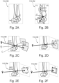
- Fig. 2 illustrates in subfigures 2A to 2F exemplary model parameters of an exemplary 3D pose estimation computer model CM that is utilized by the data processor 110.
- This is only an exemplary selection of possible model parameters, wherein not all model parameters must necessarily be considered or encoded.
- the at least one geometric parameter as explained above is an additional free parameter to the model parameters shown in Figs. 2A to 2F .
- Each of Figs. 2A to 2F is an example of an ROI including a bone constellation, which is here joint, especially an exemplary ankle joint.
- the 3D pose estimation computer model CM may also be referred to as a 3D articulation model.
- the model parameter of Fig. 2A is a flexion angle.
- Fig. 2B illustrates the model parameter of a viewing direction, in which (1) indicates an azimuthal rotation and (2) indicates a polar rotation.
- the model parameter illustrated in Fig. 2C is a detector position.
- Fig. 2D illustrates the model parameter of a translator in detector plane.
- the model parameter of Fig. 2E is a rotation in detector plane.
- Fig. 2F illustrates the model parameter of scaling in detector plane, wherein the scaling may comprise, for example, indicating changing the joint size or the image sampling.
- the 3D pose estimation model CM is trained to regress the model parameters considered or encoded therein. For example, such training of the 3D pose estimation model CM may be performed by using artificially generated images by using a 3D articulation shape model and its possible poses. This is not limited herein, since the training data may also be generated in a different way.
- Fig. 3 shows an example graph encoding neighboring bones in an exemplary bone constellation.
- the bone constellation included in the ROI is again an ankle joint comprising a fibula (a), a tibia (b), a talus (c), and a calcaneus (d), which can be illustrated as the graph encoding these bones.
- the at least one geometric parameter comprises one or more of a distance d between the bones, an orthogonal translation t and a rotation r.
- each of the geometric parameters of distance d, orthogonal translation and rotation r is a model parameter that may be encoded in the 3D pose estimation model in addition to the model parameters shown in Figs. 2A to 2F .
- the talo-tibial distance d as an additional free parameter allows more accurate adaptation of the 3D pose estimation computer model to cases with different or extraordinary distances.
- the talo-tibial distance d encoded in the model parameter(s) provides diagnostic valuable information.
- the distance d may reflect the size of the upper ankle joint gap, wherein a low value may indicate cartilage degeneration by age or osteoarthritis, and a high value may indicate tear of ligament and/or foot dislocation.
- a tree or graph may be used to encode a number of distances between bones in the 3D pose estimation computer model, wherein the bones are vertices and the distances are modelled by edges.
- the apparatus 100 e.g. the data processor 110, may be configured to construct an axis between two neighboring bones and to measure the distance d along that axis.
- Fig. 4 shows in subfigures 4A and 4B in a respective X-ray image a geometric parameter for determining the soft tissue-related property of the bone constellation from X-ray imaging data.
- the distance d, the translation t and/or rotation r relate to a scapho-lunate widening that can be taken as an indicator for degenerative arthritis.
- the apparatus 100 is configured to utilize the 3D pose estimation computer model CM, which is added by including at least one additional free parameter, i.e. the at least one geometric parameter, e.g. the distance d, the translation t and/or rotation r to determine the scapho-lunate widening as an example of the soft tissue-related property. Comparing the scapho-lunate widening to normal values, statistics, or the like, may be utilized by the apparatus 100 to determine an injury, lesion, abnormality, etc. in the soft tissue, although the soft tissue is not visible in the X-ray imaging data.
- CM 3D pose estimation computer model
- the apparatus 100 is configured to utilize the 3D pose estimation computer model CM, which is added by including at least one additional free parameter, i.e. the at least one geometric parameter, e.g. the distance d, the translation t and/or rotation r to determine the scapho-lunate widening as an example of the soft tissue-related property. Comparing the s
- Fig. 5 illustrates in a flow chart a computer-implemented method for determining the soft tissue-related property of the bone constellation from X-ray imaging data.
- the method may be performed by the above apparatus 100.
- the method comprises, as step S1, obtaining the X-ray imaging data of a region of interest including the bone constellation, and, as step S2, determining, by utilizing the 3D pose estimation computer model CM into which the X-ray imaging data is fed, a pose estimation of the bone constellation, and a relative distance between bones of the bone constellation which relative distance is assigned to at least one soft tissue-related property of the bone constellation.
- the at least one soft tissue-related property may be used for further diagnostic purposes, or the like.
- a computer program element for controlling the X-ray imaging system of the third aspect which, when being executed by processing unit, is adapted to perform the method steps of the fourth aspect.
- a computer program element might therefore be stored on a computer unit, which might also be an embodiment of the present invention.
- This computing unit may be adapted to perform or induce performance of the steps of the method described above. Moreover, it may be adapted to operate the components of the above-described apparatus.
- the computing unit can be adapted to operate automatically and/or to execute orders of a user.
- a computer program may be loaded into a working memory of a data processor. The data processor may thus be equipped to carry out the method of the invention.
- This exemplary embodiment of the invention covers both the computer program that has the intervention installed from the beginning, and a computer program that by means of an update turns an existing program into a program that uses the invention.
- a computer program may be stored and/or distributed on a suitable medium, such as optical storage media, or a solid state medium supplied together with, or as a part of other hardware, but may also be distributed in other forms, such as via the Internet or other wired or wireless telecommunication systems.
- the computer program may also be presented over a network like the World Wide Web, and can also be downloaded into the working memory of a data processor from such a network.
- a medium for making a computer program element available for downloading is provided, which computer program element is arranged to perform a method according to one of the previously described embodiments of the invention.
Landscapes
- Health & Medical Sciences (AREA)
- Life Sciences & Earth Sciences (AREA)
- Engineering & Computer Science (AREA)
- Medical Informatics (AREA)
- Pathology (AREA)
- Heart & Thoracic Surgery (AREA)
- Veterinary Medicine (AREA)
- Biophysics (AREA)
- High Energy & Nuclear Physics (AREA)
- Public Health (AREA)
- Nuclear Medicine, Radiotherapy & Molecular Imaging (AREA)
- Optics & Photonics (AREA)
- General Health & Medical Sciences (AREA)
- Radiology & Medical Imaging (AREA)
- Biomedical Technology (AREA)
- Physics & Mathematics (AREA)
- Molecular Biology (AREA)
- Surgery (AREA)
- Animal Behavior & Ethology (AREA)
- Orthopedic Medicine & Surgery (AREA)
- Dentistry (AREA)
- Oral & Maxillofacial Surgery (AREA)
- Physiology (AREA)
- Computer Vision & Pattern Recognition (AREA)
- Apparatus For Radiation Diagnosis (AREA)
Priority Applications (5)
| Application Number | Priority Date | Filing Date | Title |
|---|---|---|---|
| EP22195311.0A EP4338673A1 (de) | 2022-09-13 | 2022-09-13 | Bestimmung einer weichgewebebezogenen eigenschaft aus röntgenbildgebungsdaten |
| JP2025513216A JP2025527892A (ja) | 2022-09-13 | 2023-09-07 | X線撮像データからの軟組織関連特性の決定 |
| CN202380065881.2A CN119894450A (zh) | 2022-09-13 | 2023-09-07 | 根据x射线成像数据确定软组织相关的性质 |
| PCT/EP2023/074515 WO2024056494A1 (en) | 2022-09-13 | 2023-09-07 | Determination of a soft tissue-related property from x-ray imaging data |
| EP23767883.4A EP4586914B1 (de) | 2022-09-13 | 2023-09-07 | Bestimmung einer weichgewebebezogenen eigenschaft aus röntgenbildgebungsdaten |
Applications Claiming Priority (1)
| Application Number | Priority Date | Filing Date | Title |
|---|---|---|---|
| EP22195311.0A EP4338673A1 (de) | 2022-09-13 | 2022-09-13 | Bestimmung einer weichgewebebezogenen eigenschaft aus röntgenbildgebungsdaten |
Publications (1)
| Publication Number | Publication Date |
|---|---|
| EP4338673A1 true EP4338673A1 (de) | 2024-03-20 |
Family
ID=83318757
Family Applications (2)
| Application Number | Title | Priority Date | Filing Date |
|---|---|---|---|
| EP22195311.0A Withdrawn EP4338673A1 (de) | 2022-09-13 | 2022-09-13 | Bestimmung einer weichgewebebezogenen eigenschaft aus röntgenbildgebungsdaten |
| EP23767883.4A Active EP4586914B1 (de) | 2022-09-13 | 2023-09-07 | Bestimmung einer weichgewebebezogenen eigenschaft aus röntgenbildgebungsdaten |
Family Applications After (1)
| Application Number | Title | Priority Date | Filing Date |
|---|---|---|---|
| EP23767883.4A Active EP4586914B1 (de) | 2022-09-13 | 2023-09-07 | Bestimmung einer weichgewebebezogenen eigenschaft aus röntgenbildgebungsdaten |
Country Status (4)
| Country | Link |
|---|---|
| EP (2) | EP4338673A1 (de) |
| JP (1) | JP2025527892A (de) |
| CN (1) | CN119894450A (de) |
| WO (1) | WO2024056494A1 (de) |
Citations (1)
| Publication number | Priority date | Publication date | Assignee | Title |
|---|---|---|---|---|
| WO2013166299A1 (en) * | 2012-05-03 | 2013-11-07 | University Of Pittsburgh - Of The Commonwealth System Of Higher Education | Intelligent algorithms for tracking three-dimensional skeletal movement from radiographic image sequences |
Family Cites Families (1)
| Publication number | Priority date | Publication date | Assignee | Title |
|---|---|---|---|---|
| CN110944594A (zh) * | 2017-06-19 | 2020-03-31 | 穆罕默德·R·马赫福兹 | 使用荧光透视和跟踪传感器的髋部外科手术导航 |
-
2022
- 2022-09-13 EP EP22195311.0A patent/EP4338673A1/de not_active Withdrawn
-
2023
- 2023-09-07 JP JP2025513216A patent/JP2025527892A/ja active Pending
- 2023-09-07 WO PCT/EP2023/074515 patent/WO2024056494A1/en not_active Ceased
- 2023-09-07 EP EP23767883.4A patent/EP4586914B1/de active Active
- 2023-09-07 CN CN202380065881.2A patent/CN119894450A/zh active Pending
Patent Citations (1)
| Publication number | Priority date | Publication date | Assignee | Title |
|---|---|---|---|---|
| WO2013166299A1 (en) * | 2012-05-03 | 2013-11-07 | University Of Pittsburgh - Of The Commonwealth System Of Higher Education | Intelligent algorithms for tracking three-dimensional skeletal movement from radiographic image sequences |
Non-Patent Citations (2)
| Title |
|---|
| KRONKE ET AL.: "CNN-based pose estimation for assessing quality of ankle-joint X-ray images", SPIE MEDICAL IMAGING, 2022 |
| KRÖNKE SVEN ET AL: "CNN-based pose estimation for assessing quality of ankle-joint X-ray images", PROGRESS IN BIOMEDICAL OPTICS AND IMAGING, SPIE - INTERNATIONAL SOCIETY FOR OPTICAL ENGINEERING, BELLINGHAM, WA, US, vol. 12032, 4 April 2022 (2022-04-04), pages 120321A - 120321A, XP060156232, ISSN: 1605-7422, ISBN: 978-1-5106-0027-0, DOI: 10.1117/12.2611734 * |
Also Published As
| Publication number | Publication date |
|---|---|
| EP4586914B1 (de) | 2026-02-11 |
| JP2025527892A (ja) | 2025-08-22 |
| WO2024056494A1 (en) | 2024-03-21 |
| EP4586914A1 (de) | 2025-07-23 |
| CN119894450A (zh) | 2025-04-25 |
Similar Documents
| Publication | Publication Date | Title |
|---|---|---|
| US11937888B2 (en) | Artificial intelligence intra-operative surgical guidance system | |
| US11883219B2 (en) | Artificial intelligence intra-operative surgical guidance system and method of use | |
| KR102708979B1 (ko) | 추골 골절을 식별하기 위한 3차원 의료 이미지 분석 방법 및 시스템 | |
| US20210174503A1 (en) | Method, system and storage medium with a program for the automatic analysis of medical image data | |
| US20240206990A1 (en) | Artificial Intelligence Intra-Operative Surgical Guidance System and Method of Use | |
| US20210193326A1 (en) | Method and apparatus of providing osteoarthritis prediction information | |
| EP3905129B1 (de) | Procedimiento de identificación de imágenes óseas | |
| WO2021155373A1 (en) | Systems and methods for detection of musculoskeletal anomalies | |
| Kim et al. | Automatic spine segmentation and parameter measurement for radiological analysis of whole-spine lateral radiographs using deep learning and computer vision | |
| Kuiper et al. | Efficient cascaded V‐net optimization for lower extremity CT segmentation validated using bone morphology assessment | |
| US20250032187A1 (en) | Automatic Orthopedic Surgery Planning Systems and Methods | |
| US20250345120A1 (en) | Surgery assisting methods, systems and devices | |
| Zhang et al. | A novel tool to provide predictable alignment data irrespective of source and image quality acquired on mobile phones: what engineers can offer clinicians | |
| EP4586914B1 (de) | Bestimmung einer weichgewebebezogenen eigenschaft aus röntgenbildgebungsdaten | |
| EP4581567B1 (de) | Erkennung anatomischer anomalien in medizinischen 2d-bildern | |
| WO2024220642A2 (en) | Three-dimensional model generation and surgical planning based thereon | |
| KR102786520B1 (ko) | 방사선 영상을 이용한 척추질환 판단 장치 및 방법 | |
| US20250245822A1 (en) | Automated Pre-Checks To Evaluate Whether Medical Imaging Data Is Suitable For Surgical Planning Purposes | |
| EP4592949A1 (de) | Automatisierte bestimmung der knochenmineraldichte aus einem medizinischen bild | |
| CN120259300B (zh) | 一种基于计算机断层扫描图像的髋臼杯预测系统和方法 | |
| Yousefvand et al. | A fully automated measurement of migration percentage on ultrasound images in children with cerebral palsy | |
| US20250191203A1 (en) | Method for modelling a joint | |
| Goswami et al. | Deep Learning Segmentation Method for Uneviling Lower Limb Deformative on the Knee | |
| WO2026048941A1 (ja) | プログラム、情報処理方法、情報処理装置、及びモデル生成方法 | |
| Khayyeri et al. | A novel semi-automatic hip morphology assessment tool is more accurate than manual radiographic evaluations |
Legal Events
| Date | Code | Title | Description |
|---|---|---|---|
| PUAI | Public reference made under article 153(3) epc to a published international application that has entered the european phase |
Free format text: ORIGINAL CODE: 0009012 |
|
| STAA | Information on the status of an ep patent application or granted ep patent |
Free format text: STATUS: THE APPLICATION HAS BEEN PUBLISHED |
|
| AK | Designated contracting states |
Kind code of ref document: A1 Designated state(s): AL AT BE BG CH CY CZ DE DK EE ES FI FR GB GR HR HU IE IS IT LI LT LU LV MC MK MT NL NO PL PT RO RS SE SI SK SM TR |
|
| STAA | Information on the status of an ep patent application or granted ep patent |
Free format text: STATUS: THE APPLICATION IS DEEMED TO BE WITHDRAWN |
|
| 18D | Application deemed to be withdrawn |
Effective date: 20240921 |