EP4276800A2 - Method for reproducible production of defined bone fractures - Google Patents
Method for reproducible production of defined bone fractures Download PDFInfo
- Publication number
- EP4276800A2 EP4276800A2 EP23200419.2A EP23200419A EP4276800A2 EP 4276800 A2 EP4276800 A2 EP 4276800A2 EP 23200419 A EP23200419 A EP 23200419A EP 4276800 A2 EP4276800 A2 EP 4276800A2
- Authority
- EP
- European Patent Office
- Prior art keywords
- fracture
- preparation
- bone
- compression
- mass
- Prior art date
- Legal status (The legal status is an assumption and is not a legal conclusion. Google has not performed a legal analysis and makes no representation as to the accuracy of the status listed.)
- Pending
Links
- 208000010392 Bone Fractures Diseases 0.000 title claims abstract description 435
- 238000000034 method Methods 0.000 title claims abstract description 177
- 238000004519 manufacturing process Methods 0.000 title claims description 53
- 238000002360 preparation method Methods 0.000 claims abstract description 656
- 208000026137 Soft tissue injury Diseases 0.000 claims abstract description 73
- 238000012549 training Methods 0.000 claims abstract description 19
- 238000011161 development Methods 0.000 claims abstract description 9
- 239000007943 implant Substances 0.000 claims abstract description 8
- 206010017076 Fracture Diseases 0.000 claims description 259
- 230000006835 compression Effects 0.000 claims description 179
- 238000007906 compression Methods 0.000 claims description 179
- 238000013016 damping Methods 0.000 claims description 133
- 230000007246 mechanism Effects 0.000 claims description 114
- 206010037802 Radius fracture Diseases 0.000 claims description 100
- 210000000988 bone and bone Anatomy 0.000 claims description 47
- 210000004872 soft tissue Anatomy 0.000 claims description 44
- 206010020462 Humerus fracture Diseases 0.000 claims description 37
- 206010016454 Femur fracture Diseases 0.000 claims description 23
- 206010009245 Clavicle fracture Diseases 0.000 claims description 20
- 206010016970 Foot fracture Diseases 0.000 claims description 19
- 208000017013 ulna fracture Diseases 0.000 claims description 17
- 208000011224 Monteggia Fracture Diseases 0.000 claims description 16
- 230000003902 lesion Effects 0.000 claims description 16
- 210000004233 talus Anatomy 0.000 claims description 16
- 230000035939 shock Effects 0.000 claims description 15
- 239000006096 absorbing agent Substances 0.000 claims description 14
- 241000131317 Capitulum Species 0.000 claims description 10
- 210000001872 metatarsal bone Anatomy 0.000 claims description 8
- 239000013598 vector Substances 0.000 claims description 8
- 241001567848 Capitellum Species 0.000 claims description 7
- MBNGWHIJMBWFHU-UHFFFAOYSA-N diosmetin Chemical compound C1=C(O)C(OC)=CC=C1C1=CC(=O)C2=C(O)C=C(O)C=C2O1 MBNGWHIJMBWFHU-UHFFFAOYSA-N 0.000 claims description 7
- 208000027790 Rib fracture Diseases 0.000 claims description 6
- 238000004458 analytical method Methods 0.000 claims description 6
- 208000007356 Fracture Dislocation Diseases 0.000 claims description 5
- 208000031294 Upper limb fractures Diseases 0.000 claims description 5
- 230000008859 change Effects 0.000 claims description 5
- 208000027502 Ankle fracture Diseases 0.000 claims description 4
- 206010019114 Hand fracture Diseases 0.000 claims description 4
- 206010020100 Hip fracture Diseases 0.000 claims description 4
- 206010039579 Scapula fracture Diseases 0.000 claims description 4
- 206010048049 Wrist fracture Diseases 0.000 claims description 4
- 206010041569 spinal fracture Diseases 0.000 claims description 4
- 238000010200 validation analysis Methods 0.000 abstract description 3
- 230000001960 triggered effect Effects 0.000 description 80
- 210000000245 forearm Anatomy 0.000 description 60
- 210000002758 humerus Anatomy 0.000 description 54
- 208000014674 injury Diseases 0.000 description 31
- 230000006378 damage Effects 0.000 description 26
- 229920000642 polymer Polymers 0.000 description 25
- 239000006260 foam Substances 0.000 description 23
- 208000027418 Wounds and injury Diseases 0.000 description 22
- 230000007935 neutral effect Effects 0.000 description 22
- 210000002683 foot Anatomy 0.000 description 21
- 210000003109 clavicle Anatomy 0.000 description 19
- 210000004095 humeral head Anatomy 0.000 description 18
- 210000000707 wrist Anatomy 0.000 description 18
- 210000001699 lower leg Anatomy 0.000 description 17
- 210000001991 scapula Anatomy 0.000 description 17
- 210000003484 anatomy Anatomy 0.000 description 16
- 210000000623 ulna Anatomy 0.000 description 15
- 210000002221 olecranon process Anatomy 0.000 description 12
- 238000005516 engineering process Methods 0.000 description 11
- 238000004364 calculation method Methods 0.000 description 9
- 210000001519 tissue Anatomy 0.000 description 9
- 230000005540 biological transmission Effects 0.000 description 8
- 238000011156 evaluation Methods 0.000 description 8
- 238000005259 measurement Methods 0.000 description 8
- 230000008733 trauma Effects 0.000 description 8
- 238000005253 cladding Methods 0.000 description 7
- 238000010276 construction Methods 0.000 description 7
- 210000002082 fibula Anatomy 0.000 description 7
- 210000001503 joint Anatomy 0.000 description 7
- 230000008569 process Effects 0.000 description 7
- 210000001562 sternum Anatomy 0.000 description 7
- 210000002303 tibia Anatomy 0.000 description 7
- 210000000689 upper leg Anatomy 0.000 description 7
- VYPSYNLAJGMNEJ-UHFFFAOYSA-N Silicium dioxide Chemical compound O=[Si]=O VYPSYNLAJGMNEJ-UHFFFAOYSA-N 0.000 description 6
- 210000000544 articulatio talocruralis Anatomy 0.000 description 6
- 210000001513 elbow Anatomy 0.000 description 6
- 239000000463 material Substances 0.000 description 6
- 238000003032 molecular docking Methods 0.000 description 6
- 230000000399 orthopedic effect Effects 0.000 description 6
- 238000012360 testing method Methods 0.000 description 6
- 239000006004 Quartz sand Substances 0.000 description 5
- 241001227561 Valgus Species 0.000 description 5
- 238000013461 design Methods 0.000 description 5
- 210000003811 finger Anatomy 0.000 description 5
- 239000012634 fragment Substances 0.000 description 5
- 210000003127 knee Anatomy 0.000 description 5
- 210000000629 knee joint Anatomy 0.000 description 5
- 238000012821 model calculation Methods 0.000 description 5
- 238000001356 surgical procedure Methods 0.000 description 5
- 241001465754 Metazoa Species 0.000 description 4
- 241000469816 Varus Species 0.000 description 4
- 210000002659 acromion Anatomy 0.000 description 4
- 238000002474 experimental method Methods 0.000 description 3
- 238000011835 investigation Methods 0.000 description 3
- 210000003041 ligament Anatomy 0.000 description 3
- 229910052751 metal Inorganic materials 0.000 description 3
- 239000002184 metal Substances 0.000 description 3
- 238000011160 research Methods 0.000 description 3
- 238000005070 sampling Methods 0.000 description 3
- 210000002832 shoulder Anatomy 0.000 description 3
- 238000004088 simulation Methods 0.000 description 3
- 239000004425 Makrolon Substances 0.000 description 2
- 208000001132 Osteoporosis Diseases 0.000 description 2
- 206010071051 Soft tissue mass Diseases 0.000 description 2
- 230000001133 acceleration Effects 0.000 description 2
- 229910052782 aluminium Inorganic materials 0.000 description 2
- XAGFODPZIPBFFR-UHFFFAOYSA-N aluminium Chemical compound [Al] XAGFODPZIPBFFR-UHFFFAOYSA-N 0.000 description 2
- 238000005452 bending Methods 0.000 description 2
- 210000000459 calcaneus Anatomy 0.000 description 2
- 230000007257 malfunction Effects 0.000 description 2
- 230000010534 mechanism of action Effects 0.000 description 2
- 210000003205 muscle Anatomy 0.000 description 2
- 208000015122 neurodegenerative disease Diseases 0.000 description 2
- 230000003287 optical effect Effects 0.000 description 2
- 229920000515 polycarbonate Polymers 0.000 description 2
- 230000009467 reduction Effects 0.000 description 2
- 210000003491 skin Anatomy 0.000 description 2
- 239000007787 solid Substances 0.000 description 2
- 210000002435 tendon Anatomy 0.000 description 2
- 238000010257 thawing Methods 0.000 description 2
- 229910000851 Alloy steel Inorganic materials 0.000 description 1
- 208000025978 Athletic injury Diseases 0.000 description 1
- 241001260012 Bursa Species 0.000 description 1
- 101100298222 Caenorhabditis elegans pot-1 gene Proteins 0.000 description 1
- 208000024779 Comminuted Fractures Diseases 0.000 description 1
- 244000035744 Hura crepitans Species 0.000 description 1
- 208000005137 Joint instability Diseases 0.000 description 1
- 241000150100 Margo Species 0.000 description 1
- 241000906034 Orthops Species 0.000 description 1
- 208000008558 Osteophyte Diseases 0.000 description 1
- 206010034122 Patella fracture Diseases 0.000 description 1
- 208000006735 Periostitis Diseases 0.000 description 1
- 208000018286 Shoulder injury Diseases 0.000 description 1
- 230000009471 action Effects 0.000 description 1
- 230000006978 adaptation Effects 0.000 description 1
- 210000003423 ankle Anatomy 0.000 description 1
- 238000011888 autopsy Methods 0.000 description 1
- 239000012620 biological material Substances 0.000 description 1
- 230000015572 biosynthetic process Effects 0.000 description 1
- 230000037396 body weight Effects 0.000 description 1
- 239000002639 bone cement Substances 0.000 description 1
- 230000037182 bone density Effects 0.000 description 1
- 239000002775 capsule Substances 0.000 description 1
- 238000005266 casting Methods 0.000 description 1
- 210000000038 chest Anatomy 0.000 description 1
- 239000002131 composite material Substances 0.000 description 1
- 238000002591 computed tomography Methods 0.000 description 1
- 210000002808 connective tissue Anatomy 0.000 description 1
- 238000005520 cutting process Methods 0.000 description 1
- 230000006735 deficit Effects 0.000 description 1
- 238000001739 density measurement Methods 0.000 description 1
- 238000005553 drilling Methods 0.000 description 1
- 210000002310 elbow joint Anatomy 0.000 description 1
- 239000003822 epoxy resin Substances 0.000 description 1
- 230000003203 everyday effect Effects 0.000 description 1
- 210000003195 fascia Anatomy 0.000 description 1
- 238000011990 functional testing Methods 0.000 description 1
- 230000002068 genetic effect Effects 0.000 description 1
- 230000005484 gravity Effects 0.000 description 1
- 210000004247 hand Anatomy 0.000 description 1
- 210000001624 hip Anatomy 0.000 description 1
- 210000004394 hip joint Anatomy 0.000 description 1
- 238000010191 image analysis Methods 0.000 description 1
- 238000009863 impact test Methods 0.000 description 1
- 230000003116 impacting effect Effects 0.000 description 1
- 230000036540 impulse transmission Effects 0.000 description 1
- 238000003780 insertion Methods 0.000 description 1
- 230000037431 insertion Effects 0.000 description 1
- 210000000281 joint capsule Anatomy 0.000 description 1
- 238000000691 measurement method Methods 0.000 description 1
- 238000003801 milling Methods 0.000 description 1
- 210000005036 nerve Anatomy 0.000 description 1
- 235000016709 nutrition Nutrition 0.000 description 1
- 230000010355 oscillation Effects 0.000 description 1
- 201000008482 osteoarthritis Diseases 0.000 description 1
- 239000005022 packaging material Substances 0.000 description 1
- 206010033675 panniculitis Diseases 0.000 description 1
- 230000007310 pathophysiology Effects 0.000 description 1
- 210000004197 pelvis Anatomy 0.000 description 1
- 238000009527 percussion Methods 0.000 description 1
- 210000003460 periosteum Anatomy 0.000 description 1
- 229920000647 polyepoxide Polymers 0.000 description 1
- 229920000193 polymethacrylate Polymers 0.000 description 1
- 230000002980 postoperative effect Effects 0.000 description 1
- 238000003825 pressing Methods 0.000 description 1
- 238000012545 processing Methods 0.000 description 1
- 238000012797 qualification Methods 0.000 description 1
- 239000011347 resin Substances 0.000 description 1
- 229920005989 resin Polymers 0.000 description 1
- 238000012552 review Methods 0.000 description 1
- 239000004576 sand Substances 0.000 description 1
- 230000011664 signaling Effects 0.000 description 1
- 238000003860 storage Methods 0.000 description 1
- 238000007920 subcutaneous administration Methods 0.000 description 1
- 210000004304 subcutaneous tissue Anatomy 0.000 description 1
- 230000001360 synchronised effect Effects 0.000 description 1
- 210000003371 toe Anatomy 0.000 description 1
- 238000012546 transfer Methods 0.000 description 1
- 230000007704 transition Effects 0.000 description 1
- 238000013519 translation Methods 0.000 description 1
- 210000001364 upper extremity Anatomy 0.000 description 1
Images


Classifications
-
- G—PHYSICS
- G09—EDUCATION; CRYPTOGRAPHY; DISPLAY; ADVERTISING; SEALS
- G09B—EDUCATIONAL OR DEMONSTRATION APPLIANCES; APPLIANCES FOR TEACHING, OR COMMUNICATING WITH, THE BLIND, DEAF OR MUTE; MODELS; PLANETARIA; GLOBES; MAPS; DIAGRAMS
- G09B23/00—Models for scientific, medical, or mathematical purposes, e.g. full-sized devices for demonstration purposes
- G09B23/28—Models for scientific, medical, or mathematical purposes, e.g. full-sized devices for demonstration purposes for medicine
- G09B23/30—Anatomical models
- G09B23/306—Anatomical models comprising real biological tissue
-
- G—PHYSICS
- G09—EDUCATION; CRYPTOGRAPHY; DISPLAY; ADVERTISING; SEALS
- G09B—EDUCATIONAL OR DEMONSTRATION APPLIANCES; APPLIANCES FOR TEACHING, OR COMMUNICATING WITH, THE BLIND, DEAF OR MUTE; MODELS; PLANETARIA; GLOBES; MAPS; DIAGRAMS
- G09B23/00—Models for scientific, medical, or mathematical purposes, e.g. full-sized devices for demonstration purposes
- G09B23/28—Models for scientific, medical, or mathematical purposes, e.g. full-sized devices for demonstration purposes for medicine
- G09B23/30—Anatomical models
Definitions
- the subject of the invention is a method for the reproducible generation of defined bone fractures with accompanying soft tissue injuries in preparations, in particular in human preparations, devices for using the method and the preparations produced with the aid of the method, in particular human preparations, which are characterized by a defined bone fracture with accompanying soft tissue injuries.
- the preparations produced using the method according to the invention, in particular human preparations can be used for the training, education and further training of medical personnel, for the development and validation of medical devices, implants and prostheses, for accident analyzes and reports.
- the bone fractures that occur in this way do not correspond to those of real bone fractures caused by indirect force. In particular, they differ from the typical fracture patterns that arise in a real accident in terms of their geometry and the nature of the bone fragments involved. In addition, no typical accompanying ligamentous injuries (to capsules, ligaments and tendons) are caused.
- the manual application of force with tools at non-standard heights and angles leads to different results on the specimen and is not standardized. The individuality of the specimens in terms of morphology and geometry is not taken into account.
- An isolated radial head fracture was created at a given forearm rotation of 40, 41, 42, 45, 50 and 53 degrees (44.4 +/- 5.2 degrees) and Essex-Lopresti fractures were created at a given forearm rotation of 51, 54, 58, 90 , 108 and 110 degrees (70 +/- 25.2 degrees).
- a proximal radial fracture with an accompanying distal ulna fracture was detected in 4 of 20 specimens (ie hit rate 20%), the isolated radial head fracture in 7 of 20 specimens (ie hit rate 35%) and an Essex-Lopresti fracture in 9 of 20 specimens (ie hit rate 45%) generated.
- the force was used to test the failure limits of biological material and did not simulate a real accident. No defined fractures were created.
- Robert Holz (2013) (Master's thesis "The mechanism of the Essex-Lopresti: Investigation of tissue failure using a newly developed simulator ") used a simulator with a gravity-accelerated falling body to investigate the biomechanics and injury sequence of the Essex-Lopresti fracture.
- the human specimens were dissected, i.e. skin and subcutaneous tissue including the muscles were removed and the arms except for the IOM (interosseous membrane) and the Joint capsules around the elbow and wrist are prepared free.
- Holz describes the alignment and clamping of the preparation in the simulator, the optical analysis of the fracture creation and the determination of the horizontal and vertical as well as the relative movement of the segments when the fracture is created.
- the Essex-Lopresti fracture was able to be done by Holz in 4 out of 30 cases, ie with a hit rate of 13.3%, were produced in specimens without a soft tissue coating.
- the bone fractures created in the prior art are products of chance. There are no known methods that can be used to specifically create defined bone fractures.
- Bone fractures can be divided into defined fracture classes.
- the defined bone fractures are the same in terms of their location and fracture pattern or, if the individual anatomical variations in the accident victims are taken into account, very similar.
- the defined bone fractures are also with regard to the accompanying soft tissue injuries in individual accident victims, the same or very similar.
- the subject of the invention is a method for producing at least one defined bone fracture with accompanying soft tissue injuries in a preparation 106, characterized in that a defined force impact is exerted on the fixed preparation 106 and the change in the length of the preparation 106 along the force vector is limited to a maximum of 80 mm .
- the change in the length of the preparation 106 is limited to a maximum of 80 mm by setting a defined compression to which the preparation 106 is subjected to the force impact.
- the defined bone fracture can be generated in the preparation 106 by the method according to the invention by a force impact resulting from a kinetic energy of 5 to 500 joules.
- the method according to the invention leads to the reproducible creation of a defined bone fracture with accompanying soft tissue injuries in a preparation 106 with a probability of at least 50%, preferably at least 55%, 60%, 65%, 70%, 75%, 80%, 85%, 90% , 95%.
- the method limits the change in length of the preparation 106 along the force vector.
- the reduction in the length of the preparation 106 is a maximum of 80 mm, for example preferably a maximum of 65 mm, particularly preferably a maximum of 52 mm or less.
- the maximum compression of the preparation is 80 mm, preferably a maximum of 65 mm, particularly preferably a maximum of 52 mm.
- the defined compression that the preparation experiences upon impact with the mass is a maximum of 80 mm, preferably 1 mm to 60 mm, particularly preferably 2 mm to 55 mm.
- the force shock upon impact with the preparation is dampened.
- the impact on the preparation is undamped.
- the force impact is exerted by the impact of a defined mass moving at a defined speed in the direction of the preparation.
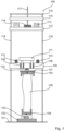
- the method according to the invention can be carried out using a device 100, 200 according to Fig. 1 or Fig. 2 be performed.
- the defined speed in Method step d) can be adjusted using a device 100, 200 by means of a defined fall height.
- the defined compression and the defined damping can be adjusted using means for adjusting the defined damping upon impact 110, 210.
- the defined bone fracture can be selected, for example, from shaft fracture of the phalanges, shaft fracture of the metacarpals, radius fracture, distal radius fracture, distal radius fracture extension, distal radius fracture flexion, distal radius fracture die-punch fracture, distal radius fracture chauffeur fracture, scaphoid fracture, radial head fracture, coronoid fracture, terrible triad , Olecranon fracture, Monteggia fracture, Monteggia like lesion, Galeazzi fracture, capitulum fracture, humerus fracture, distal humerus fracture, proximal humerus fracture, clavicular shaft fracture, lateral clavicle fracture, medial clavicle fracture, femur fracture, distal femur fracture, proximal femur fracture, tibial plateau fracture, proximal tibial plateau fracture, distal tibial plateau fracture, pillon fracture, calcaneus fracture , maleoli fracture, navicular fracture, patellar fracture, meta
- the preparation can be a human preparation or an animal preparation.
- the preparation may be a formalin-fixed preparation, a Thiel-fixed preparation or a thawed preparation.
- the defined mass has a weight of at least 1 kg, preferably a maximum of 72 kg, particularly preferably a weight of 5 kg to 33 kg or 4 to 40 kg.
- the defined mass is positioned in the axial direction, preferably in the vertical direction, with respect to the preparation.
- the defined mass can be adjusted, for example, by a mass 112 and one or more additional weights 113.
- the defined speed of the defined mass upon impact with the preparation is at least 0.5 m/s, preferably at least 3 m/s to 10 m/s, particularly preferably 5 m/s to 6 m/s.
- the defined fall height is, for example, 10 cm to 150 cm, preferably 20 cm to 120 cm.
- the defined bone fracture can be generated in the preparation 106 by the method according to the invention by a force impact resulting from a kinetic energy of 5 to 500 joules, preferably 15 to 300 J.
- the force that is exerted on the preparation when the defined mass impacts is preferably at least 50 N, preferably a maximum of 34 kN.
- the kinetic energies generated are, for example, 15 - 450 J, preferably 120 to 250 J (see Table 2).
- the defined damping with which the defined mass is braked upon impact with the preparation can be adjusted, for example, with at least one shock absorber, preferably at least one hydraulic shock absorber.
- the impact can occur undamped (defined attenuation equal to zero) or a defined attenuation can be set (defined attenuation greater than 0).
- the defined damping, which is set by means of one or more means for adjusting the defined damping 110, 210, for example shock absorbers, is, for example, a maximum of 50 mm, preferably from 0 mm to 40 mm, particularly preferably from 5 mm to 25 mm or 37 mm.
- Bone fractures are bone fractures that occur in real accidents.
- Defined bone fractures are known to those skilled in the art, for example from the AO classification (Maurice E. Müller: The Comprehensive Classification of Fractures of Long Bones in: ME Müller et al. (ed.): Manual of Internal Fixation. 3. Edition. P. 118 ff. Springer-Verlag, Berlin/Heidelberg/New York/Tokyo 1991, ISBN 3-540-52523-8 ), Orthopedics and Accident Surgery Essentials (Steffen Ruchholtz, Dieter Christian Wirtz), intensive course for further training (2nd, completely revised and expanded edition. 1155 illustrations. Paperback.
- Bone fractures include shaft fractures (diaphyseal fractures), fractures near the joint (metaphyseal fractures) and articular fractures (fractures involving the articular surface and dislocation fractures).
- the defined bone fracture is reproducibly produced using the method.
- a defined bone fracture is selected and generated with a certain probability, that is, with a certain hit rate, using the method according to the invention.
- Reproducible means that the defined bone fracture is generated with a probability of at least 50%, preferably at least 60%, 70%, 80%, 85%, 90%, 95% or more.
- the method according to the invention enables, for the first time, the predictable generation of defined bone fractures in preparations (not a random product). This makes it possible to ethically produce human specimens with defined bone fractures for commercial use. The reproducibility also leads to a significant cost reduction in all areas where these preparations are needed, as fewer rejected preparations are produced.
- the accompanying soft tissue injuries produced with the method according to the invention are characteristic of the respective defined bone fracture and are therefore realistic. Open and closed bone fractures are included. In a preferred embodiment of the method, defined bone fractures with a closed soft tissue shell are created.
- the accompanying soft tissue injuries that are characteristic of each defined bone fracture are known to the person skilled in the art, for example from Tscherne H, Oestern HJ: Pathophysiology and classification of soft tissue injuries associated with fractures. In: Fractures with soft tissue injuries. Tscherne H Gotzen L.: Berlin; Springer Verlag (1984), pp. 1-9 .
- the invention also relates to the preparations produced using the method according to the invention.
- the subject of the invention is a preparation 106, in particular a human preparation with at least one defined bone fracture and accompanying soft tissue injuries, obtainable by the method according to the invention or produced by the method according to the invention.
- a preparation 106 in particular a human preparation with at least one defined bone fracture and accompanying soft tissue injuries, obtainable by the method according to the invention or produced by the method according to the invention.
- Preparations with defined bone fractures and accompanying soft tissue injuries cannot yet be created artificially. So far, these injuries have only occurred in real accidents involving living people.
- Corresponding preparations can be produced for the first time using the method according to the invention.
- the subject of the invention is a preparation 106, in particular a human preparation, with at least one defined bone fracture with accompanying soft tissue injuries.
- the preparation 106 preferably comprises a bone fracture with accompanying soft tissue injury selected from shaft fracture of the phalanges, shaft fracture of the metacarpals, radius fracture, distal radius fracture, distal radius fracture extension, distal radius fracture flexion, distal radius fracture die-punch fracture, distal radius fracture chauffeur fracture, scaphoid fracture, radius head fracture tur, Coronoid fracture, Terrible Triad, Olecranon fracture, Monteggia fracture, Monteggia like lesion, Galeazzi fracture, Capitulum fracture, Humerus fracture, Distal humerus fracture, Proximal humerus fracture, Clavicular shaft fracture, Lateral clavicle fracture, Medial clavicle fracture, Femur fracture, Distal femur fracture, Proximal femur fracture, Tibial plateau fracture, proximal tibial plateau fracture, distal
- the soft tissue coat is understood to mean all of the body's own tissue that surrounds the bone of a preparation.
- the biological tissue that surrounds the bone is more elastic, more deformable (softer) than the bone.
- soft tissue sheath includes, among others, the main groups: muscles, ligaments, tendons, joint capsules, nerves, skin and vessels. Other components include fascia, connective tissue, periosteum and bursa.
- These biological structures have different functions and morphologies and therefore show different mechanical properties. Due to these different properties, these structures respond differently to injury mechanisms. In real accidents, different types of damage occur in different tissues. This tissue-specific damage to a bone fracture is therefore also called “typical” or “accompanying soft tissue injuries”.
- the preparation 106 is characterized in that the soft tissue envelope is closed.
- the preparation 106 is characterized in that the soft tissue jacket is opened.
- the method according to the invention can be used to produce preparations with defined bone fractures in which the soft tissues have injuries.
- specimens with defined bone fractures can be created in which the soft tissue mantle is opened. These are openings that can occur when pointed or sharp-edged bone fragments pierce the soft tissues and ultimately the skin. It is clearly visible that the skin is opened and the soft tissues are penetrated from the inside out. This can be clearly seen in the shape and form of the openings, as well as the damaged tissue underneath. These bone fractures can therefore be clearly distinguished from those in which the damage to the soft tissue occurs from the outside in.
- Particular embodiments of the invention relate to preparations and methods for their production in detail.
- the defined bone fracture is a shaft fracture of the phalanges and the defined compression of the preparation 106 is set to 2 to 8 mm and the defined damping to 0 to 5 mm.
- Preparation comprising a shaft fracture of the phalanges with accompanying soft tissue injuries, obtainable by the method according to the invention.
- the defined bone fracture is a shaft fracture of the metacarpal and the defined compression of the preparation 106 is set to 6 to 14 mm and the defined damping to 0 to 9 mm.
- Preparation comprising a metacarpal shaft fracture with accompanying soft tissue injuries, obtainable by the method according to the invention.
- the defined bone fracture is a distal radius fracture and the defined compression of the preparation 106 is set to 20 to 36 mm and the defined damping to 6 to 17 mm.
- Preparation comprising a distal Radius fracture with accompanying soft tissue injuries, obtainable using the method according to the invention.
- the defined bone fracture is a distal radius fracture extension of the classification 23 A2, 23 C1 - C3 (dorsal) according to AO and the defined compression of the preparation 106 is set to 22 to 30 mm and the defined damping to 6 to 14 mm .
- Preparation comprising a distal radius fracture extension of classification 23 A2, 23 C1 - C3 (dorsal) according to AO with accompanying soft tissue injuries, obtainable by the method according to the invention.
- the defined bone fracture is a distal radius fracture of classification 23 A2 (palmar) according to AO and the defined compression of the preparation 106 is set to 25 to 35 mm and the defined damping to 5 to 17 mm.
- Preparation comprising a distal radius fracture of classification 23 A2 (palmar) according to AO with accompanying soft tissue injuries, obtainable using the method according to the invention.
- the defined bone fracture is a distal radius fracture/die-punch fracture of classification 23 C1 - C2 according to AO and the defined compression of the preparation 106 is set to 22 to 31 mm and the defined damping to 9 to 15 mm.
- Preparation comprising a distal radius fracture die-punch fracture of classification 23 C1 - C2 according to AO with accompanying soft tissue injuries, obtainable by the method according to the invention.
- the defined bone fracture is a radius fracture/chauffeur fracture of classification 23 B1 according to AO and the defined compression of the preparation 106 is set to 20 to 28 mm and the defined damping to 6 to 14 mm.
- Preparation comprising a radius fracture chauffeur fracture of classification 23 B1 according to AO with accompanying soft tissue injuries, obtainable by the method according to the invention.
- the defined bone fracture is a scaphoid fracture 72 A2, B2 - B3 according to AO and the defined compression of the preparation 106 is set to 24 to 32 mm and the defined damping to 10 to 17 mm.
- Preparation comprising a shaft fracture of the scaphoid fracture 72 A2, B2 - B3 according to AO with accompanying soft tissue injuries, obtainable by the method according to the invention.
- the defined bone fracture is a radial head fracture 21 B2 according to AO and the defined compression of the preparation 106 is set to 21 to 29 mm and the defined damping to 9 to 15 mm.
- Preparation comprising a radial head fracture 21 B2 according to AO with accompanying soft tissue injuries, obtainable by the method according to the invention.
- the defined bone fracture is a coronoid fracture and the defined compression of the preparation 106 is set to 20 to 33 mm and the defined damping to 8 to 16 mm.
- Preparation comprising a coronoid fracture with accompanying soft tissue injuries, obtainable by the method according to the invention.
- the defined bone fracture is a Terrible Triad and the defined compression of the preparation 106 is set to 24 to 38 mm and the defined damping to 10 to 18 mm.
- Preparation comprising a Terrible Triad with accompanying soft tissue injuries, obtainable by the method according to the invention.
- the defined bone fracture is an olecranon fracture and the defined compression of the preparation 106 is set to 4 to 17 mm and the defined damping to 0 to 9 mm.
- Preparation comprising an olecranon fracture with accompanying soft tissue injuries, obtainable by the method according to the invention.
- the defined bone fracture is a Monteggia fracture and the defined compression of the preparation 106 is set to 28 to 46 mm and the defined damping to 10 to 17 mm.
- Preparation comprising a Monteggia fracture with accompanying soft tissue injuries, obtainable by the method according to the invention.
- the defined bone fracture is a Monteggia like lesion and the defined compression of the preparation 106 is set to 30 to 46 mm and the defined damping to 9 to 21 mm.
- Preparation comprising a Monteggia like lesion with accompanying soft tissue injuries, obtainable by the method according to the invention.
- the defined bone fracture is a Galeazzi fracture and the defined compression of the preparation 106 is set to 24 to 39 mm and the defined damping to 6 to 17 mm.
- Preparation comprising a Galeazzi fracture with accompanying soft tissue injuries, obtainable by the method according to the invention.
- the defined bone fracture is a capitellum fracture and the defined compression of the preparation 106 is set to 14 to 22 mm and the defined damping to 6 to 13 mm.
- Preparation comprising a capitellum fracture with accompanying soft tissue injuries, obtainable by the method according to the invention.
- the defined bone fracture is a humerus fracture and the defined compression of the preparation 106 is set to 26 to 44 mm and the defined damping to 0 to 16 mm.
- Preparation comprising a humerus fracture with accompanying soft tissue injuries, obtainable by the method according to the invention.
- the defined bone fracture is a distal humerus fracture and the defined compression of the preparation 106 is set to 26 to 37 mm and the defined damping to 0 to 15 mm.
- Preparation comprising a distal humerus fracture with accompanying soft tissue injuries, obtainable by the method according to the invention.
- the defined bone fracture is a clavicular shaft fracture and the defined compression of the preparation 106 is set to 4 to 12 mm and the defined damping to 0 to 6 mm.
- Preparation comprising a clavicular shaft fracture with accompanying soft tissue injuries, obtainable by the method according to the invention.
- the defined bone fracture is a lateral clavicle fracture and the defined compression of the preparation 106 is set to 5 to 14 mm and the defined damping to 0 to 7 mm.
- Preparation comprising a lateral clavicle fracture with accompanying soft tissue injuries, obtainable by the method according to the invention.
- the defined bone fracture is a proximal humerus fracture 11 B1, B3, C1 - C3 according to AO and the defined compression of the preparation 106 is set to 29 to 44 mm and the defined damping to 0 to 16 mm.
- Preparation comprising a proximal humerus fracture 11 B1, B3, C1 - C3 according to AO with accompanying soft tissue injuries, obtainable by the method according to the invention.
- the defined bone fracture is a distal femur fracture and the defined compression of the preparation 106 is set to 31 to 49 mm and the defined damping to 0 to 37 mm.
- Preparation comprising a distal femur fracture with accompanying soft tissue injuries, obtainable by the method according to the invention.
- the defined bone fracture is a tibial plateau fracture and the defined compression of the preparation 106 is set to 35 to 47 mm and the defined damping to 10 to 13 mm.
- Preparation comprising a tibial plateau fracture with accompanying soft tissue injuries, obtainable by the method according to the invention.
- the defined bone fracture is a talus fracture and the defined compression of the preparation 106 is set to 26 to 48 mm and the defined damping to 0 to 22 mm.
- Preparation comprising a talus fracture with accompanying soft tissue injuries, obtainable by the method according to the invention.
- the defined bone fracture is a pillion fracture and the defined compression of the preparation 106 to 30 to 51 mm and the defined damping 0 to 25 mm can be set.
- Preparation comprising a pillon fracture with accompanying soft tissue injuries, obtainable by the method according to the invention.
- the defined bone fracture is a calcaneus fracture and the defined compression of the preparation 106 is set to 25 to 43 mm and the defined damping to 0 to 18 mm.
- Preparation comprising a calcaneus fracture with accompanying soft tissue injuries, obtainable by the method according to the invention.
- the defined bone fracture is a distal radius fracture 23 B3 according to AO and the defined compression of the preparation 106 is set to 25 to 36 mm and the defined damping to 10 to 16 mm.
- Preparation comprising a distal radius fracture 23 B3 according to AO with accompanying soft tissue injuries, obtainable by the method according to the invention.
- the preparation When carrying out the procedure, the preparation is cast, clamped or clamped at one or more points, preferably at the proximal and distal ends, for fixation. Before fixation, the specimen can be aligned in a defined geometry.
- the traverse 109, 209, 409 can be height-adjustable or not height-adjustable.
- the device 100, 500, 600 may include means for testing, for example one or more cameras 528 and/or one or more force sensors 103, 503, in order to continually improve the reproducibility (the probability) that a defined bone fracture will be produced and/or or to better understand the sequences of various events during stress.
- a device 100, 500, 600, which includes means for testing, can be used to determine one or more parameters selected from the Parameters for determining the defined mass, the defined direction, the defined speed of the defined mass, the defined geometry of the preparation 106, the defined compression of the preparation 106, the defined damping upon impact of the defined mass can be used.
- the procedure for determining defined parameters is described below and can be used in an analogous manner by a person skilled in the art to determine the defined parameters to produce further defined bone fractures in preparations.
- the device 200 can be dismantled and is therefore easier to transport. This means, for example, that preparations can be produced on site immediately before use. This is desirable because the preparations with defined bone fractures and accompanying soft tissue injuries require special storage conditions, which is avoided by only producing them immediately before use.
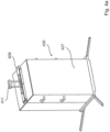
- the subject of the invention is a device 200 for carrying out the method according to the invention, which can be dismantled into a drive module 229, 329 and a structure module 230, 430 for transporting the device.
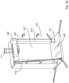
- the device 100, 200 should always be specially secured to avoid injury to persons using the device 100, 200 for the procedure.
- a special security includes, for example, specially secured holding mechanisms for the defined mass and a casing 227.
- the subject of the invention is a device 100, 200, 500 or drive module 329, 229 comprising an at least twice secured holding mechanism 214, 314 for positioning the defined mass.
- Device 100, 200, drive module 329, 229 and/or assembly module 430, 230 comprising at least one casing 227, 327, 427.
- the subject of the invention is the use of the device 100, 200, 300, 400, 500, 600 for determining one or more parameters selected from the parameters determining the defined mass, the defined direction, the defined speed of the defined mass, the defined geometry of the preparation 106, the defined compression of the preparation 106, the defined damping upon impact of the defined mass.
- the subject of the invention is the use of the device 100, 200, 300, 400, 500, 600 for carrying out the methods according to the invention.
- the subject of the invention is the use of the device 100, 200, 300, 400, 500, 600 for producing a defined bone fracture with accompanying soft tissue injuries in a preparation 106, 506, preferably for reproducibly producing the defined bone fracture with a probability of at least 50%.
- a preparation 106 is a dead human body or a dead animal body or a part of a dead human body, for example a severed body part (eg arm, foot, knee) or a part of a dead animal body.
- the preparation 106 can be frozen. The thawing process is initiated 15 to 24 hours before performing the procedure to create the bone fracture, depending on the objective and anatomical region. To do this, the preparation 106 is removed from the refrigerator (at minus 20 degrees Celsius), the packaging material is removed and stored at room temperature (20 to 22 degrees Celsius). Processing is possible at temperatures of 10 degrees Celsius to 25 degrees Celsius, preferably 15 degrees Celsius to 23 degrees Celsius. A formalin- or thiel-fixed specimen 106 can be processed directly without major preparation.
- the donors of the preparations are usually between 78 and 86 years old However, they can also be older or younger at the time the preparations are donated.
- the preparation 106 comes from a donor with an age of more than 60 years, 70 to 90 years, preferably 78 to 86 years.
- the preparation 106 according to the invention which comprises the defined bone fracture with accompanying soft tissue injuries, has an age of more than 60 years, preferably from 70 to 90 years, particularly preferably from 78 to 86 years.
- the specimen 106 may be a whole body specimen or a body part or a defined anatomical region.
- the preparation 106 may include at least one anatomical region selected from the anatomical regions of hand and 1 to 5 fingers, wrist, elbow, shoulder, knee, ankle, foot and 1 to 5 toes, hip, pelvis, spine, thorax, ribs.
- the preparation 106 can comprise at least one joint affected by the force, preferably 1 to 3 joints affected by the force.
- the joint or joints can have a joint position in the defined geometry, selected from neutral position, flexed or extended, rotated, varus or valgus position.
- the introduction of force into the preparation is preferably not direct, but rather indirect, for example via a punch 111, 211, 511 using a device 100, 200, 500.
- the indirect introduction of force enables the preparation 106, 506 to be precisely fixed.
- the interface between device 100, 500 and preparation 106, 506 should produce a flush adhesion, for example by pouring the ends of the preparation 106, 506 with a cold-curing polymer such as epoxy resin in a mold 105, 505 and screwing the mold 105, 505 to the device 100, 500.
- the preparation 106 can be clamped proximally and/or distally for fixation in a defined geometry.
- the specimen can be rotated by a defined angle around at least one of the clamps and fixed in this defined geometry.
- the defined geometry of the preparation 106 in relation to the defined force impact when carrying out the method corresponds to the joint position and the joint angles of a person or an animal in relation to the force impact in a real accident.
- the defined geometry of the preparation 106 can be easily determined, for example, by analyzing the course of the accident, for example through documents, images, video recordings and / or eyewitness reports.
- One or more adapters can be used to fix the preparation 106 in the defined geometry when carrying out the method for producing a defined bone fracture with accompanying soft tissue injuries.
- a piece of bone is freely prepared at the proximal and distal ends of the preparation 106 and cast into a mold 105, 505 in a defined geometry with a hardening material and then fixed at the proximal and distal end in a device 100, 500 with a clamping plate 107, 507 and / or at least one means for fixing the preparation 102, 502.
- a piece of bone is freely prepared at the proximal or distal end of the preparation 106 and cast in a defined geometry with a hardening material into a mold 105, 505 and then at the proximal or distal end a device 100, 500 with a clamping plate 107, 507 or at least one means for fixing the preparation 102, 502.
- the fixation of the preparation 106 is also referred to as clamping of the preparation 106.
- Each person e.g. the accident victim and each specimen 106, has three axes of movement (sagital, transverse and longitudinal axes), which in turn span the three body planes (sagital, transverse and frontal planes) (internal coordinate system).
- the internal coordinate system of the preparation which is predetermined by the desired joint position of the preparation 106 in the event of an accident, is compared with the external coordinate system, which is determined by the Device 100, 200, 500 is specified, synchronized.
- the external coordinate system is not variable, but rather fixed by the device 100, 200, 500.
- the internal coordinate system of the preparation 106 is flexible and it is adapted to the external coordinate system of the device 100, 200, 500 in such a way that when carrying out the method according to the invention, the joint position and, if necessary, the joint angles in the preparation 106 are adjusted, which would occur in the event of an accident create the defined bone fracture under real conditions.
- the method according to the invention simulates the realistic generation of the defined bone fracture in a preparation 106, 506. Therefore, the method according to the invention does not produce random products, but specifically pre-selected defined bone fractures with the real accompanying soft tissue injuries.
- the fixation of the preparation 106 in the defined geometry recreates the joint position of the real accident in relation to the effective direction of force.
- the selected clamping of the preparation 106 in the device 100, 200 while carrying out the method results from the theoretical preparatory work in stages 1 and 2 (see description below) in determining the parameters for a new defined bone fracture. Since the method according to the invention is intended to simulate a real trauma or accident, the preparation 106 is clamped in the device 100, 200 in a defined geometry that results from accident analyses.
- the angle settings of the Joints can be made, for example, using a goniometer.
- the desired geometry of the joint or joints in the preparation 106 must be able to be mapped in relation to the striking stamp 111, 211.
- This means that a preparation 106 is fixed in a defined geometry to the punch 111, 211 of the device 100, 200.
- the mechanism of action of a device 100, 200 for producing the defined bone fracture in the preparation 106 is always the same. For example, by means of a gravitationally accelerated, defined mass that impacts the preparation 106 from a vertical direction with a defined kinetic energy and results in a defined force impact on the preparation 106.
- adapters and molds 105 such as pouring devices, foam mats, bandages, tension straps, cold-curing polymers, clamps, angle pieces and other aids are used.
- the clamping options for the preparation 106 in the device 100, 200 are very variable and every conceivable defined bone fracture can be produced in this way.
- Foam mats or other devices with similar properties can be used to protect the skin of the fractured specimen 106.
- Foam mats protect the biological structures in the preparation 106, for example the wrist area, by passively increasing the area of force transmission. This prevents the preparation 106 from fracturing below the targeted position.
- one or more adapters can be used, which support the alignment and fixation in the defined geometry.
- the geometry of the adapter results from the joint position and the joint angles in the underlying real accident.
- adapter 04 can be used to create different classes of distal radius fractures.
- the construction of the respective adapter depends on the orientation of the anatomical structures of the bones in the preparation 106 during a real accident, on the movement of the anatomical region that includes the bone in question during the accident and on the operation of the device 100, 200.
- Adapter 01 has the shape of a shell and can be fixed at various points in the device 100, 200 in order to align a preparation 106 in the defined geometry.
- Adapter 02 (hemisphere) has a spherical surface.
- the adapter 02 can be supported on the base plate 101, 201 of the device 100, 200.
- a hand can be moved from the neutral position on the round surface of the adapter 02.
- Adapter 03 has the shape of a truncated cone. It can be supported on the base plate 101, 201 of the device 100, 200. The hand can be moved laterally on the inclined surface of the adapter 03 until it exhibits radial abduction from the neutral position.
- Adapter 04 is modeled on a handle or a bicycle handlebar. Adapter 04 can be supported on the base plate 101, 201 of the device 100, 200.
- Adapter 05 has an inclined surface with an angle of 15 degrees and can be fixed at various points in the device 100, 200 in order to align a preparation 106 in the defined geometry.
- Adapters 06 and 07 have the shape of a pin, with one end of the pin being rounded and the adapter 06 having an area of approximately 3 cm 2 and the adapter 07 having an area of 5 cm 2 .
- the pin stands vertically, with the rounded side facing the preparation 106 under the punch 111, 211.
- the end of the adapter 06 or 07 is placed centrally over the desired fracture site.
- Foam mats can be applied between the surface of the adapter and the preparation 106.
- the foam mats can have different degrees of hardness and, on the one hand, prevent the adapter 06 or 07 from slipping from the targeted fracture site, and on the other hand, they passively increase the area of force transmission.
- Adapter 08 has an inclined surface with an angle of 30 degrees and can be fixed at various points in the device 100, 200 in order to align a preparation 106 in the defined geometry.
- Adapter 09 has an inclined surface with an angle of 45 degrees and can be fixed at various points in the device 100, 200 in order to align a preparation 106 in the defined geometry.
- Adapter 10 has an inclined surface with an angle of 60 degrees and can be fixed at various points in the device 100, 200 in order to align a preparation 106 in the defined geometry.
- Adapter 11 double finger table
- adapter 12 triple finger table
- the own weight of the punch 111, 211 holds the specimen 106 in this clamping desired defined geometry.
- the adapter 11 or 12 is supported flat on the base plate 101, 201 of the device 100, 200 and can be moved on the base plate 101, 201. With the phalanges inserted, the hand cannot be moved laterally from the neutral position, or can only be moved out of the neutral position if it does not stand rigid under the weight of the punch 111, 211, but rather evades.
- Adapter 13 is a humerus box for embedding the humerus and can be fixed at various locations in the device 100, 200 in order to align a preparation 106 in the defined geometry.
- Adapter 14 is a height-adjustable clavicle frame for fixing the clavicle and can be fixed at various points in the device 100, 200 in order to align a preparation 106 in the defined geometry.
- the medial end of the clavicle can be fixed in a clamping ring on the adapter 14.
- Adapter 15 is an angle plate with which an angle of 90 to 130 degrees can be specified in the preparation 106.
- Adapter 16 is a form that can be filled with sand and on which the preparation 106 can be supported. Foam mats can be applied to the base of the adapter 16 under the supported preparation 106.
- the adapter 16 can be filled with quartz sand and screwed onto the base plate 101, 201 of the device 100, 200. The filling quantity of the adapter 16 can vary depending on the defined bone fracture and the preparation 106 used.
- Adapter 17 (knee flex chamber) is based on the model of an inverted vice. This means that adapter 17 applies a parallel clamping from 2 sides to the selected preparation 106. This means that the specimen 106 is firmly fixed.
- the adapter 17 can be screwed to the punch 111, 211. The force impact when the defined mass hits the punch 111, 211 is thereby passed on directly to the preparation 106.
- the adapter 17 has a shaft joint through which the surface that presses on the preparation 106 can be adjusted. This means that the force application point can be targeted precisely for the defined bone fracture in the joint.
- Adapter 18 (Monteggia clamp) is used to clamp, e.g. the lower beam.
- the adapter can be fixed on the base plate 101, 201 or at a point on the preparation 106.
- the preparation 106 or a specific anatomical structure in the preparation 106 is fixed centrally under the point of force application, for example the punch 111, 211. This ensures that the kinetic energy results in the force impact at the correct location on the preparation and leads to a defined bone fracture and the accompanying soft tissue injuries.
- the preparation 106 is fixed decentrally under the point of force application, for example the punch 111, 211, to produce the defined bone fracture.
- the clamping of the preparation 106 can differ depending on the defined bone fracture and the anatomical region of the preparation 106.
- the preparation 106 is fixed in such a way that it remains fixed during the force impact.
- the preparation 106 can move during the force impact, but should preferably not evade or slip.
- At least one end of the bone on the preparation 106 is prepared and fixed in a mold 105, for example with a casting resin.
- both ends of the preparation 106 are fixed in a mold 105, for example cast.
- the mold or molds 105 are fixed in the device 100, 200, for example by means for fixing the preparation 102, for example an adjustable slide or a clamping plate 107. If one end of the preparation 106 is not cast, it is preferably "clamped". There are two options for this, among others: a) the end is clamped between at least 2 metal jaws - like in a vice, e.g.
- the end is clamped vertically or at a 90 degree angle below the point of force application, for example the punch 111, 211 placed so that the preparation 106 is held in position by its own weight and by the weight of the punch 111, 211.
- a device 100, 200, 300 is used together with 400, 500.
- a gravitationally accelerated mass is used as the defined mass, which exerts a force impact on the specimens in a vertical direction.
- the defined speed is therefore set in the device 100, 200, 400, 500 by means of a height from which the defined mass falls onto the preparation 106.
- the defined compression and the defined damping are adjusted by the means for adjusting the damping upon impact 110, 210, 510.
- shock absorbers are preferably used in the device 100, 200, 300, 400, 500. When using shock absorbers, the adjustment is then made over a distance (travel path).
- the defined damping is then also set as a distance using the damped portion of the compression.
- Method for producing a shaft fracture of the phalanges I - V (78 A2, B2, C2 according to AO) in a preparation 106 with a device 100, 200 characterized in that a) a shaft fracture of the phalanges I - V) is selected, b) a preparation 106 comprising or consisting of hand and forearm is selected, c) a defined mass of 5.2 to 9.8 kg is set, d) the preparation 106 in a defined geometry in relation to the direction from which the defined mass impacts on the preparation 106 when the holding mechanism 114, 214 is triggered, is aligned with the aid of means for fixing the preparation 102, for example as described in Example 1, e) the defined speed is set to 29 to 46 cm by means of the fall height, f) the defined compression is set to 2 to 8 mm, g) the defined damping as the damped portion of the compression is set to 0 to 5 mm, h) the holding mechanism 114, 214 is triggered, i) the preparation 106 from
- the subject of the invention is a preparation 106 comprising a shaft fracture of the phalanges I - V (78 A2, B2, C2 according to AO) obtainable by the above method Creation of a shaft fracture of the phalanges I - V (78 A2, B2, C2 according to AO) in a specimen 106.
- Method for producing a shaft fracture of the metacarpals I - V, (77 A2, B2, C2 according to AO) in a preparation 106 with a device 100, 200 characterized in that a) a shaft fracture of the metacarpals I - V is selected, b) a preparation 106 comprising or consisting of a hand and forearm is selected, c) a defined mass of 7 to 11.2 kg is set, d) the preparation 106 in a defined geometry with respect to the direction from which the defined mass is applied to the Preparation 106 impacts when the holding mechanism 114, 214 is triggered, is aligned with the aid of means for fixing the preparation 102, for example as described in Example 2, e) the defined speed is set to 35 to 52 cm by means of the fall height, f) the defined compression is set to 6 to 14 mm, g) the defined damping as a damped portion of the defined compression is set to 0 to 9 mm, h) the holding mechanism 114, 214 is triggered,
- the subject of the invention is a preparation comprising a shaft fracture of the metacarpals I - V, (77 A2, B2, C2 according to AO) obtainable by the above method for producing a shaft fracture of the metacarpals I - V, (77 A2, B2, C2 according to AO) in a preparation 106.
- Method for producing a distal radius fracture of the classification 23 A2, 23 C1 - C3 (dorsal) according to AO in a preparation 106 with a device 100, 200 characterized in that a) a distal radius fracture of the classification 23 A2, 23 C1 - C3 ( dorsal) is selected according to AO, b) a preparation 106 comprising or consisting of hand, forearm and upper arm is selected, c) a defined mass of 16.8 to 19.3 kg is set, d) the preparation 106 in a defined geometry with respect to the direction from which the defined mass impacts the preparation 106 when the holding mechanism 114, 214 is triggered, with the aid of means for fixing the preparation 102, for example as described in Example 3, e) the defined Speed is set to 76 to 102 cm by means of the drop height, f) the defined compression is set to 22 to 30 mm, g) the defined damping as a damped portion of the defined compression is set to 6 to 14 mm, h) the holding mechanism
- the subject of the invention is a preparation 106 comprising a distal radius fracture of the classification 23 A2, 23 C1 - C3 (dorsal) according to AO, obtainable by the above method for producing a distal radius fracture of the classification 23 A2, 23 C1 - C3 (dorsal) according to AO in a preparation 106.
- Method for producing a distal radius fracture of classification 23 A2, (palmar) according to AO in a preparation 106 with a device 100, 200 characterized in that a) a distal radius fracture of classification 23 A2, (palmar) according to AO is selected, b ) a preparation 106 comprising or consisting of hand, forearm and upper arm is selected, c) a defined mass of 16.8 to 20.5 kg is set, d) the preparation 106 in a defined geometry with respect to the direction from which the defined mass is the preparation 106 impacts when the holding mechanism 114, 214 is triggered, is aligned with the aid of means for fixing the preparation 102, for example as described in Example 4, e) the defined speed is set to 82 to 102 cm by means of the fall height, f ) the defined compression is set to 25 to 35 mm, g) the defined damping as a damped portion of the defined compression is set to 5 to 17 mm, h) the holding mechanism 114, 214 is triggered, i) the
- the subject of the invention is a preparation 106 comprising a distal radius fracture of classification 23 A2, (palmar) according to AO obtainable by the above method for producing a distal radius fracture of classification 23 A2, (palmar) according to AO in a preparation 106.
- Method for producing a distal radius fracture die-punch fracture of classification 23 C1 - C2 according to AO in a preparation 106 with a device 100, 200 characterized in that a) a distal radius fracture die-punch fracture of classification (conditional) 23 C1 - C2 is selected according to AO, b) a preparation 106 comprising or consisting of hand, forearm and upper arm is selected, c) a defined mass of 17 to 23.1 kg is set, d) the preparation 106 in a defined geometry With reference to the direction from which the defined mass impacts the preparation 106 when the holding mechanism 114, 214 is triggered, with the aid of means for fixing the preparation 102, is aligned, for example as described in Example 5, e) the defined speed is set to 90 to 110 cm by means of the drop height, f) the defined compression is set to 22 to 31 mm, g) the defined damping as a damped portion of the defined compression is set to 9 to 15 mm, h) the holding mechanism 114,
- the subject of the invention is a preparation 106 comprising a distal radius fracture die-punch fracture of classification 23 C1 - C2 according to AO obtainable by the above method for producing a distal radius fracture die-punch fracture of classification 23 C1 - C2 according to AO in one preparation 106.
- Method for producing a distal radius fracture chauffeur fracture of classification 23 B1 according to AO in a preparation 106 with a device 100, 200 characterized in that a) a distal radius fracture chauffeur fracture of classification 23 B1 according to AO is selected, b) a preparation 106 comprising or consisting of hand, forearm and upper arm is selected, c) a defined mass of 16.6 to 18.3 kg is set, d) the preparation 106 in a defined geometry with respect to the direction from which the defined mass hits the preparation 106 impacts when the holding mechanism 114, 214 is triggered, is aligned with the aid of means for fixing the preparation 102, for example as described in Example 6, e) the defined speed is set to 80 to 93 cm by means of the drop height, f) the defined compression is set to 20 to 28 mm, g) the defined damping as a damped portion of the defined compression is set to 6 to 14 mm, h) the holding mechanism 114 , 214 is triggered, i) the preparation 106 is removed from
- the subject of the invention is a preparation 106 comprising a distal radius fracture/chauffeur fracture of classification 23 B1 according to AO, obtainable by the above method for producing a distal radius fracture/chauffeur fracture of classification 23 B1 according to AO in a preparation 106.
- Method for producing a scaphoid fracture 72 A2, B2 - B3 according to AO in a preparation 106 with a device 100, 200 characterized in that a) a scaphoid fracture 72 A2, B2 - B3 according to AO is selected, b) a preparation 106 comprising or consisting of hand and forearm is selected, c) a defined mass of 16.8 to 19.5 kg is set, d) the preparation 106 in a defined geometry with respect to the direction from which the defined mass impacts the preparation 106 , when the holding mechanism 114, 214 is triggered, is aligned with the aid of means for fixing the preparation 102, for example as described in Example 7, e) the defined speed is set to 75 to 88 cm by means of the drop height, f) the defined compression is set to 24 to 32 mm, g) the defined damping as a damped portion of the defined compression is set to 10 to 17 mm, h) the holding mechanism 114, 214 is triggered, i)
- the subject of the invention is a preparation comprising a scaphoid fracture 72 A2, B2 - B3 according to AO obtainable by the above method for producing a scaphoid fracture 72 A2, B2 - B3 according to AO in a preparation 106.
- MASON ML in a preparation 106 with a device 100, 200, characterized in that a) a radial head fracture 21 B2 according to AO or type I - III according to Mason is selected, b) a preparation 106 comprising or consisting of hand, forearm and upper arm is selected, c) a defined mass of 18.3 to 21.5 kg is set, d) the preparation 106 in a defined geometry with respect to the direction from which the defined mass impacts the preparation 106 when the holding mechanism 114 , 214 is triggered, with the help of means for fixing the preparation 102, is aligned, for example as described in Example 8, e) the defined speed is set to 75 to 88 cm by means of the drop height, f) the defined compression to 21 to 29 mm is set, g) the defined damping as a damped portion of the defined compression is set to 9 to 15 mm, h) the holding mechanism 114, 214 is triggered, i) the preparation 106 is removed from the device 100.
- the subject of the invention is a preparation 106 comprising a radial head fracture 21 B2 according to AO or types I - III according to Mason, obtainable by the above method Creation of a radial head fracture 21 B2 according to AO or type I - III according to Mason in a specimen 106.
- the subject of the invention is a preparation 106 comprising a coronoid fracture (21 B1 according to AO or type I - III Regan & Morrey) obtainable by the above method for producing a coronoid fracture (21 B1 according to AO or type I - III Regan & Morrey) in one preparation 106.
- Method for producing a Terrible Triad (21 C1 according to AO) in a preparation 106 with a device 100, 200 characterized in that a) a Terrible Triad (21 C1 according to AO is selected, b) a preparation 106 comprising or consisting of hand , forearm and upper arm is selected, c) a defined mass of 18.9 to 26.8 kg is set, d) the preparation 106 in a defined geometry with respect to the direction from which the defined mass impacts the preparation 106, when the holding mechanism 114, 214 is triggered, is aligned with the aid of means for fixing the preparation 102, for example as described in Example 10, e) the defined speed is set to 85 to 100 cm by means of the drop height, f) the defined compression to 24 up to 38 mm is set, g) the defined damping as a damped portion of the defined compression is set to 10 to 18 mm, h) the holding mechanism 114, 214 is triggered, i) the preparation 106 is removed from the device
- Method for producing an olecranon fracture (21 B1, C1 according to AO) in a preparation 106 with a device 100, 200 characterized in that a) an olecranon fracture (21 B1, C1 according to AO) is selected, b) comprising a preparation 106 or consisting of hand, forearm and upper arm is selected, c) a defined mass of 17.1 to 20 kg is set, d) the preparation 106 in a defined geometry with respect to the direction from which the defined mass impacts the preparation 106 when the holding mechanism 114 , 214 is triggered, with the help of means for fixing the preparation 102, is aligned, for example as described in Example 11, e) the defined speed is set to 61 to 79 cm by means of the drop height, f) the defined compression is set to 4 to 17 mm g) the defined damping is set as a damped portion of the defined compression to 0 to 9 mm, h) the holding mechanism 114, 214 is triggered, i) the preparation
- the subject of the invention is a preparation 106 comprising an olecranon fracture (21 B1, C1 according to AO) obtainable by the above method for producing an olecranon fracture (21 B1, C1 according to AO) in a preparation 106.
- Method for producing a Monteggia fracture (21 A1, B1 according to AO) in a preparation 106 with a device 100, 200 characterized in that a) a Monteggia fracture (21 A1, B1 according to AO) is selected, b) comprising a preparation 106 or consisting of hand, forearm and upper arm is selected, c) a defined mass of 16.8 to 17.9 kg is set, d) the preparation 106 in a defined geometry with respect to the direction from which the defined mass hits the preparation 106 impacts when the holding mechanism 114, 214 is triggered, is aligned with the aid of means for fixing the preparation 102, for example as described in Example 12, e) the defined speed is set to 72 to 88 cm by means of the fall height, f) the defined compression is set to 28 to 46 mm, g) the defined damping as a damped portion of the defined compression is set to 10 to 17 mm, h) the holding mechanism 114, 214 is triggered, i) the preparation 106 is removed from the
- the subject of the invention is a preparation 106 comprising a Monteggia fracture (21 A1, B1 according to AO) obtainable by the above method for producing a Monteggia fracture (conditional 21 A1, B1 according to AO) in a preparation 106.
- Method for producing a Monteggia like lesion (for example 21 B3 according to AO) in a preparation 106 with a device 100, 200 characterized in that a) a Monteggia like lesion (for example 21 B3 according to AO) is selected, b) a preparation 106 comprising or consisting of hand, forearm and upper arm is selected, c) a defined mass of 16.8 to 18.4 kg is set, d) the preparation 106 in a defined geometry with respect to the direction from which the defined mass is the preparation 106 impacts when the holding mechanism 114, 214 is triggered, is aligned with the aid of means for fixing the preparation 102, for example as described in Example 13, e) the defined speed is set to 75 to 92 cm by means of the fall height, f ) the defined compression is set to 30 to 46 mm, g) the defined damping as a damped portion of the defined compression is set to 9 to 21 mm, h) the holding mechanism 114, 214 is triggered, i) the preparation
- the subject of the invention is a preparation 106 comprising a Monteggia like lesion (for example 21 B3 according to AO) obtainable by the above method for producing a Monteggia like lesion (for example 21 B3 according to AO) in a preparation 106.
- a Monteggia like lesion for example 21 B3 according to AO
- a Galeazzi fracture for example 22 A3, B3, C1 - C3 according to AO
- a preparation 106 comprising or consisting of hand, forearm and upper arm is selected
- a defined mass of 18.5 to 22.6 kg is set
- the preparation 106 in a defined geometry in relation the direction from which the defined mass impacts the preparation 106 when the holding mechanism 114, 214 is triggered is aligned with the aid of means for fixing the preparation 102, for example as described in Example 14,
- the defined speed by means of Fall height is set to 95 to 107 cm
- f) the defined compression is set to 24 to 39 mm
- the defined damping as a damped portion of the defined compression is set to 6 to 17 mm
- the holding mechanism 114, 214 is
- the subject of the invention is a preparation 106 comprising a Galeazzi fracture (for example 22 A3, B3, C1 - C3 according to AO) obtainable by the above method for producing a Galeazzi fracture (for example 22 A3, B3, C1 - C3 according to AO) in a preparation 106.
- a Galeazzi fracture for example 22 A3, B3, C1 - C3 according to AO
- Method for producing a capitulum fracture (for example 13 B3 according to AO) in a preparation 106 with a device 100, 200 characterized in that a) a capitulum fracture (for example 13 B3 according to AO) is selected, b) a preparation 106 comprising or consisting of hand, forearm and upper arm is selected, c) a defined mass of 20.5 to 24.2 kg is set, d) the preparation 106 in a defined geometry with respect to the direction from which the defined mass impacts the preparation 106 , when the holding mechanism 114, 214 is triggered, with the help of means for fixing the preparation 102, is aligned, for example as described in Example 15, e) the defined speed is set to 70 to 81 cm by means of the drop height, f) the defined compression is set to 14 to 22 mm, g) the defined damping as a damped portion of the defined compression is set to 6 to 13 mm, h) the holding mechanism 114, 214 is triggered, i) the preparation 106 is removed
- Method for producing a distal humerus fracture for example 13 B1, B2, C1 - C3 according to AO
- a distal humerus fracture for example 13 B1, B2, C1 - C3 according to AO
- a preparation 106 comprising or consisting of hand, forearm and upper arm is selected
- a defined mass of 20.2 to 27.2 kg is set
- the preparation 106 is aligned in a defined geometry with respect to the direction from which the defined mass impacts the preparation 106 when the holding mechanism 114, 214 is triggered, with the aid of means for fixing the preparation 102, for example as described in Example 16,
- the defined speed is set to 68 to 81 cm by means of the drop height
- f) the defined compression is set to 26 to 37 mm
- g) the defined damping as a damped portion of the defined compression to 0 to 15 mm is set
- the subject of the invention is a preparation 106 comprising a distal humerus fracture (for example 13 B1, B2, C1 - C3 according to AO) obtainable by the above method for producing a distal humerus fracture (for example 13 B1, B2, C1 - C3 according to AO) in one preparation 106.
- a distal humerus fracture for example 13 B1, B2, C1 - C3 according to AO
- Method for producing a clavicular shaft fracture for example types A and B according to AO) in a preparation 106 with a device 100, 200, characterized in that a) a clavicular shaft fracture (for example types A and B according to AO) is selected, b) a preparation 106 comprising or consisting of humerus, scapula, clavicle and sternum attachment is selected, c) a defined mass of 12.3 to 16.5 kg is set, d) the preparation 106 in a defined geometry in relation to the direction the defined mass impacts the preparation 106 when the holding mechanism 114, 214 is triggered, is aligned with the aid of means for fixing the preparation 102, for example as described in Example 17, e) the defined speed by means of the fall height to 55 to 68 cm is set, f) the defined compression is set to 4 to 12 mm, g) the defined damping as a damped portion of the defined compression is set to 0 to 6 mm, h) the holding mechanism 114,
- the subject of the invention is a preparation 106 comprising a clavicular shaft fracture (for example types A and B according to AO) obtainable by the above method for producing a clavicular shaft fracture (for example types A and B according to AO) in a preparation 106.
- a clavicular shaft fracture for example types A and B according to AO
- a lateral clavicle fracture for example types I and II according to Neer
- a preparation 106 comprising or consisting of humerus, scapula, clavicle and sternum attachment
- a defined mass of 10.3 to 21.9 kg d) the preparation 106 is aligned in a defined geometry with respect to the direction from which the defined mass impacts the preparation 106 when the holding mechanism 114, 214 is triggered, with the aid of means for fixing the preparation 102 , for example as described in Example 18,
- the defined speed is set to 57 to 76 cm using the drop height
- f) the defined compression is set to 5 to 14 mm
- the defined damping as a damped portion of the defined compression to 0 to 7 mm is set
- the holding mechanism 114, 214 is triggered
- Method for producing a proximal humerus fracture for example 11 B1, B3, C1 - C3 according to AO
- a proximal humerus fracture for example 11 B1, B3, C1 - C3 according to AO
- a preparation 106 comprising or consisting of humerus, scapula, clavicle and sternum attachment is selected
- a defined mass of 19.2 to 28.8 kg is set
- the defined Speed is set to 65 to 88 cm by means of the drop height
- f) the defined compression is set to 29 to 40 mm
- g) the defined damping as a damped portion of the defined compression is
- the subject of the invention is a preparation 106 comprising a proximal humerus fracture (for example 11 B1, B3, C1 - C3 according to AO) obtainable by the above method for producing a proximal humerus fracture (for example 11 B1, B3, C1 - C3 according to AO) in one preparation 106.
- a proximal humerus fracture for example 11 B1, B3, C1 - C3 according to AO
- Method for producing a distal femur fracture for example 33 C1 - C3 according to AO in a preparation 106 with a device 100, 200, characterized in that a) a femur fracture (for example 33 C1 - C3 according to AO) is selected, b) a preparation 106 comprising or consisting of foot, lower leg and thigh is selected, c) a defined mass of 26 to 38.7 kg is set, d) the preparation 106 in a defined geometry with respect to the direction from which the defined mass is the preparation 106 impacts when the holding mechanism 114, 214 is triggered, is aligned with the aid of means for fixing the preparation 102, for example as described in Example 20, e) the defined speed is set to 99 to 116 cm by means of the fall height, f ) the defined compression is set to 31 to 49 mm, g) the defined damping as a damped portion of the defined compression is set to 0 to 37 mm, h) the holding mechanism 114, 214 is
- the subject of the invention is a preparation 106 comprising a femur fracture (for example 33 C1 - C3 according to AO) obtainable by the above method for producing a femur fracture (for example 33 C1 - C3 according to AO) in a preparation 106.
- a femur fracture for example 33 C1 - C3 according to AO
- Method for producing a tibial plateau fracture for example 41 B1 according to AO), for example a proximal tibial plateau fracture, in a preparation 106 with a device 100, 200, characterized in that a) a tibial plateau fracture (for example 41 B1 according to AO), for example a proximal tibial plateau fracture, is selected, b) a preparation 106 comprising or consisting of foot, lower leg and thigh is selected, c) a defined mass of 26 to 31 kg is set, d) the preparation 106 in a defined geometry in relation to the direction the defined mass impacts the preparation 106 when the holding mechanism 114, 214 is triggered, is aligned with the aid of means for fixing the preparation 102, for example as described in Example 21, e) the defined speed by means of the fall height to 96 to 112 cm is set, f) the defined compression is set to 35 to 47 mm, g) the defined damping as a damped portion of the defined compression is set to 10 to
- the subject of the invention is a preparation 106 comprising a tibial plateau fracture (for example 41 B1 according to AO), for example a proximal tibial plateau fracture, obtainable by the above method for producing a tibial plateau fracture (for example 41 B1 according to AO), for example a proximal tibial plateau fracture, in a preparation 106.
- a tibial plateau fracture for example 41 B1 according to AO
- a proximal tibial plateau fracture for example 41 B1 according to AO
- Procedure for creating a talus fracture (e.g. Type II, Type III according to Hawkins ( LELAND G. HAWKINS. J Bone Joint Surg Am, 1970 Jul; 52(5):991-1002 )) in a preparation 106 with a device 100, 200, characterized in that a) a talus fracture (for example type II, type III according to Hawkins) is selected, b) a preparation 106 comprising or consisting of the foot and lower leg is selected, c ) a defined mass of 24.8 to 37.2 kg is set, d) the preparation 106 in a defined geometry with respect to the direction from which the defined mass impacts the preparation 106 when the holding mechanism 114, 214 is triggered , with the help of means for fixing the preparation 102, is aligned, for example as described in Example 22, e) the defined speed is set to 68 to 83 cm by means of the drop height, f) the defined compression is set to 26 to 48 mm, g ) the defined damping is set as a
- the subject of the invention is a Preparation 106 comprising a talus fracture (e.g. Type II, Hawkins Type III) obtainable by the above method for producing a talus fracture (e.g. Type II, Hawkins Type III) in a preparation 106.
- a talus fracture e.g. Type II, Hawkins Type III
- a talus fracture e.g. Type II, Hawkins Type III
- Method for producing a pillion fracture for example 43 B3 - B4, C1 - C3 according to AO
- a pillion fracture for example 43 B3 - B4, C1 - C3 according to AO
- a preparation 106 comprising or consisting of a foot and lower leg is selected
- a defined mass of 24.7 to 38.5 kg is set
- the defined speed by means of the drop height to 100 to 111 cm is set
- the defined compression is set to 30 to 51 mm
- g) the defined damping as a damped portion of the defined compression is set to 0 to 25 mm
- the holding mechanism for example 43 B3 - B4, C1 - C3 according to AO
- the subject of the invention is a preparation 106 comprising a pillion fracture (for example 43 B3 - B4, C1 - C3 according to AO) obtainable by the above method for producing pillion fracture (for example 43 B3 - B4, C1 - C3 according to AO) in a preparation 106.
- a pillion fracture for example 43 B3 - B4, C1 - C3 according to AO
- Procedure for creating a calcaneus fracture (e.g. Type 2A, 2C, Type 3AB, 3AC according to Sanders ( Sanders R et al. (1993) Operative Treatment in 120 Displaced Intraarticular Calcaneal Fractures. Clin Orthopedics 290 pp.87-95 )) in a preparation 106 with a device 100, 200, characterized in that a) a calcaneus fracture (for example type 2A, 2C, type 3AB, 3AC according to Sanders) is selected, b) a preparation 106 comprising or consisting of the foot and lower leg is selected, c) a defined mass of 24.1 to 32.7 kg is set, d) the preparation 106 in a defined geometry with respect to the direction from which the defined mass impacts the preparation 106 when the holding mechanism 114 , 214 is triggered, with the help of means for fixing the preparation 102, is aligned, for example as described in Example 24, e) the defined speed is set to 90 to 98 cm by means of the drop
- the subject of the invention is a preparation 106 comprising calcaneus fracture (for example type 2A, 2C, type 3AB, 3AC according to Sanders) obtainable by the above method for producing a calcaneus fracture (for example type 2A, 2C, type 3AB, 3AC according to Sanders) in a preparation 106 .
- calcaneus fracture for example type 2A, 2C, type 3AB, 3AC according to Sanders
- Method for producing a distal radius fracture (for example 23 B3 according to AO) in a preparation 106 with a device 100, 200 characterized in that a) a distal radius fracture (for example 23 B3 according to AO) is selected, b) comprising a preparation 106 or consisting of hand and forearm is selected, c) a defined mass of 20 to 23 kg is set, d) the preparation 106 in a defined geometry with respect to the direction from which the defined mass impacts the preparation 106 when the holding mechanism 114, 214 is triggered, with the help of means for fixing the preparation 102, is aligned, e) the defined speed is set to 76 to 102 cm by means of the drop height, f) the defined compression is set to 25 to 36 mm, g) the defined damping as a damped portion of the defined compression is set to 10 to 16 mm, h) the holding mechanism 114, 214 is triggered, i) the preparation 106 is removed from the device 100, 200.
- the individuality of the preparations can have an influence on the defined parameters such as the defined mass, the defined speed, the defined compression, the defined damping and the defined geometry. For this reason, a range/range of values is specified for these parameters.
- the defined parameters such as the defined mass, the defined speed, the defined compression, the defined damping and the defined geometry. For this reason, a range/range of values is specified for these parameters.
- the procedure for adapting the method according to the invention to produce a newly selected, defined bone fracture with accompanying soft tissue injuries is described below. Because it is almost impossible to recreate an accident (e.g. a motorcycle accident) in reality, as well as in reality To use the entire human body as a preparation 106, the re-enactment of reality must apply the necessary forces and speeds to a preparation 106 using the method according to the invention, for example by means of the device 100, 200. The device 100, 200 always works according to the same principle. However, the injuries and accidents underlying the defined bone fractures always differ.
- the specimen 106 is fixed in the device 100, 200 in the defined geometry with respect to the direction from which the defined mass impacts the specimen 106 when the holding mechanism 114, 214 is triggered, optionally using Adapters that support fixation in the defined geometry.
- the defined compression of the preparations 106 depends on the anatomical region and the selected bone fracture.
- the defined compression can be determined by the expert depending on the selected defined bone fracture and the preparation 106.
- the defined attenuation depends on the anatomical region of the preparation 106 and the defined bone fracture selected.
- the defined attenuation can be determined by the expert depending on the selected defined bone fracture and the preparation 106.
- the defined compression and damping prevent the preparation 106 from being subjected to excessive stress and the anatomical structures (e.g. bones and soft tissues) in the preparation 106 from being destroyed unrealistically.
- the defined speed of the defined mass is the speed that the defined mass has reached at the time of the force impact, i.e.
- the time of the impact of the defined mass on the preparation 106 or, when using a device 100, 200, at the impact on the punch 111, 211 should. This depends on the defined bone fracture selected and the forces involved in the underlying accident.
- the defined speed can be determined by the person skilled in the art depending on the selected defined bone fracture, the defined mass and the preparation 106.
- individual preparations can be examined before carrying out the method for producing a defined bone fracture in the preparation 106, for example by making and assessing them X-rays and CT scans, carrying out mechanical and/or orthopedic functional tests (e.g. manually) on the joint(s).
- This allows deficits or anatomical peculiarities to be taken into account in individual preparations, such as the weight of the preparation 106, the fat content of the soft tissue mass, the length, width and diameter of the bones in question, the joint distances, the maximum joint angles, the bone quality and degenerative diseases.
- Degenerative diseases include, among others, the formation of osteophytes, joint instability, osteoarthrosis and, above all, Osteoporosis. If a preparation 106 is affected by such restrictions, bone fractures can only be caused to a limited extent or not at all. For example, bone density measurements can be carried out on the specimens. Accordingly, in order to produce a defined bone fracture in an "old" specimen 106 (e.g. 90 years old, female, minor osteoporosis, minor limitation of joint mobility), a slightly different setting of the defined parameters and the defined geometry and clamping on the device 100, 200 is set , than with a "young" specimen 106 (e.g. 60 years old, male, no further restrictions).
- an "old" specimen 106 e.g. 90 years old, female, minor osteoporosis, minor limitation of joint mobility
- a slightly different setting of the defined parameters and the defined geometry and clamping on the device 100, 200 is set , than with a "young" specimen 106 (e.g. 60 years old
- the methods and procedures for determining the quality of a preparation 106 are known to those skilled in the art.
- the quality of preparations can be assessed by an expert based on age, stature, nutritional habits, gender and the like and taken into account accordingly, even without precise measurement.
- the determination of the defined parameters for a newly selected bone fracture with accompanying soft tissue injuries includes several steps i.) to xi.), which can be carried out one after the other or next to each other and in a variable order (steps iii.) to xi.)).
- a defined bone fracture is selected from the common fracture classifications (e.g. AO), which is to be created in specimens and the underlying injury history of the defined bone fracture is analyzed and the physical parameters and biomechanical parameters are determined (e.g. research, databases, German trauma register). .
- the analysis of the development of the bone fracture in real injury situations is carried out, for example, through eyewitness and/or patient reports, the evaluation of images and/or videos.
- the physical parameters that are determined are the speeds, e.g.
- the biomechanical parameters that are determined are the behavior of the biological material in the accident underlying the bone fracture, such as the joint angles of the affected anatomical region (e.g. the upper extremity), the inertia of the moving body (usually the body of the accident victim), the direction of movement of the body during the accident and the fracture classifications.
- the joint positions in accidents can be determined, for example, by analyzing video recordings, literature research, biomechanical studies on sports technology and sports injuries or ergonomics studies.
- anthropometry, kinematics, dynamics, kinetics), movement analysis, dynamometry and kinemetry are used. These model calculations are known to those skilled in the art, for example from Georg Kassat, Biomechanics for non-biomechanics, Fitness-Contur-Verl., Bünde 1993 ; David A. Winter, Biomechanics and Motor Control of Human Movement, 4th ed. Wiley, J, New York, NY 2009 ; Benno Kummer: Biomechanics. Dt. ⁇ vide-Verl., Cologne 2004 .
- a defined bone fracture can of course occur in different ways.
- the defined bone fracture is preferably generated using the method according to the invention, preferably using a device 100, 200.
- the method according to the invention is characterized in that a defined bone fracture can be produced with little stress on the preparation 106 and with little outlay on equipment. This means that the defined bone fracture can be created more quickly and with a high degree of probability.
- the theoretical calculation therefore includes the transfer of the calculated parameters to the generation of the defined bone fracture in a preparation 106 with the aid of a device 100, 200.
- the defined bone fracture in the preparation 106 is caused by the force of a force gravitationally accelerated mass.
- the defined geometry in which the preparation 106 must be aligned in the device 100, 200 is inevitably defined by the biomechanical parameters.
- the method according to the invention is carried out on preparations using the calculated defined parameters.
- a high level of reproducibility is sought in the creation of the defined bone fracture with accompanying soft tissue injuries.
- the establishment of reproducibility includes that the defined parameters lead to the creation of the defined bone fracture with accompanying soft tissue injuries, regardless of the respective individual properties of the preparation 106. This means that a defined bone fracture is created with a probability (hit rate) of at least 50%, preferably at least 60%.
- Corresponding bone fractures are checked, for example, with X-rays or CT images, which are checked and assessed by experienced trauma surgeons and compared with the common fracture classifications (including AO). If the images and later the bone fractures are classified as consistent, ie realistic, the focus is on reproducibility in order to achieve the desired probability (hit rate) of at least 50%, 60%, 70%, 80% or more. A total of 300 simulations are needed to reproduce all relevant fractures of an anatomical region (which includes 1 joint) with a probability (hit rate) of at least 60%.
- the method according to the invention is preferably carried out on preparations 106, 506 with the devices 100, 200, 500, 600 according to the invention.
- the biological structures in the preparations106, 506 are exposed to high-energy impulses and/or shear forces and bending moments, depending on the defined bone fracture selected.
- the acting force is measured using dynamometry and the movements of the individual segments of the preparation 106 are recorded on a video basis. The data collected is analyzed and evaluated.
- the creation of a defined bone fracture with accompanying soft tissue injuries includes the following steps: if necessary, thawing the preparation 106, if necessary pouring in the severed stump, alignment and clamping of the preparation 106 into the device 100, 200, setting the defined parameters, triggering the holding mechanism 114, 214, if necessary checking and documenting the results.
- Table 1 Reproducible (ie with a probability of at least 50% or more) defined bone fractures with accompanying soft tissue injuries in human specimens.
- the defined attenuation can be zero, which means that the impact occurs undamped.
- the defined attenuation cannot be set to zero, otherwise the biological structures will be damaged in an uncontrolled manner.
- the invention also relates to the use of preparations according to the invention or the method according to the invention or the commercial device 200 according to the invention for the training or further training of medical personnel, clinics and doctors, in particular from the fields of orthopedics and trauma surgery, in particular surgeons.
- Another area of application for preparations according to the invention or the method according to the invention or the commercial device 200 according to the invention is the manufacturers of articles and devices for accident surgery and orthopedics.
- This industry includes all manufacturers of implants for replacing joints (e.g. artificial hip or knee joints) and for treating fractures (osteosynthesis).
- Preparations according to the invention or the method according to the invention or the commercial device 200 according to the invention are further used in the Consumer goods industry (e.g. car industry, sports equipment manufacturers), in accident research and accident analysis, in disaster control, in military training and for the preparation of reports.
- Consumer goods industry e.g. car industry, sports equipment manufacturers
- This scientific prototype includes implemented measurement technology, software-controlled synchronization of measurement technology, solid construction for reliable and valid data acquisition, mechanical safety system (electrically supported). At least 2 people are required to operate the device 100, 500.
- a device 200, drive module 329, 229 in combination with assembly module 430, 230 for commercial use characterized in that the device 100, 200 does not include any measuring technology (for faster, more effective work), has a lighter modular design, is transportable and can be assembled quickly and can be dismantled, has at least one electrical safety system that is mechanically supported, and only requires at least 1 person for operation.
- the devices 100, 200 can be adjusted variably and are therefore suitable for producing different defined bone fractures.
- the device of McGinley et al. had a fixed kinetic energy at impact of 238 J, neglecting air and sliding friction.
- the device 100, 200 can be adjusted so that the speed when the defined mass hits the preparation 106 is 4.2 m/s or more and the energy at the impact is 240 J or more.
- the technical parameters that can be set on the device 100, 200 include the defined mass, here the mass of the falling body, which can be adjusted from 11.8 to 62.9 kg, the defined speed, here the height of the falling body, from 0 adjustable up to 1100 mm, the defined compression, here the path (travel) of the Preparation 106 is allowed to move in the direction of the acting force and the defined damping, here the point in time at which the shock absorbers take the (residual) energy out of the system.
- the requirements for the mechanics of the device 100, 200 The energy, the speed and, from this, the acceleration can be calculated from the parameters of the defined mass, the time required until impact and the height of the fall. Furthermore, the momentum and, as a result, the kinetic energy and the force can be calculated (theoretically) from the momentum law. The results of such calculations can then in turn be compared with the calculations that were created in advance to carry out the method for producing a defined bone fracture with accompanying soft tissue injuries (see procedure for adapting the method according to the invention for generating a newly selected defined bone fracture with accompanying soft tissue injuries). They also serve to compare with the forces and velocities actually measured while carrying out the procedure.
- the fixation and clamping of the specimens in the device 100, 200 can be adapted to anatomical deviations and is stable at the same time. Setting options are taken into account that enable the central positioning of the different preparations under the point of force application in the device 100, 200 when carrying out the method.
- the proximal and distal clamping of the preparation 106 can be rotated separately in order to give the preparation 106 a pronation.
- the device 100, 200 also includes safety devices that guarantee safe working on and with the device 100, 200.
- the device 500, 600 may include measurement technology.
- the deformation of the biological structures and the chronological order of the injuries that occur are important.
- an optical method can be used, for example.
- other high-resolution methods can also be used.
- An optical process must have a frequency of 1000 Hz or more in order to be able to record enough evaluable images over the short period of time when the force is applied ( ⁇ 5 ms). In order to be able to make statements about the defined speed, here the amount of force acting on the preparations, this is measured directly while the procedure is being carried out.
- the impact force should be recorded with a frequency of at least 2000 Hz in order to fulfill the sampling theorem according to Shannon and Nyquist ( Harry Nyquist: Certain Topics in Brass Transmission Theory. In: Transactions of the American Institute of Electrical Engineers. Vol. 47, 1928 ; Michael Republic: Sampling - 50 Years after Shannon. In: Proceedings of the IEEE. Vol. 88, No. 4, 2000, pp. 569-587 ).
- a gravity-accelerated falling body is used as a defined mass.
- the same impact speed can always be applied to specimens of different lengths.
- the variable mass of the falling body can be used to generate the calculated force and energy when the defined mass impacts specimens of different lengths.
- the device 100, 200 has a solid base plate 101, 201 with dimensions of 75 mm x 75 mm x 5 mm and a weight of approximately 220 kg, two guide columns firmly embedded in it and two crossbars running between the guide columns.
- the device 100, 200 has a height of 280 cm and is surrounded on the outside with a cladding 227 made of aluminum profiles and Makrolon panes.
- the upper cross member 115, 215 serves to stabilize the guide columns 118, 218.
- the vertically stable falling body of the device 100, 200 comprises a mass 112, 212 and one or more additional weights 113, 213 for adjusting the defined mass.
- the defined mass is held at the initial height with an electromagnet (Kendrion GmbH, Donaueschingen) and represents the holding mechanism 114, 214 of the device 100, 200.
- electromagnet Keratrion GmbH, Donaueschingen
- the height of the fall body is variable and can be adjusted from 50 to 110 cm using an adjusting rod 117.
- the mass can also be increased from 11.8 kg (empty weight) to up to 27 kg using additional weights 113, 213.
- a traverse 109, 209 which is either height adjustable or not.
- a height-adjustable crossbar can be adapted to the length of the preparations 106.
- the traverse 109, 209 is used for the upper clamping (alignment in a defined geometry and fixation) of the preparations 106 and contains an axially guided and friction-free percussion stamp 111, 211. This transmits the impulse of the falling body to the preparation 106.
- the traverse 109, 209 can contain one or more force sensors 103, preferably three force sensors 103 (type 9011A Kistler, Winterthur, Switzerland), which measure the force that occurs when the falling body impacts the punch 111, 211 (and thus the preparation 106).
- shock absorbers preferably two industrial shock absorbers (ACE SCS33-25EU, ACE Schock absorber GmbH, Langenfeld). These two shock absorbers can absorb a maximum energy of 310 J over a braking distance of 2.6 mm and are also height adjustable (65 mm). Through this adjustment option, the impact of the defined mass on the preparation 106 can be damped or undamped.
- the base plate 101, 201 comprises means for fixing the preparation 106, for example a slide that can be moved in translation on the base plate and, if necessary, a mold 105 attached thereto, for example a pouring pot.
- One or more force sensors 103 e.g. type 9061A, Kistler, Winterthur, Switzerland
- preparations 106 can be fixed in various defined geometries in the device 100, 200 and preferably positioned centrally under the punch 111, 211. This means that a preparation 106 can also be given a defined position of the joints, for example pronation or supination.
- the adjusting rod 117 in the device 100 is used for height adjustment to adjust the fall height.
- the height adjustment can also be achieved in other ways, for example using a cable pull, electric winch or bolt-on system.
- the focus is on the economy, robustness and transportability of the device 200.
- defined bone fractures are preferably reproducibly produced in preparations for customer orders. Therefore, this device 200 preferably has no measurement technology. This means that the device 200 can be used to work faster and more effectively.
- the components are preferably made of steel alloys and no longer made of aluminum in order to enable a longer service life of the material and less wear during increased running times.
- a commercial device 200 preferably includes at least two modules that can be separated for easy transportation.
- the preparations 106 are clamped in the lower module 400 (working module).
- the module 400 is aligned so that all adapters and other tools for clamping the preparations 106 in a defined geometry can be used to align the preparation 106 in the defined geometry.
- the dimensions of the work area in all 3 directions are increased in order to be able to work faster.
- the upper module 300 (drive module) has been expanded to include a few components. The reason for this is to increase the cost-effectiveness of the device 200 and to make work on the device 200 safer.
- a falling body is magnetically held on 2 approximately 2 m long, vertical guide columns 218, which, when released, slides along the guide columns 218 up to the punch 211.
- the settings of the fall height and the defined mass can be carried out by one person alone.
- the holding mechanism 214 for the defined mass is controlled so that the holding mechanism 214 is not triggered unintentionally. For example, by programming the electrically controlled holder of 2 magnets in such a way that that these are permanently magnetic, i.e.
- the fall body is mechanically secured during work in the work and/or drive module 329, 229, 400 via locking bolts, which are only removed immediately before the fall.
- the (free) fall of the falling body is triggered by an electrical signal to the magnets.
- a traverse 215, 315 is lowered along the guide columns 218, 318 within the drive module 300, which connects to the falling body via the magnet system described above.
- the composite is then pulled upwards using a pulley 225.
- the release of the mass 212, 312 can also be triggered via an identical safety switch 226, 326 at the push of a button and cannot be affected by a power failure or other technical malfunction.
- the mass 212, 312 is additionally secured by safety bolts that are inserted into the holes 221, 321.
- the device 200 comprises a construction module 430, 230 and a drive module 329, 229.
- This device 200 is particularly suitable for a commercial application since the two modules can be easily separated, transported and reassembled to form the device 200.
- the entire device 200 has a height of 315 cm.
- the assembly module 430, 230 includes a base plate 401 (e.g. 70 cm x 82 cm) on which two support columns 419 are installed, for example at a distance of 54 cm.
- the support columns 419 have a height of at least 50 cm, preferably 110 cm, and carry the traverse 409 with the integrated punch 411.
- Various clamping devices or adapters can be attached to the lower side of the punch 411.
- the height of the traverse 409 is adjustable or not adjustable.
- the height of the cross member 409 is not adjustable, so that the working height is kept constant and the base plate 401, on which the preparation 106 is stored, is adapted to the height of the punch 411. This adjustment is carried out, for example, using tables (e.g. with an area of 40 x 40cm) with different heights, which can be screwed into the base plate using holes 402.
- One or more means for adjusting the damping for example two shock absorbers, may be installed in the traverse 409.
- the generation of the force impulse in the drive module 329, 229 is based on the same technology as in the device 100.
- the drive module 329, 229 comprises at least one guide column 318, preferably two, for example 190cm long, guide columns 318.
- the Guide columns 318 can have holes 321, for example at 3cm intervals. These holes 321 are used to place the securing bolts and to securely position the defined mass.
- the mass 312 and at least two further traverses 315 and 324 run on the guide columns 318.
- the mass 318 has a dead weight of, for example, 18 kg and can be adjusted to a defined mass using additional weights 313, for example a defined mass of max. 72 kg.
- the drive module preferably includes safety mechanisms to avoid accidents when carrying out the method according to the invention.
- the following non-limiting examples of corresponding security mechanisms can be part of the drive module 300, individually or in combination.
- the mass 318 there are at least two holding mechanisms 314, e.g. two docking plates, to which permanent magnets can dock on the underside of the traverse 315.
- the permanent magnets fit onto the docking plates of the falling mass.
- the traverse 315 can be moved, for example, using a cable pull 225 (e.g. with 3 deflection rollers). It is used to safely raise the falling mass again after the procedure has been carried out. Once the magnets have docked to the falling mass, the falling mass can be lifted with the connected cross member 315 using the cable pull 225.
- a cable pull 225 e.g. with 3 deflection rollers
- the traverse 315 there can be at least one pin of an electrical plug-in signal generator and two further docking plates. If the traverse 315 is lifted below the traverse 324 (the appropriate inlet for the pin 323 is located there), the pin 322 on the traverse 315 connects to the inlet for the pin 323 on the underside of the traverse 324. This creates an electric lock closed. At the same time, the docking plates of the traverse 315 connect to the permanent magnets of the traverse 324. This means that the traverse 324 and the traverse 315 and the falling mass are firmly connected. The traverse 324 is used to hold this assembly and to regulate the fall height.
- the traverse 324 and 7 or the traverse 315 is held in the guide columns 318 via safety bolts. To adjust the fall height, the safety bolts must be removed. Below the falling mass, security can also be secured using safety bolts in the bores 321 of the guide column 318.
- the permanent magnets on the traverse 315 and 324 must be reversed. This happens via an electrical signal. This means that the falling mass is only released by the magnets when current flows. If the device 200 is disconnected from the electrical connection, the falling mass cannot fall down.
- the electrical signal that triggers the free fall of the mass is transmitted via a safety switch 226, 326 on the outside of the drive module 300 generated.
- the safety switch 226, 326 must be released manually using a key and triggered by pressing an additional button. The signaling of the safety switch 226, 326 can only be triggered if the traverse 315 and 324 have been properly connected to the falling mass via the plug-in signal generator 322 and 323.
- Both the structure and the drive module 329, 229, 400 are surrounded by an opaque cladding 227, 327, 427 (e.g. sheet metal cladding).
- the claddings are, for example, Makrolon panes and/or sheet metal cladding. This prevents a person from reaching into the device 200 and injuring themselves while carrying out the method according to the invention.
- a device 500, 600 according to the invention with measuring technology is used to validate the theoretically calculated defined speed of the defined mass.
- An appropriate measurement technique can be used to improve the hit rate for creating the defined bone fracture.
- FIG 1 Device 100 comprising base plate 101, means for fixing the preparation 102, force sensor 103, preparation 106, clamping plate 107, ball bearing 108, cross member 109 with punch 111, means for adjusting the damping 110, mass 112 and additional weight 113, holding mechanism 114, cross member 115 , roof 116, adjusting rod 117, guide column 118.
- FIG 2 Device 200 comprising drive module 229 and assembly module 230, base plate 201, clamping plate 207, traverse 209 with punch 211, means for adjusting the damping 210, mass 212, additional weight 213, holding mechanism 214, traverse 215, roof 216, guide column 218, support column 219, Docking plate 220, hole 221, pin for signal transmitter 222, inlet for the pin 223, cross member 224, pulley 225, safety switch 226, cover 227.
- FIG 3 Drive module 329 for a device 200 comprising mass 312, additional weight 313, holding mechanism 314, traverse 315, roof 316, guide column 318, docking plate 320, bore 321, pin for signal generator 322, inlet for pin 323, traverse 324, safety switch 326, panel 327 .
- FIG 4a Assembly module 430 for a device 200 with a closed casing 427
- FIG 4b Assembly module 430 for a device 200 with open casing 427, base plate 401, means for fixing the preparation 402, clamping plate 407, non-height-adjustable cross member 409 with punch 411.
- FIG 5 Device 500 for confirming the calculated parameters and for establishing the method according to the invention using the calculated parameters, comprising a clamped preparation 506, which is cast in a mold 505 at the proximal and distal ends, cameras 528 and force sensors 503.
- a mold 505 is on the Means for fixing the preparation 502 is attached, the second mold 505 is attached to the clamping plate 507.
- the device 500 includes base plate 501, height-adjustable cross member 509 with impact stamp 511, mass 512 with additional weight 513, adjusting rod 517, guide columns 518.
- FIG 6 Experimental setup with device 600 for confirming the calculated parameters and for establishing the method according to the invention using the calculated parameters.
- the following examples serve to explain the method according to the invention and the human preparations according to the invention, which include a defined bone fracture.
- the invention is not limited to the preparations with defined bone fractures that can already be produced reproducibly, but can be applied to other defined bone fractures that can be produced in preparations in an analogous manner to that explained in the description and the examples.
- Example 1 Alignment in a defined geometry for the reproducible creation of shaft fractures of phalanges I - V (78 A2, B2, C2 according to AO).
- a preparation 106 consisting of the hand and forearm is used.
- the forearm is placed approximately 6 - 10 cm distal to the elbow.
- approximately 5cm of the soft tissue around the radius and ulna are dissected and the bones are cast vertically in a mold 105 with cold-curing polymer.
- the mold 105 is in the device 100 with the Clamping plate 107 connected to punch 111.
- the cut surfaces of the radius and ulna are centrally located under the point of force application.
- the wrist is held in the neutral position and the phalanges of the relevant phalanges are inserted into an adapter (11 or 12).
- the phalanges should stand in a vertical line as an imaginary extension under the radius and ulna.
- the own weight of the punch 111 holds the specimen 106 in the desired position in this clamping.
- the adapter (11 or 12) is supported flat on the base plate 101 of the device 100. With the phalanges inserted, the hand should not be moved laterally out of the neutral position, or only if it does not stand rigid under the weight of the punch 111, but rather evades. The direction and the amount by which the adapter with the inserted phalanges is moved on the base plate depends on the preparation.
- the following settings are made on the device 100: The defined mass (fall mass) 5.2 to 9.8 kg, the defined speed using the drop height to 29 to 46 cm, the defined compression to 2 to 8 mm and the defined damping through the damped Proportion of defined compression to 0 to 5 mm.
- the holding mechanism 114 is triggered and a shaft fracture of the phalanges I - V, 78 A2, B2, C2 according to AO is generated in the preparation 106 with a probability of 86%.
- Example 2 Alignment in a defined geometry for the reproducible creation of shaft fractures of the metacarpals I - V (77 A2, B2, C2 according to AO).
- a preparation 106 consisting of the hand and forearm is used.
- the forearm is placed approximately 6 - 10 cm distal to the elbow.
- the preparation 106 is placed flat on a straight plate with the palm facing down and the forearm is fixed in the supination position using tension straps.
- an adapter (6 or 7) is attached to the underside of the punch 111.
- the pin of the adapter stands vertically, with the rounded side facing the preparation 106 under the punch 111.
- the end of the adapter is placed centrally over the desired fracture site.
- one or more foam mats are applied between the surface of the adapter and the preparation 106 (depending on the preparation).
- the foam mats prevent the adapter from slipping away from the targeted fracture site, and on the other hand, they passively increase the area of force transmission. With the help of the foam mats, the skin of the fractured specimen 106 remains intact.
- the following settings are made on the device 100: The defined mass (fall mass) 7 to 11.2 kg, the defined speed using the drop height to 35 to 52 cm, the defined compression to 6 to 14 mm and the defined damping through the damped portion of the defined compression to 0 to 9 mm.
- the holding mechanism is triggered and a shaft fracture of the metacarpals I - V, 77 A2, B2, C2 after AO is created with a probability of 79% in the specimen 106.
- Example 3 Alignment in a defined geometry for the reproducible creation of a distal radius fracture (extension, 23 A2, 23 C1 - C3 dorsal to AO).
- a specimen 106 consisting of hand, forearm and upper arm is selected.
- the upper arm is placed approximately 10 - 12 cm distal to the humeral head. Approximately 5 cm of the soft tissue is removed from the humerus stump and the humerus bone is cast vertically in a mold 105 using cold-curing polymer. The mold 105 is connected to the clamping plate 107 on the punch 111 in the device 100. The cut surface of the humerus is centrally under the point of force application. The humeroulnar joint is flexed from maximum extension (usually between 176 and 189 degrees, the angle depends on the specimen) by 10 to 12 degrees. In this position, the forearm is rotated from maximum supination to pronation of 60 to 70 degrees. The wrist is extended to the maximum (maximum means 59 - 68 degrees, depending on the preparation) starting from the neutral position and supported on an adapter 04.
- the following settings are made on the device 100: The defined mass (fall mass) 16.8 to 19.3 kg, the defined speed using the drop height to 76 to 102 cm, the defined compression to 22 to 30 mm and the defined damping through the damped Proportion of defined compression to 6 to 14 mm.
- the holding mechanism 114 is triggered and the distal radius fracture of classification 23 A2, 23 C1 - C3 (dorsal) according to AO is generated in the preparation 106 with a probability of 90%.
- Example 4 Alignment in a defined geometry for the reproducible creation of a distal radius fracture (flexion, 23 A2, -palmar according to AO)
- a specimen 106 consisting of hand, forearm and upper arm is prepared.
- the upper arm is placed approximately 10 - 12 cm distal to the humeral head. Approximately 5 cm of the soft tissue is removed from the humerus stump and the humerus bone is cast vertically in a mold 105 using cold-curing polymer. The mold 105 is connected to the clamping plate 107 on the punch 111 in the device 100. The cut surface of the humerus is centrally under the point of force application. The humeroulnar joint is extended from maximum extension (usually between 176 and 189 degrees, the angle depends on the specimen) bent by 10 to 12 degrees. In this position, the forearm is rotated from maximum supination to pronation of 50 to 60 degrees. The wrist is flexed by 45 - 58 degrees (starting from the neutral position) and supported on a flat surface against the base plate 101 of the device 100.
- the following settings are made on the device 100: The defined mass (fall mass) 16.8 to 20.5 kg, the defined speed using the drop height to 82 to 102 cm, the defined compression to 25 to 35 mm and the defined damping through the damped Proportion of defined compression to 5 to 17 mm.
- the holding mechanism 114 is triggered and the distal radius fracture of classification 23 A2 (palmar) according to AO is generated in the preparation 106 with a probability of 86%.
- Example 5 Alignment in a defined geometry for the reproducible creation of a distal radius fracture/die-punch fracture (conditional 23 C1 - C2 according to AO).
- a preparation 106 consisting of hand, forearm and upper arm is prepared.
- the upper arm is placed approximately 10 - 12 cm distal to the humeral head. Approximately 5 cm of the soft tissue is removed from the humerus stump and the humerus bone is cast vertically in a mold 105 with cold-curing polymer.
- the mold 105 is connected in the device 100 to the clamping plate 107 on the punch 111.
- the cut surface of the humerus is centrally under the point of force application.
- the humeroulnar joint is flexed from maximum extension (usually between 176 and 189 degrees, the angle depends on the specimen) by 0 to 8 degrees. In this position, the forearm is rotated from maximum supination to pronation of 45 to 52 degrees.
- the wrist is held in a neutral position and supported on an adapter 04 on the base plate 101 of the device 100.
- the hand encloses the handlebar of adapter 04 and the phalanges are bent.
- the hand thus forms a fist, which encloses the handle bar and supports it with the phalanges II - IV against the base plate 101 of the device 100.
- the following settings are made on the device 100: The defined mass (fall mass) 17 to 23.1 kg, the defined speed using the drop height to 90 to 110 cm, the defined compression to 22 to 31 mm and the defined damping through the damped portion of the defined Compression to 9 to 15 mm.
- the holding mechanism 114 is triggered and the distal radius fracture/die-punch fracture of the classification (conditional) 23 C1 - C2 according to AO is generated in the preparation 106 with a probability of 69%.
- Example 6 Alignment in a defined geometry for the reproducible creation of a distal radius fracture chauffeur fracture (23 B1 according to AO).
- a specimen 106 consisting of hand, forearm and upper arm is prepared.
- the upper arm is placed approximately 10 - 12 cm distal to the humeral head. Approximately 5 cm of the soft tissue is removed from the humerus stump and the humerus bone is cast vertically in a mold 105 with cold-curing polymer.
- the mold 105 is connected in the device 100 to the clamping plate 107 on the punch 111.
- the cut surface of the humerus is centrally located under the point of application of force.
- the humeroulnar joint is flexed from maximum extension (usually between 176 and 189 degrees, the angle depends on the specimen) by 0 to 8 degrees. In this position, the forearm is rotated from maximum supination to pronation of 45 to 52 degrees.
- the wrist is extended 35-43 degrees from the neutral position and supported on an adapter 02, which has a spherical surface, on the base of the device 100.
- the support point should be in the transverse plane 3 - 8 cm (depending on the preparation) in front of the humeral shaft/force application point.
- the hand On the round surface of adapter 02, the hand should be moved laterally until it has a radial abduction of 20 degrees (from the neutral position).
- the following settings are made on the device 100: The defined mass (fall mass) 16.6 to 18.3 kg, the defined speed using the drop height to 80 to 93 cm, the defined compression to 20 to 28 mm and the defined damping through the damped Proportion of defined compression to 6 to 14 mm.
- the holding mechanism 114 is triggered and the distal radius fracture/chauffeur fracture (conditional) of classification 23 B1 according to AO is generated in the preparation 106 with a probability of 75%.
- Example 7 Alignment in a defined geometry for the reproducible creation of a scaphoid fracture (72 A2, B2 - B3 according to AO).
- a specimen 106 consisting of the hand and forearm is prepared.
- the forearm is placed approximately 6 - 10 cm distal to the elbow.
- approximately 5cm of the soft tissue around the radius and ulna are dissected and the bones are cast vertically in a mold 105 using cold-curing polymer.
- the mold 105 is connected in the device 100 to the clamping plate 107 on the punch 111.
- the cut surfaces of the radius and ulna are centrally located under the point of force application.
- the wrist is extended 35 - 50 degrees from the neutral position and supported on an adapter 04 on the base plate 101.
- the support point is in the transverse plane 0 - 4 cm behind the cast parts of the radius and ulna/point of force application.
- the hand is moved laterally on the surface of the adapter 04 until it has a radial abduction of 3 - 6 degrees (from the neutral position).
- the following settings are made on the device 100: The defined mass (fall mass) 16.8 to 19.5 kg, the defined speed using the drop height to 75 to 88 cm, the defined compression to 24 to 32 mm and the defined damping through the damped Proportion of defined compression to 10 to 17 mm.
- the holding mechanism 114 is triggered and the scaphoid fracture of classification 72 A2, B2 - B3 according to AO is generated in the preparation 106 with a probability of 60%.
- Example 8 Alignment in a defined geometry for the reproducible creation of a radial head fracture (Type I - III according to Mason, 21 B2 according to AO).
- the upper arm is placed approximately 10 - 12 cm distal to the humeral head. Approximately 5 cm of the soft tissue is removed from the humerus stump and the humerus bone is cast vertically in a mold 105 with cold-curing polymer.
- the mold 105 is connected in the device 100 to the clamping plate 107 on the punch 111.
- the cut surface of the humerus is centrally under the point of force application.
- the humeroulnar joint is flexed from maximum extension (usually between 176 and 189 degrees, the angle depends on the specimen) by 0 to 8 degrees. In this position, the forearm is rotated from neutral to 45 to 52 degrees of pronation.
- the hand is fixed with bandages in a fist position, the wrist is stiffened in the neutral position.
- the bandaged portion of the preparation is supported vertically in an adapter 16. If necessary, one or more foam mats are applied to the bottom of the adapter 16 under the supported hand (depending on the preparation).
- the adapter 16 is filled with quartz sand and screwed onto the base plate 101 of the device 100.
- the capacity of the adapter should be 12 - 15 cm from the base plate 101.
- the support point on the base plate 101 is in the transverse plane 3 - 8 cm (depending on the preparation) in front of the humeral shaft/force application point.
- foam mats protect the biological structures in the wrist area by passively increasing the area of force transmission.
- the foam mats prevent the preparation 106 from fracturing below the targeted position.
- the following settings are made on the device 100: The defined mass (fall mass) 18.3 to 21.5 kg, the defined speed using the drop height to 75 to 88 cm, the defined compression to 21 to 29 mm and the defined damping through the damped Proportion of defined compression to 9 to 15 mm.
- the holding mechanism 114 is triggered and The radial head fracture of the classification type I - III according to Mason, 21 B2 according to AO is caused with a probability of 79% in the specimen 106.
- Example 9 Alignment in a defined geometry for the reproducible creation of a coronoid fracture (Regan & Morrey Type I - III, 21 B1 according to AO).
- the upper arm is placed approximately 10 - 12 cm distal to the humeral head.
- Approximately 5cm of the soft tissue is removed from the humerus stump and the humerus bone is cast vertically in a mold 105 with cold-curing polymer.
- the mold 105 is connected to the clamping plate 107 on the punch 111 in the device 100.
- the cut surface of the humerus is centrally under the point of force application.
- the humeroulnar joint is held in maximum extension (usually between 176 and 189 degrees, the angle depends on the specimen) and the forearm is fixed in the neutral position.
- the hand is fixed with bandages in a fist position, the wrist is stiffened in the neutral position.
- the bandaged portion of the preparation 106 is supported vertically in an adapter 16. If necessary, one or more foam mats are applied to the bottom of the adapter 16 under the supported hand (depending on the preparation).
- the adapter 16 is filled with quartz sand and screwed onto the base plate 101. The capacity of the adapter should be 12 - 15 cm from the ground.
- the support point on the base plate 101 is in the transverse plane 3 - 8 cm (depending on the preparation) in front of the humeral shaft/force application point.
- the foam mats protect the biological structures in the wrist area by passively increasing the area of force transmission. With the help of these foams, the preparation 106 is prevented from fracturing below the targeted position.
- the following settings are made on the device 100: The defined mass (fall mass) 18.2 to 22.8 kg, the defined speed using the drop height to 75 to 86 cm, the defined compression to 20 to 33 mm and the defined damping through the damped Proportion of defined compression to 8 to 16 mm.
- the holding mechanism 114 is triggered and the coronoid fracture of the Regan & Morrey classification Type I - III, 21 B1 according to AO is produced in the preparation 106 with a probability of 90%.
- Example 10 Alignment in a defined geometry for the reproducible creation of a Terrible Triad (21 C1 according to AO).
- a specimen 106 consisting of the hand, forearm and upper arm is prepared.
- the upper arm is placed approximately 10 - 12 cm distal to the humeral head.
- Approximately 5cm of the soft tissue is removed from the humerus stump and the humerus bone is cast vertically in a mold 105 with cold-curing polymer.
- the mold 105 is connected to the clamping plate 107 on the punch 111 in the device 100.
- the cut surface of the humerus is centrally under the point of force application.
- the humeroulnar joint is flexed from maximum extension (usually between 176 and 189 degrees, the angle depends on the specimen) by 0 to 8 degrees. In this position, the forearm is rotated from neutral to maximum pronation.
- the hand is fixed with bandages in a fist position, the wrist is stiffened in the neutral position.
- the bandaged portion of the preparation is supported vertically in an adapter 16.
- One or more foam mats are applied to the base of the adapter 16 under the supported hand (depending on the preparation).
- the adapter 16 is filled with quartz sand and screwed onto the base plate 101.
- the capacity of the adapter should be 12 - 15 cm from the base plate 101.
- the support point on the base plate 101 is located in the transverse plane 3 - 8 cm (depending on the preparation) in front of the humeral shaft or point of force application.
- foam mats protect the biological structures in the wrist area by passively increasing the area of force transmission. With the help of these foams, the preparation 106 is prevented from fracturing below the targeted position.
- the following settings are made on the device 100: The defined mass (fall mass) 18.9 to 26.8 kg, the defined speed using the drop height to 85 to 100 cm, the defined compression to 24 to 38 mm and the defined damping through the damped portion the defined compression to 10 to 18 mm.
- the holding mechanism 114 is triggered and the Terrible Triad of classification 21 C1 according to AO is generated in the preparation 106 with a probability of 92%.
- Example 11 Alignment in a defined geometry for the reproducible creation of an olecranon fracture (21 B1, C1 according to AO).
- a specimen 106 consisting of the hand, forearm and upper arm is prepared.
- the humerus is placed approx. 6 - 10 cm distal to the humeral head.
- approximately 5 cm of the soft tissue is dissected and the bone is cast vertically in a mold 105 with cold-curing polymer.
- the mold 105 is connected to the clamping plate 107 on the punch 111 in the device 100 according to the invention.
- the cut surface of the humerus is centrally located under the point of application of force.
- the humeroulnar joint is flexed by 90 degrees, the olecranon on an adapter 03 which has the shape of a truncated cone has, supported on the base plate 101 of the device 100.
- the forearm should be kept in a supination position.
- the support point is in the transverse plane under the cast humerus.
- the following settings are made on the device 100: The defined mass (fall mass) 17.1 to 20 kg, the defined speed using the drop height to 61 to 79 cm, the defined compression to 4 to 17 mm and the defined damping through the damped portion of the defined Compression to 0 to 9 mm.
- the holding mechanism 114 is triggered and the olecranon fracture of classification 21 B1, C1 according to AO is produced in the preparation 106 with a probability of 94%.
- Example 12 Alignment in a defined geometry for the reproducible creation of a Monteggia fracture (21 A1, B1 according to AO).
- a specimen 106 consisting of the hand, forearm and upper arm is prepared.
- the humerus is placed approximately 10 - 12 cm distal to the humeral head.
- approximately 5cm of the soft tissue is dissected and the bone is cast vertically in a mold using cold-curing polymer.
- the mold 105 is connected in the device 100 according to the invention to means for fixing the preparation 102, for example an adjustable carriage on the base plate 101.
- the cut surface of the humerus is centrally under the point of force application.
- the preparation 106 is thus positioned with the ulna/olecranon upwards in the device 100.
- An adapter 18 is attached to the clamping plate 107 under the punch 111.
- the forearm is trapped in this from the medial and lateral sides.
- the flexion angle of the humeroulnar joint is between 90 and 100 degrees.
- the support point of the adapter on the ulna is in the transverse plane 4 - 6 cm in front of the pouring point / or 7 - 9 cm distal to the tip of the olecranon, on the radius 2 - 5 cm in front of the pouring point / 5 - 7 cm distal to the tip of the olecranon. To do this, the forearm is fixed in maximum supination.
- the following settings are made on the device 100: The defined mass (fall mass) 16.8 to 17.9 kg, the defined speed using the drop height to 72 to 88 cm, the defined compression to 28 to 46 mm and the defined damping through the damped Proportion of defined compression to 10 to 17 mm.
- the holding mechanism 114 is triggered and the Monteggia fracture of classification 21 A1, B1 according to AO is generated in the preparation 106 with a probability of 60%.
- Example 13 Alignment in a defined geometry for the reproducible creation of a Monteggia like lesion (21 B3 according to AO).
- a specimen 106 consisting of the hand, forearm and upper arm is prepared.
- the humerus is placed approximately 10 - 12 cm distal to the humeral head.
- approximately 5cm of the soft tissue is dissected and the bone is cast vertically in a mold using cold-curing polymer.
- the mold 105 is connected in the device 100 to means for fixing the preparation 102, for example an adjustable carriage on the base plate 101.
- the cut surface of the humerus is centrally under the point of force application.
- the preparation 106 is thus positioned with the ulna/olecranon upwards in the device 100.
- An adapter 18 is attached to the clamping plate 107 under the punch 111. The forearm is trapped in this from the medial and lateral sides.
- the flexion angle of the humeroulnar joint should be between 80 and 95 degrees.
- the support point of the adapter on the ulna should be in the transverse plane 3 - 5 cm in front of the pouring point / or 6 - 7 cm distal to the tip of the olecranon, on the radius 2 - 5 cm in front of the pouring point / 5 - 7 cm distal to the tip of the olecranon.
- the forearm must be fixed in maximum supination.
- the following settings are made on the device 100: The defined mass (fall mass) 16.8 to 18.4 kg, the defined speed using the drop height to 75 to 92 cm, the defined compression to 30 to 46 mm and the defined damping through the damped Proportion of defined compression to 9 to 21 mm.
- the holding mechanism 114 is triggered and the Monteggia like lesion of classification 21 B3 according to AO is generated in the preparation 106 with a probability of 66%.
- Example 14 Alignment using a defined geometry to reproducibly create a Galeazzi fracture (22 A3, B3, C1 - C3 according to AO).
- a specimen 106 consisting of the hand, forearm and upper arm is prepared.
- the upper arm is placed approximately 10 - 12 cm distal to the humeral head.
- Approximately 5cm of the soft tissue is removed from the humerus stump and the humerus bone is cast vertically in a mold 105 with cold-curing polymer.
- the mold 105 is connected to the clamping plate 107 on the punch 111 in the device 100 according to the invention.
- the cut surface of the humerus is centrally located under the point of application of force.
- the humeroulnar joint is flexed 5 to 12 degrees from maximum extension (usually between 176 and 189 degrees, the angle depends on the specimen). In this position, the forearm is rotated from the neutral position into maximum supination.
- the wrist is fixed with bandages in an extension position of 80 - 90 degrees and supported vertically in an adapter 16.
- Various foam mats are applied under the supported hand (depending on the preparation). These foam mats (there are 3 different degrees of hardness) protect the biological structures in the wrist area by passively increasing the area of force transmission. With the help of these foams, the preparation 106 is prevented from fracturing below the targeted position.
- the adapter 16 is filled with quartz sand and screwed onto the base plate 101 of the device 100.
- the capacity of the adapter should be 6 - 8 cm from the ground.
- the support point on the base plate 101 should be in the transverse plane 3 - 8 cm (depending on the preparation) in front of the humeral shaft/force application point.
- the following settings are made on the device 100: The defined mass (fall mass) 18.5 to 22.6 kg, the defined speed using the drop height to 95 to 107 cm, the defined compression to 24 to 39 mm and the defined damping through the damped portion the defined compression to 6 to 17 mm.
- the holding mechanism 114 is triggered and the Galeazzi fracture of classification 22 A3, B3, C1 - C3 according to AO is created in the preparation 106 with a probability of 53%.
- Example 15 Alignment in a defined geometry for the reproducible creation of a capitellum fracture (13 B3 according to AO).
- a specimen 106 consisting of the hand, forearm and upper arm is prepared.
- the humerus is placed approximately 10 - 12 cm distal to the humeral head.
- approximately 5 cm of the soft tissue around the tibia and fibula are dissected and the bones are cast vertically in a mold using cold-curing polymer.
- the mold 105 is connected in the device 100 to means for fixing the preparation 102, for example an adjustable carriage on the base plate 101.
- the cut surface of the humerus is centrally located under the point of application of force.
- the preparation 106 is thus positioned with the ulna/olecranon upwards in the device 100.
- An adapter 05 or 08 is attached to the clamping plate 107 under the punch 111.
- the ulna is supported against the sloping surface of the adapter 05 or 08.
- the flexion angle of the humeroulnar joint should be between 90 and 115 degrees.
- the support point of the olecranon should be in the transverse plane above the pouring point and the forearm should be fixed in maximum supination.
- the following settings are made on the device 100: The defined mass (fall mass) 20.5 to 24.2 kg, the defined speed using the drop height to 70 to 81 cm, the defined compression to 14 to 22 mm and the defined damping through the damped Proportion of defined compression to 6 to 13 mm.
- the holding mechanism 114 is triggered and The capitellum fracture of classification 13 B3 according to AO is created in specimen 106 with a probability of 72%.
- Example 16 Alignment in a defined geometry for the reproducible creation of a distal humerus fracture (13 B1, B2, C1 - C3 according to AO).
- a specimen 106 consisting of the hand, forearm and upper arm is prepared.
- the humerus is placed approx. 16 - 20 cm distal to the humeral head.
- approximately 5cm of the soft tissue is dissected and the bone is cast vertically in a mold 105 using cold-curing polymer.
- the mold 105 is connected in the device 100 to means for fixing the preparation 102, for example an adjustable carriage on the base plate 101.
- the cut surface of the humerus is centrally located under the point of application of force.
- the preparation 106 is thus positioned with the ulna/olecranon upwards in the device 100.
- An adapter 05, 09 or 10 is attached to the clamping plate 107 under the punch 111.
- the ulna is supported against the sloping surface of adapter 05, 09 or 10.
- the flexion angle of the humeroulnar joint should be between 120 and 150 degrees.
- the support point of the olecranon should be in the transverse plane above the pouring point and the forearm should be fixed in maximum supination.
- the following settings are made on the device 100: The defined mass (fall mass) 20.2 to 27.2 kg, the defined speed using the drop height to 68 to 81 cm, the defined compression to 26 to 37 mm and the defined damping through the damped Proportion of defined compression to 0 to 15 mm.
- the holding mechanism 114 is triggered and the distal humerus fracture of classification 13 B1, B2, C1 - C3 according to AO is produced in the preparation 106 with a probability of 79%.
- Example 17 Alignment in a defined geometry for the reproducible creation of a clavicular shaft fracture (types A and B according to AO).
- a preparation 106 consisting of humerus, scapula, clavicle and sternum attachment is prepared.
- the upper arm is placed approximately 10 - 12 cm distal to the humeral head. Approximately 3 cm of the soft tissue is dissected from the humeral stump. On the scapula, the inferior angle is separated horizontally 7 - 8 cm below the scapula so that the cutting edge runs parallel to the spina scapulae.
- the scapula and the humerus are cast vertically on their cut edges in an adapter 12 using cold-curing polymer to a depth of approx. 3 cm. The vertical alignment of the scapula on the cut edge below the spine without tilting to the side must be taken into account.
- the Adapter 12 is then connected in the device 100 to means for fixing the preparation 102, for example an adjustable carriage on the base plate 101.
- An adapter 03 with the shape of a truncated cone is integrated on the clamping plate 107 below the punch 111.
- Means for fixing the preparation 102 for example an adjustable slide, is positioned on the base plate 101 so that the targeted (marking) fracture site of the clavicle is centrally under the force application point/adapter 03.
- the marking is in the transition of the S-shaped oscillation (5 - 8 cm medial to the sternum insertion).
- the medial end of the clavicle is fixed in a clamping ring on a height-adjustable adapter 14. Inside the clamping ring there is a 5 mm thick foam that allows minimal movement of the clavicle in all directions. The height of the medial end is adjusted to the height of the lateral end/humeral head.
- the following settings are made on the device 100: The defined mass (fall mass) 12.3 to 16.5 kg, the defined speed using the drop height to 55 to 68 cm, the defined compression to 4 to 12 mm and the defined damping through the damped Proportion of defined compression to 0 to 6 mm.
- the holding mechanism 114 is triggered and the clavicular shaft fracture of the AO classification types A and B is produced in the preparation 106 with a probability of 70%.
- Example 18 Alignment in a defined geometry for the reproducible creation of a lateral clavicle fracture (types I and II according to Neer).
- a preparation 106 consisting of the humerus, scapula, clavicle and sternum attachment is prepared.
- the upper arm is placed approximately 10 - 12 cm distal to the humeral head. Approximately 3 cm of the soft tissue is dissected from the humeral stump. On the scapula, the inferior angle is separated 7 - 8 cm below the horizontal angle so that the cut edge runs parallel to the spina scapulae.
- the scapula and the humerus are cast vertically on their cut edges in an adapter 12 using cold-curing polymer to a depth of approx. 3 cm. The vertical alignment of the scapula on the cut edge below the spine without tilting to the side must be taken into account.
- the adapter 12 is then connected in the device 100 to means for fixing the preparation 102, for example an adjustable carriage on the base plate 101.
- An adapter 02 with a spherical surface is integrated on the clamping plate 107 below the punch 111.
- the means for fixing the preparation 102 for example the carriage, is positioned on the base plate (101) so that the targeted fracture site of the clavicle is centrally under the force application point/adapter 02.
- the marking should be for all of the above fractures in the shoulder triangle (between the clavicle, coracoid process and Acromion).
- the medial end of the clavicle is fixed in a clamping ring on a height-adjustable adapter 14. Inside the clamping ring there is a 5 mm thick foam that allows minimal movement of the clavicle in all directions. The height of the medial end is adjusted to the height of the lateral end/humeral head.
- the following settings are made on the device 100: The defined mass (fall mass) 10.3 to 21.9 kg, the defined speed using the drop height to 57 to 76 cm, the defined compression to 4 to 14 mm and the defined damping through the damped portion the defined compression to 0 to 7 mm.
- the holding mechanism 114 is triggered and the lateral clavicle fracture of Neer classification types I and II is generated in the preparation 106 with a probability of 52%.
- Example 19 Alignment in a defined geometry for the reproducible creation of a proximal humerus fracture (11 B1, B3, C1 - C3 according to AO).
- a preparation 106 consisting of humerus, scapula, clavicle and sternum attachment is prepared.
- the upper arm is placed approximately 16 - 20 cm distal to the humeral head.
- Approximately 5cm of the soft tissue is removed from the humerus stump and the humerus bone is cast vertically in a mold 105 with cold-curing polymer.
- the scapula is prepared approx. 3 cm free at its medial edge (margo medialis) and the medial edge is cast in an adapter 12 with cold-curing polymer to a depth of approx. 3 cm. It is important to ensure that the scapula is aligned vertically on the medial edge without tilting to the side.
- the humerus should be held in a 90 degree abduction position to simulate the later position in the device 100.
- the adapter 12 is then connected in the device 100 to means for fixing the preparation 102, for example an adjustable carriage on the base plate 101.
- the mold 105 is connected to the clamping plate 107 on the punch 111 in the device 100 according to the invention.
- the cut surface of the humerus is centrally located under the point of application of force.
- the means for fixing the preparation 102 for example the adjustable carriage, is positioned on the base plate 101 so that the fixed humerus stands at an abduction angle of 85 - 95 degrees in the joint socket of the scapula.
- the humerus should have an internal rotation of 10 - 15 degrees in relation to the scapula.
- the following settings are made on the device 100: The defined mass (fall mass) 19.2 to 28.8 kg, the defined speed using the drop height to 65 to 88 cm, the defined compression to 29 to 44 mm and the defined damping through the damped Proportion of defined compression to 0 to 16 mm.
- the holding mechanism 114 is triggered and The proximal humerus fracture of classification 11 B1, B3, C1 - C3 according to AO is produced in the specimen 106 with a probability of 76%.
- Example 20 Alignment in a defined geometry for the reproducible creation of a distal femur fracture (33 C1 - C3 according to AO).
- a specimen 106 consisting of the foot, lower leg and thigh is clamped.
- the foot is placed on the base plate 101 of the device 100, the knee joint is bent between 110 degrees and 130 degrees and fixed with an adapter 17 under the punch 111.
- the Adapter17 is based on the model of an upside down vice. This means that he applies a parallel clamp from 2 sides (lateral and medial) to the selected specimen 106. This means that the specimen 106 is firmly fixed.
- the adapter is screwed to the punch 111. The impulse is therefore passed on directly to the preparation 106.
- the adapter 17 has a shaft joint through which the surface that presses on the femur can be adjusted. This means that the force application point can be targeted precisely/fracture-specifically in the joint.
- the knee joint should be positioned in the transverse plane 4 - 8 cm in front of the ankle joint; the internal angle of the knee joint should be between 100 degrees and 130 degrees. Tibial rotation should not be influenced. The side bending (varus/valgus) should also not be influenced, but should not exceed 5 degrees.
- the following settings are made on the device 100: The defined mass (fall mass) 26.0 to 38.7 kg, the defined speed using the drop height to 99 to 116 cm, the defined compression to 31 to 49 mm and the defined damping through the damped Proportion of defined compression to 0 to 37 mm.
- the holding mechanism 114 is triggered and the distal femur fracture of classification 33 C1 - C3 according to AO is created in the preparation 106.
- Example 21 Alignment in a defined geometry for the reproducible creation of a tibial plateau fracture (41 B1 according to AO).
- a specimen 106 consisting of the foot, lower leg and thigh is clamped.
- the foot is placed on the base plate 101 of the device 100 and fixed under the punch 111 with an adapter 17.
- the Adapter17 is based on the model of an upside down vice. This means that he applies a parallel clamp from 2 sides (lateral and medial) to the selected preparation 106. This means that the specimen 106 is firmly fixed.
- the adapter 17 is screwed to the punch 111. The impulse is therefore passed on directly to the preparation 106.
- the adapter 17 has a shaft joint through which the surface that presses on the femur is adjusted can be. This allows the force application point in the joint to be controlled specifically for the fracture.
- the knee angle should be between 90 degrees and 105 degrees.
- the knee joint should be positioned in the transverse plane 2 - 4 cm in front of the ankle joint, the dorsal extension of the foot should be between 0 degrees and 10 degrees. Tibial rotation should not be influenced.
- the side bend (varus/valgus) should not exceed 0 to 5 degrees of valgus.
- the following settings are made on the device 100: The defined mass (fall mass) 26 to 31 kg, the defined speed using the drop height to 96 to 112 cm, the defined compression to 35 to 47 mm and the defined damping through the damped portion of the defined compression 10 to 13mm.
- the holding mechanism 114 is triggered and the tibial plateau fracture of classification 41 B1 according to AO is produced in the preparation 106 with a probability of 72%.
- Example 22 Alignment in a defined geometry for the reproducible creation of a talus fracture (Type II, Type III according to Hawkins).
- the lower leg is placed approximately 15 - 20 cm distal to the tibial plateau.
- approximately 5cm of the soft tissue around the tibia and fibula are dissected and the bones are cast vertically in a mold using cold-curing polymer.
- the mold 105 is connected to the clamping plate 107 on the punch 111 in the device 100 according to the invention.
- the cut surfaces of the tibia and fibula are centrally located under the point of force application.
- the ball of the foot is placed stably on an adapter 03 and held in place using tension straps; the plantar flexion of the foot should be 10 to 15 degrees.
- the lateral tilt (inversion and eversion) should not be influenced.
- the ankle joint should be 1 - 3 cm in front of the pouring point in the transverse plane.
- the following settings are made on the device 100: The defined mass (fall mass) 24.8 to 37.2 kg, the defined speed using the drop height to 68 to 83 cm, the defined compression to 26 to 48 mm and the defined damping through the damped Proportion of defined compression to 0 to 22 mm.
- the holding mechanism 114 is triggered and the talus fracture of Hawkins classification type II, type III is created in the preparation 106.
- Example 23 Alignment in a defined geometry for the reproducible creation of a pillion fracture (43 B3 - B4, C1 - C3 according to AO).
- the lower leg is placed approximately 15 - 20 cm distal to the tibial plateau.
- approximately 5 cm of the soft tissue around the tibia and fibula are dissected and the bones are cast vertically in a mold 105 using cold-curing polymer.
- the mold 105 is connected in the device 100 to means for fixing the preparation 102, for example an adjustable carriage on the base plate 101.
- the cut surfaces of the tibia and fibula are centrally located under the point of force application.
- the foot is therefore flat with the sole upwards against the clamping plate 107 under the punch 111.
- the ankle joint should be in the transverse plane above the pouring point, the dorsal extension of the foot should not exceed 5 degrees.
- the following settings are made on the device 100: The defined mass (fall mass) 24.7 to 38.5 kg, the defined speed using the drop height to 100 to 111 cm, the defined compression to 30 to 51 mm and the defined damping through the damped Proportion of defined compression to 0 to 25 mm.
- the holding mechanism 114 is triggered and the pillion fracture of classification 43 B3 - B4, C1 - C3 according to AO is generated in the preparation 106 with a probability of 62%.
- Example 24 Alignment in a defined geometry for the reproducible creation of a calcaneus fracture (type 2A, 2C, type 3AB, 3AC according to Sanders).
- the lower leg is placed approximately 15 - 20 cm distal to the tibial plateau.
- approximately 5 cm of the soft tissue around the tibia and fibula are dissected and the bones are cast vertically in a mold 105 using cold-curing polymer.
- the mold 105 is connected to the clamping plate 107 on the punch 111 in the device 100 according to the invention.
- the cut surfaces of the tibia and fibula are centrally located under the point of force application.
- the ankle joint should therefore be in the transverse plane below the pouring point.
- the calcaneus is placed stably on an adapter 03; the plantar flexion of the foot should not exceed 10 degrees.
- the following settings are made on the device 100: The defined mass (fall mass) 24.1 to 32.7 kg, the defined speed using the drop height to 90 to 98 cm, the defined compression to 25 to 43 mm and the defined damping through the damped Proportion of defined compression to 0 to 18 mm.
- the holding mechanism 114 is triggered and the calcaneus fracture of Sanders classification type 2A, 2C, type 3AB, 3AC is generated in the preparation 106 with a probability of 61%.
- Example 25 Exemplary procedure for creating a new defined bone fracture in a specimen 106
- the cyclist therefore has almost 1.2 kilojoules of energy before she hits the ground.
- the joint position in the arm or hand of the accident victim when impacting the road the defined speed of the calculated defined mass upon impact, the defined geometry of the preparation 106 in relation to the calculated force impact upon impact, the defined compression of the preparation 106, calculate or determine the defined damping upon impact of the defined mass.
- the angle settings of the joints are made using a goniometer and documented in all simulations. Varus/Valgus adjustments/pre-tensioning can also be carried out using tension straps.
- the preparation 106 is clamped in the defined geometry in the device 100, 500 and the settings (defined mass, defined speed as defined height of the falling mass, defined compression and defined damping) are made on the device 100, 500.
- the force transmitted to the specimens by the impulse during the force impact is measured by the three force measuring rings (type 9011A Kistler, Winterthur, Switzerland) or force sensors inserted in the punch.
- the signal from the measuring rings in the experiments with the prototype was recorded at 100,000 Hz.
- the force sensors were arranged in the transverse plane as an equilateral triangle.
- the force vector can be subsequently determined from the direction vectors and thus the force application point.
- the force vector should run axially (in the z direction) through the preparation 106.
- Another force sensor (type 9061A, Kistler, Winterthur, Switzerland) was installed below the clamped specimen and also sampled at 100,000 Hz. The difference between the two force signals can be used to estimate the energy absorbed by the biological tissue.
- Three high-speed cameras 528 are used to optically record the order of injuries.
- the image section of the cameras is variable but is usually set to 1024 x 256 pixels in order to achieve the highest possible recording rate of 1825 fps (frames per second). Due to the rapid introduction of force, the recorded data sets result in 106 images per specimen and camera for evaluation.
- the cameras are optimally aligned with the markings applied to the specimen 106 and positioned around the specimen 106 at approximately 120 degree intervals ( FIG 5 ). Before and after the practical tests, a calibration is carried out for all cameras. This serves as a scaling of the length ratios of the recorded specimens in the later image analysis
Landscapes
- Engineering & Computer Science (AREA)
- Physics & Mathematics (AREA)
- General Physics & Mathematics (AREA)
- Health & Medical Sciences (AREA)
- General Health & Medical Sciences (AREA)
- Mathematical Physics (AREA)
- Educational Technology (AREA)
- Algebra (AREA)
- Computational Mathematics (AREA)
- Medical Informatics (AREA)
- Mathematical Analysis (AREA)
- Mathematical Optimization (AREA)
- Chemical & Material Sciences (AREA)
- Pure & Applied Mathematics (AREA)
- Business, Economics & Management (AREA)
- Educational Administration (AREA)
- Medicinal Chemistry (AREA)
- Theoretical Computer Science (AREA)
- Life Sciences & Earth Sciences (AREA)
- Biomedical Technology (AREA)
- Molecular Biology (AREA)
- Prostheses (AREA)
- Materials For Medical Uses (AREA)
- Electrically Operated Instructional Devices (AREA)
- Agricultural Chemicals And Associated Chemicals (AREA)
- Measuring Or Testing Involving Enzymes Or Micro-Organisms (AREA)
Abstract
Gegenstand der Erfindung ist ein Verfahren zur reproduzierbaren Erzeugung von definierten Knochenfrakturen mit begleitenden Weichteilverletzungen in Präparaten, insbesondere in Humanpräparaten, Vorrichtungen zur Anwendung des Verfahrens sowie die mit Hilfe des Verfahrens erzeugten Präparate, insbesondere Humanpräparate, die durch eine definierte Knochenfraktur mit begleitenden Weichteilverletzungen gekennzeichnet sind. Die mit dem erfindungsgemäßen Verfahren erzeugten Präparate, insbesondere Humanpräparate sind für die Schulung, Aus- und Weiterbildung von medizinischem Personal, für die Entwicklung und Validierung von medizinischen Geräten, Implantaten und Prothesen, für Unfallanalysen und Gutachten anwendbar.The subject of the invention is a method for the reproducible generation of defined bone fractures with accompanying soft tissue injuries in preparations, in particular in human preparations, devices for using the method and the preparations produced with the aid of the method, in particular human preparations, which are characterized by a defined bone fracture with accompanying soft tissue injuries. The preparations produced using the method according to the invention, in particular human preparations, can be used for the training, education and further training of medical personnel, for the development and validation of medical devices, implants and prostheses, for accident analyzes and reports.
Description
Gegenstand der Erfindung ist ein Verfahren zur reproduzierbaren Erzeugung von definierten Knochenfrakturen mit begleitenden Weichteilverletzungen in Präparaten, insbesondere in Humanpräparaten, Vorrichtungen zur Anwendung des Verfahrens sowie die mit Hilfe des Verfahrens erzeugten Präparate, insbesondere Humanpräparate, die durch eine definierte Knochenfraktur mit begleitenden Weichteilverletzungen gekennzeichnet sind. Die mit dem erfindungsgemäßen Verfahren erzeugten Präparate, insbesondere Humanpräparate sind für die Schulung, Aus- und Weiterbildung von medizinischem Personal, für die Entwicklung und Validierung von medizinischen Geräten, Implantaten und Prothesen, für Unfallanalysen und Gutachten anwendbar.The subject of the invention is a method for the reproducible generation of defined bone fractures with accompanying soft tissue injuries in preparations, in particular in human preparations, devices for using the method and the preparations produced with the aid of the method, in particular human preparations, which are characterized by a defined bone fracture with accompanying soft tissue injuries. The preparations produced using the method according to the invention, in particular human preparations, can be used for the training, education and further training of medical personnel, for the development and validation of medical devices, implants and prostheses, for accident analyzes and reports.
In der chirurgischen Aus- und Weiterbildung gibt es zertifizierte Kurse in denen das praktische Operieren geübt wird, wobei in den Praxisanteilen Kunstknochen oder intakte, unverletzte Humanpräparate als "Übungspatienten" dienen. Dadurch besteht eine große Diskrepanz zwischen der Situation in den Fortbildungskursen und der Realität im Operationssaal. Die Mehrzahl der Operationsmethoden kann deshalb nur theoretisch besprochen werden.In surgical training and further education, there are certified courses in which practical operations are practiced, with artificial bones or intact, uninjured human specimens serving as "practice patients" in the practical parts. As a result, there is a large discrepancy between the situation in the training courses and the reality in the operating room. The majority of surgical methods can therefore only be discussed theoretically.
Kunstknochen besitzen nicht die gleichen biomechanischen Eigenschaften wie menschliche Knochen. Das bedeutet, dass z.B. Schrauben einen ganz anderen Halt in Kunstknochen im Vergleich zu humanen Knochen bieten. Ebenso spielen die unterschiedlichen Knochenqualitäten von Patienten im klinischen Alltag eine große Rolle für die Art der Versorgung. So hängt die Wahl der einzusetzenden Implantate maßgeblich von der genetischen und altersbedingten Knochenqualität, sowie der inhomogenen Struktur des Knochens selbst ab. Kunstknochen vermögen diese Unterschiede nicht adäquat darzustellen. Aus diesem Gründen kann der Umgang mit unterschiedlichen Medizintechnikprodukten wie Osteosynthese-Materialien, Schrauben und Implantaten, oder Arbeitsvorgänge wie das Bohren und Fräsen mit Hilfe von Kunstknochen nicht hinreichend geübt werden. Ein weiterer Nachteil besteht darin, dass die Weichteile (Haut, subkutanes Gewebe, Muskeln, etc.) in den bisherigen Kursangeboten nicht berücksichtigt werden. Es wird am "nackten" Knochen operiert und der Umgang mit den Weichteilen, der entscheidend für das postoperative Ergebnis ist, kann mit dieser Methode nicht vermittelt werden.Artificial bones do not have the same biomechanical properties as human bones. This means that screws, for example, offer a completely different hold in artificial bones compared to human bones. The different bone qualities of patients also play a major role in the type of care in everyday clinical practice. The choice of implants to be inserted depends largely on the genetic and age-related bone quality, as well as the inhomogeneous structure of the bone itself. Artificial bones cannot adequately represent these differences. For this reason, handling different medical technology products such as osteosynthesis materials, screws and implants, or work processes such as drilling and milling with the help of artificial bones cannot be sufficiently practiced. Another disadvantage is that the soft tissues (skin, subcutaneous tissue, muscles, etc.) are not taken into account in the previous course offerings. The operation is carried out on the “bare” bone and the handling of the soft tissues, which is crucial for the postoperative result, cannot be taught using this method.
Deshalb werden Aus- und Weiterbildungskurse angeboten, bei denen die praktischen Anteile am Humanpräparat durchgeführt werden. An Humanpräparaten kann zwar mit erhaltenem Weichteilmantel operiert werden, allerdings sind die Knochen und die umgebenden Weichteile dabei unversehrt. Osteosynthese Materialien können nur am unversehrten Knochen angebracht werden. Für fortgeschrittene Ärzte ist dies trivial. Der Anspruch dieser Weiterbildung kann aus diesem Grund als mangelhaft angesehen werden. Bislang gibt es keine Präparate mit realitätsnahen Knochenfrakturen, d.h. Knochenfrakturen mit begleitenden Weichteilverletzungen wie sie bei realen Unfällen entstehen. Eine realitätsnahe Aus- und Weiterbildung mit realitätsnahen Knochenfrakturen und realitätsnahen Weichteilverletzung ist derzeit nicht möglich.For this reason, training and further education courses are offered in which the practical aspects of the human preparation are carried out. It is possible to operate on human specimens with the soft tissue layer preserved, but the bones and surrounding soft tissues are intact. Osteosynthesis materials can only be attached to intact bone. For advanced doctors this is trivial. The claim for this further training can be made from: be considered inadequate for this reason. To date, there are no specimens with realistic bone fractures, ie bone fractures with accompanying soft tissue injuries such as those that occur in real accidents. Realistic training and further education with realistic bone fractures and realistic soft tissue injuries is currently not possible.
In der Praxis wird versucht, das Problem der nicht vorhandenen Knochenfraktur in Humanpräparaten durch direkte, invasive Krafteinwirkung auf das Präparat zu kompensieren. Die Knochenfrakturen werden von den Teilnehmern selbst mit Werkzeugen wie Säge, Meißel, Hammer oder chirurgischen Instrumenten an den Humanpräparaten erzeugt. Dabei muss auf das Präparat eine hohe Energie aufgebracht werden. Die aktuelle Praxis führt zu Kollateralschäden am Präparat und einer mangelhaften Qualität sowohl der dadurch erzeugten Knochenfrakturen als auch der den Knochen umgebenden Weichteile. Durch diese direkte Krafteinleitung kann zwar das Zielgebiet meist zielsicher unter Sicht angesteuert werden, aber Richtung der eingeleiteten Kraft stimmt nicht mit der Wirkungslinie bei einem realen Unfallmechanismus überein. Durch die direkte Krafteinwirkung wird der Weichteilmantel geöffnet und die den Knochen umgebenden Weichteile werden massiv und realitätsfern geschädigt. Die auf diese Weise entstehenden Knochenfrakturen entsprechen deshalb nicht denen bei realen, durch indirekte Krafteinwirkung entstehenden Knochenfrakturen. Insbesondere unterscheiden sie sich hinsichtlich ihrer Geometrie und der Beschaffenheit der beteiligten Knochenfragmente von den bei einem realen Unfall entstehenden typischen Frakturmustern. Außerdem werden keine typischen begleitenden Ligamentärverletzungen (an Kapseln, Bändern und Sehnen) erzeugt. Die manuelle Krafteinwirkung mit Werkzeugen aus nicht standardisierten Höhen und Winkeln führt dabei zu unterschiedlichen Ergebnissen am Präparat und ist nicht standardisiert. Die Individualität der Präparate in Morphologie und Geometrie bleibt unberücksichtigt.In practice, attempts are made to compensate for the problem of the absence of bone fractures in human specimens by applying direct, invasive force to the specimen. The participants create the bone fractures themselves using tools such as saws, chisels, hammers or surgical instruments on the human specimens. A high amount of energy must be applied to the preparation. Current practice results in collateral damage to the specimen and poor quality of both the bone fractures it creates and the soft tissues surrounding the bone. This direct introduction of force means that the target area can usually be approached accurately under visibility, but the direction of the force introduced does not correspond to the line of action in a real accident mechanism. Due to the direct force, the soft tissue shell is opened and the soft tissues surrounding the bone are massively and unrealistically damaged. The bone fractures that occur in this way do not correspond to those of real bone fractures caused by indirect force. In particular, they differ from the typical fracture patterns that arise in a real accident in terms of their geometry and the nature of the bone fragments involved. In addition, no typical accompanying ligamentous injuries (to capsules, ligaments and tendons) are caused. The manual application of force with tools at non-standard heights and angles leads to different results on the specimen and is not standardized. The individuality of the specimens in terms of morphology and geometry is not taken into account.
Bei den anderen im Stand der Technik bekannten Verfahren handelt es sich um einfache physikalische Experimente bei denen hohe Energien in Präparate geleitet werden. Die Fragestellungen befassten sich stets damit, wie eine Verletzung entsteht oder wie Präparate reagieren, wenn sie einem möglichen Verletzungsmechanismus in einen praktischen Versuch ausgesetzt werden.The other methods known in the prior art are simple physical experiments in which high energies are passed into preparations. The questions always dealt with how an injury occurs or how preparations react when they are exposed to a possible injury mechanism in a practical test.
In
Die Krafteinwirkung diente der Erprobung der Versagensgrenzen biologischen Materials und simulierte kein reales Unfallgeschehen. Definierte Frakturen wurden nicht erzeugt.The force was used to test the failure limits of biological material and did not simulate a real accident. No defined fractures were created.
Marc Ebinger (2013) (Masterarbeit "Design and evaluation of a novel simulator for high-speed injuries of the human forearm") offenbart einen Fallprüfstand zur Erzeugung axialer Stoßbelastungen. Dabei dienten piezoelektrische Kraftsensoren zur Aufnahme des Kraftverlaufs. Die Kinematik wurde über drei Hochgeschwindigkeitskameras aufgenommen. Mit dem Prüfstand konnten Essex-Lopresti, Monteggia- und Galeazzi-Verletzungen in Humanpräparaten ohne Weichteilmantel erzeugt und analysiert werden. Ebinger empfiehlt für weitere Arbeiten Adapter zu konstruieren, um die Fixierung der Präparate zu standardisieren, so dass Fehlerquellen durch den Bediener minimiert werden.Marc Ebinger (2013) (master's thesis "Design and evaluation of a novel simulator for high-speed injuries of the human forearm") reveals a drop test bench for generating axial shock loads. Piezoelectric force sensors were used to record the force curve. The kinematics were recorded using three high-speed cameras. With the test stand, Essex-Lopresti, Monteggia and Galeazzi injuries could be created and analyzed in human specimens without a soft tissue cover. For further work, Ebinger recommends constructing adapters to standardize the fixation of the specimens so that sources of error by the operator are minimized.
Dieter Fink (2013) (Masterarbeit "Konzeption und Erstellung eines Softwarepakets zur Synchronisation, Datenerfassung und Messsignaldarstellung zum Simulator der Essex Lopresti") offenbart die Auswahl geeigneter Messtechnik und Methoden der Auswertung der Messergebnisse zur Aufklärung und Validierung der Verletzungsabfolge bei der Entstehung einer Essex Lopresti an Humanpräparaten.Dieter Fink (2013) (master's thesis "Conception and creation of a software package for synchronization, data acquisition and measurement signal representation for the Essex simulator Lopresti") discloses the selection of suitable measurement technology and methods of evaluating the measurement results to clarify and validate the sequence of injuries in the development of Essex Lopresti on human specimens.
Deborah R. Marth (2002) (Disseration "Biomechanics of the shoulder in lateral impact") untersuchte die Verletzungen an zwölf Ganzkörper-Leichen bei einem Autounfall, wenn ein Aufprall von der Seite erfolgt. Die Leichenpräparate wurden auf einem Stuhl platziert, festgebunden und der Kopf mithilfe eines Flaschenzugsystems angehoben, um eine aufrechte Position des Humanpräparats zu ermöglichen. Beschleunigungsmesser wurden auf dem Präparat an unterschiedlichen anatomischen Eckpunkten befestigt, wobei der Knochen nicht vorbelastet wurde. Der seitliche Kraftstoß wurde mithilfe einer pneumatischen Maschine in Form eines Zylinders (23,4 kg) durchgeführt. Dabei wurde der Mittelpunkt des Zylinders auf das von lateral sichtbare Akromion zentrier. Die Präparate wurden in zwei Gruppen eingeteilt. Eine Gruppe (n=6) erhielt den Kraftstoß mit einer Geschwindigkeit von 4,47 m/s (Gruppe A), die andere (n=6) mit 6,71 m/s (Gruppe B). Zur Auswertung wurden Röntgenbilder aufgenommen und Autopsien durchgeführt. In Gruppe A waren die häufigsten Verletzungen Rippenbrüche. Bei Gruppe B hatten 5 von 6 Präparaten entweder eine Klavikulafraktur (genaue Lokalisation wird nicht genannt) oder eine Akromionfraktur sowie mindestens 4 Rippenbrüche. Bei einer Geschwindigkeit des Zylinders von 5,7 m/s, einer Auftreffkraft von 2916 N und einer Deformationssteifigkeit zwischen Acromion und dem T1-Wirbel von 23%, lag die Wahrscheinlichkeit einer schweren Schulterverletzung (AIS 2+) bei 50%. Kritische Anmerkungen zum Studiendesign wurden hinsichtlich der Treffgenauigkeit des Kraftstoßes in Kombination mit den unterschiedlichen anthropometrischen Merkmalen und Weichteilmassen der Prüfkörper gemacht. Marth konnte beobachten, dass der Schlag nicht immer gleich war, weshalb die Präparate, die dem simulierten Unfallszenario ausgesetzt waren, zufällige Verletzungen aufwiesen.Deborah R. Marth (2002) (dissertation "Biomechanics of the shoulder in lateral impact") examined the injuries to twelve full-body cadavers in a car accident when an impact occurs from the side. The cadaveric specimens were placed on a chair, tied down, and the head was raised using a pulley system to allow the human specimen to be in an upright position. Accelerometers were attached to different anatomical points on the specimen without preloading the bone. The lateral force impact was performed using a pneumatic machine in the form of a cylinder (23.4 kg). The center of the cylinder was centered on the acromion, which was visible from the side. The preparations were divided into two groups. One group (n=6) received the power shock at a speed of 4.47 m/s (group A), the other (n=6) at 6.71 m/s (group B). X-rays were taken for evaluation and autopsies were carried out. In group A, the most common injuries were rib fractures. In group B, 5 of 6 specimens had either a clavicle fracture (the exact location is not specified) or an acromion fracture as well as at least 4 rib fractures. At a cylinder speed of 5.7 m/s, an impact force of 2916 N and a deformation stiffness between the acromion and the T1 vertebra of 23%, the probability of a severe shoulder injury (
Die im Stand der Technik erzeugten Knochenfrakturen sind Zufallsprodukte. Es sind keine Verfahren bekannt, mit denen definierte Knochenfrakturen gezielt erzeugt werden können.The bone fractures created in the prior art are products of chance. There are no known methods that can be used to specifically create defined bone fractures.
Knochenfrakturen lassen sich in definierte Frakturklassen einteilen. Die definierten Knochenfrakturen sind hinsichtlich ihrer Lokalisation und hinsichtlich ihres Frakturmusters gleich beziehungsweise, wenn man die individuellen anatomischen Abweichungen bei den Unfallopfern berücksichtigt, sehr ähnlich. Die definierten Knochenfrakturen sind auch hinsichtlich der begleitenden Weichteilverletzungen bei einzelnen Unfallopfern gleich beziehungsweise sehr ähnlich.Bone fractures can be divided into defined fracture classes. The defined bone fractures are the same in terms of their location and fracture pattern or, if the individual anatomical variations in the accident victims are taken into account, very similar. The defined bone fractures are also with regard to the accompanying soft tissue injuries in individual accident victims, the same or very similar.
Für die Entwicklung besserer Implantate, Prothesen, Osteosynthese Materialien und die bessere Schulung von medizinischem Personal werden Präparate mit realitätsnahen Knochenfrakturen benötigt. Ärzte, insbesondere Chirurgen, müssen eine gewisse Anzahl von Operationen vorweisen, um ihre Qualifikationen zu erlangen und eigenständig Operationen durchführen zu dürfen. Dies kostet viel Zeit und schadet potentiell durch das "Üben am Patienten" den Patienten selbst. Ärzte nutzen zertifizierte Fortbildungskurse, in denen operative Versorgungen an Humanpräparaten geübt werden. Bislang existieren keine Kurse mit realitätsnahen Knochenfrakturen an den Präparaten.For the development of better implants, prostheses, osteosynthesis materials and better training of medical personnel, specimens with realistic bone fractures are required. Doctors, especially surgeons, must perform a certain number of operations in order to obtain their qualifications and be able to carry out operations independently. This costs a lot of time and potentially harms the patient themselves through "practicing on the patient". Doctors use certified training courses in which surgical procedures are practiced on human specimens. To date, there are no courses with realistic bone fractures on the specimens.
Bei Humanpräparaten handelt es sich um Körperspenden. Aus ethischen Gründen besteht ein Bedarf an Verfahren, mit denen definierte, realitätsnahe Knochenfrakturen mit einer hohen Trefferquote in den Präparaten erzeugt werden können. Verfahren, mit denen Knochenfrakturen zufällig, mit einer niedrigen Wahrscheinlichkeit erzeugt werden, sind aus ethischen Gründen für eine gewerbliche Nutzung nicht geeignet.Human preparations are body donations. For ethical reasons, there is a need for procedures that can produce defined, realistic bone fractures with a high hit rate in the specimens. Methods that produce bone fractures randomly with a low probability are not suitable for commercial use for ethical reasons.
Es besteht deshalb ein großer Bedarf an Verfahren, mit denen definierte Knochenfrakturen gezielt und reproduzierbar in Humanpräparaten hergestellt werden können. Ein großer Bedarf besteht auch an den mit Hilfe der Verfahren erzeugten Humanpräparaten und deren Verwendung für die Aus- und Weiterbildung und für die Medizintechnik Industrie.There is therefore a great need for methods with which defined bone fractures can be produced in a targeted and reproducible manner in human specimens. There is also a great need for the human preparations produced using the processes and their use for training and further education and for the medical technology industry.
Diese Aufgaben werden durch die erfindungsgemäßen Verfahren, Präparate und Vorrichtungen gelöst.These tasks are solved by the methods, preparations and devices according to the invention.
Gegenstand der Erfindung ist ein Verfahren zur Erzeugung mindestens einer definierten Knochenfraktur mit begleitenden Weichteilverletzungen in einem Präparat 106 dadurch gekennzeichnet, dass ein definierter Kraftstoß auf das fixierte Präparat 106 ausgeübt wird und die Änderung der Länge des Präparats 106 entlang des Kraftvektors auf maximal 80 mm begrenzt ist. Vorzugsweise wird die Änderung der Länge des Präparats 106 auf maximal 80 mm durch Einstellung einer definierten Stauchung, der das Präparat 106 beim Kraftstoß ausgesetzt wird, begrenzt. Die definierte Knochenfraktur kann in dem Präparat 106 durch das erfindungsgemäßen Verfahren durch einen Kraftsstoß resultierend aus einer kinetischen Energie von 5 bis 500 Joule erzeugt werden.The subject of the invention is a method for producing at least one defined bone fracture with accompanying soft tissue injuries in a
Gegenstand der Erfindung ist ein Verfahren zur Erzeugung mindestens einer definierten Knochenfraktur mit begleitenden Weichteilverletzungen in einem Präparat 106 durch Einstellung einer definierten Stauchung umfassend
- a) Auswahl einer definierten Knochenfraktur;
- b)
Fixierung eines Präparats 106; - c) Einstellung einer definierten Masse und Positionierung der definierten Masse
mit einem Haltemechanismus - d) Einstellung einer definierten Geschwindigkeit, mit der die definierte Masse auf
das Präparat 106 aufprallt; - e) Einstellung einer definierten Stauchung, der
das Präparat 106 beim Aufprall der definierten Masse ausgesetzt wird,wenn der Haltemechanismus - f) Einstellung einer definierten Dämpfung, der
das Präparat 106 beim Aufprall der definierten Masse ausgesetzt wird,wenn der Haltemechanismus - g) Auslösung des
Haltemechanismus auf das Präparat 106; - h) Entfernen der Fixierung des
Präparats 106;
- a) Selection of a defined bone fracture;
- b) fixation of a
specimen 106; - c) setting a defined mass and positioning the defined mass with a
holding mechanism - d) Setting a defined speed at which the defined mass impacts the
preparation 106; - e) Setting a defined compression to which the
preparation 106 is exposed upon impact with the defined mass when theholding mechanism - f) Setting a defined damping to which the
preparation 106 is exposed upon impact with the defined mass when theholding mechanism - g) triggering the
holding mechanism preparation 106; - h) removing the fixation of the
preparation 106;
Das erfindungsgemäße Verfahren führt zur reproduzierbaren Erzeugung einer definierten Knochenfraktur mit begleitenden Weichteilverletzungen in einem Präparat 106 mit einer Wahrscheinlichkeit von mindestens 50 %, vorzugsweise mindestens 55 %, 60 %, 65 %, 70 %, 75 %, 80 %, 85 %, 90 %, 95 %.The method according to the invention leads to the reproducible creation of a defined bone fracture with accompanying soft tissue injuries in a
Bei dem Verfahren wird die Änderung der Länge des Präparats 106 entlang des Kraftvektors begrenzt. Vorzugsweise beträgt die Verringerung der Länge des Präparats 106 maximal 80 mm, beispielsweise vorzugsweise maximal 65 mm, besonders bevorzugt maximal 52 mm oder weniger. Die maximale Stauchung des Präparats beträgt 80 mm, vorzugsweise maximal 65 mm, besonders bevorzugt maximal 52 mm. Die definierte Stauchung, die das Präparat beim Aufprall der Masse erfährt, beträgt maximal 80 mm, vorzugsweise 1 mm bis 60 mm, besonders bevorzugt 2 mm bis 55 mm.The method limits the change in length of the
In einer besonderen Ausführungsform des Verfahren wird der Kraftstoß beim Aufprall auf das Präparat gedämpft. In einer anderen Ausführungsform des Verfahrens ist der Aufprall auf das Präparat ungedämpft. In einer bevorzugten Ausführungsform des Verfahrens wird der Kraftstoß durch Aufprall einer mit definierter Geschwindigkeit in Richtung auf das Präparat bewegten definierten Masse ausgeübt. Das erfindungsgemäße Verfahren kann mittels einer Vorrichtung 100, 200 gemäß
Die definierte Knochenfraktur kann beispielsweise ausgewählt werden aus Schaftfraktur der Phalangen, Schaftfraktur der Metacarpale, Radiusfraktur, distaler Radiusfraktur, distaler Radiusfraktur Extension, distaler Radiusfraktur Flexion, distaler Radiusfraktur Die-punch-Fraktur, distaler Radiusfraktur Chauffeurfraktur, Skaphoidfraktur, Radiuskopffraktur, Coronoidfraktur, Terrible Triad, Olecranonfraktur, Monteggiafraktur, Monteggia like lesion, Galeazzifraktur, Capitulumfraktur, Humerusfraktur, distaler Humerusfraktur, proximaler Humerusfraktur, clavicularen Schaftfraktur, lateralen Claviculafraktur, mediale Claviculafraktur, Femurfraktur, distaler Femurfraktur, proximaler Femurfraktur, Tibiakopffraktur, proximaler Tibiakopffraktur, distaler Tibiakopffraktur, Talusfraktur, Pillonfraktur, Calcaneusfraktur, Maleolifraktur, Navicularfraktur, Patellafraktur, Metatarsalefraktur, Skapulafraktur, Armfraktur, Handfraktur, Sprunggelenksfraktur, Wirbelfraktur, Rippenfraktur, Kreuzbeinfraktur, Fußfraktur, Mittelfußfraktur, Hüftfraktur, Luxationsfraktur.The defined bone fracture can be selected, for example, from shaft fracture of the phalanges, shaft fracture of the metacarpals, radius fracture, distal radius fracture, distal radius fracture extension, distal radius fracture flexion, distal radius fracture die-punch fracture, distal radius fracture chauffeur fracture, scaphoid fracture, radial head fracture, coronoid fracture, terrible triad , Olecranon fracture, Monteggia fracture, Monteggia like lesion, Galeazzi fracture, capitulum fracture, humerus fracture, distal humerus fracture, proximal humerus fracture, clavicular shaft fracture, lateral clavicle fracture, medial clavicle fracture, femur fracture, distal femur fracture, proximal femur fracture, tibial plateau fracture, proximal tibial plateau fracture, distal tibial plateau fracture, talus fracture, pillon fracture, calcaneus fracture , maleoli fracture, navicular fracture, patellar fracture, metatarsal fracture, scapula fracture, arm fracture, hand fracture, ankle fracture, vertebral fracture, rib fracture, sacrum fracture, foot fracture, metatarsal fracture, hip fracture, dislocation fracture.
Das Präparat kann ein Humanpräparat oder ein tierisches Präparat sein. Das Präparat kann ein formalinfixiertes Präparat, ein thielfixiertes Präparat oder aufgetautes Präparat sein.The preparation can be a human preparation or an animal preparation. The preparation may be a formalin-fixed preparation, a Thiel-fixed preparation or a thawed preparation.
Die definierte Masse hat ein Gewicht von mindestens 1 kg, vorzugsweise ein Gewicht von maximal 72 kg, besonders bevorzugt ein Gewicht von 5 kg bis 33 kg oder 4 bis 40 kg. Die definierte Masse wird in axialer Richtung, vorzugsweise in vertikaler Richtung in Bezug auf das Präparat positioniert. Die definierte Masse kann beispielsweise durch eine Masse 112 und ein oder mehrere Zusatzgewichte 113 eingestellt werden.The defined mass has a weight of at least 1 kg, preferably a maximum of 72 kg, particularly preferably a weight of 5 kg to 33 kg or 4 to 40 kg. The defined mass is positioned in the axial direction, preferably in the vertical direction, with respect to the preparation. The defined mass can be adjusted, for example, by a
Die definierte Geschwindigkeit der definierten Masse beim Aufprall auf das Präparat beträgt mindestens 0,5 m/s, vorzugsweise mindestens 3 m/s bis 10 m/s, besonders bevorzugt 5 m/s bis 6 m/s. Die definierte Fallhöhe beträgt beispielsweise 10 cm bis 150 cm, vorzugsweise 20 cm bis 120 cm.The defined speed of the defined mass upon impact with the preparation is at least 0.5 m/s, preferably at least 3 m/s to 10 m/s, particularly preferably 5 m/s to 6 m/s. The defined fall height is, for example, 10 cm to 150 cm, preferably 20 cm to 120 cm.
Die definierte Knochenfraktur kann in dem Präparat 106 durch das erfindungsgemäßen Verfahren durch einen Kraftsstoß resultierend aus einer kinetischen Energie von 5 bis 500 Joule, vorzugsweise 15 bis 300 J erzeugt werden. Die Kraft, die auf das Präparat beim Aufprall der definierten Masse ausgeübt wird, beträgt vorzugsweise mindestens 50 N, vorzugsweise maximal 34 kN. Die erzeugten kinetischen Energien betragen beispielsweise 15 - 450 J, vorzugsweise 120 bis 250 J (siehe Tabelle 2).The defined bone fracture can be generated in the
Die definierte Dämpfung mit der die definierte Masse beim Aufprall auf das Präparat abgebremst, kann beispielsweise mit mindestens einem Stoßdämpfer, vorzugsweise mindestens einem hydraulischen Stoßdämpfer eingestellt werden. Der Aufprall kann ungedämpft erfolgen (definierte Dämpfung gleich Null) oder eine definierte Dämpfung eingestellt werden (definierte Dämpfung größer 0). Die definierte Dämpfung, die mittels eines oder mehrerer Mittel zur Einstellung der definierten Dämpfung 110, 210 beispielsweise Stoßdämpfern eingestellt wird, beträgt beispielsweise maximal 50 mm, vorzugsweise von 0 mm bis 40 mm, besonders bevorzugt von 5 mm bis 25 mm oder 37 mm.The defined damping with which the defined mass is braked upon impact with the preparation can be adjusted, for example, with at least one shock absorber, preferably at least one hydraulic shock absorber. The impact can occur undamped (defined attenuation equal to zero) or a defined attenuation can be set (defined attenuation greater than 0). The defined damping, which is set by means of one or more means for adjusting the defined damping 110, 210, for example shock absorbers, is, for example, a maximum of 50 mm, preferably from 0 mm to 40 mm, particularly preferably from 5 mm to 25 mm or 37 mm.
Definierte Knochenfrakturen sind Knochenfrakturen, die bei realen Unfällen an den Knochen entstehen. Definierte Knochenfrakturen sind dem Fachmann bekannt, beispielsweise aus AO-Klassifikation (Maurice E. Müller: The Comprehensive Classification of Fractures of Long Bones in:
Erfindungsgemäß umfasst sind Verfahren zur Erzeugung von Einfragmentfrakturen (nur ein Frakturspalt), Stückfrakturen (bis zu drei zusätzliche Fragmente) und Trümmerfrakturen (mehr als drei zusätzliche Fragmente). Definierte Knochenfrakturen umfassen Schaftfrakturen (diaphysäre Frakturen), gelenknahe Frakturen (metaphysäre Frakturen) und Gelenkfrakturen (Frakturen mit Beteiligung der Gelenkfläche und Luxationsfrakturen).Included according to the invention are methods for producing single-fragment fractures (only one fracture gap), piece fractures (up to three additional fragments) and comminuted fractures (more than three additional fragments). Defined bone fractures include shaft fractures (diaphyseal fractures), fractures near the joint (metaphyseal fractures) and articular fractures (fractures involving the articular surface and dislocation fractures).
Erfindungsgemäß wird die definierte Knochenfraktur mit dem Verfahren reproduzierbar erzeugt. Das bedeutet, dass eine definierte Knochenfraktur ausgewählt und mit einer gewissen Wahrscheinlichkeit, das bedeutet mit einer gewissen Trefferquote, mit dem erfindungsgemäßen Verfahren erzeugt wird. Reproduzierbar bedeutet, dass die definierte Knochenfraktur mit einer Wahrscheinlichkeit von mindestens 50 %, vorzugsweise mindestens 60 %, 70 %, 80 %, 85 %, 90 %, 95 % oder mehr erzeugt wird. Das erfindungsgemäße Verfahren ermöglicht erstmals die vorhersehbare Erzeugung definierter Knochenfrakturen in Präparaten (kein Zufallsprodukt). Damit ist eine ethisch vertretbare Erzeugung von Humanpräparaten mit definierten Knochenfrakturen für die gewerbliche Nutzung möglich. Die Reproduzierbarkeit führt auch zu einer deutlichen Kostenreduktion in allen Bereichen, in denen diese Präparate benötigt werden, da weniger Ausschuss Präparate erzeugt werden.According to the invention, the defined bone fracture is reproducibly produced using the method. This means that a defined bone fracture is selected and generated with a certain probability, that is, with a certain hit rate, using the method according to the invention. Reproducible means that the defined bone fracture is generated with a probability of at least 50%, preferably at least 60%, 70%, 80%, 85%, 90%, 95% or more. The method according to the invention enables, for the first time, the predictable generation of defined bone fractures in preparations (not a random product). This makes it possible to ethically produce human specimens with defined bone fractures for commercial use. The reproducibility also leads to a significant cost reduction in all areas where these preparations are needed, as fewer rejected preparations are produced.
Die mit dem erfindungsgemäßen Verfahren erzeugten begleitenden Weichteilverletzungen sind für die jeweilige definierte Knochenfraktur charakteristisch und somit realitätsnah. Es sind offene und geschlossene Knochenfrakturen umfasst. In einer bevorzugten Ausführungsform des Verfahrens werden definierte Knochenfrakturen mit geschlossenem Weichteilmantel erzeugt. Die für jeweilige definierte Knochenfrakturen charakteristischen begleitenden Weichteilverletzungen sind dem Fachmann bekannt, beispielsweise aus
Gegenstand der Erfindung sind auch die mit Hilfe des erfindungsgemäßen Verfahrens erzeugten Präparate.The invention also relates to the preparations produced using the method according to the invention.
Gegenstand der Erfindung ist ein Präparat 106, insbesondere Humanpräparat mit mindestens einer definierten Knochenfraktur und begleitenden Weichteilverletzungen erhältlich durch das erfindungsgemäße Verfahren oder hergestellt nach dem erfindungsgemäßen Verfahren. Präparate mit definierten Knochenfrakturen und begleitenden Weichteilverletzungen können bisher nicht künstlich erzeugt werden. Diese Verletzungen entstehen bisher nur bei realen Unfällen lebender Menschen. Entsprechende Präparate sind mit dem erfindungsgemäßen Verfahren erstmals herstellbar.The subject of the invention is a
Gegenstand der Erfindung ist ein Präparat 106, insbesondere Humanpräparat mit mindestens einer definierten Knochenfraktur mit begleitenden Weichteilverletzungen. Das erfindungsgemäße Präparat 106 umfasst vorzugsweise eine Knochenfraktur mit begleitenden Weichteilverletzung ausgewählt aus Schaftfraktur der Phalangen, Schaftfraktur der Metacarpale, Radiusfraktur, distaler Radiusfraktur, distaler Radiusfraktur Extension, distaler Radiusfraktur Flexion, distaler Radiusfraktur Die-punch-Fraktur, distaler Radiusfraktur Chauffeurfraktur, Skaphoidfraktur, Radiuskopffraktur, Coronoidfraktur, Terrible Triad, Olecranonfraktur, Monteggiafraktur, Monteggia like lesion, Galeazzifraktur, Capitulumfraktur, Humerusfraktur, distaler Humerusfraktur, proximaler Humerusfraktur, clavicularen Schaftfraktur, lateraler Claviculafraktur, medialer Claviculafraktur, Femurfraktur, distaler Femurfraktur, proximaler Femurfraktur, Tibiakopffraktur, proximaler Tibiakopffraktur, distaler Tibiakopffraktur, Talusfraktur, Pillonfraktur, Calcaneusfraktur, Maleolifraktur, Navicularfraktur, Patellafraktur, Metatarsalefraktur, Skapulafraktur, Armfraktur, Handfraktur, Sprunggelenksfraktur, Wirbelfraktur, Rippenfraktur, Kreuzbeinfraktur, Fußfraktur, Mittelfußfraktur, Hüftfraktur, Luxationsfraktur.The subject of the invention is a
Als Weichteilmantel wird erfindungsgemäß sämtliches körpereigenes Gewebe verstanden, dass den Knochen eines Präparates umgibt. Das biologische Gewebe, das den Knochen umgibt ist elastischer, verformbarer (weicher) als der Knochen. Zu dem Begriff "Weichteilmantel" zählen unter anderem die Hauptgruppen: Muskeln, Bänder, Sehnen, Gelenkkapseln, Nerven, Haut und Gefäße. Weitere Bestandteile stellen Faszien, Bindegewebe, Knochenhaut, Schleimbeutel. Diese biologischen Strukturen besitzen verschiedene Funktionen und Morphologieen und zeigen aus diesem Grund unterschiedliche mechanische Eigenschaften. Auf Grund dieser verschiedenen Eigenschaften reagieren diese Strukturen unterschiedlich auf Verletzungsmechanismen. Bei realen Unfällen kommt es daher in den unterschiedlichen Geweben zu verschiedenen Schädigungen. Man nennt diese gewebespezifischen Schädigungen bei einer Knochenfraktur deshalb auch "typische" oder "begleitende Weichteilverletzungen".According to the invention, the soft tissue coat is understood to mean all of the body's own tissue that surrounds the bone of a preparation. The biological tissue that surrounds the bone is more elastic, more deformable (softer) than the bone. The term “soft tissue sheath” includes, among others, the main groups: muscles, ligaments, tendons, joint capsules, nerves, skin and vessels. Other components include fascia, connective tissue, periosteum and bursa. These biological structures have different functions and morphologies and therefore show different mechanical properties. Due to these different properties, these structures respond differently to injury mechanisms. In real accidents, different types of damage occur in different tissues. This tissue-specific damage to a bone fracture is therefore also called “typical” or “accompanying soft tissue injuries”.
Vorzugsweise ist das Präparat 106 dadurch gekennzeichnet, dass der Weichteilmantel geschlossen ist. Alternativ ist das Präparat 106 dadurch gekennzeichnet, dass der Weichteilmantel geöffnet ist. Mit dem erfindungsgemäßen Verfahren können Präparate mit definierten Knochenfrakturen erzeugt werden, bei denen die Weichteile Verletzungen aufweisen. Darüber hinaus können Präparate mit definierten Knochenfrakturen erzeugt werden, bei denen der Weichteilmantel geöffnet ist. Dabei handelt es sich um Öffnungen, welche entstehen können, wenn spitze oder scharfkantige Knochenfragmente die Weichteile und zuletzt die Haut durchstoßen. Dabei ist klar erkennbar, dass die Eröffnungen der Haut und die Penetration der Weichteile von innen nach außen erfolgt. Dies ist an den Formen und der Gestalt der Öffnungen, sowie den darunterliegenden geschädigten Geweben eindeutig zu erkennen. Diese Knochenfrakturen sind damit klar von solchen, bei denen die Schädigung der Weichteile von außen nach innen erfolgt, zu unterscheiden.Preferably, the
Besondere Ausführungsformen der Erfindung betreffen Präparate und Verfahren zu deren Herstellung im Einzelnen.Particular embodiments of the invention relate to preparations and methods for their production in detail.
Verfahren dadurch gekennzeichnet, dass die definierte Knochenfraktur eine Schaftfraktur der Phalangen ist und die definierte Stauchung des Präparats 106 auf 2 bis 8 mm und die definierte Dämpfung auf 0 bis 5 mm eingestellt wird. Präparat umfassend eine Schaftfraktur der Phalangen mit begleitenden Weichteilverletzungen, erhältlich nach dem erfindungsgemäßen Verfahren.Method characterized in that the defined bone fracture is a shaft fracture of the phalanges and the defined compression of the
Verfahren dadurch gekennzeichnet, dass die definierte Knochenfraktur eine Schaftfraktur der Metacarpale ist und die definierte Stauchung des Präparats 106 auf 6 bis 14 mm und die definierte Dämpfung auf 0 bis 9 mm eingestellt wird. Präparat umfassend eine Schaftfraktur der Metacarpale mit begleitenden Weichteilverletzungen, erhältlich nach dem erfindungsgemäßen Verfahren.Method characterized in that the defined bone fracture is a shaft fracture of the metacarpal and the defined compression of the
Verfahren dadurch gekennzeichnet, dass die definierte Knochenfraktur eine distale Radiusfraktur ist und die definierte Stauchung des Präparats 106 auf 20 bis 36 mm und die definierte Dämpfung auf 6 bis 17 mm eingestellt wird. Präparat umfassend eine distale Radiusfraktur mit begleitenden Weichteilverletzungen, erhältlich nach dem erfindungsgemäßen Verfahren.Method characterized in that the defined bone fracture is a distal radius fracture and the defined compression of the
Verfahren dadurch gekennzeichnet, dass die definierte Knochenfraktur eine distale Radiusfraktur Extension der Klassifikation 23 A2, 23 C1 - C3 (dorsal) nach AO ist und die definierte Stauchung des Präparats 106 auf 22 bis 30 mm und die definierte Dämpfung auf 6 bis 14 mm eingestellt wird. Präparat umfassend eine distale Radiusfraktur Extension der Klassifikation 23 A2, 23 C1 - C3 (dorsal) nach AO mit begleitenden Weichteilverletzungen, erhältlich nach dem erfindungsgemäßen Verfahren.Method characterized in that the defined bone fracture is a distal radius fracture extension of the classification 23 A2, 23 C1 - C3 (dorsal) according to AO and the defined compression of the
Verfahren dadurch gekennzeichnet, dass die definierte Knochenfraktur eine distale Radiusfraktur der Klassifikation 23 A2 (palmar) nach AO ist und die definierte Stauchung des Präparats 106 auf 25 bis 35 mm und die definierte Dämpfung auf 5 bis 17 mm eingestellt wird. Präparat umfassend eine distale Radiusfraktur der Klassifikation 23 A2 (palmar) nach AO mit begleitenden Weichteilverletzungen, erhältlich nach dem erfindungsgemäßen Verfahren.Method characterized in that the defined bone fracture is a distal radius fracture of classification 23 A2 (palmar) according to AO and the defined compression of the
Verfahren dadurch gekennzeichnet, dass die definierte Knochenfraktur eine distale Radiusfraktur/ Die-punch-fraktur der Klassifikation 23 C1 - C2 nach AO ist und die definierte Stauchung des Präparats106 auf 22 bis 31 mm und die definierte Dämpfung auf 9 bis 15 mm eingestellt wird. Präparat umfassend eine distale Radiusfraktur Die-punch-fraktur der Klassifikation 23 C1 - C2 nach AO mit begleitenden Weichteilverletzungen, erhältlich nach dem erfindungsgemäßen Verfahren.Method characterized in that the defined bone fracture is a distal radius fracture/die-punch fracture of classification 23 C1 - C2 according to AO and the defined compression of the
Verfahren dadurch gekennzeichnet, dass die definierte Knochenfraktur eine Radiusfraktur/ Chauffeurfraktur der Klassifikation 23 B1 nach AO ist und die definierte Stauchung des Präparats 106 auf 20 bis 28 mm und die definierte Dämpfung auf 6 bis 14 mm eingestellt wird. Präparat umfassend eine Radiusfraktur Chauffeurfraktur der Klassifikation 23 B1 nach AO mit begleitenden Weichteilverletzungen, erhältlich nach dem erfindungsgemäßen Verfahren.Method characterized in that the defined bone fracture is a radius fracture/chauffeur fracture of classification 23 B1 according to AO and the defined compression of the
Verfahren dadurch gekennzeichnet, dass die definierte Knochenfraktur eine Skaphoidfraktur 72 A2, B2 - B3 nach AO ist und die definierte Stauchung des Präparats 106 auf 24 bis 32 mm und die definierte Dämpfung auf 10 bis 17 mm eingestellt wird. Präparat umfassend eine Schaftfraktur der Skaphoidfraktur 72 A2, B2 - B3 nach AO mit begleitenden Weichteilverletzungen, erhältlich nach dem erfindungsgemäßen Verfahren.Method characterized in that the defined bone fracture is a scaphoid fracture 72 A2, B2 - B3 according to AO and the defined compression of the
Verfahren dadurch gekennzeichnet, dass die definierte Knochenfraktur eine Radiuskopffraktur 21 B2 nach AO ist und die definierte Stauchung des Präparats 106 auf 21 bis 29 mm und die definierte Dämpfung auf 9 bis 15 mm eingestellt wird. Präparat umfassend eine Radiuskopffraktur 21 B2 nach AO mit begleitenden Weichteilverletzungen, erhältlich nach dem erfindungsgemäßen Verfahren.Method characterized in that the defined bone fracture is a radial head fracture 21 B2 according to AO and the defined compression of the
Verfahren dadurch gekennzeichnet, dass die definierte Knochenfraktur eine Coronoidfraktur ist und die definierte Stauchung des Präparats 106 auf 20 bis 33 mm und die definierte Dämpfung auf 8 bis 16 mm eingestellt wird. Präparat umfassend eine Coronoidfraktur mit begleitenden Weichteilverletzungen, erhältlich nach dem erfindungsgemäßen Verfahren.Method characterized in that the defined bone fracture is a coronoid fracture and the defined compression of the
Verfahren dadurch gekennzeichnet, dass die definierte Knochenfraktur eine Terrible Triad ist und die definierte Stauchung des Präparats 106 auf 24 bis 38 mm und die definierte Dämpfung auf 10 bis 18 mm eingestellt wird. Präparat umfassend eine Terrible Triad mit begleitenden Weichteilverletzungen, erhältlich nach dem erfindungsgemäßen Verfahren.Method characterized in that the defined bone fracture is a Terrible Triad and the defined compression of the
Verfahren dadurch gekennzeichnet, dass die definierte Knochenfraktur eine Olecranonfraktur ist und die definierte Stauchung des Präparats 106 auf 4 bis 17 mm und die definierte Dämpfung auf 0 bis 9 mm eingestellt wird. Präparat umfassend eine Olecranonfraktur mit begleitenden Weichteilverletzungen, erhältlich nach dem erfindungsgemäßen Verfahren.Method characterized in that the defined bone fracture is an olecranon fracture and the defined compression of the
Verfahren dadurch gekennzeichnet, dass die definierte Knochenfraktur eine Monteggiafraktur ist und die definierte Stauchung des Präparats 106 auf 28 bis 46 mm und die definierte Dämpfung auf 10 bis 17 mm eingestellt wird. Präparat umfassend eine Monteggiafraktur mit begleitenden Weichteilverletzungen, erhältlich nach dem erfindungsgemäßen Verfahren.Method characterized in that the defined bone fracture is a Monteggia fracture and the defined compression of the
Verfahren dadurch gekennzeichnet, dass die definierte Knochenfraktur eine Monteggia like lesion ist und die definierte Stauchung des Präparats 106 auf 30 bis 46 mm und die definierte Dämpfung auf 9 bis 21 mm eingestellt wird. Präparat umfassend eine Monteggia like lesion mit begleitenden Weichteilverletzungen, erhältlich nach dem erfindungsgemäßen Verfahren.Method characterized in that the defined bone fracture is a Monteggia like lesion and the defined compression of the
Verfahren dadurch gekennzeichnet, dass die definierte Knochenfraktur eine Galeazzi Fraktur ist und die definierte Stauchung des Präparats 106 auf 24 bis 39 mm und die definierte Dämpfung auf 6 bis 17 mm eingestellt wird. Präparat umfassend eine Galeazzi Frakturmit begleitenden Weichteilverletzungen, erhältlich nach dem erfindungsgemäßen Verfahren.Method characterized in that the defined bone fracture is a Galeazzi fracture and the defined compression of the
Verfahren dadurch gekennzeichnet, dass die definierte Knochenfraktur eine Capitulumfraktur ist und die definierte Stauchung des Präparats 106 auf 14 bis 22 mm und die definierte Dämpfung auf 6 bis 13 mm eingestellt wird. Präparat umfassend eine Capitulumfraktur mit begleitenden Weichteilverletzungen, erhältlich nach dem erfindungsgemäßen Verfahren.Method characterized in that the defined bone fracture is a capitellum fracture and the defined compression of the
Verfahren dadurch gekennzeichnet, dass die definierte Knochenfraktur eine Humerusfraktur ist und die definierte Stauchung des Präparats 106 auf 26 bis 44 mm und die definierte Dämpfung auf 0 bis 16 mm eingestellt wird. Präparat umfassend eine Humerusfraktur mit begleitenden Weichteilverletzungen, erhältlich nach dem erfindungsgemäßen Verfahren.Method characterized in that the defined bone fracture is a humerus fracture and the defined compression of the
Verfahren dadurch gekennzeichnet, dass die definierte Knochenfraktur eine distale Humerusfraktur ist und die definierte Stauchung des Präparats 106 auf 26 bis 37 mm und die definierte Dämpfung auf 0 bis 15 mm eingestellt wird. Präparat umfassend eine distale Humerusfraktur mit begleitenden Weichteilverletzungen, erhältlich nach dem erfindungsgemäßen Verfahren.Method characterized in that the defined bone fracture is a distal humerus fracture and the defined compression of the
Verfahren dadurch gekennzeichnet, dass die definierte Knochenfraktur eine claviculare Schaftfraktur ist und die definierte Stauchung des Präparats 106 auf 4 bis 12 mm und die definierte Dämpfung auf 0 bis 6 mm eingestellt wird. Präparat umfassend eine claviculare Schaftfrakturmit begleitenden Weichteilverletzungen, erhältlich nach dem erfindungsgemäßen Verfahren.Method characterized in that the defined bone fracture is a clavicular shaft fracture and the defined compression of the
Verfahren dadurch gekennzeichnet, dass die definierte Knochenfraktur eine laterale Claviculafraktur ist und die definierte Stauchung des Präparats 106 auf 5 bis 14 mm und die definierte Dämpfung auf 0 bis 7 mm eingestellt wird. Präparat umfassend eine laterale Claviculafraktur mit begleitenden Weichteilverletzungen, erhältlich nach dem erfindungsgemäßen Verfahren.Method characterized in that the defined bone fracture is a lateral clavicle fracture and the defined compression of the
Verfahren dadurch gekennzeichnet, dass die definierte Knochenfraktur eine proximale Humerusfraktur 11 B1, B3, C1 - C3 nach AO ist und die definierte Stauchung des Präparats 106 auf 29 bis 44 mm und die definierte Dämpfung auf 0 bis 16 mm eingestellt wird. Präparat umfassend eine proximale Humerusfraktur 11 B1, B3, C1 - C3 nach AO mit begleitenden Weichteilverletzungen, erhältlich nach dem erfindungsgemäßen Verfahren.Method characterized in that the defined bone fracture is a proximal humerus fracture 11 B1, B3, C1 - C3 according to AO and the defined compression of the
Verfahren dadurch gekennzeichnet, dass die definierte Knochenfraktur eine distale Femurfraktur ist und die definierte Stauchung des Präparats 106 auf 31 bis 49 mm und die definierte Dämpfung auf 0 bis 37 mm eingestellt wird. Präparat umfassend eine distale Femurfraktur mit begleitenden Weichteilverletzungen, erhältlich nach dem erfindungsgemäßen Verfahren.Method characterized in that the defined bone fracture is a distal femur fracture and the defined compression of the
Verfahren dadurch gekennzeichnet, dass die definierte Knochenfraktur eine Tibiakopffraktur ist und die definierte Stauchung des Präparats 106 auf 35 bis 47 mm und die definierte Dämpfung auf 10 bis 13 mm eingestellt wird. Präparat umfassend eine Tibiakopffrakturmit begleitenden Weichteilverletzungen, erhältlich nach dem erfindungsgemäßen Verfahren.Method characterized in that the defined bone fracture is a tibial plateau fracture and the defined compression of the
Verfahren dadurch gekennzeichnet, dass die definierte Knochenfraktur eine Talusfraktur ist und die definierte Stauchung des Präparats 106 auf 26 bis 48 mm und die definierte Dämpfung auf 0 bis 22 mm eingestellt wird. Präparat umfassend eine Talusfraktur mit begleitenden Weichteilverletzungen, erhältlich nach dem erfindungsgemäßen Verfahren.Method characterized in that the defined bone fracture is a talus fracture and the defined compression of the
Verfahren dadurch gekennzeichnet, dass die definierte Knochenfraktur eine Pillonfraktur ist und die definierte Stauchung des Präparats 106 auf 30 bis 51 mm und die definierte Dämpfung auf 0 bis 25 mm eingestellt wird. Präparat umfassend eine Pillonfraktur mit begleitenden Weichteilverletzungen, erhältlich nach dem erfindungsgemäßen Verfahren.Method characterized in that the defined bone fracture is a pillion fracture and the defined compression of the
Verfahren dadurch gekennzeichnet, dass die definierte Knochenfraktur eine Calcaneusfraktur ist und die definierte Stauchung des Präparats 106 auf 25 bis 43 mm und die definierte Dämpfung auf 0 bis 18 mm eingestellt wird. Präparat umfassend eine Calcaneusfraktur mit begleitenden Weichteilverletzungen, erhältlich nach dem erfindungsgemäßen Verfahren.Method characterized in that the defined bone fracture is a calcaneus fracture and the defined compression of the
Verfahren dadurch gekennzeichnet, dass die definierte Knochenfraktur eine distale Radiusfraktur 23 B3 nach AO ist und die definierte Stauchung des Präparats 106 auf 25 bis 36 mm und die definierte Dämpfung auf 10 bis 16 mm eingestellt wird. Präparat umfassend eine distale Radiusfraktur 23 B3 nach AO mit begleitenden Weichteilverletzungen, erhältlich nach dem erfindungsgemäßen Verfahren.Method characterized in that the defined bone fracture is a distal radius fracture 23 B3 according to AO and the defined compression of the
Bei der Durchführung des Verfahrens wird das Präparat zur Fixierung an einer oder mehreren Stellen, vorzugsweise am proximalen und distalen Ende eingegossen, eingeklemmt oder eingespannt. Vor der Fixierung kann das Präparat in einer definierten Geometrie ausgerichtet werden.When carrying out the procedure, the preparation is cast, clamped or clamped at one or more points, preferably at the proximal and distal ends, for fixation. Before fixation, the specimen can be aligned in a defined geometry.
In einer bevorzugten Ausführungsform wird das Verfahren zur Erzeugung mindestens einer definierten Knochenfraktur mit begleitenden Weichteilverletzungen in einem Präparat 106 mit einer Vorrichtung 100, 200 durchgeführt. Die Vorrichtung 100, 200 umfasst
- i.
mindestens eine Führungssäule - ii. an einem Ende der
Führungssäule eine Grundplatte - iii.
eine Traverse mit Schlagstempel - iv. gegebenenfalls mindestens einem Mittel zur Einstellung der Dämpfung beim Aufprall der definierten Masse 110, 210,
- v. mindestens eine Einspannplatte zur Fixierung des
Präparats - vi.
eine Masse - vii. mindestens eine weitere
Traverse
- i. at least one
guide column - ii. at one end of the
guide column 118, 218 abase plate - iii. a
traverse punch - iv. optionally at least one means for adjusting the damping upon impact of the defined
mass - v. at least one clamping plate for fixing the
specimen - vi. a
mass additional weight - vii. at least one
further traverse releasable holding mechanism
Die Traverse 109, 209, 409 kann höhenverstellbar oder nicht höhenverstellbar sein.The
Die Vorrichtung 100, 500, 600 kann Mittel zur Prüfung umfassen, beispielsweise ein oder mehrere Kameras 528 und/oder ein oder mehrere Kraftsensoren 103, 503, um die Reproduzierbarkeit (die Wahrscheinlichkeit), dass eine definierte Knochenfraktur erzeugt wird, stetig zu verbessern und / oder die Abläufe der verschiedenen Ereignisse während der Belastung besser verstehen zu können. Eine Vorrichtung 100, 500, 600, die Mittel zur Prüfung umfasst, kann zur Bestimmung eines oder mehrerer Parameter ausgewählt aus den Parametern Bestimmung der definierten Masse, der definierten Richtung, der definierten Geschwindigkeit der definierten Masse, der definierten Geometrie des Präparats 106, der definierten Stauchung des Präparats 106, der definierten Dämpfung beim Aufprall der definierten Masse, verwendet werden. Das Vorgehen zur Bestimmung definierter Parameter ist nachstehend beschrieben und kann von einem Fachmann zur Bestimmung der definierten Parameter zur Erzeugung weiterer definierter Knochenfrakturen in Präparaten in analoger Weise angewendet werden.The
In einer besonders Ausführungsform ist die Vorrichtung 200 zerlegbar und dadurch besser transportabel. Dadurch sind beispielsweise Präparate vor Ort unmittelbar vor der jeweiligen Verwendung herstellbar. Dies ist wünschenswert, da die Präparate mit definierten Knochenfrakturen und begleitenden Weichteilverletzungen besondere Bedingungen der Aufbewahrung benötigen, was dadurch vermieden wird, dass sie erst unmittelbar vor der Verwendung hergestellt werden. Gegenstand der Erfindung ist eine Vorrichtung 200 zur Durchführung des erfindungsgemäßen Verfahrens, zerlegbar in ein Antriebsmodul 229, 329 und ein Aufbaumodul 230, 430 zum Transport der Vorrichtung.In a particular embodiment, the
Gegenstand der Erfindung ist ein Antriebsmodul 329, 229 für eine Vorrichtung 200 zur reproduzierbaren Erzeugung mindestens einer definierten Knochenfraktur mit begleitenden Weichteilverletzungen in einem Präparat 106 umfassend oder bestehend aus
- i. mindestens einer
Führungssäule - ii. einer
Masse - iii. mindestens einer
Traverse - iv. gegebenenfalls einer
Verkleidung
- i. at least one
guide column - ii. a
mass additional weight - iii. at least one
traverse releasable holding mechanism - iv. optionally a
cladding
Gegenstand der Erfindung ist ein Aufbaumodul 430, 230 für eine Vorrichtung 200 zur reproduzierbaren Erzeugung mindestens einer definierten Knochenfraktur mit begleitenden Weichteilverletzungen in einem Präparat 106, dadurch gekennzeichnet, dass das Aufbaumodul 430, 230 keine definierte Masse umfasst. Gegenstand der Erfindung ist ein Aufbaumodul 430, 230 für eine Vorrichtung 200 zur reproduzierbaren Erzeugung mindestens einer definierten Knochenfraktur mit begleitenden Weichteilverletzungen in einem Präparat 106 umfassend oder bestehend aus
- i.
Mindestens einer Trägersäule - ii. an einem Ende der
Trägersäule eine Grundplatte - iii. Mittel zur Fixierung des
Präparats 402, - iv. mindestens einer
Traverse mit Schlagstempel - v. gegebenenfalls mindestens einem Mittel zur Einstellung der definierten Dämpfung 210 beim Aufprall der definierten Masse und
- vi. mindestens einer
Einspannplatte 202, 402 zur Fixierung desPräparats 106, - vii.
gegebenenfalls eine Verkleidung
- i. At least one
support column - ii. at one end of the support column a
base plate - iii. Means for fixing the
preparation 402, - iv. at least one
traverse punch - v. optionally at least one means for adjusting the defined damping 210 upon impact of the defined mass and
- vi. at least one
clamping plate 202, 402 for fixing thepreparation 106, - vii. optionally a
cladding
Die Vorrichtung 100, 200 sollte immer speziell gesichert sein, um Verletzungen bei Personen, die die Vorrichtung 100, 200 für das Verfahren benutzen, zu vermeiden. Eine solche spezielle Sicherung umfasst beispielsweise speziell gesicherte Haltemechanismen für die definierte Masse und eine Verkleidung 227. Gegenstand der Erfindung ist eine Vorrichtung 100, 200, 500 oder Antriebsmodul 329, 229 umfassend einen mindestens zweifach gesicherten Haltemechanismus 214, 314 für die Positionierung der definierten Masse. Vorrichtung 100, 200, Antriebsmodul 329, 229 und / oder Aufbaumodul 430, 230 umfassend mindestens eine Verkleidung 227, 327, 427.The
Gegenstand der Erfindung ist die Verwendung der Vorrichtung 100, 200, 300, 400, 500, 600 zur Bestimmung eines oder mehrerer Parameter ausgewählt aus den Parametern Bestimmung der definierten Masse, der definierten Richtung, der definierten Geschwindigkeit der definierten Masse, der definierten Geometrie des Präparats 106, der definierten Stauchung des Präparats 106, der definierten Dämpfung beim Aufprall der definierten Masse. Gegenstand der Erfindung ist die Verwendung der Vorrichtung 100, 200, 300, 400, 500, 600 zur Durchführung der erfindungsgemäßen Verfahren. Gegenstand der Erfindung ist die Verwendung der Vorrichtung 100, 200, 300, 400, 500, 600 zur Erzeugung einer definierten Knochenfraktur mit begleitenden Weichteilverletzungen in einem Präparat 106, 506, vorzugsweise zur reproduzierbaren Erzeugung der definierten Knochenfraktur mit einer Wahrscheinlichkeit von mindestens 50 %.The subject of the invention is the use of the
Als Präparat 106 wird erfindungsgemäß ein toter humaner Körper oder toter tierischer Körper oder ein Teil eines toten humanen Körpers, beispielsweise ein abgetrenntes Körperteil (z.B. Arm, Fuß, Knie) oder ein Teil eines toten tierischen Körpers bezeichnet. Das Präparat 106 kann gefroren sein. Der Auftauprozess Prozess wird je nach Zielsetzung und anatomischer Region 15 bis 24 Stunden vor der Durchführung des Verfahrens zur Erzeugung der Knochenfraktur eingeleitet. Dazu wird das Präparat 106 aus der Kühlung (bei minus 20 Grad Celsius) genommen, das Verpackungsmaterial entfernt und bei Raumtemperatur (20 bis 22 Grad Celsius) gelagert. Die Bearbeitung ist bei Temperaturen von 10 Grad Celsius bis 25 Grad Celsius, vorzugsweise 15 Grad Celsius bis 23 Grad Celsius möglich. Ein formalin- oder thielfixiertes Präparat 106 kann ohne größere Vorbereitung direkt bearbeitet werden. Die Spender der Präparate weisen in der Regel ein Alter von 78 bis 86 Jahren auf, die Spender können zum Zeitpunkt der Spende der Präparate aber auch älter oder jünger sein. In einer besonderen Ausführungsform des Verfahrens stammt das Präparat 106 von einem Spender mit einem Alter von mehr als 60 Jahren, 70 bis 90 Jahren, vorzugsweise 78 bis 86 Jahren. In einer besonderen Ausführungsform der Erfindung hat das erfindungsgemäße Präparat 106, dass die definierte Knochenfraktur mit begleitenden Weichteilverletzungen umfasst, ein Alter von mehr als 60 Jahren, vorzugsweis von 70 bis 90 Jahren, besonders bevorzugt von 78 bis 86 Jahren.According to the invention, a
Das Präparat 106 kann ein Ganzkörperpräparat oder ein Körperteil oder eine definierte anatomische Region sein. Das Präparat 106 kann mindestens eine anatomische Region ausgewählt aus den anatomischen Regionen Hand und 1 bis 5 Finger, Handgelenk, Ellenbogen, Schulter, Knie, Sprunggelenk, Fuß und 1 bis 5 Zehen, Hüfte, Becken, Wirbelsäule, Thorax, Rippen umfassen. Das Präparat 106 kann mindestens ein von der Krafteinwirkung betroffenes Gelenk, vorzugsweise 1 bis 3 von der Krafteinwirkung betroffene Gelenke, umfassen. Das Gelenk kann oder die Gelenke können in der definierten Geometrie eine Gelenkstellung haben, ausgewählt aus Neutralstellung, Gebeugt oder Gestreckt, Rotiert, Varus- oder Valgusstellung.The
Die Krafteinleitung in das Präparat erfolgt vorzugsweise nicht direkt, sondern indirekt, beispielsweise über einen Schlagstempel 111, 211, 511 mittels einer Vorrichtung 100, 200, 500. Die indirekte Krafteinleitung ermöglicht eine exakte Fixierung des Präparats 106, 506. Die Schnittstelle zwischen Vorrichtung 100, 500 und Präparat 106, 506 sollte einen bündigen Kraftschluss herstellen, beispielsweise durch Eingießen der Enden des Präparats 106, 506 mit einem kaltaushärtenden Polymer wie Epoxidharz in einer Form 105, 505 und Verschraubung der Form 105, 505 mit der Vorrichtung 100, 500.The introduction of force into the preparation is preferably not direct, but rather indirect, for example via a
Das Präparat 106 kann zur Fixierung in einer definierten Geometrie proximal und / oder distal eingespannt sein. Das Präparat kann um mindestens eine der Einspannungen um einen definierten Winkel rotiert und in dieser definierten Geometrie fixiert werden. Die definierte Geometrie des Präparats 106 in Bezug auf den definierten Kraftstoß bei der Durchführung des Verfahrens entspricht der Gelenkstellung und den Gelenkwinkeln einer Person oder eines Tieres in Bezug auf den einwirkenden Kraftstoß bei einem realen Unfall. Die definierte Geometrie des Präparats 106 kann beispielsweise durch Analyse des Unfallhergangs, beispielsweise durch Dokumente, Bilder, Videoaufnahmen und / oder Augenzeugenberichte einfach ermittelt werden. Für die Fixierung des Präparats 106 in der definierten Geometrie bei der Durchführung des Verfahrens zur Erzeugung einer definierten Knochenfraktur mit begleitenden Weichteilverletzungen können ein oder mehrere Adapter verwendet werden.The
In einer Ausführungsform des Verfahrens wird vor der Fixierung des Präparats 106 ein Stück Knochen am proximalen und distalen Ende des Präparats 106 frei präpariert und in einer definierten Geometrie mit einem aushärtenden Material in eine Form 105, 505 eingegossen und anschließend am proximal und distalen Ende in einer Vorrichtung 100, 500 mit einer Einspannplatte 107, 507 und / oder mindestens einem Mittel zur Fixierung des Präparats 102, 502 fixiert. In einer anderen Ausführungsform des Verfahrens wird vor der Fixierung des Präparats 106 ein Stück Knochen am proximalen oder distalen Ende des Präparats 106 frei präpariert und in einer definierten Geometrie mit einem aushärtenden Material in eine Form 105, 505 eingegossen und anschließend am proximal oder distalen Ende in einer Vorrichtung 100, 500 mit einer Einspannplatte 107, 507 oder mindestens einem Mittel zur Fixierung des Präparats 102, 502 fixiert wird.In one embodiment of the method, before the
Die Fixierung des Präparats106 wird erfindungsgemäß auch als Einspannung des Präparats 106 bezeichnet.According to the invention, the fixation of the
Jede Person, z.B. der Verunfallte und jedes Präparat 106 besitzt drei Bewegungsachsen (Sagital-, Transversal- und Longitudinalachse), die wiederum die drei Körperebenen (Sagital-, Transversal- und Frontalebene) aufspannen (inneres Koordinatensystem). Gleiches gilt für den Raum (äußeres Koordinatensystem), z.B. die Vorrichtung 100. Bei der Fixierung des Präparats 106 in einer definierten Geometrie wird das durch die gewünschte Gelenkstellung des Präparats 106 bei einem Unfall vorgegebene innere Koordinatensystem des Präparats mit dem äußeren Koordinatensystem, das durch die Vorrichtung 100, 200, 500 vorgegeben ist, synchronisiert. Das äußere Koordinatensystem ist bei Verwendung einer Vorrichtung 100, 200, 500 nicht variabel, sondern durch die Vorrichtung 100, 200, 500 fest vorgegeben. Das innere Koordinatensystem des Präparats 106 ist flexibel und es wird an das äußere Koordinatensystem der Vorrichtung 100, 200, 500 so angepasst, dass bei der Durchführung des erfindungsgemäßen Verfahrens die Gelenkstellung und gegebenenfalls die Gelenkwinkel in dem Präparat 106 nachgestellt werden, die bei einem Unfall unter realen Bedingungen die definierte Knochenfraktur erzeugen. Dadurch simuliert das erfindungsgemäße Verfahren die realitätsgetreue Erzeugung der definierten Knochenfraktur in einem Präparat 106, 506. Deshalb werden mit dem erfindungsgemäßen Verfahren keine Zufallsprodukte, sondern gezielt vorher ausgewählte definierte Knochenfrakturen mit den realen begleitenden Weichteilverletzungen erzeugen.Each person, e.g. the accident victim and each
Die Fixierung des Präparats 106 in der definierten Geometrie stellt die Gelenkstellung des realen Unfallhergangs bezogen zur wirkenden Kraftrichtung nach. Die gewählte Einspannung des Präparats 106 in der Vorrichtung 100, 200 während der Durchführung des Verfahrens resultiert aus der theoretischen Vorarbeit in den Stufen 1 und 2 (siehe nachfolgende Beschreibung) bei der Bestimmung der Parameter für eine neue definierte Knochenfraktur. Da es sich bei dem erfindungsgemäßen Verfahren um die Nachbildung eines realen Traumas beziehungsweise Unfalls handeln soll, wird das Präparats 106 in der Vorrichtung 100, 200 in einer definierten Geometrie eingespannt, die sich aus Unfallanalysen ergeben. Die Winkeleinstellungen der Gelenke können dabei beispielsweise mittels Goniometer vorgenommen werden. Da in der Vorrichtung 100, 200 die Impulsübertragung durch den Schlagstempel 111, 211 erfolgt, muss sich die angestrebte Geometrie des oder der Gelenke in dem Präparat 106 in Bezug zu dem Schlagstempel 111, 211 abbilden lassen. Das bedeutet, dass ein Präparat 106 in einer definierten Geometrie zum Schlagstempel 111, 211 der Vorrichtung 100, 200 fixiert wird. Der Wirkungsmechanismus einer Vorrichtung 100, 200 zur Erzeugung der definierten Knochenfraktur in dem Präparat 106 ist immer gleich. Beispielsweise mittelseiner gravitationsbeschleunigten, definierten Masse, die aus vertikaler Richtung mit einer definierten kinetischen Energie auf das Präparat 106 aufprallt und in einem definierten Kraftstoß auf das Präparat 106 resultiert.The fixation of the
Um das Präparat 106 in der definierten Geometrie auszurichten, werden beispielsweise Adapter und Formen 105 wie z.B. Eingießvorrichtungen, Schaummatten, Bandagen, Spanngurte, kaltaushärtende Polymere, Klammern, Winkelstücke und andere Hilfsmittel verwendet. Dadurch sind die Einspannmöglichkeiten für das Präparat 106 in der Vorrichtung 100, 200 sehr variabel und jede denkbare definierte Knochenfraktur kann auf diese Weise hergestellt werden.In order to align the
Schaummatten oder andere Hilfsmittel mit ähnlichen Eigenschaften können eingesetzt werden, um die die Haut des frakturierten Präparats 106 zu schützen. Schaummatten schonen die biologischen Strukturen im Präparat 106, beispielsweise den Handgelenksbereich dadurch, dass sie sie passiv die Fläche der Kraftübertragung vergrößern. Dadurch wird verhindert, dass das Präparat 106 unterhalb der angepeilten Position frakturiert.Foam mats or other devices with similar properties can be used to protect the skin of the fractured
Zur Ausrichtung und Fixierung (Einspannung) des Präparats 106 können ein oder mehrere Adapter verwendet werden, die die Ausrichtung und Fixierung in der definierten Geometrie unterstützen. Die Geometrie der Adapter ergibt sich für einen Fachmann aus der Gelenkstellung und den Gelenkwinkeln bei dem zugrundeliegenden realen Unfallhergang.To align and fix (clamp) the
Beispielsweise können Gruppen von Unfallszenarien zusammengefasst und für den dieser Gruppe zugrundeliegenden Wirkungsmechanismus technische Anpassungen vorgenommen beziehungsweise Adapter entwickelt werden, um eine optimale Einspannung der Präparate in der Vorrichtung 100, 200 zu ermöglichen. Beispielsweise kann der Adapter 04 für die Erzeugung unterschiedlicher Klassen distaler Radiusfrakturen verwendet werden. Die Konstruktion des jeweiligen Adapters richtet sich nach der Orientierung der anatomischen Strukturen der Knochen im Präparat 106 während eines realen Unfalls, nach der Bewegung der anatomischen Region, die den betreffenden Knochen umfasst, während des Unfalls und der Wirkungsweise der Vorrichtung 100, 200.For example, groups of accident scenarios can be combined and technical adjustments made or adapters developed for the mechanism of action underlying this group in order to enable optimal clamping of the preparations in the
Folgende Adapter können verwendet werden, um ein Präparat 106 in der definierten Geometrie einzuspannen beziehungsweise zu fixieren:
Adapter 01 hat die Form einer Schale und kann an verschiedenen Stellen in der Vorrichtung 100, 200 fixiert werden, um ein Präparat 106 in der definierten Geometrie auszurichten.The following adapters can be used to clamp or fix a
Adapter 01 has the shape of a shell and can be fixed at various points in the
Adapter 02 (Halbkugel) besitzt eine spährische Oberfläche. Der Adapter 02 kann auf der Grundplatte 101, 201 der Vorrichtung 100, 200 aufgestützt werden. Auf der runden Oberfläche des Adapters 02 kann beispielsweise eine Hand aus der Neutralstellung verschoben werden.Adapter 02 (hemisphere) has a spherical surface. The adapter 02 can be supported on the
Adapter 03 hat die Form eines Kegelstumpfs. Er kann auf der Grundplatte 101, 201 der Vorrichtung 100, 200 aufgestützt werden. Auf der schrägen Oberfläche des Adapters 03 kann die Hand seitlich verschoben werden, bis sie eine radiale Abduktion aus der Neutralstellung aufweist.Adapter 03 has the shape of a truncated cone. It can be supported on the
Adapter 04 ist einem Griff oder einem Fahrradlenker nachempfunden. Adapter 04 kann auf der Grundplatte 101, 201 der Vorrichtung 100, 200 aufgestützt werden.Adapter 04 is modeled on a handle or a bicycle handlebar. Adapter 04 can be supported on the
Adapter 05 hat eine schräge Fläche mit einem Winkel von 15 Grad und kann an verschiedenen Stellen in der Vorrichtung 100, 200 fixiert werden, um ein Präparat 106 in der definierten Geometrie auszurichten.Adapter 05 has an inclined surface with an angle of 15 degrees and can be fixed at various points in the
Adapter 06 und 07 haben die Form eines Stifts, wobei ein Ende des Stiftes abgerundet ist und der Adapter 06 eine Fläche von ca. 3 cm2 und der Adapter 07 eine Fläche von 5 cm2 aufweist. Der Stift steht senkrecht, mit der gerundeten Seite zum Präparat 106 unter dem Schlagstempel 111, 211. Dabei wird das Ende des Adapters 06 oder 07 zentral über der angestrebten Frakturstelle platziert. Zwischen die Oberfläche des Adapters und dem Präparat 106 können Schaummatten appliziert werden. Die Schaummatten können verschieden Härtegrade aufweisen und verhindern zum einen das Abrutschen des Adapters 06 oder 07 von der angepeilten Frakturstelle, zum anderen vergrößern sie passiv die Fläche der Kraftübertragung.Adapters 06 and 07 have the shape of a pin, with one end of the pin being rounded and the adapter 06 having an area of approximately 3 cm 2 and the adapter 07 having an area of 5 cm 2 . The pin stands vertically, with the rounded side facing the
Adapter 08 hat eine schräge Fläche mit einem Winkel von 30 Grad und kann an verschiedenen Stellen in der Vorrichtung 100, 200 fixiert werden, um ein Präparat 106 in der definierten Geometrie auszurichten.Adapter 08 has an inclined surface with an angle of 30 degrees and can be fixed at various points in the
Adapter 09 hat eine schräge Fläche mit einem Winkel von 45 Grad und kann an verschiedenen Stellen in der Vorrichtung 100, 200 fixiert werden, um ein Präparat 106 in der definierten Geometrie auszurichten.Adapter 09 has an inclined surface with an angle of 45 degrees and can be fixed at various points in the
Adapter 10 hat eine schräge Fläche mit einem Winkel von 60 Grad und kann an verschiedenen Stellen in der Vorrichtung 100, 200 fixiert werden, um ein Präparat 106 in der definierten Geometrie auszurichten.Adapter 10 has an inclined surface with an angle of 60 degrees and can be fixed at various points in the
Adapter 11 (Fingertisch double) und Adapter 12 (Fingertisch tripple) dienen der Einspannung von Fingern. In senkrechter Position wird das Handgelenk in Neutralstellung gehalten und die Phalangen der betreffenden Fingerglieder in den Adapter 11 oder 12 eingesteckt. Das Eigengewicht des Schlagstempels 111, 211 hält in dieser Einspannung das Präparat 106 in der gewünschten definierten Geometrie. Der Adapter 11 oder 12 wird auf der Grundplatte 101, 201 der Vorrichtung 100, 200 flächig aufgestützt und kann auf der Grundplatte 101, 201 verschoben werden. Mit den eingesteckten Fingergliedern kann die Hand seitlich nicht, bzw. nur dann aus der Neutralstellung verschoben werden, wenn sie unter dem Eigengewicht des Schlagstempels 111, 211 nicht steif steht, sondern ausweicht.Adapter 11 (double finger table) and adapter 12 (triple finger table) are used to clamp fingers. In a vertical position, the wrist is held in neutral position and the phalanges of the relevant phalanges are inserted into the adapter 11 or 12. The own weight of the
Adapter 13 ist eine Humeruskiste zum Einbetten des Humerus und kann an verschiedenen Stellen in der Vorrichtung 100, 200 fixiert werden, um ein Präparat 106 in der definierten Geometrie auszurichten.Adapter 13 is a humerus box for embedding the humerus and can be fixed at various locations in the
Adapter 14 ist ein höhenverstellbares Claviculagestell zur Fixierung der Clavicula und kann an verschiedenen Stellen in der Vorrichtung 100, 200 fixiert werden, um ein Präparat 106 in der definierten Geometrie auszurichten. Das mediale Ende der Clavicula kann in einem Klemmring auf dem Adapter 14 fixiert werden.Adapter 14 is a height-adjustable clavicle frame for fixing the clavicle and can be fixed at various points in the
Adapter 15 ist eine Winkelplatte, mit der ein Winkel von 90 bis 130 Grad in dem Präparat 106 vorgegeben werden kann.Adapter 15 is an angle plate with which an angle of 90 to 130 degrees can be specified in the
Adapter 16 (Sandkiste) ist eine Form, die mit Sand gefüllt werden kann und auf dem das Präparat 106 aufgestützt werden kann. Auf dem Boden des Adapters 16 können unter das aufgestützte Präparat 106 Schaummatten appliziert werden. Der Adapter 16 kann mit Quarzsand befüllt und auf der Grundplatte 101, 201 der Vorrichtung 100, 200 verschraubt werden. Die Füllmenge des Adapters 16 kann je nach definierter Knochenfraktur und verwendetem Präparat 106 variieren.Adapter 16 (sandbox) is a form that can be filled with sand and on which the
Adapter 17 (Knieflexkammer) basiert auf dem Modell eines umgedrehten Schraubstocks. Das bedeutet, dass Adapter 17 eine parallele Klemmung von 2 Seiten auf das gewählte Präparat 106 aufbringt. Damit ist das Präparat 106 stabil fixiert. Der Adapter 17 kann mit dem Schlagstempel 111, 211 verschraubt werden. Der Kraftstoß beim Aufprall der definierten Masse auf den Schlagstempel 111, 211 wird dadurch direkt auf das Präparat 106 weitergegeben. Der Adapter 17 verfügt über ein Wellengelenk, worüber die Oberfläche, die auf das Präparat 106 drückt, eingestellt werden kann. Dadurch kann der Kraftangriffspunkt zielgenau für die definierte Knochenfraktur im Gelenk angesteuert werden.Adapter 17 (knee flex chamber) is based on the model of an inverted vice. This means that adapter 17 applies a parallel clamping from 2 sides to the selected
Adapter 18 (Monteggiaklammer) dient zum Einklemmen, z.B. des Unterams. Der Adapter kann auf der Grundlatte 101, 201 oder an einer Stelle des Präparats 106 fixiert werden.Adapter 18 (Monteggia clamp) is used to clamp, e.g. the lower beam. The adapter can be fixed on the
In analoger Weise können weitere Adapter entwickelt werden, wenn die definierte Geometrie des Präparats 106 und / oder die Vorrichtung 100, 200 dies erfordern. Geeignete Adapter und andere Hilfsmittel sind dem Fachmann bekannt.In an analogous manner, further adapters can be developed if the defined geometry of the
In einer Ausführungsform des Verfahrens wird das Präparat 106 oder eine bestimmte anatomische Struktur in dem Präparat 106 zentral unter dem Kraftangriffspunkt, beispielsweise dem Schlagstempel 111, 211, fixiert. Dadurch wird gewährleistet, dass die kinetische Energie an der richtigen Stelle des Präparats in dem Kraftstoß resultiert und zur definierten Knochenfraktur und den begleitenden Weichteilverletzungen führt. In einer anderen Ausführungsform des Verfahrens wird das Präparat 106 zur Erzeugung der definierten Knochenfraktur dezentral unter dem Kraftangriffspunkt, beispielsweise dem Schlagstempel 111, 211, fixiert. Die Einspannung des Präparats 106 kann sich nach definierter Knochenfraktur und anatomischer Region des Präparats 106 unterscheiden. Dabei wird das Präparat 106 so fixiert, dass es bei dem Kraftstoß fixiert bleibt. Das Präparat 106 kann sich während des Kraftstoßes zwar bewegen, sollte vorzugsweise aber nicht ausweichen oder abrutschen. Aus diesem Grund wird in einer bevorzugten Ausführungsform der Erfindung mindestens ein Knochenende am Präparat 106 freipräpariert und in einer Form 105 fixiert, beispielsweise mit einem Gießharz. In einer anderen Ausführungsform der Erfindung werden zur Fixierungen beide Enden des Präparats 106 in einer Form 105 fixiert, beispielsweise eingegossen. Die Form bzw. Formen 105 werden in der Vorrichtung 100, 200 fixiert, beispielsweise durch Mittel zur Fixierung des Präparats 102, z.B. einen verstellbaren Schlitten oder eine Einspannplatte 107. Wird ein Ende des Präparats 106 nicht eingegossen, wird es vorzugsweise "geklemmt". Dazu bestehen unter anderem folgende zwei Möglichkeiten: a) das Ende wird zwischen mindestens 2 Metallbacken eingeklemmt - wie bei einem Schraubstock, z.B. mit Adapter 17 oder b) das Ende wird senkrecht oder in einem 90 Grad Winkel unter dem Kraftangriffspunkt, beispielsweise dem Schlagstempel 111, 211 platziert, so dass das Präparat 106 durch sein Eigengewicht und durch das Gewicht des Schlagstempels 111, 211 in Position gehalten wird.In one embodiment of the method, the
Bei den nachfolgend beschriebenen Verfahren zur Erzeugung definierter Knochenfrakturen in Präparaten 106, 506 wird eine Vorrichtung 100, 200, 300 zusammen mit 400, 500 verwendet. Als definierte Masse wird eine gravitationsbeschleunigte Masse eingesetzt, die auf die Präparate einen Kraftstoß in vertikaler Richtung ausübt. Die definierte Geschwindigkeit wird deshalb in der Vorrichtung 100, 200, 400, 500 mittels einer Höhe eingestellt, aus der die definierte Masse auf das Präparat 106 fällt. Die definierte Stauchung und die definierte Dämpfung werden durch die Mittel zur Einstellung der Dämpfung beim Aufprall eingestellt 110, 210, 510. Hierzu werden in der Vorrichtung 100, 200, 300, 400, 500 vorzugsweise Stoßdämpfer verwendet. Bei der Verwendung von Stoßdämpfern erfolgt die Einstellung dann über eine Strecke (Verfahrweg). Die definierte Dämpfung wird durch den gedämpften Anteil der Stauchung dann ebenfalls als Strecke eingestellt.In the methods described below for producing defined bone fractures in
Verfahren zur Erzeugung mindestens einer definierten Knochenfraktur mit begleitenden Weichteilverletzungen in einem Präparat 106 mit Hilfe einer Vorrichtung 100, 200 umfassend die Schritte
- a) Auswahl einer definierten Knochenfraktur;
- b)
Auswahl eines Präparats 106; - c) Einstellung einer definierten Masse und Positionierung der definierten Masse in einer definierten Richtung in Bezug
auf das Präparat 106 mittels einesHaltemechanismus - d) Ausrichtung des Präparats 106 in einer definierten Geometrie in Bezug auf die Richtung aus der die definierte Masse auf
das Präparat 106 aufprallt,wenn der Haltemechanismus Präparats - e) Einstellung einer definierten Geschwindigkeit, mit der die definierte Masse auf
das Präparat 106 aufprallt,wenn der Haltemechanismus - f) Einstellung einer definierten Stauchung, der
das Präparat 106 beim Aufprall der definierten Masse ausgesetzt wird,wenn der Haltemechanismus - g) Einstellung einer definierten Dämpfung mit der die definierte Masse beim Aufprall auf
das Präparat 106 abgebremst wird,wenn der Haltemechanismus - h) Auslösung des
Haltemechanismus das Präparat 106; - i) Entfernen der Mittel zur Fixierung des
Präparats 102;
- a) Selection of a defined bone fracture;
- b) selection of a
preparation 106; - c) setting a defined mass and positioning the defined mass in a defined direction with respect to the
preparation 106 by means of aholding mechanism - d) alignment of the
preparation 106 in a defined geometry with respect to the direction from which the defined mass impacts thepreparation 106 when theholding mechanism preparation - e) setting a defined speed at which the defined mass impacts the
preparation 106 when theholding mechanism - f) Setting a defined compression to which the
preparation 106 is exposed upon impact with the defined mass when theholding mechanism - g) Setting a defined damping with which the defined mass is braked upon impact with the
preparation 106 when theholding mechanism - h) triggering the
holding mechanism preparation 106; - i) removing the means for fixing the
preparation 102;
Nachfolgende Bespiele verdeutlichen das Verfahren und die Präparate 106.The following examples illustrate the process and the
Verfahren zur Erzeugung einer Schaftfraktur der Phalangen I - V (78 A2, B2, C2 nach AO) in einem Präparat 106 mit einer Vorrichtung 100, 200, dadurch gekennzeichnet, dass a) eine Schaftfraktur der Phalangen I - V) ausgewählt wird, b) ein Präparat 106 umfassend oder bestehend aus Hand und Unterarm ausgewählt wird, c) eine definierte Masse von 5,2 bis 9,8 kg eingestellt wird, d) das Präparat 106 in einer definierten Geometrie in Bezug auf die Richtung, aus der die definierte Masse auf das Präparat 106 aufprallt, wenn der Haltemechanismus 114, 214 ausgelöst wird, mit Hilfe von Mitteln zur Fixierung des Präparats 102, ausgerichtet wird, beispielsweise wie in Beispiel 1 beschrieben, e) die definierte Geschwindigkeit mittels Fallhöhe auf 29 bis 46 cm eingestellt wird, f) die definierte Stauchung auf 2 bis 8 mm eingestellt wird, g) die definierte Dämpfung als gedämpfter Anteil der Stauchung auf 0 bis 5 mm eingestellt wird, h) der Haltemechanismus 114, 214 ausgelöst wird, i) das Präparat 106 aus der Vorrichtung 100, 200 entfernt wird. Gegenstand der Erfindung ist ein Präparat 106 umfassend eine Schaftfraktur der Phalangen I - V (78 A2, B2, C2 nach AO) erhältlich durch das vorstehende Verfahren zur Erzeugung einer Schaftfraktur der Phalangen I - V (78 A2, B2, C2 nach AO) in einem Präparat 106.Method for producing a shaft fracture of the phalanges I - V (78 A2, B2, C2 according to AO) in a
Verfahren zur Erzeugung einer Schaftfraktur der Metacarpale I - V, (77 A2, B2, C2 nach AO) in einem Präparat 106 mit einer Vorrichtung 100, 200, dadurch gekennzeichnet, dass a) eine Schaftfraktur der Metacarpale I - V ausgewählt wird, b) ein Präparat 106 umfassend oder bestehend aus Hand und Unterarm ausgewählt wird, c) eine definierte Masse von 7 bis 11,2 kg eingestellt wird, d) das Präparat 106 in einer definierten Geometrie in Bezug auf die Richtung, aus der die definierte Masse auf das Präparat 106 aufprallt, wenn der Haltemechanismus 114, 214 ausgelöst wird, mit Hilfe von Mitteln zur Fixierung des Präparats 102, ausgerichtet wird, beispielsweise wie in Beispiel 2 beschrieben, e) die definierte Geschwindigkeit mittels Fallhöhe auf 35 bis 52 cm eingestellt wird, f) die definierte Stauchung auf 6 bis 14 mm eingestellt wird, g) die definierte Dämpfung als gedämpfter Anteil der Definierten Stauchung auf 0 bis 9 mm eingestellt wird, h) der Haltemechanismus 114, 214 ausgelöst wird, i) das Präparat 106 aus der Vorrichtung 100, 200 entfernt wird. Gegenstand der Erfindung ist ein Präparat umfassend eine Schaftfraktur der der Metacarpale I - V, (77 A2, B2, C2 nach AO) erhältlich durch das vorstehende Verfahren zur Erzeugung einer Schaftfraktur der der Metacarpale I - V, (77 A2, B2, C2 nach AO) in einem Präparat 106.Method for producing a shaft fracture of the metacarpals I - V, (77 A2, B2, C2 according to AO) in a
Verfahren zur Erzeugung einer distalen Radiusfraktur der Klassifikation 23 A2, 23 C1 - C3 (dorsal) nach AO in einem Präparat 106 mit einer Vorrichtung 100, 200, dadurch gekennzeichnet, dass a) eine distale Radiusfraktur der Klassifikation 23 A2, 23 C1 - C3 (dorsal) nach AO ausgewählt wird, b) ein Präparat 106 umfassend oder bestehend aus Hand, Unterarm und Oberarm ausgewählt wird, c) eine definierte Masse von 16,8 bis 19,3 kg eingestellt wird, d) das Präparat 106 in einer definierten Geometrie in Bezug auf die Richtung, aus der die definierte Masse auf das Präparat 106 aufprallt, wenn der Haltemechanismus 114, 214 ausgelöst wird, mit Hilfe von Mitteln zur Fixierung des Präparats 102, ausgerichtet wird, beispielsweise wie in Beispiel 3 beschrieben, e) die definierte Geschwindigkeit mittels Fallhöhe auf 76 bis 102 cm eingestellt wird, f) die definierte Stauchung auf 22 bis 30 mm eingestellt wird, g) die definierte Dämpfung als gedämpfter Anteil der Definierten Stauchung auf 6 bis 14 mm eingestellt wird, h) der Haltemechanismus 114, 214 ausgelöst wird, i) das Präparat 106 aus der Vorrichtung 100, 200 entfernt wird. Gegenstand der Erfindung ist ein Präparat 106 umfassend eine distale Radiusfraktur der Klassifikation 23 A2, 23 C1 - C3 (dorsal) nach AO erhältlich durch das vorstehende Verfahren zur Erzeugung einer distalen Radiusfraktur der Klassifikation 23 A2, 23 C1 - C3 (dorsal) nach AO in einem Präparat 106.Method for producing a distal radius fracture of the classification 23 A2, 23 C1 - C3 (dorsal) according to AO in a
Verfahren zur Erzeugung einer distalen Radiusfraktur der Klassifikation 23 A2, (palmar) nach AO in einem Präparat 106 mit einer Vorrichtung 100, 200, dadurch gekennzeichnet, dass a) eine distale Radiusfraktur der Klassifikation 23 A2, (palmar) nach AO ausgewählt wird, b) ein Präparat 106 umfassend oderbestehend aus Hand, Unterarm und Oberarm ausgewählt wird, c) eine definierte Masse von 16,8 bis 20,5 kg eingestellt wird, d) das Präparat 106 in einer definierten Geometrie in Bezug auf die Richtung, aus der die definierte Masse auf das Präparat 106 aufprallt, wenn der Haltemechanismus 114, 214 ausgelöst wird, mit Hilfe von Mitteln zur Fixierung des Präparats 102, ausgerichtet wird, beispielsweise wie in Beispiel 4 beschrieben, e) die definierte Geschwindigkeit mittels Fallhöhe auf 82 bis 102 cm eingestellt wird, f) die definierte Stauchung auf 25 bis 35 mm eingestellt wird, g) die definierte Dämpfung als gedämpfter Anteil der Definierten Stauchung auf 5 bis 17 mm eingestellt wird, h) der Haltemechanismus 114, 214 ausgelöst wird, i) das Präparat 106 aus der Vorrichtung 100, 200 entfernt wird. Gegenstand der Erfindung ist ein Präparat 106 umfassend eine distale Radiusfraktur der Klassifikation 23 A2, (palmar) nach AO erhältlich durch das vorstehende Verfahren zur Erzeugung einer distalen Radiusfraktur der Klassifikation 23 A2, (palmar) nach AO in einem Präparat 106.Method for producing a distal radius fracture of classification 23 A2, (palmar) according to AO in a
Verfahren zur Erzeugung einer distale Radiusfraktur Die-punch-fraktur der Klassifikation 23 C1 - C2 nach AO in einem Präparat 106 mit einer Vorrichtung 100, 200, dadurch gekennzeichnet, dass a) eine distale Radiusfraktur Die-punch-fraktur der Klassifikation (bedingt) 23 C1 - C2 nach AO ausgewählt wird, b) ein Präparat 106 umfassend oder bestehend aus Hand, Unterarm und Oberarm ausgewählt wird, c) eine definierte Masse von 17 bis 23,1 kg eingestellt wird, d) das Präparat 106 in einer definierten Geometrie in Bezug auf die Richtung, aus der die definierte Masse auf das Präparat 106 aufprallt, wenn der Haltemechanismus 114, 214 ausgelöst wird, mit Hilfe von Mitteln zur Fixierung des Präparats 102, ausgerichtet wird, beispielsweise wie in Beispiel 5 beschrieben, e) die definierte Geschwindigkeit mittels Fallhöhe auf 90 bis 110 cm eingestellt wird, f) die definierte Stauchung auf 22 bis 31 mm eingestellt wird, g) die definierte Dämpfung als gedämpfter Anteil der Definierten Stauchung auf 9 bis 15 mm eingestellt wird, h) der Haltemechanismus 114, 214 ausgelöst wird, i) das Präparat 106 aus der Vorrichtung 100, 200 entfernt wird. Gegenstand der Erfindung ist ein Präparat 106 umfassend eine distale Radiusfraktur Die-punch-fraktur der Klassifikation 23 C1 - C2 nach AO erhältlich durch das vorstehende Verfahren zur Erzeugung einer distalen Radiusfraktur Die-punch-fraktur der Klassifikation 23 C1 - C2 nach AO in einem Präparat 106.Method for producing a distal radius fracture die-punch fracture of classification 23 C1 - C2 according to AO in a
Verfahren zur Erzeugung einer distalen Radiusfraktur Chauffeurfraktur der Klassifikation 23 B1 nach AO in einem Präparat 106 mit einer Vorrichtung 100, 200, dadurch gekennzeichnet, dass a) eine distale Radiusfraktur Chauffeurfraktur der Klassifikation 23 B1 nach AO ausgewählt wird, b) ein Präparat 106 umfassend oder bestehend aus Hand, Unterarm und Oberarm ausgewählt wird, c) eine definierte Masse von 16,6 bis 18,3 kg eingestellt wird, d) das Präparat 106 in einer definierten Geometrie in Bezug auf die Richtung, aus der die definierte Masse auf das Präparat 106 aufprallt, wenn der Haltemechanismus 114, 214 ausgelöst wird, mit Hilfe von Mitteln zur Fixierung des Präparats 102, ausgerichtet wird, beispielsweise wie in Beispiel 6 beschrieben, e) die definierte Geschwindigkeit mittels Fallhöhe auf 80 bis 93 cm eingestellt wird, f) die definierte Stauchung auf 20 bis 28 mm eingestellt wird, g) die definierte Dämpfung als gedämpfter Anteil der Definierten Stauchung auf 6 bis 14 mm eingestellt wird, h) der Haltemechanismus 114, 214 ausgelöst wird, i) das Präparat 106 aus der Vorrichtung 100 entfernt wird. Gegenstand der Erfindung ist ein Präparat 106 umfassend eine distale Radiusfraktur/ Chauffeurfraktur der Klassifikation 23 B1 nach AO erhältlich durch das vorstehende Verfahren zur Erzeugung einer distalen Radiusfraktur/ Chauffeurfraktur der Klassifikation 23 B1 nach AO in einem Präparat 106.Method for producing a distal radius fracture chauffeur fracture of classification 23 B1 according to AO in a
Verfahren zur Erzeugung einer Skaphoidfraktur 72 A2, B2 - B3 nach AO in einem Präparat 106 mit einer Vorrichtung 100, 200, dadurch gekennzeichnet, dass a) eine Skaphoidfraktur 72 A2, B2 - B3 nach AO ausgewählt wird, b) ein Präparat 106 umfassend oder bestehend aus Hand und Unterarm ausgewählt wird, c) eine definierte Masse von 16,8 bis 19,5 kg eingestellt wird, d) das Präparat 106 in einer definierten Geometrie in Bezug auf die Richtung, aus der die definierte Masse auf das Präparat 106 aufprallt, wenn der Haltemechanismus 114, 214 ausgelöst wird, mit Hilfe von Mitteln zur Fixierung des Präparats 102, ausgerichtet wird, beispielsweise wie in Beispiel 7 beschrieben, e) die definierte Geschwindigkeit mittels Fallhöhe auf 75 bis 88 cm eingestellt wird, f) die definierte Stauchung auf 24 bis 32mm eingestellt wird, g) die definierte Dämpfung als gedämpfter Anteil der Definierten Stauchung auf 10 bis 17 mm eingestellt wird, h) der Haltemechanismus 114, 214 ausgelöst wird, i) das Präparat 106 aus der Vorrichtung 100 entfernt wird. Gegenstand der Erfindung ist ein Präparat umfassend eine Skaphoidfraktur 72 A2, B2 - B3 nach AO erhältlich durch das vorstehende Verfahren zur Erzeugung einer Skaphoidfraktur 72 A2, B2 - B3 nach AO in einem Präparat 106.Method for producing a scaphoid fracture 72 A2, B2 - B3 according to AO in a
Verfahren zur Erzeugung einer Radiuskopffraktur 21 B2 nach AO oder Typ I - III nach Mason (
Verfahren zur Erzeugung einer Coronoidfraktur (21 B1 nach AO oder Typ I - III Regan & Morrey (
Verfahren zur Erzeugung einer Terrible Triad (21 C1 nach AO)in einem Präparat 106 mit einer Vorrichtung 100, 200, dadurch gekennzeichnet, dass a) eine Terrible Triad (21 C1 nach AO ausgewählt wird, b) ein Präparat 106 umfassend oder bestehend aus Hand, Unterarm und Oberarm ausgewählt wird, c) eine definierte Masse von 18,9 bis 26,8 kg eingestellt wird, d) das Präparat 106 in einer definierten Geometrie in Bezug auf die Richtung, aus der die definierte Masse auf das Präparat 106 aufprallt, wenn der Haltemechanismus 114, 214 ausgelöst wird, mit Hilfe von Mitteln zur Fixierung des Präparats 102, ausgerichtet wird, beispielsweise wie in Beispiel 10 beschrieben, e) die definierte Geschwindigkeit mittels Fallhöhe auf 85 bis100 cm eingestellt wird, f) die definierte Stauchung auf 24 bis 38 mm eingestellt wird, g) die definierte Dämpfung als gedämpfter Anteil der definierten Stauchung auf 10 bis 18 mm eingestellt wird, h) der Haltemechanismus 114, 214 ausgelöst wird, i) das Präparat 106 aus der Vorrichtung 100 entfernt wird. Gegenstand der Erfindung ist ein Präparat 106 umfassend eine Terrible Triad (21 C1 nach AO) erhältlich durch das vorstehende Verfahren zur Erzeugung einer Terrible Triad (21 C1 nach AO in einem Präparat 106.Method for producing a Terrible Triad (21 C1 according to AO) in a
Verfahren zur Erzeugung einer Olecranonfraktur (21 B1, C1 nach AO) in einem Präparat 106 mit einer Vorrichtung 100, 200, dadurch gekennzeichnet, dass a) eine Olecranonfraktur (21 B1, C1 nach AO)ausgewählt wird, b) ein Präparat 106 umfassend oder bestehend aus Hand, Unterarm und Oberarm ausgewählt wird, c) eine definierte Masse von 17,1 bis 20 kg eingestellt wird, d) das Präparat 106 in einer definierten Geometrie in Bezug auf die Richtung, aus der die definierte Masse auf das Präparat 106 aufprallt, wenn der Haltemechanismus 114, 214 ausgelöst wird, mit Hilfe von Mitteln zur Fixierung des Präparats 102, ausgerichtet wird, beispielsweise wie in Beispiel 11 beschrieben, e) die definierte Geschwindigkeit mittels Fallhöhe auf 61 bis79 cm eingestellt wird, f) die definierte Stauchung auf 4 bis 17 mm eingestellt wird, g) die definierte Dämpfung als gedämpfter Anteil der definierten Stauchung auf 0 bis 9 mm eingestellt wird, h) der Haltemechanismus 114, 214 ausgelöst wird, i) das Präparat 106 aus der Vorrichtung 100 entfernt wird. Gegenstand der Erfindung ist ein Präparat 106 umfassend eine Olecranonfraktur (21 B1, C1 nach AO) erhältlich durch das vorstehende Verfahren zur Erzeugung einer Olecranonfraktur (21 B1, C1 nach AO) in einem Präparat 106.Method for producing an olecranon fracture (21 B1, C1 according to AO) in a
Verfahren zur Erzeugung einer Monteggiafraktur (21 A1, B1 nach AO) in einem Präparat 106 mit einer Vorrichtung 100, 200, dadurch gekennzeichnet, dass a) eine Monteggiafraktur (21 A1, B1 nach AO) ausgewählt wird, b) ein Präparat 106 umfassend oder bestehend aus Hand, Unterarm und Oberarm ausgewählt wird, c) eine definierte Masse von 16,8 bis 17,9 kg eingestellt wird, d) das Präparat 106 in einer definierten Geometrie in Bezug auf die Richtung, aus der die definierte Masse auf das Präparat 106 aufprallt, wenn der Haltemechanismus 114, 214 ausgelöst wird, mit Hilfe von Mitteln zur Fixierung des Präparats 102, ausgerichtet wird, beispielsweise wie in Beispiel 12 beschrieben, e) die definierte Geschwindigkeit mittels Fallhöhe auf 72 bis 88 cm eingestellt wird, f) die definierte Stauchung auf 28 bis 46 mm eingestellt wird, g) die definierte Dämpfung als gedämpfter Anteil der definierten Stauchung auf 10 bis 17 mm eingestellt wird, h) der Haltemechanismus 114, 214 ausgelöst wird, i) das Präparat 106 aus der Vorrichtung 100 entfernt wird. Gegenstand der Erfindung ist ein Präparat 106 umfassend eine Monteggiafraktur (21 A1, B1 nach AO) erhältlich durch das vorstehende Verfahren zur Erzeugung einer Monteggiafraktur (bedingt 21 A1, B1 nach AO) in einem Präparat 106.Method for producing a Monteggia fracture (21 A1, B1 according to AO) in a
Verfahren zur Erzeugung einer Monteggia like lesion (beispielsweise 21 B3 nach AO) in einem Präparat 106 mit einer Vorrichtung 100, 200, dadurch gekennzeichnet, dass a) eine Monteggia like lesion (beispielsweise 21 B3 nach AO) ausgewählt wird, b) ein Präparat 106 umfassend oder bestehend aus Hand, Unterarm und Oberarm ausgewählt wird, c) eine definierte Masse von 16,8 bis 18,4 kg eingestellt wird, d) das Präparat 106 in einer definierten Geometrie in Bezug auf die Richtung, aus der die definierte Masse auf das Präparat 106 aufprallt, wenn der Haltemechanismus 114, 214 ausgelöst wird, mit Hilfe von Mitteln zur Fixierung des Präparats 102, ausgerichtet wird, beispielsweise wie in Beispiel 13 beschrieben, e) die definierte Geschwindigkeit mittels Fallhöhe auf 75 bis 92 cm eingestellt wird, f) die definierte Stauchung auf 30 bis 46 mm eingestellt wird, g) die definierte Dämpfung als gedämpfter Anteil der Definierten Stauchung auf 9 bis 21 mm eingestellt wird, h) der Haltemechanismus 114, 214 ausgelöst wird, i) das Präparat 106 aus der Vorrichtung 100, 200 entfernt wird. Gegenstand der Erfindung ist ein Präparat 106 umfassend eine Monteggia like lesion (beispielsweise 21 B3 nach AO) erhältlich durch das vorstehende Verfahren zur Erzeugung einer Monteggia like lesion (beispielsweise 21 B3 nach AO) in einem Präparat 106.Method for producing a Monteggia like lesion (for example 21 B3 according to AO) in a
Verfahren zur Erzeugung einer Galeazzi Fraktur (beispielsweise 22 A3, B3, C1 - C3 nach AO) in einem Präparat 106 mit einer Vorrichtung 100, 200, dadurch gekennzeichnet, dass a) eine Galeazzi Fraktur (beispielsweise 22 A3, B3, C1 - C3 nach AO) ausgewählt wird, b) ein Präparat 106 umfassend oder bestehend aus Hand, Unterarm und Oberarm ausgewählt wird, c) eine definierte Masse von 18,5 bis 22,6 kg eingestellt wird, d) das Präparat 106 in einer definierten Geometrie in Bezug auf die Richtung, aus der die definierte Masse auf das Präparat 106 aufprallt, wenn der Haltemechanismus 114, 214 ausgelöst wird, mit Hilfe von Mitteln zur Fixierung des Präparats 102, ausgerichtet wird, beispielsweise wie in Beispiel 14 beschrieben, e) die definierte Geschwindigkeit mittels Fallhöhe auf 95 bis 107 cm eingestellt wird, f) die definierte Stauchung auf 24 bis 39 mm eingestellt wird, g) die definierte Dämpfung als gedämpfter Anteil der definierten Stauchung auf 6 bis 17 mm eingestellt wird, h) der Haltemechanismus 114, 214 ausgelöst wird, i) das Präparat 106 aus der Vorrichtung 100, 200 entfernt wird. Gegenstand der Erfindung ist ein Präparat 106 umfassend eine Galeazzi Fraktur (beispielsweise 22 A3, B3, C1 - C3 nach AO) erhältlich durch das vorstehende Verfahren zur Erzeugung einer Galeazzi Fraktur (beispielsweise 22 A3, B3, C1 - C3 nach AO) in einem Präparat 106.Method for producing a Galeazzi fracture (for example 22 A3, B3, C1 - C3 according to AO) in a
Verfahren zur Erzeugung einer Capitulumfraktur (beispielsweise 13 B3 nach AO) in einem Präparat 106 mit einer Vorrichtung 100, 200, dadurch gekennzeichnet, dass a) eine Capitulumfraktur (beispielsweise 13 B3 nach AO) ausgewählt wird, b) ein Präparat 106 umfassend oder bestehend aus Hand, Unterarm und Oberarm ausgewählt wird, c) eine definierte Masse von 20,5 bis 24,2 kg eingestellt wird, d) das Präparat 106 in einer definierten Geometrie in Bezug auf die Richtung, aus der die definierte Masse auf das Präparat 106 aufprallt, wenn der Haltemechanismus 114, 214 ausgelöst wird, mit Hilfe von Mitteln zur Fixierung des Präparats 102, ausgerichtet wird, beispielsweise wie in Beispiel 15 beschrieben, e) die definierte Geschwindigkeit mittels Fallhöhe auf 70 bis 81 cm eingestellt wird, f) die definierte Stauchung auf 14 bis 22 mm eingestellt wird, g) die definierte Dämpfung als gedämpfter Anteil der Definierten Stauchung auf 6 bis 13 mm eingestellt wird, h) der Haltemechanismus 114, 214 ausgelöst wird, i) das Präparat 106 aus der Vorrichtung 100, 200 entfernt wird. Gegenstand der Erfindung ist ein Präparat umfassend eine Capitulumfraktur (beispielsweise 13 B3 nach AO) erhältlich durch das vorstehende Verfahren zur Erzeugung einer Capitulumfraktur (beispielsweise 13 B3 nach AO) in einem Präparat 106.Method for producing a capitulum fracture (for example 13 B3 according to AO) in a
Verfahren zur Erzeugung einer distalen Humerusfraktur (beispielsweise 13 B1, B2, C1 - C3 nach AO) in einem Präparat 106 mit einer Vorrichtung 100, 200, dadurch gekennzeichnet, dass a) eine distalen Humerusfraktur (beispielsweise 13 B1, B2, C1 - C3 nach AO) ausgewählt wird, b) ein Präparat 106 umfassend oder bestehend aus Hand, Unterarm und Oberarm ausgewählt wird, c) eine definierte Masse von 20,2 bis 27,2 kg eingestellt wird, d) das Präparat 106 in einer definierten Geometrie in Bezug auf die Richtung, aus der die definierte Masse auf das Präparat 106 aufprallt, wenn der Haltemechanismus 114, 214 ausgelöst wird, mit Hilfe von Mitteln zur Fixierung des Präparats 102, ausgerichtet wird, beispielsweise wie in Beispiel 16 beschrieben, e) die definierte Geschwindigkeit mittels Fallhöhe auf 68 bis 81 cm eingestellt wird, f) die definierte Stauchung auf 26 bis 37 mm eingestellt wird, g) die definierte Dämpfung als gedämpfter Anteil der definierten Stauchung auf 0 bis 15 mm eingestellt wird, h) der Haltemechanismus 114, 214 ausgelöst wird, i) das Präparat 106 aus der Vorrichtung 100, 200 entfernt wird. Gegenstand der Erfindung ist ein Präparat 106 umfassend eine distalen Humerusfraktur (beispielsweise 13 B1, B2, C1 - C3 nach AO) erhältlich durch das vorstehende Verfahren zur Erzeugung einer distalen Humerusfraktur (beispielsweise 13 B1, B2, C1 - C3 nach AO) in einem Präparat 106.Method for producing a distal humerus fracture (for example 13 B1, B2, C1 - C3 according to AO) in a
Verfahren zur Erzeugung einer clavicularen Schaftfraktur (beispielsweise Typ A und B nach AO)) in einem Präparat 106 mit einer Vorrichtung 100, 200, dadurch gekennzeichnet, dass a) eine clavicularen Schaftfraktur (beispielsweise Typ A und B nach AO) ausgewählt wird, b) ein Präparat 106 umfassend oder bestehend aus Humerus, Scapula, Clavicula und Sternumansatz ausgewählt wird, c) eine definierte Masse von 12,3 bis 16,5 kg eingestellt wird, d) das Präparat 106 in einer definierten Geometrie in Bezug auf die Richtung, aus der die definierte Masse auf das Präparat 106 aufprallt, wenn der Haltemechanismus 114, 214 ausgelöst wird, mit Hilfe von Mitteln zur Fixierung des Präparats 102, ausgerichtet wird, beispielsweise wie in Beispiel 17 beschrieben, e) die definierte Geschwindigkeit mittels Fallhöhe auf 55 bis 68 cm eingestellt wird, f) die definierte Stauchung auf 4 bis 12 mm eingestellt wird, g) die definierte Dämpfung als gedämpfter Anteil der definierten Stauchung auf 0 bis 6 mm eingestellt wird, h) der Haltemechanismus 114, 214 ausgelöst wird, i) das Präparat 106 aus der Vorrichtung 100, 200 entfernt wird. Gegenstand der Erfindung ist ein Präparat 106 umfassend eine clavicularen Schaftfraktur (beispielsweise Typ A und B nach AO) erhältlich durch das vorstehende Verfahren zur Erzeugung einer clavicularen Schaftfraktur (beispielsweise Typ A und B nach AO) in einem Präparat 106.Method for producing a clavicular shaft fracture (for example types A and B according to AO)) in a
Verfahren zur Erzeugung einer lateralen Claviculafraktur (beispielsweise Typ I und II nach Neer (Neer CS 2nd (1963) Fracture of the distal clavicle with detachment of the coracoclavicular ligaments in adults.
Verfahren zur Erzeugung einer proximalen Humerusfraktur (beispielsweise 11 B1, B3, C1 - C3 nach AO) in einem Präparat 106 mit einer Vorrichtung 100, 200, dadurch gekennzeichnet, dass a) eine proximalen Humerusfraktur (beispielsweise 11 B1, B3, C1 - C3 nach AO) ausgewählt wird, b) ein Präparat 106 umfassend oder bestehend aus Humerus, Scapula, Clavicula und Sternumansatz ausgewählt wird, c) eine definierte Masse von 19,2 bis 28,8 kg eingestellt wird, d) das Präparat 106 in einer definierten Geometrie in Bezug auf die Richtung, aus der die definierte Masse auf das Präparat 106 aufprallt, wenn der Haltemechanismus 114, 214 ausgelöst wird, mit Hilfe von Mitteln zur Fixierung des Präparats 102, ausgerichtet wird, beispielsweise wie in Beispiel 19 beschrieben, e) die definierte Geschwindigkeit mittels Fallhöhe auf 65 bis 88 cm eingestellt wird, f) die definierte Stauchung auf 29 bis 40 mm eingestellt wird, g) die definierte Dämpfung als gedämpfter Anteil der Definierten Stauchung auf 0 bis 16 mm eingestellt wird, h) der Haltemechanismus 114, 214 ausgelöst wird, i) das Präparat 106 aus der Vorrichtung 100, 200 entfernt wird. Gegenstand der Erfindung ist ein Präparat 106 umfassend eine proximalen Humerusfraktur (beispielsweise 11 B1, B3, C1 - C3 nach AO) erhältlich durch das vorstehende Verfahren zur Erzeugung einer proximalen Humerusfraktur (beispielsweise 11 B1, B3, C1 - C3 nach AO) in einem Präparat 106.Method for producing a proximal humerus fracture (for example 11 B1, B3, C1 - C3 according to AO) in a
Verfahren zur Erzeugung einer distalen Femurfraktur (beispielsweise 33 C1 - C3 nach AO) in einem Präparat 106 mit einer Vorrichtung 100, 200, dadurch gekennzeichnet, dass a) eine Femurfraktur (beispielsweise 33 C1 - C3 nach AO) ausgewählt wird, b) ein Präparat 106 umfassend oder bestehend aus Fuß, Unter- und Oberschenkel ausgewählt wird, c) eine definierte Masse von 26 bis 38,7 kg eingestellt wird, d) das Präparat 106 in einer definierten Geometrie in Bezug auf die Richtung, aus der die definierte Masse auf das Präparat 106 aufprallt, wenn der Haltemechanismus 114, 214 ausgelöst wird, mit Hilfe von Mitteln zur Fixierung des Präparats 102, ausgerichtet wird, beispielsweise wie in Beispiel 20 beschrieben, e) die definierte Geschwindigkeit mittels Fallhöhe auf 99 bis 116 cm eingestellt wird, f) die definierte Stauchung auf 31 bis 49 mm eingestellt wird, g) die definierte Dämpfung als gedämpfter Anteil der definierten Stauchung auf 0 bis 37 mm eingestellt wird, h) der Haltemechanismus 114, 214 ausgelöst wird, i) das Präparat 106 aus der Vorrichtung 100, 200 entfernt wird. Gegenstand der Erfindung ist ein Präparat 106 umfassend eine Femurfraktur (beispielsweise 33 C1 - C3 nach AO) erhältlich durch das vorstehende Verfahren zur Erzeugung einer Femurfraktur (beispielsweise 33 C1 - C3 nach AO) in einem Präparat 106.Method for producing a distal femur fracture (for example 33 C1 - C3 according to AO) in a
Verfahren zur Erzeugung einer Tibiakopffraktur (beispielsweise 41 B1 nach AO), beispielsweise eine proximale Tibiakopffraktur, in einem Präparat 106 mit einer Vorrichtung 100, 200, dadurch gekennzeichnet, dass a) eine Tibiakopffraktur (beispielsweise 41 B1 nach AO), beispielsweise eine proximale Tibiakopffraktur, ausgewählt wird, b) ein Präparat 106 umfassend oder bestehend aus Fuß, Unter- und Oberschenkel ausgewählt wird, c) eine definierte Masse von 26 bis 31 kg eingestellt wird, d) das Präparat 106 in einer definierten Geometrie in Bezug auf die Richtung, aus der die definierte Masse auf das Präparat 106 aufprallt, wenn der Haltemechanismus 114, 214 ausgelöst wird, mit Hilfe von Mitteln zur Fixierung des Präparats 102, ausgerichtet wird, beispielsweise wie in Beispiel 21 beschrieben, e) die definierte Geschwindigkeit mittels Fallhöhe auf 96 bis 112 cm eingestellt wird, f) die definierte Stauchung auf 35 bis 47 mm eingestellt wird, g) die definierte Dämpfung als gedämpfter Anteil der definierten Stauchung auf 10 bis 13 mm eingestellt wird, h) der Haltemechanismus 114, 214 ausgelöst wird, i) das Präparat 106 aus der Vorrichtung 100, 200 entfernt wird. Gegenstand der Erfindung ist ein Präparat 106 umfassend eine Tibiakopffraktur (beispielsweise 41 B1 nach AO), beispielsweise eine proximale Tibiakopffraktur, erhältlich durch das vorstehende Verfahren zur Erzeugung einer Tibiakopffraktur (beispielsweise 41 B1 nach AO), beispielsweise einer proximalen Tibiakopffraktur, in einem Präparat 106.Method for producing a tibial plateau fracture (for example 41 B1 according to AO), for example a proximal tibial plateau fracture, in a
Verfahren zur Erzeugung einer Talusfraktur (beispielsweise Typ II, Typ III nach Hawkins (
Verfahren zur Erzeugung einer Pillonfraktur (beispielsweise 43 B3 - B4, C1 - C3 nach AO) in einem Präparat 106 mit einer Vorrichtung 100, 200, dadurch gekennzeichnet, dass a) eine Pillonfraktur (beispielsweise 43 B3 - B4, C1 - C3 nach AO) ausgewählt wird, b) ein Präparat 106 umfassend oder bestehend aus Fuß und Unterschenkel ausgewählt wird, c) eine definierte Masse von 24,7 bis 38,5 kg eingestellt wird, d) das Präparat 106 in einer definierten Geometrie in Bezug auf die Richtung, aus der die definierte Masse auf das Präparat 106 aufprallt, wenn der Haltemechanismus 114, 214 ausgelöst wird, mit Hilfe von Mitteln zur Fixierung des Präparats 102, ausgerichtet wird, beispielsweise wie in Beispiel 23 beschrieben, e) die definierte Geschwindigkeit mittels Fallhöhe auf 100 bis 111 cm eingestellt wird, f) die definierte Stauchung auf 30 bis 51 mm eingestellt wird, g) die definierte Dämpfung als gedämpfter Anteil der Definierten Stauchung auf 0 bis 25 mm eingestellt wird, h) der Haltemechanismus 114, 214 ausgelöst wird, i) das Präparat 106 aus der Vorrichtung 100, 200 entfernt wird. Gegenstand der Erfindung ist ein Präparat 106 umfassend eine Pillonfraktur (beispielsweise 43 B3 - B4, C1 - C3 nach AO) erhältlich durch das vorstehende Verfahren zur Erzeugung Pillonfraktur (beispielsweise 43 B3 - B4, C1 - C3 nach AO) in einem Präparat 106.Method for producing a pillion fracture (for example 43 B3 - B4, C1 - C3 according to AO) in a
Verfahren zur Erzeugung einer Calcaneusfraktur (beispielsweise Typ 2A, 2C, Typ 3AB, 3AC nach Sanders (
Verfahren zur Erzeugung einer distalen Radiusfraktur (beispielsweise 23 B3 nach AO) in einem Präparat 106 mit einer Vorrichtung 100, 200, dadurch gekennzeichnet, dass a) eine distalen Radiusfraktur (beispielsweise 23 B3 nach AO) ausgewählt wird, b) ein Präparat 106 umfassend oder bestehend aus Hand und Unterarm ausgewählt wird, c) eine definierte Masse von 20 bis 23 kg eingestellt wird, d) das Präparat 106 in einer definierten Geometrie in Bezug auf die Richtung, aus der die definierte Masse auf das Präparat 106 aufprallt, wenn der Haltemechanismus 114, 214 ausgelöst wird, mit Hilfe von Mitteln zur Fixierung des Präparats 102, ausgerichtet wird, e) die definierte Geschwindigkeit mittels Fallhöhe auf 76 bis 102 cm eingestellt wird, f) die definierte Stauchung auf 25 bis 36 mm eingestellt wird, g) die definierte Dämpfung als gedämpfter Anteil der Definierten Stauchung auf 10 bis 16 mm eingestellt wird, h) der Haltemechanismus 114, 214 ausgelöst wird, i) das Präparat 106 aus der Vorrichtung 100, 200 entfernt wird. Gegenstand der Erfindung ist ein Präparat 106 umfassend eine distale Radiusfraktur (beispielsweise 23 B3 nach AO) erhältlich durch das vorstehende Verfahren zur Erzeugung einer distalen Radiusfraktur (beispielsweise 23 B3 nach AO) in einem Präparat 106.Method for producing a distal radius fracture (for example 23 B3 according to AO) in a
Weitere Knochenfrakturen können in analoger Weise erzeugt werden.Further bone fractures can be created in an analogous manner.
Bei der Erzeugung der definierten Frakturen mit dem erfindungsgemäßen Verfahren kann die Individualität der Präparate (anatomisch, geometrisch, biomechanisch) einen Einfluss auf die definierten Parameter wie die definierte Masse, die definierte Geschwindigkeit, die definierte Stauchung, die definierte Dämpfung und die definierte Geometrie haben. Aus diesem Grund wird für diese Parameter ein Bereich / Wertebereich angegeben. Mit den beschriebenen Möglichkeiten der Einspannungen bzw. Fixierung des Präparats in der Vorrichtung 100, 200 und der Einstellung der technischen Parameter in der Vorrichtung 100, 200 ergeben sich Überlappungen bei verwandten Frakturen, z. B. distaler Radiusfraktur Extension und distaler Radiusfraktur Flexion. Das bedeutet, dass z.B. für eine distale Radiusfraktur der Klassifikation 23 A2 (dorsal) nach AO die gleiche Einspannung nötig ist, wie für eine distale Radiusfraktur der Klassifikation 23 C1 - C3 (dorsal) nach AO. Da sich die verschiedenen definierten Knochenfrakturen teilweise nur minimal im Hinblick auf den Frakturverlauf oder die Krafteinwirkung bei einem realen Unfall unterscheiden, unterscheiden, sich auch die Einstellungen an der Vorrichtung 100, 200 zur Erzeugung der definierten Knochenfrakturen kaum. Zu berücksichtigen ist in diesen Fällen die von einem Fachmann erkennbare Individualität des einzelnen Präparats 106 bei der Einstellung der definierten Parameter und der Fixierung bzw. Einspannung des Präparats 106.When producing the defined fractures with the method according to the invention, the individuality of the preparations (anatomical, geometric, biomechanical) can have an influence on the defined parameters such as the defined mass, the defined speed, the defined compression, the defined damping and the defined geometry. For this reason, a range/range of values is specified for these parameters. With the described options for clamping or fixing the specimen in the
Nachfolgend ist die Vorgehensweise zur Anpassung des erfindungsgemäßen Verfahrens zur Erzeugung einer neu ausgewählten definierten Knochenfraktur mit begleitenden Weichteilverletzungen beschrieben. Da es nahezu unmöglich ist, einen Unfallhergang (z.B. einen Motoradunfall) in der Realität nachzustellen, und dazu wie in der Realität einen vollständigen menschlichen Körper als Präparat 106 zu nutzen, muss die Nachstellung der Realität in durch das erfindungsgemäße Verfahren beispielsweise mittels der Vorrichtung 100, 200 die nötigen Kräfte, Geschwindigkeiten auf ein Präparat 106 aufbringen. Dabei arbeitet die Vorrichtung 100, 200 immer nach dem gleichen Prinzip. Die den definierten Knochenfrakturen zugrundeliegenden Verletzungen und Unfälle unterscheiden sich allerdings stets. Für jede neue Knochenfraktur wird das Präparat 106 in der definierten Geometrie in Bezug auf die Richtung, aus der die definierte Masse auf das Präparat 106 aufprallt, wenn der Haltemechanismus 114, 214 ausgelöst wird, in der Vorrichtung 100, 200 fixiert, gegebenenfalls unter Verwendung von Adaptern, die die Fixierung in der definierten Geometrie unterstützen.The procedure for adapting the method according to the invention to produce a newly selected, defined bone fracture with accompanying soft tissue injuries is described below. Because it is almost impossible to recreate an accident (e.g. a motorcycle accident) in reality, as well as in reality To use the entire human body as a
Die definierte Stauchung der Präparate 106 ist abhängig von der anatomischen Region und der ausgewählten Knochenfraktur. Die definierte Stauchung kann abhängig von der ausgewählten definierten Knochenfraktur und dem Präparat 106 vom Fachmann bestimmt werden. Die definierte Dämpfung ist abhängig von der anatomischen Region des Präparats 106 und der ausgewählten definierten Knochenfraktur. Die definierte Dämpfung kann abhängig von der ausgewählten definierten Knochenfraktur und dem Präparat 106 vom Fachmann bestimmt werden. Die definierte Stauchung und Dämpfung verhindern, dass das Präparat 106 übermäßig beansprucht und die anatomischen Strukturen (z.B. Knochen und Weichteile) in dem Präparat 106 realitätsfern zerstört werden. Die definierte Geschwindigkeit der definierten Masse, ist die Geschwindigkeit, die die definierte Masse zum Zeitpunkt des Kraftstoßes, d.h. dem Zeitpunkt des Aufpralls der definierten Masse auf das Präparat 106 beziehungsweise bei Verwendung einer Vorrichtung 100, 200 beim Aufprall auf den Schlagstempel 111, 211 erreicht haben soll. Diese ist abhängig von der ausgewählten definierten Knochenfraktur und den beim zugrundeliegenden Unfallhergang wirkenden Kräften. Die definierte Geschwindigkeit kann abhängig von der ausgewählten definierten Knochenfraktur, der definierten Masse und dem Präparat 106 vom Fachmann bestimmt werden.The defined compression of the
Um die Einspannung der Präparate und die Einstellungen an der Vorrichtung 100, 200 an die Gegebenheiten des jeweiligen Präparats 106 optimal anzupassen, können individuelle Präparate vor der Durchführung des Verfahrens zur Erzeugung einer definierten Knochenfraktur in dem Präparat 106 untersucht werden, beispielsweise durch das Anfertigen und Begutachten von Röntgen und CT- Aufnahmen, die Durchführung mechanischen und / oder orthopädischen Funktionstests (z.B. manuell) an dem oder den Gelenken. Dadurch können Defizite oder anatomische Besonderheiten in individuellen Präparaten berücksichtigt werden, wie das Gewicht des Präparats 106, der Fettanteil der Weichteilmasse, die Länge, Breite und der Durchmesser der betreffenden Knochen, die Gelenkabstände, die maximalen Gelenkwinkel, die Knochenqualität und degenerative Erkrankungen. Zu den degenerativen Erkrankungen zählen unter anderem die Bildung von Osteophyten, Gelenkinstabilitäten, Osteoarthrose und vor allem Osteoporose. Ist ein Präparat 106 von solchen Einschränkungen betroffen, können Knochenfrakturen nur bedingt oder gar nicht erzeugt werden. Beispielsweis können Knochendichtemessungen an den Präparaten durchgeführt werden. Entsprechend werden für die Erzeugung einer definierten Knochenfraktur bei einem "alten" Präparat 106 (z.B. 90 Jahre, weiblich, geringe Osteoporose, geringe Einschränkung der Gelenkbeweglichkeit) eine etwas abweichende Einstellung der definierten Parameter und der definierten Geometrie und Einspannung an der Vorrichtung 100, 200 eingestellt, als bei einem "jungen" Präparat 106 (z.B. 60 Jahre, männlich, keine weiteren Einschränkungen). Die Methoden und Verfahren zur Bestimmung der Qualität eines Präparats 106 sind dem Fachmann bekannt. Insbesondere kann die Qualität von Präparaten von einem Fachmann auch ohne genaue Messung bereits aufgrund des Alters, der Statur, der Ernährungsgewohnheiten, des Geschlechts und ähnlichem beurteilt und entsprechend berücksichtigt werden.In order to optimally adapt the clamping of the preparations and the settings on the
Die Bestimmung der definierten Parameter für eine neu ausgewählte Knochenfraktur mit begleitenden Weichteilverletzungen umfasst mehrere Schritte i.) bis xi.), die nacheinander oder nebeneinander und in variabler Reihenfolge (Schritte iii.) bis xi.)) durchgeführt werden können.The determination of the defined parameters for a newly selected bone fracture with accompanying soft tissue injuries includes several steps i.) to xi.), which can be carried out one after the other or next to each other and in a variable order (steps iii.) to xi.)).
Die Bestimmung der definierten Parameter umfasst die Schritte
- i. Auswahl einer neuen definierten Knochenfraktur;
- ii. Auswertung mindestens eines Augenzeugenberichts, Patientenberichts, Bildes, Videos oder Dokuments zur Entstehung der definierten Knochenfraktur bei mindestens einem Verunfallten;
- iii. Ausgehend von i.) und ii.) Bestimmung der Geschwindigkeit, Bewegungsrichtung und Gelenkstellung in der anatomischen Region bezogen zur wirkenden Kraftrichtung bei der Entstehung der definierten Knochenfraktur;
- iv. Theoretische Annahme der Masse des oder der Verunfallten und Berechnung der Massenträgheit und der Bewegungsrichtung des oder der Verunfallten;
- v. Zuordnung der Verletzung zu einer Frakturklasse, beispielsweise nach AO Traumataregister;
- vi. Entwicklung mindestens einer Theorie zur reproduzierbaren Erzeugung der definierten Knochenfraktur in einem
Präparat 106; - vii. Berechnung des Energiebereichs zur Erzeugung der definierten Knochenfraktur in einem
Präparat 106 und Festlegung der definierten Masse und der definierten Geschwindigkeit der definierten Masse, - viii. Auswahl einer definierten anatomischen Region
für das Präparat 106, - ix. Bestimmung der Achsensymmetrie des Präparats 106 in Bezug auf den wirkenden Kraftvektor beim Aufprall der definierten Masse und Festlegung der definierten Geometrie für die Fixierung des Präparats 106 in Bezug auf die axial, vorzugsweise vertikal geführte Masse, beispielsweise durch Anpassung des inneren Koordinatensystems des Präparats 106 an das äußere Koordinatensystem einer
Vorrichtung - x. Berechnung der definierten Stauchung, der
das Präparat 106 beim Aufprall der definierten Masse ausgesetzt werden darf, - xi. Berechnung der definierten Dämpfung beim Aufprall der definierten Masse auf
das Präparat 106, wobei die Reihenfolge der Schritte variabel ist.
- i. Selection of a new defined bone fracture;
- ii. Evaluation of at least one eyewitness report, patient report, image, video or document on the development of the defined bone fracture in at least one accident victim;
- iii. Based on i.) and ii.) determination of the speed, direction of movement and joint position in the anatomical region in relation to the direction of force acting when the defined bone fracture occurs;
- iv. Theoretical assumption of the mass of the casualty and calculation of the mass inertia and the direction of movement of the casualty;
- v. Assignment of the injury to a fracture class, for example according to the AO trauma register;
- vi. Development of at least one theory for the reproducible generation of the defined bone fracture in a
specimen 106; - vii. Calculating the energy range for producing the defined bone fracture in a
preparation 106 and determining the defined mass and the defined speed of the defined mass, - viii. Selection of a defined anatomical region for the
specimen 106, - ix. Determination of the axial symmetry of the
preparation 106 with respect to the force vector acting upon impact of the defined mass and determination of the defined geometry for the fixation of thepreparation 106 with respect to the axial, preferably vertically guided mass, for example by adapting the internal coordinate system of thepreparation 106 to the external coordinate system of adevice - x. Calculation of the defined compression to which the
preparation 106 may be exposed upon impact with the defined mass, - xi. Calculation of the defined attenuation when the defined mass impacts the
preparation 106, the order of the steps being variable.
Die Anpassung des erfindungsgemäßen Verfahrens zur reproduzierbaren Erzeugung einer neuen definierten Knochenfraktur mit begleitenden Weichteilverletzungen ist ein dreistufiger Prozess:
In der ersten Stufe wird eine definierte Knochenfraktur aus den gängigen Frakturklassifikationen (z.B. AO) ausgewählt, die in Präparaten erzeugt werden soll und der definierten Knochenfraktur zu grundeliegende Verletzungshergang analysiert und die physikalischen Parameter und die biomechanischen Parameter bestimmt (z.B. Recherchen, Datenbanken, deutsches Traumataregister). Die Analyse der Entstehung der Knochenfraktur bei realen Verletzungshergängen erfolgt beispielsweise durch Augenzeugen- und/oder Patientenberichte, die Auswertung von Bildern und/oder Videos. Die physikalischen Parameter, die bestimmt werden, sind die Geschwindigkeiten, z.B. die Geschwindigkeit mit der sich ein Körper oder eine Person, vorzugsweise der Verunfallte, bewegt, die Bewegungsrichtung von einzelnen Körpersegmenten (wie z.B. einem Fuß, einem Unterschenkel) die Masse eines solchen Segments, eines Körpers oder einer Person, insbesondere die Masse des Verunfallten und die daraus resultierende Energie beim Unfall. Die biomechanischen Parameter, die bestimmt werden, sind das Verhalten des biologischen Materials bei dem der Knochenfraktur zugrundeliegenden Unfall, wie die Gelenkwinkel der betroffenen anatomischen Region (z.B. der oberen Extremität), die Massenträgheit des bewegten Körpers (in der Regel der Körper des Verunfallten), die Bewegungsrichtung des Körpers bei dem Unfall und die Frakturklassifikationen. Die Gelenkstellungen bei Unfällen können beispielsweise durch Analyse von Videoaufnahmen erfolgen, Literaturrecherchen, biomechanischen Studien zu Sporttechnik und Sportverletzungen oder Ergonomiestudien.The adaptation of the method according to the invention for the reproducible creation of a new defined bone fracture with accompanying soft tissue injuries is a three-stage process:
In the first stage, a defined bone fracture is selected from the common fracture classifications (e.g. AO), which is to be created in specimens and the underlying injury history of the defined bone fracture is analyzed and the physical parameters and biomechanical parameters are determined (e.g. research, databases, German trauma register). . The analysis of the development of the bone fracture in real injury situations is carried out, for example, through eyewitness and/or patient reports, the evaluation of images and/or videos. The physical parameters that are determined are the speeds, e.g. the speed at which a body or a person, preferably the accident victim, moves, the direction of movement of individual body segments (such as a foot, a lower leg), the mass of such a segment, of a body or a person, in particular the mass of the accident victim and the resulting energy in the accident. The biomechanical parameters that are determined are the behavior of the biological material in the accident underlying the bone fracture, such as the joint angles of the affected anatomical region (e.g. the upper extremity), the inertia of the moving body (usually the body of the accident victim), the direction of movement of the body during the accident and the fracture classifications. The joint positions in accidents can be determined, for example, by analyzing video recordings, literature research, biomechanical studies on sports technology and sports injuries or ergonomics studies.
In der zweiten Stufe wird eine Theorie zum Verletzungsmechanismus erarbeitet. Diese Theorie wird anhand biomechanischer Berechnungen und Modellsimulationen überprüft, beispielsweise kinematische Modellrechnungen zur Bestimmung von Geschwindigkeiten, Beschleunigungen, Positionen und Gelenkwinkeln, inverse dynamische Berechnungen der wirkenden Kräfte und Reaktionskräfte, sowie die auf die osseären und ligamentären Strukturen der Präparate wirkenden Momente. Die berechneten definierten Parameter, nämlich die berechnete definierte Masse, die berechnete definierte Richtung, die berechnete definierte Geschwindigkeit der berechneten definierten Masse beim Aufprall, der berechneten definierten Geometrie des Präparats 106 in Bezug auf den berechneten Kraftstoß beim Aufprall, der berechneten definierten Stauchung des Präparats 106, der berechneten definierten Dämpfung beim Aufprall werden durch Modellrechnungen überprüft. Dabei werden Methoden der angewandten Biomechanik (z.B. Anthropometrie, Kinematik, Dynamik, Kinetik), Bewegungsanalyse, Dynamometrie und Kinemetrie angewendet. Diese Modellrechnungen sind dem Fachmann bekannt, beispielsweise aus
Eine definierte Knochenfraktur kann natürlich auf verschiedene Arten entstehen. Erfindungsgemäß wird die definierte Knochenfraktur vorzugsweise nach dem erfindungsgemäßen Verfahren erzeugt, vorzugsweis unter Verwendung einer Vorrichtung 100, 200. Das bedeutet, dass die Daten und die Ergebnisse der Berechnungen aus den Stufen eins und zwei auf das Wirkungsprinzip des erfindungsgemäßen Verfahrens übersetzt werden. Das erfindungsgemäße Verfahren zeichnet sich dadurch aus, dass eine definierte Knochenfraktur mit geringer Belastung für das Präparat 106 und mit geringem apparativem Aufwand erzeugt werden kann. Dadurch ist eine Erzeugung der definierten Knochenfraktur schneller und mit hoher Wahrscheinlichkeit möglich. In einer bevorzugten Ausführungsform umfasst die theoretische Berechnung deshalb die Übertragung der berechneten Parameter auf die Erzeugung der definierten Knochenfraktur in einem Präparat 106 mit Hilfe einer Vorrichtung 100, 200. In der Vorrichtung 100, 200 wird die definierte Knochenfraktur in dem Präparat 106 durch den Kraftstoß einer gravitationsbeschleunigten Masse erzeugt. Durch die Vorgabe der definierten Richtung, aus der die definierten Masse auf das Präparat 106 aufprallt, ist die definierte Geometrie, in der das Präparat 106 in der Vorrichtung 100, 200 ausgerichtet werden muss, durch die biomechanischen Parameter zwangsläufig definiert.A defined bone fracture can of course occur in different ways. According to the invention, the defined bone fracture is preferably generated using the method according to the invention, preferably using a
In der dritten Stufe wird das erfindungsgemäße Verfahren unter Anwendung der berechneten definierten Parameter an Präparaten durchgeführt. Um wirtschaftlich und ethisch verantwortlich mit den Präparaten, insbesondere den Humanpräparaten, bei denen es sich um menschliche Körperspenden handelt, umzugehen, wird eine hohe Reproduzierbarkeit bei der Erzeugung der definierten Knochenfraktur mit begleitenden Weichteilverletzungen angestrebt. Die Etablierung der Reproduzierbarkeit umfasst, dass die definierten Parameter unabhängig von den jeweiligen individuellen Eigenschaften des Präparats 106 zur Erzeugung der definierten Knochenfraktur mit begleitenden Weichteilverletzungen führt. Das bedeutet, dass eine definierte Knochenfraktur mit einer Wahrscheinlichkeit (Trefferquote) von mindestens 50 %, vorzugsweise mindestens 60 % erzeugt wird. Ob die erzeugten Knochenfrakturen den ausgewählten definierten Knochenfrakturen entsprechen, wird beispielsweise mit Röntgenaufnahmen oder CT-Bildern überprüft, die von erfahrenen Unfallchirurgen geprüft und begutachtet werden und mit den gängigen Fraktur-Klassifikationen (unter anderem AO) abgeglichen. Werden die Bilder und später die Knochenfrakturen als übereinstimmend, d.h. realistisch eingestuft, wird der Fokus auf die Reproduzierbarkeit gelegt, um die gewünschte Wahrscheinlichkeit (Trefferquote) von mindestens 50 %, 60 %, 70%, 80 % oder mehr zu erreichen. 300 Simulationen werden insgesamt benötigt, um alle relevanten Frakturen einer anatomischen Region (die 1 Gelenk umfasst) mit einer Wahrscheinlichkeit (Trefferquote) von mindestens 60% zu reproduzieren. Vorzugsweise wird das erfindungsgemäße Verfahren an Präparaten 106, 506 mit den erfindungsgemäßen Vorrichtungen 100, 200, 500, 600 durchgeführt. Dabei werden die biologischen Strukturen in den Präparaten106, 506 je nach ausgewählter definierter Knochenfraktur hochenergetischen Impulsen und/oder Scherkräften und Biegemomenten ausgesetzt. Über Dynamometrie wird dabei die wirkende Kraft gemessen und die Bewegungen der einzelnen Segmente des Präparats 106 videobasiert aufgenommen. Die erhobenen Daten werden analysiert und ausgewertet. Das Vorgehen ist beispielsweise aus Dieter Fink (2013) (Masterarbeit: Konzeption und Erstellung eines Softwarepakets zur Synchronisation, Datenerfassung und Messsignaldarstellung zum Simulator der Essex Lopresti"), Marc Ebinger (2013) (Masterarbeit "Design and Evaluation of a novel simulator for high-speed injuries of the human forearm"), Robert Holz (2013) (Masterarbeit "Der Mechanismus der Essex- Lopresti: Untersuchung des Gewebeversagens unter Verwendung eines neu entwickelten Simulators") bekannt. Für jede definierte Knochenfraktur wird nach dieser Vorgehensweise eine eigene Kombination aus technischen Parametern (Einstellungen an der Vorrichtung 100, 200), biomechanischen Parametern (Ausrichtung des Präparats 106 in der definierten Geometrie in Bezug auf die Richtung, aus der definierte Masse auf das Präparat 106 aufprallt und Fixierung des Präparats 106 in dieser definierten Geometrie) bestimmt. Diese Parameter sind in den Beispielen für die Erzeugung verschiedener definierter Knochenfrakturen erläutert.In the third stage, the method according to the invention is carried out on preparations using the calculated defined parameters. In order to handle the preparations in an economically and ethically responsible manner, especially the human preparations, which are human body donations, a high level of reproducibility is sought in the creation of the defined bone fracture with accompanying soft tissue injuries. The establishment of reproducibility includes that the defined parameters lead to the creation of the defined bone fracture with accompanying soft tissue injuries, regardless of the respective individual properties of the
Die Erzeugung einer definierten Knochenfraktur mit begleitenden Weichteilverletzungen nach dem erfindungsgemäßen Verfahren, beispielsweise bei der Prüfung der berechneten Parameter, der Etablierung des Verfahrens oder der Anwendung zur reproduzierbaren Erzeugung umfasst folgende Schritte: gegebenenfalls Auftauen des Präparats 106, gegebenenfalls Eingiessen des abgetrennten Stumpfes, Ausrichtung und Einspannen des Präparats 106 in die Vorrichtung 100, 200, Einstellen der definierten Parameter, Auslösen des Haltemechanismus 114, 214, gegebenenfalls Überprüfen und Dokumentieren der Ergebnisse.
Die definierte Dämpfung kann gleich Null sein, dass heißt, der Aufprall erfolgt ungedämpft. Bei der Erzeugung einer Capitulumfraktur, Galeazzi-Fraktur oder Monteggia-Fraktur in Präparaten kann die definierte Dämpfung nicht auf Null gesetzt werden, da die biologischen Strukturen sonst unkontrolliert geschädigt werden.The defined attenuation can be zero, which means that the impact occurs undamped. When creating a capitulum fracture, Galeazzi fracture or Monteggia fracture in specimens, the defined attenuation cannot be set to zero, otherwise the biological structures will be damaged in an uncontrolled manner.
Gegenstand der Erfindung ist auch die Anwendung erfindungsgemäßer Präparate oder des erfindungsgemäßen Verfahrens oder der erfindungsgemäßen kommerziellen Vorrichtung 200 für die Ausbildung oder Weiterbildung von medizinischem Personal, Kliniken und Ärzte, insbesondere aus den Bereichen Orthopädie und Unfallchirurgie, insbesondere Chirurgen.The invention also relates to the use of preparations according to the invention or the method according to the invention or the
Ein weiterer Anwendungsbereich erfindungsgemäßer Präparate oder des erfindungsgemäßen Verfahrens oder der erfindungsgemäßen kommerziellen Vorrichtung 200 sind die Hersteller von Artikeln und Geräten für die Unfallchirurgie und Orthopädie. Dieser Industriezweig umfasst alle Hersteller von Implantaten zum Ersatz von Gelenken (z.B. künstliches Hüft- oder Kniegelenk) und zur Versorgung von Frakturen (Osteosynthese).Another area of application for preparations according to the invention or the method according to the invention or the
Weitere Anwendung finden erfindungsgemäße Präparate oder das erfindungsgemäße Verfahren oder die erfindungsgemäße kommerzielle Vorrichtung 200 in der Konsumgüterindustrie (z.B. Autoindustrie, Sportgerätehersteller), in der Unfallforschung und Unfallanalyse, im Katastrophenschutz, bei der militärischen Ausbildung und zur Erstellung von Gutachten.Preparations according to the invention or the method according to the invention or the
Vorrichtungen 100, 200, 500, Antriebsmodul 329, 229 in Kombination mit Aufbaumodul 430, 230, die zur Durchführung des erfindungsgemäßen Verfahrens geeignet sind.
Einfache Varianten sind im Stand der Technik bekannt, beispielsweise aus
Darauf aufbauend wurden zwei unterschiedliche Vorrichtungen 100, 200, 500 entwickelt. Ein wissenschaftlicher Prototyp der Vorrichtung 100, 500 zur Bestimmung und Validierung der definierten Parameter, so dass die definierte Knochenfraktur reproduzierbar in einem Präparat 106 erzeugt werden kann. Dieser wissenschaftliche Prototyp umfasst Implementierte Messtechnik, Softwaregesteuerte Synchronisierung der Messtechnik, Massive Bauweise zur zuverlässigen und validen Datengewinnung, Sicherheitssystem mechanisch (elektrisch unterstützt). Für den Betrieb der Vorrichtung 100, 500 sind mindestens 2 Personen notwendig.Based on this, two
Zweitens eine Vorrichtung 200, Antriebsmodul 329, 229 in Kombination mit Aufbaumodul 430, 230 zur kommerziellen Anwendung, dadurch gekennzeichnet, dass die Vorrichtung 100, 200 keine Messtechnik umfasst (für schnellere, effektivere Arbeit), eine leichtere Modulbauweise aufweist, transportabel und schnell montierbar und demontierbar ist, mindestens ein elektrisches Sicherheitssystem, das mechanisch unterstützt ist, aufweist, und für den Betrieb nur mindestens 1 Person notwendig ist.Secondly, a
Im Unterschied zu der Vorrichtung von McGinley et al. können die Vorrichtungen100, 200 variabel eingestellt werden und sind dadurch zur Erzeugung von unterschiedlichen definierten Knochenfrakturen in geeignet. Die Vorrichtung von McGinley et al. hatte eine feste kinetische Energie beim Aufprall von 238 J, unter Vernachlässigung der Luft- und Gleitreibung.In contrast to the device of McGinley et al. The
Die Vorrichtung 100, 200 kann so eingestellt werden, dass die Geschwindigkeit beim Aufprall der definierten Masse auf das Präparat 106 4,2 m/s oder mehr und die Energie beim Aufprall 240 J oder mehr beträgt. Zu den technischen Parametern die an der Vorrichtung 100, 200 eingestellt werden können zählen die definierte Masse, hier die Masse des Fallkörpers, die von 11,8 bis 62,9 kg einstellbar ist, die definierte Geschwindigkeit, hier die Höhe des Fallkörpers, von 0 bis 1100 mm einstellbar, die definierte Stauchung, hier der Weg (Verfahrweg) der dem Präparat 106 gestattet wird um sich in Richtung der wirkenden Kraft zu bewegen und die definierte Dämpfung, hier der Zeitpunkt an dem die Stoßdämpfer die (Rest-) Energie aus dem System nehmen.The
Die Anforderungen an die Mechanik der Vorrichtung 100, 200. Aus den Parametern der definierten Masse, der benötigten Zeit bis zum Aufprall und der Fallhöhe lassen sich die Energie, die Geschwindigkeit und daraus die Beschleunigung errechnen. Weiter lassen sich dazu der Impuls und daraus folgend aus dem Impulssatz (theoretisch) die kinetische Energie und die Kraft berechnen. Die Ergebnisse solcher Rechnungen lassen sich dann wiederum mit den Berechnungen die im Vorfeld zur Durchführung des Verfahrens zur Erzeugung einer definierten Knochenfraktur mit begleitenden Weichteilverletzungen erstellt wurden (vergl. Vorgehensweise zur Anpassung des erfindungsgemäßen Verfahrens zur Erzeugung einer neu ausgewählten definierten Knochenfraktur mit begleitenden Weichteilverletzungen) vergleichen. Ebenso dienen sie zum Vergleich mit den tatsächlich gemessenen Kräften und Geschwindigkeiten während der Durchführung des Verfahrens. Die Fixierung und Einspannung der Präparate kann in der Vorrichtung 100, 200 an anatomische Abweichungen angepasst werden und ist gleichzeitig stabil. Es sind Einstellmöglichkeiten berücksichtigt, die die zentrale Positionierung der unterschiedlichen Präparate unter dem Kraftangriffspunkt in der Vorrichtung 100, 200 bei der Durchführung des Verfahrens ermöglichen. Außerdem kann in der Vorrichtung 100, 200 die proximale und distale Einspannung des Präparats106 separat rotiert werden, um dem Präparat 106 eine Pronation vorzugeben. Weiter umfasst die Vorrichtung 100, 200 Sicherheitsvorrichtungen, die das sichere Arbeiten am und mit der Vorrichtung100, 200 garantieren.The requirements for the mechanics of the
Die Vorrichtung 500, 600 kann eine Messtechnik umfassen. Zur Analyse des Verletzungsmechanismus sind die Verformung der biologischen Strukturen und die zeitliche Reihenfolge der auftretenden Verletzungen von Bedeutung. Um diese während der Durchführung des Verfahrens zu erfassen und anschließend analysieren zu können, kann beispielsweise ein optisches Verfahren verwendet werden. Es sind aber auch andere hochauflösende Verfahren anwendbar. Ein optisches Verfahren muss eine Frequenz von 1000 Hz oder mehr aufbringen, um über die kurze Zeit der Krafteinwirkung (≤ 5 ms) genügend auswertbare Bilder aufnehmen zu können. Um Aussagen über die definierte Geschwindigkeit, hier die Höhe der auf die Präparate wirkenden Kraft machen zu können, wird diese direkt während der Durchführung des Verfahrens gemessen. Entsprechend der Videoanalyse sollte die Aufschlagkraft mit einer Frequenz von mindestens 2000 Hz erfasst werden um das Abtasttheorem nach Shannon und Nyquist zu erfüllen (
In der Vorrichtung 100, 200 wird ein gravitationsbeschleunigter Fallkörper als definierte Masse eingesetzt. Über die einstellbare Fallhöhe kann eine immer gleiche Aufschlaggeschwindigkeit auf verschieden lange Präparate ausgeübt werden. Über die variable Masse des Fallkörpers kann die berechnete Kraft und Energie beim Aufprall der definierten Masse auf unterschiedlich lange Präparate generiert werden.In the
In einer besonderen Ausführungsform hat die Vorrichtung 100, 200 eine massive Grundplatte 101, 201 mit den Abmessungen 75 mm x75 mm x5 mm und einem Gewicht von ca. 220 kg, zwei fest darin eingelassenen Führungssäulen und zwei zwischen den Führungssäulen verlaufenden Traversen. Die Vorrichtung 100, 200 besitzt eine Höhe von 280 cm und ist von außen mit einer Verkleidung 227 aus Aluminiumprofilen und Makrolonscheiben umgeben. Die obere Traverse 115, 215 dient zur Stabilisierung der Führungssäulen 118, 218. Der vertikal stabil geführte Fallkörper der Vorrichtung 100, 200 umfasst eine Masse 112, 212 und ein oder mehrere Zusatzgewichte 113, 213 zur Einstellung der definierten Masse. Die definierte Masse wird mit einem Elektromagneten (Kendrion GmbH, Donaueschingen) in der Ausgangshöhe gehalten und stellt den Haltemechanismus 114, 214 der Vorrichtung 100, 200 dar. Die definierte Masse gleitet beim vertikalen Fall nahezu reibungsfrei und gravitationsbeschleunigt in Richtung auf das Präparat 106. Die Höhe des Fallkörpers ist variabel und kann von 50 bis 110 cm über einen Stellstab 117 eingestellt werden. Ebenso lässt sich die Masse über Zusatzgewichte 113, 213 von 11,8 kg (Leergewicht) auf bis zu 27 kg steigern. Darunter befindet sich eine Traverse 109, 209, die entweder höhenverstellbar ist oder nicht. Eine höhenverstellbare Traverse kann an die Länge der Präparate 106 angepasst werden. Die Traverse 109, 209 dient zur oberen Einspannung (Ausrichtung in einer definierten Geometrie und Fixierung) der Präparate 106 und beinhaltet einen axial geführten und reibungsfrei gelagerten Schlagstempel 111, 211. Dieser überträgt den Impuls des Fallkörpers auf das Präparat 106. Die Traverse 109, 209 kann ein oder mehrere Kraftsensoren 103 enthalten, vorzugsweise drei Kraftsensoren 103 (Typ 9011A Kistler, Winterthur, Schweiz), welche die auftretende Kraft beim Aufprall des Fallkörpers auf den Schlagstempel 111, 211 (und somit auf das Präparat 106) messen. Um den Fallkörper noch während des Aufschlags auffangen zu können, befindet sich in der Traverse 109, 209 mindestens ein Mittel zur Dämpfung 110, 210, vorzugsweise Stoßdämpfer, vorzugsweise zwei Industriestoßdämpfer (ACE SCS33-25EU, ACE Stoßdämpfer GmbH, Langenfeld). Diese zwei Stoßdämpfer können über einen Bremsweg von 2,6 mm zusammen eine maximale Energie von 310 J aufnehmen und sind ebenfalls höhenverstellbar (65 mm). Durch diese Einstellmöglichkeit kann der Aufprall der definierten Masse auf das Präparat 106 gedämpfte oder ungedämpft erfolgen.In a special embodiment, the
Unterhalb der Traverse 109, 209 befindet sich eine Grundplatte 101, 201 zur unteren Einspannung der Präparate 106. Die Grundplatte101, 201 umfasst Mittel zur Fixierung des Präparats 106, beispielsweise einen translatorisch auf der Grundplatte verschiebbaren Schlitten und gegebenenfalls eine darauf befestigte Form 105, beispielsweise einen Eingießtopf. Zwischen der Form 105 und Mittel zur Fixierung des Präparats 106 können gegebenenfalls ein oder mehrere Kraftsensoren 103 (z.B. Typ 9061A, Kistler, Winterthur, Schweiz) eingespannt sein. Mit der oberen und unteren Einspannmöglichkeit können Präparate 106 in verschiedenen definierten Geometrienin der Vorrichtung 100, 200 fixiert und vorzugsweise zentral unter dem Schlagstempel 111, 211 positionieren werden. Dadurch kann einem Präparat 106 auch eine definierte Stellung der Gelenke, beispielsweise eine Pronation oder Supination vorgegeben werden.Below the
Der Stellstab 117 in der Vorrichtung 100 dient der Höhenverstellung zur Einstellung der Fallhöhe. Die Höhenverstellung kann auch auf andere Weise erreicht werden, beispielsweise über Seilzug, elektrische Seilwinde oder Bolzenstecksystem.The adjusting
Bei einer bevorzugten Ausführungsform der Vorrichtung 200 wird der Schwerpunkt auf Wirtschaftlichkeit, Robustheit und Transportfähigkeit der Vorrichtung 200 gelegt. An dieser Vorrichtung 200 werden vorzugsweise definierte Knochenfrakturen in Präparaten für Kundenbestellungen reproduzierbar erzeugt. Deshalb weist diese Vorrichtung 200 vorzugsweise keine Messtechnik auf. Dadurch kann mit der Vorrichtung 200 schneller und effektiver gearbeitet werden. Die Komponenten sind vorzugsweis aus Stahllegierungen und nicht mehr aus Aluminium, um eine längere Lebensdauer des Materials und einen geringeren Verschleiß bei erhöhten Laufzeiten zu ermöglichen. Eine kommerzielle Vorrichtung 200 umfasst vorzugsweise mindestens zwei Module, die für einen einfachen Transport getrennt werden können. Im unteren Modul 400 (Arbeitsmodul) werden die Präparate 106 eingespannt. Das Modul 400 ist so ausgerichtet, dass alle Adaptoren und andere Hilfsmittel zur Einspannung der Präparate 106 in einer definierten Geometriegenutzt werden können, um das Präparat 106 in der definierten Geometrie auszurichten. Die Dimensionierung des Arbeitsraumes in alle 3 Richtungen ist erhöht, um schneller arbeiten zu können. Das obere Modul 300 (Antriebsmodul) wurde um einige Komponenten erweitert. Der Grund dafür ist, die Wirtschaftlichkeit der Vorrichtung 200 zu erhöhen und die Arbeit an der Vorrichtung 200 sicherer zu gestalten. An 2 ca. 2 m langen, senkrecht stehenden Führungssäulen 218 wird ein Fallkörper magnetisch gehalten, der bei Freigabe entlang der Führungssäulen 218 bis auf den Schlagstempel 211 gleitet. Die Einstellungen der Fallhöhe und der definierten Masse können von einer Person alleine ausgeführt werden. Dazu wird der Haltemechanismus 214 für die definierte Masse so gesteuert, dass der Haltemechanismus 214 nicht unbeabsichtigt ausgelöst wird. Beispielsweise dadurch, dass die elektrisch angesteuerte Halterung von 2 Magneten so programmiert wird, dass diese permanent magnetisch sind, also das Gewicht der definierten Masse halten. Nur wenn diese Magnete den über ein Sicherheitsschloss gesteuerten elektrischen Befehl erhalten, lösen diese die Verbindung zur definierten Masse (dem Fallkörper). Ein Stromausfall oder eine andere technische Störung kann den Haltemechanismus 214 somit nicht unterbrechen. Zusätzlich wird der Fallkörper während der Arbeiten im Arbeits- und/ oder Antriebsmodul 329, 229, 400 über Sperrbolzen mechanisch gesichert, die erst unmittelbar vor dem Fall entfernt werden. Der (freie) Fall des Fallkörpers wird über ein elektrisches Signal an die Magnete ausgelöst. Um den Fallkörper wieder anzuheben wird innerhalb des Antriebsmoduls 300 eine Traverse 215, 315 entlang der Führungssäulen 218, 318 herabgelassen, die sich über oben beschriebenes Magnetsystem mit dem Fallkörper verbindet. Anschließend wird der Verbund über einen Flaschenzug 225 nach oben gezogen. Die Freigabe der Masse 212, 312 ist ebenfalls über einen baugleichen Sicherheitsschalter 226, 326 per Knopfdruck auszulösen, und kann nicht durch einen Stromausfall oder eine sonstige technische Störung tangiert werden. Außerdem wird die Masse 212, 312 zusätzlich durch Sicherheitsbolzen gesichert, die in die Bohrungen 221, 321 gesteckt werden.In a preferred embodiment of the
In einer weiteren beispielhaften Ausführungsform der Vorrichtung 200 umfasst diese in Aufbaumodul 430, 230 und ein Antriebsmodul 329, 229. Diese Vorrichtung 200 eignet sich besonders für eine kommerzielle Anwendung, da die beiden Module einfach getrennt, transportiert und wieder zur Vorrichtung 200 zusammengebaut werden können. Die gesamte Vorrichtung 200 hat eine Höhe von 315 cm.In a further exemplary embodiment of the
Das Aufbaumodul 430, 230 umfasst eine Grundplatte 401 (z.B. die 70 cm x 82 cm) auf der zwei Trägersäulen419, beispielsweise mit einem Abstand von 54cm eingebracht sind. Die Trägersäulen 419 verfügen über eine Höhe von mindestens 50 cm, vorzugsweise 110cm und tragen die Traverse 409 mit dem integrierten Schlagstempel 411. An der unteren Seite des Schlagstempels 411 können verschiedene Einspannvorrichtungen oder Adapter angebracht werden. In dem Aufbaumodul 430, 230 ist die Traverse 409 in der Höhe verstellbar oder nicht verstellbar. Vorzugsweise ist die Traverse 409 nicht in ihrer Höhe verstellbar, so dass die Arbeitshöhe konstant gehalten und die Grundplatte 401, auf der das Präparat 106 gelagert wird, an die Höhe des Schlagstempels 411 angepasst wird. Diese Anpassung wird beispielsweise über Tische (z.B. mit einer Fläche 40 x 40cm) mit unterschiedlichen Höhen, die in der Grundplatte mittels Bohrungen 402 verschraubbar sind, durchgeführt. In der Traverse 409 sind gegebenenfalls ein oder mehrere Mittel zur Einstellung der Dämpfung, beispielsweise zwei Stossdämpfer eingebaut.The
Die Erzeugung des Kraftstosses im Antriebsmodul 329, 229 basiert auf der gleichen Technik wie in der Vorrichtung 100. Das Antriebsmodul 329, 229umfasst mindestens eine Führungssäule 318, vorzugsweisezwei z.B. 190cm lange Führungssäulen 318. Die Führungssäulen 318 können über Bohrungen 321, beispielsweise in 3cm Abständen aufweisen. Diese Bohrungen 321 dienen zur Platzierung der Sicherungsbolzen und zur sicheren Positionierung der definierten Masse. Auf den Führungssäulen 318 laufen die Masse 312, und mindestens zwei weitere Traversen 315 und 324. Die Masse 318 besitzt ein Eigengewicht von z.B. 18 kg und kann über Zusatzgewichte 313 auf eine definierte Masse eingestellt werden, z.B. eine definierte Masse von max. 72 kg.The generation of the force impulse in the
Das Antriebsmodul umfasst vorzugsweise Sicherungsmechanismen zur Vermeidung von Unfällen bei der Durchführung des erfindungsgemäßen Verfahrens. Folgende nicht limitierende Beispiele für entsprechende Sicherungsmechanismen können einzeln oder in Kombination Bestandteil des Antriebsmoduls 300 sein.The drive module preferably includes safety mechanisms to avoid accidents when carrying out the method according to the invention. The following non-limiting examples of corresponding security mechanisms can be part of the drive module 300, individually or in combination.
An der Masse 318 befinden sich mindestens zwei Haltemechanismen 314, z.B. zwei Andockplatten, an denen Dauermagneten an der Unterseite der Traverse 315 andocken können. Die Dauermagnete passen auf die Andockplatten der Fallmasse.On the
Die Traverse 315 ist z.B. über einen Seilzug 225 (mit z.B. 3 Umlenkrollen) zu bewegen. Sie dient dazu, die Fallmasse nach der Durchführung des Verfahrens sicher wieder anzuheben. Haben die Magnete an der Fallmasse angedockt, lässt sich die Fallmasse mit der verbundenen Traverse 315 über den Seilzug 225 anheben.The
Auf der Oberseite der Traverse 315 kann sich mindestens ein Stift eines elektrischen Stecksignalgebers befinden und zwei weitere Andockplatten. Wird die Traverse 315 bis unter die Traverse 324 gehoben (dort befindet sich der passende Einlass für den Stift 323) verbinden sich der Stift 322 auf der Traverse 315 mit dem Einlass für den Stift 323 auf der Unterseite der Traverse 324. Dadurch wird ein elektrisches Schloss geschlossen. Gleichzeitig verbinden sich die Andockplatten der Traverse 315 mit den Dauermagneten der Traverse 324. Damit sind die Traverse 324 und die Traverse 315und die Fallmasse fest verbunden. Die Traverse 324 dient der Halterung dieses Verbundes und zur Regulierung der Fallhöhe. Die Traverse 324 und 7 oder die Traverse 315 wird über Sicherheitsbolzen in den Führungssäulen 318 gehalten. Zur Verstellung der Fallhöhe müssen die Sicherheitsbolzen se entfernt werden. Unterhalb der Fallmasse kann ebenfalls eine Sicherung über Sicherheitsbolzen in den Bohrungen 321 der Führungssäule 318 erfolgen.On the top of the
Um die Fallmasse für die Durchführung des Verfahrens frei zu geben, müssen die Dauermagnete an der Traverse 315 und 324 umgepolt werden. Dies geschieht über ein elektrisches Signal. Das bedeutet, dass die Fallmasse nur von den Magneten freigegeben wird, wenn Strom fließt. Wird die Vorrichtung200 vom elektrischen Anschluss getrennt, kann die Fallmasse nicht herunterfallen. Das elektrische Signal, das den freien Fall der Masse auslöst, wird über einen Sicherheitsschalter 226, 326 an der Außenseite des Antriebsmoduls 300 erzeugt. Der Sicherheitsschalter 226, 326 muss manuell über einen Schlüssel freigegeben und durch einen zusätzlichen Knopfdruck ausgelöst werden. Die Signalgabe des Sicherheitsschalters 226, 326 kann nur ausgelöst werden, wenn sich die Traverse 315 und 324 mit der Fallmasse über den Stecksignalgeber 322 und 323 ordnungsgemäß verbunden haben.In order to release the falling mass for carrying out the procedure, the permanent magnets on the
Sowohl das Aufbau- als auch das Antriebsmodul 329, 229, 400 sind von einer undurchsichtigen Verkleidung 227,327, 427 (z.B. Blechverkleidung) umgeben. Die Verkleidungen sind beispielsweise Makrolonscheiben und / oder Blechverkleidungen. Diese verhindert, dass eine Person während der Durchführung des erfindungsgemäßen Verfahrens in die Vorrichtung 200 greift und sich verletzt.Both the structure and the
Zur Validierung der theoretisch berechneten definierten Geometrie des Präparats 106 in Bezug auf die definierte Richtung aus der die definierte Masse auf das Präparat 106 aufprallt, zur Validierung der theoretisch berechneten definierten Stauchung des Präparats 106 beim Aufprall der definierten Masse, zur Validierung der theoretisch berechneten definierten Dämpfung beim Aufprall der definierten Masse, zur Validierung der theoretisch berechneten definierten Geschwindigkeit der definierten Masse wird eine erfindungsgemäße Vorrichtung 500, 600 mit Messtechnik verwendet. Eine entsprechende Messtechnik kann verwendet werden, um die Trefferquote zur Erzeugung der definierten Knochenfraktur zu verbessern. Der Aufbau und die Auswertung sind aus
Die Figuren dienen der Beschreibung von erfindungsgemäßen Vorrichtungen, die zur Durchführung des erfindungsgemäßen Verfahrens und zur Erzeugung der erfindungsgemäßen Präparate geeignet sind.The figures serve to describe devices according to the invention which are suitable for carrying out the method according to the invention and for producing the preparations according to the invention.
Die nachfolgenden Beispiele dienen der Erläuterung des erfindungsgemäßen Verfahrens und der erfindungsgemäßen Humanpräparate, die eine definierte Knochenfraktur umfassen. Die Erfindung ist jedoch nicht auf die bereits reproduzierbar herstellbaren Präparate mit definierten Knochenfrakturen beschränkt, sondern lässt sich auf weitere definierte Knochenfrakturen anwenden, die in analoger Weise, wie in der Beschreibung und den Beispielen erläutert, in Präparaten erzeugt werden können.The following examples serve to explain the method according to the invention and the human preparations according to the invention, which include a defined bone fracture. However, the invention is not limited to the preparations with defined bone fractures that can already be produced reproducibly, but can be applied to other defined bone fractures that can be produced in preparations in an analogous manner to that explained in the description and the examples.
Beispiel 1: Ausrichtung in einer definierten Geometrie zur reproduzierbaren Erzeugung von Schaftfrakturen der Phalangen I - V (78 A2, B2, C2 nach AO).Example 1: Alignment in a defined geometry for the reproducible creation of shaft fractures of phalanges I - V (78 A2, B2, C2 according to AO).
Für eine Schaftfraktur der Phalangen I - V (78 A2, B2, C2 nach AO) wird ein Präparat 106 bestehend aus Hand und Unterarm verwendet.For a shaft fracture of phalanges I - V (78 A2, B2, C2 according to AO), a
Zur Ausrichtung des Präparats 106 in der definierten Geometrie wird der Unterarm ca. 6 - 10 cm distal des Ellenbogens abgesetzt. Am proximalen Ende des Unterarmstumpfes werden ca. 5cm der Weichteile um Radius und Ulna abpräpariert und die Knochen senkrecht in einer Form 105 mit kaltaushärtendem Polymer eingegossen. Die Form 105wird in der Vorrichtung100 mit der Einspannplatte 107 am Schlagstempel 111 verbunden. Dabei stehen die Schnittflächen von Radius und Ulna zentral unter dem Kraftangriffspunkt stehen. In dieser senkrechten Position wird das Handgelenk in der Neutralstellunggehalten und die Phalangen der betreffenden Fingerglieder in einem Adapter (11 oder 12), eingesteckt. Die Phalangen sollten in senkrechter Linie als gedachte Verlängerung unter Radius und Ulna stehen. Das Eigengewicht des Schlagstempels 111 hält in dieser Einspannung das Präparat 106 in der gewünschten Position. Der Adapter (11 oder 12) wird auf der Grundplatte 101 der Vorrichtung 100 flächig aufgestützt. Mit den eingesteckten Fingergliedern sollte die Hand seitlich nicht, bzw. nur dann aus der Neutralstellung verschoben werden, wenn sie unter dem Eigengewicht des Schlagstempels 111 nicht steif steht, sondern ausweicht. Die Richtung und der Betrag, um den der Adapter mit den eingesteckten Fingergliedern auf der Grundplatte verschoben wird, ist dabei präparatabhängig.To align the
An der Vorrichtung 100 werden folgende Einstellungen vorgenommen: Die definierte Masse (Fallmasse) 5,2 bis 9,8 kg, die definierte Geschwindigkeit mittels Fallhöhe auf 29 bis 46 cm, die definierte Stauchung auf 2 bis 8 mm und die definierte Dämpfung durch den gedämpften Anteil der definierten Stauchung auf 0 bis 5 mm. Der Haltemechanismus 114 wird ausgelöst und eine Schaftfraktur der Phalangen I - V, 78 A2, B2, C2 nach AO mit einer Wahrscheinlichkeit von 86 % in dem Präparat 106 erzeugt.The following settings are made on the device 100: The defined mass (fall mass) 5.2 to 9.8 kg, the defined speed using the drop height to 29 to 46 cm, the defined compression to 2 to 8 mm and the defined damping through the damped Proportion of defined compression to 0 to 5 mm. The
Beispiel 2: Ausrichtung in einer definierten Geometrie zur reproduzierbaren Erzeugung von Schaftfrakturen der Metacarpale I - V (77 A2, B2, C2 nach AO).Example 2: Alignment in a defined geometry for the reproducible creation of shaft fractures of the metacarpals I - V (77 A2, B2, C2 according to AO).
Für eine Schaftfraktur der Metacarpale I - V, (77 A2, B2, C2 nach AO) wird ein Präparat 106 bestehend aus Hand und Unterarm verwendet.For a shaft fracture of the metacarpals I - V (77 A2, B2, C2 according to AO), a
Zur Ausrichtung des Präparats 106 in der definierten Geometrie wird der Unterarm wird ca. 6 - 10 cm distal des Ellenbogens abgesetzt. Das Präparat 106 wird mit der Handfläche nach unten auf einer geraden Platte flächig aufgelegt, der Unterarm mittels Spanngurten in Supinationsstellung fixiert. In der Vorrichtung 100 wird an der Unterseite des Schlagstempels111 ein Adapter (6 oder 7) angebracht. Der Stift des Adapters steht senkrecht, mit der gerundeten Seite zum Präparat 106 unter dem Schlagstempel 111. Dabei wird das Ende des Adapters zentral über der angestrebten Frakturstelle platziert.To align the
Gegebenenfalls werden zwischen die Oberfläche des Adapters und dem Präparat 106 ein oder mehrere Schaummatten appliziert (präparatabhängig). Die Schaummatten verhindern zum einen das Abrutschen des Adapters von der angepeilten Frakturstelle, zum anderen vergrößern sie passiv die Fläche der Kraftübertragung. Mit Hilfe der Schaummatten bleibt die Haut des frakturierten Präparats 106 unversehrt.If necessary, one or more foam mats are applied between the surface of the adapter and the preparation 106 (depending on the preparation). On the one hand, the foam mats prevent the adapter from slipping away from the targeted fracture site, and on the other hand, they passively increase the area of force transmission. With the help of the foam mats, the skin of the fractured
An der Vorrichtung 100 werden folgende Einstellungen vorgenommen: Die definierte Masse (Fallmasse) 7 bis 11,2 kg, die definierte Geschwindigkeit mittels Fallhöhe auf 35 bis 52 cm, die definierte Stauchung auf 6 bis 14 mm und die definierte Dämpfung durch den gedämpften Anteil der Definierten Stauchung auf 0 bis 9 mm.The following settings are made on the device 100: The defined mass (fall mass) 7 to 11.2 kg, the defined speed using the drop height to 35 to 52 cm, the defined compression to 6 to 14 mm and the defined damping through the damped portion of the defined compression to 0 to 9 mm.
Der Haltemechanismus wird ausgelöst und eine Schaftfraktur der Metacarpale I - V, 77 A2, B2, C2 nach AO mit einer Wahrscheinlichkeit von 79 % in dem Präparat 106 erzeugt.The holding mechanism is triggered and a shaft fracture of the metacarpals I - V, 77 A2, B2, C2 after AO is created with a probability of 79% in the
Beispiel 3: Ausrichtung in einer definierten Geometrie zur reproduzierbaren Erzeugung einer distalen Radiusfraktur (Extension, 23 A2, 23 C1 - C3 dorsal nach AO).Example 3: Alignment in a defined geometry for the reproducible creation of a distal radius fracture (extension, 23 A2, 23 C1 - C3 dorsal to AO).
Für eine distale Radiusfraktur der Klassifikation 23 A2, 23 C1 - C3 (dorsal) nach AO wird ein Präparat 106 bestehend aus Hand, Unterarm und Oberarm ausgewählt.For a distal radius fracture of classification 23 A2, 23 C1 - C3 (dorsal) according to AO, a
Zur Ausrichtung des Präparats 106 in der definierten Geometrie wird der Oberarm ca. 10 - 12 cm distal des Humeruskopfes abgesetzt. Am Humerusstumpf werden ca. 5 cm der Weichteile abpräpariert und der Humerus Knochen senkrecht in einer Form 105 mittels kaltaushärtendem Polymer eingegossen. Die Form 105wird in der Vorrichtung 100 mit der Einspannplatte107 am Schlagstempel 111 verbunden. Dabei steht die Schnittfläche des Humerus zentral unter dem Kraftangriffspunkt. Das Humeroulnargelenk wird aus der maximalen Streckung (meist zwischen 176 und 189 Grad, der Winkel ist Präparat-abhängig) um 10 bis 12 Grad gebeugt. In dieser Position wird der Unterarm aus der maximalen Supination in eine Pronation von 60 bis 70 Grad gedreht. Das Handgelenk wird maximal (maximal bedeutet Präparat-abhängig 59 - 68 Grad) ausgehend von der Neutralstellungextendiert und auf einem Adapter 04 aufgestützt.To align the
An der Vorrichtung 100 werden folgende Einstellungen vorgenommen: Die definierte Masse (Fallmasse) 16,8 bis 19,3 kg, die definierte Geschwindigkeit mittels Fallhöhe auf 76 bis 102 cm, die definierte Stauchung auf 22 bis 30 mm und die definierte Dämpfung durch den gedämpften Anteil der definierten Stauchung auf 6 bis 14 mm.The following settings are made on the device 100: The defined mass (fall mass) 16.8 to 19.3 kg, the defined speed using the drop height to 76 to 102 cm, the defined compression to 22 to 30 mm and the defined damping through the damped Proportion of defined compression to 6 to 14 mm.
Der Haltemechanismus 114 wird ausgelöst und die distale Radiusfraktur der Klassifikation 23 A2, 23 C1 - C3 (dorsal) nach AO mit einer Wahrscheinlichkeit von 90 % in dem Präparat 106 erzeugt.The
Beispiel 4: Ausrichtung in einer definierten Geometrie zur reproduzierbaren Erzeugung einer distalen Radiusfraktur (Flexion, 23 A2, -palmar nach AO)Example 4: Alignment in a defined geometry for the reproducible creation of a distal radius fracture (flexion, 23 A2, -palmar according to AO)
Für eine distale Radiusfraktur der Klassifikation 23 A2, (palmar) nach AO wird ein Präparat 106 bestehend aus Hand, Unterarm und Oberarm vorbereitet.For a distal radius fracture of classification 23 A2, (palmar) according to AO, a
Zur Ausrichtung des Präparats 106 in der definierten Geometrie wird der Oberarm ca. 10 - 12 cm distal des Humeruskopfes abgesetzt. Am Humerusstumpf werden ca. 5 cm der Weichteile abpräpariert und der Humerus Knochen senkrecht in einer Form 105 mittels kaltaushärtendem Polymer eingegossen. Die Form 105 wird in der Vorrichtung 100 mit der Einspannplatte107 am Schlagstempel 111 verbunden. Dabei steht die Schnittfläche des Humerus zentral unter dem Kraftangriffspunkt. Das Humeroulnargelenk wird aus der maximalen Streckung (meist zwischen 176 und 189 Grad, der Winkel ist Präparat-abhängig) um 10 bis 12 Grad gebeugt. In dieser Position wird der Unterarm aus der maximalen Supination in eine Pronation von 50 bis 60 Grad gedreht. Das Handgelenk wird um 45 - 58 Grad (ausgehend von der Neutralstellung) flektiert und auf einem flachen Untergrund gegen die Grundplatte 101 der Vorrichtung 100 aufgestützt.To align the
An der Vorrichtung 100 werden folgende Einstellungen vorgenommen: Die definierte Masse (Fallmasse) 16,8 bis 20,5 kg, die definierte Geschwindigkeit mittels Fallhöhe auf 82 bis 102 cm, die definierte Stauchung auf 25 bis 35 mm und die definierte Dämpfung durch den gedämpften Anteil der Definierten Stauchung auf 5bis 17 mm. Der Haltemechanismus 114 wird ausgelöst und die distale Radiusfraktur der Klassifikation 23 A2 (palmar) nach AO mit einer Wahrscheinlichkeit von 86 % in dem Präparat 106 erzeugt.The following settings are made on the device 100: The defined mass (fall mass) 16.8 to 20.5 kg, the defined speed using the drop height to 82 to 102 cm, the defined compression to 25 to 35 mm and the defined damping through the damped Proportion of defined compression to 5 to 17 mm. The
Beispiel 5: Ausrichtung in einer definierten Geometrie zur reproduzierbaren Erzeugung einer distalen Radiusfraktur/ Die-punch-fraktur (bedingt 23 C1 - C2 nach AO).Example 5: Alignment in a defined geometry for the reproducible creation of a distal radius fracture/die-punch fracture (conditional 23 C1 - C2 according to AO).
Für eine distale Radiusfraktur Die-punch-fraktur der Klassifikation (bedingt) 23 C1 - C2 nach AO wird ein Präparat 106 bestehend aus Hand, Unterarm und Oberarm vorbereitet.For a distal radius fracture die-punch fracture of classification (conditional) 23 C1 - C2 according to AO, a
Zur Ausrichtung des Präparats 106 in der definierten Geometrie wird der Oberarm ca. 10 - 12cm distal des Humeruskopfes abgesetzt. Am Humerusstumpf werden ca. 5 cm der Weichteile abpräpariert und der Humerus Knochen senkrecht in einer Form 105 mit kaltaushärtendem Polymer eingegossen. Die Form 105 wird in der Vorrichtung 100 mit der Einspannplatte 107 am Schlagstempel 111 verbunden. Dabei steht die Schnittfläche des Humerus zentral unter dem Kraftangriffspunkt. Das Humeroulnargelenk wird aus der maximalen Streckung (meist zwischen 176 und 189 Grad, der Winkel ist Präparat-abhängig) um 0 bis 8 Grad gebeugt. In dieser Position wird der Unterarm aus der maximalen Supination in eine Pronation von 45 bis 52 Grad gedreht. Das Handgelenk wird in Neutralstellunggehalten und auf einem Adapter 04auf der Grundplatte 101 der Vorrichtung 100 aufgestützt. Die Hand umschließt dabei die Griffstange des Adapters 04, die Phalangen werden gebeugt. Die Hand bildet damit eine Faust, welche die Griffstange umschließt und mit den Phalangen II - IV gegen die Grundplatte 101 der Vorrichtung 100 stützt.To align the
An der Vorrichtung 100 werden folgende Einstellungen vorgenommen: Die definierte Masse (Fallmasse) 17 bis 23,1 kg, die definierte Geschwindigkeit mittels Fallhöhe auf 90 bis110 cm, die definierte Stauchung auf 22 bis 31 mm und die definierte Dämpfung durch den gedämpften Anteil der definierten Stauchung auf 9 bis 15 mm. Der Haltemechanismus 114 wird ausgelöst und die distale Radiusfraktur/Die-punch-fraktur der Klassifikation (bedingt) 23 C1 - C2 nach AO wird mit einer Wahrscheinlichkeit von 69 % in dem Präparat 106 erzeugt.The following settings are made on the device 100: The defined mass (fall mass) 17 to 23.1 kg, the defined speed using the drop height to 90 to 110 cm, the defined compression to 22 to 31 mm and the defined damping through the damped portion of the defined Compression to 9 to 15 mm. The
Beispiel 6: Ausrichtung in einer definierten Geometrie zur reproduzierbaren Erzeugung einer distalen Radiusfraktur Chauffeurfraktur (23 B1 nach AO).Example 6: Alignment in a defined geometry for the reproducible creation of a distal radius fracture chauffeur fracture (23 B1 according to AO).
Für eine distale Radiusfraktur Chauffeurfraktur der Klassifikation 23 B1 nach AO wird ein Präparat 106 bestehend aus Hand, Unterarm und Oberarm vorbereitet.For a distal radius fracture chauffeur fracture of classification 23 B1 according to AO, a
Zur Ausrichtung des Präparats 106 in der definierten Geometrie wird der Oberarm ca. 10 - 12 cm distal des Humeruskopfes abgesetzt. Am Humerusstumpf werden ca. 5 cm der Weichteile abpräpariert und der Humerus Knochen senkrecht in einer Form 105 mit kaltaushärtendem Polymer eingegossen. Die Form 105 wird in der Vorrichtung 100 mit der Einspannplatte 107 am Schlagstempel 111 verbunden. Dabei steht die Schnittfläche des Humerus zentral unter dem Kraftangriffspunkt steht. Das Humeroulnargelenk wird aus der maximalen Streckung (meist zwischen 176 und 189 Grad, der Winkel ist Präparat-abhängig) um 0 bis 8 Grad gebeugt. In dieser Position wird der Unterarm aus der maximalen Supination in eine Pronation von 45 bis 52 Grad gedreht. Das Handgelenk wird 35 - 43 Grad aus der Neutralstellungextendiert und auf einem Adapter 02 der eine spährische Oberfläche besitzt, auf dem Grund der Vorrichtung 100aufgestützt. Die Stützstelle sollte in der Transversalebene 3 - 8 cm (präparatabhängig) vor dem Humerusschaft/ Kraftangriffspunkt liegen. Auf der runden Oberfläche des Adapters 02 sollte die Hand seitlich verschoben werden bis sie eine radiale Abduktion von 20 Grad (aus der Neutralstellung) aufweist.To align the
An der Vorrichtung 100 werden folgende Einstellungen vorgenommen: Die definierte Masse (Fallmasse) 16,6 bis 18,3 kg, die definierte Geschwindigkeit mittels Fallhöhe auf 80 bis 93 cm, die definierte Stauchung auf 20 bis 28 mm und die definierte Dämpfung durch den gedämpften Anteil der definierten Stauchung auf 6 bis 14 mm. Der Haltemechanismus 114 wird ausgelöst und die distale Radiusfraktur/ Chauffeurfraktur (bedingt) der Klassifikation 23 B1 nach AO wird mit einer Wahrscheinlichkeit von 75 % in dem Präparat 106 erzeugt.The following settings are made on the device 100: The defined mass (fall mass) 16.6 to 18.3 kg, the defined speed using the drop height to 80 to 93 cm, the defined compression to 20 to 28 mm and the defined damping through the damped Proportion of defined compression to 6 to 14 mm. The
Beispiel 7: Ausrichtung in einer definierten Geometrie zur reproduzierbaren Erzeugung einer Skaphoidfraktur (72 A2, B2 - B3 nach AO).Example 7: Alignment in a defined geometry for the reproducible creation of a scaphoid fracture (72 A2, B2 - B3 according to AO).
Für eine Skaphoidfraktur 72 A2, B2 - B3 nach AO wird ein Präparat 106 bestehend aus Hand und Unterarm vorbereitet.For a scaphoid fracture 72 A2, B2 - B3 according to AO, a
Zur Ausrichtung des Präparats 106 in der definierten Geometrie wird der Unterarm ca. 6 - 10 cm distal des Ellenbogens abgesetzt. Am proximalen Ende des Unterarmstumpfes werden ca. 5cm der Weichteile um Radius und Ulna abpräpariert und die Knochen senkrecht in einer Form 105 mittels kaltaushärtendem Polymer eingegossen. Die Form 105 wird in der Vorrichtung 100 mit der Einspannplatte 107 am Schlagstempel 111 verbunden. Dabei stehen die Schnittflächen von Radius und Ulna zentral unter dem Kraftangriffspunkt. In dieser senkrechten Position wird das Handgelenk aus der Neutralstellung 35 - 50 Grad extendiert und auf einem Adapter 04, auf der Grundplatte 101 aufgestützt. Die Stützstelle liegt in der Transversalebene 0 - 4 cm hinter den eingegossenen Anteilen von Radius und Ulna/ Kraftangriffspunkt. Auf der zylindrischen Oberfläche des Adapters 04 wird die Hand seitlich verschoben, bis sie eine radiale Abduktion von 3 - 6 Grad (aus der Neutralstellung) aufweist.To align the
An der Vorrichtung 100 werden folgende Einstellungen vorgenommen: Die definierte Masse (Fallmasse) 16,8 bis 19,5 kg, die definierte Geschwindigkeit mittels Fallhöhe auf 75 bis 88 cm, die definierte Stauchung auf 24 bis 32 mm und die definierte Dämpfung durch den gedämpften Anteil der definierten Stauchung auf 10 bis 17 mm. Der Haltemechanismus 114 wird ausgelöst und die Skaphoidfraktur der Klassifikation 72 A2, B2 - B3 nach AO wird mit einer Wahrscheinlichkeit von 60 % in dem Präparat 106 erzeugt.The following settings are made on the device 100: The defined mass (fall mass) 16.8 to 19.5 kg, the defined speed using the drop height to 75 to 88 cm, the defined compression to 24 to 32 mm and the defined damping through the damped Proportion of defined compression to 10 to 17 mm. The
Beispiel 8: Ausrichtung in einer definierten Geometrie zur reproduzierbaren Erzeugung einer Radiuskopffraktur (Typ I - III nach Mason, 21 B2 nach AO).Example 8: Alignment in a defined geometry for the reproducible creation of a radial head fracture (Type I - III according to Mason, 21 B2 according to AO).
Für eine Radiuskopffraktur Typ I - III nach Mason, 21 B2 nach AO wird ein Präparat 106 bestehend aus Hand, Unterarm und Oberarm vorbereitet.For a radial head fracture type I - III according to Mason, 21 B2 according to AO, a
Zur Ausrichtung des Präparats 106 in der definierten Geometrie wird der Oberarm ca. 10 - 12cm distal des Humeruskopfes abgesetzt. Am Humerusstumpf werden ca. 5 cm der Weichteile abpräpariert und der Humerus Knochen senkrecht in einer Form 105 mit kaltaushärtendem Polymer eingegossen. Die Form 105 wird in der Vorrichtung 100 mit der Einspannplatte 107 am Schlagstempel 111verbunden. Dabei steht die Schnittfläche des Humerus zentral unter dem Kraftangriffspunkt. Das Humeroulnargelenk wird aus der maximalen Streckung (meist zwischen 176 und 189 Grad, der Winkel ist Präparat-abhängig) um 0 bis 8 Grad gebeugt. In dieser Position wird der Unterarm aus der Neutralstellung in eine Pronation von 45 bis 52 Grad gedreht. Die Hand wird mit Bandagen in einer Fausthaltung fixiert, das Handgelenk wird dabei in der Neutralstellungversteift. Der bandagierte Anteil des Präparats wird senkrecht in einem Adapter 16 aufgestützt. Gegebenenfalls werden auf dem Boden des Adapters 16 unter die aufgestützte Hand ein oder mehrere Schaummatten appliziert (präparatabhängig). Der Adapter 16 wird mit Quarzsand befüllt und auf der Grundplatte 101 der Vorrichtung 100 verschraubt. Die Füllmenge des Adapters sollte 12 - 15 cm von der Grundplatte 101 gerechnet, betragen. Die Stützstelle auf der Grundplatte 101 liegt in der Transversalebene 3 - 8 cm (präparatabhängig) vor dem Humerusschaft/ Kraftangriffspunkt.To align the
Diese Schaummatten schonen die biologischen Strukturen im Handgelenksbereich dadurch, dass sie sie passiv die Fläche der Kraftübertragung vergrößern. Mit Hilfe der Schaummatten wird verhindert, dass das Präparat 106 unterhalb der angepeilten Position frakturiert.These foam mats protect the biological structures in the wrist area by passively increasing the area of force transmission. The foam mats prevent the
An der Vorrichtung 100 werden folgende Einstellungen vorgenommen: Die definierte Masse (Fallmasse) 18,3 bis 21,5 kg, die definierte Geschwindigkeit mittels Fallhöhe auf 75 bis 88 cm, die definierte Stauchung auf 21 bis 29 mm und die definierte Dämpfung durch den gedämpften Anteil der definierten Stauchung auf 9 bis 15 mm. Der Haltemechanismus 114 wird ausgelöst und die Radiuskopffraktur der Klassifikation Typ I - III nach Mason, 21 B2 nach AO wird mit einer Wahrscheinlichkeit von 79 % in dem Präparat 106 erzeugt.The following settings are made on the device 100: The defined mass (fall mass) 18.3 to 21.5 kg, the defined speed using the drop height to 75 to 88 cm, the defined compression to 21 to 29 mm and the defined damping through the damped Proportion of defined compression to 9 to 15 mm. The
Beispiel 9: Ausrichtung in einer definierten Geometrie zur reproduzierbaren Erzeugung einer Coronoidfraktur (Regan & Morrey Typ I - III, 21 B1 nach AO).Example 9: Alignment in a defined geometry for the reproducible creation of a coronoid fracture (Regan & Morrey Type I - III, 21 B1 according to AO).
Für eine Coronoidfraktur Regan & Morrey Typ I - III, 21 B1 nach AO wird ein Präparat 106 bestehend aus Hand, Unterarm und Oberarm vorbereitet.For a coronoid fracture Regan & Morrey type I - III, 21 B1 according to AO, a
Zur Ausrichtung des Präparats 106 in der definierten Geometrie wird der Oberarm ca. 10 - 12 cm distal des Humeruskopfes abgesetzt. Am Humerusstumpf werden ca. 5cm der Weichteile abpräpariert und der Humerus Knochen senkrecht in einer Form 105 mit kaltaushärtendem Polymer eingegossen. Die Form 105 wird in der Vorrichtung100 mit der Einspannplatte 107 am Schlagstempel 111verbunden. Dabei steht die Schnittfläche des Humerus zentral unter dem Kraftangriffspunkt. Das Humeroulnargelenk wird in der maximalen Streckung (meist zwischen 176 und 189 Grad, der Winkel ist Präparat-abhängig) gehalten und der Unterarm in der Neutralstellung fixiert. Die Hand wird mit Bandagen in einer Fausthaltung fixiert, das Handgelenk wird dabei in der Neutralstellungversteift. Der bandagierte Anteil des Präparats 106 wird senkrecht in einem Adapter 16 aufgestützt. Auf dem Boden des Adapters 16 werden unter die aufgestützte Hand gegebenenfalls ein oder mehrere Schaummatten appliziert (präparatabhängig). Der Adapter 16 wird mit Quarzsand befüllt und auf der Grundplatte 101verschraubt. Die Füllmenge des Adapters sollte 12 - 15 cm vom Grund auf betragen. Die Stützstelle auf der Grundplatte 101 liegt in der Transversalebene 3 - 8 cm (präparatabhängig) vor dem Humerusschaft/ Kraftangriffspunkt.To align the
Die Schaummatten schonen die biologischen Strukturen im Handgelenksbereich dadurch, dass sie sie passiv die Fläche der Kraftübertragung vergrößern. Mit Hilfe dieser Schäume wird verhindert, dass das Präparat 106 unterhalb der angepeilten Position frakturiert.The foam mats protect the biological structures in the wrist area by passively increasing the area of force transmission. With the help of these foams, the
An der Vorrichtung 100 werden folgende Einstellungen vorgenommen: Die definierte Masse (Fallmasse) 18,2 bis 22,8 kg, die definierte Geschwindigkeit mittels Fallhöhe auf 75 bis 86 cm, die definierte Stauchung auf 20 bis 33 mm und die definierte Dämpfung durch den gedämpften Anteil der definierten Stauchung auf 8 bis 16 mm. Der Haltemechanismus 114 wird ausgelöst und die Coronoidfraktur der Klassifikation Regan & Morrey Typ I - III, 21 B1 nach AO wird mit einer Wahrscheinlichkeit von 90 % in dem Präparat 106 erzeugt.The following settings are made on the device 100: The defined mass (fall mass) 18.2 to 22.8 kg, the defined speed using the drop height to 75 to 86 cm, the defined compression to 20 to 33 mm and the defined damping through the damped Proportion of defined compression to 8 to 16 mm. The
Beispiel 10: Ausrichtung in einer definierten Geometrie zur reproduzierbaren Erzeugung einer Terrible Triad (21 C1 nach AO).Example 10: Alignment in a defined geometry for the reproducible creation of a Terrible Triad (21 C1 according to AO).
Für eine Terrible Triad 21 C1 nach AO wird ein Präparat 106 bestehend aus Hand, Unterarm und Oberarm vorbereitet.For a Terrible Triad 21 C1 according to AO, a
Zur Ausrichtung des Präparats 106 in der definierten Geometrie wird der Oberarm ca. 10 - 12cm distal des Humeruskopfes abgesetzt. Am Humerusstumpf werden ca. 5cm der Weichteile abpräpariert und der Humerus Knochen senkrecht in einer Form 105 mit kaltaushärtendem Polymer eingegossen. Die Form 105 wird in der Vorrichtung100 mit der Einspannplatte 107 am Schlagstempel 111 verbunden. Dabei steht die Schnittfläche des Humerus zentral unter dem Kraftangriffspunkt. Das Humeroulnargelenk wird aus der maximalen Streckung (meist zwischen 176 und 189 Grad, der Winkel ist Präparat-abhängig) um 0 bis 8 Grad gebeugt. In dieser Position wird der Unterarm aus der Neutralstellung in eine maximale Pronation gedreht. Die Hand wird mit Bandagen in einer Fausthaltung fixiert, das Handgelenk wird dabei in der Neutralstellungversteift. Der bandagierte Anteil des Präparats wird senkrecht in einem Adapter 16 aufgestützt. Auf dem Boden des Adapters 16 werden unter die aufgestützte Hand ein oder mehrere Schaummatten appliziert (präparatabhängig). Der Adapter 16 wird mit Quarzsand befüllt und auf der Grundplatte 101 verschraubt. Die Füllmenge des Adapters sollte 12 - 15 cm vom Grundplatte 101 auf betragen. Die Stützstelle auf der Grundplatte 101 liegt in der Transversalebene 3 - 8 cm (präparatabhängig) vor dem Humerusschaft beziehungsweise Kraftangriffspunkt.To align the
Diese Schaummatten schonen die biologischen Strukturen im Handgelenksbereich dadurch, dass sie sie passiv die Fläche der Kraftübertragung vergrößern. Mit Hilfe dieser Schäume wird verhindert, dass das Präparat 106 unterhalb der angepeilten Position frakturiert.These foam mats protect the biological structures in the wrist area by passively increasing the area of force transmission. With the help of these foams, the
An der Vorrichtung 100 werden folgende Einstellungen vorgenommen: Die definierte Masse (Fallmasse) 18,9 bis 26,8 kg, die definierte Geschwindigkeit mittels Fallhöhe auf 85 bis100 cm, die definierte Stauchung auf 24 bis 38 mm und die definierte Dämpfung durch den gedämpften Anteil der definierten Stauchung auf 10 bis 18 mm. Der Haltemechanismus 114 wird ausgelöst und die Terrible Triad der Klassifikation 21 C1nach AO wird mit einer Wahrscheinlichkeit von 92 % in dem Präparat 106 erzeugt.The following settings are made on the device 100: The defined mass (fall mass) 18.9 to 26.8 kg, the defined speed using the drop height to 85 to 100 cm, the defined compression to 24 to 38 mm and the defined damping through the damped portion the defined compression to 10 to 18 mm. The
Beispiel 11: Ausrichtung in einer definierten Geometrie zur reproduzierbaren Erzeugung einer Olecranonfraktur (21 B1, C1 nach AO).Example 11: Alignment in a defined geometry for the reproducible creation of an olecranon fracture (21 B1, C1 according to AO).
Für eine Olecranonfraktur 21 B1, C1 nach AO wird ein Präparat 106 bestehend aus Hand, Unterarm und Oberarm vorbereitet.For an olecranon fracture 21 B1, C1 according to AO, a
Zur Ausrichtung des Präparats 106 in der definierten Geometrie wird der Humerus ca. 6 - 10 cm distal des Humeruskopfes abgesetzt. Am proximalen Ende des Humerusstumpfes werden ca. 5cm der Weichteile abpräpariert und der Knochen senkrecht in einer Form 105mit kaltaushärtendem Polymer eingegossen. Die Form 105 wird in der erfindungsgemäßen Vorrichtung 100 mit der Einspannplatte 107 am Schlagstempel 111 verbunden. Dabei steht die Schnittfläche des Humerus zentral unter dem Kraftangriffspunkt steht. Das Humeroulnargelenk wird um 90 Grad flektiert, das Olecranon auf einem Adapter 03 der die Form eines Kegelstumpfs besitzt, auf dem Grundplatte 101 der Vorrichtung 100 aufgestützt. Der Unterarm sollte in Supinationsstellung gehalten werden. Die Stützstelle liegt in der Transversalebene unter dem eingegossenen Humerus.To align the
An der Vorrichtung 100 werden folgende Einstellungen vorgenommen: Die definierte Masse (Fallmasse) 17,1 bis 20 kg, die definierte Geschwindigkeit mittels Fallhöhe auf 61 bis79 cm, die definierte Stauchung auf 4 bis 17 mm und die definierte Dämpfung durch den gedämpften Anteil der definierten Stauchung auf 0 bis 9 mm. Der Haltemechanismus 114 wird ausgelöst und die Olecranonfraktur der Klassifikation 21 B1, C1 nach AO wird mit einer Wahrscheinlichkeit von 94 % in dem Präparat 106 erzeugt.The following settings are made on the device 100: The defined mass (fall mass) 17.1 to 20 kg, the defined speed using the drop height to 61 to 79 cm, the defined compression to 4 to 17 mm and the defined damping through the damped portion of the defined Compression to 0 to 9 mm. The
Beispiel 12: Ausrichtung in einer definierten Geometrie zur reproduzierbaren Erzeugung einer Monteggiafraktur (21 A1, B1 nach AO).Example 12: Alignment in a defined geometry for the reproducible creation of a Monteggia fracture (21 A1, B1 according to AO).
Für eine Monteggiafraktur 21 A1, B1 nach AO wird ein Präparat 106 bestehend aus Hand, Unterarm und Oberarm vorbereitet.For a Monteggia fracture 21 A1, B1 according to AO, a
Zur Ausrichtung des Präparats 106 in der definierten Geometrie wird der Humerus wird ca. 10 - 12 cm distal des Humeruskopfes abgesetzt. Am proximalen Ende des Humerusstumpfes werden ca. 5cm der Weichteile abpräpariert und der Knochen senkrecht in einer Form 105mittels kaltaushärtendem Polymer eingegossen. Die Form 105 wird in der erfindungsgemäßen Vorrichtung 100 mit Mitteln zur Fixierung des Präparats 102, beispielsweise einem verstellbaren Schlitten auf der Grundplatte 101verbunden. Dabei steht die Schnittfläche des Humerus zentral unter dem Kraftangriffspunkt. Das Präparat 106 steht somit mit der Ulna/ dem Olecranon nach oben in der Vorrichtung 100. Auf der Einspannplatte 107unter dem Schlagstempel 111 wird ein Adapter 18 angebracht. Darin wird der Unterarm von medial und lateral eingeklemmt. Der Flexionswinkel des Humeroulnargelenks liegt dabei zwischen 90 und 100 Grad. Die Stützstelle des Adapters auf der Ulna steht in der Transversalebene 4 - 6 cm vorder Eingießstelle / bzw. 7 - 9 cm distal der Olecranonspitze, auf dem Radius 2 - 5 cm vor der Eingießstelle/ 5 - 7 cm distal der Olecranonspitze. Der Unterarm wird dazu in maximaler Supination fixiert.To align the
An der Vorrichtung 100 werden folgende Einstellungen vorgenommen: Die definierte Masse (Fallmasse) 16,8 bis 17,9 kg, die definierte Geschwindigkeit mittels Fallhöhe auf 72 bis 88 cm, die definierte Stauchung auf 28 bis 46 mm und die definierte Dämpfung durch den gedämpften Anteil der definierten Stauchung auf 10 bis 17 mm. Der Haltemechanismus 114 wird ausgelöst und die Monteggia Fraktur der Klassifikation 21 A1, B1 nach AO wird mit einer Wahrscheinlichkeit von 60 % in dem Präparat 106 erzeugt.The following settings are made on the device 100: The defined mass (fall mass) 16.8 to 17.9 kg, the defined speed using the drop height to 72 to 88 cm, the defined compression to 28 to 46 mm and the defined damping through the damped Proportion of defined compression to 10 to 17 mm. The
Beispiel 13: Ausrichtung in einer definierten Geometrie zur reproduzierbaren Erzeugung einer Monteggia like lesion (21 B3 nach AO).Example 13: Alignment in a defined geometry for the reproducible creation of a Monteggia like lesion (21 B3 according to AO).
Für eine Monteggia like lesion 21 B3 nach AO wird ein Präparat 106 bestehend aus Hand, Unterarm und Oberarm vorbereitet.For a Monteggia like lesion 21 B3 according to AO, a
Zur Ausrichtung des Präparats 106 in der definierten Geometrie wird der Humerus ca. 10 - 12 cm distal des Humeruskopfes abgesetzt. Am proximalen Ende des Humerusstumpfes werden ca. 5cm der Weichteile abpräpariert und der Knochen senkrecht in einer Form 105mittels kaltaushärtendem Polymer eingegossen. Die Form 105 wird in der Vorrichtung 100 mit Mitteln zur Fixierung des Präparats 102, beispielsweise einem verstellbaren Schlitten auf der Grundplatte 101verbunden. Dabei steht die Schnittfläche des Humerus zentral unter dem Kraftangriffspunkt. Das Präparat 106 steht somit mit der Ulna/dem Olecranon nach oben in der Vorrichtung 100. Auf der Einspannplatte 107 unter dem Schlagstempel 111 wird ein Adapter 18 angebracht. Darin wird der Unterarm von medial und lateral eingeklemmt. Der Flexionswinkel des Humeroulnargelenks sollte dabei zwischen 80 und 95 Grad liegen. Die Stützstelle des Adapters auf der Ulna sollte in der Transversalebene 3 - 5 cm vor der Eingießstelle / bzw. 6 - 7 cm distal der Olecranonspitze, auf dem Radius 2 - 5 cm vor der Eingießstelle/ 5 - 7 cm distal der Olecranonspitze stehen. Der Unterarm muss dazu in maximaler Supination fixiert sein.To align the
An der Vorrichtung 100 werden folgende Einstellungen vorgenommen: Die definierte Masse (Fallmasse) 16,8 bis 18,4 kg, die definierte Geschwindigkeit mittels Fallhöhe auf 75 bis 92 cm, die definierte Stauchung auf 30 bis 46 mm und die definierte Dämpfung durch den gedämpften Anteil der definierten Stauchung auf 9 bis 21 mm. Der Haltemechanismus 114 wird ausgelöst und die Monteggia like lesion der Klassifikation 21 B3 nach AO wird mit einer Wahrscheinlichkeit von 66 % in dem Präparat 106 erzeugt.The following settings are made on the device 100: The defined mass (fall mass) 16.8 to 18.4 kg, the defined speed using the drop height to 75 to 92 cm, the defined compression to 30 to 46 mm and the defined damping through the damped Proportion of defined compression to 9 to 21 mm. The
Beispiel 14: Ausrichtung mittels in einer definierten Geometrie zur reproduzierbaren Erzeugung einer Galeazzi Fraktur (22 A3, B3, C1 - C3 nach AO).Example 14: Alignment using a defined geometry to reproducibly create a Galeazzi fracture (22 A3, B3, C1 - C3 according to AO).
Für eine Galeazzi Fraktur 22 A3, B3, C1 - C3 nach AO wird ein Präparat 106 bestehend aus Hand, Unterarm und Oberarm vorbereitet.For a Galeazzi fracture 22 A3, B3, C1 - C3 according to AO, a
Zur Ausrichtung des Präparats 106 in der definierten Geometrie wird der Oberarm ca. 10 - 12cm distal des Humeruskopfes abgesetzt. Am Humerusstumpf werden ca. 5cm der Weichteile abpräpariert und der Humerus Knochen senkrecht in einer Form 105 mit kaltaushärtendem Polymer eingegossen. Die Form 105 wird in der erfindungsgemäßen Vorrichtung 100 mit der Einspannplatte 107 am Schlagstempel 111verbunden. Dabei steht die Schnittfläche des Humerus zentral unter dem Kraftangriffspunkt steht. Das Humeroulnargelenk wird aus der maximalen Streckung (meist zwischen 176 und 189 Grad, der Winkel ist Präparat-abhängig) 5 bis 12 Grad gebeugt. In dieser Position wird der Unterarm aus der Neutralstellung in eine maximale Supination gedreht. Das Handgelenk wird mit Bandagen in einer Extensionshaltung von 80 - 90 Grad fixiert, und senkrecht in einem Adapter 16 aufgestützt. Auf dem Boden des Adapters 16 werden unter die aufgestützte Hand verschiedene Schaummatten appliziert (präparatabhängig). Diese Schaummatten (es gibt 3 verschiedene Härtegrade) schonen die biologischen Strukturen im Handgelenksbereich dadurch, dass sie sie passiv die Fläche der Kraftübertragung vergrößern. Mit Hilfe dieser Schäume wird verhindert, dass das Präparat 106 unterhalb der angepeilten Position frakturiert. Der Adapter 16 wird mit Quarzsand befüllt und auf dem Grundplatte 101 der Vorrichtung 100 verschraubt. Die Füllmenge des Adapters sollte 6 - 8 cm vom Grund auf betragen. Die Stützstelle auf der Grundplatte 101 sollte in der Transversalebene 3 - 8 cm (präparatabhängig) vor dem Humerusschaft/ Kraftangriffspunkt liegen.To align the
An der Vorrichtung 100 werden folgende Einstellungen vorgenommen: Die definierte Masse (Fallmasse) 18,5 bis 22,6 kg, die definierte Geschwindigkeit mittels Fallhöhe auf 95 bis107 cm, die definierte Stauchung auf 24 bis 39 mm und die definierte Dämpfung durch den gedämpften Anteil der definierten Stauchung auf 6 bis 17 mm. Der Haltemechanismus 114 wird ausgelöst und die Galeazzi Frakturder Klassifikation 22 A3, B3, C1 - C3 nach AO wird mit einer Wahrscheinlichkeit von 53 % in dem Präparat 106 erzeugt.The following settings are made on the device 100: The defined mass (fall mass) 18.5 to 22.6 kg, the defined speed using the drop height to 95 to 107 cm, the defined compression to 24 to 39 mm and the defined damping through the damped portion the defined compression to 6 to 17 mm. The
Beispiel 15: Ausrichtung in einer definierten Geometrie zur reproduzierbaren Erzeugung einer Capitulumfraktur (13 B3 nach AO).Example 15: Alignment in a defined geometry for the reproducible creation of a capitellum fracture (13 B3 according to AO).
Für eine Capitulumfraktur 13 B3 nach AO wird ein Präparat 106bestehend aus Hand, Unterarm und Oberarm vorbereitet.For a capitellum fracture 13 B3 according to AO, a
Zur Ausrichtung des Präparats 106 in der definierten Geometrie wird der Humerus ca. 10 - 12 cm distal des Humeruskopfes abgesetzt. Am proximalen Ende des Humerusstumpfes werden ca. 5 cm der Weichteile um Tibia und Fibula abpräpariert und die Knochen senkrecht in einer Form 105mittels kaltaushärtendem Polymer eingegossen. Die Form 105 wird in der Vorrichtung 100 mit Mitteln zur Fixierung des Präparats 102, beispielsweise einem verstellbaren Schlitten auf der Grundplatte 101 verbunden. Dabei steht die Schnittfläche des Humerus zentral unter dem Kraftangriffspunkt steht. Das Präparat 106 steht somit mit der Ulna/ dem Olecranon nach oben in der Vorrichtung 100. Auf der Einspannplatte 107 unter dem Schlagstempel 111 wird ein Adapter 05 oder 08 angebracht. Die Ulna wird flächig gegen die schräge Fläche des Adapters 05 oder 08 gestützt. Der Flexionswinkel des Humeroulnargelenks sollte dabei zwischen 90 und 115 Grad liegen. Die Stützstelle des Olecranons sollte in der Transversalebene über der Eingießstelle stehen, der Unterarm in maximaler Supination fixiert sein.To align the
An der Vorrichtung 100 werden folgende Einstellungen vorgenommen: Die definierte Masse (Fallmasse) 20,5 bis 24,2 kg, die definierte Geschwindigkeit mittels Fallhöhe auf 70 bis 81 cm, die definierte Stauchung auf 14 bis 22 mm und die definierte Dämpfung durch den gedämpften Anteil der definierten Stauchung auf 6bis 13 mm. Der Haltemechanismus 114 wird ausgelöst und die Capitulumfraktur der Klassifikation 13 B3 nach AO wird mit einer Wahrscheinlichkeit von 72 % in dem Präparat 106 erzeugt.The following settings are made on the device 100: The defined mass (fall mass) 20.5 to 24.2 kg, the defined speed using the drop height to 70 to 81 cm, the defined compression to 14 to 22 mm and the defined damping through the damped Proportion of defined compression to 6 to 13 mm. The
Beispiel 16: Ausrichtung in einer definierten Geometrie zur reproduzierbaren Erzeugung einer distalen Humerusfraktur (13 B1, B2, C1 - C3 nach AO).Example 16: Alignment in a defined geometry for the reproducible creation of a distal humerus fracture (13 B1, B2, C1 - C3 according to AO).
Für eine distale Humerusfraktur 13 B1, B2, C1 - C3 nach AO wird ein Präparat 106 bestehend aus Hand, Unterarm und Oberarm vorbereitet.For a distal humerus fracture 13 B1, B2, C1 - C3 after AO, a
Zur Ausrichtung des Präparats 106 in der definierten Geometrie wird der Humerus ca. 16 - 20 cm distal des Humeruskopfes abgesetzt. Am proximalen Ende des Humerusstumpfes werden ca. 5cm der Weichteile abpräpariert und der Knochen senkrecht in einer Form 105 mittels kaltaushärtendem Polymer eingegossen. Die Form 105 wird in der Vorrichtung 100 mit Mitteln zur Fixierung des Präparats 102, beispielsweise einem verstellbaren Schlitten auf der Grundplatte 101 verbunden. Dabei steht die Schnittfläche des Humerus zentral unter dem Kraftangriffspunkt steht. Das Präparat 106 steht somit mit der Ulna/ dem Olecranon nach oben in der Vorrichtung 100. Auf der Einspannplatte 107unter dem Schlagstempel 111 wird ein Adapter 05, 09 oder 10 angebracht. Die Ulna wird flächig gegen die schräge Fläche des Adapters 05, 09 oder 10 gestützt. Der Flexionswinkel des Humeroulnargelenks sollte dabei zwischen 120 und 150 Grad liegen. Die Stützstelle des Olecranons sollte in der Transversalebene über der Eingießstelle stehen, der Unterarm in maximaler Supination fixiert sein.To align the
An der Vorrichtung 100 werden folgende Einstellungen vorgenommen: Die definierte Masse (Fallmasse) 20,2 bis 27,2 kg, die definierte Geschwindigkeit mittels Fallhöhe auf 68 bis 81 cm, die definierte Stauchung auf 26 bis 37 mm und die definierte Dämpfung durch den gedämpften Anteil der definierten Stauchung auf 0 bis 15 mm. Der Haltemechanismus 114 wird ausgelöst und die distale Humerusfraktur der Klassifikation 13 B1, B2, C1 - C3 nach AO wird mit einer Wahrscheinlichkeit von 79 % in dem Präparat 106 erzeugt.The following settings are made on the device 100: The defined mass (fall mass) 20.2 to 27.2 kg, the defined speed using the drop height to 68 to 81 cm, the defined compression to 26 to 37 mm and the defined damping through the damped Proportion of defined compression to 0 to 15 mm. The
Beispiel 17: Ausrichtung in einer definierten Geometrie zur reproduzierbaren Erzeugung einer clavicularen Schaftfraktur (Typ A und B nach AO).Example 17: Alignment in a defined geometry for the reproducible creation of a clavicular shaft fracture (types A and B according to AO).
Für eine claviculäre Schaftfraktur Typ A und B nach AO wird ein Präparat 106 bestehend aus Humerus, Scapula, Clavicula und Sternumansatz vorbereitet.For a clavicular shaft fracture type A and B according to AO, a
Zur Ausrichtung des Präparats 106 in der definierten Geometrie wird der Oberarm ca. 10 - 12 cm distal des Humeruskopfes abgesetzt. Am Humerusstumpf werden ca. 3 cm der Weichteile abpräpariert. An der Scapula wird der Angulus inferior 7 - 8 cm unterhalb der waagerecht abgetrennt, sodass die schnittkannte parallel zur Spina scapulae verläuft. Die Scapula und der Humerus werden auf ihren Schnittkanten senkrecht in einem Adapter 12 mittels kaltaushärtendem Polymer ca. 3 cm tief eingegossen. Die senkrechte Ausrichtung der Scapula auf der Schnittkannte unterhalb der Spina ohne seitliches Verkippen ist dabei zu beachten. Der Adapter 12 wird anschließend in der Vorrichtung 100 mit Mitteln zur Fixierung des Präparats 102, beispielsweise einem verstellbaren Schlitten auf der Grundplatte 101 verbunden. An der Einspannplatte 107 unterhalb des Schlagstempels 111 wird ein Adapter 03 mit der Form eines stumpfen Kegels integriert. Mittel zur Fixierung des Präparats 102, beispielsweise ein verstellbarer Schlitten wird auf der Grundplatte 101 so positioniert, dass die angepeilte (Markierung) Frakturstelle der Clavicula zentral unter dem Kraftangriffspunkt/ dem Adapter 03 steht. Die Markierung liegt für alle claviculare Schaftfraktur Typ A und B im Übergang der S-förmigen Schwingung (5 - 8 cm medial des Sternum-Ansatzes). Das mediale Ende der Clavicula wird in einem Klemmring auf einem höhenverstellbaren Adapter 14 fixiert. Innerhalb des Klemmrings befindet sich ein 5 mm dicker Schaum, der eine minimale Bewegung der Clavikula in alle Richtungen zulässt. Die Höhe des medialen Endes wird an die Höhe des lateralen Endes/ des Humeruskopfes angepasst.To align the
An der Vorrichtung 100 werden folgende Einstellungen vorgenommen: Die definierte Masse (Fallmasse) 12,3 bis 16,5 kg, die definierte Geschwindigkeit mittels Fallhöhe auf 55 bis 68 cm, die definierte Stauchung auf 4 bis 12 mm und die definierte Dämpfung durch den gedämpften Anteil der definierten Stauchung auf 0 bis 6 mm. Der Haltemechanismus 114 wird ausgelöst und dieclaviculare Schaftfraktur der Klassifikation Typ A und B nach AO wird mit einer Wahrscheinlichkeit von 70 % in dem Präparat 106 erzeugt.The following settings are made on the device 100: The defined mass (fall mass) 12.3 to 16.5 kg, the defined speed using the drop height to 55 to 68 cm, the defined compression to 4 to 12 mm and the defined damping through the damped Proportion of defined compression to 0 to 6 mm. The
Beispiel 18: Ausrichtung in einer definierten Geometrie zur reproduzierbaren Erzeugung einer lateralen Claviculafraktur (Typ I und II nach Neer).Example 18: Alignment in a defined geometry for the reproducible creation of a lateral clavicle fracture (types I and II according to Neer).
Für eine laterale Claviculafraktur Typ I und II nach Neer wird ein Präparat 106 bestehend aus Humerus, Scapula, Clavicula und Sternumansatz vorbereitet.For a lateral clavicle fracture type I and II according to Neer, a
Zur Ausrichtung des Präparats 106 in der definierten Geometrie wird der Oberarm ca. 10 - 12 cm distal des Humeruskopfes abgesetzt. Am Humerusstumpf werden ca. 3 cm der Weichteile abpräpariert. An der Scapula wird der Angulus inferior 7 - 8 cm unterhalb der wagerecht abgetrennt, sodass die schnittkannte parallel zur Spina scapulae verläuft. Die Scapula und der Humerus werden auf ihren Schnittkanten senkrecht in einem Adapter 12 mittels kaltaushärtendem Polymer ca. 3 cm tief eingegossen. Die senkrechte Ausrichtung der Scapula auf der Schnittkannte unterhalb der Spina ohne seitliches Verkippen ist dabei zu beachten. Der Adapter 12 wird anschließend in der Vorrichtung 100 mit Mitteln zur Fixierung des Präparats 102, beispielsweise einem verstellbaren Schlitten auf der Grundplatte 101 verbunden. An der Einspannplatte 107 unterhalb des Schlagstempels111 wird ein Adapter 02 mit einer spärischen Oberfläche integriert. Die Mittel zur Fixierung des Präparats 102, beispielsweise der Schlitten wird auf der Grundplatte (101) so positioniert, dass die angepeilte Frakturstelle der Clavicula zentral unter dem Kraftangriffspunkt/ dem Adapter 02 steht. Die Markierung sollte für alle oben genannten Frakturen im Schulterdreieck (zwischen Clavicula, Processus coracoideus und Acromion) liegen. Das mediale Ende der Clavicula wird in einem Klemmring auf einem höhenverstellbaren Adapter 14 fixiert. Innerhalb des Klemmrings befindet sich ein 5 mm dicker Schaum, der eine minimale Bewegung der Clavikula in alle Richtungen zulässt. Die Höhe des medialen Endes wird an die Höhe des lateralen Endes/ des Humeruskopfes angepasst.To align the
An der Vorrichtung 100 werden folgende Einstellungen vorgenommen: Die definierte Masse (Fallmasse) 10,3 bis 21,9 kg, die definierte Geschwindigkeit mittels Fallhöhe auf 57 bis76 cm, die definierte Stauchung auf 4 bis 14 mm und die definierte Dämpfung durch den gedämpften Anteil der definierten Stauchung auf 0 bis 7 mm. Der Haltemechanismus 114 wird ausgelöst und die laterale Claviculafraktur der Klassifikation Typ I und II nach Neer wird mit einer Wahrscheinlichkeit von 52 % in dem Präparat 106 erzeugt.The following settings are made on the device 100: The defined mass (fall mass) 10.3 to 21.9 kg, the defined speed using the drop height to 57 to 76 cm, the defined compression to 4 to 14 mm and the defined damping through the damped portion the defined compression to 0 to 7 mm. The
Beispiel 19: Ausrichtung in einer definierten Geometrie zur reproduzierbaren Erzeugung einer proximalen Humerusfraktur (11 B1, B3, C1 - C3 nach AO).Example 19: Alignment in a defined geometry for the reproducible creation of a proximal humerus fracture (11 B1, B3, C1 - C3 according to AO).
Für eine proximale Humerusfraktur 11 B1, B3, C1 - C3 nach AO wird ein Präparat 106 bestehend aus Humerus, Scapula, Clavicula und Sternumansatz vorbereitet.For a proximal humerus fracture 11 B1, B3, C1 - C3 after AO, a
Zur Ausrichtung des Präparats 106 in der definierten Geometrie wird der Oberarm ca. 16 - 20 cm distal des Humeruskopfes abgesetzt. Am Humerusstumpf werden ca. 5cm der Weichteile abpräpariert und der Humerus Knochen senkrecht in einer Form 105 mit kaltaushärtendem Polymer eingegossen. Die Scapula wird an ihrem medialen Rand (Margo medialis) ca. 3 cm freipräpariert und mit der medialen Kante in einem Adapter 12 mit kaltaushärtendem Polymer ca. 3 cm tief eingegossen. Die senkrechte Ausrichtung der Scapula auf der medialen Kante ohne seitliches Verkippen ist dabei unbedingt zu beachten. Während der Aushärtung des Polymers sollte der Humerus in einer 90 Grad Abduktionsstellung gehalten werden, um die spätere Stellung in der Vorrichtung 100 zu simulieren. Der Adapter 12 wird anschließend in der Vorrichtung 100 mit Mitteln zur Fixierung des Präparats 102, beispielsweise einem verstellbaren Schlitten auf der Grundplatte 101 verbunden. Die Form 105 wird in der erfindungsgemäßen Vorrichtung 100 mit der Einspannplatte 107 am Schlagstempel 111 verbunden. Dabei steht die Schnittfläche des Humerus zentral unter dem Kraftangriffspunkt steht. Das Mitteln zur Fixierung des Präparats 102, beispielsweise der verstellbare Schlitten wird auf der Grundplatte 101 so positioniert, dass der fixierte Humerus in einem Abduktionswinkel von 85 - 95 Grad in der Gelenkpfanne der Scapula steht. Weiter sollte der Humerus zur Scapula eine Innenrotation von 10 - 15 Grad aufweisen.To align the
An der Vorrichtung 100 werden folgende Einstellungen vorgenommen: Die definierte Masse (Fallmasse) 19,2 bis 28,8 kg, die definierte Geschwindigkeit mittels Fallhöhe auf 65 bis 88 cm, die definierte Stauchung auf 29 bis 44 mm und die definierte Dämpfung durch den gedämpften Anteil der definierten Stauchung auf 0 bis 16 mm. Der Haltemechanismus 114 wird ausgelöst und die proximale Humerusfraktur der Klassifikation 11 B1, B3, C1 - C3 nach AO wird mit einer Wahrscheinlichkeit von 76 % in dem Präparat 106 erzeugt.The following settings are made on the device 100: The defined mass (fall mass) 19.2 to 28.8 kg, the defined speed using the drop height to 65 to 88 cm, the defined compression to 29 to 44 mm and the defined damping through the damped Proportion of defined compression to 0 to 16 mm. The
Beispiel 20: Ausrichtung in einer definierten Geometrie zur reproduzierbaren Erzeugung einer distalen Femurfraktur (33 C1 - C3 nach AO).Example 20: Alignment in a defined geometry for the reproducible creation of a distal femur fracture (33 C1 - C3 according to AO).
Für eine Femurfraktur 33 C1 - C3B3 nach AO wird ein Präparat 106 bestehend aus Fuß, Unter- und Oberschenkel eingespannt.For a femur fracture 33 C1 - C3B3 according to AO, a
Zur Ausrichtung des Präparats 106 in der definierten Geometrie wird der Fuß auf der Grundplatte 101 der Vorrichtung 100 aufgestellt, das Kniegelenk wird zwischen 110 Grad und 130 Grad gebeugt und mit einem Adapter 17 unter dem Schlagstempel 111 fixiert. Der Adapter17 basiert auf dem Modell eines umgedrehten Schraubstocks. Das bedeutet, dass er eine parallele Klemmung von 2 Seiten (lateral und medial) auf das gewählte Präparat 106 aufbringt. Damit ist das Präparat 106 stabil fixiert. Der Adapter ist mit dem Schlagstempel 111 verschraubt. Der Impuls wird also direkt auf das Präparat 106 weitergegeben. Der Adapter 17verfügt über ein Wellengelenk, worüber die Oberfläche die auf das Femur drückt, eingestellt werden kann. Dadurch kann der Kraftangriffspunkt zielgenau/ frakturspezifisch im Gelenk angesteuert werden. Das Kniegelenk sollte in der Transversalebene 4 - 8 cm vor dem Sprunggelenk positioniert werden, der Innenwinkel des Kniegelenks sollte zwischen 100 Grad und 130 Grad liegen. Die Tibiarotation sollte nicht beeinflusst werden. Die Seitneigung (varus/ valgus) sollte ebenfalls nicht beeinflusst werden, jedoch 5 Grad nicht überschreiten.To align the
An der Vorrichtung 100 werden folgende Einstellungen vorgenommen: Die definierte Masse (Fallmasse) 26,0 bis 38,7 kg, die definierte Geschwindigkeit mittels Fallhöhe auf 99 bis 116 cm, die definierte Stauchung auf 31 bis 49 mm und die definierte Dämpfung durch den gedämpften Anteil der definierten Stauchung auf 0 bis 37 mm. Der Haltemechanismus 114 wird ausgelöst und die distale Femurfraktur der Klassifikation 33 C1 - C3 nach AO in dem Präparat 106 erzeugt.The following settings are made on the device 100: The defined mass (fall mass) 26.0 to 38.7 kg, the defined speed using the drop height to 99 to 116 cm, the defined compression to 31 to 49 mm and the defined damping through the damped Proportion of defined compression to 0 to 37 mm. The
Beispiel 21: Ausrichtung in einer definierten Geometrie zur reproduzierbaren Erzeugung einer Tibiakopffraktur (41 B1 nach AO).Example 21: Alignment in a defined geometry for the reproducible creation of a tibial plateau fracture (41 B1 according to AO).
Für eine (proximale) Tibiakopffraktur der Klassifikation 41 B1 nach AO wird ein Präparat 106 bestehend aus Fuß, Unter- und Oberschenkel eingespannt.For a (proximal) tibial plateau fracture of classification 41 B1 according to AO, a
Zur Ausrichtung des Präparats 106 in der definierten Geometrie wird der Fuß auf der Grundplatte 101der Vorrichtung 100 aufgestellt, und mit einem Adapter 17 unter dem Schlagstempel 111 fixiert. Der Adapter17 basiert auf dem Modell eines umgedrehten Schraubstocks. Das bedeutet, dass er eine parallele Klemmung von 2 Seiten (lateral und medial) auf das gewählte Präparat 106 aufbringt. Damit ist das Präparat 106 stabil fixiert. Der Adapter 17ist mit dem Schlagstempel 111verschraubt. Der Impuls wird also direkt auf das Präparat 106 weitergegeben. Der Adapter 17 verfügt über ein Wellengelenk, worüber die Oberfläche die auf das Femur drückt, eingestellt werden kann. Dadurch kann der Kraftangriffspunkt frakturspezifisch im Gelenk angesteuert werden. Der Kniewinkel sollte zwischen 90 Grad und 105 Grad liegen. Das Kniegelenk sollte in der Transversalebene2 - 4 cm vor dem Sprunggelenk positioniert werden, die dorsale Extension des Fußes sollte zwischen 0 Grad und 10 Grad liegen. Die Tibiarotation sollte nicht beeinflusst werden. Die Seitneigung (varus/ valgus) sollte 0 bis 5 Grad Valgus nicht überschreiten.To align the
An der Vorrichtung 100 werden folgende Einstellungen vorgenommen: Die definierte Masse (Fallmasse) 26 bis 31 kg, die definierte Geschwindigkeit mittels Fallhöhe auf 96 bis 112 cm, die definierte Stauchung auf 35 bis 47 mm und die definierte Dämpfung durch den gedämpften Anteil der definierten Stauchungauf 10 bis 13 mm. Der Haltemechanismus 114 wird ausgelöst und die Tibiakopffraktur der Klassifikation 41 B1 nach AO mit einer Wahrscheinlichkeit von 72 % in dem Präparat 106 erzeugt.The following settings are made on the device 100: The defined mass (fall mass) 26 to 31 kg, the defined speed using the drop height to 96 to 112 cm, the defined compression to 35 to 47 mm and the defined damping through the damped portion of the defined compression 10 to 13mm. The
Beispiel 22: Ausrichtung in einer definierten Geometrie zur reproduzierbaren Erzeugung einer Talusfraktur (Typ II, Typ III nach Hawkins).Example 22: Alignment in a defined geometry for the reproducible creation of a talus fracture (Type II, Type III according to Hawkins).
Für eine Talusfraktur Typ II, Typ III nach Hawkins wird ein Präparat 106 bestehend aus Fuß und Unterschenkel eingespannt.For a type II, type III talus fracture according to Hawkins, a
Zur Ausrichtung des Präparats 106 in der definierten Geometrie wird der Unterschenkel ca. 15 - 20 cm distal des Tibiakopfes abgesetzt. Am proximalen Ende des Unterschenkelstumpfes werden ca. 5cm der Weichteile um Tibia und Fibula abpräpariert und die Knochen senkrecht in einer Form 105mittels kaltaushärtendem Polymer eingegossen. Die Form 105 wird in der erfindungsgemäßen Vorrichtung 100mit der Einspannplatte 107 am Schlagstempel 111 verbunden. Dabei stehen die Schnittflächen von Tibia und Fibula zentral unter dem Kraftangriffspunkt stehen. Der Fußballen wird stabil auf einem Adapter 03 platziert und mittels Spanngurten gehalten, die Plantarflexion des Fußes sollte dabei 10 bis 15 Grad betragen. Die seitliche Kippung (In- und Eversion) sollte nicht beinflusst werden. Das Sprunggelenk sollte in der Transversalebene 1 - 3 cm vor der Eingießstelle stehen.To align the
An der Vorrichtung 100 werden folgende Einstellungen vorgenommen: Die definierte Masse (Fallmasse) 24,8 bis 37,2 kg, die definierte Geschwindigkeit mittels Fallhöhe auf 68 bis 83 cm, die definierte Stauchung auf 26 bis 48 mm und die definierte Dämpfung durch den gedämpften Anteil der definierten Stauchung auf 0 bis 22 mm. Der Haltemechanismus 114 wird ausgelöst und die Talusfraktur der Klassifikation Typ II, Typ III nach Hawkins in dem Präparat 106 erzeugt.The following settings are made on the device 100: The defined mass (fall mass) 24.8 to 37.2 kg, the defined speed using the drop height to 68 to 83 cm, the defined compression to 26 to 48 mm and the defined damping through the damped Proportion of defined compression to 0 to 22 mm. The
Beispiel 23: Ausrichtung in einer definierten Geometrie zur reproduzierbaren Erzeugung einer Pillonfraktur (43 B3 - B4, C1 - C3 nach AO).Example 23: Alignment in a defined geometry for the reproducible creation of a pillion fracture (43 B3 - B4, C1 - C3 according to AO).
Für eine Pillonfraktur 43 B3 - B4, C1 - C3 nach AO wird ein Präparat 106 bestehend aus Fuß und Unterschenkel eingespannt.For a pillion fracture 43 B3 - B4, C1 - C3 according to AO, a
Zur Ausrichtung des Präparats 106 in der definierten Geometrie wird der Unterschenkel ca. 15 - 20 cm distal des Tibiakopfes abgesetzt. Am proximalen Ende des Unterschenkelstumpfes werden ca. 5 cm der Weichteile um Tibia und Fibula abpräpariert und die Knochen senkrecht in einer Form 105mittels kaltaushärtendem Polymer eingegossen. Die Form 105 wird in der Vorrichtung 100 mit Mitteln zur Fixierung des Präparats 102, beispielsweise einem verstellbaren Schlitten auf der Grundplatte 101 verbunden. Dabei stehen die Schnittflächen von Tibia und Fibula zentral unter dem Kraftangriffspunkt stehen. Der Fuß steht somit flach mit der Sohle nach oben gegen die Einspannplatte 107 unter dem Schlagstempel 111. Das Sprunggelenk sollte in der Transversalebene über der Eingießstelle stehen, die dorsale Extension des Fußes sollte 5 Grad nicht überschreiten.To align the
An der Vorrichtung 100 werden folgende Einstellungen vorgenommen: Die definierte Masse (Fallmasse) 24,7 bis 38,5 kg, die definierte Geschwindigkeit mittels Fallhöhe auf 100 bis 111 cm, die definierte Stauchung auf 30 bis 51 mm und die definierte Dämpfung durch den gedämpften Anteil der definierten Stauchung auf 0bis 25 mm. Der Haltemechanismus 114 wird ausgelöst und die Pillonfraktur der Klassifikation 43 B3 - B4, C1 - C3 nach AO wird mit einer Wahrscheinlichkeit von 62 % in dem Präparat 106 erzeugt.The following settings are made on the device 100: The defined mass (fall mass) 24.7 to 38.5 kg, the defined speed using the drop height to 100 to 111 cm, the defined compression to 30 to 51 mm and the defined damping through the damped Proportion of defined compression to 0 to 25 mm. The
Beispiel 24: Ausrichtung in einer definierten Geometrie zur reproduzierbaren Erzeugung einer Calcaneusfraktur (Typ 2A, 2C, Typ 3AB, 3AC nach Sanders).Example 24: Alignment in a defined geometry for the reproducible creation of a calcaneus fracture (type 2A, 2C, type 3AB, 3AC according to Sanders).
Für eine Calcaneusfraktur Typ 2A, 2C, Typ 3AB, 3AC nach Sanders wird ein Präparat 106 bestehend aus Fuß und Unterschenkel eingespannt.For a calcaneus fracture type 2A, 2C, type 3AB, 3AC according to Sanders, a
Zur Ausrichtung des Präparats 106 in der definierten Geometrie wird der Unterschenkel ca. 15 - 20 cm distal des Tibiakopfes abgesetzt. Am proximalen Ende des Unterschenkelstumpfes werden ca. 5 cm der Weichteile um Tibia und Fibula abpräpariert und die Knochen senkrecht in einer Form 105 mittels kaltaushärtendem Polymer eingegossen. Die Form 105 wird in der erfindungsgemäßen Vorrichtung 100 mit der Einspannplatte 107 am Schlagstempel 111 verbunden. Dabei stehen die Schnittflächen von Tibia und Fibula zentral unter dem Kraftangriffspunkt. Das Sprunggelenk sollte somit in der Transversalebene unter der Eingießstelle stehen. Der Calcaneus wird stabil auf einem Adapter 03 platziert, die Plantarflexion des Fußes sollte dabei 10 Grad nicht überschreiten.To align the
An der Vorrichtung 100 werden folgende Einstellungen vorgenommen: Die definierte Masse (Fallmasse) 24,1 bis 32,7 kg, die definierte Geschwindigkeit mittels Fallhöhe auf 90 bis 98 cm, die definierte Stauchung auf 25 bis 43 mm und die definierte Dämpfung durch den gedämpften Anteil der definierten Stauchung auf 0 bis 18 mm. Der Haltemechanismus 114 wird ausgelöst und die Calcaneusfraktur der Klassifikation Typ 2A, 2C, Typ 3AB, 3AC nach Sanders wird mit einer Wahrscheinlichkeit von 61 % in dem Präparat 106 erzeugt.The following settings are made on the device 100: The defined mass (fall mass) 24.1 to 32.7 kg, the defined speed using the drop height to 90 to 98 cm, the defined compression to 25 to 43 mm and the defined damping through the damped Proportion of defined compression to 0 to 18 mm. The
Beispiel 25: Beispielhaftes Vorgehen zur Erzeugung einer neuen definierten Knochenfraktur in einem Präparat 106Example 25: Exemplary procedure for creating a new defined bone fracture in a
Geht man davon aus, dass die neue definierte Knochenfraktur bei einem Fahrradunfall erzeugt wird, bei dem eine Person mit einer Körpergröße von 165 cm und einem Körpergewicht von 50 kg vorn über auf die ausgestreckten Arme beziehungsweise Hände auf die Straße fällt, herrscht folgende Ausgangssituation:
Die Fallhöhe beträgt 155 cm und die Anfangsgeschwindigkeit ist 15 km/h. 
The fall height is 155 cm and the initial speed is 15 km/h.
Die Fahrradfahrerin besitzt demnach knapp 1,2 Kilojoule Energie, bevor sie auf den Boden aufprallt.The cyclist therefore has almost 1.2 kilojoules of energy before she hits the ground.
Dieses Beispiel zeigt in welchen Dimensionen sich die ersten Modellrechnungen bewegen. Dem zu Grunde liegen Beispiele von Fahrradunfällen aus der Realität. Durch das Einsetzen verschiedener Massen für die Verunfallten und verschiedene Geschwindigkeiten beim Aufprall der Verunfallten beim Sturz vom Fahrrad erhält man einen Energiebereich, der für die Erzeugung der bei einem Fahrradunfall typischen Knochenfaktur (= ausgewählte definierte Knochenfraktur mit begleitenden Weichteilverletzungen) in Frage kommt. Diese definierte Knochenfraktur, die bei einem Fahrradunfall beim Sturz über den Lenker Typisch ist, lässt sich beispielsweis nach der AO Trauma Klassifizierung, klassifizieren. Mit Hilfe der bekannten Modellrechnungen lassen sich aus der berechneten Energie beim Aufprall in dem realen Unfall und den biochemischen Parametern beim Verunfallten, z.B. der Gelenkstellung im Arm beziehungsweise der Hand des Verunfallten beim Aufprall auf die Straße die Parameter, nämlich die definierte Masse, die definierte Richtung, die definierte Geschwindigkeit der berechneten definierten Masse beim Aufprall, die definierte Geometrie des Präparats 106 in Bezug auf den berechneten Kraftstoß beim Aufprall, die definierte Stauchung des Präparats 106, die definierte Dämpfung beim Aufprall der definierten Masse berechnen beziehungsweise bestimmen.This example shows the dimensions in which the first model calculations move. This is based on real-life examples of bicycle accidents. By using different masses for the victims and different velocities when the victims fall from a bicycle, an energy range is obtained that is suitable for generating the bone fracture typical of a bicycle accident (= selected, defined bone fracture with accompanying soft tissue injuries). This defined bone fracture, which is typical in a bicycle accident when falling over the handlebars, can be classified, for example, according to the AO Trauma classification. With the help of the known model calculations, the parameters, namely the defined mass, the defined direction, can be derived from the calculated energy during the impact in the real accident and the biochemical parameters of the accident victim, e.g. the joint position in the arm or hand of the accident victim when impacting the road , the defined speed of the calculated defined mass upon impact, the defined geometry of the
Die Winkeleinstellungen der Gelenke werden mittels Goniometer vorgenommen und bei allen Simulationen dokumentiert. Die Einstellungen/ Vorspannungen von Varus/Valgus können auch mit Spanngurten vorgenommen werden.The angle settings of the joints are made using a goniometer and documented in all simulations. Varus/Valgus adjustments/pre-tensioning can also be carried out using tension straps.
Das Präparat 106 wird in der definierten Geometrie in der Vorrichtung 100, 500 eingespannt und die Einstellungen (definierte Masse, definierte Geschwindigkeit als definierte Höhe der Fallmasse, definierte Stauchung und definierte Dämpfung) an der Vorrichtung 100, 500 vorgenommen. Die durch den Impuls beim Kraftstoß auf die Präparate übertragene Kraft wird von den drei im Schlagstempel eingesetzten Kraftmessringen (Typ 9011A Kistler, Winterthur, Schweiz) beziehungsweise Kraftsensoren gemessen. Um das oben genannte Abtasttheorem zu erfüllen und über die geringe Dauer des Kraftstoßes genug Messwerte zu erhalten, wurde das Signal der Messringe in den Versuchen mit dem Prototyp mit 100.000 Hz aufgezeichnet. Die Kraftsensoren wurden in der Transversalebene als gleichseitiges Dreieck angeordnet. Auf Grund dieser Anordnung und den einzeln ausgegebenen Kraftwerten der drei Sensoren, kann der Kraftvektor aus den Richtungsvektoren und damit der Kraftangriffspunkt nachträglich ermittelt werden. Der Kraftvektor sollte axial (in z-Richtung) durch das Präparat 106 verlaufen. Unterhalb des eingespannten Präparates wurde ein weiterer Kraftsensor (Typ 9061A, Kistler, Winterthur, Schweiz) verbaut und ebenfalls mit 100.000 Hz abgetastet. Über die Differenz der beiden Kraftsignale lässt sich die über das biologische Gewebe aufgenommene Energie abschätzen.The
Zur optischen Datenerfassung der Verletzungsreihenfolge werden drei Hochgeschwindigkeitskameras 528 (Typ HCC 1000 (F) BGE, Vosskühler, Allied Vision Technologies GmbH, Stadtroda) eingesetzt. Der Bildausschnitt der Kameras ist variabel wird aber meist auf 1024 x 256 Pixel festgelegt, um die höchst mögliche Aufnahmerate von 1825 fps (frames per second) zu erreichen. Auf Grund der schnellen Krafteinleitung ergeben sich so bei den aufgenommenen Datensätzen pro Präparat 106 und Kamera 15 bis 20 Bilder zur Auswertung. Die Kameras werden bei allen Versuchen optimal zu den auf dem Präparat 106 aufgebrachten Markierungen ausgerichtet und um das Präparat 106 in ca. 120 Grad Abständen aufgestellt (
Claims (17)
Priority Applications (1)
| Application Number | Priority Date | Filing Date | Title |
|---|---|---|---|
| EP23200419.2A EP4276800A3 (en) | 2016-07-29 | 2016-07-29 | Method for reproducible production of defined bone fractures |
Applications Claiming Priority (4)
| Application Number | Priority Date | Filing Date | Title |
|---|---|---|---|
| EP16753848.7A EP3491635B1 (en) | 2016-07-29 | 2016-07-29 | Method for generating reproducible defined bone fractures |
| EP21192123.4A EP3937157B1 (en) | 2016-07-29 | 2016-07-29 | Method for reproducible production of defined bone fractures |
| PCT/EP2016/068217 WO2018019390A1 (en) | 2016-07-29 | 2016-07-29 | Method for reproducible production of defined bone fractures |
| EP23200419.2A EP4276800A3 (en) | 2016-07-29 | 2016-07-29 | Method for reproducible production of defined bone fractures |
Related Parent Applications (3)
| Application Number | Title | Priority Date | Filing Date |
|---|---|---|---|
| EP21192123.4A Division EP3937157B1 (en) | 2016-07-29 | 2016-07-29 | Method for reproducible production of defined bone fractures |
| EP21192123.4A Division-Into EP3937157B1 (en) | 2016-07-29 | 2016-07-29 | Method for reproducible production of defined bone fractures |
| EP16753848.7A Division EP3491635B1 (en) | 2016-07-29 | 2016-07-29 | Method for generating reproducible defined bone fractures |
Publications (2)
| Publication Number | Publication Date |
|---|---|
| EP4276800A2 true EP4276800A2 (en) | 2023-11-15 |
| EP4276800A3 EP4276800A3 (en) | 2024-01-24 |
Family
ID=56740198
Family Applications (3)
| Application Number | Title | Priority Date | Filing Date |
|---|---|---|---|
| EP21192123.4A Active EP3937157B1 (en) | 2016-07-29 | 2016-07-29 | Method for reproducible production of defined bone fractures |
| EP16753848.7A Active EP3491635B1 (en) | 2016-07-29 | 2016-07-29 | Method for generating reproducible defined bone fractures |
| EP23200419.2A Pending EP4276800A3 (en) | 2016-07-29 | 2016-07-29 | Method for reproducible production of defined bone fractures |
Family Applications Before (2)
| Application Number | Title | Priority Date | Filing Date |
|---|---|---|---|
| EP21192123.4A Active EP3937157B1 (en) | 2016-07-29 | 2016-07-29 | Method for reproducible production of defined bone fractures |
| EP16753848.7A Active EP3491635B1 (en) | 2016-07-29 | 2016-07-29 | Method for generating reproducible defined bone fractures |
Country Status (7)
| Country | Link |
|---|---|
| US (1) | US20210012681A1 (en) |
| EP (3) | EP3937157B1 (en) |
| AU (1) | AU2016416631B2 (en) |
| CH (1) | CH713083B1 (en) |
| DE (1) | DE112016003246B4 (en) |
| ES (1) | ES2959611T3 (en) |
| WO (1) | WO2018019390A1 (en) |
Families Citing this family (1)
| Publication number | Priority date | Publication date | Assignee | Title |
|---|---|---|---|---|
| DE102021107222A1 (en) | 2021-03-23 | 2022-09-29 | Universität zu Köln, Körperschaft des öffentlichen Rechts | Methods and devices for creating a bone fracture |
Family Cites Families (1)
| Publication number | Priority date | Publication date | Assignee | Title |
|---|---|---|---|---|
| US9607528B2 (en) | 2012-08-24 | 2017-03-28 | Simquest International, Llc | Combined soft tissue and bone surgical simulator |
-
2016
- 2016-07-29 WO PCT/EP2016/068217 patent/WO2018019390A1/en active Application Filing
- 2016-07-29 EP EP21192123.4A patent/EP3937157B1/en active Active
- 2016-07-29 AU AU2016416631A patent/AU2016416631B2/en active Active
- 2016-07-29 EP EP16753848.7A patent/EP3491635B1/en active Active
- 2016-07-29 US US16/321,320 patent/US20210012681A1/en active Pending
- 2016-07-29 DE DE112016003246.3T patent/DE112016003246B4/en active Active
- 2016-07-29 CH CH00370/18A patent/CH713083B1/en unknown
- 2016-07-29 EP EP23200419.2A patent/EP4276800A3/en active Pending
- 2016-07-29 ES ES16753848T patent/ES2959611T3/en active Active
Non-Patent Citations (20)
| Title |
|---|
| AMIS, A.MILLER, J., INJURY, vol. 26, no. 3, 1995, pages 163 - 168 |
| BENNO KUMMER, BIOMECHANIK. DT. ÄRZTE-VERL., 2004 |
| BR J SURG., vol. 42, no. 172, September 1954 (1954-09-01), pages 123 - 32 |
| DAVID A. WINTER: "Biomechanics and Motor Control of Human Movement", vol. 4, 2009, WILEY |
| DELYE, H. ET AL., JOURNAL OF NEUROTRAUMA, vol. 24, 2007, pages 1576 - 1586 |
| FITZPATRICK, M. ET AL., J. ORTHOP TRAUMA, vol. 0, no. 0, 2012, pages 1 - 6 |
| GEORG KASSAT: "Biomechanik für Nicht-Biomechaniker", FITNESS-CONTUR-VERL., 1993 |
| HARRY NYQUIST: "Transactions of the American Institute of Electrical Engineers", vol. 47, 1928, article "Certain Topics in Telegraph Transmission Theory" |
| HENDERSON, K. ET AL.: "Biomechanical Response of the Lower Leg under High Rate Loading", IRCOBI, 2013 |
| LELAND G. HAWKINS., J BONE JOINT SURG AM, vol. 52, no. 5, July 1970 (1970-07-01), pages 991 - 1002 |
| M. E. MÜLLER: "Manual of Internal Fixation.", vol. 3, 1991, SPRINGER-VERLAG, pages: 118 |
| MASOUROS, S. ET AL., ANNALS OF BIOMEDICAL ENGINEERING, 2013 |
| MCGINLEY, J ET AL., THE JOURNAL OF BONE AND JOINT SURGERY, 2003, pages 2403 - 2409 |
| MCGINLEY, J. ET AL., SKELETAL RADIOL., vol. 35, 2006, pages 275 - 281 |
| MICHAEL UNSER: "Sampling - 50 Years after Shannon.", PROCEEDINGS OF THE IEEE., vol. 88, no. 4, 2000, pages 569 - 587, XP011044358 |
| SANDERS R ET AL.: "Operative Treatment in 120 Displaced Intraarticular Calcaneal Fractures", CLIN ORTHOPAEDICS, vol. 290, 1993, pages 87 - 95 |
| SCHARF, HANNS-PETERRÜTER, AXELPOHLEMANN, TIMMARZI, INGOKOHN, DIETERGÜNTHER, KLAUS-PETER: "Orthopädie und Unfallchirurgie", 2011, article "Facharztwissen nach der neuen Weiterbildungsordnung" |
| STEFFEN RUCHHOLTZDIETER CHRISTIAN WIRTZ: "Orthopädie und Unfallchirurgie Essentials", vol. 1155, November 2012, THIEME GEORG VERLAG, article "Intensivkurs zur Weiterbildung", pages: 770 |
| TSCHERNE HOESTERN HJ: "Fractures with soft tissue injuries", 1984, SPRINGER VERLAG, article "Pathophysiology and classification of soft tissue injuries associated with fractures", pages: 1 - 9 |
| WEGMANN, K. ET AL., ACTA ORTHOPAEDICA, vol. 85, no. 2, 2014, pages 177 - 180 |
Also Published As
| Publication number | Publication date |
|---|---|
| AU2016416631A1 (en) | 2019-03-07 |
| EP3491635B1 (en) | 2023-07-26 |
| DE112016003246A5 (en) | 2018-04-12 |
| US20210012681A1 (en) | 2021-01-14 |
| WO2018019390A1 (en) | 2018-02-01 |
| CH713083B1 (en) | 2019-09-13 |
| EP3937157B1 (en) | 2024-04-24 |
| DE112016003246B4 (en) | 2021-09-02 |
| AU2016416631B2 (en) | 2023-03-23 |
| EP3491635A1 (en) | 2019-06-05 |
| ES2959611T3 (en) | 2024-02-27 |
| EP3491635C0 (en) | 2023-07-26 |
| EP4276800A3 (en) | 2024-01-24 |
| EP3937157A1 (en) | 2022-01-12 |
Similar Documents
| Publication | Publication Date | Title |
|---|---|---|
| McElhaney | Dynamic response of bone and muscle tissue. | |
| Gilbertson et al. | Finite element methods in spine biomechanics research | |
| Oleksik et al. | The influence of geometrical parameters on the incremental forming process for knee implants analyzed by numerical simulation | |
| Acklin et al. | Biomechanical comparison of plate and screw fixation in anterior pelvic ring fractures with low bone mineral density | |
| De Bastiani et al. | Orthofix external fixation in trauma and orthopaedics | |
| Lu et al. | Fixation for comminuted phalangeal fractures: a biomechanical study of five methods | |
| DE112016003246B4 (en) | Process for the reproducible creation of defined bone fractures | |
| Tannous et al. | A three-dimensional finite element model of the human ankle: development and preliminary application to axial impulsive loading | |
| Wang | Biomechanical analysis of the human knee joint | |
| Freitas et al. | Analysis on the mechanical resistance of fixation of femoral neck fractures in synthetic bone, using the dynamic hip system and an anti-rotation screw | |
| Porta | Biomechanics of impact injury | |
| Harbrecht et al. | Fractures around the hip: inducing life-like fractures as a basis for enhanced surgical training | |
| DE102019122374B4 (en) | Method for producing a prosthesis socket | |
| Marieswaran et al. | A cadaveric study on the rate of strain-dependent behaviour of human anterior cruciate ligament | |
| Khor | Computational modeling of hard tissue response and fracture in the lower cervical spine under compression including age effects | |
| Zhang et al. | Prediction of proximal femoral fracture in sideways falls using nonlinear dynamic finite element analysis | |
| Moulgada et al. | Modelling of the femoral fracture under dynamic loading | |
| Ma et al. | Analysis of lower-limb muscle activities during whole body vibration with different standing postures | |
| Kimsal et al. | Finite element analysis of plate-screw systems used in medial opening wedge proximal tibial osteotomies | |
| Islas-Jiménez et al. | Biomodeling and Numerical Analysis of the Different Pathologies of the Upper Limb (Arm) that Limit Movement in Patients | |
| Silvestri et al. | An LS-DYNA model for the investigation of the human knee joint response to axial tibial loadings | |
| Psihogios | Fracture patterns of human cadaver long bones | |
| Hughes | Design and Validation of a Glenohumeral force assessment medium | |
| Cruce et al. | Peri-prosthetic fracture vibration testing | |
| DE102007029311A1 (en) | Device for training or analysis of locomotors system of user, has base element that is displaceable into periodic and aperiodic movements by driving device |
Legal Events
| Date | Code | Title | Description |
|---|---|---|---|
| PUAI | Public reference made under article 153(3) epc to a published international application that has entered the european phase |
Free format text: ORIGINAL CODE: 0009012 |
|
| STAA | Information on the status of an ep patent application or granted ep patent |
Free format text: STATUS: THE APPLICATION HAS BEEN PUBLISHED |
|
| AC | Divisional application: reference to earlier application |
Ref document number: 3491635 Country of ref document: EP Kind code of ref document: P Ref document number: 3937157 Country of ref document: EP Kind code of ref document: P |
|
| AK | Designated contracting states |
Kind code of ref document: A2 Designated state(s): AL AT BE BG CH CY CZ DE DK EE ES FI FR GB GR HR HU IE IS IT LI LT LU LV MC MK MT NL NO PL PT RO RS SE SI SK SM TR |
|
| PUAL | Search report despatched |
Free format text: ORIGINAL CODE: 0009013 |
|
| AK | Designated contracting states |
Kind code of ref document: A3 Designated state(s): AL AT BE BG CH CY CZ DE DK EE ES FI FR GB GR HR HU IE IS IT LI LT LU LV MC MK MT NL NO PL PT RO RS SE SI SK SM TR |
|
| RIC1 | Information provided on ipc code assigned before grant |
Ipc: G09B 23/30 20060101AFI20231219BHEP |