EP4276751A2 - Procédé mis en uvre par ordinateur pour l'évaluation d'un ensemble de données de tomodensitométrie concernant un tissu périvasculaire, dispositif d'évaluation, programme informatique et support d'informations lisible électroniquement - Google Patents
Procédé mis en uvre par ordinateur pour l'évaluation d'un ensemble de données de tomodensitométrie concernant un tissu périvasculaire, dispositif d'évaluation, programme informatique et support d'informations lisible électroniquement Download PDFInfo
- Publication number
- EP4276751A2 EP4276751A2 EP23192012.5A EP23192012A EP4276751A2 EP 4276751 A2 EP4276751 A2 EP 4276751A2 EP 23192012 A EP23192012 A EP 23192012A EP 4276751 A2 EP4276751 A2 EP 4276751A2
- Authority
- EP
- European Patent Office
- Prior art keywords
- region
- centerline
- interest
- blood vessel
- data set
- Prior art date
- Legal status (The legal status is an assumption and is not a legal conclusion. Google has not performed a legal analysis and makes no representation as to the accuracy of the status listed.)
- Pending
Links
- 238000000034 method Methods 0.000 title claims abstract description 35
- 238000011156 evaluation Methods 0.000 title claims description 28
- 238000004590 computer program Methods 0.000 title claims description 12
- 210000004204 blood vessel Anatomy 0.000 claims abstract description 60
- 210000004351 coronary vessel Anatomy 0.000 claims abstract description 23
- 238000004422 calculation algorithm Methods 0.000 claims description 25
- 210000001519 tissue Anatomy 0.000 claims description 19
- 238000001514 detection method Methods 0.000 claims description 12
- 210000000577 adipose tissue Anatomy 0.000 claims description 8
- 210000000709 aorta Anatomy 0.000 claims description 6
- 238000004364 calculation method Methods 0.000 claims description 5
- 238000012986 modification Methods 0.000 claims description 5
- 230000004048 modification Effects 0.000 claims description 5
- 230000003247 decreasing effect Effects 0.000 claims description 3
- 230000001419 dependent effect Effects 0.000 claims description 3
- XLYOFNOQVPJJNP-UHFFFAOYSA-N water Substances O XLYOFNOQVPJJNP-UHFFFAOYSA-N 0.000 claims description 3
- 238000002591 computed tomography Methods 0.000 description 44
- 230000011218 segmentation Effects 0.000 description 14
- 206010061218 Inflammation Diseases 0.000 description 7
- 230000004054 inflammatory process Effects 0.000 description 7
- 230000003902 lesion Effects 0.000 description 7
- 238000013528 artificial neural network Methods 0.000 description 6
- 239000002872 contrast media Substances 0.000 description 5
- 238000004458 analytical method Methods 0.000 description 4
- 230000008901 benefit Effects 0.000 description 4
- 238000000605 extraction Methods 0.000 description 4
- 238000003384 imaging method Methods 0.000 description 4
- 238000013459 approach Methods 0.000 description 3
- 238000013473 artificial intelligence Methods 0.000 description 3
- 238000010968 computed tomography angiography Methods 0.000 description 3
- 230000003993 interaction Effects 0.000 description 3
- 210000004165 myocardium Anatomy 0.000 description 3
- 238000011160 research Methods 0.000 description 3
- 238000012549 training Methods 0.000 description 3
- 210000003484 anatomy Anatomy 0.000 description 2
- 210000001367 artery Anatomy 0.000 description 2
- 238000013527 convolutional neural network Methods 0.000 description 2
- 230000008569 process Effects 0.000 description 2
- 208000019553 vascular disease Diseases 0.000 description 2
- 208000004434 Calcinosis Diseases 0.000 description 1
- 101710098555 NADPH-cytochrome P450 reductase 1 Proteins 0.000 description 1
- 206010028980 Neoplasm Diseases 0.000 description 1
- 208000031662 Noncommunicable disease Diseases 0.000 description 1
- 230000003143 atherosclerotic effect Effects 0.000 description 1
- 238000011511 automated evaluation Methods 0.000 description 1
- 239000008280 blood Substances 0.000 description 1
- 210000004369 blood Anatomy 0.000 description 1
- 230000017531 blood circulation Effects 0.000 description 1
- 230000002308 calcification Effects 0.000 description 1
- 201000011510 cancer Diseases 0.000 description 1
- 238000005229 chemical vapour deposition Methods 0.000 description 1
- 208000037998 chronic venous disease Diseases 0.000 description 1
- 238000007635 classification algorithm Methods 0.000 description 1
- 230000007423 decrease Effects 0.000 description 1
- 238000013135 deep learning Methods 0.000 description 1
- 238000013461 design Methods 0.000 description 1
- 238000003745 diagnosis Methods 0.000 description 1
- 201000010099 disease Diseases 0.000 description 1
- 208000037265 diseases, disorders, signs and symptoms Diseases 0.000 description 1
- 229940079593 drug Drugs 0.000 description 1
- 239000003814 drug Substances 0.000 description 1
- 230000003176 fibrotic effect Effects 0.000 description 1
- 230000036541 health Effects 0.000 description 1
- 230000003116 impacting effect Effects 0.000 description 1
- 238000010801 machine learning Methods 0.000 description 1
- 238000005259 measurement Methods 0.000 description 1
- 230000008520 organization Effects 0.000 description 1
- 238000007781 pre-processing Methods 0.000 description 1
- 230000002035 prolonged effect Effects 0.000 description 1
- 238000012502 risk assessment Methods 0.000 description 1
- 208000023516 stroke disease Diseases 0.000 description 1
Images


Classifications
-
- G—PHYSICS
- G06—COMPUTING; CALCULATING OR COUNTING
- G06T—IMAGE DATA PROCESSING OR GENERATION, IN GENERAL
- G06T7/00—Image analysis
- G06T7/0002—Inspection of images, e.g. flaw detection
- G06T7/0012—Biomedical image inspection
- G06T7/0014—Biomedical image inspection using an image reference approach
-
- G—PHYSICS
- G06—COMPUTING; CALCULATING OR COUNTING
- G06T—IMAGE DATA PROCESSING OR GENERATION, IN GENERAL
- G06T7/00—Image analysis
- G06T7/0002—Inspection of images, e.g. flaw detection
- G06T7/0012—Biomedical image inspection
-
- A—HUMAN NECESSITIES
- A61—MEDICAL OR VETERINARY SCIENCE; HYGIENE
- A61B—DIAGNOSIS; SURGERY; IDENTIFICATION
- A61B6/00—Apparatus or devices for radiation diagnosis; Apparatus or devices for radiation diagnosis combined with radiation therapy equipment
- A61B6/02—Arrangements for diagnosis sequentially in different planes; Stereoscopic radiation diagnosis
- A61B6/03—Computed tomography [CT]
- A61B6/032—Transmission computed tomography [CT]
-
- A—HUMAN NECESSITIES
- A61—MEDICAL OR VETERINARY SCIENCE; HYGIENE
- A61B—DIAGNOSIS; SURGERY; IDENTIFICATION
- A61B6/00—Apparatus or devices for radiation diagnosis; Apparatus or devices for radiation diagnosis combined with radiation therapy equipment
- A61B6/46—Arrangements for interfacing with the operator or the patient
- A61B6/461—Displaying means of special interest
-
- A—HUMAN NECESSITIES
- A61—MEDICAL OR VETERINARY SCIENCE; HYGIENE
- A61B—DIAGNOSIS; SURGERY; IDENTIFICATION
- A61B6/00—Apparatus or devices for radiation diagnosis; Apparatus or devices for radiation diagnosis combined with radiation therapy equipment
- A61B6/46—Arrangements for interfacing with the operator or the patient
- A61B6/467—Arrangements for interfacing with the operator or the patient characterised by special input means
- A61B6/469—Arrangements for interfacing with the operator or the patient characterised by special input means for selecting a region of interest [ROI]
-
- A—HUMAN NECESSITIES
- A61—MEDICAL OR VETERINARY SCIENCE; HYGIENE
- A61B—DIAGNOSIS; SURGERY; IDENTIFICATION
- A61B6/00—Apparatus or devices for radiation diagnosis; Apparatus or devices for radiation diagnosis combined with radiation therapy equipment
- A61B6/48—Diagnostic techniques
- A61B6/481—Diagnostic techniques involving the use of contrast agents
-
- A—HUMAN NECESSITIES
- A61—MEDICAL OR VETERINARY SCIENCE; HYGIENE
- A61B—DIAGNOSIS; SURGERY; IDENTIFICATION
- A61B6/00—Apparatus or devices for radiation diagnosis; Apparatus or devices for radiation diagnosis combined with radiation therapy equipment
- A61B6/50—Apparatus or devices for radiation diagnosis; Apparatus or devices for radiation diagnosis combined with radiation therapy equipment specially adapted for specific body parts; specially adapted for specific clinical applications
- A61B6/504—Apparatus or devices for radiation diagnosis; Apparatus or devices for radiation diagnosis combined with radiation therapy equipment specially adapted for specific body parts; specially adapted for specific clinical applications for diagnosis of blood vessels, e.g. by angiography
-
- A—HUMAN NECESSITIES
- A61—MEDICAL OR VETERINARY SCIENCE; HYGIENE
- A61B—DIAGNOSIS; SURGERY; IDENTIFICATION
- A61B6/00—Apparatus or devices for radiation diagnosis; Apparatus or devices for radiation diagnosis combined with radiation therapy equipment
- A61B6/52—Devices using data or image processing specially adapted for radiation diagnosis
- A61B6/5211—Devices using data or image processing specially adapted for radiation diagnosis involving processing of medical diagnostic data
- A61B6/5217—Devices using data or image processing specially adapted for radiation diagnosis involving processing of medical diagnostic data extracting a diagnostic or physiological parameter from medical diagnostic data
-
- G—PHYSICS
- G06—COMPUTING; CALCULATING OR COUNTING
- G06T—IMAGE DATA PROCESSING OR GENERATION, IN GENERAL
- G06T7/00—Image analysis
- G06T7/10—Segmentation; Edge detection
- G06T7/11—Region-based segmentation
-
- G—PHYSICS
- G06—COMPUTING; CALCULATING OR COUNTING
- G06T—IMAGE DATA PROCESSING OR GENERATION, IN GENERAL
- G06T7/00—Image analysis
- G06T7/60—Analysis of geometric attributes
- G06T7/62—Analysis of geometric attributes of area, perimeter, diameter or volume
-
- G—PHYSICS
- G16—INFORMATION AND COMMUNICATION TECHNOLOGY [ICT] SPECIALLY ADAPTED FOR SPECIFIC APPLICATION FIELDS
- G16H—HEALTHCARE INFORMATICS, i.e. INFORMATION AND COMMUNICATION TECHNOLOGY [ICT] SPECIALLY ADAPTED FOR THE HANDLING OR PROCESSING OF MEDICAL OR HEALTHCARE DATA
- G16H50/00—ICT specially adapted for medical diagnosis, medical simulation or medical data mining; ICT specially adapted for detecting, monitoring or modelling epidemics or pandemics
- G16H50/30—ICT specially adapted for medical diagnosis, medical simulation or medical data mining; ICT specially adapted for detecting, monitoring or modelling epidemics or pandemics for calculating health indices; for individual health risk assessment
-
- G—PHYSICS
- G16—INFORMATION AND COMMUNICATION TECHNOLOGY [ICT] SPECIALLY ADAPTED FOR SPECIFIC APPLICATION FIELDS
- G16H—HEALTHCARE INFORMATICS, i.e. INFORMATION AND COMMUNICATION TECHNOLOGY [ICT] SPECIALLY ADAPTED FOR THE HANDLING OR PROCESSING OF MEDICAL OR HEALTHCARE DATA
- G16H70/00—ICT specially adapted for the handling or processing of medical references
- G16H70/20—ICT specially adapted for the handling or processing of medical references relating to practices or guidelines
-
- G—PHYSICS
- G06—COMPUTING; CALCULATING OR COUNTING
- G06T—IMAGE DATA PROCESSING OR GENERATION, IN GENERAL
- G06T2207/00—Indexing scheme for image analysis or image enhancement
- G06T2207/10—Image acquisition modality
- G06T2207/10072—Tomographic images
- G06T2207/10081—Computed x-ray tomography [CT]
-
- G—PHYSICS
- G06—COMPUTING; CALCULATING OR COUNTING
- G06T—IMAGE DATA PROCESSING OR GENERATION, IN GENERAL
- G06T2207/00—Indexing scheme for image analysis or image enhancement
- G06T2207/30—Subject of image; Context of image processing
- G06T2207/30004—Biomedical image processing
- G06T2207/30048—Heart; Cardiac
-
- G—PHYSICS
- G06—COMPUTING; CALCULATING OR COUNTING
- G06T—IMAGE DATA PROCESSING OR GENERATION, IN GENERAL
- G06T2207/00—Indexing scheme for image analysis or image enhancement
- G06T2207/30—Subject of image; Context of image processing
- G06T2207/30004—Biomedical image processing
- G06T2207/30101—Blood vessel; Artery; Vein; Vascular
-
- G—PHYSICS
- G06—COMPUTING; CALCULATING OR COUNTING
- G06T—IMAGE DATA PROCESSING OR GENERATION, IN GENERAL
- G06T2207/00—Indexing scheme for image analysis or image enhancement
- G06T2207/30—Subject of image; Context of image processing
- G06T2207/30172—Centreline of tubular or elongated structure
Definitions
- the invention concerns a computer-implemented method for evaluating a CT data set of a coronary region of a patient regarding the perivascular tissue of at least one target blood vessel, in particular at least one coronary artery.
- the invention further concerns an evaluation device, a computer program and an electronically readable storage medium.
- CVD coronary vascular disease
- CT computed tomography
- CCTA coronary computed tomography angiography
- the ROIs proposed by the authors of the above-named article start 10 mm from the ostium or the branching between the left artery descending (LAD) and the arteria circumflex (CX), have a length of 40 mm and are defined as the area between the outer wall of the blood vessel and a radial distance to the vessel wall.
- the ROIs need to be located at least partly manually, in particular regarding the outer wall of the blood vessel, which is cumbersome and prone to error.
- anatomic variants need special handling when calculating these regions of interest.
- the shepherd's crook in the right coronary artery which is defined as a loop directly adjacent to the ostium, causes the standard default 40 mm long ROI to overlap with itself for large perivascular radii and does not in general evaluate the same area of pericardial tissue as for other variants. With a prevalence of around 5% this variant is definitely relevant for evaluation of CT data sets. Also, this definition of ROI does not take into account that the size of the heart varies for each patient.
- the fact that the region of interest, which may also be called “perivascular wall”, is determined relative to the outer wall requires an exact estimation of the outer wall, which usually is not directly discernible from the CT data set, such that the required manual marking of the outer wall is prone to error.
- EP 3 179 917 B1 discloses that concentric perivascular areas of interest relative to the outer wall can be analyzed with respect to the mean radiodensity value in the range of adipose tissue.
- the ROIs may be defined as 10 mm - 50 mm from the ostium along the centerline for the RCA and 0 - 40 mm along the centerline from the LAD/CX bifurcation.
- the mean HU value correlates with coronary inflammation and can be an important risk factor to assess patient specific risk.
- the CT data set which can also be termed CCTA data set, has preferably been acquired using a contrast agent, such that blood vessels, in particular their lumina, may be easily identified and segmented.
- the CT data set may be acquired using a computed tomography (CT) device, which may also comprise an inventive evaluation device configured to perform the steps of the method according to the invention, for example as or comprised by a control device of the CT device.
- CT computed tomography
- the evaluation device may, however, also be realised as or as part of a workstation or other computing device.
- the region of interest is defined as including the vessel wall.
- tissue for example adipose tissue, in the vessel wall, delimited by the inner wall and the outer wall, is also evaluated. Inflamed tissue inside the vessel wall is also relevant, in particular regarding CVD, and is advantageously also considered in the current invention.
- HU ranges associated with adipose tissue (fatty tissue) and/or water are, in particular exclusively, used to calculate the quantitative values, for example to calculate the FAI, fibrotic and calcified tissue inside the vessel wall are still ignored since their HU values are not included in the range of interest.
- perivascular wall i.e. the region of interest
- a defined outer region radius around the centerline not only allows faster computation, but is also more robust with respect to segmentation errors, while not impacting the results of the perivascular segmentation.
- a fixed (that is, defined with respect to only the centerline) region radius segmentation is far more reliable than a segmentation relative to the (variable and difficult to locate) outer wall.
- the inventive method allows a fully-automated workflow to extract perivascular tissue features, in particular to detect inflammation around target blood vessels, like coronary arteries, in a further, following step. This allows background evaluation/preprocessing without the need for interaction by a physician.
- the definition of the region of interest may of course and will usually be target blood vessel specific. That is, for different target blood vessels, for example different coronary arteries, different sizes of the ROIs may be used, in particular different extension intervals and/or different outer region radii.
- the outer region radius may be about or essentially equal to the target blood vessel radius plus 4 to 10, in particular 5, mm, for example in the interval of 6 to 12 mm (for 2 to 3 mm vessel diameter, e.g.).
- the length of the extension interval may, for example, be in the range of several tens of millimeters.
- the target blood vessels are preferably coronary arteries.
- at least one target blood vessel is chosen from the group comprising the right coronary artery (RCA), the left artery descending (LAD), the arteria circumflex (CX) and, if existent, the ramus intermedius (RIM). Concrete examples regarding the locations and length of the extension interval for these target blood vessels will be given below regarding concrete embodiments.
- At least the extension interval preferably also the region radius, defining the part of the centerline along which the region of interest extends, is defined patient-specific, such that, in particular, different heart sizes of patients can be taken into account and the results exhibit improved comparability.
- the extension interval and/or the outer region radius are defined by multiplying a predetermined, fixed basis value with a patient specific scaling factor.
- a certain scaling factor which is determined patient specifically, in particular also from the CT data set.
- the scaling factor may be determined dependent on the heart size of the patient, in particular from the CT data set and/or as a deviation from a mean heart size of a population of reference patients, wherein the basis value or basis values, respectively, correspond to the mean heart size.
- the scaling factor is thus determined in a clearly defined, objective measurement, such that even results regarding patients having different heart sizes may be compared. This also possibly impacts clinical studies and research in the field, improving quality and reliability.
- the scaling factor may be determined as an absolute or relative extension of the heart, in particular the longest principal axis of the heart, and/or as an absolute or relative linear distance of the beginning and the end of the centerline of at least one of the at least one target vessel, in particular the right coronary artery (arteria coronaria dextra).
- the scaling factor may be defined as the ratio between the longest principal axis of the heart for the specific patient and the median value within the population of reference patients (patient cohort).
- Another option well approximating the heart size is to use the distance between the first (most proximal) and the last (most distal) detected centerline point of a certain blood vessel, preferably the RCA. The latter variant is less preferred, since the last detectable point may depend on image quality and/or used centerline algorithm.
- Other parameters in particular if automatically determinable from the CT data set, describing the size of the heart may, however, also be used.
- both the outer region radius and the extension interval are defined using the scaling factor.
- the extension interval while it is conceivable to scale its length, preferably the starting and end point, in particular as a distance from the ostium, or any parameter for their definition, may both be scaled using the scaling factor.
- the extension interval is defined relatively to an ostium marking the beginning of the at least one target vessel, wherein the extension interval begins at a starting point and ends at an end point, wherein the end point is defined either as an extension length from the starting point or distance from the ostium along the centerline, or as the intersection of a circle centered on the ostium and having a defined circle radius with the centerline, wherein in particular the scaling factor is applied in the calculation of the extension length or distance, or the defined circle radius.
- the starting point and the end point are both defined as a certain distance along the centerline from the ostium, preferably scaled using the scaling factor.
- the starting point may be defined as being r*10 mm from the ostium and the end point may be defined as being r*50 mm from the ostium along the centerline, where r is the scaling factor.
- r*5mm and r*45 mm may be used.
- the end point is defined according to an intersection between a circle around the ostium and the centerline, for example having a radius of 40 to 60 mm.
- the advantage here is that anatomical variants, like for example the shepherd's crook, are automatically taken into account, since, for example, when having a loop in the target blood vessel, the length of the extension interval simply increases and it is ensured that the relevant tissue in the circle area is included in any case.
- the ostium may be defined relatively to a bifurcation of determined centerlines and/or using an at least essentially cylindrical volume model of a larger vessel from which the target blood vessel branches off, in particular the aorta.
- the aorta is modelled cylindrically, that is as a model having a certain volume.
- the ostium may be defined as the position where the centerline intersects with the cylindrical volume model or begins from the cylindrical volume model.
- the ostium may, for example, be defined as being 0.5 mm downstream from the bifurcation or as a spot where the centerline downstream from the bifurcation has a defined distance from the other centerline, for example 0.5 mm.
- both are usually used as target blood vessels.
- an inner wall of the at least one target vessel can be segmented in the CT data set, wherein an inner region boundary of the region of interest is defined as the respective inner wall location.
- the lumen of the target blood vessel is excluded from the region of interest, while the vessel wall is explicitly included with the advantages laid out above.
- the lumen does not have to be excluded in any case, since the blood carrying the contrast agent usually has a distinctive HU range.
- segmentation algorithms already known in the art may be employed. For example, the algorithm proposed by Lugauer et al. in "Precise Lumen Segmentation in Coronary Computed Tomography Angiography", DOI: 10.1007/978-3-319-13972-2_13, September 2014 , may be used.
- the outer region radius may be defined as decreasing towards the distal end of the region of interest.
- a conical region of interest regarding the centerline may be defined using a linearly decreasing outer region radius along the centerline. In this manner, the fact that blood vessels usually become smaller downstream may be considered.
- At least one radiodensity relating to adipose tissue and/or water or a value derived therefrom may be calculated, in particular for layers of the region of interest along the centerline. For example, a calculation as proposed by Antonopoulos, cited above, may be performed.
- radiomics algorithm is used to calculate the in particular multiple quantitative values, in particular as a feature vector.
- radiomic features of the perivascular tissue may be extracted using algorithms known in the art, for example from the PyRadiomics open-source library. For more information on radiomics, it may be referred to P. Lambin et al., "Radiomics: Extracting more information from medical images using advanced feature analysis", European Journal of Cancer 48 (4), 2012, pages 441-446 .
- Using radiomics does not only enable the selection of more suited features than, for example, the FAI, but it also allows the combination of different features, that is, different quantitative values.
- the extraction of a vast number of features allows for further research topics and also increases the robustness of clinical risk assessment procedures and the prediction of clinical outcomes.
- At least one anomaly detection algorithm for detecting at least one anatomical anomaly in the coronary region may be applied to the CT data set, wherein, if an anatomical anomaly is detected, the definition of the region of interest is modified according to at least one modification instruction associated with the detected anatomical anomaly and/or, if the detected anomaly concerns the presence of at least one predetermined additional blood vessel, the additional blood vessel is added as an additional target blood vessel. For example, if the detected anatomical anomaly concerns the presence of a loop in at least one of the at least one target blood vessel, in particular the presence of a shepherd's crook, the length of the extension interval for the affected target blood vessel is prolonged.
- the additional vessel may be the ramus intermedius (RIM).
- RIM ramus intermedius
- the anatomy shows not only a bifurcation into the LAD and the CX, but a further bifurcation or even a trifurcation, such that a third coronary artery is present, namely the ramus intermedius, which may advantageously be added as an additional target blood vessel, such that all relevant perivascular tissue is examined.
- anomalies comprise a high take-off, wherein the position of an ostium of a coronary artery is 5 mm or more above the aortic sinotubular junction. Since this anomaly may already be relevant for centerline detection, a corresponding anomaly detection algorithm may be executed before centerlines are determined. Further, a myocardium bridging, i.e. a coronary artery passing through the myocardium instead of through epicardial fat, may be determined by, for example, myocardium segmentation, such that the bridging section may, for example, be excluded from the region of interest.
- the anomaly detection algorithm may analyse the course of at least one centerline of at least one of the at least one target blood vessel and/or the structure of a centerline tree comprising the centerline of at least one of the at least one target blood vessel.
- the at least one anomaly detection algorithm comprises at least one artificial intelligence classification algorithm, in particular a neural network, which has been trained by machine learning, in particular deep learning.
- a neural network can be a deep neural network, a convolutional neural network, a convolutional deep neural network or a graph convolutional neural network.
- a neural network can be an adversarial network, a deep adversarial network and/or a generative adversarial network. This is advantageous in the case of more complex anatomical anomalies to be detected.
- the training data for training the artificial intelligence training algorithm may comprise annotated CT data sets of other patients, wherein in particular at least the presence or absence of the anatomical anomaly is marked.
- a user interface allowing modification user input may be displayed and, if modification user input is received, the region of interest may be modified according to the received modification user input.
- a display for example a monitor
- an input device for example a keyboard and/or a mouse
- the user is enabled to adjust the region of interest, for example the starting and end points of the extension interval, prior to further evaluation.
- the adjustment may be performed in a depiction of the CT data set and/or a schematic view of the vessel tree taken from a segmentation.
- the quantitative values may, as already discussed, be further evaluated, in particular by a user, for diagnosis, in particular for diagnosing inflammation in the perivascular tissue.
- a coronary lesion detection algorithm may evaluate the CT data set, wherein at least one additional region of interest may be defined spanning a detected coronary lesion.
- the analysis is not limited to proximal segments of the target blood vessels, in particular the main coronary arteries, but may be performed for detected coronary lesions, in particular separately for each of those lesions.
- at least one quantitative value may also, preferably additionally, be summed up and/or averaged over all lesions. For example, an accumulated FAI may be calculated by summing over all lesions.
- the invention further concerns an evaluation device, comprising at least one processor and at least one storage means and configured to perform the steps of a method according to the invention. All comments and advantages regarding the method according to the invention also apply to the evaluation device and vice versa.
- the evaluation device may comprise a first interface for receiving the CT data set, a determination unit for determining the centerline, a definition unit for defining the region of interest and a calculation unit for calculating the at least one quantitative value from the CT data in the region of interest. Further functional units regarding the above discussed embodiments are also conceivable.
- the evaluation device may further comprise a second interface for outputting the at least one quantitative value.
- the evaluation device may form part of a CT device, in particular as or as part of a control device of the CT device.
- a computer program according to the invention can be loaded into a storage means of an evaluation device and comprises program means to perform the steps of a method according to the invention when the computer program is executed on the evaluation device.
- the computer program may be stored on an electronically readable storage medium according to the invention, on which control information comprising at least a computer program according to the invention is stored.
- the control information is configured such that using the electronically readable storage medium in an evaluation device allows the evaluation device to perform the steps of a method according to the invention.
- the electronically readable storage medium is preferably a non-transitory storage medium, for example a CD ROM.
- Fig. 1 is a flowchart of an embodiment of a method according to the current invention.
- a CT data set of an imaging region comprising the heart of a patient is evaluated to derive quantitative values regarding the perivascular tissue. These quantitative values may then, after being provided by the method, be further evaluated, in particular by a physician and/or with respect to CVD.
- the CT data set is acquired using a CT device and CCTA as an imaging method and provided to an evaluation device, which may also be part of the CT device or external to the CT device, for example as or as part of a workstation.
- a centerline extraction algorithm and/or an inner wall segmentation algorithm which may be implemented as a single, common coronary artery segmentation algorithm, are applied to the CT data set to derive the centerlines of coronary arteries, comprising at least the RCA, the LAD and the CX, and, if present, additionally the RIM, and optionally also the inner wall, that is, the edge of the lumen. Since contrast agent was used, this process is robustly executable. For example, the algorithms described in the papers by Zheng et al. and Lugauer et al., as cited above, may be used.
- the centerlines may, for example, be determined as at least one centerline tree having at least one centerline each and originating from a volume model of the aorta.
- the inner wall is defined as delimiting the lumen of the vessel with respect to the vessel wall, which also has an outer wall not clearly discernable from the CT data set automatically, that is, by an algorithm.
- step S2 additionally, the heart size of the patient is measured to derive a scaling factor for later use. For example, the longest principal axis of the heart is measured, or the first and last determinable point of the RCA centerline may be used to calculate a direct linear distance between those points.
- the scaling factor is then calculated as the heart size relative to the mean of a large population of reference patients.
- At least the RCA, the LAD, and the CX are predetermined target blood vessels, whose perivascular tissue is to be automatically examined.
- anatomic anomalies of the coronary blood vessels may be detected using an anomaly detection algorithm, which may evaluate the centerlines of target blood vessels determined in step S2 and/or the whole CT data set. For example, the presence of loops, like a shepherd's crook of the RCA, may be detected by analysing the course of centerlines. Additional blood vessels, like the RIM, may be detected by analysing the vessel tree.
- the anomaly detection algorithm may employ artificial intelligence, for example comprise a neural network.
- step S4 it is checked whether relevant anatomic anomalies have been detected in step S3. If this is true, in optional step S5, parameters used in following steps for defining a region of interest may be adapted, for example, the length of an extension interval of the region of interest along a centerline may be increased due to loops. In particular, for example in the case of detection of the RIM, additional target blood vessels may be added in step S5.
- step S6 the starting and end points defining an extension interval along the centerline are determined for each target blood vessel, according to target blood vessel specific rules.
- an ostium is defined as the position where the centerline of the RCA intersects or starts from the wall of the cylindrical volume model of the aorta. From this ostium point, the starting point is defined as lying r*10 mm along the centerline, the end point as lying r*50mm along the centerline, each from the ostium point, r is the scaling factor determined in step S3, such that the basis values of 10 and 50 mm correspond to the mean heart size of the population of reference patients.
- the ostium points are defined as the first proximal point where the centerline is at least 0.5 mm away from the other centerline branching off at the bifurcation. From this ostium point, the starting point is defined as lying r*5 mm along the centerline and the end point is defined as lying r*45 mm along the centerline.
- the basis values of 10, 50, 5 and 45 mm may be modified or the determination rule for the end point may be modified, for example as the first, that is most proximal, point along the centerline having a certain direct linear distance to the ostium point, for example 50 mm.
- the end point may be chosen as the (most proximal) point where a circle of a certain circle radius (determined as a basis value times the scaling parameter) around the ostium point intersects the centerline.
- the definition of the end point according to a circle centered on the ostium point is used as the only variant, since it leads to the analysis of desired perivascular areas independently of the presence of loops or other course anomalies, such that those anomalies do not have to be detected in optional step S3.
- step S7 which may have been included into step S2 anyways, the lumen and thus the inner wall of each target blood vessel are segmented, as described above.
- the inner wall location along the centerline in the extension interval defined in step S6 serves a definition for the inner region boundary of the region of interest to be defined.
- step S8 the outer region radius for the region of interest to be defined is determined.
- the outer wall of the vessel wall is not used as a reference, but a fixed radius (regarding the circumferential direction) around the centerline is used.
- the outer region radius may be constant along the extension interval, for example chosen as r*10 mm for the RCA and r*9 mm for the LAD/CX/RIM, that is, a basis value multiplied with the scaling factor as determined in step S2.
- the basis value varies along the extension interval, such that, for example, a conical region of interest with regard to the centerline is defined, wherein the outer region radius decreases in a distal direction.
- the region of interest for each target blood vessel is clearly and unambiguously defined.
- Fig. 2 and Fig. 3 show a schematic CPR 1 and a schematic MPR 2, respectively, of a target blood vessel 3 as well as the defined region of interest 4.
- the centerline 5 and the inner wall 6, in this embodiment defining the inner boundary of the region of interest 4, are determined in step S2.
- the starting point 7 and the end point 8 are defined in step S6.
- the outer boundary uses a fixed outer region radius 9 around the centerline 5. Only the lumen of the target blood vessel 3 is excluded from the region of interest 4, since the HU values of calcifications and the like in the vessel wall tissue usually lie outside the relevant HU ranges for the following evaluation, for example the HU range for adipose tissue.
- Fig. 4 illustrates the use of a circle 10 to define the end point 8 of the extension interval.
- a cylindrical volume model 11 of the aorta is indicated.
- the RCA 12 (and thus its centerline 5) begins at an ostium point 13, which is also the centre of the circle 10 having the circle radius 14.
- the end point 8 is the most proximal intersection point between the circle 10 and the centerline 5.
- loops like the shown shepherd's crook 15, do not influence how far away from the ostium the perivascular tissue will be analysed.
- An alternate course 16 of the RCA 12 is indicated for comparison.
- a quantitative value is calculated from the CT data for the perivascular tissue in the region of interest 4 for each target blood vessel 3. While the quantitative value may, for example, be the FAI, it is preferred to apply at least one radiomics algorithm in each region of interest 4.
- the radiomics algorithm yields a feature vector.
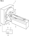
- Fig. 5 shows a principle drawing of a CT device 17.
- the CT device 17 comprises a patient table 18 onto which a patient 19 can be placed.
- an acquisition arrangement comprising an x-ray source 21 and an x-ray detector 22 are rotatably mounted.
- the patient 19 may be placed inside a patient bore 24, in this case for acquiring CCTA data.
- the CT device 17 further comprises a control device 25 having an acquisition unit 26 for controlling the acquisition of CT data, in particular also the CT data set of step S1.
- a control device 25 having an acquisition unit 26 for controlling the acquisition of CT data, in particular also the CT data set of step S1.
- an evaluation device 27 is provided for performing the steps of a method according to the invention.
- the functional structure of the evaluation device 27 is shown in more detail in Fig. 6 .
- the evaluation device 27 comprises a first interface 28 for receiving the CT data set and a second interface 29 for providing the quantitative values, for example, to a display and/or an external storage additional to an internal storage means 30.
- a determination unit 31 the centerline 5 and the inner wall 6 as well as the heart size are determined according to step S2.
- An optional anomaly detection unit 32 may be provided for executing the anomaly detection algorithm (step S3) as well as steps S4, S5.
- the evaluation device 27 further comprises a definition unit 33 for defining the region of interest according to steps S6 to S8 and a calculation unit 34 for calculating the at least one quantitative value from the CT data in the region of interest according to step S9. All functional units 31 to 34 may be implemented on at least one processor (not shown) of the evaluation device 27 and/or as computer program means of a computer program according to the invention.
Landscapes
- Engineering & Computer Science (AREA)
- Health & Medical Sciences (AREA)
- Life Sciences & Earth Sciences (AREA)
- Medical Informatics (AREA)
- Physics & Mathematics (AREA)
- General Health & Medical Sciences (AREA)
- Public Health (AREA)
- Radiology & Medical Imaging (AREA)
- Nuclear Medicine, Radiotherapy & Molecular Imaging (AREA)
- Biomedical Technology (AREA)
- Pathology (AREA)
- Optics & Photonics (AREA)
- Heart & Thoracic Surgery (AREA)
- Molecular Biology (AREA)
- Surgery (AREA)
- Animal Behavior & Ethology (AREA)
- Biophysics (AREA)
- Veterinary Medicine (AREA)
- High Energy & Nuclear Physics (AREA)
- Theoretical Computer Science (AREA)
- Computer Vision & Pattern Recognition (AREA)
- General Physics & Mathematics (AREA)
- Quality & Reliability (AREA)
- Epidemiology (AREA)
- Primary Health Care (AREA)
- Vascular Medicine (AREA)
- Dentistry (AREA)
- Oral & Maxillofacial Surgery (AREA)
- Pulmonology (AREA)
- Human Computer Interaction (AREA)
- Data Mining & Analysis (AREA)
- Databases & Information Systems (AREA)
- Geometry (AREA)
- Bioethics (AREA)
- Physiology (AREA)
- Apparatus For Radiation Diagnosis (AREA)
Priority Applications (1)
| Application Number | Priority Date | Filing Date | Title |
|---|---|---|---|
| EP23192012.5A EP4276751A3 (fr) | 2019-11-28 | 2019-11-28 | Procédé mis en uvre par ordinateur pour l'évaluation d'un ensemble de données de tomodensitométrie concernant un tissu périvasculaire, dispositif d'évaluation, programme informatique et support d'informations lisible électroniquement |
Applications Claiming Priority (2)
| Application Number | Priority Date | Filing Date | Title |
|---|---|---|---|
| EP23192012.5A EP4276751A3 (fr) | 2019-11-28 | 2019-11-28 | Procédé mis en uvre par ordinateur pour l'évaluation d'un ensemble de données de tomodensitométrie concernant un tissu périvasculaire, dispositif d'évaluation, programme informatique et support d'informations lisible électroniquement |
| EP19212263.8A EP3828817B1 (fr) | 2019-11-28 | 2019-11-28 | Procédé mis en uvre par ordinateur pour l'évaluation d'un ensemble de données de tomodensitométrie concernant un tissu périvasculaire, dispositif d'évaluation, programme informatique et support d'informations lisible électroniquement |
Related Parent Applications (1)
| Application Number | Title | Priority Date | Filing Date |
|---|---|---|---|
| EP19212263.8A Division EP3828817B1 (fr) | 2019-11-28 | 2019-11-28 | Procédé mis en uvre par ordinateur pour l'évaluation d'un ensemble de données de tomodensitométrie concernant un tissu périvasculaire, dispositif d'évaluation, programme informatique et support d'informations lisible électroniquement |
Publications (2)
| Publication Number | Publication Date |
|---|---|
| EP4276751A2 true EP4276751A2 (fr) | 2023-11-15 |
| EP4276751A3 EP4276751A3 (fr) | 2024-01-10 |
Family
ID=68732737
Family Applications (2)
| Application Number | Title | Priority Date | Filing Date |
|---|---|---|---|
| EP23192012.5A Pending EP4276751A3 (fr) | 2019-11-28 | 2019-11-28 | Procédé mis en uvre par ordinateur pour l'évaluation d'un ensemble de données de tomodensitométrie concernant un tissu périvasculaire, dispositif d'évaluation, programme informatique et support d'informations lisible électroniquement |
| EP19212263.8A Active EP3828817B1 (fr) | 2019-11-28 | 2019-11-28 | Procédé mis en uvre par ordinateur pour l'évaluation d'un ensemble de données de tomodensitométrie concernant un tissu périvasculaire, dispositif d'évaluation, programme informatique et support d'informations lisible électroniquement |
Family Applications After (1)
| Application Number | Title | Priority Date | Filing Date |
|---|---|---|---|
| EP19212263.8A Active EP3828817B1 (fr) | 2019-11-28 | 2019-11-28 | Procédé mis en uvre par ordinateur pour l'évaluation d'un ensemble de données de tomodensitométrie concernant un tissu périvasculaire, dispositif d'évaluation, programme informatique et support d'informations lisible électroniquement |
Country Status (3)
| Country | Link |
|---|---|
| US (2) | US11803966B2 (fr) |
| EP (2) | EP4276751A3 (fr) |
| CN (1) | CN112967220B (fr) |
Families Citing this family (7)
| Publication number | Priority date | Publication date | Assignee | Title |
|---|---|---|---|---|
| GB2577349B (en) * | 2018-09-18 | 2023-06-14 | Univ Oxford Innovation Ltd | Radiomic signature of adipose |
| GB201820044D0 (en) * | 2018-10-29 | 2019-01-23 | Univ Oxford Innovation Ltd | Radiomic signature of an epicardial region |
| EP3913638A1 (fr) * | 2020-09-30 | 2021-11-24 | Siemens Healthcare GmbH | Procédé et système de traitement de données pour fournir des informations liées à la radiomique |
| CN113744223A (zh) * | 2021-08-26 | 2021-12-03 | 联影智能医疗科技(北京)有限公司 | 血管的风险评估方法、计算机设备和存储介质 |
| CN113763403B (zh) * | 2021-09-07 | 2024-03-08 | 北京深睿博联科技有限责任公司 | 一种冠状动脉血管分割方法及装置 |
| EP4156020A1 (fr) * | 2021-09-22 | 2023-03-29 | Siemens Healthcare GmbH | Procédé mis en uvre par ordinateur pour évaluer un ensemble de données d'angiographie tridimensionnelle, système d'évaluation, programme informatique et support d'informations lisible électroniquement |
| CN115240429B (zh) * | 2022-08-11 | 2023-02-14 | 深圳市城市交通规划设计研究中心股份有限公司 | 一种人车流量统计方法、电子设备及存储介质 |
Citations (1)
| Publication number | Priority date | Publication date | Assignee | Title |
|---|---|---|---|---|
| EP3179917B1 (fr) | 2014-08-15 | 2019-03-06 | Oxford University Innovation Limited | Procédé de caractérisation d'un tissu périvasculaire |
Family Cites Families (11)
| Publication number | Priority date | Publication date | Assignee | Title |
|---|---|---|---|---|
| US8526699B2 (en) * | 2010-03-12 | 2013-09-03 | Siemens Aktiengesellschaft | Method and system for automatic detection and classification of coronary stenoses in cardiac CT volumes |
| US8315812B2 (en) * | 2010-08-12 | 2012-11-20 | Heartflow, Inc. | Method and system for patient-specific modeling of blood flow |
| CN106537392B (zh) * | 2014-04-22 | 2019-07-26 | 西门子保健有限责任公司 | 用于冠状动脉中的血液动力学计算的方法和系统 |
| CN109843161B (zh) * | 2016-09-30 | 2022-07-12 | 皇家飞利浦有限公司 | 用于确定针对狭窄评估的功能指数的装置 |
| CN117918872A (zh) | 2016-10-31 | 2024-04-26 | 牛津大学创新有限公司 | 测定心血管风险的方法 |
| US11471063B2 (en) * | 2016-11-10 | 2022-10-18 | Auburn University | Information processing method, device, and system for evaluating blood vessels |
| EP3585253A1 (fr) | 2017-02-24 | 2020-01-01 | HeartFlow, Inc. | Systèmes et méthodes pour identifier des caractéristiques de flux sanguin pertinentes sur le plan anatomique chez un patient |
| JP7149286B2 (ja) * | 2017-03-24 | 2022-10-06 | パイ メディカル イメージング ビー ヴイ | 機械学習に基づいて血管閉塞を評価する方法およびシステム |
| EP3489893B1 (fr) | 2017-11-22 | 2020-06-24 | Siemens Healthcare GmbH | Procédé et système pour évaluer un paramètre hémodynamique |
| US10699407B2 (en) * | 2018-04-11 | 2020-06-30 | Pie Medical Imaging B.V. | Method and system for assessing vessel obstruction based on machine learning |
| CN110428420B (zh) | 2018-09-05 | 2022-05-17 | 深圳科亚医疗科技有限公司 | 基于患者的冠状动脉ct血管造影图像来确定冠状动脉的流动信息的方法、装置和介质 |
-
2019
- 2019-11-28 EP EP23192012.5A patent/EP4276751A3/fr active Pending
- 2019-11-28 EP EP19212263.8A patent/EP3828817B1/fr active Active
-
2020
- 2020-11-11 US US17/095,057 patent/US11803966B2/en active Active
- 2020-11-27 CN CN202011364498.1A patent/CN112967220B/zh active Active
-
2023
- 2023-08-04 US US18/365,507 patent/US20230386037A1/en active Pending
Patent Citations (1)
| Publication number | Priority date | Publication date | Assignee | Title |
|---|---|---|---|---|
| EP3179917B1 (fr) | 2014-08-15 | 2019-03-06 | Oxford University Innovation Limited | Procédé de caractérisation d'un tissu périvasculaire |
Non-Patent Citations (5)
| Title |
|---|
| ALEXIOS N. ANTONOPOULOS ET AL.: "Detecting human coronary inflammation by imaging perivascular fat", SCIENCE TRANSLATIONAL MEDICINE, vol. 9, 2017, XP055657398, DOI: 10.1126/scitranslmed.aal2658 |
| LUGAUER ET AL., PRECISE LUMEN SEGMENTATION IN CORONARY COMPUTED TOMOGRAPHY ANGIOGRAPHY, September 2014 (2014-09-01) |
| P. LAMBIN ET AL.: "Radiomics: Extracting more information from medical images using advanced feature analysis", EUROPEAN JOURNAL OF CANCER, vol. 48, no. 4, 2012, pages 441 - 446, XP055674485, DOI: 10.1016/j.ejca.2011.11.036 |
| STEPHEN MENDIS ET AL.: "Organizational Update: The World Health Organization Global Status Report on Noncommunicable Diseases 2014", ONE MORE LANDMARK STEP IN THE COMBAT AGAINST STROKE AND VASCULAR DISEASE, 2015 |
| YEFENG ZHENG: "Robust and Accurate Coronary Artery Centerline Extraction in CTA by Combining Model-Driven and Data-Driven Approaches", MICCAI 2013, vol. 8151, 2013, pages 74 - 81, XP047490657, DOI: 10.1007/978-3-642-40760-4_10 |
Also Published As
| Publication number | Publication date |
|---|---|
| EP4276751A3 (fr) | 2024-01-10 |
| CN112967220A (zh) | 2021-06-15 |
| EP3828817C0 (fr) | 2023-08-23 |
| US20230386037A1 (en) | 2023-11-30 |
| CN112967220B (zh) | 2024-05-14 |
| EP3828817A1 (fr) | 2021-06-02 |
| EP3828817B1 (fr) | 2023-08-23 |
| US11803966B2 (en) | 2023-10-31 |
| US20210166389A1 (en) | 2021-06-03 |
Similar Documents
| Publication | Publication Date | Title |
|---|---|---|
| EP3828817B1 (fr) | Procédé mis en uvre par ordinateur pour l'évaluation d'un ensemble de données de tomodensitométrie concernant un tissu périvasculaire, dispositif d'évaluation, programme informatique et support d'informations lisible électroniquement | |
| EP3795070B1 (fr) | Systèmes et procédés d'estimation du diamètre de lumière saine et quantification d'une sténose dans des artères coronaires | |
| US7940977B2 (en) | Method and system for automatic analysis of blood vessel structures to identify calcium or soft plaque pathologies | |
| US7940970B2 (en) | Method and system for automatic quality control used in computerized analysis of CT angiography | |
| US7860283B2 (en) | Method and system for the presentation of blood vessel structures and identified pathologies | |
| NL2009379C2 (en) | System and method for blood vessel stenosis visualization and navigation. | |
| US9842401B2 (en) | Segmentation apparatus for interactively segmenting blood vessels in angiographic image data | |
| EP2849631B1 (fr) | Détermination de valeur de réserve de débit fractionnaire (ffr) pour sténose de vaisseau | |
| US7873194B2 (en) | Method and system for automatic analysis of blood vessel structures and pathologies in support of a triple rule-out procedure | |
| JP6484760B2 (ja) | 非侵襲的血流予備量比(ffr)に対する側副血流モデル化 | |
| US20080101674A1 (en) | Method and system for automatic analysis of blood vessel structures and pathologies | |
| US20230172451A1 (en) | Medical image visualization apparatus and method for diagnosis of aorta | |
| US20220215541A1 (en) | Method, device and computer-readable medium for automatically classifying coronary lesion according to cad-rads classification by a deep neural network | |
| US20220319004A1 (en) | Automatic vessel analysis from 2d images | |
| US11877880B2 (en) | Method and apparatus for calculating coronary artery calcium score | |
| JP6981807B2 (ja) | 医用情報処理装置、x線ct装置、医用情報処理プログラム、医用情報処理方法及び医用情報処理システム | |
| CN110612063B (zh) | 标准化冠状动脉疾病度量 | |
| Denzinger et al. | Coronary plaque analysis for CT angiography clinical research | |
| US20220335612A1 (en) | Automated analysis of image data to determine fractional flow reserve | |
| US20240127435A1 (en) | System and method for detecting and quantifying a plaque/stenosis in a vascular ultrasound scan data | |
| CN113313687A (zh) | 一种基于能谱ct的钙化分数计算的方法和装置 | |
| Sedghi Gamechi | Automatic Quantification of the Aorta and Pulmonary Artery in Chest CT: methods and validation in lung screening | |
| CN117241735A (zh) | 确定用于支架的端点位置 | |
| KR20240057147A (ko) | 기계 학습 모델에 기초하여 혈관을 분석하는 방법 및 장치 | |
| IL269223B2 (en) | Automatic analysis of information from images to determine ffr |
Legal Events
| Date | Code | Title | Description |
|---|---|---|---|
| PUAI | Public reference made under article 153(3) epc to a published international application that has entered the european phase |
Free format text: ORIGINAL CODE: 0009012 |
|
| STAA | Information on the status of an ep patent application or granted ep patent |
Free format text: STATUS: THE APPLICATION HAS BEEN PUBLISHED |
|
| AC | Divisional application: reference to earlier application |
Ref document number: 3828817 Country of ref document: EP Kind code of ref document: P |
|
| AK | Designated contracting states |
Kind code of ref document: A2 Designated state(s): AL AT BE BG CH CY CZ DE DK EE ES FI FR GB GR HR HU IE IS IT LI LT LU LV MC MK MT NL NO PL PT RO RS SE SI SK SM TR |
|
| PUAL | Search report despatched |
Free format text: ORIGINAL CODE: 0009013 |
|
| AK | Designated contracting states |
Kind code of ref document: A3 Designated state(s): AL AT BE BG CH CY CZ DE DK EE ES FI FR GB GR HR HU IE IS IT LI LT LU LV MC MK MT NL NO PL PT RO RS SE SI SK SM TR |
|
| RIC1 | Information provided on ipc code assigned before grant |
Ipc: G06T 7/00 20170101AFI20231206BHEP |
|
| RAP1 | Party data changed (applicant data changed or rights of an application transferred) |
Owner name: SIEMENS HEALTHINEERS AG |