EP4268823A1 - Verbindung zum behandeln von nervenentzündungen - Google Patents
Verbindung zum behandeln von nervenentzündungen Download PDFInfo
- Publication number
- EP4268823A1 EP4268823A1 EP22169820.2A EP22169820A EP4268823A1 EP 4268823 A1 EP4268823 A1 EP 4268823A1 EP 22169820 A EP22169820 A EP 22169820A EP 4268823 A1 EP4268823 A1 EP 4268823A1
- Authority
- EP
- European Patent Office
- Prior art keywords
- compound
- multiple sclerosis
- methyltransferase
- unc0642
- neuronal
- Prior art date
- Legal status (The legal status is an assumption and is not a legal conclusion. Google has not performed a legal analysis and makes no representation as to the accuracy of the status listed.)
- Pending
Links
- 150000001875 compounds Chemical class 0.000 title claims abstract description 127
- 208000036110 Neuroinflammatory disease Diseases 0.000 title description 7
- 230000003959 neuroinflammation Effects 0.000 title description 6
- 201000006417 multiple sclerosis Diseases 0.000 claims abstract description 83
- 238000000034 method Methods 0.000 claims abstract description 41
- 230000004806 ferroptosis Effects 0.000 claims abstract description 35
- 230000002401 inhibitory effect Effects 0.000 claims abstract description 26
- 239000008194 pharmaceutical composition Substances 0.000 claims abstract description 18
- 238000000338 in vitro Methods 0.000 claims abstract description 10
- RNAMYOYQYRYFQY-UHFFFAOYSA-N 2-(4,4-difluoropiperidin-1-yl)-6-methoxy-n-(1-propan-2-ylpiperidin-4-yl)-7-(3-pyrrolidin-1-ylpropoxy)quinazolin-4-amine Chemical compound N1=C(N2CCC(F)(F)CC2)N=C2C=C(OCCCN3CCCC3)C(OC)=CC2=C1NC1CCN(C(C)C)CC1 RNAMYOYQYRYFQY-UHFFFAOYSA-N 0.000 claims description 93
- 238000011282 treatment Methods 0.000 claims description 24
- 230000037396 body weight Effects 0.000 claims description 19
- 108091016367 Histone-lysine N-methyltransferase EHMT2 Proteins 0.000 claims description 14
- 108020004999 messenger RNA Proteins 0.000 claims description 13
- 102100035042 Histone-lysine N-methyltransferase EHMT2 Human genes 0.000 claims description 12
- 230000007423 decrease Effects 0.000 claims description 9
- 238000013519 translation Methods 0.000 claims description 9
- 108010033040 Histones Proteins 0.000 claims description 8
- 239000000203 mixture Substances 0.000 claims description 8
- 238000013518 transcription Methods 0.000 claims description 8
- 230000035897 transcription Effects 0.000 claims description 8
- KDXKERNSBIXSRK-UHFFFAOYSA-N Lysine Natural products NCCCCC(N)C(O)=O KDXKERNSBIXSRK-UHFFFAOYSA-N 0.000 claims description 5
- 239000004472 Lysine Substances 0.000 claims description 5
- 208000007400 Relapsing-Remitting Multiple Sclerosis Diseases 0.000 claims description 5
- 150000003384 small molecules Chemical class 0.000 claims description 4
- QOECJCJVIMVJGX-UHFFFAOYSA-N 2-cyclohexyl-6-methoxy-N-(1-propan-2-yl-4-piperidinyl)-7-[3-(1-pyrrolidinyl)propoxy]-4-quinazolinamine Chemical compound N1=C(C2CCCCC2)N=C2C=C(OCCCN3CCCC3)C(OC)=CC2=C1NC1CCN(C(C)C)CC1 QOECJCJVIMVJGX-UHFFFAOYSA-N 0.000 claims description 3
- OSXFATOLZGZLSK-UHFFFAOYSA-N 6,7-dimethoxy-2-(4-methyl-1,4-diazepan-1-yl)-N-[1-(phenylmethyl)-4-piperidinyl]-4-quinazolinamine Chemical compound C=12C=C(OC)C(OC)=CC2=NC(N2CCN(C)CCC2)=NC=1NC(CC1)CCN1CC1=CC=CC=C1 OSXFATOLZGZLSK-UHFFFAOYSA-N 0.000 claims description 3
- 239000000546 pharmaceutical excipient Substances 0.000 claims description 3
- 239000003085 diluting agent Substances 0.000 claims description 2
- 108010036115 Histone Methyltransferases Proteins 0.000 abstract description 14
- 102000011787 Histone Methyltransferases Human genes 0.000 abstract description 13
- 101100225547 Mus musculus Ehmt2 gene Proteins 0.000 description 149
- 201000002491 encephalomyelitis Diseases 0.000 description 69
- WHUUTDBJXJRKMK-VKHMYHEASA-N L-glutamic acid Chemical compound OC(=O)[C@@H](N)CCC(O)=O WHUUTDBJXJRKMK-VKHMYHEASA-N 0.000 description 56
- 230000000694 effects Effects 0.000 description 45
- 230000001537 neural effect Effects 0.000 description 45
- 210000002569 neuron Anatomy 0.000 description 41
- 229930195712 glutamate Natural products 0.000 description 39
- 241000699670 Mus sp. Species 0.000 description 35
- 210000004027 cell Anatomy 0.000 description 34
- IAZDPXIOMUYVGZ-UHFFFAOYSA-N Dimethylsulphoxide Chemical compound CS(C)=O IAZDPXIOMUYVGZ-UHFFFAOYSA-N 0.000 description 33
- BKQFRNYHFIQEKN-UHFFFAOYSA-N erastin Chemical compound CCOC1=CC=CC=C1N1C(=O)C2=CC=CC=C2N=C1C(C)N1CCN(C(=O)COC=2C=CC(Cl)=CC=2)CC1 BKQFRNYHFIQEKN-UHFFFAOYSA-N 0.000 description 32
- 238000002474 experimental method Methods 0.000 description 29
- RWSXRVCMGQZWBV-WDSKDSINSA-N glutathione Chemical compound OC(=O)[C@@H](N)CCC(=O)N[C@@H](CS)C(=O)NCC(O)=O RWSXRVCMGQZWBV-WDSKDSINSA-N 0.000 description 28
- 108090000623 proteins and genes Proteins 0.000 description 26
- 108060004795 Methyltransferase Proteins 0.000 description 25
- 210000000278 spinal cord Anatomy 0.000 description 24
- 230000004770 neurodegeneration Effects 0.000 description 23
- 230000006870 function Effects 0.000 description 22
- 230000014509 gene expression Effects 0.000 description 22
- 108090000765 processed proteins & peptides Proteins 0.000 description 22
- 102000016397 Methyltransferase Human genes 0.000 description 21
- 230000006698 induction Effects 0.000 description 21
- 102000004196 processed proteins & peptides Human genes 0.000 description 20
- 238000011002 quantification Methods 0.000 description 20
- 230000005764 inhibitory process Effects 0.000 description 18
- 101001092197 Homo sapiens RNA binding protein fox-1 homolog 3 Proteins 0.000 description 17
- 102100035530 RNA binding protein fox-1 homolog 3 Human genes 0.000 description 17
- 206010061218 Inflammation Diseases 0.000 description 16
- 210000003169 central nervous system Anatomy 0.000 description 16
- 230000004054 inflammatory process Effects 0.000 description 16
- 229920001184 polypeptide Polymers 0.000 description 16
- 238000000692 Student's t-test Methods 0.000 description 15
- 238000010162 Tukey test Methods 0.000 description 15
- 108010024636 Glutathione Proteins 0.000 description 14
- 229960003180 glutathione Drugs 0.000 description 14
- 230000004044 response Effects 0.000 description 14
- 208000037265 diseases, disorders, signs and symptoms Diseases 0.000 description 13
- 230000001973 epigenetic effect Effects 0.000 description 13
- 210000004940 nucleus Anatomy 0.000 description 13
- 238000001543 one-way ANOVA Methods 0.000 description 13
- 230000036542 oxidative stress Effects 0.000 description 13
- 241001465754 Metazoa Species 0.000 description 12
- 241000699666 Mus <mouse, genus> Species 0.000 description 12
- 238000005516 engineering process Methods 0.000 description 12
- 210000000130 stem cell Anatomy 0.000 description 12
- OYPRJOBELJOOCE-UHFFFAOYSA-N Calcium Chemical compound [Ca] OYPRJOBELJOOCE-UHFFFAOYSA-N 0.000 description 11
- 125000003275 alpha amino acid group Chemical group 0.000 description 11
- 150000001413 amino acids Chemical class 0.000 description 11
- 238000003556 assay Methods 0.000 description 11
- 229910052791 calcium Inorganic materials 0.000 description 11
- 239000011575 calcium Substances 0.000 description 11
- 201000010099 disease Diseases 0.000 description 11
- 239000002609 medium Substances 0.000 description 11
- MHAJPDPJQMAIIY-UHFFFAOYSA-N Hydrogen peroxide Chemical compound OO MHAJPDPJQMAIIY-UHFFFAOYSA-N 0.000 description 10
- 210000004556 brain Anatomy 0.000 description 10
- 230000003492 excitotoxic effect Effects 0.000 description 10
- 231100000063 excitotoxicity Toxicity 0.000 description 10
- 101000829725 Homo sapiens Phospholipid hydroperoxide glutathione peroxidase Proteins 0.000 description 9
- 102100023410 Phospholipid hydroperoxide glutathione peroxidase Human genes 0.000 description 9
- 230000006378 damage Effects 0.000 description 9
- LOKCTEFSRHRXRJ-UHFFFAOYSA-I dipotassium trisodium dihydrogen phosphate hydrogen phosphate dichloride Chemical compound P(=O)(O)(O)[O-].[K+].P(=O)(O)([O-])[O-].[Na+].[Na+].[Cl-].[K+].[Cl-].[Na+] LOKCTEFSRHRXRJ-UHFFFAOYSA-I 0.000 description 9
- 239000002953 phosphate buffered saline Substances 0.000 description 9
- 238000010186 staining Methods 0.000 description 9
- 210000001519 tissue Anatomy 0.000 description 9
- 239000003153 chemical reaction reagent Substances 0.000 description 8
- 230000002068 genetic effect Effects 0.000 description 8
- 238000001727 in vivo Methods 0.000 description 8
- 238000001802 infusion Methods 0.000 description 8
- 239000003112 inhibitor Substances 0.000 description 8
- 210000002161 motor neuron Anatomy 0.000 description 8
- 238000004458 analytical method Methods 0.000 description 7
- 230000002555 anti-neurodegenerative effect Effects 0.000 description 7
- 230000001964 calcium overload Effects 0.000 description 7
- 238000004113 cell culture Methods 0.000 description 7
- 230000001684 chronic effect Effects 0.000 description 7
- 230000001054 cortical effect Effects 0.000 description 7
- 238000010166 immunofluorescence Methods 0.000 description 7
- 238000011534 incubation Methods 0.000 description 7
- 230000002757 inflammatory effect Effects 0.000 description 7
- 230000000144 pharmacologic effect Effects 0.000 description 7
- 238000012216 screening Methods 0.000 description 7
- 230000035882 stress Effects 0.000 description 7
- 230000003827 upregulation Effects 0.000 description 7
- JVJFIQYAHPMBBX-UHFFFAOYSA-N 4-hydroxynonenal Chemical compound CCCCCC(O)C=CC=O JVJFIQYAHPMBBX-UHFFFAOYSA-N 0.000 description 6
- 102000004190 Enzymes Human genes 0.000 description 6
- 108090000790 Enzymes Proteins 0.000 description 6
- 101150005894 GCLC gene Proteins 0.000 description 6
- 101150041639 GPX4 gene Proteins 0.000 description 6
- 238000000585 Mann–Whitney U test Methods 0.000 description 6
- 229940045835 RSL3 Drugs 0.000 description 6
- 239000000872 buffer Substances 0.000 description 6
- 230000003833 cell viability Effects 0.000 description 6
- 229940088598 enzyme Drugs 0.000 description 6
- 230000003902 lesion Effects 0.000 description 6
- TXJZRSRTYPUYRW-NQIIRXRSSA-N methyl (1s,3r)-2-(2-chloroacetyl)-1-(4-methoxycarbonylphenyl)-1,3,4,9-tetrahydropyrido[3,4-b]indole-3-carboxylate Chemical compound C1([C@H]2C3=C(C4=CC=CC=C4N3)C[C@@H](N2C(=O)CCl)C(=O)OC)=CC=C(C(=O)OC)C=C1 TXJZRSRTYPUYRW-NQIIRXRSSA-N 0.000 description 6
- 230000037361 pathway Effects 0.000 description 6
- 238000010149 post-hoc-test Methods 0.000 description 6
- 230000009467 reduction Effects 0.000 description 6
- 230000002829 reductive effect Effects 0.000 description 6
- 239000000523 sample Substances 0.000 description 6
- 231100000331 toxic Toxicity 0.000 description 6
- 230000002588 toxic effect Effects 0.000 description 6
- 108091032973 (ribonucleotides)n+m Proteins 0.000 description 5
- -1 P = 0.0043 Chemical compound 0.000 description 5
- 229930040373 Paraformaldehyde Natural products 0.000 description 5
- 229940121363 anti-inflammatory agent Drugs 0.000 description 5
- 239000002260 anti-inflammatory agent Substances 0.000 description 5
- 239000003963 antioxidant agent Substances 0.000 description 5
- 230000003078 antioxidant effect Effects 0.000 description 5
- 235000006708 antioxidants Nutrition 0.000 description 5
- 230000030833 cell death Effects 0.000 description 5
- 230000006721 cell death pathway Effects 0.000 description 5
- 210000003618 cortical neuron Anatomy 0.000 description 5
- 230000001086 cytosolic effect Effects 0.000 description 5
- 230000003247 decreasing effect Effects 0.000 description 5
- 238000000684 flow cytometry Methods 0.000 description 5
- 239000012634 fragment Substances 0.000 description 5
- 238000002347 injection Methods 0.000 description 5
- 239000007924 injection Substances 0.000 description 5
- 238000001990 intravenous administration Methods 0.000 description 5
- 125000002496 methyl group Chemical group [H]C([H])([H])* 0.000 description 5
- 230000006576 neuronal survival Effects 0.000 description 5
- 229920002866 paraformaldehyde Polymers 0.000 description 5
- 238000003753 real-time PCR Methods 0.000 description 5
- 230000000638 stimulation Effects 0.000 description 5
- 238000007492 two-way ANOVA Methods 0.000 description 5
- NEZONWMXZKDMKF-JTQLQIEISA-N Alkannin Chemical compound C1=CC(O)=C2C(=O)C([C@@H](O)CC=C(C)C)=CC(=O)C2=C1O NEZONWMXZKDMKF-JTQLQIEISA-N 0.000 description 4
- CIWBSHSKHKDKBQ-JLAZNSOCSA-N Ascorbic acid Chemical compound OC[C@H](O)[C@H]1OC(=O)C(O)=C1O CIWBSHSKHKDKBQ-JLAZNSOCSA-N 0.000 description 4
- 108010037462 Cyclooxygenase 2 Proteins 0.000 description 4
- 241001071917 Lithospermum Species 0.000 description 4
- 102000004868 N-Methyl-D-Aspartate Receptors Human genes 0.000 description 4
- 108090001041 N-Methyl-D-Aspartate Receptors Proteins 0.000 description 4
- 102100038280 Prostaglandin G/H synthase 2 Human genes 0.000 description 4
- 238000011529 RT qPCR Methods 0.000 description 4
- 230000004913 activation Effects 0.000 description 4
- 230000001154 acute effect Effects 0.000 description 4
- UNNKKUDWEASWDN-UHFFFAOYSA-N alkannin Natural products CC(=CCC(O)c1cc(O)c2C(=O)C=CC(=O)c2c1O)C UNNKKUDWEASWDN-UHFFFAOYSA-N 0.000 description 4
- 230000008499 blood brain barrier function Effects 0.000 description 4
- 210000001218 blood-brain barrier Anatomy 0.000 description 4
- 230000008859 change Effects 0.000 description 4
- 238000004624 confocal microscopy Methods 0.000 description 4
- 230000004069 differentiation Effects 0.000 description 4
- UJHBVMHOBZBWMX-UHFFFAOYSA-N ferrostatin-1 Chemical compound NC1=CC(C(=O)OCC)=CC=C1NC1CCCCC1 UJHBVMHOBZBWMX-UHFFFAOYSA-N 0.000 description 4
- 210000002865 immune cell Anatomy 0.000 description 4
- 238000002649 immunization Methods 0.000 description 4
- 230000003053 immunization Effects 0.000 description 4
- 238000003365 immunocytochemistry Methods 0.000 description 4
- 238000012744 immunostaining Methods 0.000 description 4
- 238000002955 isolation Methods 0.000 description 4
- 108010082117 matrigel Proteins 0.000 description 4
- 230000001404 mediated effect Effects 0.000 description 4
- 230000004048 modification Effects 0.000 description 4
- 238000012986 modification Methods 0.000 description 4
- 239000003607 modifier Substances 0.000 description 4
- 230000000324 neuroprotective effect Effects 0.000 description 4
- 102000004169 proteins and genes Human genes 0.000 description 4
- 238000012360 testing method Methods 0.000 description 4
- XLYOFNOQVPJJNP-UHFFFAOYSA-N water Substances O XLYOFNOQVPJJNP-UHFFFAOYSA-N 0.000 description 4
- 230000003442 weekly effect Effects 0.000 description 4
- 102100035300 Cystine/glutamate transporter Human genes 0.000 description 3
- 101150010771 Ehmt2 gene Proteins 0.000 description 3
- 102100039696 Glutamate-cysteine ligase catalytic subunit Human genes 0.000 description 3
- PEDCQBHIVMGVHV-UHFFFAOYSA-N Glycerine Chemical compound OCC(O)CO PEDCQBHIVMGVHV-UHFFFAOYSA-N 0.000 description 3
- OKKJLVBELUTLKV-UHFFFAOYSA-N Methanol Chemical compound OC OKKJLVBELUTLKV-UHFFFAOYSA-N 0.000 description 3
- 108010029485 Protein Isoforms Proteins 0.000 description 3
- 102000001708 Protein Isoforms Human genes 0.000 description 3
- 108091006241 SLC7A11 Proteins 0.000 description 3
- 108060008682 Tumor Necrosis Factor Proteins 0.000 description 3
- 102000000852 Tumor Necrosis Factor-alpha Human genes 0.000 description 3
- 108010076089 accutase Proteins 0.000 description 3
- 230000006907 apoptotic process Effects 0.000 description 3
- 238000013459 approach Methods 0.000 description 3
- 238000003570 cell viability assay Methods 0.000 description 3
- 239000003795 chemical substances by application Substances 0.000 description 3
- 238000007405 data analysis Methods 0.000 description 3
- 230000001419 dependent effect Effects 0.000 description 3
- 238000001514 detection method Methods 0.000 description 3
- 238000003384 imaging method Methods 0.000 description 3
- 238000003364 immunohistochemistry Methods 0.000 description 3
- 238000007901 in situ hybridization Methods 0.000 description 3
- 238000004020 luminiscence type Methods 0.000 description 3
- 238000004519 manufacturing process Methods 0.000 description 3
- 238000005259 measurement Methods 0.000 description 3
- 230000011987 methylation Effects 0.000 description 3
- 238000007069 methylation reaction Methods 0.000 description 3
- 230000021597 necroptosis Effects 0.000 description 3
- 230000016273 neuron death Effects 0.000 description 3
- 230000009038 pharmacological inhibition Effects 0.000 description 3
- 238000003752 polymerase chain reaction Methods 0.000 description 3
- 230000003389 potentiating effect Effects 0.000 description 3
- 230000001718 repressive effect Effects 0.000 description 3
- 230000003938 response to stress Effects 0.000 description 3
- 239000000243 solution Substances 0.000 description 3
- 238000010561 standard procedure Methods 0.000 description 3
- 230000001629 suppression Effects 0.000 description 3
- 230000037426 transcriptional repression Effects 0.000 description 3
- 239000003981 vehicle Substances 0.000 description 3
- 238000005406 washing Methods 0.000 description 3
- MZOFCQQQCNRIBI-VMXHOPILSA-N (3s)-4-[[(2s)-1-[[(2s)-1-[[(1s)-1-carboxy-2-hydroxyethyl]amino]-4-methyl-1-oxopentan-2-yl]amino]-5-(diaminomethylideneamino)-1-oxopentan-2-yl]amino]-3-[[2-[[(2s)-2,6-diaminohexanoyl]amino]acetyl]amino]-4-oxobutanoic acid Chemical compound OC[C@@H](C(O)=O)NC(=O)[C@H](CC(C)C)NC(=O)[C@H](CCCN=C(N)N)NC(=O)[C@H](CC(O)=O)NC(=O)CNC(=O)[C@@H](N)CCCCN MZOFCQQQCNRIBI-VMXHOPILSA-N 0.000 description 2
- IJGRMHOSHXDMSA-UHFFFAOYSA-N Atomic nitrogen Chemical compound N#N IJGRMHOSHXDMSA-UHFFFAOYSA-N 0.000 description 2
- 208000032116 Autoimmune Experimental Encephalomyelitis Diseases 0.000 description 2
- 101100173542 Caenorhabditis elegans fer-1 gene Proteins 0.000 description 2
- 102000053642 Catalytic RNA Human genes 0.000 description 2
- 108090000994 Catalytic RNA Proteins 0.000 description 2
- UHDGCWIWMRVCDJ-CCXZUQQUSA-N Cytarabine Chemical compound O=C1N=C(N)C=CN1[C@H]1[C@@H](O)[C@H](O)[C@@H](CO)O1 UHDGCWIWMRVCDJ-CCXZUQQUSA-N 0.000 description 2
- 108010024491 DNA Methyltransferase 3A Proteins 0.000 description 2
- 239000012591 Dulbecco’s Phosphate Buffered Saline Substances 0.000 description 2
- 238000002965 ELISA Methods 0.000 description 2
- 241000283074 Equus asinus Species 0.000 description 2
- WZUVPPKBWHMQCE-UHFFFAOYSA-N Haematoxylin Chemical compound C12=CC(O)=C(O)C=C2CC2(O)C1C1=CC=C(O)C(O)=C1OC2 WZUVPPKBWHMQCE-UHFFFAOYSA-N 0.000 description 2
- 102100033636 Histone H3.2 Human genes 0.000 description 2
- 101001034527 Homo sapiens Glutamate-cysteine ligase catalytic subunit Proteins 0.000 description 2
- ZDXPYRJPNDTMRX-VKHMYHEASA-N L-glutamine Chemical compound OC(=O)[C@@H](N)CCC(N)=O ZDXPYRJPNDTMRX-VKHMYHEASA-N 0.000 description 2
- 229930182816 L-glutamine Natural products 0.000 description 2
- 108010085895 Laminin Proteins 0.000 description 2
- TWRXJAOTZQYOKJ-UHFFFAOYSA-L Magnesium chloride Chemical compound [Mg+2].[Cl-].[Cl-] TWRXJAOTZQYOKJ-UHFFFAOYSA-L 0.000 description 2
- 206010027925 Monoparesis Diseases 0.000 description 2
- 241001529936 Murinae Species 0.000 description 2
- 208000012902 Nervous system disease Diseases 0.000 description 2
- 208000025966 Neurological disease Diseases 0.000 description 2
- ISWSIDIOOBJBQZ-UHFFFAOYSA-N Phenol Chemical compound OC1=CC=CC=C1 ISWSIDIOOBJBQZ-UHFFFAOYSA-N 0.000 description 2
- 108091023040 Transcription factor Proteins 0.000 description 2
- 102000040945 Transcription factor Human genes 0.000 description 2
- 229920004890 Triton X-100 Polymers 0.000 description 2
- 239000013504 Triton X-100 Substances 0.000 description 2
- 208000027418 Wounds and injury Diseases 0.000 description 2
- 230000009471 action Effects 0.000 description 2
- 238000010171 animal model Methods 0.000 description 2
- 210000004960 anterior grey column Anatomy 0.000 description 2
- 230000000692 anti-sense effect Effects 0.000 description 2
- 238000003782 apoptosis assay Methods 0.000 description 2
- 229960005070 ascorbic acid Drugs 0.000 description 2
- 238000003149 assay kit Methods 0.000 description 2
- 230000008901 benefit Effects 0.000 description 2
- 230000033228 biological regulation Effects 0.000 description 2
- 230000015572 biosynthetic process Effects 0.000 description 2
- 230000000903 blocking effect Effects 0.000 description 2
- 210000005013 brain tissue Anatomy 0.000 description 2
- 230000001201 calcium accumulation Effects 0.000 description 2
- 230000009460 calcium influx Effects 0.000 description 2
- 239000000969 carrier Substances 0.000 description 2
- 230000003197 catalytic effect Effects 0.000 description 2
- 230000001413 cellular effect Effects 0.000 description 2
- OSASVXMJTNOKOY-UHFFFAOYSA-N chlorobutanol Chemical compound CC(C)(O)C(Cl)(Cl)Cl OSASVXMJTNOKOY-UHFFFAOYSA-N 0.000 description 2
- 230000019771 cognition Effects 0.000 description 2
- 238000004590 computer program Methods 0.000 description 2
- 230000009537 cortical lesion Effects 0.000 description 2
- 230000001186 cumulative effect Effects 0.000 description 2
- 229960000684 cytarabine Drugs 0.000 description 2
- 230000006735 deficit Effects 0.000 description 2
- 125000000118 dimethyl group Chemical group [H]C([H])([H])* 0.000 description 2
- 239000006185 dispersion Substances 0.000 description 2
- 239000003814 drug Substances 0.000 description 2
- 210000002242 embryoid body Anatomy 0.000 description 2
- 230000007717 exclusion Effects 0.000 description 2
- 208000012997 experimental autoimmune encephalomyelitis Diseases 0.000 description 2
- 210000001723 extracellular space Anatomy 0.000 description 2
- 238000001943 fluorescence-activated cell sorting Methods 0.000 description 2
- 230000030279 gene silencing Effects 0.000 description 2
- 230000009368 gene silencing by RNA Effects 0.000 description 2
- 210000004884 grey matter Anatomy 0.000 description 2
- 238000013537 high throughput screening Methods 0.000 description 2
- 208000010726 hind limb paralysis Diseases 0.000 description 2
- 210000005260 human cell Anatomy 0.000 description 2
- 210000004263 induced pluripotent stem cell Anatomy 0.000 description 2
- 230000001939 inductive effect Effects 0.000 description 2
- 230000008595 infiltration Effects 0.000 description 2
- 238000001764 infiltration Methods 0.000 description 2
- 208000014674 injury Diseases 0.000 description 2
- PGHMRUGBZOYCAA-ADZNBVRBSA-N ionomycin Chemical compound O1[C@H](C[C@H](O)[C@H](C)[C@H](O)[C@H](C)/C=C/C[C@@H](C)C[C@@H](C)C(/O)=C/C(=O)[C@@H](C)C[C@@H](C)C[C@@H](CCC(O)=O)C)CC[C@@]1(C)[C@@H]1O[C@](C)([C@@H](C)O)CC1 PGHMRUGBZOYCAA-ADZNBVRBSA-N 0.000 description 2
- PGHMRUGBZOYCAA-UHFFFAOYSA-N ionomycin Natural products O1C(CC(O)C(C)C(O)C(C)C=CCC(C)CC(C)C(O)=CC(=O)C(C)CC(C)CC(CCC(O)=O)C)CCC1(C)C1OC(C)(C(C)O)CC1 PGHMRUGBZOYCAA-UHFFFAOYSA-N 0.000 description 2
- 210000000274 microglia Anatomy 0.000 description 2
- 230000004065 mitochondrial dysfunction Effects 0.000 description 2
- 238000010172 mouse model Methods 0.000 description 2
- 208000015122 neurodegenerative disease Diseases 0.000 description 2
- 238000010606 normalization Methods 0.000 description 2
- 239000012188 paraffin wax Substances 0.000 description 2
- 230000001575 pathological effect Effects 0.000 description 2
- 239000008188 pellet Substances 0.000 description 2
- 108091033319 polynucleotide Proteins 0.000 description 2
- 102000040430 polynucleotide Human genes 0.000 description 2
- 239000002157 polynucleotide Substances 0.000 description 2
- 239000000843 powder Substances 0.000 description 2
- 238000002203 pretreatment Methods 0.000 description 2
- 210000002243 primary neuron Anatomy 0.000 description 2
- 206010063401 primary progressive multiple sclerosis Diseases 0.000 description 2
- 230000005522 programmed cell death Effects 0.000 description 2
- 230000000750 progressive effect Effects 0.000 description 2
- 230000007101 progressive neurodegeneration Effects 0.000 description 2
- 239000003642 reactive oxygen metabolite Substances 0.000 description 2
- 230000022532 regulation of transcription, DNA-dependent Effects 0.000 description 2
- 108091092562 ribozyme Proteins 0.000 description 2
- 150000003839 salts Chemical class 0.000 description 2
- 201000008628 secondary progressive multiple sclerosis Diseases 0.000 description 2
- 210000002966 serum Anatomy 0.000 description 2
- 238000007619 statistical method Methods 0.000 description 2
- UCSJYZPVAKXKNQ-HZYVHMACSA-N streptomycin Chemical compound CN[C@H]1[C@H](O)[C@@H](O)[C@H](CO)O[C@H]1O[C@@H]1[C@](C=O)(O)[C@H](C)O[C@H]1O[C@@H]1[C@@H](NC(N)=N)[C@H](O)[C@@H](NC(N)=N)[C@H](O)[C@H]1O UCSJYZPVAKXKNQ-HZYVHMACSA-N 0.000 description 2
- 239000000126 substance Substances 0.000 description 2
- 238000003786 synthesis reaction Methods 0.000 description 2
- 230000035899 viability Effects 0.000 description 2
- OZFAFGSSMRRTDW-UHFFFAOYSA-N (2,4-dichlorophenyl) benzenesulfonate Chemical compound ClC1=CC(Cl)=CC=C1OS(=O)(=O)C1=CC=CC=C1 OZFAFGSSMRRTDW-UHFFFAOYSA-N 0.000 description 1
- UFPFGVNKHCLJJO-SSKFGXFMSA-N (2s)-n-[(1s)-1-cyclohexyl-2-[(2s)-2-[4-(4-fluorobenzoyl)-1,3-thiazol-2-yl]pyrrolidin-1-yl]-2-oxoethyl]-2-(methylamino)propanamide Chemical compound C1([C@H](NC(=O)[C@H](C)NC)C(=O)N2[C@@H](CCC2)C=2SC=C(N=2)C(=O)C=2C=CC(F)=CC=2)CCCCC1 UFPFGVNKHCLJJO-SSKFGXFMSA-N 0.000 description 1
- KIHYPELVXPAIDH-HNSNBQBZSA-N 1-[[4-[(e)-n-[[4-cyclohexyl-3-(trifluoromethyl)phenyl]methoxy]-c-methylcarbonimidoyl]-2-ethylphenyl]methyl]azetidine-3-carboxylic acid Chemical compound CCC1=CC(C(\C)=N\OCC=2C=C(C(C3CCCCC3)=CC=2)C(F)(F)F)=CC=C1CN1CC(C(O)=O)C1 KIHYPELVXPAIDH-HNSNBQBZSA-N 0.000 description 1
- PRDFBSVERLRRMY-UHFFFAOYSA-N 2'-(4-ethoxyphenyl)-5-(4-methylpiperazin-1-yl)-2,5'-bibenzimidazole Chemical compound C1=CC(OCC)=CC=C1C1=NC2=CC=C(C=3NC4=CC(=CC=C4N=3)N3CCN(C)CC3)C=C2N1 PRDFBSVERLRRMY-UHFFFAOYSA-N 0.000 description 1
- VLEIUWBSEKKKFX-UHFFFAOYSA-N 2-amino-2-(hydroxymethyl)propane-1,3-diol;2-[2-[bis(carboxymethyl)amino]ethyl-(carboxymethyl)amino]acetic acid Chemical compound OCC(N)(CO)CO.OC(=O)CN(CC(O)=O)CCN(CC(O)=O)CC(O)=O VLEIUWBSEKKKFX-UHFFFAOYSA-N 0.000 description 1
- UCGWYCMPZXDHNR-UHFFFAOYSA-N 2-benzamido-1-(3-phenylpropyl)-5-benzimidazolecarboxylic acid methyl ester Chemical compound C=1C=CC=CC=1C(=O)NC1=NC2=CC(C(=O)OC)=CC=C2N1CCCC1=CC=CC=C1 UCGWYCMPZXDHNR-UHFFFAOYSA-N 0.000 description 1
- WPXMEOBILYVKBC-UHFFFAOYSA-N 2-benzamido-1-(3-phenylpropyl)benzimidazole-5-carboxylic acid Chemical compound C=1C=CC=CC=1C(=O)NC1=NC2=CC(C(=O)O)=CC=C2N1CCCC1=CC=CC=C1 WPXMEOBILYVKBC-UHFFFAOYSA-N 0.000 description 1
- HSTOKWSFWGCZMH-UHFFFAOYSA-N 3,3'-diaminobenzidine Chemical compound C1=C(N)C(N)=CC=C1C1=CC=C(N)C(N)=C1 HSTOKWSFWGCZMH-UHFFFAOYSA-N 0.000 description 1
- PMPKMTDYPOAEEH-UHFFFAOYSA-N 3-[4-[[4-[hydroxy-bis[4-(trifluoromethyl)phenyl]methyl]piperidin-1-yl]methyl]phenyl]-1,1-dimethylurea Chemical compound C1=CC(NC(=O)N(C)C)=CC=C1CN1CCC(C(O)(C=2C=CC(=CC=2)C(F)(F)F)C=2C=CC(=CC=2)C(F)(F)F)CC1 PMPKMTDYPOAEEH-UHFFFAOYSA-N 0.000 description 1
- NUKYPUAOHBNCPY-UHFFFAOYSA-N 4-aminopyridine Chemical compound NC1=CC=NC=C1 NUKYPUAOHBNCPY-UHFFFAOYSA-N 0.000 description 1
- XRVDGNKRPOAQTN-FQEVSTJZSA-N 5-[3-[(1s)-1-(2-hydroxyethylamino)-2,3-dihydro-1h-inden-4-yl]-1,2,4-oxadiazol-5-yl]-2-propan-2-yloxybenzonitrile Chemical compound C1=C(C#N)C(OC(C)C)=CC=C1C1=NC(C=2C=3CC[C@@H](C=3C=CC=2)NCCO)=NO1 XRVDGNKRPOAQTN-FQEVSTJZSA-N 0.000 description 1
- NQNAQPVPGUVZAF-UHFFFAOYSA-N 6-chloro-n-(4-ethoxyphenyl)-2-methylquinolin-4-amine Chemical compound C1=CC(OCC)=CC=C1NC1=CC(C)=NC2=CC=C(Cl)C=C12 NQNAQPVPGUVZAF-UHFFFAOYSA-N 0.000 description 1
- AULLUGALUBVBDD-UHFFFAOYSA-N 7-[2-[2-(dimethylamino)ethoxy]ethoxy]-6-methoxy-2-(4-methyl-1,4-diazepan-1-yl)-N-(1-methyl-4-piperidinyl)-4-quinazolinamine Chemical compound N1=C(N2CCN(C)CCC2)N=C2C=C(OCCOCCN(C)C)C(OC)=CC2=C1NC1CCN(C)CC1 AULLUGALUBVBDD-UHFFFAOYSA-N 0.000 description 1
- XIVUGRBSBIXXJE-UHFFFAOYSA-N 7-[3-(dimethylamino)propoxy]-6-methoxy-2-(4-methyl-1,4-diazepan-1-yl)-n-(1-methylpiperidin-4-yl)quinazolin-4-amine Chemical compound N1=C(N2CCN(C)CCC2)N=C2C=C(OCCCN(C)C)C(OC)=CC2=C1NC1CCN(C)CC1 XIVUGRBSBIXXJE-UHFFFAOYSA-N 0.000 description 1
- ZCYVEMRRCGMTRW-UHFFFAOYSA-N 7553-56-2 Chemical group [I] ZCYVEMRRCGMTRW-UHFFFAOYSA-N 0.000 description 1
- 101150053137 AIF1 gene Proteins 0.000 description 1
- 241001164823 Adeno-associated virus - 7 Species 0.000 description 1
- 239000012103 Alexa Fluor 488 Substances 0.000 description 1
- 108020004491 Antisense DNA Proteins 0.000 description 1
- 208000031648 Body Weight Changes Diseases 0.000 description 1
- 102000004219 Brain-derived neurotrophic factor Human genes 0.000 description 1
- 108090000715 Brain-derived neurotrophic factor Proteins 0.000 description 1
- WKBOTKDWSSQWDR-UHFFFAOYSA-N Bromine atom Chemical compound [Br] WKBOTKDWSSQWDR-UHFFFAOYSA-N 0.000 description 1
- 241000283707 Capra Species 0.000 description 1
- 108010078791 Carrier Proteins Proteins 0.000 description 1
- ZAMOUSCENKQFHK-UHFFFAOYSA-N Chlorine atom Chemical compound [Cl] ZAMOUSCENKQFHK-UHFFFAOYSA-N 0.000 description 1
- PTOAARAWEBMLNO-KVQBGUIXSA-N Cladribine Chemical compound C1=NC=2C(N)=NC(Cl)=NC=2N1[C@H]1C[C@H](O)[C@@H](CO)O1 PTOAARAWEBMLNO-KVQBGUIXSA-N 0.000 description 1
- ACTIUHUUMQJHFO-UHFFFAOYSA-N Coenzym Q10 Natural products COC1=C(OC)C(=O)C(CC=C(C)CCC=C(C)CCC=C(C)CCC=C(C)CCC=C(C)CCC=C(C)CCC=C(C)CCC=C(C)CCC=C(C)CCC=C(C)C)=C(C)C1=O ACTIUHUUMQJHFO-UHFFFAOYSA-N 0.000 description 1
- 108060005980 Collagenase Proteins 0.000 description 1
- 102000029816 Collagenase Human genes 0.000 description 1
- 108020004635 Complementary DNA Proteins 0.000 description 1
- QASFUMOKHFSJGL-LAFRSMQTSA-N Cyclopamine Chemical compound C1C=C2C[C@@H](O)CC[C@]2(C)[C@@H](CC2=C3C)[C@@H]1[C@@H]2CC[C@@]13O[C@@H]2C[C@H](C)CN[C@H]2[C@H]1C QASFUMOKHFSJGL-LAFRSMQTSA-N 0.000 description 1
- 102100034976 Cystathionine beta-synthase Human genes 0.000 description 1
- 108010073644 Cystathionine beta-synthase Proteins 0.000 description 1
- IGXWBGJHJZYPQS-SSDOTTSWSA-N D-Luciferin Chemical compound OC(=O)[C@H]1CSC(C=2SC3=CC=C(O)C=C3N=2)=N1 IGXWBGJHJZYPQS-SSDOTTSWSA-N 0.000 description 1
- KDXKERNSBIXSRK-RXMQYKEDSA-N D-lysine Chemical compound NCCCC[C@@H](N)C(O)=O KDXKERNSBIXSRK-RXMQYKEDSA-N 0.000 description 1
- 108020004414 DNA Proteins 0.000 description 1
- CYCGRDQQIOGCKX-UHFFFAOYSA-N Dehydro-luciferin Natural products OC(=O)C1=CSC(C=2SC3=CC(O)=CC=C3N=2)=N1 CYCGRDQQIOGCKX-UHFFFAOYSA-N 0.000 description 1
- 102000007260 Deoxyribonuclease I Human genes 0.000 description 1
- 108010008532 Deoxyribonuclease I Proteins 0.000 description 1
- 241000702421 Dependoparvovirus Species 0.000 description 1
- KCXVZYZYPLLWCC-UHFFFAOYSA-N EDTA Chemical compound OC(=O)CN(CC(O)=O)CCN(CC(O)=O)CC(O)=O KCXVZYZYPLLWCC-UHFFFAOYSA-N 0.000 description 1
- 229940124245 Ferroptosis inhibitor Drugs 0.000 description 1
- 102100021056 Ferroptosis suppressor protein 1 Human genes 0.000 description 1
- 101710204931 Ferroptosis suppressor protein 1 Proteins 0.000 description 1
- BJGNCJDXODQBOB-UHFFFAOYSA-N Fivefly Luciferin Natural products OC(=O)C1CSC(C=2SC3=CC(O)=CC=C3N=2)=N1 BJGNCJDXODQBOB-UHFFFAOYSA-N 0.000 description 1
- CEAZRRDELHUEMR-URQXQFDESA-N Gentamicin Chemical compound O1[C@H](C(C)NC)CC[C@@H](N)[C@H]1O[C@H]1[C@H](O)[C@@H](O[C@@H]2[C@@H]([C@@H](NC)[C@@](C)(O)CO2)O)[C@H](N)C[C@@H]1N CEAZRRDELHUEMR-URQXQFDESA-N 0.000 description 1
- 229930182566 Gentamicin Natural products 0.000 description 1
- 108010072051 Glatiramer Acetate Proteins 0.000 description 1
- 102000034615 Glial cell line-derived neurotrophic factor Human genes 0.000 description 1
- 108091010837 Glial cell line-derived neurotrophic factor Proteins 0.000 description 1
- 108010027915 Glutamate Receptors Proteins 0.000 description 1
- 102000018899 Glutamate Receptors Human genes 0.000 description 1
- 101710109147 Glutamate-cysteine ligase catalytic subunit Proteins 0.000 description 1
- 102000005720 Glutathione transferase Human genes 0.000 description 1
- 108010070675 Glutathione transferase Proteins 0.000 description 1
- 101000877312 Homo sapiens Histone-lysine N-methyltransferase EHMT2 Proteins 0.000 description 1
- UFHFLCQGNIYNRP-UHFFFAOYSA-N Hydrogen Chemical compound [H][H] UFHFLCQGNIYNRP-UHFFFAOYSA-N 0.000 description 1
- 102000008394 Immunoglobulin Fragments Human genes 0.000 description 1
- 108010021625 Immunoglobulin Fragments Proteins 0.000 description 1
- 108090000467 Interferon-beta Proteins 0.000 description 1
- 102000003996 Interferon-beta Human genes 0.000 description 1
- 102000008070 Interferon-gamma Human genes 0.000 description 1
- 108010074328 Interferon-gamma Proteins 0.000 description 1
- 108090000862 Ion Channels Proteins 0.000 description 1
- 102000004310 Ion Channels Human genes 0.000 description 1
- 239000002211 L-ascorbic acid Substances 0.000 description 1
- 235000000069 L-ascorbic acid Nutrition 0.000 description 1
- DDWFXDSYGUXRAY-UHFFFAOYSA-N Luciferin Natural products CCc1c(C)c(CC2NC(=O)C(=C2C=C)C)[nH]c1Cc3[nH]c4C(=C5/NC(CC(=O)O)C(C)C5CC(=O)O)CC(=O)c4c3C DDWFXDSYGUXRAY-UHFFFAOYSA-N 0.000 description 1
- 241000124008 Mammalia Species 0.000 description 1
- 241000187479 Mycobacterium tuberculosis Species 0.000 description 1
- JQJIDGVCXCBSHT-UHFFFAOYSA-N N-(1-benzylpiperidin-4-yl)-6,7-dimethoxy-2-(4-methylpiperazin-1-yl)quinolin-4-amine Chemical compound COc1cc2nc(cc(NC3CCN(Cc4ccccc4)CC3)c2cc1OC)N1CCN(C)CC1 JQJIDGVCXCBSHT-UHFFFAOYSA-N 0.000 description 1
- OUKWLRHRXOPODD-UHFFFAOYSA-N N-(1-cyclohexyl-4-piperidinyl)-6-methoxy-7-[3-(1-piperidinyl)propoxy]-2-(4-propan-2-yl-1,4-diazepan-1-yl)-4-quinazolinamine Chemical compound N1=C(N2CCN(CCC2)C(C)C)N=C2C=C(OCCCN3CCCCC3)C(OC)=CC2=C1NC(CC1)CCN1C1CCCCC1 OUKWLRHRXOPODD-UHFFFAOYSA-N 0.000 description 1
- 238000000636 Northern blotting Methods 0.000 description 1
- 101710114687 Nuclear factor erythroid 2-related factor 2 Proteins 0.000 description 1
- 102100031701 Nuclear factor erythroid 2-related factor 2 Human genes 0.000 description 1
- 108091028043 Nucleic acid sequence Proteins 0.000 description 1
- 241000283973 Oryctolagus cuniculus Species 0.000 description 1
- 208000037273 Pathologic Processes Diseases 0.000 description 1
- 229930182555 Penicillin Natural products 0.000 description 1
- JGSARLDLIJGVTE-MBNYWOFBSA-N Penicillin G Chemical compound N([C@H]1[C@H]2SC([C@@H](N2C1=O)C(O)=O)(C)C)C(=O)CC1=CC=CC=C1 JGSARLDLIJGVTE-MBNYWOFBSA-N 0.000 description 1
- 108010067902 Peptide Library Proteins 0.000 description 1
- 108700020962 Peroxidase Proteins 0.000 description 1
- 102000003992 Peroxidases Human genes 0.000 description 1
- 108010081690 Pertussis Toxin Proteins 0.000 description 1
- 102000004245 Proteasome Endopeptidase Complex Human genes 0.000 description 1
- 108090000708 Proteasome Endopeptidase Complex Proteins 0.000 description 1
- 102100025364 Protein bassoon Human genes 0.000 description 1
- 101710157359 Protein bassoon Proteins 0.000 description 1
- 238000012228 RNA interference-mediated gene silencing Methods 0.000 description 1
- 238000003559 RNA-seq method Methods 0.000 description 1
- 108091030071 RNAI Proteins 0.000 description 1
- 238000011530 RNeasy Mini Kit Methods 0.000 description 1
- 239000006146 Roswell Park Memorial Institute medium Substances 0.000 description 1
- 108020004459 Small interfering RNA Proteins 0.000 description 1
- 229930006000 Sucrose Natural products 0.000 description 1
- CZMRCDWAGMRECN-UGDNZRGBSA-N Sucrose Chemical compound O[C@H]1[C@H](O)[C@@H](CO)O[C@@]1(CO)O[C@@H]1[C@H](O)[C@@H](O)[C@H](O)[C@@H](CO)O1 CZMRCDWAGMRECN-UGDNZRGBSA-N 0.000 description 1
- 102000004183 Synaptosomal-Associated Protein 25 Human genes 0.000 description 1
- 108010057722 Synaptosomal-Associated Protein 25 Proteins 0.000 description 1
- 210000001744 T-lymphocyte Anatomy 0.000 description 1
- MIFGOLAMNLSLGH-QOKNQOGYSA-N Z-Val-Ala-Asp(OMe)-CH2F Chemical compound COC(=O)C[C@@H](C(=O)CF)NC(=O)[C@H](C)NC(=O)[C@H](C(C)C)NC(=O)OCC1=CC=CC=C1 MIFGOLAMNLSLGH-QOKNQOGYSA-N 0.000 description 1
- 238000009825 accumulation Methods 0.000 description 1
- FHEAIOHRHQGZPC-KIWGSFCNSA-N acetic acid;(2s)-2-amino-3-(4-hydroxyphenyl)propanoic acid;(2s)-2-aminopentanedioic acid;(2s)-2-aminopropanoic acid;(2s)-2,6-diaminohexanoic acid Chemical compound CC(O)=O.C[C@H](N)C(O)=O.NCCCC[C@H](N)C(O)=O.OC(=O)[C@@H](N)CCC(O)=O.OC(=O)[C@@H](N)CC1=CC=C(O)C=C1 FHEAIOHRHQGZPC-KIWGSFCNSA-N 0.000 description 1
- 230000021736 acetylation Effects 0.000 description 1
- 238000006640 acetylation reaction Methods 0.000 description 1
- 239000004480 active ingredient Substances 0.000 description 1
- 230000006978 adaptation Effects 0.000 description 1
- 101150063416 add gene Proteins 0.000 description 1
- 238000001261 affinity purification Methods 0.000 description 1
- 230000002776 aggregation Effects 0.000 description 1
- 238000004220 aggregation Methods 0.000 description 1
- 238000013019 agitation Methods 0.000 description 1
- 229960000548 alemtuzumab Drugs 0.000 description 1
- 125000000217 alkyl group Chemical group 0.000 description 1
- AWUCVROLDVIAJX-UHFFFAOYSA-N alpha-glycerophosphate Natural products OCC(O)COP(O)(O)=O AWUCVROLDVIAJX-UHFFFAOYSA-N 0.000 description 1
- 230000001668 ameliorated effect Effects 0.000 description 1
- 125000000539 amino acid group Chemical group 0.000 description 1
- 238000000540 analysis of variance Methods 0.000 description 1
- 239000005557 antagonist Substances 0.000 description 1
- 230000003110 anti-inflammatory effect Effects 0.000 description 1
- 239000000427 antigen Substances 0.000 description 1
- 108091007433 antigens Proteins 0.000 description 1
- 102000036639 antigens Human genes 0.000 description 1
- 239000003816 antisense DNA Substances 0.000 description 1
- 235000010323 ascorbic acid Nutrition 0.000 description 1
- 239000011668 ascorbic acid Substances 0.000 description 1
- QVGXLLKOCUKJST-UHFFFAOYSA-N atomic oxygen Chemical compound [O] QVGXLLKOCUKJST-UHFFFAOYSA-N 0.000 description 1
- 230000003385 bacteriostatic effect Effects 0.000 description 1
- 239000000090 biomarker Substances 0.000 description 1
- 229960002685 biotin Drugs 0.000 description 1
- 239000011616 biotin Substances 0.000 description 1
- 210000004369 blood Anatomy 0.000 description 1
- 239000008280 blood Substances 0.000 description 1
- 230000004579 body weight change Effects 0.000 description 1
- 210000004958 brain cell Anatomy 0.000 description 1
- 210000000133 brain stem Anatomy 0.000 description 1
- 229940077737 brain-derived neurotrophic factor Drugs 0.000 description 1
- 238000009395 breeding Methods 0.000 description 1
- 230000001488 breeding effect Effects 0.000 description 1
- GDTBXPJZTBHREO-UHFFFAOYSA-N bromine Substances BrBr GDTBXPJZTBHREO-UHFFFAOYSA-N 0.000 description 1
- 229910052794 bromium Inorganic materials 0.000 description 1
- CJGYSWNGNKCJSB-YVLZZHOMSA-N bucladesine Chemical compound C([C@H]1O2)OP(O)(=O)O[C@H]1[C@@H](OC(=O)CCC)[C@@H]2N1C(N=CN=C2NC(=O)CCC)=C2N=C1 CJGYSWNGNKCJSB-YVLZZHOMSA-N 0.000 description 1
- 229960005263 bucladesine Drugs 0.000 description 1
- 238000010804 cDNA synthesis Methods 0.000 description 1
- 238000010805 cDNA synthesis kit Methods 0.000 description 1
- 239000002775 capsule Substances 0.000 description 1
- 230000004663 cell proliferation Effects 0.000 description 1
- 239000006285 cell suspension Substances 0.000 description 1
- 230000002032 cellular defenses Effects 0.000 description 1
- 230000006364 cellular survival Effects 0.000 description 1
- 238000005119 centrifugation Methods 0.000 description 1
- LPAUOXUZGSBGDU-STDDISTJSA-N chembl1096146 Chemical compound O=C1N(C=2C(=CC=CC=2)C)C(=N/CCC)/S\C1=C/C1=CC=C(OC[C@H](O)CO)C(Cl)=C1 LPAUOXUZGSBGDU-STDDISTJSA-N 0.000 description 1
- OEUUFNIKLCFNLN-LLVKDONJSA-N chembl432481 Chemical compound OC(=O)[C@@]1(C)CSC(C=2C(=CC(O)=CC=2)O)=N1 OEUUFNIKLCFNLN-LLVKDONJSA-N 0.000 description 1
- 229910052801 chlorine Inorganic materials 0.000 description 1
- 239000000460 chlorine Substances 0.000 description 1
- 229960004926 chlorobutanol Drugs 0.000 description 1
- 229960002436 cladribine Drugs 0.000 description 1
- 235000017471 coenzyme Q10 Nutrition 0.000 description 1
- 229940110767 coenzyme Q10 Drugs 0.000 description 1
- ACTIUHUUMQJHFO-UPTCCGCDSA-N coenzyme Q10 Chemical compound COC1=C(OC)C(=O)C(C\C=C(/C)CC\C=C(/C)CC\C=C(/C)CC\C=C(/C)CC\C=C(/C)CC\C=C(/C)CC\C=C(/C)CC\C=C(/C)CC\C=C(/C)CCC=C(C)C)=C(C)C1=O ACTIUHUUMQJHFO-UPTCCGCDSA-N 0.000 description 1
- 229960002424 collagenase Drugs 0.000 description 1
- 230000000295 complement effect Effects 0.000 description 1
- 239000002299 complementary DNA Substances 0.000 description 1
- QASFUMOKHFSJGL-UHFFFAOYSA-N cyclopamine Natural products C1C=C2CC(O)CCC2(C)C(CC2=C3C)C1C2CCC13OC2CC(C)CNC2C1C QASFUMOKHFSJGL-UHFFFAOYSA-N 0.000 description 1
- 238000002784 cytotoxicity assay Methods 0.000 description 1
- 231100000263 cytotoxicity test Toxicity 0.000 description 1
- 230000002950 deficient Effects 0.000 description 1
- 230000002939 deleterious effect Effects 0.000 description 1
- 238000012217 deletion Methods 0.000 description 1
- 230000037430 deletion Effects 0.000 description 1
- 238000013461 design Methods 0.000 description 1
- 230000017119 detection of oxidative stress Effects 0.000 description 1
- 230000001627 detrimental effect Effects 0.000 description 1
- 238000003745 diagnosis Methods 0.000 description 1
- 235000014113 dietary fatty acids Nutrition 0.000 description 1
- 238000009792 diffusion process Methods 0.000 description 1
- 230000003292 diminished effect Effects 0.000 description 1
- 239000003937 drug carrier Substances 0.000 description 1
- 238000007877 drug screening Methods 0.000 description 1
- 239000000975 dye Substances 0.000 description 1
- 238000004043 dyeing Methods 0.000 description 1
- 230000008482 dysregulation Effects 0.000 description 1
- 239000012636 effector Substances 0.000 description 1
- VLCYCQAOQCDTCN-UHFFFAOYSA-N eflornithine Chemical compound NCCCC(N)(C(F)F)C(O)=O VLCYCQAOQCDTCN-UHFFFAOYSA-N 0.000 description 1
- 210000002257 embryonic structure Anatomy 0.000 description 1
- 230000002708 enhancing effect Effects 0.000 description 1
- HKSZLNNOFSGOKW-UHFFFAOYSA-N ent-staurosporine Natural products C12=C3N4C5=CC=CC=C5C3=C3CNC(=O)C3=C2C2=CC=CC=C2N1C1CC(NC)C(OC)C4(C)O1 HKSZLNNOFSGOKW-UHFFFAOYSA-N 0.000 description 1
- 238000011067 equilibration Methods 0.000 description 1
- 239000003797 essential amino acid Substances 0.000 description 1
- 235000020776 essential amino acid Nutrition 0.000 description 1
- 150000002148 esters Chemical class 0.000 description 1
- 125000001495 ethyl group Chemical group [H]C([H])([H])C([H])([H])* 0.000 description 1
- DEFVIWRASFVYLL-UHFFFAOYSA-N ethylene glycol bis(2-aminoethyl)tetraacetic acid Chemical compound OC(=O)CN(CC(O)=O)CCOCCOCCN(CC(O)=O)CC(O)=O DEFVIWRASFVYLL-UHFFFAOYSA-N 0.000 description 1
- 238000010195 expression analysis Methods 0.000 description 1
- 238000000605 extraction Methods 0.000 description 1
- 229960004979 fampridine Drugs 0.000 description 1
- 229930195729 fatty acid Natural products 0.000 description 1
- 239000000194 fatty acid Substances 0.000 description 1
- 150000004665 fatty acids Chemical class 0.000 description 1
- 238000001914 filtration Methods 0.000 description 1
- 229960000556 fingolimod Drugs 0.000 description 1
- KKGQTZUTZRNORY-UHFFFAOYSA-N fingolimod Chemical compound CCCCCCCCC1=CC=C(CCC(N)(CO)CO)C=C1 KKGQTZUTZRNORY-UHFFFAOYSA-N 0.000 description 1
- 210000003194 forelimb Anatomy 0.000 description 1
- 239000000446 fuel Substances 0.000 description 1
- 238000003633 gene expression assay Methods 0.000 description 1
- 238000003205 genotyping method Methods 0.000 description 1
- 239000011521 glass Substances 0.000 description 1
- 229960003776 glatiramer acetate Drugs 0.000 description 1
- 230000000848 glutamatergic effect Effects 0.000 description 1
- 229960002743 glutamine Drugs 0.000 description 1
- 150000004676 glycans Chemical class 0.000 description 1
- 230000013595 glycosylation Effects 0.000 description 1
- 238000006206 glycosylation reaction Methods 0.000 description 1
- 239000008187 granular material Substances 0.000 description 1
- 239000001963 growth medium Substances 0.000 description 1
- 125000005843 halogen group Chemical group 0.000 description 1
- 230000036541 health Effects 0.000 description 1
- 125000000623 heterocyclic group Chemical group 0.000 description 1
- 102000045486 human EHMT2 Human genes 0.000 description 1
- 150000003840 hydrochlorides Chemical class 0.000 description 1
- 229910052739 hydrogen Inorganic materials 0.000 description 1
- 239000001257 hydrogen Substances 0.000 description 1
- 125000004435 hydrogen atom Chemical group [H]* 0.000 description 1
- 230000002209 hydrophobic effect Effects 0.000 description 1
- 238000010191 image analysis Methods 0.000 description 1
- 238000003125 immunofluorescent labeling Methods 0.000 description 1
- 230000001506 immunosuppresive effect Effects 0.000 description 1
- 230000001771 impaired effect Effects 0.000 description 1
- 230000001976 improved effect Effects 0.000 description 1
- 239000000411 inducer Substances 0.000 description 1
- 208000015181 infectious disease Diseases 0.000 description 1
- 230000008798 inflammatory stress Effects 0.000 description 1
- 230000004941 influx Effects 0.000 description 1
- 230000000977 initiatory effect Effects 0.000 description 1
- 238000003780 insertion Methods 0.000 description 1
- 230000037431 insertion Effects 0.000 description 1
- 229960003130 interferon gamma Drugs 0.000 description 1
- 229960001388 interferon-beta Drugs 0.000 description 1
- 230000016507 interphase Effects 0.000 description 1
- 230000003834 intracellular effect Effects 0.000 description 1
- 238000007917 intracranial administration Methods 0.000 description 1
- 238000010253 intravenous injection Methods 0.000 description 1
- 230000002147 killing effect Effects 0.000 description 1
- 230000000670 limiting effect Effects 0.000 description 1
- 230000003859 lipid peroxidation Effects 0.000 description 1
- 239000007788 liquid Substances 0.000 description 1
- 230000007774 longterm Effects 0.000 description 1
- 210000003141 lower extremity Anatomy 0.000 description 1
- 229910001629 magnesium chloride Inorganic materials 0.000 description 1
- 210000004962 mammalian cell Anatomy 0.000 description 1
- 210000001161 mammalian embryo Anatomy 0.000 description 1
- 239000003550 marker Substances 0.000 description 1
- 238000004949 mass spectrometry Methods 0.000 description 1
- 239000000463 material Substances 0.000 description 1
- 230000007246 mechanism Effects 0.000 description 1
- 229910052751 metal Inorganic materials 0.000 description 1
- 239000002184 metal Substances 0.000 description 1
- 230000004898 mitochondrial function Effects 0.000 description 1
- 230000003990 molecular pathway Effects 0.000 description 1
- MMSYVEFXRAMWBA-UHFFFAOYSA-N molport-007-651-283 Chemical compound C1=2C=3C(=O)C4=CC=CC=C4C=2ON=C1C(N1CCCCC1)=CC=3NCCN1CCCC1 MMSYVEFXRAMWBA-UHFFFAOYSA-N 0.000 description 1
- XFAXSWXKPQWHDW-UHFFFAOYSA-N n-[1-(cyclohexylmethyl)piperidin-4-yl]-6-methoxy-7-(3-piperidin-1-ylpropoxy)-2-(4-propan-2-yl-1,4-diazepan-1-yl)quinazolin-4-amine Chemical compound N1=C(N2CCN(CCC2)C(C)C)N=C2C=C(OCCCN3CCCCC3)C(OC)=CC2=C1NC(CC1)CCN1CC1CCCCC1 XFAXSWXKPQWHDW-UHFFFAOYSA-N 0.000 description 1
- 108700043045 nanoluc Proteins 0.000 description 1
- 229960005027 natalizumab Drugs 0.000 description 1
- 229930014626 natural product Natural products 0.000 description 1
- 239000013642 negative control Substances 0.000 description 1
- 210000000653 nervous system Anatomy 0.000 description 1
- 210000003061 neural cell Anatomy 0.000 description 1
- 210000004498 neuroglial cell Anatomy 0.000 description 1
- 230000000926 neurological effect Effects 0.000 description 1
- 230000006764 neuronal dysfunction Effects 0.000 description 1
- 230000006776 neuronal homeostasis Effects 0.000 description 1
- 230000003961 neuronal insult Effects 0.000 description 1
- 230000004112 neuroprotection Effects 0.000 description 1
- 238000011859 neuroprotective therapy Methods 0.000 description 1
- 229910052757 nitrogen Inorganic materials 0.000 description 1
- 231100000252 nontoxic Toxicity 0.000 description 1
- 230000003000 nontoxic effect Effects 0.000 description 1
- 238000010899 nucleation Methods 0.000 description 1
- 229950005751 ocrelizumab Drugs 0.000 description 1
- 229960002450 ofatumumab Drugs 0.000 description 1
- 150000002482 oligosaccharides Polymers 0.000 description 1
- 229910052760 oxygen Inorganic materials 0.000 description 1
- 239000001301 oxygen Substances 0.000 description 1
- 229950008141 ozanimod Drugs 0.000 description 1
- 230000036961 partial effect Effects 0.000 description 1
- 208000010713 partial hind limb paralysis Diseases 0.000 description 1
- 239000002245 particle Substances 0.000 description 1
- 230000009054 pathological process Effects 0.000 description 1
- 230000007170 pathology Effects 0.000 description 1
- 229940049954 penicillin Drugs 0.000 description 1
- 230000002093 peripheral effect Effects 0.000 description 1
- 230000002085 persistent effect Effects 0.000 description 1
- 229960003742 phenol Drugs 0.000 description 1
- 230000026731 phosphorylation Effects 0.000 description 1
- 238000006366 phosphorylation reaction Methods 0.000 description 1
- 239000002504 physiological saline solution Substances 0.000 description 1
- 239000008389 polyethoxylated castor oil Substances 0.000 description 1
- 108010055896 polyornithine Proteins 0.000 description 1
- 229920001282 polysaccharide Polymers 0.000 description 1
- 239000005017 polysaccharide Substances 0.000 description 1
- 229950009275 ponesimod Drugs 0.000 description 1
- 239000002243 precursor Substances 0.000 description 1
- 238000002360 preparation method Methods 0.000 description 1
- 239000003755 preservative agent Substances 0.000 description 1
- 230000002335 preservative effect Effects 0.000 description 1
- 230000003518 presynaptic effect Effects 0.000 description 1
- 230000002265 prevention Effects 0.000 description 1
- 230000008569 process Effects 0.000 description 1
- 238000012545 processing Methods 0.000 description 1
- 239000000047 product Substances 0.000 description 1
- XJMOSONTPMZWPB-UHFFFAOYSA-M propidium iodide Chemical compound [I-].[I-].C12=CC(N)=CC=C2C2=CC=C(N)C=C2[N+](CCC[N+](C)(CC)CC)=C1C1=CC=CC=C1 XJMOSONTPMZWPB-UHFFFAOYSA-M 0.000 description 1
- 125000001436 propyl group Chemical group [H]C([*])([H])C([H])([H])C([H])([H])[H] 0.000 description 1
- 230000007111 proteostasis Effects 0.000 description 1
- 208000020016 psychiatric disease Diseases 0.000 description 1
- 230000001105 regulatory effect Effects 0.000 description 1
- 238000007363 ring formation reaction Methods 0.000 description 1
- 229960004641 rituximab Drugs 0.000 description 1
- 238000007423 screening assay Methods 0.000 description 1
- 229950005693 siponimod Drugs 0.000 description 1
- AWUCVROLDVIAJX-GSVOUGTGSA-N sn-glycerol 3-phosphate Chemical compound OC[C@@H](O)COP(O)(O)=O AWUCVROLDVIAJX-GSVOUGTGSA-N 0.000 description 1
- 238000002415 sodium dodecyl sulfate polyacrylamide gel electrophoresis Methods 0.000 description 1
- 239000012453 solvate Substances 0.000 description 1
- 239000002904 solvent Substances 0.000 description 1
- 241000894007 species Species 0.000 description 1
- CGPUWJWCVCFERF-UHFFFAOYSA-N staurosporine Natural products C12=C3N4C5=CC=CC=C5C3=C3CNC(=O)C3=C2C2=CC=CC=C2N1C1CC(NC)C(OC)C4(OC)O1 CGPUWJWCVCFERF-UHFFFAOYSA-N 0.000 description 1
- HKSZLNNOFSGOKW-FYTWVXJKSA-N staurosporine Chemical compound C12=C3N4C5=CC=CC=C5C3=C3CNC(=O)C3=C2C2=CC=CC=C2N1[C@H]1C[C@@H](NC)[C@@H](OC)[C@]4(C)O1 HKSZLNNOFSGOKW-FYTWVXJKSA-N 0.000 description 1
- 239000008174 sterile solution Substances 0.000 description 1
- 230000001954 sterilising effect Effects 0.000 description 1
- 238000004659 sterilization and disinfection Methods 0.000 description 1
- 150000003431 steroids Chemical class 0.000 description 1
- 238000003860 storage Methods 0.000 description 1
- 229960005322 streptomycin Drugs 0.000 description 1
- 239000000758 substrate Substances 0.000 description 1
- 239000005720 sucrose Substances 0.000 description 1
- 150000003467 sulfuric acid derivatives Chemical class 0.000 description 1
- 239000013589 supplement Substances 0.000 description 1
- 230000008093 supporting effect Effects 0.000 description 1
- 230000004083 survival effect Effects 0.000 description 1
- 208000024891 symptom Diseases 0.000 description 1
- 239000003826 tablet Substances 0.000 description 1
- 230000008685 targeting Effects 0.000 description 1
- 229960000331 teriflunomide Drugs 0.000 description 1
- UTNUDOFZCWSZMS-YFHOEESVSA-N teriflunomide Chemical compound C\C(O)=C(/C#N)C(=O)NC1=CC=C(C(F)(F)F)C=C1 UTNUDOFZCWSZMS-YFHOEESVSA-N 0.000 description 1
- 229940124597 therapeutic agent Drugs 0.000 description 1
- 230000001225 therapeutic effect Effects 0.000 description 1
- RTKIYNMVFMVABJ-UHFFFAOYSA-L thimerosal Chemical compound [Na+].CC[Hg]SC1=CC=CC=C1C([O-])=O RTKIYNMVFMVABJ-UHFFFAOYSA-L 0.000 description 1
- 229940033663 thimerosal Drugs 0.000 description 1
- 238000006177 thiolation reaction Methods 0.000 description 1
- 231100000419 toxicity Toxicity 0.000 description 1
- 230000001988 toxicity Effects 0.000 description 1
- 230000002103 transcriptional effect Effects 0.000 description 1
- 238000011269 treatment regimen Methods 0.000 description 1
- 238000011144 upstream manufacturing Methods 0.000 description 1
- 238000001262 western blot Methods 0.000 description 1
- 210000004885 white matter Anatomy 0.000 description 1
- VLCYCQAOQCDTCN-ZCFIWIBFSA-N α-difluoromethylornithine Chemical compound NCCC[C@@](N)(C(F)F)C(O)=O VLCYCQAOQCDTCN-ZCFIWIBFSA-N 0.000 description 1
Images


Classifications
-
- A—HUMAN NECESSITIES
- A61—MEDICAL OR VETERINARY SCIENCE; HYGIENE
- A61K—PREPARATIONS FOR MEDICAL, DENTAL OR TOILETRY PURPOSES
- A61K31/00—Medicinal preparations containing organic active ingredients
- A61K31/33—Heterocyclic compounds
- A61K31/395—Heterocyclic compounds having nitrogen as a ring hetero atom, e.g. guanethidine or rifamycins
- A61K31/495—Heterocyclic compounds having nitrogen as a ring hetero atom, e.g. guanethidine or rifamycins having six-membered rings with two or more nitrogen atoms as the only ring heteroatoms, e.g. piperazine or tetrazines
- A61K31/505—Pyrimidines; Hydrogenated pyrimidines, e.g. trimethoprim
- A61K31/517—Pyrimidines; Hydrogenated pyrimidines, e.g. trimethoprim ortho- or peri-condensed with carbocyclic ring systems, e.g. quinazoline, perimidine
-
- A—HUMAN NECESSITIES
- A61—MEDICAL OR VETERINARY SCIENCE; HYGIENE
- A61K—PREPARATIONS FOR MEDICAL, DENTAL OR TOILETRY PURPOSES
- A61K31/00—Medicinal preparations containing organic active ingredients
- A61K31/33—Heterocyclic compounds
- A61K31/395—Heterocyclic compounds having nitrogen as a ring hetero atom, e.g. guanethidine or rifamycins
- A61K31/55—Heterocyclic compounds having nitrogen as a ring hetero atom, e.g. guanethidine or rifamycins having seven-membered rings, e.g. azelastine, pentylenetetrazole
- A61K31/551—Heterocyclic compounds having nitrogen as a ring hetero atom, e.g. guanethidine or rifamycins having seven-membered rings, e.g. azelastine, pentylenetetrazole having two nitrogen atoms, e.g. dilazep
-
- A—HUMAN NECESSITIES
- A61—MEDICAL OR VETERINARY SCIENCE; HYGIENE
- A61P—SPECIFIC THERAPEUTIC ACTIVITY OF CHEMICAL COMPOUNDS OR MEDICINAL PREPARATIONS
- A61P25/00—Drugs for disorders of the nervous system
-
- A—HUMAN NECESSITIES
- A61—MEDICAL OR VETERINARY SCIENCE; HYGIENE
- A61P—SPECIFIC THERAPEUTIC ACTIVITY OF CHEMICAL COMPOUNDS OR MEDICINAL PREPARATIONS
- A61P25/00—Drugs for disorders of the nervous system
- A61P25/28—Drugs for disorders of the nervous system for treating neurodegenerative disorders of the central nervous system, e.g. nootropic agents, cognition enhancers, drugs for treating Alzheimer's disease or other forms of dementia
Definitions
- the invention relates to a compound which is effective in inhibiting the function of the histone methyltransferase G9a and the use of such compound in treating or preventing the progression of Multiple Sclerosis in a subject.
- the invention further relates to a compound which is effective in inhibiting the function of the histone methyltransferase G9a and the use of such compound in a method of reducing or ameliorating ferroptosis in a MS patient.
- the invention also provides a pharmaceutical composition comprising a compound which is effective in inhibiting the function of the histone methyltransferase G9a.
- the invention further relates to in vitro methods for identifying pharmaceutically active compounds that are useful for treating or preventing the progression of Multiple Sclerosis in a subject.
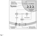
- Neuroinflammation is involved in several neurological and psychiatric diseases, in which activation of brain-resident microglia (1) and infiltration of peripheral immune cells cause neuronal stress and injury (2).
- multiple sclerosis (MS) is characterized by inflammatory lesions that cause damage to the central nervous system (CNS) and lead to neuronal dysfunction and clinical impairment (3).
- CNS central nervous system
- inflammatory activity declines over the course of MS, persistent low-grade inflammation (4) and inflammation-independent progression of neurodegeneration lead to continuous worsening of disability (5).
- chronic progressive MS is pronounced of primary neurodegenerative diseases that are accompanied by low grade inflammation (6).
- Neurodegeneration in MS has been attributed to the generation of reactive oxygen and nitrogen species (8), mitochondrial dysfunction with impaired energy production (9), and glutamate excitotoxicity that causes influx of calcium into neurons (10) resulting in ionic dyshomeostasis and cell death (2, 5). Similar pathways have been picked up in recent single cell transcriptomics analysis of MS postmortem brain samples. Affected neurons showed a strong upregulation of signatures associated with oxidative stress, mitochondrial dysfunction, and cell death pathways (11). However, the extraction of master regulators of neuronal maladaptation remains challenging (12).
- the present invention is based on the insight that the histone methyltransferase G9a is strongly induced in neurons during neuroinflammation in EAE mice and MS patients. It was furthermore found that the histone methyltransferase G9a strongly influences neuronal gene expression in a way that promotes ferroptosis, neuronal loss and clinical disability. Specifically, G9a promotes ferroptosis via the epigenetic repression of anti-ferroptotic genes resulting in neuronal loss during inflammatory neurodegeneration. More importantly, it was found that a robust neuroprotective effect can be achieved in neuronal cultures and during CNS inflammation in vivo by inhibition of the histone methyltransferase G9a. These findings reveal G9a as a stress-induced epigenetic amplifier of neuronal ferroptosis and highlight G9a inhibition as a neuroprotective therapy.
- the present invention specifically contemplates to use antagonists and/or inhibitors of the histone methyltransferase G9a for treating MS or preventing the progression of MS in a subject.
- Compounds that inhibit the function of G9a have been described in the art. However, the use of such compounds for treating MS has not been proposed.
- the invention relates to a compound which is effective in inhibiting the function of the methyltransferase G9a for use in a method of treating or preventing the progression of Multiple Sclerosis in a subject.
- the invention relates to a compound which is effective in inhibiting the function of the methyltransferase G9a for use in a method of reducing or ameliorating ferroptosis in a MS patient.
- the invention relates to a compound which is effective in inhibiting the function of the methyltransferase G9a for use in a method of ferroptosis-mediated neuronal loss in a MS patient.
- the MS to be treated can be any form of MS, including relapsing-remitting MS (RRMS), primary progressive MS (PPMS), and secondary progressive MS (SPMS).
- RRMS relapsing-remitting MS
- PPMS primary progressive MS
- SPMS secondary progressive MS
- the MS to be treated is relapsing-remitting MS.
- the subject to be treated with the G9a-inhibitory compound will normally be a mammal, and preferably is a human. Generally, the subject can be of any age. The subject preferably suffers from MS, more preferably from relapsing-remitting MS. The subject may be in any stage of the disease, i.e. the disease may be in an early stage wherein the subject shows only the first pathological signs that are normally associated with MS, or the subject may be in a late stage of the disease. The subject to be treated may have a genetic or other predisposition for developing MS.
- the inhibitory compounds of the invention effectively prevent damage and/or loss of neurons in the nervous system of a subject, preferably in the central nervous system (CNS).
- the CNS is to be understood to comprise the brain and the spinal cord.
- the compounds of the invention are administered for preventing damage and/or loss of neurons in the brain.
- the compounds of the invention are administered for preventing damage and/or loss of neurons in the spinal cord.
- progressive damage and/or loss of neurons may be halted by administration of the compounds of the invention. Halting the damage and/or the loss of neurons means that the pathological processes of the neurodegenerative disease which finally result in damage and/or loss of neurons are stopped or at least reduced.
- the compounds of the present invention are pharmaceutically active compounds which are effective in decreasing the expression and/or the activity of the methyltransferase G9a.
- the compounds can include all different types of organic or inorganic molecules, including peptides, polypeptides, oligo- or polysaccharides, fatty acids, steroids, and the like.
- the compounds will be small molecules with a molecular weight of less than about 2,500 daltons, less than 2000 daltons, less than 1500 daltons, less than 1000 daltons, or less than 550 daltons.
- the compounds which inhibit the function of methyltransferase G9a have a molecular weight of less than about 550 daltons.
- the compounds of the present invention are able to cross the blood-brain barrier, i.e. they are blood-brain barrier-permeable. This means that the compounds are able to reach a site in the brain which is affected by neuronal injury and/or loss.
- Inhibitory compounds with a molecular weight of below 550 Daltons are particularly suitable, since they are often able to cross the blood-brain barrier, or their transport across the blood-brain barrier is achieved by other means, such as by transporters or by hydrophobic stretches in the compound which allow diffusion across the blood-brain barrier.
- the compounds contemplated by the invention affect the function of a methyltransferase G9a protein.
- the inhibitory compounds interfere with the human histone methyltransferase G9a protein.
- histone methyltransferase G9a refers to the epigenetic regulator euchromatic histone lysine N -methyltransferase-2, an enzyme which is encoded by the gene Ehmt2 gene.
- G9a catalyzes the dimethylation of lysine 9 on core histone 3 (H3K9me2), wherein said dimethylation results in local transcriptional repression.
- the human methyltransferase G9a protein occurs in four different isoforms.
- the nucleotide sequences of the isoforms are depicted in SEQ ID NOs:1-4.
- the corresponding protein sequences of the isoforms are depicted in SEQ ID NOs:5-8.
- the compounds contemplated by the present invention inhibit the function of a methyltransferase G9a comprising or consisting of the amino acid sequence depicted in SEQ ID NOs:5-8 or an amino acid sequence having at least 90% sequence identity to the sequence of SEQ ID NOs:5-8.
- the compounds contemplated by the invention inhibit the function of a methyltransferase G9a comprising or consisting of the amino acid sequence depicted in SEQ ID NOs:5-8 or an amino acid sequence having at least 90% sequence identity to the sequence of SEQ ID NOs:5-8.
- the compounds which are contemplated herein for use in the treatment of MS are effective in inhibiting the function of the methyltransferase G9a. This means that the compounds effectively reduce the extent of dimethylation of lysine 9 on core histone 3 (H3K9me2).
- the compound decreases the G9a activity by at least 10%, 20%, 30%, 40%, 50%, 60%, 70%, 80%, 90%, or 95% compared to the activity in the absence of the compound.
- the reduction of the G9a activity is understood as the prevention of a full dimethylation of lysine 9 on core histone 3.
- the inhibitory compound completely blocks the G9a activity such that there is no detectable methylation or dimethylation activity of core histone 3.
- a decrease in the activity of the methyltransferase G9a may be determined by any suitable assay.
- G9a activity can be measured in a cell culture system by determining the H3K9me2 level after application of an inhibitor via immunocytochemistry staining. Fluorescence of the H3K9me2 signal can be analyzed by confocal microscopy or by measuring the fluorescence in a microplate reader.
- suitable assays are sold by several manufacturers, for example the Histone H3 Panel (mono methyl K9, dimethyl K9, tri methyl K9) (ab113754) of abcam (Cambridge, UK) and the G9a Chemiluminescent Assay Kit of BPSBioscience (San Diego, USA).
- the inhibitory compound is specific for the methyltransferase G9a, i.e. the compound inhibits the expression and/or activity of G9a, while it does essentially not inhibit the expression and/or activity of other proteins or enzymes, e.g. other methyltransferase.
- the compounds according to the invention elicit no or only tolerable side effects when administered to a subject.
- the G9a-inhibitory compound of the invention can be derived from different groups of molecules.
- inhibitory compounds can include antisense polynucleotides which bind to the gene encoding G9a and block transcription, RNA processing and/or translation of said gene.
- the antisense molecules can be RNA or DNA molecules.
- the G9a-inhibitory compound of the invention can be a RNA molecule which exerts its effect by RNA interference. Examples for such compounds are RNAi molecules and siRNA molecules that are capable of blocking translation of the G9a-encoding mRNA.
- the inhibitory compound of the invention can be a ribozyme that cleaves the G9a-encoding mRNA.
- Other classes of molecules which may give rise to suitable G9a-inhibitory compounds include peptides, antibodies and antibody fragments. Peptides that bind and interfere with the G9a protein may be conveniently screened in random peptide libraries.
- inhibitors of G9a have been described in the art. These inhibitors include UNC0642 (45), BIX01294 (45), UNC0224 (45), UNC0321 (45), E72 (45), UNC0638 (45), BRD9539 (45), UNC1479 (45), CSV0C018875 (89), A-366 (90), UNC0646 (87), UNC0631 (87), HKMTI-1-247 (87), HKMTI-1-248 (87), CM-272 (87), BRD4770 (87), CBC-12 (87), DCG066 (87), CPUY074001 (87), CPUY074020 (87), TM2-115 (87), 867750 (87), 867751 (87), DS79932728 (88), and EZM-8266 (88).
- the compound which is effective in inhibiting the function of the histone methyltransferase G9a and used for treating or preventing the progression of Multiple Sclerosis is UNC0642 (2-(4,4-difluoropiperidin-1-yl)-6-methoxy-N-[1-(propan-2-yl)piperidin-4-yl]-7-[3-(pyrrolidin-1-yl)propoxy]quinazolin-4-amine; CAS Number 1481677-78-4 ) or a pharmaceutically acceptable salt, solvate, tautomer or ester thereof.
- pharmaceutically acceptable salts of UNC0642 include hydrochlorides and sulphates.
- the structure of UNC0642 is as follows:
- the above structure can be substituted or otherwise modified at one or more sites, as long as these modifications do not substantially impair the inhibitory effect of UNC0642 and do not result in any undesired toxic side effects.
- one or more hydrogen atoms of the C-H bonds of the heterocyclic ring system can be substituted with halogen atoms, such as chlorine, bromine or iodine atoms.
- the hydrogen of the C-H bonds can also be replaced by an alkyl group, such as methyl, ethyl or propyl.
- compositions of the present invention will comprise an amount of the G9a-inhibitory compound that is effective to inhibit G9a activity, thereby reducing the extent of neurodegeneration by protecting the CNS from neuronal loss.
- the therapeutically effective amount of the G9a-inhibitory compound to be administered will depend on several parameters, such as the mode of administration, the particular type of MS disease, the severity of the disease, and the age, height, weight, health, and physical condition of the individual to be treated.
- a therapeutically effective amount of the G9a-inhibitory compound can be determined by one of ordinary skill in the art without undue experimentation given the disclosure set forth herein.
- the G9a-inhibitory compound will preferably be administered to the subject to be treated, e.g. the subject suffering from MS, in an amount that ranges from 0.1 ⁇ g/kg body weight to 10,000 ⁇ g/kg body weight, e.g. from 0.5 ⁇ g/kg to 7,500 ⁇ g/kg body weight, from 1.0 ⁇ g/kg to 5,000 ⁇ g/kg body weight, from 5.0 ⁇ g/kg to 3,000 ⁇ g/kg body weight, from 7.5 ⁇ g/kg to 2,500 ⁇ g/kg body weight, from 10 ⁇ g/kg to 2,000 ⁇ g/kg body weight, from 25 ⁇ g/kg to 1,500 ⁇ g/kg body weight, from 50 ⁇ g/kg to 1,000 ⁇ g/kg body weight, from 100 ⁇ g/kg to 800 ⁇ g/kg body weight, from 300 ⁇ g/kg to 600 ⁇ g/kg body weight, and more preferably from 400 ⁇ g/kg to 500 ⁇ g/kg body weight.
- the G9a-inhibitory compound of the present invention will normally be provided in the form of a pharmaceutical composition. Therefore, in a fourth aspect, the invention relates to a pharmaceutical composition comprising a compound which is effective in inhibiting the function of the Vietnameseromatic histone lysine N-methyltransferase-2 (G9a) for use in a method of treating or preventing the progression of Multiple Sclerosis in a subject. In a fifth aspect, the invention relates to a pharmaceutical composition comprising a compound which is effective in inhibiting the function of the Vietnamese histone lysine N-methyltransferase-2 (G9a) for use in a method of reducing or ameliorating ferroptosis in a Multiple Sclerosis patient.
- compositions of the invention normally comprise one or more excipients, carriers and/or diluents suitable for the intended way of administration.
- the G9a-inhibitory compound may be administered in any suitable form that does not interfere with its G9a-inhibitory activity.
- the compound may be administered orally in the form of tablets, capsules, granule, powder, liquids, and the like.
- the G9a-inhibitory compound may be formulated for being administered parenterally, e.g. by intravenous injection or intravenous infusion.
- the G9a-inhibitory compound is administered to the subject by intravenous infusion, more preferably by short-term infusion within less than 60 min, e.g. within 30 min, 20 min or 15 min.
- compositions suitable for injection and/or infusion include solutions or dispersions and powders for the extemporaneous preparation of such injectable solutions or dispersions.
- the composition for injection must be sterile and should be stable under the conditions of manufacturing and storage.
- the compositions for injection and/or infusion also include a preservative, such as a chlorobutanol, phenol, ascorbic acid, thimerosal, and the like.
- suitable carriers may comprise physiological saline, bacteriostatic water, Cremophor EL TM (BASF) or phosphate-buffered saline (PBS).
- Sterile solutions for injection and/or infusion can be prepared by incorporating the G9a-inhibitory compound in the required amount in an appropriate solvent followed by filter sterilization.
- the pharmaceutical composition provided by the present invention may further comprise other anti-neurodegenerative or anti-inflammatory compounds which are commonly used in the treatment of MS, such as interferon beta, glatiramer acetate, teriflunomide, cladribine, fampridine, fingolimod, ozanimod, ponesimod, siponimod, alemtuzumab, natalizumab, ocrelizumab, ofatumumab or rituximab.
- other anti-neurodegenerative or anti-inflammatory compounds which are commonly used in the treatment of MS, such as interferon beta, glatiramer acetate, teriflunomide, cladribine, fampridine, fingolimod, ozanimod, ponesimod, siponimod, alemtuzumab, natalizumab, ocrelizumab, ofatumumab or rituximab.
- the two active ingredients can be administered to the subject in the form of a single pharmaceutical composition comprising both agents and pharmaceutically acceptable excipients and carriers. Administration of such a pharmaceutical composition will automatically result in a simultaneous administration of both agents.
- the two therapeutic agents may also be administered separately from each other, i.e. in the form of two separate pharmaceutical compositions, one containing the G9a-inhibitory compound, and the other containing the additional anti-neurodegenerative or anti-inflammatory agent.
- the two separate compositions can be administered simultaneously, i.e. at the same time at two distinct sites of administration, or they may be administered sequentially (in either order) to the same site or to different sites of administration.
- both the composition comprising the G9a-inhibitory compound and the composition comprising the additional anti-neurodegenerative or anti-inflammatory agent are administered according to a weekly dosing regimen, more preferably a regimen in which a single dose of the G9a-inhibitory compound and a single dose of the anti-neurodegenerative or anti-inflammatory agent is administered every week for a treatment period of 2 or more weeks, for example, 3, 4, 5, 6, 7, 8, 9, 10, 11, 12, 13, 14, 15, 16, 17, 18, 19, 20, or more weeks. It will also be possible to administer the overall weekly dose of the G9a-inhibitory compound and/or the anti-neurodegenerative or anti-inflammatory agent in more than one administration per week, e.g. in 2 or 3 administrations per week.
- the amount of the G9a-inhibitory compound to be administered weekly is delivered as a single intravenous infusion per week, and the amount of the anti-neurodegenerative or anti-inflammatory agent to be administered weekly is also given as a single intravenous infusion per week, either at the same day of administration of the G9a-inhibitory compound, e.g. within 6-12 hours after administration of the G9a-inhibitory compound has been completed.
- the present invention provides methods for identifying a pharmaceutically active compound that could be used for treating or preventing MS.
- the methods of the invention can be used for drug screening approaches that aim to identify new pharmaceutically active for treating MS.
- the invention relates to an in vitro method for identifying a pharmaceutically active compound for treating Multiple Sclerosis or preventing the progression of Multiple Sclerosis in a subject, comprising
- a decrease in the activity of the methyltransferase G9a may be determined by any suitable assay.
- G9a activity can be measured in a cell culture system by determining the H3K9me2 level after application of an inhibitor via immunocytochemistry staining. Fluorescence of the H3K9me2 signal can be analyzed by confocal microscopy or by measuring the fluorescence in a microplate reader.
- suitable assays are sold by several manufacturers, for example the Histone H3 Panel (mono methyl K9, dimethyl K9, tri methyl K9) (ab113754) of abcam (Cambridge, UK) and the G9a Chemiluminescent Assay Kit of BPSBioscience (San Diego, USA).
- the screening can target compounds which interfere with G9a transcription and/or translation.
- the invention therefore relates, in a seventh aspect, to an in vitro method for identifying a pharmaceutically active compound for treating Multiple Sclerosis or preventing the progression of Multiple Sclerosis in a subject, comprising
- Screening methods which monitor the transcription of the gene encoding G9a and/or the translation of the G9a mRNA are particularly useful, e.g. for identifying compounds which inhibit the G9a by decreasing G9a expression. By use of these methods, it will for example be possible to identify compounds that target transcription factors which are relevant for G9a expression. Also, suitable ribozymes or antisense DNA molecules can be identified in this way.
- a number of different screening methods to evaluate the effects of candidate compounds on gene expression or enzyme activity may be used.
- the methods of the invention make use of any suitable method to detect a decrease in the expression level of the gene encoding G9a and/or in the activity of G9a.
- Suitable methods include e.g. biochemical or cellular methods. The skilled person would be able to identify such methods with his or her common knowledge.
- a decrease in the expression level of the gene encoding G9a may be detected by methods which allow the quantification of the transcription or the translation product of the gene encoding G9a, e.g. the quantification of G9a mRNA or protein.
- methods which allow the quantification of the transcription or the translation product of the gene encoding G9a, e.g. the quantification of G9a mRNA or protein.
- PCR polymerase chain reaction
- PCR polymerase chain reaction
- standard PCR real-time PCR and quantitative real-time PCR, or Northern Blots
- the concentration will be determined relative to a sample which is not contacted with the respective candidate compound, i.e. relative to a negative control, or to the same sample before contacting it with the respective compound.
- the concentration of G9a protein may analogously be determined with standard methods such as SDS-PAGE, enzyme linked immunosorbent assays (ELISA), Western Blots, mass spectrometry or any other suitable means.
- the G9a protein concentration will then be compared to a sample which has not been contacted with the respective compound or to the same sample before contacting it with the respective compound.
- the cell that expresses the functional G9a enzyme is preferably a mammalian cell, more preferably a human cell.
- the cell can be derived from a cell line, preferably a neuron- or brain cell-derived cell line, or from a primary cell culture, more preferably from a primary neuronal cell culture.
- the in vitro methods of the present invention may preferably be carried out as high throughput screening techniques which allow for the examination of thousands of different compounds in a short period of time.
- Candidate compounds for use in screening methods can be provided in the form of libraries which comprise a high number of synthetic or natural compounds. High throughput screening techniques are described in detail in the prior art.
- the compounds identified by the screening methods may be validated in animal models, such as mouse models to confirm their activity in vivo.
- the candidate compounds used in the screening methods of the invention can include all different types of molecules as described for the compounds of the present invention above.
- the protein can be one that has been recombinantly expressed.
- the G9a protein used in the assay comprises the amino acid sequences set out in SEQ ID NO:5, 6, 7, or 8.
- the above assays can be performed not only with the specific sequences depicted in SEQ ID NO:5, 6, 7, or 8, but also with variants, derivatives and enzymatically active fragments of these G9a protein sequences.
- variants of G9a are polypeptides that differ by one or more amino acid exchanges from the sequence shown in SEQ ID NOs:5-8.
- any amino acid residue of the amino acid sequence shown in SEQ ID NOs:5-8 can be exchanged for a different amino acid, provided the resultant sequence of the variant is still capable of catalyzing dimethylation of lysine 9 on core histone 3 (H3K9me2).
- variants may differ from their reference sequence shown in SEQ ID NOs:5-8 in up to 5%, 10%, 15%, or 20% of the amino acids.
- Polypeptides in which one or more amino acids were inserted in the amino acid sequence of SEQ ID NOs:5-8 are also included as variants. Such insertions can be made at any position of the polypeptide shown in SEQ ID NOs:5-8 as long as the variant is still able to catalyze dimethylation of H3K9me2.
- polypeptides in which one or more amino acids are missing in comparison with SEQ ID NOs:5-8 are also considered to be variants of the polypeptides of SEQ ID NOs:5-8. Such deletions can apply to any amino acid position of the sequence of SEQ ID NOs:5-8.
- Variants of the G9a protein will preferably have at least 80% sequence identity, more preferably at least 85% sequence identity, and even more preferably at least 90% sequence identity, e.g. 91%, 92%, 93%, 94%, 95%, 96%, 97%, 98%, 99% sequence identity, with the human G9a protein shown in SEQ ID NOs:5-8 when these sequences are optimally arranged, e.g. with the computer program GAP or BESTFIT using the default gap method.
- Computer programs for determination of the amino acid identity are known to the skilled person.
- Enzymatically active fragments of a G9a methyltransferase may also be used in the above assay.
- the assay may be conducted with enzymatically active fragments of the sequence shown in SEQ ID NOs:5-8 or variants thereof.
- enzymatically active fragments of the sequence shown in SEQ ID NOs:5-8 refer to peptides or polypeptides that differ from the amino acid sequence shown in SEQ ID NOs:5-8 or from the above-defined variants thereof by the absence of one or more amino acids at the N-terminus and/or at the C-terminus of the peptide or polypeptide.
- Derivatives of the polypeptides shown in SEQ ID NOs:5-8 or of the variants thereof refer to polypeptides that possess amino acids with structural modifications, for example, modified amino acids, relative to a polypeptide shown in SEQ ID NOs:5-8 or variants thereof.
- modified amino acids can be, e.g. amino acids that have been altered by phosphorylation, glycosylation, acetylation, thiolation, branching and/or cyclization.
- variants or derivatives of the G9a polypeptide shown in SEQ ID NOs:5-8 or the active fragments of this polypeptide or its variants retain at least 75%, and preferably up to 80%, 85%, 90% or even up to 99% of the dimethylation activity of the G9a polypeptide shown in SEQ ID NOs:5-8.
- the invention relates to the use of: (a) a polypeptide comprising the amino acid sequence shown in SEQ ID NOs:5-8 or a variant thereof having at least 80%, more preferably at least 90% sequence identity to the amino acid sequence shown in SEQ ID NOs:5-8; or (b) a polynucleotide encoding a polypeptide of (a) or the complement thereof for identifying pharmaceutically active compounds for treating MS or preventing the progression of MS.
- Figure 1 shows that G9a-depedent H3K9me2 is induced by inflammation and excitotoxicity.
- B Normalized RNA-Seq expression of Ehmt2 in healthy and inflamed motor neurons.
- n 5 biologically independent samples per group, each pooled from 3 mice, motor neurons versus inflamed motor neurons.
- FIG. 2 shows that G9a promotes neuronal damage during glutamate-induced excitotoxicity.
- Figure 3 shows that G9a potentiates oxidative stress in neurons.
- FIG. 4 shows that G9a induces neuronal cell death via ferroptosis.
- Figure 5 shows that G9a amplifies neurodegeneration and represses anti-ferroptotic genes in CNS inflammation.
- Data are shown as mean values ⁇ s.e.m. ⁇ P ⁇ 0.05, ⁇ P ⁇ 0.01, ⁇ P ⁇ 0.001.
- FIG. 7 shows that G9a propagates neurodegeneration by enhancing ferroptosis.
- CNS inflammation enhances oxidative stress and increases extracellular glutamate levels, together inducing G9a activity in neurons.
- G9a catalyzes the repressive mark H3K9me2 that suppresses the expression of Gclc, Cbs and Gpx4.
- H3K9me2 that suppresses the expression of Gclc, Cbs and Gpx4.
- ferroptosis itself enhances G9a activity in a self-reinforcing feedback loop.
- Pharmacological inhibition of G9a by UNC0642 counteracts these effects and results in neuroprotection.
- mice and human neuronal cell culture assays and histological analysis of human specimens from MS patients and controls were conducted.
- experimental units correspond to individually prepared cultures derived from one pregnant mouse and individually differentiated batches of neuronal progenitor cells, respectively. Depending on the readout, up to five technical replicates were acquired per experimental unit. Technical replicates that deviated more than 20% from the mean of replicates, e.g., wells with dead cells, were excluded from the analysis.
- mice For in vivo mouse studies, experimental units correspond to individual female mice. Age-matched mice were randomized into experimental groups before the start of the experiment and scored and analyzed with the investigator blinded to the treatment received. Mice that did not develop disease due to immunization failure were excluded from the analysis on the basis of predefined exclusion criteria.
- mice C57BL/6J wild-type (The Jackson Laboratory), G9afl/fl (82) and G9afl/fl;Snap25-Cre (Snap25-Cre from the Jackson Laboratory) were kept under specific pathogen-free conditions in the central animal facility of the University Medical Center Hamburg-Eppendorf (UKE).
- G9afl/fl 82)
- G9afl/fl G9afl/fl
- Snap25-Cre Snap25-Cre from the Jackson Laboratory
- mice were immunized subcutaneously with 200 ⁇ g MOG35-55 peptide (Schafer-N) in CFA (Difco, cat. no. DF0639-60-6) containing 4 mg ml-1 Mycobacterium tuberculosis (Difco, cat. no. DF3114-33-8).
- CFA CFA
- ml-1 Mycobacterium tuberculosis Difco, cat. no. DF3114-33-8
- 300 ng pertussis toxin (Calbiochem, cat. no. CAS70323-44-3 ) was injected intraperitoneally on the day of immunization and 48 hours later.
- UNC0642 was prediluted in DMSO and the final injection contained 10% DMSO ⁇ UNC0642 and 90% Dulbecco's phosphate buffered saline (DPBS; Pan Biotech). The investigators were blind to the treatment in the EAE experiments.
- DMSO DMSO ⁇ UNC0642
- DPBS Dulbecco's phosphate buffered saline
- iPSCs Human iPSCs (ZIPi013-B) (83) were maintained under feeder-free conditions on Matrigel (Corning)-coated plates in mTeSR1 medium (Stem Cell Technologies, cat. no. 85850).
- iPSCs were dissociated with Accutase and seeded at a density of 3 ⁇ 10 6 cells per well on AggreWell800 plates (10,000 cells per embryoid body (EB), Stem Cell Technologies) in SMADi Neural Induction Medium (SMADi NIM, Stem Cell Technologies, cat. no. 08582) supplemented with 10 Y-27632 (Stem Cell Technologies, cat. no. 72302).
- SMADi NIM SMADi Neural Induction Medium
- EBs were harvested and cultivated on Matrigel-coated plates in SMADi NIM for 12 days. Newly formed neural rosettes were manually picked and cultured for another 4 days.
- neural precursor cells NPCs
- neural rosettes were dissociated with Accutase and were maintained for several passages at high density in Neural Progenitor Medium (Stem Cell Technologies, cat. no. 05833) on Matrigel-coated plates.
- HiPSC-derived NPCs were differentiated into neurons as previously described (84, 85) with some modifications. Briefly, NPCs were seeded at a density of 1 ⁇ 10 5 cells per cm 2 in Neural Progenitor Medium onto Matrigel-coated plates.
- neural differentiation medium (day 0 of differentiation) composed of Neurobasal Plus medium (Gibco, cat. no. A3582901) containing 1 ⁇ B27 Plus Supplement (Gibco, cat. no. A3582801), 1 ⁇ N2 Supplement-A (Stem Cell Technologies, cat. no. 07152), 1 ⁇ MEM non-essential amino acids (Gibco, cat. no. 11140050), 1 ⁇ g ml -1 laminin (Sigma-Aldrich, cat. no. L2020), 1 dibutyryl-cAMP (Stem Cell Technologies, cat. no. 73882), 10 ng ml -1 L-ascorbic acid (Stem Cell Technologies, cat. no.
- CNS tissues were fixed with 4% paraformaldehyde and embedded in paraffin. After deparaffination and antigen retrieval (Pascal Tris EDTA ph 9.0), the 2 ⁇ m sections were incubated with a peroxidase-blocking solution (Dako, cat. No. K0672) to inactivate endogenous peroxidases and unspecific binding was blocked (PBS + 10% goat serum). Tissue sections were incubated overnight with a mouse anti-H3K9me2 (Abcam, cat. no. ab1220, 1:200) and a rabbit anti-NeuN AlexaFluor 488 labelled (Abcam, cat. no. ab190195, 1:50).
- a peroxidase-blocking solution Daako, cat. No. K0672
- H3K9me2 bound antibodies were visualized using a donkey anti-mouse IgG2a (Jackson Immunoresearch, cat. no. 715-605-151, 1:200). Immunostained human samples were scanned using the Pannoramic 250 FLASH II (3DHISTECH) Digital Slide Scanner at 20 ⁇ magnification.
- RNAscope Fluorescent Multiplex V2 (Advanced Cell Diagnostic (ADC), cat. no. 323100) was performed according to the manufacturer standard protocol for PFA fixed tissue.
- RNAscope human samples were scanned using the Pannoramic 250 FLASH II (3DHISTECH) Digital Slide Scanner at 20 ⁇ magnification.
- GPX4+ SNAP25+ neurons were quantified by a blinded experimenter using a custom-made script, which was based on Cognition Network Language (Definiens Cognition Network Technology; Definiens Developer XD software).
- Immunocytochemistry was performed as described previously (15). HiPSC-neurons and mouse neurons were cultivated on 12 mm diameter cover slips or on 96-well plates suited for confocal microscopy, fixed with 4% paraformaldehyde (PFA), incubated in 10% normal donkey's serum (NDS) containing 0.25% Triton X-100, and subsequently immunolabeling was performed. Immunostainings were analyzed with a Zeiss LSM 700 confocal microscope.
- MT cell viability substrate and NanoLuc Enzyme was mixed together, added to neuronal cultures and incubated for 5 hours for equilibration of luminescence signal before respective treatments were applied.
- Luminescence was acquired with a Spark 10M multimode microplate reader (Tecan) at 37°C and 5% CO 2 every 30 min over a total time period of 24 hours. At least five technical replicates per condition were used. For analysis every well's data point was normalized to its last value before the stressor was added and then normalized to the mean of the control wells for every time point using a customized MATLAB script. Thereby, it was normalized for well-to-well seeding variability.
- Neurons were imaged on a Zeiss LSM 700 confocal microscope using 20 ⁇ magnification.
- Dyeing cells were quantified with Fiji (ImageJ) as displayed by bright green nuclear fluorescence and quantified as percentage of total cells, measured by the number of Hoechst positive nuclei.
- CellROX green reagent (Thermofisher) was used for the detection of oxidative stress in response to glutamate treatment. Neuronal cultures were stimulated with 20 ⁇ M glutamate for a total incubation time of 1 hour. CellROX reagent was added to the wells in a 5 ⁇ M final concentration after 30 min of glutamate stress. Hoechst 33342, a cell permeant nuclear counterstain, was added for 10 min. Cells were washed two times with PBS before image acquisition. The cells were subsequently imaged on a Zeiss LSM 700 confocal microscope using 20 ⁇ magnification. Immunofluorescence of nuclear CellROX dye was quantified using Fiji (ImageJ).
- GSH-Glo Glutathione assay (Promega) was used as described by the manufacturer. Primary cortical neurons were cultured in a 96-well plate and stimulated with glutamate for 2 hours. Cells were washed with PBS and GSH-Glo reagent was added to the wells for 30 min at room temperature, followed by the addition of luciferin detection reagent. After 15 min, luminescence was detected at a Spark 10M multimode microplate reader (Tecan).
- mice were isolated with the Nuclei Isolation Kit (Sigma Aldrich, cat. no. NUC101) according to the manufacturer's protocol with minor modifications. Briefly, mice were sacrificed with CO 2 and perfused with cold PBS. Whole spinal cords were removed and mechanically dissociated with a scalpel on a petri dish placed on a cooled metal block. Tissue was added to 2 ml of EZ buffer (Sigma Aldrich, cat. no. NUC101) and further dissociated using a glass douncer (Sigma-Aldrich D9063).
- nuclei incubation buffer 340 mM sucrose, 2 mM MgCl2, 25 mM KCl, 65 mM glycerophosphate, 5% glycerol, 1 mM EDTA, 1% BSA.
- Nuclei were filtered using a 30 ⁇ M filter and directly stained with primary labelled NeuN antibody (ab 190565, Abcam) and propidium iodide. NeuN-positive nuclei were sorted using a BDAriaIII cell sorter (BD Bioscience). RNA for real-time PCR was processed as described above.
- nuclei of mouse spinal cords were isolated as described above. After filtering of the homogenate, nuclei were fixed with methanol for 10 min on ice, followed by two washing steps with nuclei incubation buffer. For intranuclear staining, nuclei were permeabilized with nuclei incubation buffer supplemented with 0.5% Triton-X-100 for 15 min on ice. After blocking with nucleus incubation buffer and 10% NDS, primary antibody was added for 1 hour at room temperature, followed by a washing step and subsequent incubation in secondary antibody for 30 min at room temperature. Immunofluorescence was acquired on an LSR II FACS analyzer (BD Biosciences). Data analysis was performed with the FlowJo v.10 analysis software (FlowJo LLC).
- mice were perfused intracardially with ice-cold PBS immediately after killing to remove blood from the intracranial vessels.
- the brain and spinal cord were prepared with sterile instruments, minced with a scalpel and incubated with agitation in Roswell Park Memorial Institute medium 1640 (PAA) containing 1 mg ml-1 collagenase A (Roche) and 0.1 mg ml -1 DNase I recombinant, RNase-free (Roche) for 45 min at 37 °C. Tissue was triturated through a 100- ⁇ m cell strainer and washed with PBS (pellet was centrifuged at 300g for 10 min at 4 °C).
- PPAA Roswell Park Memorial Institute medium 1640
- the homogenized tissue was resuspended in 30% isotonic Percoll (GE Healthcare) and carefully underlaid with 78% isotonic Percoll. After gradient centrifugation (1,500g for 30 min at 4 °C), CNS-infiltrating immune cells were recovered from the gradient interphase and washed in ice-cold PBS. Single-cell suspensions were stained in the presence of 123 count eBeads (Thermofisher). Data was acquired on an LSR II FACS analyzer (BD Biosciences). Data analysis was performed with the FlowJo v.10 analysis software (FlowJo LLC).
- Flow cytometric data were analyzed by using FlowJo (LLC). Images were analyzed using Fiji software (NIH). Experimental data obtained from Real-time Glo cell viability assay was analyzed with a customized MATLAB script and calcium imaging data were processed using a customized R script. Further experimental data were analyzed with the Prism 9 software (GraphPad Software) for Mac and are presented as the means ⁇ s.e.m. Unless otherwise stated, differences between two experimental groups were determined using an unpaired, two-tailed Student's t-test; differences between three or more experimental groups were determined using one-way analysis of variance (ANOVA) with Tukey's post hoc test; differences between two or more experimental groups depending on two different variables were analyzed using two-way ANOVA with Tukey's post hoc test.
- ANOVA analysis of variance
- Example 1 Candidate gene identification
- a neuronal translatome profiling data set of inflamed spinal cord motor neurons of EAE mice (13) was leveraged and ranked de-regulated candidates by statistical significance.
- the dataset focuses on known epigenetic regulators (dbEM - a Database of Epigenetic Modifiers) that were differentially expressed in inflamed versus healthy motor neurons.
- Differential expression analysis was performed with DESeq2 (v.1.14.1) calling genes with a minimal twofold change and false discovery rate (FDR)-adjusted P ⁇ 0.05 differentially expressed.
- histone methyltransferase G9a encoded by the gene Ehmt2
- Ehmt2 histone methyltransferase G9a
- H3K9me2 levels in neuronal nuclei in EAE were measured in comparison to healthy spinal cord by flow cytometry.
- an increase of H3K9me2 levels in neuronal nuclei was found during acute (day 15) and chronic (day 30) EAE, but not prior to disease onset (day 11).
- This inflammation-dependent induction of H3K9me2 was accompanied by neuronal loss, as quantified by the percentage of NeuN-positive neuronal nuclei in spinal cords of acute and chronic EAE mice, as depicted in Figure 1D .
- H3K9me2 induction might be a general feature of stress-induced neurodegeneration
- glutamate was applied to primary cortical cultures as a model of excitotoxicity during neuroinflammation (43, 44). Resembling the in vivo situation, glutamate exposure of primary cortical cultures led to an upregulation of H3K9me2, as shown in Figure 1F . The upregulation was associated with significantly decreased cell viability, as can be seen from Figure 1G . Notably, stimulation with other inflammatory mediators such as tumor necrosis factor-alpha (TNF- ⁇ ) or interferon-gamma (IFN-y) did not increase H3K9me2 levels (data not shown).
- TNF- ⁇ tumor necrosis factor-alpha
- IFN-y interferon-gamma
- H3K9me1, H3K9me3, H3K27me3 or permissive (H3K27ac) epigenetic marks nor did it change the levels of DNA-methyltransferase 3a (DNMT3a).
- H3K9me1, which is also catalyzed by G9a showed a trend towards being induced by glutamate exposure.
- AP5 or EGTA were applied to block NMDA receptors or prevent calcium influx from the extracellular space, respectively. Both substances potently suppressed the induction of H3K9me2 in response to glutamate indicating NMDA receptor-mediated calcium influx from the extracellular space as an inducer of increased G9a activity. Besides H3K9me2 levels, NMDA receptor blockage by AP5 also rescued cellular survival. Together, both CNS inflammation in vivo and glutamate excitotoxicity in neuronal cultures resulted in increased G9a-dependent H3K9me2 levels and neuronal loss.
- Example 4 G9a potentiates oxidative stress in neurons
- G9a activity might compromise neuronal resistance to oxidative stress.
- CellROX green was used to measure oxidative stress in primary neuronal cultures after glutamate exposure. While glutamate stimulation of G9a fl/fl controls resulted in strong oxidative stress within one hour, genetic G9a disruption in G9a fl/fl ;Snap25-Cre neurons led to a near-complete rescue to the level of cultures without glutamate treatment, as shown in Figure 3A . Similarly, pre-treatment with UNC0642 reduced the generation of ROS close to baseline levels as seen without glutamate, as shown in Figure 3B .
- Example 5 G9a induces neuronal cell death via ferroptosis
- G9a perturbation supported neuronal viability
- a downstream effect of G9a in a central pathway of programmed cell death was hypothesized. Therefore, dedicated compounds were used to induce different forms of programmed cell death that have been implicated in neurodegeneration and oxidative stress and assessed their dependence on G9a function. While neither apoptosis nor necroptosis was affected by G9a perturbation, ferroptosis induced by either erastin or RSL3 was substantially rescued in G9a knockout neurons and after G9a inhibition with UNC0642, as can be seen in Figures 4A-B . Moreover, G9a activity was measured by quantification of H3K9me2 immunofluorescence in response to the activation of the different cell death pathways.
- ferrostatin-1 also the iron chelator alpha-difluoro-methylornithine (DFMO), and the lipophilic radical-trapping antioxidant coenzyme Q10 rescued glutamate-induced calcium overload, as shown in Figure 4E .
- DFMO iron chelator alpha-difluoro-methylornithine
- DFMO iron chelator alpha-difluoro-methylornithine
- lipophilic radical-trapping antioxidant coenzyme Q10 rescued glutamate-induced calcium overload, as shown in Figure 4E .
- This substantiates ferroptosis as a central cell death pathway in glutamate excitotoxicity.
- pre-treatment with the G9a inhibitor UNC0642 did not further potentiate the rescue effects of ferroptosis inhibition suggesting a shared pathway of both interventions, as shown in Figure 4E .
- G9a perturbation could increase the levels of glutathione, a key anti-ferroptotic effector molecule with
- Example 6 G9a amplifies neurodegeneration and represses anti-ferroptotic genes
- Gpx4 encoding the primary cellular defense system against ferroptosis by converting lipid peroxides into non-toxic lipid alcohols (47) and Gclc, encoding the glutamate-cysteine ligase catalytic subunit, the first rate limiting enzyme of glutathione synthesis (48), as depicted in Figure 5I .
- UNC0642 treatment in EAE significantly boosted the expression of Gpx4 and Gclc as well as Cbs, encoding cystathionine ⁇ -synthase, the first enzyme of the transsulfuration pathway, an alternative way to fuel glutathione production (49), and Nfe212, encoding nuclear factor erythroid 2-related factor 2, a transcription factor that drives several anti-oxidant genes including Gclc (50-52).
- multiple glutathione S-transferases followed the same pattern, showing suppression in EAE and derepression upon UNC0642 treatment.
- Example 7 Anti-ferroptotic GPX4 is suppressed in MS and induced by G9a inhibition
Priority Applications (1)
| Application Number | Priority Date | Filing Date | Title |
|---|---|---|---|
| EP22169820.2A EP4268823A1 (de) | 2022-04-25 | 2022-04-25 | Verbindung zum behandeln von nervenentzündungen |
Applications Claiming Priority (1)
| Application Number | Priority Date | Filing Date | Title |
|---|---|---|---|
| EP22169820.2A EP4268823A1 (de) | 2022-04-25 | 2022-04-25 | Verbindung zum behandeln von nervenentzündungen |
Publications (1)
| Publication Number | Publication Date |
|---|---|
| EP4268823A1 true EP4268823A1 (de) | 2023-11-01 |
Family
ID=81386731
Family Applications (1)
| Application Number | Title | Priority Date | Filing Date |
|---|---|---|---|
| EP22169820.2A Pending EP4268823A1 (de) | 2022-04-25 | 2022-04-25 | Verbindung zum behandeln von nervenentzündungen |
Country Status (1)
| Country | Link |
|---|---|
| EP (1) | EP4268823A1 (de) |
Citations (3)
| Publication number | Priority date | Publication date | Assignee | Title |
|---|---|---|---|---|
| WO2019079596A1 (en) * | 2017-10-18 | 2019-04-25 | Epizyme, Inc. | METHODS OF USING EHMT2 INHIBITORS IN IMMUNOTHERAPIES |
| WO2019210192A1 (en) * | 2018-04-26 | 2019-10-31 | Rush University Medical Center | Use of methylation inhibitors for the treatment of autoimmune diseases |
| WO2022031939A1 (en) * | 2020-08-07 | 2022-02-10 | Athos Therapeutics, Inc. | Small molecules for the treatment of autoimmune diseases and cancer |
-
2022
- 2022-04-25 EP EP22169820.2A patent/EP4268823A1/de active Pending
Patent Citations (3)
| Publication number | Priority date | Publication date | Assignee | Title |
|---|---|---|---|---|
| WO2019079596A1 (en) * | 2017-10-18 | 2019-04-25 | Epizyme, Inc. | METHODS OF USING EHMT2 INHIBITORS IN IMMUNOTHERAPIES |
| WO2019210192A1 (en) * | 2018-04-26 | 2019-10-31 | Rush University Medical Center | Use of methylation inhibitors for the treatment of autoimmune diseases |
| WO2022031939A1 (en) * | 2020-08-07 | 2022-02-10 | Athos Therapeutics, Inc. | Small molecules for the treatment of autoimmune diseases and cancer |
Non-Patent Citations (91)
| Title |
|---|
| A. ANANDHANM. DODSONC. J. SCHMIDLINP. LIUD. D. ZHANG: "Breakdown of an Ironclad Defense System: The Critical Role of NRF2 in Mediating Ferroptosis", CELL CHEM BIOL, vol. 27, 2020, pages 436 - 447, XP086136386, DOI: 10.1016/j.chembiol.2020.03.011 |
| A. INSINGAS. MONESTIROLIS. RONZONIV. GELMETTIF. MARCHESIA. VIALEL. ALTUCCIC. NERVIS. MINUCCIP. G. PELICCI: "Inhibitors of histone deacetylases induce tumor-selective apoptosis through activation of the death receptor pathway", NAT MED, vol. 11, 2005, pages 71 - 76 |
| B. A. C. CREEJ. A. HOLLENBACHR. BOVEG. KIRKISHS. SACCOE. CAVERZASIA. BISCHOFT. GUNDELA. H. ZHUN. PAPINUTTO: "Silent progression in disease activity-free relapsing multiple sclerosis", ANN NEUROL, vol. 85, 2019, pages 653 - 666 |
| B. J. ANDREONEM. LARHAMMARJ. W. LEWCOCK: "Cell Death and Neurodegeneration", COLD SPRING HARB PERSPECT BIOL, vol. 12, 2020 |
| B. S. XIEY. Q. WANGY. LINQ. MAOJ. F. FENGG. Y. GAOJ. Y. JIANG: "Inhibition of ferroptosis attenuates tissue damage and improves long-term outcomes after traumatic brain injury in mice", CNS NEUROSCI THER, vol. 25, 2019, pages 465 - 475 |
| B. SCHATTLINGJ. B. ENGLERC. VOLKMANNN. ROTHAMMERM. S. WOOM. PETERSENI. WINKLERM. KAUFMANNS. C. ROSENKRANZA. FEJTOVA: "Bassoon proteinopathy drives neurodegeneration in multiple sclerosis", NAT NEUROSCI, vol. 22, 2019, pages 887 - 896, XP036789607, DOI: 10.1038/s41593-019-0385-4 |
| B. SCHATTLINGK. STEINBACHE. THIESM. KRUSEA. MENIGOZF. UFERV. FLOCKERZIW. BRUCKO. PONGSR. VENNEKENS: "TRPM4 cation channel mediates axonal and neuronal degeneration in experimental autoimmune encephalomyelitis and multiple sclerosis", NAT MED, vol. 18, 2012, pages 1805 - 1811, XP055594049, DOI: 10.1038/nm.3015 |
| C. A. DENDROUL. FUGGERM. A. FRIESE: "Immunopathology of multiple sclerosis", NAT REV IMMUNOL, vol. 15, 2015, pages 545 - 558 |
| C. J. HARVEYR. K. THIMMULAPPAA. SINGHD. J. BLAKEG. LINGN. WAKABAYASHIJ. FUJIIA. MYERSS. BISWAL: "Nrf2-regulated glutathione recycling independent of biosynthesis is critical for cell survival during oxidative stress", FREE RADIC BIOL MED, vol. 46, 2009, pages 443 - 453, XP025922778, DOI: 10.1016/j.freeradbiomed.2008.10.040 |
| C. L. HUM. NYDESK. L. SHANLEYI. E. MORALES PANTOJAT. A. HOWARDO. A. BIZZOZERO: "Reduced expression of the ferroptosis inhibitor glutathione peroxidase-4 in multiple sclerosis and experimental autoimmune encephalomyelitis", J NEUROCHEM, vol. 148, 2019, pages 426 - 439 |
| C. MUKHERJEET. KLINGB. RUSSOK. MIEBACHE. KESSM. SCHIFFERERL. D. PEDROU. WEIKERTM. K. FARDN. KANNAIYAN: "Oligodendrocytes Provide Antioxidant Defense Function for Neurons by Secreting Ferritin Heavy Chain", CELL METAB, vol. 32, 2020, pages 259 - 272 |
| C. PICONA. JAYARAMANR. JAMESC. BECKP. GALLEGOM. E. WITTEJ. VAN HORSSENN. D. MAZARAKISR. REYNOLDS: "Neuron-specific activation of necroptosis signaling in multiple sclerosis cortical grey matter", ACTA NEUROPATHOL, vol. 141, 2021, pages 585 - 604, XP037408082, DOI: 10.1007/s00401-021-02274-7 |
| C. SEGOVIAE. SAN JOSE-ENERIZE. MUNERA-MARAVILLAM. MARTINEZ-FERNANDEZL. GARATEE. MIRANDAA. VILAS-ZORNOZAI. LODEWIJKC. RUBIOC. SEGRE: "Inhibition of a G9a/DNMT network triggers immune-mediated bladder cancer regression", NAT MED, vol. 25, 2019, pages 1073 - 1081 |
| C. TONELLII. I. C. CHIOD. A. TUVESON: "Transcriptional Regulation by Nrf2", ANTIOXID REDOX SIGNAL, vol. 29, 2018, pages 1727 - 1745 |
| D. G. BRICKNERI. CAJIGASY. FONDUFE-MITTENDORFS. AHMEDP. C. LEEJ. WIDOMJ. H. BRICKNER: "H2A.Z-mediated localization of genes at the nuclear periphery confers epigenetic memory of previous transcriptional state", PLOS BIOL, vol. 5, 2007, pages e81 |
| D. OFENGEIMY. ITOA. NAJAFOVY. ZHANGB. SHANJ. P. DEWITTJ. YEX. ZHANGA. CHANGH. VAKIFAHMETOGLU-NORBERG: "Activation of necroptosis in multiple sclerosis", CELL REP, vol. 10, 2015, pages 1836 - 1849 |
| D. PITTP. WERNERC. RAINE: "Glutamate excitotoxicity in a model of multiple sclerosis", NATURE MEDICINE, vol. 6, 2000, pages 67 - 70, XP000867880, DOI: 10.1038/71555 |
| D. SULZERR. N. ALCALAYF. GARRETTIL. COTEE. KANTERJ. AGIN-LIEBESC. LIONGC. MCMURTREYW. H. HILDEBRANDX. MAO: "T cells from patients with Parkinson's disease recognize alpha-synuclein peptides", NATURE, vol. 546, 2017, pages 656 - 661 |
| D. TANGX. CHENR. KANGG. KROEMER: "Ferroptosis: molecular mechanisms and health implications", CELL RES, vol. 31, 2021, pages 107 - 125, XP037354365, DOI: 10.1038/s41422-020-00441-1 |
| F. DANGONDA. DONNELLYR. HOHLFELDC. LUBETZKIS. KOHLHAASL. LEOCANIO. CICCARELLIB. STANKOFFM. P. SORMANIJ. CHATAWAY: "Facing the urgency of therapies for progressive MS - a Progressive MS Alliance proposal", NAT REV NEUROL, vol. 17, 2021, pages 185 - 192, XP037388929, DOI: 10.1038/s41582-020-00446-9 |
| F. LIUD. BARSYTE-LOVEJOYF. LIY. XIONGV. KORBOUKHX. P. HUANGA. ALLALI-HASSANIW. P. JANZENB. L. ROTHS. V. FRYE: "Discovery of an in vivo chemical probe of the lysine methyltransferases G9a and GLP", J MED CHEM, vol. 56, 2013, pages 8931 - 8942, XP055456510, DOI: 10.1021/jm401480r |
| G. R. CAMPBELLI. ZIABREVAA. K. REEVEK. J. KRISHNANR. REYNOLDSO. HOWELLH. LASSMANND. M. TURNBULLD. J. MAHAD: "Mitochondrial DNA deletions and neurodegeneration in multiple sclerosis", ANN NEUROL, vol. 69, 2011, pages 481 - 492 |
| H. CAO L. LIAD YANGL. ZENGX. YEWEIB. YUG. LIAOJ. CHEN: "Recent progress in histone methyltransferase (G9a) inhibitors as anticancer agents", EUROPEAN JOURNAL OF MEDICINAL CHEMISTRY, vol. 179, 2019, pages 537 - 546, XP085772612, DOI: 10.1016/j.ejmech.2019.06.072 |
| H. HEZ. HUH. XIAOF. ZHOUB. YANG: "The tale of histone modifications and its role in multiple sclerosis", HUM GENOMICS, vol. 12, 2018, pages 31 |
| H. LASSMANNJ. VAN HORSSEN: "Oxidative stress and its impact on neurons and glia in multiple sclerosis lesions", BIOCHIM BIOPHYS ACTA, vol. 1862, 2016, pages 506 - 510 |
| H. P. MOHAMMADO. BARBASHC. L. CREASY: "Targeting epigenetic modifications in cancer therapy: erasing the roadmap to cancer", NAT MED, vol. 25, 2019, pages 403 - 418, XP036722152, DOI: 10.1038/s41591-019-0376-8 |
| H. U. KLEINC. MCCABEE. GJONESKAS. E. SULLIVANB. J. KASKOWA. TANGR. V. SMITHJ. XUA. R. PFENNINGB. E. BERNSTEIN: "Epigenome-wide study uncovers large-scale changes in histone acetylation driven by tau pathology in aging and Alzheimer's human brains", NAT NEUROSCI, vol. 22, 2019, pages 37 - 46, XP036657712, DOI: 10.1038/s41593-018-0291-1 |
| I. ALIMJ. T. CAULFIELDY. CHENV. SWARUPD. H. GESCHWINDE. IVANOVAJ. SERAVALLIY. AIL. H. SANSINGE. J. STE MARIE: "Selenium Drives a Transcriptional Adaptive Program to Block Ferroptosis and Treat Stroke", CELL, vol. 177, 2019, pages 1262 - 1279 |
| J. A. BLACKS. LIUB. C. HAINSC. Y. SAABS. G. WAXMAN: "Long-term protection of central axons with phenytoin in monophasic and chronic-relapsing EAE", BRAIN, vol. 129, 2006, pages 3196 - 3208 |
| J. L. HUYNHP. CASACCIA: "Epigenetic mechanisms in multiple sclerosis: implications for pathogenesis and treatment", THE LANCET NEUROLOGY, vol. 12, 2013, pages 195 - 206 |
| J. L. HUYNHP. GARGT. H. THINS. YOOR. DUTTAB. D. TRAPPV. HAROUTUNIANJ. ZHUM. J. DONOVANA. J. SHARP: "Epigenome-wide differences in pathology-free regions of multiple sclerosis-affected brains", NAT NEUROSCI, vol. 17, 2014, pages 121 - 130 |
| J. R. HAEBEC. J. BERGINT. SANDOUKAY. D. BENOIT: "Emerging role of G9a in cancer sternness and promises as a therapeutic target", REVIEW ONCOGENESIS, vol. 10, no. 11, 2021, pages 76 |
| J. Y. HWANGK. A. AROMOLARANR. S. ZUKIN: "The emerging field of epigenetics in neurodegeneration and neuroprotection", NAT REV NEUROSCI, vol. 18, 2017, pages 347 - 361 |
| J. YANC. P. BENGTSONB. BUCHTHALA. M. HAGENSTONH. BADING: "Coupling of NMDA receptors and TRPM4 guides discovery of unconventional neuroprotectants", SCIENCE, vol. 370, 2020 |
| K. BIRKNERB. WASSERT. RUCKC. THALMAND. LUCHTMANK. PAPES. SCHMAULL. BITARE. M. KRAMER-ALBERSA. STROH: "betal-Integrin- and KV1.3 channel-dependent signaling stimulates glutamate release from Thl7 cells", J CLIN INVEST, vol. 130, 2020, pages 715 - 732 |
| K. F. BELLJ. H. FOWLERB. AL-MUBARAKK. HORSBURGHG. E. HARDINGHAM: "Activation of Nrf2-regulated glutathione pathway genes by ischemic preconditioning", OXID MED CELL LONGEV, vol. 2011, 2011, pages 689524 |
| K. J. BRENNANDA. SIMONEJ. JOUC. GELBOIN-BURKHARTN. TRANS. SANGARY. LIY. MUG. CHEND. YU: "Modelling schizophrenia using human induced pluripotent stem cells", NATURE, vol. 473, 2011, pages 221 - 225 |
| K. P. BHATH. UMIT KANISKANJ. JINO. GOZANI: "Epigenetics and beyond: targeting writers of protein lysine methylation to treat disease", NAT REV DRUG DISCOV, 2021 |
| L. GANM. R. COOKSONL. PETRUCELLIA. R. LA SPADA: "Converging pathways in neurodegeneration, from genetics to mechanisms", NAT NEUROSCI, vol. 21, 2018, pages 1300 - 1309 |
| L. HAIDERM. T. FISCHERJ. M. FRISCHERJ. BAUERR. HOFTBERGERG. BOTONDH. ESTERBAUERC. J. BINDERJ. L. WITZTUMH. LASSMANN: "Oxidative damage in multiple sclerosis lesions", BRAIN, vol. 134, 2011, pages 1914 - 1924 |
| L. KULARM. NEEDHAMSENM. Z. ADZEMOVICT. KRAMAROVAD. GOMEZ-CABREROE. EWINGE. PIKETJ. TEGNERS. BECKF. PIEHL: "Neuronal methylome reveals CREB-associated neuro-axonal impairment in multiple sclerosis", CLIN EPIGENETICS, vol. 11, 2019, pages 86 |
| L. SCHIRMERD. VELMESHEVS. HOLMQVISTM. KAUFMANNS. WERNEBURGD. JUNGS. VISTNESJ. H. STOCKLEYA. YOUNGM. STEINDEL: "Neuronal vulnerability and multilineage diversity in multiple sclerosis", NATURE, vol. 573, 2019, pages 75 - 82, XP036878162, DOI: 10.1038/s41586-019-1404-z |
| M. A. FRIESEB. SCHATTLINGL. FUGGER: "Mechanisms of neurodegeneration and axonal dysfunction in multiple sclerosis", NAT REV NEUROL, vol. 10, 2014, pages 225 - 238 |
| M. A. FRIESEM. J. CRANERR. ETZENSPERGERS. VERGOJ. A. WEMMIEM. J. WELSHA. VINCENTL. FUGGER: "Acid-sensing ion channel-1 contributes to axonal degeneration in autoimmune inflammation of the central nervous system", NAT MED, vol. 13, 2007, pages 1483 - 1489 |
| M. A. WHEELERI. C. CLARKE. C. TJONZ. LIS. E. J. ZANDEEC. P. COUTURIERB. R. WATSONG. SCALISIS. ALKWAIV. ROTHHAMMER: "MAFG-driven astrocytes promote CNS inflammation", NATURE, vol. 578, 2020, pages 593 - 599, XP037040660, DOI: 10.1038/s41586-020-1999-0 |
| M. ABSINTAH. LASSMANNB. D. TRAPP: "Mechanisms underlying progression in multiple sclerosis", CURR OPIN NEUROL, vol. 33, 2020, pages 277 - 285 |
| M. CONRADV. E. KAGANH. BAYIRG. C. PAGNUSSATB. HEADM. G. TRABERB. R. STOCKWELL: "Regulation of lipid peroxidation and ferroptosis in diverse species", GENES DEV, vol. 32, 2018, pages 602 - 619 |
| M. EUGENEM. R. CRANFORDR. BANERJEE: "The Quantitatively Important Relationship between Homocysteine Metabolism and Glutathione Synthesis by the Transsulfuration Pathway and Its Regulation by Redox Changes", BIOCHEMISTRY, vol. 39, 2000, pages 13005 - 13011 |
| M. KAUFMANNH. EVANSA. L. SCHAUPPJ. B. ENGLERG. KAURA. WILLINGN. KURSAWEC. SCHUBERTK. E. ATTFIELDL. FUGGER: "Identifying CNS-colonizing T cells as potential therapeutic targets to prevent progression of multiple sclerosis", MED (N Y, vol. 2, 2021, pages 296 - 312 |
| M. M. FARLEYT. A. WATKINS: "Intrinsic Neuronal Stress Response Pathways in Injury and Disease", ANNUAL REVIEW OF PATHOLOGY: MECHANISMS OF DISEASE, vol. 13, 2018, pages 93 - 116 |
| M. PRINZT. MASUDAM. A. WHEELERF. J. QUINTANA: "Microglia and Central Nervous System-Associated Macrophages-From Origin to Disease Modulation", ANNU REV IMMUNOL, vol. 39, 2021, pages 251 - 277 |
| M. R. NICHOLSM. K. ST-PIERREA. C. WENDELNN. J. MAKONIL. K. GOUWENSE. C. GARRADM. SOHRABIJ. J. NEHERM. E. TREMBLAYC. K. COMBS: "Inflammatory mechanisms in neurodegeneration", J NEUROCHEM, vol. 149, 2019, pages 562 - 581 |
| M. S. WOOF. UFERN. ROTHAMMERG. DI LIBERTOL. BINKLEU. HAFERKAMPJ. K. SONNERJ. B. ENGLERS. HORNIGS. BAUER: "Neuronal metabotropic glutamate receptor 8 protects against neurodegeneration in CNS inflammation", J EXP MED, vol. 218, 2021 |
| M. T. FISCHERI. WIMMERR. HOFTBERGERS. GERLACHL. HAIDERT. ZRZAVYS. HAMETNERD. MAHADC. J. BINDERM. KRUMBHOLZ: "Disease-specific molecular events in cortical multiple sclerosis lesions", BRAIN, vol. 136, 2013, pages 1799 - 1815 |
| M.R.C. CHARLESA. MAHESHS.Y. LINH.P. HSIEHA. DHAYALANM.S. COUMAR: "Identification of novel quinoline inhibitor for EHMT2/G9a through virtual screening", BIOCHIMIE, vol. 168, 2020, pages 220 - 230, XP085958634, DOI: 10.1016/j.biochi.2019.11.006 |
| N. TSANKOVAW. RENTHALA. KUMARE. J. NESTLER: "Epigenetic regulation in psychiatric disorders", NAT REV NEUROSCI, vol. 8, 2007, pages 355 - 367 |
| O. VERGUNA. I. SOBOLEVSKYM. V. YELSHANSKYJ. KEELANB. I. KHODOROVM. R. DUCHEN: "Exploration of the role of reactive oxygen species in glutamate neurotoxicity in rat hippocampal neurones in culture", JOURNAL OF PHYSIOLOGY, vol. 531, 2001, pages 147 - 163 |
| P. M. MATTHEWS: "Chronic inflammation in multiple sclerosis - seeing what was always there", NAT REV NEUROL, vol. 15, 2019, pages 582 - 593, XP037237790, DOI: 10.1038/s41582-019-0240-y |
| R. ALCALA-VIDAJ. SEGUINC. LOTZA. M. MOLITORI. IRASTORZA-AZCARATEA. AWADAN. KARASUA. BOMBARDIERB. COSQUERJ. L. G. SKARMETA: "Age-related and disease locus-specific mechanisms contribute to early remodelling of chromatin structure in Huntington's disease mice", NAT COMMUN, vol. 12, 2021, pages 364 |
| R. GUPTAP. SAHAT. SENN. SEN: "An augmentation in histone dimethylation at lysine nine residues elicits vision impairment following traumatic brain injury", FREE RADIC BIOL MED, vol. 134, 2019, pages 630 - 643, XP085718622, DOI: 10.1016/j.freeradbiomed.2019.02.015 |
| R. M. RANSOHOFF: "How neuroinflammation contributes to neurodegeneration", SCIENCE, 2016, pages 353 |
| R. MACREZP. K. STYSD. VIVIENS. A. LIPTONF. DOCAGNE: "Mechanisms of glutamate toxicity in multiple sclerosis: biomarker and therapeutic opportunities", THE LANCET NEUROLOGY, vol. 15, 2016, pages 1089 - 1102, XP029678901, DOI: 10.1016/S1474-4422(16)30165-X |
| R. MEYERR. WEISSERTR. DIEMM. K. STORCHK. L. DE GRAAFB. KRAMERM. BAHR: "Acute Neuronal Apoptosis in a Rat Model of Multiple Sclerosis", THE JOURNAL OF NEUROSCIENCE, vol. 21, no. 16, 2001, pages 6214 - 6220 |
| R. NATIVIOG. DONAHUEA. BERSONY. LANA. AMLIE-WOLFF. TUZERJ. B. TOLEDOS. J. GOSAIB. D. GREGORYC. TORRES: "Dysregulation of the epigenetic landscape of normal aging in Alzheimer's disease", NAT NEUROSCI, vol. 21, 2018, pages 497 - 505 |
| R. PLUMMERL. VIDALM. GRIFFINM. LESLEYJ. DE BONOS. COULTHARDJ. SLUDDENL. L. SIUE. X. CHENA. M. OZA: "Phase I study of MG98, an oligonucleotide antisense inhibitor of human DNA methyltransferase 1, given as a 7-day infusion in patients with advanced solid tumors", CLIN CANCER RES, vol. 15, 2009, pages 3177 - 3183 |
| R. S. KLEINC. GARBERK. E. FUNKH. SALIMIA. K. SOUNGM. MANIVASAGAM, S.S. AGNERM. CAIN: "Neuroinflammation During RNA Viral Infections", ANNUAL REVIEW IMMUNOLOGY, vol. 37, 2019, pages 73 - 95 |
| R. TANDONB. BRANDLN. BARYSHNIKOVAA. LANDSHAMMERL. STEENPASSO. KEMINERO. PLESSF. J. MULLER: "Generation of two human isogenic iPSC lines from fetal dermal fibroblasts", STEM CELL RES, vol. 33, 2018, pages 120 - 124 |
| R. TIANA. ABARIENTOSJ. HONGS. H. HASHEMIR. YANN. DRAGERK. LENGM. A. NAILSA. B. SINGLETONK. XU: "Genome-wide CRISPRi/a screens in human neurons link lysosomal failure to ferroptosis", NAT NEUROSCI, vol. 24, 2021, pages 1020 - 1034, XP037496274, DOI: 10.1038/s41593-021-00862-0 |
| ROTHAMMER NICOLA: "Histone methyltransferase G9a drives inflammation-induced neurodegeneration by transcriptional control of ferroptosis", 31 May 2022 (2022-05-31), XP055966728, Retrieved from the Internet <URL:https://ediss.sub.uni-hamburg.de/handle/ediss/9630> [retrieved on 20220930] * |
| S. C. LU: "Regulation of glutathione synthesis", MOL ASPECTS MED, vol. 30, 2009, pages 42 - 59, XP026096208, DOI: 10.1016/j.mam.2008.05.005 |
| S. C. ROSENKRANZA. A. SHAPOSHNYKOVS. TRAGERJ. B. ENGLERM. E. WITTEV. ROTHV. VIEIRAN. PAAUWS. BAUERC. SCHWENCKE-WESTPHAL: "Enhancing mitochondrial activity in neurons protects against neurodegeneration in a mouse model of multiple sclerosis", ELIFE, vol. 10, 2021 |
| S. C. SAMPATHI. MARAZZIK. L. YAPS. C. SAMPATHA. N. KRUTCHINSKYI. MECKLENBRAUKERA. VIALEE. RUDENSKYM. M. ZHOUB. T. CHAIT: "Methylation of a histone mimic within the histone methyltransferase G9a regulates protein complex assembly", MOL CELL, vol. 27, 2007, pages 596 - 608 |
| S. DOLLF. P. FREITASR. SHAHM. ALDROVANDIM. C. DA SILVAI. INGOLDA. GOYA GROCINT. N. XAVIER DA SILVAE. PANZILIUSC. H. SCHEEL: "FSP1 is a glutathione-independent ferroptosis suppressor", NATURE, vol. 575, 2019, pages 693 - 698 |
| S. FAISSNERJ. R. PLEMELR. GOLDV. W. YONG: "Progressive multiple sclerosis: from pathophysiology to therapeutic strategies", NAT REV DRUG DISCOV, vol. 18, 2019, pages 905 - 922, XP036929735, DOI: 10.1038/s41573-019-0035-2 |
| S. HAMETNERI. WIMMERL. HAIDERS. PFEIFENBRINGW. BRUCKH. LASSMANN: "Iron and neurodegeneration in the multiple sclerosis brain", ANN NEUROL, vol. 74, 2013, pages 848 - 861, XP055499555, DOI: 10.1002/ana.23974 |
| S. J. DIXONB. R. STOCKWELL: "The Hallmarks of Ferroptosis", ANNUAL REVIEW OF CANCER BIOLOGY, vol. 3, 2019, pages 35 - 54 |
| S. J. DIXONK. M. LEMBERGM. R. LAMPRECHTR. SKOUTAE. M. ZAITSEVC. E. GLEASOND. N. PATELA. J. BAUERA. M. CANTLEYW. S. YANG: "Ferroptosis: an iron-dependent form of nonapoptotic cell death", CELL, vol. 149, 2012, pages 1060 - 1072, XP055213838, DOI: 10.1016/j.cell.2012.03.042 |
| S. JANM. I. DARR. WANIJ. SANDEYI. MUSHTAQS. LATEEFS. H. SYED: "Targeting EHMT2/ G9a for cancer therapy: Progress and perspective", EUR J PHARMACOL, vol. 893, 2021, pages 173827 |
| S. LUCHETTIN. L. FRANSENC. G. VAN EDENV. RAMAGLIAM. MASONI. HUITINGA: "Progressive multiple sclerosis patients show substantial lesion activity that correlates with clinical disease severity and sex: a retrospective autopsy cohort analysis", ACTA NEUROPATHOL, vol. 135, 2018, pages 511 - 528, XP036464434, DOI: 10.1007/s00401-018-1818-y |
| S. M. MATTAE. L. HILL-YARDINP. J. CRACK: "The influence of neuroinflammation in Autism Spectrum Disorder", BRAIN BEHAV IMMUN, vol. 79, 2019, pages 75 - 90 |
| T. SMITHA. GROOMB. ZHUL. TURSKI: "Autoimmune encephalomyelitis ameliorated by AMPA antagonists", NATURE MEDICINE, vol. 6, 2000, pages 62 - 66 |
| T. W. CHENT. J. WARDILLY. SUNS. R. PULVERS. L. RENNINGERA. BAOHANE. R. SCHREITERR. A. KERRM. B. ORGERV. JAYARAMAN: "Ultrasensitive fluorescent proteins for imaging neuronal activity", NATURE, vol. 499, 2013, pages 295 - 300, XP055194180, DOI: 10.1038/nature12354 |
| U. DJURICA. Y. L. CHEUNGW. ZHANGR. S. MOKW. LAIA. PIEKNAJ. A. HENDRYP. J. ROSSP. PASCERID. S. KIM: "MECP2el isoform mutation affects the form and function of neurons derived from Rett syndrome patient iPS cells", NEUROBIOL DIS, vol. 76, 2015, pages 37 - 45, XP029147738, DOI: 10.1016/j.nbd.2015.01.001 |
| W. S. YANGR. SRIRAMARATNAMM. E. WELSCHK. SHIMADAR. SKOUTAV. S. VISWANATHANJ. H. CHEAHP. A. CLEMONSA. F. SHAMJIC. B. CLISH: "Regulation of ferroptotic cancer cell death by GPX4", CELL, vol. 156, 2014, pages 317 - 331, XP028811713, DOI: 10.1016/j.cell.2013.12.010 |
| W.N. PAPPANOJ. GUOY. HED. FERGUSONS. JAGADEESWARAND.J. OSTERLINGW. GAOJ.K. SPENCEM. PLIUSHCHEVR.F. SWEIS: "The Histone Methyltransferase Inhibitor A-366 Uncovers a Role for G9a/GLP in the Epigenetics of Leukemia", PLOS ONE, vol. 10, no. 7, 2015, XP055431629, DOI: 10.1371/journal.pone.0131716 |
| X. JIANGB. R. STOCKWELLM. CONRAD: "Ferroptosis: mechanisms, biology and role in disease", NAT REV MOL CELL BIOL, 2021 |
| Y. CHENGC. HEM. WANGX. MAF. MOS. YANGJ. HANX. WEI: "Targeting epigenetic regulators for cancer therapy: mechanisms and advances in clinical trials", SIGNAL TRANSDUCT TARGET THER, vol. 4, 2019, pages 62 |
| Y. GILGUN-SHERKIH. PANETE. MELAMEDD. OFFEN: "Riluzole suppresses experimental autoimmune encephalomyelitis: implications for the treatment of multiple sclerosis", BRAIN RESEARCH, vol. 989, 2003, pages 196 - 204, XP002282875, DOI: 10.1016/S0006-8993(03)03343-2 |
| Y. ZHENGA. LIUZ. J. WANGQ. CAOW. WANGL. LINK. MAF. ZHANGJ. WEIE. MATAS: "Inhibition of EHMT1/2 rescues synaptic and cognitive functions for Alzheimer's disease", BRAIN, vol. 142, 2019, pages 787 - 807 |
| Z. J. WANGP. ZHONGK. MAJ. S. SEOF. YANGZ. HUF. ZHANGL. LINJ. WANGT. LIU: "Amelioration of autism-like social deficits by targeting histone methyltransferases EHMT1/2 in Shank3-deficient mice", MOL PSYCHIATRY, vol. 25, 2020, pages 2517 - 2533 |
| Z. ZHANGR. ZHANG: "Epigenetics in autoimmune diseases: Pathogenesis and prospects for therapy", AUTOIMMUN REV, vol. 14, 2015, pages 854 - 863 |
Similar Documents
| Publication | Publication Date | Title |
|---|---|---|
| Kaiser et al. | Kynurenine, a tryptophan metabolite that increases with age, induces muscle atrophy and lipid peroxidation | |
| Ribas et al. | Skeletal muscle action of estrogen receptor α is critical for the maintenance of mitochondrial function and metabolic homeostasis in females | |
| Tintignac et al. | Mechanisms regulating neuromuscular junction development and function and causes of muscle wasting | |
| Pichon et al. | Disrupting 5-HT2A receptor/PDZ protein interactions reduces hyperalgesia and enhances SSRI efficacy in neuropathic pain | |
| Mihailidou et al. | CHOP-dependent regulation of p21/waf1 during ER stress | |
| Wang et al. | CD36 upregulation mediated by intranasal LV-NRF2 treatment mitigates hypoxia-induced progression of Alzheimer's-like pathogenesis | |
| Cao et al. | Sepsis induces muscle atrophy by inhibiting proliferation and promoting apoptosis via PLK1‐AKT signalling | |
| Rothammer et al. | G9a dictates neuronal vulnerability to inflammatory stress via transcriptional control of ferroptosis | |
| Wan et al. | AMPK-autophagy-mediated inhibition of microRNA-30a-5p alleviates morphine tolerance via SOCS3-dependent neuroinflammation suppression | |
| Yamashita et al. | Mitophagy reporter mouse analysis reveals increased mitophagy activity in disuse‐induced muscle atrophy | |
| Meyer et al. | Novel role of the nociceptin system as a regulator of glutamate transporter expression in developing astrocytes | |
| Wu et al. | Recombinant adiponectin peptide promotes neuronal survival after intracerebral haemorrhage by suppressing mitochondrial and ATF4‐CHOP apoptosis pathways in diabetic mice via Smad3 signalling inhibition | |
| Battaglia et al. | Activation of mGlu3 receptors stimulates the production of GDNF in striatal neurons | |
| US20220195524A1 (en) | Methods and compositions for diagnosis and management of neurodegerative diseases | |
| WO2007143629A1 (en) | Treatment of neurofibromatosis with inhibitors of a signal transduction pathway | |
| Fuchs et al. | Pharmacotherapy with sertraline rescues brain development and behavior in a mouse model of CDKL5 deficiency disorder | |
| WO2020138385A1 (en) | A method for treating swi/snf complex-deficient cancers comprising glutathione (gsh) metabolic pathway inhibitor | |
| EP3858339A1 (de) | Trpm-4 ionenkanal-hemmer zur behandlung oder vermeidung von multipler sklerose | |
| EP4268823A1 (de) | Verbindung zum behandeln von nervenentzündungen | |
| US10350204B2 (en) | Methods for treating cognitive deficits associated with fragile X syndrome | |
| WO2021035048A1 (en) | Use of inhibitors of yap and sox2 for the treatment of cancer | |
| Amodeo et al. | Diet suppresses glioblastoma initiation in mice by maintaining quiescence of mutation-bearing neural stem cells | |
| Zhou et al. | RNA cytosine methyltransferase NSUN5 promotes protein synthesis and tumorigenic phenotypes in glioblastoma | |
| Ma et al. | Fetal growth restriction mice are more likely to exhibit depression‐like behaviors due to stress‐induced loss of dopaminergic neurons in the VTA | |
| US11946046B2 (en) | Staufen1 regulating agents and associated methods |
Legal Events
| Date | Code | Title | Description |
|---|---|---|---|
| PUAI | Public reference made under article 153(3) epc to a published international application that has entered the european phase |
Free format text: ORIGINAL CODE: 0009012 |
|
| STAA | Information on the status of an ep patent application or granted ep patent |
Free format text: STATUS: THE APPLICATION HAS BEEN PUBLISHED |
|
| AK | Designated contracting states |
Kind code of ref document: A1 Designated state(s): AL AT BE BG CH CY CZ DE DK EE ES FI FR GB GR HR HU IE IS IT LI LT LU LV MC MK MT NL NO PL PT RO RS SE SI SK SM TR |
|
| STAA | Information on the status of an ep patent application or granted ep patent |
Free format text: STATUS: REQUEST FOR EXAMINATION WAS MADE |