EP4268738A1 - An instrument fixation assembly and a trocar fixation system - Google Patents
An instrument fixation assembly and a trocar fixation system Download PDFInfo
- Publication number
- EP4268738A1 EP4268738A1 EP22170525.4A EP22170525A EP4268738A1 EP 4268738 A1 EP4268738 A1 EP 4268738A1 EP 22170525 A EP22170525 A EP 22170525A EP 4268738 A1 EP4268738 A1 EP 4268738A1
- Authority
- EP
- European Patent Office
- Prior art keywords
- trocar
- fixation device
- instrument
- instrument fixation
- medical
- Prior art date
- Legal status (The legal status is an assumption and is not a legal conclusion. Google has not performed a legal analysis and makes no representation as to the accuracy of the status listed.)
- Pending
Links
- 230000007246 mechanism Effects 0.000 claims description 7
- 230000002441 reversible effect Effects 0.000 claims description 7
- 239000000853 adhesive Substances 0.000 description 14
- 230000001070 adhesive effect Effects 0.000 description 14
- 238000001356 surgical procedure Methods 0.000 description 14
- 238000000034 method Methods 0.000 description 9
- 230000002745 absorbent Effects 0.000 description 8
- 239000002250 absorbent Substances 0.000 description 8
- 210000004369 blood Anatomy 0.000 description 8
- 239000008280 blood Substances 0.000 description 8
- 230000009286 beneficial effect Effects 0.000 description 7
- 229920005830 Polyurethane Foam Polymers 0.000 description 6
- 229920001296 polysiloxane Polymers 0.000 description 6
- 239000011496 polyurethane foam Substances 0.000 description 6
- 210000001124 body fluid Anatomy 0.000 description 4
- 239000010839 body fluid Substances 0.000 description 4
- 230000008859 change Effects 0.000 description 4
- 230000000694 effects Effects 0.000 description 4
- 230000036512 infertility Effects 0.000 description 4
- 230000008901 benefit Effects 0.000 description 3
- 238000012978 minimally invasive surgical procedure Methods 0.000 description 3
- 241001631457 Cannula Species 0.000 description 2
- 208000004210 Pressure Ulcer Diseases 0.000 description 2
- 238000013459 approach Methods 0.000 description 2
- 239000000356 contaminant Substances 0.000 description 2
- 239000012530 fluid Substances 0.000 description 2
- 208000014674 injury Diseases 0.000 description 2
- 238000003780 insertion Methods 0.000 description 2
- 230000037431 insertion Effects 0.000 description 2
- 230000002452 interceptive effect Effects 0.000 description 2
- 244000005700 microbiome Species 0.000 description 2
- 238000011477 surgical intervention Methods 0.000 description 2
- 230000008733 trauma Effects 0.000 description 2
- 208000002847 Surgical Wound Diseases 0.000 description 1
- 210000000683 abdominal cavity Anatomy 0.000 description 1
- 230000003187 abdominal effect Effects 0.000 description 1
- 230000004075 alteration Effects 0.000 description 1
- 238000006073 displacement reaction Methods 0.000 description 1
- 230000002209 hydrophobic effect Effects 0.000 description 1
- 238000007689 inspection Methods 0.000 description 1
- 210000000936 intestine Anatomy 0.000 description 1
- 238000002324 minimally invasive surgery Methods 0.000 description 1
- 238000012986 modification Methods 0.000 description 1
- 230000004048 modification Effects 0.000 description 1
- 210000000056 organ Anatomy 0.000 description 1
- 238000011084 recovery Methods 0.000 description 1
- 229910052710 silicon Inorganic materials 0.000 description 1
- 239000010703 silicon Substances 0.000 description 1
Images


Classifications
-
- A—HUMAN NECESSITIES
- A61—MEDICAL OR VETERINARY SCIENCE; HYGIENE
- A61B—DIAGNOSIS; SURGERY; IDENTIFICATION
- A61B17/00—Surgical instruments, devices or methods, e.g. tourniquets
- A61B17/34—Trocars; Puncturing needles
- A61B17/3417—Details of tips or shafts, e.g. grooves, expandable, bendable; Multiple coaxial sliding cannulas, e.g. for dilating
- A61B17/3421—Cannulas
-
- A—HUMAN NECESSITIES
- A61—MEDICAL OR VETERINARY SCIENCE; HYGIENE
- A61B—DIAGNOSIS; SURGERY; IDENTIFICATION
- A61B17/00—Surgical instruments, devices or methods, e.g. tourniquets
- A61B17/34—Trocars; Puncturing needles
- A61B2017/347—Locking means, e.g. for locking instrument in cannula
-
- A—HUMAN NECESSITIES
- A61—MEDICAL OR VETERINARY SCIENCE; HYGIENE
- A61B—DIAGNOSIS; SURGERY; IDENTIFICATION
- A61B17/00—Surgical instruments, devices or methods, e.g. tourniquets
- A61B17/34—Trocars; Puncturing needles
- A61B2017/348—Means for supporting the trocar against the body or retaining the trocar inside the body
- A61B2017/3492—Means for supporting the trocar against the body or retaining the trocar inside the body against the outside of the body
Definitions
- the present disclosure relates to an instrument fixation device for holding a medical instrument in a trocar system, a trocar fixation system comprising a trocar fixation device for holding a trocar during use and an instrument fixation device and a trocar system comprising a trocar and the trocar fixation system.
- MIS minimal invasive surgery
- a small incision is typically made through the skin of a patient, and a trocar is thereafter inserted into the incision.
- the trocar serves as a working channel that allows for the entry of various medical instruments and cameras into a body cavity, e.g. an abdominal cavity.
- a trocar typically comprises a cannula and an obturator.
- the cannula is a hollow and tube-shaped shaft that is placed inside the patient to provide access to the body cavity during the MIS procedure.
- the obturator is a sharp tool that allows the cannula to penetrate the body cavity for initial placement. When the cannula has entered the body cavity, the obturator is removed.
- a plurality of trocars is typically utilized. Accordingly, the surgeon can utilize a variety of medical instruments and tools and alternate between the different trocars throughout the procedure. Often, the surgeon utilizes more than one surgical tool at the same time. In such scenarios, the medical staff must typically support the surgeon in fixating the trocar and the medical instruments in the trocar, and in facilitating the access to the various trocars.
- the trocar needs to be held still in a straight or a tilted configuration in the body cavity.
- a member of the surgical staff may need to hold the trocar in position. If the trocar is allowed to move freely within the body cavity and is repeatedly exposed to dislocation, this may cause undesired movement of the trocar inside the patient, and also increase the tension of the surgical incision and the skin area surrounding the incision.
- the surgeon must typically lean over the patient in order to access the plurality of trocars inserted into the skin of the patient. This may be challenging from an ergonomic perspective.
- an instrument fixation device for holding a medical instrument in a trocar system.
- the trocar system comprises a trocar cannula and a trocar housing.
- the instrument fixation device comprises a trocar housing connector adapted for connection between the instrument fixation device and the trocar housing.
- the instrument fixation device comprises a first side intended to face the trocar housing during use and a second side being an opposite side to the first side.
- the instrument fixation device comprises an instrument receiving opening extending in a longitudinal direction of the instrument fixation device between the first and the second side of the instrument fixation device.
- the instrument fixation device has an open configuration allowing the medical instrument to move freely in the longitudinal direction and a closed configuration wherein the medical instrument is restrained from movement in the longitudinal direction by clamping of the elongated portion of the medical instrument between a first and second opposing clamping surface of the instrument fixation device with a clamping force being directed in a transverse direction being perpendicular to the longitudinal direction.
- the instrument fixation device comprises an actuation member configured for shifting the configuration of the instrument fixation device between the open configuration and the closed configuration, by means of a translational movement of the actuation member. The translational movement being in the transverse direction of the instrument fixation device and wherein the closed configuration of the instrument fixation device includes a sub-set of positions with varying distances between the first and the second opposing clamping surface in each of the positions.
- the present invention is based on the realization that by providing an instrument fixation device being connectable to a trocar housing and which enables medical instruments of varying sizes to be locked in a fixed position within the cannula according to the present disclose, an improved, more flexible and facilitated minimally invasive surgical procedure could be achieved for the surgeon and for the surgical staff.
- the trocar may be used for more than one medical instrument.
- One or more of the trocars may be used for a medical instrument comprising a camera for inspection during the surgical procedure.
- Medical instruments for clamping or cutting of tissue may also be required and scissors, retractors or clip applier may be introduced during the surgical procedures, and at a different stage of the procedure be removed for introducing another one of the instruments via the same trocar.
- Medical instruments may vary in size and may have a variety of different diameters, and the instrument fixation device according to the present disclosure provides flexibility and facilitates the surgical procedure by providing a locked position irrespective of the diameter of the instrument fixation device. A limitation of the flexibility in the larger size diameter range evidentially being the size of the opening in the trocar.
- the actuation device may be configured for gradually moving, continuously or discontinuously, the first and/or second opposing clamping surface(s) in the transverse direction.
- the sub-sets of positions in the locked configuration may be either continuous or discontinuous positions.
- An actuation device and an instrument fixation device configured for gradually moving the first and/or second opposing clamping surfaces may provide a locked configuration of the instrument fixation device which is adapted to the diameter of the medical instrument, thus providing a highly flexible and improved instrument fixation device.
- the actuation device may be a self-locking device, which may be configured for locking the medical instruments, the sub-set of positions in the locked configuration being continuous or discontinuous positions, upon upon shifting the configuration of the instrument fixation device between the open and closed.
- continuous is meant that the position of the first and/or second opposing clamping surfaces is determined by the diameter of the medical instrument and a frictional engagement between the medical instrument and the first and second opposing clamping surfaces.
- discontinuous is meant that the positions of the first and second opposing clamping surfaces are predetermined positions.
- the actuation member may comprise one of; a first and a second cooperating threaded member, a ratched releasable locking mechanism or a lathe chuck mechanism.
- the actuation member may comprise a male threaded member and a female threaded member being engaged with each other, and wherein the instrument fixation device is shifted between the open configuration and the closed configuration by rotation of the male threaded member relative to the female threaded member.
- the first clamping surface of the instrument fixation device may constitute of an end portion of the male threaded member.
- the female threaded member may consequently be a threaded aperture extending, in the vertical direction, from an outer surface of the instrument fixation device and into the instrument receiving opening, the female threaded member being configured for threaded engagement with the male threaded member.
- the instrument fixation device may comprise a first clamping section and a second clamping section, the first and the second clamping sections being opposing sections forming together the instrument receiving opening, the first clamping section comprising the first opposing clamping surface and the second clamping section comprising the second opposing clamping surface of the instrument fixation device, wherein the male threaded member is connected to the first clamping section and the female threaded member is connected to the second clamping section.
- the female threaded member and the male threaded member may be arranged externally with respect to the instrument receiving opening and a main portion of the instrument fixation device.
- the trocar housing connector of the instrument fixation device may enable a reversible connection with the trocar housing, such as a snap-fit connection, a threaded connection or a press-fit connection. This enhances the flexibility of the instrument fixation device, which may be connected and reconnected with trocars of different position during surgery in a simple manner.
- the present disclosure relates to a trocar fixation assembly
- a trocar fixation assembly comprising a trocar fixation device for holding a trocar during use and an instrument fixation device for holding a medical instrument inserted into the trocar.
- the trocar extending in a longitudinal direction and a transverse direction and comprises a trocar cannula and a trocar housing.
- the trocar fixation device comprises a skin attachment element, comprising an opening configured for receiving the trocar during use and being disposed about a longitudinal center line, and a trocar support member configured for hold the trocar in position, as seen in the transverse direction of the trocar.
- the instrument fixation device comprises a trocar housing connector enabling connection between the instrument fixation device and the trocar housing.
- the instrument fixation device comprises a first side intended to face the trocar housing during use and a second side being an opposite side to the first side.
- the instrument fixation device furthermore comprises an instrument receiving opening extending in a longitudinal direction of the instrument fixation device between the first and the second side of the instrument fixation device.
- the instrument fixation device has an open position allowing the medical instrument to move freely in the longitudinal direction and a closed position wherein the medical instrument is restrained from movement in the longitudinal direction by clamping of the medical instrument between a first and second opposing clamping surface of the instrument fixation device, such that a distance between the first and the second opposing clamping surfaces is less when in the closed position and with a clamping force being directed in a transverse direction, being perpendicular to the longitudinal direction.
- a trocar fixation assembly greatly facilitates the minimally invasive surgical procedure for the surgeon and for the surgical staff.
- the trocar fixation assembly comprises a trocar fixation device which may be attached to the skin of the patient by means of the skin attachment element and a trocar support member.
- the skin attachment element may comprise a first side being the skin facing side provided with for example a hydrophobic gel, such as silicon, and a second side, being the opposed side to the first side.
- the trocar support member may be arranged on, such as be connected to, the second side of the skin attachment element at the opening of skin attachment element.
- the trocar support member may alternatively be an integrated part of the skin attachment element.
- the trocar support member is configured for holding the trocar in position, as seen in the transverse direction of the trocar.
- the trocar is inserted into the patient via the opening in the skin attachment element and is held in position during surgery. This allows for the surgeon to leave a first trocar in the trocar fixation assembly and proceed with a second trocar (inserted into a second trocar fixation assembly) utilizing a different medical instrument.
- the first trocar may for example be utilized for inserting a medical instrument for holding, for example, tissue or part of an intestine by means of a retractor.
- the trocar fixation assembly comprises an instrument fixation device for holding a medical instrument inserted into the trocar. This means that undesired movement of the first trocar with the medical instrument may be prevented by the trocar fixation device and undesired movement of the medical instrument in the trocar, as seen in the longitudinal direction, may be prevented by the instrument fixation device, without the need of surgical staff needing to hold the trocar and the medical instrument in position, which may be challenging ergonomically.
- the trocar fixation assembly according to the present disclosure thus greatly reliefs the burden for the surgical staff in performing the minimally invasive surgery and
- the instrument fixation device may comprise an actuation member configured for gradually moving, continuously or discontinuously, the first and/or second opposing clamping surface(s) in the transverse direction between the open configuration and the closed configuration.
- the actuation member may be configured for gradually moving the first and/or second opposing clamping surface(s) by means of a translational movement of the actuation member being in the transverse direction of the instrument fixation device and wherein the instrument fixation device may hold elongated portions of medical instruments with varying diameters by varying the distance between the first and the second opposing clamping surfaces when the instrument fixation device is in the closed position.
- the trocar support member of the trocar fixation device may be shifted between a first configuration, in which the trocar is held in a straight configuration and coincides with the longitudinal center line, and a second configured, wherein the trocar is held in a slanted configuration, in which a longitudinal axis of the trocar is arranged at an angle, ⁇ , with respect to the longitudinal center line.
- the trocar is movable and may be held in a straight configuration and an angled configuration may allow the surgeon to change the position of the trocar in a simplified and more ergonomic manner.
- the surgeon does not need to lean over the patient or change position in the room in order to operate through the trocars, e.g. by inserting a variety of surgical tools into the trocar cannulas.
- the trocar housing connector of the instrument fixation device may enable a reversible connection with the trocar housing, such as a snap-fit connection, a threaded connection or a press-fit connection.
- the trocar fixation device may have an open configuration allowing the trocar to move freely in the longitudinal direction and a closed configuration wherein the trocar is restrained from movement in the longitudinal direction, by clamping of the trocar.
- the locking mechanism may for example be a screw mechanism or a lathe chuck mechanism.
- the trocar fixation assembly may comprise a medical dressing, the medical dressing comprising an aperture configured to encircle the trocar during use.
- the skin attachment element may comprise a base member surrounding the bottom surface of the trocar support member.
- the base member is arranged to contact the skin of the patient, i.e. the skin circumventing the incision.
- the medical dressing may comprise a centrally disposed aperture configured to encircle the trocar when inserted into the patient.
- the medical dressing may encircle the trocar support member such that essentially no gaps are formed between the fixation device and the dressing. This is to avoid potential entry of contaminants.
- the medical dressing may be arranged to overlap the base member of the skin-attachment element. Accordingly, the parts of the dressing extending beyond the contour of the base member of the skin attachment assembly may be adhesively attached to the skin. Furthermore, the provision of the base member secures that no gaps between the trocar support member and the dressing are formed.
- the medical dressing may be arranged between the base member and the trocar support member.
- the medical dressing may be clamped between the base member and the trocar support member during use.
- the medical dressing may be detachably or fixedly attached to the base member.
- the medical dressing is typically fixed between the base member and the trocar support member.
- each of the components may be removed separately, or together, if desired.
- the medical dressing extends uninterrupted around the trocar support member.
- a tight seal is thereby provided, and the sterility is improved in the area circumventing the surgical site since the dressing contains no cuts or openable parts.
- the medical dressing may comprise a backing layer and an adhesive skin contact layer.
- the adhesive skin contact layer secures a tight fit to the skin, and the dressing is fixed in place in the area circumventing the surgical site (and the fixation device). Blood and body fluids exuded from the surgical site may be evaporated through the backing layer.
- the adhesive skin contact layer may comprise a silicone based adhesive.
- a silicone based adhesive is skin-friendly and gentle to the skin.
- the silicone based adhesive allows for the dressing to be removed in a gentle manner from the skin without causing trauma and without interfering with the incision.
- the medical dressing may comprise an absorbent pad between the backing layer and the adhesive skin contact layer.
- An absorbent pad is beneficial to absorb the blood arising from the surgical intervention. It is desirable to prevent blood from accumulating at the skin, and instead secure removal and proper handling of the blood and body fluids by means of the absorbent pad. Furthermore, contaminating microorganisms are prevented from accumulating at the skin, and the sterility is thereby improved.
- the absorbent pad may comprise a polyurethane foam.
- a polyurethane foam is beneficial since it is capable of absorbing large amounts of fluids, and also has a pressure relieving effect.
- the pressure relieving effect is beneficial to prevent pressure ulcers from occurring.
- a dressing comprising a polyurethane foam is also conformable.
- the present disclosure relates to a trocar system comprising a trocar, the trocar comprising a trocar cannula and a trocar housing.
- the trocar system furthermore comprises a trocar fixation assembly according to the second aspect.
- the instrument fixation device 1 is configured for holding a medical instrument 32 (shown in Fig. 2c-2e ) in a trocar system.
- the trocar system comprises a trocar cannula 21a and a trocar housing 21b (shown in Fig. 1c ).
- the instrument fixation device 1 comprises a trocar housing connector 2 adapted for connection between the instrument fixation device 1 and the trocar housing 21b.
- the trocar housing connector 2 of the instrument fixation device 1 enables a reversible connection with the trocar housing 21b.
- the reversible connection is illustrated as a snap-fit connection. However, the connection may alternatively by a threaded connection or a press-fit connection.
- the instrument fixation device 1 comprises a first side 1a intended to face the trocar housing 21b during use and a second side 1b being an opposite side to the first side 1a.
- the instrument fixation device 1 comprises an instrument receiving opening 3 extending in a longitudinal direction L of the instrument fixation device 1 between the first and the second side 1a, 1b of the instrument fixation device 1.
- the instrument fixation device 1 has an open configuration 4, as illustrated in Figs. 1a and 1b , allowing the medical instrument to move freely in the longitudinal direction L (shown in Fig. 2d ) and a closed configuration 5 (shown in Fig. 2e ) wherein the medical instrument is restrained from movement in the longitudinal direction L by clamping of the medical instrument 32 between a first and a second opposing clamping surface 6,7 of the instrument fixation device 1 with a clamping force being directed in a transverse direction T.
- the transverse direction T is perpendicular to the longitudinal direction L.
- the instrument fixation device 1 comprises an actuation member 8 for shifting the configuration of the instrument fixation device 1 between the open configuration 4 and the closed configuration 5, by means of a translational movement of the actuation member 8, the translational movement being in the transverse direction T of the instrument fixation device 1.
- the actuation member 8 comprises an actuation member knob 11, a first and a second cooperating threaded member in the form of a male threaded member 9 and a female threaded member 10 being threadedly engaged with each other.
- An end portion 9a of the male threaded member 9, arranged at an opposite end portion with respect to the actuation member knob 11, forms the first clamping surface 6 of the instrument fixation device 1.
- the instrument fixation device 1 may shifted between the open configuration 4 and the closed configuration 5 by rotating the actuation member knob 11, and thus the male threaded member 9 relative to the female threaded member 10.
- the actuation member 8 is a mechanical linear actuator configured for gradually moving the first opposing clamping surface 6 in the transverse direction T by converting a rotary motion into linear motion such that the first clamping surface 6, here the end portion 9a of the male threaded member 9, approaches the second clamping surface 7 of the instrument fixation device 1.
- the female threaded member 10 is a threaded aperture extending, in the transverse direction T, from an outer surface of the instrument fixation device 1 and into the instrument receiving opening 3.
- the term "trocar" means a device through which a minimal invasive surgery can be accomplished.
- the trocar typically comprises at least a cannula; i.e. a hollow tube shaped member, a gas-tight valve and a trocar housing.
- the trocar allows for the entry of surgical instruments, such as graspers, scissors, staplers, cameras etc.
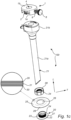
- Fig. 1c is an exploded view of a trocar fixation assembly 100 according to the present disclosure.
- the trocar fixation assembly 100 comprises a trocar fixation device 20 for holding a trocar 21 during use and an instrument fixation device 1 for holding a medical instrument 32 (as illustrated in Figs. 2c-2e ) inserted into the trocar 21.
- the trocar 21 extends in a longitudinal direction L and a transverse direction T.
- the trocar fixation device 20 comprises a skin attachment element 22, comprising an opening 23 configured for receiving the trocar 21 during use and being disposed about a longitudinal center line Lc (see Fig. 2a ), and a trocar support member 24 configured for hold the trocar 21 in position, as seen in the transverse direction T of the trocar 21.
- the trocar fixation assembly 20 illustrated in Fig. 1c comprises a medical dressing 25 which is optional to the present invention.
- the medical dressing 25 comprises an aperture 26 configured to encircle the trocar 21 during use.
- the aperture 26 may be a centrally disposed aperture configured to encircle the trocar 21 when inserted into the patient.
- the term "trocar fixation assembly” means an assembly comprising at least a trocar fixation device and an instrument fixation device.
- the trocar fixation assembly is to be used in conjunction with a trocar. More particularly, the trocar fixation assembly is used to receive and engage with a trocar cannula.
- the medical dressing may be fixedly or detachably attached to the trocar support member.
- the medical dressing is typically arranged to extend around the contour of the trocar support member and to be arranged in contact with the skin of the patient.
- skin attachment element means an element adapted to be arranged in contact with the skin of a patient.
- the skin attachment element is arranged in an area of the skin surrounding an incision. Accordingly, after an incision has been made in the skin, the skin attachment element is typically arranged to surround the incision and facilitate the positioning and insertion of a trocar.
- the skin attachment element 22 may comprise a base member 27 surrounding the bottom surface of the trocar support member 24.
- the base member 27 is arranged to contact the skin of the patient, i.e., the skin circumventing the incision.
- trocar support member means a member adapted to be in contact with the trocar and to hold and support the trocar in a predefined position, such that unwanted movements of the trocar, as seen in the transverse direction, is prevented.
- the trocar support member may be a separate part from or an integrated part with the skin attachment element. When the trocar support member is a separate part with respect to the skin attachment element, these may be connectable with each other with cooperating connection elements.
- the medical dressing may encircle the trocar support member such that essentially no gaps are formed between the fixation device and the dressing. This is to avoid potential entry of contaminants.
- the medical dressing 25 may be arranged to overlap the base member 27 of the skin-attachment element 22. Accordingly, the parts of the dressing 25 extending beyond the contour of the base member 27 of the skin attachment element 22 may be adhesively attached to the skin. Furthermore, the provision of the base member 27 secures that no gaps between the trocar fixation device 20 and the dressing 25 are formed.
- the trocar support member 24 comprises an upper support portion 28.
- the upper support portion 28 and the trocar support member 24 are connected by means of a screw mechanisms.
- the trocar fixation device 20 may have an open configuration allowing the trocar 21 to move freely in the longitudinal direction L and a closed configuration wherein the trocar 21 is restrained from movement in the longitudinal direction L, by clamping of the trocar 21.
- the medical dressing 25 may be arranged between the base member 27 and the upper support portion 28 of the trocar support member 24.
- the medical dressing 25 may be clamped between the base member 27 and the upper support portion 28 during use.
- the medical dressing may be detachably or fixedly attached to the base member.
- the medical dressing 25 may extend uninterrupted around the trocar support member 24 as disclosed in Fig. 1c . A tight seal is thereby provided, and the sterility is improved in the area circumventing the surgical site since the dressing contains no cuts or openable parts.
- the medical dressing 25 may comprise a backing layer 29 and an adhesive skin contact layer 30, as may be seen in the zoomed-in view of the medical dressing 25.
- the adhesive skin contact layer 30 secures a tight fit to the skin, and the dressing 25 is fixed in place in the area circumventing the surgical site (and the fixation device). Blood and body fluids exuded from the surgical site may be evaporated through the backing layer 29.
- the adhesive skin contact layer 30 may comprise a silicone-based adhesive.
- a silicone-based adhesive is skin-friendly and gentle to the skin.
- the silicone-based adhesive allows for the dressing 25 to be removed in a gentle manner from the skin without causing trauma and without interfering with the incision.
- the medical dressing 25 may furthermore comprise an absorbent pad 31 between the backing layer 29 and the adhesive skin contact layer 30.
- An absorbent pad 31 is beneficial to absorb the blood arising from the surgical intervention. It is desirable to prevent blood from accumulating at the skin, and instead secure removal and proper handling of the blood and body fluids by means of the absorbent pad 31. Furthermore, contaminating microorganisms are prevented from accumulating at the skin, and the sterility is thereby improved.
- the absorbent pad 31 may comprise a polyurethane foam.
- a polyurethane foam is beneficial since it is capable of absorbing large amounts of fluids, and also has a pressure relieving effect.
- the pressure relieving effect is beneficial to prevent pressure ulcers from occurring.
- a dressing comprising a polyurethane foam is also conformable.
- Fig. 2a illustrates the trocar fixation assembly 100 of Fig. 1c when the trocar fixation device 20 is attached to the skin of a patient and the trocar 20 is inserted into the patent and supported by the trocar fixation device 20.
- the trocar support member 24 of the trocar fixation device 20 holds the trocar in a straight configuration, coinciding with the longitudinal center line Lc.
- the instrument fixation device 1 comprises a trocar housing connector 2 adapted for connection between the instrument fixation device 1 and the trocar housing 21b.
- the trocar housing connector 2 of the instrument fixation device 1 is a snap-fit connector enabling a reversible connection with the trocar housing 21b.
- the trocar housing 21b comprises a corresponding fixation device connector 36, adapted to engage with the trocar housing connector 2 of the instrument fixation device 1, here in the form of a respective slit in the trocar housing connector 21b.
- the trocar support member 24 of the trocar fixation device 20 may be shifted between a first configuration 33, in which the trocar 21 is held in a straight configuration and coincides with the longitudinal center line Lc (shown in Fig. 2a ), and a second configured, wherein the trocar 21 is held in a slanted configuration 34, in which a longitudinal axis of the trocar 21 is arranged at an angle, ⁇ , with respect to the longitudinal center line Lc, as illustrated in Fig. 2b .
- the trocar is movable and may be held in a straight configuration and an angled configuration may allow the surgeon to change the position of the trocar in a simplified and more ergonomic manner.
- the surgeon does not need to lean over the patient or change position in the room in order to operate through the trocars, e.g. by inserting a variety of surgical tools into the trocar cannulas.
- Figs. 2c-2e illustrate the trocar fixation assembly 100 according to Fig. 1c , comprising the instrument fixation device 1 and the trocar fixation device 20.
- the instrument fixation device 1 is connected to the trocar housing 21b by means of the trocar housing connector 2.
- the instrument fixation device 1 comprises an instrument receiving opening 3 for receiving a medical instrument 32 being inserted into the trocar 21.
- the instrument fixation device is in an open configuration allowing the medical instrument to move freely in the longitudinal direction L.
- the instrument fixation device 1 is in a closed configuration wherein the medical instrument 32 is restrained from movement in the longitudinal direction L by clamping of the medical instrument 32 between the first and second opposing clamping surface 6,7 of the instrument fixation device 1 with a clamping force being directed in a transverse direction T, the transverse direction T being perpendicular to the longitudinal direction L.
- the instrument fixation device 1 may be shifted between the open configuration and the closed configuration by rotating the actuation member 8 such that the first clamping surface 6, here corresponding to the end portion 9a of the male threaded member 9, clamps, and thus frictionally engages, the medical instrument 32 between the first clamping surface 6 and the second clamping surface 7, the second clamping surface 7 here corresponding to an inner wall of the instrument receiving opening 3.
- Figs. 3a and 3b illustrate an alternative instrument fixation device 1 according to the present disclosure.
- the instrument fixation device 1 comprises in this embodiment, a first clamping section 35, a second clamping section 36 and an instrument fixation device housing 37, the first and the second clamping sections 35,36 being opposing sections forming together the opening 3 of the instrument fixation device 1 when being brought together.
- the instrument fixation device housing 37 comprises a trocar housing connector (not shown).
- a width of the second clamping section 36 is greater than a width of the first section 35, thus allowing the instrument fixation device 1 to hold smaller-sized medical instruments in the closed configuration.
- the first clamping section 35 comprises a first opposing clamping surface 6 on an inner surface thereof and a male threaded member 9 is connected to an outer surface of the first clamping section 35.
- the first clamping section 35 and the second clamping section 36 are inner sections and housed in the instrument fixation device housing 37.
- the instrument fixation device housing 37 is connected to the male threaded member 9 vi a female threaded housing member 38.
- the second clamping section 36 comprises a second opposing clamping surface 7 on an inner surface thereof and a female threaded member 10 is connected to the second clamping section 36 via a first and a second support arm 39,40.
- the male threaded member 9 and the female threaded member 10 are threadedly engaged with each other and upon rotation of an actuation member 8, the male threaded member 9 will gradually move in the transverse direction T and pull the first and second clamping sections 35,36 such that the first and second opposing clamping surfaces 6,7 approaches each other, as illustrated in Fig. 3a , until the medical instrument 32 is clamped between the first and the second clamping surfaces 6,7 and frictionally engaged in the closed position, as illustrated in Fig. 3b .
Abstract
An instrument fixation device 1 for holding a medical instrument 32 in a trocar system 21 is disclosed herein. The trocar system 21 comprising a trocar cannula 21a and a trocar housing 21b, the instrument fixation device 1 comprising a trocar housing connector 2 adapted for connection between the instrument fixation device 1 and the trocar housing 21b. The instrument fixation device 1 comprising an instrument receiving opening 3 and an open configuration allowing the medical instrument 32 to move freely in the longitudinal direction L and a closed configuration wherein the medical instrument 32 is restrained from movement in the longitudinal direction L by clamping of the medical instrument 32 between a first and second opposing clamping surface 6,7 of the instrument fixation device 1 with a clamping force being directed in a transverse direction T. The instrument fixation device 1 comprising an actuation member 8 configured shifting the configuration of the instrument fixation device 1 between the open configuration and the closed configuration, by means of a translational movement of the actuation member 8, the translational movement being in the transverse direction T of the instrument fixation device 1 and wherein the closed configuration of the instrument fixation device 1 includes a sub-set of positions with varying distances between the first and the second opposing clamping surface 6,7 in each of the positions in the sub-set of positions. Disclosed herein is also a trocar fixation assembly comprising a trocar fixation device and an instrument fixation device and a trocar system comprising a trocar and the trocar fixation assembly.
Description
- The present disclosure relates to an instrument fixation device for holding a medical instrument in a trocar system, a trocar fixation system comprising a trocar fixation device for holding a trocar during use and an instrument fixation device and a trocar system comprising a trocar and the trocar fixation system.
- Minimal invasive surgery (MIS) is a technique for performing surgery that is associated with less pain, shorter recovery and fewer complications compared to traditional open surgery.
- In an MIS procedure, a small incision is typically made through the skin of a patient, and a trocar is thereafter inserted into the incision. The trocar serves as a working channel that allows for the entry of various medical instruments and cameras into a body cavity, e.g. an abdominal cavity.
- A trocar typically comprises a cannula and an obturator. The cannula is a hollow and tube-shaped shaft that is placed inside the patient to provide access to the body cavity during the MIS procedure. The obturator is a sharp tool that allows the cannula to penetrate the body cavity for initial placement. When the cannula has entered the body cavity, the obturator is removed.
- During an MIS procedure, a plurality of trocars is typically utilized. Accordingly, the surgeon can utilize a variety of medical instruments and tools and alternate between the different trocars throughout the procedure. Often, the surgeon utilizes more than one surgical tool at the same time. In such scenarios, the medical staff must typically support the surgeon in fixating the trocar and the medical instruments in the trocar, and in facilitating the access to the various trocars.
- During laparascopic and other minimally invasive surgical procedures, it may be desired to lock the position of the medical instruments or tools inserted into the cannula. This is particularly the case for medical instruments intended for retracting and pushing away abdominal structures to facilitate access to a particular body cavity or organ where the surgical procedure is to be performed. This may currently be done by means of a staff member holding the medical instrument in position during the procedure.
- Similarly, the trocar needs to be held still in a straight or a tilted configuration in the body cavity. In order to keep a trocar in a straight or in a tilted configuration, a member of the surgical staff may need to hold the trocar in position. If the trocar is allowed to move freely within the body cavity and is repeatedly exposed to dislocation, this may cause undesired movement of the trocar inside the patient, and also increase the tension of the surgical incision and the skin area surrounding the incision. Furthermore, the surgeon must typically lean over the patient in order to access the plurality of trocars inserted into the skin of the patient. This may be challenging from an ergonomic perspective.
- In view of this, it would be desirable to be able to lock and unlock the position of the medical instrument after inserting the medical instrument in the trocar system in an improved and facilitated manner, to prevent dislocation of the trocar after insertion into the body cavity and also to facilitate the access to the trocars during the MIS procedure.
- Accordingly, there is a need to provide an improved and simplified means for fixating a medical instrument in a trocar system and as well fixating a trocar while also relieving the burden for the surgical staff in performing the minimal invasive surgery. Such means should provide for a simplified, and safe MIS procedure to be carried out.
- In view of the above-mentioned problems, it is an object of the present disclosure to provide improvements with respect to relieving the burden for surgical staff and providing a facilitated and improved means to handle and fixate a medical instrument in a trocar and to handle and fixate a medical instrument and a trocar during minimal invasive surgery.
- According to a first aspect, an instrument fixation device for holding a medical instrument in a trocar system is provided. The trocar system comprises a trocar cannula and a trocar housing. The instrument fixation device comprises a trocar housing connector adapted for connection between the instrument fixation device and the trocar housing. The instrument fixation device comprises a first side intended to face the trocar housing during use and a second side being an opposite side to the first side. The instrument fixation device comprises an instrument receiving opening extending in a longitudinal direction of the instrument fixation device between the first and the second side of the instrument fixation device. The instrument fixation device has an open configuration allowing the medical instrument to move freely in the longitudinal direction and a closed configuration wherein the medical instrument is restrained from movement in the longitudinal direction by clamping of the elongated portion of the medical instrument between a first and second opposing clamping surface of the instrument fixation device with a clamping force being directed in a transverse direction being perpendicular to the longitudinal direction. The instrument fixation device comprises an actuation member configured for shifting the configuration of the instrument fixation device between the open configuration and the closed configuration, by means of a translational movement of the actuation member. The translational movement being in the transverse direction of the instrument fixation device and wherein the closed configuration of the instrument fixation device includes a sub-set of positions with varying distances between the first and the second opposing clamping surface in each of the positions.
- The present invention is based on the realization that by providing an instrument fixation device being connectable to a trocar housing and which enables medical instruments of varying sizes to be locked in a fixed position within the cannula according to the present disclose, an improved, more flexible and facilitated minimally invasive surgical procedure could be achieved for the surgeon and for the surgical staff.
- The fact that the translational movement of the actuation member is in the transverse direction has been found to facilitate gripping of the instrument fixation device and handling of the actuation member, such that the configuration of the instrument fixation device may be shifted from open to closed configuration and the medical instrument may be locked in position, as seen in the longitudinal direction. Furthermore, as the translational movement of the actuation member is in the transverse direction when handling of the actuation member and shifting from the open to the closed position, the risk of accidentally rotating the trocar or displacing the trocar further into the incision, corresponding to the longitudinal direction of the instrument fixation device, is reduced and the handling of the instrument fixation device facilitated and improved.
- During the surgical procedure, the trocar may be used for more than one medical instrument. One or more of the trocars may be used for a medical instrument comprising a camera for inspection during the surgical procedure. Medical instruments for clamping or cutting of tissue may also be required and scissors, retractors or clip applier may be introduced during the surgical procedures, and at a different stage of the procedure be removed for introducing another one of the instruments via the same trocar. Medical instruments may vary in size and may have a variety of different diameters, and the instrument fixation device according to the present disclosure provides flexibility and facilitates the surgical procedure by providing a locked position irrespective of the diameter of the instrument fixation device. A limitation of the flexibility in the larger size diameter range evidentially being the size of the opening in the trocar.
- The actuation device may be configured for gradually moving, continuously or discontinuously, the first and/or second opposing clamping surface(s) in the transverse direction.
- The sub-sets of positions in the locked configuration may be either continuous or discontinuous positions. An actuation device and an instrument fixation device configured for gradually moving the first and/or second opposing clamping surfaces may provide a locked configuration of the instrument fixation device which is adapted to the diameter of the medical instrument, thus providing a highly flexible and improved instrument fixation device.
- The actuation device may be a self-locking device, which may be configured for locking the medical instruments, the sub-set of positions in the locked configuration being continuous or discontinuous positions, upon upon shifting the configuration of the instrument fixation device between the open and closed. By "continuous" is meant that the position of the first and/or second opposing clamping surfaces is determined by the diameter of the medical instrument and a frictional engagement between the medical instrument and the first and second opposing clamping surfaces. With "discontinuous" is meant that the positions of the first and second opposing clamping surfaces are predetermined positions.
- The actuation member may comprise one of; a first and a second cooperating threaded member, a ratched releasable locking mechanism or a lathe chuck mechanism.
- The actuation member may comprise a male threaded member and a female threaded member being engaged with each other, and wherein the instrument fixation device is shifted between the open configuration and the closed configuration by rotation of the male threaded member relative to the female threaded member. An advantage of such actuation member may be that it is easy to handle for the surgical staff and that it requires a relatively constant and low actuation pressure in the transverse direction, which has been found reducing the risk of undesired displacement of the medical instrument when shifting between the open and the close configuration.
- The first clamping surface of the instrument fixation device may constitute of an end portion of the male threaded member. The female threaded member may consequently be a threaded aperture extending, in the vertical direction, from an outer surface of the instrument fixation device and into the instrument receiving opening, the female threaded member being configured for threaded engagement with the male threaded member. Such configuration enables medical instrument of a wide variety of sizes to be fixed with the instrument fixation device and a constant and low actuation pressure to be used when shifting between the closed and the open configuration.
- The instrument fixation device may comprise a first clamping section and a second clamping section, the first and the second clamping sections being opposing sections forming together the instrument receiving opening, the first clamping section comprising the first opposing clamping surface and the second clamping section comprising the second opposing clamping surface of the instrument fixation device, wherein the male threaded member is connected to the first clamping section and the female threaded member is connected to the second clamping section. The female threaded member and the male threaded member may be arranged externally with respect to the instrument receiving opening and a main portion of the instrument fixation device.
- The trocar housing connector of the instrument fixation device may enable a reversible connection with the trocar housing, such as a snap-fit connection, a threaded connection or a press-fit connection. This enhances the flexibility of the instrument fixation device, which may be connected and reconnected with trocars of different position during surgery in a simple manner.
- According to a second aspect, the present disclosure relates to a trocar fixation assembly comprising a trocar fixation device for holding a trocar during use and an instrument fixation device for holding a medical instrument inserted into the trocar. The trocar extending in a longitudinal direction and a transverse direction and comprises a trocar cannula and a trocar housing. The trocar fixation device comprises a skin attachment element, comprising an opening configured for receiving the trocar during use and being disposed about a longitudinal center line, and a trocar support member configured for hold the trocar in position, as seen in the transverse direction of the trocar. The instrument fixation device comprises a trocar housing connector enabling connection between the instrument fixation device and the trocar housing. The instrument fixation device comprises a first side intended to face the trocar housing during use and a second side being an opposite side to the first side. The instrument fixation device furthermore comprises an instrument receiving opening extending in a longitudinal direction of the instrument fixation device between the first and the second side of the instrument fixation device. The instrument fixation device has an open position allowing the medical instrument to move freely in the longitudinal direction and a closed position wherein the medical instrument is restrained from movement in the longitudinal direction by clamping of the medical instrument between a first and second opposing clamping surface of the instrument fixation device, such that a distance between the first and the second opposing clamping surfaces is less when in the closed position and with a clamping force being directed in a transverse direction, being perpendicular to the longitudinal direction.
- A trocar fixation assembly according to the second aspect greatly facilitates the minimally invasive surgical procedure for the surgeon and for the surgical staff.
- The trocar fixation assembly comprises a trocar fixation device which may be attached to the skin of the patient by means of the skin attachment element and a trocar support member. The skin attachment element may comprise a first side being the skin facing side provided with for example a hydrophobic gel, such as silicon, and a second side, being the opposed side to the first side. The trocar support member may be arranged on, such as be connected to, the second side of the skin attachment element at the opening of skin attachment element. The trocar support member may alternatively be an integrated part of the skin attachment element. The trocar support member is configured for holding the trocar in position, as seen in the transverse direction of the trocar.
- The trocar is inserted into the patient via the opening in the skin attachment element and is held in position during surgery. This allows for the surgeon to leave a first trocar in the trocar fixation assembly and proceed with a second trocar (inserted into a second trocar fixation assembly) utilizing a different medical instrument.
- The first trocar may for example be utilized for inserting a medical instrument for holding, for example, tissue or part of an intestine by means of a retractor. The trocar fixation assembly comprises an instrument fixation device for holding a medical instrument inserted into the trocar. This means that undesired movement of the first trocar with the medical instrument may be prevented by the trocar fixation device and undesired movement of the medical instrument in the trocar, as seen in the longitudinal direction, may be prevented by the instrument fixation device, without the need of surgical staff needing to hold the trocar and the medical instrument in position, which may be challenging ergonomically. The trocar fixation assembly according to the present disclosure thus greatly reliefs the burden for the surgical staff in performing the minimally invasive surgery and
- This is beneficial since it prevents undesired movement of a trocar inside a body cavity of the patient (which may be risky), but still allows the surgeon to adjust the position of the trocar in a situation specific manner.
- The instrument fixation device may comprise an actuation member configured for gradually moving, continuously or discontinuously, the first and/or second opposing clamping surface(s) in the transverse direction between the open configuration and the closed configuration.
- The actuation member may be configured for gradually moving the first and/or second opposing clamping surface(s) by means of a translational movement of the actuation member being in the transverse direction of the instrument fixation device and wherein the instrument fixation device may hold elongated portions of medical instruments with varying diameters by varying the distance between the first and the second opposing clamping surfaces when the instrument fixation device is in the closed position.
- The trocar support member of the trocar fixation device may be shifted between a first configuration, in which the trocar is held in a straight configuration and coincides with the longitudinal center line, and a second configured, wherein the trocar is held in a slanted configuration, in which a longitudinal axis of the trocar is arranged at an angle, α, with respect to the longitudinal center line.
- This allows the surgeon to adjust the angular position of the trocar depending on his/her position in the room and depending on the specific surgical situation. Furthermore, the fact that the trocar is movable and may be held in a straight configuration and an angled configuration may allow the surgeon to change the position of the trocar in a simplified and more ergonomic manner. The surgeon does not need to lean over the patient or change position in the room in order to operate through the trocars, e.g. by inserting a variety of surgical tools into the trocar cannulas.
- The trocar housing connector of the instrument fixation device may enable a reversible connection with the trocar housing, such as a snap-fit connection, a threaded connection or a press-fit connection.
- The trocar fixation device may have an open configuration allowing the trocar to move freely in the longitudinal direction and a closed configuration wherein the trocar is restrained from movement in the longitudinal direction, by clamping of the trocar. The locking mechanism may for example be a screw mechanism or a lathe chuck mechanism.
- The trocar fixation assembly may comprise a medical dressing, the medical dressing comprising an aperture configured to encircle the trocar during use.
- The skin attachment element may comprise a base member surrounding the bottom surface of the trocar support member.
- The base member is arranged to contact the skin of the patient, i.e. the skin circumventing the incision.
- The medical dressing may comprise a centrally disposed aperture configured to encircle the trocar when inserted into the patient.
- The medical dressing may encircle the trocar support member such that essentially no gaps are formed between the fixation device and the dressing. This is to avoid potential entry of contaminants.
- The medical dressing may be arranged to overlap the base member of the skin-attachment element. Accordingly, the parts of the dressing extending beyond the contour of the base member of the skin attachment assembly may be adhesively attached to the skin. Furthermore, the provision of the base member secures that no gaps between the trocar support member and the dressing are formed.
- The medical dressing may be arranged between the base member and the trocar support member.
- Accordingly, the medical dressing may be clamped between the base member and the trocar support member during use. The medical dressing may be detachably or fixedly attached to the base member.
- During use, the medical dressing is typically fixed between the base member and the trocar support member. After surgery, when the trocar is to be exerted from the body cavity and the trocar fixation assembly is to be removed, each of the components may be removed separately, or together, if desired.
- The medical dressing extends uninterrupted around the trocar support member.
- A tight seal is thereby provided, and the sterility is improved in the area circumventing the surgical site since the dressing contains no cuts or openable parts.
- The medical dressing may comprise a backing layer and an adhesive skin contact layer.
- The adhesive skin contact layer secures a tight fit to the skin, and the dressing is fixed in place in the area circumventing the surgical site (and the fixation device). Blood and body fluids exuded from the surgical site may be evaporated through the backing layer.
- The adhesive skin contact layer may comprise a silicone based adhesive.
- A silicone based adhesive is skin-friendly and gentle to the skin. The silicone based adhesive allows for the dressing to be removed in a gentle manner from the skin without causing trauma and without interfering with the incision.
- The medical dressing may comprise an absorbent pad between the backing layer and the adhesive skin contact layer.
- An absorbent pad is beneficial to absorb the blood arising from the surgical intervention. It is desirable to prevent blood from accumulating at the skin, and instead secure removal and proper handling of the blood and body fluids by means of the absorbent pad. Furthermore, contaminating microorganisms are prevented from accumulating at the skin, and the sterility is thereby improved.
- The absorbent pad may comprise a polyurethane foam.
- A polyurethane foam is beneficial since it is capable of absorbing large amounts of fluids, and also has a pressure relieving effect. The pressure relieving effect is beneficial to prevent pressure ulcers from occurring. A dressing comprising a polyurethane foam is also conformable.
- According to a third aspect, the present disclosure relates to a trocar system comprising a trocar, the trocar comprising a trocar cannula and a trocar housing. The trocar system furthermore comprises a trocar fixation assembly according to the second aspect.
- Further features of, and advantages with, the present disclosure will become apparent when studying the appended claims and the following description. The skilled addressee realizes that different features of the present disclosure may be combined to create embodiments other than those described in the following, without departing from the scope of the present disclosure.
- The various aspects of the present disclosure, including its particular features and advantages, will be readily understood from the following detailed description and the accompanying drawings, in which:
-
Figure 1a-1b illustrate an instrument fixation device according to an exemplary embodiment of the present disclosure; -
Figure 1c is an exploded view of a trocar fixation assembly according to an exemplary embodiment of the present disclosure associated with a trocar -
Figure 2a-2d illustrate the trocar fixation assembly according toFig. 1c ; and -
Figure 3a-3b illustrate an instrument fixation device according to an exemplary embodiment of the present disclosure - The present disclosure will now be described more fully hereinafter with reference to the accompanying drawings, in which currently preferred embodiments of the present disclosure are shown. The present disclosure may, however, be embodied in many different forms and should not be construed as limited to the embodiments set forth herein; rather, these embodiments are provided for thoroughness and completeness, and fully convey the scope of the present disclosure to the skilled person. Like reference number denote similar features throughout the figures. Reference numbers may be omitted in some figures for better visibility, in which case reference is made to the other figures.
- With reference to
Figs 1a and 1b , aninstrument fixation device 1 according to an exemplary embodiment of the present disclosure is conceptually illustrated. Theinstrument fixation device 1 is configured for holding a medical instrument 32 (shown inFig. 2c-2e ) in a trocar system. The trocar system comprises atrocar cannula 21a and atrocar housing 21b (shown inFig. 1c ). Theinstrument fixation device 1 comprises atrocar housing connector 2 adapted for connection between theinstrument fixation device 1 and thetrocar housing 21b. Thetrocar housing connector 2 of theinstrument fixation device 1 enables a reversible connection with thetrocar housing 21b. InFigs. 1a and 1b , the reversible connection is illustrated as a snap-fit connection. However, the connection may alternatively by a threaded connection or a press-fit connection. - The
instrument fixation device 1 comprises afirst side 1a intended to face thetrocar housing 21b during use and asecond side 1b being an opposite side to thefirst side 1a. Theinstrument fixation device 1 comprises aninstrument receiving opening 3 extending in a longitudinal direction L of theinstrument fixation device 1 between the first and thesecond side instrument fixation device 1. - The
instrument fixation device 1 has anopen configuration 4, as illustrated inFigs. 1a and 1b , allowing the medical instrument to move freely in the longitudinal direction L (shown inFig. 2d ) and a closed configuration 5 (shown inFig. 2e ) wherein the medical instrument is restrained from movement in the longitudinal direction L by clamping of themedical instrument 32 between a first and a second opposing clampingsurface instrument fixation device 1 with a clamping force being directed in a transverse direction T. The transverse direction T is perpendicular to the longitudinal direction L. Theinstrument fixation device 1 comprises anactuation member 8 for shifting the configuration of theinstrument fixation device 1 between theopen configuration 4 and the closed configuration 5, by means of a translational movement of theactuation member 8, the translational movement being in the transverse direction T of theinstrument fixation device 1. In the exemplary embodiment shown inFigs. 1a and 1b , theactuation member 8 comprises anactuation member knob 11, a first and a second cooperating threaded member in the form of a male threadedmember 9 and a female threadedmember 10 being threadedly engaged with each other. Anend portion 9a of the male threadedmember 9, arranged at an opposite end portion with respect to theactuation member knob 11, forms thefirst clamping surface 6 of theinstrument fixation device 1. Theinstrument fixation device 1 may shifted between theopen configuration 4 and the closed configuration 5 by rotating theactuation member knob 11, and thus the male threadedmember 9 relative to the female threadedmember 10. - The
actuation member 8 is a mechanical linear actuator configured for gradually moving the first opposing clampingsurface 6 in the transverse direction T by converting a rotary motion into linear motion such that thefirst clamping surface 6, here theend portion 9a of the male threadedmember 9, approaches thesecond clamping surface 7 of theinstrument fixation device 1. The female threadedmember 10 is a threaded aperture extending, in the transverse direction T, from an outer surface of theinstrument fixation device 1 and into theinstrument receiving opening 3. Such configuration enables medical instrument of a wide variety of sizes to be fixed with the instrument fixation device and a constant and low actuation pressure to be used when shifting between the open and the closed configuration. - As used herein, the term "trocar" means a device through which a minimal invasive surgery can be accomplished. The trocar typically comprises at least a cannula; i.e. a hollow tube shaped member, a gas-tight valve and a trocar housing. The trocar allows for the entry of surgical instruments, such as graspers, scissors, staplers, cameras etc.
-
Fig. 1c is an exploded view of atrocar fixation assembly 100 according to the present disclosure. Thetrocar fixation assembly 100 comprises atrocar fixation device 20 for holding atrocar 21 during use and aninstrument fixation device 1 for holding a medical instrument 32 (as illustrated inFigs. 2c-2e ) inserted into thetrocar 21. Thetrocar 21 extends in a longitudinal direction L and a transverse direction T. Thetrocar fixation device 20 comprises askin attachment element 22, comprising anopening 23 configured for receiving thetrocar 21 during use and being disposed about a longitudinal center line Lc (seeFig. 2a ), and atrocar support member 24 configured for hold thetrocar 21 in position, as seen in the transverse direction T of thetrocar 21. Thetrocar fixation assembly 20 illustrated inFig. 1c comprises amedical dressing 25 which is optional to the present invention. Themedical dressing 25 comprises anaperture 26 configured to encircle thetrocar 21 during use. Theaperture 26 may be a centrally disposed aperture configured to encircle thetrocar 21 when inserted into the patient. - As used herein, the term "trocar fixation assembly" means an assembly comprising at least a trocar fixation device and an instrument fixation device. The trocar fixation assembly is to be used in conjunction with a trocar. More particularly, the trocar fixation assembly is used to receive and engage with a trocar cannula.
- The medical dressing may be fixedly or detachably attached to the trocar support member. The medical dressing is typically arranged to extend around the contour of the trocar support member and to be arranged in contact with the skin of the patient.
- The term "skin attachment element" means an element adapted to be arranged in contact with the skin of a patient. Typically, the skin attachment element is arranged in an area of the skin surrounding an incision. Accordingly, after an incision has been made in the skin, the skin attachment element is typically arranged to surround the incision and facilitate the positioning and insertion of a trocar.
- The
skin attachment element 22 may comprise abase member 27 surrounding the bottom surface of thetrocar support member 24. Thebase member 27 is arranged to contact the skin of the patient, i.e., the skin circumventing the incision. - The term "trocar support member" means a member adapted to be in contact with the trocar and to hold and support the trocar in a predefined position, such that unwanted movements of the trocar, as seen in the transverse direction, is prevented. The trocar support member may be a separate part from or an integrated part with the skin attachment element. When the trocar support member is a separate part with respect to the skin attachment element, these may be connectable with each other with cooperating connection elements.
- The medical dressing may encircle the trocar support member such that essentially no gaps are formed between the fixation device and the dressing. This is to avoid potential entry of contaminants.
- The
medical dressing 25 may be arranged to overlap thebase member 27 of the skin-attachment element 22. Accordingly, the parts of the dressing 25 extending beyond the contour of thebase member 27 of theskin attachment element 22 may be adhesively attached to the skin. Furthermore, the provision of thebase member 27 secures that no gaps between thetrocar fixation device 20 and the dressing 25 are formed. - Optionally, and as disclosed in
Fig. 1c , thetrocar support member 24 comprises anupper support portion 28. In this figure, theupper support portion 28 and thetrocar support member 24 are connected by means of a screw mechanisms. Thetrocar fixation device 20 may have an open configuration allowing thetrocar 21 to move freely in the longitudinal direction L and a closed configuration wherein thetrocar 21 is restrained from movement in the longitudinal direction L, by clamping of thetrocar 21. Themedical dressing 25 may be arranged between thebase member 27 and theupper support portion 28 of thetrocar support member 24. - Accordingly, the
medical dressing 25 may be clamped between thebase member 27 and theupper support portion 28 during use. The medical dressing may be detachably or fixedly attached to the base member. - The
medical dressing 25 may extend uninterrupted around thetrocar support member 24 as disclosed inFig. 1c . A tight seal is thereby provided, and the sterility is improved in the area circumventing the surgical site since the dressing contains no cuts or openable parts. Themedical dressing 25 may comprise abacking layer 29 and an adhesiveskin contact layer 30, as may be seen in the zoomed-in view of themedical dressing 25. - The adhesive
skin contact layer 30 secures a tight fit to the skin, and the dressing 25 is fixed in place in the area circumventing the surgical site (and the fixation device). Blood and body fluids exuded from the surgical site may be evaporated through thebacking layer 29. The adhesiveskin contact layer 30 may comprise a silicone-based adhesive. A silicone-based adhesive is skin-friendly and gentle to the skin. The silicone-based adhesive allows for the dressing 25 to be removed in a gentle manner from the skin without causing trauma and without interfering with the incision. As illustrated in the zoomed-in view of themedical dressing 25, themedical dressing 25 may furthermore comprise anabsorbent pad 31 between thebacking layer 29 and the adhesiveskin contact layer 30. - An
absorbent pad 31 is beneficial to absorb the blood arising from the surgical intervention. It is desirable to prevent blood from accumulating at the skin, and instead secure removal and proper handling of the blood and body fluids by means of theabsorbent pad 31. Furthermore, contaminating microorganisms are prevented from accumulating at the skin, and the sterility is thereby improved. Theabsorbent pad 31 may comprise a polyurethane foam. - A polyurethane foam is beneficial since it is capable of absorbing large amounts of fluids, and also has a pressure relieving effect. The pressure relieving effect is beneficial to prevent pressure ulcers from occurring. A dressing comprising a polyurethane foam is also conformable.
-
Fig. 2a illustrates thetrocar fixation assembly 100 ofFig. 1c when thetrocar fixation device 20 is attached to the skin of a patient and thetrocar 20 is inserted into the patent and supported by thetrocar fixation device 20. Thetrocar support member 24 of thetrocar fixation device 20 holds the trocar in a straight configuration, coinciding with the longitudinal center line Lc. - The
instrument fixation device 1 comprises atrocar housing connector 2 adapted for connection between theinstrument fixation device 1 and thetrocar housing 21b. Thetrocar housing connector 2 of theinstrument fixation device 1 is a snap-fit connector enabling a reversible connection with thetrocar housing 21b. Thetrocar housing 21b comprises a correspondingfixation device connector 36, adapted to engage with thetrocar housing connector 2 of theinstrument fixation device 1, here in the form of a respective slit in thetrocar housing connector 21b. - As illustrated in
Figs. 2a and2b , thetrocar support member 24 of thetrocar fixation device 20 may be shifted between afirst configuration 33, in which thetrocar 21 is held in a straight configuration and coincides with the longitudinal center line Lc (shown inFig. 2a ), and a second configured, wherein thetrocar 21 is held in a slantedconfiguration 34, in which a longitudinal axis of thetrocar 21 is arranged at an angle, α, with respect to the longitudinal center line Lc, as illustrated inFig. 2b . - This allows the surgeon to adjust the angular position of the trocar depending on his/her position in the room and depending on the specific surgical situation. Furthermore, the fact that the trocar is movable and may be held in a straight configuration and an angled configuration may allow the surgeon to change the position of the trocar in a simplified and more ergonomic manner. The surgeon does not need to lean over the patient or change position in the room in order to operate through the trocars, e.g. by inserting a variety of surgical tools into the trocar cannulas.
-
Figs. 2c-2e illustrate thetrocar fixation assembly 100 according toFig. 1c , comprising theinstrument fixation device 1 and thetrocar fixation device 20. Theinstrument fixation device 1 is connected to thetrocar housing 21b by means of thetrocar housing connector 2. Theinstrument fixation device 1 comprises aninstrument receiving opening 3 for receiving amedical instrument 32 being inserted into thetrocar 21. InFigs. 2c and2d , the instrument fixation device is in an open configuration allowing the medical instrument to move freely in the longitudinal direction L. InFig. 2e , theinstrument fixation device 1 is in a closed configuration wherein themedical instrument 32 is restrained from movement in the longitudinal direction L by clamping of themedical instrument 32 between the first and second opposing clampingsurface instrument fixation device 1 with a clamping force being directed in a transverse direction T, the transverse direction T being perpendicular to the longitudinal direction L. Theinstrument fixation device 1 may be shifted between the open configuration and the closed configuration by rotating theactuation member 8 such that thefirst clamping surface 6, here corresponding to theend portion 9a of the male threadedmember 9, clamps, and thus frictionally engages, themedical instrument 32 between thefirst clamping surface 6 and thesecond clamping surface 7, thesecond clamping surface 7 here corresponding to an inner wall of theinstrument receiving opening 3. -
Figs. 3a and 3b illustrate an alternativeinstrument fixation device 1 according to the present disclosure. Theinstrument fixation device 1 comprises in this embodiment, afirst clamping section 35, asecond clamping section 36 and an instrumentfixation device housing 37, the first and thesecond clamping sections opening 3 of theinstrument fixation device 1 when being brought together. The instrumentfixation device housing 37 comprises a trocar housing connector (not shown). As may be seen in the figures, a width of thesecond clamping section 36 is greater than a width of thefirst section 35, thus allowing theinstrument fixation device 1 to hold smaller-sized medical instruments in the closed configuration. Thefirst clamping section 35 comprises a first opposing clampingsurface 6 on an inner surface thereof and a male threadedmember 9 is connected to an outer surface of thefirst clamping section 35. Thefirst clamping section 35 and thesecond clamping section 36 are inner sections and housed in the instrumentfixation device housing 37. The instrumentfixation device housing 37 is connected to the male threadedmember 9 vi a female threadedhousing member 38. Thesecond clamping section 36 comprises a second opposing clampingsurface 7 on an inner surface thereof and a female threadedmember 10 is connected to thesecond clamping section 36 via a first and asecond support arm member 9 and the female threadedmember 10 are threadedly engaged with each other and upon rotation of anactuation member 8, the male threadedmember 9 will gradually move in the transverse direction T and pull the first andsecond clamping sections surfaces Fig. 3a , until themedical instrument 32 is clamped between the first and the second clamping surfaces 6,7 and frictionally engaged in the closed position, as illustrated inFig. 3b . - Even though the present disclosure has been described with reference to specific exemplifying embodiments thereof, many different alterations, modifications and the like will become apparent for those skilled in the art.
- Variations to the disclosed embodiments can be understood and effected by the skilled addressee in practicing the present disclosure, from a study of the drawings, the disclosure, and the appended claims. Furthermore, in the claims, the word "comprising" does not exclude other elements or steps, and the indefinite article "a" or "an" does not exclude a plurality.
Claims (15)
- An instrument fixation device (1) for holding a medical instrument (32) in a trocar system (21), the trocar system (21) comprising a trocar cannula (21a) and a trocar housing (21b), the instrument fixation device (1) comprising a trocar housing connector (2) adapted for connection between the instrument fixation device (1) and the trocar housing (21b), the instrument fixation device (1) comprising a first side (1a) intended to face the trocar housing (21b) during use and a second side (1b) being an opposite side to the first side (1a), the instrument fixation device (1) comprising an instrument receiving opening (3) extending in a longitudinal direction (L) of the instrument fixation device (1) between the first and the second side (1a, 1b) of the instrument fixation device (1), the instrument fixation device (1) having an open configuration allowing the medical instrument (32) to move freely in the longitudinal direction (L) and a closed configuration wherein the medical instrument (32) is restrained from movement in the longitudinal direction (L) by clamping of the medical instrument (32) between a first and second opposing clamping surface (6,7) of the instrument fixation device (1) with a clamping force being directed in a transverse direction (T) being perpendicular to the longitudinal direction (L), the instrument fixation device (1) comprising an actuation member (8) configured for shifting the configuration of the instrument fixation device (1) between the open configuration and the closed configuration, by means of a translational movement of the actuation member (8), the translational movement being in the transverse direction (T) of the instrument fixation device (1) and wherein the closed configuration of the instrument fixation device (1) includes a sub-set of positions with varying distances between the first and the second opposing clamping surface (6,7) in each of the positions in the sub-set of positions.
- The instrument fixation device (1) according to claim 1, wherein the actuation device (8) is configured for gradually moving, continuously or discontinuously, the first and/or second opposing clamping surface(s) (6,7) in the transverse direction (T).
- The instrument fixation device (1) according to any claim 1 or 2, wherein the actuation member (8) comprises one of; a first and a second cooperating threaded member (9,10), a ratched releasable locking mechanism or a lathe chuck.
- The instrument fixation device (1) according to any one of the preceding claims, wherein the actuation member (8) comprises a male threaded member (9) and a female threaded member (10) being engaged with each other, and wherein the instrument fixation device (1) is shifted between the open configuration and the closed configuration by rotating the male threaded member (9) relative to the female threaded member (10).
- The instrument fixation device (1) according to claim 4, wherein the first clamping surface (6) of the instrument fixation device (1) constitutes of an end portion (9a) of the male threaded member (9).
- The instrument fixation device (1) according to claim 4, wherein the instrument fixation device (1) comprises a first clamping section (35) and a second clamping section (36), the first and the second clamping sections (35,36) being opposing sections forming together the opening (3), the first clamping section (35) comprising the first opposing clamping surface (6) and the second clamping section (36) comprising the second opposing clamping surface (7) of the instrument fixation device (1), wherein the male threaded member (9) is connected to the first clamping section (35) and the female threaded member (10) is connected to the second clamping section (36).
- The instrument fixation device (1) according to any one of the preceding claims, wherein the trocar housing connector (21b) of the instrument fixation device (1) enables a reversible connection with the trocar housing (21b), such as a snap-fit connection, a threaded connection, or a press-fit connection.
- A trocar fixation assembly (100) comprising a trocar fixation device (20) for holding a trocar (21) during use and an instrument fixation device (1) for holding a medical instrument () inserted into the trocar (), the trocar comprising a trocar cannula () and a trocar housing () and extends in a longitudinal direction (L) and a transverse direction (T), wherein the trocar fixation device (20) comprises a skin attachment element (22), comprising an opening (23) configured for receiving the trocar (21) during use and being disposed about a longitudinal center line (Lc), and a trocar support member (24) configured for hold the trocar in position, as seen in the transverse direction of the trocar (21), the instrument fixation device (1) comprises a trocar housing connector (21b) enabling connection between the instrument fixation device (1) and the trocar housing (21b), the instrument fixation device (1) comprising a first side (1a) intended to face the trocar housing (21b) during use and a second side (1b) being an opposite side to the first side (1a), the instrument fixation device (1) comprising an instrument receiving opening (3) extending in a longitudinal direction (L) of the instrument fixation device (1) between the first and the second side (1a, 1b) of the instrument fixation device (1), the instrument fixation device (1) has an open position allowing the medical instrument (32) to move freely in the longitudinal direction (L) and a closed position wherein the medical instrument (32) is retrained from movement in the longitudinal direction (L) by clamping of the medical instrument (32) between a first and second opposing clamping surface (6,7) of the instrument fixation device (1) with a distance between the first and the second opposing clamping surfaces (6,7) is less when in the closed position compared to when in the open position, and with a clamping force being directed in a transverse direction (T), being perpendicular to the longitudinal direction (L).
- The trocar fixation assembly (100) according to claim 8, wherein the instrument fixation device (1) comprising an actuation member (8) configured for gradually moving, continuously or discontinuously, the first and/or second opposing clamping surface(s) (6,7) in the transverse direction (T) between the open configuration and the closed configuration.
- The trocar fixation assembly (100) according to claim 8 or 9, wherein the actuation member (8) is configured for gradually moving the first and/or second opposing clamping surface(s) (6,7) by means of a translational movement of the actuation member (8) being in the transverse direction (T) of the instrument fixation device (1) and wherein the instrument fixation device (1) may hold medical instruments with varying diameters by varying the distance between the first and the second opposing clamping surfaces when the instrument fixation device (1) is in the closed position.
- The trocar fixation assembly (100) according to any one of claims 8 to 10, wherein the trocar support member (24) of the trocar fixation device (20) may be shifted between a first configuration (33), in which the trocar (21) is held in a straight configuration and coincides with the longitudinal center line (Lc), and a second configured (34), wherein the trocar (21) is held in a slanted configuration, in which a longitudinal axis of the trocar is arranged at an angle, α, with respect to the longitudinal center line (Lc).
- The trocar fixation assembly (100) according to any one of claims 8 to 11, wherein the trocar housing connector (2) of the instrument fixation device (1) enables a reversible connection with the trocar housing (21b), such as a snap-fit connection, a threaded connection or a press-fit connection.
- The trocar fixation assembly (100) according to any one of claims 8 to 12, wherein the trocar fixation assembly (100) comprises a medical dressing (25), the medical dressing (25) comprising an aperture (26) configured to encircle the trocar (21) during use.
- The trocar fixation assembly (100) according to any one of claims 8 to 13, wherein the trocar fixation device (20) has an open configuration allowing the trocar (21) to move freely in the longitudinal direction (L) and a closed configuration wherein the trocar (21) is restrained from movement in the longitudinal direction (L), by clamping of the trocar (21).
- A trocar system comprising a trocar (21) comprising a trocar cannula (21a) and a trocar housing (21b), the trocar system comprising a trocar fixation assembly (100) according to any one of claim 8 to 14.
Priority Applications (1)
| Application Number | Priority Date | Filing Date | Title |
|---|---|---|---|
| EP22170525.4A EP4268738A1 (en) | 2022-04-28 | 2022-04-28 | An instrument fixation assembly and a trocar fixation system |
Applications Claiming Priority (1)
| Application Number | Priority Date | Filing Date | Title |
|---|---|---|---|
| EP22170525.4A EP4268738A1 (en) | 2022-04-28 | 2022-04-28 | An instrument fixation assembly and a trocar fixation system |
Publications (1)
| Publication Number | Publication Date |
|---|---|
| EP4268738A1 true EP4268738A1 (en) | 2023-11-01 |
Family
ID=81392567
Family Applications (1)
| Application Number | Title | Priority Date | Filing Date |
|---|---|---|---|
| EP22170525.4A Pending EP4268738A1 (en) | 2022-04-28 | 2022-04-28 | An instrument fixation assembly and a trocar fixation system |
Country Status (1)
| Country | Link |
|---|---|
| EP (1) | EP4268738A1 (en) |
Citations (9)
| Publication number | Priority date | Publication date | Assignee | Title |
|---|---|---|---|---|
| US5190561A (en) * | 1991-01-23 | 1993-03-02 | Surgical Innovations, Inc. | Tissue and organ extractor |
| US5662613A (en) * | 1996-05-14 | 1997-09-02 | Astarita; Denis C. | Endoscopic instrument lock |
| US5713869A (en) * | 1995-03-08 | 1998-02-03 | Morejon; Orlando | Trocar assembly |
| US20100010449A1 (en) * | 2008-07-09 | 2010-01-14 | Kyphon Sarl | Cannula Stabilization Device, System, And Method Of Use |
| US20100292724A1 (en) * | 2006-03-13 | 2010-11-18 | Sundaram Ravikumar | Minimally Invasive Surgical Clamps, Assemblies and Methods |
| US20130085342A1 (en) * | 2011-09-30 | 2013-04-04 | Ethicon Endo-Surgery, Inc. | Patient-referenced surgical support frame |
| EP2965700A1 (en) * | 2014-07-09 | 2016-01-13 | Covidien LP | Instrument fixation device for depth and angle fixation |
| WO2020243156A1 (en) * | 2019-05-31 | 2020-12-03 | Nico Corporation | Adapter for a surgical access device |
| US20210068917A1 (en) * | 2018-03-21 | 2021-03-11 | The Provost, Fellows, Scholars And Other Members Of Board Of Trinity College Dublin | Stabiliser |
-
2022
- 2022-04-28 EP EP22170525.4A patent/EP4268738A1/en active Pending
Patent Citations (9)
| Publication number | Priority date | Publication date | Assignee | Title |
|---|---|---|---|---|
| US5190561A (en) * | 1991-01-23 | 1993-03-02 | Surgical Innovations, Inc. | Tissue and organ extractor |
| US5713869A (en) * | 1995-03-08 | 1998-02-03 | Morejon; Orlando | Trocar assembly |
| US5662613A (en) * | 1996-05-14 | 1997-09-02 | Astarita; Denis C. | Endoscopic instrument lock |
| US20100292724A1 (en) * | 2006-03-13 | 2010-11-18 | Sundaram Ravikumar | Minimally Invasive Surgical Clamps, Assemblies and Methods |
| US20100010449A1 (en) * | 2008-07-09 | 2010-01-14 | Kyphon Sarl | Cannula Stabilization Device, System, And Method Of Use |
| US20130085342A1 (en) * | 2011-09-30 | 2013-04-04 | Ethicon Endo-Surgery, Inc. | Patient-referenced surgical support frame |
| EP2965700A1 (en) * | 2014-07-09 | 2016-01-13 | Covidien LP | Instrument fixation device for depth and angle fixation |
| US20210068917A1 (en) * | 2018-03-21 | 2021-03-11 | The Provost, Fellows, Scholars And Other Members Of Board Of Trinity College Dublin | Stabiliser |
| WO2020243156A1 (en) * | 2019-05-31 | 2020-12-03 | Nico Corporation | Adapter for a surgical access device |
Similar Documents
| Publication | Publication Date | Title |
|---|---|---|
| EP2621348B1 (en) | Natural orifice surgery system | |
| AU2005275261B2 (en) | Laparoscopic instrument and cannula assembly and related surgical method | |
| US5263939A (en) | Retainer for laparoscopic cannula | |
| US20120101341A1 (en) | Surgical access device system and methods of use | |
| EP2181657A2 (en) | Surgical access port with adjustable ring geometry | |
| US20130053896A1 (en) | Adaptable systems, methods, and devices for percutaneously implanting a spinal screw | |
| KR20100112087A (en) | Retractor with flexible sleeve | |
| US20140051934A1 (en) | Stabilizing Port for Surgery for Facilitating Concurrent Introduction of Multiple Instruments | |
| US9414830B2 (en) | Surgical access assembly including adhesive members for secure attachment to skin surfaces | |
| US20100305407A1 (en) | Malleable Port Retractor | |
| US20220117626A1 (en) | Skin foundation access portal | |
| JP3743512B2 (en) | Surgical mantle | |
| EP4268738A1 (en) | An instrument fixation assembly and a trocar fixation system | |
| US20210220009A1 (en) | Surgical retractor fixation device/shim | |
| EP4186448A1 (en) | A trocar fixation assembly | |
| EP4186449A1 (en) | A trocar fixation assembly | |
| KR102205181B1 (en) | Trocar | |
| EP3698741A1 (en) | Access assembly including flexible cannula | |
| US11612386B2 (en) | Universal surgical access system | |
| US20190053916A1 (en) | Oblique inserter |
Legal Events
| Date | Code | Title | Description |
|---|---|---|---|
| PUAI | Public reference made under article 153(3) epc to a published international application that has entered the european phase |
Free format text: ORIGINAL CODE: 0009012 |
|
| STAA | Information on the status of an ep patent application or granted ep patent |
Free format text: STATUS: THE APPLICATION HAS BEEN PUBLISHED |
|
| AK | Designated contracting states |
Kind code of ref document: A1 Designated state(s): AL AT BE BG CH CY CZ DE DK EE ES FI FR GB GR HR HU IE IS IT LI LT LU LV MC MK MT NL NO PL PT RO RS SE SI SK SM TR |
|
| STAA | Information on the status of an ep patent application or granted ep patent |
Free format text: STATUS: REQUEST FOR EXAMINATION WAS MADE |