EP4266321A1 - Ocr-based extraction of clinical data from dicom sc images - Google Patents
Ocr-based extraction of clinical data from dicom sc images Download PDFInfo
- Publication number
- EP4266321A1 EP4266321A1 EP22169432.6A EP22169432A EP4266321A1 EP 4266321 A1 EP4266321 A1 EP 4266321A1 EP 22169432 A EP22169432 A EP 22169432A EP 4266321 A1 EP4266321 A1 EP 4266321A1
- Authority
- EP
- European Patent Office
- Prior art keywords
- image
- dicom
- images
- characters
- sub
- Prior art date
- Legal status (The legal status is an assumption and is not a legal conclusion. Google has not performed a legal analysis and makes no representation as to the accuracy of the status listed.)
- Pending
Links
- 238000000605 extraction Methods 0.000 title description 16
- 238000000034 method Methods 0.000 claims abstract description 73
- 238000012015 optical character recognition Methods 0.000 claims description 50
- 238000004891 communication Methods 0.000 claims description 8
- 229940079593 drug Drugs 0.000 claims description 6
- 239000003814 drug Substances 0.000 claims description 6
- 238000003384 imaging method Methods 0.000 claims description 6
- 230000036541 health Effects 0.000 claims description 4
- 238000009966 trimming Methods 0.000 claims description 4
- 238000012545 processing Methods 0.000 abstract description 10
- 238000011160 research Methods 0.000 description 12
- 238000002059 diagnostic imaging Methods 0.000 description 9
- 238000002591 computed tomography Methods 0.000 description 8
- 230000008569 process Effects 0.000 description 7
- 238000002595 magnetic resonance imaging Methods 0.000 description 6
- 229920003259 poly(silylenemethylene) Polymers 0.000 description 5
- 238000003745 diagnosis Methods 0.000 description 4
- 238000002600 positron emission tomography Methods 0.000 description 4
- 238000002604 ultrasonography Methods 0.000 description 4
- 230000008878 coupling Effects 0.000 description 3
- 238000010168 coupling process Methods 0.000 description 3
- 238000005859 coupling reaction Methods 0.000 description 3
- 230000001419 dependent effect Effects 0.000 description 3
- 238000012805 post-processing Methods 0.000 description 3
- 230000006835 compression Effects 0.000 description 2
- 238000007906 compression Methods 0.000 description 2
- 238000004590 computer program Methods 0.000 description 2
- 238000007418 data mining Methods 0.000 description 2
- 238000013135 deep learning Methods 0.000 description 2
- 238000010586 diagram Methods 0.000 description 2
- 238000005516 engineering process Methods 0.000 description 2
- 230000006870 function Effects 0.000 description 2
- 238000010191 image analysis Methods 0.000 description 2
- 238000012986 modification Methods 0.000 description 2
- 230000004048 modification Effects 0.000 description 2
- 238000007781 pre-processing Methods 0.000 description 2
- 238000002601 radiography Methods 0.000 description 2
- OYPRJOBELJOOCE-UHFFFAOYSA-N Calcium Chemical compound [Ca] OYPRJOBELJOOCE-UHFFFAOYSA-N 0.000 description 1
- 241001386813 Kraken Species 0.000 description 1
- 230000008901 benefit Effects 0.000 description 1
- 210000004556 brain Anatomy 0.000 description 1
- 229910052791 calcium Inorganic materials 0.000 description 1
- 239000011575 calcium Substances 0.000 description 1
- 238000006243 chemical reaction Methods 0.000 description 1
- 230000001149 cognitive effect Effects 0.000 description 1
- 239000012141 concentrate Substances 0.000 description 1
- 238000013461 design Methods 0.000 description 1
- 201000010099 disease Diseases 0.000 description 1
- 208000037265 diseases, disorders, signs and symptoms Diseases 0.000 description 1
- 230000002526 effect on cardiovascular system Effects 0.000 description 1
- 238000003709 image segmentation Methods 0.000 description 1
- 238000009434 installation Methods 0.000 description 1
- 238000002955 isolation Methods 0.000 description 1
- 210000004185 liver Anatomy 0.000 description 1
- 238000005259 measurement Methods 0.000 description 1
- 238000003909 pattern recognition Methods 0.000 description 1
- 230000011218 segmentation Effects 0.000 description 1
- 238000012795 verification Methods 0.000 description 1
Images


Classifications
-
- G—PHYSICS
- G06—COMPUTING; CALCULATING OR COUNTING
- G06V—IMAGE OR VIDEO RECOGNITION OR UNDERSTANDING
- G06V30/00—Character recognition; Recognising digital ink; Document-oriented image-based pattern recognition
- G06V30/40—Document-oriented image-based pattern recognition
- G06V30/42—Document-oriented image-based pattern recognition based on the type of document
-
- G—PHYSICS
- G06—COMPUTING; CALCULATING OR COUNTING
- G06V—IMAGE OR VIDEO RECOGNITION OR UNDERSTANDING
- G06V30/00—Character recognition; Recognising digital ink; Document-oriented image-based pattern recognition
- G06V30/10—Character recognition
- G06V30/14—Image acquisition
- G06V30/148—Segmentation of character regions
- G06V30/153—Segmentation of character regions using recognition of characters or words
-
- G—PHYSICS
- G16—INFORMATION AND COMMUNICATION TECHNOLOGY [ICT] SPECIALLY ADAPTED FOR SPECIFIC APPLICATION FIELDS
- G16H—HEALTHCARE INFORMATICS, i.e. INFORMATION AND COMMUNICATION TECHNOLOGY [ICT] SPECIALLY ADAPTED FOR THE HANDLING OR PROCESSING OF MEDICAL OR HEALTHCARE DATA
- G16H30/00—ICT specially adapted for the handling or processing of medical images
- G16H30/40—ICT specially adapted for the handling or processing of medical images for processing medical images, e.g. editing
-
- G—PHYSICS
- G06—COMPUTING; CALCULATING OR COUNTING
- G06V—IMAGE OR VIDEO RECOGNITION OR UNDERSTANDING
- G06V30/00—Character recognition; Recognising digital ink; Document-oriented image-based pattern recognition
- G06V30/10—Character recognition
- G06V30/14—Image acquisition
- G06V30/148—Segmentation of character regions
- G06V30/15—Cutting or merging image elements, e.g. region growing, watershed or clustering-based techniques
-
- G—PHYSICS
- G06—COMPUTING; CALCULATING OR COUNTING
- G06V—IMAGE OR VIDEO RECOGNITION OR UNDERSTANDING
- G06V30/00—Character recognition; Recognising digital ink; Document-oriented image-based pattern recognition
- G06V30/10—Character recognition
- G06V30/24—Character recognition characterised by the processing or recognition method
- G06V30/248—Character recognition characterised by the processing or recognition method involving plural approaches, e.g. verification by template match; Resolving confusion among similar patterns, e.g. "O" versus "Q"
-
- G—PHYSICS
- G06—COMPUTING; CALCULATING OR COUNTING
- G06V—IMAGE OR VIDEO RECOGNITION OR UNDERSTANDING
- G06V30/00—Character recognition; Recognising digital ink; Document-oriented image-based pattern recognition
- G06V30/40—Document-oriented image-based pattern recognition
- G06V30/41—Analysis of document content
- G06V30/413—Classification of content, e.g. text, photographs or tables
-
- G—PHYSICS
- G16—INFORMATION AND COMMUNICATION TECHNOLOGY [ICT] SPECIALLY ADAPTED FOR SPECIFIC APPLICATION FIELDS
- G16H—HEALTHCARE INFORMATICS, i.e. INFORMATION AND COMMUNICATION TECHNOLOGY [ICT] SPECIALLY ADAPTED FOR THE HANDLING OR PROCESSING OF MEDICAL OR HEALTHCARE DATA
- G16H30/00—ICT specially adapted for the handling or processing of medical images
- G16H30/20—ICT specially adapted for the handling or processing of medical images for handling medical images, e.g. DICOM, HL7 or PACS
-
- G—PHYSICS
- G16—INFORMATION AND COMMUNICATION TECHNOLOGY [ICT] SPECIALLY ADAPTED FOR SPECIFIC APPLICATION FIELDS
- G16H—HEALTHCARE INFORMATICS, i.e. INFORMATION AND COMMUNICATION TECHNOLOGY [ICT] SPECIALLY ADAPTED FOR THE HANDLING OR PROCESSING OF MEDICAL OR HEALTHCARE DATA
- G16H50/00—ICT specially adapted for medical diagnosis, medical simulation or medical data mining; ICT specially adapted for detecting, monitoring or modelling epidemics or pandemics
- G16H50/70—ICT specially adapted for medical diagnosis, medical simulation or medical data mining; ICT specially adapted for detecting, monitoring or modelling epidemics or pandemics for mining of medical data, e.g. analysing previous cases of other patients
Definitions
- Various examples of the disclosure relate to facilitating processing of DICOM SC images to automatically extract relevant clinical data therein.
- Various examples of the disclosure specifically relate to extracting characters associated with clinical data from at least one DICOM SC image based on configuration information associated with the at least one DICOM SC image, which is obtained based on the at least one DICOM SC image.
- DICOM Digital Imaging and Communications in Medicine
- SC Secondary Capture
- IOD Image Information Object Definition
- DICOM SC images can hold important curated clinical data that allow a better understanding of the accompanying DICOM images in series of a study of a patient, giving the physicians additional clinical context of the patient. For example, this could be strain information, calcium scores, or cardiovascular volumes.
- DICOM SC images are created, for example, by image post-processing tools in the scanner or by screen captures of the scanner display, such as an ultrasound scanner display. They are manually viewed along with the other DICOM images during diagnosis stages by the radiologists and then discarded. The data in them could be useful for future research as it provides more contextual information to the DICOM header tags and other scan images in the acquisition.
- SC images have a heterogeneous format and the relevant data in the SC images vary depending on the use case. Hence, there is no constant definition of what is needed and what is not. Further, SC images have a heterogeneous structure and the content pattern of SC images varies widely based on the source software version, the scanner, and the configuration of the installation.
- OCR Optical Character Recognition
- a further exemplary OCR-based technique is directed to removing Patient Health Information (PHI).
- PHI Patient Health Information
- Reliable Patient Health Information (PHI) removal is an important aspect of medical research as it allows data sharing while complying with the local data privacy laws.
- SC images are sometimes screen captures of scanners, they usually contain the patient data embedded into the SC images.
- OCR optical character recognition
- Such literature uses the open-source Tesseract OCR to extract characters from SC images and then looks for words that match with the PHI in the DICOM header tags. For example, it checks if the PatientName tag value in the DICOM header is present in the extracted characters.
- the advantage as opposed to DoseUtility, is that it works on any kind of SC image. But since it is purpose-built to remove PHI, it only identifies words that match the value of specific PHI DICOM tags. It cannot be used by the user to extract relevant clinical information from SC images. Thus, similar to DoseUtility, these efforts serve a specific purpose and cannot be used within a research context to extract values of specific user-defined variables.
- a computer-implemented method is provided.
- the method is used to process at least one DICOM SC image to automatically extract clinical data therein.
- the method comprises obtaining at least one Digital Imaging and Communications in Medicine, DICOM, Secondary Capture, SC, image, and obtaining configuration information associated with the at least one DICOM SC image based on the at least one DICOM SC image.
- the method further comprises extracting characters associated with clinical data from the at least one DICOM SC image based on the configuration information.
- a computer program or a computer-program product or a computer-readable storage medium that includes program code is provided.
- the program code can be loaded and executed by at least one processor.
- the at least one processor Upon loading and executing the program code, the at least one processor performs a method.
- the method is used to process at least one DICOM SC image to automatically extract clinical data therein.
- the method comprises obtaining at least one Digital Imaging and Communications in Medicine, DICOM, Secondary Capture, SC, image, and obtaining configuration information associated with the at least one DICOM SC image based on the at least one DICOM SC image.
- the method further comprises extracting characters associated with clinical data from the at least one DICOM SC image based on the configuration information.
- a computing device comprising at least one processor and at least one memory.
- the at least one processor is configured to load program code from the at least one memory and execute the program code.
- the at least one processor is configured to process at least one DICOM SC image to automatically extract clinical data therein.
- the at least one processor is configured to obtain at least one Digital Imaging and Communications in Medicine, DICOM, Secondary Capture, SC, image, and obtain configuration information associated with the at least one DICOM SC image based on the at least one DICOM SC image.
- the at least one processor is further configured to extract characters associated with clinical data from the at least one DICOM SC image based on the configuration information.
- circuits and other electrical devices generally provide for a plurality of circuits or other electrical devices. All references to the circuits and other electrical devices and the functionality provided by each are not intended to be limited to encompassing only what is illustrated and described herein. While particular labels may be assigned to the various circuits or other electrical devices disclosed, such labels are not intended to limit the scope of operation for the circuits and the other electrical devices. Such circuits and other electrical devices may be combined with each other and/or separated in any manner based on the particular type of electrical implementation that is desired.
- any circuit or other electrical device disclosed herein may include any number of microcontrollers, a graphics processor unit (GPU), integrated circuits, memory devices (e.g., FLASH, random access memory (RAM), read only memory (ROM), electrically programmable read only memory (EPROM), electrically erasable programmable read only memory (EEPROM), or other suitable variants thereof), and software which co-act with one another to perform operation(s) disclosed herein.
- any one or more of the electrical devices may be configured to execute a program code that is embodied in a non-transitory computer readable medium programmed to perform any number of the functions as disclosed.
- Various techniques disclosed herein generally relate to facilitating processing of at least one DICOM SC image - e.g., using a PC or workstation in a hospital or an institution - to automatically extract clinical data therein. Characters associated with the clinical data are extracted from the at least one DICOM SC image based on configuration information associated with the at least one DICOM SC image, which is obtained based on the at least one DICOM SC image.
- the at least one DICOM SC image may be obtained from a data repository, e.g., a Picture Archiving and Communication System (PACS).
- a data repository e.g., a Picture Archiving and Communication System (PACS).
- the at least one DICOM SC image may be directly obtained from a medical imaging scanner, such as an X-ray radiography scanner, an ultrasound scanner, a computed tomography (CT) scanner, a positron emission tomography (PET) scanner, or a magnetic resonance imaging (MRI) scanner. Therefore, the at least one DICOM SC image may be associated with one or more medical images, for example, X-ray radiography images, ultrasound images, CT images, PET images, or MRI images.
- CT computed tomography
- PET positron emission tomography
- MRI magnetic resonance imaging
- the at least one DICOM SC image may be created by image post-processing tools/software running in any one of an X-ray scanner, CT scanner, PET scanner, or MRI scanner, or by screen captures of a display connected/connectable to any one of an X-ray scanner, CT scanner, PET scanner, or MRI scanner.
- exemplary DICOM SC images are illustrated in FIGs. 1-4 , respectively.
- the terminology "DICOM SC image(s)” is equivalent to "SC image(s)”.
- the exemplary DICOM SC images illustrated in FIGs. 1-4 are used for illustrative purposes, which may not look the same as clinical DICOM SC images.
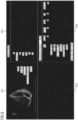
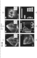
- FIG. 1 depicts an exemplary DICOM SC image with a 2*2 grid-size.
- the exemplary DICOM SC image shown in FIG. 1 has four quadrants 101, 102, 103, and 104, among which there are two quadrants, i.e., 102 and 104, containing characters associated with clinical data.
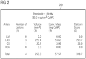
- FIG. 2 depicts another exemplary DICOM SC image with a 1*1 grid-size, i.e., only one quadrant 201 which contains characters associated with clinical data.
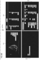
- FIG. 3 depicts a further exemplary DICOM SC image with a 2*3 grid-size, i.e., six quadrants 301-306, among which only quadrant 306 contains characters associated with clinical data.
- FIG. 1 depicts an exemplary DICOM SC image with a 2*2 grid-size.
- the exemplary DICOM SC image shown in FIG. 1 has four quadrants 101, 102, 103, and 104, among which there are two quadrants, i.e., 102 and
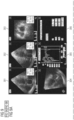
- FIG. 4 depicts a still further exemplary DICOM SC image with a 2*2 grid-size, i.e., four quadrants 401-404, among which quadrats 402 and 404 respectively contain characters associated with clinical data.
- Each of the four exemplary DICOM SC images may respectively represent a specific type of DICOM SC image.
- the at least one DICOM SC image may contain characters associated with clinical data associated with an anatomical target region of a patient, e.g., the heart, the liver, the brain, or a part thereof.
- an SC image can include, multiple characters associated with clinical data but not every character is needed, e.g., for a further use.
- the specific data that are needed to be extracted from the SC image may be defined by the configuration information associated with the at least one DICOM SC image.
- the configuration may comprise one or more keywords of the characters associated with the clinical data, e.g., volume and/or longitudinal strain as shown in FIG. 5 .
- the configuration may comprise one or more pre-defined variables associated with the clinical data including, for example, one or more row names, and/or one or more column names. For example, referring to FIG.
- such row names may be EF, Global EF, HR, EDV, ESV, SV, CO in quadrant 502, as well as 01-Left Wall, 03-Roof, 02-Right Wall, Average, Standard Dev, Global in quadrant 504.
- Column names may be Seg, PreStr, PKSys, PkAll, PSI, TPk Ovrl in quadrant 504. The more specific the keywords and/or the pre-defined variables are, the more accurate the results of the extracted characters would be.
- the configuration information may also comprise information about quadrants in an SC image and/or an orientation of a table in the SC image.
- the configuration information associated with the SC image depicted in FIG. 5 may comprise a keyword - Volume, and/or pre-defined variables - EDV, ESV, and SV.
- which quadrant(s) containing the characters to be extracted can be determined by the keyword - Volume, i.e., quadrant 502, and/or which rows the characters to be extracted are located can be determined by the pre-defined variables - EDV, ESV, and SV, i.e., the three rows starting with EDV, ESV, and SV, respectively.
- the configuration information only comprises one or more keywords, all the characters in quadrant(s) which contain(s) the one or more keywords may be extracted.
- the configuration information only comprises one or more pre-defined variables
- all the characters in the same row(s) or the same column(s) as the one or more pre-defined variables may be extracted.
- the configuration information associated with the SC image depicted in FIG. 5 may comprise a keyword - longitudinal strain, and a combination of pre-defined variables - (Average, PkAll). Then, quadrant 504 is determined as the quadrant containing the characters to be extracted, and character(s) being positioned in the same row as Average and the same column as PkAll is the character(s) to be extracted, i.e., 34.30 as shown in FIG. 5 .
- the configuration may comprise a grid-size of each of the at least one DICOM SC image, e.g., 2*2 of the SC image shown in FIG. 5 , and/or 2*3 of the SC image shown in FIG. 3 .
- the accuracy of extracting the characters associated with clinical data may be improved by splitting the whole SC image into sub-images according to the grid-size.
- the configuration information may comprise a template of the at least one DICOM SC image.
- a template may comprise all the common information shared by the same type of DICOM SC images.
- the same type of SC images may be SC images sharing the same grid-size, the same row names, and/or the same column names.
- FIG. 6 an exemplary template corresponding to the SC image shown in FIG. 5 is illustrated in FIG. 6 .
- Such an exemplary template has a 2*2 grid-size, i.e., four quadrants 601-602, and the same row and column names as those of the SC image shown in FIG. 5 , i.e., quadrants 602 and 604, respectively.
- the medical image within quadrant 601 may be removed or replaced with an indication which indicates that quadrant 601 contains a medical image but not characters associated with clinical data. Based on such a template, it is possible to accurately split SC images into sub-images or quadrants.
- the configuration information may comprise an output format of the extracted characters associated with clinical data, such as an Excel file or a json (JavaScript Object Notation) file.
- a required structure of content may be also defined in connection with the output format.
- the output format may be in Excel format with one row per SC image, and the column names of the Excel format may include one or more pre-defined variables contained in the configuration information.
- Sun an output format can facilitate utilization of the extracted characters, e.g., for diagnosis or research.
- the extracted characters and corresponding DICOM images may be used for deep-learning and/or data-mining research.
- the extracted characters may be added into DICOM tags of the series containing the SC image.
- the extracted characters in conjunction with data available in other DICOM tags and other scan images, help in improving research by providing more contextual information. They also help in cohort selections of patients and classifying datasets based on the variable values in SC images.
- the configuration information associated with the at least one DICOM SC image may be obtained by user inputs based on the at least one SC image.
- the user may input the configuration information to a computing device, e.g., a PC or workstation, which is used to process SC images based on what is needed to be extracted, e.g., by defining one or more keywords and/or one or more pre-defined variables.
- the configuration information may be automatically determined based on specific structure and/or content pattern of SC images to be processed, which are determined by, e.g., using image analysis techniques. Such image analysis techniques may comprise at least one of object recognition, image segmentation.
- the configuration information may be automatically determined based on IOD of the at least one DICOM SC image, such as IOD modules associated with patient, study, series, equipment, image as specified according to DICOM standards.
- OCR may be used to extract one or more characters associated with clinical data from at least one DICOM SC image based on the configuration information explained above.
- OCR is the electronic or mechanical conversion of images of typed, handwritten or printed text into machine-encoded text, whether from a scanned document, a photo of a document, a scene-photo (for example the text on signs and billboards in a landscape photo) or from subtitle text superimposed on an image (for example: from a television broadcast).
- Various OCR software is available and may be applied to this disclosure, such as, Tesseract Open Source OCR as presented in non-patent literature - Kay, Anthony.
- a parameter of Tesseract OCR - page segmentation mode (PSM) - may be used to improve the accuracy of OCR results.
- the PSM explains the layout of the data and the form of the image to be processed. More information on PSM can be found on the Tesseract OCR Wiki page, e.g., https://github.com/tesseract-ocr/tesseract/blob/master/doc/tesseract.1.asc .
- Different PSMs may be suitable for different arrangements of the characters, e.g., row-wise, column-wise, or tabular.
- PSM 7 assumes that each image is a single text/character line
- PSM 4 assumes that each image is a single column of text/character of variable sizes. I.e., PSMs 4 and 7 are respectively suitable for processing column-wise and row-wise characters.
- the configuration information may rely on a specific type of the at least one SC image and thereby the extraction of characters may be adapted to such a specific type based on the configuration information. Accordingly, the accuracy and efficiency of the extraction of the characters can be improved.
- FIG. 7 is a flowchart of a computer-implemented method 2000 according to various examples.
- the method 2000 pertains to processing at least one DICOM SC image to extract clinical data therein. Characters associated with the clinical data are extracted from the at least one DICOM SC image based on configuration information associated with the at least one DICOM SC image, which is obtained based on the at least one DICOM SC image.
- the method 2000 may be executed by a computer or a workstation comprising at least one processing unit upon loading program code.
- the computer or the workstation may be positioned in a local network of a hospital or an institution.
- the computer or the workstation may be connected/connectable to a PACS, or directly to a medical imaging scanner.
- the computer or the workstation may be a node of a cloud-based computing system or of an edge computing system. Details of the method 2000 are described below.
- the at least one DICOM SC image could be loaded from a PACS.
- Block 2010 could include controlling a medical imaging scanner, such as an MRI scanner to acquire DICOM images and to generate the at least one SC image by post-processing tools/software running in the medical imaging scanner.
- the at least one DICOM SC image may be received directly from a medical imaging scanner during a scan to perform a real-time examination of a patient.
- configuration information associated with the at least one DICOM SC image are obtained based on the at least one DICOM SC image.
- the configuration information associated with the at least one DICOM SC image may be obtained by user inputs based on the at least one SC image. Alternatively or additionally, the configuration information may be obtained based on specific structure and/or content pattern of the at least one SC image. Alternatively or additionally, the configuration information may be obtained based on DICOM tags of the at least one DICOM SC image.
- characters associated with clinical data are extracted from the at least one DICOM SC image based on the configuration information.
- the characters associated with the clinical data may be extracted using OCR.
- the configuration information may associate with a specific type of the at least one SC image and thereby the extraction of characters may be adapted to such a specific type based on the configuration information. Accordingly, the accuracy and efficiency of the extraction of the characters can be improved.
- OCR can be applied on the whole SC image, but its accuracy cannot be sufficient. Noise and sparsity are exemplary factors that can drastically deteriorate the performance of any OCR algorithm as explained in non-patent literature - Gupta, Maya R., Nathaniel P. Jacobson, and Eric K. Garcia. "OCR binarization and image pre-processing for searching historical documents.” Pattern Recognition 40.2 (2007): 389-397 . Since the data that needs to be extracted forms only one part of an image, it is best to condense pixel data of the at least one DICOM SC image to only those regions that need OCR extraction. Accordingly, the method 2000 may optionally comprise the following pre-processing steps.
- the at least one DICOM SC image may be converted to any one of the following image formats: tag image file format, TIFF, raw image format, RAW, bitmap image file format, BMP, and portable network graphic format, PNG. Detailed explanation will be described in connection with TIFF in the following.
- the pixel data of the secondary capture DICOM may be extracted and stored separately as a raw image format.
- TIFF format could be used since it handles lossless compression, can be used across multiple devices and operating systems and requires less storage space than RAW image format as described in international standard - ISO 12639:2004.
- the TIFF files being generated may be temporary files for the purpose of OCR extraction and may be deleted once the output of the extraction is created.
- margins of the at least one DICOM SC image may be trimmed.
- margins of the converted TIFF images of the at least one DICOM SC image may be trimmed.
- automated margin alignment may be applied to the at least one DICOM SC image and/or the converted TIFF images of the at least one DICOM SC image.
- characters to be extracted may be large enough to achieve a high accuracy for some SC images, e.g., the SC image shown in FIG. 2 , in which the SC image only contains one quadrant, i.e., 1*1 grid-size.
- splitting may not be needed but only trimming the margins would be enough so that the content can be as focused as possible.
- the accuracy of extraction of characters may be further improved by splitting the SC image to sub-images.
- the at least one DICOM SC image or the converted TIFF images of the at least one DICOM SC image may be split to sub-images to concentrate the OCR only on those regions that contain the characters to be extracted. To decide where these sub-images are would be very much dependent on the content of the SC image and on the characters to be extracted.
- the method 2000 may optionally further comprise splitting the at least one DICOM SC image into sub-images based on the configuration information, and selecting one or more sub-images containing the characters associated with the clinical data from the split sub-images, wherein extracting the characters comprises respectively extracting the characters from the selected one or more sub-images. If the at least one SC image is converted to TIFF image, the same splitting and selecting process could be applied to the converted TIFF images of the at least one DICOM SC image.
- the configuration information may comprise a grid-size of the at least one DICOM SC image and/or one or more keywords of the characters associated with the clinical data, and the splitting may be based on the grid-size and/or the one or more keywords.
- the one or more keywords may be determined by applying OCR to the at least one DICOM SC image. For example, Tesseract OCR may be used on the whole image of each of the at least one DICOM SC image (or the converted TIFF images of the at least one DICOM SC image) to extract characters which have sharper contrast, large font size and thus easily extractable even though the overall accuracy might be low for other characters in the whole image. Certain keywords may be then needed to identify how the splitting could happen and what the grid geometry is like. This would be dependent on the content of the SC images, e.g., those respectively shown in FIGs. 1-5 , and thus would be very use case based.
- each SC image may have a specific format depending on its content and the scanner software producing it. For example, Syngo.Via writes its longitudinal strain measurements into grid sizes of 2x2, e.g., as shown in FIGs. 1 , 4 , and 5 , and Ejection Fraction graphs in grid sizes of 2x3, e.g., as shown in FIG. 3 .
- This information thus may facilitate clearly defining the keywords needed to identify the grid sizes as they may not vary much in different SC images of the same type.
- FIGs. 8 and 9 schematically illustrate exemplary workflows for splitting the SC image shown in FIGs. 5 and 3 , respectively.
- the splitting of the SC image of FIG. 5 can be based on a grid-size of 2*2 and/or keywords - Volume and Beat.
- the splitting of the SC image of FIG. 3 can be based on a grid-size of 2*3 and/or keywords - A4C and SAX.
- each row can be split into three equal columns based on the keywords - A4C and SAX, i.e., the left and right columns are respectively starting from A4C and SAX, and the rest part is the middle column.
- the configuration information may comprise a template of the at least one DICOM SC image, and the splitting of the at least one DICOM SC image is based on the template.
- a template may be pre-defined and stored for each type of SC image, e.g., respective SC image shown in FIGs. 1-5 . Then, the template can be selected from the pre-defined templates based on the type of the at least one DICOM SC image to be processed.
- each split sub-image contains the relevant characters to be extracted.
- the required variables or relevant characters are only present in quadrants (or sub-images) 502 and 504.
- quadrants (or sub-image) 306. such variables or characters are only contained in quadrant (or sub-image) 306. Accordingly, the sub-images 502 and 504 are selected for the SC image shown in FIG. 8 , and the sub-image 306 is selected for the SC image shown in FIG. 9 .
- the selecting of the one or more sub-images may comprise respectively applying OCR to each of the split sub-images, e.g., 501-504 in FIG. 8 , or 301-306 in FIG. 9 ; searching one or more pre-defined variables, e.g., EDV and/or ESV in FIGs. 8 and 9 , respectively, associated with the clinical data in respective result of the OCR of respective sub-image, wherein the configuration information comprises the one or more pre-defined variables; and selecting respective sub-images, e.g., 502 and 504 in FIG. 8 or 306 in FIG.
- the pre-defined variables e.g., EDV and/or ESV
- the pre-defined variables e.g., EDV and/or ESV
- PSM modes could be applied to each sub-image and with manual sample verification, an optimal PSM value could be determined.
- margin alignment and trimming may be performed to centralize the content of the selected one or more sub-images.
- the arrangement of the characters in each of the selected one or more sub-images may comprise row-wise, column-wise, or tabular.
- the method 2000 may optionally comprise determining an arrangement of the characters in each of the selected one or more sub-images.
- the sub-images 502 and 504 are respectively row-wise and tabular.
- Different PSMs e.g., PSM 4 and PSM 7, may be used to determine the arrangement of the characters in each of the selected one or more sub-images.
- PSM 4 and PSM 7 may be respectively applied to each of the selected one or more sub-images to obtain a column-wise OCR result and a row-wise OCR result. Then, the arrangement of the characters can be determined by comparing the accuracy of the column-wise OCR result and the row-wise OCR result.
- the configuration information associated with the at least one DICOM SC image may comprise one or more PSMs, e.g., for each quadrant.
- the method 2000 may further comprise: splitting the selected one or more sub-images into rows, and applying OCR to each of the split rows to extract characters therein.
- Each character extracted from each of the split rows should be a whole character as else it means the row was split across text. Accordingly, the selected one or more sub-images would be split again until each character extracted from each of the split rows is a whole character.
- FIG. 10 schematically illustrates an exemplary workflow for extracting row-wise characters from the selected sub-image 502.
- the sub-image 502 may be split into rows.
- OCR may be applied to each of the split rows to extract characters therein.
- the characters associated with the pre-defined variables, e.g., EDV, ESV, and SV, comprised in the configuration information may be selected from the extracted characters of all the split rows.
- the units may be also extracted from the sub-image 502.
- the configuration information may comprise the units and the units may be added to the output based on the configuration information. Since the characters of the sub-image 502 is row-wise, PSM 7 may be used.
- the method 2000 may further comprise: splitting the selected one or more sub-images into columns, and applying OCR to each of the split columns to extract characters therein.
- the method 2000 may further comprise: splitting the selected one or more sub-images into both rows and columns, respectively, applying OCR to each of the split rows and to each of the split columns to extract characters therein, respectively, and determining a position within a table of each of the extracted characters based on its positions in both the row-wisely and column-wisely extracted characters.
- FIG. 11 schematically illustrates an exemplary workflow for extracting tabular characters from the selected sub-image 504.
- the selected sub-image 504 is split into rows, and at 3020, the selected sub-image 504 is split into columns.
- OCR is applied to each of the split rows to extract characters therein, and at 3040, OCR is applied to each of the split columns to extract characters therein.
- PSMs 4 and 7 may be utilized on each of the split columns and each of the split rows, respectively.
- a position within a table of each of the extracted characters is determined based on its positions in both the row-wisely and column-wisely extracted characters.
- the pre-defined variables e.g., column names - PreStr, PkSys, and PkAll, may be used to determine the width of corresponding columns.
- the row names - Global and Average may be used to determine the height of correspond rows.
- the common character(s) present in both the first row and the first column may be used to determine the width of the first column and the height of the first row, and from the second column onwards, the width is equally divided based on the remaining width of the whole sub-image, and from the second row onwards, the height is equally divided based on the remaining height of the whole sub-image.
- the rich DICOM tag library could contain information in an SC image itself and there are well-defined tools to extract the DICOM tags. Hence, it is possible to identify if there are any DICOM header tags that could be used instead of extracting similar information from the pixel data of the SC image. For example, average dose applied while taking medical images could be stored in the CTDIvol (0018,9345) DICOM tag as well as embedded in the pixel data of the SC image. DICOM tags would contain information on the attributes of image acquisition and not of further diagnosis of the image. Moreover, the tags that are needed to add contextual information to the OCR-extracted clinical data can also be included in the configuration information. For example, PatientID, StudyID, AccessionNumber, and StudyDate are the DICOM tags that may be added to an output excel file in order to provide context.
- the method 2000 may optionally comprise extracting DICOM tags from a header of the at least one DICOM SC image.
- the extracted DICOM tags may be paired with the extracted characters associated with the clinical data.
- FIG. 12 schematically illustrates an exemplary output 510 of extracted clinical data of an SC image.
- the output 510 is in Excel format and contains clinical data extracted from sub-images 502 and 504 as well as DICOM tags extracted from the header of the DICOM SC image, i.e., PatientID, StudyID, AccessionNumber and StudyDate, whose values are respectively PatID1, 123456, 8888888, and 20190101.
- the method 2000 may optionally comprise removing patient health information, e.g., PatientID, StudyID, AccessionNumber and StudyDate, from the extracted characters associated with the clinical data.
- patient health information e.g., PatientID, StudyID, AccessionNumber and StudyDate.
- the above-outlined steps may be repeated for each SC image and then output the extracted clinical data, for example, in a new row of an Excel file like the output 510.
- the method 2000 described above can be used to process any kind of DICOM SC image based on configuration information associated with the DICOM SC image to extract characters associated with clinical data.
- the method 2000 can extract whatever clinical data a user needs from any type of SC image.
- the configuration information can be customized based on a specific user's need, and/or be configured based on specific structures and content patterns of a specific type of SC image. Accordingly, there is no need to utilize different purpose-built techniques for different use cases.
- the method 2000 also facilitates the utilization of clinical data extractable from SC images, e.g., in various researches, while labor cost and/or time required for converting the characters associated with clinical data in SC images to text are significantly reduced due to the automatic nature of the method 2000, particularly for large datasets.
- the extracted clinical data can also facilitate the diagnosis of diseases, for example by automatically plugging such clinical data into radiology reports instead of radiologist dictations, and further can augment patient digital healthcare data and create a more accurate record for future analytics.
- the method 2000 can improve the accuracy of extraction of characters associated with clinical data from SC images, for example by splitting a whole SC image into sub-images and selecting sub-images containing characters to be extracted to condense pixel data of the DICOM SC image to only those regions that need OCR extraction. Further, specific techniques for respectively extracting row-wise, column-wise, and tabular characters can further improve the extraction accuracy.
- DICOM is just an exemplary type of image that can be processed by the method 2000 but it is not a limitation of the method 2000.
- Other types of images or documents such as a scanned document, a photo of a document, a scene-photo (for example the text on signs and billboards in a landscape photo), or subtitle text superimposed on an image (for example: from a television broadcast), can be processed by the method 2000 as well.
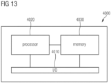
- FIG. 13 is a block diagram of a computing device 4000 according to various examples.
- the computing device 4000 provides a functionality of processing at least one DICOM SC image based on the method 2000.
- the computing device 4000 may comprise at least one processor 4020, at least one memory 4030, and at least one input/output interface 4010.
- the at least one processor 4020 is configured to load program code from the at least one memory 4030 and execute the program code. Upon executing the program code, the at least one processor 4020 performs the method 2000.
- a medical imaging scanner such as a CT scanner, an MRI scanner, an ultrasound scanner, or an x-ray scanner, may comprise the computing device 4000 of FIG. 13 .
- the medical imaging scanner may generate at least one DICOM SC image and process the generated SC image based on the method 2000.
- the computing device 4000 may be embedded in or connected with the medical imaging scanner and thereby the medical imaging scanner may be also configured to perform the method 2000.
- the computing device 4000 may be a PC a computer or a workstation within a local network of a hospital or an institution or a node of a cloud-based computing system or of an edge computing system.
- the characters can be extracted from any type of DICOM SC image based on specific configuration information associated with the DICOM SC image. The more specific the configuration information the more accurate the extraction of the characters.
- the extracted clinical data can further facilitate research, such as deep learning and/or data-mining research.
- the accuracy of the extraction of the characters can be further improved by splitting a whole SC image into sub-images and selecting sub-images containing characters to be extracted to condense pixel data of the DICOM SC image to only those regions that need OCR extraction. Further, specific techniques for respectively extracting row-wise, column-wise, and tabular characters can further improve the extraction accuracy.
Abstract
Techniques of facilitating processing of at least one DICOM SC image - e.g., using a PC or workstation in a hospital or an institution - to automatically extract clinical data therein will be described. Characters associated with the clinical data are extracted from the at least one DICOM SC image based on configuration information associated with the at least one DICOM SC image, which is obtained based on the at least one DICOM SC image.
Description
- Various examples of the disclosure relate to facilitating processing of DICOM SC images to automatically extract relevant clinical data therein. Various examples of the disclosure specifically relate to extracting characters associated with clinical data from at least one DICOM SC image based on configuration information associated with the at least one DICOM SC image, which is obtained based on the at least one DICOM SC image.
- Digital Imaging and Communications in Medicine (DICOM) is one of the most popular file formats for storing, transmitting, and viewing medical images. The Secondary Capture (SC) Image Information Object Definition (IOD) specifies images that are converted from a non-DICOM format to a modality independent DICOM format. I.e., a DICOM SC image could be regarded as an image that are converted from a non-DICOM format to a modality independent DICOM format. DICOM SC images can hold important curated clinical data that allow a better understanding of the accompanying DICOM images in series of a study of a patient, giving the physicians additional clinical context of the patient. For example, this could be strain information, calcium scores, or cardiovascular volumes. DICOM SC images are created, for example, by image post-processing tools in the scanner or by screen captures of the scanner display, such as an ultrasound scanner display. They are manually viewed along with the other DICOM images during diagnosis stages by the radiologists and then discarded. The data in them could be useful for future research as it provides more contextual information to the DICOM header tags and other scan images in the acquisition.
- SC images have a heterogeneous format and the relevant data in the SC images vary depending on the use case. Hence, there is no constant definition of what is needed and what is not. Further, SC images have a heterogeneous structure and the content pattern of SC images varies widely based on the source software version, the scanner, and the configuration of the installation.
- There is thus no one-size-fits-all solution for extracting clinical data from various SC images since not every data is relevant from SC images and not every SC image is equal in design. The major reason that they are not used in research, despite their value, is the enormous labor cost and time needed to extract the data that is burnt into the pixel image. Despite advances in Image-to-text research, such as techniques discloses in a non-patent literature - Manwatkar, Pratik Madhukar, and Shashank H. Yadav. "Text recognition from images." 2015 International Conference on Innovations in Information, Embedded and Communication Systems (ICIIECS). IEEE, 2015, a viable solution has never been developed that allows the user to extract what is needed from whatever type of SC images.
- Due to the popular domain of image-text and Optical Character Recognition (OCR) research, e.g., non-patent literature - "Optical Character Recognition (OCR) - How it works" - described in Nicomsoft.com, there have been efforts to use it within the DICOM image domain. Due to the varied content in SC images, the relevant data differs based on the use case. Hence there have been efforts to extract particular information from particular types of SC images. These tools are purpose-built for the sole purpose of extracting a specific variable for the use case it was designed for. An example is DoseUtility. It uses OCR to extract radiant dose information from General Electric (GE), Siemens, or Toshiba Computed Tomography (CT) SC images. These images have particular series codes based on the manufacturer and thus have a well-defined pattern. DoseUtility thus works very well to extract the dose information from these SC images since the required variable is well-defined and the pattern as well. The disadvantage is of course that it is purpose-built and cannot be used to extract any other information from the same SC image or the same information from a non-standardized SC image. This is also the major drawback of such tools. There are many such dedicated software available, but they only serve a specific purpose and cannot be used in other use cases.
- A further exemplary OCR-based technique is directed to removing Patient Health Information (PHI). Reliable Patient Health Information (PHI) removal is an important aspect of medical research as it allows data sharing while complying with the local data privacy laws. Since SC images are sometimes screen captures of scanners, they usually contain the patient data embedded into the SC images. Hence, there have been efforts to use OCR to identify and remove PHI from SC images, such as techniques disclosed in non-patent literature - Tsui, Gary Kin-wai, and Tao Chan. "Automatic selective removal of embedded patient information from image content of DICOM files." American Journal of Roentgenology 198.4 (2012): 769-772. Such literature uses the open-source Tesseract OCR to extract characters from SC images and then looks for words that match with the PHI in the DICOM header tags. For example, it checks if the PatientName tag value in the DICOM header is present in the extracted characters. The advantage, as opposed to DoseUtility, is that it works on any kind of SC image. But since it is purpose-built to remove PHI, it only identifies words that match the value of specific PHI DICOM tags. It cannot be used by the user to extract relevant clinical information from SC images. Thus, similar to DoseUtility, these efforts serve a specific purpose and cannot be used within a research context to extract values of specific user-defined variables.
- Consequently, the existing techniques cannot allow the user to define what he or she needs and cannot also work on arbitrarily user-selected SC images. That is, there is no versatile technique that can be used to extract whatever clinical data a user needs for any kind of SC image.
- Accordingly, there is a need for advanced techniques that mitigate or overcome the above-identified drawbacks or restrictions. There is a need for advanced techniques of automatically extracting whatever clinical data a user needs for any kind of SC image.
- This need is met by the features of the independent claims. The features of the dependent claims define embodiments.
- Hereinafter, techniques of facilitating processing of at least one DICOM SC image - e.g., using a PC or workstation in a hospital or an institution - to automatically extract clinical data therein will be described. Characters associated with the clinical data are extracted from the at least one DICOM SC image based on configuration information associated with the at least one DICOM SC image, which is obtained based on the at least one DICOM SC image.
- A computer-implemented method is provided. The method is used to process at least one DICOM SC image to automatically extract clinical data therein. The method comprises obtaining at least one Digital Imaging and Communications in Medicine, DICOM, Secondary Capture, SC, image, and obtaining configuration information associated with the at least one DICOM SC image based on the at least one DICOM SC image. The method further comprises extracting characters associated with clinical data from the at least one DICOM SC image based on the configuration information.
- A computer program or a computer-program product or a computer-readable storage medium that includes program code is provided. The program code can be loaded and executed by at least one processor. Upon loading and executing the program code, the at least one processor performs a method. The method is used to process at least one DICOM SC image to automatically extract clinical data therein. The method comprises obtaining at least one Digital Imaging and Communications in Medicine, DICOM, Secondary Capture, SC, image, and obtaining configuration information associated with the at least one DICOM SC image based on the at least one DICOM SC image. The method further comprises extracting characters associated with clinical data from the at least one DICOM SC image based on the configuration information.
- A computing device comprising at least one processor and at least one memory is provided. The at least one processor is configured to load program code from the at least one memory and execute the program code. Upon executing the program code, the at least one processor is configured to process at least one DICOM SC image to automatically extract clinical data therein. The at least one processor is configured to obtain at least one Digital Imaging and Communications in Medicine, DICOM, Secondary Capture, SC, image, and obtain configuration information associated with the at least one DICOM SC image based on the at least one DICOM SC image. The at least one processor is further configured to extract characters associated with clinical data from the at least one DICOM SC image based on the configuration information.
- It is to be understood that the features mentioned above and those yet to be explained below may be used not only in the respective combinations indicated, but also in other combinations or in isolation without departing from the scope of the invention.
-
-
FIG. 1 schematically illustrates an exemplary DICOM SC image. -
FIG. 2 schematically illustrates another exemplary DICOM SC image. -
FIG. 3 schematically illustrates a further exemplary DICOM SC image. -
FIG. 4 schematically illustrates a still further exemplary DICOM SC image. -
FIG. 5 schematically illustrates a still further exemplary DICOM SC image. -
FIG. 6 schematically illustrates an exemplary template corresponding to the exemplary DICOM SC image ofFIG. 5 . -
FIG. 7 is a flowchart of a method according to various examples. -
FIG. 8 schematically illustrates an exemplary workflow for splitting an SC image according to various examples. -
FIG. 9 schematically illustrates a further exemplary workflow for splitting a further SC image according to various examples. -
FIG. 10 schematically illustrates an exemplary workflow for extracting row-wise characters. -
FIG. 11 schematically illustrates an exemplary workflow for extracting tabular characters. -
FIG. 12 schematically illustrates an exemplary output of extracted clinical data of an SC image. -
FIG. 13 is a block diagram of a computing device according to various examples. - Some examples of the present disclosure generally provide for a plurality of circuits or other electrical devices. All references to the circuits and other electrical devices and the functionality provided by each are not intended to be limited to encompassing only what is illustrated and described herein. While particular labels may be assigned to the various circuits or other electrical devices disclosed, such labels are not intended to limit the scope of operation for the circuits and the other electrical devices. Such circuits and other electrical devices may be combined with each other and/or separated in any manner based on the particular type of electrical implementation that is desired. It is recognized that any circuit or other electrical device disclosed herein may include any number of microcontrollers, a graphics processor unit (GPU), integrated circuits, memory devices (e.g., FLASH, random access memory (RAM), read only memory (ROM), electrically programmable read only memory (EPROM), electrically erasable programmable read only memory (EEPROM), or other suitable variants thereof), and software which co-act with one another to perform operation(s) disclosed herein. In addition, any one or more of the electrical devices may be configured to execute a program code that is embodied in a non-transitory computer readable medium programmed to perform any number of the functions as disclosed.
- In the following, embodiments of the invention will be described in detail with reference to the accompanying drawings. It is to be understood that the following description of embodiments is not to be taken in a limiting sense. The scope of the invention is not intended to be limited by the embodiments described hereinafter or by the drawings, which are taken to be illustrative only.
- The drawings are to be regarded as being schematic representations and elements illustrated in the drawings are not necessarily shown to scale. Rather, the various elements are represented such that their function and general purpose become apparent to a person skilled in the art. Any connection or coupling between functional blocks, devices, components, or other physical or functional units shown in the drawings or described herein may also be implemented by an indirect connection or coupling. A coupling between components may also be established over a wireless connection. Functional blocks may be implemented in hardware, firmware, software, or a combination thereof.
- Various techniques disclosed herein generally relate to facilitating processing of at least one DICOM SC image - e.g., using a PC or workstation in a hospital or an institution - to automatically extract clinical data therein. Characters associated with the clinical data are extracted from the at least one DICOM SC image based on configuration information associated with the at least one DICOM SC image, which is obtained based on the at least one DICOM SC image.
- The at least one DICOM SC image may be obtained from a data repository, e.g., a Picture Archiving and Communication System (PACS). Alternatively or additionally, the at least one DICOM SC image may be directly obtained from a medical imaging scanner, such as an X-ray radiography scanner, an ultrasound scanner, a computed tomography (CT) scanner, a positron emission tomography (PET) scanner, or a magnetic resonance imaging (MRI) scanner. Therefore, the at least one DICOM SC image may be associated with one or more medical images, for example, X-ray radiography images, ultrasound images, CT images, PET images, or MRI images. For example, the at least one DICOM SC image may be created by image post-processing tools/software running in any one of an X-ray scanner, CT scanner, PET scanner, or MRI scanner, or by screen captures of a display connected/connectable to any one of an X-ray scanner, CT scanner, PET scanner, or MRI scanner. Four exemplary DICOM SC images are illustrated in
FIGs. 1-4 , respectively. In this disclosure, the terminology "DICOM SC image(s)" is equivalent to "SC image(s)". The exemplary DICOM SC images illustrated inFIGs. 1-4 are used for illustrative purposes, which may not look the same as clinical DICOM SC images. -
FIG. 1 depicts an exemplary DICOM SC image with a 2*2 grid-size. I.e., the exemplary DICOM SC image shown inFIG. 1 has fourquadrants FIG. 2 depicts another exemplary DICOM SC image with a 1*1 grid-size, i.e., only onequadrant 201 which contains characters associated with clinical data.FIG. 3 depicts a further exemplary DICOM SC image with a 2*3 grid-size, i.e., six quadrants 301-306, among which onlyquadrant 306 contains characters associated with clinical data.FIG. 4 depicts a still further exemplary DICOM SC image with a 2*2 grid-size, i.e., four quadrants 401-404, among whichquadrats - According to the disclosure, the at least one DICOM SC image may contain characters associated with clinical data associated with an anatomical target region of a patient, e.g., the heart, the liver, the brain, or a part thereof.
- In general, an SC image can include, multiple characters associated with clinical data but not every character is needed, e.g., for a further use. The specific data that are needed to be extracted from the SC image may be defined by the configuration information associated with the at least one DICOM SC image. For example, the configuration may comprise one or more keywords of the characters associated with the clinical data, e.g., volume and/or longitudinal strain as shown in
FIG. 5 . Alternatively or additionally, the configuration may comprise one or more pre-defined variables associated with the clinical data including, for example, one or more row names, and/or one or more column names. For example, referring toFIG. 5 , such row names may be EF, Global EF, HR, EDV, ESV, SV, CO inquadrant 502, as well as 01-Left Wall, 03-Roof, 02-Right Wall, Average, Standard Dev, Global inquadrant 504. Column names may be Seg, PreStr, PKSys, PkAll, PSI, TPk Ovrl inquadrant 504. The more specific the keywords and/or the pre-defined variables are, the more accurate the results of the extracted characters would be. Hence, the configuration information may also comprise information about quadrants in an SC image and/or an orientation of a table in the SC image. - For example, the configuration information associated with the SC image depicted in
FIG. 5 may comprise a keyword - Volume, and/or pre-defined variables - EDV, ESV, and SV. According to such configuration information, which quadrant(s) containing the characters to be extracted can be determined by the keyword - Volume, i.e.,quadrant 502, and/or which rows the characters to be extracted are located can be determined by the pre-defined variables - EDV, ESV, and SV, i.e., the three rows starting with EDV, ESV, and SV, respectively. If the configuration information only comprises one or more keywords, all the characters in quadrant(s) which contain(s) the one or more keywords may be extracted. Similarly, if the configuration information only comprises one or more pre-defined variables, all the characters in the same row(s) or the same column(s) as the one or more pre-defined variables may be extracted. Alternatively, the configuration information associated with the SC image depicted inFIG. 5 may comprise a keyword - longitudinal strain, and a combination of pre-defined variables - (Average, PkAll). Then,quadrant 504 is determined as the quadrant containing the characters to be extracted, and character(s) being positioned in the same row as Average and the same column as PkAll is the character(s) to be extracted, i.e., 34.30 as shown inFIG. 5 . - Alternatively or additionally, the configuration may comprise a grid-size of each of the at least one DICOM SC image, e.g., 2*2 of the SC image shown in
FIG. 5 , and/or 2*3 of the SC image shown inFIG. 3 . The accuracy of extracting the characters associated with clinical data may be improved by splitting the whole SC image into sub-images according to the grid-size. - Alternatively or additionally, the configuration information may comprise a template of the at least one DICOM SC image. Such a template may comprise all the common information shared by the same type of DICOM SC images. Herein, the same type of SC images may be SC images sharing the same grid-size, the same row names, and/or the same column names. For example, an exemplary template corresponding to the SC image shown in
FIG. 5 is illustrated inFIG. 6 . Such an exemplary template has a 2*2 grid-size, i.e., four quadrants 601-602, and the same row and column names as those of the SC image shown inFIG. 5 , i.e.,quadrants quadrant 601 may be removed or replaced with an indication which indicates thatquadrant 601 contains a medical image but not characters associated with clinical data. Based on such a template, it is possible to accurately split SC images into sub-images or quadrants. - Optionally or additionally, the configuration information may comprise an output format of the extracted characters associated with clinical data, such as an Excel file or a json (JavaScript Object Notation) file. Optionally or additionally, a required structure of content may be also defined in connection with the output format. For example, the output format may be in Excel format with one row per SC image, and the column names of the Excel format may include one or more pre-defined variables contained in the configuration information. Sun an output format can facilitate utilization of the extracted characters, e.g., for diagnosis or research. For example, the extracted characters and corresponding DICOM images may be used for deep-learning and/or data-mining research. Additionally or optionally, the extracted characters may be added into DICOM tags of the series containing the SC image. The extracted characters in conjunction with data available in other DICOM tags and other scan images, help in improving research by providing more contextual information. They also help in cohort selections of patients and classifying datasets based on the variable values in SC images.
- According to various examples, the configuration information associated with the at least one DICOM SC image may be obtained by user inputs based on the at least one SC image. For example, the user may input the configuration information to a computing device, e.g., a PC or workstation, which is used to process SC images based on what is needed to be extracted, e.g., by defining one or more keywords and/or one or more pre-defined variables. Alternatively or additionally, the configuration information may be automatically determined based on specific structure and/or content pattern of SC images to be processed, which are determined by, e.g., using image analysis techniques. Such image analysis techniques may comprise at least one of object recognition, image segmentation. Additionally or optionally, the configuration information may be automatically determined based on IOD of the at least one DICOM SC image, such as IOD modules associated with patient, study, series, equipment, image as specified according to DICOM standards.
- According to this disclosure, OCR may be used to extract one or more characters associated with clinical data from at least one DICOM SC image based on the configuration information explained above. OCR is the electronic or mechanical conversion of images of typed, handwritten or printed text into machine-encoded text, whether from a scanned document, a photo of a document, a scene-photo (for example the text on signs and billboards in a landscape photo) or from subtitle text superimposed on an image (for example: from a television broadcast). Various OCR software is available and may be applied to this disclosure, such as, Tesseract Open Source OCR as presented in non-patent literature - Kay, Anthony. "Tesseract: an open-source optical character recognition engine." Linux Journal 2007.159 (2007): 2, GOCR (or JOCR), Cognitive OpenOCR (i.e., CuneiForm), Kraken, and A9T9. This disclosure will be explained in connection with Tesseract as an example.
- According to various examples, when utilizing Tesseract OCR, a parameter of Tesseract OCR - page segmentation mode (PSM) - may be used to improve the accuracy of OCR results. The PSM explains the layout of the data and the form of the image to be processed. More information on PSM can be found on the Tesseract OCR Wiki page, e.g., https://github.com/tesseract-ocr/tesseract/blob/master/doc/tesseract.1.asc. Different PSMs may be suitable for different arrangements of the characters, e.g., row-wise, column-wise, or tabular. For example, PSM 7 assumes that each image is a single text/character line, and
PSM 4 assumes that each image is a single column of text/character of variable sizes. I.e.,PSMs 4 and 7 are respectively suitable for processing column-wise and row-wise characters. - As outlined above, by obtaining configuration information associated with at least one DICOM SC image, and further extracting characters associated with clinical data from the at least one DICOM SC image based on the obtained configuration information, characters associated with whatever clinical data can be automatically extracted for any kind of SC image. The configuration information may rely on a specific type of the at least one SC image and thereby the extraction of characters may be adapted to such a specific type based on the configuration information. Accordingly, the accuracy and efficiency of the extraction of the characters can be improved.
-
FIG. 7 is a flowchart of a computer-implementedmethod 2000 according to various examples. Themethod 2000 pertains to processing at least one DICOM SC image to extract clinical data therein. Characters associated with the clinical data are extracted from the at least one DICOM SC image based on configuration information associated with the at least one DICOM SC image, which is obtained based on the at least one DICOM SC image. - The
method 2000 may be executed by a computer or a workstation comprising at least one processing unit upon loading program code. The computer or the workstation may be positioned in a local network of a hospital or an institution. The computer or the workstation may be connected/connectable to a PACS, or directly to a medical imaging scanner. Alternatively, the computer or the workstation may be a node of a cloud-based computing system or of an edge computing system. Details of themethod 2000 are described below. - At
block 2010, at least one DICOM SC image is obtained. - The at least one DICOM SC image could be loaded from a PACS.
Block 2010 could include controlling a medical imaging scanner, such as an MRI scanner to acquire DICOM images and to generate the at least one SC image by post-processing tools/software running in the medical imaging scanner. Alternatively, the at least one DICOM SC image may be received directly from a medical imaging scanner during a scan to perform a real-time examination of a patient. - At
block 2020, configuration information associated with the at least one DICOM SC image are obtained based on the at least one DICOM SC image. - The configuration information associated with the at least one DICOM SC image may be obtained by user inputs based on the at least one SC image. Alternatively or additionally, the configuration information may be obtained based on specific structure and/or content pattern of the at least one SC image. Alternatively or additionally, the configuration information may be obtained based on DICOM tags of the at least one DICOM SC image.
- At
block 2030, characters associated with clinical data are extracted from the at least one DICOM SC image based on the configuration information. - The characters associated with the clinical data may be extracted using OCR. The configuration information may associate with a specific type of the at least one SC image and thereby the extraction of characters may be adapted to such a specific type based on the configuration information. Accordingly, the accuracy and efficiency of the extraction of the characters can be improved.
- OCR can be applied on the whole SC image, but its accuracy cannot be sufficient. Noise and sparsity are exemplary factors that can drastically deteriorate the performance of any OCR algorithm as explained in non-patent literature - Gupta, Maya R., Nathaniel P. Jacobson, and Eric K. Garcia. "OCR binarization and image pre-processing for searching historical documents." Pattern Recognition 40.2 (2007): 389-397. Since the data that needs to be extracted forms only one part of an image, it is best to condense pixel data of the at least one DICOM SC image to only those regions that need OCR extraction. Accordingly, the
method 2000 may optionally comprise the following pre-processing steps. - The at least one DICOM SC image may be converted to any one of the following image formats: tag image file format, TIFF, raw image format, RAW, bitmap image file format, BMP, and portable network graphic format, PNG. Detailed explanation will be described in connection with TIFF in the following.
- In this step, the pixel data of the secondary capture DICOM may be extracted and stored separately as a raw image format. TIFF format could be used since it handles lossless compression, can be used across multiple devices and operating systems and requires less storage space than RAW image format as described in international standard - ISO 12639:2004. Graphic technology - Prepress digital data exchange - Tag image file format for image technology (TIFF/IT). Lossless compression may be required to maintain the image quality and thus also improve the OCR accuracy. Storage size would be also a major requirement since many DICOM SC images may be processed at once and hence should not take up a lot of temporary storage size. The TIFF files being generated may be temporary files for the purpose of OCR extraction and may be deleted once the output of the extraction is created.
- Further, margins of the at least one DICOM SC image may be trimmed. Alternatively or additionally, margins of the converted TIFF images of the at least one DICOM SC image may be trimmed. Optionally, automated margin alignment may be applied to the at least one DICOM SC image and/or the converted TIFF images of the at least one DICOM SC image.
- After trimming margins, characters to be extracted may be large enough to achieve a high accuracy for some SC images, e.g., the SC image shown in
FIG. 2 , in which the SC image only contains one quadrant, i.e., 1*1 grid-size. In such a case, splitting may not be needed but only trimming the margins would be enough so that the content can be as focused as possible. However, for some other SC images, e.g., those shown inFIGs. 1 ,3 ,5, and 5 , the accuracy of extraction of characters may be further improved by splitting the SC image to sub-images. I.e., the at least one DICOM SC image or the converted TIFF images of the at least one DICOM SC image may be split to sub-images to concentrate the OCR only on those regions that contain the characters to be extracted. To decide where these sub-images are would be very much dependent on the content of the SC image and on the characters to be extracted. - Accordingly, the
method 2000 may optionally further comprise splitting the at least one DICOM SC image into sub-images based on the configuration information, and selecting one or more sub-images containing the characters associated with the clinical data from the split sub-images, wherein extracting the characters comprises respectively extracting the characters from the selected one or more sub-images. If the at least one SC image is converted to TIFF image, the same splitting and selecting process could be applied to the converted TIFF images of the at least one DICOM SC image. - The configuration information may comprise a grid-size of the at least one DICOM SC image and/or one or more keywords of the characters associated with the clinical data, and the splitting may be based on the grid-size and/or the one or more keywords. The one or more keywords may be determined by applying OCR to the at least one DICOM SC image. For example, Tesseract OCR may be used on the whole image of each of the at least one DICOM SC image (or the converted TIFF images of the at least one DICOM SC image) to extract characters which have sharper contrast, large font size and thus easily extractable even though the overall accuracy might be low for other characters in the whole image. Certain keywords may be then needed to identify how the splitting could happen and what the grid geometry is like. This would be dependent on the content of the SC images, e.g., those respectively shown in
FIGs. 1-5 , and thus would be very use case based. - Generally, each SC image may have a specific format depending on its content and the scanner software producing it. For example, Syngo.Via writes its longitudinal strain measurements into grid sizes of 2x2, e.g., as shown in
FIGs. 1 ,4 , and5 , and Ejection Fraction graphs in grid sizes of 2x3, e.g., as shown inFIG. 3 . This information thus may facilitate clearly defining the keywords needed to identify the grid sizes as they may not vary much in different SC images of the same type. Further, there are a very limited number of types of SC image in each use-case setting and each type always follows the same format. -
FIGs. 8 and9 schematically illustrate exemplary workflows for splitting the SC image shown inFIGs. 5 and3 , respectively. Referring toFIG. 8 , the splitting of the SC image ofFIG. 5 can be based on a grid-size of 2*2 and/or keywords - Volume and Beat. Similarly, as shown inFIG. 9 , the splitting of the SC image ofFIG. 3 can be based on a grid-size of 2*3 and/or keywords - A4C and SAX. For example, as shown inFIG. 9 , the whole SC image ofFIG. 3 can be firstly split into two equal rows based on the grid-size of 2*3, and then each row can be split into three equal columns based on the keywords - A4C and SAX, i.e., the left and right columns are respectively starting from A4C and SAX, and the rest part is the middle column. - Alternatively, the configuration information may comprise a template of the at least one DICOM SC image, and the splitting of the at least one DICOM SC image is based on the template. For example, a template may be pre-defined and stored for each type of SC image, e.g., respective SC image shown in
FIGs. 1-5 . Then, the template can be selected from the pre-defined templates based on the type of the at least one DICOM SC image to be processed. - As shown in
FIGs. 8 and9 , not every split sub-image contains the relevant characters to be extracted. For example, inFIG. 8 , the required variables or relevant characters are only present in quadrants (or sub-images) 502 and 504. Similarly, inFIG. 9 , such variables or characters are only contained in quadrant (or sub-image) 306. Accordingly, thesub-images FIG. 8 , and the sub-image 306 is selected for the SC image shown inFIG. 9 . - To precisely select one or more sub-images containing the characters associated with the clinical data from the split sub-images, the selecting of the one or more sub-images may comprise respectively applying OCR to each of the split sub-images, e.g., 501-504 in
FIG. 8 , or 301-306 inFIG. 9 ; searching one or more pre-defined variables, e.g., EDV and/or ESV inFIGs. 8 and9 , respectively, associated with the clinical data in respective result of the OCR of respective sub-image, wherein the configuration information comprises the one or more pre-defined variables; and selecting respective sub-images, e.g., 502 and 504 inFIG. 8 or 306 inFIG. 9 , containing the one or more pre-defined variables, e.g., EDV and/or ESV, as the one or more sub-images containing the characters associated with the clinical data. AS the respective OCR is applied on each of the split sub-images, the accuracy of the respective result of the OCR is more accurate than that of the OCR result of the whole SC image. Accordingly, the pre-defined variables can be identified more clearly and extracted more precisely. - Additionally or optionally, different PSM modes could be applied to each sub-image and with manual sample verification, an optimal PSM value could be determined.
- Optionally, after selecting the one or more sub-images containing the characters associated with the clinical data, margin alignment and trimming may be performed to centralize the content of the selected one or more sub-images.
- Once the one or more sub-images containing the characters associated with the clinical data are selected, OCR will be performed on the selected sub-images to extract the characters associated with the clinical data. The arrangement of the characters in each of the selected one or more sub-images may comprise row-wise, column-wise, or tabular. The
method 2000 may optionally comprise determining an arrangement of the characters in each of the selected one or more sub-images. For example, referring toFIG. 8 , thesub-images PSM 4 and PSM 7, may be used to determine the arrangement of the characters in each of the selected one or more sub-images. For example,PSM 4 and PSM 7 may be respectively applied to each of the selected one or more sub-images to obtain a column-wise OCR result and a row-wise OCR result. Then, the arrangement of the characters can be determined by comparing the accuracy of the column-wise OCR result and the row-wise OCR result. Alternatively, the configuration information associated with the at least one DICOM SC image may comprise one or more PSMs, e.g., for each quadrant. - When the arrangement of the characters is row-wise, the
method 2000 may further comprise: splitting the selected one or more sub-images into rows, and applying OCR to each of the split rows to extract characters therein. Each character extracted from each of the split rows should be a whole character as else it means the row was split across text. Accordingly, the selected one or more sub-images would be split again until each character extracted from each of the split rows is a whole character. -
FIG. 10 schematically illustrates an exemplary workflow for extracting row-wise characters from the selectedsub-image 502. At 2040, the sub-image 502 may be split into rows. Then, at 2050, OCR may be applied to each of the split rows to extract characters therein. Further, at 2060, the characters associated with the pre-defined variables, e.g., EDV, ESV, and SV, comprised in the configuration information may be selected from the extracted characters of all the split rows. The units may be also extracted from the sub-image 502. Alternatively, the configuration information may comprise the units and the units may be added to the output based on the configuration information. Since the characters of the sub-image 502 is row-wise, PSM 7 may be used. - Similar to row-wise characters, when the arrangement of the characters is column-wise, the
method 2000 may further comprise: splitting the selected one or more sub-images into columns, and applying OCR to each of the split columns to extract characters therein. - When the arrangement of the characters is tabular, the
method 2000 may further comprise: splitting the selected one or more sub-images into both rows and columns, respectively, applying OCR to each of the split rows and to each of the split columns to extract characters therein, respectively, and determining a position within a table of each of the extracted characters based on its positions in both the row-wisely and column-wisely extracted characters. -
FIG. 11 schematically illustrates an exemplary workflow for extracting tabular characters from the selectedsub-image 504. At 3010, the selectedsub-image 504 is split into rows, and at 3020, the selectedsub-image 504 is split into columns. At 3030, OCR is applied to each of the split rows to extract characters therein, and at 3040, OCR is applied to each of the split columns to extract characters therein.PSMs 4 and 7 may be utilized on each of the split columns and each of the split rows, respectively. At 3050, a position within a table of each of the extracted characters is determined based on its positions in both the row-wisely and column-wisely extracted characters. The pre-defined variables, e.g., column names - PreStr, PkSys, and PkAll, may be used to determine the width of corresponding columns. Similarly, the row names - Global and Average may be used to determine the height of correspond rows. Alternatively, the common character(s) present in both the first row and the first column may be used to determine the width of the first column and the height of the first row, and from the second column onwards, the width is equally divided based on the remaining width of the whole sub-image, and from the second row onwards, the height is equally divided based on the remaining height of the whole sub-image. - The rich DICOM tag library could contain information in an SC image itself and there are well-defined tools to extract the DICOM tags. Hence, it is possible to identify if there are any DICOM header tags that could be used instead of extracting similar information from the pixel data of the SC image. For example, average dose applied while taking medical images could be stored in the CTDIvol (0018,9345) DICOM tag as well as embedded in the pixel data of the SC image. DICOM tags would contain information on the attributes of image acquisition and not of further diagnosis of the image. Moreover, the tags that are needed to add contextual information to the OCR-extracted clinical data can also be included in the configuration information. For example, PatientID, StudyID, AccessionNumber, and StudyDate are the DICOM tags that may be added to an output excel file in order to provide context.
- Accordingly, the
method 2000 may optionally comprise extracting DICOM tags from a header of the at least one DICOM SC image. The extracted DICOM tags may be paired with the extracted characters associated with the clinical data. -
FIG. 12 schematically illustrates anexemplary output 510 of extracted clinical data of an SC image. Theoutput 510 is in Excel format and contains clinical data extracted fromsub-images - Alternatively, the
method 2000 may optionally comprise removing patient health information, e.g., PatientID, StudyID, AccessionNumber and StudyDate, from the extracted characters associated with the clinical data. Thus, privacy of patients can be protected and thereby it is possible to share the extracted clinical data while complying with national data privacy laws. - In general, the above-outlined steps may be repeated for each SC image and then output the extracted clinical data, for example, in a new row of an Excel file like the
output 510. - The
method 2000 described above can be used to process any kind of DICOM SC image based on configuration information associated with the DICOM SC image to extract characters associated with clinical data. Thus, themethod 2000 can extract whatever clinical data a user needs from any type of SC image. When processing different types of SC images, the configuration information can be customized based on a specific user's need, and/or be configured based on specific structures and content patterns of a specific type of SC image. Accordingly, there is no need to utilize different purpose-built techniques for different use cases. Further, themethod 2000 also facilitates the utilization of clinical data extractable from SC images, e.g., in various researches, while labor cost and/or time required for converting the characters associated with clinical data in SC images to text are significantly reduced due to the automatic nature of themethod 2000, particularly for large datasets. The extracted clinical data can also facilitate the diagnosis of diseases, for example by automatically plugging such clinical data into radiology reports instead of radiologist dictations, and further can augment patient digital healthcare data and create a more accurate record for future analytics. In addition, themethod 2000 can improve the accuracy of extraction of characters associated with clinical data from SC images, for example by splitting a whole SC image into sub-images and selecting sub-images containing characters to be extracted to condense pixel data of the DICOM SC image to only those regions that need OCR extraction. Further, specific techniques for respectively extracting row-wise, column-wise, and tabular characters can further improve the extraction accuracy. - DICOM is just an exemplary type of image that can be processed by the
method 2000 but it is not a limitation of themethod 2000. Other types of images or documents, such as a scanned document, a photo of a document, a scene-photo (for example the text on signs and billboards in a landscape photo), or subtitle text superimposed on an image (for example: from a television broadcast), can be processed by themethod 2000 as well. -
FIG. 13 is a block diagram of acomputing device 4000 according to various examples. Thecomputing device 4000 provides a functionality of processing at least one DICOM SC image based on themethod 2000. - The
computing device 4000 may comprise at least oneprocessor 4020, at least onememory 4030, and at least one input/output interface 4010. The at least oneprocessor 4020 is configured to load program code from the at least onememory 4030 and execute the program code. Upon executing the program code, the at least oneprocessor 4020 performs themethod 2000. - According to the disclosure, a medical imaging scanner, such as a CT scanner, an MRI scanner, an ultrasound scanner, or an x-ray scanner, may comprise the
computing device 4000 ofFIG. 13 . The medical imaging scanner may generate at least one DICOM SC image and process the generated SC image based on themethod 2000. - Alternatively, the
computing device 4000 may be embedded in or connected with the medical imaging scanner and thereby the medical imaging scanner may be also configured to perform themethod 2000. - Alternatively, the
computing device 4000 may be a PC a computer or a workstation within a local network of a hospital or an institution or a node of a cloud-based computing system or of an edge computing system. - To summarize, techniques have been described that facilitate the processing of any type of DICOM SC images to extract characters associated with whatever clinical data a user needs. The characters can be extracted from any type of DICOM SC image based on specific configuration information associated with the DICOM SC image. The more specific the configuration information the more accurate the extraction of the characters. The extracted clinical data can further facilitate research, such as deep learning and/or data-mining research. The accuracy of the extraction of the characters can be further improved by splitting a whole SC image into sub-images and selecting sub-images containing characters to be extracted to condense pixel data of the DICOM SC image to only those regions that need OCR extraction. Further, specific techniques for respectively extracting row-wise, column-wise, and tabular characters can further improve the extraction accuracy.
- Although the disclosure has been shown and described with respect to certain preferred embodiments, equivalents and modifications will occur to others skilled in the art upon the reading and understanding of the specification. The present disclosure includes all such equivalents and modifications and is limited only by the scope of the appended claims.
Claims (15)
- A computer-implemented method (2000), comprising:- obtaining (2010) at least one Digital Imaging and Communications in Medicine, DICOM, Secondary Capture, SC, image,- obtaining (2020) configuration information associated with the at least one DICOM SC image based on the at least one DICOM SC image,- extracting (2030) characters associated with clinical data from the at least one DICOM SC image based on the configuration information.
- The method of claim 1, further comprising:- converting the at least one DICOM SC image to any one of the following image formats: tag image file format, TIFF, raw image format, RAW, bitmap image file format, BMP, and portable network graphic format, PNG.
- The method of claim 1 or 2, further comprising:- trimming margins of the at least one DICOM SC image.
- The method of any one of the preceding claims, further comprising:- splitting the at least one DICOM SC image into sub-images (501, 502, 503, 504) based on the configuration information, and- selecting one or more sub-images (502, 504) containingthe characters associated with the clinical data from the split sub-images (501, 502, 503, 504),wherein said extracting comprises respectivelyextracting the characters from the selected one or more sub-images (502, 504).
- The method of claim 4,
wherein the configuration information comprises a grid-size of the at least one DICOM SC image and one or more keywords of the characters associated with the clinical data, and said splitting is based on the grid-size and the one or more keywords. - The method of claim 5,
wherein the one or more keywords are determined by applying Optical Character Recognition, OCR, to the at least one DICOM SC image. - The method of claim 4,
wherein the configuration information comprises a template of the at least one DICOM SC image, and said splitting is based on the template. - The method of any one of claims 4-7, wherein said selecting one or more sub-images (502, 504) containing the characters associated with the clinical data from the split sub-images (501, 502, 503, 504) comprises:- respectively applying OCR to each of the split sub-images (501, 502, 503, 504),- searching one or more pre-defined variables associated with the clinical data in respective result of the OCR of respective sub-image (501, 502, 503, 504), wherein the configuration information comprises the one or more pre-defined variables,- selecting respective sub-images (502, 504) containing the one or more pre-defined variables as the one or more sub-images (502, 504) containing the characters associated with the clinical data.
- The method of any one of claims 4-7, further comprising- determining an arrangement of the characters in each of the selected one or more sub-images (502, 504), wherein the arrangement comprises row-wise, column-wise, or tabular.
- The method of claim 9,
wherein the arrangement is row-wise, the method further comprising:- splitting (2040) the selected one or more sub-images (502) into rows, and- applying (2050) OCR to each of the split rows to extract characters therein. - The method of claim 9,
wherein the arrangement is column-wise, the method further comprising:- splitting the selected one or more sub-images into columns, and- applying OCR to each of the split columns to extract characters therein. - The method of claim 9,
wherein the arrangement is tabular, the method further comprising:- splitting (3010, 3020)the selected one or more sub-images (504) into both rows and columns, respectively, and- applying (3030, 3040) OCR to each of the split rows and to each of the split columns to extract characters therein, respectively,- determining (3050) a position within a table of each of the extracted characters based on its positions in both the row-wisely and column-wisely extracted characters. - The method of the preceding claims, further comprising:- extracting DICOM tags from a header of the at least one DICOM SC image.
- The method of claim 13, further comprising:- pairing the extracted DICOM tags with the extracted characters associated with the clinical data; or- removing patient health information from the extracted characters associated with the clinical data.
- A computing device (4000), the computing device (4000) comprising at least one processor (4020) and at least one memory (4030), wherein upon loading and executing program code from the at least one memory (4030), the at least one processor (4020) is configured to:- obtain at least one Digital Imaging and Communications in Medicine, DICOM, Secondary Capture, SC, image,- obtain configuration information associated with the at least one DICOM SC image based on the at least one DICOM SC image,- extract characters associated with clinical data from the at least one DICOM SC image based on the configuration information.
Priority Applications (3)
| Application Number | Priority Date | Filing Date | Title |
|---|---|---|---|
| EP22169432.6A EP4266321A1 (en) | 2022-04-22 | 2022-04-22 | Ocr-based extraction of clinical data from dicom sc images |
| US18/298,400 US20230343121A1 (en) | 2022-04-22 | 2023-04-11 | OCR-based Extraction of Clinical Data from DICOM SC Images |
| CN202310437301.XA CN116935427A (en) | 2022-04-22 | 2023-04-21 | OCR-based extraction of clinical data from DICOM SC images |
Applications Claiming Priority (1)
| Application Number | Priority Date | Filing Date | Title |
|---|---|---|---|
| EP22169432.6A EP4266321A1 (en) | 2022-04-22 | 2022-04-22 | Ocr-based extraction of clinical data from dicom sc images |
Publications (1)
| Publication Number | Publication Date |
|---|---|
| EP4266321A1 true EP4266321A1 (en) | 2023-10-25 |
Family
ID=81346397
Family Applications (1)
| Application Number | Title | Priority Date | Filing Date |
|---|---|---|---|
| EP22169432.6A Pending EP4266321A1 (en) | 2022-04-22 | 2022-04-22 | Ocr-based extraction of clinical data from dicom sc images |
Country Status (3)
| Country | Link |
|---|---|
| US (1) | US20230343121A1 (en) |
| EP (1) | EP4266321A1 (en) |
| CN (1) | CN116935427A (en) |
Citations (2)
| Publication number | Priority date | Publication date | Assignee | Title |
|---|---|---|---|---|
| US20080109250A1 (en) * | 2006-11-03 | 2008-05-08 | Craig Allan Walker | System and method for creating and rendering DICOM structured clinical reporting via the internet |
| US20130060579A1 (en) * | 2007-10-30 | 2013-03-07 | Onemednet Corporation | Methods, systems, and devices for managing medical images and records |
-
2022
- 2022-04-22 EP EP22169432.6A patent/EP4266321A1/en active Pending
-
2023
- 2023-04-11 US US18/298,400 patent/US20230343121A1/en active Pending
- 2023-04-21 CN CN202310437301.XA patent/CN116935427A/en active Pending
Patent Citations (2)
| Publication number | Priority date | Publication date | Assignee | Title |
|---|---|---|---|---|
| US20080109250A1 (en) * | 2006-11-03 | 2008-05-08 | Craig Allan Walker | System and method for creating and rendering DICOM structured clinical reporting via the internet |
| US20130060579A1 (en) * | 2007-10-30 | 2013-03-07 | Onemednet Corporation | Methods, systems, and devices for managing medical images and records |
Non-Patent Citations (4)
| Title |
|---|
| GUPTAMAYA R.NATHANIEL P. JACOBSONERIC K. GARCIA: "OCR binarization and image pre-processing for searching historical documents", PATTERN RECOGNITION, vol. 40, no. 2, 2007, pages 389 - 397, XP005837118, DOI: 10.1016/j.patcog.2006.04.043 |
| KAYANTHONY: "Tesseract: an open-source optical character recognition engine", LINUX JOURNAL, 2007, pages 159 |
| MANWATKARPRATIK MADHUKARSHASHANK H. YADAV: "International Conference on Innovations in Information, Embedded and Communication Systems (ICIIECS).", 2015, IEEE, article "Text recognition from images" |
| TSUIGARY KIN-WAITAO CHAN: "Automatic selective removal of embedded patient information from image content of DICOM files", AMERICAN JOURNAL OF ROENTGENOLOGY, vol. 198, no. 4, 2012, pages 769 - 772, XP055763033, DOI: 10.2214/AJR.10.6352 |
Also Published As
| Publication number | Publication date |
|---|---|
| CN116935427A (en) | 2023-10-24 |
| US20230343121A1 (en) | 2023-10-26 |
Similar Documents
| Publication | Publication Date | Title |
|---|---|---|
| US10810735B2 (en) | Method and apparatus for analyzing medical image | |
| US20190279751A1 (en) | Medical document creation support apparatus, method, and program | |
| US8494238B2 (en) | Redundant spatial ensemble for computer-aided detection and image understanding | |
| US9779505B2 (en) | Medical data processing apparatus and method | |
| CN106170799B (en) | Extracting information from images and including information in clinical reports | |
| US20070019853A1 (en) | Method for indentifying markers in radiographic images | |
| JP2008059071A (en) | Medical image processor | |
| US10142499B2 (en) | Document distribution system, document distribution apparatus, information processing method, and storage medium | |
| US10698981B2 (en) | Automatic detection of medical image acquisition protocol | |
| US20190267120A1 (en) | Medical document creation support apparatus, method, and program | |
| CN108172275B (en) | Medical image processing method and device | |
| US20070238948A1 (en) | System and method to navigate to a slice image in a patient volume data set based on a-priori knowledge and/or prior medical reports | |
| US20230298759A1 (en) | Information processing apparatus, information processing method, and program | |
| CN112102235B (en) | Human body part recognition method, computer device, and storage medium | |
| US11238974B2 (en) | Information processing apparatus, information processing method, and storage medium storing program | |
| KR101885562B1 (en) | Method for mapping region of interest in first medical image onto second medical image and apparatus using the same | |
| US9538920B2 (en) | Standalone annotations of axial-view spine images | |
| US20200286615A1 (en) | Method for analysing a medical imaging data set, system for analysing a medical imaging data set, computer program product and a computer-readable medium | |
| EP4266321A1 (en) | Ocr-based extraction of clinical data from dicom sc images | |
| US20230005580A1 (en) | Document creation support apparatus, method, and program | |
| US7379576B2 (en) | Method and system for patient identification in 3D digital medical images | |
| KR102034827B1 (en) | Method for improving reproducibility of trained deep neural network model and apparatus using the same | |
| JP2003058822A (en) | Character information extraction device and its method, program and storage medium | |
| US20230420096A1 (en) | Document creation apparatus, document creation method, and document creation program | |
| US20240062883A1 (en) | Document creation support apparatus, document creation support method, and document creation support program |
Legal Events
| Date | Code | Title | Description |
|---|---|---|---|
| STAA | Information on the status of an ep patent application or granted ep patent |
Free format text: STATUS: REQUEST FOR EXAMINATION WAS MADE |
|
| PUAI | Public reference made under article 153(3) epc to a published international application that has entered the european phase |
Free format text: ORIGINAL CODE: 0009012 |
|
| 17P | Request for examination filed |
Effective date: 20220422 |
|
| AK | Designated contracting states |
Kind code of ref document: A1 Designated state(s): AL AT BE BG CH CY CZ DE DK EE ES FI FR GB GR HR HU IE IS IT LI LT LU LV MC MK MT NL NO PL PT RO RS SE SI SK SM TR |
|
| RAP1 | Party data changed (applicant data changed or rights of an application transferred) |
Owner name: SIEMENS HEALTHINEERS AG |