EP4266262A1 - Training of machine-learning algorithms for digital pathology using healthy-tissue whole slide images - Google Patents
Training of machine-learning algorithms for digital pathology using healthy-tissue whole slide images Download PDFInfo
- Publication number
- EP4266262A1 EP4266262A1 EP22168971.4A EP22168971A EP4266262A1 EP 4266262 A1 EP4266262 A1 EP 4266262A1 EP 22168971 A EP22168971 A EP 22168971A EP 4266262 A1 EP4266262 A1 EP 4266262A1
- Authority
- EP
- European Patent Office
- Prior art keywords
- tissue
- slide images
- algorithm
- healthy
- whole
- Prior art date
- Legal status (The legal status is an assumption and is not a legal conclusion. Google has not performed a legal analysis and makes no representation as to the accuracy of the status listed.)
- Pending
Links
- 238000012549 training Methods 0.000 title claims abstract description 80
- 238000010801 machine learning Methods 0.000 title claims abstract description 76
- 230000007170 pathology Effects 0.000 title claims abstract description 32
- 238000000034 method Methods 0.000 claims abstract description 98
- 230000008569 process Effects 0.000 claims abstract description 56
- 238000013528 artificial neural network Methods 0.000 claims abstract description 4
- 206010028980 Neoplasm Diseases 0.000 claims description 30
- 238000001514 detection method Methods 0.000 claims description 20
- 201000011510 cancer Diseases 0.000 claims description 16
- 210000000056 organ Anatomy 0.000 claims description 11
- 230000005856 abnormality Effects 0.000 claims description 7
- 230000002452 interceptive effect Effects 0.000 claims description 7
- 238000012545 processing Methods 0.000 claims description 6
- 230000004614 tumor growth Effects 0.000 claims description 5
- 238000004590 computer program Methods 0.000 claims description 4
- 210000001519 tissue Anatomy 0.000 description 89
- 230000006870 function Effects 0.000 description 5
- 238000013527 convolutional neural network Methods 0.000 description 4
- 238000013459 approach Methods 0.000 description 3
- 238000001574 biopsy Methods 0.000 description 3
- 230000008878 coupling Effects 0.000 description 3
- 238000010168 coupling process Methods 0.000 description 3
- 238000005859 coupling reaction Methods 0.000 description 3
- 201000010099 disease Diseases 0.000 description 3
- 208000037265 diseases, disorders, signs and symptoms Diseases 0.000 description 3
- 238000003384 imaging method Methods 0.000 description 3
- 238000011068 loading method Methods 0.000 description 3
- 230000011218 segmentation Effects 0.000 description 3
- 208000026310 Breast neoplasm Diseases 0.000 description 2
- 208000032818 Microsatellite Instability Diseases 0.000 description 2
- 230000009471 action Effects 0.000 description 2
- 238000004458 analytical method Methods 0.000 description 2
- 238000010923 batch production Methods 0.000 description 2
- 238000009826 distribution Methods 0.000 description 2
- 238000012986 modification Methods 0.000 description 2
- 230000004048 modification Effects 0.000 description 2
- 239000000126 substance Substances 0.000 description 2
- 208000003174 Brain Neoplasms Diseases 0.000 description 1
- 206010006187 Breast cancer Diseases 0.000 description 1
- 206010009944 Colon cancer Diseases 0.000 description 1
- 208000001333 Colorectal Neoplasms Diseases 0.000 description 1
- 208000009119 Giant Axonal Neuropathy Diseases 0.000 description 1
- 206010027476 Metastases Diseases 0.000 description 1
- 206010060862 Prostate cancer Diseases 0.000 description 1
- 208000000236 Prostatic Neoplasms Diseases 0.000 description 1
- 230000006978 adaptation Effects 0.000 description 1
- 210000003484 anatomy Anatomy 0.000 description 1
- 238000013473 artificial intelligence Methods 0.000 description 1
- 230000008901 benefit Effects 0.000 description 1
- 239000000090 biomarker Substances 0.000 description 1
- 239000006227 byproduct Substances 0.000 description 1
- 230000002596 correlated effect Effects 0.000 description 1
- 230000000875 corresponding effect Effects 0.000 description 1
- 230000001419 dependent effect Effects 0.000 description 1
- 238000013461 design Methods 0.000 description 1
- 230000000694 effects Effects 0.000 description 1
- 238000011156 evaluation Methods 0.000 description 1
- 230000005284 excitation Effects 0.000 description 1
- 238000001914 filtration Methods 0.000 description 1
- 230000004927 fusion Effects 0.000 description 1
- 230000004077 genetic alteration Effects 0.000 description 1
- 231100000118 genetic alteration Toxicity 0.000 description 1
- 201000003382 giant axonal neuropathy 1 Diseases 0.000 description 1
- 239000011521 glass Substances 0.000 description 1
- 238000005286 illumination Methods 0.000 description 1
- 238000010191 image analysis Methods 0.000 description 1
- 238000007689 inspection Methods 0.000 description 1
- 230000003993 interaction Effects 0.000 description 1
- 238000002955 isolation Methods 0.000 description 1
- 238000012804 iterative process Methods 0.000 description 1
- 238000002372 labelling Methods 0.000 description 1
- 230000009401 metastasis Effects 0.000 description 1
- 230000004660 morphological change Effects 0.000 description 1
- 238000000399 optical microscopy Methods 0.000 description 1
- 230000037361 pathway Effects 0.000 description 1
- 238000003909 pattern recognition Methods 0.000 description 1
- 238000011176 pooling Methods 0.000 description 1
- 238000002360 preparation method Methods 0.000 description 1
- 230000000750 progressive effect Effects 0.000 description 1
- 210000002307 prostate Anatomy 0.000 description 1
- 238000011160 research Methods 0.000 description 1
- 238000002271 resection Methods 0.000 description 1
- 238000010186 staining Methods 0.000 description 1
- 238000003860 storage Methods 0.000 description 1
- 238000010200 validation analysis Methods 0.000 description 1
Images


Classifications
-
- G—PHYSICS
- G06—COMPUTING; CALCULATING OR COUNTING
- G06T—IMAGE DATA PROCESSING OR GENERATION, IN GENERAL
- G06T7/00—Image analysis
- G06T7/0002—Inspection of images, e.g. flaw detection
- G06T7/0012—Biomedical image inspection
-
- G—PHYSICS
- G06—COMPUTING; CALCULATING OR COUNTING
- G06V—IMAGE OR VIDEO RECOGNITION OR UNDERSTANDING
- G06V10/00—Arrangements for image or video recognition or understanding
- G06V10/70—Arrangements for image or video recognition or understanding using pattern recognition or machine learning
- G06V10/82—Arrangements for image or video recognition or understanding using pattern recognition or machine learning using neural networks
-
- G—PHYSICS
- G06—COMPUTING; CALCULATING OR COUNTING
- G06F—ELECTRIC DIGITAL DATA PROCESSING
- G06F18/00—Pattern recognition
- G06F18/20—Analysing
- G06F18/24—Classification techniques
- G06F18/243—Classification techniques relating to the number of classes
- G06F18/2433—Single-class perspective, e.g. one-against-all classification; Novelty detection; Outlier detection
-
- G—PHYSICS
- G06—COMPUTING; CALCULATING OR COUNTING
- G06V—IMAGE OR VIDEO RECOGNITION OR UNDERSTANDING
- G06V10/00—Arrangements for image or video recognition or understanding
- G06V10/70—Arrangements for image or video recognition or understanding using pattern recognition or machine learning
- G06V10/77—Processing image or video features in feature spaces; using data integration or data reduction, e.g. principal component analysis [PCA] or independent component analysis [ICA] or self-organising maps [SOM]; Blind source separation
- G06V10/778—Active pattern-learning, e.g. online learning of image or video features
- G06V10/7784—Active pattern-learning, e.g. online learning of image or video features based on feedback from supervisors
- G06V10/7788—Active pattern-learning, e.g. online learning of image or video features based on feedback from supervisors the supervisor being a human, e.g. interactive learning with a human teacher
-
- G—PHYSICS
- G06—COMPUTING; CALCULATING OR COUNTING
- G06V—IMAGE OR VIDEO RECOGNITION OR UNDERSTANDING
- G06V10/00—Arrangements for image or video recognition or understanding
- G06V10/70—Arrangements for image or video recognition or understanding using pattern recognition or machine learning
- G06V10/77—Processing image or video features in feature spaces; using data integration or data reduction, e.g. principal component analysis [PCA] or independent component analysis [ICA] or self-organising maps [SOM]; Blind source separation
- G06V10/80—Fusion, i.e. combining data from various sources at the sensor level, preprocessing level, feature extraction level or classification level
- G06V10/809—Fusion, i.e. combining data from various sources at the sensor level, preprocessing level, feature extraction level or classification level of classification results, e.g. where the classifiers operate on the same input data
-
- G—PHYSICS
- G06—COMPUTING; CALCULATING OR COUNTING
- G06T—IMAGE DATA PROCESSING OR GENERATION, IN GENERAL
- G06T2207/00—Indexing scheme for image analysis or image enhancement
- G06T2207/20—Special algorithmic details
- G06T2207/20081—Training; Learning
-
- G—PHYSICS
- G06—COMPUTING; CALCULATING OR COUNTING
- G06T—IMAGE DATA PROCESSING OR GENERATION, IN GENERAL
- G06T2207/00—Indexing scheme for image analysis or image enhancement
- G06T2207/20—Special algorithmic details
- G06T2207/20084—Artificial neural networks [ANN]
-
- G—PHYSICS
- G06—COMPUTING; CALCULATING OR COUNTING
- G06V—IMAGE OR VIDEO RECOGNITION OR UNDERSTANDING
- G06V2201/00—Indexing scheme relating to image or video recognition or understanding
- G06V2201/03—Recognition of patterns in medical or anatomical images
Definitions
- Various examples of the disclosure generally relate to digital pathology, i.e., using machine-learning algorithms in the context of analyzing images depicting tissue to detect, e.g., cancer or tumor or other pathologies.
- Various examples of the disclosure specifically relate to training of machine-learning algorithms for digital pathology.
- tissue sample of a patient is inspected to study a manifestation of a disease such as cancer.
- tissue samples e.g., stained using a chemical stain - using a microscope.
- thin tissue slides are prepared by sectioning a tissue sample. Then, an image is acquired depicting the tissue slide.
- CNNs Convolutional Neural Networks
- ML machine-learning
- Such techniques are described in: Karen Simonyan and Andrew Zisserman. Very deep convolutional networks for large-scale image recognition. arXiv preprint arXiv:1409.1556, 2014 . Such techniques are further described in: Kaiming He, Xiangyu Zhang, Shaoqing Ren, and Jian Sun. Deep residual learning for image recognition. CoRR, abs/1512.03385, 2015 .
- Mingxing Tan and Quoc V Le. Efficientnet Rethinking model scaling for convolutional neural networks.
- Such techniques are further described in: Ilija Radosavovic, Raj Prateek Kosaraju, Ross Girshick, Kaiming He, and Piotr Dollar. Designing network design spaces. In Proceedings of the IEEE/CVF Conference on Computer Vision and Pattern Recognition, pages 10428-10436, 2020 .
- a WSI depicts a microscopically magnified tissue slide of macroscopic scale, e.g., centimeters; thus, the WSI can include a large number of pixels.
- WSIs typically come at a size of tens of thousands of pixels in each dimension.
- Tissue slides either from biopsies or resected tumor tissue are prepared on glass slides, which can be scanned by a digital microscope in high resolution (gigapixels) and high throughput. Because of their size and number, WSI is especially well-suited for data-hungry ML algorithms. They can be processed by an ML algorithm as a whole or on the basis of small image patches cut out from the large images.
- a computer-implemented method of facilitating a training process of at least one ML algorithm is disclosed.
- the at least one ML algorithm is for digital pathology.
- the method includes obtaining multiple WSI depicting healthy tissue and further includes providing, to the training process, the multiple WSI that depict the healthy tissue, along with an indication that the multiple WSI exclusively depict healthy tissue.
- a computer program or a computer-program product or a computer-readable storage medium includes program code.
- the program code can be loaded and executed by at least one processor.
- the at least one processor Upon loading and executing the program code, the at least one processor performs a computer-implemented method of facilitating a training process of at least one ML algorithm.
- the at least one ML algorithm is for digital pathology.
- the method includes obtaining multiple WSI depicting healthy tissue and further includes providing, to the training process, the multiple WSI that depict the healthy tissue, along with an indication that the multiple WSI exclusively depict healthy tissue.
- a device includes a processor and a memory.
- the processor upon loading program code from the memory and executing the program code, is configured to execute a computer-implemented method of facilitating a training process of at least one ML algorithm.
- the at least one ML algorithm is for digital pathology.
- the method includes obtaining multiple WSI depicting healthy tissue and further includes providing, to the training process, the multiple WSI that depict the healthy tissue, along with an indication that the multiple WSI exclusively depict healthy tissue.
- circuits and other electrical devices generally provide for a plurality of circuits or other electrical devices. All references to the circuits and other electrical devices and the functionality provided by each are not intended to be limited to encompassing only what is illustrated and described herein. While particular labels may be assigned to the various circuits or other electrical devices disclosed, such labels are not intended to limit the scope of operation for the circuits and the other electrical devices. Such circuits and other electrical devices may be combined with each other and/or separated in any manner based on the particular type of electrical implementation that is desired.
- any circuit or other electrical device disclosed herein may include any number of microcontrollers, a graphics processor unit (GPU), integrated circuits, memory devices (e.g., FLASH, random access memory (RAM), read only memory (ROM), electrically programmable read only memory (EPROM), electrically erasable programmable read only memory (EEPROM), or other suitable variants thereof), and software which co-act with one another to perform operation(s) disclosed herein.
- any one or more of the electrical devices may be configured to execute a program code that is embodied in a non-transitory computer readable medium programmed to perform any number of the functions as disclosed.
- Input images depicting tissue samples can be processed using an ML algorithm.
- the ML algorithm can receive an input image and process the input image.
- An example machine-learning (ML) algorithm is a neural network algorithm (NN).
- the NN includes multiple layers.
- the input to a first layer is the input image.
- Each layer can apply one or more mathematical operations on the input values, e.g., convolutions, nonlinear excitations, pooling operations, to give just a few examples.
- the input to a layer can be formed by the output of a preceding layer (feed-forward). Feedback of values or skip-connection skipping layers are possible.
- the NN for digital pathology can infer at least one semantic histopathology feature.
- the at least one semantic histopathology feature can describe whether the tissues sample is a manifestation of a disease. Healthy and/or unhealthy tissue may be detected and specifically localized. It would be possible to rate an organ fitness of an organ anatomy in view of tumor growth. A tumor may be graded in accordance with a predefined scale, e.g., to determine a severity.
- Example semantic histopathology features that could be inferred can be selected from the group consisting of: Gleason scoring, cancer grade/cancer stage estimation, clinical pathway prediction, sub-tumor classification, metastasis evaluation, microsatellite instability (MSI) or stability.
- a cancer grade can be in accordance with a predefined grading system/scale. Examples would include the Bloom-Richardson score or TNM classification of malignant tumors. Classification systems are available for brain tumors, breast tumors, prostate cancer (Gleason scoring), and other kind of tumors.
- a practitioner can be assisted in performing an analysis of the tissue sample.
- the input image received by the ML algorithm could have a size of at least 4.000 x 4.000 pixels, or at least 10.000 x 10.000 pixels, or at least 1E6 x 1E6 pixels.
- the input image could be a WSI.
- the input image could be acquired using optical microscopy.
- the tissue slide for which the input image is acquired can be stained using a chemical stain. Illumination can be used to generate a respective contrast.
- Various techniques are based on the further finding that, since re-sectioning tissue slides from a tissue sample typically include some slides that include entirely healthy tissue, WSIs from those healthy tissue slides can be readily acquired.
- the training process of at least one ML algorithm is facilitated by obtaining multiple WSIs that depict healthy tissue. These WSIs that only depict healthy tissue and do not depict unhealthy tissues will be referred to as healthy-tissue WSIs, hereinafter.
- healthy-tissue WSIs As a general rule, various options are available for obtaining the healthy-tissue WSIs. For example, it would be possible to prepare multiple tissue slides from a tissue sample; some of these tissue slides may depict healthy and unhealthy tissue, while other tissue slides may only depict healthy tissue. This may be readily apparent by macroscopic inspection.
- the healthy-tissue WSIs can be acquired based on those tissue slides that only depict healthy tissue. It would be possible to take a tissue sample from a patient that is known not to suffer from cancer or other diseases, so as to ensure that healthy-tissue WSIs do not depict unhealthy tissue. For instance, it would be possible to take a tissue sample from an animal.
- healthy-tissue WSIs may me loaded from a data repository, e.g., a picture archiving system, where the respective image files are labeled as only depicting healthy tissue.
- the multiple healthy-tissue WSIs can be provided to the training process along with an indication that the multiple healthy-tissue WSIs exclusively depict healthy tissue.
- the training process can accordingly appropriately use the multiple healthy-tissue WSIs that are identified as such by the indication.
- training process may be implemented as an unsupervised training process, at least for the healthy-tissue WSIs. This means that manual annotations are not required for the healthy-tissue WSIs. This allows training on a much greater corpus of data, since rather than the availability of data the annotation is the limiting factor for supervised learning in digital pathology.
- ground-truth labels associated with the healthy-tissue WSIs may not be required to query additional ground-truth labels associated with the healthy-tissue WSIs from an expert, in addition to automatically determined ground-truth labels that indicate that the multiple healthy-tissue WSIs depict healthy tissue.
- ML algorithms configured to determine which areas of cancer-afflicted WSIs differ conceptually from the healthy-tissue WSIs may be used. These type of ML algorithms go by the name of "novelty detection” algorithms, because novel features - here: unhealthy tissue - are detected, which novel features have not been included in the training data.
- FIG. 1 schematically illustrates a device 91 according to various examples.
- the device 91 is configured to implement logic associated with digital pathology.
- the device 91 includes a processor 92, a memory 93, and an interface 94.
- WSIs 80 may be received by the processor 92 via the interface 94.
- One or more semantic histopathology features may be output via the interface 94, e.g., annotated to whole-slide images 80.
- the processor 92 can load program code from the memory 93 and execute the program code. Upon loading and executing the program code, the processor 92 can perform techniques as disclosed herein, e.g.: facilitating a training process of an ML algorithm for digital pathology; executing the training process of the ML algorithm for digital pathology; implementing inference by executing the ML algorithm for digital pathology; etc.
- FIG. 2 is a flowchart of a method according to various examples.
- the method of FIG. 2 implements digital pathology.
- the method of FIG. 2 could be at least partially implemented by the processor 92, e.g., upon loading and executing program code.
- training data for training at least one ML algorithm is obtained, e.g., gathered, loaded, collected, and/or measured. For instance, it would be possible to obtain multiple healthy-tissue WSIs. Optionally it would be possible to also obtain WSIs that depict both healthy as well as unhealthy tissue.
- the training process is executed.
- the training process can include multiple stages.
- TAB. 1 Two stages of a training process for training an ML algorithm. Stage Example description I Determining ground-truth labels
- ground-truth labels may be determined for the training data that is obtained at box 2002. Determining ground-truth labels can be implemented using manual annotation by an expert. A respective annotation process can be used.
- some patches of a WSI can be presented to an expert via a human-machine interface (e.g., monitor) along with a respective query for a ground-truth label; the expert can input the ground-truth label. Segmentation could be used to discern different tissue types as a function of position.
- the user-interaction protocol helps determining ground-truth labels to WSIs based on respective user queries and user input.
- the annotation process can be an active-learning annotation process that may be interactive.
- the queries are iteratively adapted, e.g., depending on the user action, to ensure a steep learning curve of the at least one ML algorithm.
- the particular patch of a WSI may be selected ad hoc in each iteration, e.g., based on a current training state of the ML algorithm to-be-trained.
- the current training state depends on the preceding annotation which is typically based at least in parts on user action, e.g., manual determination of labels, etc..
- the labels can be determined without user interaction. For instance, it would be possible to determine one or more ground-truth labels based on an indication that certain WSIs of the training data exclusively depict healthy tissue. This can be termed unsupervised learning. II Parameterization Then, once the ground-truth labels have been determined, it would be possible to set weights of the at least one ML algorithm based on the training data, as well as the associated one or more ground-truth labels.
- a loss value can be determined and then in an iterative process it can be attempted to minimize the loss value by appropriately setting the weights.
- a loss function can define a comparison between the ground-truth label and an output of the ML algorithm in its current training state.
- FIG. 3 schematically illustrates a WSI 80.
- the WSI 80 can include more than E9 or E10 pixels.
- the WSI 80 can depict a tissue slide having extents of mm.
- the WSI 80 depicts healthy tissue 70 and unhealthy tissue 71.
- FIG. 3 illustrates an example WSI 80 that depicts, both, healthy tissue 70 and unhealthy tissue 71
- a healthy-tissue WSI 80 would not depict unhealthy tissue 71, but only depict healthy tissue 70.
- TAB. 2 Various options for using healthy tissue WSIs in a training process for a ML algorithm. Such options can also be combined with each other, e.g., as explained in connection with a combination of example II an example I above, or as explained in connection with a combination of example IV and example III above.
- Example details I Standard annotation informed by entirely healthy instances of training data One option to incorporate information from healthy data is to include it in the usual training process of supervised learning (cf. TAB. 1: stage I).
- stage I training process of supervised learning
- the healthy-tissue WSIs in the annotation process, to thereby assign the ground-truth labels to the healthy-tissue WSIs that identify each one of the multiple healthy-tissue WSIs as exclusively depicting healthy tissue.
- Other WSIs that do not exclusively depict healthy tissues may be treated conventionally in the annotation process, e.g., using supervised learning in a respective user-interaction protocol. Said batch processing of the healthy-tissue WSIs yields large amount of training data with little effort.
- the annotation process can include an interactive active-learning user-interaction protocol.
- Such interactive active-learning user-interaction protocol can include multiple iterations. Each iteration can include at least one query to obtain at least one respective ground-truth label.
- the expert can determine at least one ground-truth label based on respective parts of the training data that are presented by means of the user-interaction protocol.
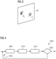
- the user-interaction protocol is interactive. I.e., user queries of subsequent iterations depend on user queries of preceding iterations. This is also illustrated in FIG. 4 .
- FIG. 4 schematically illustrates an inter- active active-learning user-interaction protocol 3002.
- one or more parts of the training data are selected in a respective iteration 3032 form basis of a user query. For instance, a specific WSI or a specific patch of a WSI may be selected.
- the user is queried to determine at least one ground-truth label for the currently selected one or more parts of the training data. It is then possible at box 3015 to (re-)train the ML algorithm, to obtain a current training state.
- Such interactive active-learning user-interaction protocol 3002 it is possible to implement the batch processing of the multiple healthy-tissue WSIs in a selected one of the multiple iterations 3030. For instance, it would be possible to batch process the multiple healthy tissue WSIs at the first one or an early iteration 3030, so as to avoid that further instances of the training data are selected at box 3005 that correspond to healthy tissue. For instance, the availability of many healthy examples will suppress uncertainty in healthy regions automatically putting focus on diseased tissue (in subsequent selections at box 3005) and thereby saving annotations. III Novelty detection to identify diseased regions The availability of a large corpus of healthy training data' also opens the door to new problem classes in ML logic.
- an ML algorithm could analyze only the healthy data and build a detailed model of its distribution (for instance, a so-called autoencoder NN). This model can then be used to detect out-of-distribution examples.
- the at least one ML algorithm that is trained by the training process based on the multiple healthy tissue WSIs can include a novelty-detection algorithm.
- unhealthy tissue can be detected in further WSIs - e.g., during inference, cf. FIG. 2 - box 2010 - using the novelty-detection algorithm, wherein the unhealthy tissue is associated with abnormalities identified by the novelty-detection algorithm.
- FIG. 5 illustrates an example of such novelty-detection algorithm 510, here implemented by an autoencoder NN.
- the autoencoder NN includes an encoder branch 511 and a decoder branch 513.
- a WSI 80 is provided as input data to the encoder branch 511 and an encoded representation 512 is determined as an output of the encoder branch.
- the encoded representation 512 of the WSI 80 forms the input to the decoder branch 513.
- a decoded representation 85 of the WSI 80 is obtained as an output of the overall autoencoder NN 510.
- the decoded representation 85 can be compared to the WSI 80 that forms the input, i.e., determining a reconstruction error 520.
- a pixel-wise comparison would be possible.
- Deviations - i.e., reconstruction errors - correspond to abnormalities.
- the abnormalities can be localized within the WSI 80 where a pixel-wise comparison is used.
- the autoencoder NN 510 is exclusively trained based on healthy-tissue WSIs (and specifically not trained based on WSIs that also depict unhealthy tissue), the autoencoder NN 510 is trained to reconstruct such healthy-tissue WSIs; however, the autoencoder NN 510 does not learn to reconstruct WSIs that also depict unhealthy tissue.
- a WSI 80 is input that also includes unhealthy tissue, this would result in an imperfectly reconstructed representation 85 and corresponding abnormalities can be detected at 520. Thereby, unhealthy tissue can be localized.
- an autoencoder NN trained on healthy-tissue WSI high reconstruction loss of an image region in a cancerous tissue sample should be highly correlated to morphological changes due to the tumor or the tumor itself. IV Building anatomical models For tumor staging - i.e., determining a cancer grade in accordance with a predetermined scale -, the extent of tumor growth plays an important role. Definitions of tumor stage for various organs is related to the anatomical structures that have been affected or passed through.
- an ML algorithm rating organ fitness of an organ anatomy in view of tumor growth can be trained using (e.g., exclusively) healthy-tissue WSIs. If combined with novelty-detection (cf. example III), this could inform a cancer grading and TNM staging system, for example, while requiring less annotations due to lesser variability.
- FIG. 6 Such combination of multiple ML algorithms is illustrated in FIG. 6 .
- a WSI 80 is provided as input to a 1st ML algorithm 3105 and a 2nd ML algorithm 3110.
- the 1st ML algorithm 3105 could be a novelty-detection algorithm, e.g., the autoencoder NN 510 as discussed in connection with FIG. 5 .
- the 1st ML algorithm 3105 it would be possible to detect unhealthy tissue 71.
- the 2nd ML algorithm 3110 it is possible to rate an organ fitness of an organ anatomy in view of tumor growth. Accordingly, the 2nd ML algorithm 3110 could also be referred to as "anatomical modeling algorithm”.
- the outputs of both ML algorithms 3105, 3110 can be then be fused in a respective fusion algorithm 3115, to determine a cancer grade of a tumor based on a result of the 1st ML algorithm 3105, as well as based on a result of the 2nd ML algorithm 3110.
Abstract
Various examples of the disclosure pertain to using whole-slide images that depict healthy tissue for a training process for at least one machine-learning algorithm for digital pathology. For instance, an autoencoder neural network can be trained based on the healthy tissue.
Description
- Various examples of the disclosure generally relate to digital pathology, i.e., using machine-learning algorithms in the context of analyzing images depicting tissue to detect, e.g., cancer or tumor or other pathologies. Various examples of the disclosure specifically relate to training of machine-learning algorithms for digital pathology.
- In histopathology, a tissue sample of a patient is inspected to study a manifestation of a disease such as cancer.
- Conventionally, a practitioner can manually inspect the tissue samples - e.g., stained using a chemical stain - using a microscope. In further detail, thin tissue slides are prepared by sectioning a tissue sample. Then, an image is acquired depicting the tissue slide.
- Recently, algorithmic analysis of tissue samples has become possible. This is referred to as digital pathology. Reference techniques of digital pathology use Convolutional Neural Networks (CNNs) as an example of machine-learning (ML) algorithms. Such techniques are described in: Karen Simonyan and Andrew Zisserman. Very deep convolutional networks for large-scale image recognition. arXiv preprint arXiv:1409.1556, 2014. Such techniques are further described in: Kaiming He, Xiangyu Zhang, Shaoqing Ren, and Jian Sun. Deep residual learning for image recognition. CoRR, abs/1512.03385, 2015. Such techniques are further described in: Mingxing Tan and Quoc V Le. Efficientnet: Rethinking model scaling for convolutional neural networks. arXiv preprint arXiv:1905.11946, 2019. Such techniques are further described in: Ilija Radosavovic, Raj Prateek Kosaraju, Ross Girshick, Kaiming He, and Piotr Dollar. Designing network design spaces. In Proceedings of the IEEE/CVF Conference on Computer Vision and Pattern Recognition, pages 10428-10436, 2020.
- Oftentimes, digital pathology requires processing large images, e.g., Whole Slide Tissue Images (WSI). A WSI depicts a microscopically magnified tissue slide of macroscopic scale, e.g., centimeters; thus, the WSI can include a large number of pixels. WSIs typically come at a size of tens of thousands of pixels in each dimension. Tissue slides either from biopsies or resected tumor tissue are prepared on glass slides, which can be scanned by a digital microscope in high resolution (gigapixels) and high throughput. Because of their size and number, WSI is especially well-suited for data-hungry ML algorithms. They can be processed by an ML algorithm as a whole or on the basis of small image patches cut out from the large images.
- It has been found that training ML algorithms to process WSIs can be time consuming and/or error prone. The sheer amount of image data oftentimes requires large amounts of expensive annotations for supervised learning. Annotations - to determine ground-truth labels for the training process - are expensive and time consuming and pathology requires specific solutions with training and validation data for a myriad of specific tissue or cancer types, organs, resection versus biopsy and possibly parameters of the preparation of the physical tissue sample (fixation, dies, slicing etc.). For example, for a standard tumor recognition task which is used as intermediate representation in many ML-based pathology systems, the pathologist is required to provide local, ideally pixelwise ground-truth labels for the training process of the machine-learning algorithm. In addition, obtaining high-resolution ground-truth labels can be challenging since there are many fine structures and areas which are hard to categorize, even for a domain expert.
- Various approaches to facilitate training of ML algorithms that process WSIs have been disclosed.
- For example, minimizing the amount of required annotations by choosing difficult and diverse examples has been proposed, which enable training of high quality models with few data. See Representative Region Based Active Learning For Histological Classification Of Colorectal Cancer, Yiqing Shen, Jing Ke, 2021 IEEE 18th International Symposium on Biomedical Imaging (ISBI); also see Using Progressive Context Encoders for Anomaly Detection in Digital Pathology Images, Ryan Gillard, Chady Meroueh, Qiangqiang Gu, Naresh Prodduturi, Sandhya Patil, Thomas J Flotte, Steven N Hart https://www.biorxiv.org/content/10.1101/2021.07.02.450957v1.f ull.pdf
- In addition, it has been tried to avoid manual labeling altogether by using semi-manual approaches based an unsupervised Segmentation, like laplace filtering. See Pathologist-Level Grading of Prostate Biopsies with Artificial Intelligence Ström, Peter, et al. arXiv preprint 2019; arXiv:190701368
- The publication Uncertainty-Based Method for Improving Poorly Labeled Segmentation Datasets Ekaterina Redekop, Alexey Chernyavskiy International Symposium on Biomedical Imaging (ISBI) 2021 suggests an automatic approach to improve poorly labeled pathology data, making it potentially easier to annotate data.
- Also known are the following publications:
- Unsupervised anomaly detection in digital pathology using GANs Milda Pocevičiu̅tė, Gabriel Eilertsen, Claes Lundström 18th International Symposium on Biomedical Imaging (ISBI) 2021
- Pan-cancer image-based detection of clinically actionable genetic alterations Kather, Jakob Nikolas, et al. Nature cancer 1.8 (2020): 789-799.
- Two-Stage Convolutional Neural Network for Breast Cancer Histology Image Classification K Nazeri, A Aminpour, M Ebrahimi International Conference Image Analysis and Recognition 2018
- Accordingly, a need exists for advanced techniques for digital pathology. Specifically, a need exists for advanced techniques of obtaining training data for facilitating a training process of at least one ML algorithm for digital pathology.
- This need is met by the features of the independent claims. The features of the dependent claims define embodiments.
- According to an example, a computer-implemented method of facilitating a training process of at least one ML algorithm is disclosed. The at least one ML algorithm is for digital pathology. The method includes obtaining multiple WSI depicting healthy tissue and further includes providing, to the training process, the multiple WSI that depict the healthy tissue, along with an indication that the multiple WSI exclusively depict healthy tissue.
- A computer program or a computer-program product or a computer-readable storage medium includes program code. The program code can be loaded and executed by at least one processor. Upon loading and executing the program code, the at least one processor performs a computer-implemented method of facilitating a training process of at least one ML algorithm. The at least one ML algorithm is for digital pathology. The method includes obtaining multiple WSI depicting healthy tissue and further includes providing, to the training process, the multiple WSI that depict the healthy tissue, along with an indication that the multiple WSI exclusively depict healthy tissue.
- A device includes a processor and a memory. The processor, upon loading program code from the memory and executing the program code, is configured to execute a computer-implemented method of facilitating a training process of at least one ML algorithm. The at least one ML algorithm is for digital pathology. The method includes obtaining multiple WSI depicting healthy tissue and further includes providing, to the training process, the multiple WSI that depict the healthy tissue, along with an indication that the multiple WSI exclusively depict healthy tissue.
- It is to be understood that the features mentioned above and those yet to be explained below may be used not only in the respective combinations indicated, but also in other combinations or in isolation without departing from the scope of the invention.
-
FIG. 1 schematically illustrates a device according to various examples. -
FIG. 2 is a flowchart of a method according to various examples. -
FIG. 3 schematically illustrates a WSI according to various examples. -
FIG. 4 schematically illustrates an annotation process of a training process for training an ML algorithm according to various examples. -
FIG. 5 schematically illustrates an autoencoder neural network algorithm according to various examples. -
FIG. 6 schematically illustrates an architecture including multiple ML algorithms according to various examples. - Some examples of the present disclosure generally provide for a plurality of circuits or other electrical devices. All references to the circuits and other electrical devices and the functionality provided by each are not intended to be limited to encompassing only what is illustrated and described herein. While particular labels may be assigned to the various circuits or other electrical devices disclosed, such labels are not intended to limit the scope of operation for the circuits and the other electrical devices. Such circuits and other electrical devices may be combined with each other and/or separated in any manner based on the particular type of electrical implementation that is desired. It is recognized that any circuit or other electrical device disclosed herein may include any number of microcontrollers, a graphics processor unit (GPU), integrated circuits, memory devices (e.g., FLASH, random access memory (RAM), read only memory (ROM), electrically programmable read only memory (EPROM), electrically erasable programmable read only memory (EEPROM), or other suitable variants thereof), and software which co-act with one another to perform operation(s) disclosed herein. In addition, any one or more of the electrical devices may be configured to execute a program code that is embodied in a non-transitory computer readable medium programmed to perform any number of the functions as disclosed.
- In the following, embodiments of the invention will be described in detail with reference to the accompanying drawings. It is to be understood that the following description of embodiments is not to be taken in a limiting sense. The scope of the invention is not intended to be limited by the embodiments described hereinafter or by the drawings, which are taken to be illustrative only.
- The drawings are to be regarded as being schematic representations and elements illustrated in the drawings are not necessarily shown to scale. Rather, the various elements are represented such that their function and general purpose become apparent to a person skilled in the art. Any connection or coupling between functional blocks, devices, components, or other physical or functional units shown in the drawings or described herein may also be implemented by an indirect connection or coupling. A coupling between components may also be established over a wireless connection. Functional blocks may be implemented in hardware, firmware, software, or a combination thereof.
- Hereinafter, techniques of digital pathology are described. Input images depicting tissue samples can be processed using an ML algorithm. The ML algorithm can receive an input image and process the input image. An example machine-learning (ML) algorithm is a neural network algorithm (NN).
- As a general rule, the NN includes multiple layers. The input to a first layer is the input image. Each layer can apply one or more mathematical operations on the input values, e.g., convolutions, nonlinear excitations, pooling operations, to give just a few examples. The input to a layer can be formed by the output of a preceding layer (feed-forward). Feedback of values or skip-connection skipping layers are possible.
- The NN for digital pathology can infer at least one semantic histopathology feature. The at least one semantic histopathology feature can describe whether the tissues sample is a manifestation of a disease. Healthy and/or unhealthy tissue may be detected and specifically localized. It would be possible to rate an organ fitness of an organ anatomy in view of tumor growth. A tumor may be graded in accordance with a predefined scale, e.g., to determine a severity. Example semantic histopathology features that could be inferred can be selected from the group consisting of: Gleason scoring, cancer grade/cancer stage estimation, clinical pathway prediction, sub-tumor classification, metastasis evaluation, microsatellite instability (MSI) or stability. For example, a cancer grade can be in accordance with a predefined grading system/scale. Examples would include the Bloom-Richardson score or TNM classification of malignant tumors. Classification systems are available for brain tumors, breast tumors, prostate cancer (Gleason scoring), and other kind of tumors.
- By means of a machine learning, a practitioner can be assisted in performing an analysis of the tissue sample.
- The input image received by the ML algorithm could have a size of at least 4.000 x 4.000 pixels, or at least 10.000 x 10.000 pixels, or at least 1E6 x 1E6 pixels. The input image could be a WSI. The input image could be acquired using optical microscopy. The tissue slide for which the input image is acquired can be stained using a chemical stain. Illumination can be used to generate a respective contrast.
- Various techniques are based on the finding that staining of tissue and acquisition of additional WSIs is considerably cheaper than obtaining ground-truth labels from an expert.
- Various techniques are based on the further finding that, since re-sectioning tissue slides from a tissue sample typically include some slides that include entirely healthy tissue, WSIs from those healthy tissue slides can be readily acquired.
- It has been found that such WSI that do not depict unhealthy tissue - i.e., exclusively depict healthy tissue - can have a benefit for a subsequent training process of a ML algorithm.
- According to examples disclosed herein, the possibility of acquiring healthy tissue slides in digital pathology is exploited, to make explicit use of healthy examples while training ML algorithms.
- According to an example, the training process of at least one ML algorithm is facilitated by obtaining multiple WSIs that depict healthy tissue. These WSIs that only depict healthy tissue and do not depict unhealthy tissues will be referred to as healthy-tissue WSIs, hereinafter.
- As a general rule, various options are available for obtaining the healthy-tissue WSIs. For example, it would be possible to prepare multiple tissue slides from a tissue sample; some of these tissue slides may depict healthy and unhealthy tissue, while other tissue slides may only depict healthy tissue. This may be readily apparent by macroscopic inspection. The healthy-tissue WSIs can be acquired based on those tissue slides that only depict healthy tissue. It would be possible to take a tissue sample from a patient that is known not to suffer from cancer or other diseases, so as to ensure that healthy-tissue WSIs do not depict unhealthy tissue. For instance, it would be possible to take a tissue sample from an animal. In some examples, healthy-tissue WSIs may me loaded from a data repository, e.g., a picture archiving system, where the respective image files are labeled as only depicting healthy tissue.
- Then, the multiple healthy-tissue WSIs can be provided to the training process along with an indication that the multiple healthy-tissue WSIs exclusively depict healthy tissue.
- The training process can accordingly appropriately use the multiple healthy-tissue WSIs that are identified as such by the indication.
- To give an example, training process may be implemented as an unsupervised training process, at least for the healthy-tissue WSIs. This means that manual annotations are not required for the healthy-tissue WSIs. This allows training on a much greater corpus of data, since rather than the availability of data the annotation is the limiting factor for supervised learning in digital pathology.
- It may not be required to query additional ground-truth labels associated with the healthy-tissue WSIs from an expert, in addition to automatically determined ground-truth labels that indicate that the multiple healthy-tissue WSIs depict healthy tissue.
- As a general rule, various options are available for making use of the healthy-tissue WSIs. Various scenarios for facilitating the training process of the at least one ML algorithm are conceivable. Along with such variations in the use case implemented based on the healthy-tissue WSIs, different types of ML algorithms may be trained using the training process.
- For example, ML algorithms configured to determine which areas of cancer-afflicted WSIs differ conceptually from the healthy-tissue WSIs may be used. These type of ML algorithms go by the name of "novelty detection" algorithms, because novel features - here: unhealthy tissue - are detected, which novel features have not been included in the training data.
-
FIG. 1 schematically illustrates adevice 91 according to various examples. Thedevice 91 is configured to implement logic associated with digital pathology. Thedevice 91 includes aprocessor 92, amemory 93, and aninterface 94. For instance,WSIs 80 may be received by theprocessor 92 via theinterface 94. One or more semantic histopathology features may be output via theinterface 94, e.g., annotated to whole-slide images 80. - The
processor 92 can load program code from thememory 93 and execute the program code. Upon loading and executing the program code, theprocessor 92 can perform techniques as disclosed herein, e.g.: facilitating a training process of an ML algorithm for digital pathology; executing the training process of the ML algorithm for digital pathology; implementing inference by executing the ML algorithm for digital pathology; etc. -
FIG. 2 is a flowchart of a method according to various examples. The method ofFIG. 2 implements digital pathology. For instance, the method ofFIG. 2 could be at least partially implemented by theprocessor 92, e.g., upon loading and executing program code. - At
box 2002, training data for training at least one ML algorithm is obtained, e.g., gathered, loaded, collected, and/or measured. For instance, it would be possible to obtain multiple healthy-tissue WSIs. Optionally it would be possible to also obtain WSIs that depict both healthy as well as unhealthy tissue. - At
box 2005, based on the training data ofbox 2002, the training process is executed. - As a general rule, the training process can include multiple stages.
- Some stages are summarized in TAB. 1 below.
TAB. 1: Two stages of a training process for training an ML algorithm. Stage Example description I Determining ground-truth labels First, ground-truth labels may be determined for the training data that is obtained at box 2002.Determining ground-truth labels can be implemented using manual annotation by an expert. A respective annotation process can be used. As part of the annotation process, using a user-interaction protocol, some patches of a WSI can be presented to an expert via a human-machine interface (e.g., monitor) along with a respective query for a ground-truth label; the expert can input the ground-truth label. Segmentation could be used to discern different tissue types as a function of position. Such manual determining of ground-truth labels is sometimes referred to as supervised learning. Thus, the user-interaction protocol helps determining ground-truth labels to WSIs based on respective user queries and user input. In some specific examples, the annotation process can be an active-learning annotation process that may be interactive. Here, in multiple iterations the queries are iteratively adapted, e.g., depending on the user action, to ensure a steep learning curve of the at least one ML algorithm. For instance, the particular patch of a WSI may be selected ad hoc in each iteration, e.g., based on a current training state of the ML algorithm to-be-trained. The current training state depends on the preceding annotation which is typically based at least in parts on user action, e.g., manual determination of labels, etc.. Alternatively or additionally, for at least some of the training data, the labels can be determined without user interaction. For instance, it would be possible to determine one or more ground-truth labels based on an indication that certain WSIs of the training data exclusively depict healthy tissue. This can be termed unsupervised learning. II Parameterization Then, once the ground-truth labels have been determined, it would be possible to set weights of the at least one ML algorithm based on the training data, as well as the associated one or more ground-truth labels. In particular, based on one or more predetermined loss functions, a loss value can be determined and then in an iterative process it can be attempted to minimize the loss value by appropriately setting the weights. A loss function can define a comparison between the ground-truth label and an output of the ML algorithm in its current training state. Respective techniques of setting weights of at least one ML algorithm are, in principle, well known in the art and need not be described here in further detail. - Once the at least one ML algorithm has been trained using the training process, at
box 2010, inference can be implemented. This means that without knowledge of ground-truth labels, further input data - e.g., further WSIs - can be processed by the at least one ML algorithm to make a prediction of one or more semantic histopathology features. -
FIG. 3 schematically illustrates aWSI 80. TheWSI 80 can include more than E9 or E10 pixels. TheWSI 80 can depict a tissue slide having extents of mm. TheWSI 80 depictshealthy tissue 70 andunhealthy tissue 71. - While
FIG. 3 illustrates anexample WSI 80 that depicts, both,healthy tissue 70 andunhealthy tissue 71, a healthy-tissue WSI 80 would not depictunhealthy tissue 71, but only depicthealthy tissue 70. - According to various examples, it is possible to consider such healthy-
tissue WSI 80 in connection with a training process of at least one ML algorithm for digital pathology. There are various options available for making use of healthy-tissue WSIs 80 in connection with the training process of at least one ML algorithm and some of the possible examples are summarized below in TAB. 2.TAB. 2: Various options for using healthy tissue WSIs in a training process for a ML algorithm. Such options can also be combined with each other, e.g., as explained in connection with a combination of example II an example I above, or as explained in connection with a combination of example IV and example III above. Brief description Example details I Standard annotation informed by entirely healthy instances of training data One option to incorporate information from healthy data is to include it in the usual training process of supervised learning (cf. TAB. 1: stage I). Depending on the concrete problem at hand, it is possible to automatically assign ground-truth labels that label all tissue on the healthy tissue WSI as 'healthy' or 'no-tumor' tissue. This can be done whenever the training processes obtained a WSI along with the indication that this WSI exclusively depicts healthy tissue. This means that it is possible to automatically determine spatially-resolved ground-truth labels that uniformly label the entire healthy-tissue WSI as "no tumor". In further detail, it would be possible to batch process the healthy-tissue WSIs in the annotation process, to thereby assign the ground-truth labels to the healthy-tissue WSIs that identify each one of the multiple healthy-tissue WSIs as exclusively depicting healthy tissue. Other WSIs that do not exclusively depict healthy tissues (and are, thus, not accompanied by the indication that they would exclusively depict healthy tissue) may be treated conventionally in the annotation process, e.g., using supervised learning in a respective user-interaction protocol. Said batch processing of the healthy-tissue WSIs yields large amount of training data with little effort. This enables to require significantly fewer or even no amount of annotations for healthy regions on cancerous slides, when subsequently annotating, using expert input, further WSIs. Healthy slides are also a good way to improve robustness and generalization of the model, for example by adding healthy slides from additional patients or even laboratories. Doing so is particularly cost effective, since healthy slides do not require any additional annotation effort. II Informing an active-learning annotation process The annotation process can include an interactive active-learning user-interaction protocol. Such interactive active-learning user-interaction protocol can include multiple iterations. Each iteration can include at least one query to obtain at least one respective ground-truth label. I.e., per iteration, the expert can determine at least one ground-truth label based on respective parts of the training data that are presented by means of the user-interaction protocol. The user-interaction protocol is interactive. I.e., user queries of subsequent iterations depend on user queries of preceding iterations. This is also illustrated in FIG. 4 .FIG. 4 schematically illustrates an inter-active active-learning user- interaction protocol 3002. Atbox 3005, one or more parts of the training data are selected in a respective iteration 3032 form basis of a user query. For instance, a specific WSI or a specific patch of a WSI may be selected. Atbox 3010, the user is queried to determine at least one ground-truth label for the currently selected one or more parts of the training data. It is then possible atbox 3015 to (re-)train the ML algorithm, to obtain a current training state. Atbox 3020, it can be judged whether afurther iteration 3030 is required. For instance, the accuracy of the ML algorithm in its current training state could be judged. If afurther iteration 3030 is required, then, at the next iteration ofbox 3005, further/different one or more parts of the training data are selected. This depends on the current training state of the ML algorithm. For instance, unknown or heterogeneous samples of the training data could be preferred, in order to establish a steep learning curve.In such interactive active-learning user- interaction protocol 3002, it is possible to implement the batch processing of the multiple healthy-tissue WSIs in a selected one of themultiple iterations 3030. For instance, it would be possible to batch process the multiple healthy tissue WSIs at the first one or anearly iteration 3030, so as to avoid that further instances of the training data are selected atbox 3005 that correspond to healthy tissue.For instance, the availability of many healthy examples will suppress uncertainty in healthy regions automatically putting focus on diseased tissue (in subsequent selections at box 3005) and thereby saving annotations. III Novelty detection to identify diseased regions The availability of a large corpus of healthy training data' also opens the door to new problem classes in ML logic. Specifically, an ML algorithm could analyze only the healthy data and build a detailed model of its distribution (for instance, a so-called autoencoder NN). This model can then be used to detect out-of-distribution examples. Concretely, the at least one ML algorithm that is trained by the training process based on the multiple healthy tissue WSIs can include a novelty-detection algorithm. Here, upon training the novelty-detection algorithm, unhealthy tissue can be detected in further WSIs - e.g., during inference, cf. FIG. 2 - box 2010 - using the novelty-detection algorithm, wherein the unhealthy tissue is associated with abnormalities identified by the novelty-detection algorithm. The abnormalities correspond to differences vis-á-vis features expected in view of the training process (which is based, as explained above, on the healthy-tissue WSIs).For instance, FIG. 5 illustrates an example of such novelty-detection algorithm 510, here implemented by an autoencoderNN. The autoencoder NN includes an encoder branch 511 and adecoder branch 513. AWSI 80 is provided as input data to theencoder branch 511 and an encodedrepresentation 512 is determined as an output of the encoder branch. The encodedrepresentation 512 of theWSI 80 forms the input to thedecoder branch 513. Thus, a decodedrepresentation 85 of theWSI 80 is obtained as an output of theoverall autoencoder NN 510. The decodedrepresentation 85 can be compared to theWSI 80 that forms the input, i.e., determining areconstruction error 520. A pixel-wise comparison would be possible. Deviations - i.e., reconstruction errors - correspond to abnormalities. The abnormalities can be localized within theWSI 80 where a pixel-wise comparison is used.In a scenario in which the autoencoder NN 510 is exclusively trained based on healthy-tissue WSIs (and specifically not trained based on WSIs that also depict unhealthy tissue), theautoencoder NN 510 is trained to reconstruct such healthy-tissue WSIs; however, theautoencoder NN 510 does not learn to reconstruct WSIs that also depict unhealthy tissue. Thus, whenever, during inference, aWSI 80 is input that also includes unhealthy tissue, this would result in an imperfectly reconstructedrepresentation 85 and corresponding abnormalities can be detected at 520. Thereby, unhealthy tissue can be localized.In the case of an autoencoder NN trained on healthy-tissue WSI, high reconstruction loss of an image region in a cancerous tissue sample should be highly correlated to morphological changes due to the tumor or the tumor itself. IV Building anatomical models For tumor staging - i.e., determining a cancer grade in accordance with a predetermined scale -, the extent of tumor growth plays an important role. Definitions of tumor stage for various organs is related to the anatomical structures that have been affected or passed through. Due to the high variability in tumor morphology (e.g., various cancer types and changing appearance during progression) training an ML algorithm for understanding organ anatomy is an easier and better-defined task in healthy-tissue WSIs. Thus, an ML algorithm rating organ fitness of an organ anatomy in view of tumor growth can be trained using (e.g., exclusively) healthy-tissue WSIs. If combined with novelty-detection (cf. example III), this could inform a cancer grading and TNM staging system, for example, while requiring less annotations due to lesser variability. Such combination of multiple ML algorithms is illustrated in FIG. 6 .In FIG. 6 , aWSI 80 is provided as input to a1st ML algorithm 3105 and a2nd ML algorithm 3110. For instance, the1st ML algorithm 3105 could be a novelty-detection algorithm, e.g., theautoencoder NN 510 as discussed in connection withFIG. 5 . Using the1st ML algorithm 3105, it would be possible to detectunhealthy tissue 71. Using the2nd ML algorithm 3110, it is possible to rate an organ fitness of an organ anatomy in view of tumor growth. Accordingly, the2nd ML algorithm 3110 could also be referred to as "anatomical modeling algorithm". The outputs of bothML algorithms respective fusion algorithm 3115, to determine a cancer grade of a tumor based on a result of the1st ML algorithm 3105, as well as based on a result of the2nd ML algorithm 3110. - Summarizing, techniques for digital pathology, specifically for facilitating a training process for at least one ML algorithm have been disclosed. Such and further effects are achieved by using healthy-tissue WSIs as at least parts of training data for a least one ML algorithm. Techniques have been disclosed in which the at least one ML algorithm includes a novelty-detection algorithm. Unhealthy tissue can be detected in WSIs during inference based on a reconstruction error of such novelty-detection algorithm.
- Using the techniques, costs can be reduced by requiring fewer manual annotations. Performance can be improved by including additional training data, possibly without increasing the number of patients - by preparing more than one tissue slide per tissue sample, healthy tissue slides can be available as a side product to unhealthy tissue slides. The techniques disclosed herein facilitate quicker adaptation of ML algorithm to new domains within digital pathology. Further, research can be supported, e.g., by identifying biomarkers from tissue morphology.
- Although the invention has been shown and described with respect to certain preferred embodiments, equivalents and modifications will occur to others skilled in the art upon the reading and understanding of the specification. The present invention includes all such equivalents and modifications and is limited only by the scope of the appended claims.
Claims (11)
- A computer-implemented method of facilitating a training process of at least one machine-learning algorithm (510, 3105, 3110) for digital pathology, the method comprising:- obtaining multiple whole-slide images (80) depicting healthy tissue (70), and- providing, to the training process (2005), the multiple whole-slide images (80) that depict healthy tissue (70) along with an indication that the multiple whole-slide images (80) exclusively depict healthy tissue (70).
- The computer-implemented method of claim 1,wherein the training process (2005) comprises an annotation process for determining ground-truth labels for a plurality of whole-slide images (80), the plurality of whole-slide images (80) comprising the multiple whole-slide images and multiple further whole-slide images (80), the multiple further whole-slide images depicting healthy tissue (70) and unhealthy tissue (71),wherein the method further comprises:- batch processing the multiple whole-slide images (80) in the annotation process, to thereby assign ground-truth labels to the multiple whole-slide images (80) that identify each one of the multiple whole-slide images (80) as exclusively depicting healthy tissue (70).
- The computer-implemented method of claim 2,wherein the annotation process comprises an interactive active-learning user-interaction protocol (3002) comprising multiple iterations (3030), each iteration (3030) comprising at least one user query (3010) to obtain at least one respective ground-truth label, wherein user queries of subsequent iterations (3030) depend on user queries of preceding iterations (3030),wherein said batch processing of the multiple whole-slide images (80) is implemented in a selected one of the multiple iterations (3030).
- The computer-implemented method of any one of the preceding claims,wherein the at least one machine-learning algorithm comprises a novelty-detection algorithm (510, 3105),wherein the method further comprises:- executing the training process based on the multiple whole-slide images (80) that depict healthy tissue (70), and- upon training the novelty-detection algorithm, detecting unhealthy tissue (71) by processing a further whole-slide image (80) using the novelty-detection algorithm, unhealthy tissue (71) being associated with abnormalities identified by the novelty-detection algorithm (510, 3105).
- The computer-implemented method of claim 4,wherein the novelty-detection algorithm comprises an autoencoder neural network algorithm (510, 3105) comprising an encoder branch (511) configured to encode an input data (80) and a decoder branch (513) configured to decode an encoded representation (512) of the input data output by the encoder branch (511),wherein the abnormalities are detected based on a reconstruction error (520) between the input data and an output data output by the decoder branch (513).
- The computer-implemented method of any one of the preceding claims,wherein the at least one machine-learning algorithm comprises an anatomical modelling algorithm (3110) to rate an organ fitness of an organ anatomy in view of tumor growth,wherein the method further comprises:- executing the training process, to train the anatomical modelling algorithm based on the multiple whole-slide images (80).
- The computer-implemented method of claim 4 or 5, and of claim 6, further comprising:- determining a cancer grade of a tumor based on a result of the novelty-detection algorithm and a further result of the anatomical modelling algorithm.
- The computer-implemented method of any one of the preceding claims, further comprising:- executing the training process (2005) in an unsupervised manner.
- The computer-implemented method of any one of the preceding claims, further comprising:- automatically determining one or more ground-truth labels based on the indication that the multiple whole-slide images exclusively depict healthy tissue, and- setting weights of the at least one machine-learning algorithm based on the multiple whole-slide images that depict healthy tissue and the one or more ground-truth labels.
- A computer program comprising program code that can be loaded and executed by a processor, wherein execution of the program code causes the processor to execute a method of facilitating a training process of at least one machine-learning algorithm (510, 3105, 3110) for digital pathology, the method comprising:- obtaining multiple whole-slide images (80) depicting healthy tissue (70), and- providing, to the training process (2005), the multiple whole-slide images (80) that depict healthy tissue (70) along with an indication that the multiple whole-slide images (80) exclusively depict healthy tissue (70).
- The computer program according to claim 10, wherein the processor, upon execution of the program code, executes the method of any one of claims 1 to 9.
Priority Applications (2)
| Application Number | Priority Date | Filing Date | Title |
|---|---|---|---|
| EP22168971.4A EP4266262A1 (en) | 2022-04-20 | 2022-04-20 | Training of machine-learning algorithms for digital pathology using healthy-tissue whole slide images |
| US18/302,243 US20230342927A1 (en) | 2022-04-20 | 2023-04-18 | Training of machine-learning algorithms for digital pathalogy using healthy-tissue whole slide images |
Applications Claiming Priority (1)
| Application Number | Priority Date | Filing Date | Title |
|---|---|---|---|
| EP22168971.4A EP4266262A1 (en) | 2022-04-20 | 2022-04-20 | Training of machine-learning algorithms for digital pathology using healthy-tissue whole slide images |
Publications (1)
| Publication Number | Publication Date |
|---|---|
| EP4266262A1 true EP4266262A1 (en) | 2023-10-25 |
Family
ID=81454734
Family Applications (1)
| Application Number | Title | Priority Date | Filing Date |
|---|---|---|---|
| EP22168971.4A Pending EP4266262A1 (en) | 2022-04-20 | 2022-04-20 | Training of machine-learning algorithms for digital pathology using healthy-tissue whole slide images |
Country Status (2)
| Country | Link |
|---|---|
| US (1) | US20230342927A1 (en) |
| EP (1) | EP4266262A1 (en) |
Citations (2)
| Publication number | Priority date | Publication date | Assignee | Title |
|---|---|---|---|---|
| US20210248736A1 (en) * | 2018-06-13 | 2021-08-12 | Siemens Healthcare Gmbh | Localization and classification of abnormalities in medical images |
| US20220076053A1 (en) * | 2020-09-07 | 2022-03-10 | Siemens Healthcare Gmbh | System and method for detecting anomalies in images |
-
2022
- 2022-04-20 EP EP22168971.4A patent/EP4266262A1/en active Pending
-
2023
- 2023-04-18 US US18/302,243 patent/US20230342927A1/en active Pending
Patent Citations (2)
| Publication number | Priority date | Publication date | Assignee | Title |
|---|---|---|---|---|
| US20210248736A1 (en) * | 2018-06-13 | 2021-08-12 | Siemens Healthcare Gmbh | Localization and classification of abnormalities in medical images |
| US20220076053A1 (en) * | 2020-09-07 | 2022-03-10 | Siemens Healthcare Gmbh | System and method for detecting anomalies in images |
Non-Patent Citations (10)
| Title |
|---|
| "Uncertainty-Based Method for Improving Poorly Labeled Segmentation Datasets Ekaterina Redekop", ALEXEY CHERNYAVSKIY INTERNATIONAL SYMPOSIUM ON BIOMEDICAL IMAGING (ISBI, 2021 |
| ILIJA RADOS-AVOVICRAJ PRATEEK KOSARAJUROSS GIRSHICKKAIMING HEPIOTR DOLLAR: "Designing network design spaces", PROCEEDINGS, 2020, pages 10428 - 10436 |
| K NAZERIA AMINPOURM EBRAHIMI: "Two-Stage Convolutional Neural Network for Breast Cancer Histology Image Classification", INTERNATIONAL CONFERENCE IMAGE ANALYSIS AND RECOGNITION, 2018 |
| KATHER, JAKOB NIKOLAS ET AL., NATURE CANCER, vol. 1, no. 8, 2020, pages 789 - 799 |
| LEE SANGHOON ET AL: "Interactive Classification of Whole-Slide Imaging Data for Cancer Researchers", CANCER RESEARCH, vol. 81, no. 4, 15 February 2021 (2021-02-15), pages 1171 - 1177, XP055963044 * |
| MILDA POCEVICIUTEGABRIEL EILERTSEN, CLAES LUNDSTROM 18TH INTERNATIONAL SYMPOSIUM ON BIOMEDICAL IMAGING (ISBI, 2021 |
| RYAN GILLARDCHADY MEROUEHQIANGQIANG GUNARESH PRODDUTURISANDHYA PATILTHOMAS J FLOTTESTEVEN N HART, USING PROGRESSIVE CONTEXT ENCODERS FOR ANOMALY DETECTION IN DIGITAL PATHOLOGY IMAGES, Retrieved from the Internet <URL:https://www.biorxiv.org/content/10.1101/2021.07.02.450957v1.full.pdf> |
| STROM, PETER ET AL.: "Pathologist-Level Grading of Prostate Biopsies with Artificial Intelligence", ARXIV:190701368, 2019 |
| XINGYU LI ET AL: "Discriminative Pattern Mining for Breast Cancer Histopathology Image Classification via Fully Convolutional Autoencoder", ARXIV.ORG, 5 May 2020 (2020-05-05), XP081657733, Retrieved from the Internet <URL:https://arxiv.org/pdf/1902.08670.pdf> * |
| YIQING SHENJING KE: "Representative Region Based Active Learning For Histological Classification Of Colorectal Cancer", 2021 IEEE 18TH INTERNATIONAL SYMPOSIUM ON BIOMEDICAL IMAGING (ISBI |
Also Published As
| Publication number | Publication date |
|---|---|
| US20230342927A1 (en) | 2023-10-26 |
Similar Documents
| Publication | Publication Date | Title |
|---|---|---|
| Stegmaier et al. | Real-time three-dimensional cell segmentation in large-scale microscopy data of developing embryos | |
| Jung et al. | An automatic nuclei segmentation method based on deep convolutional neural networks for histopathology images | |
| Belevich et al. | Microscopy image browser: a platform for segmentation and analysis of multidimensional datasets | |
| Oskal et al. | A U-net based approach to epidermal tissue segmentation in whole slide histopathological images | |
| Xu et al. | Attention by selection: A deep selective attention approach to breast cancer classification | |
| JP5795717B2 (en) | Image processing method, image processing apparatus, computer-readable medium, and computer program | |
| AlZubaidi et al. | Computer aided diagnosis in digital pathology application: Review and perspective approach in lung cancer classification | |
| US11645753B2 (en) | Deep learning-based multi-site, multi-primitive segmentation for nephropathology using renal biopsy whole slide images | |
| CN111784671A (en) | Pathological image focus region detection method based on multi-scale deep learning | |
| Muñoz-Aguirre et al. | PyHIST: a histological image segmentation tool | |
| Yuan et al. | MDL constrained 3-D grayscale skeletonization algorithm for automated extraction of dendrites and spines from fluorescence confocal images | |
| Gupta et al. | PCSeg: Color model driven probabilistic multiphase level set based tool for plasma cell segmentation in multiple myeloma | |
| Obando et al. | Multi-staining registration of large histology images | |
| Sato et al. | Evaluation of kidney histological images using unsupervised deep learning | |
| Gatenbee et al. | Virtual alignment of pathology image series for multi-gigapixel whole slide images | |
| Kloeckner et al. | Multi-categorical classification using deep learning applied to the diagnosis of gastric cancer | |
| Scheurer et al. | Semantic segmentation of histopathological slides for the classification of cutaneous lymphoma and eczema | |
| Kårsnäs | Image analysis methods and tools for digital histopathology applications relevant to breast cancer diagnosis | |
| EP4266262A1 (en) | Training of machine-learning algorithms for digital pathology using healthy-tissue whole slide images | |
| US20220375070A1 (en) | Determining region(s) for tissue dissection in pathology slides | |
| Alegro et al. | Deep learning for Alzheimer’s disease: Mapping large-scale histological tau protein for neuroimaging biomarker validation | |
| Liang et al. | Neural network calibration for medical imaging classification using DCA regularization | |
| Vidhya et al. | Deep learning based approach for efficient segmentation and classification using VGGNet 16 for tissue analysis to predict colorectal cancer | |
| Lee et al. | Classification of mouse lung metastatic tumor with deep learning | |
| Cao et al. | Automatic detection of adult cardiomyocyte for high throughput measurements of calcium and contractility |
Legal Events
| Date | Code | Title | Description |
|---|---|---|---|
| STAA | Information on the status of an ep patent application or granted ep patent |
Free format text: STATUS: THE APPLICATION HAS BEEN PUBLISHED |
|
| PUAI | Public reference made under article 153(3) epc to a published international application that has entered the european phase |
Free format text: ORIGINAL CODE: 0009012 |
|
| AK | Designated contracting states |
Kind code of ref document: A1 Designated state(s): AL AT BE BG CH CY CZ DE DK EE ES FI FR GB GR HR HU IE IS IT LI LT LU LV MC MK MT NL NO PL PT RO RS SE SI SK SM TR |
|
| RAP1 | Party data changed (applicant data changed or rights of an application transferred) |
Owner name: GEORG-AUGUST-UNIVERSITAET GOETTINGEN STIFTUNG OEFFENTLICHEN RECHTS UNIVERSITAETSMEDIZIN GOETTINGEN Owner name: SIEMENS HEALTHINEERS AG |