EP4255301B1 - Einstellung langfristiger intraindividualer änderungen bei bandähnlichen ekg-amplituden - Google Patents
Einstellung langfristiger intraindividualer änderungen bei bandähnlichen ekg-amplituden Download PDFInfo
- Publication number
- EP4255301B1 EP4255301B1 EP21823558.8A EP21823558A EP4255301B1 EP 4255301 B1 EP4255301 B1 EP 4255301B1 EP 21823558 A EP21823558 A EP 21823558A EP 4255301 B1 EP4255301 B1 EP 4255301B1
- Authority
- EP
- European Patent Office
- Prior art keywords
- ecg
- lead
- precordial
- subject
- transition zone
- Prior art date
- Legal status (The legal status is an assumption and is not a legal conclusion. Google has not performed a legal analysis and makes no representation as to the accuracy of the status listed.)
- Active
Links
Images


Classifications
-
- A—HUMAN NECESSITIES
- A61—MEDICAL OR VETERINARY SCIENCE; HYGIENE
- A61B—DIAGNOSIS; SURGERY; IDENTIFICATION
- A61B5/00—Measuring for diagnostic purposes; Identification of persons
- A61B5/24—Detecting, measuring or recording bioelectric or biomagnetic signals of the body or parts thereof
- A61B5/316—Modalities, i.e. specific diagnostic methods
- A61B5/318—Heart-related electrical modalities, e.g. electrocardiography [ECG]
- A61B5/346—Analysis of electrocardiograms
- A61B5/349—Detecting specific parameters of the electrocardiograph cycle
- A61B5/352—Detecting R peaks, e.g. for synchronising diagnostic apparatus; Estimating R-R interval
-
- A—HUMAN NECESSITIES
- A61—MEDICAL OR VETERINARY SCIENCE; HYGIENE
- A61B—DIAGNOSIS; SURGERY; IDENTIFICATION
- A61B5/00—Measuring for diagnostic purposes; Identification of persons
- A61B5/24—Detecting, measuring or recording bioelectric or biomagnetic signals of the body or parts thereof
- A61B5/30—Input circuits therefor
- A61B5/303—Patient cord assembly, e.g. cable harness
-
- A—HUMAN NECESSITIES
- A61—MEDICAL OR VETERINARY SCIENCE; HYGIENE
- A61B—DIAGNOSIS; SURGERY; IDENTIFICATION
- A61B5/00—Measuring for diagnostic purposes; Identification of persons
- A61B5/24—Detecting, measuring or recording bioelectric or biomagnetic signals of the body or parts thereof
- A61B5/316—Modalities, i.e. specific diagnostic methods
- A61B5/318—Heart-related electrical modalities, e.g. electrocardiography [ECG]
- A61B5/333—Recording apparatus specially adapted therefor
- A61B5/338—Recording by printing on paper
-
- A—HUMAN NECESSITIES
- A61—MEDICAL OR VETERINARY SCIENCE; HYGIENE
- A61B—DIAGNOSIS; SURGERY; IDENTIFICATION
- A61B5/00—Measuring for diagnostic purposes; Identification of persons
- A61B5/24—Detecting, measuring or recording bioelectric or biomagnetic signals of the body or parts thereof
- A61B5/316—Modalities, i.e. specific diagnostic methods
- A61B5/318—Heart-related electrical modalities, e.g. electrocardiography [ECG]
- A61B5/346—Analysis of electrocardiograms
- A61B5/349—Detecting specific parameters of the electrocardiograph cycle
- A61B5/366—Detecting abnormal QRS complex, e.g. widening
-
- A—HUMAN NECESSITIES
- A61—MEDICAL OR VETERINARY SCIENCE; HYGIENE
- A61B—DIAGNOSIS; SURGERY; IDENTIFICATION
- A61B5/00—Measuring for diagnostic purposes; Identification of persons
- A61B5/68—Arrangements of detecting, measuring or recording means, e.g. sensors, in relation to patient
- A61B5/6801—Arrangements of detecting, measuring or recording means, e.g. sensors, in relation to patient specially adapted to be attached to or worn on the body surface
- A61B5/6813—Specially adapted to be attached to a specific body part
- A61B5/6823—Trunk, e.g., chest, back, abdomen, hip
Definitions
- the present disclosure lies in the general field of clinical electrocardiography (ECG), more specifically in ECG methods comprising precordial ECG amplitude measurements.
- ECG electrocardiography
- the present invention relates to methods of determining a subject's corrected precordial ECG amplitude from an ECG measurement, which corrections are based on intraindividual changes such as the rotation position of the heart on the horizontal plane, and/or changes in body mass index. The methods are thus useful for adjustment of intraindividual changes over time in precordial ECG amplitudes, such as between two ECG measurements.
- the present invention further relates to a device, an apparatus and a computer program product, and to uses of said device, apparatus and computer program product in a method of determining a subject's corrected precordial ECG amplitude from an ECG measurement, and for improving the accuracy of electrocardiographic methods depending on precordial ECG amplitude measurements.
- the methods and the device, apparatus or product can be advantageously used in a method of diagnosing left ventricular hypertrophy and/or hypertension in a subject.
- Clinical ECG represents the magnitude and direction of the electrical forces generated by the heart as a function of time measured on the subject's body surface. Besides the absolute magnitudes of the individual deflections like P, QRS, and T waves, called amplitudes, also the form of electrocardiogram is dependent on accurate and reproducible measurement of voltage.
- ECG is the most commonly used method for clinical heart study in the world (Kligfield et al. 2007). Its great advantages are non-invasiveness, low cost and its easy use. It can be recorded all over the world. The same properties are intended to be included in the method and device of the present invention.
- the ECG graph measures the height (amplitude) of a given wave or deflection.
- the amplitude, or voltage, of the recorded electrical signal is expressed on an ECG in the vertical dimension and is measured in millivolts (mV).
- mV millivolts
- intraindividual amplitude changes the congenital differences between different individuals, like for instance differences in skeletal dimensions and possible differences in the conduction system of the heart, play practically no role. However, there is variability also in intraindividual amplitude changes.
- Intraindividual variability is a hindrance in serial ECG analysis, where ECG's of the same individual, but taken at different times, are compared.
- Two sources of intraindividual variability can be distinguished as follows: variability related to the technical circumstances during ECG recording and non-pathologic biologic variability.
- variability related to the technical circumstances during ECG recording is the most confusing (Schijvenaars et al. 2008). According to Shijvenaars the most important biological sources are age and weight.
- the magnitude of the electrical potential generated by the heart, measured on the thoracic surface is dependent on the solid angle of the contour of the heart seen from the measurement point on the subjects thoracic surface (0os-terom et al 2000).
- the magnitude of the solid angle is dependent both on the square of the distance between the measurement point on the thoracic surface and the heart mass, and the area of the contour of the heart mass seen from the measurement point on the body surface (Oosterom et al.2000).
- the position of the heart within the thorax virtually determines the electrical force generated by the heart as measured on the thoracic surface.
- the position of the heart within the thorax is such, that the contour of the left ventricle resembles an asymmetrical ellipsoid, as seen from the anterolateral thoracic surface.
- the heart can be rotated around its length axis clockwise or counterclockwise. The more the heart rotates clockwise, the shorter. Vice versa, the longer is the transverse axis of the ellipsoid contour of the left ventricle in case the heart rotates counterclockwise.
- the surface area of the ellipsoid is directly related to the length of the shorter axis.
- the magnitude of the solid angle seen from the anterolateral surface of the thorax is the greater the more counterclockwise the heart rotates, and the smaller the more the heart rotates clockwise. This phenomenon makes the amplitudes measured on the anterolateral thoracic surface greater, when the heart is counterclockwise rotated and smaller, when the heart is clockwise rotated.
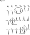
- transitional zone The electrical axis of the heart on the horizontal plane, which is called transitional zone, is defined to be, when in precordial ECG leads V1 - V6 the amplitudes of Rand S waves in the same QRS complex are equal.
- the axis is considered to be normal, if the transition zone is between the leads V3 and V4, when in practice the S wave is more prominent than the R wave in the lead V3, and the R wave is more prominent than the S wave in the lead V4. If the transition zone is more to right, the situation is called counterclockwise rotation, and more to the left the situation is called clockwise rotation.
- the decreasing or increasing difference between the actual and the "ideal" weight determines the percentage correction for the measured amplitude according the method of the present disclosure.
- a method of determining a subject's corrected precordial ECG amplitude from an ECG measurement comprising the steps of (a) providing a first BMI value of a subject, wherein said first BMI value is obtained from a first time point of a first ECG measurement; (b) providing a second BMI value of said subject, wherein said second BMI value is obtained from a second time point of a second ECG measurement which is after said first time point; (c) providing said subject's ECG data in paper form or stored on a memory device obtained from said first ECG measurement, wherein said ECG was recorded using a standardized and reproducible electrode placement method, and wherein said ECG data comprises data for a six lead V1-V6 precordial ECG; (d) providing said subject's ECG data in paper form or stored on a memory device obtained from said second ECG measurement, wherein said ECG was recorded using a standardized and reproducible electrode placement method, and wherein said ECG data comprises data for a six lead V1-V6 precor
- the device (1) of the present disclosure can be used for determining a subject's corrected precordial ECG amplitude in an ECG measurement, wherein said use comprises comparing the illustrations of said device of the respective QRS complex of the transitional zone at different positions of a six lead precordial ECG horizontal plane as disclosed herein above with the QRS complexes of a six lead V1-V6 precordial ECG measurement of said subject provided in paper form or stored on a memory device, such as to determine the position of the transitional zone in said six lead precordial ECG measurement, and conducting the method of the present disclosure.
- the device of the present disclosure can be used for improving the accuracy of electrocardiographic methods depending on precordial ECG amplitude measurements; wherein said use comprises conducting the method(s) of the present disclosure.
- the present invention provides a fast and easy way for cost effective comparison of serial, intraindividual precordial ECG amplitudes in long term follow-up.
- the method and device can be employed all over the world, anywhere the recording of ECG is possible. No extra devices or even electricity is needed.
- the invention improves remarkably the accuracy of long-term follow-up of, for instance, heart left ventricular hypertrophy and hypertension.
- a method of diagnosing left ventricular hypertrophy and/or hypertension in a subject and a use of the device of the present disclosure for diagnosing left ventricular hypertrophy and/or hypertension in a subject, as further defined in the claims.
- precordial ECG amplitudes are standardized and reproduced satisfactory, also the ageing of cardiovascular system and the development of prehypertension in an individual human subject can be followed.
- Nakamura et al 2012 found in 9067 subjects 35.8 % in CCWR, 56 % in NR and 8.2 % in CWR. There was a significant positive association of clockwise rotation and a significant inverse association of counterclockwise rotation with CVD mortality in men as well as in men and women combined, independent of confounding factors including other ECG changes.
- transition zone right from V3 is defined herein below as counterclockwise and from the lead V4 and left from it, clockwise.
- the angle and the area of the asymmetrical ellipsoid between the electrodes for the leads V3 and V2 is about 10 %, at the electrode for the lead V2 it is about 20 %, and between the electrodes for the leads V1 and V2 about 30 % greater.
- the angle and the area of the ellipse is about 10 %, between the electrodes for the leads V4 and V5 about 20 %, at the electrode for the lead V5 about 30 %, and between the electrodes for the leads V5 and V6 about 40 % smaller.
- the amplitude measured between the electrodes V3 and V2 is about 10 % greater than in the normal rotation area, and correspondingly about 20 % greater at the electrode V2, and about 30 % greater between the electrodes V1 and V2.
- the amplitude is at the electrode V4 about 10 % smaller, between the electrodes V4 and V5 about 20 % smaller, at the electrode V5 about 30 % smaller, and between the electrodes V5 and V6 about 40 % smaller than in the normal rotation area.
- the measured amplitudes must be corrected by the measured percentages according to the horizontal rotation decreasing in counterclockwise rotation and increasing in clockwise rotation.
- the term "about” as used herein is intended to mean ⁇ 2%, preferably ⁇ 1%, more preferably ⁇ 0.5%, even more preferably ⁇ 0.2%, and most preferably ⁇ 0.1%.
- "about 10%” is intended to mean 10 ⁇ 2%, preferably 10 ⁇ 1%, more preferably 10 ⁇ 0.5%, even more preferably 10 ⁇ 0.2%, and most preferably 10 ⁇ 0.1%.
- the precondition for the successful use of the present method is a standardized and reproducible placement of precordial ECG electrodes. If electrode placement is not standardized and reproducible, the position of the electrode change, which affects both factors of the solid angle. Suitable methods and devices for a standardized and reproducible placement of precordial ECG electrodes are disclosed, for example, in FI20196062 . Specifically, the precordial electrodes can be placed by
- the electrode for the lead V6 is supposed to be straight to left from the left ventricle of the heart, and the electrode for the lead V2 straight forward from the left ventricle projised on the horizontal plane of the electrodes V4, V5 and V6.
- the electrode for the lead V4 is supposed to be about at the half-point between the electrodes V2 and V6 on the same horizontal plane.
- the electrode for the lead V3 as projised at the same horizontal level than the electrodes V4, V5 and V6 is estimated to be at the half-point between the electrodes V2 and V3, and the electrode for the lead V5 is at the half-point between the electrodes V4 and V6 at the same horizontal level.
- the angle between each electrode would be 22.5 degrees. However, for practical reasons an angle of 20 degrees is used in the method.
- the methods disclosed herein are advantageously used for cost effective comparison of serial, intraindividual precordial ECG amplitudes in long term follow-up.
- the method is especially useful in the long-term follow-up of individual human subjects which undergo weight changes.
- the present inventors provide a method of determining a subject's corrected precordial ECG amplitude from an ECG measurement, comprising the steps of
- Said method may be carried out fully by a computing device. Said method comprises:
- the providing steps of blocks 301, 302 may correspond to maintaining the associated information (and optionally also the information based on which said information has been calculated or obtained) in a memory.
- the outputting in block 305 may comprise, for example, displaying said subject's corrected precordial ECG amplitude in the ECG measurement to a user via a display comprised in or electrically connected to the apparatus. Additionally or alternatively, the outputting in block 305 may comprise outputting said subject's corrected precordial ECG amplitude in the ECG measurement to a second apparatus for displaying said subject's corrected precordial ECG amplitude in the ECG measurement to a user, where said second apparatus is connected to the apparatus via at least one communication link and/or at least one communication network.
- said subject's corrected precordial ECG amplitude may be stored to a memory.
- the body mass index is derived from the mass (weight in kg) and height of a person (in metres). Specifically, the BMI is defined as the body mass divided by the square of the body height.
- the subject is a human subject, preferably an adult human subject, such as a human subject being at least 18, 19, or 20 years old. At this age, a human subject statistically has a normal (healthy) BMI at a value of 23.
- the methods disclosed herein improves the accuracy of all electrocardiographic methods depending on precordial amplitude measurements.
- the determination of a correct ECG amplitude is of high diagnostic relevance.
- the herein disclosed correction can be applied to all precordial amplitude measurements, like P wave, Q wave, R wave, S wave, ST line, T wave and U wave.
- the Sokolow Lyon index is used in the diagnosis of left ventricular hypertrophy (LVH), which is characterized by a thickening of the heart muscle of the left ventricle of the heart.
- the Sokolow-Lyon index is calculated by the sum of the amplitude of the S wave in the V1 lead and of the amplitude of the R wave in the V5 or V6 lead (whichever is larger).
- a sum of more than 3.5 mV (or more than 35 mm) is indicative for the presence of left ventricular hypertrophy (LVH).
- the mean BMI was 20 in 1949, when the Sokolow-Lyon index was expressed, and at the present the mean BMI is about 27, which has not been noticed in the common prior art.
- the possible changes in intraindividual amplitudes are more accurate than the absolute amplitudes.
- the adjustment of amplitude measurements in the present invention is not made more accurate than to the nearest 5 %.
- Tafeit et al found in their study, that the mean thickness of the subcutaneous fat layer on upper back, front chest, lateral chest, upper abdomen, lower abdomen and lower back in men, without any special training, was 9.5 mm.
- the square of the distance should be used.
- the gain in weight caused by obesity is lifting the diaphragma and turning the heart to a more transverse position, which decreases at least anterolateral precordial amplitudes. So, using the square of the distance should decrease amplitudes too much. Therefore, the simple magnitude of the distance was used in counting.
- a change of 4 units of BMI (12 kg) changes the measured precordial ECG amplitudes by 10 %, decreasing the amplitude after gain in weight and increasing the amplitudes after loss in weight.
- the BMI value 23 which is defined good for health (Fontana and Frank 2014)
- the amplitudes are decreased by 5 % for every two units in BMI.
- the amplitudes are increased by 5 % for every two units in BMI.
- a method of determining a subject's corrected precordial ECG amplitude from an ECG measurement comprising the steps of (a) providing a first BMI value of a subject, wherein said first BMI value is obtained or obtainable from a first time point; (b) providing a second BMI value of said subject, wherein said second BMI value is obtained or obtainable from a second time point of an ECG measurement, which is after said first time point; (c) providing said subject's ECG data in paper form or stored on a memory device obtained or obtainable from said ECG measurement made at said second time point, wherein said ECG was recorded using a standardized and reproducible electrode placement method, and wherein said ECG data comprises data for a precordial ECG amplitude A; (d) determining a percentage correction value B by assigning said first BMI value a first percentage correction value B1, and said second BMI value a second percentage correction value B2, wherein B1 and B2 are independently assigned a percentage correction value
- Said method may be carried out fully by a computing device. Said method comprises:
- the providing steps of blocks 401 to 403 may correspond here to maintaining the associated information (and optionally also the information based on which said information has been calculated or obtained) in a memory.
- the outputting in block 406 may be defined as described above in connection with block 305 of Figure 3 .
- said subject's corrected precordial ECG amplitude may be stored to a memory.
- both methods can be combined to even further increase the accuracy of a measured ECG amplitude.
- a method of determining a subject's corrected precordial ECG amplitude from an ECG measurement comprising the steps of (a) providing a first BMI value of a subject, wherein said first BMI value is obtained or obtainable from a first time point of a first ECG measurement; (b) providing a second BMI value of said subject, wherein said second BMI value is obtained or obtainable from a second time point of a second ECG measurement which is after said first time point; (c) providing said subject's ECG data in paper form or stored on a memory device obtained or obtainable from said first ECG measurement, wherein said ECG was recorded using a standardized and reproducible electrode placement method, and wherein said ECG data comprises data for a six lead V1-V6 precordial ECG; (d) providing said subject's ECG data in paper form or stored on a memory device obtained or obtainable from said second ECG
- Said method may be carried out fully by a computing device. Said method comprises:
- the providing steps of blocks 501 to 504 may correspond here to maintaining the associated information (and optionally also the information based on which said information has been calculated or obtained) in a memory.
- the outputting in block 508 may be defined, e.g., as described above in connection with block 305 of Figure 3 .
- said subject's corrected precordial ECG amplitude may be stored to a memory.
- the first time point is a time point, when an earlier ECG measurement of said subject was made, such that the method allows a accurate monitoring of serial, intraindividual precordial ECG amplitudes in long term follow-up.
- the long-term changes mean in this connection an opposite to acute changes, that have an effect in minutes or hours like, for instance, acute bleeding.
- the effect of an acute change returns usually in hours or days.
- the cause of long-term changes in amplitudes are changes in the rotation position of the heart and BMI, that both occur over months or years, but at least in weeks.
- the first and the second time point may be at least 2 weeks apart, preferably at least 3 weeks apart, more preferably at least 4 weeks apart, more preferably at least 1 month apart, more preferably at least 2 months apart, more preferably at least 3 months apart, more preferably at least 4 months apart, more preferably at least 5 months apart, more preferably at least 6 months apart, more preferably at least 7 months apart, more preferably at least 8 months apart, more preferably at least 9 months apart, more preferably at least 10 months apart, more preferably at least 11 months apart, more preferably at least 12 months apart, such as more than 18 months apart or more than 24 months apart.
- the first and the second time point may be at most 40 years apart, preferably at most 30 years apart, preferably at most 20 years apart, preferably at most 15 years apart, more preferably at most 10 years apart, more preferably at most 8 years apart, more preferably at most 6 years apart, more preferably at most 4 years apart, more preferably at most 2 years apart, more preferably at most 22 months apart, more preferably at most 20 months apart, more preferably at most 18 months apart, more preferably at most 16 months apart, more preferably at most 14 months apart, more preferably at most 12 months apart, more preferably at most 10 months apart, more preferably at most 8 months apart, more preferably at most 6 months apart, more preferably at most 4 months apart, more preferably at most 2 months apart, such as at most 1 month apart.
- the method has been exemplified for two time points, and/or two ECG measurements, respectively.
- the method allows accurate monitoring of serial, intraindividual precordial ECG amplitudes in long term follow-up.
- the methods described herein can, of course, also be repeated multiple times, i.e. for at least one further third time point, when a further ECG measurement is made. This opens up two possibilities for comparison or monitoring.
- the change in BMI and/or the change in the transition zone of the measurement made at the at least one further third time point is always determined in comparison to a fixed value.
- the change in BMI and/or the change in the transition zone at the third time point is compared to the BMI and/or the transition zone at said first time point and the data of said first ECG measurement.
- the change in BMI and/or the change in the transition zone at the at least one further third time point is compared to the BMI and/or the transition zone of the directly preceding time point and the data of the directly preceding ECG measurement.
- the ECG amplitude is determined by the sum of the amplitude of the S wave in lead V1 and the amplitude of the R wave in lead V5 or lead V6, whatever amplitude is higher (Sokolow-Lyon index).
- a method of diagnosing left ventricular hypertrophy and/or hypertension in a subject comprising conducting the method as disclosed herein above, wherein a corrected ECG amplitude A* of more than 3.5 mV is indicative of left ventricular hypertrophy and/or hypertension.
- a further aspect of the present application pertains to a device (see Figure 2 ), which implements and simplifies the method of the present disclosure as described herein above.
- the inventors aimed at to develop a non-invasive and easy to use device with an advantageous price.
- the device is useable all over the world in all circumstances where the ECG can be registered, without the need of any other devices.
- the present disclosure further provides a device (1), which is suitable for use in a method as defined herein above, wherein the device is in the form of a rectangular sheet, having an left and right short side (8) and an upper and lower long side (7).
- Said device (1) comprises an alignment (2) of BMI values and a first percentage correction value according the following table BMI 17 or 18 -15 19 or 20 -10 21 or 22 -5 23 0 24 or 25 5 26 or 27 10 28 or 29 15 30 or 31 20 32 or 33 25 34 or 35 30 36 or 37 35 38 or 39 40 40 or 41 45 42 or 43 50 44 or 45 55 46 or 47 60 48 or 49 65 50 or 51 70
- the device (1) further comprises illustrations (3) of the form of the respective QRS complex of the transitional zone at different positions of a six lead V1-V6 precordial ECG horizontal plane; and indications (4) of said position of the transitional zone relative to precordial leads V1-V6.
- the device (1) comprises an alignment of said indications (4) of said position of the transitional zone with a corresponding second percentage correction value (5), wherein said alignment indicates one or more of the following (i) to (ix): (i) -30 V1-V2 (ii) -20 V2 (iii) -10 V2-V3 (iv) 0 V3 (v) 0 V3-V4 (vi) 10 V4 (vii) 20 V4-V5 (viii) 30 V5 (ix) 40 V5-V6 ; preferably wherein said alignment indicates all of (i) to (ix).
- These illustrations and alignments are preferably aligned in three horizontal items (6,9,10).
- the form of the QRS complex in different positions of ECG horizontal plane transitional zone In embodiments, in the uppermost item (6) is illustrated the form of the QRS complex in different positions of ECG horizontal plane transitional zone.
- the middle item (9) In the middle item (9) are upper the electrodes compared to the different transitional zones and lower the percentage adjustments.
- the lowest item (10) In the lowest item (10) are upper two BMI values and lower the comparable percentage adjustment. In the lowest item are upper two values of BMI and lower the comparable percentage correction.
- the part / the horizontal item (6) of the device showing the illustrations (3) are made of a transparent material. This allows a better comparison of the ECG data with the illustrations (3).
- the whole device is basically made of a transparent material. In principle, any suitable transparent material may be used.
- the device (1) may comprises at least one handle (11), preferably at least one handle on the middle of the device, and optionally at least one further handle (12) on the right side of the device, such as at the right end of the device. The handles allow for easy moving of the device.
- the device described herein may be used for determining a subject's corrected precordial ECG amplitude in an ECG measurement. Indeed, the device described herein may be used for improving the accuracy of any sort of electrocardiographic methods depending on precordial ECG amplitude measurements. For example, the device (1) may be used for diagnosing left ventricular hypertrophy and/or hypertension in a subject, as described herein above.
- Said uses have in common that they usually comprise the step of comparing the illustrations of said device of the respective QRS complex of the transitional zone at different positions of a six lead precordial ECG horizontal plane (3,6) with the QRS complexes of a six lead V1-V6 precordial ECG measurement of said subject, such as to determine the position of the transitional zone in said six lead precordial ECG measurement.
- a next step one may then conduct the method(s) as described herein above.
- the device is used to identify which one of the precordial electrode is the position of the transitional zone, at which position the amplitudes of Rand S waves look to be most similar in size to each other. Then the device is moved on the electrocardiogram so, that the QRS complex of the corresponding electrode of the device (3) is on the QRS complex of the ECG. If visually cannot be clearly decided whether the R and S waves are equal, or not, the transition zone is defined to be at this electrode. If the R wave is clearly bigger, the transition zone is between this and the next electrode clockwise, and if the S wave is clearly bigger, is the transition zone between this and the previous electrode counterclockwise.
- Figure 6 provides an apparatus 601 (e.g., a computing device) according to some embodiments.
- Figure 6 may illustrate an apparatus configured to carry out at least the functions described above in connection with determining a subject's corrected precordial ECG amplitude in an ECG measurement.
- the apparatus 601 may comprise one or more control circuitry 620, such as at least one processor, and at least one memory 630, including one or more algorithms 631, such as a computer program code (software) wherein the at least one memory and the computer program code (software) are configured, with the at least one processor, to cause, respectively, the apparatus to carry out any one of the exemplified functionalities relating to determining a subject's corrected precordial ECG amplitude in an ECG measurement as described above.
- control circuitry 620 such as at least one processor
- at least one memory 630 including one or more algorithms 631, such as a computer program code (software) wherein the at least one memory and the computer program code (software) are configured, with the at least one processor
- the control circuitry 620 of the apparatus 601 comprises at least precordial ECG amplitude correction circuitry 621.
- the precordial ECG amplitude correction circuitry 621 may be configured to carry out at least some of the functionalities described above by means of any of Figures 3 to 5 using one or more individual circuitries.
- the at least one memory 630 may comprise at least one database 632 which may comprise, for example, subject's ECG data obtainable from the first ECG measurement made at the first time point, the first ECG measurement, subject's ECG data obtainable from the second ECG measurement made at the second time point, the second ECG measurement, the first BMI value of a subject, the second BMI value of said subject, said subject's corrected precordial ECG amplitude and/or any other information used and/or produced in calculations according to embodiments.
- Each memory 630 may comprise software 631 and at last one database 632.
- the memory 630 may also comprise other databases which may not be related to the functionalities of the apparatus according to any of presented embodiments.
- the at least one memory 630 may be implemented using any suitable data storage technology, such as semiconductor-based memory devices, flash memory, magnetic memory devices and systems, optical memory devices and systems, fixed memory and removable memory.
- the apparatus 601 may further comprise different interfaces 610 such as one or more communication interfaces (TX/RX) comprising hardware and/or software for realizing communication connectivity over one or more communications network according to one or more communication protocols.
- the one or more communication interfaces 610 may comprise standard well-known components such as an amplifier, filter, frequency-converter, analog-to-digital converts, (de)modulator, and encoder/decoder circuitries, controlled by the corresponding controlling units, and one or more antennas.
- the interfaces 610 may comprise a user input interface and/or an interface for a display.
- circuitry' also covers an implementation of merely a hardware circuit or processor (or multiple processors) or a portion of a hardware circuit or processor and its (or their) accompanying software and/or firmware.
- the term 'circuitry' also covers, for example and if applicable to the particular claim element, a baseband integrated circuit for an access node or a terminal device or other computing or network device.
- the at least one processor, the memory, and the computer program code form processing means or comprises one or more computer program code portions for carrying out one or more operations according to any one of the embodiments of Figures 3 to 5 or operations thereof.
- Embodiments as described may also be carried out in the form of a computer process defined by a computer program or portions thereof. Embodiments of the methods described in connection with Figures 3 to 5 may be carried out by executing at least one portion of a computer program comprising corresponding instructions.
- the computer program may be provided as a computer readable medium comprising program instructions stored thereon or as a non-transitory computer readable medium comprising program instructions stored thereon.
- the computer program may be in source code form, object code form, or in some intermediate form, and it may be stored in some sort of carrier, which may be any entity or device capable of carrying the program.
- the computer program may be stored on a computer program distribution medium readable by a computer or a processor.
- the computer program medium may be, for example but not limited to, a record medium, computer memory, read-only memory, electrical carrier signal, telecommunications signal, and software distribution package, for example.
- the computer program medium may be a non-transitory medium. Coding of software for carrying out the embodiments as shown and described is well within the scope of a person of ordinary skill in the art.
- the Sokolow-Lyon (S-L) index of left ventricular electrocardiographic hypertrophy is determined to be 3.2 mV. Such a value is below the borderline of electrocardiographic left ventricular hypertrophy (3.5).
- the same person comes to a follow-up control after one year.
- the measured index is 3.2 mV, which would be considered healthy.
- the person has gained 15 kg in body weight, and the person's BMI is now 32. Looking on the device of the present disclosure, the user assigns a B1 correction value of 10%, and a B2 correction value of 25%. The difference B2-B1 is thus 15%. Since the person gained weight, i.e. showed an increase in BMI, said value is positive. Thus, the correction percentage for increase in BMI is 15 %.
- the subject has lost weight by 30 kg and now has a BMI of 24.
- the physician sees that the position of the transition zone has changed to the no rotation area (percentage correction 0%) from the area on V4 (to which a percentage correction of 10% is assigned to), such that the difference is -10 %.
- About 1/3 of the increase in the measured, uncorrected Sokolow-Lyon index is due to the lost in weight, and there is no practical difference when considering the corrected amplitude.
Landscapes
- Health & Medical Sciences (AREA)
- Life Sciences & Earth Sciences (AREA)
- Cardiology (AREA)
- Heart & Thoracic Surgery (AREA)
- Molecular Biology (AREA)
- Pathology (AREA)
- Engineering & Computer Science (AREA)
- Biomedical Technology (AREA)
- Physics & Mathematics (AREA)
- Medical Informatics (AREA)
- Biophysics (AREA)
- Surgery (AREA)
- Animal Behavior & Ethology (AREA)
- General Health & Medical Sciences (AREA)
- Public Health (AREA)
- Veterinary Medicine (AREA)
- Measurement And Recording Of Electrical Phenomena And Electrical Characteristics Of The Living Body (AREA)
Claims (15)
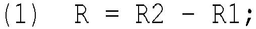
- Verfahren zum Bestimmen einer korrigierten Brustwand-EKG-Amplitude einer Person in einer EKG-Messung, das die folgenden Schritte umfasst:(a) Bereitstellen der EKG-Daten der Person, die von einer ersten EKG-Messung erhalten wurden, die an einem ersten Zeitpunkt vorgenommen wurde, in Papierform oder auf einer Speichervorrichtung gespeichert, wobei das EKG unter Verwendung eines standardisierten und reproduzierbaren Elektrodenplatzierungsverfahrens aufgezeichnet wurde und wobei die EKG-Daten Daten für ein Brustwand-EKG mit sechs Ableitungen V1 bis V6 umfassen;(b) Bereitstellen der EKG-Daten der Person, die von einer zweiten EKG-Messung erhalten wurden, die an einem zweiten Zeitpunkt vorgenommen wurde, der nach dem ersten Zeitpunkt liegt, in Papierform oder auf einer Speichervorrichtung gespeichert, wobei das EKG unter Verwendung eines standardisierten und reproduzierbaren Elektrodenplatzierungsverfahrens aufgezeichnet wurde und wobei die EKG-Daten Daten für ein Brustwand-EKG mit sechs Ableitungen V1 bis V6 und eine Brustwand-EKG-Amplitude A umfassen; und(c) Bestimmen eines Korrekturprozentwerts R durch Bestimmen der Position der Übergangszone in den Daten eines Brustwand-EKG mit sechs Ableitungen V1 bis V6 von der ersten EKG-Messung in Papierform oder auf einer Speichervorrichtung gespeichert, und Zuweisen eines Korrelationsprozentwerts R1 dazu und Bestimmen der Position der Übergangszone in den Daten eines Brustwand-EKG mit sechs Ableitungen V1 bis V6 von der zweiten EKG-Messung in Papierform oder auf einer Speichervorrichtung gespeichert, und Zuweisen eines Korrelationsprozentwerts R2 dazu, wobei R1 oder R2 unabhängig beträgt:(i) etwa -30 % in dem Fall, in dem die Übergangszone zwischen der Ableitung V1 und der Ableitung V2 liegt,(ii) etwa -20 % in dem Fall, in dem die Übergangszone in der Umgebung der Ableitung V2 liegt,(iii) etwa -10 % in dem Fall, in dem die Übergangszone zwischen der Ableitung V2 und der Ableitung V3 liegt,(iv) etwa 0 in dem Fall, in dem die Übergangszone in der Umgebung der Ableitung V3 oder zwischen der Ableitung V3 und der Ableitung V4 liegt,(v) etwa 10 % in dem Fall, in dem die Übergangszone in der Umgebung der Ableitung V4 liegt,(vi) etwa 20 % in dem Fall, in dem die Übergangszone zwischen der Ableitung V4 und der Ableitung V5 liegt,(vii) etwa 30 % in dem Fall, in dem die Übergangszone in der Umgebung der Ableitung V5 liegt, und(viii) etwa 40 % in dem Fall, in dem die Übergangszone zwischen der Ableitung V5 und der Ableitung V6 liegt;und Berechnen des Korrekturprozentwerts R unter Verwendung der Formel (1)
 (d) Berechnen einer korrigierten Brustwand-EKG-Amplitude A* einer Person von einem Brustwand-EKG-Amplitudenwert A von der EKG-Messung von dem zweiten Zeitpunkt und dem Korrekturprozentwert R unter Verwendung der Formel (2)dadurch Bestimmen einer korrigierten Brustwand-EKG-Amplitude einer Person in einer EKG-Messung und Bereitstellen der korrigierten Brustwand-EKG-Amplitude der Person in einer EKG-Messung in Papierform oder über eine Anzeige.
- Verfahren nach Anspruch 1, das die folgenden Schritte umfasst:(a) Bereitstellen eines ersten BMI-Werts einer Person, wobei der erste BMI-Wert von einem ersten Zeitpunkt einer ersten EKG-Messung erhalten wird;(b) Bereitstellen eines zweiten BMI-Werts der Person, wobei der zweite BMI-Wert von einem zweiten Zeitpunkt einer zweiten EKG-Messung erhalten wird, der nach dem ersten Zeitpunkt liegt;(c) Bereitstellen der EKG-Daten der Person, die von der ersten EKG-Messung erhalten wurden, in Papierform oder auf einer Speichervorrichtung gespeichert, wobei das EKG unter Verwendung eines standardisierten und reproduzierbaren Elektrodenplatzierungsverfahrens aufgezeichnet wurde und wobei die EKG-Daten Daten für ein Brustwand-EKG mit sechs Ableitungen V1 bis V6 umfassen;(d) Bereitstellen der EKG-Daten der Person, die von der zweiten EKG-Messung erhalten wurden, in Papierform oder auf einer Speichervorrichtung gespeichert, wobei das EKG unter Verwendung eines standardisierten und reproduzierbaren Elektrodenplatzierungsverfahrens aufgezeichnet wurde und wobei die EKG-Daten Daten für ein Brustwand-EKG mit sechs Ableitungen V1 bis V6 und eine Brustwand-EKG-Amplitude A umfassen;(e) Bestimmen eines ersten Korrekturprozentwerts B durch Zuweisen eines ersten Korrekturprozentwerts B1 zu dem ersten BMI-Wert und eines zweiten Korrekturprozentwerts B2 zu dem zweiten BMI-Wert, wobei B1 und B2 unabhängig gemäß der folgenden Tabelle zugewiesen werden:
BMI B1 oder B2 in % 17 oder 18 -15 19 oder 20 -10 21 oder 22 -5 23 0 24 oder 25 5 26 oder 27 10 28 oder 29 15 30 oder 31 20 32 oder 33 25 34 oder 35 30 36 oder 37 35 38 oder 39 40 40 oder 41 45 42 oder 43 50 44 oder 45 55 46 oder 47 60 48 oder 49 65 50 oder 51 70  (f) Bestimmen eines zweiten Korrekturprozentwerts R durch Bestimmen der Position der Übergangszone in den Daten eines Brustwand-EKGs mit sechs Ableitungen V1 bis V6 von der ersten EKG-Messung und Zuweisen eines Korrelationsprozentwerts R1 dazu und Bestimmen der Position der Übergangszone in den Daten eines Brustwand-EKG mit sechs Ableitungen V1 bis V6 von der zweiten EKG-Messung und Zuweisen eines Korrelationsprozentwerts R2 dazu, wobei R1 oder R2 unabhängig beträgt:(i) etwa -30 % in dem Fall, in dem die Übergangszone zwischen der Ableitung V1 und der Ableitung V2 liegt,(ii) etwa -20 % in dem Fall, in dem die Übergangszone in der Umgebung der Ableitung V2 liegt,(iii) etwa -10 % in dem Fall, in dem die Übergangszone zwischen der Ableitung V2 und der Ableitung V3 liegt,(iv) etwa 0 in dem Fall, in dem die Übergangszone in der Umgebung der Ableitung V3 oder zwischen der Ableitung V3 und der Ableitung V4 liegt,(v) etwa 10 % in dem Fall, in dem die Übergangszone in der Umgebung der Ableitung V4 liegt,(vi) etwa 20 % in dem Fall, in dem die Übergangszone zwischen der Ableitung V4 und der Ableitung V5 liegt,(vii) etwa 30 % in dem Fall, in dem die Übergangszone in der Umgebung der Ableitung V5 liegt, und(viii) etwa 40 % in dem Fall, in dem die Übergangszone zwischen der Ableitung V5 und der Ableitung V6 liegt;und Berechnen des Korrekturprozentwerts R unter Verwendung der Formel (1)
 (g) Berechnen einer korrigierten Brustwand-EKG-Amplitude A* einer Person von einem Brustwand-EKG-Amplitudenwert A von der zweiten EKG-Messung unter Verwendung der Formel (5)
- Verfahren nach Anspruch 1 oder 2, wobeider erste und der zweite Zeitpunkt höchstens 40 Jahre auseinander, vorzugsweise höchstens 30 Jahre auseinander, vorzugsweise höchstens 20 Jahre auseinander, vorzugsweise höchstens 15 Jahre auseinander, mehr zu bevorzugen höchstens 10 Jahre auseinander, mehr zu bevorzugen höchstens 8 Jahre auseinander, mehr zu bevorzugen höchstens 6 Jahre auseinander, mehr zu bevorzugen höchstens 4 Jahre auseinander, mehr zu bevorzugen höchstens 2 Jahre auseinander, mehr zu bevorzugen höchstens 22 Monate auseinander, mehr zu bevorzugen höchstens 20 Monate auseinander, mehr zu bevorzugen höchstens 18 Monate auseinander, mehr zu bevorzugen 16 Monate auseinander, mehr zu bevorzugen höchstens 14 Monate auseinander, mehr zu bevorzugen höchstens 12 Monate auseinander, mehr zu bevorzugen höchstens 10 Monate auseinander, mehr zu bevorzugen höchstens 8 Monate auseinander, mehr zu bevorzugen höchstens 6 Monate auseinander, mehr zu bevorzugen höchstens 4 Monate auseinander, mehr zu bevorzugen höchstens 2 Monate auseinander, wie beispielsweise höchstens 1 Monat auseinander, liegen; und/oderwobei der erste und der zweite Zeitpunkt mindestens 2 Wochen auseinander, vorzugsweise mindestens 3 Wochen auseinander, mehr zu bevorzugen mindestens 4 Wochen auseinander, mehr zu bevorzugen mindestens 1 Monat auseinander, mehr zu bevorzugen mindestens 2 Monate auseinander, mehr zu bevorzugen mindestens 3 Monate auseinander, mehr zu bevorzugen mindestens 4 Monate auseinander, mehr zu bevorzugen mindestens 5 Monate auseinander, mehr zu bevorzugen mindestens 6 Monate auseinander, mehr zu bevorzugen mindestens 7 Monate auseinander, mehr zu bevorzugen mindestens 8 Monate auseinander, mehr zu bevorzugen mindestens 9 Monate auseinander, mehr zu bevorzugen mindestens 10 Monate auseinander, mehr zu bevorzugen mindestens 11 Monate auseinander, mehr zu bevorzugen mindestens 12 Monate auseinander, wie beispielsweise mehr als 18 Monate auseinander oder mehr als 24 Monate auseinander, liegen.
- Verfahren nach einem der Ansprüche 1 bis 3, wobei das Verfahren für Daten in Papierform oder auf einer Speichervorrichtung gespeichert wiederholt wird, die an mindestens einem weiteren, dritten Zeitpunkt erhalten wurden, wenn eine weitere EKG-Messung vorgenommen wurde, und die korrigierte EKG-Amplitude der Person entweder (i) unter Verwendung des BMI-Werts von dem ersten Zeitpunkt und dem dritten Zeitpunkt oder (ii) von einem Zeitpunkt berechnet wird, an dem eine weitere EKG-Messung vorgenommen wurde, der dem mindestens einen weiteren Zeitpunkt direkt vorangeht.
- Verfahren nach einem der Ansprüche 1 bis 4, wobei die EKG-Amplitude in den Daten in Papierform oder auf einer Speichervorrichtung gespeichert durch die Summe der Amplitude der S-Welle in der Ableitung V1 und der Amplitude der R-Welle in der Ableitung V5 oder der Ableitung V6, je nachdem, welche Amplitude höher ist (Sokolow-Lyon-Index), bestimmt wird.
- Vorrichtung (1), die sich zur Verwendung in einem Verfahren nach einem der Ansprüche 1 bis 5 eignet, wobei die Vorrichtung umfasst:(a) eine Ausrichtung (2) von BMI-Werten und eines ersten Korrekturprozentwerts gemäß der folgenden Tabelle:
BMI 17 oder 18 -15 19 oder 20 -10 21 oder 22 -5 23 0 24 oder 25 5 26 oder 27 10 28 oder 29 15 30 oder 31 20 32 oder 33 25 34 oder 35 30 36 oder 37 35 38 oder 39 40 40 oder 41 45 42 oder 43 50 44 oder 45 55 46 oder 47 60 48 oder 49 65 50 oder 51 70 (b) Veranschaulichungen (3) der Form des jeweiligen QRS-Komplexes der Übergangszone an unterschiedlichen Positionen einer horizontalen Ebene eines Brustwand-EKG mit sechs Ableitungen V1 bis V6; und Angaben (4) der Position der Übergangszone in Bezug auf die Brustwandableitungen V1 bis V6;(c) eine Ausrichtung der Angaben (4) der Position von (b) mit einem entsprechenden zweiten Korrekturprozentwert (5), wobei die Ausrichtung eines oder mehrere der folgenden (i) bis (ix) angibt:wobei die Ausrichtung vorzugsweise alle von (i) bis (ix) angibt.(i) -30 V1-V2 (ii) -20 V2 (iii) -10 V2-V3 (iv) 0 V3 (v) 0 V3-V4 (vi) 10 V4 (vii) 20 V4-V5 (viii) 30 V5 (ix) 40 V5-V6 - Vorrichtung nach Anspruch 6, wobei die Vorrichtung in der Form einer rechtwinkligen Lage (7, 8) vorliegt und/oder wobei zumindest der Teil der Vorrichtung, der die Veranschaulichungen (6) nach Anspruch 6(b) zeigt, aus einem transparenten Material besteht.
- Verwendung einer Vorrichtung nach Anspruch 6 oder 7 zum Bestimmen einer korrigierten Brustwand-EKG-Amplitude einer Person in einer EKG-Messung, wobei die Verwendung das Vergleichen der Veranschaulichungen der Vorrichtung des entsprechenden QRS-Komplexes der Übergangszone an unterschiedlichen Positionen einer horizontalen Ebene eines Brustwand-EKG mit sechs Ableitungen nach Anspruch 6(b) mit den QRS-Komplexen einer Brustwand-EKG-Messung mit sechs Ableitungen V1 bis V6 der Person, die in Papierform oder auf einer Speichervorrichtung gespeichert bereitgestellt wird, derart dass die Position der Übergangszone in der Brustwand-EKG-Messung mit sechs Ableitungen bestimmt wird, und das Durchführen des Verfahrens nach einem der Ansprüche 1 bis 5 umfasst.
- Verwendung einer Vorrichtung nach Anspruch 6 oder 7 zum Verbessern der Genauigkeit elektrokardiografischer Verfahren in Abhängigkeit von Brustwand-EKG-Amplitudenmessungen; wobei die Verwendung das Durchführen des Verfahrens nach einem der Ansprüche 1 bis 5 umfasst;
wobei die Verwendung insbesondere das Vergleichen der Veranschaulichungen der Vorrichtung des entsprechenden QRS-Komplexes der Übergangszone an unterschiedlichen Positionen einer horizontalen Ebene eines Brustwand-EKG mit sechs Ableitungen nach Anspruch 6(b) mit den QRS-Komplexen einer Brustwand-EKG-Messung mit sechs Ableitungen der Person umfasst, derart dass die Position der Übergangszone in der Brustwand-EKG-Messung mit sechs Ableitungen bestimmt wird. - Verfahren zum Diagnostizieren von linksventrikulärer Hypertrophie und/oder Bluthochdruck bei einer Person,wobei das Verfahren das Durchführen des Verfahrens nach Anspruch 5 umfasst, undwobei insbesondere eine korrigierte EKG-Amplitude A* von höher als 3,5 mV auf linksventrikuläre Hypertrophie und/oder Bluthochdruck hindeutet.
- Verwendung einer Vorrichtung nach Anspruch 6 oder 7 zum Diagnostizieren von linksventrikulärer Hypertrophie und/oder Bluthochdruck bei einer Person,
wobei die Verwendung insbesondere das Vergleichen der Veranschaulichungen der Vorrichtung des entsprechenden QRS-Komplexes der Übergangszone an unterschiedlichen Positionen einer horizontalen Ebene eines Brustwand-EKG mit sechs Ableitungen nach Anspruch 6(b) mit den QRS-Komplexen einer Brustwand-EKG-Messung mit sechs Ableitungen der Person in Papierform oder auf einer Speichervorrichtung gespeichert, derart dass die Position der Übergangszone in der Brustwand-EKG-Messung mit sechs Ableitungen bestimmt wird, und das Durchführen des Verfahrens nach Anspruch 10 umfasst. - Vorrichtung (601) zum Bestimmen einer korrigierten Brustwand-EKG-Amplitude einer Person in einer EKG-Messung, umfassend:mindestens einen Prozessor (620), undmindestens einen Speicher (630) zum Speichern von Anweisungen (631), die durch den mindestens einen Prozessor (620) auszuführen sind, wobei der mindestens eine Speicher (630) und die Anweisungen dazu ausgestaltet sind, mit dem mindestens einen Prozessor (620) zu bewirken, dass die Vorrichtung (601) mindestens durchführt:Bewahren (301) der EKG-Daten der Person, die von einer ersten EKG-Messung erhalten wurden, die an einem ersten Zeitpunkt vorgenommen wurde, in dem mindestens einen Speicher, wobei das EKG unter Verwendung eines standardisierten und reproduzierbaren Elektrodenplatzierungsverfahrens aufgezeichnet wurde und wobei die EKG-Daten Daten für ein Brustwand-EKG mit sechs Ableitungen V1 bis V6 umfassen;Bewahren (302) der EKG-Daten der Person, die von einer zweiten EKG-Messung erhalten wurden, die an einem zweiten Zeitpunkt vorgenommen wurde, der nach dem ersten Zeitpunkt liegt, in dem mindestens einen Speicher, wobei das EKG unter Verwendung eines standardisierten und reproduzierbaren Elektrodenplatzierungsverfahrens aufgezeichnet wurde und wobei die EKG-Daten Daten für ein Brustwand-EKG mit sechs Ableitungen V1 bis V6 und eine Brustwand-EKG-Amplitude A umfassen;Bestimmen (303) eines Korrekturprozentwerts R durch Bestimmen der Position der Übergangszone in den Daten eines Brustwand-EKG mit sechs Ableitungen V1 bis V6 von der ersten EKG-Messung und Zuweisen (303) eines Korrelationsprozentwerts R1 dazu, und Bestimmen (303) der Position der Übergangszone in den Daten eines Brustwand-EKGs mit sechs Ableitungen V1 bis V6 von der zweiten EKG-Messung und Zuweisen (303) eines Korrelationsprozentwerts R2 dazu, wobei R1 oder R2 unabhängig beträgt:(i) etwa -30 % in dem Fall, in dem die Übergangszone zwischen der Ableitung V1 und der Ableitung V2 liegt,(ii) etwa -20 % in dem Fall, in dem die Übergangszone in der Umgebung der Ableitung V2 liegt,(iii) etwa -10 % in dem Fall, in dem die Übergangszone zwischen der Ableitung V2 und der Ableitung V3 liegt,(iv) etwa 0 in dem Fall, in dem die Übergangszone in der Umgebung der Ableitung V3 oder zwischen der Ableitung V3 und der Ableitung V4 liegt,(v) etwa 10 % in dem Fall, in dem die Übergangszone in der Umgebung der Ableitung V4 liegt,(vi) etwa 20 % in dem Fall, in dem die Übergangszone zwischen der Ableitung V4 und der Ableitung V5 liegt,(vii) etwa 30 % in dem Fall, in dem die Übergangszone in der Umgebung der Ableitung V5 liegt, und(viii) etwa 40 % in dem Fall, in dem die Übergangszone zwischen der Ableitung V5 und der Ableitung V6 liegt;und Berechnen (303) des Korrekturprozentwerts R unter Verwendung der Formel (1)
 Berechnen (304) einer korrigierten Brustwand-EKG-Amplitude A* einer Person von einem Brustwand-EKG-Amplitudenwert A von der EKG-Messung von dem zweiten Zeitpunkt und dem Korrekturprozentwert R unter Verwendung der Formel (2)
 dadurch Bestimmen der korrigierten Brustwand-EKG-Ableitung der Person in der EKG-Messung; undAusgeben (305) der korrigierten Brustwand-EKG-Amplitude der Person.
 - Vorrichtung (601) nach Anspruch 12, wobei der mindestens eine Speicher (630) und die Anweisungen (631) dazu ausgestaltet sind, mit dem mindestens einen Prozessor (620) zu bewirken, dass die Vorrichtung (601) ferner durchführt:Bewahren (501) eines ersten BMI-Werts einer Person in dem mindestens einen Speicher, wobei der erste BMI-Wert von einem ersten Zeitpunkt einer ersten EKG-Messung erhalten wird;Bewahren (502) eines zweiten BMI-Werts der Person in dem mindestens einen Speicher, wobei der zweite BMI-Wert von einem zweiten Zeitpunkt einer zweiten EKG-Messung erhalten wird, der nach dem ersten Zeitpunkt liegt;Bewahren (503) der EKG-Daten der Person, die von der ersten EKG-Messung erhalten wurden, in dem mindestens einen Speicher, wobei das EKG unter Verwendung eines standardisierten und reproduzierbaren Elektrodenplatzierungsverfahrens aufgezeichnet wurde und wobei die EKG-Daten Daten für ein Brustwand-EKG mit sechs Ableitungen V1 bis V6 umfassen;Bewahren (504) der EKG-Daten der Person, die von der zweiten EKG-Messung erhalten wurden, in dem mindestens einen Speicher, wobei das EKG unter Verwendung eines standardisierten und reproduzierbaren Elektrodenplatzierungsverfahrens aufgezeichnet wurde und wobei die EKG-Daten Daten für ein Brustwand-EKG mit sechs Ableitungen V1 bis V6 und eine Brustwand-EKG-Amplitude A umfassen;Bestimmen (505) eines ersten Korrekturprozentwerts B durch Zuweisen eines ersten Korrekturprozentwerts B1 zu dem ersten BMI-Wert und eines zweiten Korrekturprozentwerts B2 zu dem zweiten BMI-Wert, wobei B1 und B2 unabhängig gemäß der folgenden Tabelle zugewiesen werden:
BMI B1 oder B2 in % 17 oder 18 -15 19 oder 20 -10 21 oder 22 -5 23 0 24 oder 25 5 26 oder 27 10 28 oder 29 15 30 oder 31 20 32 oder 33 25 34 oder 35 30 36 oder 37 35 38 oder 39 40 40 oder 41 45 42 oder 43 50 44 oder 45 55 46 oder 47 60 48 oder 49 65 50 oder 51 70 und Berechnen (505) des Korrekturprozentwerts B unter Verwendung der Formel (3) Bestimmen (506) eines zweiten Korrekturprozentwerts R durch Bestimmen der Position der Übergangszone in den Daten eines Brustwand-EKG mit sechs Ableitungen V1 bis V6 von der ersten EKG-Messung und Zuweisen (506) eines Korrelationsprozentwerts R1 dazu und Bestimmen (506) der Position der Übergangszone in den Daten eines Brustwand-EKG mit sechs Ableitungen V1 bis V6 von der zweiten EKG-Messung und Zuweisen (506) eines Korrelationsprozentwerts R2 dazu, wobei R1 oder R2 unabhängig beträgt:(i) etwa -30 % in dem Fall, in dem die Übergangszone zwischen der Ableitung V1 und der Ableitung V2 liegt,(ii) etwa -20 % in dem Fall, in dem die Übergangszone in der Umgebung der Ableitung V2 liegt,(iii) etwa -10 % in dem Fall, in dem die Übergangszone zwischen der Ableitung V2 und der Ableitung V3 liegt,(iv) etwa 0 in dem Fall, in dem die Übergangszone in der Umgebung der Ableitung V3 oder zwischen der Ableitung V3 und der Ableitung V4 liegt,(v) etwa 10 % in dem Fall, in dem die Übergangszone in der Umgebung der Ableitung V4 liegt,(vi) etwa 20 % in dem Fall, in dem die Übergangszone zwischen der Ableitung V4 und der Ableitung V5 liegt,(vii) etwa 30 % in dem Fall, in dem die Übergangszone in der Umgebung der Ableitung V5 liegt, und(viii) etwa 40 % in dem Fall, in dem die Übergangszone zwischen der Ableitung V5 und der Ableitung V6 liegt;und Berechnen (506) des Korrekturprozentwerts R unter Verwendung der Formel (1)
 Berechnen (507) einer korrigierten Brustwand-EKG-Amplitude A* einer Person von einem Brustwand-EKG-Amplitudenwert A von der zweiten EKG-Messung unter Verwendung der Formel (5)
 dadurch Bestimmen einer korrigierten Brustwand-EKG-Amplitude einer Person von einer EKG-Messung; undAusgeben (508) der korrigierten Brustwand-EKG-Amplitude der Person.
 - Vorrichtung (601) nach Anspruch 12 oder 13, wobei der mindestens eine Speicher (630) und die Anweisungen (631) dazu ausgestaltet sind, mit dem mindestens einen Prozessor (620) zu bewirken, dass die Vorrichtung (601) das Ausgeben (305, 508) durchführt durch:Bewirken des Anzeigens der korrigierten Brustwand-EKG-Amplitude der Person in der EKG-Messung für einen Benutzer über eine Anzeige, die in der Vorrichtung enthalten oder elektrisch damit verbunden ist; und/oderAusgeben der korrigierten Brustwand-EKG-Amplitude der Person in der EKG-Messung an eine zweite Vorrichtung zum Anzeigen der korrigierten Brustwand-EKG-Amplitude der Person in der EKG-Messung für einen Benutzer, wobei die zweite Vorrichtung über mindestens eine Kommunikationsverbindung und/oder mindestens ein Kommunikationsnetzwerk mit der Vorrichtung verbunden ist.
- Computerprogrammprodukt, das auf einem nichtflüchtigen maschinenlesbaren Datenträger ausgeführt ist und Programmanweisungen umfasst, die, wenn sie durch eine Rechenvorrichtung ausgeführt werden, dazu angepasst sind, eine korrigierte Brustwand-EKG-Amplitude einer Person in einer EKG-Messung zu bestimmen, durch Durchführen von:Bewahren (301) der EKG-Daten der Person, die von einer ersten EKG-Messung erhalten wurden, die an einem ersten Zeitpunkt vorgenommen wurde, in dem mindestens einen Speicher, wobei die EKG-Daten unter Verwendung eines standardisierten und reproduzierbaren Elektrodenplatzierungsverfahrens aufgezeichnet wurden und wobei die EKG-Daten Daten für ein Brustwand-EKG mit sechs Ableitungen V1 bis V6 umfassen;Bewahren (302) der EKG-Daten der Person, die von einer zweiten EKG-Messung erhalten wurden, die an einem zweiten Zeitpunkt vorgenommen wurde, der nach dem ersten Zeitpunkt liegt, in dem mindestens einen Speicher, wobei das EKG unter Verwendung eines standardisierten und reproduzierbaren Elektrodenplatzierungsverfahrens aufgezeichnet wurde und wobei die EKG-Daten Daten für ein Brustwand-EKG mit sechs Ableitungen V1 bis V6 und eine Brustwand-EKG-Amplitude A umfassen;Bestimmen (303) eines Korrekturprozentwerts R durch Bestimmen der Position der Übergangszone in den Daten eines Brustwand-EKG mit sechs Ableitungen V1 bis V6 von der ersten EKG-Messung und Zuweisen (303) eines Korrekturprozentwerts R1 dazu, und Bestimmen (303) der Position der Übergangszone in den Daten eines Brustwand-EKGs mit sechs Ableitungen V1 bis V6 von der zweiten EKG-Messung und Zuweisen (303) eines Korrelationsprozentwerts R2 dazu, wobei R1 oder R2 unabhängig beträgt:(i) etwa -30 % in dem Fall, in dem die Übergangszone zwischen der Ableitung V1 und der Ableitung V2 liegt,(ii) etwa -20 % in dem Fall, in dem die Übergangszone in der Umgebung der Ableitung V2 liegt,(iii) etwa -10 % in dem Fall, in dem die Übergangszone zwischen der Ableitung V2 und der Ableitung V3 liegt,(iv) etwa 0 in dem Fall, in dem die Übergangszone in der Umgebung der Ableitung V3 oder zwischen der Ableitung V3 und der Ableitung V4 liegt,(v) etwa 10 % in dem Fall, in dem die Übergangszone in der Umgebung der Ableitung V4 liegt,(vi) etwa 20 % in dem Fall, in dem die Übergangszone zwischen der Ableitung V4 und der Ableitung V5 liegt,(vii) etwa 30 % in dem Fall, in dem die Übergangszone in der Umgebung der Ableitung V5 liegt, und(viii) etwa 40 % in dem Fall, in dem die Übergangszone zwischen der Ableitung V5 und der Ableitung V6 liegt;und Berechnen (303) des Korrekturprozentwerts R unter Verwendung der Formel (1)
 Berechnen (304) einer korrigierten Brustwand-EKG-Amplitude A* einer Person von einem Brustwand-EKG-Amplitudenwert A von der EKG-Messung von dem zweiten Zeitpunkt und dem Korrekturprozentwert R unter Verwendung der Formel (2)
 dadurch Bestimmen einer korrigierten Brustwand-EKG-Ableitung einer Person in der EKG-Messung; undBewirken (305) des Ausgebens der korrigierten Brustwand-EKG-Amplitude der Person.
Applications Claiming Priority (2)
| Application Number | Priority Date | Filing Date | Title |
|---|---|---|---|
| PCT/EP2020/084668 WO2022117209A1 (en) | 2020-12-04 | 2020-12-04 | Adjustment of long term intraindividual changes in precordial ecg amplitudes |
| PCT/EP2021/084034 WO2022117755A1 (en) | 2020-12-04 | 2021-12-02 | Adjustment of long term intraindividual changes in precordial ecg amplitudes |
Publications (3)
| Publication Number | Publication Date |
|---|---|
| EP4255301A1 EP4255301A1 (de) | 2023-10-11 |
| EP4255301C0 EP4255301C0 (de) | 2024-08-28 |
| EP4255301B1 true EP4255301B1 (de) | 2024-08-28 |
Family
ID=73748068
Family Applications (1)
| Application Number | Title | Priority Date | Filing Date |
|---|---|---|---|
| EP21823558.8A Active EP4255301B1 (de) | 2020-12-04 | 2021-12-02 | Einstellung langfristiger intraindividualer änderungen bei bandähnlichen ekg-amplituden |
Country Status (3)
| Country | Link |
|---|---|
| US (1) | US20240041380A1 (de) |
| EP (1) | EP4255301B1 (de) |
| WO (2) | WO2022117209A1 (de) |
Families Citing this family (1)
| Publication number | Priority date | Publication date | Assignee | Title |
|---|---|---|---|---|
| US20240215898A1 (en) * | 2023-01-03 | 2024-07-04 | GE Precision Healthcare LLC | Systems for diagnosis structural heart disease based on electrocardogram data |
Family Cites Families (1)
| Publication number | Priority date | Publication date | Assignee | Title |
|---|---|---|---|---|
| WO2021110937A1 (en) | 2019-12-05 | 2021-06-10 | Biopotential Oy Ltd | Method and device for reproducible placement of ecg chest electrodes |
-
2020
- 2020-12-04 WO PCT/EP2020/084668 patent/WO2022117209A1/en not_active Ceased
-
2021
- 2021-12-02 WO PCT/EP2021/084034 patent/WO2022117755A1/en not_active Ceased
- 2021-12-02 US US18/265,253 patent/US20240041380A1/en active Pending
- 2021-12-02 EP EP21823558.8A patent/EP4255301B1/de active Active
Also Published As
| Publication number | Publication date |
|---|---|
| WO2022117209A1 (en) | 2022-06-09 |
| WO2022117755A1 (en) | 2022-06-09 |
| EP4255301C0 (de) | 2024-08-28 |
| US20240041380A1 (en) | 2024-02-08 |
| EP4255301A1 (de) | 2023-10-11 |
Similar Documents
| Publication | Publication Date | Title |
|---|---|---|
| EP3003140B1 (de) | Elektrokardiogrammanalyse | |
| US10561329B2 (en) | Method and system for ECG based cardiac ischemia detection | |
| EP0760225B1 (de) | Verfahren und Gerät zur Berichtigung von nicht-physiologischen Änderungen in EKG Signalen | |
| KR20240096098A (ko) | 휴대용 심전도계를 이용한 심전도 측정 서비스 제공방법 및 시스템 | |
| AU730170B2 (en) | Apparatus for body surface mapping | |
| CN108135518A (zh) | 用于评估心肌状况的数据处理设备 | |
| EP4255301B1 (de) | Einstellung langfristiger intraindividualer änderungen bei bandähnlichen ekg-amplituden | |
| EP2505135B1 (de) | Elektrokardiograph zur TWA-Messung, Verfahren zur TWA-Messung und System zur TWA-Messung | |
| US20060047212A1 (en) | Method for deriving standard 12-lead electrocardiogram, and monitoring apparatus using the same | |
| US11576617B2 (en) | Detecting artifacts in a signal | |
| EP2754390B1 (de) | TWA-Messvorrichtung und TWA-Messverfahren | |
| Mansi et al. | Ethnic differences in electrocardiographic intervals and axes | |
| Wu et al. | Clinical validation of a capacitive electrocardiogram cushion utilized for arrhythmias monitoring | |
| Matveev et al. | Possibilities of signal-averaged orthogonal and vector electrocardiography for locating and size evaluation of acute myocardial infarction with ST-elevation | |
| Cruces et al. | Quaternion-based study of angular velocity of the cardiac vector during myocardial ischaemia | |
| Martínez et al. | Assessment of QT-measurement accuracy using the 12-lead electrocardiogram derived from EASI leads | |
| Madias | Superiority of the limb leads over the precordial leads on the 12-lead ECG in monitoring fluctuating fluid overload in a patient with congestive heart failure | |
| Nelwan | Evaluation of12-LeadElectrocardiogramReconstruction Methods forPatientMonitoring | |
| Li et al. | Agreement between actual and synthesized right-sided and posterior electrocardiographic leads in identifying ischemia | |
| Bonomini et al. | 2D ECG differences in frontal vs preferential planes inpatients referred for percutaneous transluminal coronary angioplasty | |
| US20030040677A1 (en) | Apparatus for body surface mapping | |
| Fereniec et al. | Relation between depolarization and repolarization phases in body surface QRST integral map | |
| Maynard et al. | Body surface potential mapping improves detection of ST segment alteration during percutaneous coronary intervention | |
| Přibilová et al. | Image Similarity Measures for Autocorrelation Maps Comparison | |
| Kligfield et al. | Technical considerations in the automated ECG diagnosis of left ventricular hypertrophy |
Legal Events
| Date | Code | Title | Description |
|---|---|---|---|
| STAA | Information on the status of an ep patent application or granted ep patent |
Free format text: STATUS: UNKNOWN |
|
| STAA | Information on the status of an ep patent application or granted ep patent |
Free format text: STATUS: THE INTERNATIONAL PUBLICATION HAS BEEN MADE |
|
| PUAI | Public reference made under article 153(3) epc to a published international application that has entered the european phase |
Free format text: ORIGINAL CODE: 0009012 |
|
| STAA | Information on the status of an ep patent application or granted ep patent |
Free format text: STATUS: REQUEST FOR EXAMINATION WAS MADE |
|
| 17P | Request for examination filed |
Effective date: 20230629 |
|
| AK | Designated contracting states |
Kind code of ref document: A1 Designated state(s): AL AT BE BG CH CY CZ DE DK EE ES FI FR GB GR HR HU IE IS IT LI LT LU LV MC MK MT NL NO PL PT RO RS SE SI SK SM TR |
|
| DAV | Request for validation of the european patent (deleted) | ||
| DAX | Request for extension of the european patent (deleted) | ||
| GRAP | Despatch of communication of intention to grant a patent |
Free format text: ORIGINAL CODE: EPIDOSNIGR1 |
|
| STAA | Information on the status of an ep patent application or granted ep patent |
Free format text: STATUS: GRANT OF PATENT IS INTENDED |
|
| INTG | Intention to grant announced |
Effective date: 20240325 |
|
| GRAS | Grant fee paid |
Free format text: ORIGINAL CODE: EPIDOSNIGR3 |
|
| GRAA | (expected) grant |
Free format text: ORIGINAL CODE: 0009210 |
|
| STAA | Information on the status of an ep patent application or granted ep patent |
Free format text: STATUS: THE PATENT HAS BEEN GRANTED |
|
| AK | Designated contracting states |
Kind code of ref document: B1 Designated state(s): AL AT BE BG CH CY CZ DE DK EE ES FI FR GB GR HR HU IE IS IT LI LT LU LV MC MK MT NL NO PL PT RO RS SE SI SK SM TR |
|
| REG | Reference to a national code |
Ref country code: CH Ref legal event code: EP |
|
| REG | Reference to a national code |
Ref country code: DE Ref legal event code: R096 Ref document number: 602021018080 Country of ref document: DE |
|
| REG | Reference to a national code |
Ref country code: IE Ref legal event code: FG4D |
|
| U01 | Request for unitary effect filed |
Effective date: 20240920 |
|
| U07 | Unitary effect registered |
Designated state(s): AT BE BG DE DK EE FI FR IT LT LU LV MT NL PT RO SE SI Effective date: 20241014 |
|
| U20 | Renewal fee for the european patent with unitary effect paid |
Year of fee payment: 4 Effective date: 20241106 |
|
| PG25 | Lapsed in a contracting state [announced via postgrant information from national office to epo] |
Ref country code: NO Free format text: LAPSE BECAUSE OF FAILURE TO SUBMIT A TRANSLATION OF THE DESCRIPTION OR TO PAY THE FEE WITHIN THE PRESCRIBED TIME-LIMIT Effective date: 20241128 |
|
| PG25 | Lapsed in a contracting state [announced via postgrant information from national office to epo] |
Ref country code: PL Free format text: LAPSE BECAUSE OF FAILURE TO SUBMIT A TRANSLATION OF THE DESCRIPTION OR TO PAY THE FEE WITHIN THE PRESCRIBED TIME-LIMIT Effective date: 20240828 Ref country code: GR Free format text: LAPSE BECAUSE OF FAILURE TO SUBMIT A TRANSLATION OF THE DESCRIPTION OR TO PAY THE FEE WITHIN THE PRESCRIBED TIME-LIMIT Effective date: 20241129 |
|
| PG25 | Lapsed in a contracting state [announced via postgrant information from national office to epo] |
Ref country code: IS Free format text: LAPSE BECAUSE OF FAILURE TO SUBMIT A TRANSLATION OF THE DESCRIPTION OR TO PAY THE FEE WITHIN THE PRESCRIBED TIME-LIMIT Effective date: 20241228 |
|
| PG25 | Lapsed in a contracting state [announced via postgrant information from national office to epo] |
Ref country code: HR Free format text: LAPSE BECAUSE OF FAILURE TO SUBMIT A TRANSLATION OF THE DESCRIPTION OR TO PAY THE FEE WITHIN THE PRESCRIBED TIME-LIMIT Effective date: 20240828 |
|
| PG25 | Lapsed in a contracting state [announced via postgrant information from national office to epo] |
Ref country code: RS Free format text: LAPSE BECAUSE OF FAILURE TO SUBMIT A TRANSLATION OF THE DESCRIPTION OR TO PAY THE FEE WITHIN THE PRESCRIBED TIME-LIMIT Effective date: 20241128 Ref country code: ES Free format text: LAPSE BECAUSE OF FAILURE TO SUBMIT A TRANSLATION OF THE DESCRIPTION OR TO PAY THE FEE WITHIN THE PRESCRIBED TIME-LIMIT Effective date: 20240828 |
|
| PG25 | Lapsed in a contracting state [announced via postgrant information from national office to epo] |
Ref country code: RS Free format text: LAPSE BECAUSE OF FAILURE TO SUBMIT A TRANSLATION OF THE DESCRIPTION OR TO PAY THE FEE WITHIN THE PRESCRIBED TIME-LIMIT Effective date: 20241128 Ref country code: PL Free format text: LAPSE BECAUSE OF FAILURE TO SUBMIT A TRANSLATION OF THE DESCRIPTION OR TO PAY THE FEE WITHIN THE PRESCRIBED TIME-LIMIT Effective date: 20240828 Ref country code: NO Free format text: LAPSE BECAUSE OF FAILURE TO SUBMIT A TRANSLATION OF THE DESCRIPTION OR TO PAY THE FEE WITHIN THE PRESCRIBED TIME-LIMIT Effective date: 20241128 Ref country code: IS Free format text: LAPSE BECAUSE OF FAILURE TO SUBMIT A TRANSLATION OF THE DESCRIPTION OR TO PAY THE FEE WITHIN THE PRESCRIBED TIME-LIMIT Effective date: 20241228 Ref country code: HR Free format text: LAPSE BECAUSE OF FAILURE TO SUBMIT A TRANSLATION OF THE DESCRIPTION OR TO PAY THE FEE WITHIN THE PRESCRIBED TIME-LIMIT Effective date: 20240828 Ref country code: GR Free format text: LAPSE BECAUSE OF FAILURE TO SUBMIT A TRANSLATION OF THE DESCRIPTION OR TO PAY THE FEE WITHIN THE PRESCRIBED TIME-LIMIT Effective date: 20241129 Ref country code: ES Free format text: LAPSE BECAUSE OF FAILURE TO SUBMIT A TRANSLATION OF THE DESCRIPTION OR TO PAY THE FEE WITHIN THE PRESCRIBED TIME-LIMIT Effective date: 20240828 |
|
| PG25 | Lapsed in a contracting state [announced via postgrant information from national office to epo] |
Ref country code: SM Free format text: LAPSE BECAUSE OF FAILURE TO SUBMIT A TRANSLATION OF THE DESCRIPTION OR TO PAY THE FEE WITHIN THE PRESCRIBED TIME-LIMIT Effective date: 20240828 |
|
| PG25 | Lapsed in a contracting state [announced via postgrant information from national office to epo] |
Ref country code: CZ Free format text: LAPSE BECAUSE OF FAILURE TO SUBMIT A TRANSLATION OF THE DESCRIPTION OR TO PAY THE FEE WITHIN THE PRESCRIBED TIME-LIMIT Effective date: 20240828 |
|
| PG25 | Lapsed in a contracting state [announced via postgrant information from national office to epo] |
Ref country code: SK Free format text: LAPSE BECAUSE OF FAILURE TO SUBMIT A TRANSLATION OF THE DESCRIPTION OR TO PAY THE FEE WITHIN THE PRESCRIBED TIME-LIMIT Effective date: 20240828 |
|
| PLBE | No opposition filed within time limit |
Free format text: ORIGINAL CODE: 0009261 |
|
| STAA | Information on the status of an ep patent application or granted ep patent |
Free format text: STATUS: NO OPPOSITION FILED WITHIN TIME LIMIT |
|
| PG25 | Lapsed in a contracting state [announced via postgrant information from national office to epo] |
Ref country code: MC Free format text: LAPSE BECAUSE OF FAILURE TO SUBMIT A TRANSLATION OF THE DESCRIPTION OR TO PAY THE FEE WITHIN THE PRESCRIBED TIME-LIMIT Effective date: 20240828 |
|
| REG | Reference to a national code |
Ref country code: CH Ref legal event code: PL |
|
| 26N | No opposition filed |
Effective date: 20250530 |
|
| PG25 | Lapsed in a contracting state [announced via postgrant information from national office to epo] |
Ref country code: CH Free format text: LAPSE BECAUSE OF NON-PAYMENT OF DUE FEES Effective date: 20241231 |
|
| PG25 | Lapsed in a contracting state [announced via postgrant information from national office to epo] |
Ref country code: IE Free format text: LAPSE BECAUSE OF NON-PAYMENT OF DUE FEES Effective date: 20241202 |
|
| U20 | Renewal fee for the european patent with unitary effect paid |
Year of fee payment: 5 Effective date: 20251204 |