EP4239357A1 - Diffusion mapping by mr fingerprinting - Google Patents
Diffusion mapping by mr fingerprinting Download PDFInfo
- Publication number
- EP4239357A1 EP4239357A1 EP22159441.9A EP22159441A EP4239357A1 EP 4239357 A1 EP4239357 A1 EP 4239357A1 EP 22159441 A EP22159441 A EP 22159441A EP 4239357 A1 EP4239357 A1 EP 4239357A1
- Authority
- EP
- European Patent Office
- Prior art keywords
- magnetic field
- field gradient
- sequence
- spoiling
- imaging
- Prior art date
- Legal status (The legal status is an assumption and is not a legal conclusion. Google has not performed a legal analysis and makes no representation as to the accuracy of the status listed.)
- Withdrawn
Links
Images


Classifications
-
- G—PHYSICS
- G01—MEASURING; TESTING
- G01R—MEASURING ELECTRIC VARIABLES; MEASURING MAGNETIC VARIABLES
- G01R33/00—Arrangements or instruments for measuring magnetic variables
- G01R33/20—Arrangements or instruments for measuring magnetic variables involving magnetic resonance
- G01R33/44—Arrangements or instruments for measuring magnetic variables involving magnetic resonance using nuclear magnetic resonance [NMR]
- G01R33/48—NMR imaging systems
- G01R33/54—Signal processing systems, e.g. using pulse sequences ; Generation or control of pulse sequences; Operator console
- G01R33/56—Image enhancement or correction, e.g. subtraction or averaging techniques, e.g. improvement of signal-to-noise ratio and resolution
- G01R33/563—Image enhancement or correction, e.g. subtraction or averaging techniques, e.g. improvement of signal-to-noise ratio and resolution of moving material, e.g. flow contrast angiography
- G01R33/56341—Diffusion imaging
-
- G—PHYSICS
- G01—MEASURING; TESTING
- G01R—MEASURING ELECTRIC VARIABLES; MEASURING MAGNETIC VARIABLES
- G01R33/00—Arrangements or instruments for measuring magnetic variables
- G01R33/20—Arrangements or instruments for measuring magnetic variables involving magnetic resonance
- G01R33/44—Arrangements or instruments for measuring magnetic variables involving magnetic resonance using nuclear magnetic resonance [NMR]
- G01R33/48—NMR imaging systems
- G01R33/54—Signal processing systems, e.g. using pulse sequences ; Generation or control of pulse sequences; Operator console
- G01R33/56—Image enhancement or correction, e.g. subtraction or averaging techniques, e.g. improvement of signal-to-noise ratio and resolution
- G01R33/561—Image enhancement or correction, e.g. subtraction or averaging techniques, e.g. improvement of signal-to-noise ratio and resolution by reduction of the scanning time, i.e. fast acquiring systems, e.g. using echo-planar pulse sequences
-
- G—PHYSICS
- G01—MEASURING; TESTING
- G01R—MEASURING ELECTRIC VARIABLES; MEASURING MAGNETIC VARIABLES
- G01R33/00—Arrangements or instruments for measuring magnetic variables
- G01R33/20—Arrangements or instruments for measuring magnetic variables involving magnetic resonance
- G01R33/44—Arrangements or instruments for measuring magnetic variables involving magnetic resonance using nuclear magnetic resonance [NMR]
- G01R33/48—NMR imaging systems
- G01R33/483—NMR imaging systems with selection of signals or spectra from particular regions of the volume, e.g. in vivo spectroscopy
- G01R33/485—NMR imaging systems with selection of signals or spectra from particular regions of the volume, e.g. in vivo spectroscopy based on chemical shift information [CSI] or spectroscopic imaging, e.g. to acquire the spatial distributions of metabolites
-
- G—PHYSICS
- G01—MEASURING; TESTING
- G01R—MEASURING ELECTRIC VARIABLES; MEASURING MAGNETIC VARIABLES
- G01R33/00—Arrangements or instruments for measuring magnetic variables
- G01R33/20—Arrangements or instruments for measuring magnetic variables involving magnetic resonance
- G01R33/44—Arrangements or instruments for measuring magnetic variables involving magnetic resonance using nuclear magnetic resonance [NMR]
- G01R33/48—NMR imaging systems
- G01R33/50—NMR imaging systems based on the determination of relaxation times, e.g. T1 measurement by IR sequences; T2 measurement by multiple-echo sequences
-
- G—PHYSICS
- G01—MEASURING; TESTING
- G01R—MEASURING ELECTRIC VARIABLES; MEASURING MAGNETIC VARIABLES
- G01R33/00—Arrangements or instruments for measuring magnetic variables
- G01R33/20—Arrangements or instruments for measuring magnetic variables involving magnetic resonance
- G01R33/44—Arrangements or instruments for measuring magnetic variables involving magnetic resonance using nuclear magnetic resonance [NMR]
- G01R33/48—NMR imaging systems
- G01R33/54—Signal processing systems, e.g. using pulse sequences ; Generation or control of pulse sequences; Operator console
- G01R33/543—Control of the operation of the MR system, e.g. setting of acquisition parameters prior to or during MR data acquisition, dynamic shimming, use of one or more scout images for scan plane prescription
-
- G—PHYSICS
- G01—MEASURING; TESTING
- G01R—MEASURING ELECTRIC VARIABLES; MEASURING MAGNETIC VARIABLES
- G01R33/00—Arrangements or instruments for measuring magnetic variables
- G01R33/20—Arrangements or instruments for measuring magnetic variables involving magnetic resonance
- G01R33/44—Arrangements or instruments for measuring magnetic variables involving magnetic resonance using nuclear magnetic resonance [NMR]
- G01R33/48—NMR imaging systems
- G01R33/54—Signal processing systems, e.g. using pulse sequences ; Generation or control of pulse sequences; Operator console
- G01R33/56—Image enhancement or correction, e.g. subtraction or averaging techniques, e.g. improvement of signal-to-noise ratio and resolution
- G01R33/5608—Data processing and visualization specially adapted for MR, e.g. for feature analysis and pattern recognition on the basis of measured MR data, segmentation of measured MR data, edge contour detection on the basis of measured MR data, for enhancing measured MR data in terms of signal-to-noise ratio by means of noise filtering or apodization, for enhancing measured MR data in terms of resolution by means for deblurring, windowing, zero filling, or generation of gray-scaled images, colour-coded images or images displaying vectors instead of pixels
-
- G—PHYSICS
- G01—MEASURING; TESTING
- G01R—MEASURING ELECTRIC VARIABLES; MEASURING MAGNETIC VARIABLES
- G01R33/00—Arrangements or instruments for measuring magnetic variables
- G01R33/20—Arrangements or instruments for measuring magnetic variables involving magnetic resonance
- G01R33/44—Arrangements or instruments for measuring magnetic variables involving magnetic resonance using nuclear magnetic resonance [NMR]
- G01R33/443—Assessment of an electric or a magnetic field, e.g. spatial mapping, determination of a B0 drift or dosimetry
-
- G—PHYSICS
- G01—MEASURING; TESTING
- G01R—MEASURING ELECTRIC VARIABLES; MEASURING MAGNETIC VARIABLES
- G01R33/00—Arrangements or instruments for measuring magnetic variables
- G01R33/20—Arrangements or instruments for measuring magnetic variables involving magnetic resonance
- G01R33/44—Arrangements or instruments for measuring magnetic variables involving magnetic resonance using nuclear magnetic resonance [NMR]
- G01R33/448—Relaxometry, i.e. quantification of relaxation times or spin density
Definitions
- the invention relates to the field of magnetic resonance (MR) imaging. It concerns a method of MR imaging of an object.
- the invention also relates to an MR system and to a computer program to be run on an MR system.
- Image-forming MR methods which utilize the interaction between magnetic fields and nuclear spins in order to form two-dimensional or three-dimensional images are widely used nowadays, notably in the field of medical diagnostics, because for imaging of soft tissue they are superior to other imaging methods in many respects, do not require ionizing radiation and are usually not invasive.
- MR fingerprinting uses the generation of a temporally incoherent MR signal evolution (forming a "fingerprint") for different material (tissue) types through continuous variation of the acquisition parameters, such as the flip angle, the radiofrequency (RF) phase, the repetition time (TR), and the k-space sampling pattern, during the course of the used imaging sequence (see: Nature 495:187-192, 2013 ).
- the imaging sequence is composed of a train of sequence blocks, of which each comprises at least one RF pulse and at least one switched readout magnetic field gradient defining a k-space sampling pattern.
- An individual set of acquisition parameters is associated with each sequence block.
- a pattern matching algorithm matches the fingerprints to a predefined dictionary of predicted MR signal evolution patterns.
- MR parameter maps such as T 1 , T 2 , frequency shift and proton density maps, others are conceivable as well, are estimated from the best signal matching. Because of the incoherent sampling and the nature of the matching procedure based on prior knowledge, MRF has been shown to be comparatively insensitive with regard to errors even in combination with a highly undersampled k-space acquisition causing severe aliasing artifacts.
- MR STAT technique Magnetic Resonance Spin TomogrAphy in Time-domain, see: Magnetic Resonance Imaging 46:56-62, 2018
- the ensemble of magnetic spins in the examined object is treated as a large-scale nonlinear dynamical system, which is probed by superimposing a train of imaging sequence blocks with incoherently varying acquisition parameters (as in MRF).
- Quantitative MR imaging is performed on this basis as a one step process; signal localization and parameter quantification are simultaneously obtained by the solution of a large scale nonlinear inversion problem.
- Quantitative parameter maps are reconstructed by employing nonlinear optimization algorithms and parallel computing infrastructures which do not rely on Fourier transformation.
- MR STAT The advantage of MR STAT is that the constraints on the measurement process can be relaxed and acquisition schemes that are time efficient and widely available in clinical MR imaging scanners can be employed.
- the downside is that the reconstruction procedure (involving solving an inversion problem with typically about 10 5 unknowns) is computationally very complex and demanding.
- Diffusion-weighted imaging is an important MRI technique, e.g. for the detection of acute ischemic stroke or for the characterization and differentiation of brain tumors.
- ADC apparent diffusion coefficient as a measure of the magnitude of diffusion (of water molecules) within tissue can be quantitatively assessed by varying the amplitude and/or the duration of switched magnetic field gradients and/or TR.
- a major drawback of the EPG approach is that for the TR and the zeroth gradient moment k 0 of the spoiling magnetic field gradients contained in the imaging sequence fixed values have to be used throughout the entire pulse train to have a synchronous phase evolution of switched and static (field inhomogeneity, susceptibility or T 2 ⁇ -induced) magnetic field gradients, which ensures that MR echo signals generated by both switched and static magnetic field gradients are simultaneously refocused (hereinafter referred to as pure coherences).
- a non-synchronous phase evolution would refocus MR echo signals caused by switched and static magnetic field gradients at different points in time, leading to artifacts in the MR signal data due to a superposition of different echo times (hereinafter referred to as impure coherences).
- a method of MR imaging of an object placed in an examination volume of an MR system comprises the steps of:
- the technique of the invention generally corresponds to the above-mentioned MR imaging techniques (MRF or MR STAT).
- MR signals are generated by a train of sequence blocks with varying acquisition parameters generating a characteristic incoherent MR signal evolution (fingerprint) from which the MR parameters are quantitatively derived for each image position.
- fingerprint characteristic incoherent MR signal evolution
- the invention proposes a gradient echo sequence scheme with variable spoiling magnetic field gradients and, optionally, further variable acquisition parameters.
- the method of the invention enables diffusion mapping as the variation of the zeroth moment ( k 0 ) of the spoiling magnetic field gradients provides diffusion encoding in the generated incoherent temporal evolution of the acquired MR signals, namely in such a fashion that diffusion is well distinguishable from other MR parameters (such as transverse relaxation times).
- the imaging sequence is comprised of two or more types of sequence blocks, e.g. type a and type b, wherein each type is assigned a fixed value of k 0 ( k 0a and k 0b ). Each type may further be assigned a fixed repetition time TR a and TR b . Sequence blocks of the two (or more) types can be arbitrarily interleaved.
- the fixed values of k 0 of the different sequence block types are pairwise coprime integer multiples of a unit spoiling gradient value.
- n a and n b are coprime positive integer numbers (i.e. share no divisors except 1), and k 0 is a unit spoiling gradient moment large enough to de-phase the MR signal (i.e., k 0 should be large enough to cause a phase shift of the transverse magnetization of 2 ⁇ or more).
- k 0a and k 0b at least n b repetitions of type ⁇ sequence blocks and n a repetitions of type b sequence blocks are required before refocusing of an impure coherence occurs. The actual number depends also on the interleaving scheme of the different sequence block types.
- the undesirable impure coherences can be suppressed by choosing a sufficiently large value for at least one of the coprime numbers n a and n b .
- the ratio of two coprime numbers is at least 5, preferably at least 10, most preferably at least 20.
- Suppression of impure coherences is effected in this way by the long train of sequence blocks this coherence undergoes before having a chance to be refocused.
- the train of sequence blocks comprising many RF pulses of different flip angles reduces the absolute strength of this coherence from RF pulse to RF pulse.
- the impure coherences are diluted and damped by relaxation and hence, are too weak to significantly degrade the acquired MR signal data.
- Both the choice of the coprime numbers n a and n b and of the interleaving pattern of the different sequence block types may be used to optimize suppression of impure coherences. It has to be noted that the TRs for the two or more sequence block types may be arbitrarily selected. It is conceivable to use more than two different sequence block types.
- the further acquisition parameters varied during the course of the imaging sequence are one or more of: repetition time, echo time, flip angle, RF pulse phase and/or frequency, k-space sampling pattern, readout magnetic field gradient and/or longitudinal magnetization preparation.
- an MR signal acquisition scheme is applied that causes MR signals from different materials to be spatially and temporally incoherent by continuously varying the acquisition parameters throughout the data acquisition process.
- the mentioned acquisition parameters can be used for this purpose in addition to the variation of the zeroth moment of the spoiling magnetic field gradient as described before.
- the acquisition parameters can be varied in a random manner, pseudorandom manner, or other manner that results in MR signals from different materials to be spatially incoherent, temporally incoherent, or both.
- the variation of the acquisition parameters achieves spatial incoherence, temporal incoherence, or both, by varying the acquisition parameters (including k 0 ) from one sequence block to the next.
- This creates a time series of MR images with varying contrast.
- the MRF reconstruction process is designed to map any of a wide variety of MR parameters, such, as discussed above, the diffusion coefficient and, optionally, the longitudinal relaxation time T 1 , the transverse relaxation time T 2 , the main or static magnetic field B 0 , the RF magnetic field B 1 , and the spin (proton) density.
- the temporal evolution of the acquired MR signal data is compared with a dictionary of MR signal evolution patterns that have been generated in advance for different acquisition parameters based on MR signal models, such as Bloch equation-based physics simulations or, preferably, the (computationally much simpler and, thus, more efficient) extended phase graphs (EPG) formalism (see above).
- EPG extended phase graphs
- This comparison allows for the estimation of the MR parameters of interest.
- the MR parameters at a given image position are estimated to be the parameters that provide the best match between the acquired MR signal evolution and the predicted MR signal evolution pattern.
- a conventional pattern matching algorithm may be used for this purpose.
- the RF pulses of at least some of the sequence blocks are chemical shift-selective. This can be used, e.g., to achieve fat suppression.
- an MR system comprising at least one main magnet coil for generating a main magnetic field within an examination volume, a number of gradient coils for generating switched magnetic field gradients in different spatial directions within the examination volume, at least one RF coil for generating RF pulses within the examination volume and/or for receiving MR signals from an object positioned in the examination volume, a control computer for controlling a temporal succession of RF pulses and switched magnetic field gradients based on an examination protocol, and a reconstruction unit for reconstructing MR images from the received MR signals.
- the method of the invention can be implemented, for example, by a corresponding programming of the reconstruction unit and/or the control unit of the MR system.
- the method of the invention can be advantageously carried out in most MR systems in clinical use at present. To this end it is merely necessary to utilize a computer program by which the MR system is controlled such that it performs the above-explained method steps of the invention.
- the computer program may be present either on a data carrier or be present in a data network so as to be downloaded for installation in the control unit of the MR system.
- an MR system 1 is shown.
- the system comprises superconducting or resistive main magnet coils 2 such that a substantially uniform, temporally constant main magnetic field is created along a z-axis through an examination volume.
- a magnetic resonance generation and manipulation system applies a series of RF pulses and switched magnetic field gradients to invert or excite nuclear magnetic spins, induce magnetic resonance, refocus magnetic resonance, manipulate magnetic resonance, spatially and otherwise encode the magnetic resonance, saturate spins, and the like to perform MR imaging.
- a gradient pulse amplifier 3 applies current pulses to selected ones of whole-body gradient coils 4, 5 and 6 along x, y and z-axes of the examination volume.
- a digital RF frequency transmitter 7 transmits RF pulses or pulse packets, via a send-/receive switch 8, to a whole-body volume RF coil 9 to transmit RF pulses into the examination volume.
- a typical MR imaging sequence is composed of a packet of RF pulse segments of short duration which taken together with each other and any applied magnetic field gradients achieve a selected manipulation of nuclear magnetic resonance. The RF pulses are used to saturate, excite resonance, invert magnetization, refocus resonance, or manipulate resonance and select a portion of an object (a body of a patient) 10 positioned in the examination volume.
- a set of local RF coils 11, 12, 13 are placed contiguous to the region selected for imaging.
- the resultant MR signals are picked up by the RF coils 11, 12, 13 and demodulated by a receiver 14 preferably including a preamplifier (not shown).
- the receiver 14 is connected to the RF coils 9, 11, 12 and 13 via send-/receive switch 8.
- a host computer 15 controls the gradient pulse amplifier 3 and the transmitter 7 to generate any of a plurality of MR imaging sequences, such as echo planar imaging (EPI), echo volume imaging, gradient and spin echo imaging, fast spin echo imaging, and the like.
- EPI echo planar imaging
- the receiver 14 receives a single or a plurality of MR data lines in rapid succession following each RF excitation pulse.
- a data acquisition system 16 performs analog-to-digital conversion of the received signals and converts each MR data line to a digital format suitable for further processing.
- the data acquisition system 16 is a separate computer, possibly a remote server "in the cloud", which is specialized in acquisition of raw image data.
- the digital raw image data is reconstructed into an image representation by a reconstruction processor 17 which applies a Fourier transform or other appropriate reconstruction algorithms.
- the MR image may represent a planar slice through the patient, an array of parallel planar slices, a three-dimensional volume, or the like.
- the image is then stored in an image memory where it may be accessed for converting slices, projections, or other portions of the image representation into appropriate format for visualization, for example via a video monitor 18 which provides a man-readable display of the resultant MR image.
- MR signals are generated according to the invention by a train of sequence blocks of a spoiled gradient echo imaging sequence with varying acquisition parameters generating a characteristic incoherent MR signal evolution (fingerprint) from which maps of the diffusion coefficient and further MR parameters can be derived.
- the invention uses a gradient echo sequence scheme with variable spoiling magnetic field gradients to enable diffusion mapping, wherein contributions from impure coherences, in which MR echo signals caused by static magnetic field inhomogeneities are not fully refocused, are suppressed.
- the imaging sequence is comprised of two types of sequence blocks, type a and type b, wherein each type is assigned a fixed value of the zeroth moment of the spoiling magnetic field gradient applied in the sequence blocks of the respective type.
- the two different fixed values are referred to as k 0a and k 0b .
- k 0 is a unit spoiling gradient moment large enough to de-phase the MR signal.
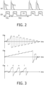
- Coherence pathways leading to impure coherences curves, a, b
- to pure coherences curves c, d, e, f
- pure coherences occur already after three sequence blocks (stimulated echoes, curves c and e) and four sequence blocks (spin echoes, curves d and f).
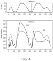
- dictionaries were calculated for a range of T 1 , T 2 and ADC values, using the EPG formalism.
- the corresponding fingerprints of water and oil reflect the different diffusion constants of water and oil.
- the bottom diagram in Fig. 4 shows two fingerprints of water and oil reflecting the different diffusion coefficients of water and oil.
Landscapes
- Physics & Mathematics (AREA)
- General Physics & Mathematics (AREA)
- Condensed Matter Physics & Semiconductors (AREA)
- High Energy & Nuclear Physics (AREA)
- Engineering & Computer Science (AREA)
- Health & Medical Sciences (AREA)
- Nuclear Medicine, Radiotherapy & Molecular Imaging (AREA)
- Signal Processing (AREA)
- General Health & Medical Sciences (AREA)
- Radiology & Medical Imaging (AREA)
- Spectroscopy & Molecular Physics (AREA)
- Computer Vision & Pattern Recognition (AREA)
- Artificial Intelligence (AREA)
- Optics & Photonics (AREA)
- Vascular Medicine (AREA)
- Magnetic Resonance Imaging Apparatus (AREA)
Priority Applications (6)
| Application Number | Priority Date | Filing Date | Title |
|---|---|---|---|
| EP22159441.9A EP4239357A1 (en) | 2022-03-01 | 2022-03-01 | Diffusion mapping by mr fingerprinting |
| CN202380024650.7A CN118805095A (zh) | 2022-03-01 | 2023-02-22 | 通过mr指纹进行扩散映射 |
| EP23705600.7A EP4487138B1 (en) | 2022-03-01 | 2023-02-22 | Diffusion mapping by mr fingerprinting |
| US18/842,186 US20250180685A1 (en) | 2022-03-01 | 2023-02-22 | Diffusion Mapping by MR Fingerprinting |
| JP2024550691A JP2025506863A (ja) | 2022-03-01 | 2023-02-22 | Mrフィンガープリンティングによる拡散マッピング |
| PCT/EP2023/054407 WO2023165865A1 (en) | 2022-03-01 | 2023-02-22 | Diffusion mapping by mr fingerprinting |
Applications Claiming Priority (1)
| Application Number | Priority Date | Filing Date | Title |
|---|---|---|---|
| EP22159441.9A EP4239357A1 (en) | 2022-03-01 | 2022-03-01 | Diffusion mapping by mr fingerprinting |
Publications (1)
| Publication Number | Publication Date |
|---|---|
| EP4239357A1 true EP4239357A1 (en) | 2023-09-06 |
Family
ID=80623477
Family Applications (2)
| Application Number | Title | Priority Date | Filing Date |
|---|---|---|---|
| EP22159441.9A Withdrawn EP4239357A1 (en) | 2022-03-01 | 2022-03-01 | Diffusion mapping by mr fingerprinting |
| EP23705600.7A Active EP4487138B1 (en) | 2022-03-01 | 2023-02-22 | Diffusion mapping by mr fingerprinting |
Family Applications After (1)
| Application Number | Title | Priority Date | Filing Date |
|---|---|---|---|
| EP23705600.7A Active EP4487138B1 (en) | 2022-03-01 | 2023-02-22 | Diffusion mapping by mr fingerprinting |
Country Status (5)
| Country | Link |
|---|---|
| US (1) | US20250180685A1 (cg-RX-API-DMAC7.html) |
| EP (2) | EP4239357A1 (cg-RX-API-DMAC7.html) |
| JP (1) | JP2025506863A (cg-RX-API-DMAC7.html) |
| CN (1) | CN118805095A (cg-RX-API-DMAC7.html) |
| WO (1) | WO2023165865A1 (cg-RX-API-DMAC7.html) |
Cited By (1)
| Publication number | Priority date | Publication date | Assignee | Title |
|---|---|---|---|---|
| WO2025116808A1 (en) * | 2023-12-01 | 2025-06-05 | Corsmed Ab | A method intended in the magnetic resonance imaging (mri) field for simulation-based reconstruction (sbr) |
Citations (1)
| Publication number | Priority date | Publication date | Assignee | Title |
|---|---|---|---|---|
| US20210259775A1 (en) * | 2018-06-18 | 2021-08-26 | Koninklijke Philips N.V. | Therapy planning device |
Family Cites Families (2)
| Publication number | Priority date | Publication date | Assignee | Title |
|---|---|---|---|---|
| CN109917315B (zh) * | 2019-04-30 | 2021-09-28 | 上海联影医疗科技股份有限公司 | 磁共振成像扫描方法、装置、计算机设备和存储介质 |
| DE102020212166A1 (de) * | 2020-09-28 | 2022-03-31 | Siemens Healthcare Gmbh | Verfahren zur Aufnahme von Magnetresonanzdaten eines Untersuchungsobjekts mittels einer Magnetresonanzanlage |
-
2022
- 2022-03-01 EP EP22159441.9A patent/EP4239357A1/en not_active Withdrawn
-
2023
- 2023-02-22 US US18/842,186 patent/US20250180685A1/en active Pending
- 2023-02-22 JP JP2024550691A patent/JP2025506863A/ja active Pending
- 2023-02-22 CN CN202380024650.7A patent/CN118805095A/zh active Pending
- 2023-02-22 EP EP23705600.7A patent/EP4487138B1/en active Active
- 2023-02-22 WO PCT/EP2023/054407 patent/WO2023165865A1/en not_active Ceased
Patent Citations (1)
| Publication number | Priority date | Publication date | Assignee | Title |
|---|---|---|---|---|
| US20210259775A1 (en) * | 2018-06-18 | 2021-08-26 | Koninklijke Philips N.V. | Therapy planning device |
Non-Patent Citations (6)
| Title |
|---|
| COHEN OURI, ROSEN MATTHEW S.: "Simultaneous Diffusion, PD, T1, and T2 Mapping with Optimized MR Fingerprinting EPI", PROCEEDINGS OF THE INTERNATIONAL SOCIETY FOR MAGNETIC RESONANCE IN MEDICINE, 26TH ANNUAL MEETING AND EXHIBITION, PARIS, FRANCE, 16-21 JUNE 2018, vol. 26, 4270, 1 June 2018 (2018-06-01), XP040703478 * |
| JOURNAL OF MAGNETIC RESONANCE IMAGING, vol. 41, 2015, pages 266 - 295 |
| M. T. VLAARDINGERBROEKJ. A. DEN BOER: "Magnetic Resonance Imaging, Theory and Practice", 2002, SPRINGER |
| MAGNETIC RESONANCE IMAGING, vol. 46, 2018, pages 56 - 62 |
| NATURE, vol. 495, 2013, pages 187 - 192 |
| OLIVIER JAUBERT ET AL: "Water-fat Dixon cardiac magnetic resonance fingerprinting", MAGNETIC RESONANCE IN MEDICINE, vol. 83, no. 6, 18 November 2019 (2019-11-18), US, pages 2107 - 2123, XP055693732, ISSN: 0740-3194, DOI: 10.1002/mrm.28070 * |
Cited By (1)
| Publication number | Priority date | Publication date | Assignee | Title |
|---|---|---|---|---|
| WO2025116808A1 (en) * | 2023-12-01 | 2025-06-05 | Corsmed Ab | A method intended in the magnetic resonance imaging (mri) field for simulation-based reconstruction (sbr) |
Also Published As
| Publication number | Publication date |
|---|---|
| WO2023165865A1 (en) | 2023-09-07 |
| EP4487138B1 (en) | 2025-11-05 |
| EP4487138A1 (en) | 2025-01-08 |
| CN118805095A (zh) | 2024-10-18 |
| US20250180685A1 (en) | 2025-06-05 |
| JP2025506863A (ja) | 2025-03-13 |
Similar Documents
| Publication | Publication Date | Title |
|---|---|---|
| US9977108B2 (en) | Metal resistant MR imaging reference scan | |
| US10234523B2 (en) | MRI with dixon-type water/fat separation with estimation of the main magnetic field variations | |
| US10401456B2 (en) | Parallel MR imaging with Nyquist ghost correction for EPI | |
| US10234522B2 (en) | MR imaging with dixon-type water/fat separation | |
| EP2610632A1 (en) | MRI with Dixon-type water/fat separation and prior knowledge about inhomogeneity of the main magnetic field | |
| US10591565B2 (en) | Parallel MR imaging with RF coil sensitivity mapping | |
| US10359489B2 (en) | Phase sensitive inversion recovery MRI with water/fat separation | |
| US10120054B2 (en) | Metal resistant MR imaging | |
| US10859652B2 (en) | MR imaging with dixon-type water/fat separation | |
| JP2017530761A (ja) | ゼロエコー時間mrイメージング | |
| US20220057467A1 (en) | Epi mr imaging with distortion correction | |
| US20140121492A1 (en) | Mri with separation of different chemical species using a spectral model | |
| JP7023954B2 (ja) | プロペラmrイメージング | |
| EP4043902A1 (en) | Dixon-type water/fat separation mr imaging | |
| EP4487138B1 (en) | Diffusion mapping by mr fingerprinting | |
| EP3185029A1 (en) | Mr imaging using propeller acquisition with t2 decay correction | |
| EP3859366A1 (en) | Mr imaging using dixon-type water/fat separation with suppression of flow-induced leakage and/or swapping artifacts | |
| US12140653B2 (en) | Dixon-type water/fat separation MR imaging | |
| EP4137834A1 (en) | Multi-region mr fingerprinting |
Legal Events
| Date | Code | Title | Description |
|---|---|---|---|
| PUAI | Public reference made under article 153(3) epc to a published international application that has entered the european phase |
Free format text: ORIGINAL CODE: 0009012 |
|
| STAA | Information on the status of an ep patent application or granted ep patent |
Free format text: STATUS: THE APPLICATION HAS BEEN PUBLISHED |
|
| AK | Designated contracting states |
Kind code of ref document: A1 Designated state(s): AL AT BE BG CH CY CZ DE DK EE ES FI FR GB GR HR HU IE IS IT LI LT LU LV MC MK MT NL NO PL PT RO RS SE SI SK SM TR |
|
| STAA | Information on the status of an ep patent application or granted ep patent |
Free format text: STATUS: THE APPLICATION IS DEEMED TO BE WITHDRAWN |
|
| 18D | Application deemed to be withdrawn |
Effective date: 20240307 |