EP4235580B1 - Computerimplementiertes verfahren zum bestimmen nuklearmedizinischer bilddatensätze in der dynamischen nuklearmedizinischen bildgebung, bestimmungsvorrichtung, computerprogramm und elektronisch lesbares speichermedium - Google Patents
Computerimplementiertes verfahren zum bestimmen nuklearmedizinischer bilddatensätze in der dynamischen nuklearmedizinischen bildgebung, bestimmungsvorrichtung, computerprogramm und elektronisch lesbares speichermedium Download PDFInfo
- Publication number
- EP4235580B1 EP4235580B1 EP22159187.8A EP22159187A EP4235580B1 EP 4235580 B1 EP4235580 B1 EP 4235580B1 EP 22159187 A EP22159187 A EP 22159187A EP 4235580 B1 EP4235580 B1 EP 4235580B1
- Authority
- EP
- European Patent Office
- Prior art keywords
- nuclear medical
- data set
- raw data
- image
- frame
- Prior art date
- Legal status (The legal status is an assumption and is not a legal conclusion. Google has not performed a legal analysis and makes no representation as to the accuracy of the status listed.)
- Active
Links
Images


Classifications
-
- G—PHYSICS
- G06—COMPUTING OR CALCULATING; COUNTING
- G06T—IMAGE DATA PROCESSING OR GENERATION, IN GENERAL
- G06T12/00—Tomographic reconstruction from projections
- G06T12/20—Inverse problem, i.e. transformations from projection space into object space
-
- G—PHYSICS
- G06—COMPUTING OR CALCULATING; COUNTING
- G06T—IMAGE DATA PROCESSING OR GENERATION, IN GENERAL
- G06T12/00—Tomographic reconstruction from projections
- G06T12/30—Image post-processing, e.g. metal artefact correction
-
- A—HUMAN NECESSITIES
- A61—MEDICAL OR VETERINARY SCIENCE; HYGIENE
- A61B—DIAGNOSIS; SURGERY; IDENTIFICATION
- A61B5/00—Measuring for diagnostic purposes; Identification of persons
- A61B5/05—Detecting, measuring or recording for diagnosis by means of electric currents or magnetic fields; Measuring using microwaves or radio waves
- A61B5/055—Detecting, measuring or recording for diagnosis by means of electric currents or magnetic fields; Measuring using microwaves or radio waves involving electronic [EMR] or nuclear [NMR] magnetic resonance, e.g. magnetic resonance imaging
-
- G—PHYSICS
- G06—COMPUTING OR CALCULATING; COUNTING
- G06T—IMAGE DATA PROCESSING OR GENERATION, IN GENERAL
- G06T7/00—Image analysis
- G06T7/0002—Inspection of images, e.g. flaw detection
- G06T7/0012—Biomedical image inspection
-
- G—PHYSICS
- G06—COMPUTING OR CALCULATING; COUNTING
- G06T—IMAGE DATA PROCESSING OR GENERATION, IN GENERAL
- G06T2207/00—Indexing scheme for image analysis or image enhancement
- G06T2207/10—Image acquisition modality
- G06T2207/10072—Tomographic images
- G06T2207/10088—Magnetic resonance imaging [MRI]
-
- G—PHYSICS
- G06—COMPUTING OR CALCULATING; COUNTING
- G06T—IMAGE DATA PROCESSING OR GENERATION, IN GENERAL
- G06T2207/00—Indexing scheme for image analysis or image enhancement
- G06T2207/10—Image acquisition modality
- G06T2207/10072—Tomographic images
- G06T2207/10104—Positron emission tomography [PET]
-
- G—PHYSICS
- G06—COMPUTING OR CALCULATING; COUNTING
- G06T—IMAGE DATA PROCESSING OR GENERATION, IN GENERAL
- G06T2207/00—Indexing scheme for image analysis or image enhancement
- G06T2207/20—Special algorithmic details
- G06T2207/20084—Artificial neural networks [ANN]
-
- G—PHYSICS
- G06—COMPUTING OR CALCULATING; COUNTING
- G06T—IMAGE DATA PROCESSING OR GENERATION, IN GENERAL
- G06T2211/00—Image generation
- G06T2211/40—Computed tomography
- G06T2211/412—Dynamic
-
- G—PHYSICS
- G06—COMPUTING OR CALCULATING; COUNTING
- G06T—IMAGE DATA PROCESSING OR GENERATION, IN GENERAL
- G06T2211/00—Image generation
- G06T2211/40—Computed tomography
- G06T2211/441—AI-based methods, deep learning or artificial neural networks
Definitions
- the invention concerns a computer-implemented method for determining nuclear medical image data sets in dynamic nuclear medical imaging, wherein nuclear medical raw data sets of a field of view acquired over a time interval, wherein the time interval comprises a progression period of a tracer in the field of view and each nuclear medical raw data set is acquired in one of multiple time frames, into which the time interval is divided, and an anatomical image showing the anatomy in the field of view are provided.
- the invention further concerns a determining device, a computer program and an electronically readable storage medium.
- Nuclear medical imaging sometimes also called emission tomography
- PET positron emission tomography
- SPECT single photon emission computer tomography
- back projection methods for example filtered back projection
- attenuation corrections based on attenuation maps may be performed.
- Such preliminary backprojected images which are not attenuation corrected, may also be called NAC images, while attenuation-corrected preliminary backprojected images are often called AC images.
- Such preliminary backprojected images are often of a low quality, in particular because of noise.
- the degrading factors in a nuclear medical imaging device may not be modelled and the stochastic variability in photon detection cannot be taken into account.
- advanced reconstruction methods have been proposed.
- One of those advanced reconstruction methods comprises iterative image reconstruction.
- the statistical noise of nuclear medical data and physical effects of the imaging process can be modelled, leading to improved performance.
- a preliminary backprojected image may be used as a first assumption, which is then optimized by repeatedly forward-projecting and comparing to the original nuclear medical raw data. Correction terms are computed based on the difference of projected and measured data and backprojected to update the preliminary image. Based on the comparison, the estimate is adjusted due to certain criteria.
- Further approaches include partitioning the nuclear medical raw data into subsets and use only one subset for each update. This approach is also called OSEM - ordered subsets expectation maximization.
- the imaging time interval is divided into multiple frames, wherein, for each frame, a nuclear medical image data set is determined to be able to evaluate the propagation of the tracer in the field of view as well as other aspects.
- the frame-specific nuclear medical images of dynamic studies are usually lower in quality, since less events are recorded.
- Registering the anatomical image to the respective nuclear raw data set and, consecutively, performing an analytical iterative reconstruction using a prior or applying a trained function for each frame results in a very time-consuming process.
- the computational effort grows considerably with the number of frames. Due to lower count rates in the frames and larger amounts of motion throughout a dynamic study, registration quality may be impacted and registration results can be suboptimal or even limited to specific tracers with substantial non-focal background uptake.
- this object is achieved by providing a computer-implemented method, a determining device, a computer program and an electronically readable storage medium according to the independent claims.
- Advantageous embodiments are described in the dependent claims.
- the computer-implemented method for determining nuclear medical image data sets in dynamic nuclear medical imaging comprises:
- the already determined nuclear medical image data set may be the first nuclear medical image data set, in particular for the first of the further nuclear medical image data sets.
- the already determined nuclear medical image data set comprises a further nuclear medical image data set, for example an already determined further nuclear medical image data set of a neighboring frame.
- the already determined nuclear medical image data set of a frame adjacent in time may be used. For example, starting from one single frame chosen as the basis raw data set, the at least one frame directly adjacent in time may be determined first using the first nuclear medical image data set as input, whereafter the next adjacent frame may be determined using the first further nuclear medical image data set as input, and so on.
- the basis raw data set only comprises the nuclear medical raw data set of the first frame
- the first nuclear medical image data set already serves as a result for the first frame
- the nuclear medical image data set for the second frame is determined based on the result for the first frame
- the nuclear medical image data set for the third frame is determined based on the result for the second frame, and so on until the last frame.
- a first nuclear medical image data set is determined using a standard analytical iterative reconstruction approach or a first trained function "mimicking" this approach and being trained by its results.
- Input data for the first trained function hence comprises a) the anatomical image and b) the basis raw data set and/or a preliminary reconstructed, for example backprojected, data set (image) thereof.
- multi-modality co-registration either is not necessary or has to be performed only once for this first reconstruction, which may be understood as a nuclear medical prior image.
- an analytical MLEM nuclear medical reconstruction with a Bowsher prior or a first trained function as suggested in the already cited article by D.
- Another, second trained function has already been trained offline to compute nuclear medical image data sets from such a first nuclear medical image data set (nuclear medical prior image) and the nuclear medical raw data set of an arbitrary or adjacent frame of the same acquisition, wherein the resulting further nuclear medical image data set corresponds to a registration and reconstruction result as used to determine the first nuclear medical image data set.
- the second trained function yields a sharpened and denoised high resolution nuclear medical image data set from the results of a standard reconstruction of at least one other frame.
- the nuclear medical raw data sets and the anatomical image are acquired using the same imaging device, in particular at least partly at the same time.
- imaging devices combining two modalities, for example PET/MRI devices, PET/CT devices, or SPECT/CT devices, have already been proposed in the art and can advantageously be applied here.
- the anatomical image can be acquired during at least one of the frames, in particular in a pre-determined, chosen frame.
- the nuclear medical imaging data sets can be determined very fast, in particular in real time.
- the results of the examination may be provided immediately after the examination has been performed.
- a basis raw data set may be chosen as a single nuclear medical raw data set already co-registered to the anatomical image and/or from one single frame during which the anatomical image was acquired or closest to the acquisition of the anatomical image.
- the one single frame may be the first frame of the time interval, or, preferably, a frame showing a higher uptake of the tracer in the field of view, in particular a central frame.
- the one single frame can also be called a reference frame. It is preferably chosen to have an optimal matching to the anatomical image.
- the first nuclear medical image data set may be determined for the first frame as the reference frame, whereafter further nuclear medical image data sets may be determined using the second trained function for the second, third, fourth, and so on frame.
- the respective previous result may be used as input for determining the current result, as already discussed above.
- the changes regarding the respectively used nuclear medical prior image in the second trained function are minimized.
- the first frame might not have meaningful image content, since very little uptake is present, and hence may not be the optimal choice for a reference frame, in such cases, it is preferred to use a central frame having a high uptake, for example based on a head curve analysis, as a reference and then determine the remaining nuclear medical image data sets relative to this central frame, for example by progressing as described above in different directions.
- the one single frame may also be chosen as the one where a measure indicating the quality of co-registration between the nuclear medical raw data set and the anatomical image is optimal.
- the one single frame may be chosen as the frame where such a measure, which may be a deviation measure, is minimal or maximal, depending on its definition.
- a measure which may be a deviation measure
- I may be checked whether the measure is lower or high than a threshold value for the measure indicating a certain minimum quality of the co-registration which is required, wherein, if the threshold is not held, a co-registration process of the optimally co-registered nuclear medical raw data set and the anatomical image may be triggered.
- a threshold value for the measure indicating a certain minimum quality of the co-registration which is required wherein, if the threshold is not held, a co-registration process of the optimally co-registered nuclear medical raw data set and the anatomical image may be triggered.
- the measure for example, a mean squares measure and/or a maximum cross correlation may be used. Since different modalities are used, mutual information is preferably employed.
- the invention can preferably by applied to neurological and/or perfusion imaging.
- a neurological and/or perfusion tracer can be imaged.
- the frame duration may, for example, be ten to thirty seconds, wherein five to twenty dynamic frames may be acquired.
- the current invention is also advantageous if co-registration of the anatomical image with any of the nuclear medical raw data is not inherently provided, for example by using a combined imaging device providing both modalities.
- the anatomical image may be co-registered to the basis raw data set. In this manner, a time-consuming registration may have to be performed, but only once. This is already a huge improvement compared to previous approaches, in which a registration had to be performed for each of the frames.
- the basis raw data set may comprise the nuclear medical raw data sets of all frames. That is, in this case, the basis raw data set is the full count data set of the examination, in particular the time interval. In this manner, more data are available, allowing easier and more robust co-registration, for example in a case that the anatomical image has been acquired outside the time interval and/or even by another imaging device.
- the iterative reconstruction may be based on maximum likelihood expectation maximization (MLEM) and/or use a Bowsher prior.
- MLEM maximum likelihood expectation maximization
- Working based on MLEM and integrating anatomical information into the reconstruction by using a Bowsher prior has proven to yield results of particular high quality, which is also advantageous as a starting point for the second trained function, which also provides sharpened and denoised high-quality and high-resolution nuclear medical image data sets, in other words Bowsher-like results.
- MLEM maximum likelihood expectation maximization
- a trained function mimics cognitive functions that humans associate with other human minds.
- the trained function is able to adapt to new circumstances and to detect and extrapolate patterns.
- parameters of a trained function can be adapted by means of training.
- supervised training semi-supervised training, unsupervised training, reinforcement learning and/or active learning can be used.
- representation learning an alternative term is "feature learning”
- the parameters of the trained functions can be adapted iteratively by several steps of training.
- a trained function can comprise a neural network, a support vector machine, a decision tree and/or a Bayesian network, and/or the trained function can be based on k-means clustering, Q-learning, genetic algorithms and/or association rules.
- a neural network can be a deep neural network, a convolutional neural network (CNN) or a convolutional deep neural network.
- CNN convolutional neural network
- a neural network can be an adversarial network, a deep adversarial network and/or a generative adversarial network (GAN).
- the second trained function of the method according to the invention may, for example, be trained using data from previous dynamic studies as training data.
- second trained function may be provided by:
- pairs of training input data and training output data may be determined by using the determination process for one frame to determine the first or further training nuclear medical image data set as input data and, for the other frame, use the determination process to determine the further training nuclear medical image data set as output data.
- this may of course also be done vice versa, and in the variant described before, multiple frames may be choses as reference frame to generate more training data.
- multi-modality co-registration may be performed based on a preliminary, in particular backprojection, reconstruction of the basis raw data set.
- Multiple such registration approaches have been proposed in the state of the art, exemplarily, it can be referred to Frederik Maes et al., "Medical Image Registration Using Mutual Information", Proceedings of the IEEE, Vol. 91, No. 10, October 2003, pages 1699-1722 .
- the first trained function may comprise a convolutional neural network and/or use a preliminary reconstructed, in particular backprojected, image and the anatomical image as input data, as, for example, proposed in the article by Georg Schramm et al. cited above.
- the raw data are not needed as input for this reconstruction approach.
- the second trained function may comprise a convolutional neural network and/or a ResNet. Both options have proven to be efficient regarding the training phase as well as robust and fast in the application phase.
- the convolutional neural network may comprise at least one fully connected layer and/or at least one patchwise fully connected layer.
- a convolutional neural network comprising only fully connected layers may be used.
- such a convolutional neural network may comprise three to five fully connected layers and/or patchwise fully connected layers. Patchwise fully connected layers can be understood as providing local transformation processes, since the effects addressed here can also be seen as local.
- varying definitions of patches in the second trained function may be used for varying pairs of layers to analyze different local neighborhoods.
- Attenuation correction may be applied based on an attenuation map, which is also used in the reconstruction of the first nuclear medical image data set.
- the attenuation map may, for example, be used for modelling count loss by integration of attenuation along a line of response and corresponding corrections for comparison of a current estimate with the nuclear medical raw data. Attenuation correction further improves the image quality of the resulting nuclear medical image data sets. Attenuation correction approaches using attenuation maps, which are often also called ⁇ -maps, are in principle known in the art.
- the attenuation map may be determined from the anatomical image.
- the attenuation map is preferably determined from the co-registered anatomical image.
- the attenuation map is also inherently co-registered, in particular also if co-registration was already inherently provided, for example for acquiring the basis raw data and the anatomical image in the same frame. Since the anatomical image describes the anatomy of the field of view of the patient, attenuation coefficients can be assigned to certain anatomical structures, in the case of CT images as anatomical images derived from or as image values, since CT images also describe attenuation. In the case of MRI mostly discussed here, Dixon methods are usually applied.
- an attenuation-corrected preliminary reconstructed, for example backprojected, image based on the attenuation map is used as input data for the second trained function.
- the attenuation map can be used for all frames unchanged.
- the attenuation map can be used to determine an attenuation-corrected preliminary reconstructed image, which, in turn, can be used as input to the second trained function, which thus does not need to incorporate the attenuation correction.
- supplying already attenuation-corrected input data to the second trained function allows a more efficient learning procedure, since less correlations have to be learned.
- the second or third concrete embodiments in this context can advantageously be used.
- a corrected attenuation map is determined for each frame, to which the second trained function is to be applied, by determining correction parameters from not attenuation-corrected preliminary reconstructed images of the relevant frames and correcting the attenuation map according to the correction parameters, wherein an attenuation-corrected preliminary reconstructed image based on the respective corrected attenuation map is used as input data for the second trained function.
- correction parameters describing motion are registration parameters between not attenuation-corrected preliminary reconstructed images (NAC images) of the frames, which may be applied to the attenuation map.
- the corrected attenuation map may then serve as input to the preliminary attenuation-corrected reconstruction, the result of which, that is the attenuation-corrected preliminary reconstructed image (AC image), may be used as input to the second trained function.
- AC image attenuation-corrected preliminary reconstructed image
- the pairs of NAC images to determine correction parameters may be chosen according to the frames from which input data is used in the second trained function, such that neighboring frames may be used both for correcting the attenuation map and for determining the further nuclear medical image data set, it is, of course, also possible to choose different pairs of frames for the input to the second trained function and the determination of the correction parameters, as long as corrected attenuation maps for all relevant frames are determined.
- Attenuation correction it is also possible to train the second trained function to also learn the attenuation correction, which may require more training data, but allows to skip a correction process in the case of motion of the patient. That is, the attenuation map is only used for determining the first nuclear medical image data set, wherein a not attenuation-corrected preliminary reconstructed image is used as input data for the second trained function.
- the nuclear medical raw data sets may be PET raw data sets and/or the anatomical image may be a magnetic resonance image.
- CT images have also been proposed as carrying useful anatomical information which may be incorporated into the reconstruction process.
- dynamic studies have also been proposed for SPECT, such that the current invention can also be applied to these examinations.
- the invention further concerns a determining device for determining nuclear medical image data sets in dynamic nuclear medical imaging, comprising:
- first nuclear medical image data set refers to a single frame, it will also be output via the third interface; also first nuclear medical image data sets based on raw data of multiple frames, in particular all frames, may of course be output if they are of any value for the evaluation.
- the determining device may comprise at least one processor and/or a storage means and is configured to perform a method according to the invention. All features and remarks discussed regarding the method according to the invention can be analogously applied to the determining device according to the invention and vice versa. In particular, further functional units may be provided to implement additional, optional steps.
- the determining device may additionally comprise a registration unit for co-registering the anatomical image to the basis raw data set.
- the determining device may be integrated into an imaging device, preferably into a combined imaging device providing both the nuclear medical imaging modality and the anatomical imaging modality.
- a combined imaging device in particular a PET/MRI device or a PET/CT device, is conceivable, wherein the control device of the combined imaging device comprises a determining device according to the invention.
- a computer program according to the invention can be directly loaded into a determining device and enables the determining device to perform the steps of a method according to the invention when the computer program is executed on the determining device.
- the computer program according to the invention may be stored on an electronically readable storage medium according to the invention, which thus comprises control information comprising a computer program according to the invention such that, when the electronically readable storage medium is used in a determining device, the determining device is configured to perform the steps of a method according to the invention when the computer program according to the invention is executed in the determining device.
- the electronically readable storage medium according to the invention may preferably be a non-transitory medium, for example a CD ROM.
- Fig. 1 displays an embodiment of an artificial neural network 1.
- artificial neural network alternatives terms for “artificial neural network” are “neural network”, “artificial neural net” or “neural net”.
- the artificial neural network 1 comprises nodes 6 - 18 and edges 19 - 21, wherein each edge 19 - 21 is a directed connection from a first node 6 - 18 to a second node 6 - 18.
- the first node 6 - 18 and the second node 6 - 18 are different nodes 6 - 18.
- the first node 6 - 18 and the second node 6 - 18 are identical.
- the edge 19 is a directed connection from the node 6 to the node 9
- the edge 20 is a directed connection from the node 7 to the node 9.
- An edge 19 - 21 from a first node 6 - 18 to a second node 6 - 18 is also denoted as "ingoing edge" for the second node 6 - 18 and as "outgoing edge” for the first node 6 - 18.
- the nodes 6 - 18 of the artificial neural network 1 can be arranged in layers 2 - 5, wherein the layers 2 - 5 can comprise an intrinsic order introduced by the edges 19 - 21 between the nodes 6 - 18.
- edges 19 - 21 can exist only between neighboring layers of nodes 6 - 18.
- the number of hidden layers 3, 4 can be chosen arbitrarily.
- the number of nodes 6 - 8 within the input layer 2 usually relates to the number of input values of the neural network
- the number of nodes 17, 18 within the output layer 5 usually relates to the number of output values of the neural network.
- a (real) number can be assigned as a value to every node 6 - 18 of the neural network 1.
- x (n) i denotes the value of the i-th node 6 - 18 of the n-th layer 2 - 5.
- the values of the nodes 6 - 8 of the input layer 2 are equivalent to the input values of the neural network 1
- the values of the nodes 17, 18 of the output layer 5 are equivalent to the output values of the neural network 1.
- each edge 19 - 21 can comprise a weight being a real number, in particular, the weight is a real number within the interval [-1, 1] or within the interval [0, 1] .
- w (m,n) i,j denotes the weight of the edge between the i-th node 6 - 18 of the m-th layer 2 - 5 and the j-th node 6 - 18 of the n-th layer 2 - 5. Furthermore, the abbreviation w (n) i,j is defined for the weight w (n,n+1) i,j .
- the input values are propagated through the neural network 1.
- the function f is a transfer function (another term is "activation function").
- transfer functions are step functions, sigmoid function (e.g. the logistic function, the generalized logistic function, the hyperbolic tangent, the Arctangent function, the error function, the smoothstep function) or rectifier functions.
- the transfer function is mainly used for normalization purposes.
- the values are propagated layer-wise through the neural network 1, wherein values of the input layer 2 are given by the input of the neural network 1, wherein values of the first hidden layer 3 can be calculated based on the values of the input layer 2 of the neural network 1, wherein values of the second hidden layer 4 can be calculated based in the values of the first hidden layer 3, etc.
- training data comprises training input data and training output data (denoted as t i ).
- training output data denoted as t i .
- the neural network 1 is applied to the training input data to generate calculated output data.
- the training data and the calculated output data comprise a number of values, said number being equal to the number of nodes 17, 18 of the output layer 5.
- a comparison between the calculated output data and the training data is used to recursively adapt the weights within the neural network 1 (backpropagation algorithm).
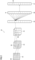
- Fig. 2 displays an embodiment of a convolutional neural network 22.
- the convolutional neural network 22 comprises an input layer 23, a convolutional layer 24, a pooling layer 25, a fully connected layer 26 and an output layer 27.
- the convolutional neural network 22 can comprise several convolutional layers 24, several pooling layers 25 and several fully connected layers 26 as well as other types of layers.
- the order of the layers can be chosen arbitrarily, usually fully connected layers 26 are used as the last layers before the output layer 27.
- the nodes 28 - 32 of one layer 23 - 27 can be considered to be arranged as a d-dimensional matrix or as a d-dimensional image.
- the value of the node 28 - 32 indexed with i and j in the n-th layer 23 - 27 can be denoted as x (n) [i,j].
- the arrangement of the nodes 28 - 32 of one layer 23 - 27 does not have an effect on the calculations executed within the convolutional neural network 22 as such, since these are given solely by the structure and the weights of the edges.
- a convolutional layer 24 is characterized by the structure and the weights of the incoming edges forming a convolution operation based on a certain number of kernels.
- the k-th kernel K k is a d-dimensional matrix (in this embodiment a two-dimensional matrix), which is usually small compared to the number of nodes 28 - 32 (e.g. a 3x3 matrix, or a 5x5 matrix).
- a kernel being a 3x3 matrix, there are only 9 independent weights (each entry of the kernel matrix corresponding to one independent weight), irrespectively of the number of nodes 28 - 32 in the respective layer 23 - 27.
- the number of nodes 29 in the convolutional layer is equivalent to the number of nodes 28 in the preceding layer 23 multiplied with the number of kernels.
- nodes 28 of the preceding layer 23 are arranged as a d-dimensional matrix, using a plurality of kernels can be interpreted as adding a further dimension (denoted as "depth” dimension), so that the nodes 29 of the convolutional layer 24 are arranged as a (d+1)-dimensional matrix.
- nodes 28 of the preceding layer 23 are already arranged as a (d+1)-dimensional matrix comprising a depth dimension, using a plurality of kernels can be interpreted as expanding along the depth dimension, so that the nodes 29 of the convolutional layer 64 are arranged also as a (d+1)-dimensional matrix, wherein the size of the (d+1)-dimensional matrix with respect to the depth dimension is by a factor of the number of kernels larger than in the preceding layer 23.
- convolutional layers 24 The advantage of using convolutional layers 24 is that spatially local correlation of the input data can exploited by enforcing a local connectivity pattern between nodes of adjacent layers, in particular by each node being connected to only a small region of the nodes of the preceding layer.
- the input layer 23 comprises 36 nodes 28, arranged as a two-dimensional 6x6 matrix.
- the convolutional layer 24 comprises 72 nodes 29, arranged as two two-dimensional 6x6 matrices, each of the two matrices being the result of a convolution of the values of the input layer 23 with a kernel. Equivalently, the nodes 29 of the convolutional layer 24 can be interpreted as arranged as a three-dimensional 6x6x2 matrix, wherein the last dimension is the depth dimension.
- a pooling layer 25 can be characterized by the structure and the weights of the incoming edges and the activation function of its nodes 30 forming a pooling operation based on a nonlinear pooling function f.
- the number of nodes 29, 30 can be reduced, by replacing a number d 1 ⁇ d 2 of neighboring nodes 29 in the preceding layer 24 with a single node 30 being calculated as a function of the values of said number of neighboring nodes in the pooling layer 25.
- the pooling function f can be the max-function, the average or the L2-Norm.
- the weights of the incoming edges are fixed and are not modified by training.
- the advantage of using a pooling layer 25 is that the number of nodes 29, 30 and the number of parameters is reduced. This leads to the amount of computation in the network 22 being reduced and to a control of overfitting.
- the pooling layer 25 is a max-pooling, replacing four neighboring nodes with only one node, the value being the maximum of the values of the four neighboring nodes.
- the max-pooling is applied to each d-dimensional matrix of the previous layer 24; in this embodiment, the max-pooling is applied to each of the two two-dimensional matrices, reducing the number of nodes from 72 to 18.
- a fully-connected layer 26 can be characterized by the fact that a majority, in particular, all edges between nodes 30 of the previous layer 25 and the nodes 31 of the fully-connected layer 26 are present, and wherein the weight of each of the edges can be adjusted individually.
- the nodes 30 of the preceding layer 25 of the fully-connected layer 26 are displayed both as two-dimensional matrices, and additionally as non-related nodes (indicated as a line of nodes, wherein the number of nodes was reduced for a better presentability).
- the number of nodes 31 in the fully connected layer 26 is equal to the number of nodes 30 in the preceding layer 25.
- the number of nodes 30, 31 can differ.
- the values of the nodes 32 of the output layer 27 are determined by applying the Softmax function onto the values of the nodes 31 of the preceding layer 26.
- the Softmax function By applying the Softmax function, the sum of the values of all nodes 32 of the output layer 27 is 1, and all values of all nodes 32 of the output layer 27 are real numbers between 0 and 1.
- the values of the output layer can be interpreted as the probability of the input data falling into one of the different categories.
- a convolutional neural network 22 can also comprise a ReLU (acronym for "rectified linear units") layer.
- a ReLU layer is equivalent to the number of nodes and the structure of the nodes contained in the preceding layer.
- convolutional neural networks 22 can be trained based on the backpropagation algorithm.
- methods of regularization can be used, e.g. dropout of nodes 28 - 32, stochastic pooling, use of artificial data, weight decay based on the L1 or the L2 norm, or max norm constraints.
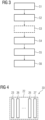
- Fig. 3 is a general flowchart for embodiments of a method according to the invention.
- the method concerns generating nuclear medical image data sets for different frames in a dynamic nuclear medical imaging study.
- the propagation of the tracer for example a neurological and/or perfusion tracer
- the time interval for imaging spans a progression period of the tracer in the field of view, which may, for example, comprise the brain of a patient.
- the time interval is divided into multiple frames, for example five to twenty frames, such that, for each frame, a nuclear medical raw data set is acquired.
- an anatomical image showing the anatomy in the field of view is also acquired, for example an magnetic resonance (MR) image and/or a computational tomography (CT) image.
- MR magnetic resonance
- CT computational tomography
- a step S1 the nuclear medical raw data sets for all the frames and the anatomical image are provided via first and second interfaces of a determining device performing the method.
- an attenuation map may be provided or preferably derived from the anatomical image, such that attenuation correction can also be performed in the method.
- attenuation correction will be discussed in detail with respect to the concrete embodiments of figs. 5 to 7 .
- the determining device is integrated into a control device of a, preferably combined (hybrid), imaging device, which hence preferably provides both the modalities for nuclear medical imaging as well as for anatomical imaging
- the first and second interfaces may be internal, for example to respective acquisition units controlling the acquisition of the nuclear medical raw data and the anatomical image.
- a step S2 at least one, preferably exactly one, of the frames is selected such that its nuclear medical raw data forms a basis raw data set.
- the first frame can be selected as the one single frame for the basis raw data set; however, when certain tracers are used, the uptake may still be low in the first frame, so that, alternatively, a central frame having a higher uptake may be chosen as the one single frame.
- the embodiments shown exemplarily for the first frame can analogously be applied.
- the one single frame is preferably chosen such that its nuclear medical raw data set is already co-registered with the anatomical image.
- the anatomical image may be acquired during one of the frames, such that the respective frame can be chosen as the one single frame for the basis raw data set.
- acquisition may be dedicatedly controlled to have the anatomical image acquired in such a high uptake frame.
- the first or respectively last frame may be chosen as the one single frame to be used for the basis raw data set.
- the one single frame may be chosen as the one where co-registration is already optimal.
- a measure indicating the quality of co-registration already existent between respective nuclear medical raw data sets and the anatomical image may be determined and the one single frame may be chosen as the frame where this measure, which may be a deviation measure, is minimal or maximal, depending on its definition.
- a measure for example, a mean squares measure and/or a maximum cross correlation may be used. Since different modalities are used, mutual information is preferably employed.
- an optional co-registration step S3 may be provided or possible, but less preferred. For example, if the threshold for the measure discussed above is exceeded, co-registration may be automatically prompted. In other cases, for example, if the time between the acquisition of the anatomical image and the nuclear medical raw data is very long, co-registration in a step S3 may generally be required. However, as will become apparent, in comparison to the state of the art, multi-modality co-registration has to be performed at most once, as opposed to the state of the art, where it had to be performed once for each frame.
- step S4 iterative reconstruction, in particular MLEM reconstruction, is applied using a prior based on the anatomical image, wherein the co-registration between the anatomical image and the basis raw data set is exploited.
- the iterative registration applied is based on a Bowsher prior.
- the iterative reconstruction may be performed analytically, however, regarding computational effort, a first trained function modelling the iterative reconstruction (and thus applying it implicitly) may be used, for example as described in the cited articles by D. Rigio et al. and Georg Schramm et al.
- a first nuclear medical image data set is determined, which, if the basis raw data is taken from one single frame, also forms the result image for this frame.
- the basis raw data is taken from one single frame, also forms the result image for this frame.
- more frames may be included into the basis raw data set, in particular all frames, such that the result would be a full-count nuclear medical image for the time interval.
- a second trained function is applied.
- the second trained function has been trained to use input data comprising an already reconstructed nuclear medical image data set as well as data from the respective frame as input to derive the reconstructed nuclear medical image data set for the respective frame therefrom.
- previously acquired data from dynamic studies may be employed.
- the first nuclear medical image data set has to be used. While it is in principle possible to use the first nuclear medical image data set for all the frames to be reconstructed, in preferred embodiments as discussed here, the second trained function is in particular trained to derive the nuclear medical image data set for a certain frame from the result of a neighboring frame.
- the second frame is next reconstructed using the result for the first frame derived in step S4.
- the third frame is reconstructed using the result of the second frame, and so on. If a central frame (i.e., not the first or last frame) has been chosen in step S2, propagation may be conducted in both time directions starting from the central frame.
- a preliminary reconstructed, in particular backprojected, image is used as input data from the respective frame, as will be further discussed below.
- the respective nuclear medical raw data set is used.
- nuclear medical image data sets showing high-resolution high-quality distributions of the tracer in the respective frames are determined for each frame.
- result images for all frames may be provided in real time.
- the resulting nuclear medical image data sets may be output, for example for further evaluation, storing in a picture archive and/or displaying to a user.
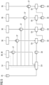
- Fig. 4 shows an exemplary structure of the second trained function 33 used in step S5.
- a convolutional neural network 22 is used, which comprises an input layer 23, a series of fully connected layers 26 and/or patchwise fully connected layers 26 and an output layer 27.
- the second trained function 33 a convolutional neural network 22 is used, which comprises an input layer 23, a series of fully connected layers 26 and/or patchwise fully connected layers 26 and an output layer 27.
- three to ten fully connected layers and/or patchwise fully connected layers 26 can be used.
- an attenuation map 34 preferably derived from the anatomical image 35, can be used for attenuation correction in all frames.
- the attenuation map 34 is used to determine an attenuation corrected preliminary reconstructed, here backprojected, image 37 (AC image) that can be used as input data for steps S4, S5 respectively.
- AC image an attenuation corrected preliminary reconstructed, here backprojected, image 37
- a Dixon technique could be used to acquire it, such that an attenuation map can be derived from it based in the different material classes distinguished.
- the attenuation map 34 is also used in step S4, since modelling the forward projection requires attenuation knowledge to allow correct comparison between forward projected data and nuclear medical raw data.
- step S4 it may not be necessary to also supply the respective basis raw data to the first trained function as input data, but only a preliminary backprojected image 37.
- the first frame is always shown as the one single frame for the basis raw data set 38, which then is the nuclear medical raw data set 36 of the first frame.
- these embodiments may be analogously transferred to other frames as the single one frame.
- the first nuclear medical image data set 39 is determined as result of the step S4.
- the previously determined first nuclear medical image data set 39 or further nuclear medical image data set 40 together with the NAC images 37 is used as input data for the second trained function 33 in step S5, such that, finally, nuclear medical image data sets 39, 40 have been determined for all frames, wherein attenuation correction has already been applied during or after preliminary backprojection.
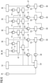
- the second and third embodiments shown in figs. 6 and 7 refer to patient motion occurring during the time interval.
- a first approach to attenuation correction in this case is shown in fig. 6 , where, again, the first frame is used for the basis raw data set 38 and optimally registered to the anatomical image 35, from which the attenuation map 34 is derived.
- the attenuation map 34 can again be used to determine an attenuation-corrected preliminary backprojected image 37 for the first frame.
- non-attenuation-corrected preliminary backprojected images 41 are determined for each frame from the respective nuclear medical raw data sets 36 and registered between frames adjacent in time to yield correction parameters 42 (registration parameters).
- correction parameters 42 describing the motion occurring between the frames is used to determine corrected attenuation maps 34' for each frame, which can be used to determine the respective AC images 37.
- These AC images 37 are, again, used in addition to the result of the previous frame as input data to the second trained function 33 in steps S5.
- the second trained function 33 has also been trained to perform attenuation correction, such that NAC images 41 may be used as input data to the second trained function 33 in step S5.
- the attenuation map 34 is only used in step S4, and, optionally, to determine an AC image 37 if required in step S4.
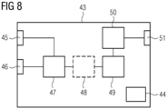
- Fig. 8 shows the functional structure of a determining device 43 according to the invention, which may comprise a storage means 44 for storing input information, intermediate results and/or final results.
- the nuclear medical raw data sets 36 are provided via a first interface 45, while the anatomical image 35 is provided via a second interface 46 (step S1).
- a selection unit 47 the basis raw data set 38 is defined as discussed with respect to step S2.
- a registration unit 48 may be provided for co-registration (step S3).
- the first nuclear medical image data set 39 can be determined according to step S4.
- Further nuclear medical image data sets 40 are determined according to step S5 in a determination unit 50.
- the resulting nuclear medical image data sets 39, 40 can be output via a third interface 51.
- the determining device 43 may also be integrated into the control device of a combined imaging device providing both nuclear medical imaging, in particular PET imaging, and anatomical imaging, in particular magnetic resonance imaging (MRI).
- the interfaces 45, 46 and 51 may be internal interfaces, wherein the control device may additionally comprise acquisition units for acquiring the nuclear medical raw data sets 36 and the anatomical image 35.
- the acquisition units may be controlled such that, if synchronous imaging is possible, the anatomical image 35 is acquired in a certain frame of the time interval, which is then chosen as the one single frame to define the basis raw data set 38.
Landscapes
- Engineering & Computer Science (AREA)
- Health & Medical Sciences (AREA)
- Physics & Mathematics (AREA)
- Nuclear Medicine, Radiotherapy & Molecular Imaging (AREA)
- Life Sciences & Earth Sciences (AREA)
- General Physics & Mathematics (AREA)
- Theoretical Computer Science (AREA)
- Medical Informatics (AREA)
- General Health & Medical Sciences (AREA)
- Radiology & Medical Imaging (AREA)
- Heart & Thoracic Surgery (AREA)
- Molecular Biology (AREA)
- Computer Vision & Pattern Recognition (AREA)
- Biophysics (AREA)
- Pathology (AREA)
- Biomedical Technology (AREA)
- Quality & Reliability (AREA)
- High Energy & Nuclear Physics (AREA)
- Surgery (AREA)
- Animal Behavior & Ethology (AREA)
- Public Health (AREA)
- Veterinary Medicine (AREA)
- Nuclear Medicine (AREA)
- Image Analysis (AREA)
Claims (15)
- Computerimplementiertes Verfahren zur Bestimmung nuklearmedizinischer Bilddatensätze (39, 40) in der dynamischen nuklearmedizinischen Bildgebung, umfassend:- Bereitstellen nuklearmedizinischer Rohdatensätze (36) eines Sichtfeldes, die über ein Zeitintervall erfasst werden, wobei das Zeitintervall einen Progressionszeitraum eines Tracers in dem Sichtfeld umfasst und jeder nuklearmedizinische Rohdatensatz (36) in einem von mehreren Zeitrahmen erfasst wird, in die das Zeitintervall geteilt ist,- Bereitstellen eines anatomischen Bildes (35), dass die Anatomie in dem Sichtfeld darstellt,- Auswählen mindestens eines der nuklearmedizinischen Rohdatensätze (36) als Basis-Rohdatensatz (38),- Rekonstruieren eines ersten nuklearmedizinischen Bilddatensatzes (39) entweder durch* iterative Rekonstruktion des Basis-Rohdatensatzes (38) unter Verwendung eines auf dem anatomischen Bild (35) basierenden Priors, um anatomische Informationen einzubeziehen,
oder* Verwenden einer ersten trainierten Funktion, die solch eine iterative Rekonstruktion auf das anatomische Bild (35) und den Basis-Rohdatensatz (38) und/oder einen vorläufig rekonstruierten Datensatz (37, 41) davon anwendet,- Verwenden einer zweiten trainierten Funktion (33) zum Bestimmen mindestens eines weiteren nuklearmedizinischen Bilddatensatzes (40),* wenn der Basis-Rohdatensatz (38) einem einzelnen Frame entnommen ist, für mindestens einen anderen Frame, insbesondere alle anderen Frames,* sonst mindestens einen Frame, insbesondere alle Frame, als Ausgabedaten, wobei Eingabedaten der zweiten trainierten Funktion (33) umfassen:* den nuklearmedizinischen Rohdatensatz (36) des jeweiligen Frames und/oder ein daraus rekonstruiertes vorläufig rekonstruiertes Bild (37, 41) und* einen bereits bestimmten nuklearmedizinischen Bilddatensatz (39, 40) . - Verfahren nach Anspruch 1, dadurch gekennzeichnet, dass der Basis-Rohdatensatz (38) als einzelner nuklearmedizinischer Rohdatensatz (36), der bereits mit dem anatomischen Bild (35) mitregistriert ist, und/oder aus einem einzelnen Frame gewählt wird, in dem das anatomische Bild (35) aufgenommen wurde oder der der Erfassung des anatomischen Bildes (35) am nächsten kommt, und/oder dadurch, dass vor der Rekonstruktion das anatomische Bild (35) mit den Basis-Rohdatensatz (38) mitregistriert wird.
- Verfahren nach Anspruch 1 oder 2, dadurch gekennzeichnet, dass der Basis-Rohdatensatz (38) die nuklearmedizinischen Rohdatensätze (36) aller Frames umfasst.
- Verfahren nach einem der vorhergehenden Ansprüche, dadurch gekennzeichnet, dass die iterative Rekonstruktion auf einer Maximum-Likelihood-Erwartungsmaximierung basiert und/oder einen Bowsher-Prior verwendet.
- Verfahren nach einem der vorhergehenden Ansprüche, dadurch gekennzeichnet, dass die erste trainierte Funktion ein neuronales Faltungsnetzwerk (22) umfasst und/oder ein vorläufig rekonstruiertes, insbesondere rückprojiziertes, Bild (37, 31) und das anatomische Bild (35) als Eingabedaten verwendet.
- Verfahren nach einem der vorhergehenden Ansprüche, dadurch gekennzeichnet, dass die zweite trainierte Funktion (33) ein neuronales Faltungsnetzwerk (22) und/oder ein ResNet umfasst.
- Verfahren nach einem der vorhergehenden Ansprüche, dadurch gekennzeichnet, dass eine Dämpfungskorrektur basierend auf einer Dämpfungskarte (34, 34') angewendet wird, die auch bei der Rekonstruktion des ersten nuklearmedizinischen Bilddatensatzes (39) verwendet wird.
- Verfahren nach Anspruch 7, dadurch gekennzeichnet, dass die Dämpfungskarte (34, 34') aus dem anatomischen Bild (35) bestimmt wird.
- Verfahren nach Anspruch 7 oder 8, dadurch gekennzeichnet, dass insbesondere, wenn keine relevante Patientenbewegung stattfindet, ein dämpfungskorrigiertes vorläufig rekonstruiertes Bild (37) basierend auf der Dämpfungskarte (34) als Eingabedaten für die zweite trainierte Funktion (33) verwendet wird.
- Verfahren nach Anspruch 7 oder 8, dadurch gekennzeichnet, dass im Falle von Patientenbewegung eine korrigierte Dämpfungskarte (34') für jeden Frame bestimmt wird, auf den die zweite trainierte Funktion (33) anzuwenden ist, indem Korrekturparameter (42) aus nicht dämpfungskorrigierten vorläufig rekonstruierten Bildern (41) der relevanten Frames bestimmt werden und die Dämpfungskarte(34, 34') gemäß den Korrekturparametern (42) korrigiert wird, wobei ein dämpfungskorrigiertes vorläufig rekonstruiertes Bild (37) basierend auf der jeweiligen korrigierten Dämpfungskarte (34') als Eingabedaten für die zweite trainierte Funktion (33) verwendet wird.
- Verfahren nach Anspruch 7 oder 8, dadurch gekennzeichnet, dass die Dämpfungskarte (34) nur zum Bestimmen des ersten nuklearmedizinischen Bilddatensatzes (39) verwendet wird, wobei ein nicht dämpfungskorrigiertes vorläufig rekonstruiertes Bild (41) als Eingabedaten für die zweite trainierte Funktion (33) verwendet wird.
- Verfahren nach einem der vorhergehenden Ansprüche, dadurch gekennzeichnet, dass die nuklearmedizinischen Rohdatensätze (36) PET-Rohdatensätze sind und/oder das anatomische Bild (35) ein Magnetresonanzbild ist.
- Bestimmungsvorrichtung (43) zum Bestimmen nuklearmedizinischer Bilddatensätze (39, 40) in der dynamischen nuklearmedizinischen Bildgebung, umfassend:- eine erste Schnittstelle (45) zum Bereitstellen nuklearmedizinischer Rohdatensätze (36) eines Sichtfeldes, die über ein Zeitintervall erfasst werden, wobei das Zeitintervall einen Progressionszeitraum eines Tracers in dem Sichtfeld umfasst und jeder nuklearmedizinische Rohdatensatz (36) in einem von mehreren Zeitrahmen erfasst wird, in die das Zeitintervall geteilt ist,- eine zweite Schnittstelle (46) zum Bereitstellen eines anatomischen Bildes (35), dass die Anatomie in dem Sichtfeld darstellt,- eine Auswahleinheit (47) zum Auswählen mindestens eines der nuklearmedizinischen Rohdatensätze (36) als Basis-Rohdatensatz (38),- eine Rekonstruktionseinheit (49) zum Rekonstruieren eines ersten nuklearmedizinischen Bilddatensatzes (39) entweder durch* iterative Rekonstruktion des Basis-Rohdatensatzes (38) unter Verwendung eines auf dem anatomischen Bild (35) basierenden Priors, um anatomische Informationen einzubeziehen,
oder* Verwenden einer ersten trainierten Funktion, die solch eine iterative Rekonstruktion auf das anatomische Bild (35) und den Basis-Rohdatensatz (38) und/oder einen vorläufig rekonstruierten Datensatz (37, 41) davon anwendet,- eine Bestimmungseinheit (50) zum Verwenden einer zweiten trainierten Funktion (33) zum Bestimmen mindestens eines weiteren nuklearmedizinischen Bilddatensatzes (40),* wenn der Basis-Rohdatensatz (38) einem einzelnen Frame entnommen ist, für mindestens einen anderen Frame, insbesondere alle anderen Frames,* sonst mindestens einen Frame, insbesondere alle Frames, als Ausgabedaten, wobei Eingabedaten der zweiten trainierten Funktion (33) umfassen:* den nuklearmedizinischen Rohdatensatz (36) des jeweiligen Frames und/oder ein daraus rekonstruiertes vorläufig rekonstruiertes Bild (37, 41) und* einen bereits bestimmten nuklearmedizinischen Bilddatensatz (39, 40),- eine dritte Schnittstelle (51) zum Ausgeben zumindest des mindestens einen weiteren nuklearmedizinischen Bilddatensatzes (39, 40). - Computerprogramm, das bei Ausführung des Computerprogramms aus einer Bestimmungsvorrichtung (43) die Schritte eines Verfahrens nach einem der Ansprüche 1 bis 12 ausführt.
- Elektronisch lesbares Speichermedium, auf dem ein Computerprogramm nach Anspruch 14 gespeichert ist.
Priority Applications (2)
| Application Number | Priority Date | Filing Date | Title |
|---|---|---|---|
| EP22159187.8A EP4235580B1 (de) | 2022-02-28 | 2022-02-28 | Computerimplementiertes verfahren zum bestimmen nuklearmedizinischer bilddatensätze in der dynamischen nuklearmedizinischen bildgebung, bestimmungsvorrichtung, computerprogramm und elektronisch lesbares speichermedium |
| US18/173,951 US11756243B1 (en) | 2022-02-28 | 2023-02-24 | Computer-implemented method for determining nuclear medical image data sets in dynamic nuclear medical imaging, determining device and electronically readable storage medium |
Applications Claiming Priority (1)
| Application Number | Priority Date | Filing Date | Title |
|---|---|---|---|
| EP22159187.8A EP4235580B1 (de) | 2022-02-28 | 2022-02-28 | Computerimplementiertes verfahren zum bestimmen nuklearmedizinischer bilddatensätze in der dynamischen nuklearmedizinischen bildgebung, bestimmungsvorrichtung, computerprogramm und elektronisch lesbares speichermedium |
Publications (3)
| Publication Number | Publication Date |
|---|---|
| EP4235580A1 EP4235580A1 (de) | 2023-08-30 |
| EP4235580C0 EP4235580C0 (de) | 2024-07-17 |
| EP4235580B1 true EP4235580B1 (de) | 2024-07-17 |
Family
ID=80595492
Family Applications (1)
| Application Number | Title | Priority Date | Filing Date |
|---|---|---|---|
| EP22159187.8A Active EP4235580B1 (de) | 2022-02-28 | 2022-02-28 | Computerimplementiertes verfahren zum bestimmen nuklearmedizinischer bilddatensätze in der dynamischen nuklearmedizinischen bildgebung, bestimmungsvorrichtung, computerprogramm und elektronisch lesbares speichermedium |
Country Status (2)
| Country | Link |
|---|---|
| US (1) | US11756243B1 (de) |
| EP (1) | EP4235580B1 (de) |
Family Cites Families (7)
| Publication number | Priority date | Publication date | Assignee | Title |
|---|---|---|---|---|
| US20180197317A1 (en) * | 2017-01-06 | 2018-07-12 | General Electric Company | Deep learning based acceleration for iterative tomographic reconstruction |
| US11481934B2 (en) * | 2018-10-10 | 2022-10-25 | New York University | System, method, and computer-accessible medium for generating magnetic resonance imaging-based anatomically guided positron emission tomography reconstruction images with a convolutional neural network |
| WO2021041772A1 (en) * | 2019-08-30 | 2021-03-04 | The Research Foundation For The State University Of New York | Dilated convolutional neural network system and method for positron emission tomography (pet) image denoising |
| CN111325686B (zh) * | 2020-02-11 | 2021-03-30 | 之江实验室 | 一种基于深度学习的低剂量pet三维重建方法 |
| US11481937B2 (en) * | 2020-05-21 | 2022-10-25 | Zhejiang University | Positron emission tomography image reconstruction method |
| EP4168991A1 (de) * | 2020-06-23 | 2023-04-26 | Koninklijke Philips N.V. | Ki-aktivierte früher-pet-erfassung |
| US12020351B2 (en) * | 2020-12-18 | 2024-06-25 | Shenzhen Institutes Of Advanced Technology | Method, device and equipment for reconstructing PET images |
-
2022
- 2022-02-28 EP EP22159187.8A patent/EP4235580B1/de active Active
-
2023
- 2023-02-24 US US18/173,951 patent/US11756243B1/en active Active
Also Published As
| Publication number | Publication date |
|---|---|
| US20230274476A1 (en) | 2023-08-31 |
| US11756243B1 (en) | 2023-09-12 |
| EP4235580C0 (de) | 2024-07-17 |
| EP4235580A1 (de) | 2023-08-30 |
Similar Documents
| Publication | Publication Date | Title |
|---|---|---|
| JP7381224B2 (ja) | 医用画像処理装置及びプログラム | |
| Zhou et al. | Limited view tomographic reconstruction using a cascaded residual dense spatial-channel attention network with projection data fidelity layer | |
| US11308613B2 (en) | Synthesis of contrast enhanced medical images | |
| US20240185485A1 (en) | Machine learning-based improvement in iterative image reconstruction | |
| Wernick et al. | Fast spatio-temporal image reconstruction for dynamic PET | |
| US20210304457A1 (en) | Using neural networks to estimate motion vectors for motion corrected pet image reconstruction | |
| JP2012506530A (ja) | Pet−mrにおける減衰マップの生成方法 | |
| US12354259B2 (en) | Semi-supervised learning leveraging cross-domain data for medical imaging analysis | |
| EP3853818B1 (de) | Rekonstruktion eines aktivitätsbildes unter verwendung von anatomiedaten | |
| US11908047B2 (en) | Generating synthetic x-ray images and object annotations from CT scans for augmenting x-ray abnormality assessment systems | |
| US12053320B2 (en) | Computer-implemented method, computer program, systems and x-ray facility for correction of x-ray image data with regard to noise effects | |
| Somayajula et al. | PET image reconstruction using anatomical information through mutual information based priors | |
| WO2022043910A1 (en) | Systems and methods for automatically enhancing low-dose pet images with robustness to out-of-distribution (ood) data | |
| US12248045B2 (en) | Collocated PET and MRI attenuation map estimation for RF coils attenuation correction via machine learning | |
| WO2022056508A1 (en) | System and method to estimate attenuation correction for repeated scans and low dose scans in long axial fov pet scanners | |
| US20230230297A1 (en) | Ai-enabled early-pet acquisition | |
| US12518443B2 (en) | System method estimate attenuation correction for repeated scans and low dose scans in long axial FOV PET scanners | |
| US12412282B2 (en) | Semi-supervised tracking in medical images with cycle tracking | |
| EP4235580A1 (de) | Computerimplementiertes verfahren zum bestimmen nuklearmedizinischer bilddatensätze in der dynamischen nuklearmedizinischen bildgebung, bestimmungsvorrichtung, computerprogramm und elektronisch lesbares speichermedium | |
| EP3961570A1 (de) | Ki-basierte virtuelle fov-erweiterung zur streukorrektur in der emissionstomographie | |
| JP7690573B2 (ja) | Lsoバックグラウンドで発生する減弱マップの改善 | |
| EP4546214A1 (de) | Multimodale foundationale modelle für medizinische bilder unter verwendung von modalitätsspezifischen und modalitätsübergreifenden expertenteilnetzwerken | |
| Kawai et al. | Simultaneous PET image reconstruction and feature extraction method using non-negative, smooth, and sparse matrix factorization | |
| US20250285266A1 (en) | Flexible transformer for multiple heterogeneous image input for medical imaging analysis | |
| US20250014174A1 (en) | Training ai systems on photon counting data |
Legal Events
| Date | Code | Title | Description |
|---|---|---|---|
| PUAI | Public reference made under article 153(3) epc to a published international application that has entered the european phase |
Free format text: ORIGINAL CODE: 0009012 |
|
| STAA | Information on the status of an ep patent application or granted ep patent |
Free format text: STATUS: THE APPLICATION HAS BEEN PUBLISHED |
|
| AK | Designated contracting states |
Kind code of ref document: A1 Designated state(s): AL AT BE BG CH CY CZ DE DK EE ES FI FR GB GR HR HU IE IS IT LI LT LU LV MC MK MT NL NO PL PT RO RS SE SI SK SM TR |
|
| STAA | Information on the status of an ep patent application or granted ep patent |
Free format text: STATUS: REQUEST FOR EXAMINATION WAS MADE |
|
| 17P | Request for examination filed |
Effective date: 20230905 |
|
| RBV | Designated contracting states (corrected) |
Designated state(s): AL AT BE BG CH CY CZ DE DK EE ES FI FR GB GR HR HU IE IS IT LI LT LU LV MC MK MT NL NO PL PT RO RS SE SI SK SM TR |
|
| RAP1 | Party data changed (applicant data changed or rights of an application transferred) |
Owner name: SIEMENS HEALTHINEERS AG |
|
| GRAP | Despatch of communication of intention to grant a patent |
Free format text: ORIGINAL CODE: EPIDOSNIGR1 |
|
| STAA | Information on the status of an ep patent application or granted ep patent |
Free format text: STATUS: GRANT OF PATENT IS INTENDED |
|
| INTG | Intention to grant announced |
Effective date: 20240228 |
|
| GRAS | Grant fee paid |
Free format text: ORIGINAL CODE: EPIDOSNIGR3 |
|
| GRAA | (expected) grant |
Free format text: ORIGINAL CODE: 0009210 |
|
| STAA | Information on the status of an ep patent application or granted ep patent |
Free format text: STATUS: THE PATENT HAS BEEN GRANTED |
|
| AK | Designated contracting states |
Kind code of ref document: B1 Designated state(s): AL AT BE BG CH CY CZ DE DK EE ES FI FR GB GR HR HU IE IS IT LI LT LU LV MC MK MT NL NO PL PT RO RS SE SI SK SM TR |
|
| REG | Reference to a national code |
Ref country code: CH Ref legal event code: EP |
|
| REG | Reference to a national code |
Ref country code: DE Ref legal event code: R096 Ref document number: 602022004536 Country of ref document: DE |
|
| REG | Reference to a national code |
Ref country code: IE Ref legal event code: FG4D |
|
| U01 | Request for unitary effect filed |
Effective date: 20240717 |
|
| U07 | Unitary effect registered |
Designated state(s): AT BE BG DE DK EE FI FR IT LT LU LV MT NL PT RO SE SI Effective date: 20240902 |
|
| PG25 | Lapsed in a contracting state [announced via postgrant information from national office to epo] |
Ref country code: NO Free format text: LAPSE BECAUSE OF FAILURE TO SUBMIT A TRANSLATION OF THE DESCRIPTION OR TO PAY THE FEE WITHIN THE PRESCRIBED TIME-LIMIT Effective date: 20241017 |
|
| PG25 | Lapsed in a contracting state [announced via postgrant information from national office to epo] |
Ref country code: GR Free format text: LAPSE BECAUSE OF FAILURE TO SUBMIT A TRANSLATION OF THE DESCRIPTION OR TO PAY THE FEE WITHIN THE PRESCRIBED TIME-LIMIT Effective date: 20241018 Ref country code: PL Free format text: LAPSE BECAUSE OF FAILURE TO SUBMIT A TRANSLATION OF THE DESCRIPTION OR TO PAY THE FEE WITHIN THE PRESCRIBED TIME-LIMIT Effective date: 20240717 |
|
| PG25 | Lapsed in a contracting state [announced via postgrant information from national office to epo] |
Ref country code: IS Free format text: LAPSE BECAUSE OF FAILURE TO SUBMIT A TRANSLATION OF THE DESCRIPTION OR TO PAY THE FEE WITHIN THE PRESCRIBED TIME-LIMIT Effective date: 20241117 |
|
| PG25 | Lapsed in a contracting state [announced via postgrant information from national office to epo] |
Ref country code: HR Free format text: LAPSE BECAUSE OF FAILURE TO SUBMIT A TRANSLATION OF THE DESCRIPTION OR TO PAY THE FEE WITHIN THE PRESCRIBED TIME-LIMIT Effective date: 20240717 |
|
| PG25 | Lapsed in a contracting state [announced via postgrant information from national office to epo] |
Ref country code: ES Free format text: LAPSE BECAUSE OF FAILURE TO SUBMIT A TRANSLATION OF THE DESCRIPTION OR TO PAY THE FEE WITHIN THE PRESCRIBED TIME-LIMIT Effective date: 20240717 Ref country code: RS Free format text: LAPSE BECAUSE OF FAILURE TO SUBMIT A TRANSLATION OF THE DESCRIPTION OR TO PAY THE FEE WITHIN THE PRESCRIBED TIME-LIMIT Effective date: 20241017 |
|
| PG25 | Lapsed in a contracting state [announced via postgrant information from national office to epo] |
Ref country code: RS Free format text: LAPSE BECAUSE OF FAILURE TO SUBMIT A TRANSLATION OF THE DESCRIPTION OR TO PAY THE FEE WITHIN THE PRESCRIBED TIME-LIMIT Effective date: 20241017 Ref country code: PL Free format text: LAPSE BECAUSE OF FAILURE TO SUBMIT A TRANSLATION OF THE DESCRIPTION OR TO PAY THE FEE WITHIN THE PRESCRIBED TIME-LIMIT Effective date: 20240717 Ref country code: NO Free format text: LAPSE BECAUSE OF FAILURE TO SUBMIT A TRANSLATION OF THE DESCRIPTION OR TO PAY THE FEE WITHIN THE PRESCRIBED TIME-LIMIT Effective date: 20241017 Ref country code: IS Free format text: LAPSE BECAUSE OF FAILURE TO SUBMIT A TRANSLATION OF THE DESCRIPTION OR TO PAY THE FEE WITHIN THE PRESCRIBED TIME-LIMIT Effective date: 20241117 Ref country code: HR Free format text: LAPSE BECAUSE OF FAILURE TO SUBMIT A TRANSLATION OF THE DESCRIPTION OR TO PAY THE FEE WITHIN THE PRESCRIBED TIME-LIMIT Effective date: 20240717 Ref country code: GR Free format text: LAPSE BECAUSE OF FAILURE TO SUBMIT A TRANSLATION OF THE DESCRIPTION OR TO PAY THE FEE WITHIN THE PRESCRIBED TIME-LIMIT Effective date: 20241018 Ref country code: ES Free format text: LAPSE BECAUSE OF FAILURE TO SUBMIT A TRANSLATION OF THE DESCRIPTION OR TO PAY THE FEE WITHIN THE PRESCRIBED TIME-LIMIT Effective date: 20240717 |
|
| U20 | Renewal fee for the european patent with unitary effect paid |
Year of fee payment: 4 Effective date: 20250220 |
|
| PG25 | Lapsed in a contracting state [announced via postgrant information from national office to epo] |
Ref country code: SM Free format text: LAPSE BECAUSE OF FAILURE TO SUBMIT A TRANSLATION OF THE DESCRIPTION OR TO PAY THE FEE WITHIN THE PRESCRIBED TIME-LIMIT Effective date: 20240717 |
|
| PG25 | Lapsed in a contracting state [announced via postgrant information from national office to epo] |
Ref country code: CZ Free format text: LAPSE BECAUSE OF FAILURE TO SUBMIT A TRANSLATION OF THE DESCRIPTION OR TO PAY THE FEE WITHIN THE PRESCRIBED TIME-LIMIT Effective date: 20240717 |
|
| PG25 | Lapsed in a contracting state [announced via postgrant information from national office to epo] |
Ref country code: SK Free format text: LAPSE BECAUSE OF FAILURE TO SUBMIT A TRANSLATION OF THE DESCRIPTION OR TO PAY THE FEE WITHIN THE PRESCRIBED TIME-LIMIT Effective date: 20240717 |
|
| PLBE | No opposition filed within time limit |
Free format text: ORIGINAL CODE: 0009261 |
|
| STAA | Information on the status of an ep patent application or granted ep patent |
Free format text: STATUS: NO OPPOSITION FILED WITHIN TIME LIMIT |
|
| 26N | No opposition filed |
Effective date: 20250422 |
|
| PG25 | Lapsed in a contracting state [announced via postgrant information from national office to epo] |
Ref country code: MC Free format text: LAPSE BECAUSE OF FAILURE TO SUBMIT A TRANSLATION OF THE DESCRIPTION OR TO PAY THE FEE WITHIN THE PRESCRIBED TIME-LIMIT Effective date: 20240717 |
|
| REG | Reference to a national code |
Ref country code: CH Ref legal event code: PL |
|
| PG25 | Lapsed in a contracting state [announced via postgrant information from national office to epo] |
Ref country code: CH Free format text: LAPSE BECAUSE OF NON-PAYMENT OF DUE FEES Effective date: 20250228 |
|
| PG25 | Lapsed in a contracting state [announced via postgrant information from national office to epo] |
Ref country code: IE Free format text: LAPSE BECAUSE OF NON-PAYMENT OF DUE FEES Effective date: 20250228 |
|
| U20 | Renewal fee for the european patent with unitary effect paid |
Year of fee payment: 5 Effective date: 20260219 |
|
| PGFP | Annual fee paid to national office [announced via postgrant information from national office to epo] |
Ref country code: GB Payment date: 20260310 Year of fee payment: 5 |