EP4234024A2 - Verfahren und zusammensetzungen zur herstellung von retinapigmentpithelzellen - Google Patents
Verfahren und zusammensetzungen zur herstellung von retinapigmentpithelzellen Download PDFInfo
- Publication number
- EP4234024A2 EP4234024A2 EP23179217.7A EP23179217A EP4234024A2 EP 4234024 A2 EP4234024 A2 EP 4234024A2 EP 23179217 A EP23179217 A EP 23179217A EP 4234024 A2 EP4234024 A2 EP 4234024A2
- Authority
- EP
- European Patent Office
- Prior art keywords
- cells
- rpe
- cd140b
- cell
- laminin
- Prior art date
- Legal status (The legal status is an assumption and is not a legal conclusion. Google has not performed a legal analysis and makes no representation as to the accuracy of the status listed.)
- Granted
Links
Images















Classifications
-
- C—CHEMISTRY; METALLURGY
- C12—BIOCHEMISTRY; BEER; SPIRITS; WINE; VINEGAR; MICROBIOLOGY; ENZYMOLOGY; MUTATION OR GENETIC ENGINEERING
- C12N—MICROORGANISMS OR ENZYMES; COMPOSITIONS THEREOF; PROPAGATING, PRESERVING, OR MAINTAINING MICROORGANISMS; MUTATION OR GENETIC ENGINEERING; CULTURE MEDIA
- C12N5/00—Undifferentiated human, animal or plant cells, e.g. cell lines; Tissues; Cultivation or maintenance thereof; Culture media therefor
- C12N5/06—Animal cells or tissues; Human cells or tissues
- C12N5/0602—Vertebrate cells
- C12N5/0618—Cells of the nervous system
- C12N5/062—Sensory transducers, e.g. photoreceptors; Sensory neurons, e.g. for hearing, taste, smell, pH, touch, temperature, pain
-
- A—HUMAN NECESSITIES
- A61—MEDICAL OR VETERINARY SCIENCE; HYGIENE
- A61K—PREPARATIONS FOR MEDICAL, DENTAL OR TOILETRY PURPOSES
- A61K35/00—Medicinal preparations containing materials or reaction products thereof with undetermined constitution
- A61K35/12—Materials from mammals; Compositions comprising non-specified tissues or cells; Compositions comprising non-embryonic stem cells; Genetically modified cells
- A61K35/48—Reproductive organs
- A61K35/54—Ovaries; Ova; Ovules; Embryos; Foetal cells; Germ cells
- A61K35/545—Embryonic stem cells; Pluripotent stem cells; Induced pluripotent stem cells; Uncharacterised stem cells
-
- A—HUMAN NECESSITIES
- A61—MEDICAL OR VETERINARY SCIENCE; HYGIENE
- A61P—SPECIFIC THERAPEUTIC ACTIVITY OF CHEMICAL COMPOUNDS OR MEDICINAL PREPARATIONS
- A61P27/00—Drugs for disorders of the senses
- A61P27/02—Ophthalmic agents
-
- C—CHEMISTRY; METALLURGY
- C12—BIOCHEMISTRY; BEER; SPIRITS; WINE; VINEGAR; MICROBIOLOGY; ENZYMOLOGY; MUTATION OR GENETIC ENGINEERING
- C12N—MICROORGANISMS OR ENZYMES; COMPOSITIONS THEREOF; PROPAGATING, PRESERVING, OR MAINTAINING MICROORGANISMS; MUTATION OR GENETIC ENGINEERING; CULTURE MEDIA
- C12N5/00—Undifferentiated human, animal or plant cells, e.g. cell lines; Tissues; Cultivation or maintenance thereof; Culture media therefor
- C12N5/06—Animal cells or tissues; Human cells or tissues
- C12N5/0602—Vertebrate cells
- C12N5/0618—Cells of the nervous system
- C12N5/0621—Eye cells, e.g. cornea, iris pigmented cells
-
- C—CHEMISTRY; METALLURGY
- C12—BIOCHEMISTRY; BEER; SPIRITS; WINE; VINEGAR; MICROBIOLOGY; ENZYMOLOGY; MUTATION OR GENETIC ENGINEERING
- C12N—MICROORGANISMS OR ENZYMES; COMPOSITIONS THEREOF; PROPAGATING, PRESERVING, OR MAINTAINING MICROORGANISMS; MUTATION OR GENETIC ENGINEERING; CULTURE MEDIA
- C12N2500/00—Specific components of cell culture medium
- C12N2500/98—Xeno-free medium and culture conditions
-
- C—CHEMISTRY; METALLURGY
- C12—BIOCHEMISTRY; BEER; SPIRITS; WINE; VINEGAR; MICROBIOLOGY; ENZYMOLOGY; MUTATION OR GENETIC ENGINEERING
- C12N—MICROORGANISMS OR ENZYMES; COMPOSITIONS THEREOF; PROPAGATING, PRESERVING, OR MAINTAINING MICROORGANISMS; MUTATION OR GENETIC ENGINEERING; CULTURE MEDIA
- C12N2501/00—Active agents used in cell culture processes, e.g. differentation
- C12N2501/10—Growth factors
- C12N2501/115—Basic fibroblast growth factor (bFGF, FGF-2)
-
- C—CHEMISTRY; METALLURGY
- C12—BIOCHEMISTRY; BEER; SPIRITS; WINE; VINEGAR; MICROBIOLOGY; ENZYMOLOGY; MUTATION OR GENETIC ENGINEERING
- C12N—MICROORGANISMS OR ENZYMES; COMPOSITIONS THEREOF; PROPAGATING, PRESERVING, OR MAINTAINING MICROORGANISMS; MUTATION OR GENETIC ENGINEERING; CULTURE MEDIA
- C12N2501/00—Active agents used in cell culture processes, e.g. differentation
- C12N2501/10—Growth factors
- C12N2501/15—Transforming growth factor beta (TGF-β)
-
- C—CHEMISTRY; METALLURGY
- C12—BIOCHEMISTRY; BEER; SPIRITS; WINE; VINEGAR; MICROBIOLOGY; ENZYMOLOGY; MUTATION OR GENETIC ENGINEERING
- C12N—MICROORGANISMS OR ENZYMES; COMPOSITIONS THEREOF; PROPAGATING, PRESERVING, OR MAINTAINING MICROORGANISMS; MUTATION OR GENETIC ENGINEERING; CULTURE MEDIA
- C12N2501/00—Active agents used in cell culture processes, e.g. differentation
- C12N2501/10—Growth factors
- C12N2501/16—Activin; Inhibin; Mullerian inhibiting substance
-
- C—CHEMISTRY; METALLURGY
- C12—BIOCHEMISTRY; BEER; SPIRITS; WINE; VINEGAR; MICROBIOLOGY; ENZYMOLOGY; MUTATION OR GENETIC ENGINEERING
- C12N—MICROORGANISMS OR ENZYMES; COMPOSITIONS THEREOF; PROPAGATING, PRESERVING, OR MAINTAINING MICROORGANISMS; MUTATION OR GENETIC ENGINEERING; CULTURE MEDIA
- C12N2501/00—Active agents used in cell culture processes, e.g. differentation
- C12N2501/70—Enzymes
- C12N2501/72—Transferases [EC 2.]
- C12N2501/727—Kinases (EC 2.7.)
-
- C—CHEMISTRY; METALLURGY
- C12—BIOCHEMISTRY; BEER; SPIRITS; WINE; VINEGAR; MICROBIOLOGY; ENZYMOLOGY; MUTATION OR GENETIC ENGINEERING
- C12N—MICROORGANISMS OR ENZYMES; COMPOSITIONS THEREOF; PROPAGATING, PRESERVING, OR MAINTAINING MICROORGANISMS; MUTATION OR GENETIC ENGINEERING; CULTURE MEDIA
- C12N2506/00—Differentiation of animal cells from one lineage to another; Differentiation of pluripotent cells
- C12N2506/02—Differentiation of animal cells from one lineage to another; Differentiation of pluripotent cells from embryonic cells
-
- C—CHEMISTRY; METALLURGY
- C12—BIOCHEMISTRY; BEER; SPIRITS; WINE; VINEGAR; MICROBIOLOGY; ENZYMOLOGY; MUTATION OR GENETIC ENGINEERING
- C12N—MICROORGANISMS OR ENZYMES; COMPOSITIONS THEREOF; PROPAGATING, PRESERVING, OR MAINTAINING MICROORGANISMS; MUTATION OR GENETIC ENGINEERING; CULTURE MEDIA
- C12N2533/00—Supports or coatings for cell culture, characterised by material
- C12N2533/50—Proteins
- C12N2533/52—Fibronectin; Laminin
Definitions
- the present disclosure relates to methods and compositions for producing retinal pigment epithelium (RPE) cells from stem cells.
- RPE retinal pigment epithelium
- Laminin-coated substrates are used, along with specific cell culture mediums.
- Cell surface markers are also identified, which can be used for various applications in RPE in vitro production (such as quality control and enrichment). Additionally disclosed are the specific populations of RPE cells produced utilizing the methods and compositions described herein, as well as various treatments and other processes utilizing the same.
- a stem cell is an undifferentiated cell from which specialized cells are subsequently derived.
- the process by which a stem cell changes into a more specialized cell is referred to as differentiation.
- Pluripotent stem cells can be differentiated into any of the three germ layers: endoderm, mesoderm, or ectoderm. Post fertilization, pluripotent stem cells form every cell type in the human body, including less plastic stem cell populations such as adult stem cells, fetal stem cells, and amniotic stem cells.
- Embryonic stem cells are a type of pluripotent stem cell, and possess extensive self-renewal capacity and pluripotency. Induced pluripotent stem cells are produced from mammalian terminally differentiated cells by a process termed somatic cell reprogramming.
- Laminins are a family of heterotrimeric glycoproteins that reside primarily in the basal lamina. They function via binding interactions with neighboring cell receptors on the one side, and by binding to other laminin molecules or other matrix proteins such as collagens, nidogens or proteoglycans. The laminin molecules are also important signaling molecules that can strongly influence cellular behavior and function. Laminins are important in both maintaining cell/tissue phenotype, as well as in promoting cell growth and differentiation in tissue repair and development.
- Laminins are large, multi-domain proteins, with a common structural organization.
- the laminin molecule integrates various matrix and cell interactive functions into one molecule.
- a laminin protein molecule comprises one ⁇ -chain subunit, one ⁇ -chain subunit, and one ⁇ -chain subunit, all joined together in a trimer through a coiled-coil domain.
- the twelve known laminin subunit chains can form at least 15 trimeric laminin types in native tissues.
- Within the trimeric laminin structures are identifiable domains that possess binding activity towards other laminin and basal lamina molecules, and membrane-bound receptors. For example, domains Vl, IVb, and IVa form globular structures, and domains V, IIIb, and IIIa (which contain cysteine-rich EGF-like elements) form rod-like structures. Domains I and II of the three chains participate in the formation of a triple-stranded coiled-coil structure (the long arm).
- laminin-1 to laminin-15 There exist five different alpha chains, three beta chains, and three gamma chains that in human tissues have been found in at least fifteen different combinations. These molecules are termed laminin-1 to laminin-15 based on their historical discovery, but an alternative nomenclature describes the isoforms based on their chain composition, e.g. laminin-111 (laminin-1) that contains alpha-1, beta-1, and gamma-1 chains.
- laminin-111 laminin-111
- laminin-1 contains alpha-1, beta-1, and gamma-1 chains.
- Four structurally defined family groups of laminins have been identified. The first group of five identified laminin molecules all share the ⁇ 1 and ⁇ 1 chains, and vary by their ⁇ -chain composition ( ⁇ 1 to ⁇ 5 chain).
- the second group of five identified laminin molecules including laminin-521, all share the ⁇ 2 and ⁇ 1 chain, and again vary by their ⁇ -chain composition.
- the third group of identified laminin molecules has one identified member, laminin-332, with a chain composition of ⁇ 3 ⁇ 3 ⁇ 2.
- the fourth group of identified laminin molecules has one identified member, laminin-213, with the newly identified ⁇ 3 chain ( ⁇ 2 ⁇ 1 ⁇ 3).
- the pigmented layer of retina or Retinal Pigment Epithelium is the pigmented cell layer just outside the neurosensory retina that nourishes retinal visual cells and is firmly attached to the underlying choroid and overlying retinal visual cells.
- the RPE is composed of a single layer of hexagonal cells (RPE-cells) that are densely packed with pigment granules.
- the RPE has several functions, such as scattered light absorption, epithelial transport, spatial ion buffering, visual cycle, phagocytosis, secretion and immune modulation.
- Age-related macular degeneration is the major cause of severe vision loss in people over 60 years of age especially among individuals with Caucasian ethnicities, with 500.000 new cases each year in the Western countries. AMD is estimated to affect 170 million currently worldwide, a number predicted to increase to 196 million in the coming five years and up to 288 million in 2040, implying substantial social and financial consequences. AMD comes in two forms: neovascular or "wet” AMD, characterized by the abnormal growth of choroidal vessels through the Bruch's membrane causing subretinal edema and hemorrhage, and "dry” AMD, which in advanced stages is characterized by well demarcated areas of RPE loss and outer retinal degeneration, also known as geographic atrophy. The “dry” form accommodates 80-90% of the AMD patients and there is no treatment available for "dry” AMD patients.
- RPE cells are one of the main cell types lost, which leads to subsequent degeneration of essential retinal structures (e.g. photoreceptors) causing severe vision impairment.
- essential retinal structures e.g. photoreceptors
- RPE cell transplantation has a great potential as cell replacement therapy.
- sources for obtaining healthy RPE cells have been limited, as they would either come from fetal or adult donors, or be generated from stem cells in vitro using cumbersome methods.
- Another strategy to reduce the risk of lingering pluripotent cells is to establish a differentiation protocol which is efficient enough to eliminate all undifferentiated cells. Generally, this is achieved with most differentiation strategies towards RPE cells as current protocols span over several weeks, thereby eliminating undifferentiated cells. However, it is also important to generate a pure RPE product which does not contain alternative differentiated cell types. Previous studies have evaluated purity by combined image analysis of pigmentation together with staining for several intracellular markers. However, global transcriptional analysis has revealed that contaminants of cells with alternative fate can nevertheless be found, such as lense-like cells expressing CRYSTALLIN genes in ⁇ 10% of otherwise apparently homogenous cultures. Such contaminants may be difficult to identify by image analysis for pigmentation and intracellular staining of RPE markers, so the advent of transcriptional analysis sets a new standard for the quality of RPE-cells generated from pluripotent stem cells.
- the present disclosure provides methods for culturing pluripotent stem cells to obtain retinal pigment epithelium (RPE) cells.
- the pluripotent stem cells are cultured adherent on a cell culture substrate comprising a laminin, which may be laminin-521, laminin-511, laminin-121, laminin-111, or a combination thereof.
- the stem cells are then exposed to suitable cell culture medium to obtain first population of cells comprising RPE cells and/or progenitors thereof.
- the first population of cells is dissociated and replated on a second substrate comprising a laminin, which may be laminin-521, laminin-511, laminin-121, laminin-111, or a combination thereof.
- the first population of cells is then expanded and matured further by culturing on the second substrate in a differentiation medium to obtain a second population of cells comprising the RPE cells to be obtained.
- this early dissociation/replating step in combination with adherent culture on the laminin substrate results in a population of cells which with a very high fraction of RPE cells without the need for complicated/laborious manual dissection or any other type of selective dissociation commonly used in the prior art.
- the method is also comparatively fast and results in high yields of RPE cells, so it is better suited to producing cells for clinical applications than the earlier methods.
- the present disclosure also provides the use of certain cell surface markers (CD140b, CD56, CD104, CD164, CD220, EGFR, GD2, CD184, CD10, CD30, CD49a, CD49b, CD50, CD171, TRA-1-60 and CD326) in in vitro generation of RPE cells.
- certain cell surface markers CD140b, CD56, CD104, CD164, CD220, EGFR, GD2, CD184, CD10, CD30, CD49a, CD49b, CD50, CD171, TRA-1-60 and CD326) in in vitro generation of RPE cells.
- the present invention relates to the following items.
- the subject matter disclosed in the items below should be regarded disclosed in the same manner as if the subject matter were disclosed in patent claims.
- the term “comprising” may include the embodiments “consisting of” and “consisting essentially of.”
- the terms “comprise(s),” “include(s),” “having,” “has,” “can,” “contain(s),” and variants thereof, as used herein, are intended to be open-ended transitional phrases, terms, or words that require the presence of the named ingredients/steps and permit the presence of other ingredients/steps.
- compositions or processes as “consisting of” and “consisting essentially of” the enumerated ingredients/steps, which allows the presence of only the named ingredients/steps, along with any impurities that might result therefrom, and excludes other ingredients/steps.
- LN-521 refers to the protein formed by joining ⁇ 5, ⁇ 2 and ⁇ 1 chains together.
- the term should be construed as encompassing both recombinant LN-521 and heterotrimeric LN-521 from naturally occurring sources.
- LN-511 refers to the protein formed by joining ⁇ 5, ⁇ 1 and ⁇ 1 chains together.
- the term should be construed as encompassing both recombinant LN-511 and heterotrimeric LN-511 from naturally occurring sources.
- LN-121 refers to the protein formed by joining ⁇ 1, ⁇ 2 and ⁇ 1 chains together.
- the term should be construed as encompassing both recombinant LN-121 and heterotrimeric LN-121 from naturally occurring sources.
- LN-111 refers to the protein formed by joining ⁇ 1, ⁇ 1 and ⁇ 1 chains together.
- the term should be construed as encompassing both recombinant LN-111 and heterotrimeric LN-111 from naturally occurring sources.
- the term "intact” refers to the protein being composed of all of the domains of the a-chain, ⁇ -chain, and ⁇ -chain, with the three chains being joined together to form the heterotrimeric structure.
- the protein is not broken down into separate chains, fragments, or functional domains.
- the term "chain” refers to the entirety of the alpha, beta, or gamma chain of the laminin protein.
- fragment refers to any protein fragment which contains one, two, or three functional domains that possesses binding activity to another molecule or receptor. However, a chain should not be considered a fragment because each chain possesses more than three such domains. Similarly, an intact laminin protein should not be considered a fragment. Examples of functional domains include Domains I, II, III, IV, V, VI, and the G domain.
- RPE cells refers to cells having characteristics of mature RPE cells. Being "mature" in the present context includes that the RPE cells are capable of formation of tight monolayers exhibiting pigmentation, phagocytic activity, expression of RPE-specific markers BEST-1, CRALBP, RPE65 and polarized secretion of VEGF and PEDF.
- progenitors of RPE cells or induced RPE cells refer to immature cells that are no longer pluripotent, having acquired a capacity and tendency to differentiate into an RPE cell (i.e. have been induced towards RPE fate). They may share certain specific characteristics such as cell surface markers with mature RPE cells and other specific characteristics with pluripotent stem cells.
- immature is used in this context as a contrast to "mature”, indicating that the cells have begun differentiation towards mature RPE cells but have not yet acquired all the characteristics of mature RPE cells as defined above.
- hPSC stands for "human pluripotent stem cells”.
- hESC and hES stand for "human embryonic stem cells", a subset of hPSC. On the area of overlap, the terms can be used interchangeably.
- hPSC-RPE and hESC-RPE stand for hPSC-derived RPE-cells and hESC-derived RPE-cells, respectively. As will be apparent from in the present context, these terms generally refer to RPE-cells generated in vitro from the stem cells.
- treatment in the present context refers to treatments resulting in a beneficial effect on a subject or patient afflicted with the condition to be treated, including any degree of alleviation, including minor alleviation, substantial alleviation, major alleviation as well as cure.
- degree of alleviation is at least a minor alleviation.
- the present invention is in part based on the results of a comprehensive cell surface antibody screen identifying positive and negative markers for RPE cells (Example 1). These markers can be used to enrich for RPE cells during and after differentiation as well as quantitative measures to track in vitro differentiation.
- the inventors have identified CD140b ( PDGFR ⁇ ) as being expressed by RPE cells as they emerge during in vitro differentiations in several independent hES cell lines and can prospectively identify RPE cells from alternative cell types.
- CD140b/PDGFR ⁇ has important roles in the regulation of many biological processes including embryonic development, angiogenesis, cell proliferation and differentiation, thus appearing highly expressed in several tissues and cell types such as vascular cells, decidual cells or fibroblasts.
- CD140b is not uniquely expressed on RPE cells, the inventors show that it is specific to RPE cells in the setting of in vitro differentiation of pluripotent stem cells as the undifferentiated cells are negative for CD140b.
- CD140b as a marker was further validated by the discovery that it is expressed in endogenous RPE cells of the retina and interestingly it was found to be expressed in a polarized and apical manner.
- NCAM1/CD56 may be a good marker to combine with CD140b to evaluate presence of eye-field progenitors or immature RPE.
- the method of the invention using a basal culture media together with culturing on relevant laminin is very robust ( Examples 2, 3 ) and translatable to multiple lines ( Example 4 ).
- the dissociation step allows elimination of EB differentiation involving manual dissection of pigmented areas, making the protocol much more streamlined and amenable to automatization in closed systems as it only requires media changes and one bulk passage.
- the present disclosure identifies several cell surface markers (especially CD140b, CD56, CD184 and/or GD2) which together can be used as quantitative quality control assays to evaluate maturation and purity of hPSC-RPE differentiation, as well as for positive and negative enrichment to generate an RPE cell population with improved purity.
- cell surface markers especially CD140b, CD56, CD184 and/or GD2
- the inventors have established a differentiation process which can use xeno-free and defined components, does not require a selective dissociation step, and therefore is amenable to being GMP-compliant industrial manufacturing allowing large-scale production and banking of hPSC-derived RPE cells.
- RPE Retinal Pigment Epithelium
- the present invention provides a method for obtaining Retinal Pigment Epithelium (RPE) cells, comprising:
- the RPE cells may be obtained after a total of 6-12 weeks, preferably 8-10 weeks, more preferably about 9 weeks or most preferably 60-66 days, counted from the start of the first time period.
- the first and second laminins for the first aspect are independently selected from LN-521, LN-511, LN-111 and LN-121, and are an intact protein or protein fragment (e.g. an active fragment).
- laminins are very large multidomain proteins, certain fragments have essentially the same activity as an intact protein, provided that the relevant domains are retained intact. Thus, the use of active laminin fragments is also contemplated in addition to intact proteins.
- the first and second laminin may be coated at an amount of about 5 ⁇ g/cm 2 to about 20 ⁇ g/cm 2 .
- the first and/or second substrate(s) is/are xeno-free.
- the first and/or second substrate may preferably contain only a single type of laminin.
- the first laminin is LN-111 or LN-521 and the second laminin is LN-521.
- the first and second laminins may both be LN-521.
- the first laminin is LN-111 and the second laminin is LN-521.
- the both the first and the second laminins are intact proteins.
- the culture of pluripotent stem cells may be provided at a cell density of about 1.2 ⁇ 10 4 cells/cm 2 to about 4.8 ⁇ 10 4 cells/cm 2 .
- the pluripotent stem cells Prior to exposure to the differentiation medium, the pluripotent stem cells may be cultured in a stem cell culture medium comprising bFGF and/or TGFbeta. Prior to exposure the differentiation medium, the stem cells may be cultured in a stem cell culture medium comprising bFGF or TGFbeta, and a Rho-kinase inhibitor may be included in the stem cell culture medium for a time period of 12-48h prior to the initiation of the exposure to the differentiation medium.
- the pluripotent stem cells may be propagated in undifferentiated state e.g. on a LN-521 or LN-511 substrate in a stem cell culture medium comprising bFGF and/or TGFbeta.
- a stem cell culture medium comprising bFGF and/or TGFbeta.
- the pluripotent stem cells are seeded at a cell density of about 1.2 ⁇ 10 4 cells/cm 2 to about 4.8 ⁇ 10 4 cells/cm 2 on the first substrate in a stem cell culture medium comprising bFGF and/or TGFbeta further comprising a Rho kinase inhibitor.
- Rho-kinase inhibitors are known in the art, for instance Y-27632 (CAS number 129830-38-2), thiazovivin, fasudil, GSK429286A, RKI-1447 etc.
- Using an effective amount of a Rho-kinase inhibitor in a stem cell culture medium increases survival of stem cells by blocking apoptotic signaling, and thereby ultimately increases RPE cell yield.
- the blockade of apoptotic signaling with a Rho-kinase inhibitor is in particular useful at a time period following seeding of pluripotent stem cells on the first substrate (preferably at the above density), prior to the exposure to the differentiation medium.
- the pluripotent stem cells are preferably pluripotent human stem cells.
- the pluripotent stem cells can include induced pluripotent stem cells, embryonic stem cells, adult stem cells, fetal stem cells, amniotic stem cells, and generally any pluripotent stem cell.
- the differentiation medium used in a method according to the first aspect may be a stem cell culture medium devoid of any composition for maintaining the stem cells in a pluripotent state (i.e. a differentiation inhibitor).
- the differentiation medium is preferably effectively devoid of bFGF and TGFbeta, as these growth factors would act to prevent differentiation and promote proliferation of the stem cells at a pluripotent state.
- RPE cells are spontaneously induced from pluripotent stem cells, when growth factors acting to prevent differentiation and promote proliferation of the stem cells at a pluripotent state are withdrawn. In other words, it is not essential to add any specific factors to the differentiation medium to induce or promote RPE cell fate. However, Activin A can be used to significantly increase the RPE cell yield by promoting induction of an RPE cell fate.
- Activin A is included in the differentiation medium, at an amount effective for increasing RPE cell yield. More preferably, Activin A is included in the differentiation medium at a concentration of about 50 ng/ml to about 200 ng/ml.
- Activin A is preferably included in the differentiation medium no earlier than a time point at 4-8 days from the exposure, preferably for a time period starting from 5-7 days from the exposure and ending at the time of the dissociation step, most preferably 6 days from the exposure and ending at the time of the dissociation step.
- Basal composition of the differentiation medium can be any of the known cell culture media suitable for culture and maintenance of pluripotent stem cells, provided that any growth factors acting to prevent differentiation and promoting proliferation of the stem cells at a pluripotent state are omitted.
- the differentiation medium is xeno-free and chemically defined.
- a differentiation medium is Nutristem TM XF medium (Biological Industries) devoid of bFGF and TGFbeta.
- the timing of the dissociation step is important.
- the first time period is about 15 to about 50 days, preferably about 15 to about 40 days, more preferably about 15 to about 33 days, yet more preferably about 18 to about 30 days, most preferably about 18 to about 22 days.
- additional dissociation steps may optionally take place later on, when the cell population has expanded and is to be passaged or harvested.
- Timing of the dissociation/replating step discussed above is an important feature of the present invention.
- the optimal timing allows the dissociation to be carried out without a cumbersome manual dissection or selective dissociation, thus rendering the method more robust and more suitable for industrial application (i.e. for clinical purposes).
- the optimal timing also allows for the combination of high yields and high purity in a short culture period. In addition to costs, long culture periods are generally undesirable, as the risk of acquiring unwanted mutations increases with time in culture.
- the method the first aspect may comprise quantitating the number or fraction of cells expressing a marker selected from CD140b, CD56, CD104, CD164, CD220, or EGFR, preferably CD140b or CD56, most preferably CD140b.
- the dissociation step (c) is initiated when at least 30%, preferably at least 40%, more preferably at least 50%, most preferably at least 60% of the cells are positive for the marker of the preceding list.
- the culturing step (e) may be continued until at least 80%, preferably at least 85%, more preferably at least 90%, most preferably at least 95% of the cells are positive for the marker.
- the method of the first aspect may comprise quantitating the number or fraction of cells expressing a marker selected from GD2, CD184, CD10, CD30, CD49a, CD49b, CD50, CD171, TRA-1-60, TRA-1-60, or CD326, preferably GD2 or CD184.
- the dissociation step (c) is preferably initiated when at least 30%, preferably at least 40%, more preferably at least 50%, most preferably at least 60% of the cells are negative for the marker of the aforementioned list.
- the culturing step (e) may be continued until at least 80%, preferably at least 85%, more preferably at least 90%, most preferably at least 95% of the cells are negative for the marker.
- the markers as described above is helpful in determining the optimum timing for dissociation/replating.
- the markers allow the timing of the dissociation/replating to be adapted and optimized to the unavoidable variations inherent to different batches of cell culture.
- the dissociation of the first population of cells from the first substrate is performed indiscriminately.
- the present invention is particularly advantageous in that the laborious manual dissection step used in several prior art methods to select for pigmented optical vesicle cells can be dispensed with.
- the provided method has the advantage that it does not necessarily require any type of selection of cells during the entire procedure (although in some embodiments selection steps may be optionally included).
- the dissociation of the first population of cells from the first substrate may involve dissociating substantially the entire culture into a single suspension of cells.
- the dissociated first population of cells are replated without any selection.
- the entire adherent culture may be dislodged mechanically, chemically or through a combination of mechanical and chemical dissociation.
- any of the many methods for dissociation known in the art may be used.
- the cells may be subjected to trypsin, dispase, EDTA, trypsin-EDTA, other collagenolytic and proteolytic enzymes or the like for a period of a few minutes, to dissociate the cells from the substrate.
- Dissociation can be aided mechanically by gentle pipetting or gentle shaking of the cell culture vessel.
- the dissociation agent may then be suitably inactivated or removed, depending on the type of the agent.
- the dissociation from the first substrate may involve forming a suspension of single cells of the first population of cells.
- the dissociated cells are preferably replated on a second substrate at a cell density from about 1.4 ⁇ 10 4 cells/cm 2 to about 1.4 ⁇ 10 6 cells/cm 2 .
- the dissociated first population of cells may be enriched (prior to replating) by the steps of:
- the starting mixture of cells may be the cells dissociated in step (c), and the final mixture is then replated in step (d).
- Said cell surface marker can be selected from CD140b, CD56, CD104, CD164, CD220, CD165 and EGFR, preferably CD140b or CD56. If the marker is selected from the aforementioned group, cells positive for said cell surface marker are selected for the enriched population of cells that make the final mixture to be replated.
- the cell surface marker can also be selected from GD2, CD184, CD10, CD30, CD49a, CD49b, CD50, CD171, TRA-1-60, and CD326, preferably GD2 or CD184. If the marker is selected from the aforementioned group, cells negative for said cell surface marker are selected for the enriched population of cells that make the final mixture to be replated. This may be achieved by removing cells positive for said cell surface marker from the final mixture.
- the first population of cells (which was optionally enriched as discussed above) is cultured further on the second substrate for a second time period.
- a differentiation medium as discussed above may be used. Activin A is generally no longer useful, as the RPE fate has by this point already been induced.
- the population of replated cells expands and matures further forming a second population of cells comprising the desired RPE cells.
- the purity of the second population may be assessed e.g. using cell surface markers known in the art and disclosed herein or using single cell RNA sequencing.
- the second population preferably comprises at least 70%, more preferably at least 80%, yet more preferably at least 90%, still more preferably at least 95%, most preferably at least 97% RPE cells.
- the second population preferably comprises no more than 30%, more preferably no more than 20%, yet more preferably no more than 10%, still more preferably no more than 5%, most preferably no more than 3% of non-RPE eye field progenitor cells.
- the second population preferably comprises less than 10%, more preferably less than 5%, yet more preferably less than 3%, still more preferably less than 2%, even more preferably less than 1%, most preferably less than 0.5% of cell of mesodermal lineage.
- the second population comprises at least 70% RPE-cells, less than 30% non-RPE eye field progenitor cells, and less than 10% cells of the mesodermal lineage. More preferably, the second population comprises at least 80% RPE-cells, less than 20% non-RPE eye field progenitor cells, and less than 5% cells of the mesodermal lineage. Even more preferably, the second population comprises at least 90% RPE-cells, less than 10% non-RPE eye field progenitor cells, and less than 5% cells of the mesodermal lineage.
- the second population comprises at least 95% RPE-cells, less than 5% non-RPE eye field progenitor cells, and less than 1% cells of the mesodermal lineage. Most preferably, the second population comprises at least 95% RPE-cells, less than 3% non-RPE eye field progenitor cells, and less than 0.5% cells of the mesodermal lineage.
- the RPE cells obtained can be harvested e.g. by dissociating the cells, as described above.
- the duration of the second time period (i.e. culturing after the dissociation to obtain the RPE cells) may be at least 15 days, more preferably at least 20 days, yet more preferably at least 25 days, most preferably at least 30 days.
- the second time period may be 15-60 days, preferably 20-50 days, more preferably 25-40 days, most preferably 30-35 days.
- the second population will contain a larger fraction of immature cells, which may be desired for some applications, and conversely longer durations will provide a population with higher fraction of mature cells, which may be desired in other applications.
- the resulting second population is as specified for the second aspect below.
- an RPE-cell population obtained or obtainable by the method according to the first aspect.
- an RPE-cells obtained or obtainable by the method according to the first aspect.
- the RPE cell population preferably comprises at least 70%, more preferably at least 80%, yet more preferably at least 90%, still more preferably at least 95%, most preferably at least 97% RPE cells.
- the RPE cell population preferably comprises no more than 30%, more preferably no more than 20%, yet more preferably no more than 10%, still more preferably no more than 5%, most preferably no more than 3% of non-RPE eye field progenitor cells.
- the RPE cell population preferably comprises less than 10%, more preferably less than 5%, yet more preferably less than 3%, still more preferably less than 2%, even more preferably less than 1%, most preferably less than 0.5% of cells of mesodermal lineage.
- the RPE cell population comprises at least 70% RPE-cells, less than 30% non-RPE eye field progenitor cells, and less than 10% cells of the mesodermal lineage. More preferably, the RPE cell population comprises at least 80% RPE-cells, less than 20% non-RPE eye field progenitor cells, and less than 5% cells of the mesodermal lineage. Even more preferably, the RPE cell population comprises at least 90% RPE-cells, less than 10% non-RPE eye field progenitor cells, and less than 5% cells of the mesodermal lineage.
- the RPE cell population comprises at least 95% RPE-cells, less than 5% non-RPE eye field progenitor cells, and less than 1% cells of the mesodermal lineage. Most preferably, the RPE cell population comprises at least 95% RPE-cells, less than 3% non-RPE eye field progenitor cells, and less than 0.5% cells of the mesodermal lineage.
- the RPE cell population of the second aspect is preferably for use in the treatment of age-related macular degeneration.
- the treatment may comprise injecting the cells from said RPE cell population into an eye of the patient to be treated for age-related macular degeneration.
- the RPE cell population of the second aspect may be for use in the treatment of Stargardt's macular dystrophy (SMD).
- the treatment may comprise injecting the cells of said RPE cell population into an eye of the patient to be treated for Stargardt's macular dystrophy (SMD).
- the second aspect also encompasses a method of treatment of age-related macular degeneration or Stargardt's macular dystrophy, comprising injecting cells from an RPE cell population obtained or obtainable by the method according to the first aspect into an eye of the patient to be treated.
- the injection preferably delivers about 50,000-400,000 of the RPE cells (more preferably about 100,000 to 200,000) to the eye.
- a method of enriching RPE cells or progenitors of RPE cells in a mixture of cells comprising:
- the method of the third aspect may comprise selecting cells positive for CD140b, CD56, CD104, CD164, CD220, CD165 and/or EGFR, preferably CD140b and/or CD56, most preferably CD140b, for the enriched final mixture of cells.
- these markers are able to distinguish pluripotent stem cells from progenitors of RPE cells or RPE cells.
- 2, 3, 4, 5, 6, or 7 of these markers are used for the positive cell selection.
- the method of the third aspect may comprise selecting cells negative for GD2, CD184, CD10, CD30, CD49a, CD49b, CD50, CD171, TRA-1-60 and/or CD326, preferably GD2 and/or CD184, for the enriched final mixture of cells.
- these markers are able to distinguish pluripotent stem cells from progenitors of RPE cells or RPE cells.
- 2, 3, 4, 5, 6, 7 or 8 of these markers are used for the negative cell selection.
- two or more markers selected from CD140b, CD56, CD104, CD164, CD220, CD165 and/or EGFR, preferably CD140b and CD56 are used for positive selection, in combination with using two or more markers selected from GD2, CD184, CD10, CD30, CD49a, CD49b, CD50, CD171, TRA-1-60 and/or CD326, preferably GD2 and CD184 for negative selection.
- the method of the third aspect comprises selecting cells that are positive for CD140b and negative for GD2 for the enriched final mixture of cells.
- the method of the third aspect comprises selecting cells that are positive for CD140b and negative for CD184 for the enriched final mixture of cells.
- the enriched final mixture of cells preferably comprises at least 70%, more preferably at least 80%, yet more preferably at least 90%, still more preferably at least 95%, most preferably at least 97% RPE cells.
- the enriched final mixture of cells preferably comprises no more than 30%, more preferably no more than 20%, yet more preferably no more than 10%, still more preferably no more than 5%, most preferably no more than 3% of non-RPE eye field progenitor cells.
- the enriched final mixture of cells preferably comprises less than 10%, more preferably less than 5%, yet more preferably less than 3%, still more preferably less than 2%, even more preferably less than 1%, most preferably less than 0.5% of cell of mesodermal lineage.
- the enriched final mixture of cells comprises at least 70% RPE-cells, less than 30% non-RPE eye field progenitor cells, and less than 10% cells of the mesodermal lineage. More preferably, the enriched final mixture of cells comprises at least 80% RPE-cells, less than 20% non-RPE eye field progenitor cells, and less than 5% cells of the mesodermal lineage. Even more preferably, the enriched final mixture of cells comprises at least 90% RPE-cells, less than 10% non-RPE eye field progenitor cells, and less than 5% cells of the mesodermal lineage.
- the enriched final mixture of cells comprises at least 95% RPE-cells, less than 5% non-RPE eye field progenitor cells, and less than 1% cells of the mesodermal lineage. Most preferably, the enriched final mixture of cells comprises at least 95% RPE-cells, less than 3% non-RPE eye field progenitor cells, and less than 0.5% cells of the mesodermal lineage.
- the labelling is preferably performed using an antibody targeting the cell surface marker.
- the present invention provides an RPE cell mixture obtained or obtainable by a method according to the third aspect.
- the RPE cell mixture may be for use in the treatment of age-related macular degeneration or Stargardt's macular dystrophy.
- the treatment may comprise injecting the cells into an eye of the patient to be treated for age-related macular degeneration or Stargardt's macular dystrophy.
- the fourth aspect also encompasses a method of treatment of age-related macular degeneration or Stargardt's macular dystrophy, comprising injecting the RPE cell mixture obtained or obtainable by the method according to the third aspect into an eye of the patient to be treated.
- the injection preferably delivers about 50,000-400,000 of the RPE cells (more preferably about 100,000 to 200,000) to the eye.
- the present invention provides a method for analyzing an in vitro cell culture comprising mature RPE cells, progenitors of RPE cells and/or undifferentiated stem cells (in particular pluripotent stem cells), comprising quantitating the number or fraction of cells expressing one or more cell surface markers selected from CD140b, CD56, CD104, CD164, CD220, EGFR, GD2, CD184, CD10, CD30, CD49a, CD49b, CD50, CD171, TRA-1-60 and CD326, preferably CD140b, CD56, GD2 and/or CD184, most preferably CD140b.
- CD140b, CD56, CD104, CD164, CD220 and/or EGFR, preferably CD140b and/or CD56 may be regarded as indicative of the presence of mature RPE cells or progenitors of RPE cells. Preferably, 2, 3, 4, 5, 6, or 7 of these markers are used in combination for the analysis.
- GD2 and/or CD184 may be regarded as indicative of undifferentiated precursor cells.
- 2, 3, 4, 5, 6, 7 or 8 of these markers are used in combination for the analysis.
- two or more markers selected from CD140b, CD56, CD104, CD164, CD220, CD165 and/or EGFR, preferably CD140b and CD56 are used to quantitate cells in the method of the fifth aspect, in combination with using two or more markers selected from GD2, CD184, CD10, CD30, CD49a, CD49b, CD50, CD171, TRA-1-60 and/or CD326, preferably GD2 and CD184.
- the method of the fifth aspect comprises quantitating cells that are positive for CD140b and negative for GD2.
- the method of the fifth aspect comprises quantitating cells that are positive for CD140b and negative for CD184 for the enriched first population of cells.
- RPE Retinal Pigment Epithelium
- the sixth aspect of the present invention provides a method of obtaining Retinal Pigment Epithelium (RPE) cells, comprising:
- the single first laminin and the single second laminin of the sixth aspect may independently be laminin-521, laminin-511, laminin-121, or laminin-111.
- the first laminin may be LN-111 or LN-521 and the second laminin LN-521.
- the first and second laminins may both be LN-521.
- the first laminin is LN-111 and the second laminin LN-521.
- the single first laminin and the single second laminin may be coated at a concentration of about 5 ⁇ g/cm 2 to about 20 ⁇ g/cm 2 .
- the first laminin and the second laminin are preferably recombinant laminins.
- the first cell culture medium may comprise a Rho-kinase inhibitor that is present in the first cell culture medium at a concentration of about 5 ⁇ M to about 20 ⁇ M.
- the first cell culture medium may comprise at least one growth factor.
- the at least one growth factor in the first cell culture medium may comprise bFGF and TGF ⁇ .
- the second cell culture medium does not contain growth factors or a Rho-kinase inhibitor.
- the Activin A may be present at a concentration of about 50 ng/mL to about 200 ng/mL.
- the third cell culture medium comprises an effective amount of Activin A, preferably about 50 ng/mL to about 200 ng/mL.
- the third cell culture medium does not contain other growth factors in addition to Activin A (in particular, is devoid of bFGF and TGF ⁇ ).
- the third cell culture medium does not contain a Rho-kinase inhibitor.
- the first cell culture medium, the second cell culture medium, and the third cell culture medium do not contain any differentiation inhibitors, feeder cells, or apoptosis inhibitors.
- the stem cells are plated onto the first substrate at a cell density of about 1.2 ⁇ 10 4 cells/cm 2 to 4.8 ⁇ 10 4 cells/cm 2 .
- the induced RPE cells may be plated on the second substrate at a cell density from about 1.4 ⁇ 10 6 cells/cm 2 to about 1.4 ⁇ 10 4 cells/cm 2 .
- the present disclosure relates to methods of culturing stem cells to obtain retinal pigment epithelium (RPE) cells.
- the stem cells are cultured on a first substrate of either LN-521, LN-511, LN-121, or LN-111, then differentiated.
- the differentiated cells are then replated onto a second substrate of either LN-521, LN-511, LN-121, or LN-111.
- Specified cell culture media are also used. It is specifically contemplated that the first and second substrates each contain only one particular laminin (i.e. a single laminin), though other ingredients may also be present in the substrate.
- the term "single" is used to indicate that only one laminin is present in the substrate.
- Differentiated cells typically require two things to survive and reproduce: (1) a substrate or coating that provides a structural support for the cell; and (2) a cell culture medium to provide nutrition to the cell.
- the substrate or coating (1) is typically formed as a layer in a container, for example a petri dish or in the well of a multi-well plate.
- Application of different cell culture mediums at appropriate time intervals in combination with the substrates containing a single laminin result in mature RPE cells.
- the stem cells that can be used with the methods and materials disclosed in the seventh aspect are pluripotent human stem cells.
- Such stem cells can include induced pluripotent stem cells, embryonic stem cells, adult stem cells, fetal stem cells, amniotic stem cells, and generally any pluripotent stem cell.
- the pluripotent stem cells are plated on a first cell culture substrate. After being exposed to three different cell culture mediums, the cells are replated on a second cell culture substrate.
- the stem cells can be plated on the surface of the cell culture substrate to obtain conventional monolayer cultures (i.e. two-dimensional or 2D).
- Both substrates of the seventh aspect comprise a single laminin, which is either LN-111, LN-121, LN-511, or LN-521.
- Each laminin can be an intact protein or a protein fragment, although in preferred embodiments the laminins are intact proteins.
- the laminin may be present at a concentration of about 5 ⁇ g/cm 2 to about 20 ⁇ g/cm 2 , and in more specific embodiments a concentration of about 5.3 ⁇ g/cm 2 to about 15.8 ⁇ g/cm 2 .
- LN-521 and LN-111 activate ⁇ 6 ⁇ 1 integrins, which in turn leads to activation of the PI3K/Akt pathway.
- the first and second cell culture substrates are used in combination with multiple cell culture mediums to obtain the desired RPE cells. Three different cell culture mediums, which are described below, are used in the method of the seventh aspect
- the first cell culture medium maintains pluripotency and favors cell proliferation.
- the first cell culture medium may contain a Rho-associated protein kinase (i.e. Rho-kinase or ROCK) inhibitor.
- the ROCK inhibitor may be present at a concentration of about 5 ⁇ M to about 20 ⁇ M.
- An example of a ROCK inhibitor is Y-27632 (Millipore).
- the first cell culture medium may also contain at least one growth factor.
- the first cell culture medium may include growth factors bFGF and TGF ⁇ . The amounts of these growth factors are relatively low.
- the concentration of the bFGF may range from greater than zero to about 4 ng/mL, and the concentration of the TGF ⁇ is less than 5 ng/mL.
- the first cell culture medium can be Nutristem TM XF medium (Biological Industries), which contains bFGF and TGF ⁇ , which is supplemented with a ROCK inhibitor.
- the second cell culture medium can be considered a differentiation medium.
- the second cell culture does not contain a ROCK inhibitor.
- the second cell culture medium does not contain growth factors, such as bFGF or TGF ⁇ .
- An example of a suitable second cell culture medium is Nutristem TM XF medium without bFGF or TGF ⁇ (and not supplemented with a ROCK inhibitor).
- the third cell culture medium contains a growth factor.
- the growth factor in the third cell culture medium is different from the growth factors which may be present in the first cell culture medium.
- the growth factor may be Activin A, which may be present in a concentration of about 50 ng/mL to about 200 ng/mL.
- An example of a suitable third cell culture medium is Nutristem TM XF medium with Activin A added thereto (and without bFGF or TGF ⁇ ).
- the stem cells are first plated onto the first cell culture substrate comprising a single laminin, as described above.
- the stem cells can be plated onto the first substrate at a cell density of about 1.2 ⁇ 10 4 cells/cm 2 to 4.8 ⁇ 10 4 cells/cm 2 .
- the stem cells are then exposed to the first cell culture medium for a first time period of about 12 hours to about 48 hours, including about 24 hours (i.e. two days).
- the stem cells are usually kept at 37°C, 5% CO 2 / 5% O 2 atmosphere.
- the first cell culture medium is removed, and the second cell culture medium is applied to the plated stem cells.
- the stem cells are exposed to the second cell culture medium for a second time period of about 72 hours (3 days) to about 288 hours (12 days), including about 144 hours (6 days).
- the medium may be periodically changed if desired.
- the second cell culture medium is removed, and the third cell culture medium is applied to the plated stem cells.
- the stem cells are exposed to the third cell culture medium for a third time period of about 432 hours (18 days) to about 1176 hours (49 days), including about 840 hours (35 days).
- the medium may be periodically changed if desired. After exposure to the third cell culture medium, these cells can be considered to be induced RPE cells.
- the third cell culture medium is then removed, and the induced RPE cells are replated onto the second cell culture substrate, which also contains a single laminin (LN-111, LN-121, LN-511, or LN-521).
- the induced RPE cells can be replated, for example, by trypsinizing the monolayers from the first cell culture substrate using a medium without ROCK inhibitor or growth factors (e.g. the second cell culture medium) and then forming a single cell suspension that can be used for seeding the second cell culture substrate.
- the induced RPE cells can be plated on the second substrate at a cell density from about 1.4 ⁇ 10 6 cells/cm 2 to about 1.4 ⁇ 10 4 cells/cm 2 .
- the stem cells are then exposed to the second cell culture medium (i.e. without ROCK inhibitor or growth factors) on the second substrate for a fourth time period of about 336 hours (14 days) to about 2400 hours (100 days), including about 672 hours (28 days).
- the medium may be periodically changed if desired.
- the differentiation protocol may result in a total time of about nine (9) weeks from the beginning of differentiation to the maturation of the resulting RPE cells.
- the total time for differentiation can range from about 6 weeks (42 days) to about 10 weeks (70 days).
- Differentiation from stem cells to the desired RPE cells can be identified by the upregulation or downregulation of various markers, as described in the examples.
- the methods of the seventh aspect of the present disclosure provide defined and xeno-free differentiation of RPE cells in monolayers without manual dissection that allows for easy and highly scalable production according to GMP.
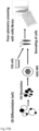
- Antibody-based purification of retinal pigment epithelial cells can be used during production of retinal pigmented epithelial cell production from pluripotent stem cells (e.g. for RPE enrichment, quality control purposes, etc.). Release and quality control criteria for retinal pigmented epithelial cell purity are also described herein. Isolation of native RPE cells can also be used for autologous or allogenic transplantations.
- Positive selection markers include CD140b, CD104, CD164, CD220, or EGFR.
- Negative selection markers include GD2, CD184, CD10, CD30, CD49a, CD49b, CD50, CD171, TRA-1-60, or CD326. These cell surface markers can be used, for example to enrich RPE cells in a mixture of cells. The cell surface marker is labeled in a starting mixture of cells; and then one separates the cells that are positive for said cell surface marker from cells that are negative for said cell surface marker to obtain a final mixture that is more enriched in RPE cells.
- RPE retinal pigment epithelium
- hPSC-RPE 3D embryoid body
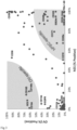
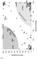
- the screening identified subsets of cell surface markers expressed in both cell types and specific for either the OVs or the hESCs.
- CD59 has been suggested to be a useful RPE marker, but both the present screen and immunostaining data show that it is expressed in both RPE and undifferentiated embryonic stem cells, then making it less suitable ( FIGS 3 and 4 and 38 of the present disclosure).
- OVs consisting of several cell types homogenous labelling of all cells in the OV-sample was not detected. Instead, the OV-specific markers labeled a fraction of the cells, suggesting that they tag different cell types within the dissected OVs.
- Table 1 Summary of the screening data
- CD140b was identified as a marker of early as well as more mature hESC-RPE cells whereas GD2 and CD184 label alternative lineages in the early cultures.
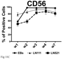
- CD56 was found to be expressed in the early stages of hPSC-RPE cells, but to a lower degree than in the mature cultures.
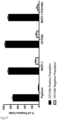
- CD140b proved to be more restricted to the dissected pigmented OVs over the remaining non-pigmented EB structures, whereas GD2 and CD184 were detected at higher levels in the non-pigmented structures ( FIG. 26 ). Additionally, CD140b positive cells were present in mature hESC-derived RPE cell (hESC-RPE) cultures while GD2 and CD184 expressing cells were completely absent, as immunofluorescence staining confirmed in hESC-RPE cells in culture ( FIG. 5 ). Histology of transplanted hESC-RPE into albino rabbit subretina showed apical expression of CD140b on pigmented and basal BEST1 positive hESC-RPE cells.
- CD140b The apical expression of CD140b was also confirmed by immunohistochemistry in adult human RPE ( FIG. 6 ) in agreement with known expression pattern in the mouse. Functionally, it was seen that after cell sorting using flow cytometry, only the CD140b positive, but not the negative cell fraction of early differentiations, expanded into hESC-RPE cells with cobblestone and homogeneous morphology, evidencing that this cell surface marker selectively captures the early hESC-RPE cells during in vitro differentiation ( FIG. 7 ).
- EXAMPLE 2 Monolayer differentiation on human recombinant laminin potently increase RPE differentiation compared to suspension culture
- hESCs were plated at a cell density of 2.4 ⁇ 10 4 cells/cm 2 on LN-111 and LN-521 substrates and evaluated 30 days after plating. Prominent pigmentation was observed on both substrates (data not shown). In agreement with previous studies suggesting that Activin A is a potent retinal fate inducer, a significant increase of pigmentation together with corresponding transcriptional maturation towards RPE fate with addition of Activin A was observed (data not shown).
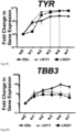
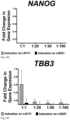
- the strong RPE induction was further validated by measuring gene expression for various RPE markers ( FIG 9A-C ).
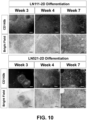
- LN-111 generally induced pigmentation and RPE markers faster, but LN-521 caught up between 4 and 7 weeks.
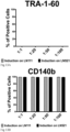
- the expression of pluripotency marker TRA-1-60 ( FIG. 11A ) was strongly reduced over time, supporting the conclusion that robust differentiation occurred.
- the final product further passed functional in vitro testing with monolayer integrity measured by transepithelial resistance (TEER), polarized secretion of pigment epithelium-derived factor (PEDF) measured by ELISA and phagocytosis of photoreceptor outer segments (POS) ( FIGS. 16A-16D and data not shown).
- TEER transepithelial resistance
- PEDF pigment epithelium-derived factor
- POS photoreceptor outer segments
- Multi-color en face and spectral domain-optical coherence tomography (SD-OCT) sagittal imaging in transplanted albino rabbits showed integration of pigmented cells and conserved retinal structure with hESC-RPE initially derived on hrLN-521 and hrLN-111 ( FIG. 16D ).
- Example 2 Although the method demonstrated in Example 2 did generate seemingly pure hESC-RPE cells, there was still the question whether using isolation with the identified cell surface markers (Example 1) could further improve the purity of the final product. Another aim was to ensure that there were no contaminating pluripotent cells or other alternative cell type in the final culture with or without the use of cell isolation.
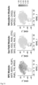
- NCAM1 also known as CD56 was enriched in this fraction of cells compared to the more mature RPE cluster ( FIG. 30 ). Staining of the replated cultures showed small clusters with higher CD56 intensity, which also coincided with low pigmentation levels consistent with less mature cells ( FIG. 29 ). In line with the sequencing data a larger CD56 "high" population was detected in the replated cultures compared to the sorted ones ( FIG. 28 ). Furthermore, NCAM1/CD56 was not detected in mature cells following integration in the rabbit subretina or in adult human RPE, thus supporting that NCAM1/CD56 is lost in mature RPE cells ( FIG. 34 ).
- EXAMPLE 4 The identified cell surface markers show reproducible differentiation in multiple hESCs
- the HS983a line failed to reach 20% positive cells for either differentiation marker at 30 days indicating poor differentiation.
- the same line was found to still express the pluripotency marker TRA-1-60 in 80% of the cells ( FIGS. 20A-21B ).
- the three performing lines had reached close to 100% CD140b levels and now reduced the progenitor marker NCAM1/CD56 to levels below 10%, correlating with the previous sequencing results ( FIGS. 22A-23B ). They had further reduced the fraction of cells positive for GD2 and CD184 to levels generally below 10% ( FIGS. 24A-25B ).
- Example 2 As three out of four lines differentiated well with similar kinetics, it can be concluded that the monolayer differentiation protocol of Example 2 is reproducible in a majority of hPSC lines without the need for additional modification. Finally, these data also illustrate the utility of the markers identified in Example 1 as quality control during the production to eliminate batch-to-batch variability or to eliminate hPSC lines that might be resistant to differentiation.
- a dissection-free methodology using in vitro hPSC-RPE differentiation in a 2D format on the biologically relevant substrate, human recombinant LN-111 or LN-521 has been demonstrated.
- a set of unique cell surface markers was also identified for RPE cells that can be used to optimize production methodology, perform quality control of production process and to define the final cellular composition of the product or to isolate these cells from other contaminating cells types arising from the in vitro differentiation process.
- the cells can be re-plated to enrich and expand the RPE population while eliminating other cell types.
- This dissection-free, xeno-free and defined and highly scalable protocol is compatible with GMP production of RPE cells, which can be used for subretinal transplantation.
- hESC and hESC-RPE cells were dissociated into single cells using TrypLE TM for 5-10 minutes. Optic vesicles were dissociated into single cells using TrypLE TM for 10 minutes followed by physical dissociation through a 20G needle.
- hESC, hESC-RPE and OV-cells were labeled with CellTrace TM CFSE (0.25 ⁇ M) for 7 minutes at 37°C or CellTrace TM Violet (5 ⁇ M) for 20 minutes at 37°C following manufacturer's protocol (Thermo Fisher Scientific). Then the three cell types were stained using the BD Lyoplate TM Screening Panels (BD Biosciences) following manufacturer's protocol.
- hESC lines HS980, HS975 and HS983 were derived and cultured under xeno-free and defined conditions according to the previously described method ( Rodin, S. et al. Nat. Commun. 5, 3195 (2014 )).
- the hESC line WA09/H9 was obtained from Wicell and was adapted to feeder-free culture on hrLN-521.
- the cells were maintained by clonal propagation on hrLN-521 coated plates (BioLamina AB) in NutriStem TM hPSC XF medium (Biological Industries), in a 5% CO 2 /5% O 2 incubator and passaged enzymatically at 1:10 ratio every 5-6 days.
- hESC were plated at a cell density of 2.4 ⁇ 10 4 cells/cm 2 on laminin coated dishes (human recombinant LN-111 or LN-521, Biolamina AB) using NutriStem TM hPSC XF medium.
- Rhokinase inhibitor Y-27632, Millipore
- hESC medium was replaced with differentiation medium (NutriStem TM hPSC XF without bFGF and TGF ⁇ ) and cells were placed at 37°C, 5% CO 2 /21%O 2 .
- Activin A R&D Systems
- 100 ng/mL of Activin A R&D Systems was added to the media.
- Cells were fed three times a week and kept for 30 days. Monolayers were then trypsinized using TrypLE TM Select (GIBCO, Invitrogen) for 10 minutes at 37°C, 5% CO 2 .
- the enzyme was carefully removed and the cells were collected in fresh pre-warmed NutriStem TM hPSC XF medium without bFGF and TGF ⁇ by gentle pipetting to obtain a single cell suspension.
- the cells were centrifuged at 300G for 4 minutes, the pellet was resuspended, passed through a cell strainer ( ⁇ 40 ⁇ m, BD Bioscience) and cells were seeded on laminin coated dishes (hrLN-111 and hrLN521 at 20 ⁇ g/mL) at different cell densities ranging from 1.4 ⁇ 10 6 to 1.4 ⁇ 10 4 cells/cm 2 .
- Re-plated cells were fed three times a week during the subsequent 30 days with NutriStem TM hPSC XF medium without bFGF and TGF ⁇ .
- Bright field images were acquired with Nikon Eclipse TM TE2000-S microscope and a Canon SX170 IS camera was used to capture pigmentation from the top of the wells.
- Taq-polymerase together with Taqman TM probes for GAPDH (cat. no. 4333764F), NANOG (cat. no. Hs02387400_g1), POU5F1 (cat. no. Hs03005111_g1), BEST1 (cat. no. Hs00188249_m1), RPE65 (cat. no. Hs01071462_m1), TYR (cat. no. Hs00165976_m1), PMEL (cat. no. Hs00173854_m1) and TBB3 (cat no. Hs00801390_s1) were used.
- hESC-RPE growing on the tested substrates were dissociated into single cells using TrypLE TM Select.
- Samples were stained with BV421 Mouse Anti-Human CD140b (BD, #564124, clone 28D4, at a concentration of 10 ⁇ g/mL), PE Mouse Anti-Human CD140b (BD, #558821, clone 28D4, at a concentration of 10 ⁇ g/mL), BB515 Mouse Anti-Human CD56 (BD, #564489, clone B159, at a concentration of 2.5 ⁇ g/mL), Alexa Fluor 647 Mouse Anti-Human TRA-1-60 (BD, #560850, at a concentration of 0.6 ⁇ g/mL), BV421 Mouse Anti-Human CD184 (BD, #562448, clone 12G5, at a concentration of 2.5 ⁇ g/mL) and BV421 Mouse Anti-Human Disialoganglioside GD2 (BD, #564223, clone
- Protein expression of mature hESC-RPE monolayers was assessed with immunofluorescence.
- Cells were fixed with 4% methanol free formaldehyde at room temperature for 10 minutes, followed by permeabilization with 0.3% Triton X-100 TM in Dulbecco's phosphate-buffered saline (DPBS) for 10 minutes and blocking with 4% fetal bovine serum (FBS) and 0.1% Tween-20 TM in DPBS for 1 hour.
- DPBS Dulbecco's phosphate-buffered saline
- FBS fetal bovine serum
- the primary antibodies were incubated overnight at 4°C followed by 2 hours incubation at room temperature with secondary antibodies: Alexa Fluor 647 donkey anti-rabbit IgG and Alexa Fluor 488 donkey anti-mouse IgG (both from Thermo Fisher Scientific, A31573 and A21202, respectively) diluted 1:1000 in 4% FBS, 0.1% Tween-20, DPBS solution. Nuclei were stained with Hoechst 33342 (1:1000, Invitrogen). Images were acquired with Zeiss LSM710-NLO TM point scanning confocal microscope. Post-acquisition analysis of the pictures was performed using IMARIS TM (Bitplane) and/or Fiji/ImageJ TM .
- TMD green Tissue Marking Dye
- slides were deparaffinized in xylene, dehydrated in graded alcohols, and rinsed with dH 2 O and Tris Buffered Saline (TBS, pH 7.6).
- Antigen retrieval was done in 10 mM citrate buffer (trisodium citrate dihydrate, Sigma-Aldrich, pH 6.0) with 1:2000 Tween-20 TM (Sigma-Aldrich) at 96°C for 30 min, followed by 30 min cooling at room temperature.
- Slides were washed with TBS and blocked for 30 min with 10% Normal Donkey Serum (Abcam) diluted in TBS containing 5% (w/v) IgG and protease free bovine serum albumin (Jackson Immunoresearch) in a humidified chamber.
- FITC-labelled bovine photoreceptor outer segments were isolated and kindly given by Dr. E.F. Nandrot from Institut de la Vision, Paris ( Parinot et al. J. Vis. Exp. (2014). doi:10.3791/52100 ).
- hESC-RPE were cultured on transwell membrane (0.33 cm 2 ,Corning) coated with hrLN-521 20 ⁇ g/mL for one month after seeding. Cells were incubated at 37°C or 4°C for 16 hours with 2.42 ⁇ 10 6 thawed POS/Transwell diluted in DMEM or CO 2 independent media (both from Thermo Fisher Scientific), respectively.
- hESC-RPE were cultured on Transwell TM membranes (0.33 cm 2 , Millipore) coated with different substrates. Supernatants from both the hESC-RPE apical and basal sides (meaning upper and lower compartments of the transwell, respectively) were collected 60 hours after the medium was changed.
- VEGF and PEDF secretion levels were measured in triplicates for each condition with commercially available human VEGF and PEDF ELISA Kits (VEGF: Cat#DVE00, R&D Systems; PEDF: Cat#RD191114200R, BioVendor), in accordance with the instructions of the manufacturers, after 60 days of culture
- the optical density readings were measured using SpectraMax TM 250 Microplate Reader (MolecularDevices). Results are presented as mean ⁇ SEM (standard error of the mean).
- Transepithelial electrical resistance RPE cells plated on Transwells TM (0.33 cm 2 , Millipore) was measured using the Millicell TM Electrical Resistance System volt-ohm meter (Millicell ERS-2, Millipore), according to the manufacturer's instructions. 60-day cultures were equilibrated outside the incubator at room temperature for 15-20 min before the experiment. Measurements were performed in unchanged culture media in triplicates for each condition, at three different positions of each well. Averages were used for further analysis. The background resistance was determined from a blank culture insert in the same media coated with the corresponding substrate but without cells, and subtracted from the respective experiment condition. Measurements are reported as resistance in ohms times the area in square centimeter ( ⁇ ⁇ cm 2 ). Results are presented as mean ⁇ SEM (standard error of the mean).
- hESC-RPE cells were dissociated using TrypLE TM Select and passed through a cell strainer ( ⁇ 40 ⁇ m, BD Bioscience). They were resuspended in a 1000 cells/ ⁇ L in 0.04% BSA in PBS. Cells were transported at 4°C to the Eukaryotic Single Cell Genomics Facility (ESCG, SciLifeLab, Sweden) where a 3' cDNA library was prepared for single cell RNA sequencing with the 10X Genomics platform.
- Cell Ranger TM 2.1.1 (10X Genomics) pipeline was used to convert Illumina base call files to fastq format, align sequencing reads to the hg19 transcriptome using the STAR aligner, and generate feature-barcode matrices.
- Cell Ranger TM quality-control filtered cells were analyzed in R version 3.5.1 (R Core Team), using Seurat TM suite version 2.3.4. As a further quality-control measure, RPE cells with unique expressed genes ( ⁇ 2000 to ⁇ 5000), UMIs ( ⁇ 10000 to ⁇ 30000) and percentage of UMIs mapping to MT-genes ( ⁇ 0.025 to ⁇ 0.10) were selected.
- hESC cells with unique expressed genes ⁇ 2000 to ⁇ 8000
- UMIs ⁇ 10000 to ⁇ 80000
- percentage of UMIs mapping to MT-genes ⁇ 0.025 to ⁇ _ 0.10
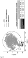
- This filtration step resulted in final dataset of 905, 616, 725 and 779 cells for hESC, CD140b+GD2-, CD140b+CD184- and re-plated 1:20 samples, respectively.
- PCA principal-component analysis
- PC principal component
- Cell clusters were analyzed by two approaches. Top differential genes were first identified for each cluster using Wilcoxon Rank Sum test. Secondary, signature gene expression (module scores) was computed for undifferentiated hESC and several cell types present in human retina. Cells expressing mesoderm markers were manually subdivided in a separate cluster using interactive plotting features of Seurat TM .
- hESC-RPE monolayers were washed with PBS, incubated with TrypLE TM and dissociated to single cell suspension.
- Cells were counted in a Neubauer haemocytometer chamber using 0.4% trypan blue (ThermoFisher Scientific Corp.), centrifuged at 300g for 4 minutes, and the cell pellet was resuspended in freshly filter-sterilized PBS to a final concentration of 1000 cells/ ⁇ L. The cell suspension was then aseptically aliquoted into 600 ⁇ L units and kept on ice until surgery.
- the cell suspension was drawn into a 1 mL syringe connected to an extension tube and a 38G polytip cannula (MedOne Surgical Inc). Without prior vitrectomy the cannula was inserted through the upper temporal trocar. After proper tip positioning, ascertained by a focal whitening of the retina, 50 ⁇ L of cell suspension (equivalent to 50,000 cells) was injected slowly subretinally approximately 6 mm below the inferior margin of the optic nerve head, forming a uniform bleb that was clearly visible under the operating microscope. Care was taken to maintain the tip within the bleb during the injection to minimize reflux. After instrument removal light pressure was applied to the self-sealing suture-less sclerotomies. 2 mg (100 ⁇ L) of intravitreal triamcinolone (Triescence TM , Alcon Nordic) was administered one week prior to the surgery, and no post-surgical antibiotics were given.
- Triescence TM intravitreal triamcinolone
Landscapes
- Health & Medical Sciences (AREA)
- Engineering & Computer Science (AREA)
- Life Sciences & Earth Sciences (AREA)
- Biomedical Technology (AREA)
- Chemical & Material Sciences (AREA)
- Bioinformatics & Cheminformatics (AREA)
- Zoology (AREA)
- Organic Chemistry (AREA)
- Biotechnology (AREA)
- Cell Biology (AREA)
- Genetics & Genomics (AREA)
- Wood Science & Technology (AREA)
- General Health & Medical Sciences (AREA)
- Neurology (AREA)
- Developmental Biology & Embryology (AREA)
- Neurosurgery (AREA)
- Microbiology (AREA)
- General Engineering & Computer Science (AREA)
- Biochemistry (AREA)
- Ophthalmology & Optometry (AREA)
- Pharmacology & Pharmacy (AREA)
- Medicinal Chemistry (AREA)
- Public Health (AREA)
- Reproductive Health (AREA)
- Animal Behavior & Ethology (AREA)
- Veterinary Medicine (AREA)
- Epidemiology (AREA)
- General Chemical & Material Sciences (AREA)
- Chemical Kinetics & Catalysis (AREA)
- Nuclear Medicine, Radiotherapy & Molecular Imaging (AREA)
- Virology (AREA)
- Gynecology & Obstetrics (AREA)
- Immunology (AREA)
- Physics & Mathematics (AREA)
- Acoustics & Sound (AREA)
- Analytical Chemistry (AREA)
- Micro-Organisms Or Cultivation Processes Thereof (AREA)
- Measuring Or Testing Involving Enzymes Or Micro-Organisms (AREA)
- Medicines Containing Material From Animals Or Micro-Organisms (AREA)
Priority Applications (1)
| Application Number | Priority Date | Filing Date | Title |
|---|---|---|---|
| EP25216797.8A EP4706764A2 (de) | 2018-06-20 | 2019-06-19 | Verfahren und zusammensetzungen zur herstellung von retinapigmentpithelzellen |
Applications Claiming Priority (3)
| Application Number | Priority Date | Filing Date | Title |
|---|---|---|---|
| US201862687416P | 2018-06-20 | 2018-06-20 | |
| PCT/EP2019/066285 WO2019243462A1 (en) | 2018-06-20 | 2019-06-19 | Methods and compositions for producing retinal pigment epithelium cells |
| EP19733709.0A EP3810758B1 (de) | 2018-06-20 | 2019-06-19 | Verfahren und zusammensetzungen zur herstellung von retinapigmentpithelzellen |
Related Parent Applications (2)
| Application Number | Title | Priority Date | Filing Date |
|---|---|---|---|
| EP19733709.0A Division EP3810758B1 (de) | 2018-06-20 | 2019-06-19 | Verfahren und zusammensetzungen zur herstellung von retinapigmentpithelzellen |
| EP19733709.0A Division-Into EP3810758B1 (de) | 2018-06-20 | 2019-06-19 | Verfahren und zusammensetzungen zur herstellung von retinapigmentpithelzellen |
Related Child Applications (1)
| Application Number | Title | Priority Date | Filing Date |
|---|---|---|---|
| EP25216797.8A Division EP4706764A2 (de) | 2018-06-20 | 2019-06-19 | Verfahren und zusammensetzungen zur herstellung von retinapigmentpithelzellen |
Publications (3)
| Publication Number | Publication Date |
|---|---|
| EP4234024A2 true EP4234024A2 (de) | 2023-08-30 |
| EP4234024A3 EP4234024A3 (de) | 2023-09-27 |
| EP4234024B1 EP4234024B1 (de) | 2025-11-19 |
Family
ID=67070817
Family Applications (3)
| Application Number | Title | Priority Date | Filing Date |
|---|---|---|---|
| EP23179217.7A Active EP4234024B1 (de) | 2018-06-20 | 2019-06-19 | Verfahren und zusammensetzungen zur herstellung von retinapigmentpithelzellen |
| EP19733709.0A Active EP3810758B1 (de) | 2018-06-20 | 2019-06-19 | Verfahren und zusammensetzungen zur herstellung von retinapigmentpithelzellen |
| EP25216797.8A Pending EP4706764A2 (de) | 2018-06-20 | 2019-06-19 | Verfahren und zusammensetzungen zur herstellung von retinapigmentpithelzellen |
Family Applications After (2)
| Application Number | Title | Priority Date | Filing Date |
|---|---|---|---|
| EP19733709.0A Active EP3810758B1 (de) | 2018-06-20 | 2019-06-19 | Verfahren und zusammensetzungen zur herstellung von retinapigmentpithelzellen |
| EP25216797.8A Pending EP4706764A2 (de) | 2018-06-20 | 2019-06-19 | Verfahren und zusammensetzungen zur herstellung von retinapigmentpithelzellen |
Country Status (9)
| Country | Link |
|---|---|
| US (2) | US12516287B2 (de) |
| EP (3) | EP4234024B1 (de) |
| CN (2) | CN119410584A (de) |
| DK (2) | DK4234024T3 (de) |
| ES (2) | ES2959705T3 (de) |
| FI (2) | FI4234024T3 (de) |
| PT (2) | PT4234024T (de) |
| SG (1) | SG11202011929VA (de) |
| WO (1) | WO2019243462A1 (de) |
Families Citing this family (1)
| Publication number | Priority date | Publication date | Assignee | Title |
|---|---|---|---|---|
| US20240358764A1 (en) * | 2023-04-28 | 2024-10-31 | Cellatoz Therapeutics, Inc. | NEURONAL REGENERATION PROMOTING CELLS (NRPCs) AND TREATING DAMAGED NERVE CELLS |
Family Cites Families (13)
| Publication number | Priority date | Publication date | Assignee | Title |
|---|---|---|---|---|
| AU2015201435B2 (en) | 2007-10-12 | 2017-02-02 | Astellas Institute For Regenerative Medicine | Improved methods of producing RPE cells and compositions of RPE cells |
| US20100184033A1 (en) * | 2008-07-16 | 2010-07-22 | West Michael D | Methods to accelerate the isolation of novel cell strains from pluripotent stem cells and cells obtained thereby |
| EP2655601A4 (de) * | 2010-12-22 | 2014-09-10 | Fate Therapeutics Inc | Zellkulturplattform für einzelzellsortierung und verbesserte neuprogrammierung von ipscs |
| CA2835215C (en) * | 2011-05-18 | 2023-10-10 | The Regents Of The University Of California | Non-immortal human retinal cell compositions, method for preparing them, and their use for treating retinal diseases |
| US9458428B2 (en) * | 2012-06-05 | 2016-10-04 | The Regents Of The University Of California | Methods and compositions for the rapid production of retinal pigmented epithelial cells from pluripotent cells |
| TR201807259T4 (tr) | 2012-12-03 | 2018-06-21 | Biolamina Ab | Rpe hücreleri üretim yöntemleri. |
| EP3047017B1 (de) * | 2013-10-09 | 2025-09-10 | The Regents of the University of California | Verfahren zur erzeugung von menschlichen retinalen stammzellen und anwendungen |
| JP6518878B2 (ja) * | 2013-10-09 | 2019-05-29 | 株式会社ヘリオス | 網膜色素上皮細胞の製造方法 |
| EP3189135B1 (de) * | 2014-09-05 | 2021-06-02 | Astellas Institute for Regenerative Medicine | Retinale ganglienzellen und vorläufer dafür |
| AU2016318774B2 (en) | 2015-09-08 | 2022-08-18 | FUJIFILM Cellular Dynamics, Inc. | MACS-based purification of stem cell-derived retinal pigment epithelium |
| JP6987769B2 (ja) * | 2015-10-26 | 2022-01-05 | セル キュア ニューロサイエンシズ リミテッド | 網膜色素上皮細胞の調製法 |
| US20180362925A1 (en) * | 2015-12-04 | 2018-12-20 | Bond University Ltd | Methods for differentiating cells |
| WO2018034618A1 (en) * | 2016-08-15 | 2018-02-22 | National University Of Singapore | Methods for producing endothelial cells using laminins |
-
2019
- 2019-06-19 PT PT231792177T patent/PT4234024T/pt unknown
- 2019-06-19 EP EP23179217.7A patent/EP4234024B1/de active Active
- 2019-06-19 ES ES19733709T patent/ES2959705T3/es active Active
- 2019-06-19 WO PCT/EP2019/066285 patent/WO2019243462A1/en not_active Ceased
- 2019-06-19 FI FIEP23179217.7T patent/FI4234024T3/fi active
- 2019-06-19 DK DK23179217.7T patent/DK4234024T3/da active
- 2019-06-19 DK DK19733709.0T patent/DK3810758T3/da active
- 2019-06-19 SG SG11202011929VA patent/SG11202011929VA/en unknown
- 2019-06-19 EP EP19733709.0A patent/EP3810758B1/de active Active
- 2019-06-19 US US17/252,789 patent/US12516287B2/en active Active
- 2019-06-19 FI FIEP19733709.0T patent/FI3810758T3/sv active
- 2019-06-19 ES ES23179217T patent/ES3061658T3/es active Active
- 2019-06-19 EP EP25216797.8A patent/EP4706764A2/de active Pending
- 2019-06-19 CN CN202411298872.0A patent/CN119410584A/zh active Pending
- 2019-06-19 CN CN201980041644.6A patent/CN112334574B/zh active Active
- 2019-06-19 PT PT197337090T patent/PT3810758T/pt unknown
-
2025
- 2025-12-04 US US19/408,516 patent/US20260085285A1/en active Pending
Non-Patent Citations (4)
| Title |
|---|
| CHOUDHARY, P.WHITING, P. J., STEM CELL RES. THER., vol. 7, 2016, pages 127 |
| PARINOT ET AL., J. VIS. EXP., 2014 |
| PETRUS-REURER, S. ET AL., J. VIS. EXP., 2018 |
| RODIN, S. ET AL., NAT. COMMUN., vol. 5, 2014, pages 3195 |
Also Published As
| Publication number | Publication date |
|---|---|
| CN112334574B (zh) | 2024-10-11 |
| US12516287B2 (en) | 2026-01-06 |
| ES3061658T3 (en) | 2026-04-06 |
| PT4234024T (pt) | 2026-01-21 |
| PT3810758T (pt) | 2023-10-13 |
| EP4234024A3 (de) | 2023-09-27 |
| EP3810758A1 (de) | 2021-04-28 |
| SG11202011929VA (en) | 2021-01-28 |
| FI3810758T3 (sv) | 2023-10-11 |
| EP3810758B1 (de) | 2023-07-19 |
| DK4234024T3 (da) | 2026-01-19 |
| CN112334574A (zh) | 2021-02-05 |
| EP4234024B1 (de) | 2025-11-19 |
| DK3810758T3 (da) | 2023-10-16 |
| US20260085285A1 (en) | 2026-03-26 |
| CN119410584A (zh) | 2025-02-11 |
| EP4706764A2 (de) | 2026-03-11 |
| ES2959705T3 (es) | 2024-02-27 |
| WO2019243462A1 (en) | 2019-12-26 |
| US20210261918A1 (en) | 2021-08-26 |
| FI4234024T3 (fi) | 2026-02-17 |
Similar Documents
| Publication | Publication Date | Title |
|---|---|---|
| US10329534B2 (en) | Methods for purifying cells derived from pluripotent stem cells | |
| Pennington et al. | Defined culture of human embryonic stem cells and xeno-free derivation of retinal pigmented epithelial cells on a novel, synthetic substrate | |
| US20260085285A1 (en) | Methods for producing retinal pigment epithelium cells | |
| JP2023052646A (ja) | Rpe細胞集団およびそれを作製する方法 | |
| KR20170075023A (ko) | 줄기 세포 및 전구 세포를 배양하는 방법 | |
| AU2002367091A1 (en) | A method for the establishment of a pluripotent human blastocyst-derived stem cell line | |
| EP3704227A1 (de) | Zusammensetzung und verfahren | |
| KR20140113691A (ko) | 인간 제대 조직-유래된 세포로부터의 유도 만능 줄기 세포 | |
| EP2444483A2 (de) | Materialien und Verfahren im Zusammenhang mit zellbasierten Therapien | |
| WO2010059738A1 (en) | Differentiation of stem cells into dopaminergic cells | |
| US20230081733A1 (en) | Methods for preparing keratinocytes | |
| RU2542964C1 (ru) | Способ получения прогениторных клеток миокарда | |
| AU2017276263B2 (en) | Methods for enriching primitive gut tube | |
| KR20250073682A (ko) | 라미닌 세포 배양 기질 및 프로토콜 |
Legal Events
| Date | Code | Title | Description |
|---|---|---|---|
| PUAI | Public reference made under article 153(3) epc to a published international application that has entered the european phase |
Free format text: ORIGINAL CODE: 0009012 |
|
| STAA | Information on the status of an ep patent application or granted ep patent |
Free format text: STATUS: THE APPLICATION HAS BEEN PUBLISHED |
|
| REG | Reference to a national code |
Ref legal event code: R079 Ipc: C12N0005079300 Ref country code: DE Ref legal event code: R079 Ref document number: 602019078413 Country of ref document: DE Free format text: PREVIOUS MAIN CLASS: A61P0027020000 Ipc: C12N0005079300 |
|
| PUAL | Search report despatched |
Free format text: ORIGINAL CODE: 0009013 |
|
| AC | Divisional application: reference to earlier application |
Ref document number: 3810758 Country of ref document: EP Kind code of ref document: P |
|
| AK | Designated contracting states |
Kind code of ref document: A2 Designated state(s): AL AT BE BG CH CY CZ DE DK EE ES FI FR GB GR HR HU IE IS IT LI LT LU LV MC MK MT NL NO PL PT RO RS SE SI SK SM TR |
|
| AK | Designated contracting states |
Kind code of ref document: A3 Designated state(s): AL AT BE BG CH CY CZ DE DK EE ES FI FR GB GR HR HU IE IS IT LI LT LU LV MC MK MT NL NO PL PT RO RS SE SI SK SM TR |
|
| RIC1 | Information provided on ipc code assigned before grant |
Ipc: A61P 27/02 20060101ALI20230821BHEP Ipc: A61K 35/545 20150101ALI20230821BHEP Ipc: C12N 5/0793 20100101AFI20230821BHEP |
|
| STAA | Information on the status of an ep patent application or granted ep patent |
Free format text: STATUS: REQUEST FOR EXAMINATION WAS MADE |
|
| 17P | Request for examination filed |
Effective date: 20240326 |
|
| RBV | Designated contracting states (corrected) |
Designated state(s): AL AT BE BG CH CY CZ DE DK EE ES FI FR GB GR HR HU IE IS IT LI LT LU LV MC MK MT NL NO PL PT RO RS SE SI SK SM TR |
|
| STAA | Information on the status of an ep patent application or granted ep patent |
Free format text: STATUS: EXAMINATION IS IN PROGRESS |
|
| 17Q | First examination report despatched |
Effective date: 20240614 |
|
| 17Q | First examination report despatched |
Effective date: 20240627 |
|
| GRAP | Despatch of communication of intention to grant a patent |
Free format text: ORIGINAL CODE: EPIDOSNIGR1 |
|
| STAA | Information on the status of an ep patent application or granted ep patent |
Free format text: STATUS: GRANT OF PATENT IS INTENDED |
|
| INTG | Intention to grant announced |
Effective date: 20250617 |
|
| GRAS | Grant fee paid |
Free format text: ORIGINAL CODE: EPIDOSNIGR3 |
|
| GRAA | (expected) grant |
Free format text: ORIGINAL CODE: 0009210 |
|
| STAA | Information on the status of an ep patent application or granted ep patent |
Free format text: STATUS: THE PATENT HAS BEEN GRANTED |
|
| AC | Divisional application: reference to earlier application |
Ref document number: 3810758 Country of ref document: EP Kind code of ref document: P |
|
| AK | Designated contracting states |
Kind code of ref document: B1 Designated state(s): AL AT BE BG CH CY CZ DE DK EE ES FI FR GB GR HR HU IE IS IT LI LT LU LV MC MK MT NL NO PL PT RO RS SE SI SK SM TR |
|
| REG | Reference to a national code |
Ref country code: CH Ref legal event code: F10 Free format text: ST27 STATUS EVENT CODE: U-0-0-F10-F00 (AS PROVIDED BY THE NATIONAL OFFICE) Effective date: 20251119 Ref country code: GB Ref legal event code: FG4D |
|
| REG | Reference to a national code |
Ref country code: DE Ref legal event code: R096 Ref document number: 602019078413 Country of ref document: DE |
|
| REG | Reference to a national code |
Ref country code: IE Ref legal event code: FG4D |
|
| REG | Reference to a national code |
Ref country code: DK Ref legal event code: T3 Effective date: 20260116 |
|
| REG | Reference to a national code |
Ref country code: PT Ref legal event code: SC4A Ref document number: 4234024 Country of ref document: PT Date of ref document: 20260121 Kind code of ref document: T Free format text: AVAILABILITY OF NATIONAL TRANSLATION Effective date: 20260114 |
|
| REG | Reference to a national code |
Ref country code: SE Ref legal event code: TRGR |
|
| REG | Reference to a national code |
Ref country code: NL Ref legal event code: FP |
|
| REG | Reference to a national code |
Ref country code: CH Ref legal event code: R17 Free format text: ST27 STATUS EVENT CODE: U-0-0-R10-R17 (AS PROVIDED BY THE NATIONAL OFFICE) Effective date: 20260204 |
|
| REG | Reference to a national code |
Ref country code: FI Ref legal event code: FGE |
|
| P01 | Opt-out of the competence of the unified patent court (upc) registered |
Free format text: CASE NUMBER: UPC_APP_0004101_4234024/2026 Effective date: 20260204 |
|
| REG | Reference to a national code |
Ref country code: ES Ref legal event code: FG2A Ref document number: 3061658 Country of ref document: ES Kind code of ref document: T3 Effective date: 20260406 |
|
| REG | Reference to a national code |
Ref country code: LT Ref legal event code: MG9D |
|
| PG25 | Lapsed in a contracting state [announced via postgrant information from national office to epo] |
Ref country code: HR Free format text: LAPSE BECAUSE OF FAILURE TO SUBMIT A TRANSLATION OF THE DESCRIPTION OR TO PAY THE FEE WITHIN THE PRESCRIBED TIME-LIMIT Effective date: 20251119 Ref country code: AT Free format text: LAPSE BECAUSE OF FAILURE TO SUBMIT A TRANSLATION OF THE DESCRIPTION OR TO PAY THE FEE WITHIN THE PRESCRIBED TIME-LIMIT Effective date: 20251119 |
|
| REG | Reference to a national code |
Ref country code: AT Ref legal event code: MK05 Ref document number: 1858856 Country of ref document: AT Kind code of ref document: T Effective date: 20251119 |
|
| PG25 | Lapsed in a contracting state [announced via postgrant information from national office to epo] |
Ref country code: RS Free format text: LAPSE BECAUSE OF FAILURE TO SUBMIT A TRANSLATION OF THE DESCRIPTION OR TO PAY THE FEE WITHIN THE PRESCRIBED TIME-LIMIT Effective date: 20260219 |
|
| PG25 | Lapsed in a contracting state [announced via postgrant information from national office to epo] |
Ref country code: IS Free format text: LAPSE BECAUSE OF FAILURE TO SUBMIT A TRANSLATION OF THE DESCRIPTION OR TO PAY THE FEE WITHIN THE PRESCRIBED TIME-LIMIT Effective date: 20260319 |
|
| PG25 | Lapsed in a contracting state [announced via postgrant information from national office to epo] |
Ref country code: PL Free format text: LAPSE BECAUSE OF FAILURE TO SUBMIT A TRANSLATION OF THE DESCRIPTION OR TO PAY THE FEE WITHIN THE PRESCRIBED TIME-LIMIT Effective date: 20251119 |
|
| PG25 | Lapsed in a contracting state [announced via postgrant information from national office to epo] |
Ref country code: LV Free format text: LAPSE BECAUSE OF FAILURE TO SUBMIT A TRANSLATION OF THE DESCRIPTION OR TO PAY THE FEE WITHIN THE PRESCRIBED TIME-LIMIT Effective date: 20251119 |