EP4227902A1 - Verfahren, vorrichtung und computervorrichtung zur ausrichtung eines externen blutgefässbildes mit einem intraluminalen bild - Google Patents
Verfahren, vorrichtung und computervorrichtung zur ausrichtung eines externen blutgefässbildes mit einem intraluminalen bild Download PDFInfo
- Publication number
- EP4227902A1 EP4227902A1 EP21902253.0A EP21902253A EP4227902A1 EP 4227902 A1 EP4227902 A1 EP 4227902A1 EP 21902253 A EP21902253 A EP 21902253A EP 4227902 A1 EP4227902 A1 EP 4227902A1
- Authority
- EP
- European Patent Office
- Prior art keywords
- blood vessel
- image
- interest
- point set
- feature point
- Prior art date
- Legal status (The legal status is an assumption and is not a legal conclusion. Google has not performed a legal analysis and makes no representation as to the accuracy of the status listed.)
- Pending
Links
Images

Classifications
-
- G—PHYSICS
- G06—COMPUTING OR CALCULATING; COUNTING
- G06T—IMAGE DATA PROCESSING OR GENERATION, IN GENERAL
- G06T7/00—Image analysis
- G06T7/30—Determination of transform parameters for the alignment of images, i.e. image registration
- G06T7/33—Determination of transform parameters for the alignment of images, i.e. image registration using feature-based methods
- G06T7/337—Determination of transform parameters for the alignment of images, i.e. image registration using feature-based methods involving reference images or patches
-
- A—HUMAN NECESSITIES
- A61—MEDICAL OR VETERINARY SCIENCE; HYGIENE
- A61B—DIAGNOSIS; SURGERY; IDENTIFICATION
- A61B6/00—Apparatus or devices for radiation diagnosis; Apparatus or devices for radiation diagnosis combined with radiation therapy equipment
- A61B6/50—Apparatus or devices for radiation diagnosis; Apparatus or devices for radiation diagnosis combined with radiation therapy equipment specially adapted for specific body parts; specially adapted for specific clinical applications
- A61B6/504—Apparatus or devices for radiation diagnosis; Apparatus or devices for radiation diagnosis combined with radiation therapy equipment specially adapted for specific body parts; specially adapted for specific clinical applications for diagnosis of blood vessels, e.g. by angiography
-
- A—HUMAN NECESSITIES
- A61—MEDICAL OR VETERINARY SCIENCE; HYGIENE
- A61B—DIAGNOSIS; SURGERY; IDENTIFICATION
- A61B6/00—Apparatus or devices for radiation diagnosis; Apparatus or devices for radiation diagnosis combined with radiation therapy equipment
- A61B6/52—Devices using data or image processing specially adapted for radiation diagnosis
- A61B6/5211—Devices using data or image processing specially adapted for radiation diagnosis involving processing of medical diagnostic data
- A61B6/5217—Devices using data or image processing specially adapted for radiation diagnosis involving processing of medical diagnostic data extracting a diagnostic or physiological parameter from medical diagnostic data
-
- A—HUMAN NECESSITIES
- A61—MEDICAL OR VETERINARY SCIENCE; HYGIENE
- A61B—DIAGNOSIS; SURGERY; IDENTIFICATION
- A61B6/00—Apparatus or devices for radiation diagnosis; Apparatus or devices for radiation diagnosis combined with radiation therapy equipment
- A61B6/52—Devices using data or image processing specially adapted for radiation diagnosis
- A61B6/5211—Devices using data or image processing specially adapted for radiation diagnosis involving processing of medical diagnostic data
- A61B6/5229—Devices using data or image processing specially adapted for radiation diagnosis involving processing of medical diagnostic data combining image data of a patient, e.g. combining a functional image with an anatomical image
- A61B6/5247—Devices using data or image processing specially adapted for radiation diagnosis involving processing of medical diagnostic data combining image data of a patient, e.g. combining a functional image with an anatomical image combining images from an ionising-radiation diagnostic technique and a non-ionising radiation diagnostic technique, e.g. X-ray and ultrasound
-
- G—PHYSICS
- G06—COMPUTING OR CALCULATING; COUNTING
- G06T—IMAGE DATA PROCESSING OR GENERATION, IN GENERAL
- G06T7/00—Image analysis
- G06T7/10—Segmentation; Edge detection
- G06T7/13—Edge detection
-
- G—PHYSICS
- G06—COMPUTING OR CALCULATING; COUNTING
- G06T—IMAGE DATA PROCESSING OR GENERATION, IN GENERAL
- G06T7/00—Image analysis
- G06T7/60—Analysis of geometric attributes
- G06T7/62—Analysis of geometric attributes of area, perimeter, diameter or volume
-
- G—PHYSICS
- G06—COMPUTING OR CALCULATING; COUNTING
- G06T—IMAGE DATA PROCESSING OR GENERATION, IN GENERAL
- G06T2207/00—Indexing scheme for image analysis or image enhancement
- G06T2207/10—Image acquisition modality
- G06T2207/10072—Tomographic images
- G06T2207/10101—Optical tomography; Optical coherence tomography [OCT]
-
- G—PHYSICS
- G06—COMPUTING OR CALCULATING; COUNTING
- G06T—IMAGE DATA PROCESSING OR GENERATION, IN GENERAL
- G06T2207/00—Indexing scheme for image analysis or image enhancement
- G06T2207/10—Image acquisition modality
- G06T2207/10116—X-ray image
-
- G—PHYSICS
- G06—COMPUTING OR CALCULATING; COUNTING
- G06T—IMAGE DATA PROCESSING OR GENERATION, IN GENERAL
- G06T2207/00—Indexing scheme for image analysis or image enhancement
- G06T2207/10—Image acquisition modality
- G06T2207/10132—Ultrasound image
-
- G—PHYSICS
- G06—COMPUTING OR CALCULATING; COUNTING
- G06T—IMAGE DATA PROCESSING OR GENERATION, IN GENERAL
- G06T2207/00—Indexing scheme for image analysis or image enhancement
- G06T2207/20—Special algorithmic details
- G06T2207/20016—Hierarchical, coarse-to-fine, multiscale or multiresolution image processing; Pyramid transform
-
- G—PHYSICS
- G06—COMPUTING OR CALCULATING; COUNTING
- G06T—IMAGE DATA PROCESSING OR GENERATION, IN GENERAL
- G06T2207/00—Indexing scheme for image analysis or image enhancement
- G06T2207/30—Subject of image; Context of image processing
- G06T2207/30004—Biomedical image processing
- G06T2207/30101—Blood vessel; Artery; Vein; Vascular
Definitions
- the present invention relates to the medical field, and in particular, to a method and an apparatus for registering an extravascular image with an intravascular image, and a computing device.
- Main imaging methods used in clinical diagnosis and treatment of coronary heart diseases include an extraluminal imaging technique such as coronary angiography and intravascular imaging such as optical coherence tomography (Optic Coherence Tomography, OCT) and intravenous ultrasound (Intravenous Ultrasound, IVUS).
- the coronary angiography can reflect a two-dimensional anatomical shape of coronary arteries, so that a specific location and a degree of stenosis of the coronary heart disease can be determined, but an abnormal position in a blood vessel wall is difficult to be clearly displayed, and information such as the shape and nature of the plaque cannot be provided.
- the intravascular image can clearly display a cross-section of the blood vessel, and can reflect a tissue structure of the blood vessel wall and a fine geometric anatomy, but the intravascular image cannot well reflect topological structure information of the blood vessel.
- registration and fusion of coronary angiography images and intravascular images can not only observe a spatial position of a coronary vessel, but also obtain the inner and outer membranes and quantitative data of plaques of the blood vessel segment of interest, so that clinicians can be better assisted in a clinical assessment of the coronary heart disease.
- the registration of coronary angiography images and intravascular images is mainly implemented by three-dimensional reconstruction of coronary angiography images.
- a three-dimensional model of a blood vessel was reconstructed by taking two pairs of angiography images at starting and ending points of a pullback path of a catheter and by using a biplane angiography device, and then was fused with IVUS according to a retraction speed of the catheter to obtain a three-dimensional model of the coronary artery.
- An objective of the present invention is to provide a method for registering an extravascular image with an intravascular image, to solve a problem that convenience and accuracy of registration cannot be taken into account in the prior art.
- an implementation of the present invention discloses a method for registering an extravascular image with an intravascular image, including the following steps:
- the method for registering an extravascular image with an intravascular image in the technical solution is adopted, so that automatic registration of the coronary angiography image and the intravascular image can be directly implemented by using the diameter and branch information of the blood vessel and by using the multi-level registration method.
- the method only one frame of coronary angiography image needs to be detected and segmented, there is no need to perform manual labeling and three-dimensional reconstruction, and the method has advantages of rapidity, simplicity, high accuracy, and high robustness.
- the primary registration step includes:
- the obtaining the third feature information according to the contour feature point set of the reference blood vessel and the contour feature point set of the blood vessel of interest includes:
- the external branch information of the reference blood vessel includes a serial number of each branch blood vessel of the reference blood vessel obtained in the extravascular image and a branch blood vessel external length from a bifurcation point of each branch blood vessel to the proximal end of the reference blood vessel; and the internal branch information of the blood vessel segment of interest includes a serial number of each branch blood vessel of the blood vessel segment of interest obtained from the intravascular image and a branch blood vessel internal length of each branch blood vessel from the starting point.
- the method for calculating a matching degree between each region in the reference region and the sliding window includes:
- S d indicates the diameter matching score
- N indicates the total number of feature points in the sliding window
- d si indicates the blood vessel internal diameter corresponding to the i th feature point in the sliding window
- d ti indicates the blood vessel external diameter corresponding to the i th feature point in the reference region.
- ⁇ ranges from 0.3 to 0.7
- the target blood vessel segment is a blood vessel segment corresponding to a region having a highest matching degree with the sliding window in the reference region.
- the performing uniform sampling on an external contour of the reference blood vessel, and obtaining a contour feature point set of the reference blood vessel according to the external contour information of the reference blood vessel includes:
- the performing uniform sampling on the intravascular image of the blood vessel segment of interest, and obtaining a contour feature point set of the blood vessel of interest according to the internal contour information of the blood vessel segment of interest includes:
- the primary registration step further includes: performing uniform sampling on the external contour of the reference blood vessel in the extravascular image according to a distance between any two adjacent points in the contour feature point set of the blood vessel of interest.
- the primary registration step further includes:
- the target registration result is a target registration relationship between the contour feature point set of the blood vessel of interest and the contour feature point set of the target blood vessel.
- the secondary registration step includes the following steps performed in sequence:
- the method further includes: when the feature error is more than or equal to the preset value, determining whether a number of iteration times is more than a preset number, and if yes, determining the normalized contour feature point set S' of the blood vessel of interest as the target feature information, and calculating the target registration relationship according to the target feature information and the normalized contour feature point set T' of the target blood vessel; otherwise, increasing the number of iteration times by one, and performing the next step.
- the method further includes: setting the number of iteration times to zero.
- the secondary registration step includes: performing the secondary registration on the first feature information and the third feature information by using an iterative closest point algorithm or a coherent point drift algorithm or a robust point matching algorithm, to obtain the target registration result
- the intravascular image is an OCT image or an IVUS image.
- the present invention further provides an apparatus for registering an extravascular image with an intravascular image, including:
- the primary registration module includes:
- the obtaining module includes:
- the external branch information of the reference blood vessel includes a serial number of each branch blood vessel of the reference blood vessel obtained in the extravascular image and a branch blood vessel external length from a bifurcation point of each branch blood vessel to the proximal end of the reference blood vessel;
- the internal branch information of the blood vessel segment of interest includes a serial number of each branch blood vessel of the blood vessel segment of interest obtained in the intravascular image and a branch blood vessel internal length of each branch blood vessel from the starting point.
- the method for calculating a matching degree between each region in the reference region and the sliding window by the matching degree calculation unit includes:
- ⁇ ranges from 0.3 to 0.7
- the target blood vessel segment is a blood vessel segment corresponding to a region having a highest matching degree with the sliding window in the reference region.
- the first sampling unit includes:
- the second sampling unit includes:
- the primary registration module further includes:
- the first sampling unit is further configured to: according to the third feature information, determine a blood vessel external diameter and a lateral vascular length corresponding to each point on an external contour curve of the target blood vessel segment in the extravascular image, and determine external branch information of the target blood vessel segment; construct an external contour feature point corresponding to each point on the target blood vessel segment by taking the lateral vascular length as the first-dimensional feature, and taking the blood vessel external diameter as the second-dimensional feature; and obtain the contour feature point set of the target blood vessel according to all the external contour feature points.
- the target registration result is a target registration relationship between the contour feature point set of the blood vessel of interest and the contour feature point set of the target blood vessel.
- the secondary registration module includes:
- the secondary registration module further includes:
- the secondary registration module further includes: an initialization unit, configured to set the number of iteration times to zero before the error calculation unit calculates a feature error between the normalized contour feature point set S' of the blood vessel of interest and the normalized contour feature point set T' of the target blood vessel.
- an initialization unit configured to set the number of iteration times to zero before the error calculation unit calculates a feature error between the normalized contour feature point set S' of the blood vessel of interest and the normalized contour feature point set T' of the target blood vessel.
- the secondary registration module performs secondary registration on the first feature information and the third feature information by using an iterative closest point algorithm or a coherent point drift algorithm or a robust point matching algorithm, to obtain the target registration result.
- the intravascular image is an OCT image or an IVUS image.
- the present invention further provides a computing device, including:
- the computing device of the above technical solution is adopted, so that rapid and accurate registration of the extravascular image with the intravascular image can be implemented.
- the present invention further provides a storage medium, which stores a plurality of instructions for a processor to load and execute the method for registering an extravascular image with an intravascular image.
- the storage medium of the above technical solution is adopted, so that rapid and accurate registration of the extravascular image with the intravascular image can be implemented.
- proximal end and distal end are relative positions based on the distance of a blood vessel from an aorta.
- the end near the aorta is the proximal end, and the end far from the aorta is the distal end.
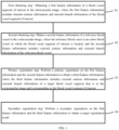
- the present invention discloses a method for registering an extravascular image with an intravascular image, including the following steps: A first obtaining step S1: Obtain a first feature information of a blood vessel segment of interest in the intravascular image, where the first feature information includes internal contour information and internal branch information of the blood vessel segment of interest.
- the blood vessel segment of interest may be a certain blood vessel segment that is abnormal relative to a normal blood vessel.
- the intravascular image may be an OCT image or may be an IVUS image. That is, the internal contour information and internal branch information are obtained from the intravascular image such as the OCT/IVUS image.
- the internal contour information may include a blood vessel internal diameter and a medial vascular length at each point in the blood vessel.
- the blood vessel internal diameter refers to a blood vessel diameter obtained from the intravascular image; and the medial vascular length is a length along an axis of the blood vessel from a certain point in the blood vessel to a starting point.
- the starting point is an endpoint that is near a proximal end of the blood vessel segment of interest and that is collected in the intravascular image. Because pullback and imaging of OCT are performed from a distal end to a proximal end, this starting point can also be said to be an ending point of the OCT pullback.
- the internal branch information may include a serial number of an internal branch (the serial number includes information of which branch on the blood vessel the branch belongs to) and position information of the branch relative to the blood vessel segment of interest.
- the intravascular image may be a directly obtained image that only includes the blood vessel segment of interest and that has been isolated, or may be an image that is selected from an intravascular image of a segment of blood vessel and that is corresponding to the blood vessel segment of interest.
- the intravascular image can be directly imported related data, or may be obtained from real-time configuration connections from other resource libraries, or may be obtained after searching a stored image database based on information such as a name of a user. This is not limited in the present invention.
- the intravascular image data may be in a DICOM (Digital Imaging and Communications in Medicine) format.
- DICOM has the following advantages: Almost all protocols of information exchange such as collection, archiving, communication, display and query of medical digital images are covered; and a set of object sets including information such as various types of medical diagnostic images, and related analyses and reports of the medical diagnostic images is defined by using an open and interconnected architecture and an object-oriented method; service classes and command sets used for information transmission and exchange, and standard responses to messages are defined; the techniques for identifying various information objects are described in detail; service support applied to a network environment (OSI or TCP/IP) is provided; and a conformance statement (Conformance Statement) of a manufacturer is structurally defined.
- the implementation of medical image information exchange can be greatly simplified by using the DICOM format, and synergy with other medical application systems such as HIS and RIS is facilitated.
- a second obtaining step S2 Obtain a second feature information of a reference blood vessel in the extravascular image, wherein the reference blood vessel is an entire blood vessel in which the blood vessel segment of interest is located, and the second feature information includes external contour information and external branch information of the reference blood vessel.
- the extravascular image may be a coronary angiography image, which reflects external contour information of the blood vessel.
- an external image of a blood vessel segment corresponding to the blood vessel segment of interest can also be directly captured.
- information of the external image of the entire blood vessel that is, the entire blood vessel from a proximal end of the blood vessel to a distal end of the blood vessel) in which the blood vessel segment of interest is located is collected in this step in this implementation. It seems that redundant information is collected. In fact, combined with the subsequent processing steps, it is more beneficial to improve processing efficiency as a whole.
- the external contour information may include a blood vessel external diameter and a lateral vascular length at each position in the blood vessel, the blood vessel external diameter refers to a blood vessel diameter obtained according to the extravascular image, and the lateral vascular length is a length along an axis of the blood vessel from a position on an external contour of the blood vessel to a proximal end of the blood vessel.
- the external branch information may include a serial number of an external branch blood vessel (the serial number includes information of which branch on the blood vessel the branch belongs to) and position information of the external branch blood vessel relative to the reference blood vessel.
- Primary registration step S3 Perform a primary registration on the first feature information and the second feature information to obtain a third feature information, where the third feature information includes external contour information and external branch information of a target blood vessel segment that is in the extravascular image and corresponding to the blood vessel segment of interest.
- the blood vessel segment of interest mentioned in the text is located inside the blood vessel
- the target blood vessel segment refers to a blood vessel segment that is located outside the blood vessel and that is corresponding to the blood vessel segment of interest.
- the second feature information is the external contour information of the entire reference blood vessel. Therefore, registration is performed on the first feature information and the second feature information to obtain the required external contour information of the target blood vessel segment.
- Secondary registration step S4 Perform a secondary registration on the first feature information and the third feature information to obtain a target registration result.
- the target registration result may be target feature information, where a feature error between the target feature information and the third feature information satisfies a preset condition.
- the target registration result may be a target registration relationship between the first feature information and the third feature information.
- the target registration result may include not only the above target feature information, but also a target registration relationship between the first feature information and the third feature information.
- step S3 registration is roughly performed on only the first feature information and the second feature information to obtain the required external contour information of the blood vessel of interest. Therefore, there may be a wrong matching or a large deviation between the first feature information and the third feature information obtained in step S3, and matching needs to be further performed on the first feature information and the third feature information by using secondary registration.
- the sequence of the first obtaining step and the sequence of the second obtaining step can be exchanged, that is, the first feature information of the blood vessel segment of interest can be obtained first, and then the second feature information of the entire blood vessel in which the blood vessel segment of interest is located is obtained.
- the second feature information of the entire blood vessel in which the blood vessel segment of interest is located may be obtained first, and then the first feature information of the blood vessel segment of interest may be obtained.
- the first feature information of the blood vessel segment of interest and the second feature information of the entire blood vessel in which the blood vessel segment of interest is located may be simultaneously obtained.
- the sequence of obtaining the first feature information of the blood vessel segment of interest and the second feature information of the entire blood vessel in which the blood vessel segment of interest is located is not limited in the present invention.
- the method for registering an extravascular image with an intravascular image mentioned in the present invention can be applied to registration of an external image and an intravascular image of a coronary artery blood vessel such as a left circumflex coronary artery, a left anterior descending coronary artery, or a right coronary artery.
- a coronary artery blood vessel such as a left circumflex coronary artery, a left anterior descending coronary artery, or a right coronary artery.
- the diameter and branch information of the blood vessel are fully used, and automatic registration of the coronary angiography image and the intravascular image is directly implemented by using the multi-level registration method.
- the method only one frame of coronary angiography image needs to be detected and segmented, and there is no need to perform manual labeling and three-dimensional reconstruction, and the method has the advantages of quickness, simplicity, high accuracy and high robustness.
- the robustness here refers to the degree to which the method is affected by the data needed in the calculation, and high robustness means that the registration method is less affected by data fluctuations, that is, the registration method has high stability.
- angle correction can be performed on the obtained OCT/IVUS image, to improve accuracy of coarse registration and fine registration.
- angle correction can be performed on the OCT/IVUS image by using the method proposed in the paper entitled " Study on Angle Correction of IVUS Fusion in Coronary Artery CAG 3D Reconstruction Model" published by Zhao Haisheng, Yang Feng, Lin Mudan, et al. in Science Technology and Engineering, 2015, 15(36): 84-90 .
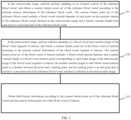
- the primary registration step S3 may specifically include the following steps: Step S31: In the extravascular image, perform uniform sampling on an external contour of the reference blood vessel, and obtain a contour feature point set of the reference blood vessel according to the external contour information of the reference blood vessel.
- the contour feature point set of the reference blood vessel includes a blood vessel external diameter of each point on the external contour of the reference blood vessel obtained in the extravascular image and a lateral vascular length from each point to a proximal end of the reference blood vessel.
- a process of obtaining the contour feature point set of the reference blood vessel may include: performing uniform sampling on an external contour curve of the reference blood vessel in the extravascular image, to discretize the external contour of the reference blood vessel into various points; constructing external contour feature points for the points by taking the lateral vascular length of each point from a proximal end of the reference blood vessel as the first-dimensional feature, and taking the blood vessel external diameter of this point as the second-dimensional feature; and obtaining the reference blood vessel contour feature point set according to all the external contour feature points.
- uniform sampling may be performed on the external image by using each pixel in the external image as a sampling point, to obtain a blood vessel external diameter and a lateral vascular length corresponding to each pixel on the external contour curve of the blood vessel.
- uniform sampling may be performed on the external image at an interval of one or two or more pixels. This is not limited herein.
- the coronary blood vessel in the external image can be segmented by combining a prediction result obtained by a traditional segmentation algorithm or a deep learning algorithm or deep learning with prior knowledge obtained by designing based on a conventional method, and information such as the lateral vascular length and the blood vessel external diameter is obtained through calculation by using a segmentation result.
- the coronary blood vessel is segmented by using the method of deep learning model to obtain information such as the length and the diameter.
- Step S32 In the intravascular image, perform uniform sampling on a blood vessel intravascular image of the blood vessel segment of interest, and obtain a contour feature point set of the blood vessel of interest according to the internal contour information of the blood vessel segment of interest.
- the contour feature point set of the blood vessel of interest includes: a blood vessel internal diameter and a medial vascular length at a blood vessel position point corresponding to each frame image in the intravascular image of the blood vessel segment of interest.
- the intravascular image is a sequence image, and one frame image is corresponding to a blood vessel cross-sectional image at a certain position inside the blood vessel, that is, the intravascular image includes blood vessel cross-sectional images at various positions inside the blood vessel.
- a shooting frequency of an OCT/IVUS intravascular image is 100 frame/s or 180 frame/s
- a pullback speed of a catheter is 18 mm/s or 20 mm/s or 36 mm/s
- an internal structure of the blood vessel is photographed by pulling back the catheter inside the blood vessel.
- the blood vessel internal diameter and the medial vascular length of each position point inside the blood vessel corresponding to each frame intravascular image may be obtained by using each frame intravascular image can be used as a sampling point.
- the process of obtaining the contour feature point set of the blood vessel of interest may include the following steps: Detect the intravascular image by using each frame in the sequence image of the intravascular image as a detection point; construct an internal contour feature point of each frame image by taking a medial vascular length between a blood vessel position point corresponding to each frame image and a starting point as the first-dimensional feature, and taking a blood vessel internal diameter of the blood vessel position point as the second-dimensional feature; and obtain the contour feature point set of the blood vessel of interest according to all the internal contour feature points.
- the contour feature point set of the blood vessel of interest can also be obtained by performing detection at an interval of one frame or performing detection at a fixed interval of frames. Specifically, uniform sampling is performed on the image sequence image of the intravascular image at an interval of one frame, two frames, and multiple frames.
- An internal contour feature point of each frame image is constructed by taking a medial vascular length between a blood vessel position point corresponding to each detection point image and a starting point as one-dimensional feature, and taking a blood vessel internal diameter of the blood vessel position point as the second-dimensional feature.
- a contour feature point set of the blood vessel of interest is obtained according to all the internal contour feature points.
- the blood vessel may be segmented by using the method mentioned in the Chinese patent with the patent number of CN201710438256.4 to obtain contour information such as the medial vascular length and the blood vessel internal diameter.
- contour feature point set of the reference blood vessel when a contour feature point set of the reference blood vessel is constructed, firstly, an external contour curve of the reference blood vessel is discretized, each point on the contour curve of the reference blood vessel is represented by a two-dimensional vector, the first-dimensional feature of the two-dimensional vector is a lateral vascular length at the point, the second-dimensional feature of the two-dimensional vector is a blood vessel external diameter at the point, and then a set of two-dimensional vectors corresponding to the points is used as a contour feature point set of the reference blood vessel.
- the construction process of the contour feature point set of the blood vessel of interest is generally similar to that of the contour feature point set of the reference blood vessel.
- each frame image is directly used as a sampling point
- blood vessel position information corresponding to each frame image may also be represented by a two-dimensional vector
- the first-dimensional feature of the two-dimensional vector may be a medial vascular length at a blood vessel point corresponding to the corresponding frame image
- the second-dimensional feature may be a blood vessel internal diameter at this point
- a set of two-dimensional vectors corresponding to frame images is used as a contour feature point set of the blood vessel of interest.
- each point in the contour feature point set of the reference blood vessel and the contour feature point set of the blood vessel of interest may be represented not only by a two-dimensional vector, but also may be represented by a three-dimensional vector or a multi-dimensional vector.
- each-dimensional feature in the vector may be feature information such as respectively a length, a diameter, a blood vessel cross-sectional area, a branch blood vessel diameter, a branch opening direction.
- Step S33 Obtain a third feature information according to the contour feature point set of the reference blood vessel and the contour feature point set of the blood vessel of interest.
- primary registration may be performed on the contour feature point set of the reference blood vessel and the contour feature point set of the blood vessel of interest based on a sliding window fast target detection method to obtain the third feature information.
- the primary registration process may include the following steps: Step S331: Construct a reference region according to the contour feature point set of the reference blood vessel, and construct a sliding window according to the contour feature point set of the blood vessel of interest.
- a starting point of the blood vessel segment of interest in the intravascular image (that is, an endpoint near a proximal endpoint of the reference blood vessel) may be used as a left boundary of the sliding window
- an ending point of the blood vessel segment of interest in the intravascular image (that is, an endpoint near a distal endpoint of the reference blood vessel) can be used as a right boundary of the sliding window.
- the boundaries of a sliding region are respectively the proximal endpoint and distal endpoint of the reference blood vessel.
- Step S332 Make the sliding window slide in the reference region, calculate and obtain a matching degree between each region in the reference region and the sliding window based on the contour feature point set of the reference blood vessel, the contour feature point set of the blood vessel of interest, the external branch information of the reference blood vessel, and the internal branch information of the blood vessel segment of interest.
- the external branch information of the reference blood vessel may include a serial number of each branch blood vessel of the reference blood vessel obtained in the extravascular image and a branch blood external length from a bifurcation point of each branch blood vessel to the proximal end of the reference blood vessel;
- the internal branch information of the blood vessel segment of interest includes a serial number of each branch blood vessel of the blood vessel segment of interest obtained in the intravascular image and a branch blood internal length of each branch blood vessel from the starting point. Because the feature points in the contour feature point set of the reference blood vessel and the contour feature point set of the blood vessel of interest are arranged in a sequence, serial numbers of branch blood vessels are also automatically sorted according to positions of the branches on the blood vessel contour line.
- the left boundary of the sliding window can coincide with the proximal endpoint of the reference region, and then the sliding window is gradually slid from the proximal endpoint of the reference region to the distal endpoint of the reference region.
- the right boundary of the sliding window may coincide with the distal endpoint of the reference region, and then the sliding window is gradually slid from the distal endpoint of the reference region to the proximal endpoint of the reference region.
- the sliding direction is not limited herein.
- a matching degree between the sliding window and a sub-region corresponding to the current sliding window in the reference region is calculated once, that is, during the sliding process, the total times of calculations of the matching degree is equal to a difference between the total number of points in the contour feature point set of the reference blood vessel and the total number of points in the contour feature point set of the blood vessel of interest.
- the matching degree between the sliding window and the sub-region corresponding to the current sliding window in the reference region can be calculated once each time the sliding window moves by two or more points in the reference region. That is, in the present invention, it is only necessary to ensure that primary registration of the first feature information and the second feature information can be implemented by using the calculated matching degrees, and there is no requirement for the quantity of matching degrees to be used.
- a matching degree between the coronary artery reference blood vessel segment in the external image of the current reference region and the coronary artery blood vessel segment of interest in the OCT/IVUS intravascular image may be calculated by using a target evaluation function, and the matching degree can be calculated based on the diameter information and branch information.
- calculation of the matching degree is performed when each point in the feature point set is a two-dimensional vector.
- the matching degree may be further calculated based on the diameter information, branch information and other information included in the vector. This is not limited herein.
- a distance between the points in the sliding window can be set to be equal to a distance between the points in the reference region. Therefore, in the present invention, prior to the reference region and the sliding window are constructed, the following step may be further included: Perform uniform resampling on the external contour of the reference blood vessel in the extravascular image according to the distance between any two adjacent points in the contour feature point set of the blood vessel of interest.
- a distance between two adjacent points in the contour feature point set of the blood vessel of interest can be calculated according to a pullback speed of a catheter and a shooting frequency of OCT/IVUS, and then the distance is taken as a sampling step, and uniform re-sampling is performed on the external contour of the reference blood vessel in the extravascular image.
- a distance between two adjacent points in the contour feature point set of the blood vessel of interest is equal to a product of the catheter pullback speed and the time taken to capture a frame of intravascular image, and the time taken to capture a frame of intravascular image is equal to the reciprocal of the shooting frequency, so that the distance is equal to the catheter pullback speed divided by the shooting frequency.
- the method of calculating the matching degree between each region in the reference region and the sliding window includes the following steps: A diameter matching score is calculated according to the blood vessel external diameter in the contour feature point set of the reference blood vessel and the blood vessel internal diameter in the contour feature point set of the blood vessel of interest.
- S d indicates a diameter matching score
- N indicates the total number of feature points in the sliding window, that is, the total number of points in the contour feature point set of the blood vessel of interest
- d si indicates a blood vessel internal diameter corresponding to the i th feature point in the sliding window
- d ti indicates a blood vessel external diameter corresponding to the i th feature point in the reference region.
- d ti is a blood vessel external diameter corresponding to the i th feature point in a sub-reference region in which the current sliding window is located in the reference region. That is, the i th feature point of the reference region mentioned in the formula (1) is actually the i th feature point in the sub-reference region in which the current sliding window is located in the reference region, and changes during movement of the sliding window.
- a branch matching score is calculated based on the external branch information of the reference blood vessel and the internal branch information of the blood vessel segment of interest.
- S b indicates a branch matching score
- M indicates the total number of branches in the sliding window, that is, the total number of all branch blood vessels in the blood vessel segment of interest
- dist indicates a distance between the i th branch blood vessel in the sliding window and a branch blood vessel that is closest to the i th branch blood vessel in the reference region.
- the distance dist can be calculated by using the position of the branch at the blood vessel (that is, a length of the branch blood vessel from the starting point of the blood vessel).
- a branch blood vessel internal length vessel at a position of the i th branch of the sliding window is l1
- a branch blood vessel external length at a position of a branch that is closest to the i th branch and that is in the reference region is l2
- the distance dist i
- a matching function is constructed, and the matching degree is calculated by using the matching function.
- S indicates a matching function
- S d represents the diameter matching score
- S b indicates the branch matching score
- ⁇ indicates a weighting factor
- ⁇ can be set according to the importance of the diameter information and the branch information or the obtained precision of the two. For example, when the diameter information and branch information are considered equally important, ⁇ can be set to 0.5. If detection precision or an accuracy rate of one of the two items is low, reducing the weighting factor of this item may be considered.
- ⁇ can be regarded as a weighting factor of diameter information
- 1- ⁇ can be regarded as a weighting factor of the branch information.
- ⁇ is set to be 0.3-0.7.
- Step S333 Compare the matching degrees to obtain third feature information.
- the matching degree between the sliding window and each sub-region in the reference region is calculated according to the formula (3)
- the target blood vessel segment is a blood vessel segment corresponding to the sub-region having the highest matching degree of the sliding window in the reference region
- the third feature information is the contour feature point set of the target blood vessel corresponding to the target blood vessel segment.
- the primary registration step is to perform rough registration on the external image and the OCT/IVUS intravascular image by combining the diameter information and the branch information and by using the sliding window fast target detection method.
- the calculation process is simple, a problem of manually locating the starting point and the ending point of the blood vessel of the corresponding OCT/IVUS image in the external coronary angiography image is solved, so that manual operation steps are reduced, and the degree of automation of the algorithm is improved.
- the primary registration step S3 may further include: establishing a contour feature point set of the target blood vessel, and specifics are as follows: a blood vessel external diameter and a lateral vascular length corresponding to each point on an external contour curve of the target blood vessel segment in the extravascular image are determined according to the third feature information, and the external branch information of the target blood vessel segment is determined.
- An external contour feature point corresponding to each point on the target blood vessel segment is constructed by taking the lateral vascular length as the first-dimensional feature, and taking the blood vessel external diameter as the second-dimensional feature.
- the contour feature point set of the target blood vessel is obtained according to all the external contour feature points.
- the secondary registration step S4 may perform iterative computing on the contour feature point set of the blood vessel of interest and the contour feature point set of the target blood vessel by using a registration algorithm that fuses the diameter information and the branch information to minimize a difference between the two contour feature point sets, so as to achieve a purpose of precise registration.
- the target registration result can be selected as a corresponding registration relationship between the first feature information and the third feature information.
- the corresponding registration relationship can be calculated based on the contour feature point set of the target blood vessel and the contour feature point set of the blood vessel of interest.
- the feature point set is calculated, that is, the corresponding registration relationship between the first feature information and the third feature information may specifically be the target registration relationship between the contour feature point set of the target blood vessel and the contour feature point set of the blood vessel of interest.
- the contour feature point set of the blood vessel of interest in the OCT/IVUS intravascular image is set to be S
- the contour feature point set of the target blood vessel in the external angiography image is set to be T
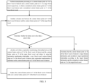
- the secondary registration step S4 may include the following steps:
- the preset value is set to be relatively small, no matter how the above iteration process is performed, the calculated feature error is always more than or equal to the preset value, which may fall into an infinite loop, thereby affecting registration efficiency.
- the applicant finds that in the iteration process, the feature error between the updated contour feature point set of the blood vessel of interest and the contour feature point set of the target blood vessel gradually decreases with the increase of the number of iteration times. Therefore, in one embodiment of the present invention, the occurrence of the infinite loop in the iteration process can be avoided by setting the maximum number of iteration times. That is, even if in the iteration process, the feature error is not less than the preset value, when the final cumulative number of iteration times reaches a certain value, it can be considered that the purpose of fine registration has been achieved at this time.
- a determining condition can be added between step c) and step d), and the determining condition can include: When the feature error is more than or equal to the preset value, it is determined whether the number of iteration times is more than a preset number, and if yes, it is considered that registration is completed, and the normalized contour feature point set S' of the blood vessel of interest is determined as the target feature information, and the target registration relationship is calculated according to the target feature information and the normalized contour feature point set T' of the target blood vessel; otherwise, the number of iteration times is increased by one, and the following step is performed.
- the preset number may be set according to an actual situation, for example, may be set to 100 times.
- the number of iteration times may be set to zero prior to step b).
- an iterative closest point algorithm or a coherent point drift algorithm or a robust point matching algorithm can be used to perform secondary registration on the first feature information and the third feature information to obtain a target registration result.
- a non-rigid registration algorithm that fuses diameter information and branch information is adopted for the secondary registration step.
- the feature difference between the two point sets is minimized, mapping parameters between the two point sets are obtained, and the corresponding relationship between the two point sets is found, so that the registration is implemented.
- the diameter and the branch information can directly reflect contour topological features of the vascular tissue, and this method has higher stability than the method in which registration is performed by positioning of an IVUS sensor tip.
- the registration method of the extravascular image and the intravascular image provided by the present invention, accurate registration of multimodal blood vessel images is implemented by multi-level registration (coarse registration and fine registration) based on the contour information of the external coronary image and the OCT/IVUS intravascular image.
- multi-level registration coarse registration and fine registration
- the registration process does not require three-dimensional reconstruction of the extravascular image, so that complexity and time-consuming of the registration algorithm are reduced.
- the present invention further provides an apparatus for registering an extravascular image with an intravascular image, including a first obtaining module 1, a second obtaining module 2, a primary registration module 3, and a secondary registration module 4.
- the first obtaining module 1 is configured to obtain a first feature information of a blood vessel segment of interest in the intravascular image, and the first feature information includes internal contour information and internal branch information of the blood vessel segment of interest.
- the second acquisition module 2 is configured to obtain a second feature information of a reference blood vessel in the extravascular image, the reference blood vessel is an entire blood vessel in which the blood vessel segment of interest is located, and the second feature information includes external contour information and external branch information of the reference blood vessel.
- the primary registration module 3 is configured to perform a primary registration on the first feature information and the second feature information to obtain a third feature information
- the third feature information includes external contour information and external branch information of the target blood vessel segment corresponding to the blood vessel segment of interest in the extravascular image.
- the secondary registration module 4 is configured to perform a secondary registration on the first feature information and the third feature information to obtain a target registration result.
- the target registration result may be target feature information that a feature error between the target feature information and the third feature information meets a preset condition.
- the target registration result may be a target registration relationship between the first feature information and the third feature information.
- the target registration result may include not only the target feature information, but also include the target registration relationship between the first feature information and the third feature information.
- the primary registration module 3 may include:
- the first sampling unit 31 may perform uniform sampling on the external image by using each pixel in the external image as a sampling point, to obtain a blood vessel external diameter and a lateral vascular length corresponding to each pixel on the external contour curve of the blood vessel.
- the intravascular image is an OCT image or an IVUS image.
- a shooting frequency of an OCT/IVUS intravascular image is 100 frame/s or 180 frame/s
- a pullback speed of a catheter is 18 mm/s or 20 mm/s or 36 mm/s
- an internal structure of the blood vessel is photographed by pulling back the catheter inside the blood vessel.
- the second sampling unit 32 may obtain the blood vessel internal diameter and the medial vascular length of each position point inside the blood vessel corresponding to each frame intravascular image by using each frame intravascular image as a sampling point.
- the obtaining unit 33 may specifically include: a window construction unit 331, configured to construct a reference region according to the contour feature point set of the reference blood vessel, and construct a sliding window according to the contour feature point set of the blood vessel of interest; a matching degree calculation unit 332, configured to make the sliding window slide in the reference region, and calculate a matching degree between each region in the reference region and the sliding window according to the contour feature point set of the reference blood vessel, the contour feature point set of the blood vessel of interest, the external branch information of the reference blood vessel and the internal branch information of the blood vessel segment of interest; and a comparison unit 333, configure to compare the matching degrees to obtain the third feature information.
- a window construction unit 331 configured to construct a reference region according to the contour feature point set of the reference blood vessel, and construct a sliding window according to the contour feature point set of the blood vessel of interest
- a matching degree calculation unit 332 configured to make the sliding window slide in the reference region, and calculate a matching degree between each region in the reference region and the sliding window according to the contour feature point
- the external branch information of the reference blood vessel includes a serial number of each branch blood vessel of the reference blood vessel obtained in the extravascular image and a branch blood vessel external length from a bifurcation point of each branch blood vessel to the proximal end of the reference blood vessel;
- the internal branch information of the blood vessel segment of interest includes a serial number of each branch blood vessel of the blood vessel segment of interest obtained in the intravascular image and a branch blood vessel internal length of each branch blood vessel from the starting point.
- the method for calculating the matching degree between each region in the reference region and the sliding window by the matching degree calculation unit includes: according to the blood vessel external diameter in the contour feature point set of the reference blood vessel and the blood vessel internal diameter in the contour feature point set of the blood vessel of interest, calculating a diameter matching score; calculating a branch matching score according to the external branch information of the reference blood vessel and the internal branch information of the blood vessel segment of interest; building a matching function according to the diameter matching score and the branch matching score, and calculating the matching degree by using the matching function.
- S d indicates a diameter matching score
- N indicates the total number of feature points in the sliding window
- d si indicates a blood vessel internal diameter corresponding to the i th feature point in the sliding window
- d ti indicates a blood vessel external diameter corresponding to the i th feature point in the reference region.
- d ti is a blood vessel external diameter corresponding to the i th feature point in a sub-reference region in which the current sliding window is located in the reference region. That is, the i th feature point of the reference region mentioned in the above formula is actually the i th feature point in the sub-reference region in which the current sliding window is located in the reference region, and changes during the moving process of the sliding window.
- S b indicates a branch matching score
- M indicates the total number of branches in the sliding window
- dist indicates a distance between the i th branch blood vessel in the sliding window and a branch blood vessel closest to the i th branch blood vessel in the reference region.
- S indicates a matching function
- S d indicates a diameter matching score
- S b indicates a branch matching score
- ⁇ indicates a weighting factor
- ⁇ can be set according to the importance of the diameter information and the branch information or the obtained precision of the two. For example, when the diameter information and the branch information are considered equally important, ⁇ can be set to 0.5. If detection accuracy or an accuracy rate of one of the two items is low, reducing the weighting factor of this item may be considered.
- ⁇ can be regarded as the weighting factor of diameter information
- 1- ⁇ can be regarded as the weighting factor of branch information.
- ⁇ ranges from 0.3 to 0.7.
- the blood vessel segment corresponding to the region with the greatest matching degree with the sliding window in the reference region is the target blood vessel segment.
- the first sampling unit 31 includes: a first discretizing subunit, configured to perform uniform sampling on an external contour curve of the reference blood vessel in the extravascular image, and discretize the external contour of the reference blood vessel into various points; a first construction subunit, configured to construct external contour feature points for the points by taking the lateral vascular length from each point to the proximal end of the reference blood vessel as the first-dimensional feature, and taking the blood vessel external diameter of this point as the second-dimensional feature; and obtain the contour feature point set of the reference blood vessel according to all the external contour feature points.
- the second sampling unit 32 includes: a second frame division subunit, configured to detect the intravascular image by using each frame in the sequence image of the intravascular image as a detection point; and a second construction subunit, configured to construct an internal contour feature point of each frame image by taking the medial vascular length between the blood vessel position point and the starting point corresponding to each frame image as the first-dimensional feature, and taking the blood vessel internal diameter of the blood vessel position point as the second-dimensional feature; and obtain a contour feature point set of the blood vessel of interest according to all the internal contour feature points.
- the second frame division subunit can not only detect each frame of blood vessel intravascular image, so that the second construction subunit obtains the contour feature point set of the blood vessel of interest, but also can perform detection on the intravascular image at an interval of one frame or can perform detection at a fixed interval of frames, so that the second construction subunit obtains the contour feature point set of the blood vessel of interest.
- the first discretizing subunit discretizes the external contour curve of the reference blood vessel
- the first construction subunit uses a two-dimensional vector to refer to each point on the contour curve of the reference blood vessel
- the first-dimensional feature of the two-dimensional vector is the lateral vascular length at the point
- the second-dimensional feature of the two-dimensional vector is the blood vessel external diameter at the point.
- a set of two-dimensional vectors corresponding to the points is the contour feature point set of the reference blood vessel.
- the process of constructing the contour feature point set of the blood vessel of interest by the second sampling unit 32 is generally similar to the above construction process.
- the second frame division subunit directly uses each frame of image as a sampling point.
- the second construction subunit uses a two-dimensional vector to refer to the blood vessel position information corresponding to each frame image
- the first-dimensional feature of the two-dimensional vector can be the medial vascular length at the blood vessel point corresponding to the frame image
- the second-dimensional feature of the two-dimensional vector can be the blood vessel internal diameter at the point
- the second construction subunit takes a set of two-dimensional vectors corresponding to frame images as the contour feature point set of the blood vessel of interest .
- each point in the reference blood vessel contour feature point set and the blood vessel contour feature point set of interest can be represented not only by a two-dimensional vector, but also by a three-dimensional vector or a multi-dimensional vector; when the point is represented by the three-dimensional or multi-dimensional vector, the each-dimensional feature of the vector can be feature information such as respectively a length, a diameter, a blood vessel cross-sectional area, a branch blood vessel diameter, and a branch opening direction.
- the primary registration module 3 further includes: a distance calculation unit, configured to calculate a distance between two adjacent points in the contour feature point set of the blood vessel of interest before the window construction unit constructs the reference region and the sliding window.
- the first sampling unit 31 is further configured to: in the extravascular image, perform uniform re-sampling on the external contour of the reference blood vessel according to the distance.
- the distance calculation unit can calculate a distance between two adjacent points in the contour feature point set of the blood vessel of interest according to a pullback speed of a catheter and a shooting frequency of OCT/IVUS, and the distance is equal to a result of dividing the pullback speed of the catheter by the photographing frequency.
- the first sampling unit 31 uses the distance as a sampling step to perform uniform sampling on the external contour of the reference blood vessel in the extravascular image. In this way, the distance between the points in the sliding window is set to be equal to the distance between the points in the reference region, which makes it easier to calculate the matching degree between the sliding window in the sliding process and each sub-region in the corresponding region.
- the first sampling unit 31 is further configured to: according to the third feature information, determine a blood vessel external diameter and a lateral vascular length corresponding to each point on an external contour curve of the target blood vessel segment in the extravascular image, and determine external branch information of the target blood vessel segment; construct an external contour feature point corresponding to each point on the target blood vessel segment by taking the lateral vascular length as the first-dimensional feature, and taking the blood vessel external diameter as the second-dimensional feature; and obtain the contour feature point set of the target blood vessel according to all the external contour feature points.
- the target registration result can be selected as the corresponding registration relationship between the first feature information and the third feature information.
- the corresponding registration relationship can be calculated based on the contour feature point set of the target blood vessel and the contour feature point set of the blood vessel of interest, that is, the corresponding registration relationship between the first feature information and the third feature information may specifically be the target registration relationship between the contour feature point set of the target blood vessel and the contour feature point set of the blood vessel of interest.
- the secondary registration module 4 includes:
- the preset value can be set according to an actual situation, and can be set to 0.01 in general, that is, when the feature error is less than 0.01, it is considered that registration is completed.
- the secondary registration module 4 may further include:
- the registration relationship calculation unit 46 is further configured to: when the iteration times determining unit determines that the number of iteration times is more than the preset number, determine that the normalized contour feature point set S' of the blood vessel of interest is the target feature information, and calculate the target registration relationship based on the target feature information and the normalized contour feature point set T' of the target blood vessel.
- the preset number may be set according to an actual situation, for example, 100 times.
- the secondary registration module 4 further includes an initialization unit, configured to set the quantity of iteration times to zero before the error calculation unit calculates a feature error between the normalized contour feature point set S' of the blood vessel of interest and the normalized contour feature point set T' of the target blood vessel.
- an initialization unit configured to set the quantity of iteration times to zero before the error calculation unit calculates a feature error between the normalized contour feature point set S' of the blood vessel of interest and the normalized contour feature point set T' of the target blood vessel.
- the secondary registration module 4 performs secondary registration on the first feature information and the third feature information by using an iterative closest point algorithm or a coherent point drift algorithm or a robust point matching algorithm, to obtain the target registration result.
- the apparatus for registering an extravascular image with an intravascular image in the technical solution is adopted, so that automatic registration of the coronary angiography image and the intravascular image can be directly implemented by using the diameter and branch information of the blood vessel and by using the multi-level registration method.
- the apparatus needs to detect and segment only one frame of coronary angiography image, there is no need to perform manual labeling and three-dimensional reconstruction, and the apparatus has advantages of rapidity, simplicity, high accuracy, and high robustness.
- the present invention further provides a computing device, including: a processor, suitable to implement various instructions; a memory, suitable to store a plurality of instructions, where the instructions are suitable to be loaded by the processor and execute the method for registering an extravascular image with an intravascular image.
- a computing device including: a processor, suitable to implement various instructions; a memory, suitable to store a plurality of instructions, where the instructions are suitable to be loaded by the processor and execute the method for registering an extravascular image with an intravascular image.
- the computing device of the above technical solution is adopted, so that rapid and accurate registration of an extravascular image with an intravascular image can be implemented.
- An embodiment of the present invention further discloses a storage medium, the storage medium stores a plurality of instructions, and the instructions are suitable to be loaded by the processor and execute the method for registering an extravascular image with an intravascular image.
- the storage medium of the above technical solution is adopted, so that rapid and accurate registration of an extravascular image and an intravascular image can be implemented.
- the various embodiments disclosed herein may be implemented in hardware, software, firmware, or a combination of these implementation methods.
- the embodiments of the present application may be implemented as a computer program or program code executing on a programmable system including at least one processor, a storage system (including a volatile and nonvolatile memory and/or a storage element), at least one input device, and at least one output device.
- Program code may be applied to input instructions to perform the functions described herein and to generate output information.
- the output information can be applied to one or more output devices in a known manner.
- a processing system includes any system having a processor such as, for example, a digital signal processor (DSP), a microcontroller, an application specific integrated circuit (ASIC), or a microprocessor.
- DSP digital signal processor
- ASIC application specific integrated circuit
- the program code may be implemented in a high-level procedural language or an object-oriented programming language to communicate with the processing system.
- the program code may also be implemented in assembly or machine language when needed.
- the mechanism described in this application is not limited in the scope of any particular programming language. In either case, the language may be a compiled language or an interpreted language.
- the disclosed embodiments may be implemented in hardware, firmware, software, or any combination thereof.
- the disclosed embodiments can further be implemented as instructions carried by or stored on one or more transitory or non-transitory machine-readable (for example, computer-readable) storage media, which can be read and executed by one or more processors.
- the instructions may be distributed over a network or over another computer-readable medium.
- a machine-readable medium can include any mechanism for storing or transmitting information in a machine (for example, a computer) readable form, and includes, but is not limited to, a floppy disk, an optical disk, an optical disk, a compact disc read-only memory (CD-ROM), a magnetic optical disc, a read only memory (ROM), a random access memory (RAM), an erasable programmable read only memory (EPROM), an electrically erasable programmable read only memory (EEPROM), a magnetic card or an optical card, a flash memory, or a tangible machine-readable memory for transmitting information (for example, a carrier wave, an infrared signal, and a digital signal) using the Internet in electrical, optical, acoustic, or other forms of propagating signals. Therefore, the machine-readable medium includes any type of machine-readable medium suitable for storing or transmitting electronic instructions or information in a machine (for example, a computer) readable form.
- each module/unit mentioned in each device embodiment of this application is a logical module/unit.
- a logical module/unit may be a physical module/unit or may be a part of a physical module/unit, or may be implemented by a combination of multiple physical modules/units.
- the physical implementation of these logical modules/units is not the most important, and the combination of functions implemented by these logical modules/units is the key to solving the technical problems raised by the present application.
- the above device embodiments of the present application do not introduce modules/units that are not closely related to solving the technical problems raised in the present application, which does not mean that other modules/units do not exist in the device embodiments.
Landscapes
- Engineering & Computer Science (AREA)
- Physics & Mathematics (AREA)
- Health & Medical Sciences (AREA)
- Computer Vision & Pattern Recognition (AREA)
- Life Sciences & Earth Sciences (AREA)
- General Physics & Mathematics (AREA)
- Theoretical Computer Science (AREA)
- Medical Informatics (AREA)
- Surgery (AREA)
- Heart & Thoracic Surgery (AREA)
- Veterinary Medicine (AREA)
- Biophysics (AREA)
- High Energy & Nuclear Physics (AREA)
- Public Health (AREA)
- Nuclear Medicine, Radiotherapy & Molecular Imaging (AREA)
- Optics & Photonics (AREA)
- Pathology (AREA)
- Radiology & Medical Imaging (AREA)
- Biomedical Technology (AREA)
- General Health & Medical Sciences (AREA)
- Molecular Biology (AREA)
- Animal Behavior & Ethology (AREA)
- Geometry (AREA)
- Oral & Maxillofacial Surgery (AREA)
- Vascular Medicine (AREA)
- Dentistry (AREA)
- Physiology (AREA)
- Image Analysis (AREA)
- Ultra Sonic Daignosis Equipment (AREA)
- Image Processing (AREA)
Applications Claiming Priority (2)
| Application Number | Priority Date | Filing Date | Title |
|---|---|---|---|
| CN202011418428.XA CN112509020B (zh) | 2020-12-07 | 2020-12-07 | 血管外部影像与腔内影像的配准方法、装置及计算设备 |
| PCT/CN2021/127049 WO2022121546A1 (zh) | 2020-12-07 | 2021-10-28 | 血管外部影像与腔内影像的配准方法、装置及计算设备 |
Publications (2)
| Publication Number | Publication Date |
|---|---|
| EP4227902A1 true EP4227902A1 (de) | 2023-08-16 |
| EP4227902A4 EP4227902A4 (de) | 2024-04-17 |
Family
ID=74970929
Family Applications (1)
| Application Number | Title | Priority Date | Filing Date |
|---|---|---|---|
| EP21902253.0A Pending EP4227902A4 (de) | 2020-12-07 | 2021-10-28 | Verfahren, vorrichtung und computervorrichtung zur ausrichtung eines externen blutgefässbildes mit einem intraluminalen bild |
Country Status (5)
| Country | Link |
|---|---|
| US (1) | US20240013413A1 (de) |
| EP (1) | EP4227902A4 (de) |
| JP (1) | JP7610880B2 (de) |
| CN (1) | CN112509020B (de) |
| WO (1) | WO2022121546A1 (de) |
Cited By (1)
| Publication number | Priority date | Publication date | Assignee | Title |
|---|---|---|---|---|
| WO2025076024A1 (en) * | 2023-10-06 | 2025-04-10 | Boston Scientific Scimed, Inc. | Cross-modality vascular image side branch matching |
Families Citing this family (12)
| Publication number | Priority date | Publication date | Assignee | Title |
|---|---|---|---|---|
| CN112509020B (zh) * | 2020-12-07 | 2021-09-28 | 昆山戎影医疗科技有限公司 | 血管外部影像与腔内影像的配准方法、装置及计算设备 |
| CN113616160B (zh) * | 2021-09-14 | 2024-02-06 | 苏州博动戎影医疗科技有限公司 | 基于多模态医学影像的ffr确定方法、装置、设备及介质 |
| CN114155243B (zh) * | 2022-02-09 | 2022-07-05 | 天津恒宇医疗科技有限公司 | 基于特征信息的ivus和oct图像融合方法及系统 |
| CN114820600B (zh) * | 2022-06-27 | 2022-09-27 | 天津恒宇医疗科技有限公司 | 基于oct图像的冠脉血管内支架检测方法及检测系统 |
| CN115359100A (zh) * | 2022-07-13 | 2022-11-18 | 深圳市中科微光医疗器械技术有限公司 | 融合造影方法、腔内图像和造影图像的配准方法及装置 |
| CN115482261B (zh) * | 2022-09-21 | 2025-09-09 | 深圳睿心智能医疗科技有限公司 | 血管配准方法、装置、电子设备及存储介质 |
| CN115534304B (zh) * | 2022-09-29 | 2023-04-11 | 灰觋有限公司 | 一种fdm打印装置及打印制品质量的自动检测方法 |
| CN116152314B (zh) * | 2023-01-13 | 2026-03-24 | 杭州朗博康医疗科技有限公司 | 一种血管介入手术的图像配准方法、系统及存储介质 |
| CN116919433B (zh) * | 2023-09-19 | 2024-01-09 | 北京唯迈医疗设备有限公司 | 一种融合dsa影像的图像分析方法和系统 |
| CN118015053B (zh) * | 2024-04-08 | 2024-06-11 | 重庆医科大学绍兴柯桥医学检验技术研究中心 | 一种多模态医学影像配准处理方法及系统 |
| CN119417868B (zh) * | 2024-09-13 | 2025-12-09 | 中南大学 | 一种冶金熔体射流多模态图像的时空配准方法及系统 |
| CN119091239B (zh) * | 2024-11-11 | 2025-01-24 | 达州市中心医院(达州市人民医院) | 甲襞微循环成像控制及图像识别分析方法和系统 |
Family Cites Families (21)
| Publication number | Priority date | Publication date | Assignee | Title |
|---|---|---|---|---|
| CA2427014A1 (en) | 2000-11-02 | 2002-08-22 | Cornell Research Foundation, Inc. | In vivo multiphoton diagnostic detection and imaging of a neurodegenerative disease |
| WO2007002685A2 (en) * | 2005-06-24 | 2007-01-04 | Volcano Corporation | Co-registration of graphical image data representing three-dimensional vascular features |
| WO2010017356A2 (en) * | 2008-08-08 | 2010-02-11 | University Of Pittsburgh - Of The Commonwealth System Of Higher Education | Establishing compatibility between two-and three dimensional optical coherence tomography scans |
| US8565859B2 (en) | 2010-06-29 | 2013-10-22 | Siemens Aktiengesellschaft | Method and system for image based device tracking for co-registration of angiography and intravascular ultrasound images |
| WO2012071110A1 (en) * | 2010-11-24 | 2012-05-31 | Boston Scientific Scimed, Inc. | Systems and methods for detecting and displaying body lumen bifurcations |
| AU2013387679B2 (en) * | 2013-03-12 | 2018-07-05 | Lightlab Imaging, Inc. | Vascular data processing and image registration systems, methods, and apparatuses |
| JP5977214B2 (ja) | 2013-08-23 | 2016-08-24 | ジーイー・メディカル・システムズ・グローバル・テクノロジー・カンパニー・エルエルシー | 画像処理方法および装置並びにプログラム |
| CN103810709B (zh) * | 2014-02-25 | 2016-08-17 | 南京理工大学 | 基于血管的眼底图像与sd-oct投影图像配准方法 |
| KR102250086B1 (ko) * | 2014-05-16 | 2021-05-10 | 삼성전자주식회사 | 의료 영상 정합 방법, 이를 포함하는 장치 및 컴퓨터 기록 매체 |
| US9934566B2 (en) | 2015-07-14 | 2018-04-03 | Siemens Healthcare Gmbh | 3-D vessel tree surface reconstruction method |
| CN105761254B (zh) * | 2016-02-04 | 2019-01-01 | 浙江工商大学 | 基于图像特征的眼底图像配准方法 |
| CN107123137B (zh) * | 2017-04-28 | 2020-05-22 | 上海联影医疗科技有限公司 | 医学图像处理方法及设备 |
| CN107133959B (zh) * | 2017-06-12 | 2020-04-28 | 上海交通大学 | 一种快速的血管边界三维分割方法及系统 |
| CN108198211A (zh) * | 2017-11-20 | 2018-06-22 | 海纳医信(北京)软件科技有限责任公司 | 眼底图像的处理方法及装置、存储介质、处理器 |
| CN108038848B (zh) * | 2017-12-07 | 2020-08-11 | 上海交通大学 | 基于医学影像序列斑块稳定性指标的快速计算方法及系统 |
| CN108198174B (zh) * | 2017-12-28 | 2021-11-19 | 中科微光医疗研究中心(西安)有限公司 | 一种心血管ivoct与ivus自动配准方法与装置 |
| CN108053429B (zh) * | 2017-12-28 | 2021-11-02 | 中科微光医疗研究中心(西安)有限公司 | 一种心血管oct与冠脉造影自动配准方法与装置 |
| US12076177B2 (en) * | 2019-01-30 | 2024-09-03 | Canon U.S.A., Inc. | Apparatuses, systems, methods and storage mediums for performance of co-registration |
| CN110288637B (zh) * | 2019-06-13 | 2021-09-21 | 北京理工大学 | 多角度dsa造影图像血管匹配方法及装置 |
| CN111091567B (zh) * | 2020-03-23 | 2020-06-23 | 南京景三医疗科技有限公司 | 医学图像配准方法、医疗设备及存储介质 |
| CN112509020B (zh) * | 2020-12-07 | 2021-09-28 | 昆山戎影医疗科技有限公司 | 血管外部影像与腔内影像的配准方法、装置及计算设备 |
-
2020
- 2020-12-07 CN CN202011418428.XA patent/CN112509020B/zh active Active
-
2021
- 2021-10-28 US US18/038,640 patent/US20240013413A1/en active Pending
- 2021-10-28 JP JP2023528692A patent/JP7610880B2/ja active Active
- 2021-10-28 WO PCT/CN2021/127049 patent/WO2022121546A1/zh not_active Ceased
- 2021-10-28 EP EP21902253.0A patent/EP4227902A4/de active Pending
Cited By (1)
| Publication number | Priority date | Publication date | Assignee | Title |
|---|---|---|---|---|
| WO2025076024A1 (en) * | 2023-10-06 | 2025-04-10 | Boston Scientific Scimed, Inc. | Cross-modality vascular image side branch matching |
Also Published As
| Publication number | Publication date |
|---|---|
| US20240013413A1 (en) | 2024-01-11 |
| JP7610880B2 (ja) | 2025-01-09 |
| WO2022121546A1 (zh) | 2022-06-16 |
| EP4227902A4 (de) | 2024-04-17 |
| JP2023550068A (ja) | 2023-11-30 |
| CN112509020B (zh) | 2021-09-28 |
| CN112509020A (zh) | 2021-03-16 |
Similar Documents
| Publication | Publication Date | Title |
|---|---|---|
| EP4227902A1 (de) | Verfahren, vorrichtung und computervorrichtung zur ausrichtung eines externen blutgefässbildes mit einem intraluminalen bild | |
| JP7743376B2 (ja) | リアルタイムで診断上有用な結果 | |
| US11969287B2 (en) | Artificial intelligence ultrasound-medical-diagnosis apparatus using semantic segmentation and remote medical-diagnosis method using the same | |
| EP3659114B1 (de) | Auswertung der herzbewegung unter verwendung eines angiographiebildes | |
| EP2434454B1 (de) | Computerisierte Kennzeichnung der Herzbewegung in einem medizinisch diagnostischen Ultraschall | |
| US8787635B2 (en) | Optimization of multiple candidates in medical device or feature tracking | |
| EP3624056B1 (de) | Verarbeitung von bildrahmen einer sequenz von herzbildern | |
| US20230230231A1 (en) | Training method and apparatus for angiography image processing, and automatic processing method and apparatus | |
| US10685438B2 (en) | Automated measurement based on deep learning | |
| US8565859B2 (en) | Method and system for image based device tracking for co-registration of angiography and intravascular ultrasound images | |
| JP6243453B2 (ja) | 血管内画像におけるマルチモーダルセグメンテーション | |
| US8605976B2 (en) | System and method of detection of optimal angiography frames for quantitative coronary analysis using wavelet-based motion analysis | |
| JP4981938B2 (ja) | 診断支援装置、冠動脈解析プログラムおよび冠動脈解析方法 | |
| CN104519793A (zh) | 用于内腔的线性映射的系统 | |
| US8175684B2 (en) | Method for processing images and associated medical imaging system | |
| CN115359100A (zh) | 融合造影方法、腔内图像和造影图像的配准方法及装置 | |
| CN108053429A (zh) | 一种心血管oct与冠脉造影自动配准方法与装置 | |
| CN108335732A (zh) | 一种oct影像的病例推荐方法及其系统 | |
| KR20200139491A (ko) | 의료영상 처리 방법 | |
| EP4713875A2 (de) | System, verfahren und computerzugängliches medium zur bereitstellung einer plaquebelastungsbeurteilung | |
| van Herten et al. | The role of artificial intelligence in coronary CT angiography | |
| CN114119688A (zh) | 基于深度学习的冠状动脉造影前后单模医学图像配准方法 | |
| US20230306620A1 (en) | Live Angiography Registration | |
| CN117649350B (zh) | 一种血管内图像和造影图像的融合方法、装置和设备 | |
| WO2026007315A1 (zh) | 一种冠状动脉夹层的自动检测方法和系统 |
Legal Events
| Date | Code | Title | Description |
|---|---|---|---|
| STAA | Information on the status of an ep patent application or granted ep patent |
Free format text: STATUS: THE INTERNATIONAL PUBLICATION HAS BEEN MADE |
|
| PUAI | Public reference made under article 153(3) epc to a published international application that has entered the european phase |
Free format text: ORIGINAL CODE: 0009012 |
|
| STAA | Information on the status of an ep patent application or granted ep patent |
Free format text: STATUS: REQUEST FOR EXAMINATION WAS MADE |
|
| 17P | Request for examination filed |
Effective date: 20230510 |
|
| AK | Designated contracting states |
Kind code of ref document: A1 Designated state(s): AL AT BE BG CH CY CZ DE DK EE ES FI FR GB GR HR HU IE IS IT LI LT LU LV MC MK MT NL NO PL PT RO RS SE SI SK SM TR |
|
| DAV | Request for validation of the european patent (deleted) | ||
| DAX | Request for extension of the european patent (deleted) | ||
| A4 | Supplementary search report drawn up and despatched |
Effective date: 20240320 |
|
| RIC1 | Information provided on ipc code assigned before grant |
Ipc: A61B 6/50 20240101ALI20240314BHEP Ipc: G06T 7/33 20170101AFI20240314BHEP |