EP4219556A2 - Human monoclonal antibodies specific for flt3 and uses thereof - Google Patents
Human monoclonal antibodies specific for flt3 and uses thereof Download PDFInfo
- Publication number
- EP4219556A2 EP4219556A2 EP22209293.4A EP22209293A EP4219556A2 EP 4219556 A2 EP4219556 A2 EP 4219556A2 EP 22209293 A EP22209293 A EP 22209293A EP 4219556 A2 EP4219556 A2 EP 4219556A2
- Authority
- EP
- European Patent Office
- Prior art keywords
- antibody
- flt3
- antigen
- binding fragment
- monoclonal antibody
- Prior art date
- Legal status (The legal status is an assumption and is not a legal conclusion. Google has not performed a legal analysis and makes no representation as to the accuracy of the status listed.)
- Pending
Links
- 101100335081 Mus musculus Flt3 gene Proteins 0.000 title 1
- 108010003374 fms-Like Tyrosine Kinase 3 Proteins 0.000 claims abstract description 237
- 102000004632 fms-Like Tyrosine Kinase 3 Human genes 0.000 claims abstract description 237
- 108010019670 Chimeric Antigen Receptors Proteins 0.000 claims abstract description 127
- 206010028980 Neoplasm Diseases 0.000 claims abstract description 106
- 201000011510 cancer Diseases 0.000 claims abstract description 69
- 238000000034 method Methods 0.000 claims abstract description 66
- 229940127121 immunoconjugate Drugs 0.000 claims abstract description 45
- 208000024893 Acute lymphoblastic leukemia Diseases 0.000 claims abstract description 43
- 208000031261 Acute myeloid leukaemia Diseases 0.000 claims abstract description 43
- 230000027455 binding Effects 0.000 claims description 176
- 210000004027 cell Anatomy 0.000 claims description 158
- 239000000427 antigen Substances 0.000 claims description 141
- 108091007433 antigens Proteins 0.000 claims description 141
- 102000036639 antigens Human genes 0.000 claims description 141
- 239000012634 fragment Substances 0.000 claims description 131
- 125000003275 alpha amino acid group Chemical group 0.000 claims description 78
- 239000003814 drug Substances 0.000 claims description 70
- 239000000611 antibody drug conjugate Substances 0.000 claims description 59
- 229940049595 antibody-drug conjugate Drugs 0.000 claims description 59
- 239000002105 nanoparticle Substances 0.000 claims description 59
- 229940079593 drug Drugs 0.000 claims description 54
- 108090000765 processed proteins & peptides Proteins 0.000 claims description 53
- 150000007523 nucleic acids Chemical class 0.000 claims description 50
- 102000004169 proteins and genes Human genes 0.000 claims description 47
- 108090000623 proteins and genes Proteins 0.000 claims description 46
- 239000012636 effector Substances 0.000 claims description 44
- 239000000203 mixture Substances 0.000 claims description 43
- 102000039446 nucleic acids Human genes 0.000 claims description 42
- 108020004707 nucleic acids Proteins 0.000 claims description 42
- 101000762949 Pseudomonas aeruginosa (strain ATCC 15692 / DSM 22644 / CIP 104116 / JCM 14847 / LMG 12228 / 1C / PRS 101 / PAO1) Exotoxin A Proteins 0.000 claims description 40
- 239000000562 conjugate Substances 0.000 claims description 32
- 208000032839 leukemia Diseases 0.000 claims description 31
- 229940127089 cytotoxic agent Drugs 0.000 claims description 28
- 210000001151 cytotoxic T lymphocyte Anatomy 0.000 claims description 27
- 108010047041 Complementarity Determining Regions Proteins 0.000 claims description 24
- 231100000599 cytotoxic agent Toxicity 0.000 claims description 24
- 239000002502 liposome Substances 0.000 claims description 24
- 230000011664 signaling Effects 0.000 claims description 24
- 239000002254 cytotoxic agent Substances 0.000 claims description 21
- 108020001507 fusion proteins Proteins 0.000 claims description 20
- 102000037865 fusion proteins Human genes 0.000 claims description 20
- 239000003053 toxin Substances 0.000 claims description 20
- 231100000765 toxin Toxicity 0.000 claims description 20
- 108700012359 toxins Proteins 0.000 claims description 20
- 102000004190 Enzymes Human genes 0.000 claims description 17
- 108090000790 Enzymes Proteins 0.000 claims description 17
- 101000851370 Homo sapiens Tumor necrosis factor receptor superfamily member 9 Proteins 0.000 claims description 12
- 102100036856 Tumor necrosis factor receptor superfamily member 9 Human genes 0.000 claims description 12
- 210000004369 blood Anatomy 0.000 claims description 12
- 239000008280 blood Substances 0.000 claims description 12
- 239000013598 vector Substances 0.000 claims description 11
- 229940044684 anti-microtubule agent Drugs 0.000 claims description 10
- 239000000693 micelle Substances 0.000 claims description 10
- 206010027476 Metastases Diseases 0.000 claims description 9
- 108091008874 T cell receptors Proteins 0.000 claims description 9
- 102000016266 T-Cell Antigen Receptors Human genes 0.000 claims description 9
- 238000001574 biopsy Methods 0.000 claims description 9
- 230000000139 costimulatory effect Effects 0.000 claims description 9
- 239000002088 nanocapsule Substances 0.000 claims description 9
- 239000002077 nanosphere Substances 0.000 claims description 9
- 239000000412 dendrimer Substances 0.000 claims description 8
- 229920000736 dendritic polymer Polymers 0.000 claims description 8
- 230000002401 inhibitory effect Effects 0.000 claims description 8
- 230000009401 metastasis Effects 0.000 claims description 8
- 102000005962 receptors Human genes 0.000 claims description 8
- 108020003175 receptors Proteins 0.000 claims description 8
- 239000003937 drug carrier Substances 0.000 claims description 7
- 239000003080 antimitotic agent Substances 0.000 claims description 5
- 210000001185 bone marrow Anatomy 0.000 claims description 5
- 150000003384 small molecules Chemical class 0.000 claims description 5
- BWGNESOTFCXPMA-UHFFFAOYSA-N Dihydrogen disulfide Chemical compound SS BWGNESOTFCXPMA-UHFFFAOYSA-N 0.000 claims description 4
- 108010054477 Immunoglobulin Fab Fragments Proteins 0.000 claims description 3
- 102000001706 Immunoglobulin Fab Fragments Human genes 0.000 claims description 3
- 230000003213 activating effect Effects 0.000 claims description 3
- 238000011282 treatment Methods 0.000 abstract description 14
- 238000003745 diagnosis Methods 0.000 abstract description 9
- 210000001744 T-lymphocyte Anatomy 0.000 description 52
- 239000000523 sample Substances 0.000 description 47
- 235000018102 proteins Nutrition 0.000 description 44
- 208000014697 Acute lymphocytic leukaemia Diseases 0.000 description 34
- 208000033776 Myeloid Acute Leukemia Diseases 0.000 description 34
- 208000006664 Precursor Cell Lymphoblastic Leukemia-Lymphoma Diseases 0.000 description 34
- -1 radioisotopes Chemical class 0.000 description 31
- 235000001014 amino acid Nutrition 0.000 description 30
- 239000004698 Polyethylene Substances 0.000 description 27
- 229940024606 amino acid Drugs 0.000 description 26
- 150000001413 amino acids Chemical class 0.000 description 26
- 102000004196 processed proteins & peptides Human genes 0.000 description 23
- 230000001225 therapeutic effect Effects 0.000 description 21
- 239000003795 chemical substances by application Substances 0.000 description 20
- 229920001184 polypeptide Polymers 0.000 description 19
- 108010002350 Interleukin-2 Proteins 0.000 description 16
- 102000000588 Interleukin-2 Human genes 0.000 description 16
- 150000001875 compounds Chemical class 0.000 description 16
- 229940088598 enzyme Drugs 0.000 description 16
- 230000008685 targeting Effects 0.000 description 16
- 229940124597 therapeutic agent Drugs 0.000 description 16
- 238000002965 ELISA Methods 0.000 description 15
- AOJJSUZBOXZQNB-TZSSRYMLSA-N Doxorubicin Chemical compound O([C@H]1C[C@@](O)(CC=2C(O)=C3C(=O)C=4C=CC=C(C=4C(=O)C3=C(O)C=21)OC)C(=O)CO)[C@H]1C[C@H](N)[C@H](O)[C@H](C)O1 AOJJSUZBOXZQNB-TZSSRYMLSA-N 0.000 description 14
- 230000000670 limiting effect Effects 0.000 description 14
- 239000002773 nucleotide Substances 0.000 description 14
- 125000003729 nucleotide group Chemical group 0.000 description 14
- 210000001519 tissue Anatomy 0.000 description 14
- 108091007741 Chimeric antigen receptor T cells Proteins 0.000 description 13
- 108020004414 DNA Proteins 0.000 description 13
- 239000002246 antineoplastic agent Substances 0.000 description 13
- 201000010099 disease Diseases 0.000 description 13
- 208000037265 diseases, disorders, signs and symptoms Diseases 0.000 description 13
- 239000000126 substance Substances 0.000 description 13
- 102000016607 Diphtheria Toxin Human genes 0.000 description 12
- 108010053187 Diphtheria Toxin Proteins 0.000 description 12
- 108060003951 Immunoglobulin Proteins 0.000 description 12
- 239000012472 biological sample Substances 0.000 description 12
- 102000018358 immunoglobulin Human genes 0.000 description 12
- 108010074328 Interferon-gamma Proteins 0.000 description 11
- 230000001472 cytotoxic effect Effects 0.000 description 11
- 238000001514 detection method Methods 0.000 description 11
- YUOCYTRGANSSRY-UHFFFAOYSA-N pyrrolo[2,3-i][1,2]benzodiazepine Chemical compound C1=CN=NC2=C3C=CN=C3C=CC2=C1 YUOCYTRGANSSRY-UHFFFAOYSA-N 0.000 description 11
- FWMNVWWHGCHHJJ-SKKKGAJSSA-N 4-amino-1-[(2r)-6-amino-2-[[(2r)-2-[[(2r)-2-[[(2r)-2-amino-3-phenylpropanoyl]amino]-3-phenylpropanoyl]amino]-4-methylpentanoyl]amino]hexanoyl]piperidine-4-carboxylic acid Chemical compound C([C@H](C(=O)N[C@H](CC(C)C)C(=O)N[C@H](CCCCN)C(=O)N1CCC(N)(CC1)C(O)=O)NC(=O)[C@H](N)CC=1C=CC=CC=1)C1=CC=CC=C1 FWMNVWWHGCHHJJ-SKKKGAJSSA-N 0.000 description 10
- 102100037850 Interferon gamma Human genes 0.000 description 10
- 230000002637 immunotoxin Effects 0.000 description 10
- 239000002596 immunotoxin Substances 0.000 description 10
- 229940051026 immunotoxin Drugs 0.000 description 10
- 231100000608 immunotoxin Toxicity 0.000 description 10
- 239000000463 material Substances 0.000 description 10
- 239000013642 negative control Substances 0.000 description 10
- 108091028043 Nucleic acid sequence Proteins 0.000 description 9
- 108010003723 Single-Domain Antibodies Proteins 0.000 description 9
- 231100000433 cytotoxic Toxicity 0.000 description 9
- 230000003834 intracellular effect Effects 0.000 description 9
- 230000004044 response Effects 0.000 description 9
- 238000006467 substitution reaction Methods 0.000 description 9
- 102100034922 T-cell surface glycoprotein CD8 alpha chain Human genes 0.000 description 8
- 230000002255 enzymatic effect Effects 0.000 description 8
- 238000009472 formulation Methods 0.000 description 8
- 230000005291 magnetic effect Effects 0.000 description 8
- 230000035772 mutation Effects 0.000 description 8
- 229920002857 polybutadiene Polymers 0.000 description 8
- 230000002285 radioactive effect Effects 0.000 description 8
- 230000001988 toxicity Effects 0.000 description 8
- 231100000419 toxicity Toxicity 0.000 description 8
- 210000004881 tumor cell Anatomy 0.000 description 8
- 108010043121 Green Fluorescent Proteins Proteins 0.000 description 7
- 102000004144 Green Fluorescent Proteins Human genes 0.000 description 7
- 108010090804 Streptavidin Proteins 0.000 description 7
- 238000004113 cell culture Methods 0.000 description 7
- 230000000694 effects Effects 0.000 description 7
- 239000012091 fetal bovine serum Substances 0.000 description 7
- 238000000684 flow cytometry Methods 0.000 description 7
- 230000002998 immunogenetic effect Effects 0.000 description 7
- 150000002632 lipids Chemical class 0.000 description 7
- 229920000642 polymer Polymers 0.000 description 7
- YBJHBAHKTGYVGT-ZKWXMUAHSA-N (+)-Biotin Chemical compound N1C(=O)N[C@@H]2[C@H](CCCCC(=O)O)SC[C@@H]21 YBJHBAHKTGYVGT-ZKWXMUAHSA-N 0.000 description 6
- 101000932478 Homo sapiens Receptor-type tyrosine-protein kinase FLT3 Proteins 0.000 description 6
- 241000699670 Mus sp. Species 0.000 description 6
- 239000002202 Polyethylene glycol Substances 0.000 description 6
- 230000004913 activation Effects 0.000 description 6
- 210000003719 b-lymphocyte Anatomy 0.000 description 6
- 239000003431 cross linking reagent Substances 0.000 description 6
- 229960004679 doxorubicin Drugs 0.000 description 6
- 238000012377 drug delivery Methods 0.000 description 6
- 238000001943 fluorescence-activated cell sorting Methods 0.000 description 6
- 239000005090 green fluorescent protein Substances 0.000 description 6
- 230000002209 hydrophobic effect Effects 0.000 description 6
- 238000003018 immunoassay Methods 0.000 description 6
- 238000001727 in vivo Methods 0.000 description 6
- 238000007912 intraperitoneal administration Methods 0.000 description 6
- 210000004698 lymphocyte Anatomy 0.000 description 6
- 239000002353 niosome Substances 0.000 description 6
- 229920001223 polyethylene glycol Polymers 0.000 description 6
- UCSJYZPVAKXKNQ-HZYVHMACSA-N streptomycin Chemical compound CN[C@H]1[C@H](O)[C@@H](O)[C@H](CO)O[C@H]1O[C@@H]1[C@](C=O)(O)[C@H](C)O[C@H]1O[C@@H]1[C@@H](NC(N)=N)[C@H](O)[C@@H](NC(N)=N)[C@H](O)[C@H]1O UCSJYZPVAKXKNQ-HZYVHMACSA-N 0.000 description 6
- 238000012384 transportation and delivery Methods 0.000 description 6
- NFGXHKASABOEEW-UHFFFAOYSA-N 1-methylethyl 11-methoxy-3,7,11-trimethyl-2,4-dodecadienoate Chemical compound COC(C)(C)CCCC(C)CC=CC(C)=CC(=O)OC(C)C NFGXHKASABOEEW-UHFFFAOYSA-N 0.000 description 5
- STQGQHZAVUOBTE-UHFFFAOYSA-N 7-Cyan-hept-2t-en-4,6-diinsaeure Natural products C1=2C(O)=C3C(=O)C=4C(OC)=CC=CC=4C(=O)C3=C(O)C=2CC(O)(C(C)=O)CC1OC1CC(N)C(O)C(C)O1 STQGQHZAVUOBTE-UHFFFAOYSA-N 0.000 description 5
- IAZDPXIOMUYVGZ-UHFFFAOYSA-N Dimethylsulphoxide Chemical compound CS(C)=O IAZDPXIOMUYVGZ-UHFFFAOYSA-N 0.000 description 5
- 101000914514 Homo sapiens T-cell-specific surface glycoprotein CD28 Proteins 0.000 description 5
- 108010001336 Horseradish Peroxidase Proteins 0.000 description 5
- 108091005461 Nucleic proteins Proteins 0.000 description 5
- 102100027213 T-cell-specific surface glycoprotein CD28 Human genes 0.000 description 5
- 230000001154 acute effect Effects 0.000 description 5
- 229940045799 anthracyclines and related substance Drugs 0.000 description 5
- 238000003556 assay Methods 0.000 description 5
- 239000011324 bead Substances 0.000 description 5
- 230000029918 bioluminescence Effects 0.000 description 5
- 238000005415 bioluminescence Methods 0.000 description 5
- 239000013068 control sample Substances 0.000 description 5
- 239000012228 culture supernatant Substances 0.000 description 5
- 239000002619 cytotoxin Substances 0.000 description 5
- STQGQHZAVUOBTE-VGBVRHCVSA-N daunorubicin Chemical compound O([C@H]1C[C@@](O)(CC=2C(O)=C3C(=O)C=4C=CC=C(C=4C(=O)C3=C(O)C=21)OC)C(C)=O)[C@H]1C[C@H](N)[C@H](O)[C@H](C)O1 STQGQHZAVUOBTE-VGBVRHCVSA-N 0.000 description 5
- 229930188854 dolastatin Natural products 0.000 description 5
- MHMNJMPURVTYEJ-UHFFFAOYSA-N fluorescein-5-isothiocyanate Chemical compound O1C(=O)C2=CC(N=C=S)=CC=C2C21C1=CC=C(O)C=C1OC1=CC(O)=CC=C21 MHMNJMPURVTYEJ-UHFFFAOYSA-N 0.000 description 5
- 239000003446 ligand Substances 0.000 description 5
- 239000003550 marker Substances 0.000 description 5
- 239000002245 particle Substances 0.000 description 5
- 239000013641 positive control Substances 0.000 description 5
- 230000003389 potentiating effect Effects 0.000 description 5
- 125000006850 spacer group Chemical group 0.000 description 5
- 241000894007 species Species 0.000 description 5
- 239000006228 supernatant Substances 0.000 description 5
- 230000003612 virological effect Effects 0.000 description 5
- 108091032973 (ribonucleotides)n+m Proteins 0.000 description 4
- 102000002260 Alkaline Phosphatase Human genes 0.000 description 4
- 108020004774 Alkaline Phosphatase Proteins 0.000 description 4
- IJGRMHOSHXDMSA-UHFFFAOYSA-N Atomic nitrogen Chemical compound N#N IJGRMHOSHXDMSA-UHFFFAOYSA-N 0.000 description 4
- 108090001008 Avidin Proteins 0.000 description 4
- 108091003079 Bovine Serum Albumin Proteins 0.000 description 4
- 108010021625 Immunoglobulin Fragments Proteins 0.000 description 4
- 102000008394 Immunoglobulin Fragments Human genes 0.000 description 4
- ZDXPYRJPNDTMRX-VKHMYHEASA-N L-glutamine Chemical compound OC(=O)[C@@H](N)CCC(N)=O ZDXPYRJPNDTMRX-VKHMYHEASA-N 0.000 description 4
- 108060001084 Luciferase Proteins 0.000 description 4
- 239000005089 Luciferase Substances 0.000 description 4
- 241001465754 Metazoa Species 0.000 description 4
- 108091034117 Oligonucleotide Proteins 0.000 description 4
- 108091005804 Peptidases Proteins 0.000 description 4
- FAPWRFPIFSIZLT-UHFFFAOYSA-M Sodium chloride Chemical compound [Na+].[Cl-] FAPWRFPIFSIZLT-UHFFFAOYSA-M 0.000 description 4
- JLCPHMBAVCMARE-UHFFFAOYSA-N [3-[[3-[[3-[[3-[[3-[[3-[[3-[[3-[[3-[[3-[[3-[[5-(2-amino-6-oxo-1H-purin-9-yl)-3-[[3-[[3-[[3-[[3-[[3-[[5-(2-amino-6-oxo-1H-purin-9-yl)-3-[[5-(2-amino-6-oxo-1H-purin-9-yl)-3-hydroxyoxolan-2-yl]methoxy-hydroxyphosphoryl]oxyoxolan-2-yl]methoxy-hydroxyphosphoryl]oxy-5-(5-methyl-2,4-dioxopyrimidin-1-yl)oxolan-2-yl]methoxy-hydroxyphosphoryl]oxy-5-(6-aminopurin-9-yl)oxolan-2-yl]methoxy-hydroxyphosphoryl]oxy-5-(6-aminopurin-9-yl)oxolan-2-yl]methoxy-hydroxyphosphoryl]oxy-5-(6-aminopurin-9-yl)oxolan-2-yl]methoxy-hydroxyphosphoryl]oxy-5-(6-aminopurin-9-yl)oxolan-2-yl]methoxy-hydroxyphosphoryl]oxyoxolan-2-yl]methoxy-hydroxyphosphoryl]oxy-5-(5-methyl-2,4-dioxopyrimidin-1-yl)oxolan-2-yl]methoxy-hydroxyphosphoryl]oxy-5-(4-amino-2-oxopyrimidin-1-yl)oxolan-2-yl]methoxy-hydroxyphosphoryl]oxy-5-(5-methyl-2,4-dioxopyrimidin-1-yl)oxolan-2-yl]methoxy-hydroxyphosphoryl]oxy-5-(5-methyl-2,4-dioxopyrimidin-1-yl)oxolan-2-yl]methoxy-hydroxyphosphoryl]oxy-5-(6-aminopurin-9-yl)oxolan-2-yl]methoxy-hydroxyphosphoryl]oxy-5-(6-aminopurin-9-yl)oxolan-2-yl]methoxy-hydroxyphosphoryl]oxy-5-(4-amino-2-oxopyrimidin-1-yl)oxolan-2-yl]methoxy-hydroxyphosphoryl]oxy-5-(4-amino-2-oxopyrimidin-1-yl)oxolan-2-yl]methoxy-hydroxyphosphoryl]oxy-5-(4-amino-2-oxopyrimidin-1-yl)oxolan-2-yl]methoxy-hydroxyphosphoryl]oxy-5-(6-aminopurin-9-yl)oxolan-2-yl]methoxy-hydroxyphosphoryl]oxy-5-(4-amino-2-oxopyrimidin-1-yl)oxolan-2-yl]methyl [5-(6-aminopurin-9-yl)-2-(hydroxymethyl)oxolan-3-yl] hydrogen phosphate Polymers Cc1cn(C2CC(OP(O)(=O)OCC3OC(CC3OP(O)(=O)OCC3OC(CC3O)n3cnc4c3nc(N)[nH]c4=O)n3cnc4c3nc(N)[nH]c4=O)C(COP(O)(=O)OC3CC(OC3COP(O)(=O)OC3CC(OC3COP(O)(=O)OC3CC(OC3COP(O)(=O)OC3CC(OC3COP(O)(=O)OC3CC(OC3COP(O)(=O)OC3CC(OC3COP(O)(=O)OC3CC(OC3COP(O)(=O)OC3CC(OC3COP(O)(=O)OC3CC(OC3COP(O)(=O)OC3CC(OC3COP(O)(=O)OC3CC(OC3COP(O)(=O)OC3CC(OC3COP(O)(=O)OC3CC(OC3COP(O)(=O)OC3CC(OC3COP(O)(=O)OC3CC(OC3COP(O)(=O)OC3CC(OC3COP(O)(=O)OC3CC(OC3CO)n3cnc4c(N)ncnc34)n3ccc(N)nc3=O)n3cnc4c(N)ncnc34)n3ccc(N)nc3=O)n3ccc(N)nc3=O)n3ccc(N)nc3=O)n3cnc4c(N)ncnc34)n3cnc4c(N)ncnc34)n3cc(C)c(=O)[nH]c3=O)n3cc(C)c(=O)[nH]c3=O)n3ccc(N)nc3=O)n3cc(C)c(=O)[nH]c3=O)n3cnc4c3nc(N)[nH]c4=O)n3cnc4c(N)ncnc34)n3cnc4c(N)ncnc34)n3cnc4c(N)ncnc34)n3cnc4c(N)ncnc34)O2)c(=O)[nH]c1=O JLCPHMBAVCMARE-UHFFFAOYSA-N 0.000 description 4
- 230000000692 anti-sense effect Effects 0.000 description 4
- 108010044540 auristatin Proteins 0.000 description 4
- HXCHCVDVKSCDHU-LULTVBGHSA-N calicheamicin Chemical compound C1[C@H](OC)[C@@H](NCC)CO[C@H]1O[C@H]1[C@H](O[C@@H]2C\3=C(NC(=O)OC)C(=O)C[C@](C/3=C/CSSSC)(O)C#C\C=C/C#C2)O[C@H](C)[C@@H](NO[C@@H]2O[C@H](C)[C@@H](SC(=O)C=3C(=C(OC)C(O[C@H]4[C@@H]([C@H](OC)[C@@H](O)[C@H](C)O4)O)=C(I)C=3C)OC)[C@@H](O)C2)[C@@H]1O HXCHCVDVKSCDHU-LULTVBGHSA-N 0.000 description 4
- 229930195731 calicheamicin Natural products 0.000 description 4
- 230000010261 cell growth Effects 0.000 description 4
- 230000001413 cellular effect Effects 0.000 description 4
- 238000006243 chemical reaction Methods 0.000 description 4
- 238000002512 chemotherapy Methods 0.000 description 4
- 230000021615 conjugation Effects 0.000 description 4
- 238000013270 controlled release Methods 0.000 description 4
- 239000012530 fluid Substances 0.000 description 4
- 125000000524 functional group Chemical group 0.000 description 4
- 230000002068 genetic effect Effects 0.000 description 4
- 230000012010 growth Effects 0.000 description 4
- 210000003958 hematopoietic stem cell Anatomy 0.000 description 4
- 102000049850 human FLT3 Human genes 0.000 description 4
- 230000028993 immune response Effects 0.000 description 4
- 239000003112 inhibitor Substances 0.000 description 4
- 238000002347 injection Methods 0.000 description 4
- 239000007924 injection Substances 0.000 description 4
- CTMCWCONSULRHO-UHQPFXKFSA-N nemorubicin Chemical compound C1CO[C@H](OC)CN1[C@@H]1[C@H](O)[C@H](C)O[C@@H](O[C@@H]2C3=C(O)C=4C(=O)C5=C(OC)C=CC=C5C(=O)C=4C(O)=C3C[C@](O)(C2)C(=O)CO)C1 CTMCWCONSULRHO-UHQPFXKFSA-N 0.000 description 4
- 229950010159 nemorubicin Drugs 0.000 description 4
- 239000013612 plasmid Substances 0.000 description 4
- 238000002360 preparation method Methods 0.000 description 4
- 238000003127 radioimmunoassay Methods 0.000 description 4
- 238000002271 resection Methods 0.000 description 4
- PYWVYCXTNDRMGF-UHFFFAOYSA-N rhodamine B Chemical compound [Cl-].C=12C=CC(=[N+](CC)CC)C=C2OC2=CC(N(CC)CC)=CC=C2C=1C1=CC=CC=C1C(O)=O PYWVYCXTNDRMGF-UHFFFAOYSA-N 0.000 description 4
- 210000002966 serum Anatomy 0.000 description 4
- 208000024891 symptom Diseases 0.000 description 4
- 238000002560 therapeutic procedure Methods 0.000 description 4
- 238000010361 transduction Methods 0.000 description 4
- 230000026683 transduction Effects 0.000 description 4
- LGNCNVVZCUVPOT-FUVGGWJZSA-N (2s)-2-[[(2r,3r)-3-[(2s)-1-[(3r,4s,5s)-4-[[(2s)-2-[[(2s)-2-(dimethylamino)-3-methylbutanoyl]amino]-3-methylbutanoyl]-methylamino]-3-methoxy-5-methylheptanoyl]pyrrolidin-2-yl]-3-methoxy-2-methylpropanoyl]amino]-3-phenylpropanoic acid Chemical compound CC(C)[C@H](N(C)C)C(=O)N[C@@H](C(C)C)C(=O)N(C)[C@@H]([C@@H](C)CC)[C@H](OC)CC(=O)N1CCC[C@H]1[C@H](OC)[C@@H](C)C(=O)N[C@H](C(O)=O)CC1=CC=CC=C1 LGNCNVVZCUVPOT-FUVGGWJZSA-N 0.000 description 3
- WOWDZACBATWTAU-FEFUEGSOSA-N (2s)-2-[[(2s)-2-(dimethylamino)-3-methylbutanoyl]amino]-n-[(3r,4s,5s)-1-[(2s)-2-[(1r,2r)-3-[[(1s,2r)-1-hydroxy-1-phenylpropan-2-yl]amino]-1-methoxy-2-methyl-3-oxopropyl]pyrrolidin-1-yl]-3-methoxy-5-methyl-1-oxoheptan-4-yl]-n,3-dimethylbutanamide Chemical compound CC(C)[C@H](N(C)C)C(=O)N[C@@H](C(C)C)C(=O)N(C)[C@@H]([C@@H](C)CC)[C@H](OC)CC(=O)N1CCC[C@H]1[C@H](OC)[C@@H](C)C(=O)N[C@H](C)[C@@H](O)C1=CC=CC=C1 WOWDZACBATWTAU-FEFUEGSOSA-N 0.000 description 3
- BLUGYPPOFIHFJS-UUFHNPECSA-N (2s)-n-[(2s)-1-[[(3r,4s,5s)-3-methoxy-1-[(2s)-2-[(1r,2r)-1-methoxy-2-methyl-3-oxo-3-[[(1s)-2-phenyl-1-(1,3-thiazol-2-yl)ethyl]amino]propyl]pyrrolidin-1-yl]-5-methyl-1-oxoheptan-4-yl]-methylamino]-3-methyl-1-oxobutan-2-yl]-3-methyl-2-(methylamino)butanamid Chemical compound CN[C@@H](C(C)C)C(=O)N[C@@H](C(C)C)C(=O)N(C)[C@@H]([C@@H](C)CC)[C@H](OC)CC(=O)N1CCC[C@H]1[C@H](OC)[C@@H](C)C(=O)N[C@H](C=1SC=CN=1)CC1=CC=CC=C1 BLUGYPPOFIHFJS-UUFHNPECSA-N 0.000 description 3
- 108010066676 Abrin Proteins 0.000 description 3
- 108030001720 Bontoxilysin Proteins 0.000 description 3
- 108091026890 Coding region Proteins 0.000 description 3
- UHDGCWIWMRVCDJ-CCXZUQQUSA-N Cytarabine Chemical compound O=C1N=C(N)C=CN1[C@H]1[C@@H](O)[C@H](O)[C@@H](CO)O1 UHDGCWIWMRVCDJ-CCXZUQQUSA-N 0.000 description 3
- 101710112752 Cytotoxin Proteins 0.000 description 3
- IGXWBGJHJZYPQS-SSDOTTSWSA-N D-Luciferin Chemical compound OC(=O)[C@H]1CSC(C=2SC3=CC=C(O)C=C3N=2)=N1 IGXWBGJHJZYPQS-SSDOTTSWSA-N 0.000 description 3
- 229910052688 Gadolinium Inorganic materials 0.000 description 3
- PEDCQBHIVMGVHV-UHFFFAOYSA-N Glycerine Chemical compound OCC(O)CO PEDCQBHIVMGVHV-UHFFFAOYSA-N 0.000 description 3
- 108090000288 Glycoproteins Proteins 0.000 description 3
- 102000003886 Glycoproteins Human genes 0.000 description 3
- XDXDZDZNSLXDNA-TZNDIEGXSA-N Idarubicin Chemical compound C1[C@H](N)[C@H](O)[C@H](C)O[C@H]1O[C@@H]1C2=C(O)C(C(=O)C3=CC=CC=C3C3=O)=C3C(O)=C2C[C@@](O)(C(C)=O)C1 XDXDZDZNSLXDNA-TZNDIEGXSA-N 0.000 description 3
- XDXDZDZNSLXDNA-UHFFFAOYSA-N Idarubicin Natural products C1C(N)C(O)C(C)OC1OC1C2=C(O)C(C(=O)C3=CC=CC=C3C3=O)=C3C(O)=C2CC(O)(C(C)=O)C1 XDXDZDZNSLXDNA-UHFFFAOYSA-N 0.000 description 3
- 229930182816 L-glutamine Natural products 0.000 description 3
- ROHFNLRQFUQHCH-YFKPBYRVSA-N L-leucine Chemical compound CC(C)C[C@H](N)C(O)=O ROHFNLRQFUQHCH-YFKPBYRVSA-N 0.000 description 3
- FBOZXECLQNJBKD-ZDUSSCGKSA-N L-methotrexate Chemical compound C=1N=C2N=C(N)N=C(N)C2=NC=1CN(C)C1=CC=C(C(=O)N[C@@H](CCC(O)=O)C(O)=O)C=C1 FBOZXECLQNJBKD-ZDUSSCGKSA-N 0.000 description 3
- ROHFNLRQFUQHCH-UHFFFAOYSA-N Leucine Natural products CC(C)CC(N)C(O)=O ROHFNLRQFUQHCH-UHFFFAOYSA-N 0.000 description 3
- NWIBSHFKIJFRCO-WUDYKRTCSA-N Mytomycin Chemical compound C1N2C(C(C(C)=C(N)C3=O)=O)=C3[C@@H](COC(N)=O)[C@@]2(OC)[C@@H]2[C@H]1N2 NWIBSHFKIJFRCO-WUDYKRTCSA-N 0.000 description 3
- 101150030083 PE38 gene Proteins 0.000 description 3
- 229930182555 Penicillin Natural products 0.000 description 3
- JGSARLDLIJGVTE-MBNYWOFBSA-N Penicillin G Chemical compound N([C@H]1[C@H]2SC([C@@H](N2C1=O)C(O)=O)(C)C)C(=O)CC1=CC=CC=C1 JGSARLDLIJGVTE-MBNYWOFBSA-N 0.000 description 3
- 239000004365 Protease Substances 0.000 description 3
- 108010008281 Recombinant Fusion Proteins Proteins 0.000 description 3
- 102000007056 Recombinant Fusion Proteins Human genes 0.000 description 3
- 108010039491 Ricin Proteins 0.000 description 3
- 241000700605 Viruses Species 0.000 description 3
- 150000001412 amines Chemical class 0.000 description 3
- 230000000890 antigenic effect Effects 0.000 description 3
- 102000005936 beta-Galactosidase Human genes 0.000 description 3
- 108010005774 beta-Galactosidase Proteins 0.000 description 3
- 229960002685 biotin Drugs 0.000 description 3
- 235000020958 biotin Nutrition 0.000 description 3
- 239000011616 biotin Substances 0.000 description 3
- 150000001720 carbohydrates Chemical class 0.000 description 3
- 235000014633 carbohydrates Nutrition 0.000 description 3
- 229910052799 carbon Inorganic materials 0.000 description 3
- 239000007795 chemical reaction product Substances 0.000 description 3
- 238000004132 cross linking Methods 0.000 description 3
- 229960000975 daunorubicin Drugs 0.000 description 3
- 230000034994 death Effects 0.000 description 3
- 238000011161 development Methods 0.000 description 3
- 230000018109 developmental process Effects 0.000 description 3
- AMRJKAQTDDKMCE-UHFFFAOYSA-N dolastatin Chemical compound CC(C)C(N(C)C)C(=O)NC(C(C)C)C(=O)N(C)C(C(C)C)C(OC)CC(=O)N1CCCC1C(OC)C(C)C(=O)NC(C=1SC=CN=1)CC1=CC=CC=C1 AMRJKAQTDDKMCE-UHFFFAOYSA-N 0.000 description 3
- 238000005538 encapsulation Methods 0.000 description 3
- 150000002148 esters Chemical class 0.000 description 3
- VJJPUSNTGOMMGY-MRVIYFEKSA-N etoposide Chemical compound COC1=C(O)C(OC)=CC([C@@H]2C3=CC=4OCOC=4C=C3[C@@H](O[C@H]3[C@@H]([C@@H](O)[C@@H]4O[C@H](C)OC[C@H]4O3)O)[C@@H]3[C@@H]2C(OC3)=O)=C1 VJJPUSNTGOMMGY-MRVIYFEKSA-N 0.000 description 3
- 229960005420 etoposide Drugs 0.000 description 3
- GNBHRKFJIUUOQI-UHFFFAOYSA-N fluorescein Chemical compound O1C(=O)C2=CC=CC=C2C21C1=CC=C(O)C=C1OC1=CC(O)=CC=C21 GNBHRKFJIUUOQI-UHFFFAOYSA-N 0.000 description 3
- 230000006870 function Effects 0.000 description 3
- 230000004927 fusion Effects 0.000 description 3
- UIWYJDYFSGRHKR-UHFFFAOYSA-N gadolinium atom Chemical compound [Gd] UIWYJDYFSGRHKR-UHFFFAOYSA-N 0.000 description 3
- 230000036541 health Effects 0.000 description 3
- 229960000908 idarubicin Drugs 0.000 description 3
- 238000003384 imaging method Methods 0.000 description 3
- 238000001990 intravenous administration Methods 0.000 description 3
- GURKHSYORGJETM-WAQYZQTGSA-N irinotecan hydrochloride (anhydrous) Chemical compound Cl.C1=C2C(CC)=C3CN(C(C4=C([C@@](C(=O)OC4)(O)CC)C=4)=O)C=4C3=NC2=CC=C1OC(=O)N(CC1)CCC1N1CCCCC1 GURKHSYORGJETM-WAQYZQTGSA-N 0.000 description 3
- 229910052747 lanthanoid Inorganic materials 0.000 description 3
- 150000002602 lanthanoids Chemical class 0.000 description 3
- 239000007788 liquid Substances 0.000 description 3
- 238000004519 manufacturing process Methods 0.000 description 3
- 239000011159 matrix material Substances 0.000 description 3
- 229960000485 methotrexate Drugs 0.000 description 3
- 239000003094 microcapsule Substances 0.000 description 3
- 239000004005 microsphere Substances 0.000 description 3
- 230000004048 modification Effects 0.000 description 3
- 238000012986 modification Methods 0.000 description 3
- 231100001160 nonlethal Toxicity 0.000 description 3
- 230000001575 pathological effect Effects 0.000 description 3
- 229940049954 penicillin Drugs 0.000 description 3
- 238000002823 phage display Methods 0.000 description 3
- 238000012545 processing Methods 0.000 description 3
- 238000000746 purification Methods 0.000 description 3
- 238000001959 radiotherapy Methods 0.000 description 3
- 239000007787 solid Substances 0.000 description 3
- 239000000243 solution Substances 0.000 description 3
- 229960005322 streptomycin Drugs 0.000 description 3
- 239000000758 substrate Substances 0.000 description 3
- 230000009885 systemic effect Effects 0.000 description 3
- 238000012360 testing method Methods 0.000 description 3
- 125000003396 thiol group Chemical group [H]S* 0.000 description 3
- 231100000167 toxic agent Toxicity 0.000 description 3
- 238000001890 transfection Methods 0.000 description 3
- JXLYSJRDGCGARV-CFWMRBGOSA-N vinblastine Chemical compound C([C@H](C[C@]1(C(=O)OC)C=2C(=CC3=C([C@]45[C@H]([C@@]([C@H](OC(C)=O)[C@]6(CC)C=CCN([C@H]56)CC4)(O)C(=O)OC)N3C)C=2)OC)C[C@@](C2)(O)CC)N2CCC2=C1NC1=CC=CC=C21 JXLYSJRDGCGARV-CFWMRBGOSA-N 0.000 description 3
- QWPXBEHQFHACTK-KZVYIGENSA-N (10e,12e)-86-chloro-12,14,4-trihydroxy-85,14-dimethoxy-33,2,7,10-tetramethyl-15,16-dihydro-14h-7-aza-1(6,4)-oxazina-3(2,3)-oxirana-8(1,3)-benzenacyclotetradecaphane-10,12-dien-6-one Chemical compound CN1C(=O)CC(O)C2(C)OC2C(C)C(OC(=O)N2)CC2(O)C(OC)\C=C\C=C(C)\CC2=CC(OC)=C(Cl)C1=C2 QWPXBEHQFHACTK-KZVYIGENSA-N 0.000 description 2
- MFRNYXJJRJQHNW-DEMKXPNLSA-N (2s)-2-[[(2r,3r)-3-methoxy-3-[(2s)-1-[(3r,4s,5s)-3-methoxy-5-methyl-4-[methyl-[(2s)-3-methyl-2-[[(2s)-3-methyl-2-(methylamino)butanoyl]amino]butanoyl]amino]heptanoyl]pyrrolidin-2-yl]-2-methylpropanoyl]amino]-3-phenylpropanoic acid Chemical compound CN[C@@H](C(C)C)C(=O)N[C@@H](C(C)C)C(=O)N(C)[C@@H]([C@@H](C)CC)[C@H](OC)CC(=O)N1CCC[C@H]1[C@H](OC)[C@@H](C)C(=O)N[C@H](C(O)=O)CC1=CC=CC=C1 MFRNYXJJRJQHNW-DEMKXPNLSA-N 0.000 description 2
- SLURUCSFDHKXFR-WWMWMSKMSA-N (7s,9s)-7-[[(1s,3r,4as,9s,9ar,10as)-9-methoxy-1-methyl-3,4,4a,6,7,9,9a,10a-octahydro-1h-pyrano[1,2][1,3]oxazolo[3,4-b][1,4]oxazin-3-yl]oxy]-6,9,11-trihydroxy-9-(2-hydroxyacetyl)-4-methoxy-8,10-dihydro-7h-tetracene-5,12-dione Chemical compound O=C1C2=CC=CC(OC)=C2C(=O)C(C(O)=C23)=C1C(O)=C3C[C@@](O)(C(=O)CO)C[C@@H]2O[C@H]1C[C@@H]2N3CCO[C@H](OC)[C@H]3O[C@@H]2[C@H](C)O1 SLURUCSFDHKXFR-WWMWMSKMSA-N 0.000 description 2
- FDKXTQMXEQVLRF-ZHACJKMWSA-N (E)-dacarbazine Chemical compound CN(C)\N=N\c1[nH]cnc1C(N)=O FDKXTQMXEQVLRF-ZHACJKMWSA-N 0.000 description 2
- DGHHQBMTXTWTJV-BQAIUKQQSA-N 119413-54-6 Chemical compound Cl.C1=C(O)C(CN(C)C)=C2C=C(CN3C4=CC5=C(C3=O)COC(=O)[C@]5(O)CC)C4=NC2=C1 DGHHQBMTXTWTJV-BQAIUKQQSA-N 0.000 description 2
- AOJJSUZBOXZQNB-VTZDEGQISA-N 4'-epidoxorubicin Chemical compound O([C@H]1C[C@@](O)(CC=2C(O)=C3C(=O)C=4C=CC=C(C=4C(=O)C3=C(O)C=21)OC)C(=O)CO)[C@H]1C[C@H](N)[C@@H](O)[C@H](C)O1 AOJJSUZBOXZQNB-VTZDEGQISA-N 0.000 description 2
- 241000251468 Actinopterygii Species 0.000 description 2
- 102100024222 B-lymphocyte antigen CD19 Human genes 0.000 description 2
- COVZYZSDYWQREU-UHFFFAOYSA-N Busulfan Chemical compound CS(=O)(=O)OCCCCOS(C)(=O)=O COVZYZSDYWQREU-UHFFFAOYSA-N 0.000 description 2
- 102100027207 CD27 antigen Human genes 0.000 description 2
- 241000282836 Camelus dromedarius Species 0.000 description 2
- KLWPJMFMVPTNCC-UHFFFAOYSA-N Camptothecin Natural products CCC1(O)C(=O)OCC2=C1C=C3C4Nc5ccccc5C=C4CN3C2=O KLWPJMFMVPTNCC-UHFFFAOYSA-N 0.000 description 2
- GAGWJHPBXLXJQN-UORFTKCHSA-N Capecitabine Chemical compound C1=C(F)C(NC(=O)OCCCCC)=NC(=O)N1[C@H]1[C@H](O)[C@H](O)[C@@H](C)O1 GAGWJHPBXLXJQN-UORFTKCHSA-N 0.000 description 2
- DLGOEMSEDOSKAD-UHFFFAOYSA-N Carmustine Chemical compound ClCCNC(=O)N(N=O)CCCl DLGOEMSEDOSKAD-UHFFFAOYSA-N 0.000 description 2
- 108020004705 Codon Proteins 0.000 description 2
- CMSMOCZEIVJLDB-UHFFFAOYSA-N Cyclophosphamide Chemical compound ClCCN(CCCl)P1(=O)NCCCO1 CMSMOCZEIVJLDB-UHFFFAOYSA-N 0.000 description 2
- WEAHRLBPCANXCN-UHFFFAOYSA-N Daunomycin Natural products CCC1(O)CC(OC2CC(N)C(O)C(C)O2)c3cc4C(=O)c5c(OC)cccc5C(=O)c4c(O)c3C1 WEAHRLBPCANXCN-UHFFFAOYSA-N 0.000 description 2
- OFDNQWIFNXBECV-UHFFFAOYSA-N Dolastatin 10 Natural products CC(C)C(N(C)C)C(=O)NC(C(C)C)C(=O)N(C)C(C(C)CC)C(OC)CC(=O)N1CCCC1C(OC)C(C)C(=O)NC(C=1SC=CN=1)CC1=CC=CC=C1 OFDNQWIFNXBECV-UHFFFAOYSA-N 0.000 description 2
- HTIJFSOGRVMCQR-UHFFFAOYSA-N Epirubicin Natural products COc1cccc2C(=O)c3c(O)c4CC(O)(CC(OC5CC(N)C(=O)C(C)O5)c4c(O)c3C(=O)c12)C(=O)CO HTIJFSOGRVMCQR-UHFFFAOYSA-N 0.000 description 2
- GHASVSINZRGABV-UHFFFAOYSA-N Fluorouracil Chemical compound FC1=CNC(=O)NC1=O GHASVSINZRGABV-UHFFFAOYSA-N 0.000 description 2
- 102000004457 Granulocyte-Macrophage Colony-Stimulating Factor Human genes 0.000 description 2
- 108010017213 Granulocyte-Macrophage Colony-Stimulating Factor Proteins 0.000 description 2
- 102100029360 Hematopoietic cell signal transducer Human genes 0.000 description 2
- 241000282412 Homo Species 0.000 description 2
- 101000980825 Homo sapiens B-lymphocyte antigen CD19 Proteins 0.000 description 2
- 101000914511 Homo sapiens CD27 antigen Proteins 0.000 description 2
- 101000990188 Homo sapiens Hematopoietic cell signal transducer Proteins 0.000 description 2
- 101000917858 Homo sapiens Low affinity immunoglobulin gamma Fc region receptor III-A Proteins 0.000 description 2
- 101000917839 Homo sapiens Low affinity immunoglobulin gamma Fc region receptor III-B Proteins 0.000 description 2
- 101001023379 Homo sapiens Lysosome-associated membrane glycoprotein 1 Proteins 0.000 description 2
- 101000589301 Homo sapiens Natural cytotoxicity triggering receptor 1 Proteins 0.000 description 2
- 101000589305 Homo sapiens Natural cytotoxicity triggering receptor 2 Proteins 0.000 description 2
- MHAJPDPJQMAIIY-UHFFFAOYSA-N Hydrogen peroxide Chemical compound OO MHAJPDPJQMAIIY-UHFFFAOYSA-N 0.000 description 2
- VSNHCAURESNICA-UHFFFAOYSA-N Hydroxyurea Chemical compound NC(=O)NO VSNHCAURESNICA-UHFFFAOYSA-N 0.000 description 2
- XUJNEKJLAYXESH-REOHCLBHSA-N L-Cysteine Chemical compound SC[C@H](N)C(O)=O XUJNEKJLAYXESH-REOHCLBHSA-N 0.000 description 2
- OUYCCCASQSFEME-QMMMGPOBSA-N L-tyrosine Chemical compound OC(=O)[C@@H](N)CC1=CC=C(O)C=C1 OUYCCCASQSFEME-QMMMGPOBSA-N 0.000 description 2
- 239000002147 L01XE04 - Sunitinib Substances 0.000 description 2
- UIARLYUEJFELEN-LROUJFHJSA-N LSM-1231 Chemical compound C12=C3N4C5=CC=CC=C5C3=C3C(=O)NCC3=C2C2=CC=CC=C2N1[C@]1(C)[C@](CO)(O)C[C@H]4O1 UIARLYUEJFELEN-LROUJFHJSA-N 0.000 description 2
- GQYIWUVLTXOXAJ-UHFFFAOYSA-N Lomustine Chemical compound ClCCN(N=O)C(=O)NC1CCCCC1 GQYIWUVLTXOXAJ-UHFFFAOYSA-N 0.000 description 2
- 102100029185 Low affinity immunoglobulin gamma Fc region receptor III-B Human genes 0.000 description 2
- 239000004472 Lysine Substances 0.000 description 2
- KDXKERNSBIXSRK-UHFFFAOYSA-N Lysine Natural products NCCCCC(N)C(O)=O KDXKERNSBIXSRK-UHFFFAOYSA-N 0.000 description 2
- 102100035133 Lysosome-associated membrane glycoprotein 1 Human genes 0.000 description 2
- 241000124008 Mammalia Species 0.000 description 2
- 241000699666 Mus <mouse, genus> Species 0.000 description 2
- 108010046068 N-Acetyllactosamine Synthase Proteins 0.000 description 2
- OVRNDRQMDRJTHS-RTRLPJTCSA-N N-acetyl-D-glucosamine Chemical group CC(=O)N[C@H]1C(O)O[C@H](CO)[C@@H](O)[C@@H]1O OVRNDRQMDRJTHS-RTRLPJTCSA-N 0.000 description 2
- ZDZOTLJHXYCWBA-VCVYQWHSSA-N N-debenzoyl-N-(tert-butoxycarbonyl)-10-deacetyltaxol Chemical compound O([C@H]1[C@H]2[C@@](C([C@H](O)C3=C(C)[C@@H](OC(=O)[C@H](O)[C@@H](NC(=O)OC(C)(C)C)C=4C=CC=CC=4)C[C@]1(O)C3(C)C)=O)(C)[C@@H](O)C[C@H]1OC[C@]12OC(=O)C)C(=O)C1=CC=CC=C1 ZDZOTLJHXYCWBA-VCVYQWHSSA-N 0.000 description 2
- 102100032870 Natural cytotoxicity triggering receptor 1 Human genes 0.000 description 2
- 101150112800 PE35 gene Proteins 0.000 description 2
- 102000035195 Peptidases Human genes 0.000 description 2
- 108010004729 Phycoerythrin Proteins 0.000 description 2
- WCUXLLCKKVVCTQ-UHFFFAOYSA-M Potassium chloride Chemical compound [Cl-].[K+] WCUXLLCKKVVCTQ-UHFFFAOYSA-M 0.000 description 2
- 102100020718 Receptor-type tyrosine-protein kinase FLT3 Human genes 0.000 description 2
- 102100037486 Reverse transcriptase/ribonuclease H Human genes 0.000 description 2
- VMHLLURERBWHNL-UHFFFAOYSA-M Sodium acetate Chemical compound [Na+].CC([O-])=O VMHLLURERBWHNL-UHFFFAOYSA-M 0.000 description 2
- 230000006044 T cell activation Effects 0.000 description 2
- 102100036011 T-cell surface glycoprotein CD4 Human genes 0.000 description 2
- NKANXQFJJICGDU-QPLCGJKRSA-N Tamoxifen Chemical compound C=1C=CC=CC=1C(/CC)=C(C=1C=CC(OCCN(C)C)=CC=1)/C1=CC=CC=C1 NKANXQFJJICGDU-QPLCGJKRSA-N 0.000 description 2
- 229940123237 Taxane Drugs 0.000 description 2
- JXLYSJRDGCGARV-WWYNWVTFSA-N Vinblastine Natural products O=C(O[C@H]1[C@](O)(C(=O)OC)[C@@H]2N(C)c3c(cc(c(OC)c3)[C@]3(C(=O)OC)c4[nH]c5c(c4CCN4C[C@](O)(CC)C[C@H](C3)C4)cccc5)[C@@]32[C@H]2[C@@]1(CC)C=CCN2CC3)C JXLYSJRDGCGARV-WWYNWVTFSA-N 0.000 description 2
- 229940122803 Vinca alkaloid Drugs 0.000 description 2
- IEDXPSOJFSVCKU-HOKPPMCLSA-N [4-[[(2S)-5-(carbamoylamino)-2-[[(2S)-2-[6-(2,5-dioxopyrrolidin-1-yl)hexanoylamino]-3-methylbutanoyl]amino]pentanoyl]amino]phenyl]methyl N-[(2S)-1-[[(2S)-1-[[(3R,4S,5S)-1-[(2S)-2-[(1R,2R)-3-[[(1S,2R)-1-hydroxy-1-phenylpropan-2-yl]amino]-1-methoxy-2-methyl-3-oxopropyl]pyrrolidin-1-yl]-3-methoxy-5-methyl-1-oxoheptan-4-yl]-methylamino]-3-methyl-1-oxobutan-2-yl]amino]-3-methyl-1-oxobutan-2-yl]-N-methylcarbamate Chemical compound CC[C@H](C)[C@@H]([C@@H](CC(=O)N1CCC[C@H]1[C@H](OC)[C@@H](C)C(=O)N[C@H](C)[C@@H](O)c1ccccc1)OC)N(C)C(=O)[C@@H](NC(=O)[C@H](C(C)C)N(C)C(=O)OCc1ccc(NC(=O)[C@H](CCCNC(N)=O)NC(=O)[C@@H](NC(=O)CCCCCN2C(=O)CCC2=O)C(C)C)cc1)C(C)C IEDXPSOJFSVCKU-HOKPPMCLSA-N 0.000 description 2
- 230000002159 abnormal effect Effects 0.000 description 2
- RJURFGZVJUQBHK-UHFFFAOYSA-N actinomycin D Natural products CC1OC(=O)C(C(C)C)N(C)C(=O)CN(C)C(=O)C2CCCN2C(=O)C(C(C)C)NC(=O)C1NC(=O)C1=C(N)C(=O)C(C)=C2OC(C(C)=CC=C3C(=O)NC4C(=O)NC(C(N5CCCC5C(=O)N(C)CC(=O)N(C)C(C(C)C)C(=O)OC4C)=O)C(C)C)=C3N=C21 RJURFGZVJUQBHK-UHFFFAOYSA-N 0.000 description 2
- 229940100198 alkylating agent Drugs 0.000 description 2
- 239000002168 alkylating agent Substances 0.000 description 2
- 238000010171 animal model Methods 0.000 description 2
- 239000003242 anti bacterial agent Substances 0.000 description 2
- 230000001093 anti-cancer Effects 0.000 description 2
- 230000000340 anti-metabolite Effects 0.000 description 2
- 229940088710 antibiotic agent Drugs 0.000 description 2
- 229940100197 antimetabolite Drugs 0.000 description 2
- 239000002256 antimetabolite Substances 0.000 description 2
- 239000008365 aqueous carrier Substances 0.000 description 2
- 238000011888 autopsy Methods 0.000 description 2
- 238000002869 basic local alignment search tool Methods 0.000 description 2
- 230000008901 benefit Effects 0.000 description 2
- 230000004071 biological effect Effects 0.000 description 2
- 230000015572 biosynthetic process Effects 0.000 description 2
- 125000004057 biotinyl group Chemical group [H]N1C(=O)N([H])[C@]2([H])[C@@]([H])(SC([H])([H])[C@]12[H])C([H])([H])C([H])([H])C([H])([H])C([H])([H])C(*)=O 0.000 description 2
- 108091006004 biotinylated proteins Proteins 0.000 description 2
- 210000001124 body fluid Anatomy 0.000 description 2
- 239000010839 body fluid Substances 0.000 description 2
- 230000037396 body weight Effects 0.000 description 2
- 229940053031 botulinum toxin Drugs 0.000 description 2
- 229960002092 busulfan Drugs 0.000 description 2
- VSJKWCGYPAHWDS-FQEVSTJZSA-N camptothecin Chemical compound C1=CC=C2C=C(CN3C4=CC5=C(C3=O)COC(=O)[C@]5(O)CC)C4=NC2=C1 VSJKWCGYPAHWDS-FQEVSTJZSA-N 0.000 description 2
- 229940127093 camptothecin Drugs 0.000 description 2
- 238000002619 cancer immunotherapy Methods 0.000 description 2
- 125000003178 carboxy group Chemical group [H]OC(*)=O 0.000 description 2
- 239000000969 carrier Substances 0.000 description 2
- 210000000170 cell membrane Anatomy 0.000 description 2
- 238000012512 characterization method Methods 0.000 description 2
- 125000003636 chemical group Chemical group 0.000 description 2
- 239000003153 chemical reaction reagent Substances 0.000 description 2
- 239000005081 chemiluminescent agent Substances 0.000 description 2
- 229940044683 chemotherapy drug Drugs 0.000 description 2
- DQLATGHUWYMOKM-UHFFFAOYSA-L cisplatin Chemical compound N[Pt](N)(Cl)Cl DQLATGHUWYMOKM-UHFFFAOYSA-L 0.000 description 2
- 238000011961 computed axial tomography Methods 0.000 description 2
- 238000002591 computed tomography Methods 0.000 description 2
- 239000000599 controlled substance Substances 0.000 description 2
- 229920001577 copolymer Polymers 0.000 description 2
- 229960000684 cytarabine Drugs 0.000 description 2
- 238000012217 deletion Methods 0.000 description 2
- 230000037430 deletion Effects 0.000 description 2
- 238000001212 derivatisation Methods 0.000 description 2
- 239000000032 diagnostic agent Substances 0.000 description 2
- 229940039227 diagnostic agent Drugs 0.000 description 2
- 238000002405 diagnostic procedure Methods 0.000 description 2
- 238000010586 diagram Methods 0.000 description 2
- VSJKWCGYPAHWDS-UHFFFAOYSA-N dl-camptothecin Natural products C1=CC=C2C=C(CN3C4=CC5=C(C3=O)COC(=O)C5(O)CC)C4=NC2=C1 VSJKWCGYPAHWDS-UHFFFAOYSA-N 0.000 description 2
- 108010045524 dolastatin 10 Proteins 0.000 description 2
- OFDNQWIFNXBECV-VFSYNPLYSA-N dolastatin 10 Chemical compound CC(C)[C@H](N(C)C)C(=O)N[C@@H](C(C)C)C(=O)N(C)[C@@H]([C@@H](C)CC)[C@H](OC)CC(=O)N1CCC[C@H]1[C@H](OC)[C@@H](C)C(=O)N[C@H](C=1SC=CN=1)CC1=CC=CC=C1 OFDNQWIFNXBECV-VFSYNPLYSA-N 0.000 description 2
- 229960001904 epirubicin Drugs 0.000 description 2
- 238000002474 experimental method Methods 0.000 description 2
- 239000007850 fluorescent dye Substances 0.000 description 2
- 229960002949 fluorouracil Drugs 0.000 description 2
- 150000004676 glycans Chemical class 0.000 description 2
- 231100000304 hepatotoxicity Toxicity 0.000 description 2
- 210000002865 immune cell Anatomy 0.000 description 2
- 230000002163 immunogen Effects 0.000 description 2
- 230000005847 immunogenicity Effects 0.000 description 2
- 238000009169 immunotherapy Methods 0.000 description 2
- 238000000338 in vitro Methods 0.000 description 2
- 238000010348 incorporation Methods 0.000 description 2
- 230000001939 inductive effect Effects 0.000 description 2
- 238000001802 infusion Methods 0.000 description 2
- 230000003993 interaction Effects 0.000 description 2
- WTFXARWRTYJXII-UHFFFAOYSA-N iron(2+);iron(3+);oxygen(2-) Chemical compound [O-2].[O-2].[O-2].[O-2].[Fe+2].[Fe+3].[Fe+3] WTFXARWRTYJXII-UHFFFAOYSA-N 0.000 description 2
- 238000005304 joining Methods 0.000 description 2
- 238000002372 labelling Methods 0.000 description 2
- 230000007056 liver toxicity Effects 0.000 description 2
- 238000011068 loading method Methods 0.000 description 2
- 235000018977 lysine Nutrition 0.000 description 2
- HQKMJHAJHXVSDF-UHFFFAOYSA-L magnesium stearate Chemical compound [Mg+2].CCCCCCCCCCCCCCCCCC([O-])=O.CCCCCCCCCCCCCCCCCC([O-])=O HQKMJHAJHXVSDF-UHFFFAOYSA-L 0.000 description 2
- 238000002595 magnetic resonance imaging Methods 0.000 description 2
- 238000012423 maintenance Methods 0.000 description 2
- WKPWGQKGSOKKOO-RSFHAFMBSA-N maytansine Chemical class CO[C@@H]([C@@]1(O)C[C@](OC(=O)N1)([C@H]([C@@H]1O[C@@]1(C)[C@@H](OC(=O)[C@H](C)N(C)C(C)=O)CC(=O)N1C)C)[H])\C=C\C=C(C)\CC2=CC(OC)=C(Cl)C1=C2 WKPWGQKGSOKKOO-RSFHAFMBSA-N 0.000 description 2
- 230000007246 mechanism Effects 0.000 description 2
- HAWPXGHAZFHHAD-UHFFFAOYSA-N mechlorethamine Chemical compound ClCCN(C)CCCl HAWPXGHAZFHHAD-UHFFFAOYSA-N 0.000 description 2
- 229960004961 mechlorethamine Drugs 0.000 description 2
- SGDBTWWWUNNDEQ-LBPRGKRZSA-N melphalan Chemical compound OC(=O)[C@@H](N)CC1=CC=C(N(CCCl)CCCl)C=C1 SGDBTWWWUNNDEQ-LBPRGKRZSA-N 0.000 description 2
- 239000012528 membrane Substances 0.000 description 2
- 229910052751 metal Inorganic materials 0.000 description 2
- 239000002184 metal Substances 0.000 description 2
- 239000011859 microparticle Substances 0.000 description 2
- CFCUWKMKBJTWLW-BKHRDMLASA-N mithramycin Chemical compound O([C@@H]1C[C@@H](O[C@H](C)[C@H]1O)OC=1C=C2C=C3C[C@H]([C@@H](C(=O)C3=C(O)C2=C(O)C=1C)O[C@@H]1O[C@H](C)[C@@H](O)[C@H](O[C@@H]2O[C@H](C)[C@H](O)[C@H](O[C@@H]3O[C@H](C)[C@@H](O)[C@@](C)(O)C3)C2)C1)[C@H](OC)C(=O)[C@@H](O)[C@@H](C)O)[C@H]1C[C@@H](O)[C@H](O)[C@@H](C)O1 CFCUWKMKBJTWLW-BKHRDMLASA-N 0.000 description 2
- 229960004857 mitomycin Drugs 0.000 description 2
- 230000000394 mitotic effect Effects 0.000 description 2
- KKZJGLLVHKMTCM-UHFFFAOYSA-N mitoxantrone Chemical compound O=C1C2=C(O)C=CC(O)=C2C(=O)C2=C1C(NCCNCCO)=CC=C2NCCNCCO KKZJGLLVHKMTCM-UHFFFAOYSA-N 0.000 description 2
- 229960001156 mitoxantrone Drugs 0.000 description 2
- 210000000865 mononuclear phagocyte system Anatomy 0.000 description 2
- 210000000822 natural killer cell Anatomy 0.000 description 2
- 229930014626 natural product Natural products 0.000 description 2
- 229910052757 nitrogen Inorganic materials 0.000 description 2
- 239000002736 nonionic surfactant Substances 0.000 description 2
- 231100000252 nontoxic Toxicity 0.000 description 2
- 230000003000 nontoxic effect Effects 0.000 description 2
- 238000013421 nuclear magnetic resonance imaging Methods 0.000 description 2
- 150000002482 oligosaccharides Polymers 0.000 description 2
- 230000003287 optical effect Effects 0.000 description 2
- 210000003463 organelle Anatomy 0.000 description 2
- 229940127084 other anti-cancer agent Drugs 0.000 description 2
- 238000012261 overproduction Methods 0.000 description 2
- 230000037361 pathway Effects 0.000 description 2
- 239000008177 pharmaceutical agent Substances 0.000 description 2
- 239000008194 pharmaceutical composition Substances 0.000 description 2
- 239000002831 pharmacologic agent Substances 0.000 description 2
- 150000003904 phospholipids Chemical class 0.000 description 2
- BASFCYQUMIYNBI-UHFFFAOYSA-N platinum Chemical compound [Pt] BASFCYQUMIYNBI-UHFFFAOYSA-N 0.000 description 2
- 229960003171 plicamycin Drugs 0.000 description 2
- 229920000729 poly(L-lysine) polymer Polymers 0.000 description 2
- 229920000747 poly(lactic acid) Polymers 0.000 description 2
- 230000035755 proliferation Effects 0.000 description 2
- 235000019419 proteases Nutrition 0.000 description 2
- RXWNCPJZOCPEPQ-NVWDDTSBSA-N puromycin Chemical compound C1=CC(OC)=CC=C1C[C@H](N)C(=O)N[C@H]1[C@@H](O)[C@H](N2C3=NC=NC(=C3N=C2)N(C)C)O[C@@H]1CO RXWNCPJZOCPEPQ-NVWDDTSBSA-N 0.000 description 2
- 239000012857 radioactive material Substances 0.000 description 2
- 102000027426 receptor tyrosine kinases Human genes 0.000 description 2
- 108091008598 receptor tyrosine kinases Proteins 0.000 description 2
- 230000009467 reduction Effects 0.000 description 2
- 230000010076 replication Effects 0.000 description 2
- 238000011160 research Methods 0.000 description 2
- 238000012552 review Methods 0.000 description 2
- 230000035945 sensitivity Effects 0.000 description 2
- 239000001632 sodium acetate Substances 0.000 description 2
- 235000017281 sodium acetate Nutrition 0.000 description 2
- 239000011780 sodium chloride Substances 0.000 description 2
- 235000002639 sodium chloride Nutrition 0.000 description 2
- 238000010186 staining Methods 0.000 description 2
- 238000010561 standard procedure Methods 0.000 description 2
- 238000011301 standard therapy Methods 0.000 description 2
- 229960001796 sunitinib Drugs 0.000 description 2
- WINHZLLDWRZWRT-ATVHPVEESA-N sunitinib Chemical compound CCN(CC)CCNC(=O)C1=C(C)NC(\C=C/2C3=CC(F)=CC=C3NC\2=O)=C1C WINHZLLDWRZWRT-ATVHPVEESA-N 0.000 description 2
- 230000004083 survival effect Effects 0.000 description 2
- 238000003786 synthesis reaction Methods 0.000 description 2
- WYWHKKSPHMUBEB-UHFFFAOYSA-N tioguanine Chemical compound N1C(N)=NC(=S)C2=C1N=CN2 WYWHKKSPHMUBEB-UHFFFAOYSA-N 0.000 description 2
- 238000013518 transcription Methods 0.000 description 2
- 230000035897 transcription Effects 0.000 description 2
- OUYCCCASQSFEME-UHFFFAOYSA-N tyrosine Natural products OC(=O)C(N)CC1=CC=C(O)C=C1 OUYCCCASQSFEME-UHFFFAOYSA-N 0.000 description 2
- 210000002700 urine Anatomy 0.000 description 2
- 239000003981 vehicle Substances 0.000 description 2
- 229960003048 vinblastine Drugs 0.000 description 2
- OGWKCGZFUXNPDA-XQKSVPLYSA-N vincristine Chemical compound C([N@]1C[C@@H](C[C@]2(C(=O)OC)C=3C(=CC4=C([C@]56[C@H]([C@@]([C@H](OC(C)=O)[C@]7(CC)C=CCN([C@H]67)CC5)(O)C(=O)OC)N4C=O)C=3)OC)C[C@@](C1)(O)CC)CC1=C2NC2=CC=CC=C12 OGWKCGZFUXNPDA-XQKSVPLYSA-N 0.000 description 2
- 229960004528 vincristine Drugs 0.000 description 2
- OGWKCGZFUXNPDA-UHFFFAOYSA-N vincristine Natural products C1C(CC)(O)CC(CC2(C(=O)OC)C=3C(=CC4=C(C56C(C(C(OC(C)=O)C7(CC)C=CCN(C67)CC5)(O)C(=O)OC)N4C=O)C=3)OC)CN1CCC1=C2NC2=CC=CC=C12 OGWKCGZFUXNPDA-UHFFFAOYSA-N 0.000 description 2
- XLYOFNOQVPJJNP-UHFFFAOYSA-N water Substances O XLYOFNOQVPJJNP-UHFFFAOYSA-N 0.000 description 2
- 108091005957 yellow fluorescent proteins Proteins 0.000 description 2
- KQODQNJLJQHFQV-UHFFFAOYSA-N (-)-hemiasterlin Natural products C1=CC=C2C(C(C)(C)C(C(=O)NC(C(=O)N(C)C(C=C(C)C(O)=O)C(C)C)C(C)(C)C)NC)=CN(C)C2=C1 KQODQNJLJQHFQV-UHFFFAOYSA-N 0.000 description 1
- LLXVXPPXELIDGQ-UHFFFAOYSA-N (2,5-dioxopyrrolidin-1-yl) 3-(2,5-dioxopyrrol-1-yl)benzoate Chemical compound C=1C=CC(N2C(C=CC2=O)=O)=CC=1C(=O)ON1C(=O)CCC1=O LLXVXPPXELIDGQ-UHFFFAOYSA-N 0.000 description 1
- KYYLBTDGVAUNBT-VANKVMQKSA-N (2r,3s,4s,5r)-2-azido-2,3,4,5,6-pentahydroxyhexanal Chemical compound OC[C@@H](O)[C@H](O)[C@H](O)[C@@](O)(C=O)N=[N+]=[N-] KYYLBTDGVAUNBT-VANKVMQKSA-N 0.000 description 1
- DLKUYSQUHXBYPB-NSSHGSRYSA-N (2s,4r)-4-[[2-[(1r,3r)-1-acetyloxy-4-methyl-3-[3-methylbutanoyloxymethyl-[(2s,3s)-3-methyl-2-[[(2r)-1-methylpiperidine-2-carbonyl]amino]pentanoyl]amino]pentyl]-1,3-thiazole-4-carbonyl]amino]-2-methyl-5-(4-methylphenyl)pentanoic acid Chemical compound N([C@@H]([C@@H](C)CC)C(=O)N(COC(=O)CC(C)C)[C@H](C[C@@H](OC(C)=O)C=1SC=C(N=1)C(=O)N[C@H](C[C@H](C)C(O)=O)CC=1C=CC(C)=CC=1)C(C)C)C(=O)[C@H]1CCCCN1C DLKUYSQUHXBYPB-NSSHGSRYSA-N 0.000 description 1
- OMRPLUKQNWNZAV-CONSDPRKSA-N (6as)-3-[3-[[(6as)-2-methoxy-8-(4-methoxyphenyl)-11-oxo-6a,7-dihydropyrrolo[2,1-c][1,4]benzodiazepin-3-yl]oxy]propoxy]-8-(4-aminophenyl)-2-methoxy-6a,7-dihydropyrrolo[2,1-c][1,4]benzodiazepin-11-one Chemical compound C1=CC(OC)=CC=C1C1=CN2C(=O)C3=CC(OC)=C(OCCCOC=4C(=CC=5C(=O)N6C=C(C[C@H]6C=NC=5C=4)C=4C=CC(N)=CC=4)OC)C=C3N=C[C@@H]2C1 OMRPLUKQNWNZAV-CONSDPRKSA-N 0.000 description 1
- KQODQNJLJQHFQV-MKWZWQCGSA-N (e,4s)-4-[[(2s)-3,3-dimethyl-2-[[(2s)-3-methyl-2-(methylamino)-3-(1-methylindol-3-yl)butanoyl]amino]butanoyl]-methylamino]-2,5-dimethylhex-2-enoic acid Chemical compound C1=CC=C2C(C(C)(C)[C@@H](C(=O)N[C@H](C(=O)N(C)[C@H](\C=C(/C)C(O)=O)C(C)C)C(C)(C)C)NC)=CN(C)C2=C1 KQODQNJLJQHFQV-MKWZWQCGSA-N 0.000 description 1
- 102100025573 1-alkyl-2-acetylglycerophosphocholine esterase Human genes 0.000 description 1
- MQLACMBJVPINKE-UHFFFAOYSA-N 10-[(3-hydroxy-4-methoxyphenyl)methylidene]anthracen-9-one Chemical compound C1=C(O)C(OC)=CC=C1C=C1C2=CC=CC=C2C(=O)C2=CC=CC=C21 MQLACMBJVPINKE-UHFFFAOYSA-N 0.000 description 1
- BFPYWIDHMRZLRN-UHFFFAOYSA-N 17alpha-ethynyl estradiol Natural products OC1=CC=C2C3CCC(C)(C(CC4)(O)C#C)C4C3CCC2=C1 BFPYWIDHMRZLRN-UHFFFAOYSA-N 0.000 description 1
- QUNOQBDEVTWCTA-UHFFFAOYSA-N 2-[2-[3-[2-(1,3-dioxobenzo[de]isoquinolin-2-yl)ethylamino]propylamino]ethyl]benzo[de]isoquinoline-1,3-dione Chemical compound C1=CC(C(=O)N(CCNCCCNCCN2C(C=3C=CC=C4C=CC=C(C=34)C2=O)=O)C2=O)=C3C2=CC=CC3=C1 QUNOQBDEVTWCTA-UHFFFAOYSA-N 0.000 description 1
- JKMHFZQWWAIEOD-UHFFFAOYSA-N 2-[4-(2-hydroxyethyl)piperazin-1-yl]ethanesulfonic acid Chemical compound OCC[NH+]1CCN(CCS([O-])(=O)=O)CC1 JKMHFZQWWAIEOD-UHFFFAOYSA-N 0.000 description 1
- DJQYYYCQOZMCRC-UHFFFAOYSA-N 2-aminopropane-1,3-dithiol Chemical compound SCC(N)CS DJQYYYCQOZMCRC-UHFFFAOYSA-N 0.000 description 1
- NDMPLJNOPCLANR-UHFFFAOYSA-N 3,4-dihydroxy-15-(4-hydroxy-18-methoxycarbonyl-5,18-seco-ibogamin-18-yl)-16-methoxy-1-methyl-6,7-didehydro-aspidospermidine-3-carboxylic acid methyl ester Natural products C1C(CC)(O)CC(CC2(C(=O)OC)C=3C(=CC4=C(C56C(C(C(O)C7(CC)C=CCN(C67)CC5)(O)C(=O)OC)N4C)C=3)OC)CN1CCC1=C2NC2=CC=CC=C12 NDMPLJNOPCLANR-UHFFFAOYSA-N 0.000 description 1
- HVCOBJNICQPDBP-UHFFFAOYSA-N 3-[3-[3,5-dihydroxy-6-methyl-4-(3,4,5-trihydroxy-6-methyloxan-2-yl)oxyoxan-2-yl]oxydecanoyloxy]decanoic acid;hydrate Chemical compound O.OC1C(OC(CC(=O)OC(CCCCCCC)CC(O)=O)CCCCCCC)OC(C)C(O)C1OC1C(O)C(O)C(O)C(C)O1 HVCOBJNICQPDBP-UHFFFAOYSA-N 0.000 description 1
- CYDQOEWLBCCFJZ-UHFFFAOYSA-N 4-(4-fluorophenyl)oxane-4-carboxylic acid Chemical compound C=1C=C(F)C=CC=1C1(C(=O)O)CCOCC1 CYDQOEWLBCCFJZ-UHFFFAOYSA-N 0.000 description 1
- IDPUKCWIGUEADI-UHFFFAOYSA-N 5-[bis(2-chloroethyl)amino]uracil Chemical compound ClCCN(CCCl)C1=CNC(=O)NC1=O IDPUKCWIGUEADI-UHFFFAOYSA-N 0.000 description 1
- XZIIFPSPUDAGJM-UHFFFAOYSA-N 6-chloro-2-n,2-n-diethylpyrimidine-2,4-diamine Chemical compound CCN(CC)C1=NC(N)=CC(Cl)=N1 XZIIFPSPUDAGJM-UHFFFAOYSA-N 0.000 description 1
- CJIJXIFQYOPWTF-UHFFFAOYSA-N 7-hydroxycoumarin Natural products O1C(=O)C=CC2=CC(O)=CC=C21 CJIJXIFQYOPWTF-UHFFFAOYSA-N 0.000 description 1
- QTBSBXVTEAMEQO-UHFFFAOYSA-M Acetate Chemical compound CC([O-])=O QTBSBXVTEAMEQO-UHFFFAOYSA-M 0.000 description 1
- 102000012440 Acetylcholinesterase Human genes 0.000 description 1
- 108010022752 Acetylcholinesterase Proteins 0.000 description 1
- GUBGYTABKSRVRQ-XLOQQCSPSA-N Alpha-Lactose Chemical compound O[C@@H]1[C@@H](O)[C@@H](O)[C@@H](CO)O[C@H]1O[C@@H]1[C@@H](CO)O[C@H](O)[C@H](O)[C@H]1O GUBGYTABKSRVRQ-XLOQQCSPSA-N 0.000 description 1
- 244000303258 Annona diversifolia Species 0.000 description 1
- 235000002198 Annona diversifolia Nutrition 0.000 description 1
- 239000004475 Arginine Substances 0.000 description 1
- 108010024976 Asparaginase Proteins 0.000 description 1
- DCXYFEDJOCDNAF-UHFFFAOYSA-N Asparagine Natural products OC(=O)C(N)CC(N)=O DCXYFEDJOCDNAF-UHFFFAOYSA-N 0.000 description 1
- 101000669426 Aspergillus restrictus Ribonuclease mitogillin Proteins 0.000 description 1
- MLDQJTXFUGDVEO-UHFFFAOYSA-N BAY-43-9006 Chemical compound C1=NC(C(=O)NC)=CC(OC=2C=CC(NC(=O)NC=3C=C(C(Cl)=CC=3)C(F)(F)F)=CC=2)=C1 MLDQJTXFUGDVEO-UHFFFAOYSA-N 0.000 description 1
- 108010077805 Bacterial Proteins Proteins 0.000 description 1
- 108010006654 Bleomycin Proteins 0.000 description 1
- CIUUIPMOFZIWIZ-UHFFFAOYSA-N Bropirimine Chemical compound NC1=NC(O)=C(Br)C(C=2C=CC=CC=2)=N1 CIUUIPMOFZIWIZ-UHFFFAOYSA-N 0.000 description 1
- 229960005532 CC-1065 Drugs 0.000 description 1
- FVLVBPDQNARYJU-XAHDHGMMSA-N C[C@H]1CCC(CC1)NC(=O)N(CCCl)N=O Chemical compound C[C@H]1CCC(CC1)NC(=O)N(CCCl)N=O FVLVBPDQNARYJU-XAHDHGMMSA-N 0.000 description 1
- UXVMQQNJUSDDNG-UHFFFAOYSA-L Calcium chloride Chemical compound [Cl-].[Cl-].[Ca+2] UXVMQQNJUSDDNG-UHFFFAOYSA-L 0.000 description 1
- GAGWJHPBXLXJQN-UHFFFAOYSA-N Capecitabine Natural products C1=C(F)C(NC(=O)OCCCCC)=NC(=O)N1C1C(O)C(O)C(C)O1 GAGWJHPBXLXJQN-UHFFFAOYSA-N 0.000 description 1
- 229940123587 Cell cycle inhibitor Drugs 0.000 description 1
- JWBOIMRXGHLCPP-UHFFFAOYSA-N Chloditan Chemical compound C=1C=CC=C(Cl)C=1C(C(Cl)Cl)C1=CC=C(Cl)C=C1 JWBOIMRXGHLCPP-UHFFFAOYSA-N 0.000 description 1
- 241000251730 Chondrichthyes Species 0.000 description 1
- PTOAARAWEBMLNO-KVQBGUIXSA-N Cladribine Chemical compound C1=NC=2C(N)=NC(Cl)=NC=2N1[C@H]1C[C@H](O)[C@@H](CO)O1 PTOAARAWEBMLNO-KVQBGUIXSA-N 0.000 description 1
- 241000938605 Crocodylia Species 0.000 description 1
- 239000004971 Cross linker Substances 0.000 description 1
- 102100021906 Cyclin-O Human genes 0.000 description 1
- FBPFZTCFMRRESA-KVTDHHQDSA-N D-Mannitol Chemical compound OC[C@@H](O)[C@@H](O)[C@H](O)[C@H](O)CO FBPFZTCFMRRESA-KVTDHHQDSA-N 0.000 description 1
- 230000004543 DNA replication Effects 0.000 description 1
- 108010092160 Dactinomycin Proteins 0.000 description 1
- XPDXVDYUQZHFPV-UHFFFAOYSA-N Dansyl Chloride Chemical compound C1=CC=C2C(N(C)C)=CC=CC2=C1S(Cl)(=O)=O XPDXVDYUQZHFPV-UHFFFAOYSA-N 0.000 description 1
- 238000009007 Diagnostic Kit Methods 0.000 description 1
- 239000006144 Dulbecco’s modified Eagle's medium Substances 0.000 description 1
- 229910052692 Dysprosium Inorganic materials 0.000 description 1
- BFPYWIDHMRZLRN-SLHNCBLASA-N Ethinyl estradiol Chemical compound OC1=CC=C2[C@H]3CC[C@](C)([C@](CC4)(O)C#C)[C@@H]4[C@@H]3CCC2=C1 BFPYWIDHMRZLRN-SLHNCBLASA-N 0.000 description 1
- 229910052693 Europium Inorganic materials 0.000 description 1
- 108091092566 Extrachromosomal DNA Proteins 0.000 description 1
- XZWYTXMRWQJBGX-VXBMVYAYSA-N FLAG peptide Chemical compound NCCCC[C@@H](C(O)=O)NC(=O)[C@H](CC(O)=O)NC(=O)[C@H](CC(O)=O)NC(=O)[C@H](CC(O)=O)NC(=O)[C@H](CC(O)=O)NC(=O)[C@H](CCCCN)NC(=O)[C@@H](NC(=O)[C@@H](N)CC(O)=O)CC1=CC=C(O)C=C1 XZWYTXMRWQJBGX-VXBMVYAYSA-N 0.000 description 1
- 108010020195 FLAG peptide Proteins 0.000 description 1
- 208000036307 FLT3 internal tandem duplication acute myeloid leukemia Diseases 0.000 description 1
- JRZJKWGQFNTSRN-UHFFFAOYSA-N Geldanamycin Natural products C1C(C)CC(OC)C(O)C(C)C=C(C)C(OC(N)=O)C(OC)CCC=C(C)C(=O)NC2=CC(=O)C(OC)=C1C2=O JRZJKWGQFNTSRN-UHFFFAOYSA-N 0.000 description 1
- 108700004714 Gelonium multiflorum GEL Proteins 0.000 description 1
- 241001416183 Ginglymostomatidae Species 0.000 description 1
- WQZGKKKJIJFFOK-GASJEMHNSA-N Glucose Natural products OC[C@H]1OC(O)[C@H](O)[C@@H](O)[C@@H]1O WQZGKKKJIJFFOK-GASJEMHNSA-N 0.000 description 1
- 108010015776 Glucose oxidase Proteins 0.000 description 1
- 239000004366 Glucose oxidase Substances 0.000 description 1
- WHUUTDBJXJRKMK-UHFFFAOYSA-N Glutamic acid Natural products OC(=O)C(N)CCC(O)=O WHUUTDBJXJRKMK-UHFFFAOYSA-N 0.000 description 1
- 229930186217 Glycolipid Natural products 0.000 description 1
- 239000007995 HEPES buffer Substances 0.000 description 1
- 241000123599 Hemiscylliidae Species 0.000 description 1
- 101000897441 Homo sapiens Cyclin-O Proteins 0.000 description 1
- 101000599940 Homo sapiens Interferon gamma Proteins 0.000 description 1
- 101001002657 Homo sapiens Interleukin-2 Proteins 0.000 description 1
- 101000971513 Homo sapiens Natural killer cells antigen CD94 Proteins 0.000 description 1
- DOMWKUIIPQCAJU-LJHIYBGHSA-N Hydroxyprogesterone caproate Chemical compound C1CC2=CC(=O)CC[C@]2(C)[C@@H]2[C@@H]1[C@@H]1CC[C@@](C(C)=O)(OC(=O)CCCCC)[C@@]1(C)CC2 DOMWKUIIPQCAJU-LJHIYBGHSA-N 0.000 description 1
- 102000009786 Immunoglobulin Constant Regions Human genes 0.000 description 1
- 108010009817 Immunoglobulin Constant Regions Proteins 0.000 description 1
- 108010067060 Immunoglobulin Variable Region Proteins 0.000 description 1
- 102000017727 Immunoglobulin Variable Region Human genes 0.000 description 1
- 102000008070 Interferon-gamma Human genes 0.000 description 1
- 102000015696 Interleukins Human genes 0.000 description 1
- 108010063738 Interleukins Proteins 0.000 description 1
- QNAYBMKLOCPYGJ-REOHCLBHSA-N L-alanine Chemical compound C[C@H](N)C(O)=O QNAYBMKLOCPYGJ-REOHCLBHSA-N 0.000 description 1
- DCXYFEDJOCDNAF-REOHCLBHSA-N L-asparagine Chemical compound OC(=O)[C@@H](N)CC(N)=O DCXYFEDJOCDNAF-REOHCLBHSA-N 0.000 description 1
- CKLJMWTZIZZHCS-REOHCLBHSA-N L-aspartic acid Chemical compound OC(=O)[C@@H](N)CC(O)=O CKLJMWTZIZZHCS-REOHCLBHSA-N 0.000 description 1
- AGPKZVBTJJNPAG-WHFBIAKZSA-N L-isoleucine Chemical compound CC[C@H](C)[C@H](N)C(O)=O AGPKZVBTJJNPAG-WHFBIAKZSA-N 0.000 description 1
- FFEARJCKVFRZRR-BYPYZUCNSA-N L-methionine Chemical compound CSCC[C@H](N)C(O)=O FFEARJCKVFRZRR-BYPYZUCNSA-N 0.000 description 1
- COLNVLDHVKWLRT-QMMMGPOBSA-N L-phenylalanine Chemical compound OC(=O)[C@@H](N)CC1=CC=CC=C1 COLNVLDHVKWLRT-QMMMGPOBSA-N 0.000 description 1
- QIVBCDIJIAJPQS-VIFPVBQESA-N L-tryptophane Chemical compound C1=CC=C2C(C[C@H](N)C(O)=O)=CNC2=C1 QIVBCDIJIAJPQS-VIFPVBQESA-N 0.000 description 1
- KZSNJWFQEVHDMF-BYPYZUCNSA-N L-valine Chemical compound CC(C)[C@H](N)C(O)=O KZSNJWFQEVHDMF-BYPYZUCNSA-N 0.000 description 1
- 239000005511 L01XE05 - Sorafenib Substances 0.000 description 1
- GUBGYTABKSRVRQ-QKKXKWKRSA-N Lactose Natural products OC[C@H]1O[C@@H](O[C@H]2[C@H](O)[C@@H](O)C(O)O[C@@H]2CO)[C@H](O)[C@@H](O)[C@H]1O GUBGYTABKSRVRQ-QKKXKWKRSA-N 0.000 description 1
- 241000282852 Lama guanicoe Species 0.000 description 1
- PEEHTFAAVSWFBL-UHFFFAOYSA-N Maleimide Chemical compound O=C1NC(=O)C=C1 PEEHTFAAVSWFBL-UHFFFAOYSA-N 0.000 description 1
- PWHULOQIROXLJO-UHFFFAOYSA-N Manganese Chemical compound [Mn] PWHULOQIROXLJO-UHFFFAOYSA-N 0.000 description 1
- 229930195725 Mannitol Natural products 0.000 description 1
- 229930126263 Maytansine Natural products 0.000 description 1
- QWPXBEHQFHACTK-UHFFFAOYSA-N Maytansinol Natural products CN1C(=O)CC(O)C2(C)OC2C(C)C(OC(=O)N2)CC2(O)C(OC)C=CC=C(C)CC2=CC(OC)=C(Cl)C1=C2 QWPXBEHQFHACTK-UHFFFAOYSA-N 0.000 description 1
- 241001441512 Maytenus serrata Species 0.000 description 1
- 229930192392 Mitomycin Natural products 0.000 description 1
- 241000237852 Mollusca Species 0.000 description 1
- 101100181099 Mus musculus Klra1 gene Proteins 0.000 description 1
- 102100021462 Natural killer cells antigen CD94 Human genes 0.000 description 1
- 108700026244 Open Reading Frames Proteins 0.000 description 1
- 241000415294 Orectolobidae Species 0.000 description 1
- 241000283973 Oryctolagus cuniculus Species 0.000 description 1
- 101710160107 Outer membrane protein A Proteins 0.000 description 1
- 229930012538 Paclitaxel Natural products 0.000 description 1
- 239000004743 Polypropylene Substances 0.000 description 1
- 239000004793 Polystyrene Substances 0.000 description 1
- 206010036790 Productive cough Diseases 0.000 description 1
- 102000007327 Protamines Human genes 0.000 description 1
- 108010007568 Protamines Proteins 0.000 description 1
- 108010076504 Protein Sorting Signals Proteins 0.000 description 1
- 102000004022 Protein-Tyrosine Kinases Human genes 0.000 description 1
- 108090000412 Protein-Tyrosine Kinases Proteins 0.000 description 1
- 108010026552 Proteome Proteins 0.000 description 1
- 201000004681 Psoriasis Diseases 0.000 description 1
- 239000012980 RPMI-1640 medium Substances 0.000 description 1
- 241000700159 Rattus Species 0.000 description 1
- 108010084592 Saporins Proteins 0.000 description 1
- 238000012300 Sequence Analysis Methods 0.000 description 1
- MTCFGRXMJLQNBG-UHFFFAOYSA-N Serine Natural products OCC(N)C(O)=O MTCFGRXMJLQNBG-UHFFFAOYSA-N 0.000 description 1
- 241000251778 Squalus acanthias Species 0.000 description 1
- 229920002472 Starch Polymers 0.000 description 1
- 241000187747 Streptomyces Species 0.000 description 1
- 230000005867 T cell response Effects 0.000 description 1
- AYFVYJQAPQTCCC-UHFFFAOYSA-N Threonine Natural products CC(O)C(N)C(O)=O AYFVYJQAPQTCCC-UHFFFAOYSA-N 0.000 description 1
- 239000004473 Threonine Substances 0.000 description 1
- 101710120037 Toxin CcdB Proteins 0.000 description 1
- QIVBCDIJIAJPQS-UHFFFAOYSA-N Tryptophan Natural products C1=CC=C2C(CC(N)C(O)=O)=CNC2=C1 QIVBCDIJIAJPQS-UHFFFAOYSA-N 0.000 description 1
- 108060008682 Tumor Necrosis Factor Proteins 0.000 description 1
- 102100040247 Tumor necrosis factor Human genes 0.000 description 1
- 102100022153 Tumor necrosis factor receptor superfamily member 4 Human genes 0.000 description 1
- 101710165473 Tumor necrosis factor receptor superfamily member 4 Proteins 0.000 description 1
- VGQOVCHZGQWAOI-UHFFFAOYSA-N UNPD55612 Natural products N1C(O)C2CC(C=CC(N)=O)=CN2C(=O)C2=CC=C(C)C(O)=C12 VGQOVCHZGQWAOI-UHFFFAOYSA-N 0.000 description 1
- 108010046334 Urease Proteins 0.000 description 1
- KZSNJWFQEVHDMF-UHFFFAOYSA-N Valine Natural products CC(C)C(N)C(O)=O KZSNJWFQEVHDMF-UHFFFAOYSA-N 0.000 description 1
- 241000251539 Vertebrata <Metazoa> Species 0.000 description 1
- 241001416177 Vicugna pacos Species 0.000 description 1
- 241000021375 Xenogenes Species 0.000 description 1
- SXEHKFHPFVVDIR-UHFFFAOYSA-N [4-(4-hydrazinylphenyl)phenyl]hydrazine Chemical compound C1=CC(NN)=CC=C1C1=CC=C(NN)C=C1 SXEHKFHPFVVDIR-UHFFFAOYSA-N 0.000 description 1
- 229940022698 acetylcholinesterase Drugs 0.000 description 1
- 239000002253 acid Substances 0.000 description 1
- RJURFGZVJUQBHK-IIXSONLDSA-N actinomycin D Chemical compound C[C@H]1OC(=O)[C@H](C(C)C)N(C)C(=O)CN(C)C(=O)[C@@H]2CCCN2C(=O)[C@@H](C(C)C)NC(=O)[C@H]1NC(=O)C1=C(N)C(=O)C(C)=C2OC(C(C)=CC=C3C(=O)N[C@@H]4C(=O)N[C@@H](C(N5CCC[C@H]5C(=O)N(C)CC(=O)N(C)[C@@H](C(C)C)C(=O)O[C@@H]4C)=O)C(C)C)=C3N=C21 RJURFGZVJUQBHK-IIXSONLDSA-N 0.000 description 1
- 230000009471 action Effects 0.000 description 1
- 229940009456 adriamycin Drugs 0.000 description 1
- 230000002411 adverse Effects 0.000 description 1
- 235000004279 alanine Nutrition 0.000 description 1
- 150000001299 aldehydes Chemical class 0.000 description 1
- 229940098174 alkeran Drugs 0.000 description 1
- 229940045714 alkyl sulfonate alkylating agent Drugs 0.000 description 1
- 150000008052 alkyl sulfonates Chemical class 0.000 description 1
- 125000000539 amino acid group Chemical group 0.000 description 1
- ROBVIMPUHSLWNV-UHFFFAOYSA-N aminoglutethimide Chemical compound C=1C=C(N)C=CC=1C1(CC)CCC(=O)NC1=O ROBVIMPUHSLWNV-UHFFFAOYSA-N 0.000 description 1
- 229960003437 aminoglutethimide Drugs 0.000 description 1
- 238000004458 analytical method Methods 0.000 description 1
- 239000003098 androgen Substances 0.000 description 1
- 229940030486 androgens Drugs 0.000 description 1
- 239000004037 angiogenesis inhibitor Substances 0.000 description 1
- 150000008064 anhydrides Chemical class 0.000 description 1
- 239000005557 antagonist Substances 0.000 description 1
- VGQOVCHZGQWAOI-HYUHUPJXSA-N anthramycin Chemical compound N1[C@@H](O)[C@@H]2CC(\C=C\C(N)=O)=CN2C(=O)C2=CC=C(C)C(O)=C12 VGQOVCHZGQWAOI-HYUHUPJXSA-N 0.000 description 1
- 230000002280 anti-androgenic effect Effects 0.000 description 1
- 230000002424 anti-apoptotic effect Effects 0.000 description 1
- 229940046836 anti-estrogen Drugs 0.000 description 1
- 230000001833 anti-estrogenic effect Effects 0.000 description 1
- 230000000843 anti-fungal effect Effects 0.000 description 1
- 230000003388 anti-hormonal effect Effects 0.000 description 1
- 230000000719 anti-leukaemic effect Effects 0.000 description 1
- 230000002927 anti-mitotic effect Effects 0.000 description 1
- 239000000051 antiandrogen Substances 0.000 description 1
- 229940030495 antiandrogen sex hormone and modulator of the genital system Drugs 0.000 description 1
- 229940125644 antibody drug Drugs 0.000 description 1
- 238000011394 anticancer treatment Methods 0.000 description 1
- 229940045687 antimetabolites folic acid analogs Drugs 0.000 description 1
- 229940045719 antineoplastic alkylating agent nitrosoureas Drugs 0.000 description 1
- 229940041181 antineoplastic drug Drugs 0.000 description 1
- 230000006907 apoptotic process Effects 0.000 description 1
- ODKSFYDXXFIFQN-UHFFFAOYSA-N arginine Natural products OC(=O)C(N)CCCNC(N)=N ODKSFYDXXFIFQN-UHFFFAOYSA-N 0.000 description 1
- 238000000149 argon plasma sintering Methods 0.000 description 1
- 235000009582 asparagine Nutrition 0.000 description 1
- 229960001230 asparagine Drugs 0.000 description 1
- 235000003704 aspartic acid Nutrition 0.000 description 1
- 230000035578 autophosphorylation Effects 0.000 description 1
- DVQHYTBCTGYNNN-UHFFFAOYSA-N azane;cyclobutane-1,1-dicarboxylic acid;platinum Chemical compound N.N.[Pt].OC(=O)C1(C(O)=O)CCC1 DVQHYTBCTGYNNN-UHFFFAOYSA-N 0.000 description 1
- 239000003855 balanced salt solution Substances 0.000 description 1
- WQZGKKKJIJFFOK-VFUOTHLCSA-N beta-D-glucose Chemical compound OC[C@H]1O[C@@H](O)[C@H](O)[C@@H](O)[C@@H]1O WQZGKKKJIJFFOK-VFUOTHLCSA-N 0.000 description 1
- SQVRNKJHWKZAKO-UHFFFAOYSA-N beta-N-Acetyl-D-neuraminic acid Natural products CC(=O)NC1C(O)CC(O)(C(O)=O)OC1C(O)C(O)CO SQVRNKJHWKZAKO-UHFFFAOYSA-N 0.000 description 1
- OQFSQFPPLPISGP-UHFFFAOYSA-N beta-carboxyaspartic acid Natural products OC(=O)C(N)C(C(O)=O)C(O)=O OQFSQFPPLPISGP-UHFFFAOYSA-N 0.000 description 1
- 229940108502 bicnu Drugs 0.000 description 1
- 230000001588 bifunctional effect Effects 0.000 description 1
- 239000011230 binding agent Substances 0.000 description 1
- 230000003115 biocidal effect Effects 0.000 description 1
- 230000008512 biological response Effects 0.000 description 1
- 229960001561 bleomycin Drugs 0.000 description 1
- OYVAGSVQBOHSSS-UAPAGMARSA-O bleomycin A2 Chemical compound N([C@H](C(=O)N[C@H](C)[C@@H](O)[C@H](C)C(=O)N[C@@H]([C@H](O)C)C(=O)NCCC=1SC=C(N=1)C=1SC=C(N=1)C(=O)NCCC[S+](C)C)[C@@H](O[C@H]1[C@H]([C@@H](O)[C@H](O)[C@H](CO)O1)O[C@@H]1[C@H]([C@@H](OC(N)=O)[C@H](O)[C@@H](CO)O1)O)C=1N=CNC=1)C(=O)C1=NC([C@H](CC(N)=O)NC[C@H](N)C(N)=O)=NC(N)=C1C OYVAGSVQBOHSSS-UAPAGMARSA-O 0.000 description 1
- 229920001400 block copolymer Polymers 0.000 description 1
- 230000036760 body temperature Effects 0.000 description 1
- 229950009494 bropirimine Drugs 0.000 description 1
- 239000000872 buffer Substances 0.000 description 1
- 239000007975 buffered saline Substances 0.000 description 1
- 239000006172 buffering agent Substances 0.000 description 1
- GMRQFYUYWCNGIN-NKMMMXOESA-N calcitriol Chemical compound C1(/[C@@H]2CC[C@@H]([C@]2(CCC1)C)[C@@H](CCCC(C)(C)O)C)=C\C=C1\C[C@@H](O)C[C@H](O)C1=C GMRQFYUYWCNGIN-NKMMMXOESA-N 0.000 description 1
- 229960005084 calcitriol Drugs 0.000 description 1
- 235000020964 calcitriol Nutrition 0.000 description 1
- 239000011612 calcitriol Substances 0.000 description 1
- 239000001110 calcium chloride Substances 0.000 description 1
- 229910001628 calcium chloride Inorganic materials 0.000 description 1
- 235000011148 calcium chloride Nutrition 0.000 description 1
- 238000004364 calculation method Methods 0.000 description 1
- 229940088954 camptosar Drugs 0.000 description 1
- 208000035269 cancer or benign tumor Diseases 0.000 description 1
- 229960004117 capecitabine Drugs 0.000 description 1
- 239000002775 capsule Substances 0.000 description 1
- 235000013877 carbamide Nutrition 0.000 description 1
- 125000000837 carbohydrate group Chemical group 0.000 description 1
- 150000001732 carboxylic acid derivatives Chemical class 0.000 description 1
- 229960005243 carmustine Drugs 0.000 description 1
- 125000002091 cationic group Chemical group 0.000 description 1
- 230000032823 cell division Effects 0.000 description 1
- 230000036755 cellular response Effects 0.000 description 1
- 239000013043 chemical agent Substances 0.000 description 1
- 238000012412 chemical coupling Methods 0.000 description 1
- 208000011654 childhood malignant neoplasm Diseases 0.000 description 1
- 210000004978 chinese hamster ovary cell Anatomy 0.000 description 1
- 229960004630 chlorambucil Drugs 0.000 description 1
- JCKYGMPEJWAADB-UHFFFAOYSA-N chlorambucil Chemical compound OC(=O)CCCC1=CC=C(N(CCCl)CCCl)C=C1 JCKYGMPEJWAADB-UHFFFAOYSA-N 0.000 description 1
- 150000001805 chlorine compounds Chemical class 0.000 description 1
- 230000002759 chromosomal effect Effects 0.000 description 1
- 229960004316 cisplatin Drugs 0.000 description 1
- 229960002436 cladribine Drugs 0.000 description 1
- 238000003776 cleavage reaction Methods 0.000 description 1
- WDDPHFBMKLOVOX-AYQXTPAHSA-N clofarabine Chemical compound C1=NC=2C(N)=NC(Cl)=NC=2N1[C@@H]1O[C@H](CO)[C@@H](O)[C@@H]1F WDDPHFBMKLOVOX-AYQXTPAHSA-N 0.000 description 1
- 229960000928 clofarabine Drugs 0.000 description 1
- 238000010367 cloning Methods 0.000 description 1
- 238000004737 colorimetric analysis Methods 0.000 description 1
- 238000009096 combination chemotherapy Methods 0.000 description 1
- 230000000295 complement effect Effects 0.000 description 1
- 230000001268 conjugating effect Effects 0.000 description 1
- 239000000470 constituent Substances 0.000 description 1
- 239000002872 contrast media Substances 0.000 description 1
- 230000008878 coupling Effects 0.000 description 1
- 238000010168 coupling process Methods 0.000 description 1
- 238000005859 coupling reaction Methods 0.000 description 1
- 239000001767 crosslinked sodium carboxy methyl cellulose Substances 0.000 description 1
- 229960004397 cyclophosphamide Drugs 0.000 description 1
- 235000018417 cysteine Nutrition 0.000 description 1
- XUJNEKJLAYXESH-UHFFFAOYSA-N cysteine Natural products SCC(N)C(O)=O XUJNEKJLAYXESH-UHFFFAOYSA-N 0.000 description 1
- 230000016396 cytokine production Effects 0.000 description 1
- 108010057085 cytokine receptors Proteins 0.000 description 1
- 102000003675 cytokine receptors Human genes 0.000 description 1
- 239000012641 cytoplasmic effector Substances 0.000 description 1
- 230000003013 cytotoxicity Effects 0.000 description 1
- 231100000135 cytotoxicity Toxicity 0.000 description 1
- 229960003901 dacarbazine Drugs 0.000 description 1
- 229960000640 dactinomycin Drugs 0.000 description 1
- 230000006378 damage Effects 0.000 description 1
- 229940124447 delivery agent Drugs 0.000 description 1
- CFCUWKMKBJTWLW-UHFFFAOYSA-N deoliosyl-3C-alpha-L-digitoxosyl-MTM Natural products CC=1C(O)=C2C(O)=C3C(=O)C(OC4OC(C)C(O)C(OC5OC(C)C(O)C(OC6OC(C)C(O)C(C)(O)C6)C5)C4)C(C(OC)C(=O)C(O)C(C)O)CC3=CC2=CC=1OC(OC(C)C1O)CC1OC1CC(O)C(O)C(C)O1 CFCUWKMKBJTWLW-UHFFFAOYSA-N 0.000 description 1
- 230000001419 dependent effect Effects 0.000 description 1
- 239000008121 dextrose Substances 0.000 description 1
- 238000002059 diagnostic imaging Methods 0.000 description 1
- 238000012631 diagnostic technique Methods 0.000 description 1
- RGLYKWWBQGJZGM-ISLYRVAYSA-N diethylstilbestrol Chemical compound C=1C=C(O)C=CC=1C(/CC)=C(\CC)C1=CC=C(O)C=C1 RGLYKWWBQGJZGM-ISLYRVAYSA-N 0.000 description 1
- 229960000452 diethylstilbestrol Drugs 0.000 description 1
- 230000004069 differentiation Effects 0.000 description 1
- 230000029087 digestion Effects 0.000 description 1
- 239000000539 dimer Substances 0.000 description 1
- 238000010494 dissociation reaction Methods 0.000 description 1
- 230000005593 dissociations Effects 0.000 description 1
- ZWIBGKZDAWNIFC-UHFFFAOYSA-N disuccinimidyl suberate Chemical compound O=C1CCC(=O)N1OC(=O)CCCCCCC(=O)ON1C(=O)CCC1=O ZWIBGKZDAWNIFC-UHFFFAOYSA-N 0.000 description 1
- 239000003534 dna topoisomerase inhibitor Substances 0.000 description 1
- 229960003668 docetaxel Drugs 0.000 description 1
- 239000002552 dosage form Substances 0.000 description 1
- VQNATVDKACXKTF-XELLLNAOSA-N duocarmycin Chemical compound COC1=C(OC)C(OC)=C2NC(C(=O)N3C4=CC(=O)C5=C([C@@]64C[C@@H]6C3)C=C(N5)C(=O)OC)=CC2=C1 VQNATVDKACXKTF-XELLLNAOSA-N 0.000 description 1
- 229960005501 duocarmycin Drugs 0.000 description 1
- 229930184221 duocarmycin Natural products 0.000 description 1
- KBQHZAAAGSGFKK-UHFFFAOYSA-N dysprosium atom Chemical compound [Dy] KBQHZAAAGSGFKK-UHFFFAOYSA-N 0.000 description 1
- 230000008030 elimination Effects 0.000 description 1
- 238000003379 elimination reaction Methods 0.000 description 1
- 229950004438 elinafide Drugs 0.000 description 1
- 238000000295 emission spectrum Methods 0.000 description 1
- 239000003995 emulsifying agent Substances 0.000 description 1
- 239000008393 encapsulating agent Substances 0.000 description 1
- 239000012645 endogenous antigen Substances 0.000 description 1
- 210000003743 erythrocyte Anatomy 0.000 description 1
- 229940011871 estrogen Drugs 0.000 description 1
- 239000000262 estrogen Substances 0.000 description 1
- 239000000328 estrogen antagonist Substances 0.000 description 1
- 229960002568 ethinylestradiol Drugs 0.000 description 1
- 125000001495 ethyl group Chemical group [H]C([H])([H])C([H])([H])* 0.000 description 1
- OGPBJKLSAFTDLK-UHFFFAOYSA-N europium atom Chemical compound [Eu] OGPBJKLSAFTDLK-UHFFFAOYSA-N 0.000 description 1
- 238000011347 external beam therapy Methods 0.000 description 1
- 108700014844 flt3 ligand Proteins 0.000 description 1
- 229960000390 fludarabine Drugs 0.000 description 1
- GIUYCYHIANZCFB-FJFJXFQQSA-N fludarabine phosphate Chemical compound C1=NC=2C(N)=NC(F)=NC=2N1[C@@H]1O[C@H](COP(O)(O)=O)[C@@H](O)[C@@H]1O GIUYCYHIANZCFB-FJFJXFQQSA-N 0.000 description 1
- YLRFCQOZQXIBAB-RBZZARIASA-N fluoxymesterone Chemical compound C1CC2=CC(=O)CC[C@]2(C)[C@]2(F)[C@@H]1[C@@H]1CC[C@](C)(O)[C@@]1(C)C[C@@H]2O YLRFCQOZQXIBAB-RBZZARIASA-N 0.000 description 1
- 229960001751 fluoxymesterone Drugs 0.000 description 1
- 150000002224 folic acids Chemical class 0.000 description 1
- 239000012737 fresh medium Substances 0.000 description 1
- 230000005714 functional activity Effects 0.000 description 1
- 229940044627 gamma-interferon Drugs 0.000 description 1
- QTQAWLPCGQOSGP-GBTDJJJQSA-N geldanamycin Chemical compound N1C(=O)\C(C)=C/C=C\[C@@H](OC)[C@H](OC(N)=O)\C(C)=C/[C@@H](C)[C@@H](O)[C@H](OC)C[C@@H](C)CC2=C(OC)C(=O)C=C1C2=O QTQAWLPCGQOSGP-GBTDJJJQSA-N 0.000 description 1
- SDUQYLNIPVEERB-QPPQHZFASA-N gemcitabine Chemical compound O=C1N=C(N)C=CN1[C@H]1C(F)(F)[C@H](O)[C@@H](CO)O1 SDUQYLNIPVEERB-QPPQHZFASA-N 0.000 description 1
- 238000010353 genetic engineering Methods 0.000 description 1
- GYQYAJJFPNQOOW-UHFFFAOYSA-N gilteritinib Chemical compound N1=C(NC2CCOCC2)C(CC)=NC(C(N)=O)=C1NC(C=C1OC)=CC=C1N(CC1)CCC1N1CCN(C)CC1 GYQYAJJFPNQOOW-UHFFFAOYSA-N 0.000 description 1
- 239000011521 glass Substances 0.000 description 1
- 229940116332 glucose oxidase Drugs 0.000 description 1
- 235000019420 glucose oxidase Nutrition 0.000 description 1
- 235000013922 glutamic acid Nutrition 0.000 description 1
- 239000004220 glutamic acid Substances 0.000 description 1
- ZDXPYRJPNDTMRX-UHFFFAOYSA-N glutamine Natural products OC(=O)C(N)CCC(N)=O ZDXPYRJPNDTMRX-UHFFFAOYSA-N 0.000 description 1
- 229930182470 glycoside Natural products 0.000 description 1
- PCHJSUWPFVWCPO-UHFFFAOYSA-N gold Chemical compound [Au] PCHJSUWPFVWCPO-UHFFFAOYSA-N 0.000 description 1
- 239000003102 growth factor Substances 0.000 description 1
- 229910001385 heavy metal Inorganic materials 0.000 description 1
- 108010057806 hemiasterlin Proteins 0.000 description 1
- 229930187626 hemiasterlin Natural products 0.000 description 1
- 230000011132 hemopoiesis Effects 0.000 description 1
- 229940022353 herceptin Drugs 0.000 description 1
- 125000005842 heteroatom Chemical group 0.000 description 1
- 238000010562 histological examination Methods 0.000 description 1
- 239000000710 homodimer Substances 0.000 description 1
- 229940088597 hormone Drugs 0.000 description 1
- 239000005556 hormone Substances 0.000 description 1
- 229940088013 hycamtin Drugs 0.000 description 1
- 150000007857 hydrazones Chemical class 0.000 description 1
- 229940096120 hydrea Drugs 0.000 description 1
- 230000007062 hydrolysis Effects 0.000 description 1
- 238000006460 hydrolysis reaction Methods 0.000 description 1
- 229920001477 hydrophilic polymer Polymers 0.000 description 1
- 229960001330 hydroxycarbamide Drugs 0.000 description 1
- 229910052588 hydroxylapatite Inorganic materials 0.000 description 1
- 229950000801 hydroxyprogesterone caproate Drugs 0.000 description 1
- 206010020718 hyperplasia Diseases 0.000 description 1
- 230000002390 hyperplastic effect Effects 0.000 description 1
- 229960001101 ifosfamide Drugs 0.000 description 1
- HOMGKSMUEGBAAB-UHFFFAOYSA-N ifosfamide Chemical compound ClCCNP1(=O)OCCCN1CCCl HOMGKSMUEGBAAB-UHFFFAOYSA-N 0.000 description 1
- 238000005286 illumination Methods 0.000 description 1
- 229960002411 imatinib Drugs 0.000 description 1
- KTUFNOKKBVMGRW-UHFFFAOYSA-N imatinib Chemical compound C1CN(C)CCN1CC1=CC=C(C(=O)NC=2C=C(NC=3N=C(C=CN=3)C=3C=NC=CC=3)C(C)=CC=2)C=C1 KTUFNOKKBVMGRW-UHFFFAOYSA-N 0.000 description 1
- 125000000879 imine group Chemical group 0.000 description 1
- 230000001900 immune effect Effects 0.000 description 1
- 210000000987 immune system Anatomy 0.000 description 1
- 230000000984 immunochemical effect Effects 0.000 description 1
- 230000002055 immunohistochemical effect Effects 0.000 description 1
- 238000003364 immunohistochemistry Methods 0.000 description 1
- 239000000367 immunologic factor Substances 0.000 description 1
- 239000002955 immunomodulating agent Substances 0.000 description 1
- 229940121354 immunomodulator Drugs 0.000 description 1
- 238000001114 immunoprecipitation Methods 0.000 description 1
- 239000007943 implant Substances 0.000 description 1
- 230000006872 improvement Effects 0.000 description 1
- 239000003999 initiator Substances 0.000 description 1
- 239000012444 intercalating antibiotic Substances 0.000 description 1
- 238000009830 intercalation Methods 0.000 description 1
- 230000002687 intercalation Effects 0.000 description 1
- 230000002452 interceptive effect Effects 0.000 description 1
- 150000002500 ions Chemical class 0.000 description 1
- 229960004768 irinotecan Drugs 0.000 description 1
- 239000012948 isocyanate Substances 0.000 description 1
- 150000002513 isocyanates Chemical class 0.000 description 1
- 229960000310 isoleucine Drugs 0.000 description 1
- AGPKZVBTJJNPAG-UHFFFAOYSA-N isoleucine Natural products CCC(C)C(N)C(O)=O AGPKZVBTJJNPAG-UHFFFAOYSA-N 0.000 description 1
- 150000002540 isothiocyanates Chemical class 0.000 description 1
- 239000008101 lactose Substances 0.000 description 1
- 229920000126 latex Polymers 0.000 description 1
- 239000004816 latex Substances 0.000 description 1
- 229950001845 lestaurtinib Drugs 0.000 description 1
- 230000004807 localization Effects 0.000 description 1
- 229960002247 lomustine Drugs 0.000 description 1
- 230000007774 longterm Effects 0.000 description 1
- 231100000053 low toxicity Toxicity 0.000 description 1
- HWYHZTIRURJOHG-UHFFFAOYSA-N luminol Chemical compound O=C1NNC(=O)C2=C1C(N)=CC=C2 HWYHZTIRURJOHG-UHFFFAOYSA-N 0.000 description 1
- 210000002751 lymph Anatomy 0.000 description 1
- 230000006674 lysosomal degradation Effects 0.000 description 1
- 230000002132 lysosomal effect Effects 0.000 description 1
- 210000003712 lysosome Anatomy 0.000 description 1
- 230000001868 lysosomic effect Effects 0.000 description 1
- 229920002521 macromolecule Polymers 0.000 description 1
- 210000002540 macrophage Anatomy 0.000 description 1
- 235000019359 magnesium stearate Nutrition 0.000 description 1
- 125000005439 maleimidyl group Chemical group C1(C=CC(N1*)=O)=O 0.000 description 1
- 230000036210 malignancy Effects 0.000 description 1
- 230000003211 malignant effect Effects 0.000 description 1
- 229910052748 manganese Inorganic materials 0.000 description 1
- 239000011572 manganese Substances 0.000 description 1
- 239000000594 mannitol Substances 0.000 description 1
- 235000010355 mannitol Nutrition 0.000 description 1
- 238000005259 measurement Methods 0.000 description 1
- 230000010534 mechanism of action Effects 0.000 description 1
- 239000002609 medium Substances 0.000 description 1
- 229960002985 medroxyprogesterone acetate Drugs 0.000 description 1
- PSGAAPLEWMOORI-PEINSRQWSA-N medroxyprogesterone acetate Chemical compound C([C@@]12C)CC(=O)C=C1[C@@H](C)C[C@@H]1[C@@H]2CC[C@]2(C)[C@@](OC(C)=O)(C(C)=O)CC[C@H]21 PSGAAPLEWMOORI-PEINSRQWSA-N 0.000 description 1
- 229960001924 melphalan Drugs 0.000 description 1
- GLVAUDGFNGKCSF-UHFFFAOYSA-N mercaptopurine Chemical compound S=C1NC=NC2=C1NC=N2 GLVAUDGFNGKCSF-UHFFFAOYSA-N 0.000 description 1
- 229960001428 mercaptopurine Drugs 0.000 description 1
- 108020004999 messenger RNA Proteins 0.000 description 1
- 239000002207 metabolite Substances 0.000 description 1
- 206010061289 metastatic neoplasm Diseases 0.000 description 1
- 229930182817 methionine Natural products 0.000 description 1
- 125000002496 methyl group Chemical group [H]C([H])([H])* 0.000 description 1
- 238000000386 microscopy Methods 0.000 description 1
- 230000008880 microtubule cytoskeleton organization Effects 0.000 description 1
- BMGQWWVMWDBQGC-IIFHNQTCSA-N midostaurin Chemical compound CN([C@H]1[C@H]([C@]2(C)O[C@@H](N3C4=CC=CC=C4C4=C5C(=O)NCC5=C5C6=CC=CC=C6N2C5=C43)C1)OC)C(=O)C1=CC=CC=C1 BMGQWWVMWDBQGC-IIFHNQTCSA-N 0.000 description 1
- 230000011278 mitosis Effects 0.000 description 1
- 229960000350 mitotane Drugs 0.000 description 1
- 238000010369 molecular cloning Methods 0.000 description 1
- 210000001616 monocyte Anatomy 0.000 description 1
- 239000000178 monomer Substances 0.000 description 1
- 108010093470 monomethyl auristatin E Proteins 0.000 description 1
- 108010059074 monomethylauristatin F Proteins 0.000 description 1
- HDZGCSFEDULWCS-UHFFFAOYSA-N monomethylhydrazine Chemical class CNN HDZGCSFEDULWCS-UHFFFAOYSA-N 0.000 description 1
- 210000001167 myeloblast Anatomy 0.000 description 1
- ZTLGJPIZUOVDMT-UHFFFAOYSA-N n,n-dichlorotriazin-4-amine Chemical compound ClN(Cl)C1=CC=NN=N1 ZTLGJPIZUOVDMT-UHFFFAOYSA-N 0.000 description 1
- MGPASPKEZKCJFU-DYDAQHSGSA-N n-[(3r,4r,5s,6r)-2,4,5-trihydroxy-6-(hydroxymethyl)oxan-3-yl]hexadecanamide Chemical compound CCCCCCCCCCCCCCCC(=O)N[C@H]1C(O)O[C@H](CO)[C@@H](O)[C@@H]1O MGPASPKEZKCJFU-DYDAQHSGSA-N 0.000 description 1
- OWIUPIRUAQMTTK-UHFFFAOYSA-M n-aminocarbamate Chemical compound NNC([O-])=O OWIUPIRUAQMTTK-UHFFFAOYSA-M 0.000 description 1
- 239000002159 nanocrystal Substances 0.000 description 1
- 229940086322 navelbine Drugs 0.000 description 1
- 230000009826 neoplastic cell growth Effects 0.000 description 1
- 230000007935 neutral effect Effects 0.000 description 1
- 238000001668 nucleic acid synthesis Methods 0.000 description 1
- 238000011275 oncology therapy Methods 0.000 description 1
- 210000000056 organ Anatomy 0.000 description 1
- 150000002923 oximes Chemical class 0.000 description 1
- 125000000636 p-nitrophenyl group Chemical group [H]C1=C([H])C(=C([H])C([H])=C1*)[N+]([O-])=O 0.000 description 1
- 239000003002 pH adjusting agent Substances 0.000 description 1
- 239000006179 pH buffering agent Substances 0.000 description 1
- 238000004806 packaging method and process Methods 0.000 description 1
- 229960001592 paclitaxel Drugs 0.000 description 1
- 230000005298 paramagnetic effect Effects 0.000 description 1
- 238000007911 parenteral administration Methods 0.000 description 1
- 230000007170 pathology Effects 0.000 description 1
- XYJRXVWERLGGKC-UHFFFAOYSA-D pentacalcium;hydroxide;triphosphate Chemical compound [OH-].[Ca+2].[Ca+2].[Ca+2].[Ca+2].[Ca+2].[O-]P([O-])([O-])=O.[O-]P([O-])([O-])=O.[O-]P([O-])([O-])=O XYJRXVWERLGGKC-UHFFFAOYSA-D 0.000 description 1
- 125000000538 pentafluorophenyl group Chemical group FC1=C(F)C(F)=C(*)C(F)=C1F 0.000 description 1
- 210000005259 peripheral blood Anatomy 0.000 description 1
- 239000011886 peripheral blood Substances 0.000 description 1
- 239000008196 pharmacological composition Substances 0.000 description 1
- COLNVLDHVKWLRT-UHFFFAOYSA-N phenylalanine Natural products OC(=O)C(N)CC1=CC=CC=C1 COLNVLDHVKWLRT-UHFFFAOYSA-N 0.000 description 1
- 229960000395 phenylpropanolamine Drugs 0.000 description 1
- 230000004962 physiological condition Effects 0.000 description 1
- 239000002504 physiological saline solution Substances 0.000 description 1
- 239000006187 pill Substances 0.000 description 1
- 210000002381 plasma Anatomy 0.000 description 1
- 239000004033 plastic Substances 0.000 description 1
- 229920003023 plastic Polymers 0.000 description 1
- 229910052697 platinum Inorganic materials 0.000 description 1
- 229920001432 poly(L-lactide) Polymers 0.000 description 1
- 229920000962 poly(amidoamine) Polymers 0.000 description 1
- 229920002401 polyacrylamide Polymers 0.000 description 1
- 229920002704 polyhistidine Polymers 0.000 description 1
- 108091033319 polynucleotide Proteins 0.000 description 1
- 102000040430 polynucleotide Human genes 0.000 description 1
- 239000002157 polynucleotide Substances 0.000 description 1
- 229920001155 polypropylene Polymers 0.000 description 1
- 229920002223 polystyrene Polymers 0.000 description 1
- 239000001103 potassium chloride Substances 0.000 description 1
- 235000011164 potassium chloride Nutrition 0.000 description 1
- 239000000843 powder Substances 0.000 description 1
- 239000002243 precursor Substances 0.000 description 1
- XOFYZVNMUHMLCC-ZPOLXVRWSA-N prednisone Chemical compound O=C1C=C[C@]2(C)[C@H]3C(=O)C[C@](C)([C@@](CC4)(O)C(=O)CO)[C@@H]4[C@@H]3CCC2=C1 XOFYZVNMUHMLCC-ZPOLXVRWSA-N 0.000 description 1
- 229960004618 prednisone Drugs 0.000 description 1
- 239000003755 preservative agent Substances 0.000 description 1
- CPTBDICYNRMXFX-UHFFFAOYSA-N procarbazine Chemical compound CNNCC1=CC=C(C(=O)NC(C)C)C=C1 CPTBDICYNRMXFX-UHFFFAOYSA-N 0.000 description 1
- 229960000624 procarbazine Drugs 0.000 description 1
- 239000000583 progesterone congener Substances 0.000 description 1
- 238000004393 prognosis Methods 0.000 description 1
- 230000000069 prophylactic effect Effects 0.000 description 1
- 229950008679 protamine sulfate Drugs 0.000 description 1
- 235000019833 protease Nutrition 0.000 description 1
- 230000002797 proteolythic effect Effects 0.000 description 1
- 150000003212 purines Chemical class 0.000 description 1
- 229950010131 puromycin Drugs 0.000 description 1
- 150000003230 pyrimidines Chemical class 0.000 description 1
- CVWXJKQAOSCOAB-UHFFFAOYSA-N quizartinib Chemical compound O1C(C(C)(C)C)=CC(NC(=O)NC=2C=CC(=CC=2)C=2N=C3N(C4=CC=C(OCCN5CCOCC5)C=C4S3)C=2)=N1 CVWXJKQAOSCOAB-UHFFFAOYSA-N 0.000 description 1
- 150000003254 radicals Chemical class 0.000 description 1
- 230000003439 radiotherapeutic effect Effects 0.000 description 1
- 238000003259 recombinant expression Methods 0.000 description 1
- 108010043277 recombinant soluble CD4 Proteins 0.000 description 1
- 230000002829 reductive effect Effects 0.000 description 1
- 229960004641 rituximab Drugs 0.000 description 1
- 210000003296 saliva Anatomy 0.000 description 1
- 238000013390 scatchard method Methods 0.000 description 1
- 230000007017 scission Effects 0.000 description 1
- 229960003440 semustine Drugs 0.000 description 1
- 238000000926 separation method Methods 0.000 description 1
- 238000002864 sequence alignment Methods 0.000 description 1
- SQVRNKJHWKZAKO-OQPLDHBCSA-N sialic acid Chemical compound CC(=O)N[C@@H]1[C@@H](O)C[C@@](O)(C(O)=O)OC1[C@H](O)[C@H](O)CO SQVRNKJHWKZAKO-OQPLDHBCSA-N 0.000 description 1
- 230000019491 signal transduction Effects 0.000 description 1
- 230000009131 signaling function Effects 0.000 description 1
- 230000003007 single stranded DNA break Effects 0.000 description 1
- 239000001540 sodium lactate Substances 0.000 description 1
- 229940005581 sodium lactate Drugs 0.000 description 1
- 235000011088 sodium lactate Nutrition 0.000 description 1
- 239000008247 solid mixture Substances 0.000 description 1
- 229960003787 sorafenib Drugs 0.000 description 1
- 229940035044 sorbitan monolaurate Drugs 0.000 description 1
- 230000009870 specific binding Effects 0.000 description 1
- 238000002798 spectrophotometry method Methods 0.000 description 1
- 210000003802 sputum Anatomy 0.000 description 1
- 208000024794 sputum Diseases 0.000 description 1
- 238000011255 standard chemotherapy Methods 0.000 description 1
- 239000008107 starch Substances 0.000 description 1
- 235000019698 starch Nutrition 0.000 description 1
- 210000000130 stem cell Anatomy 0.000 description 1
- 239000008174 sterile solution Substances 0.000 description 1
- 239000008223 sterile water Substances 0.000 description 1
- 230000001954 sterilising effect Effects 0.000 description 1
- 238000004659 sterilization and disinfection Methods 0.000 description 1
- 229960001052 streptozocin Drugs 0.000 description 1
- ZSJLQEPLLKMAKR-GKHCUFPYSA-N streptozocin Chemical compound O=NN(C)C(=O)N[C@H]1[C@@H](O)O[C@H](CO)[C@@H](O)[C@@H]1O ZSJLQEPLLKMAKR-GKHCUFPYSA-N 0.000 description 1
- KZNICNPSHKQLFF-UHFFFAOYSA-N succinimide Chemical class O=C1CCC(=O)N1 KZNICNPSHKQLFF-UHFFFAOYSA-N 0.000 description 1
- 235000000346 sugar Nutrition 0.000 description 1
- YBBRCQOCSYXUOC-UHFFFAOYSA-N sulfuryl dichloride Chemical class ClS(Cl)(=O)=O YBBRCQOCSYXUOC-UHFFFAOYSA-N 0.000 description 1
- 238000001356 surgical procedure Methods 0.000 description 1
- 230000002459 sustained effect Effects 0.000 description 1
- 239000003826 tablet Substances 0.000 description 1
- 229960001603 tamoxifen Drugs 0.000 description 1
- UXXQOJXBIDBUAC-UHFFFAOYSA-N tandutinib Chemical compound COC1=CC2=C(N3CCN(CC3)C(=O)NC=3C=CC(OC(C)C)=CC=3)N=CN=C2C=C1OCCCN1CCCCC1 UXXQOJXBIDBUAC-UHFFFAOYSA-N 0.000 description 1
- DKPFODGZWDEEBT-QFIAKTPHSA-N taxane Chemical class C([C@]1(C)CCC[C@@H](C)[C@H]1C1)C[C@H]2[C@H](C)CC[C@@H]1C2(C)C DKPFODGZWDEEBT-QFIAKTPHSA-N 0.000 description 1
- RCINICONZNJXQF-MZXODVADSA-N taxol Chemical compound O([C@@H]1[C@@]2(C[C@@H](C(C)=C(C2(C)C)[C@H](C([C@]2(C)[C@@H](O)C[C@H]3OC[C@]3([C@H]21)OC(C)=O)=O)OC(=O)C)OC(=O)[C@H](O)[C@@H](NC(=O)C=1C=CC=CC=1)C=1C=CC=CC=1)O)C(=O)C1=CC=CC=C1 RCINICONZNJXQF-MZXODVADSA-N 0.000 description 1
- 229940063683 taxotere Drugs 0.000 description 1
- NRUKOCRGYNPUPR-QBPJDGROSA-N teniposide Chemical compound COC1=C(O)C(OC)=CC([C@@H]2C3=CC=4OCOC=4C=C3[C@@H](O[C@H]3[C@@H]([C@@H](O)[C@@H]4O[C@@H](OC[C@H]4O3)C=3SC=CC=3)O)[C@@H]3[C@@H]2C(OC3)=O)=C1 NRUKOCRGYNPUPR-QBPJDGROSA-N 0.000 description 1
- 229960001278 teniposide Drugs 0.000 description 1
- MPLHNVLQVRSVEE-UHFFFAOYSA-N texas red Chemical compound [O-]S(=O)(=O)C1=CC(S(Cl)(=O)=O)=CC=C1C(C1=CC=2CCCN3CCCC(C=23)=C1O1)=C2C1=C(CCC1)C3=[N+]1CCCC3=C2 MPLHNVLQVRSVEE-UHFFFAOYSA-N 0.000 description 1
- SRVJKTDHMYAMHA-WUXMJOGZSA-N thioacetazone Chemical compound CC(=O)NC1=CC=C(\C=N\NC(N)=S)C=C1 SRVJKTDHMYAMHA-WUXMJOGZSA-N 0.000 description 1
- 150000003573 thiols Chemical class 0.000 description 1
- 229960003087 tioguanine Drugs 0.000 description 1
- 238000003325 tomography Methods 0.000 description 1
- 229940044693 topoisomerase inhibitor Drugs 0.000 description 1
- 229960000303 topotecan Drugs 0.000 description 1
- 239000003440 toxic substance Substances 0.000 description 1
- PYHOFAHZHOBVGV-UHFFFAOYSA-N triazane Chemical compound NNN PYHOFAHZHOBVGV-UHFFFAOYSA-N 0.000 description 1
- LZAJKCZTKKKZNT-PMNGPLLRSA-N trichothecene Chemical compound C12([C@@]3(CC[C@H]2OC2C=C(CCC23C)C)C)CO1 LZAJKCZTKKKZNT-PMNGPLLRSA-N 0.000 description 1
- 229930013292 trichothecene Natural products 0.000 description 1
- 229930184737 tubulysin Natural products 0.000 description 1
- 230000005909 tumor killing Effects 0.000 description 1
- 102000003390 tumor necrosis factor Human genes 0.000 description 1
- 238000002604 ultrasonography Methods 0.000 description 1
- ORHBXUUXSCNDEV-UHFFFAOYSA-N umbelliferone Chemical compound C1=CC(=O)OC2=CC(O)=CC=C21 ORHBXUUXSCNDEV-UHFFFAOYSA-N 0.000 description 1
- HFTAFOQKODTIJY-UHFFFAOYSA-N umbelliferone Natural products Cc1cc2C=CC(=O)Oc2cc1OCC=CC(C)(C)O HFTAFOQKODTIJY-UHFFFAOYSA-N 0.000 description 1
- 229960001055 uracil mustard Drugs 0.000 description 1
- 150000003672 ureas Chemical class 0.000 description 1
- 239000004474 valine Substances 0.000 description 1
- 229960000653 valrubicin Drugs 0.000 description 1
- ZOCKGBMQLCSHFP-KQRAQHLDSA-N valrubicin Chemical compound O([C@H]1C[C@](CC2=C(O)C=3C(=O)C4=CC=CC(OC)=C4C(=O)C=3C(O)=C21)(O)C(=O)COC(=O)CCCC)[C@H]1C[C@H](NC(=O)C(F)(F)F)[C@H](O)[C@H](C)O1 ZOCKGBMQLCSHFP-KQRAQHLDSA-N 0.000 description 1
- UGGWPQSBPIFKDZ-KOTLKJBCSA-N vindesine Chemical compound C([C@@H](C[C@]1(C(=O)OC)C=2C(=CC3=C([C@]45[C@H]([C@@]([C@H](O)[C@]6(CC)C=CCN([C@H]56)CC4)(O)C(N)=O)N3C)C=2)OC)C[C@@](C2)(O)CC)N2CCC2=C1N=C1[C]2C=CC=C1 UGGWPQSBPIFKDZ-KOTLKJBCSA-N 0.000 description 1
- 229960004355 vindesine Drugs 0.000 description 1
- CILBMBUYJCWATM-PYGJLNRPSA-N vinorelbine ditartrate Chemical compound OC(=O)[C@H](O)[C@@H](O)C(O)=O.OC(=O)[C@H](O)[C@@H](O)C(O)=O.C1N(CC=2C3=CC=CC=C3NC=22)CC(CC)=C[C@H]1C[C@]2(C(=O)OC)C1=CC([C@]23[C@H]([C@@]([C@H](OC(C)=O)[C@]4(CC)C=CCN([C@H]34)CC2)(O)C(=O)OC)N2C)=C2C=C1OC CILBMBUYJCWATM-PYGJLNRPSA-N 0.000 description 1
- 230000000007 visual effect Effects 0.000 description 1
- 230000003442 weekly effect Effects 0.000 description 1
- 238000001262 western blot Methods 0.000 description 1
- 238000009736 wetting Methods 0.000 description 1
- 239000000080 wetting agent Substances 0.000 description 1
- 229940053867 xeloda Drugs 0.000 description 1
Images
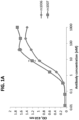
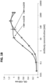
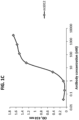
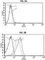


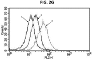


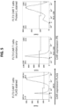


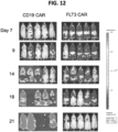
Classifications
-
- C—CHEMISTRY; METALLURGY
- C07—ORGANIC CHEMISTRY
- C07K—PEPTIDES
- C07K16/00—Immunoglobulins [IGs], e.g. monoclonal or polyclonal antibodies
- C07K16/18—Immunoglobulins [IGs], e.g. monoclonal or polyclonal antibodies against material from animals or humans
- C07K16/28—Immunoglobulins [IGs], e.g. monoclonal or polyclonal antibodies against material from animals or humans against receptors, cell surface antigens or cell surface determinants
- C07K16/2863—Immunoglobulins [IGs], e.g. monoclonal or polyclonal antibodies against material from animals or humans against receptors, cell surface antigens or cell surface determinants against receptors for growth factors, growth regulators
-
- A—HUMAN NECESSITIES
- A61—MEDICAL OR VETERINARY SCIENCE; HYGIENE
- A61K—PREPARATIONS FOR MEDICAL, DENTAL OR TOILETRY PURPOSES
- A61K47/00—Medicinal preparations characterised by the non-active ingredients used, e.g. carriers or inert additives; Targeting or modifying agents chemically bound to the active ingredient
- A61K47/50—Medicinal preparations characterised by the non-active ingredients used, e.g. carriers or inert additives; Targeting or modifying agents chemically bound to the active ingredient the non-active ingredient being chemically bound to the active ingredient, e.g. polymer-drug conjugates
- A61K47/51—Medicinal preparations characterised by the non-active ingredients used, e.g. carriers or inert additives; Targeting or modifying agents chemically bound to the active ingredient the non-active ingredient being chemically bound to the active ingredient, e.g. polymer-drug conjugates the non-active ingredient being a modifying agent
- A61K47/68—Medicinal preparations characterised by the non-active ingredients used, e.g. carriers or inert additives; Targeting or modifying agents chemically bound to the active ingredient the non-active ingredient being chemically bound to the active ingredient, e.g. polymer-drug conjugates the non-active ingredient being a modifying agent the modifying agent being an antibody, an immunoglobulin or a fragment thereof, e.g. an Fc-fragment
- A61K47/6835—Medicinal preparations characterised by the non-active ingredients used, e.g. carriers or inert additives; Targeting or modifying agents chemically bound to the active ingredient the non-active ingredient being chemically bound to the active ingredient, e.g. polymer-drug conjugates the non-active ingredient being a modifying agent the modifying agent being an antibody, an immunoglobulin or a fragment thereof, e.g. an Fc-fragment the modifying agent being an antibody or an immunoglobulin bearing at least one antigen-binding site
- A61K47/6849—Medicinal preparations characterised by the non-active ingredients used, e.g. carriers or inert additives; Targeting or modifying agents chemically bound to the active ingredient the non-active ingredient being chemically bound to the active ingredient, e.g. polymer-drug conjugates the non-active ingredient being a modifying agent the modifying agent being an antibody, an immunoglobulin or a fragment thereof, e.g. an Fc-fragment the modifying agent being an antibody or an immunoglobulin bearing at least one antigen-binding site the antibody targeting a receptor, a cell surface antigen or a cell surface determinant
-
- A—HUMAN NECESSITIES
- A61—MEDICAL OR VETERINARY SCIENCE; HYGIENE
- A61K—PREPARATIONS FOR MEDICAL, DENTAL OR TOILETRY PURPOSES
- A61K47/00—Medicinal preparations characterised by the non-active ingredients used, e.g. carriers or inert additives; Targeting or modifying agents chemically bound to the active ingredient
- A61K47/50—Medicinal preparations characterised by the non-active ingredients used, e.g. carriers or inert additives; Targeting or modifying agents chemically bound to the active ingredient the non-active ingredient being chemically bound to the active ingredient, e.g. polymer-drug conjugates
- A61K47/69—Medicinal preparations characterised by the non-active ingredients used, e.g. carriers or inert additives; Targeting or modifying agents chemically bound to the active ingredient the non-active ingredient being chemically bound to the active ingredient, e.g. polymer-drug conjugates the conjugate being characterised by physical or galenical forms, e.g. emulsion, particle, inclusion complex, stent or kit
- A61K47/6921—Medicinal preparations characterised by the non-active ingredients used, e.g. carriers or inert additives; Targeting or modifying agents chemically bound to the active ingredient the non-active ingredient being chemically bound to the active ingredient, e.g. polymer-drug conjugates the conjugate being characterised by physical or galenical forms, e.g. emulsion, particle, inclusion complex, stent or kit the form being a particulate, a powder, an adsorbate, a bead or a sphere
- A61K47/6927—Medicinal preparations characterised by the non-active ingredients used, e.g. carriers or inert additives; Targeting or modifying agents chemically bound to the active ingredient the non-active ingredient being chemically bound to the active ingredient, e.g. polymer-drug conjugates the conjugate being characterised by physical or galenical forms, e.g. emulsion, particle, inclusion complex, stent or kit the form being a particulate, a powder, an adsorbate, a bead or a sphere the form being a solid microparticle having no hollow or gas-filled cores
- A61K47/6929—Medicinal preparations characterised by the non-active ingredients used, e.g. carriers or inert additives; Targeting or modifying agents chemically bound to the active ingredient the non-active ingredient being chemically bound to the active ingredient, e.g. polymer-drug conjugates the conjugate being characterised by physical or galenical forms, e.g. emulsion, particle, inclusion complex, stent or kit the form being a particulate, a powder, an adsorbate, a bead or a sphere the form being a solid microparticle having no hollow or gas-filled cores the form being a nanoparticle, e.g. an immuno-nanoparticle
-
- A—HUMAN NECESSITIES
- A61—MEDICAL OR VETERINARY SCIENCE; HYGIENE
- A61P—SPECIFIC THERAPEUTIC ACTIVITY OF CHEMICAL COMPOUNDS OR MEDICINAL PREPARATIONS
- A61P35/00—Antineoplastic agents
-
- A—HUMAN NECESSITIES
- A61—MEDICAL OR VETERINARY SCIENCE; HYGIENE
- A61P—SPECIFIC THERAPEUTIC ACTIVITY OF CHEMICAL COMPOUNDS OR MEDICINAL PREPARATIONS
- A61P35/00—Antineoplastic agents
- A61P35/02—Antineoplastic agents specific for leukemia
-
- A—HUMAN NECESSITIES
- A61—MEDICAL OR VETERINARY SCIENCE; HYGIENE
- A61P—SPECIFIC THERAPEUTIC ACTIVITY OF CHEMICAL COMPOUNDS OR MEDICINAL PREPARATIONS
- A61P35/00—Antineoplastic agents
- A61P35/04—Antineoplastic agents specific for metastasis
-
- C—CHEMISTRY; METALLURGY
- C07—ORGANIC CHEMISTRY
- C07K—PEPTIDES
- C07K14/00—Peptides having more than 20 amino acids; Gastrins; Somatostatins; Melanotropins; Derivatives thereof
- C07K14/435—Peptides having more than 20 amino acids; Gastrins; Somatostatins; Melanotropins; Derivatives thereof from animals; from humans
- C07K14/705—Receptors; Cell surface antigens; Cell surface determinants
- C07K14/70503—Immunoglobulin superfamily
- C07K14/7051—T-cell receptor (TcR)-CD3 complex
-
- C—CHEMISTRY; METALLURGY
- C07—ORGANIC CHEMISTRY
- C07K—PEPTIDES
- C07K14/00—Peptides having more than 20 amino acids; Gastrins; Somatostatins; Melanotropins; Derivatives thereof
- C07K14/435—Peptides having more than 20 amino acids; Gastrins; Somatostatins; Melanotropins; Derivatives thereof from animals; from humans
- C07K14/705—Receptors; Cell surface antigens; Cell surface determinants
- C07K14/70503—Immunoglobulin superfamily
- C07K14/70517—CD8
-
- C—CHEMISTRY; METALLURGY
- C07—ORGANIC CHEMISTRY
- C07K—PEPTIDES
- C07K14/00—Peptides having more than 20 amino acids; Gastrins; Somatostatins; Melanotropins; Derivatives thereof
- C07K14/435—Peptides having more than 20 amino acids; Gastrins; Somatostatins; Melanotropins; Derivatives thereof from animals; from humans
- C07K14/705—Receptors; Cell surface antigens; Cell surface determinants
- C07K14/70578—NGF-receptor/TNF-receptor superfamily, e.g. CD27, CD30, CD40, CD95
-
- C—CHEMISTRY; METALLURGY
- C07—ORGANIC CHEMISTRY
- C07K—PEPTIDES
- C07K16/00—Immunoglobulins [IGs], e.g. monoclonal or polyclonal antibodies
- C07K16/46—Hybrid immunoglobulins
- C07K16/468—Immunoglobulins having two or more different antigen binding sites, e.g. multifunctional antibodies
-
- G—PHYSICS
- G01—MEASURING; TESTING
- G01N—INVESTIGATING OR ANALYSING MATERIALS BY DETERMINING THEIR CHEMICAL OR PHYSICAL PROPERTIES
- G01N33/00—Investigating or analysing materials by specific methods not covered by groups G01N1/00 - G01N31/00
- G01N33/48—Biological material, e.g. blood, urine; Haemocytometers
- G01N33/50—Chemical analysis of biological material, e.g. blood, urine; Testing involving biospecific ligand binding methods; Immunological testing
- G01N33/53—Immunoassay; Biospecific binding assay; Materials therefor
- G01N33/577—Immunoassay; Biospecific binding assay; Materials therefor involving monoclonal antibodies binding reaction mechanisms characterised by the use of monoclonal antibodies; monoclonal antibodies per se are classified with their corresponding antigens
-
- A—HUMAN NECESSITIES
- A61—MEDICAL OR VETERINARY SCIENCE; HYGIENE
- A61K—PREPARATIONS FOR MEDICAL, DENTAL OR TOILETRY PURPOSES
- A61K39/00—Medicinal preparations containing antigens or antibodies
- A61K2039/505—Medicinal preparations containing antigens or antibodies comprising antibodies
-
- C—CHEMISTRY; METALLURGY
- C07—ORGANIC CHEMISTRY
- C07K—PEPTIDES
- C07K2317/00—Immunoglobulins specific features
- C07K2317/20—Immunoglobulins specific features characterized by taxonomic origin
- C07K2317/21—Immunoglobulins specific features characterized by taxonomic origin from primates, e.g. man
-
- C—CHEMISTRY; METALLURGY
- C07—ORGANIC CHEMISTRY
- C07K—PEPTIDES
- C07K2317/00—Immunoglobulins specific features
- C07K2317/30—Immunoglobulins specific features characterized by aspects of specificity or valency
- C07K2317/31—Immunoglobulins specific features characterized by aspects of specificity or valency multispecific
-
- C—CHEMISTRY; METALLURGY
- C07—ORGANIC CHEMISTRY
- C07K—PEPTIDES
- C07K2317/00—Immunoglobulins specific features
- C07K2317/50—Immunoglobulins specific features characterized by immunoglobulin fragments
- C07K2317/52—Constant or Fc region; Isotype
-
- C—CHEMISTRY; METALLURGY
- C07—ORGANIC CHEMISTRY
- C07K—PEPTIDES
- C07K2317/00—Immunoglobulins specific features
- C07K2317/50—Immunoglobulins specific features characterized by immunoglobulin fragments
- C07K2317/56—Immunoglobulins specific features characterized by immunoglobulin fragments variable (Fv) region, i.e. VH and/or VL
- C07K2317/565—Complementarity determining region [CDR]
-
- C—CHEMISTRY; METALLURGY
- C07—ORGANIC CHEMISTRY
- C07K—PEPTIDES
- C07K2317/00—Immunoglobulins specific features
- C07K2317/60—Immunoglobulins specific features characterized by non-natural combinations of immunoglobulin fragments
- C07K2317/62—Immunoglobulins specific features characterized by non-natural combinations of immunoglobulin fragments comprising only variable region components
- C07K2317/622—Single chain antibody (scFv)
-
- C—CHEMISTRY; METALLURGY
- C07—ORGANIC CHEMISTRY
- C07K—PEPTIDES
- C07K2317/00—Immunoglobulins specific features
- C07K2317/70—Immunoglobulins specific features characterized by effect upon binding to a cell or to an antigen
- C07K2317/73—Inducing cell death, e.g. apoptosis, necrosis or inhibition of cell proliferation
-
- C—CHEMISTRY; METALLURGY
- C07—ORGANIC CHEMISTRY
- C07K—PEPTIDES
- C07K2317/00—Immunoglobulins specific features
- C07K2317/70—Immunoglobulins specific features characterized by effect upon binding to a cell or to an antigen
- C07K2317/76—Antagonist effect on antigen, e.g. neutralization or inhibition of binding
-
- C—CHEMISTRY; METALLURGY
- C07—ORGANIC CHEMISTRY
- C07K—PEPTIDES
- C07K2319/00—Fusion polypeptide
- C07K2319/01—Fusion polypeptide containing a localisation/targetting motif
- C07K2319/02—Fusion polypeptide containing a localisation/targetting motif containing a signal sequence
-
- C—CHEMISTRY; METALLURGY
- C07—ORGANIC CHEMISTRY
- C07K—PEPTIDES
- C07K2319/00—Fusion polypeptide
- C07K2319/01—Fusion polypeptide containing a localisation/targetting motif
- C07K2319/03—Fusion polypeptide containing a localisation/targetting motif containing a transmembrane segment
-
- C—CHEMISTRY; METALLURGY
- C07—ORGANIC CHEMISTRY
- C07K—PEPTIDES
- C07K2319/00—Fusion polypeptide
- C07K2319/30—Non-immunoglobulin-derived peptide or protein having an immunoglobulin constant or Fc region, or a fragment thereof, attached thereto
Definitions
- FLT3 Fms-like tyrosine kinase 3
- ALL acute lymphoblastic leukemia
- AML acute myeloid leukemia
- Fms-like tyrosine kinase 3 (FLT3), also known as CD135, is a cytokine receptor belonging to the class III receptor tyrosine kinase family.
- FLT3 is expressed on the surface of many hematopoietic progenitor cells and plays an important role in hematopoietic stem/progenitor cell survival and proliferation. It is frequently overexpressed in acute lymphoblastic leukemia (ALL) and is frequently mutated in acute myeloid leukemia (AML).
- ALL acute lymphoblastic leukemia
- AML acute myeloid leukemia
- the presence of the FLT3-internal tandem duplication (ITD) mutation is a key indicator of poor long-term prognosis.
- the FLT3-ITD mutation occurs in approximately 25% of patients with AML.
- the disclosed antibodies referred to herein as m1006, m1007, m1008, m1009 and m1012, bind to both soluble recombinant FLT3 and cell-surface FLT3 with high affinity.
- Further disclosed herein is the finding that T cells expressing an FLT3-specific chimeric antigen receptor (CAR) secrete high levels of IL-2 and IFN- ⁇ when co-cultured with FLT3-expressing AML or ALL cells.
- T cells expressing the FLT3-specific CAR were shown to eradicate FLT3-expressing ALL and AML in animal models.
- monoclonal antibodies that bind, such as specifically bind, FLT3.
- the monoclonal antibodies include one or more complementarity determining region (CDR) sequences of m1006, m1007, m1008, m1009 or m1012.
- conjugates that include a disclosed FLT3-specific monoclonal antibody or antigen-binding fragment thereof.
- CARs immunoconjugates, multi-specific antibodies, antibody-drug conjugates (ADCs), antibody-nanoparticles, conjugates or fusion proteins that include a monoclonal antibody or antigen-binding fragment disclosed herein.
- Compositions that include an FLT3-specific monoclonal antibody or antigen-binding fragment and a pharmaceutically acceptable carrier are also provided by the present disclosure.
- Methods of treating an FLT3-associated cancer in a subject, and methods of inhibiting metastasis of an FLT3-associated cancer (such as a leukemia) in a subject are also provided.
- the methods include administering to the subject a monoclonal antibody or antigen-binding fragment disclosed herein, or administering to the subject a CAR, immunoconjugate, ADC, multi-specific antibody, antibody-nanoparticle conjugate or fusion protein comprising a monoclonal antibody (or antigen-binding fragment) disclosed herein.
- the method includes contacting the sample with a monoclonal antibody or antigen-binding fragment disclosed herein, and detecting binding of the antibody to the sample.
- nucleic and amino acid sequences listed in the accompanying sequence listing are shown using standard letter abbreviations for nucleotide bases, and three letter code for amino acids, as defined in 37 C.F.R. 1.822. Only one strand of each nucleic acid sequence is shown, but the complementary strand is understood as included by any reference to the displayed strand.
- sequence Listing is submitted as an ASCII text file, created on December 7, 2017, 28.0 KB, which is incorporated by reference herein. In the accompanying sequence listing:
- Antibody variable regions contain "framework" regions and hypervariable regions, known as “complementarity determining regions” or “CDRs.”
- the CDRs are primarily responsible for binding to an epitope of an antigen.
- the framework regions of an antibody serve to position and align the CDRs in three-dimensional space.
- the amino acid sequence boundaries of a given CDR can be readily determined using any of a number of well-known numbering schemes, including those described by Kabat et al. (Sequences of Proteins of Immunological Interest, U.S. Department of Health and Human Services, 1991 ; the "Kabat” numbering scheme), Chothia et al.
- single-domain antibody refers to an antibody having a single domain (a variable domain) that is capable of specifically binding an antigen, or an epitope of an antigen, in the absence of an additional antibody domain.
- Single-domain antibodies include, for example, V H domain antibodies, V NAR antibodies, camelid V H H antibodies, and V L domain antibodies.
- V NAR antibodies are produced by cartilaginous fish, such as nurse sharks, wobbegong sharks, spiny dogfish and bamboo sharks.
- Camelid V H H antibodies are produced by several species including camel, llama, alpaca, dromedary, and guanaco, which produce heavy chain antibodies that are naturally devoid of light chains.
- a “monoclonal antibody” is an antibody produced by a single clone of lymphocytes or by a cell into which the coding sequence of a single antibody has been transfected. Monoclonal antibodies are produced by methods known to those of skill in the art. Monoclonal antibodies include humanized monoclonal antibodies.
- a “chimeric antibody” has framework residues from one species, such as human, and CDRs (which generally confer antigen binding) from another species.
- a “humanized” antibody is an immunoglobulin including a human framework region and one or more CDRs from a non-human (for example a mouse, rabbit, rat, shark or synthetic) immunoglobulin.
- the non-human immunoglobulin providing the CDRs is termed a "donor,” and the human immunoglobulin providing the framework is termed an "acceptor.”
- all CDRs are from the donor immunoglobulin in a humanized immunoglobulin. Constant regions need not be present, but if they are, they must be substantially identical to human immunoglobulin constant regions, i.e., at least about 85-90%, such as about 95% or more identical.
- a humanized immunoglobulin all parts of a humanized immunoglobulin, except possibly the CDRs, are substantially identical to corresponding parts of natural human immunoglobulin sequences.
- a humanized antibody binds to the same antigen as the donor antibody that provides the CDRs.
- Humanized or other monoclonal antibodies can have additional conservative amino acid substitutions which have substantially no effect on antigen binding or other immunoglobulin functions.
- ADC Antibody-drug conjugate
- ADC A molecule that includes an antibody (or antigen-binding fragment of an antibody) conjugated to a drug, such as a cytotoxic agent.
- ADCs can be used to specifically target a drug to cancer cells through specific binding of the antibody to a tumor antigen expressed on the cell surface.
- exemplary drugs for use with ADCs include anti-microtubule agents (such as maytansinoids, auristatin E and auristatin F) and interstrand crosslinking agents (e.g., pyrrolobenzodiazepines; PDBs).
- Anti-microtubule agent A type of drug that blocks cell growth by stopping mitosis.
- Anti-microtubule agents also referred to as “anti-mitotic agents,” are used to treat cancer.
- Binding affinity Affinity of an antibody for an antigen.
- affinity is calculated by a modification of the Scatchard method described by Frankel et al., Mol. Immunol., 16:101-106, 1979 .
- binding affinity is measured by an antigen/antibody dissociation rate.
- a high binding affinity is measured by a competition radioimmunoassay.
- binding affinity is measured by ELISA.
- antibody affinity is measured by flow cytometry.
- An antibody that "specifically binds" an antigen (such as FLT3) is an antibody that binds the antigen with high affinity and does not significantly bind other unrelated antigens.
- Bispecific antibody A recombinant protein that includes antigen-binding fragments of two different monoclonal antibodies, and is thereby capable of binding two different antigens.
- bispecific antibodies are used for cancer immunotherapy by simultaneously targeting, for example, both CTLs (such as a CTL receptor component such as CD3) or effector natural killer (NK) cells, and a tumor antigen.
- CTLs such as a CTL receptor component such as CD3
- NK effector natural killer
- a multi-specific antibody is a recombinant protein that includes antigen-binding fragments of at least two different monoclonal antibodies, such as two, three or four different monoclonal antibodies.
- Chemotherapeutic agent Any chemical agent with therapeutic usefulness in the treatment of diseases characterized by abnormal cell growth. Such diseases include tumors, neoplasms, and cancer as well as diseases characterized by hyperplastic growth such as psoriasis.
- a chemotherapeutic agent is an agent of use in treating AML or ALL.
- a chemotherapeutic agent is a radioactive compound.
- One of skill in the art can readily identify a chemotherapeutic agent of use (see for example, Slapak and Kufe, Principles of Cancer Therapy, Chapter 86 in Harrison's Principles of Internal Medicine, 14th editi on; Perry et al., Chemotherapy, Ch.
- Combination chemotherapy is the administration of more than one agent to treat cancer.
- One example is the administration of an antibody that binds FLT3 used in combination with a radioactive or chemical compound.
- Chimeric antigen receptor A chimeric molecule that includes an antigen-binding portion (such as a single domain antibody or scFv) and a signaling domain, such as a signaling domain from a T cell receptor (e.g. CD3 ⁇ ).
- CARs are comprised of an antigen-binding moiety, a transmembrane domain and an endodomain.
- the endodomain typically includes a signaling chain having an immunoreceptor tyrosine-based activation motif (ITAM), such as CD3 ⁇ or Fc ⁇ RIy.
- ITAM immunoreceptor tyrosine-based activation motif
- the endodomain further includes the intracellular portion of at least one additional co-stimulatory domain, such as CD28, 4-1BB (CD137), ICOS, OX40 (CD134), CD27 and/or DAP10.
- CDR Complementarity determining region
- a "conjugate” is an antibody or antibody fragment (such as an antigen-binding fragment) covalently linked to an effector molecule or a second protein (such as a second antibody).
- the effector molecule can be, for example, a drug, toxin, therapeutic agent, detectable label, protein, nucleic acid, lipid, nanoparticle, carbohydrate or recombinant virus.
- an antibody conjugate is often referred to as an "immunoconjugate.”
- the conjugate comprises an antibody linked to a drug (e.g., a cytotoxic agent)
- the conjugate is often referred to as an "antibody-drug conjugate” or "ADC.”
- ADC antibody-drug conjugate
- Other antibody conjugates include, for example, multi-specific (such as bispecific or trispecific) antibodies and chimeric antigen receptors (CARs).
- Constant amino acid substitutions are those substitutions that do not substantially affect or decrease the affinity of a protein, such as an antibody to FLT3.
- a monoclonal antibody that specifically binds FLT3 can include at most about 1, at most about 2, at most about 5, and most about 10, or at most about 15 conservative substitutions and specifically bind the FLT3 polypeptide.
- the term “conservative variant” also includes the use of a substituted amino acid in place of an unsubstituted parent amino acid, provided that antibody specifically binds FLT3.
- Non-conservative substitutions are those that reduce an activity or binding to FLT3.
- Placement in direct physical association includes both in solid and liquid form.
- Cytotoxic agent Any drug or compound that kills cells.
- Cytotoxicity The toxicity of a molecule, such as an immunotoxin, to the cells intended to be targeted, as opposed to the cells of the rest of an organism.
- toxicity refers to toxicity of an immunotoxin to cells other than those that are the cells intended to be targeted by the targeting moiety of the immunotoxin
- animal toxicity refers to toxicity of the immunotoxin to an animal by toxicity of the immunotoxin to cells other than those intended to be targeted by the immunotoxin.
- Degenerate variant refers to a polynucleotide encoding a FLT3 polypeptide or an antibody that binds FLT3 that includes a sequence that is degenerate as a result of the genetic code. There are 20 natural amino acids, most of which are specified by more than one codon. Therefore, all degenerate nucleotide sequences are included as long as the amino acid sequence of the FLT3 polypeptide or antibody that binds FLT3 encoded by the nucleotide sequence is unchanged.
- Diagnostic Identifying the presence or nature of a pathologic condition, such as cancer. Diagnostic methods differ in their sensitivity and specificity.
- the "sensitivity” of a diagnostic assay is the percentage of diseased individuals who test positive (percent of true positives).
- the "specificity” of a diagnostic assay is one minus the false positive rate, where the false positive rate is defined as the proportion of those without the disease who test positive. While a particular diagnostic method may not provide a definitive diagnosis of a condition, it suffices if the method provides a positive indication that aids in diagnosis.
- “Prognostic” is the probability of development (e.g., severity) of a pathologic condition, such as AML or ALL.
- Drug Any compound used to treat, ameliorate or prevent a disease or condition in a subject.
- the drug is an anti-cancer agent, for example a cytotoxic agent, such as an anti-mitotic or anti-microtubule agent.
- Effector molecule The portion of a chimeric molecule that is intended to have a desired effect on a cell to which the chimeric molecule is targeted. Effector molecule is also known as an effector moiety (EM), therapeutic agent, or diagnostic agent, or similar terms.
- Therapeutic agents include such compounds as nucleic acids, proteins, peptides, amino acids or derivatives, glycoproteins, radioisotopes, lipids, carbohydrates, or recombinant viruses.
- Nucleic acid therapeutic and diagnostic moieties include antisense nucleic acids, derivatized oligonucleotides for covalent cross-linking with single or duplex DNA, and triplex forming oligonucleotides.
- the molecule linked to a targeting moiety may be an encapsulation system, such as a liposome or micelle that contains a therapeutic composition such as a drug, a nucleic acid (such as an antisense nucleic acid), or another therapeutic moiety that can be shielded from direct exposure to the circulatory system.
- a therapeutic composition such as a drug, a nucleic acid (such as an antisense nucleic acid), or another therapeutic moiety that can be shielded from direct exposure to the circulatory system.
- Means of preparing liposomes attached to antibodies are well known to those of skill in the art (see, for example, U.S. Patent No. 4,957,735 ; and Connor et al., Pharm Ther 28:341-365, 1985 ). Diagnostic agents or moieties include radioisotopes and other detectable labels.
- Detectable labels useful for such purposes are also well known in the art, and include radioactive isotopes such as 35 S, 11 C, 13 N, 15 O, 18 F, 19 F, 99m Tc, 131 I, 3 H, 14 C, 15 N, 90 Y, 99 Tc, 111 In and 125 I, fluorophores, chemiluminescent agents, and enzymes.
- radioactive isotopes such as 35 S, 11 C, 13 N, 15 O, 18 F, 19 F, 99m Tc, 131 I, 3 H, 14 C, 15 N, 90 Y, 99 Tc, 111 In and 125 I, fluorophores, chemiluminescent agents, and enzymes.
- Epitope An antigenic determinant. These are particular chemical groups or peptide sequences on a molecule that are antigenic, i.e. that elicit a specific immune response. An antibody specifically binds a particular antigenic epitope on a polypeptide, such as FLT3.
- Fms-like tyrosine kinase 3 FLT3: A class III receptor tyrosine kinase that regulates hematopoiesis.
- FLT3 is activated by binding of FLT3 ligand to its extracellular domain, which induces homodimer formation in the plasma membrane and autophosphorylation of FLT3.
- Activated FLT3 subsequently phosphorylates and activates multiple cytoplasmic effector molecules in pathways involved in apoptosis, proliferation, and differentiation of hematopoietic cells. This receptor is frequently overexpressed in acute lymphoblastic leukemia (ALL) and is frequently mutated in acute myeloid leukemia (AML).
- FLT3 is also known as CD135.
- FLT3-associated cancer A cancer that overexpresses FLT3 or expresses a mutant form of FLT3. Mutations in FLT3-associated cancers include, but are not limited to, internal tandem duplications in or near the juxtamembrane domain (FLT3/ITD mutations) and point mutations within the activation loop of the tyrosine kinase domain (FLT3/TKD mutations) ( Levis, Hematology Am Soc Hematol Educ Program 2013:220-226, 2013 ; Levis and Small, Leukemia 17:1738-1752, 2003 ). FLT-associated cancers include, but are not limited to, ALL and AML.
- FLT3-positive cancer A cancer that overexpresses FLT3.
- Framework region Amino acid sequences interposed between CDRs. Framework regions include variable light and variable heavy framework regions. The framework regions serve to hold the CDRs in an appropriate orientation for antigen binding.
- Fusion protein A protein comprising at least a portion of two different (heterologous) proteins.
- Heterologous Originating from a separate genetic source or species.
- Immune response A response of a cell of the immune system, such as a B cell, T cell, or monocyte, to a stimulus.
- the response is specific for a particular antigen (an "antigen-specific response").
- an immune response is a T cell response, such as a CD4 + response or a CD8 + response.
- the response is a B cell response, and results in the production of specific antibodies.
- Immunoconjugate A covalent linkage of an effector molecule to an antibody or functional fragment thereof.
- the effector molecule can be a detectable label or an immunotoxin.
- toxins include, but are not limited to, abrin, ricin, Pseudomonas exotoxin (PE, such as PE35, PE37, PE38, and PE40), diphtheria toxin (DT), botulinum toxin, or modified toxins thereof, or other toxic agents that directly or indirectly inhibit cell growth or kill cells.
- PE and DT are highly toxic compounds that typically bring about death through liver toxicity.
- PE and DT can be modified into a form for use as an immunotoxin by removing the native targeting component of the toxin (such as the domain Ia of PE and the B chain of DT) and replacing it with a different targeting moiety, such as an antibody.
- a "chimeric molecule” is a targeting moiety, such as a ligand or an antibody, conjugated (coupled) to an effector molecule.
- conjugated or “linked” refers to making two polypeptides into one contiguous polypeptide molecule. In one embodiment, an antibody is joined to an effector molecule.
- an antibody joined to an effector molecule is further joined to a lipid or other molecule to a protein or peptide to increase its half-life in the body.
- the linkage can be either by chemical or recombinant means.
- the linkage is chemical, wherein a reaction between the antibody moiety and the effector molecule has produced a covalent bond formed between the two molecules to form one molecule.
- a peptide linker (short peptide sequence) can optionally be included between the antibody and the effector molecule.
- chimeric molecules refers to a targeting moiety, such as a ligand or an antibody, conjugated (coupled) to an effector molecule.
- Immunoliposome A liposome with antibodies or antibody fragments conjugated to its surface. Immunoliposomes can carry cytotoxic agents or other drugs to antibody-targeted cells, such as tumor cells.
- Interstrand crosslinking agent A type of cytotoxic drug capable of binding covalently between two strands of DNA, thereby preventing DNA replication and/or transcription.
- Isolated An "isolated" biological component, such as a nucleic acid, protein (including antibodies) or organelle, has been substantially separated or purified away from other biological components in the environment (such as a cell) in which the component naturally occurs, i.e., other chromosomal and extra-chromosomal DNA and RNA, proteins and organelles.
- Nucleic acids and proteins that have been “isolated” include nucleic acids and proteins purified by standard purification methods. The term also embraces nucleic acids and proteins prepared by recombinant expression in a host cell as well as chemically synthesized nucleic acids.
- Label A detectable compound or composition that is conjugated directly or indirectly to another molecule, such as an antibody or a protein, to facilitate detection of that molecule.
- labels include fluorescent tags, enzymatic linkages, and radioactive isotopes.
- a "labeled antibody” refers to incorporation of another molecule in the antibody.
- the label is a detectable marker, such as the incorporation of a radiolabeled amino acid or attachment to a polypeptide of biotinyl moieties that can be detected by marked avidin (for example, streptavidin containing a fluorescent marker or enzymatic activity that can be detected by optical or colorimetric methods).
- labels for polypeptides include, but are not limited to, the following: radioisotopes or radionucleotides (such as 35 S, 11 C, 13 N, 15 O, 18 F, 19 F, 99m Tc, 131 I, 3 H, 14 C, 15 N, 90 Y, 99 Tc, 111 In and 125 I), fluorescent labels (such as fluorescein isothiocyanate (FITC), rhodamine, lanthanide phosphors), enzymatic labels (such as horseradish peroxidase, beta-galactosidase, luciferase, alkaline phosphatase), chemiluminescent markers, biotinyl groups, predetermined polypeptide epitopes recognized by a secondary reporter (such as a leucine zipper pair sequences, binding sites for secondary antibodies, metal binding domains, epitope tags), or magnetic agents, such as
- Linker In some cases, a linker is a peptide within an antibody binding fragment (such as an Fv fragment) which serves to indirectly bond the variable heavy chain to the variable light chain. "Linker” can also refer to a peptide serving to link a targeting moiety, such as an antibody, to an effector molecule, such as a cytotoxin or a detectable label.
- conjugating refers to making two polypeptides into one contiguous polypeptide molecule, or to covalently attaching a radionuclide or other molecule to a polypeptide, such as an scFv.
- the terms include reference to joining a ligand, such as an antibody moiety, to an effector molecule.
- the linkage can be either by chemical or recombinant means.
- “Chemical means” refers to a reaction between the antibody moiety and the effector molecule such that there is a covalent bond formed between the two molecules to form one molecule.
- Neoplasia malignancy, cancer or tumor: A neoplasm is an abnormal growth of tissue or cells that results from excessive cell division. Neoplastic growth can produce a tumor. The amount of a tumor in an individual is the "tumor burden" which can be measured as the number, volume, or weight of the tumor. A tumor that does not metastasize is referred to as “benign.” A tumor that invades the surrounding tissue and/or can metastasize is referred to as "malignant.”
- a first nucleic acid sequence is operably linked with a second nucleic acid sequence when the first nucleic acid sequence is placed in a functional relationship with the second nucleic acid sequence.
- a promoter such as the CMV promoter
- operably linked DNA sequences are contiguous and, where necessary to join two protein-coding regions, in the same reading frame.
- Pharmaceutical agent A chemical compound or composition capable of inducing a desired therapeutic or prophylactic effect when properly administered to a subject or a cell.
- parenteral formulations usually comprise injectable fluids that include pharmaceutically and physiologically acceptable fluids such as water, physiological saline, balanced salt solutions, aqueous dextrose, glycerol or the like as a vehicle.
- pharmaceutically and physiologically acceptable fluids such as water, physiological saline, balanced salt solutions, aqueous dextrose, glycerol or the like as a vehicle.
- physiologically acceptable fluids such as water, physiological saline, balanced salt solutions, aqueous dextrose, glycerol or the like
- solid compositions such as powder, pill, tablet, or capsule forms
- conventional non-toxic solid carriers can include, for example, pharmaceutical grades of mannitol, lactose, starch, or magnesium stearate.
- compositions to be administered can contain minor amounts of non-toxic auxiliary substances, such as wetting or emulsifying agents, preservatives, and pH buffering agents and the like, for example sodium acetate or sorbitan monolaurate.
- non-toxic auxiliary substances such as wetting or emulsifying agents, preservatives, and pH buffering agents and the like, for example sodium acetate or sorbitan monolaurate.
- Preventing a disease refers to inhibiting the full development of a disease.
- Treating refers to a therapeutic intervention that ameliorates a sign or symptom of a disease or pathological condition after it has begun to develop, such as a reduction in tumor burden or a decrease in the number of size of metastases.
- Treating refers to the reduction in the number or severity of signs or symptoms of a disease, such as cancer.
- a purified peptide preparation is one in which the peptide or protein is more enriched than the peptide or protein is in its natural environment within a cell.
- a preparation is purified such that the protein or peptide represents at least 50% of the total peptide or protein content of the preparation.
- Substantial purification denotes purification from other proteins or cellular components.
- a substantially purified protein is at least 60%, 70%, 80%, 90%, 95% or 98% pure.
- a substantially purified protein is 90% free of other proteins or cellular components.
- PBD Pyrrolobenzodiazepine
- a recombinant nucleic acid or protein is one that has a sequence that is not naturally occurring or has a sequence that is made by an artificial combination of two otherwise separated segments of sequence. This artificial combination is often accomplished by chemical synthesis or by the artificial manipulation of isolated segments of nucleic acids, for example, by genetic engineering techniques.
- Sample A biological specimen containing genomic DNA, RNA (including mRNA), protein, or combinations thereof, obtained from a subject. Examples include, but are not limited to, peripheral blood, tissue, cells, urine, saliva, tissue biopsy, fine needle aspirate, surgical specimen, and autopsy material. In one example, a sample includes a tumor biopsy.
- Sequence identity The similarity between amino acid or nucleic acid sequences is expressed in terms of the similarity between the sequences, otherwise referred to as sequence identity. Sequence identity is frequently measured in terms of percentage identity (or similarity or homology); the higher the percentage, the more similar the two sequences are. Homologs or variants of a polypeptide or nucleic acid molecule will possess a relatively high degree of sequence identity when aligned using standard methods.
- NCBI Basic Local Alignment Search Tool (BLAST) ( Altschul et al., J. Mol. Biol. 215:403, 1990 ) is available from several sources, including the National Center for Biotechnology Information (NCBI, Bethesda, MD) and on the internet, for use in connection with the sequence analysis programs blastp, blastn, blastx, tblastn and tblastx. A description of how to determine sequence identity using this program is available on the NCBI website on the internet.
- Homologs and variants of a V H of an antibody that specifically binds a FLT3 polypeptide are typically characterized by possession of at least about 75%, for example at least about 80%, 90%, 95%, 96%, 97%, 98% or 99% sequence identity counted over the full length alignment with the amino acid sequence of the antibody using the NCBI Blast 2.0, gapped blastp set to default parameters.
- the Blast 2 sequences function is employed using the default BLOSUM62 matrix set to default parameters, (gap existence cost of 11, and a per residue gap cost of 1).
- the alignment should be performed using the Blast 2 sequences function, employing the PAM30 matrix set to default parameters (open gap 9, extension gap 1 penalties). Proteins with even greater similarity to the reference sequences will show increasing percentage identities when assessed by this method, such as at least 80%, at least 85%, at least 90%, at least 95%, at least 98%, or at least 99% sequence identity. When less than the entire sequence is being compared for sequence identity, homologs and variants will typically possess at least 80% sequence identity over short windows of 10-20 amino acids, and may possess sequence identities of at least 85% or at least 90% or 95% depending on their similarity to the reference sequence.
- Small molecule A molecule, typically with a molecular weight less than about 1000 Daltons, or in some embodiments, less than about 500 Daltons, wherein the molecule is capable of modulating, to some measurable extent, an activity of a target molecule.
- Subject Living multi-cellular vertebrate organisms, a category that includes both human and veterinary subjects, including human and non-human mammals.
- Synthetic Produced by artificial means in a laboratory, for example a synthetic nucleic acid or protein (for example, an antibody) can be chemically synthesized in a laboratory.
- a synthetic nucleic acid or protein for example, an antibody
- Therapeutically effective amount A quantity of a specific substance sufficient to achieve a desired effect in a subject being treated. For instance, this can be the amount necessary to inhibit or suppress growth of a tumor. In one embodiment, a therapeutically effective amount is the amount necessary to eliminate, reduce the size, or prevent metastasis of a tumor. When administered to a subject, a dosage will generally be used that will achieve target tissue concentrations (for example, in tumors) that has been shown to achieve a desired in vitro effect.
- Toxin A molecule that is cytotoxic for a cell.
- Toxins include abrin, ricin, Pseudomonas exotoxin (PE), diphtheria toxin (DT), botulinum toxin, saporin, restrictocin or gelonin, or modified toxins thereof.
- PE and DT are highly toxic compounds that typically bring about death through liver toxicity.
- PE and DT can be modified into a form for use as an immunotoxin by removing the native targeting component of the toxin (such as domain Ia of PE or the B chain of DT) and replacing it with a different targeting moiety, such as an antibody.
- a nucleic acid molecule as introduced into a host cell, thereby producing a transformed host cell.
- a vector may include nucleic acid sequences that permit it to replicate in a host cell, such as an origin of replication.
- a vector may also include one or more selectable marker genes and other genetic elements known in the art.
- the disclosed antibodies referred to herein as m1006, m1007, m1008, m1009 and m1012, bind to both soluble recombinant FLT3 and cell-surface FLT3 with high affinity.
- Further disclosed herein is the finding that T cells expressing a FLT-3specific chimeric antigen receptor (CAR) secrete high levels of IL-2 and IFN- ⁇ when co-cultured with FLT3-expressing AML or ALL cells.
- T cells expressing the FLT3-specific CAR were shown to eradicate FLT3-expressing ALL and AML in animal models.
- the nucleotide and amino acid sequences of the VH and VL domains of antibodies m1006, m1007, m1008 and m1009, and the VH domain of single-domain antibody m1012, are provided below. Also shown are the nucleotide and amino acid sequences of m1006, m1007, m1008 and m1009 scFv. In the amino acid sequences below, the CDR regions according to IMGT are shown in bold underline and the residues of CDR1, CDR2 and CDR3 are indicated below each VH domain and VL domain sequence.
- One of skill in the art could readily determine the CDR boundaries using alternative numbering schemes, such as the Kabat or Chothia numbering schemes.
- monoclonal antibodies or antigen-binding fragments that bind (such as specifically bind) FLT3, such as cell-surface FLT3 or soluble FLT3.
- the monoclonal antibody or antigen-binding fragment includes both a VH domain and a VL domain.
- the monoclonal antibody is a VH single-domain monoclonal antibody.
- the monoclonal antibody or antigen-binding fragment that binds FLT3 includes at least one CDR sequence from antibody m1006, m1007, m 1008 m 1009 or m 1012.
- the CDR sequences are determined using the IMGT, Kabat or Chothia numbering scheme.
- the FLT3-specific monoclonal antibody or antigen-binding fragment includes a VH domain and a VL domain
- the VH domain of the antibody includes one, two or all three CDR sequences of SEQ ID NO: 1, SEQ ID NO: 5, SEQ ID NO: 9 or SEQ ID NO: 13, and/or the VL domain of the antibody includes one, two or all three CDR sequences of SEQ ID NO: 2, SEQ ID NO: 6, SEQ ID NO: 10, SEQ ID NO: 14.
- the VH domain comprises residues 26-33, 51-58 and 97-105 of SEQ ID NO: 1 and the VL domain comprises residues 27-37, 55-57 and 94-102 of SEQ ID NO: 2.
- the VH domain comprises residues 26-33, 51-58 and 97-105 of SEQ ID NO: 5 and the VL domain comprises residues 27-37, 55-57 and 94-102 of SEQ ID NO: 6.
- the VH domain comprises residues 26-35, 53-59 and 98-106 of SEQ ID NO: 9 and the VL domain comprises residues 26-33, 51-53 and 90-100 of SEQ ID NO: 10.
- the VH domain comprises residues 26-33, 51-58 and 97-107 of SEQ ID NO: 13 and the VL domain comprises residues 26-33, 51-53 and 90-99 of SEQ ID NO: 14.
- the amino acid sequence of the VH domain is at least 80%, at least 85%, at least 90%, at least 95%, at least 96%, at least 97%, at least 98% or at least 99% identical to SEQ ID NO: 1 and the amino acid sequence of the VL domain is at least 80%, at least 85%, at least 90%, at least 95%, at least 96%, at least 97%, at least 98% or at least 99% identical to SEQ ID NO: 2.
- the amino acid sequence of the VH domain is at least 80%, at least 85%, at least 90%, at least 95%, at least 96%, at least 97%, at least 98% or at least 99% identical to SEQ ID NO: 5 and the amino acid sequence of the VL domain is at least 80%, at least 85%, at least 90%, at least 95%, at least 96%, at least 97%, at least 98% or at least 99% identical to SEQ ID NO: 6.
- the amino acid sequence of the VH domain is at least 80%, at least 85%, at least 90%, at least 95%, at least 96%, at least 97%, at least 98% or at least 99% identical to SEQ ID NO: 9 and the amino acid sequence of the VL domain is at least 80%, at least 85%, at least 90%, at least 95%, at least 96%, at least 97%, at least 98% or at least 99% identical to SEQ ID NO: 10.
- the amino acid sequence of the VH domain is at least 80%, at least 85%, at least 90%, at least 95%, at least 96%, at least 97%, at least 98% or at least 99% identical to SEQ ID NO: 13 and the amino acid sequence of the VL domain is at least 80%, at least 85%, at least 90%, at least 95%, at least 96%, at least 97%, at least 98% or at least 99% identical to SEQ ID NO: 14.
- the amino acid sequence of the VH domain comprises SEQ ID NO: 1 and the amino acid sequence of the VL domain comprises SEQ ID NO: 2; the amino acid sequence of the VH domain comprises SEQ ID NO: 5 and the amino acid sequence of the VL domain comprises SEQ ID NO: 6; the amino acid sequence of the VH domain comprises SEQ ID NO: 9 and the amino acid sequence of the VL domain comprises SEQ ID NO: 10; or the amino acid sequence of the VH domain comprises SEQ ID NO: 13 and the amino acid sequence of the VL domain comprises SEQ ID NO: 14.
- FLT3-specific antigen-binding fragments that include both a VH domain and a VL domain can be, for example, an Fab fragment, an Fab' fragment, an F(ab)' 2 fragment, a single chain variable fragment (scFv) or a disulfide stabilized variable fragment (dsFv).
- the antigen-binding fragment is a scFv.
- the amino acid sequence of the scFv is at least 80%, at least 85%, at least 90%, at least 95%, at least 96%, at least 97%, at least 98% or at least 99% identical to SEQ ID NO: 4, SEQ ID NO: 8, SEQ ID NO: 12 or SEQ ID NO: 16.
- the amino acid sequence of the scFv comprises or consists of SEQ ID NO: 4, SEQ ID NO: 8, SEQ ID NO: 12 or SEQ ID NO: 16.
- FLT3-specific monoclonal antibodies can be of any isotype, such as IgG, IgM, IgA, IgD or IgE.
- the monoclonal antibody is an IgG.
- the FLT3-specific monoclonal antibody is a VH single-domain antibody that includes one, two or all three CDR sequences of SEQ ID NO: 18.
- the VH domain comprises residues 26-33, 51-58 and 97-109 of SEQ ID NO: 18.
- the amino acid sequence of the VH domain is at least 80%, at least 85%, at least 90%, at least 95%, at least 96%, at least 97%, at least 98% or at least 99% identical to SEQ ID NO: 18.
- the amino acid sequence of the VH domain comprises or consists of the amino acid sequence of SEQ ID NO: 18.
- the monoclonal antibody or antigen-binding fragment is a fully human antibody or antigen-binding fragment. In some embodiments, the monoclonal antibody or antigen-binding fragment is a chimeric or synthetic antibody or antigen-binding fragment.
- CARs chimeric antigen receptors
- the CAR further includes a hinge region, a transmembrane domain, a costimulatory signaling moiety, a signaling domain, or any combination thereof.
- the hinge region includes a CD8 hinge region; the transmembrane domain includes a CD8 transmembrane domain; the costimulatory signaling moiety includes a 4-1BB signaling moiety; and/or the signaling domain comprises a CD3 ⁇ signaling domain.
- the CAR includes the amino acid sequence of SEQ ID NO: 19.
- cells expressing an FLT3-specific CAR In some examples, the cell is a CTL. CARs and CAR-expressing T cells are further described in section IV.
- immunoconjugates that include a monoclonal antibody or antigen-binding fragment disclosed herein and an effector molecule.
- the effector molecule is a toxin, such as, but not limited to, Pseudomonas exotoxin or a variant thereof.
- the effector molecule is a detectable label, such as, but not limited to, a fluorophore, an enzyme or a radioisotope. Immunoconjugates are further described in section V.
- ADCs antibody-drug conjugates
- the drug is a small molecule, for example an anti-microtubule agent, an anti-mitotic agent and/or a cytotoxic agent.
- ADCs are further described in section VI.
- multi-specific antibodies that include a monoclonal antibody or antigen-binding fragment disclosed herein and at least one additional monoclonal antibody or antigen-binding fragment thereof.
- the multi-specific antibody is a bispecific antibody. In other embodiments, the multi-specific antibody is a trispecific antibody. In some embodiments, the at least one additional monoclonal antibody or antigen binding fragment thereof specifically binds a component of the T cell receptor or a natural killer (NK) cell activating receptor.
- NK natural killer
- antibody-nanoparticle conjugates that include a nanoparticle conjugated to a monoclonal antibody or antigen-binding fragment disclosed herein.
- the nanoparticle comprises a polymeric nanoparticle, nanosphere, nanocapsule, liposome, dendrimer, polymeric micelle, or niosome.
- the nanoparticle includes a cytotoxic agent. Antibody-nanoparticle conjugates are further described in section VIII.
- fusion proteins that include a monoclonal antibody or antigen-binding fragment disclosed herein and a heterologous protein or peptide.
- the heterologous protein is an Fc protein.
- the Fc protein is a mouse Fc or a human Fc protein.
- the heterologous peptide is not endogenous to humans (for example, the heterologous peptide is a peptide neo-epitope).
- the heterologous peptide is about 8 to about 20 amino acids in length.
- the heterologous peptide is about 14 amino acids in length.
- the heterologous peptide comprises of consists of NYBLENEVARLKKL (SEQ ID NO: 20).
- compositions that include a pharmaceutically acceptable carrier and a monoclonal antibody or antigen-binding fragment, CAR, isolated cell, immunoconjugate, ADC, multi-specific antibody, antibody-nanoparticle conjugate, or fusion protein disclosed herein are further provided by the present disclosure.
- nucleic acid molecules encoding a monoclonal antibody or antigen-binding fragment disclosed herein.
- the nucleic acid molecule is at least 80%, at least 85%, at least 90%, at least 95%, at least 96%, at least 97%, at least 98% or at least 99% identical to SEQ ID NO: 3, SEQ ID NO: 7, SEQ ID NO: 11, SEQ ID NO: 15 or SEQ ID NO: 17.
- the nucleic acid molecule comprises or consists of SEQ ID NO: 3, SEQ ID NO: 7, SEQ ID NO: 11, SEQ ID NO: 15 or SEQ ID NO: 17.
- nucleic acid molecules encoding a CAR, immunoconjugate, multi-specific antibody, or fusion protein disclosed herein are further provided.
- the nucleic acid molecule is operably linked to a promoter.
- Vectors that include the nucleic acid molecules are further provided herein.
- the method includes administering to the subject a monoclonal antibody or antigen-binding fragment disclosed herein, or administering a CAR (or CAR-expressing T cell), immunoconjugate, ADC, multi-specific antibody, antibody-nanoparticle conjugate, fusion protein, or composition comprising a monoclonal antibody or antigen-binding fragment disclosed herein.
- the FLT3-associated cancer is a leukemia, such as acute lymphoblastic leukemia (ALL) or acute myeloid leukemia (AML).
- ALL acute lymphoblastic leukemia
- AML acute myeloid leukemia
- the subject with AML has the FLT3-ITD mutation.
- the method includes contacting the sample with a monoclonal antibody or antigen-binding fragment disclosed herein; and detecting binding of the antibody to the sample.
- the monoclonal antibody or antigen-binding fragment is directly labeled.
- the method further includes contacting the monoclonal antibody or antigen-binding fragment with a second antibody, and detecting the binding of the second antibody to the monoclonal antibody or antigen-binding fragment.
- the sample is obtained from a subject suspected of having a FLT3-positive cancer.
- the sample is a blood sample or a bone marrow biopsy.
- the method includes contacting a sample from the subject with an FLT3-specific monoclonal antibody antigen-binding fragment disclosed herein, and detecting binding of the antibody to the sample.
- An increase in binding of the antibody to the sample as compared to binding of the antibody to a control sample identifies the subject as having an FLT3-positive cancer.
- the sample is a blood sample or a bone marrow biopsy.
- CARs also known as chimeric T cell receptors, artificial T cell receptors or chimeric immunoreceptors
- CTLs cytotoxic T lymphocytes
- CARs include a binding moiety, an extracellular hinge and spacer element, a transmembrane region and an endodomain that performs signaling functions ( Cartellieri et al., J Biomed Biotechnol 2010:956304, 2010 ; Dai et al., J Natl Cancer Inst 108(7):djv439, 2016 ).
- the binding moiety is an antigen binding fragment of a monoclonal antibody, such as a scFv or single-domain antibody.
- the spacer/hinge region typically includes sequences from IgG subclasses, such as IgG1, IgG4, IgD and CD8 domains.
- the transmembrane domain can be can derived from a variety of different T cell proteins, such as CD3 ⁇ , CD4, CD8 or CD28.
- endodomains have been used to generate CARs.
- the endodomain can consist of a signaling chain having an ITAM, such as CD3 ⁇ or Fc ⁇ RIy.
- the endodomain further includes the intracellular portion of at least one additional co-stimulatory domain, such as CD28, 4-1BB (CD137, TNFRSF9), OX-40 (CD134), ICOS, CD27 and/or DAP10.
- CTLs expressing CARs can be used to target a specific cell type, such as an FLT3-expressing tumor cell.
- the monoclonal antibodies disclosed herein can be used to engineer CTLs that express a CAR containing the FLT3-specific monoclonal antibody (for example, an scFv or a VH single-domain antibody), thereby targeting the engineered CTLs to FLT3-expressing cells, such as FLT3-expressing AML or ALL cells.
- Engineered T cells have previously been used for adoptive therapy for some types of cancer (see, for example, Park et al., Mol Ther 15(4):825-833, 2007 ).
- the use of T cells expressing CARs is more universal than standard CTL-based immunotherapy because CTLs expressing CARs are HLA unrestricted and can therefore be used for any patient having a tumor that expresses the target antigen.
- CARs that include a FLT3-specific antibody.
- the CAR includes the sequence set forth herein as SEQ ID NO: 19: Feature Residues Human CD8 ⁇ signal peptide 1-21 FLAG peptide 22-29 Linker 30-39 Heavy Chain 40-157 Linker 158-167 Light Chain 168-279 CD8 ⁇ transmembrane 284-352 41BB intracellular domain 353-395 CD3 ⁇ intracellular domain 395-506
- CTLs expressing CARs comprised of a FLT3-specific monoclonal antibody can be used for the treatment of cancers that express FLT3.
- the CAR is a bispecific CAR.
- CAR-expressing T cells it is desirable to regulate the activation and expansion of CAR-expressing T cells after they have been infused into a patient.
- Several strategies have been developed to module CAR-expressing T cells in vivo, including the use of antibody-based switches that mediate interactions between CAR-expressing T cells and targeted tumors cells, as described by Rodgers et al. (Proc Natl Acad Sci USA 113(4):E459-E468, 2016 , which is incorporated herein by reference).
- the antibody-based switches are comprised of a tumor antigen-specific antibody that has been grafted with a peptide neo-epitope (PNE).
- Switchable CAR T (sCAR-T) cells are designed to specifically bind the PNE. Since the sCAR-T cells do not bind endogenous antigens, the presence of the switch is required for its activation.
- the heterologous peptide is not endogenous to humans (for example, it is a peptide that is not found in the human proteome).
- the heterologous peptide is about 8 amino acids to about 20 amino acids in length, such about 10 to about 18 amino acids in length, such as about 12 to about 16 amino acids in length, such as about 14 amino acids in length.
- the heterologous peptide is about 8, 9, 10, 11, 12, 13, 14, 15, 16, 17, 18, 19 or 20 amino acids in length.
- the PNE comprises or consists of NYHLENEVARLKKL (SEQ ID NO: 20).
- the disclosed monoclonal antibodies can be conjugated to a therapeutic agent or effector molecule.
- Immunoconjugates include, but are not limited to, molecules in which there is a covalent linkage of a therapeutic agent to an antibody.
- a therapeutic agent is an agent with a particular biological activity directed against a particular target molecule or a cell bearing a target molecule.
- therapeutic agents can include various drugs such as vinblastine, daunomycin and the like, cytotoxins such as native or modified Pseudomonas exotoxin or diphtheria toxin, encapsulating agents (such as liposomes) that contain pharmacological compositions, radioactive agents such as 125 I, 32 P, 14 C, 3 H and 35 S and other labels, target moieties and ligands.
- the choice of a particular therapeutic agent depends on the particular target molecule or cell, and the desired biological effect.
- the therapeutic agent can be a cytotoxin that is used to bring about the death of a particular target cell (such as a tumor cell).
- the therapeutic agent can be conjugated to a non-lethal pharmacological agent or a liposome containing a non-lethal pharmacological agent.
- nucleic acids encoding antibodies and conjugates and fusion proteins thereof.
- Effector molecules can be linked to an antibody of interest using any number of means known to those of skill in the art. Both covalent and noncovalent attachment means may be used.
- the procedure for attaching an effector molecule to an antibody varies according to the chemical structure of the effector.
- Polypeptides typically contain a variety of functional groups; such as carboxylic acid (COOH), free amine (-NH 2 ) or sulfhydryl (-SH) groups, which are available for reaction with a suitable functional group on an antibody to result in the binding of the effector molecule.
- the antibody is derivatized to expose or attach additional reactive functional groups. The derivatization may involve attachment of any of a number of known linker molecules.
- the linker can be any molecule used to join the antibody to the effector molecule.
- the linker is capable of forming covalent bonds to both the antibody and to the effector molecule.
- Suitable linkers are well known to those of skill in the art and include, but are not limited to, straight or branched-chain carbon linkers, heterocyclic carbon linkers, or peptide linkers.
- the linkers may be joined to the constituent amino acids through their side groups (such as through a disulfide linkage to cysteine) or to the alpha carbon amino and carboxyl groups of the terminal amino acids.
- immunoconjugates will comprise linkages that are cleavable in the vicinity of the target site. Cleavage of the linker to release the effector molecule from the antibody may be prompted by enzymatic activity or conditions to which the immunoconjugate is subjected either inside the target cell or in the vicinity of the target site.
- the antibodies disclosed herein can be derivatized or linked to another molecule (such as another peptide or protein).
- the antibodies or portion thereof is derivatized such that the binding to the target antigen is not affected adversely by the derivatization or labeling.
- the antibody can be functionally linked (by chemical coupling, genetic fusion, noncovalent association or otherwise) to one or more other molecular entities, such as another antibody (for example, a bispecific antibody or a diabody), a detection agent, a pharmaceutical agent, and/or a protein or peptide that can mediate association of the antibody or antibody portion with another molecule (such as a streptavidin core region or a polyhistidine tag).
- One type of derivatized antibody is produced by cross-linking two or more antibodies (of the same type or of different types, such as to create bispecific antibodies).
- Suitable crosslinkers include those that are heterobifunctional, having two distinctly reactive groups separated by an appropriate spacer (such as m-maleimidobenzoyl-N-hydroxysuccinimide ester) or homobifunctional (such as disuccinimidyl suberate).
- Such linkers are commercially available.
- the antibody can be conjugated with a detectable marker; for example, a detectable marker capable of detection by ELISA, spectrophotometry, flow cytometry, microscopy or diagnostic imaging techniques (such as computed tomography (CT), computed axial tomography (CAT) scans, magnetic resonance imaging (MRI), nuclear magnetic resonance imaging NMRI), magnetic resonance tomography (MTR), ultrasound, fiberoptic examination, and laparoscopic examination).
- a detectable marker capable of detection by ELISA, spectrophotometry, flow cytometry, microscopy or diagnostic imaging techniques (such as computed tomography (CT), computed axial tomography (CAT) scans, magnetic resonance imaging (MRI), nuclear magnetic resonance imaging NMRI), magnetic resonance tomography (MTR), ultrasound, fiberoptic examination, and laparoscopic examination).
- CT computed tomography
- CAT computed axial tomography
- MRI magnetic resonance imaging
- NMRI nuclear magnetic resonance imaging NMRI
- MMR magnetic resonance tomography
- ultrasound fiberoptic examination
- useful detectable markers include fluorescent compounds, including fluorescein, fluorescein isothiocyanate, rhodamine, 5-dimethylamine-1-napthalenesulfonyl chloride, phycoerythrin, lanthanide phosphors and the like.
- Bioluminescent markers are also of use, such as luciferase, green fluorescent protein (GFP) and yellow fluorescent protein (YFP).
- GFP green fluorescent protein
- YFP yellow fluorescent protein
- An antibody or antigen binding fragment can also be conjugated with enzymes that are useful for detection, such as horseradish peroxidase, ⁇ -galactosidase, luciferase, alkaline phosphatase, glucose oxidase and the like.
- an antibody or antigen binding fragment When an antibody or antigen binding fragment is conjugated with a detectable enzyme, it can be detected by adding additional reagents that the enzyme uses to produce a reaction product that can be discerned. For example, when the agent horseradish peroxidase is present the addition of hydrogen peroxide and diaminobenzidine leads to a colored reaction product, which is visually detectable.
- An antibody or antigen binding fragment may also be conjugated with biotin, and detected through indirect measurement of avidin or streptavidin binding. It should be noted that the avidin itself can be conjugated with an enzyme or a fluorescent label.
- An antibody may be labeled with a magnetic agent, such as gadolinium.
- Antibodies can also be labeled with lanthanides (such as europium and dysprosium), and manganese.
- Paramagnetic particles such as superparamagnetic iron oxide are also of use as labels.
- An antibody may also be labeled with a predetermined polypeptide epitopes recognized by a secondary reporter (such as leucine zipper pair sequences, binding sites for secondary antibodies, metal binding domains, epitope tags).
- labels are attached by spacer arms of various lengths to reduce potential steric hindrance.
- An antibody can also be labeled with a radiolabeled amino acid.
- the radiolabel may be used for both diagnostic and therapeutic purposes.
- the radiolabel may be used to detect expression of a target antigen by x-ray, emission spectra, or other diagnostic techniques.
- Examples of labels for polypeptides include, but are not limited to, the following radioisotopes or radionucleotides: 3 H, 14 C, 15 N, 35 S, 90 Y, 99 Tc, 111 In, 125 I, 131 I.
- An antibody can also be derivatized with a chemical group such as polyethylene glycol (PEG), a methyl or ethyl group, or a carbohydrate group. These groups may be useful to improve the biological characteristics of the antibody, such as to increase serum half-life or to increase tissue binding.
- PEG polyethylene glycol
- methyl or ethyl group a methyl or ethyl group
- carbohydrate group a chemical group such as polyethylene glycol (PEG), a methyl or ethyl group, or a carbohydrate group.
- Toxins can be employed with the monoclonal antibodies described herein to produce immunotoxins.
- Exemplary toxins include ricin, abrin, diphtheria toxin and subunits thereof, as well as botulinum toxins A through F. These toxins are readily available from commercial sources (for example, Sigma Chemical Company, St. Louis, MO). Contemplated toxins also include variants of the toxins described herein (see, for example, see, U.S. Patent Nos. 5,079,163 and 4,689,401 ).
- the toxin is Pseudomonas exotoxin (PE) ( U.S. Patent No. 5,602,095 ).
- Pseudomonas exotoxin refers to a full-length native (naturally occurring) PE or a PE that has been modified. Such modifications can include, but are not limited to, elimination of domain Ia, various amino acid deletions in domains Ib, II and III, single amino acid substitutions and the addition of one or more sequences at the carboxyl terminus (for example, see Siegall et al., J. Biol. Chem. 264:14256-14261, 1989 ).
- PE employed with the monoclonal antibodies described herein can include the native sequence, cytotoxic fragments of the native sequence, and conservatively modified variants of native PE and its cytotoxic fragments.
- Cytotoxic fragments of PE include those which are cytotoxic with or without subsequent proteolytic or other processing in the target cell. Cytotoxic fragments of PE include PE40, PE38, and PE35.
- Cytotoxic fragments of PE include PE40, PE38, and PE35.
- PE-LR protease-resistant PE variants and PE variants with reduced immunogenicity
- PE-LR protease-resistant PE variants and PE variants with reduced immunogenicity
- PE-LR protease-resistant PE variants and PE variants with reduced immunogenicity
- PE-LR protease-resistant PE variants and PE variants with reduced immunogenicity
- PE-LR protease-resistant PE variants and PE variants with reduced immunogenicity
- the PE is a variant that is resistant to lysosomal degradation, such as PE-LR ( Weldon et al., Blood 113(16):3792-3800, 2009 ; PCT Publication No. WO 2009/032954 ).
- the PE is a variant designated PE-LR/6X ( PCT Publication No. WO 2011/032022 ).
- the PE variant is PE with reducing immunogenicity.
- the PE is a variant designated PE-LR/8M ( PCT Publication No. WO 2011/032022 ).
- Modification of PE may occur in any previously described variant, including cytotoxic fragments of PE (for example, PE38, PE-LR and PE-LR/8M).
- Modified PEs may include any substitution(s), such as for one or more amino acid residues within one or more T-cell epitopes and/or B cell epitopes of PE, or deletion of one or more T-cell and/or B-cell epitopes (see, for example, U.S. Patent Application Publication No. 2015/0099707 ).
- Contemplated forms of PE also include deimmunized forms of PE, for example versions with domain II deleted (for example, PE24).
- Deimmunized forms of PE are described in, for example, PCT Publication Nos. WO 2005/052006 , WO 2007/016150 , WO 2007/014743 , WO 2007/031741 , WO 2009/32954 , WO 2011/32022 , WO 2012/154530 , and WO 2012/170617 .
- the antibodies described herein can also be used to target any number of different diagnostic or therapeutic compounds to cells expressing the tumor or viral antigen on their surface.
- an antibody of the present disclosure can be attached directly or via a linker to a drug that is to be delivered directly to cells expressing cell-surface antigen. This can be done for therapeutic, diagnostic or research purposes.
- Therapeutic agents include such compounds as nucleic acids, proteins, peptides, amino acids or derivatives, glycoproteins, radioisotopes, lipids, carbohydrates, or recombinant viruses.
- Nucleic acid therapeutic and diagnostic moieties include antisense nucleic acids, derivatized oligonucleotides for covalent cross-linking with single or duplex DNA, and triplex forming oligonucleotides.
- the molecule linked to an antibody can be an encapsulation system, such as a nanoparticle, liposome or micelle that contains a therapeutic composition such as a drug, a nucleic acid (for example, an antisense nucleic acid), or another therapeutic moiety that is preferably shielded from direct exposure to the circulatory system.
- a therapeutic composition such as a drug, a nucleic acid (for example, an antisense nucleic acid), or another therapeutic moiety that is preferably shielded from direct exposure to the circulatory system.
- Means of preparing liposomes attached to antibodies are well known to those of skill in the art (see, for example, U.S. Patent No. 4,957,735 ; Connor et al., Pharm. Ther. 28:341-365, 1985 ).
- Antibodies described herein can also be covalently or non-covalently linked to a detectable label.
- Detectable labels suitable for such use include any composition detectable by spectroscopic, photochemical, biochemical, immunochemical, electrical, optical or chemical means.
- Useful labels include magnetic beads, fluorescent dyes (for example, fluorescein isothiocyanate, Texas red, rhodamine, green fluorescent protein, and the like), radiolabels (for example, 3 H, 125 I, 35 S, 14 C, or 32 P), enzymes (such as horseradish peroxidase, alkaline phosphatase and others commonly used in an ELISA), and colorimetric labels such as colloidal gold or colored glass or plastic (such as polystyrene, polypropylene, latex, and the like) beads.
- fluorescent dyes for example, fluorescein isothiocyanate, Texas red, rhodamine, green fluorescent protein, and the like
- radiolabels for example, 3 H, 125
- radiolabels may be detected using photographic film or scintillation counters
- fluorescent markers may be detected using a photodetector to detect emitted illumination
- Enzymatic labels are typically detected by providing the enzyme with a substrate and detecting the reaction product produced by the action of the enzyme on the substrate, and colorimetric labels are detected by simply visualizing the colored label.
- ADCs Antibody-Drug Conjugates
- ADCs are compounds comprised of a tumor antigen-specific antibody (or antigen-binding fragment thereof) and a drug, typically a cytotoxic agent, such as an anti-microtubule agent or cross-linking agent. Because ADCs are capable of specifically targeting cancer cells, the drug can be much more potent than agents used for standard chemotherapy.
- cytotoxic drugs currently used with ADCs have an IC 50 that is 100- to 1000-fold more potent than conventional chemotherapeutic agents.
- Common cytotoxic drugs include anti-microtubule agents, such as maytansinoids and auristatins (such as auristatin E and auristatin F).
- cytotoxins for use with ADCs include pyrrolobenzodiazepines (PDBs), which covalently bind the minor groove of DNA to form interstrand crosslinks.
- PDBs pyrrolobenzodiazepines
- ADCs comprise a 1:2 to 1:4 ratio of antibody to drug ( Bander, Clinical Advances in Hematology & Oncology 10(8; suppl 10):3-7, 2012 ).
- the antibody and drug can be linked by a cleavable or non-cleavable linker.
- a linker that is stable in the circulation to prevent systemic release of the cytotoxic drug that could result in significant off-target toxicity.
- Non-cleavable linkers prevent release of the cytotoxic agent before the ADC is internalized by the target cell. Once in the lysosome, digestion of the antibody by lysosomal proteases results in the release of the cytotoxic agent ( Bander, Clinical Advances in Hematology & Oncology 10(8; suppl 10):3-7, 2012 ).
- Monoclonal antibodies have one conserved N-linked oligosaccharide chain at the Asn297 residue in the CH2 domain of each heavy chain ( Qasba et al., Biotechnol Prog 24:520-526, 2008 ).
- a mutant ⁇ 1,4-galactosyltransferase enzyme (Y289L-Gal-T1; U.S. Patent Application Publication Nos. 2007/0258986 and 2006/0084162 , herein incorporated by reference)
- 2-keto-galactose is transferred to free GlcNAc residues on the antibody heavy chain to provide a chemical handle for conjugation.
- the oligosaccharide chain attached to monoclonal antibodies can be classified into three groups based on the terminal galactose residues - fully galactosylated (two galactose residues; IgG-G2), one galactose residue (IgG-G1) or completely degalactosylated (IgG-G0).
- Treatment of a monoclonal antibody with ⁇ 1,4-galactosidase converts the antibody to the IgG-G0 glycoform.
- the mutant ⁇ 1,4-galactosyltransferase enzyme is capable of transferring 2-keto-galactose or 2-azidogalactose from their respective UDP derivatives to the GlcNAc residues on the IgG-G1 and IgG-G0 glycoforms.
- the chemical handle on the transferred sugar enables conjugation of a variety of molecules to the monoclonal antibody via the glycan residues ( Qasba et al., Biotechnol Prog 24:520-526, 2008 ).
- ADCs that include a drug (such as a cytotoxic agent) conjugated to a monoclonal antibody that binds (such as specifically binds) FLT3.
- a drug such as a cytotoxic agent
- the drug is a small molecule.
- the drug is a cross-linking agent, an anti-microtubule agent and/or anti-mitotic agent, or any cytotoxic agent suitable for mediating killing of tumor cells.
- cytotoxic agents include, but are not limited to, a PDB, an auristatin, a maytansinoid, dolastatin, calicheamicin, nemorubicin and its derivatives, PNU-159682, anthracycline, vinca alkaloid, taxane, trichothecene, CC1065, camptothecin, elinafide, a combretastain, a dolastatin, a duocarmycin, an enediyne, a geldanamycin, an indolino-benzodiazepine dimer, a puromycin, a tubulysin, a hemiasterlin, a spliceostatin, or a pladienolide, as well as stereoisomers, isosteres, analogs, and derivatives thereof that have cytotoxic activity.
- PDB auristatin
- a maytansinoid dolastatin
- calicheamicin
- the ADC comprises a pyrrolobenzodiazepine (PBD).
- PBD pyrrolobenzodiazepine
- the natural product anthramycin (a PBD) was first reported in 1965 ( Leimgruber et al., JAm Chem Soc, 87:5793-5795, 1965 ; Leimgruber et al., J Am Chem Soc, 87:5791-5793, 1965 ). Since then, a number of PBDs, both naturally-occurring and synthetic analogues, have been reported ( Gerratana, Med Res Rev 32(2):254-293, 2012 ; and U.S. Patent Nos.
- PDB dimers recognize and bind to specific DNA sequences, and have been shown to be useful as cytotoxic agents. PBD dimers have been conjugated to antibodies and the resulting ADC shown to have anti-cancer properties (see, for example, US 2010/0203007 ).
- Exemplary linkage sites on the PBD dimer include the five-membered pyrrolo ring, the tether between the PBD units, and the N10-C11 imine group (see WO 2009/016516 ; US 2009/304710 ; US 2010/047257 ; US 2009/036431 ; US 2011/0256157 ; and WO 2011/130598 ).
- the ADC comprises an antibody conjugated to one or more maytansinoid molecules.
- Maytansinoids are derivatives of maytansine, and are mitotic inhibitors which act by inhibiting tubulin polymerization. Maytansine was first isolated from the east African shrub Maytenus serrata ( U.S. Patent No. 3,896,111 ). Subsequently, it was discovered that certain microbes also produce maytansinoids, such as maytansinol and C-3 maytansinol esters ( U.S. Patent No. 4,151,042 ). Synthetic maytansinoids are disclosed, for example, in U.S. Patent Nos.
- the ADC includes an antibody conjugated to a dolastatin or auristatin, or an analog or derivative thereof (see U.S. Patent Nos. 5,635,483 ; 5,780,588 ; 5,767,237 ; and 6,124,431 ).
- Auristatins are derivatives of the marine mollusk compound dolastatin-10. Dolastatins and auristatins have been shown to interfere with microtubule dynamics, GTP hydrolysis, and nuclear and cellular division ( Woyke et al., Antimicrob Agents and Chemother 45(12):3580-3584, 2001 ) and have anticancer ( U.S. Patent No.
- dolastatins and auristatins include, but are not limited to, dolastatin 10, auristatin E, auristatin F, auristatin EB (AEB), auristatin EFP (AEFP), MMAD (Monomethyl Auristatin D or monomethyl dolastatin 10), MMAF (Monomethyl Auristatin F or N-methylvaline-valine-dolaisoleuine-dolaproinephenylalanine), MMAE (Monomethyl Auristatin E or N-methylvaline-valine-dolaisoleuinedolaproine-norephedrine), 5-benzoylvaleric acid-AE ester (AEVB), and other auristatins (see, for example, U.S. Publication No. 2013/0129753 ).
- the ADC comprises an antibody conjugated to one or more calicheamicin molecules.
- the calicheamicin family of antibiotics, and analogues thereof, are capable of producing double-stranded DNA breaks at sub-picomolar concentrations ( Hinman et al., Cancer Res 53:3336-3342, 1993 ; Lode et al., Cancer Res 58:2925-2928, 1998 ).
- Exemplary methods for preparing ADCs with a calicheamicin drug moiety are described in U.S. Patent Nos. 5,712,374 ; 5,714,586 ; 5,739,116 ; and 5,767,285 .
- the ADC comprises an anthracycline.
- Anthracyclines are antibiotic compounds that exhibit cytotoxic activity. It is believed that anthracyclines can operate to kill cells by a number of different mechanisms, including intercalation of the drug molecules into the DNA of the cell thereby inhibiting DNA-dependent nucleic acid synthesis; inducing production of free radicals which then react with cellular macromolecules to cause damage to the cells; and/or interactions of the drug molecules with the cell membrane.
- Non-limiting exemplary anthracyclines include doxorubicin, epirubicin, idarubicin, daunomycin, daunorubicin, doxorubicin, epirubicin, nemorubicin, valrubicin and mitoxantrone, and derivatives thereof.
- PNU-159682 is a potent metabolite (or derivative) of nemorubicin ( Quintieri et al., Clin Cancer Res 11(4): 1608-1617, 2005 ).
- Nemorubicin is a semisynthetic analog of doxorubicin with a 2-methoxymorpholino group on the glycoside amino of doxorubicin ( Grandi et al., Cancer Treat Rev 17:133, 1990 ; Ripamonti et al., Br J Cancer 65:703-707, 1992 ).
- the ADC can further include a linker.
- the linker is a bifunctional or multifunctional moiety that can be used to link one or more drug moieties to an antibody to form an ADC.
- ADCs are prepared using a linker having reactive functionalities for covalently attaching to the drug and to the antibody. For example, a cysteine thiol of an antibody can form a bond with a reactive functional group of a linker or a druglinker intermediate to make an ADC.
- a linker has a functionality that is capable of reacting with a free cysteine present on an antibody to form a covalent bond.
- exemplary linkers with such reactive functionalities include maleimide, haloacetamides, ⁇ -haloacetyl, activated esters such as succinimide esters, 4-nitrophenyl esters, pentafluorophenyl esters, tetrafluorophenyl esters, anhydrides, acid chlorides, sulfonyl chlorides, isocyanates, and isothiocyanates.
- a linker has a functionality that is capable of reacting with an electrophilic group present on an antibody.
- electrophilic groups include, but are not limited to, aldehyde and ketone carbonyl groups.
- a heteroatom of the reactive functionality of the linker can react with an electrophilic group on an antibody and form a covalent bond to an antibody unit.
- Non-limiting examples include hydrazide, oxime, amino, hydrazine, thiosemicarbazone, hydrazine carboxylate and arylhydrazide.
- the linker is a cleavable linker, which facilitates release of the drug.
- cleavable linkers include acid-labile linkers (for example, comprising hydrazone), protease-sensitive linkers (for example, peptidase-sensitive), photolabile linkers, and disulfidecontaining linkers ( Chari et al., Cancer Res 52:127-131, 1992 ; U.S. Patent No. 5,208,020 ).
- the ADCs disclosed herein can be used for the treatment of an FLT3-associated cancer alone or in combination with another therapeutic agent and/or in combination with any standard therapy for the treatment of cancer (such as surgical resection of the tumor, chemotherapy or radiation therapy).
- Multi-specific antibodies are recombinant proteins comprised of antigen-binding fragments of two or more different monoclonal antibodies.
- bispecific antibodies are comprised of antigen-binding fragments of two different monoclonal antibodies.
- bispecific antibodies bind two different antigens and trispecific antibodies bind three different antigens.
- Multi-specific antibodies can be used for cancer immunotherapy by simultaneously targeting, for example, both CTLs (such as a CTL receptor component such as CD3) or effector natural killer (NK) cells, and at least one tumor antigen.
- CTLs such as a CTL receptor component such as CD3
- NK effector natural killer
- the FLT3-specific monoclonal antibodies disclosed herein can be used to generate multi-specific (such as bispecific or trispecific) antibodies that target both FLT3 and CTLs, or target both FLT3 and NK cells, thereby providing a means to treat FLT3-expressing cancers.
- Bi-specific T-cell engagers are a type of bispecific monoclonal antibody that are fusions of a first single-chain variable fragment (scFv) that targets a tumor antigen and a second scFv that binds T cells, such as bind CD3 on T cells.
- scFv single-chain variable fragment
- a second scFv that binds T cells, such as bind CD3 on T cells.
- one of the binding moieties of the BiTE (such as one of the scFv molecules) is specific for FLT3.
- Bi-specific killer cell engagers are a type of bispecific monoclonal antibody that are fusions of a first scFv that targets a tumor antigen and a second scFv that binds a NK cell activating receptor, such as CD16.
- multi-specific monoclonal antibodies comprising a FLT3-specific monoclonal antibody.
- the multi-specific monoclonal antibody further comprises a monoclonal antibody, or antigen-binding fragment thereof, that specifically binds a component of the T cell receptor, such as CD3.
- the multi-specific monoclonal antibody further comprises a monoclonal antibody, or antigen-binding fragment thereof, that specifically binds a NK cell activating receptor, such as CD16, Ly49, or CD94.
- Multi-specific antibodies comprising a FLT3-specific antibody can be used for the treatment of cancers that express FLT3.
- methods of treating a subject with cancer by selecting a subject with a cancer that expresses FLT3, and administering to the subject a therapeutically effective amount of the FLT3-targeting multi-specific antibody.
- the monoclonal antibodies disclosed herein can be conjugated to a variety of different types of nanoparticles to deliver cytotoxic agents or other anti-cancer agents directly to tumor cells via binding of the antibody to a tumor specific antigen (e.g. FLT3) expressed on the surface of tumor cells.
- a tumor specific antigen e.g. FLT3
- the use of nanoparticles reduces off-target side effects and can also improve drug bioavailability and reduce the dose of a drug required to achieve a therapeutic effect.
- Nanoparticle formulations can be tailored to suit the drug that is to be carried or encapsulated within the nanoparticle. For example, hydrophobic molecules can be incorporated inside the core of a nanoparticle, while hydrophilic drugs can be carried within an aqueous core protected by a polymeric or lipid shell.
- nanoparticles include, but at not limited to, nanospheres, nanocapsules, liposomes, dendrimers, polymeric micelles, niosomes, and polymeric nanoparticles ( Fay and Scott, Immunotherapy 3(3):381-394, 2011 ).
- Liposomes are currently one of the most common types of nanoparticles used for drug delivery.
- An antibody conjugated to a liposome is often referred to as an "immunoliposome.”
- the liposomal component of an immunoliposome is typically a lipid vesicle of one or more concentric phospholipid bilayers.
- the phospholipids are composed of a hydrophilic head group and two hydrophobic chains to enable encapsulation of both hydrophobic and hydrophilic drugs.
- Conventional liposomes are rapidly removed from the circulation via macrophages of the reticuloendothelial system (RES). To generate long-circulating liposomes, the composition, size and charge of the liposome can be modulated.
- RES reticuloendothelial system
- the surface of the liposome may also be modified, such as with a glycolipid or sialic acid.
- a glycolipid or sialic acid for example, the inclusion of polyethylene glycol (PEG) significantly increases circulation half-life.
- PEG polyethylene glycol
- Niosomes are non-ionic surfactant-based vesicles having a structure similar to liposomes.
- the membranes of niosomes are composed only of nonionic surfactants, such as polyglyceryl-alkyl ethers or N-palmitoylglucosamine.
- Niosomes range from small, unilamellar to large, multilamellar particles. These nanoparticles are monodisperse, water-soluble, chemically stable, have low toxicity, are biodegradable and non-immunogenic, and increase bioavailability of encapsulated drugs.
- Dendrimers include a range of branched polymer complexes. These nanoparticles are water-soluble, biocompatible and are sufficiently non-immunogenic for human use. Generally, dendrimers consist of an initiator core, surrounded by a layer of a selected polymer that is grafted to the core, forming a branched macromolecular complex. Dendrimers are typically produced using polymers such as poly(amidoamine) or poly(L-lysine). Dendrimers have been used for a variety of therapeutic and diagnostic applications, including for the delivery of DNA, RNA, bioimaging contrast agents and chemotherapeutic agents.
- Polymeric micelles are composed of aggregates of amphiphilic co-polymers (consisting of both hydrophilic and hydrophobic monomer units) assembled into hydrophobic cores, surrounded by a corona of hydrophilic polymeric chains exposed to the aqueous environment.
- the polymers used to prepare polymeric micelles are heterobifunctional copolymers composed of a hydrophilic block of PEG, poly(vinyl pyrrolidone) and hydrophobic poly(L-lactide) or poly(L-lysine) that forms the particle core.
- Polymeric micelles can be used to carry drugs that have poor solubility. These nanoparticles have been used to encapsulate a number of anti-cancer drugs, including doxorubicin and camptothecin. Cationic micelles have also been developed to carry DNA or RNA molecules.
- Nanospheres consist of a solid matrix of polymer, while nanocapsules contain an aqueous core.
- the formulation selected typically depends on the solubility of the therapeutic agent to be carried/encapsulated; poorly water-soluble drugs are more readily encapsulated within a nanospheres, while water-soluble and labile drugs, such as DNA and proteins, are more readily encapsulated within nanocapsules.
- the polymers used to produce these nanoparticles include, for example, poly(acrylamide), poly(ester), poly(alkylcyanoacrylates), poly(lactic acid) (PLA), poly(glycolic acids) (PGA), and poly(D,L-lactic-co-glycolic acid) (PLGA).
- Antibodies can be conjugated to a suitable nanoparticle according to standard methods known in the art.
- conjugation can be either covalent or non-covalent.
- the nanoparticle is a liposome
- the antibody is attached to a sterically stabilized, long circulation liposome via a PEG chain.
- Coupling of antibodies or antibody fragments to a liposome can also involve thioester bonds, for example by reaction of thiols and maleimide groups.
- Cross-linking agents can be used to create sulfhydryl groups for attachment of antibodies to nanoparticles ( Paszko and Senge, Curr Med Chem 19(31)5239-5277, 2012 ).
- compositions include one or more of the disclosed monoclonal antibodies that bind (for example specifically bind) FLT3 in a carrier.
- Compositions comprising CARs (and CTLs comprising CARs), ADCs, multi-specific (such as bispecific or trispecific) antibodies, antibody-nanoparticle conjugates, immunoliposomes and immunoconjugates are also provided.
- the compositions can be prepared in unit dosage forms for administration to a subject. The amount and timing of administration are at the discretion of the treating clinician to achieve the desired outcome.
- the antibody, CAR, ADC, CTL, multi-specific antibody, antibody-nanoparticle conjugate, immunoliposome or immunoconjugate can be formulated for systemic or local administration. In one example, the antibody is formulated for parenteral administration, such as intravenous administration.
- compositions for administration can include a solution of the antibody, CAR, CTL, ADC, multi-specific (such as bispecific or trispecific) antibody, antibody-nanoparticle conjugate, immunoliposome or immunoconjugate in a pharmaceutically acceptable carrier, such as an aqueous carrier.
- a pharmaceutically acceptable carrier such as an aqueous carrier.
- aqueous carriers can be used, for example, buffered saline and the like.
- These solutions are sterile and generally free of undesirable matter.
- These compositions may be sterilized by conventional, well known sterilization techniques.
- the compositions may contain pharmaceutically acceptable auxiliary substances as required to approximate physiological conditions such as pH adjusting and buffering agents, toxicity adjusting agents and the like, for example, sodium acetate, sodium chloride, potassium chloride, calcium chloride, sodium lactate and the like.
- concentration of antibody in these formulations can vary widely, and will be selected primarily based on fluid volumes, viscosities, body weight and the like in accordance with the particular
- a typical pharmaceutical composition for intravenous administration includes about 0.1 to 10 mg of antibody (or ADC, CAR, multi-specific antibody, antibody-nanoparticle conjugate, or immunoconjugate) per subject per day. Dosages from 0.1 up to about 100 mg per subject per day may be used, particularly if the agent is administered to a secluded site and not into the circulatory or lymph system, such as into a body cavity or into a lumen of an organ. Actual methods for preparing administrable compositions will be known or apparent to those skilled in the art and are described in more detail in such publications as Remington's Pharmaceutical Science, 19th ed., Mack Publishing Company, Easton, PA (1995 ).
- Antibodies may be provided in lyophilized form and rehydrated with sterile water before administration, although they are also provided in sterile solutions of known concentration. The antibody solution is then added to an infusion bag containing 0.9% sodium chloride, USP, and in some cases administered at a dosage of from 0.5 to 15 mg/kg of body weight.
- an infusion bag containing 0.9% sodium chloride, USP, and in some cases administered at a dosage of from 0.5 to 15 mg/kg of body weight.
- Antibodies, CARs, ADCs, multi-specific (such as bispecific or trispecific) antibodies, antibody-nanoparticle conjugates, immunoliposomes or immunoconjugates can be administered by slow infusion, rather than in an intravenous push or bolus.
- a higher loading dose is administered, with subsequent, maintenance doses being administered at a lower level.
- an initial loading dose of 4 mg/kg may be infused over a period of some 90 minutes, followed by weekly maintenance doses for 4-8 weeks of 2 mg/kg infused over a 30 minute period if the previous dose was well tolerated.
- Controlled release parenteral formulations can be made as implants, oily injections, or as particulate systems.
- Particulate systems include, for example, microspheres, microparticles, microcapsules, nanocapsules, nanospheres, and nanoparticles.
- Microcapsules contain the therapeutic protein, such as a cytotoxin or a drug, as a central core. In microspheres the therapeutic is dispersed throughout the particle.
- Particles, microspheres, and microcapsules smaller than about 1 ⁇ m are generally referred to as nanoparticles, nanospheres, and nanocapsules, respectively.
- Capillaries have a diameter of approximately 5 ⁇ m so that only nanoparticles are administered intravenously.
- Microparticles are typically around 100 ⁇ m in diameter and are administered subcutaneously or intramuscularly. See, for example, Kreuter, J., Colloidal Drug Delivery Systems, J. Kreuter, ed., Marcel Dekker, Inc., New York, NY, pp. 219-342 (1994 ); and Tice & Tabibi, Treatise on Controlled Drug Delivery, A. Kydonieus, ed., Marcel Dekker, Inc. New York, NY, pp. 315-339, (1992 ).
- Polymers can be used for ion-controlled release of the antibody-based compositions disclosed herein.
- Various degradable and nondegradable polymeric matrices for use in controlled drug delivery are known in the art ( Langer, Accounts Chem. Res. 26:537-542, 1993 ).
- the block copolymer, polaxamer 407 exists as a viscous yet mobile liquid at low temperatures but forms a semisolid gel at body temperature. It has been shown to be an effective vehicle for formulation and sustained delivery of recombinant interleukin-2 and urease ( Johnston et al., Pharm. Res. 9:425-434, 1992 ; and Pec et al., J. Parent. Sci. Tech.
- hydroxyapatite has been used as a microcarrier for controlled release of proteins ( Ijntema et al., Int. J. Pharm. 112:215-224, 1994 ).
- liposomes are used for controlled release as well as drug targeting of the lipid-capsulated drug ( Betageri et al., Liposome Drug Delivery Systems, Technomic Publishing Co., Inc., Lancaster, PA (1993 )). Numerous additional systems for controlled delivery of therapeutic proteins are known (see U.S. Patent Nos.
- the antibodies, compositions, CARs (and CTLs expressing CARs), ADCs, multi-specific (such as bispecific or trispecific) antibodies, antibody-nanoparticle conjugates, immunoliposomes and immunoconjugates disclosed herein can be administered to slow or inhibit the progression of an FLT3-associated cancer, or inhibit the metastasis of an FLT3-associated cancer.
- a therapeutically effective amount of a composition is administered to a subject in an amount sufficient to inhibit growth, replication or metastasis of cancer cells, or to inhibit a sign or a symptom of the cancer.
- Suitable subjects may include those diagnosed with a cancer that expresses FLT3, such as a leukemia, for example ALL or AML.
- a method of treating a FLT3-associated cancer in a subject by administering to the subject a therapeutically effective amount of a FLT3-specific antibody, immunoconjugate, CAR (e.g. a CTL expressing a CAR), ADC, multi-specific (such as bispecific or trispecific) antibody, antibody-nanoparticle conjugate, immunoliposome or composition disclosed herein.
- a method of inhibiting metastasis of a FLT3-associated cancer in a subject by administering to the subject a therapeutically effective amount of a FLT3-specific antibody, immunoconjugate, CAR (e.g.
- the FLT3-associated cancer is a leukemia, such as ALL or AML.
- a therapeutically effective amount of a FLT3-specific monoclonal antibody, CAR (e.g. a CTL expressing a CAR), ADC, multi-specific (such as bispecific or trispecific) antibody, immunoconjugate, immunoliposome or composition disclosed herein will depend upon the severity of the disease, the type of disease, and the general state of the patient's health.
- a therapeutically effective amount of the antibody-based composition is that which provides either subjective relief of a symptom(s) or an objectively identifiable improvement as noted by the clinician or other qualified observer.
- Administration of the FLT3-specific antibodies, CARs, ADCs, immunoconjugates, multi-specific (such as bispecific or trispecific) antibodies, antibody-nanoparticle conjugates, immunoliposomes and compositions disclosed herein can also be accompanied by administration of other anti-cancer agents or therapeutic treatments (such as surgical resection of a tumor). Any suitable anti-cancer agent can be administered in combination with the antibodies, compositions and immunoconjugates disclosed herein.
- anti-cancer agents include, but are not limited to, chemotherapeutic agents, such as, for example, mitotic inhibitors, alkylating agents, antimetabolites, intercalating antibiotics, growth factor inhibitors, cell cycle inhibitors, enzymes, topoisomerase inhibitors, anti-survival agents, biological response modifiers, anti-hormones (e.g. anti-androgens) and anti-angiogenesis agents.
- chemotherapeutic agents such as, for example, mitotic inhibitors, alkylating agents, antimetabolites, intercalating antibiotics, growth factor inhibitors, cell cycle inhibitors, enzymes, topoisomerase inhibitors, anti-survival agents, biological response modifiers, anti-hormones (e.g. anti-androgens) and anti-angiogenesis agents.
- Other anti-cancer treatments include radiation therapy and other antibodies that specifically target cancer cells.
- alkylating agents include nitrogen mustards (such as mechlorethamine, cyclophosphamide, melphalan, uracil mustard or chlorambucil), alkyl sulfonates (such as busulfan), nitrosoureas (such as carmustine, lomustine, semustine, streptozocin, or dacarbazine).
- nitrogen mustards such as mechlorethamine, cyclophosphamide, melphalan, uracil mustard or chlorambucil
- alkyl sulfonates such as busulfan
- nitrosoureas such as carmustine, lomustine, semustine, streptozocin, or dacarbazine.
- Non-limiting examples of antimetabolites include folic acid analogs (such as methotrexate), pyrimidine analogs (such as 5-FU or cytarabine), and purine analogs, such as mercaptopurine or thioguanine.
- folic acid analogs such as methotrexate
- pyrimidine analogs such as 5-FU or cytarabine
- purine analogs such as mercaptopurine or thioguanine.
- Non-limiting examples of natural products include vinca alkaloids (such as vinblastine, vincristine, or vindesine), epipodophyllotoxins (such as etoposide or teniposide), antibiotics (such as dactinomycin, daunorubicin, doxorubicin, bleomycin, plicamycin, or mitomycin C), and enzymes (such as L-asparaginase).
- vinca alkaloids such as vinblastine, vincristine, or vindesine
- epipodophyllotoxins such as etoposide or teniposide
- antibiotics such as dactinomycin, daunorubicin, doxorubicin, bleomycin, plicamycin, or mitomycin C
- enzymes such as L-asparaginase
- miscellaneous agents include platinum coordination complexes (such as cis-diamine-dichloroplatinum II also known as cisplatin), substituted ureas (such as hydroxyurea), methyl hydrazine derivatives (such as procarbazine), and adrenocrotical suppressants (such as mitotane and aminoglutethimide).
- platinum coordination complexes such as cis-diamine-dichloroplatinum II also known as cisplatin
- substituted ureas such as hydroxyurea
- methyl hydrazine derivatives such as procarbazine
- adrenocrotical suppressants such as mitotane and aminoglutethimide
- hormones and antagonists include adrenocorticosteroids (such as prednisone), progestins (such as hydroxyprogesterone caproate, medroxyprogesterone acetate, and magestrol acetate), estrogens (such as diethylstilbestrol and ethinyl estradiol), antiestrogens (such as tamoxifen), and androgens (such as testerone proprionate and fluoxymesterone).
- adrenocorticosteroids such as prednisone
- progestins such as hydroxyprogesterone caproate, medroxyprogesterone acetate, and magestrol acetate
- estrogens such as diethylstilbestrol and ethinyl estradiol
- antiestrogens such as tamoxifen
- androgens such as testerone proprionate and fluoxymesterone
- Examples of the most commonly used chemotherapy drugs include Adriamycin, Alkeran, Ara-C, BiCNU, Busulfan, CCNU, Carboplatinum, Cisplatinum, Cytoxan, Daunorubicin, DTIC, 5-FU, Fludarabine, Hydrea, Idarubicin, Ifosfamide, Methotrexate, Mithramycin, Mitomycin, Mitoxantrone, Nitrogen Mustard, Taxol (or other taxanes, such as docetaxel), Velban, Vincristine, VP-16, while some more newer drugs include Gemcitabine (Gemzar), Herceptin, Irinotecan (Camptosar, CPT-11), Leustatin, Navelbine, Rituxan STI-571, Taxotere, Topotecan (Hycamtin), Xeloda (Capecitabine), Zevelin and calcitriol.
- Non-limiting examples of immunomodulators that can be used include AS-101 (Wyeth-Ayerst Labs.), bropirimine (Upjohn), gamma interferon (Genentech), GM-CSF (granulocyte macrophage colony stimulating factor; Genetics Institute), IL-2 (Cetus or Hoffman-LaRoche), human immune globulin (Cutter Biological), IMREG (from Imreg of New Jersey, La.), SK&F 106528, and TNF (tumor necrosis factor; Genentech).
- Another common treatment for some types of cancer is surgical treatment, for example surgical resection of a metastatic tumor.
- surgical treatment for example surgical resection of a metastatic tumor.
- radiotherapy for example administration of radioactive material or energy (such as external beam therapy) to the tumor site to help eradicate the tumor or shrink it prior to surgical resection.
- Therapeutic agents for the treatment of AML and/or ALL are known in the art and can be administered in combination with any of the FLT3-specific antibodies or antibody conjugates disclosed herein.
- the subject is treated with an inhibitor of FLT3, such as lestaurtinib (CEP-701), midostaurin (PKC412), quizartinib (AC220), sorafenib (BAY-93006), sunitinib (SU11248), tandutinib (MLN518) or ASP2215 ( Annesley and Brown, Front Oncol 4:263, 2014 ; Small, Semin Hematol 45(3 Suppl 2):S17-S21, 2008 ).
- an inhibitor of FLT3 such as lestaurtinib (CEP-701), midostaurin (PKC412), quizartinib (AC220), sorafenib (BAY-93006), sunitinib (SU11248), tandutinib (
- the subject is treated with an anti-leukemic therapeutic agent, such as cytarabine, idarubicin, etoposide, methotrexate or clofarabine.
- an anti-leukemic therapeutic agent such as cytarabine, idarubicin, etoposide, methotrexate or clofarabine.
- the patient receives a hematopoietic stem cell transplant.
- FLT3 expression is detected in a biological sample.
- the sample can be any sample, including, but not limited to, blood samples, tissue from biopsies, autopsies and pathology specimens. Biological samples also include sections of tissues, for example, frozen sections taken for histological purposes. Biological samples further include body fluids, such as blood, serum, plasma, sputum, spinal fluid or urine.
- a biological sample is typically obtained from a mammal, such as a human or non-human primate.
- a method of determining if a subject has a FLT3-associated cancer by contacting a sample from the subject with a FLT3-specific monoclonal antibody disclosed herein; and detecting binding of the antibody to the sample.
- An increase in binding of the antibody to the sample as compared to binding of the antibody to a control sample identifies the subject as having a FLT3-associated cancer.
- a method of confirming a diagnosis of a FLT3-associated cancer in a subject by contacting a sample from a subject diagnosed with a FLT3-associated cancer with a FLT3-specific monoclonal antibody disclosed herein; and detecting binding of the antibody to the sample.
- An increase in binding of the antibody to the sample as compared to binding of the antibody to a control sample confirms the diagnosis of a FLT3-associated cancer in the subject.
- the monoclonal antibody is directly labeled.
- the methods further include contacting a second antibody that specifically binds the monoclonal antibody with the sample; and detecting the binding of the second antibody.
- An increase in binding of the second antibody to the sample as compared to binding of the second antibody to a control sample detects a FLT3-associated cancer in the subject or confirms the diagnosis of a FLT3-associated cancer in the subject.
- the cancer is a leukemia.
- the leukemia is ALL or AML.
- control sample is a sample from a subject without cancer.
- sample is a blood or tissue sample, such as a bone marrow biopsy.
- the antibody that binds (for example specifically binds) FLT3 is directly labeled with a detectable label.
- the antibody that binds (for example, specifically binds) FLT3 (the first antibody) is unlabeled and a second antibody or other molecule that can bind the antibody that specifically binds FLT3 is labeled.
- a secondary antibody is chosen that is able to specifically bind the specific species and class of the first antibody. For example, if the first antibody is a human IgG, then the secondary antibody may be an anti-human-IgG.
- Other molecules that can bind to antibodies include, without limitation, Protein A and Protein G, both of which are available commercially.
- Suitable labels for the antibody or secondary antibody include various enzymes, prosthetic groups, fluorescent materials, luminescent materials, magnetic agents and radioactive materials.
- suitable enzymes include horseradish peroxidase, alkaline phosphatase, beta-galactosidase, or acetylcholinesterase.
- suitable prosthetic group complexes include streptavidin/biotin and avidin/biotin.
- suitable fluorescent materials include umbelliferone, fluorescein, fluorescein isothiocyanate, rhodamine, dichlorotriazinylamine fluorescein, dansyl chloride or phycoerythrin.
- a non-limiting exemplary luminescent material is luminol; a non-limiting exemplary a magnetic agent is gadolinium, and non-limiting exemplary radioactive labels include 125 I, 131 I, 35 S or 3 H.
- FLT3 can be assayed in a biological sample by a competition immunoassay utilizing FLT3 protein standards labeled with a detectable substance and an unlabeled antibody that specifically binds FLT3.
- the biological sample, the labeled FLT3 protein standards and the antibody that specifically bind FLT3 are combined and the amount of labeled FLT3 protein standard bound to the unlabeled antibody is determined.
- the amount of FLT3 in the biological sample is inversely proportional to the amount of labeled FLT3 protein standard bound to the antibody that specifically binds FLT3.
- the antibody that specifically binds may be used to detect the production of FLT3 in cells in cell culture.
- the antibody can be used to detect the amount of FLT3 in a biological sample, such as a tissue sample, or a blood or serum sample.
- the FLT3 is cell-surface FLT3.
- the FLT3 protein is soluble (e.g. in a cell culture supernatant or in a body fluid sample, such as a blood or serum sample).
- kits for detecting FLT3 in a biological sample such as a blood sample or tissue sample.
- a biological sample such as a blood sample or tissue sample.
- a biopsy can be performed to obtain a tissue sample for histological examination.
- Kits for detecting a polypeptide will typically comprise a monoclonal antibody that specifically binds FLT3, such as any of the monoclonal antibodies disclosed herein.
- the antibody is labeled (for example, with a fluorescent, radioactive, or an enzymatic label).
- kits in one embodiment, includes instructional materials disclosing means of use of an antibody that binds FLT3.
- the instructional materials may be written, in an electronic form (such as a computer diskette or compact disk) or may be visual (such as video files).
- the kits may also include additional components to facilitate the particular application for which the kit is designed.
- the kit may additionally contain means of detecting a label (such as enzyme substrates for enzymatic labels, filter sets to detect fluorescent labels, appropriate secondary labels such as a secondary antibody, or the like).
- the kits may additionally include buffers and other reagents routinely used for the practice of a particular method. Such kits and appropriate contents are well known to those of skill in the art.
- the diagnostic kit comprises an immunoassay.
- the method of detecting FLT3 in a biological sample generally includes the steps of contacting the biological sample with an antibody which specifically reacts, under immunologically reactive conditions, to FLT3.
- the antibody is allowed to specifically bind under immunologically reactive conditions to form an immune complex, and the presence of the immune complex (bound antibody) is detected directly or indirectly.
- the antibodies disclosed herein can also be utilized in immunoassays, such as, but not limited to radioimmunoassays (RIAs), ELISA, or immunohistochemical assays.
- the antibodies can also be used for fluorescence activated cell sorting (FACS).
- FACS employs a plurality of color channels, low angle and obtuse light-scattering detection channels, and impedance channels, among other more sophisticated levels of detection, to separate or sort cells (see U.S. Patent No. 5,061,620 ). Any of the monoclonal antibodies that bind FLT3, as disclosed herein, can be used in these assays.
- the antibodies can be used in a conventional immunoassay, including, without limitation, an ELISA, an RIA, FACS, tissue immunohistochemistry, Western blot or immunoprecipitation.
- This example describes the identification and characterization of five fully human FLT3-specific monoclonal antibodies referred to as m1006, 1007, 1008, 1009 and m1012.
- the human monoclonal antibodies were isolated from phage display Fab (m1006 and m1007), scFv (m 1008 and m1009) and VH domain (m 1012) libraries.
- the phage libraries were cycled through three rounds of selection with biotinylated recombinant human FLT3 and streptavidin-conjugated magnetic beads.
- FLT3 binders were identified by using monoclonal phage ELISA (for Fab library) or soluble expression-based monoclonal ELISA (for scFv and VH libraries) according to previously described protocols ( Chen et al., Mol Immunol 2010, 47:912-921, 2010 ).
- FIGS. 1A-1C To evaluate binding affinity of the identified antibodies for FLT3, binding of m 1006, 1007, 1008, 1009 and m1012 to recombinant soluble FLT3 was determined by ELISA. The results are shown in FIGS. 1A-1C . Additional experiments were performed using flow cytometry to determine whether m1006, 1007, 1008, 1009 and m 1012 could bind cell-surface FLT3. None of the FLT3-specific antibodies bound to FLT-negative cells ( FIGS. 2A , 2C and 2E ). However, m1006, m1007, m1008, m1009 and m1012 all bound FLT3-positive RS4;11 cells ( FIGS. 2B , 2D and 2F ). In addition, m1012 was shown to bind the FLT3/IDT mutant cell line MV-4-11 ( FIG. 2G ).
- This example describes the generation of characterization of a CAR that includes the FLT3-specific m1006 scFv.
- Chimeric antigen receptors combine an antibody-based binding domain (such as a single chain fragment variable region, scFv) with T cell receptor signaling domains (for example, CD3 ⁇ with a costimulatory domain, such as CD28 or 4-1BB).
- T cells express CARs they are activated in an MHC-independent manner to kill tumor cells expressing the target to which the antibody or antibody fragment binds.
- CAR-expressing T cells targeting the B cell antigen CD19 have resulted in substantial response rates in patients with pre-B cell precursor acute lymphoblastic leukemia (ALL), demonstrating the potency of CAR therapy.
- ALL acute lymphoblastic leukemia
- the number of FLT3 molecules on the surface of several different acute lymphoblastic (NALM6 and SEM) and acute myeloid (MOLM13 and MOLM14) leukemia cell lines was evaluated. Each of the cell lines expressed FLT3, with SEM cells expressing the greater number of FLT3 molecules per cell ( FIG. 3 ).
- FIG. 4 A diagram of the FLT3-specific CAR is shown in FIG. 4 .
- Antibody m1006 was converted into a single chain fragment variable (scFv) and fused to the CD8 hinge and transmembrane (TM) regions, 4-1BB intracellular T cell costimulatory domain, and CD3zeta intracellular T cell activation domain.
- scFv single chain fragment variable
- TM transmembrane
- the amino acid sequence encoding the FLT3-specific scFv was converted to DNA sequence and codon optimized synthesized using GeneArt gene synthesis (ThermoFisher Scientific; Waltham, MA) with Kozak sequence, membrane localization leader sequence from human CD8 alpha, 5' NheI restriction site, and 3' BspEI restriction site.
- the FLT3-targeted scFV sequence was then subcloned from the provided GeneArt vector and moved to the third generation lentiviral plasmid pELNS-19BB ⁇ which contains the CD8 ⁇ hinge and transmembrane, 4-1BB signaling domain, and the CD3 ⁇ domain using the NheI and BspEI cloning sites using standard molecular cloning techniques.
- 293T cells (ATCC CRL-3216) were transiently transfected with third generation lentiviral plasmids to generate viral supernatant.
- 293T cells were plated in poly-D lysine coated 15 cm tissue culture plates (Corning; Tewksbury, MA) in DMEM supplemented with 10% heat inactivated fetal bovine serum (Omega Scientific; Tarzana, CA), 100 U/mL penicillin, 100 mg/mL streptomycin, and 2 mM L-glutamine (Invitrogen) and allowed to adhere for 16 hours.
- GFP or FLT3 CAR containing plasmids, pMDLg/pRRE and pRSV-Rev packaging, and pMD-G envelope plasmids were lipid transfected into the 293T cells using LIPOFECTAMINE TM 3000 (Invitrogen), as per the manufacturer's protocol. Media containing the transfection mixture was discarded and replaced with fresh media 4-6 hours after the transfection mixture was added. Viral supernatant was collected at 24, 48 and 72 hours post-transfection, centrifuged at 1200 rpm for 6 minutes to remove cells, and stored at -80°C until use.
- Donor lymphocytes Human elutriated lymphocytes from normal donors were used as a source of T cells for experiments. Donor lymphocytes were cleared of red blood cells using Lymphocyte Separation Medium (Lonza; Basel, Switzerland) as per manufacturer's protocol and cryopreserved in heat inactivated fetal bovine serum (FBS; Omega Scientific) with 10% dimethyl sulfoxide (DMSO; Sigma Aldrich; St Louis, MO) and stored in liquid nitrogen.
- Lymphocyte Separation Medium Lymphocyte Separation Medium
- FBS heat inactivated fetal bovine serum
- DMSO dimethyl sulfoxide
- T cell expansion media which consists of AIM-V media (Invitrogen) supplemented with 5% heat inactivated FBS (Omega Scientific), 100 U/mL penicillin, 100 mg/mL streptomycin, 15 mM HEPES, and 2 mM L-glutamine (Invitrogen) and 40 IU/ml IL-2 with DYNABEADS TM Human T-Expander CD3/CD28 beads (Invitrogen) at a 3:1 bead to cell ratio. Cells were cultured for 2 days prior to transduction with viral supernatant.
- TCEM T cell expansion media
- T cells Two million T cells were plated per well of a 6-well plate in 1 ml TCEM + 3 ml viral supernatant with a final concentration of 40 IU/mL of IL-2 and 10 mg/mL of protamine sulfate.
- Six-well plates of T cells were centrifuged at 872g for 2 hours at 32°C and then incubated at 37°C overnight. The following day, DYNABEADS TM were removed using a magnetic rack and the T cells were cultured in fresh TCEM with 100 IU/mL IL2 at 500,000 cells/mL. T cells were cultured until day 9 in TCEM with 100 IU/mL of IL-2 maintaining the cells below 1 million/mL and the T cell transduction was determined by flow cytometry ( FIG. 5 ).
- FLT3 CAR transduced T cells were co-cultured with various ALL and AML cell lines with varying expression of FLT3 as determined FIG. 3 .
- Acute lymphoblastic NALM6 (DSMZ ACC 128) and SEM (ACC 546) leukemia cell lines, and acute myeloid MOLM13 (DSMZ ACC 554) and MOLM14 (DSMZ ACC 577) leukemia cell lines were used as target tumor cell lines to determine the ability of FLT3 CAR T cells to produce IFN- ⁇ and IL-2 in response to target recognition and activation of the CAR T cells.
- T cells (100,000/well) and leukemia cells (100,000/well) were coincubated in 96-well plates for 16 hours in 200 mL/well of RPMI 1640 (Invitrogen; Carlsbad, CA) media supplemented with 10% heat inactivated fetal bovine serum (Omega Scientific; Tarzana, CA), 100 U/mL penicillin, 100 mg/mL streptomycin, and 2 mM L-glutamine (Invitrogen). The following day, the plates were centrifuged at 1200 rpm for 6 minutes and 150 mL of supernatant was carefully taken for analysis by enzyme linked immunosorbent assay (ELISA).
- ELISA enzyme linked immunosorbent assay
- IFN- ⁇ the Human IFN-gamma Quantikine ELISA (R&D systems; Minneapolis, MN) was used as per manufacturer's protocol and read on a Spectramax M5 microplate reader (Molecular Devices; Sunnyvale, CA).
- IL-2 the Human IL-2 Quantikine ELISA was used according to the manufacturer's protocol (R&D systems; Minneapolis, MN) and read on a Spectramax M5 microplate reader (Molecular Devices; Sunnyvale, CA). Data was then plotted using GraphPad Prism (GraphPad Software; La Jolla, CA). As shown in FIG. 6 and FIG.
- T cells expressing the FLT3-targeted CARs secrete high levels of IFN- ⁇ and IL-2 when co-cultured with FLT3-expressing ALL cells (SEM cells).
- T cells expressing the FLT3-targeted CAR secrete high levels of IFN- ⁇ and IL-2 when co-cultured with FLT3-expressing AML cells (MOLM13 and MOLM14 cells) ( FIG. 8 and FIG. 9 ).
- T cells expressing the FLT3-targeted CAR degranulate when co-cultured with FLT3-expressing ALL cells (SEM cells).
- luciferase positive ALL or AML cell lines were intravenously (IV) injected into NSG mice (NOD scid gamma, NOD.Cg- Prkdc scid Il2rg tm1Wjl /SzJ) (The Jackson Laboratory; Bar Harbor, ME) and monitored for leukemia progression by bioluminescence using a Xenogen IVIS Lumina imaging system (Caliper Life Sciences; Hopkinton, MA). NSG mice with leukemia were imaged 4 minutes after intraperitoneal (IP) injection with 3 mg D-luciferin (Caliper Life Sciences) for 1 minute.
- IP intraperitoneal
- T cells expressing FLT3-targeted CARs are able to eradicate FLT3-expressing ALL in vivo.
- T cells expressing FLT3-targeted CARs are able to eradicate FLT3-expressing AML in vivo ( FIG. 12 ).
Abstract
Description
- This application claims the benefit of
U.S. Provisional Application No. 62/437,547, filed December 21, 2016 - This disclosure concerns human monoclonal antibodies that specifically bind Fms-like tyrosine kinase 3 (FLT3) and uses thereof, such as for the treatment of acute lymphoblastic leukemia (ALL) and acute myeloid leukemia (AML).
- This invention was made with government support under project numbers ZIA BC 010701 and ZIA BC 011565, awarded by the National Institutes of Health, National Cancer Institute. The government has certain rights in the invention.
- Fms-like tyrosine kinase 3 (FLT3), also known as CD135, is a cytokine receptor belonging to the class III receptor tyrosine kinase family. FLT3 is expressed on the surface of many hematopoietic progenitor cells and plays an important role in hematopoietic stem/progenitor cell survival and proliferation. It is frequently overexpressed in acute lymphoblastic leukemia (ALL) and is frequently mutated in acute myeloid leukemia (AML). In patients with AML, the presence of the FLT3-internal tandem duplication (ITD) mutation is a key indicator of poor long-term prognosis. The FLT3-ITD mutation occurs in approximately 25% of patients with AML. A need exists for the development of selective and potent agents against FLT3 for the treatment of ALL and FLT3-ITD AML.
- Disclosed herein are five fully human FLT3-specific monoclonal antibodies isolated from phage display libraries. The disclosed antibodies, referred to herein as m1006, m1007, m1008, m1009 and m1012, bind to both soluble recombinant FLT3 and cell-surface FLT3 with high affinity. Further disclosed herein is the finding that T cells expressing an FLT3-specific chimeric antigen receptor (CAR) secrete high levels of IL-2 and IFN-γ when co-cultured with FLT3-expressing AML or ALL cells. Furthermore, T cells expressing the FLT3-specific CAR were shown to eradicate FLT3-expressing ALL and AML in animal models.
- Provided herein are monoclonal antibodies that bind, such as specifically bind, FLT3. In some embodiments, the monoclonal antibodies include one or more complementarity determining region (CDR) sequences of m1006, m1007, m1008, m1009 or m1012. Also provided herein are conjugates that include a disclosed FLT3-specific monoclonal antibody or antigen-binding fragment thereof. In some examples, provided are CARs, immunoconjugates, multi-specific antibodies, antibody-drug conjugates (ADCs), antibody-nanoparticles, conjugates or fusion proteins that include a monoclonal antibody or antigen-binding fragment disclosed herein. Compositions that include an FLT3-specific monoclonal antibody or antigen-binding fragment and a pharmaceutically acceptable carrier are also provided by the present disclosure.
- Also provided herein are nucleic acid molecules and vectors encoding the FLT3-specific monoclonal antibodies, CARs, immunoconjugates, multi-specific antibodies and fusion proteins disclosed herein.
- Methods of treating an FLT3-associated cancer in a subject, and methods of inhibiting metastasis of an FLT3-associated cancer (such as a leukemia) in a subject are also provided. In some embodiments, the methods include administering to the subject a monoclonal antibody or antigen-binding fragment disclosed herein, or administering to the subject a CAR, immunoconjugate, ADC, multi-specific antibody, antibody-nanoparticle conjugate or fusion protein comprising a monoclonal antibody (or antigen-binding fragment) disclosed herein.
- Further provided herein are methods of detecting expression of FLT3 in a sample. In some embodiments, the method includes contacting the sample with a monoclonal antibody or antigen-binding fragment disclosed herein, and detecting binding of the antibody to the sample.
- The foregoing and other objects, features, and advantages of the invention will become more apparent from the following detailed description, which proceeds with reference to the accompanying figures.
-
-
FIGS. 1A-1C are graphs showing binding of FLT3-specific antibodies to recombinant soluble FLT3 as measured by ELISA. (FIG. 1A ) Binding of m1006 and m1007 to soluble FLT3. (FIG. 1B ) Binding of m1008 and m1009 to soluble FLT3. (FIG. 1C ) Binding of m1012 to soluble FLT3. -
FIGS. 2A-2G are FACS plots showing binding of FLT3-specific antibodies to FLT3-positve and FLT3-negative cell lines. (FIG. 2A ) Binding of m1006 and m1007 to FLT3-negative CHO cells. 1 = cells + anti-His-PE; 2 = cells + m1006 + anti-His-PE; 3 = cells + m1007 + anti-His-PE. (FIG. 2B ) Binding of m1006 and m1007 to FLT3-positive RS4;11 cells. 1 = cells + anti-His-PE; 2 = cells + m1006 + anti-His-PE; 3 = cells + m1007 + anti-His-PE. (FIG. 2C ) Binding of m1008 and m1009 to FLT3-negative 293T cells. 1 = cells + anti-His-PE; 2 = cells + m1006 (positive control) + anti-His-PE; 3 = cells + m1008 + anti-His-PE; 4 = cells +negative control antibody 1 + anti-His-PE; 5 = cells + m1009 + anti-His-PE; 6 = cells +negative control antibody 2 + anti-His-PE. (FIG. 2D ) Binding of m1008 and m1009 to FLT3-positive RS4;11 cells. 1 = cells + anti-His-PE; 2 = cells + m1006 (positive control) + anti-His-PE; 3 = cells + m1008 + anti-His-PE; 4 = cells +negative control antibody 1 + anti-His-PE; 5 = cells + m1009 + anti-His-PE; 6 = cells +negative control antibody 2 + anti-His-PE. (FIG. 2E ) Binding of m1012 to FLT3-negative 293T cells. 1 = cells + anti-His-PE; 2 = cells + anti-FLT3-PE (positive control); 3 = cells + m1012 + anti-His-PE. (FIG. 2F ) Binding of m1012 to FLT3-positive RS4;11 cells. 1 = cells + anti-His-PE; 2 = cells + anti-FLT3-PE (positive control); 3 = cells + m1012 + anti-His-PE. (FIG. 2G ) Binding of m1012 to FLT3/IDT mutant cell line MV-4-11. 1 = cells + anti-His-PE; 2 = cells + anti-FLT3-PE (positive control); 3 = cells + m1012 + anti-His-PE. -
FIG. 3 is a graph showing expression of FLT3 on acute lymphoblastic and acute myeloid leukemia cells lines. The number of FLT3 receptors per cell was quantified on acute lymphoblastic leukemia cells, NALM6 (DSMZ ACC 128) and SEM (ACC 546), and on acute myeloid leukemia cells, MOLM13 (DSMZ ACC 554) and MOLM14 (DSMZ ACC 577), by flow cytometry. -
FIG. 4 is a diagram of an FLT3-specific CAR. Illustrated is a mature FLT3 CAR consisting of an FLT3-specific scFv. The scFv is fused to the CD8 hinge and transmembrane (TM) regions, 4-1BB intracellular T cell costimulatory domain, and CD3ζ intracellular T cell activation domain. -
FIG. 5 is a series of flow cytometry plots showing FLT3 CAR T cell transduction. The transduction efficiency of FLT3 CAR transduced T cells was determined onday 9 of T cell culture. FLT3 CAR expression was determined using biotinylated protein L. Detection of FLT3 CAR on transduced T cells was detected by staining with primary conjugated anti-FLAG antibody (left panel 88.2% positive). Detection of FLT3 CAR on transduced T cells was also determined by staining with biotinylated protein L, which is a bacterial protein that binds to a subset of kappa light chains of antibodies, and streptavidin PE (right panel, 85.4% positive). Streptavidin PE only stained FLT3 CAR T cells were used as a negative control (middle panel 0.53%). -
FIG. 6 is a graph showing that T cells expressing FLT3-targeted CARs secrete high levels of IFN-γ when co-cultured with FLT3-expressing ALL cells. FLT3-targeted CAR T cells were co-cultured in 96-well plates with ALL cell lines that express varying levels of FLT3 at an effector to target ratio of 1:1. FLT3 CAR T cells plated alone and NALM-6 cells were used as negative controls. IFN-γ levels were measured from cell culture supernatant. -
FIG. 7 is a graph showing that T cells expressing FLT3-targeted CARs secrete high levels of IL-2 when co-cultured with FLT3-expressing ALL cells. FLT3-targeted CAR T cells were co-cultured in 96-well plates with ALL cell lines that express varying levels of FLT3 at an effector to target ratio of 1:1. FLT3 CAR T cells plated alone and NALM-6 cells were used as negative controls. IL-2 levels were measured from cell culture supernatant. -
FIG. 8 is a graph showing that T cells expressing FLT3-targeted CARs secrete high levels of IFN-γ when co-cultured with FLT3-expressing AML cells. FLT3-targeted CAR T cells were co-cultured in 96-well plates with AML cell lines that express varying levels of FLT3 at an effector to target ratio of 1:1. FLT3 CAR T cells plated alone and NALM-6 cells were used as negative controls. IFN-γ levels were measured from cell culture supernatant. -
FIG. 9 is a graph showing that T cells expressing FLT3-targeted CARs secrete high levels of IL-2 when co-cultured with FLT3-expressing AML cells. FLT3-targeted CAR T cells were co-cultured in 96-well plates with AML cell lines that express varying levels of FLT3 at an effector to target ratio of 1:1. FLT3 CAR T cells plated alone and NALM-6 cells were used as negative controls. IL-2 levels were measured from the cell culture supernatant. -
FIG. 10 is a graph showing that T cells expressing FLT3-targeted CARs degranulate when co-cultured with FLT3-expressing ALL cells. FLT3-targeted CAR T cells were co-cultured in 96-well plates with various SEM ALL cells that express FLT3, at an effector to target ratio of 1:2. FLT3 CAR T cells plated alone were used as negative controls. CD107a levels were measured on cells and the percentage of CD107a positive cells was determined by flow cytometry. -
FIG. 11 is a series of bioluminescence images showing that T cells expressing FLT3-targeted CARs are able to eradicate FLT3-expressing ALL in vivo. SEM ALL cells were injected intravenously (IV) into NSG mice and monitored for leukemia progression by bioluminescence imaging. NSG mice with leukemia were imaged 4 minutes after intraperitoneal (IP) injection with 3 mg D-luciferin for 1 minute. GFP or FLT3 CAR transduced T cells were injected onday 4 when a detectable amount of leukemia was observed and the leukemia progression or regression was measured. -
FIG. 12 is a series of bioluminescence images showing that T cells expressing FLT3-targeted CARs are able to eradicate FLT3-expressing AML in vivo. MOLM14 AML cells were injected intravenously (IV) into NSG mice and monitored for leukemia progression by bioluminescence imaging. NSG mice with leukemia were imaged 4 minutes after intraperitoneal (IP) injection with 3 mg D-luciferin for 1 minute. CD19 or FLT3 CAR transduced T cells were injected onday 10 when a detectable amount of leukemia was observed and the leukemia progression or regression was measured. - The nucleic and amino acid sequences listed in the accompanying sequence listing are shown using standard letter abbreviations for nucleotide bases, and three letter code for amino acids, as defined in 37 C.F.R. 1.822. Only one strand of each nucleic acid sequence is shown, but the complementary strand is understood as included by any reference to the displayed strand. The Sequence Listing is submitted as an ASCII text file, created on December 7, 2017, 28.0 KB, which is incorporated by reference herein. In the accompanying sequence listing:
- SEQ ID NO: 1 is the amino acid sequence of the m1006 VH domain.
- SEQ ID NO: 2 is the amino acid sequence of the m1006 VL domain.
- SEQ ID NO: 3 is the nucleotide sequence of the m1006 scFv.
- SEQ ID NO: 4 is the amino acid sequence of the m1006 scFv.
- SEQ ID NO: 5 is the amino acid sequence of the m1007 VH domain.
- SEQ ID NO: 6 is the amino acid sequence of the m1007 VL domain.
- SEQ ID NO: 7 is the nucleotide sequence of the m1007 scFv.
- SEQ ID NO: 8 is the amino acid sequence of the m1007 scFv.
- SEQ ID NO: 9 is the amino acid sequence of the m1008 VH domain.
- SEQ ID NO: 10 is the amino acid sequence of the m1008 VL domain.
- SEQ ID NO: 11 is the nucleotide sequence of the m1008 scFv.
- SEQ ID NO: 12 is the amino acid sequence of the m1008 scFv.
- SEQ ID NO: 13 is the amino acid sequence of the m1009 VH domain.
- SEQ ID NO: 14 is the amino acid sequence of the m1009 VL domain.
- SEQ ID NO: 15 is the nucleotide sequence of the m1009 scFv.
- SEQ ID NO: 16 is the amino acid sequence of the m1009 scFv.
- SEQ ID NO: 17 is the nucleotide sequence of the m1012 VH domain.
- SEQ ID NO: 18 is the amino acid sequence of the m1012 VH domain.
- SEQ ID NO: 19 is the amino acid sequence of a FLT3-specific chimeric antigen receptor (CAR).
- SEQ ID NO: 20 is the amino acid sequence of a peptide neo-epitope (PNE).
-
- ADC
- antibody-drug conjugate
- ALL
- acute lymphoblastic leukemia
- AML
- acute myeloid leukemia
- FBS
- fetal bovine serum
- CAR
- chimeric antigen receptor
- CDR
- complementarity determining region
- CTL
- cytotoxic T lymphocyte
- ELISA
- enzyme linked immunosorbent assay
- FACS
- fluorescent activated cell sorting
- FLT3
- Fms-
like tyrosine kinase 3 - GFP
- green fluorescent protein
- IFN
- interferon
- IL
- interleukin
- ITD
- internal tandem duplication
- NK
- natural killer
- PBD
- pyrrolobenzodiazepine
- PE
- phycoerythrin
- PE
- Pseudomonas exotoxin
- scFv
- single chain variable fragment
- TCR
- T cell receptor
- TM
- transmembrane
- VH
- variable heavy
- VL
- variable light
- Unless otherwise noted, technical terms are used according to conventional usage. Definitions of common terms in molecular biology may be found in Benjamin Lewin, Genes V, published by Oxford University Press, 1994 (ISBN 0-19-854287-9); Kendrew et al. (eds.), The Encyclopedia of Molecular Biology, published by Blackwell Science Ltd., 1994 (ISBN 0-632-02182-9); and Robert A. Meyers (ed.), Molecular Biology and Biotechnology: a Comprehensive Desk Reference, published by VCH Publishers, Inc., 1995 (ISBN 1-56081-569-8).
- In order to facilitate review of the various embodiments of the disclosure, the following explanations of specific terms are provided:
- 4-1BB: A co-stimulatory molecule expressed by T cell receptor (TCR)-activated lymphocytes, and by other cells including natural killer cells. Ligation of 4-1BB induces a signaling cascade that results in cytokine production, expression of anti-apoptotic molecules and an enhanced immune response.
- Acute lymphoblastic leukemia (ALL): An acute form of leukemia characterized by the overproduction of lymphoblasts. ALL most frequently occurs in childhood, peaking at ages 2-5. It is the most common childhood cancer. Acute lymphoblastic leukemia is also referred to as acute lymphocytic leukemia.
- Acute myeloid leukemia (AML): An aggressive form of leukemia characterized by the overproduction of myeloblasts. AML is also known as acute myeloblastic leukemia, acute myelogenous leukemia and acute nonlymphocytic leukemia (ANLL).
- Antibody: A polypeptide ligand comprising at least one variable region that recognizes and binds (such as specifically recognizes and specifically binds) an epitope of an antigen. Mammalian immunoglobulin molecules are composed of a heavy (H) chain and a light (L) chain, each of which has a variable region, termed the variable heavy (VH) region and the variable light (VL) region, respectively. Together, the VH region and the VL region are responsible for binding the antigen recognized by the antibody. There are five main heavy chain classes (or isotypes) of mammalian immunoglobulin, which determine the functional activity of an antibody molecule: IgM, IgD, IgG, IgA and IgE. Antibody isotypes not found in mammals include IgX, IgY, IgW and IgNAR. IgY is the primary antibody produced by birds and reptiles, and has some functionally similar to mammalian IgG and IgE. IgW and IgNAR antibodies are produced by cartilaginous fish, while IgX antibodies are found in amphibians.
- Antibody variable regions contain "framework" regions and hypervariable regions, known as "complementarity determining regions" or "CDRs." The CDRs are primarily responsible for binding to an epitope of an antigen. The framework regions of an antibody serve to position and align the CDRs in three-dimensional space. The amino acid sequence boundaries of a given CDR can be readily determined using any of a number of well-known numbering schemes, including those described by Kabat et al. (Sequences of Proteins of Immunological Interest, U.S. Department of Health and Human Services, 1991; the "Kabat" numbering scheme), Chothia et al. (see Chothia and Lesk, J Mol Biol 196:901-917, 1987; Chothia et al., Nature 342:877, 1989; and Al-Lazikani et al., (JMB 273,927-948, 1997; the "Chothia" numbering scheme), and the ImMunoGeneTics (IMGT) database (see, Lefranc, Nucleic Acids Res 29:207-9, 2001; the "IMGT" numbering scheme). The Kabat and IMGT databases are maintained online.
- A "single-domain antibody" refers to an antibody having a single domain (a variable domain) that is capable of specifically binding an antigen, or an epitope of an antigen, in the absence of an additional antibody domain. Single-domain antibodies include, for example, VH domain antibodies, VNAR antibodies, camelid VHH antibodies, and VL domain antibodies. VNAR antibodies are produced by cartilaginous fish, such as nurse sharks, wobbegong sharks, spiny dogfish and bamboo sharks. Camelid VHH antibodies are produced by several species including camel, llama, alpaca, dromedary, and guanaco, which produce heavy chain antibodies that are naturally devoid of light chains.
- A "monoclonal antibody" is an antibody produced by a single clone of lymphocytes or by a cell into which the coding sequence of a single antibody has been transfected. Monoclonal antibodies are produced by methods known to those of skill in the art. Monoclonal antibodies include humanized monoclonal antibodies.
- A "chimeric antibody" has framework residues from one species, such as human, and CDRs (which generally confer antigen binding) from another species.
- A "humanized" antibody is an immunoglobulin including a human framework region and one or more CDRs from a non-human (for example a mouse, rabbit, rat, shark or synthetic) immunoglobulin. The non-human immunoglobulin providing the CDRs is termed a "donor," and the human immunoglobulin providing the framework is termed an "acceptor." In one embodiment, all CDRs are from the donor immunoglobulin in a humanized immunoglobulin. Constant regions need not be present, but if they are, they must be substantially identical to human immunoglobulin constant regions, i.e., at least about 85-90%, such as about 95% or more identical. Hence, all parts of a humanized immunoglobulin, except possibly the CDRs, are substantially identical to corresponding parts of natural human immunoglobulin sequences. A humanized antibody binds to the same antigen as the donor antibody that provides the CDRs. Humanized or other monoclonal antibodies can have additional conservative amino acid substitutions which have substantially no effect on antigen binding or other immunoglobulin functions.
- Antibody-drug conjugate (ADC): A molecule that includes an antibody (or antigen-binding fragment of an antibody) conjugated to a drug, such as a cytotoxic agent. ADCs can be used to specifically target a drug to cancer cells through specific binding of the antibody to a tumor antigen expressed on the cell surface. Exemplary drugs for use with ADCs include anti-microtubule agents (such as maytansinoids, auristatin E and auristatin F) and interstrand crosslinking agents (e.g., pyrrolobenzodiazepines; PDBs).
- Anti-microtubule agent: A type of drug that blocks cell growth by stopping mitosis. Anti-microtubule agents, also referred to as "anti-mitotic agents," are used to treat cancer.
- Binding affinity: Affinity of an antibody for an antigen. In one embodiment, affinity is calculated by a modification of the Scatchard method described by Frankel et al., Mol. Immunol., 16:101-106, 1979. In another embodiment, binding affinity is measured by an antigen/antibody dissociation rate. In another embodiment, a high binding affinity is measured by a competition radioimmunoassay. In another embodiment, binding affinity is measured by ELISA. In another embodiment, antibody affinity is measured by flow cytometry. An antibody that "specifically binds" an antigen (such as FLT3) is an antibody that binds the antigen with high affinity and does not significantly bind other unrelated antigens.
- Bispecific antibody: A recombinant protein that includes antigen-binding fragments of two different monoclonal antibodies, and is thereby capable of binding two different antigens. In some embodiments, bispecific antibodies are used for cancer immunotherapy by simultaneously targeting, for example, both CTLs (such as a CTL receptor component such as CD3) or effector natural killer (NK) cells, and a tumor antigen. Similarly, a multi-specific antibody is a recombinant protein that includes antigen-binding fragments of at least two different monoclonal antibodies, such as two, three or four different monoclonal antibodies.
- Chemotherapeutic agent: Any chemical agent with therapeutic usefulness in the treatment of diseases characterized by abnormal cell growth. Such diseases include tumors, neoplasms, and cancer as well as diseases characterized by hyperplastic growth such as psoriasis. In one embodiment, a chemotherapeutic agent is an agent of use in treating AML or ALL. In one embodiment, a chemotherapeutic agent is a radioactive compound. One of skill in the art can readily identify a chemotherapeutic agent of use (see for example, Slapak and Kufe, Principles of Cancer Therapy, Chapter 86 in Harrison's Principles of Internal Medicine, 14th edition; Perry et al., Chemotherapy, Ch. 17 in Abeloff, Clinical Oncology 2nd ed., © 2000 Churchill Livingstone, Inc; Baltzer, L., Berkery, R. (eds.): Oncology Pocket Guide to Chemotherapy, 2nd ed. St. Louis, Mosby-Year Book, 1995; Fischer, D.S., Knobf, M.F., Durivage, H.J. (eds): The Cancer Chemotherapy Handbook, 4th ed. St. Louis, Mosby-Year Book, 1993). Combination chemotherapy is the administration of more than one agent to treat cancer. One example is the administration of an antibody that binds FLT3 used in combination with a radioactive or chemical compound.
- Chimeric antigen receptor (CAR): A chimeric molecule that includes an antigen-binding portion (such as a single domain antibody or scFv) and a signaling domain, such as a signaling domain from a T cell receptor (e.g. CD3ζ). Typically, CARs are comprised of an antigen-binding moiety, a transmembrane domain and an endodomain. The endodomain typically includes a signaling chain having an immunoreceptor tyrosine-based activation motif (ITAM), such as CD3ζ or FcεRIy. In some instances, the endodomain further includes the intracellular portion of at least one additional co-stimulatory domain, such as CD28, 4-1BB (CD137), ICOS, OX40 (CD134), CD27 and/or DAP10.
- Complementarity determining region (CDR): A region of hypervariable amino acid sequence that defines the binding affinity and specificity of an antibody.
- Conjugate: In the context of the present disclosure, a "conjugate" is an antibody or antibody fragment (such as an antigen-binding fragment) covalently linked to an effector molecule or a second protein (such as a second antibody). The effector molecule can be, for example, a drug, toxin, therapeutic agent, detectable label, protein, nucleic acid, lipid, nanoparticle, carbohydrate or recombinant virus. An antibody conjugate is often referred to as an "immunoconjugate." When the conjugate comprises an antibody linked to a drug (e.g., a cytotoxic agent), the conjugate is often referred to as an "antibody-drug conjugate" or "ADC." Other antibody conjugates include, for example, multi-specific (such as bispecific or trispecific) antibodies and chimeric antigen receptors (CARs).
- Conservative variant: "Conservative" amino acid substitutions are those substitutions that do not substantially affect or decrease the affinity of a protein, such as an antibody to FLT3. For example, a monoclonal antibody that specifically binds FLT3 can include at most about 1, at most about 2, at most about 5, and most about 10, or at most about 15 conservative substitutions and specifically bind the FLT3 polypeptide. The term "conservative variant" also includes the use of a substituted amino acid in place of an unsubstituted parent amino acid, provided that antibody specifically binds FLT3. Non-conservative substitutions are those that reduce an activity or binding to FLT3.
- Conservative amino acid substitution tables providing functionally similar amino acids are well known to one of ordinary skill in the art. The following six groups are examples of amino acids that are considered to be conservative substitutions for one another:
- 1) Alanine (A), Serine (S), Threonine (T);
- 2) Aspartic acid (D), Glutamic acid (E);
- 3) Asparagine (N), Glutamine (Q);
- 4) Arginine (R), Lysine (K);
- 5) Isoleucine (I), Leucine (L), Methionine (M), Valine (V); and
- 6) Phenylalanine (F), Tyrosine (Y), Tryptophan (W).
- Contacting: Placement in direct physical association; includes both in solid and liquid form.
- Cytotoxic agent: Any drug or compound that kills cells.
- Cytotoxicity: The toxicity of a molecule, such as an immunotoxin, to the cells intended to be targeted, as opposed to the cells of the rest of an organism. In one embodiment, in contrast, the term "toxicity" refers to toxicity of an immunotoxin to cells other than those that are the cells intended to be targeted by the targeting moiety of the immunotoxin, and the term "animal toxicity" refers to toxicity of the immunotoxin to an animal by toxicity of the immunotoxin to cells other than those intended to be targeted by the immunotoxin.
- Degenerate variant: In the context of the present disclosure, a "degenerate variant" refers to a polynucleotide encoding a FLT3 polypeptide or an antibody that binds FLT3 that includes a sequence that is degenerate as a result of the genetic code. There are 20 natural amino acids, most of which are specified by more than one codon. Therefore, all degenerate nucleotide sequences are included as long as the amino acid sequence of the FLT3 polypeptide or antibody that binds FLT3 encoded by the nucleotide sequence is unchanged.
- Diagnostic: Identifying the presence or nature of a pathologic condition, such as cancer. Diagnostic methods differ in their sensitivity and specificity. The "sensitivity" of a diagnostic assay is the percentage of diseased individuals who test positive (percent of true positives). The "specificity" of a diagnostic assay is one minus the false positive rate, where the false positive rate is defined as the proportion of those without the disease who test positive. While a particular diagnostic method may not provide a definitive diagnosis of a condition, it suffices if the method provides a positive indication that aids in diagnosis. "Prognostic" is the probability of development (e.g., severity) of a pathologic condition, such as AML or ALL.
- Drug: Any compound used to treat, ameliorate or prevent a disease or condition in a subject. In some embodiments herein, the drug is an anti-cancer agent, for example a cytotoxic agent, such as an anti-mitotic or anti-microtubule agent.
- Effector molecule: The portion of a chimeric molecule that is intended to have a desired effect on a cell to which the chimeric molecule is targeted. Effector molecule is also known as an effector moiety (EM), therapeutic agent, or diagnostic agent, or similar terms. Therapeutic agents (or drugs) include such compounds as nucleic acids, proteins, peptides, amino acids or derivatives, glycoproteins, radioisotopes, lipids, carbohydrates, or recombinant viruses. Nucleic acid therapeutic and diagnostic moieties include antisense nucleic acids, derivatized oligonucleotides for covalent cross-linking with single or duplex DNA, and triplex forming oligonucleotides. Alternatively, the molecule linked to a targeting moiety, such as an anti-FLT3 antibody, may be an encapsulation system, such as a liposome or micelle that contains a therapeutic composition such as a drug, a nucleic acid (such as an antisense nucleic acid), or another therapeutic moiety that can be shielded from direct exposure to the circulatory system. Means of preparing liposomes attached to antibodies are well known to those of skill in the art (see, for example,
U.S. Patent No. 4,957,735 ; and Connor et al., Pharm Ther 28:341-365, 1985). Diagnostic agents or moieties include radioisotopes and other detectable labels. Detectable labels useful for such purposes are also well known in the art, and include radioactive isotopes such as 35S, 11C, 13N, 15O, 18F, 19F, 99mTc, 131I, 3H, 14C, 15N, 90Y, 99Tc, 111In and 125I, fluorophores, chemiluminescent agents, and enzymes. - Epitope: An antigenic determinant. These are particular chemical groups or peptide sequences on a molecule that are antigenic, i.e. that elicit a specific immune response. An antibody specifically binds a particular antigenic epitope on a polypeptide, such as FLT3.
- Fms-like tyrosine kinase 3 (FLT3): A class III receptor tyrosine kinase that regulates hematopoiesis. FLT3 is activated by binding of FLT3 ligand to its extracellular domain, which induces homodimer formation in the plasma membrane and autophosphorylation of FLT3. Activated FLT3 subsequently phosphorylates and activates multiple cytoplasmic effector molecules in pathways involved in apoptosis, proliferation, and differentiation of hematopoietic cells. This receptor is frequently overexpressed in acute lymphoblastic leukemia (ALL) and is frequently mutated in acute myeloid leukemia (AML). FLT3 is also known as CD135.
- FLT3-associated cancer: A cancer that overexpresses FLT3 or expresses a mutant form of FLT3. Mutations in FLT3-associated cancers include, but are not limited to, internal tandem duplications in or near the juxtamembrane domain (FLT3/ITD mutations) and point mutations within the activation loop of the tyrosine kinase domain (FLT3/TKD mutations) (Levis, Hematology Am Soc Hematol Educ Program 2013:220-226, 2013; Levis and Small, Leukemia 17:1738-1752, 2003). FLT-associated cancers include, but are not limited to, ALL and AML.
- FLT3-positive cancer: A cancer that overexpresses FLT3.
- Framework region: Amino acid sequences interposed between CDRs. Framework regions include variable light and variable heavy framework regions. The framework regions serve to hold the CDRs in an appropriate orientation for antigen binding.
- Fusion protein: A protein comprising at least a portion of two different (heterologous) proteins.
- Heterologous: Originating from a separate genetic source or species.
- Immune response: A response of a cell of the immune system, such as a B cell, T cell, or monocyte, to a stimulus. In one embodiment, the response is specific for a particular antigen (an "antigen-specific response"). In one embodiment, an immune response is a T cell response, such as a CD4+ response or a CD8+ response. In another embodiment, the response is a B cell response, and results in the production of specific antibodies.
- Immunoconjugate: A covalent linkage of an effector molecule to an antibody or functional fragment thereof. The effector molecule can be a detectable label or an immunotoxin. Specific, non-limiting examples of toxins include, but are not limited to, abrin, ricin, Pseudomonas exotoxin (PE, such as PE35, PE37, PE38, and PE40), diphtheria toxin (DT), botulinum toxin, or modified toxins thereof, or other toxic agents that directly or indirectly inhibit cell growth or kill cells. For example, PE and DT are highly toxic compounds that typically bring about death through liver toxicity. PE and DT, however, can be modified into a form for use as an immunotoxin by removing the native targeting component of the toxin (such as the domain Ia of PE and the B chain of DT) and replacing it with a different targeting moiety, such as an antibody. A "chimeric molecule" is a targeting moiety, such as a ligand or an antibody, conjugated (coupled) to an effector molecule. The term "conjugated" or "linked" refers to making two polypeptides into one contiguous polypeptide molecule. In one embodiment, an antibody is joined to an effector molecule. In another embodiment, an antibody joined to an effector molecule is further joined to a lipid or other molecule to a protein or peptide to increase its half-life in the body. The linkage can be either by chemical or recombinant means. In one embodiment, the linkage is chemical, wherein a reaction between the antibody moiety and the effector molecule has produced a covalent bond formed between the two molecules to form one molecule. A peptide linker (short peptide sequence) can optionally be included between the antibody and the effector molecule. Because immunoconjugates were originally prepared from two molecules with separate functionalities, such as an antibody and an effector molecule, they are also sometimes referred to as "chimeric molecules." The term "chimeric molecule," as used herein, therefore refers to a targeting moiety, such as a ligand or an antibody, conjugated (coupled) to an effector molecule.
- Immunoliposome: A liposome with antibodies or antibody fragments conjugated to its surface. Immunoliposomes can carry cytotoxic agents or other drugs to antibody-targeted cells, such as tumor cells.
- Interstrand crosslinking agent: A type of cytotoxic drug capable of binding covalently between two strands of DNA, thereby preventing DNA replication and/or transcription.
- Isolated: An "isolated" biological component, such as a nucleic acid, protein (including antibodies) or organelle, has been substantially separated or purified away from other biological components in the environment (such as a cell) in which the component naturally occurs, i.e., other chromosomal and extra-chromosomal DNA and RNA, proteins and organelles. Nucleic acids and proteins that have been "isolated" include nucleic acids and proteins purified by standard purification methods. The term also embraces nucleic acids and proteins prepared by recombinant expression in a host cell as well as chemically synthesized nucleic acids.
- Label: A detectable compound or composition that is conjugated directly or indirectly to another molecule, such as an antibody or a protein, to facilitate detection of that molecule. Specific, non-limiting examples of labels include fluorescent tags, enzymatic linkages, and radioactive isotopes. In one example, a "labeled antibody" refers to incorporation of another molecule in the antibody. For example, the label is a detectable marker, such as the incorporation of a radiolabeled amino acid or attachment to a polypeptide of biotinyl moieties that can be detected by marked avidin (for example, streptavidin containing a fluorescent marker or enzymatic activity that can be detected by optical or colorimetric methods). Various methods of labeling polypeptides and glycoproteins are known in the art and may be used. Examples of labels for polypeptides include, but are not limited to, the following: radioisotopes or radionucleotides (such as 35S, 11C, 13N, 15O, 18F, 19F, 99mTc, 131I, 3H, 14C, 15N, 90Y, 99Tc, 111In and 125I), fluorescent labels (such as fluorescein isothiocyanate (FITC), rhodamine, lanthanide phosphors), enzymatic labels (such as horseradish peroxidase, beta-galactosidase, luciferase, alkaline phosphatase), chemiluminescent markers, biotinyl groups, predetermined polypeptide epitopes recognized by a secondary reporter (such as a leucine zipper pair sequences, binding sites for secondary antibodies, metal binding domains, epitope tags), or magnetic agents, such as gadolinium chelates. In some embodiments, labels are attached by spacer arms of various lengths to reduce potential steric hindrance.
- Linker: In some cases, a linker is a peptide within an antibody binding fragment (such as an Fv fragment) which serves to indirectly bond the variable heavy chain to the variable light chain. "Linker" can also refer to a peptide serving to link a targeting moiety, such as an antibody, to an effector molecule, such as a cytotoxin or a detectable label.
- The terms "conjugating," "joining," "bonding" or "linking" refer to making two polypeptides into one contiguous polypeptide molecule, or to covalently attaching a radionuclide or other molecule to a polypeptide, such as an scFv. In the specific context, the terms include reference to joining a ligand, such as an antibody moiety, to an effector molecule. The linkage can be either by chemical or recombinant means. "Chemical means" refers to a reaction between the antibody moiety and the effector molecule such that there is a covalent bond formed between the two molecules to form one molecule.
- Neoplasia, malignancy, cancer or tumor: A neoplasm is an abnormal growth of tissue or cells that results from excessive cell division. Neoplastic growth can produce a tumor. The amount of a tumor in an individual is the "tumor burden" which can be measured as the number, volume, or weight of the tumor. A tumor that does not metastasize is referred to as "benign." A tumor that invades the surrounding tissue and/or can metastasize is referred to as "malignant."
- Operably linked: A first nucleic acid sequence is operably linked with a second nucleic acid sequence when the first nucleic acid sequence is placed in a functional relationship with the second nucleic acid sequence. For instance, a promoter, such as the CMV promoter, is operably linked to a coding sequence if the promoter affects the transcription or expression of the coding sequence. Generally, operably linked DNA sequences are contiguous and, where necessary to join two protein-coding regions, in the same reading frame.
- Pharmaceutical agent: A chemical compound or composition capable of inducing a desired therapeutic or prophylactic effect when properly administered to a subject or a cell.
- Pharmaceutically acceptable carriers: The pharmaceutically acceptable carriers of use are conventional. Remington's Pharmaceutical Sciences, by E.W. Martin, Mack Publishing Co., Easton, PA, 15th Edition, 1975, describes compositions and formulations suitable for pharmaceutical delivery of the antibodies disclosed herein.
- In general, the nature of the carrier will depend on the particular mode of administration being employed. For instance, parenteral formulations usually comprise injectable fluids that include pharmaceutically and physiologically acceptable fluids such as water, physiological saline, balanced salt solutions, aqueous dextrose, glycerol or the like as a vehicle. For solid compositions (such as powder, pill, tablet, or capsule forms), conventional non-toxic solid carriers can include, for example, pharmaceutical grades of mannitol, lactose, starch, or magnesium stearate. In addition to biologically neutral carriers, pharmaceutical compositions to be administered can contain minor amounts of non-toxic auxiliary substances, such as wetting or emulsifying agents, preservatives, and pH buffering agents and the like, for example sodium acetate or sorbitan monolaurate.
- Preventing, treating or ameliorating a disease: "Preventing" a disease refers to inhibiting the full development of a disease. "Treating" refers to a therapeutic intervention that ameliorates a sign or symptom of a disease or pathological condition after it has begun to develop, such as a reduction in tumor burden or a decrease in the number of size of metastases. "Ameliorating" refers to the reduction in the number or severity of signs or symptoms of a disease, such as cancer.
- Purified: The term purified does not require absolute purity; rather, it is intended as a relative term. Thus, for example, a purified peptide preparation is one in which the peptide or protein is more enriched than the peptide or protein is in its natural environment within a cell. In one embodiment, a preparation is purified such that the protein or peptide represents at least 50% of the total peptide or protein content of the preparation. Substantial purification denotes purification from other proteins or cellular components. A substantially purified protein is at least 60%, 70%, 80%, 90%, 95% or 98% pure. Thus, in one specific, non-limiting example, a substantially purified protein is 90% free of other proteins or cellular components.
- Pyrrolobenzodiazepine (PBD): A class of sequence-selective DNA minor-groove binding crosslinking agents originally discovered in Streptomyces species. PDBs are significantly more potent than systemic chemotherapeutic drugs. The mechanism of action of PBDs is associated with their ability to form an adduct in the minor groove of DNA, thereby interfering with DNA processing. In the context of the present disclosure, PBDs include naturally produced and isolated PBDs, chemically synthesized naturally occurring PBDs, and chemically synthesized non-naturally occurring PBDs. PBDs also include monomeric, dimeric and hybrid PBDs (for a review see Gerratana, Med Res Rev 32(2):254-293, 2012).
- Recombinant: A recombinant nucleic acid or protein is one that has a sequence that is not naturally occurring or has a sequence that is made by an artificial combination of two otherwise separated segments of sequence. This artificial combination is often accomplished by chemical synthesis or by the artificial manipulation of isolated segments of nucleic acids, for example, by genetic engineering techniques.
- Sample (or biological sample): A biological specimen containing genomic DNA, RNA (including mRNA), protein, or combinations thereof, obtained from a subject. Examples include, but are not limited to, peripheral blood, tissue, cells, urine, saliva, tissue biopsy, fine needle aspirate, surgical specimen, and autopsy material. In one example, a sample includes a tumor biopsy.
- Sequence identity: The similarity between amino acid or nucleic acid sequences is expressed in terms of the similarity between the sequences, otherwise referred to as sequence identity. Sequence identity is frequently measured in terms of percentage identity (or similarity or homology); the higher the percentage, the more similar the two sequences are. Homologs or variants of a polypeptide or nucleic acid molecule will possess a relatively high degree of sequence identity when aligned using standard methods.
- Methods of alignment of sequences for comparison are well known in the art. Various programs and alignment algorithms are described in: Smith and Waterman, Adv. Appl. Math. 2:482, 1981; Needleman and Wunsch, J. Mol. Biol. 48:443, 1970; Pearson and Lipman, Proc. Natl. Acad. Sci. U.S.A. 85:2444, 1988; Higgins and Sharp, Gene 73:237, 1988; Higgins and Sharp, CABIOS 5:151, 1989; Corpet et al., Nucleic Acids Research 16:10881, 1988; and Pearson and Lipman, Proc. Natl. Acad. Sci. U.S.A. 85:2444, 1988. Altschul et al., Nature Genet. 6:119, 1994, presents a detailed consideration of sequence alignment methods and homology calculations.
- The NCBI Basic Local Alignment Search Tool (BLAST) (Altschul et al., J. Mol. Biol. 215:403, 1990) is available from several sources, including the National Center for Biotechnology Information (NCBI, Bethesda, MD) and on the internet, for use in connection with the sequence analysis programs blastp, blastn, blastx, tblastn and tblastx. A description of how to determine sequence identity using this program is available on the NCBI website on the internet.
- Homologs and variants of a VH of an antibody that specifically binds a FLT3 polypeptide are typically characterized by possession of at least about 75%, for example at least about 80%, 90%, 95%, 96%, 97%, 98% or 99% sequence identity counted over the full length alignment with the amino acid sequence of the antibody using the NCBI Blast 2.0, gapped blastp set to default parameters. For comparisons of amino acid sequences of greater than about 30 amino acids, the
Blast 2 sequences function is employed using the default BLOSUM62 matrix set to default parameters, (gap existence cost of 11, and a per residue gap cost of 1). When aligning short peptides (fewer than around 30 amino acids), the alignment should be performed using theBlast 2 sequences function, employing the PAM30 matrix set to default parameters (open gap 9,extension gap 1 penalties). Proteins with even greater similarity to the reference sequences will show increasing percentage identities when assessed by this method, such as at least 80%, at least 85%, at least 90%, at least 95%, at least 98%, or at least 99% sequence identity. When less than the entire sequence is being compared for sequence identity, homologs and variants will typically possess at least 80% sequence identity over short windows of 10-20 amino acids, and may possess sequence identities of at least 85% or at least 90% or 95% depending on their similarity to the reference sequence. Methods for determining sequence identity over such short windows are available at the NCBI website on the internet. One of skill in the art will appreciate that these sequence identity ranges are provided for guidance only; it is entirely possible that strongly significant homologs could be obtained that fall outside of the ranges provided. - Small molecule: A molecule, typically with a molecular weight less than about 1000 Daltons, or in some embodiments, less than about 500 Daltons, wherein the molecule is capable of modulating, to some measurable extent, an activity of a target molecule.
- Subject: Living multi-cellular vertebrate organisms, a category that includes both human and veterinary subjects, including human and non-human mammals.
- Synthetic: Produced by artificial means in a laboratory, for example a synthetic nucleic acid or protein (for example, an antibody) can be chemically synthesized in a laboratory.
- Therapeutically effective amount: A quantity of a specific substance sufficient to achieve a desired effect in a subject being treated. For instance, this can be the amount necessary to inhibit or suppress growth of a tumor. In one embodiment, a therapeutically effective amount is the amount necessary to eliminate, reduce the size, or prevent metastasis of a tumor. When administered to a subject, a dosage will generally be used that will achieve target tissue concentrations (for example, in tumors) that has been shown to achieve a desired in vitro effect.
- Toxin: A molecule that is cytotoxic for a cell. Toxins include abrin, ricin, Pseudomonas exotoxin (PE), diphtheria toxin (DT), botulinum toxin, saporin, restrictocin or gelonin, or modified toxins thereof. For example, PE and DT are highly toxic compounds that typically bring about death through liver toxicity. PE and DT, however, can be modified into a form for use as an immunotoxin by removing the native targeting component of the toxin (such as domain Ia of PE or the B chain of DT) and replacing it with a different targeting moiety, such as an antibody.
- Vector: A nucleic acid molecule as introduced into a host cell, thereby producing a transformed host cell. A vector may include nucleic acid sequences that permit it to replicate in a host cell, such as an origin of replication. A vector may also include one or more selectable marker genes and other genetic elements known in the art.
- Unless otherwise explained, all technical and scientific terms used herein have the same meaning as commonly understood by one of ordinary skill in the art to which this disclosure belongs. The singular terms "a," "an," and "the" include plural referents unless context clearly indicates otherwise. "Comprising A or B" means including A, or B, or A and B. It is further to be understood that all base sizes or amino acid sizes, and all molecular weight or molecular mass values, given for nucleic acids or polypeptides are approximate, and are provided for description. Although methods and materials similar or equivalent to those described herein can be used in the practice or testing of the present disclosure, suitable methods and materials are described below. All publications, patent applications, patents, and other references mentioned herein are incorporated by reference in their entirety. In case of conflict, the present specification, including explanations of terms, will control. In addition, the materials, methods, and examples are illustrative only and not intended to be limiting.
- Disclosed herein are five fully human FLT3-specific monoclonal antibodies isolated from phage display libraries. The disclosed antibodies, referred to herein as m1006, m1007, m1008, m1009 and m1012, bind to both soluble recombinant FLT3 and cell-surface FLT3 with high affinity. Further disclosed herein is the finding that T cells expressing a FLT-3specific chimeric antigen receptor (CAR) secrete high levels of IL-2 and IFN-γ when co-cultured with FLT3-expressing AML or ALL cells. Furthermore, T cells expressing the FLT3-specific CAR were shown to eradicate FLT3-expressing ALL and AML in animal models.
- The nucleotide and amino acid sequences of the VH and VL domains of antibodies m1006, m1007, m1008 and m1009, and the VH domain of single-domain antibody m1012, are provided below. Also shown are the nucleotide and amino acid sequences of m1006, m1007, m1008 and m1009 scFv. In the amino acid sequences below, the CDR regions according to IMGT are shown in bold underline and the residues of CDR1, CDR2 and CDR3 are indicated below each VH domain and VL domain sequence. One of skill in the art could readily determine the CDR boundaries using alternative numbering schemes, such as the Kabat or Chothia numbering schemes.
- m1006 VH domain VH (SEQ ID NO: 1) CDR1 = residues 26-33; CDR2 = residues 51-58; and CDR3 = residues 97-105

- m1006 VL domain (SEQ ID NO: 2) CDR1 = residues 27-37; CDR2 = residues 55-57; and CDR3 = residues 94-102

- m1006 scFv nucleotide sequence (SEQ ID NO: 3)

- m1006 scFv amino acid sequence (SEQ ID NO: 4)


- m1007 VH domain (SEQ ID NO: 5) CDR1 = residues 26-33; CDR2 = residues 51-58; and CDR3 = residues 97-105

- m1007 VL domain (SEQ ID NO: 6) CDR1 = residues 27-37; CDR2 = residues 55-57; and CDR3 = residues 94-102

- m1007 scFv nucleotide sequence (SEQ ID NO: 7)

- m1007 scFv amino acid sequence (SEQ ID NO: 8)

- m1008 VH domain (SEQ ID NO: 9) CDR1 = residues 26-35; CDR2 = residues 53-59; and CDR3 = residues 98-106

- m1008 VL domain (SEQ ID NO: 10) CDR1 = residues 26-33; CDR2 = residues 51-53; and CDR3 = residues 90-100

- m1008 scFv nucleotide sequence (SEQ ID NO: 11)

- m1008 scFv amino acid sequence (SEQ ID NO: 12)

- m1009 VH domain (SEQ ID NO: 13) CDR1 = residues 26-33; CDR2 = residues 51-58; and CDR3 = residues 97-107

- m1009 VL domain (SEQ ID NO: 14) CDR1 = residues 26-33; CDR2 = residues 51-53; and CDR3 = residues 90-99

- m1009 scFv nucleotide sequence (SEQ ID NO:15)

- m1009 scFv amino acid sequence (SEQ ID NO: 16)

- m1012 VH domain nucleotide sequence (SEQ ID NO: 17)

- m1012 VH domain amino acid sequence (SEQ ID NO: 18) CDR1 = residues 26-33; CDR2 = residues 51-58; and CDR3 = residues 97-109

- Provided herein are monoclonal antibodies or antigen-binding fragments that bind (such as specifically bind) FLT3, such as cell-surface FLT3 or soluble FLT3. In some embodiments, the monoclonal antibody or antigen-binding fragment includes both a VH domain and a VL domain. In other embodiments, the monoclonal antibody is a VH single-domain monoclonal antibody.
- In some embodiments, the monoclonal antibody or antigen-binding fragment that binds FLT3 includes at least one CDR sequence from antibody m1006, m1007, m 1008 m 1009 or m 1012. In some embodiments, the CDR sequences are determined using the IMGT, Kabat or Chothia numbering scheme.
- In some embodiments, the FLT3-specific monoclonal antibody or antigen-binding fragment includes a VH domain and a VL domain, and the VH domain of the antibody includes one, two or all three CDR sequences of SEQ ID NO: 1, SEQ ID NO: 5, SEQ ID NO: 9 or SEQ ID NO: 13, and/or the VL domain of the antibody includes one, two or all three CDR sequences of SEQ ID NO: 2, SEQ ID NO: 6, SEQ ID NO: 10, SEQ ID NO: 14. In some examples, the VH domain comprises residues 26-33, 51-58 and 97-105 of SEQ ID NO: 1 and the VL domain comprises residues 27-37, 55-57 and 94-102 of SEQ ID NO: 2. In some examples, the VH domain comprises residues 26-33, 51-58 and 97-105 of SEQ ID NO: 5 and the VL domain comprises residues 27-37, 55-57 and 94-102 of SEQ ID NO: 6. In some examples, the VH domain comprises residues 26-35, 53-59 and 98-106 of SEQ ID NO: 9 and the VL domain comprises residues 26-33, 51-53 and 90-100 of SEQ ID NO: 10. In some examples, the VH domain comprises residues 26-33, 51-58 and 97-107 of SEQ ID NO: 13 and the VL domain comprises residues 26-33, 51-53 and 90-99 of SEQ ID NO: 14.
- In particular examples, the amino acid sequence of the VH domain is at least 80%, at least 85%, at least 90%, at least 95%, at least 96%, at least 97%, at least 98% or at least 99% identical to SEQ ID NO: 1 and the amino acid sequence of the VL domain is at least 80%, at least 85%, at least 90%, at least 95%, at least 96%, at least 97%, at least 98% or at least 99% identical to SEQ ID NO: 2. In other particular examples, the amino acid sequence of the VH domain is at least 80%, at least 85%, at least 90%, at least 95%, at least 96%, at least 97%, at least 98% or at least 99% identical to SEQ ID NO: 5 and the amino acid sequence of the VL domain is at least 80%, at least 85%, at least 90%, at least 95%, at least 96%, at least 97%, at least 98% or at least 99% identical to SEQ ID NO: 6. In other particular examples, the amino acid sequence of the VH domain is at least 80%, at least 85%, at least 90%, at least 95%, at least 96%, at least 97%, at least 98% or at least 99% identical to SEQ ID NO: 9 and the amino acid sequence of the VL domain is at least 80%, at least 85%, at least 90%, at least 95%, at least 96%, at least 97%, at least 98% or at least 99% identical to SEQ ID NO: 10. In yet other particular examples, the amino acid sequence of the VH domain is at least 80%, at least 85%, at least 90%, at least 95%, at least 96%, at least 97%, at least 98% or at least 99% identical to SEQ ID NO: 13 and the amino acid sequence of the VL domain is at least 80%, at least 85%, at least 90%, at least 95%, at least 96%, at least 97%, at least 98% or at least 99% identical to SEQ ID NO: 14.
- In specific non-limiting examples, the amino acid sequence of the VH domain comprises SEQ ID NO: 1 and the amino acid sequence of the VL domain comprises SEQ ID NO: 2; the amino acid sequence of the VH domain comprises SEQ ID NO: 5 and the amino acid sequence of the VL domain comprises SEQ ID NO: 6; the amino acid sequence of the VH domain comprises SEQ ID NO: 9 and the amino acid sequence of the VL domain comprises SEQ ID NO: 10; or the amino acid sequence of the VH domain comprises SEQ ID NO: 13 and the amino acid sequence of the VL domain comprises SEQ ID NO: 14.
- FLT3-specific antigen-binding fragments that include both a VH domain and a VL domain can be, for example, an Fab fragment, an Fab' fragment, an F(ab)'2 fragment, a single chain variable fragment (scFv) or a disulfide stabilized variable fragment (dsFv). In some embodiments, the antigen-binding fragment is a scFv. In some examples, the amino acid sequence of the scFv is at least 80%, at least 85%, at least 90%, at least 95%, at least 96%, at least 97%, at least 98% or at least 99% identical to SEQ ID NO: 4, SEQ ID NO: 8, SEQ ID NO: 12 or SEQ ID NO: 16. In specific examples, the amino acid sequence of the scFv comprises or consists of SEQ ID NO: 4, SEQ ID NO: 8, SEQ ID NO: 12 or SEQ ID NO: 16.
- FLT3-specific monoclonal antibodies can be of any isotype, such as IgG, IgM, IgA, IgD or IgE. In some embodiments, the monoclonal antibody is an IgG.
- In other embodiments, the FLT3-specific monoclonal antibody is a VH single-domain antibody that includes one, two or all three CDR sequences of SEQ ID NO: 18. In some examples, the VH domain comprises residues 26-33, 51-58 and 97-109 of SEQ ID NO: 18. In particular examples, the amino acid sequence of the VH domain is at least 80%, at least 85%, at least 90%, at least 95%, at least 96%, at least 97%, at least 98% or at least 99% identical to SEQ ID NO: 18. In one non-limiting example, the amino acid sequence of the VH domain comprises or consists of the amino acid sequence of SEQ ID NO: 18.
- In some embodiments, the monoclonal antibody or antigen-binding fragment is a fully human antibody or antigen-binding fragment. In some embodiments, the monoclonal antibody or antigen-binding fragment is a chimeric or synthetic antibody or antigen-binding fragment.
- Also provided herein are chimeric antigen receptors (CARs) that include a monoclonal antibody or antigen-binding fragment disclosed herein. In some embodiments, the CAR further includes a hinge region, a transmembrane domain, a costimulatory signaling moiety, a signaling domain, or any combination thereof. In some examples, the hinge region includes a CD8 hinge region; the transmembrane domain includes a CD8 transmembrane domain; the costimulatory signaling moiety includes a 4-1BB signaling moiety; and/or the signaling domain comprises a CD3ζ signaling domain. In specific examples, the CAR includes the amino acid sequence of SEQ ID NO: 19. Further provided are cells expressing an FLT3-specific CAR. In some examples, the cell is a CTL. CARs and CAR-expressing T cells are further described in section IV.
- Also provided herein are immunoconjugates that include a monoclonal antibody or antigen-binding fragment disclosed herein and an effector molecule. In some embodiments, the effector molecule is a toxin, such as, but not limited to, Pseudomonas exotoxin or a variant thereof. In other embodiments, the effector molecule is a detectable label, such as, but not limited to, a fluorophore, an enzyme or a radioisotope. Immunoconjugates are further described in section V.
- Further provided herein are antibody-drug conjugates (ADCs) that include a drug conjugated to a monoclonal antibody or antigen-binding fragment disclosed herein. In some embodiments, the drug is a small molecule, for example an anti-microtubule agent, an anti-mitotic agent and/or a cytotoxic agent. ADCs are further described in section VI.
- Also provided herein are multi-specific antibodies that include a monoclonal antibody or antigen-binding fragment disclosed herein and at least one additional monoclonal antibody or antigen-binding fragment thereof. In some embodiments, the multi-specific antibody is a bispecific antibody. In other embodiments, the multi-specific antibody is a trispecific antibody. In some embodiments, the at least one additional monoclonal antibody or antigen binding fragment thereof specifically binds a component of the T cell receptor or a natural killer (NK) cell activating receptor. Multi-specific antibodies are further described in section VII.
- Further provided herein are antibody-nanoparticle conjugates that include a nanoparticle conjugated to a monoclonal antibody or antigen-binding fragment disclosed herein. In some embodiments, the nanoparticle comprises a polymeric nanoparticle, nanosphere, nanocapsule, liposome, dendrimer, polymeric micelle, or niosome. In some embodiments, the nanoparticle includes a cytotoxic agent. Antibody-nanoparticle conjugates are further described in section VIII.
- Also provided herein are fusion proteins that include a monoclonal antibody or antigen-binding fragment disclosed herein and a heterologous protein or peptide. In some embodiments, the heterologous protein is an Fc protein. In some examples, the Fc protein is a mouse Fc or a human Fc protein. In some embodiments, the heterologous peptide is not endogenous to humans (for example, the heterologous peptide is a peptide neo-epitope). In some embodiments, the heterologous peptide is about 8 to about 20 amino acids in length. In particular examples, the heterologous peptide is about 14 amino acids in length. In one specific, non-limiting example, the heterologous peptide comprises of consists of NYBLENEVARLKKL (SEQ ID NO: 20).
- Compositions that include a pharmaceutically acceptable carrier and a monoclonal antibody or antigen-binding fragment, CAR, isolated cell, immunoconjugate, ADC, multi-specific antibody, antibody-nanoparticle conjugate, or fusion protein disclosed herein are further provided by the present disclosure.
- Also provided are nucleic acid molecules encoding a monoclonal antibody or antigen-binding fragment disclosed herein. In some embodiments, the nucleic acid molecule is at least 80%, at least 85%, at least 90%, at least 95%, at least 96%, at least 97%, at least 98% or at least 99% identical to SEQ ID NO: 3, SEQ ID NO: 7, SEQ ID NO: 11, SEQ ID NO: 15 or SEQ ID NO: 17. In some examples, the nucleic acid molecule comprises or consists of SEQ ID NO: 3, SEQ ID NO: 7, SEQ ID NO: 11, SEQ ID NO: 15 or SEQ ID NO: 17. Further provided are nucleic acid molecules encoding a CAR, immunoconjugate, multi-specific antibody, or fusion protein disclosed herein. In some embodiments, the nucleic acid molecule is operably linked to a promoter. Vectors that include the nucleic acid molecules are further provided herein.
- Also provided herein are methods of treating an FLT3-associated cancer in a subject, and inhibiting metastasis of an FLT3-associated cancer in a subject. In some embodiments, the method includes administering to the subject a monoclonal antibody or antigen-binding fragment disclosed herein, or administering a CAR (or CAR-expressing T cell), immunoconjugate, ADC, multi-specific antibody, antibody-nanoparticle conjugate, fusion protein, or composition comprising a monoclonal antibody or antigen-binding fragment disclosed herein. In some examples, the FLT3-associated cancer is a leukemia, such as acute lymphoblastic leukemia (ALL) or acute myeloid leukemia (AML). In specific examples, the subject with AML has the FLT3-ITD mutation.
- Further provided is a method of detecting expression of FLT3 in a sample. In some embodiments, the method includes contacting the sample with a monoclonal antibody or antigen-binding fragment disclosed herein; and detecting binding of the antibody to the sample. In some examples, the monoclonal antibody or antigen-binding fragment is directly labeled. In other examples, the method further includes contacting the monoclonal antibody or antigen-binding fragment with a second antibody, and detecting the binding of the second antibody to the monoclonal antibody or antigen-binding fragment. In some examples, the sample is obtained from a subject suspected of having a FLT3-positive cancer. In some examples, the sample is a blood sample or a bone marrow biopsy.
- Also provided is a method of diagnosing a subject as having an FLT3-positive cancer. In some embodiments, the method includes contacting a sample from the subject with an FLT3-specific monoclonal antibody antigen-binding fragment disclosed herein, and detecting binding of the antibody to the sample. An increase in binding of the antibody to the sample as compared to binding of the antibody to a control sample identifies the subject as having an FLT3-positive cancer. In some examples, the sample is a blood sample or a bone marrow biopsy.
- The disclosed monoclonal antibodies can be used to produce CARs (also known as chimeric T cell receptors, artificial T cell receptors or chimeric immunoreceptors) and/or cytotoxic T lymphocytes (CTLs) engineered to express CARs. Generally, CARs include a binding moiety, an extracellular hinge and spacer element, a transmembrane region and an endodomain that performs signaling functions (Cartellieri et al., J Biomed Biotechnol 2010:956304, 2010; Dai et al., J Natl Cancer Inst 108(7):djv439, 2016). In many instances, the binding moiety is an antigen binding fragment of a monoclonal antibody, such as a scFv or single-domain antibody. The spacer/hinge region typically includes sequences from IgG subclasses, such as IgG1, IgG4, IgD and CD8 domains. The transmembrane domain can be can derived from a variety of different T cell proteins, such as CD3ζ, CD4, CD8 or CD28. Several different endodomains have been used to generate CARs. For example, the endodomain can consist of a signaling chain having an ITAM, such as CD3ζ or FcεRIy. In some instances, the endodomain further includes the intracellular portion of at least one additional co-stimulatory domain, such as CD28, 4-1BB (CD137, TNFRSF9), OX-40 (CD134), ICOS, CD27 and/or DAP10.
- CTLs expressing CARs can be used to target a specific cell type, such as an FLT3-expressing tumor cell. Thus, the monoclonal antibodies disclosed herein can be used to engineer CTLs that express a CAR containing the FLT3-specific monoclonal antibody (for example, an scFv or a VH single-domain antibody), thereby targeting the engineered CTLs to FLT3-expressing cells, such as FLT3-expressing AML or ALL cells. Engineered T cells have previously been used for adoptive therapy for some types of cancer (see, for example, Park et al., Mol Ther 15(4):825-833, 2007). The use of T cells expressing CARs is more universal than standard CTL-based immunotherapy because CTLs expressing CARs are HLA unrestricted and can therefore be used for any patient having a tumor that expresses the target antigen.
- Accordingly, provided herein are CARs that include a FLT3-specific antibody. In some embodiments, the CAR includes the sequence set forth herein as SEQ ID NO: 19:


Feature Residues Human CD8α signal peptide 1-21 FLAG peptide 22-29 Linker 30-39 Heavy Chain 40-157 Linker 158-167 Light Chain 168-279 CD8α transmembrane 284-352 41BB intracellular domain 353-395 CD3ζ intracellular domain 395-506 - Also provided are isolated nucleic acid molecules and vectors encoding the CARs, and host cells, such as CTLs, expressing the CARs. CTLs expressing CARs comprised of a FLT3-specific monoclonal antibody can be used for the treatment of cancers that express FLT3. In some embodiments herein, the CAR is a bispecific CAR.
- In some instances, it is desirable to regulate the activation and expansion of CAR-expressing T cells after they have been infused into a patient. Several strategies have been developed to module CAR-expressing T cells in vivo, including the use of antibody-based switches that mediate interactions between CAR-expressing T cells and targeted tumors cells, as described by Rodgers et al. (Proc Natl Acad Sci USA 113(4):E459-E468, 2016, which is incorporated herein by reference). The antibody-based switches are comprised of a tumor antigen-specific antibody that has been grafted with a peptide neo-epitope (PNE). Switchable CAR T (sCAR-T) cells are designed to specifically bind the PNE. Since the sCAR-T cells do not bind endogenous antigens, the presence of the switch is required for its activation.
- Thus, provided herein are antibody-based switches that include a FLT3-specific monoclonal antibody disclosed herein fused to a heterologous peptide, such as a PNE. In some embodiments, the heterologous peptide is not endogenous to humans (for example, it is a peptide that is not found in the human proteome). In some examples, the heterologous peptide is about 8 amino acids to about 20 amino acids in length, such about 10 to about 18 amino acids in length, such as about 12 to about 16 amino acids in length, such as about 14 amino acids in length. In particular examples, the heterologous peptide is about 8, 9, 10, 11, 12, 13, 14, 15, 16, 17, 18, 19 or 20 amino acids in length. In a specific non-limiting example, the PNE comprises or consists of
NYHLENEVARLKKL (SEQ ID NO: 20). - The disclosed monoclonal antibodies can be conjugated to a therapeutic agent or effector molecule. Immunoconjugates include, but are not limited to, molecules in which there is a covalent linkage of a therapeutic agent to an antibody. A therapeutic agent is an agent with a particular biological activity directed against a particular target molecule or a cell bearing a target molecule. One of skill in the art will appreciate that therapeutic agents can include various drugs such as vinblastine, daunomycin and the like, cytotoxins such as native or modified Pseudomonas exotoxin or diphtheria toxin, encapsulating agents (such as liposomes) that contain pharmacological compositions, radioactive agents such as 125I, 32P, 14C, 3H and 35S and other labels, target moieties and ligands.
- The choice of a particular therapeutic agent depends on the particular target molecule or cell, and the desired biological effect. Thus, for example, the therapeutic agent can be a cytotoxin that is used to bring about the death of a particular target cell (such as a tumor cell). Conversely, where it is desired to invoke a non-lethal biological response, the therapeutic agent can be conjugated to a non-lethal pharmacological agent or a liposome containing a non-lethal pharmacological agent.
- With the therapeutic agents and antibodies described herein, one of skill can readily construct a variety of clones containing functionally equivalent nucleic acids, such as nucleic acids which differ in sequence but which encode the same effector moiety or antibody sequence. Thus, the present disclosure provides nucleic acids encoding antibodies and conjugates and fusion proteins thereof.
- Effector molecules can be linked to an antibody of interest using any number of means known to those of skill in the art. Both covalent and noncovalent attachment means may be used. The procedure for attaching an effector molecule to an antibody varies according to the chemical structure of the effector. Polypeptides typically contain a variety of functional groups; such as carboxylic acid (COOH), free amine (-NH2) or sulfhydryl (-SH) groups, which are available for reaction with a suitable functional group on an antibody to result in the binding of the effector molecule. Alternatively, the antibody is derivatized to expose or attach additional reactive functional groups. The derivatization may involve attachment of any of a number of known linker molecules. The linker can be any molecule used to join the antibody to the effector molecule. The linker is capable of forming covalent bonds to both the antibody and to the effector molecule. Suitable linkers are well known to those of skill in the art and include, but are not limited to, straight or branched-chain carbon linkers, heterocyclic carbon linkers, or peptide linkers. Where the antibody and the effector molecule are polypeptides, the linkers may be joined to the constituent amino acids through their side groups (such as through a disulfide linkage to cysteine) or to the alpha carbon amino and carboxyl groups of the terminal amino acids.
- In some circumstances, it is desirable to free the effector molecule from the antibody when the immunoconjugate has reached its target site. Therefore, in these circumstances, immunoconjugates will comprise linkages that are cleavable in the vicinity of the target site. Cleavage of the linker to release the effector molecule from the antibody may be prompted by enzymatic activity or conditions to which the immunoconjugate is subjected either inside the target cell or in the vicinity of the target site.
- In view of the large number of methods that have been reported for attaching a variety of radiodiagnostic compounds, radiotherapeutic compounds, labels (such as enzymes or fluorescent molecules), drugs, toxins, and other agents to antibodies one skilled in the art will be able to determine a suitable method for attaching a given agent to an antibody or other polypeptide.
- The antibodies disclosed herein can be derivatized or linked to another molecule (such as another peptide or protein). In general, the antibodies or portion thereof is derivatized such that the binding to the target antigen is not affected adversely by the derivatization or labeling. For example, the antibody can be functionally linked (by chemical coupling, genetic fusion, noncovalent association or otherwise) to one or more other molecular entities, such as another antibody (for example, a bispecific antibody or a diabody), a detection agent, a pharmaceutical agent, and/or a protein or peptide that can mediate association of the antibody or antibody portion with another molecule (such as a streptavidin core region or a polyhistidine tag).
- One type of derivatized antibody is produced by cross-linking two or more antibodies (of the same type or of different types, such as to create bispecific antibodies). Suitable crosslinkers include those that are heterobifunctional, having two distinctly reactive groups separated by an appropriate spacer (such as m-maleimidobenzoyl-N-hydroxysuccinimide ester) or homobifunctional (such as disuccinimidyl suberate). Such linkers are commercially available.
- The antibody can be conjugated with a detectable marker; for example, a detectable marker capable of detection by ELISA, spectrophotometry, flow cytometry, microscopy or diagnostic imaging techniques (such as computed tomography (CT), computed axial tomography (CAT) scans, magnetic resonance imaging (MRI), nuclear magnetic resonance imaging NMRI), magnetic resonance tomography (MTR), ultrasound, fiberoptic examination, and laparoscopic examination). Specific, non-limiting examples of detectable markers include fluorophores, chemiluminescent agents, enzymatic linkages, radioactive isotopes and heavy metals or compounds (for example super paramagnetic iron oxide nanocrystals for detection by MRI). For example, useful detectable markers include fluorescent compounds, including fluorescein, fluorescein isothiocyanate, rhodamine, 5-dimethylamine-1-napthalenesulfonyl chloride, phycoerythrin, lanthanide phosphors and the like. Bioluminescent markers are also of use, such as luciferase, green fluorescent protein (GFP) and yellow fluorescent protein (YFP). An antibody or antigen binding fragment can also be conjugated with enzymes that are useful for detection, such as horseradish peroxidase, β-galactosidase, luciferase, alkaline phosphatase, glucose oxidase and the like. When an antibody or antigen binding fragment is conjugated with a detectable enzyme, it can be detected by adding additional reagents that the enzyme uses to produce a reaction product that can be discerned. For example, when the agent horseradish peroxidase is present the addition of hydrogen peroxide and diaminobenzidine leads to a colored reaction product, which is visually detectable. An antibody or antigen binding fragment may also be conjugated with biotin, and detected through indirect measurement of avidin or streptavidin binding. It should be noted that the avidin itself can be conjugated with an enzyme or a fluorescent label.
- An antibody may be labeled with a magnetic agent, such as gadolinium. Antibodies can also be labeled with lanthanides (such as europium and dysprosium), and manganese. Paramagnetic particles such as superparamagnetic iron oxide are also of use as labels. An antibody may also be labeled with a predetermined polypeptide epitopes recognized by a secondary reporter (such as leucine zipper pair sequences, binding sites for secondary antibodies, metal binding domains, epitope tags). In some embodiments, labels are attached by spacer arms of various lengths to reduce potential steric hindrance.
- An antibody can also be labeled with a radiolabeled amino acid. The radiolabel may be used for both diagnostic and therapeutic purposes. For instance, the radiolabel may be used to detect expression of a target antigen by x-ray, emission spectra, or other diagnostic techniques. Examples of labels for polypeptides include, but are not limited to, the following radioisotopes or radionucleotides: 3H, 14C, 15N, 35S, 90Y, 99Tc, 111In, 125I, 131I.
- An antibody can also be derivatized with a chemical group such as polyethylene glycol (PEG), a methyl or ethyl group, or a carbohydrate group. These groups may be useful to improve the biological characteristics of the antibody, such as to increase serum half-life or to increase tissue binding.
- Toxins can be employed with the monoclonal antibodies described herein to produce immunotoxins. Exemplary toxins include ricin, abrin, diphtheria toxin and subunits thereof, as well as botulinum toxins A through F. These toxins are readily available from commercial sources (for example, Sigma Chemical Company, St. Louis, MO). Contemplated toxins also include variants of the toxins described herein (see, for example, see,
U.S. Patent Nos. 5,079,163 and4,689,401 ). In one embodiment, the toxin is Pseudomonas exotoxin (PE) (U.S. Patent No. 5,602,095 ). As used herein "Pseudomonas exotoxin" refers to a full-length native (naturally occurring) PE or a PE that has been modified. Such modifications can include, but are not limited to, elimination of domain Ia, various amino acid deletions in domains Ib, II and III, single amino acid substitutions and the addition of one or more sequences at the carboxyl terminus (for example, see Siegall et al., J. Biol. Chem. 264:14256-14261, 1989). - PE employed with the monoclonal antibodies described herein can include the native sequence, cytotoxic fragments of the native sequence, and conservatively modified variants of native PE and its cytotoxic fragments. Cytotoxic fragments of PE include those which are cytotoxic with or without subsequent proteolytic or other processing in the target cell. Cytotoxic fragments of PE include PE40, PE38, and PE35. For additional description of PE and variants thereof, see for example,
U.S. Patent Nos. 4,892,827 ;5,512,658 ;5,602,095 ;5,608,039 ;5,821,238 ; and5,854,044 ;U.S. Patent Application Publication No. 2015/0099707 ;PCT Publication Nos. WO 99/51643 WO 2014/052064 ; Pai et al., Proc. Natl. Acad. Sci. USA 88:3358-3362, 1991; Kondo et al., J. Biol. Chem. 263:9470-9475, 1988; Pastan et al., Biochim. Biophys. Acta 1333:C1-C6, 1997. - Also contemplated herein are protease-resistant PE variants and PE variants with reduced immunogenicity, such as, but not limited to PE-LR, PE-6X, PE-8X, PE-LR/6X and PE-LR/8X (see, for example, Weldon et al., Blood 113(16):3792-3800, 2009; Onda et al., Proc Natl Acad Sci USA 105(32):11311-11316, 2008; and
PCT Publication Nos. WO 2007/016150 ,WO 2009/032954 andWO 2011/032022 , which are herein incorporated by reference). - In some examples, the PE is a variant that is resistant to lysosomal degradation, such as PE-LR (Weldon et al., Blood 113(16):3792-3800, 2009;
PCT Publication No. WO 2009/032954 ). In other examples, the PE is a variant designated PE-LR/6X (PCT Publication No. WO 2011/032022 ). In other examples, the PE variant is PE with reducing immunogenicity. In yet other examples, the PE is a variant designated PE-LR/8M (PCT Publication No. WO 2011/032022 ). - Modification of PE may occur in any previously described variant, including cytotoxic fragments of PE (for example, PE38, PE-LR and PE-LR/8M). Modified PEs may include any substitution(s), such as for one or more amino acid residues within one or more T-cell epitopes and/or B cell epitopes of PE, or deletion of one or more T-cell and/or B-cell epitopes (see, for example,
U.S. Patent Application Publication No. 2015/0099707 ). - Contemplated forms of PE also include deimmunized forms of PE, for example versions with domain II deleted (for example, PE24). Deimmunized forms of PE are described in, for example,
PCT Publication Nos. WO 2005/052006 ,WO 2007/016150 ,WO 2007/014743 ,WO 2007/031741 ,WO 2009/32954 WO 2011/32022 WO 2012/154530 , andWO 2012/170617 . - The antibodies described herein can also be used to target any number of different diagnostic or therapeutic compounds to cells expressing the tumor or viral antigen on their surface. Thus, an antibody of the present disclosure can be attached directly or via a linker to a drug that is to be delivered directly to cells expressing cell-surface antigen. This can be done for therapeutic, diagnostic or research purposes. Therapeutic agents include such compounds as nucleic acids, proteins, peptides, amino acids or derivatives, glycoproteins, radioisotopes, lipids, carbohydrates, or recombinant viruses. Nucleic acid therapeutic and diagnostic moieties include antisense nucleic acids, derivatized oligonucleotides for covalent cross-linking with single or duplex DNA, and triplex forming oligonucleotides.
- Alternatively, the molecule linked to an antibody can be an encapsulation system, such as a nanoparticle, liposome or micelle that contains a therapeutic composition such as a drug, a nucleic acid (for example, an antisense nucleic acid), or another therapeutic moiety that is preferably shielded from direct exposure to the circulatory system. Means of preparing liposomes attached to antibodies are well known to those of skill in the art (see, for example,
U.S. Patent No. 4,957,735 ; Connor et al., Pharm. Ther. 28:341-365, 1985). - Antibodies described herein can also be covalently or non-covalently linked to a detectable label. Detectable labels suitable for such use include any composition detectable by spectroscopic, photochemical, biochemical, immunochemical, electrical, optical or chemical means. Useful labels include magnetic beads, fluorescent dyes (for example, fluorescein isothiocyanate, Texas red, rhodamine, green fluorescent protein, and the like), radiolabels (for example, 3H, 125I, 35S, 14C, or 32P), enzymes (such as horseradish peroxidase, alkaline phosphatase and others commonly used in an ELISA), and colorimetric labels such as colloidal gold or colored glass or plastic (such as polystyrene, polypropylene, latex, and the like) beads.
- Means of detecting such labels are well known to those of skill in the art. Thus, for example, radiolabels may be detected using photographic film or scintillation counters, fluorescent markers may be detected using a photodetector to detect emitted illumination. Enzymatic labels are typically detected by providing the enzyme with a substrate and detecting the reaction product produced by the action of the enzyme on the substrate, and colorimetric labels are detected by simply visualizing the colored label.
- ADCs are compounds comprised of a tumor antigen-specific antibody (or antigen-binding fragment thereof) and a drug, typically a cytotoxic agent, such as an anti-microtubule agent or cross-linking agent. Because ADCs are capable of specifically targeting cancer cells, the drug can be much more potent than agents used for standard chemotherapy. The most common cytotoxic drugs currently used with ADCs have an IC50 that is 100- to 1000-fold more potent than conventional chemotherapeutic agents. Common cytotoxic drugs include anti-microtubule agents, such as maytansinoids and auristatins (such as auristatin E and auristatin F). Other cytotoxins for use with ADCs include pyrrolobenzodiazepines (PDBs), which covalently bind the minor groove of DNA to form interstrand crosslinks. In many instances, ADCs comprise a 1:2 to 1:4 ratio of antibody to drug (Bander, Clinical Advances in Hematology & Oncology 10(8; suppl 10):3-7, 2012).
- The antibody and drug can be linked by a cleavable or non-cleavable linker. However, in some instances, it is desirable to have a linker that is stable in the circulation to prevent systemic release of the cytotoxic drug that could result in significant off-target toxicity. Non-cleavable linkers prevent release of the cytotoxic agent before the ADC is internalized by the target cell. Once in the lysosome, digestion of the antibody by lysosomal proteases results in the release of the cytotoxic agent (Bander, Clinical Advances in Hematology & Oncology 10(8; suppl 10):3-7, 2012).
- One method for site-specific and stable conjugation of a drug to a monoclonal antibody is via glycan engineering. Monoclonal antibodies have one conserved N-linked oligosaccharide chain at the Asn297 residue in the CH2 domain of each heavy chain (Qasba et al., Biotechnol Prog 24:520-526, 2008). Using a mutant β1,4-galactosyltransferase enzyme (Y289L-Gal-T1;
U.S. Patent Application Publication Nos. 2007/0258986 and2006/0084162 , herein incorporated by reference), 2-keto-galactose is transferred to free GlcNAc residues on the antibody heavy chain to provide a chemical handle for conjugation. - The oligosaccharide chain attached to monoclonal antibodies can be classified into three groups based on the terminal galactose residues - fully galactosylated (two galactose residues; IgG-G2), one galactose residue (IgG-G1) or completely degalactosylated (IgG-G0). Treatment of a monoclonal antibody with β1,4-galactosidase converts the antibody to the IgG-G0 glycoform. The mutant β1,4-galactosyltransferase enzyme is capable of transferring 2-keto-galactose or 2-azidogalactose from their respective UDP derivatives to the GlcNAc residues on the IgG-G1 and IgG-G0 glycoforms. The chemical handle on the transferred sugar enables conjugation of a variety of molecules to the monoclonal antibody via the glycan residues (Qasba et al., Biotechnol Prog 24:520-526, 2008).
- Provided herein are ADCs that include a drug (such as a cytotoxic agent) conjugated to a monoclonal antibody that binds (such as specifically binds) FLT3. In some embodiments, the drug is a small molecule. In some examples, the drug is a cross-linking agent, an anti-microtubule agent and/or anti-mitotic agent, or any cytotoxic agent suitable for mediating killing of tumor cells. Exemplary cytotoxic agents include, but are not limited to, a PDB, an auristatin, a maytansinoid, dolastatin, calicheamicin, nemorubicin and its derivatives, PNU-159682, anthracycline, vinca alkaloid, taxane, trichothecene, CC1065, camptothecin, elinafide, a combretastain, a dolastatin, a duocarmycin, an enediyne, a geldanamycin, an indolino-benzodiazepine dimer, a puromycin, a tubulysin, a hemiasterlin, a spliceostatin, or a pladienolide, as well as stereoisomers, isosteres, analogs, and derivatives thereof that have cytotoxic activity.
- In some embodiments, the ADC comprises a pyrrolobenzodiazepine (PBD). The natural product anthramycin (a PBD) was first reported in 1965 (Leimgruber et al., JAm Chem Soc, 87:5793-5795, 1965; Leimgruber et al., J Am Chem Soc, 87:5791-5793, 1965). Since then, a number of PBDs, both naturally-occurring and synthetic analogues, have been reported (Gerratana, Med Res Rev 32(2):254-293, 2012; and
U.S. Patent Nos. 6,884,799 ;7,049,311 ;7,067,511 ;7,265,105 ;7,511,032 ;7,528,126 ; and7,557,099 ). As one example, PDB dimers recognize and bind to specific DNA sequences, and have been shown to be useful as cytotoxic agents. PBD dimers have been conjugated to antibodies and the resulting ADC shown to have anti-cancer properties (see, for example,US 2010/0203007 ). Exemplary linkage sites on the PBD dimer include the five-membered pyrrolo ring, the tether between the PBD units, and the N10-C11 imine group (seeWO 2009/016516 ;US 2009/304710 ;US 2010/047257 ;US 2009/036431 ;US 2011/0256157 ; andWO 2011/130598 ). - In some embodiments, the ADC comprises an antibody conjugated to one or more maytansinoid molecules. Maytansinoids are derivatives of maytansine, and are mitotic inhibitors which act by inhibiting tubulin polymerization. Maytansine was first isolated from the east African shrub Maytenus serrata (
U.S. Patent No. 3,896,111 ). Subsequently, it was discovered that certain microbes also produce maytansinoids, such as maytansinol and C-3 maytansinol esters (U.S. Patent No. 4,151,042 ). Synthetic maytansinoids are disclosed, for example, inU.S. Patent Nos. 4,137,230 ;4,248,870 ;4,256,746 ;4,260,608 ;4,265,814 ;4,294,757 ;4,307,016 ;4,308,268 ;4,308,269 ;4,309,428 ;4,313,946 ;4,315,929 ;4,317,821 ;4,322,348 ;4,331,598 ;4,361,650 ;4,364,866 ;4,424,219 ;4,450,254 ;4,362,663 ; and4,371,533 . - In some embodiments, the ADC includes an antibody conjugated to a dolastatin or auristatin, or an analog or derivative thereof (see
U.S. Patent Nos. 5,635,483 ;5,780,588 ;5,767,237 ; and6,124,431 ). Auristatins are derivatives of the marine mollusk compound dolastatin-10. Dolastatins and auristatins have been shown to interfere with microtubule dynamics, GTP hydrolysis, and nuclear and cellular division (Woyke et al., Antimicrob Agents and Chemother 45(12):3580-3584, 2001) and have anticancer (U.S. Patent No. 5,663,149 ) and antifungal activity (Pettit et al., Antimicrob Agents Chemother 42:2961-2965, 1998). Exemplary dolastatins and auristatins include, but are not limited to,dolastatin 10, auristatin E, auristatin F, auristatin EB (AEB), auristatin EFP (AEFP), MMAD (Monomethyl Auristatin D or monomethyl dolastatin 10), MMAF (Monomethyl Auristatin F or N-methylvaline-valine-dolaisoleuine-dolaproinephenylalanine), MMAE (Monomethyl Auristatin E or N-methylvaline-valine-dolaisoleuinedolaproine-norephedrine), 5-benzoylvaleric acid-AE ester (AEVB), and other auristatins (see, for example,U.S. Publication No. 2013/0129753 ). - In some embodiments, the ADC comprises an antibody conjugated to one or more calicheamicin molecules. The calicheamicin family of antibiotics, and analogues thereof, are capable of producing double-stranded DNA breaks at sub-picomolar concentrations (Hinman et al., Cancer Res 53:3336-3342, 1993; Lode et al., Cancer Res 58:2925-2928, 1998). Exemplary methods for preparing ADCs with a calicheamicin drug moiety are described in
U.S. Patent Nos. 5,712,374 ;5,714,586 ;5,739,116 ; and5,767,285 . - In some embodiments, the ADC comprises an anthracycline. Anthracyclines are antibiotic compounds that exhibit cytotoxic activity. It is believed that anthracyclines can operate to kill cells by a number of different mechanisms, including intercalation of the drug molecules into the DNA of the cell thereby inhibiting DNA-dependent nucleic acid synthesis; inducing production of free radicals which then react with cellular macromolecules to cause damage to the cells; and/or interactions of the drug molecules with the cell membrane. Non-limiting exemplary anthracyclines include doxorubicin, epirubicin, idarubicin, daunomycin, daunorubicin, doxorubicin, epirubicin, nemorubicin, valrubicin and mitoxantrone, and derivatives thereof. For example, PNU-159682 is a potent metabolite (or derivative) of nemorubicin (Quintieri et al., Clin Cancer Res 11(4): 1608-1617, 2005). Nemorubicin is a semisynthetic analog of doxorubicin with a 2-methoxymorpholino group on the glycoside amino of doxorubicin (Grandi et al., Cancer Treat Rev 17:133, 1990; Ripamonti et al., Br J Cancer 65:703-707, 1992).
- In some embodiments, the ADC can further include a linker. In some examples, the linker is a bifunctional or multifunctional moiety that can be used to link one or more drug moieties to an antibody to form an ADC. In some embodiments, ADCs are prepared using a linker having reactive functionalities for covalently attaching to the drug and to the antibody. For example, a cysteine thiol of an antibody can form a bond with a reactive functional group of a linker or a druglinker intermediate to make an ADC.
- In some examples, a linker has a functionality that is capable of reacting with a free cysteine present on an antibody to form a covalent bond. Exemplary linkers with such reactive functionalities include maleimide, haloacetamides, α-haloacetyl, activated esters such as succinimide esters, 4-nitrophenyl esters, pentafluorophenyl esters, tetrafluorophenyl esters, anhydrides, acid chlorides, sulfonyl chlorides, isocyanates, and isothiocyanates.
- In some examples, a linker has a functionality that is capable of reacting with an electrophilic group present on an antibody. Examples of such electrophilic groups include, but are not limited to, aldehyde and ketone carbonyl groups. In some cases, a heteroatom of the reactive functionality of the linker can react with an electrophilic group on an antibody and form a covalent bond to an antibody unit. Non-limiting examples include hydrazide, oxime, amino, hydrazine, thiosemicarbazone, hydrazine carboxylate and arylhydrazide.
- In some examples, the linker is a cleavable linker, which facilitates release of the drug. Examples of cleavable linkers include acid-labile linkers (for example, comprising hydrazone), protease-sensitive linkers (for example, peptidase-sensitive), photolabile linkers, and disulfidecontaining linkers (Chari et al., Cancer Res 52:127-131, 1992;
U.S. Patent No. 5,208,020 ). - The ADCs disclosed herein can be used for the treatment of an FLT3-associated cancer alone or in combination with another therapeutic agent and/or in combination with any standard therapy for the treatment of cancer (such as surgical resection of the tumor, chemotherapy or radiation therapy).
- Multi-specific antibodies are recombinant proteins comprised of antigen-binding fragments of two or more different monoclonal antibodies. For example, bispecific antibodies are comprised of antigen-binding fragments of two different monoclonal antibodies. Thus, bispecific antibodies bind two different antigens and trispecific antibodies bind three different antigens. Multi-specific antibodies can be used for cancer immunotherapy by simultaneously targeting, for example, both CTLs (such as a CTL receptor component such as CD3) or effector natural killer (NK) cells, and at least one tumor antigen. The FLT3-specific monoclonal antibodies disclosed herein can be used to generate multi-specific (such as bispecific or trispecific) antibodies that target both FLT3 and CTLs, or target both FLT3 and NK cells, thereby providing a means to treat FLT3-expressing cancers.
- Bi-specific T-cell engagers (BiTEs) are a type of bispecific monoclonal antibody that are fusions of a first single-chain variable fragment (scFv) that targets a tumor antigen and a second scFv that binds T cells, such as bind CD3 on T cells. In some embodiments herein, one of the binding moieties of the BiTE (such as one of the scFv molecules) is specific for FLT3.
- Bi-specific killer cell engagers (BiKEs) are a type of bispecific monoclonal antibody that are fusions of a first scFv that targets a tumor antigen and a second scFv that binds a NK cell activating receptor, such as CD16.
- Provided herein are multi-specific, such as trispecific or bispecific, monoclonal antibodies comprising a FLT3-specific monoclonal antibody. In some embodiments, the multi-specific monoclonal antibody further comprises a monoclonal antibody, or antigen-binding fragment thereof, that specifically binds a component of the T cell receptor, such as CD3. In other embodiments, the multi-specific monoclonal antibody further comprises a monoclonal antibody, or antigen-binding fragment thereof, that specifically binds a NK cell activating receptor, such as CD16, Ly49, or CD94. Also provided are isolated nucleic acid molecules and vectors encoding the multi-specific antibodies, and host cells comprising the nucleic acid molecules or vectors. Multi-specific antibodies comprising a FLT3-specific antibody can be used for the treatment of cancers that express FLT3. Thus, provided herein are methods of treating a subject with cancer by selecting a subject with a cancer that expresses FLT3, and administering to the subject a therapeutically effective amount of the FLT3-targeting multi-specific antibody.
- The monoclonal antibodies disclosed herein can be conjugated to a variety of different types of nanoparticles to deliver cytotoxic agents or other anti-cancer agents directly to tumor cells via binding of the antibody to a tumor specific antigen (e.g. FLT3) expressed on the surface of tumor cells. The use of nanoparticles reduces off-target side effects and can also improve drug bioavailability and reduce the dose of a drug required to achieve a therapeutic effect. Nanoparticle formulations can be tailored to suit the drug that is to be carried or encapsulated within the nanoparticle. For example, hydrophobic molecules can be incorporated inside the core of a nanoparticle, while hydrophilic drugs can be carried within an aqueous core protected by a polymeric or lipid shell. Examples of nanoparticles include, but at not limited to, nanospheres, nanocapsules, liposomes, dendrimers, polymeric micelles, niosomes, and polymeric nanoparticles (Fay and Scott, Immunotherapy 3(3):381-394, 2011).
- Liposomes are currently one of the most common types of nanoparticles used for drug delivery. An antibody conjugated to a liposome is often referred to as an "immunoliposome." The liposomal component of an immunoliposome is typically a lipid vesicle of one or more concentric phospholipid bilayers. In some cases, the phospholipids are composed of a hydrophilic head group and two hydrophobic chains to enable encapsulation of both hydrophobic and hydrophilic drugs. Conventional liposomes are rapidly removed from the circulation via macrophages of the reticuloendothelial system (RES). To generate long-circulating liposomes, the composition, size and charge of the liposome can be modulated. The surface of the liposome may also be modified, such as with a glycolipid or sialic acid. For example, the inclusion of polyethylene glycol (PEG) significantly increases circulation half-life. Liposomes for use as drug delivery agents, including for preparation of immunoliposomes, have been described in the art (see, for example, Paszko and Senge, Curr Med Chem 19(31)5239-5277, 2012; Immordino et al., Int J Nanomedicine 1(3):297-315, 2006;
U.S. Patent Application Publication Nos. 2011/0268655 ;2010/00329981 - Niosomes are non-ionic surfactant-based vesicles having a structure similar to liposomes. The membranes of niosomes are composed only of nonionic surfactants, such as polyglyceryl-alkyl ethers or N-palmitoylglucosamine. Niosomes range from small, unilamellar to large, multilamellar particles. These nanoparticles are monodisperse, water-soluble, chemically stable, have low toxicity, are biodegradable and non-immunogenic, and increase bioavailability of encapsulated drugs.
- Dendrimers include a range of branched polymer complexes. These nanoparticles are water-soluble, biocompatible and are sufficiently non-immunogenic for human use. Generally, dendrimers consist of an initiator core, surrounded by a layer of a selected polymer that is grafted to the core, forming a branched macromolecular complex. Dendrimers are typically produced using polymers such as poly(amidoamine) or poly(L-lysine). Dendrimers have been used for a variety of therapeutic and diagnostic applications, including for the delivery of DNA, RNA, bioimaging contrast agents and chemotherapeutic agents.
- Polymeric micelles are composed of aggregates of amphiphilic co-polymers (consisting of both hydrophilic and hydrophobic monomer units) assembled into hydrophobic cores, surrounded by a corona of hydrophilic polymeric chains exposed to the aqueous environment. In many cases, the polymers used to prepare polymeric micelles are heterobifunctional copolymers composed of a hydrophilic block of PEG, poly(vinyl pyrrolidone) and hydrophobic poly(L-lactide) or poly(L-lysine) that forms the particle core. Polymeric micelles can be used to carry drugs that have poor solubility. These nanoparticles have been used to encapsulate a number of anti-cancer drugs, including doxorubicin and camptothecin. Cationic micelles have also been developed to carry DNA or RNA molecules.
- Polymeric nanoparticles include both nanospheres and nanocapsules. Nanospheres consist of a solid matrix of polymer, while nanocapsules contain an aqueous core. The formulation selected typically depends on the solubility of the therapeutic agent to be carried/encapsulated; poorly water-soluble drugs are more readily encapsulated within a nanospheres, while water-soluble and labile drugs, such as DNA and proteins, are more readily encapsulated within nanocapsules. The polymers used to produce these nanoparticles include, for example, poly(acrylamide), poly(ester), poly(alkylcyanoacrylates), poly(lactic acid) (PLA), poly(glycolic acids) (PGA), and poly(D,L-lactic-co-glycolic acid) (PLGA).
- Antibodies, including scFv and single-domain antibodies, can be conjugated to a suitable nanoparticle according to standard methods known in the art. For example, conjugation can be either covalent or non-covalent. In some embodiments in which the nanoparticle is a liposome, the antibody is attached to a sterically stabilized, long circulation liposome via a PEG chain. Coupling of antibodies or antibody fragments to a liposome can also involve thioester bonds, for example by reaction of thiols and maleimide groups. Cross-linking agents can be used to create sulfhydryl groups for attachment of antibodies to nanoparticles (Paszko and Senge, Curr Med Chem 19(31)5239-5277, 2012).
- Compositions are provided that include one or more of the disclosed monoclonal antibodies that bind (for example specifically bind) FLT3 in a carrier. Compositions comprising CARs (and CTLs comprising CARs), ADCs, multi-specific (such as bispecific or trispecific) antibodies, antibody-nanoparticle conjugates, immunoliposomes and immunoconjugates are also provided. The compositions can be prepared in unit dosage forms for administration to a subject. The amount and timing of administration are at the discretion of the treating clinician to achieve the desired outcome. The antibody, CAR, ADC, CTL, multi-specific antibody, antibody-nanoparticle conjugate, immunoliposome or immunoconjugate can be formulated for systemic or local administration. In one example, the antibody is formulated for parenteral administration, such as intravenous administration.
- The compositions for administration can include a solution of the antibody, CAR, CTL, ADC, multi-specific (such as bispecific or trispecific) antibody, antibody-nanoparticle conjugate, immunoliposome or immunoconjugate in a pharmaceutically acceptable carrier, such as an aqueous carrier. A variety of aqueous carriers can be used, for example, buffered saline and the like. These solutions are sterile and generally free of undesirable matter. These compositions may be sterilized by conventional, well known sterilization techniques. The compositions may contain pharmaceutically acceptable auxiliary substances as required to approximate physiological conditions such as pH adjusting and buffering agents, toxicity adjusting agents and the like, for example, sodium acetate, sodium chloride, potassium chloride, calcium chloride, sodium lactate and the like. The concentration of antibody in these formulations can vary widely, and will be selected primarily based on fluid volumes, viscosities, body weight and the like in accordance with the particular mode of administration selected and the subject's needs.
- A typical pharmaceutical composition for intravenous administration includes about 0.1 to 10 mg of antibody (or ADC, CAR, multi-specific antibody, antibody-nanoparticle conjugate, or immunoconjugate) per subject per day. Dosages from 0.1 up to about 100 mg per subject per day may be used, particularly if the agent is administered to a secluded site and not into the circulatory or lymph system, such as into a body cavity or into a lumen of an organ. Actual methods for preparing administrable compositions will be known or apparent to those skilled in the art and are described in more detail in such publications as Remington's Pharmaceutical Science, 19th ed., Mack Publishing Company, Easton, PA (1995).
- Antibodies (or other therapeutic molecules) may be provided in lyophilized form and rehydrated with sterile water before administration, although they are also provided in sterile solutions of known concentration. The antibody solution is then added to an infusion bag containing 0.9% sodium chloride, USP, and in some cases administered at a dosage of from 0.5 to 15 mg/kg of body weight. Considerable experience is available in the art in the administration of antibody drugs, which have been marketed in the U.S. since the approval of RITUXAN™ in 1997. Antibodies, CARs, ADCs, multi-specific (such as bispecific or trispecific) antibodies, antibody-nanoparticle conjugates, immunoliposomes or immunoconjugates can be administered by slow infusion, rather than in an intravenous push or bolus. In one example, a higher loading dose is administered, with subsequent, maintenance doses being administered at a lower level. For example, an initial loading dose of 4 mg/kg may be infused over a period of some 90 minutes, followed by weekly maintenance doses for 4-8 weeks of 2 mg/kg infused over a 30 minute period if the previous dose was well tolerated.
- Controlled release parenteral formulations can be made as implants, oily injections, or as particulate systems. For a broad overview of protein delivery systems see, Banga, A.J., Therapeutic Peptides and Proteins: Formulation, Processing, and Delivery Systems, Technomic Publishing Company, Inc., Lancaster, PA, (1995). Particulate systems include, for example, microspheres, microparticles, microcapsules, nanocapsules, nanospheres, and nanoparticles. Microcapsules contain the therapeutic protein, such as a cytotoxin or a drug, as a central core. In microspheres the therapeutic is dispersed throughout the particle. Particles, microspheres, and microcapsules smaller than about 1 µm are generally referred to as nanoparticles, nanospheres, and nanocapsules, respectively. Capillaries have a diameter of approximately 5 µm so that only nanoparticles are administered intravenously. Microparticles are typically around 100 µm in diameter and are administered subcutaneously or intramuscularly. See, for example, Kreuter, J., Colloidal Drug Delivery Systems, J. Kreuter, ed., Marcel Dekker, Inc., New York, NY, pp. 219-342 (1994); and Tice & Tabibi, Treatise on Controlled Drug Delivery, A. Kydonieus, ed., Marcel Dekker, Inc. New York, NY, pp. 315-339, (1992).
- Polymers can be used for ion-controlled release of the antibody-based compositions disclosed herein. Various degradable and nondegradable polymeric matrices for use in controlled drug delivery are known in the art (Langer, Accounts Chem. Res. 26:537-542, 1993). For example, the block copolymer, polaxamer 407, exists as a viscous yet mobile liquid at low temperatures but forms a semisolid gel at body temperature. It has been shown to be an effective vehicle for formulation and sustained delivery of recombinant interleukin-2 and urease (Johnston et al., Pharm. Res. 9:425-434, 1992; and Pec et al., J. Parent. Sci. Tech. 44(2):58-65, 1990). Alternatively, hydroxyapatite has been used as a microcarrier for controlled release of proteins (Ijntema et al., Int. J. Pharm. 112:215-224, 1994). In yet another aspect, liposomes are used for controlled release as well as drug targeting of the lipid-capsulated drug (Betageri et al., Liposome Drug Delivery Systems, Technomic Publishing Co., Inc., Lancaster, PA (1993)). Numerous additional systems for controlled delivery of therapeutic proteins are known (see
U.S. Patent Nos. 5,055,303 ;5,188,837 ;4,235,871 ;4,501,728 ;4,837,028 ;4,957,735 ;5,019,369 ;5,055,303 ;5,514,670 ;5,413,797 ;5,268,164 ;5,004,697 ;4,902,505 ;5,506,206 ;5,271,961 ;5,254,342 and5,534,496 ). - The antibodies, compositions, CARs (and CTLs expressing CARs), ADCs, multi-specific (such as bispecific or trispecific) antibodies, antibody-nanoparticle conjugates, immunoliposomes and immunoconjugates disclosed herein can be administered to slow or inhibit the progression of an FLT3-associated cancer, or inhibit the metastasis of an FLT3-associated cancer. In these applications, a therapeutically effective amount of a composition is administered to a subject in an amount sufficient to inhibit growth, replication or metastasis of cancer cells, or to inhibit a sign or a symptom of the cancer. Suitable subjects may include those diagnosed with a cancer that expresses FLT3, such as a leukemia, for example ALL or AML.
- Provided herein is a method of treating a FLT3-associated cancer in a subject by administering to the subject a therapeutically effective amount of a FLT3-specific antibody, immunoconjugate, CAR (e.g. a CTL expressing a CAR), ADC, multi-specific (such as bispecific or trispecific) antibody, antibody-nanoparticle conjugate, immunoliposome or composition disclosed herein. Also provided herein is a method of inhibiting metastasis of a FLT3-associated cancer in a subject by administering to the subject a therapeutically effective amount of a FLT3-specific antibody, immunoconjugate, CAR (e.g. a CTL expressing a CAR), ADC, multi-specific (such as bispecific or trispecific) antibody, antibody-nanoparticle conjugate, immunoliposome or composition disclosed herein. In some embodiments, the FLT3-associated cancer is a leukemia, such as ALL or AML.
- A therapeutically effective amount of a FLT3-specific monoclonal antibody, CAR (e.g. a CTL expressing a CAR), ADC, multi-specific (such as bispecific or trispecific) antibody, immunoconjugate, immunoliposome or composition disclosed herein will depend upon the severity of the disease, the type of disease, and the general state of the patient's health. A therapeutically effective amount of the antibody-based composition is that which provides either subjective relief of a symptom(s) or an objectively identifiable improvement as noted by the clinician or other qualified observer.
- Administration of the FLT3-specific antibodies, CARs, ADCs, immunoconjugates, multi-specific (such as bispecific or trispecific) antibodies, antibody-nanoparticle conjugates, immunoliposomes and compositions disclosed herein can also be accompanied by administration of other anti-cancer agents or therapeutic treatments (such as surgical resection of a tumor). Any suitable anti-cancer agent can be administered in combination with the antibodies, compositions and immunoconjugates disclosed herein. Exemplary anti-cancer agents include, but are not limited to, chemotherapeutic agents, such as, for example, mitotic inhibitors, alkylating agents, antimetabolites, intercalating antibiotics, growth factor inhibitors, cell cycle inhibitors, enzymes, topoisomerase inhibitors, anti-survival agents, biological response modifiers, anti-hormones (e.g. anti-androgens) and anti-angiogenesis agents. Other anti-cancer treatments include radiation therapy and other antibodies that specifically target cancer cells.
- Non-limiting examples of alkylating agents include nitrogen mustards (such as mechlorethamine, cyclophosphamide, melphalan, uracil mustard or chlorambucil), alkyl sulfonates (such as busulfan), nitrosoureas (such as carmustine, lomustine, semustine, streptozocin, or dacarbazine).
- Non-limiting examples of antimetabolites include folic acid analogs (such as methotrexate), pyrimidine analogs (such as 5-FU or cytarabine), and purine analogs, such as mercaptopurine or thioguanine.
- Non-limiting examples of natural products include vinca alkaloids (such as vinblastine, vincristine, or vindesine), epipodophyllotoxins (such as etoposide or teniposide), antibiotics (such as dactinomycin, daunorubicin, doxorubicin, bleomycin, plicamycin, or mitomycin C), and enzymes (such as L-asparaginase).
- Non-limiting examples of miscellaneous agents include platinum coordination complexes (such as cis-diamine-dichloroplatinum II also known as cisplatin), substituted ureas (such as hydroxyurea), methyl hydrazine derivatives (such as procarbazine), and adrenocrotical suppressants (such as mitotane and aminoglutethimide).
- Non-limiting examples of hormones and antagonists include adrenocorticosteroids (such as prednisone), progestins (such as hydroxyprogesterone caproate, medroxyprogesterone acetate, and magestrol acetate), estrogens (such as diethylstilbestrol and ethinyl estradiol), antiestrogens (such as tamoxifen), and androgens (such as testerone proprionate and fluoxymesterone). Examples of the most commonly used chemotherapy drugs include Adriamycin, Alkeran, Ara-C, BiCNU, Busulfan, CCNU, Carboplatinum, Cisplatinum, Cytoxan, Daunorubicin, DTIC, 5-FU, Fludarabine, Hydrea, Idarubicin, Ifosfamide, Methotrexate, Mithramycin, Mitomycin, Mitoxantrone, Nitrogen Mustard, Taxol (or other taxanes, such as docetaxel), Velban, Vincristine, VP-16, while some more newer drugs include Gemcitabine (Gemzar), Herceptin, Irinotecan (Camptosar, CPT-11), Leustatin, Navelbine, Rituxan STI-571, Taxotere, Topotecan (Hycamtin), Xeloda (Capecitabine), Zevelin and calcitriol.
- Non-limiting examples of immunomodulators that can be used include AS-101 (Wyeth-Ayerst Labs.), bropirimine (Upjohn), gamma interferon (Genentech), GM-CSF (granulocyte macrophage colony stimulating factor; Genetics Institute), IL-2 (Cetus or Hoffman-LaRoche), human immune globulin (Cutter Biological), IMREG (from Imreg of New Orleans, La.), SK&F 106528, and TNF (tumor necrosis factor; Genentech).
- Another common treatment for some types of cancer is surgical treatment, for example surgical resection of a metastatic tumor. Another example of a treatment is radiotherapy, for example administration of radioactive material or energy (such as external beam therapy) to the tumor site to help eradicate the tumor or shrink it prior to surgical resection.
- Therapeutic agents for the treatment of AML and/or ALL are known in the art and can be administered in combination with any of the FLT3-specific antibodies or antibody conjugates disclosed herein. In some embodiments, the subject is treated with an inhibitor of FLT3, such as lestaurtinib (CEP-701), midostaurin (PKC412), quizartinib (AC220), sorafenib (BAY-93006), sunitinib (SU11248), tandutinib (MLN518) or ASP2215 (Annesley and Brown, Front Oncol 4:263, 2014; Small, Semin Hematol 45(3 Suppl 2):S17-S21, 2008). In some embodiments, the subject is treated with an anti-leukemic therapeutic agent, such as cytarabine, idarubicin, etoposide, methotrexate or clofarabine. In some embodiments, the patient receives a hematopoietic stem cell transplant.
- Methods are provided herein for detecting FLT3 protein in vitro or in vivo. In some cases, FLT3 expression is detected in a biological sample. The sample can be any sample, including, but not limited to, blood samples, tissue from biopsies, autopsies and pathology specimens. Biological samples also include sections of tissues, for example, frozen sections taken for histological purposes. Biological samples further include body fluids, such as blood, serum, plasma, sputum, spinal fluid or urine. A biological sample is typically obtained from a mammal, such as a human or non-human primate.
- Provided herein is a method of determining if a subject has a FLT3-associated cancer by contacting a sample from the subject with a FLT3-specific monoclonal antibody disclosed herein; and detecting binding of the antibody to the sample. An increase in binding of the antibody to the sample as compared to binding of the antibody to a control sample identifies the subject as having a FLT3-associated cancer.
- In another embodiment, provided is a method of confirming a diagnosis of a FLT3-associated cancer in a subject by contacting a sample from a subject diagnosed with a FLT3-associated cancer with a FLT3-specific monoclonal antibody disclosed herein; and detecting binding of the antibody to the sample. An increase in binding of the antibody to the sample as compared to binding of the antibody to a control sample confirms the diagnosis of a FLT3-associated cancer in the subject.
- In some examples of the disclosed methods, the monoclonal antibody is directly labeled.
- In other examples, the methods further include contacting a second antibody that specifically binds the monoclonal antibody with the sample; and detecting the binding of the second antibody. An increase in binding of the second antibody to the sample as compared to binding of the second antibody to a control sample detects a FLT3-associated cancer in the subject or confirms the diagnosis of a FLT3-associated cancer in the subject.
- In some cases, the cancer is a leukemia. In specific non-limiting examples, the leukemia is ALL or AML.
- In some examples, the control sample is a sample from a subject without cancer. In particular examples, the sample is a blood or tissue sample, such as a bone marrow biopsy.
- In some embodiments of the methods of diagnosis and detection, the antibody that binds (for example specifically binds) FLT3 is directly labeled with a detectable label. In another embodiment, the antibody that binds (for example, specifically binds) FLT3 (the first antibody) is unlabeled and a second antibody or other molecule that can bind the antibody that specifically binds FLT3 is labeled. As is well known to one of skill in the art, a secondary antibody is chosen that is able to specifically bind the specific species and class of the first antibody. For example, if the first antibody is a human IgG, then the secondary antibody may be an anti-human-IgG. Other molecules that can bind to antibodies include, without limitation, Protein A and Protein G, both of which are available commercially.
- Suitable labels for the antibody or secondary antibody include various enzymes, prosthetic groups, fluorescent materials, luminescent materials, magnetic agents and radioactive materials. Non-limiting examples of suitable enzymes include horseradish peroxidase, alkaline phosphatase, beta-galactosidase, or acetylcholinesterase. Non-limiting examples of suitable prosthetic group complexes include streptavidin/biotin and avidin/biotin. Non-limiting examples of suitable fluorescent materials include umbelliferone, fluorescein, fluorescein isothiocyanate, rhodamine, dichlorotriazinylamine fluorescein, dansyl chloride or phycoerythrin. A non-limiting exemplary luminescent material is luminol; a non-limiting exemplary a magnetic agent is gadolinium, and non-limiting exemplary radioactive labels include 125I, 131I, 35S or 3H.
- In an alternative embodiment, FLT3 can be assayed in a biological sample by a competition immunoassay utilizing FLT3 protein standards labeled with a detectable substance and an unlabeled antibody that specifically binds FLT3. In this assay, the biological sample, the labeled FLT3 protein standards and the antibody that specifically bind FLT3 are combined and the amount of labeled FLT3 protein standard bound to the unlabeled antibody is determined. The amount of FLT3 in the biological sample is inversely proportional to the amount of labeled FLT3 protein standard bound to the antibody that specifically binds FLT3.
- The immunoassays and methods disclosed herein can be used for a number of purposes. In one embodiment, the antibody that specifically binds may be used to detect the production of FLT3 in cells in cell culture. In another embodiment, the antibody can be used to detect the amount of FLT3 in a biological sample, such as a tissue sample, or a blood or serum sample. In some examples, the FLT3 is cell-surface FLT3. In other examples, the FLT3 protein is soluble (e.g. in a cell culture supernatant or in a body fluid sample, such as a blood or serum sample).
- In one embodiment, a kit is provided for detecting FLT3 in a biological sample, such as a blood sample or tissue sample. For example, to confirm a cancer diagnosis in a subject, a biopsy can be performed to obtain a tissue sample for histological examination. Kits for detecting a polypeptide will typically comprise a monoclonal antibody that specifically binds FLT3, such as any of the monoclonal antibodies disclosed herein. In a further embodiment, the antibody is labeled (for example, with a fluorescent, radioactive, or an enzymatic label).
- In one embodiment, a kit includes instructional materials disclosing means of use of an antibody that binds FLT3. The instructional materials may be written, in an electronic form (such as a computer diskette or compact disk) or may be visual (such as video files). The kits may also include additional components to facilitate the particular application for which the kit is designed. Thus, for example, the kit may additionally contain means of detecting a label (such as enzyme substrates for enzymatic labels, filter sets to detect fluorescent labels, appropriate secondary labels such as a secondary antibody, or the like). The kits may additionally include buffers and other reagents routinely used for the practice of a particular method. Such kits and appropriate contents are well known to those of skill in the art.
- In one embodiment, the diagnostic kit comprises an immunoassay. Although the details of the immunoassays may vary with the particular format employed, the method of detecting FLT3 in a biological sample generally includes the steps of contacting the biological sample with an antibody which specifically reacts, under immunologically reactive conditions, to FLT3. The antibody is allowed to specifically bind under immunologically reactive conditions to form an immune complex, and the presence of the immune complex (bound antibody) is detected directly or indirectly.
- The antibodies disclosed herein can also be utilized in immunoassays, such as, but not limited to radioimmunoassays (RIAs), ELISA, or immunohistochemical assays. The antibodies can also be used for fluorescence activated cell sorting (FACS). FACS employs a plurality of color channels, low angle and obtuse light-scattering detection channels, and impedance channels, among other more sophisticated levels of detection, to separate or sort cells (see
U.S. Patent No. 5,061,620 ). Any of the monoclonal antibodies that bind FLT3, as disclosed herein, can be used in these assays. Thus, the antibodies can be used in a conventional immunoassay, including, without limitation, an ELISA, an RIA, FACS, tissue immunohistochemistry, Western blot or immunoprecipitation. - The following examples are provided to illustrate certain particular features and/or embodiments. These examples should not be construed to limit the disclosure to the particular features or embodiments described.
- This example describes the identification and characterization of five fully human FLT3-specific monoclonal antibodies referred to as m1006, 1007, 1008, 1009 and m1012.
- The human monoclonal antibodies were isolated from phage display Fab (m1006 and m1007), scFv (m 1008 and m1009) and VH domain (m 1012) libraries. The phage libraries were cycled through three rounds of selection with biotinylated recombinant human FLT3 and streptavidin-conjugated magnetic beads. FLT3 binders were identified by using monoclonal phage ELISA (for Fab library) or soluble expression-based monoclonal ELISA (for scFv and VH libraries) according to previously described protocols (Chen et al., Mol Immunol 2010, 47:912-921, 2010).
- To evaluate binding affinity of the identified antibodies for FLT3, binding of m 1006, 1007, 1008, 1009 and m1012 to recombinant soluble FLT3 was determined by ELISA. The results are shown in
FIGS. 1A-1C . Additional experiments were performed using flow cytometry to determine whether m1006, 1007, 1008, 1009 and m 1012 could bind cell-surface FLT3. None of the FLT3-specific antibodies bound to FLT-negative cells (FIGS. 2A ,2C and2E ). However, m1006, m1007, m1008, m1009 and m1012 all bound FLT3-positive RS4;11 cells (FIGS. 2B ,2D and2F ). In addition, m1012 was shown to bind the FLT3/IDT mutant cell line MV-4-11 (FIG. 2G ). - This example describes the generation of characterization of a CAR that includes the FLT3-specific m1006 scFv.
- Chimeric antigen receptors (CARs) combine an antibody-based binding domain (such as a single chain fragment variable region, scFv) with T cell receptor signaling domains (for example, CD3ζ with a costimulatory domain, such as CD28 or 4-1BB). When T cells express CARs they are activated in an MHC-independent manner to kill tumor cells expressing the target to which the antibody or antibody fragment binds. CAR-expressing T cells targeting the B cell antigen CD19 have resulted in substantial response rates in patients with pre-B cell precursor acute lymphoblastic leukemia (ALL), demonstrating the potency of CAR therapy. Despite the high response rates observed in these clinical trials, there are still leukemic patient populations where standard therapies are sub-optimal. Patients with infant ALL or AML have dismal survival rates of less than 40 and 60% respectively, thus a need remains for alternative therapies. Since these groups of patients express high levels of FLT3, treatment with CAR-expressing immune cells directed at FLT3 provides a viable therapeutic option. FLT3 is frequently mutated in AMI,, causing activation of the pathway and is thought to be a major driver of disease. Thus, down-modulation of FLT3 is an improbable escape mechanism. Additionally, the mutations are found in the intracellular domain of the receptor so immune cells expressing FLT3 CARs will be able to target both wild type and mutant forms of FLT3 allowing for broad targeting of both infant ALL and AML.
- The number of FLT3 molecules on the surface of several different acute lymphoblastic (NALM6 and SEM) and acute myeloid (MOLM13 and MOLM14) leukemia cell lines was evaluated. Each of the cell lines expressed FLT3, with SEM cells expressing the greater number of FLT3 molecules per cell (
FIG. 3 ). - To target T cells to FLT3-expressing leukemia cells, a CAR was constructed. A diagram of the FLT3-specific CAR is shown in
FIG. 4 . Antibody m1006 was converted into a single chain fragment variable (scFv) and fused to the CD8 hinge and transmembrane (TM) regions, 4-1BB intracellular T cell costimulatory domain, and CD3zeta intracellular T cell activation domain. The amino acid sequence encoding the FLT3-specific scFv was converted to DNA sequence and codon optimized synthesized using GeneArt gene synthesis (ThermoFisher Scientific; Waltham, MA) with Kozak sequence, membrane localization leader sequence from human CD8 alpha, 5' NheI restriction site, and 3' BspEI restriction site. The FLT3-targeted scFV sequence was then subcloned from the provided GeneArt vector and moved to the third generation lentiviral plasmid pELNS-19BBζ which contains the CD8α hinge and transmembrane, 4-1BB signaling domain, and the CD3ζ domain using the NheI and BspEI cloning sites using standard molecular cloning techniques. - 293T cells (ATCC CRL-3216) were transiently transfected with third generation lentiviral plasmids to generate viral supernatant. 293T cells were plated in poly-D lysine coated 15 cm tissue culture plates (Corning; Tewksbury, MA) in DMEM supplemented with 10% heat inactivated fetal bovine serum (Omega Scientific; Tarzana, CA), 100 U/mL penicillin, 100 mg/mL streptomycin, and 2 mM L-glutamine (Invitrogen) and allowed to adhere for 16 hours. The following day, GFP or FLT3 CAR containing plasmids, pMDLg/pRRE and pRSV-Rev packaging, and pMD-G envelope plasmids were lipid transfected into the 293T cells using LIPOFECTAMINE™ 3000 (Invitrogen), as per the manufacturer's protocol. Media containing the transfection mixture was discarded and replaced with fresh media 4-6 hours after the transfection mixture was added. Viral supernatant was collected at 24, 48 and 72 hours post-transfection, centrifuged at 1200 rpm for 6 minutes to remove cells, and stored at -80°C until use.
- Human elutriated lymphocytes from normal donors were used as a source of T cells for experiments. Donor lymphocytes were cleared of red blood cells using Lymphocyte Separation Medium (Lonza; Basel, Switzerland) as per manufacturer's protocol and cryopreserved in heat inactivated fetal bovine serum (FBS; Omega Scientific) with 10% dimethyl sulfoxide (DMSO; Sigma Aldrich; St Louis, MO) and stored in liquid nitrogen.
- Elutriated lymphocytes were thawed and cultured in T cell expansion media (TCEM) which consists of AIM-V media (Invitrogen) supplemented with 5% heat inactivated FBS (Omega Scientific), 100 U/mL penicillin, 100 mg/mL streptomycin, 15 mM HEPES, and 2 mM L-glutamine (Invitrogen) and 40 IU/ml IL-2 with DYNABEADS™ Human T-Expander CD3/CD28 beads (Invitrogen) at a 3:1 bead to cell ratio. Cells were cultured for 2 days prior to transduction with viral supernatant. Two million T cells were plated per well of a 6-well plate in 1 ml TCEM + 3 ml viral supernatant with a final concentration of 40 IU/mL of IL-2 and 10 mg/mL of protamine sulfate. Six-well plates of T cells were centrifuged at 872g for 2 hours at 32°C and then incubated at 37°C overnight. The following day, DYNABEADS™ were removed using a magnetic rack and the T cells were cultured in fresh TCEM with 100 IU/mL IL2 at 500,000 cells/mL. T cells were cultured until
day 9 in TCEM with 100 IU/mL of IL-2 maintaining the cells below 1 million/mL and the T cell transduction was determined by flow cytometry (FIG. 5 ). - FLT3 CAR transduced T cells were co-cultured with various ALL and AML cell lines with varying expression of FLT3 as determined
FIG. 3 . Acute lymphoblastic NALM6 (DSMZ ACC 128) and SEM (ACC 546) leukemia cell lines, and acute myeloid MOLM13 (DSMZ ACC 554) and MOLM14 (DSMZ ACC 577) leukemia cell lines were used as target tumor cell lines to determine the ability of FLT3 CAR T cells to produce IFN-γ and IL-2 in response to target recognition and activation of the CAR T cells. T cells (100,000/well) and leukemia cells (100,000/well) were coincubated in 96-well plates for 16 hours in 200 mL/well of RPMI 1640 (Invitrogen; Carlsbad, CA) media supplemented with 10% heat inactivated fetal bovine serum (Omega Scientific; Tarzana, CA), 100 U/mL penicillin, 100 mg/mL streptomycin, and 2 mM L-glutamine (Invitrogen). The following day, the plates were centrifuged at 1200 rpm for 6 minutes and 150 mL of supernatant was carefully taken for analysis by enzyme linked immunosorbent assay (ELISA). For IFN-γ, the Human IFN-gamma Quantikine ELISA (R&D systems; Minneapolis, MN) was used as per manufacturer's protocol and read on a Spectramax M5 microplate reader (Molecular Devices; Sunnyvale, CA). For IL-2, the Human IL-2 Quantikine ELISA was used according to the manufacturer's protocol (R&D systems; Minneapolis, MN) and read on a Spectramax M5 microplate reader (Molecular Devices; Sunnyvale, CA). Data was then plotted using GraphPad Prism (GraphPad Software; La Jolla, CA). As shown inFIG. 6 and FIG. 7 , T cells expressing the FLT3-targeted CARs secrete high levels of IFN-γ and IL-2 when co-cultured with FLT3-expressing ALL cells (SEM cells). Similarly, T cells expressing the FLT3-targeted CAR secrete high levels of IFN-γ and IL-2 when co-cultured with FLT3-expressing AML cells (MOLM13 and MOLM14 cells) (FIG. 8 and FIG. 9 ). As shown inFIG. 10 , T cells expressing the FLT3-targeted CAR degranulate when co-cultured with FLT3-expressing ALL cells (SEM cells). - Further studies of the FLT3-targeted CAR were performed in animals. One million luciferase positive ALL or AML cell lines were intravenously (IV) injected into NSG mice (NOD scid gamma, NOD.Cg-Prkdcscid Il2rgtm1Wjl /SzJ) (The Jackson Laboratory; Bar Harbor, ME) and monitored for leukemia progression by bioluminescence using a Xenogen IVIS Lumina imaging system (Caliper Life Sciences; Hopkinton, MA). NSG mice with leukemia were imaged 4 minutes after intraperitoneal (IP) injection with 3 mg D-luciferin (Caliper Life Sciences) for 1 minute. Living Image software (Caliper Life Sciences) was used to analyze the bioluminescent signal from animals with leukemia as photons/s/cm2/sr. GFP or FLT3 CAR transduced T cells were injected on the same day when a detectable amount of leukemia was observed and the leukemia progression or regression was measured twice a week. As shown in
FIG. 11 , T cells expressing FLT3-targeted CARs are able to eradicate FLT3-expressing ALL in vivo. Similarly, T cells expressing FLT3-targeted CARs are able to eradicate FLT3-expressing AML in vivo (FIG. 12 ). - In view of the many possible embodiments to which the principles of the disclosed invention may be applied, it should be recognized that the illustrated embodiments are only preferred examples of the invention and should not be taken as limiting the scope of the invention.
-
- 1. An isolated monoclonal antibody that binds Fms-like tyrosine kinase 3 (FLT3), or an antigen-binding fragment thereof, comprising:
- a variable heavy (VH) domain and a variable light (VL) domain, wherein the VH domain of the antibody comprises the complementarity determining region (CDR) sequences of SEQ ID NO: 1, SEQ ID NO: 5, SEQ ID NO: 9 or SEQ ID NO: 13, and the VL domain of the antibody comprises the CDR sequences of SEQ ID NO: 2, SEQ ID NO: 6, SEQ ID NO: 10, SEQ ID NO: 14; or
- a VH domain comprising the CDR sequences of SEQ ID NO: 18.
- 2. The monoclonal antibody or antigen-binding fragment of
clause 1, wherein the CDR sequences are determined using the IMGT, Kabat or Chothia numbering scheme. - 3. The monoclonal antibody or antigen-binding fragment of
clause 1 orclause 2, wherein:- the VH domain comprises residues 26-33, 51-58 and 97-105 of SEQ ID NO: 1 and the VL domain comprises residues 27-37, 55-57 and 94-102 of SEQ ID NO: 2;
- the VH domain comprises residues 26-33, 51-58 and 97-105 of SEQ ID NO: 5 and the VL domain comprises residues 27-37, 55-57 and 94-102 of SEQ ID NO: 6;
- the VH domain comprises residues 26-35, 53-59 and 98-106 of SEQ ID NO: 9 and the VL domain comprises residues 26-33, 51-53 and 90-100 of SEQ ID NO: 10; or
- the VH domain comprises residues 26-33, 51-58 and 97-107 of SEQ ID NO: 13 and the VL domain comprises residues 26-33, 51-53 and 90-99 of SEQ ID NO: 14.
- 4. The monoclonal antibody or antigen-binding fragment of any one of clauses 1-3, wherein:
- the amino acid sequence of the VH domain is at least 90% identical to SEQ ID NO: 1 and the amino acid sequence of the VL domain is at least 90% identical to SEQ ID NO: 2;
- the amino acid sequence of the VH domain is at least 90% identical to SEQ ID NO: 5 and the amino acid sequence of the VL domain is at least 90% identical to SEQ ID NO: 6;
- the amino acid sequence of the VH domain is at least 90% identical to SEQ ID NO: 9 and the amino acid sequence of the VL domain is at least 90% identical to SEQ ID NO: 10; or
- the amino acid sequence of the VH domain is at least 90% identical to SEQ ID NO: 13 and the amino acid sequence of the VL domain is at least 90% identical to SEQ ID NO: 14.
- 5. The monoclonal antibody or antigen-binding fragment of any one of clauses 1-4, wherein:
- the amino acid sequence of the VH domain comprises SEQ ID NO: 1 and the amino acid sequence of the VL domain comprises SEQ ID NO: 2;
- the amino acid sequence of the VH domain comprises SEQ ID NO: 5 and the amino acid sequence of the VL domain comprises SEQ ID NO: 6;
- the amino acid sequence of the VH domain comprises SEQ ID NO: 9 and the amino acid sequence of the VL domain comprises SEQ ID NO: 10; or
- the amino acid sequence of the VH domain comprises SEQ ID NO: 13 and the amino acid sequence of the VL domain comprises SEQ ID NO: 14.
- 6. The antigen-binding fragment of any one of clauses 1-5, wherein the antigen-binding fragment is an Fab fragment, an Fab' fragment, an F(ab)'2 fragment, a single chain variable fragment (scFv) or a disulfide stabilized variable fragment (dsFv).
- 7. The monoclonal antibody of any one of clauses 1-5, wherein the antibody is an IgG.
- 8. The monoclonal antibody or antigen-binding fragment of
clause 1 orclause 2, which is a VH single domain antibody comprising residues 26-33, 51-58 and 97-109 of SEQ ID NO: 18. - 9. The monoclonal antibody or antigen-binding fragment of any one of clauses 1-8, which is a fully human antibody or antigen-binding fragment.
- 10. The monoclonal antibody or antigen-binding fragment of any one of clauses 1-8, which is a chimeric or synthetic antibody or antigen-binding fragment.
- 11. A chimeric antigen receptor (CAR) comprising the monoclonal antibody or antigen-binding fragment of any one of clauses 1-10.
- 12. The CAR of clause 11, further comprising a hinge region, a transmembrane domain, a costimulatory signaling moiety, a signaling domain, or any combination thereof.
- 13. The CAR of clause 12, wherein the hinge region comprises a CD8α hinge region.
- 14. The CAR of clause 12 or
clause 13, wherein the transmembrane domain comprises a CD8α transmembrane domain. - 15. The CAR of any one of clauses 12-14, wherein the costimulatory signaling moiety comprises a 4-1BB signaling moiety.
- 16. The CAR of any one of clauses 12-15, wherein the signaling domain comprises a CD3ζ signaling domain.
- 17. The CAR of any one of clauses 11-16, comprising the amino acid sequence of SEQ ID NO: 19.
- 18. An isolated cell expressing the CAR of any one of clauses 11-17.
- 19. The isolated cell of
clause 18, which is a cytotoxic T lymphocyte (CTL). - 20. An immunoconjugate comprising the monoclonal antibody or antigen-binding fragment of any one of clauses 1-10 and an effector molecule.
- 21. The immunoconjugate of
clause 20, wherein the effector molecule is a toxin. - 22. The immunoconjugate of
clause 21, wherein the toxin is a Pseudomonas exotoxin or a variant thereof. - 23. The immunoconjugate of
clause 20, wherein the effector molecule is a detectable label. - 24. The immunoconjugate of clause 23, wherein the detectable label comprises a fluorophore, an enzyme or a radioisotope.
- 25. An antibody-drug conjugate (ADC) comprising a drug conjugated to the monoclonal antibody or antigen-binding fragment of any one of clauses 1-10.
- 26. The ADC of clause 25, wherein the drug is a small molecule.
- 27. The ADC of clause 25 or clause 26, wherein the drug is an anti-microtubule agent, an anti-mitotic agent and/or a cytotoxic agent.
- 28. A multi-specific antibody comprising the monoclonal antibody or antigen-binding fragment of any of clauses 1-10 and at least one additional monoclonal antibody or antigen-binding fragment thereof.
- 29. The multi-specific antibody of clause 28, which is a bispecific antibody.
- 30. The multi-specific antibody of clause 28, which is a trispecific antibody.
- 31. The multi-specific antibody of any one of clauses 28-30, wherein the at least one additional monoclonal antibody or antigen binding fragment thereof specifically binds a component of the T cell receptor or a natural killer (NK) cell activating receptor.
- 32. An antibody-nanoparticle conjugate, comprising a nanoparticle conjugated to the monoclonal antibody or antigen-binding fragment of any one of clauses 1-10.
- 33. The antibody-nanoparticle conjugate of clause 32, wherein the nanoparticle comprises a polymeric nanoparticle, nanosphere, nanocapsule, liposome, dendrimer, polymeric micelle or niosome.
- 34. The antibody-nanoparticle conjugate of clause 32 or clause 33, wherein the nanoparticle comprises a cytotoxic agent.
- 35. A fusion protein comprising the monoclonal antibody or antigen-binding fragment of any one of clauses 1-10 and a heterologous protein or peptide.
- 36. The fusion protein of clause 35, wherein the heterologous protein is an Fc protein.
- 37. A composition comprising a pharmaceutically acceptable carrier and the monoclonal antibody or antigen-binding fragment of any one of clauses 1-10, the CAR of any one of clauses 11-17, the isolated cell of
clause 18 or clause 19, the immunoconjugate of any one of clauses 20-24, the ADC of any one of clauses 25-27, the multi-specific antibody of any one of clauses 28-31, the antibody-nanoparticle conjugate of any one of clauses 32-34, or the fusion protein of clause 35 or clause 36. - 38. A nucleic acid molecule encoding the monoclonal antibody or antigen-binding fragment of any one of clauses 1-10, the CAR of any one of clauses 11-17, the immunoconjugate of any one of clauses 20-24, the multi-specific antibody of any one of clauses 28-31, or the fusion protein of clause 35 or clause 36.
- 39. The nucleic acid molecule of clause 38, operably linked to a promoter.
- 40. A vector comprising the nucleic acid molecule of clause 38 or clause 39.
- 41. A method of treating an FLT3-associated cancer in a subject, comprising administering to the subject the monoclonal antibody or antigen-binding fragment of any one of clauses 1-10, the CAR of any one of clauses 11-17, the isolated cell of
clause 18 or clause 19, the immunoconjugate of any one of clauses 20-24, the ADC of any one of clauses 25-27, the multi-specific antibody of any one of clauses 28-31, the antibody-nanoparticle conjugate of any one of clauses 32-34, the fusion protein of clause 35 or clause 36, or the composition of clause 37. - 42. A method of inhibiting metastasis of an FLT3-positive cancer in a subject, comprising administering to the subject the monoclonal antibody or antigen-binding fragment of any one of clauses 1-10, the CAR of any one of clauses 11-17, the isolated cell of
clause 18 or clause 19, the immunoconjugate of any one of clauses 20-24, the ADC of any one of clauses 25-27, the multi-specific antibody of any one of clauses 28-31, the antibody-nanoparticle conjugate of any one of clauses 32-34, the fusion protein of clause 35 or clause 36, or the composition of clause 37. - 43. The method of clause 41 or clause 42, wherein the FLT3-associated cancer is a leukemia.
- 44. The method of clause 43, wherein the leukemia is acute lymphoblastic leukemia (ALL) or acute myeloid leukemia (AML).
- 45. A method of detecting expression of FLT3 in a sample, comprising:
- contacting the sample with the monoclonal antibody or antigen-binding fragment of any of clauses 1-10; and
- detecting binding of the antibody to the sample, thereby detecting expression of FLT3 in the sample.
- 46. The method of clause 45, wherein the monoclonal antibody or antigen-binding fragment is directly labeled.
- 47. The method of clause 45, further comprising:
- contacting the monoclonal antibody or antigen-binding fragment with a second antibody, and
- detecting the binding of the second antibody to the monoclonal antibody or antigen-binding fragment, thereby detecting expression of FLT3 in the sample.
- 48. The method of any one of clauses 45-47, wherein the sample is obtained from a subject suspected of having an FLT3-associated cancer.
- 49. A method of diagnosing a subject as having an FLT3-positive cancer, comprising:
- contacting a sample from the subject with the monoclonal antibody or antigen-binding fragment of any of clauses 1-10; and
- detecting binding of the antibody or antigen-binding fragment to the sample, thereby diagnosing the subject as having an FLT3-positive cancer.
- 50. The method of clause 49, wherein the monoclonal antibody or antigen-binding fragment is directly labeled.
- 51. The method of clause 49, further comprising:
- contacting the monoclonal antibody or antigen-binding fragment with a second antibody, and
- detecting the binding of the second antibody to the monoclonal antibody or antigen-binding fragment, thereby diagnosing the subject as having an FLT3-positive cancer.
- 52. The method of any one of clauses 45-51, wherein the sample is a blood sample.
- 53. The method of any one of clauses 45-51, wherein the sample is a bone marrow biopsy.
Claims (15)
- A monoclonal antibody that binds Fms-like tyrosine kinase 3 (FLT3), or an antigen-binding fragment thereof, comprising:
a variable heavy (VH) domain comprising a complementarity determining region 1 (CDR1), a CDR2 and a CDR3, and a variable light (VL) domain comprising a CDR1, a CDR2 and a CDR3, wherein the VH domain CDR1, CDR2 and CDR3 respectively comprise residues 26-33, 51-58 and 97-105 of SEQ ID NO: 5 and the VL domain CDR1, CDR2 and CDR3 respectively comprise residues 27-37, 55-57 and 94-102 of SEQ ID NO: 6. - The monoclonal antibody or antigen-binding fragment of claim 1, wherein:the amino acid sequence of the VH domain is at least 90% identical to SEQ ID NO: 5 and the amino acid sequence of the VL domain is at least 90% identical to SEQ ID NO: 6; and/orthe amino acid sequence of the VH domain comprises SEQ ID NO: 5 and the amino acid sequence of the VL domain comprises SEQ ID NO: 6; and/orwherein: the antigen-binding fragment is an Fab fragment, an Fab' fragment, an F(ab)'2 fragment, a single chain variable fragment (scFv) or a disulfide stabilized variable fragment (dsFv); orthe monoclonal antibody is an IgG; and/orthe monoclonal antibody or antigen-binding fragment is fully human antibody or antigen-binding fragment; orthe monoclonal antibody or antigen-binding fragment is a chimeric or synthetic antibody or antigen-binding fragment.
- A chimeric antigen receptor (CAR) comprising the monoclonal antibody or antigen-binding fragment of any one of claims 1-2, particularly wherein the CAR further comprises a hinge region, a transmembrane domain, a costimulatory signaling moiety, a signaling domain, or any combination thereof, particularlywherein the hinge region comprises a CD8α hinge region; and/orwherein the transmembrane domain comprises a CD8α transmembrane domain; and/orwherein the costimulatory signaling moiety comprises a 4-1BB signaling moiety; and/orwherein the signaling domain comprises a CD3ζ signaling domain.
- An isolated cell expressing the CAR of claim 3, particularly wherein the isolated cell is a cytotoxic T lymphocyte (CTL).
- An immunoconjugate comprising the monoclonal antibody or antigen-binding fragment of any one of claims 1-2 and an effector molecule, particularlywherein the effector molecule is a toxin, particularlywherein the toxin is a Pseudomonas exotoxin or a variant thereof; orwherein the effector molecule is a detectable label, particularlywherein the detectable label comprises a fluorophore, an enzyme or a radioisotope.
- An antibody-drug conjugate (ADC) comprising a drug conjugated to the monoclonal antibody or antigen-binding fragment of any one of claims 1-2, particularlywherein the drug is a small molecule; and/orwherein the drug is an anti-microtubule agent, an anti-mitotic agent and/or a cytotoxic agent.
- A multi-specific antibody comprising the monoclonal antibody or antigen-binding fragment of any one of claims 1-2 and at least one additional monoclonal antibody or antigen-binding fragment thereof, particularly wherein the multi-specific antibody is a bispecific antibody, oris a trispecific antibody; and/orwherein the at least one additional monoclonal antibody or antigen binding fragment thereof specifically binds a component of the T cell receptor or a natural killer (NK) cell activating receptor.
- An antibody-nanoparticle conjugate, comprising a nanoparticle conjugated to the monoclonal antibody or antigen-binding fragment of any one of claims 1-2, particularlywherein the nanoparticle comprises a polymeric nanoparticle, nanosphere, nanocapsule, liposome, dendrimer, polymeric micelle or noisome; and/orwherein the nanoparticle comprises a cytotoxic agent.
- A fusion protein comprising the monoclonal antibody or antigen-binding fragment of any one of claims 1-2 and a heterologous protein or peptide, particularly
wherein the heterologous protein is an Fc protein. - A composition comprising a pharmaceutically acceptable carrier and the monoclonal antibody or antigen-binding fragment of any one of claims 1-2, the CAR of claim 3, the isolated cell of claim 4, the immunoconjugate of claim 5, the ADC of claim 6, the multi-specific antibody of claim 7, the antibody-nanoparticle conjugate of claim 8, or the fusion protein of claim 9.
- A nucleic acid molecule encoding the monoclonal antibody or antigen-binding fragment of any one of claims 1-2, the CAR of claim 3, the immunoconjugate of claim 5, the multi-specific antibody of claim 7, or the fusion protein of claim 9, particularly
operably linked to a promoter. - A vector comprising the nucleic acid molecule of claim 11.
- The monoclonal antibody or antigen-binding fragment of any one of claims 1-2, the CAR of claim 3, the isolated cell of claim 4, the immunoconjugate of claim 5, the ADC of claim 6, the multi-specific antibody of claim 7, the antibody-nanoparticle conjugate of claim 8, the fusion protein of claim 9, or the composition of claim 10 for use in a method of treating an FLT3-associated cancer in a subject or inhibiting metastasis of an FLT3-positive cancer in a subject, comprising administering to the subject the monoclonal antibody or antigen-binding fragment, the CAR, the isolated cell, the immunoconjugate, the ADC, the multi-specific antibody, the antibody-nanoparticle conjugate, the fusion protein, or the composition,preferably wherein the FLT3-associated cancer is a leukemia;preferably wherein the leukemia is acute lymphoblastic leukemia (ALL) or acute myeloid leukemia (AML).
- The monoclonal antibody or antigen-binding fragment of any of claims 1-2 for use in a method of detecting expression of FLT3 in a sample, comprising:contacting the sample with the antibody or fragment; anddetecting binding of the antibody to the sample, thereby detecting expression of FLT3 in the sample, particularlywherein the monoclonal antibody or antigen-binding fragment is directly labeled, or wherein the method further comprises contacting the monoclonal antibody or antigen-binding fragment with a second antibody, anddetecting the binding of the second antibody to the monoclonal antibody or antigen-binding fragment, thereby detecting expression of FLT3 in the sample,particularly wherein the sample is obtained from a subject suspected of having an FLT3-associated cancer.
- A method of diagnosing a subject as having an FLT3-positive cancer, comprising:contacting a sample from the subject with the monoclonal antibody or antigen-binding fragment of any one of claims 1-2; anddetecting binding of the antibody or antigen-binding fragment to the sample, thereby diagnosing the subject as having an FLT3-positive cancer, particularlywherein the monoclonal antibody or antigen-binding fragment is directly labeled, or wherein the method further comprisescontacting the monoclonal antibody or antigen-binding fragment with a second antibody, anddetecting the binding of the second antibody to the monoclonal antibody or antigen-binding fragment, thereby diagnosing the subject as having an FLT3-positive cancer, and/or wherein the sample is a blood sample, or a bone marrow biopsy.
Applications Claiming Priority (3)
| Application Number | Priority Date | Filing Date | Title |
|---|---|---|---|
| US201662437547P | 2016-12-21 | 2016-12-21 | |
| PCT/US2017/067974 WO2018119279A1 (en) | 2016-12-21 | 2017-12-21 | Human monoclonal antibodies specific for flt3 and uses thereof |
| EP17835904.8A EP3559038B1 (en) | 2016-12-21 | 2017-12-21 | Human monoclonal antibodies specific for flt3 and uses thereof |
Related Parent Applications (1)
| Application Number | Title | Priority Date | Filing Date |
|---|---|---|---|
| EP17835904.8A Division EP3559038B1 (en) | 2016-12-21 | 2017-12-21 | Human monoclonal antibodies specific for flt3 and uses thereof |
Publications (2)
| Publication Number | Publication Date |
|---|---|
| EP4219556A2 true EP4219556A2 (en) | 2023-08-02 |
| EP4219556A3 EP4219556A3 (en) | 2023-10-11 |
Family
ID=61054488
Family Applications (2)
| Application Number | Title | Priority Date | Filing Date |
|---|---|---|---|
| EP17835904.8A Active EP3559038B1 (en) | 2016-12-21 | 2017-12-21 | Human monoclonal antibodies specific for flt3 and uses thereof |
| EP22209293.4A Pending EP4219556A3 (en) | 2016-12-21 | 2017-12-21 | Human monoclonal antibodies specific for flt3 and uses thereof |
Family Applications Before (1)
| Application Number | Title | Priority Date | Filing Date |
|---|---|---|---|
| EP17835904.8A Active EP3559038B1 (en) | 2016-12-21 | 2017-12-21 | Human monoclonal antibodies specific for flt3 and uses thereof |
Country Status (5)
| Country | Link |
|---|---|
| US (2) | US11236171B2 (en) |
| EP (2) | EP3559038B1 (en) |
| AU (1) | AU2017382883A1 (en) |
| CA (1) | CA3045902A1 (en) |
| WO (1) | WO2018119279A1 (en) |
Families Citing this family (5)
| Publication number | Priority date | Publication date | Assignee | Title |
|---|---|---|---|---|
| US11236171B2 (en) | 2016-12-21 | 2022-02-01 | The United States Of America, As Represented By The Secretary, Department Of Health And Human Services | Human monoclonal antibodies specific for FLT3 and uses thereof |
| CA3062328A1 (en) * | 2017-06-02 | 2018-12-06 | Pfizer Inc. | Chimeric antigen receptors targeting flt3 |
| CN115715297A (en) * | 2020-04-14 | 2023-02-24 | 法国施维雅药厂 | anti-FLT 3 antibodies and compositions |
| WO2023288267A1 (en) | 2021-07-14 | 2023-01-19 | 2Seventy Bio, Inc. | Engineered t cell receptors fused to binding domains from antibodies |
| WO2023196997A2 (en) | 2022-04-08 | 2023-10-12 | 2Seventy Bio, Inc. | Multipartite receptor and signaling complexes |
Citations (79)
| Publication number | Priority date | Publication date | Assignee | Title |
|---|---|---|---|---|
| US3896111A (en) | 1973-02-20 | 1975-07-22 | Research Corp | Ansa macrolides |
| US4137230A (en) | 1977-11-14 | 1979-01-30 | Takeda Chemical Industries, Ltd. | Method for the production of maytansinoids |
| US4151042A (en) | 1977-03-31 | 1979-04-24 | Takeda Chemical Industries, Ltd. | Method for producing maytansinol and its derivatives |
| US4235871A (en) | 1978-02-24 | 1980-11-25 | Papahadjopoulos Demetrios P | Method of encapsulating biologically active materials in lipid vesicles |
| US4248870A (en) | 1978-10-27 | 1981-02-03 | Takeda Chemical Industries, Ltd. | Maytansinoids and use |
| US4256746A (en) | 1978-11-14 | 1981-03-17 | Takeda Chemical Industries | Dechloromaytansinoids, their pharmaceutical compositions and method of use |
| US4260608A (en) | 1978-11-14 | 1981-04-07 | Takeda Chemical Industries, Ltd. | Maytansinoids, pharmaceutical compositions thereof and methods of use thereof |
| US4265814A (en) | 1978-03-24 | 1981-05-05 | Takeda Chemical Industries | Matansinol 3-n-hexadecanoate |
| US4294757A (en) | 1979-01-31 | 1981-10-13 | Takeda Chemical Industries, Ltd | 20-O-Acylmaytansinoids |
| US4307016A (en) | 1978-03-24 | 1981-12-22 | Takeda Chemical Industries, Ltd. | Demethyl maytansinoids |
| US4308268A (en) | 1979-06-11 | 1981-12-29 | Takeda Chemical Industries, Ltd. | Maytansinoids, pharmaceutical compositions thereof and method of use thereof |
| US4308269A (en) | 1979-06-11 | 1981-12-29 | Takeda Chemical Industries, Ltd. | Maytansinoids, pharmaceutical compositions thereof and method of use thereof |
| US4309428A (en) | 1979-07-30 | 1982-01-05 | Takeda Chemical Industries, Ltd. | Maytansinoids |
| US4313946A (en) | 1981-01-27 | 1982-02-02 | The United States Of America As Represented By The Secretary Of Agriculture | Chemotherapeutically active maytansinoids from Trewia nudiflora |
| US4315929A (en) | 1981-01-27 | 1982-02-16 | The United States Of America As Represented By The Secretary Of Agriculture | Method of controlling the European corn borer with trewiasine |
| US4317821A (en) | 1979-06-08 | 1982-03-02 | Takeda Chemical Industries, Ltd. | Maytansinoids, their use and pharmaceutical compositions thereof |
| US4322348A (en) | 1979-06-05 | 1982-03-30 | Takeda Chemical Industries, Ltd. | Maytansinoids |
| US4331598A (en) | 1979-09-19 | 1982-05-25 | Takeda Chemical Industries, Ltd. | Maytansinoids |
| US4362663A (en) | 1979-09-21 | 1982-12-07 | Takeda Chemical Industries, Ltd. | Maytansinoid compound |
| US4364866A (en) | 1979-09-21 | 1982-12-21 | Takeda Chemical Industries, Ltd. | Maytansinoids |
| US4371533A (en) | 1980-10-08 | 1983-02-01 | Takeda Chemical Industries, Ltd. | 4,5-Deoxymaytansinoids, their use and pharmaceutical compositions thereof |
| US4424219A (en) | 1981-05-20 | 1984-01-03 | Takeda Chemical Industries, Ltd. | 9-Thiomaytansinoids and their pharmaceutical compositions and use |
| US4450254A (en) | 1980-11-03 | 1984-05-22 | Standard Oil Company | Impact improvement of high nitrile resins |
| US4501728A (en) | 1983-01-06 | 1985-02-26 | Technology Unlimited, Inc. | Masking of liposomes from RES recognition |
| US4689401A (en) | 1986-03-06 | 1987-08-25 | Cetus Corporation | Method of recovering microbially produced recombinant ricin toxin a chain |
| US4837028A (en) | 1986-12-24 | 1989-06-06 | Liposome Technology, Inc. | Liposomes with enhanced circulation time |
| US4892827A (en) | 1986-09-24 | 1990-01-09 | The United States Of America As Represented By The Department Of Health And Human Services | Recombinant pseudomonas exotoxins: construction of an active immunotoxin with low side effects |
| US4902505A (en) | 1986-07-30 | 1990-02-20 | Alkermes | Chimeric peptides for neuropeptide delivery through the blood-brain barrier |
| US4957735A (en) | 1984-06-12 | 1990-09-18 | The University Of Tennessee Research Corporation | Target-sensitive immunoliposomes- preparation and characterization |
| US5004697A (en) | 1987-08-17 | 1991-04-02 | Univ. Of Ca | Cationized antibodies for delivery through the blood-brain barrier |
| US5019369A (en) | 1984-10-22 | 1991-05-28 | Vestar, Inc. | Method of targeting tumors in humans |
| US5055303A (en) | 1989-01-31 | 1991-10-08 | Kv Pharmaceutical Company | Solid controlled release bioadherent emulsions |
| US5061620A (en) | 1990-03-30 | 1991-10-29 | Systemix, Inc. | Human hematopoietic stem cell |
| US5079163A (en) | 1985-03-29 | 1992-01-07 | Cetus Corporation | Recombinant ricin toxin fragments |
| US5188837A (en) | 1989-11-13 | 1993-02-23 | Nova Pharmaceutical Corporation | Lipsopheres for controlled delivery of substances |
| US5208020A (en) | 1989-10-25 | 1993-05-04 | Immunogen Inc. | Cytotoxic agents comprising maytansinoids and their therapeutic use |
| US5254342A (en) | 1991-09-30 | 1993-10-19 | University Of Southern California | Compositions and methods for enhanced transepithelial and transendothelial transport or active agents |
| US5268164A (en) | 1990-04-23 | 1993-12-07 | Alkermes, Inc. | Increasing blood-brain barrier permeability with permeabilizer peptides |
| US5271961A (en) | 1989-11-06 | 1993-12-21 | Alkermes Controlled Therapeutics, Inc. | Method for producing protein microspheres |
| US5413797A (en) | 1992-03-12 | 1995-05-09 | Alkermes Controlled Therapeutics, Inc. | Controlled release ACTH containing microspheres |
| US5512658A (en) | 1990-05-11 | 1996-04-30 | The United States Of America As Represented By The Secretary Of The Department Of Health And Human Services | Pseudomonas exotoxins (PE) and conjugates thereof having lower animal toxicity with high cytocidal activity through substitution of positively charged amino acids |
| US5514670A (en) | 1993-08-13 | 1996-05-07 | Pharmos Corporation | Submicron emulsions for delivery of peptides |
| US5534496A (en) | 1992-07-07 | 1996-07-09 | University Of Southern California | Methods and compositions to enhance epithelial drug transport |
| US5602095A (en) | 1992-06-18 | 1997-02-11 | The United States Of America As Represented By The Secretary Of The Department Of Health And Human Services | Recombinant pseudomonas exotoxin with increased activity |
| US5608039A (en) | 1990-10-12 | 1997-03-04 | The United States Of America As Represented By The Secretary Of The Department Of Health And Human Services | Single chain B3 antibody fusion proteins and their uses |
| US5635483A (en) | 1992-12-03 | 1997-06-03 | Arizona Board Of Regents Acting On Behalf Of Arizona State University | Tumor inhibiting tetrapeptide bearing modified phenethyl amides |
| US5663149A (en) | 1994-12-13 | 1997-09-02 | Arizona Board Of Regents Acting On Behalf Of Arizona State University | Human cancer inhibitory pentapeptide heterocyclic and halophenyl amides |
| US5712374A (en) | 1995-06-07 | 1998-01-27 | American Cyanamid Company | Method for the preparation of substantiallly monomeric calicheamicin derivative/carrier conjugates |
| US5714586A (en) | 1995-06-07 | 1998-02-03 | American Cyanamid Company | Methods for the preparation of monomeric calicheamicin derivative/carrier conjugates |
| US5739116A (en) | 1994-06-03 | 1998-04-14 | American Cyanamid Company | Enediyne derivatives useful for the synthesis of conjugates of methyltrithio antitumor agents |
| US5767237A (en) | 1993-10-01 | 1998-06-16 | Teikoku Hormone Mfg. Co., Ltd. | Peptide derivatives |
| US5780588A (en) | 1993-01-26 | 1998-07-14 | Arizona Board Of Regents | Elucidation and synthesis of selected pentapeptides |
| WO1999051643A1 (en) | 1998-04-03 | 1999-10-14 | The Penn State Research Foundation | Mutagenized il13-based chimeric molecules |
| US6884799B2 (en) | 2003-03-31 | 2005-04-26 | Council Of Scientific And Industrial Research | Non-cross-linking pyrrolo[2,1-c][1,4]benzodiazepines and process thereof |
| WO2005052006A2 (en) | 2003-11-25 | 2005-06-09 | The Government Of The United States, As Represented By The Secretary Of Health And Human Services | Mutated anti-cd22 antibodies and immunoconjugates |
| US20060084162A1 (en) | 2003-01-10 | 2006-04-20 | Pradman Qasba | Catalytic domains of beta(1,4)-galactosyltransferase I having altered donor and acceptor specificities, domains that promote in vitro protein folding, and methods for their use |
| US7049311B1 (en) | 1998-08-27 | 2006-05-23 | Spirogen Limited | Pyrrolbenzodiazepines |
| WO2007014743A2 (en) | 2005-07-29 | 2007-02-08 | Friedrich-Alexander-Universität Erlangen-Nürnberg | Cd33-specific single-chain immunotoxin and method of use |
| WO2007016150A2 (en) | 2005-07-29 | 2007-02-08 | THE GOVERNMENT OF THE UNITED STATES OF AMERICA, as represented by THE SECRETARY OF HEALTH AND HUMAN SERVICES NATIONAL INSTITUTES OF HEALTH | Mutated pseudomonas exotoxins with reduced antigenicity |
| WO2007031741A1 (en) | 2005-09-14 | 2007-03-22 | Cambridge Antibody Technology Limited | Pseudomonas exotoxin a cd4+ t-cell epitopes |
| US20070258986A1 (en) | 2003-11-19 | 2007-11-08 | Govt of the US as represented by the secretary, | Targeted Delivery System for Bioactive Agents |
| WO2009016516A2 (en) | 2007-07-19 | 2009-02-05 | Sanofi-Aventis | Cytotoxic agents comprising new tomaymycin derivatives and their therapeutic use |
| US20090036431A1 (en) | 2006-01-25 | 2009-02-05 | Sanofi-Aventis | Cytotoxic Agents Comprising New Tomaymycin Derivatives |
| WO2009032954A1 (en) | 2007-09-04 | 2009-03-12 | The Government Of The United States Of America, As Represented By The Secretary Of The Department Of Health And Human Services | Deletions in domain ii of pseudomonas exotoxin a that reduce non-specific toxicity |
| US7511032B2 (en) | 2003-10-22 | 2009-03-31 | The United States Of America As Represented By The Secretary, Department Of Health And Human Services | Pyrrolobenzodiazepine derivatives, compositions comprising the same and methods related thereto |
| US7528126B2 (en) | 2004-03-09 | 2009-05-05 | Spirogen Limited | Pyrrolobenzodiazepines |
| US7557099B2 (en) | 2004-03-01 | 2009-07-07 | Spirogen Limited | Pyrrolobenzodiazepines as key intermediates in the synthesis of dimeric cytotoxic pyrrolobenzodiazepines |
| US20090304710A1 (en) | 2006-10-19 | 2009-12-10 | Sanofi-Aventis | Novel anti-cd38 antibodies for the treatment of cancer |
| US20100047257A1 (en) | 2006-07-18 | 2010-02-25 | Sanofi-Aventis | Antagonist antibody for the treatment of cancer |
| US20100203007A1 (en) | 2009-02-05 | 2010-08-12 | Immunogen Inc. | Novel benzodiazepine derivatives |
| US20100329981A1 (en) | 1999-02-22 | 2010-12-30 | Georgetown University | Simplified and improved method for preparing an antibody or an antibody fragment targeted immunoliposome for systemic administration of a therapeutic or diagnostic agent |
| WO2011032022A1 (en) | 2009-09-11 | 2011-03-17 | The Government Of The United States Of America As Represented By The Secretary Of The Department Of Health And Human Services | Improved pseudomonas exotoxin a with reduced immunogenicity |
| US20110256157A1 (en) | 2010-04-15 | 2011-10-20 | Spirogen Limited | Pyrrolobenzodiazepines and conjugates thereof |
| US20110268655A1 (en) | 2007-05-11 | 2011-11-03 | Joshua Goldstein | Anti-alpha v immunoliposome compositions, methods and uses |
| WO2012154530A1 (en) | 2011-05-06 | 2012-11-15 | The Government Of The United States Of America As Represented By The Secretary Of The Department Of Health And Human Services | Recombinant immunotoxin targeting mesothelin |
| WO2012170617A1 (en) | 2011-06-09 | 2012-12-13 | The United States Of America, As Represented By The Secretary, Department Of Health And Human Services | Pseudomonas exotoxin a with less immunogenic t cell and/or b cell epitopes |
| US20130129753A1 (en) | 2011-11-17 | 2013-05-23 | Pfizer Inc. | Cytotoxic peptides and antibody drug conjugates thereof |
| WO2014052064A1 (en) | 2012-09-27 | 2014-04-03 | The United States Of America, As Represented By The Secretary, Department Of Health And Human Services | Mesothelin antibodies and methods for eliciting potent antitumor activity |
| US20150099707A1 (en) | 2013-10-06 | 2015-04-09 | The United States Of America, As Represented By The Secretary, Department Of Health And Human Serv | Modified pseudomonas exotoxin a |
Family Cites Families (20)
| Publication number | Priority date | Publication date | Assignee | Title |
|---|---|---|---|---|
| US5635388A (en) | 1994-04-04 | 1997-06-03 | Genentech, Inc. | Agonist antibodies against the flk2/flt3 receptor and uses thereof |
| US7183385B2 (en) | 2002-02-20 | 2007-02-27 | Cell Signaling Technology, Inc. | Phospho-specific antibodies to Flt3 and uses thereof |
| US8372399B2 (en) | 2006-08-31 | 2013-02-12 | Cardiac Pacemakers, Inc. | Bispecific antibodies and agents to enhance stem cell homing |
| US20100093008A1 (en) | 2007-03-02 | 2010-04-15 | Cell Signaling Technology, Inc. | Phospho-specific antibodies to flt3 (tyr969) and uses thereof |
| NZ585622A (en) | 2007-12-17 | 2012-10-26 | Medical Res Council Technology | Hepatitis c virus antibodies |
| JP5730020B2 (en) | 2007-12-28 | 2015-06-03 | ネオトープ バイオサイエンシーズ リミテッド | Treatment and prevention of amyloidosis |
| AR071891A1 (en) | 2008-05-30 | 2010-07-21 | Imclone Llc | ANTI-FLT3 HUMAN ANTIBODIES (THIROSINE KINASE 3 RECEPTOR HUMAN FMS TYPE) |
| CN102770453B (en) | 2009-12-23 | 2014-10-08 | 合成免疫股份有限公司 | Anti-FLT3 antibodies and methods of using the same |
| US20160243187A1 (en) | 2013-10-31 | 2016-08-25 | Biokine Therapeutics Ltd. | Methods of treating acute myeloid leukemia with a flt3 mutation |
| CN104288765A (en) | 2014-07-03 | 2015-01-21 | 成都中联生科基因科技有限公司 | New method for applying McAb for neutralizing biological activity of FLT3 to tumor targeting treatment |
| KR20160005664A (en) | 2014-07-07 | 2016-01-15 | 한국과학기술연구원 | Targeted Therapeutic Agent of Acute Myeloid Leukemia with Reduced Side Effect |
| JP2018504144A (en) | 2015-01-26 | 2018-02-15 | セレクティスCellectis | CLL1-specific multi-chain chimeric antigen receptor |
| EP3352784A4 (en) * | 2015-09-23 | 2019-06-26 | Cytoimmune Therapeutics, LLC | Flt3 directed car cells for immunotherapy |
| CR20180518A (en) | 2016-04-01 | 2019-04-25 | Kite Pharma Inc | FLT3 CHEMICAL RECEPTORS AND SAME USE METHODS |
| US11236171B2 (en) | 2016-12-21 | 2022-02-01 | The United States Of America, As Represented By The Secretary, Department Of Health And Human Services | Human monoclonal antibodies specific for FLT3 and uses thereof |
| TWI821174B (en) | 2017-02-21 | 2023-11-11 | 加拿大商艾普托斯生物科學公司 | Methods for treating patients with hematolgic malignancies |
| KR102356984B1 (en) | 2017-06-02 | 2022-01-28 | 화이자 인코포레이티드 | Antibodies specific for FLT3 and uses thereof |
| CA3071303A1 (en) | 2017-08-01 | 2019-02-07 | Julius-Maximilians-Universitat Wurzburg | Use of flt3 car-t cells and flt3 inhibitors to treat acute myeloid leukemia |
| US11384076B2 (en) | 2017-08-18 | 2022-07-12 | Universität Regensburg | Synthesis, pharmacology and use of new and selective FMS-like tyrosine kinase 3 (FLT3) FLT3 inhibitors |
| WO2019057649A1 (en) | 2017-09-19 | 2019-03-28 | INSERM (Institut National de la Santé et de la Recherche Médicale) | Methods and pharmaceutical compositions for the treatment of acute myeloid leukemia |
-
2017
- 2017-12-21 US US16/471,485 patent/US11236171B2/en active Active
- 2017-12-21 EP EP17835904.8A patent/EP3559038B1/en active Active
- 2017-12-21 CA CA3045902A patent/CA3045902A1/en active Pending
- 2017-12-21 EP EP22209293.4A patent/EP4219556A3/en active Pending
- 2017-12-21 WO PCT/US2017/067974 patent/WO2018119279A1/en unknown
- 2017-12-21 AU AU2017382883A patent/AU2017382883A1/en active Pending
-
2022
- 2022-01-06 US US17/570,248 patent/US20220127367A1/en active Pending
Patent Citations (88)
| Publication number | Priority date | Publication date | Assignee | Title |
|---|---|---|---|---|
| US3896111A (en) | 1973-02-20 | 1975-07-22 | Research Corp | Ansa macrolides |
| US4151042A (en) | 1977-03-31 | 1979-04-24 | Takeda Chemical Industries, Ltd. | Method for producing maytansinol and its derivatives |
| US4137230A (en) | 1977-11-14 | 1979-01-30 | Takeda Chemical Industries, Ltd. | Method for the production of maytansinoids |
| US4235871A (en) | 1978-02-24 | 1980-11-25 | Papahadjopoulos Demetrios P | Method of encapsulating biologically active materials in lipid vesicles |
| US4265814A (en) | 1978-03-24 | 1981-05-05 | Takeda Chemical Industries | Matansinol 3-n-hexadecanoate |
| US4307016A (en) | 1978-03-24 | 1981-12-22 | Takeda Chemical Industries, Ltd. | Demethyl maytansinoids |
| US4361650A (en) | 1978-03-24 | 1982-11-30 | Takeda Chemical Industries, Ltd. | Fermentation process of preparing demethyl maytansinoids |
| US4248870A (en) | 1978-10-27 | 1981-02-03 | Takeda Chemical Industries, Ltd. | Maytansinoids and use |
| US4256746A (en) | 1978-11-14 | 1981-03-17 | Takeda Chemical Industries | Dechloromaytansinoids, their pharmaceutical compositions and method of use |
| US4260608A (en) | 1978-11-14 | 1981-04-07 | Takeda Chemical Industries, Ltd. | Maytansinoids, pharmaceutical compositions thereof and methods of use thereof |
| US4294757A (en) | 1979-01-31 | 1981-10-13 | Takeda Chemical Industries, Ltd | 20-O-Acylmaytansinoids |
| US4322348A (en) | 1979-06-05 | 1982-03-30 | Takeda Chemical Industries, Ltd. | Maytansinoids |
| US4317821A (en) | 1979-06-08 | 1982-03-02 | Takeda Chemical Industries, Ltd. | Maytansinoids, their use and pharmaceutical compositions thereof |
| US4308269A (en) | 1979-06-11 | 1981-12-29 | Takeda Chemical Industries, Ltd. | Maytansinoids, pharmaceutical compositions thereof and method of use thereof |
| US4308268A (en) | 1979-06-11 | 1981-12-29 | Takeda Chemical Industries, Ltd. | Maytansinoids, pharmaceutical compositions thereof and method of use thereof |
| US4309428A (en) | 1979-07-30 | 1982-01-05 | Takeda Chemical Industries, Ltd. | Maytansinoids |
| US4331598A (en) | 1979-09-19 | 1982-05-25 | Takeda Chemical Industries, Ltd. | Maytansinoids |
| US4362663A (en) | 1979-09-21 | 1982-12-07 | Takeda Chemical Industries, Ltd. | Maytansinoid compound |
| US4364866A (en) | 1979-09-21 | 1982-12-21 | Takeda Chemical Industries, Ltd. | Maytansinoids |
| US4371533A (en) | 1980-10-08 | 1983-02-01 | Takeda Chemical Industries, Ltd. | 4,5-Deoxymaytansinoids, their use and pharmaceutical compositions thereof |
| US4450254A (en) | 1980-11-03 | 1984-05-22 | Standard Oil Company | Impact improvement of high nitrile resins |
| US4313946A (en) | 1981-01-27 | 1982-02-02 | The United States Of America As Represented By The Secretary Of Agriculture | Chemotherapeutically active maytansinoids from Trewia nudiflora |
| US4315929A (en) | 1981-01-27 | 1982-02-16 | The United States Of America As Represented By The Secretary Of Agriculture | Method of controlling the European corn borer with trewiasine |
| US4424219A (en) | 1981-05-20 | 1984-01-03 | Takeda Chemical Industries, Ltd. | 9-Thiomaytansinoids and their pharmaceutical compositions and use |
| US4501728A (en) | 1983-01-06 | 1985-02-26 | Technology Unlimited, Inc. | Masking of liposomes from RES recognition |
| US4957735A (en) | 1984-06-12 | 1990-09-18 | The University Of Tennessee Research Corporation | Target-sensitive immunoliposomes- preparation and characterization |
| US5019369A (en) | 1984-10-22 | 1991-05-28 | Vestar, Inc. | Method of targeting tumors in humans |
| US5079163A (en) | 1985-03-29 | 1992-01-07 | Cetus Corporation | Recombinant ricin toxin fragments |
| US4689401A (en) | 1986-03-06 | 1987-08-25 | Cetus Corporation | Method of recovering microbially produced recombinant ricin toxin a chain |
| US4902505A (en) | 1986-07-30 | 1990-02-20 | Alkermes | Chimeric peptides for neuropeptide delivery through the blood-brain barrier |
| US4892827A (en) | 1986-09-24 | 1990-01-09 | The United States Of America As Represented By The Department Of Health And Human Services | Recombinant pseudomonas exotoxins: construction of an active immunotoxin with low side effects |
| US4837028A (en) | 1986-12-24 | 1989-06-06 | Liposome Technology, Inc. | Liposomes with enhanced circulation time |
| US5004697A (en) | 1987-08-17 | 1991-04-02 | Univ. Of Ca | Cationized antibodies for delivery through the blood-brain barrier |
| US5055303A (en) | 1989-01-31 | 1991-10-08 | Kv Pharmaceutical Company | Solid controlled release bioadherent emulsions |
| US5208020A (en) | 1989-10-25 | 1993-05-04 | Immunogen Inc. | Cytotoxic agents comprising maytansinoids and their therapeutic use |
| US5271961A (en) | 1989-11-06 | 1993-12-21 | Alkermes Controlled Therapeutics, Inc. | Method for producing protein microspheres |
| US5188837A (en) | 1989-11-13 | 1993-02-23 | Nova Pharmaceutical Corporation | Lipsopheres for controlled delivery of substances |
| US5061620A (en) | 1990-03-30 | 1991-10-29 | Systemix, Inc. | Human hematopoietic stem cell |
| US5268164A (en) | 1990-04-23 | 1993-12-07 | Alkermes, Inc. | Increasing blood-brain barrier permeability with permeabilizer peptides |
| US5506206A (en) | 1990-04-23 | 1996-04-09 | Alkermes, Inc. | Increasing blood-brain barrier permeability with permeabilizer peptides |
| US5512658A (en) | 1990-05-11 | 1996-04-30 | The United States Of America As Represented By The Secretary Of The Department Of Health And Human Services | Pseudomonas exotoxins (PE) and conjugates thereof having lower animal toxicity with high cytocidal activity through substitution of positively charged amino acids |
| US5608039A (en) | 1990-10-12 | 1997-03-04 | The United States Of America As Represented By The Secretary Of The Department Of Health And Human Services | Single chain B3 antibody fusion proteins and their uses |
| US5254342A (en) | 1991-09-30 | 1993-10-19 | University Of Southern California | Compositions and methods for enhanced transepithelial and transendothelial transport or active agents |
| US5413797A (en) | 1992-03-12 | 1995-05-09 | Alkermes Controlled Therapeutics, Inc. | Controlled release ACTH containing microspheres |
| US5854044A (en) | 1992-06-18 | 1998-12-29 | National Institutes Of Health | Recombinant pseudomonas exotoxin with increased activity |
| US5821238A (en) | 1992-06-18 | 1998-10-13 | The United States Of America As Represented By The Department Of Health And Human Services | Recombinant pseudomonas exotoxin with increased activity |
| US5602095A (en) | 1992-06-18 | 1997-02-11 | The United States Of America As Represented By The Secretary Of The Department Of Health And Human Services | Recombinant pseudomonas exotoxin with increased activity |
| US5534496A (en) | 1992-07-07 | 1996-07-09 | University Of Southern California | Methods and compositions to enhance epithelial drug transport |
| US5635483A (en) | 1992-12-03 | 1997-06-03 | Arizona Board Of Regents Acting On Behalf Of Arizona State University | Tumor inhibiting tetrapeptide bearing modified phenethyl amides |
| US5780588A (en) | 1993-01-26 | 1998-07-14 | Arizona Board Of Regents | Elucidation and synthesis of selected pentapeptides |
| US5514670A (en) | 1993-08-13 | 1996-05-07 | Pharmos Corporation | Submicron emulsions for delivery of peptides |
| US6124431A (en) | 1993-10-01 | 2000-09-26 | Teikoku Hormone Mfg. Co., Ltd. | Peptide derivatives |
| US5767237A (en) | 1993-10-01 | 1998-06-16 | Teikoku Hormone Mfg. Co., Ltd. | Peptide derivatives |
| US5739116A (en) | 1994-06-03 | 1998-04-14 | American Cyanamid Company | Enediyne derivatives useful for the synthesis of conjugates of methyltrithio antitumor agents |
| US5767285A (en) | 1994-06-03 | 1998-06-16 | American Cyanamid Company | Linkers useful for the synthesis of conjugates of methyltrithio antitumor agents |
| US5663149A (en) | 1994-12-13 | 1997-09-02 | Arizona Board Of Regents Acting On Behalf Of Arizona State University | Human cancer inhibitory pentapeptide heterocyclic and halophenyl amides |
| US5714586A (en) | 1995-06-07 | 1998-02-03 | American Cyanamid Company | Methods for the preparation of monomeric calicheamicin derivative/carrier conjugates |
| US5712374A (en) | 1995-06-07 | 1998-01-27 | American Cyanamid Company | Method for the preparation of substantiallly monomeric calicheamicin derivative/carrier conjugates |
| WO1999051643A1 (en) | 1998-04-03 | 1999-10-14 | The Penn State Research Foundation | Mutagenized il13-based chimeric molecules |
| US7049311B1 (en) | 1998-08-27 | 2006-05-23 | Spirogen Limited | Pyrrolbenzodiazepines |
| US7265105B2 (en) | 1998-08-27 | 2007-09-04 | Spirogen Limited | Pyrrolobenzodiazepines |
| US7067511B2 (en) | 1998-08-27 | 2006-06-27 | Spirogen Limited | Pyrrolobenzodiazepines |
| US20100329981A1 (en) | 1999-02-22 | 2010-12-30 | Georgetown University | Simplified and improved method for preparing an antibody or an antibody fragment targeted immunoliposome for systemic administration of a therapeutic or diagnostic agent |
| US20060084162A1 (en) | 2003-01-10 | 2006-04-20 | Pradman Qasba | Catalytic domains of beta(1,4)-galactosyltransferase I having altered donor and acceptor specificities, domains that promote in vitro protein folding, and methods for their use |
| US6884799B2 (en) | 2003-03-31 | 2005-04-26 | Council Of Scientific And Industrial Research | Non-cross-linking pyrrolo[2,1-c][1,4]benzodiazepines and process thereof |
| US7511032B2 (en) | 2003-10-22 | 2009-03-31 | The United States Of America As Represented By The Secretary, Department Of Health And Human Services | Pyrrolobenzodiazepine derivatives, compositions comprising the same and methods related thereto |
| US20070258986A1 (en) | 2003-11-19 | 2007-11-08 | Govt of the US as represented by the secretary, | Targeted Delivery System for Bioactive Agents |
| WO2005052006A2 (en) | 2003-11-25 | 2005-06-09 | The Government Of The United States, As Represented By The Secretary Of Health And Human Services | Mutated anti-cd22 antibodies and immunoconjugates |
| US7557099B2 (en) | 2004-03-01 | 2009-07-07 | Spirogen Limited | Pyrrolobenzodiazepines as key intermediates in the synthesis of dimeric cytotoxic pyrrolobenzodiazepines |
| US7528126B2 (en) | 2004-03-09 | 2009-05-05 | Spirogen Limited | Pyrrolobenzodiazepines |
| WO2007016150A2 (en) | 2005-07-29 | 2007-02-08 | THE GOVERNMENT OF THE UNITED STATES OF AMERICA, as represented by THE SECRETARY OF HEALTH AND HUMAN SERVICES NATIONAL INSTITUTES OF HEALTH | Mutated pseudomonas exotoxins with reduced antigenicity |
| WO2007014743A2 (en) | 2005-07-29 | 2007-02-08 | Friedrich-Alexander-Universität Erlangen-Nürnberg | Cd33-specific single-chain immunotoxin and method of use |
| WO2007031741A1 (en) | 2005-09-14 | 2007-03-22 | Cambridge Antibody Technology Limited | Pseudomonas exotoxin a cd4+ t-cell epitopes |
| US20090036431A1 (en) | 2006-01-25 | 2009-02-05 | Sanofi-Aventis | Cytotoxic Agents Comprising New Tomaymycin Derivatives |
| US20100047257A1 (en) | 2006-07-18 | 2010-02-25 | Sanofi-Aventis | Antagonist antibody for the treatment of cancer |
| US20090304710A1 (en) | 2006-10-19 | 2009-12-10 | Sanofi-Aventis | Novel anti-cd38 antibodies for the treatment of cancer |
| US20110268655A1 (en) | 2007-05-11 | 2011-11-03 | Joshua Goldstein | Anti-alpha v immunoliposome compositions, methods and uses |
| WO2009016516A2 (en) | 2007-07-19 | 2009-02-05 | Sanofi-Aventis | Cytotoxic agents comprising new tomaymycin derivatives and their therapeutic use |
| WO2009032954A1 (en) | 2007-09-04 | 2009-03-12 | The Government Of The United States Of America, As Represented By The Secretary Of The Department Of Health And Human Services | Deletions in domain ii of pseudomonas exotoxin a that reduce non-specific toxicity |
| US20100203007A1 (en) | 2009-02-05 | 2010-08-12 | Immunogen Inc. | Novel benzodiazepine derivatives |
| WO2011032022A1 (en) | 2009-09-11 | 2011-03-17 | The Government Of The United States Of America As Represented By The Secretary Of The Department Of Health And Human Services | Improved pseudomonas exotoxin a with reduced immunogenicity |
| US20110256157A1 (en) | 2010-04-15 | 2011-10-20 | Spirogen Limited | Pyrrolobenzodiazepines and conjugates thereof |
| WO2011130598A1 (en) | 2010-04-15 | 2011-10-20 | Spirogen Limited | Pyrrolobenzodiazepines and conjugates thereof |
| WO2012154530A1 (en) | 2011-05-06 | 2012-11-15 | The Government Of The United States Of America As Represented By The Secretary Of The Department Of Health And Human Services | Recombinant immunotoxin targeting mesothelin |
| WO2012170617A1 (en) | 2011-06-09 | 2012-12-13 | The United States Of America, As Represented By The Secretary, Department Of Health And Human Services | Pseudomonas exotoxin a with less immunogenic t cell and/or b cell epitopes |
| US20130129753A1 (en) | 2011-11-17 | 2013-05-23 | Pfizer Inc. | Cytotoxic peptides and antibody drug conjugates thereof |
| WO2014052064A1 (en) | 2012-09-27 | 2014-04-03 | The United States Of America, As Represented By The Secretary, Department Of Health And Human Services | Mesothelin antibodies and methods for eliciting potent antitumor activity |
| US20150099707A1 (en) | 2013-10-06 | 2015-04-09 | The United States Of America, As Represented By The Secretary, Department Of Health And Human Serv | Modified pseudomonas exotoxin a |
Non-Patent Citations (52)
| Title |
|---|
| AL-LAZIKANI ET AL., JMB, vol. 273, 1997, pages 927 - 948 |
| ALTSCHUL ET AL., J. MOL. BIOL., vol. 215, 1990, pages 403 |
| ALTSCHUL ET AL., NATURE GENET, vol. 6, 1994, pages 119 |
| BANDER, CLINICAL ADVANCES IN HEMATOLOGY & ONCOLOGY, vol. 10, 2012, pages 3 - 7 |
| BANGA, A.J.: "Therapeutic Peptides and Proteins: Formulation, Processing, and Delivery Systems", 1995, TECHNOMIC PUBLISHING COMPANY, INC. |
| BETAGERI ET AL.: "Liposome Drug Delivery Systems", 1993, TECHNOMIC PUBLISHING CO., INC |
| CARTELLIERI ET AL., J BIOMED BIOTECHNOL, 2010, pages 956304 |
| CHARI ET AL., CANCER RES, vol. 52, 1992, pages 127 - 131 |
| CHEN ET AL., MOL IMMUNOL, vol. 47, 2010, pages 912 - 921 |
| CHOTHIALESK: "J Mol Biol", vol. 196, 1987, pages: 901 - 917 |
| CONNOR ET AL., PHARM THER, vol. 28, 1985, pages 341 - 365 |
| CONNOR ET AL., PHARM. THER., vol. 28, 1985, pages 341 - 365 |
| CORPET ET AL., NUCLEIC ACIDS RESEARCH, vol. 16, 1988, pages 10881 |
| DAI ET AL., J NATL CANCER INST, vol. 108, no. 7, 2016, pages djv439 |
| E.W. MARTIN: "Remington's Pharmaceutical Sciences", 1975, MACK PUBLISHING CO. |
| FAYSCOTT, IMMUNOTHERAPY, vol. 3, no. 3, 2011, pages 381 - 394 |
| FRANKEL ET AL., MOL. IMMUNOL., vol. 16, 1979, pages 101 - 106 |
| GERRATANA, MED RES REV, vol. 32, no. 2, 2012, pages 254 - 293 |
| GERRATANA, MED RESREV, vol. 32, no. 2, 2012, pages 254 - 293 |
| GRANDI ET AL., CANCER TREAT REV, vol. 17, 1990, pages 133 |
| HIGGINSSHARP, CABIOS, vol. 342, 1989, pages 151 |
| HIGGINSSHARP, GENE, vol. 73, 1988, pages 237 |
| HINMAN ET AL., CANCER RES, vol. 53, 1993, pages 3336 - 3342 |
| IJNTEMA ET AL., INT. J. PHARM., vol. 112, 1994, pages 215 - 224 |
| IMMORDINO ET AL., INT J NANOMEDICINE, vol. 1, no. 3, 2006, pages 297 - 315 |
| JOHNSTON ET AL., PHARM. RES., vol. 9, 1992, pages 425 - 434 |
| KONDO ET AL., J. BIOL. CHEM., vol. 263, 1988, pages 9470 - 9475 |
| LANGER, ACCOUNTS CHEM. RES., vol. 26, 1993, pages 537 - 542 |
| LEFRANC, NUCLEIC ACIDS RES, vol. 29, 2001, pages 207 - 9 |
| LEIMGRUBER ET AL., JAM CHEM SOC, vol. 87, 1965, pages 5791 - 5793 |
| LEVIS, HEMATOLOGY AM SOC HEMATOL EDUC PROGRAM, 2013, pages 220 - 226 |
| LEVISSMALL, LEUKEMIA, vol. 17, 2003, pages 1738 - 1752 |
| LODE ET AL., CANCER RES, vol. 58, 1998, pages 2925 - 2928 |
| NEEDLEMANWUNSCH, J. MOL. BIOL., vol. 48, 1970, pages 443 |
| ONDA ET AL., PROC NATL ACAD SCI USA, vol. 105, no. 32, 2008, pages 11311 - 11316 |
| PAI ET AL., PROC. NATL. ACAD. SCI. USA, vol. 88, 1991, pages 3358 - 3362 |
| PARK ET AL., MOL THER, vol. 15, no. 4, 2007, pages 825 - 833 |
| PASTAN ET AL., BIOCHIM. BIOPHYS. ACTA, vol. 1333, 1997, pages C1 - C6 |
| PASZKOSENGE, CURR MED CHEM, vol. 19, no. 31, 2012, pages 5239 - 5277 |
| PEARSONLIPMAN, PROC. NATL. ACAD. SCI. U.S.A., vol. 85, 1988, pages 2444 |
| PEC ET AL., J. PARENT. SCI. TECH., vol. 44, no. 2, 1990, pages 58 - 65 |
| PERRY ET AL.: "Abeloff, Clinical Oncology", 2000, CHURCHILL LIVINGSTONE, INC, article "Chemotherapy" |
| PETTIT ET AL., ANTIMICROB AGENTS CHEMOTHER, vol. 42, 1998, pages 2961 - 2965 |
| QASBA ET AL., BIOTECHNOL PROG, vol. 24, 2008, pages 520 - 526 |
| QUINTIERI ET AL., CLIN CANCER RES, vol. 11, no. 4, 2005, pages 1608 - 1617 |
| RIPAMONTI ET AL., BR J CANCER, vol. 65, 1992, pages 703 - 707 |
| RODGERS ET AL., PROC NATL ACAD SCI USA, vol. 113, no. 4, 2016, pages E459 - E468 |
| SIEGALL ET AL., J. BIOL. CHEM., vol. 264, 1989, pages 14256 - 14261 |
| SLAPAKKUFE: "Harrison's Principles of Internal Medicine", article "Principles of Cancer Therapy" |
| SMITHWATERMAN, ADV. APPL. MATH., vol. 2, 1981, pages 482 |
| WELDON ET AL., BLOOD, vol. 113, no. 16, 2009, pages 3792 - 3800 |
| WOYKE ET AL., ANTIMICROB AGENTS AND CHEMOTHER, vol. 45, no. 12, 2001, pages 3580 - 3584 |
Also Published As
| Publication number | Publication date |
|---|---|
| US11236171B2 (en) | 2022-02-01 |
| WO2018119279A1 (en) | 2018-06-28 |
| EP4219556A3 (en) | 2023-10-11 |
| US20190352408A1 (en) | 2019-11-21 |
| EP3559038A1 (en) | 2019-10-30 |
| US20220127367A1 (en) | 2022-04-28 |
| CA3045902A1 (en) | 2018-06-28 |
| AU2017382883A1 (en) | 2019-06-13 |
| EP3559038B1 (en) | 2022-12-14 |
Similar Documents
| Publication | Publication Date | Title |
|---|---|---|
| US11939377B2 (en) | Affinity matured CD22-specific monoclonal antibody and uses thereof | |
| US20210324090A1 (en) | Monoclonal antibodies specific for fibroblast growth factor receptor 4 (fgfr4) and methods of their use | |
| US11066479B2 (en) | Monoclonal antibodies targeting glypican-2 (GPC2) and use thereof | |
| US20220127367A1 (en) | Human monoclonal antibodies specific for flt3 and uses thereof | |
| WO2017196847A1 (en) | Variable new antigen receptor (vnar) antibodies and antibody conjugates targeting tumor and viral antigens | |
| US11389480B2 (en) | Human monoclonal antibody targeting TNFR2 for cancer immunotherapy | |
| US20220064324A1 (en) | Cross species single domain antibodies targeting mesothelin for treating solid tumors | |
| WO2017214182A1 (en) | Fully human antibody targeting pdi for cancer immunotherapy | |
| US20220098323A1 (en) | High affinity monoclonal antibodies targeting glypican-1 and methods of use | |
| CA3066953A1 (en) | Human monoclonal antibodies specific for cd33 and methods of their use | |
| WO2019005208A1 (en) | Human mesothelin antibodies and uses in cancer therapy |
Legal Events
| Date | Code | Title | Description |
|---|---|---|---|
| PUAI | Public reference made under article 153(3) epc to a published international application that has entered the european phase |
Free format text: ORIGINAL CODE: 0009012 |
|
| STAA | Information on the status of an ep patent application or granted ep patent |
Free format text: STATUS: THE APPLICATION HAS BEEN PUBLISHED |
|
| AC | Divisional application: reference to earlier application |
Ref document number: 3559038 Country of ref document: EP Kind code of ref document: P |
|
| AK | Designated contracting states |
Kind code of ref document: A2 Designated state(s): AL AT BE BG CH CY CZ DE DK EE ES FI FR GB GR HR HU IE IS IT LI LT LU LV MC MK MT NL NO PL PT RO RS SE SI SK SM TR |
|
| PUAL | Search report despatched |
Free format text: ORIGINAL CODE: 0009013 |
|
| AK | Designated contracting states |
Kind code of ref document: A3 Designated state(s): AL AT BE BG CH CY CZ DE DK EE ES FI FR GB GR HR HU IE IS IT LI LT LU LV MC MK MT NL NO PL PT RO RS SE SI SK SM TR |
|
| RIC1 | Information provided on ipc code assigned before grant |
Ipc: A61K 39/00 20060101ALI20230904BHEP Ipc: C07K 16/28 20060101AFI20230904BHEP |
|
| STAA | Information on the status of an ep patent application or granted ep patent |
Free format text: STATUS: REQUEST FOR EXAMINATION WAS MADE |
|
| 17P | Request for examination filed |
Effective date: 20240124 |
|
| RBV | Designated contracting states (corrected) |
Designated state(s): AL AT BE BG CH CY CZ DE DK EE ES FI FR GB GR HR HU IE IS IT LI LT LU LV MC MK MT NL NO PL PT RO RS SE SI SK SM TR |