EP4218591B1 - Verfahren zur messung der muskelmasse - Google Patents
Verfahren zur messung der muskelmasse Download PDFInfo
- Publication number
- EP4218591B1 EP4218591B1 EP22211686.5A EP22211686A EP4218591B1 EP 4218591 B1 EP4218591 B1 EP 4218591B1 EP 22211686 A EP22211686 A EP 22211686A EP 4218591 B1 EP4218591 B1 EP 4218591B1
- Authority
- EP
- European Patent Office
- Prior art keywords
- muscle
- information
- computed tomography
- frame
- muscles
- Prior art date
- Legal status (The legal status is an assumption and is not a legal conclusion. Google has not performed a legal analysis and makes no representation as to the accuracy of the status listed.)
- Active
Links
Images




Classifications
-
- A—HUMAN NECESSITIES
- A61—MEDICAL OR VETERINARY SCIENCE; HYGIENE
- A61B—DIAGNOSIS; SURGERY; IDENTIFICATION
- A61B6/00—Apparatus or devices for radiation diagnosis; Apparatus or devices for radiation diagnosis combined with radiation therapy equipment
- A61B6/02—Arrangements for diagnosis sequentially in different planes; Stereoscopic radiation diagnosis
- A61B6/03—Computed tomography [CT]
- A61B6/032—Transmission computed tomography [CT]
-
- A—HUMAN NECESSITIES
- A61—MEDICAL OR VETERINARY SCIENCE; HYGIENE
- A61B—DIAGNOSIS; SURGERY; IDENTIFICATION
- A61B6/00—Apparatus or devices for radiation diagnosis; Apparatus or devices for radiation diagnosis combined with radiation therapy equipment
- A61B6/46—Arrangements for interfacing with the operator or the patient
- A61B6/467—Arrangements for interfacing with the operator or the patient characterised by special input means
- A61B6/469—Arrangements for interfacing with the operator or the patient characterised by special input means for selecting a region of interest [ROI]
-
- A—HUMAN NECESSITIES
- A61—MEDICAL OR VETERINARY SCIENCE; HYGIENE
- A61B—DIAGNOSIS; SURGERY; IDENTIFICATION
- A61B6/00—Apparatus or devices for radiation diagnosis; Apparatus or devices for radiation diagnosis combined with radiation therapy equipment
- A61B6/50—Apparatus or devices for radiation diagnosis; Apparatus or devices for radiation diagnosis combined with radiation therapy equipment specially adapted for specific body parts; specially adapted for specific clinical applications
-
- A—HUMAN NECESSITIES
- A61—MEDICAL OR VETERINARY SCIENCE; HYGIENE
- A61B—DIAGNOSIS; SURGERY; IDENTIFICATION
- A61B6/00—Apparatus or devices for radiation diagnosis; Apparatus or devices for radiation diagnosis combined with radiation therapy equipment
- A61B6/52—Devices using data or image processing specially adapted for radiation diagnosis
- A61B6/5205—Devices using data or image processing specially adapted for radiation diagnosis involving processing of raw data to produce diagnostic data
-
- A—HUMAN NECESSITIES
- A61—MEDICAL OR VETERINARY SCIENCE; HYGIENE
- A61B—DIAGNOSIS; SURGERY; IDENTIFICATION
- A61B6/00—Apparatus or devices for radiation diagnosis; Apparatus or devices for radiation diagnosis combined with radiation therapy equipment
- A61B6/52—Devices using data or image processing specially adapted for radiation diagnosis
- A61B6/5211—Devices using data or image processing specially adapted for radiation diagnosis involving processing of medical diagnostic data
- A61B6/5217—Devices using data or image processing specially adapted for radiation diagnosis involving processing of medical diagnostic data extracting a diagnostic or physiological parameter from medical diagnostic data
Definitions
- the present invention relates to a measurement method, and more particularly to a method that uses the computed tomography (CT) scan to measure the muscle mass.
- CT computed tomography
- the sarcopenia is one of the muscle diseases. It will lead to both the reduction of the weight and the degradation of function of the skeletal muscle among the whole body. Some situations are inevitable along with those and will significantly impact the life qualify even will lead to disability, such as slow fragging motion, weaken grip strength, difficulty moving, repeatedly falls, and body weight loss, difficulty moving, repeated falls and body weight loss. Because the sarcopenia usually occurs with older age and chronic disease, the loss outweights the gain if it is not detected early and then appropriately treated in the early stage. The mild symptoms essentially weaken the hands and feet and then increase the probability of falls, and the severe symptoms may lead to disability and long-term bed rest.
- Taiwan patent publication I646943 provides a method for screening the sarcopenia through functional fitness. It asks the test person to stand up from a carrier and then walk around an object along a preset distance, and then determines whether the test person suffers the sarcopenia according to the time required to walk the preset distance around the object.
- the physical ability and exercise ability of the human body unavoidably have certain errors during every measurement, also the physical ability is gradually decreased in accomplish with the accumulation of fatigues and the measurement numbers. Because the measurement results of physical fitness are affected by many external factors, by only referring to the physical ability is difficult to judge the actual status of the sarcopenia and may lead to misjudgment.
- Taiwan patent publication I652039 provides a method and system for simply measuring the sarcopenia. It determines whether a test person is a sarcopenia patient by measuring both walking speed and grip strength of the test person. Understandably, the measuring method is similar with that provided by I646943, and then both have the problems that the result and precision are affected by errors or external factors.
- a method comprising: a first selection step, wherein a frame selection information is obtained by using a frame to select a region from a provided computed tomography image under the condition that the window width ranges from 300 HU to 500 HU and the window level ranges from 40 HU to 50 HU, wherein the selected range of the region includes a muscle; and a second selection step, wherein a muscle information of the muscle is obtained by calculating a pixel value under the condition that the HU value of the CT image ranges from -29 HU to 150 HU.
- the problem to be solved by the present invention is to measure the muscle mass of human body by automatically framing a fascia area on the computed tomography image and then adjusting out a suitable HU (Hounsfield unit) value to highlight the muscle mass is the framed area.
- HU Heunsfield unit
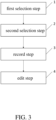
- the invention provides a method for measuring muscle mass which comprises two selection steps.
- a frame selection information is obtained by using a frame to select a fascia region from a provided computed tomography image under the condition that the window width ranges from 300 HU to 500 HU and the window level ranges from 40 HU to 50 HU, wherein the selected range of the fascia region includes a muscle.
- a muscle information of the muscle is obtained by calculating a pixel value in the frame-selected fascia region under the condition that the HU value of the CT image ranges from -29 HU to 150 HU.
- the invention also provides a method for measuring muscle mass which comprises two selection steps.
- a first frame selection information and a second frame selection information are obtained by using a frame to select a first fascia region and a second fascia region from a provided computed tomography image under the condition that the window width ranges from 300 HU to 500 HU and the window level ranges from 40 HU to 50 HU, wherein the first selected range of the fascia region includes a first muscle and the second selected range of the fascia region includes a second muscle.
- a first muscle information of the first muscle is obtained by calculating a pixel value in the frame-selected first fascia region and a second muscle information of the first muscle is obtained by calculating a pixel value in the frame-selected second fascia region under the condition that the HU value of the CT image ranges from -29 HU to 150 HU.
- the computed tomography image is selected from a group consisting of a horizontal computed tomography scan of the third vertebra or a horizontal computed tomography scan of fourth vertebra
- the muscle is chosen from a group consisting of the following: transverse abdominis, rectus abdominis, internal oblique, external oblique, psoas, quadratus lumborum, paraspinal muscle and combinations thereof.
- the computed tomography image is selected from a group consisting of a horizontal computed tomography scan of the third vertebra or a horizontal computed tomography scan of fourth vertebra
- the first muscle is chosen from a group consisting of the following: transverse abdominis, rectus abdominis, internal oblique, external oblique, psoas, quadratus lumborum, paraspinal muscle and combinations thereof
- the second muscle is chosen from a group consisting of the following: transverse abdominis, rectus abdominis, internal oblique, external oblique, psoas, quadratus lumborum, paraspinal muscle and combinations thereof.
- the present method further comprises a record step to store the frame selection information in a record module after the first selection step and to store the muscle information in the record module after the second selection step.
- the present method further comprises a record step to store the frame selection information in a record module after the first selection step and to store the first muscle information and the second muscle information in the record module after the second selection step.
- the present method further comprises using an analysis module to automatically select the fascia region from the computed tomography image in the first selection step, wherein the analysis module receives a frame selection reference object and then organize and summarize a detail feature within the frame selection range of the frame selection reference object, wherein the analysis module use the detail feature to obtain the frame selection information by framing out a portion of the computed tomography image which having the same detail feature.
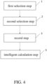
- the present method further comprises an intelligent calculation step after the record step, wherein a calculation module is used to present the muscle information in the digital format so as to express one of the following of the muscle: mass, density and distribution, wherein the calculation module communicates with the record module to receive the muscle information through signal.
- a calculation module is used to present the muscle information in the digital format so as to express one of the following of the muscle: mass, density and distribution, wherein the calculation module communicates with the record module to receive the muscle information through signal.
- the present method further comprising using an analysis module to automatically select the first fascia region and the second fascia region from the computed tomography image in the first selection step, wherein the analysis module receives a frame selection reference object and then organize and summarize a detail feature within the frame selection range of the frame selection reference object, wherein the analysis module use the detail feature to obtain the first frame selection information and the second frame selection information by framing out a portion of the computed tomography image which having the same detail feature.
- the present method further comprising an intelligent calculation step after the record step, wherein a calculation module is used to present the first muscle information and the second muscle information in the digital format so as to express one of the following the first muscle and the second muscle: mass, density and distribution, wherein the calculation module communicates with the record module to receive the first muscle information and the second muscle information through signal.
- a calculation module is used to present the first muscle information and the second muscle information in the digital format so as to express one of the following the first muscle and the second muscle: mass, density and distribution, wherein the calculation module communicates with the record module to receive the first muscle information and the second muscle information through signal.
- the effects of the present invention are that the sarcopenia is not determined by measuring the physical ability and that the computed tomography technology is used to obtain accurate muscle mass data. Hence, the accurate determination is available, also the variation of the muscle mass may be traced during the following treatment stage or rehabilitation stage.
- the present invention automatically frames out a fascia area to be measured when using the computed tomography to measure the muscle mass. This is very different from the traditional skills that the medical staff manually frame out. Therefore, it can ensure that all the fascia parts of the muscle surface are framed out without any omission for each fame selection. After that, by adjusting the HU value, the muscle parts can be highlighted on the computed tomography image to obtain accurate muscle mass data.
- the present invention provides a method for measuring muscle mass which comprises two selection steps.
- a frame selection information is obtained by using a frame to select a fascia region from a provided computed tomography image under the condition that the window width ranges from 300 HU to 500 HU and the window level ranges from 40 HU to 50 HU, wherein the selected range of the fascia region includes a muscle.
- a muscle information of the muscle is obtained by calculating a pixel value in the frame-selected fascia region under the condition that the HU value of the CT image ranges from -29 HU to 150 HU.
- the computed tomography image is optionally selected from but not limited to a group consisting of a horizontal computed tomography scan of the third vertebra or a horizontal computed tomography scan of fourth vertebra.
- the muscle is a portion of the lumbar paraspinal muscles, which is selected from but not limited to the following: transverse abdominis, rectus abdominis, internal oblique, external oblique, psoas, quadratus lumborum, paraspinal muscle and combinations thereof.
- a muscle distribution map is generated under the condition that the HU value of the computed tomography scan is ranged from -29 HU to 150 HU in the second selection step.
- the preset window is limited between 300 HU to 500 HU
- the preset window level is not limited between 40 HU to 50 HU
- the preset HU value is not limited between -29HU to 150 HU.
- a record step (3) is further included so as to store the frame selection information in a record module after the first selection step (1) and to store the muscle distribution map and the muscle information in the record module after the second selection step (2).
- an edit step is further included to sort the muscle distribution maps in sequence according to the date and time when the second selection step (2) is performed, to edit those muscle distribution maps sorted in sequence into a video such that the observer may better understand the variations in the muscles, or to integrate the muscle distribution maps into a three-dimensional stereoscopic map.
- an analysis module is used to automatically frame out the fascia area in the computed tomography scan in the first selection step (1).
- the analysis module may receive a frame selection reference object and then organize and summarize a detail features within the frame selection range of the frame selection reference object.
- a portion of the computed tomography scan image matching the detail features is framed out to obtain frame selection information.
- the detail features include but not limited to the following: orientation, shading, texture, bump or relative position. Understandably, the frame selection of the traditional computer scanning is manually framed out, and then the experience abundance and the observation of the framer is severely challenged.
- the invention presents an "intelligent learning" approach to determine the computed tomography scan image, also may adjust the classification of the frame selection reference object according to the symptom to the measure so as to be suitable for different application situations.
- one object of this invention is measure and determine the symptoms of the sarcopenia.
- the used frame selection reference object is at least a computed tomography scan image which has been framed out any muscle related to the sarcopenia such that the analysis module may use it as the benchmark for framing out.
- the object of framing out the muscle(s) related to the sarcopenia is to effectively analysis or determine the symptoms of the sarcopenia.
- the object of the invention is to measure the muscle mass. Hence, it may be adjusted according to the computed tomography scan images of muscles, tissues or bones in different portions of the human body, such that the detail information of the to be measured portion of the human body may be achieved.
- the framed out portion is not limited only to the muscles associated with the sarcopenia.
- an intelligent operation step (5) is further included after the record step (3).
- an operation module is used to present the muscle information in the digital format for expressing mass, density or distribution of the muscle.
- the operation module is connected with the record module via signal so as to receive the muscle information.
- FIG. 5B , FIG. 5C and FIG. 5 are a series of schematic of the computed tomography scan.
- FIG. 5B is result of the general computed tomography scan image
- FIG. 5C is the automatically framed out result along fascia in the computed tomography scan image according to this invention
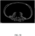
- FIG. 5E is used to present the schematic of the muscle distribution map after the HU value is adjusted in the second selection step (2).
- FIG. 5C is a computed tomography scan acquired with the situation that the window width is 450 HU and the window level is 45 HU.
- FIG. 5C is a computed tomography scan acquired with the situation that the window width is 450 HU and the window level is 45 HU.
- different ranges framed out by different dotted lines are selected respectively by the analysis module in reference with the information of the frame selection reference object and is selected according to the detail features of the frame selection reference object. Understandly, the automatic selection mechanism may avoid the experience required by the manually selection mechanism, and then reduce error, reduce manpower and improve effectiveness of muscle measurement. Still in a preferred embodiment, as shown in FIG. 5E , in the second selection step, the contrast between muscle portions and non-muscle portions in the muscle distribution map is increased because the HU value is the computed tomography scan has been adjusted, and then the muscle information in the muscle distribution map is easily determined by the human body or by the machine.
- the present invention provides another method for measuring muscle mass which comprises two selection steps.
- a first frame selection information and a second frame selection information are obtained by using a frame to select a first fascia region and a second fascia region from a provided computed tomography image under the condition that the window width ranges from 300 HU to 500 HU and the window level ranges from 40 HU to 50 HU, wherein the first selected range of the fascia region includes a first muscle and the second selected range of the fascia region includes a second muscle.
- a first muscle information of the first muscle is obtained by calculating a pixel value in the frame-selected first fascia region and a second muscle information of the first muscle is obtained by calculating a pixel value in the frame-selected second fascia region under the condition that the HU value of the CT image ranges from -29 HU to 150 HU.
- the computed tomography image is optionally selected from but not limited to a group consisting of a horizontal computed tomography scan of the third vertebra or a horizontal computed tomography scan of fourth vertebra.
- the first muscle is a portion of the lumbar paraspinal muscles, which include but not limited to the following: transverse abdominis, rectus abdominis, internal oblique, external oblique, psoas, quadratus lumborum, paraspinal muscle and combinations thereof.
- the second muscle is a portion of the lumbar paraspinal muscles, which include but not limited to the following: transverse abdominis, rectus abdominis, internal oblique, external oblique, psoas, quadratus lumborum, paraspinal muscle and combinations thereof.
- a muscle distribution map is generated under the condition that the HU value of the computed tomography scan is ranged from -29 HU to 150 HU in the second selection step.
- a record step (3) is further included so as to store the first frame selection information and the second frame selection information in a record module after the first selection step (1) and to store the first muscle information and the second muscle information in the record module after the second selection step (2).
- an edit step is further included to sort the muscle distribution maps in sequence according to the date and time when the second selection step (2) is performed, to edit those muscle distribution maps sorted in sequence into a video such that the observer may better understand the variations in the muscles, or to integrate the muscle distribution maps into a three-dimensional stereoscopic map.
- an analysis module is used to automatically frame out the first fascia area and the second fascia area in the computed tomography scan in the first selection step (1).
- the analysis module may receive a frame selection reference object and then organize and summarize a detail features within the frame selection range of the frame selection reference object. Also, by using the detail features, a portion of the computed tomography scan image matching the detail features is framed out to obtain a first frame selection information and a second frame selection information.
- the automatic frame machoism is essentially similar with that of the first preferred embodiment of this invention. Hence, the details are not repeated described herein.
- an intelligent operation step (5) is further included after the record step (3).
- an operation module is used to present the first muscle information and then second muscle information in the digital format for expressing mass, density or distribution of the first muscle and the second muscle respectively.
- the operation module is connected with the record module via signal so as to receive the first muscle information and the second muscle information.
- FIG. 5A is the framed out result after using AI (artificial intelligence) to automatically classify the muscles in the computed tomography scan image result.
- AI artificial intelligence
- any fascia on the muscle surface of any muscle on the computed tomography scan image may be framed out individually such that the first frame selection information and the second selection information may be obtained. After that, muscle mass of any specific muscle may be analyzed.
- FIG. 5B is result of the general computed tomography scan image
- FIG. 5C is frame selection result by using the computed tomography scan image shown in FIG. 5B to frame out the fascia on the muscle surface of each muscle with the situation that the window width is 440 HU and the window level is 45 HU.
- FIG. 5D the result acquired in the second selection step (2) after the HU value is adjusted is shown in FIG. 5D , wherein the HU value is adjusted to between -29 HU to 150 HU such that the muscle portions covered by the fascia are appeared and is benefit for the following operation about mass or data of the muscle
- FIG. 5E presents the quantitative results of the mass region on the framed out range in the FIG. 5D .
- the range framed out by each dotted line is selected by using the analysis module to refer the information of the frame selection reference object and then decided by the detail feature of the frame selection reference object.
- the automatic selection mechanism may avoid the experience required by the manually selection mechanism, and then reduce error, reduce manpower and improve effectiveness of muscle measurement. Still in a preferred embodiment, as shown in FIG. 5D , in the second selection step, the contrast between muscle portions and non-muscle portions in the muscle distribution map is increased because the HU value is the computed tomography scan has been adjusted, and then the muscle information in the muscle distribution map is easily determined by the human body or by the machine. Also in another preferred embodiment, more and more frame selection reference information is included. Understandably, the amount of the frame selection reference information corresponds to the amount of the muscle and is sorted by number, although it is not limited herein.
- muscles, tissues or bones of human body may be measured according to the preset HU value.
- HU value may be properly preset according to the human body portions to be measured, but not is limited to measure the skeletal muscle.
- Table 1 Tissues HU value range Skeletal muscle -29 ⁇ 150 intermuscular adipose tissue -190 ⁇ -30 Visceral adipose tissue -150 ⁇ -50 Subcutaneous adipose tissue -190 ⁇ -30 Bone 250 ⁇ 2000
- the general methods for measuring human body include but not limited to the following: bioelectrical impedance analysis (BIA), computed tomography (CT), magnetic resonance imaging (MRI), dual-energy X-ray absorptiometry (DXA), ultrasonic echo method or naked eye observation.
- BIOA bioelectrical impedance analysis
- CT computed tomography
- MRI magnetic resonance imaging
- DXA dual-energy X-ray absorptiometry
- ultrasonic echo method or naked eye observation.
- CT uses the digital geometrical process to reconstruct a three-dimensional radiological medical image, and is a medical diagnosis tool combining X-ray technology and computer operation.
- this technology mainly irradiate light along a single axis to an object in rotation and then uses the computer three-dimensional technology to reconstruct the tomographic image.
- a MRI contrast agent is used to enhance the contrast between different tissues before a computed tomography scan is performed.
- the MRI contrast agent is a material may be injected into the human body through the vein and then absorbed by the human body tissues along with the distribution of the blood vessels.
- the MRI contrast agent may modify the contrast and brightness of different tissues when shooting images, such that blood vessels, tumors, organs, muscles or other portions of the human body may be more clear and easy to read.
- the window width indicates the CT value range included in the computed tomography image and the window level indicates the average of the upper limitation and the lower limitation of the window width.
- the three-dimensional image of all tissues, organs, bones, or muscles of the human body may be clearly displayed by stacking the CI images layer by layer, Hence, it may be used by the medical staff as an important reference for diagnosing or treating the physical symptoms.
- the computed tomography scan is a non-invasive technology may quickly and simply reconstruct the three-dimensional image inside human body, it may display situations of most portions of the human body and significantly increase the accuracy of disease diagnosis. Therefore, the invention uses the computed tomography scan to measure the muscle mass.
- each of the muscle, the first muscle or the second muscle is a portion of human muscle groups.
- the human muscle groups include but not limited to the following: lumbar paraspinal muscles, leg muscles, chest muscles, back muscles, abdominal muscles, biceps muscles, triceps muscles or core muscles.
- the lumbar paraspinal muscles include but not limited to the following: intertransverse process muscles, quadratus lumborum muscles, multifidus muscles, lumbar iliocostalis muscles, longissimus muscles and psoas major muscles.
- the leg muscles include but not limited to the following: thigh muscles and calf muscles.
- the thigh muscles include but not limited to the following: sartorius muscles, quadriceps muscle, tensor fasciae late muscles.
- the back thigh muscles include but not limited to the following: biceps femoris muscles, semitendinosus muscles, and semimembranosus muscles.
- the inner thigh muscles include but not limited to the following: pectineus muscles, adductor longus muscles, adductor brevis muscles, adductor magnus muscles, and gracilis muscles.
- the calf muscles include but not limited to the following: front calf muscles, outer calf muscles and back calf muscles.
- the front calf muscles include but not limited to the following: tibialis anterior muscles, extensor longus digitorum muscles, and extensor longus pollicis muscles.
- the outer calf muscles include but not limited to following peroneus longus muscles, and peroneus brevis muscles.
- the back calf muscles include but not limited to the following: triceps surae muscles, flexor digitorum longus muscles, flexor pollicis longus muscles, and tibialis posterior muscles.
- the chest muscles include but not limited to the following: pectoralis major muscles, pectoralis minor muscles, serratus anterior muscles, and intercostal muscles.
- the back muscles include but not limited to the following: trapezius muscles, latissimus dorsi muscles, teres major muscles, teres minor muscles, erector spinae muscles, and rhomboids muscles.
- the abdominal muscles include but not limited to the following: rectus abdominis muscles, internal oblique muscles, transverse abdominis muscles and external oblique muscles.
- the shoulder muscles include but not limited to the following: anterior deltoid muscles, middle deltoid muscles, and rear deltoid muscles.
- the core muscles indicate the muscles from below the diaphragm to the pelvic floor and surrounding the waist, abdomen and central trunk.
- the core muscles include but not limited to the following: transverse abdominis muscles, lumber multifidi muscles, internal oblique muscles, external oblique muscles, rectus abdominis muscles, quadratus lumborum muscles, erector spinae muscles, respiratory diaphragm muscles and pelvic floor muscles.
- the muscle, the first muscle or the second muscle may include one or more muscles listed above although is not limited herein.
- the muscle diseases include but not limited to the following: sarcopenia, muscle atrophy and muscle inflammation.
- the sarcopenia is a symptom that weight and function of the skeletal muscle are continuously lost and weaken throughout the human body. With the reduction of muscle mass of skeletal muscle, slow walking, decreased grip strength, labored movement, repeated falls and body weight loss are unavoidable. Hence, the life qualify is greatly degraded, even disability is happened.
- the sarcopenia usually occurs with advanced age and chronic diseases. Hence, it is more than worth the loss if it is not found early and appropriately treated in the early stage of disability. Mild symptoms may easily weaken limbs health and increase falling probability. Severe symptoms may lead to disability and prolonged lying in bed.
- muscle atrophy indices the gradual loss of muscle mass and/or gradual weakening and degeneration of muscles.
- the present invention not only may measure the symptoms of sarcopenia, but also may perform computed tomography scans on the muscles of specific portions of human body. Therefore, the present invention may observe whether there are other muscle diseases so as to prevent or control muscle diseases, or may promote muscle re-growth so that the muscle mass has been reduced due to a certain reason is restored to the original level, or may restore the muscle strength has been reduced due to a certain reason back to the original level.
- the computed tomography scan may be used to measure total abdominal muscle area (TAMA), intermuscular adipose tissue area (IMAT) or skeletal muscle area (SMA).
- TAMA total abdominal muscle area
- IMAT intermuscular adipose tissue area
- SMA skeletal muscle area
- the total mass area measured by computed tomography scan measurement includes normal attenuation muscle area (NAMA) and low attenuation muscle area (LAMA).
- NAMA normal attenuation muscle area
- LAMA low attenuation muscle area
- NAMA is less prone to fat infiltration and is a high-quality muscle area
- LANA is prone to have fat cells more likely found in muscle fibers and is a poor-quality muscle area.
- LAMA is used as an indicator of physical defects or poor metabolism.
- CT scan may be performed by using Somatom Definition (Siemens Healthineers, Er Weg, Germany), Discovery CT750 HD (GE Healthcare, Milwaukee, WI, USA) or LightSpeed VCT scanner (GE Healthcare).
- the preset parameters for CT scan include but not limited to the following: 120 kVp, automatic medicine preparation (CareDose 4D, Siemen Healthineers, automA and smartmA, GE Healthcare), matrix of 512 x 512, collimation of 0.625mm or any combination thereof.
- image and data acquired by CT scan may be reconstructed by using the "soft tissue filtered back projection technology" (B30f Kernel, Siemens Healthineers, Standard Kernel, GE Healthcare) to setup a slice thickness, such as but not limited to 5mm.
- Soft tissue filtered back projection technology B30f Kernel, Siemens Healthineers, Standard Kernel, GE Healthcare
- 100e150 M1 iopromide Ultravist 370 or Ultravist 300, Bayer Schering Pharma, Berlin, Germany
- a power injector with 2.5e3mL/sec injection rate when an automatic intravenous injector is used, or scan is fixed delay 70 second after the contrast injection is fully injected.
- CT scan may be used to measure any portion of the spine but not be limited to the L3 lumbar vertebrae.
- CT image is performed by using "Analytic Morphomics" which is a semi-automatic computational image processing system written in Mathlab, wherein acquired CT image is coded in the DICOM format and stored in a space database.
- CT images may be segmented by using the medical image analysis software. Particularly, by using the CT image of third vertebra as example, it may automatically identify and separate muscle area, bone area, SAT (subcutaneous adipose tissue) area and VAT (visceral adipose tissue) area.
- the used medical image analysis software is sliceOmatic, although it is not limited to this.
- the axial CT slices of the L3 lumbar vertebra may be automatically selected by using a convolutional network (FCN) segmentation technology so as to automatically generate the following CT image segments: total order abdominal muscle area (TAMA), visceral fat area and subcutaneous fat area.
- TAMA includes all muscles on the selected image, such as psoas major muscles, paraspinal muscles, transversus abdominis muscles, rectus abdominis muscles, quadratus lumborum muscles, internal obliques muscles and external obliques muscles.
- TAMA may be divided into three portions: IMAT for showing adipose tissue between muscles and muscle fibers (shown when the HU value of the CT image is from -190 HU to -30 HU), NAMA for showing healthy muscles (shown when the HU value of the CT image is from 30 HU to 150 HU), and LAMA for showing the un-heathy muscles (shown when the HU value of the CT image is from -29 HU to 29 Hu).
- SAS software SAS institute, Inc., Cary, NC, USA
- R software R Foundation for Statistical Computing, Vienna, Austria
- Pearson correlation coefficient is used to evaluate SMA, TAMA degradation and NAMA/TAMA index.
- T value is the ratio between difference between measurement value and young auult's mean value and younger's standard deviation. If measured T value is larger than -1, LAMA value is less than 1 o IMAT value is less 1, the muscle may be viewed as normal without potential danger. If measured T value is not larger than -1.0 and not less than - 2.0, measured T value is less than -2, measured T value is not larger than 2 but is larger than 1, LAMA value is larger than 2 or IMAT value is larger than 2, the condition of the muscle is abnormal. At this time, muscle steatosis or sarcopenia may happen, and further health examination is required.
- SMRA mean skeletal muscle radiation attenuation
- SI skeletal muscle index
- the critical values of SMA, SMI and SMRA is 99.2 cm 2 , 34.4 cm 2 / m 2 and 34.3 Hu in sequence for female sarcopenia patients and is 144.3 cm 2 , 45.4 cm 2 / m 2 and 38.5 Hu in sequence for male sarcopenia patients. Therefore, these critical values listed above may be used as reference for determining whether sarcopenia is existed or not.
- the effect of the present invention is that the sarcopenia is not determined by using physical fitness but uses computed tomography technology to obtain accurate muscle mass date. Hence, precise determine results may be achieved, and muscle may be traced during the following treating stage or recovering stage for recording the results of increasing muscle mass.
- the present invention automatically frames out a fascia area to be measured and is significantly different than the prior arts that the medical staff manually frame out. Hence, the present invention may ensure the fascia portions on the surfaces of all muscles are all framed out without omission during each frame selection. Further, by adjusting HU value, the muscles portions on the computed tomography scan image are significantly highlighted so that precise muscle mass data may be acquired.
Landscapes
- Health & Medical Sciences (AREA)
- Life Sciences & Earth Sciences (AREA)
- Engineering & Computer Science (AREA)
- Medical Informatics (AREA)
- Heart & Thoracic Surgery (AREA)
- Molecular Biology (AREA)
- Biophysics (AREA)
- High Energy & Nuclear Physics (AREA)
- Veterinary Medicine (AREA)
- Nuclear Medicine, Radiotherapy & Molecular Imaging (AREA)
- Optics & Photonics (AREA)
- Pathology (AREA)
- Radiology & Medical Imaging (AREA)
- Biomedical Technology (AREA)
- Public Health (AREA)
- Physics & Mathematics (AREA)
- Surgery (AREA)
- Animal Behavior & Ethology (AREA)
- General Health & Medical Sciences (AREA)
- Computer Vision & Pattern Recognition (AREA)
- Pulmonology (AREA)
- Theoretical Computer Science (AREA)
- Dentistry (AREA)
- Oral & Maxillofacial Surgery (AREA)
- Physiology (AREA)
- Human Computer Interaction (AREA)
- Apparatus For Radiation Diagnosis (AREA)
- Other Investigation Or Analysis Of Materials By Electrical Means (AREA)
Claims (10)
- - Verfahren zur Messung der Muskelmasse, umfassend:einen ersten Auswahlschritt, wobei eine Rahmenauswahlinformation durch Verwenden eines Rahmens zum Auswählen einer Faszienregion aus einem bereitgestellten Computertomographiebild unter der Bedingung erhalten wird, dass die Fensterbreite im Bereich von 300 HU bis 500 HU liegt und die Fensterebene im Bereich von 40 HU bis 50 HU liegt, wobei der ausgewählte Bereich der Faszienregion einen Muskel umfasst; undeinen zweiten Auswahlschritt, wobei eine Muskelinformation des Muskels durch Berechnen eines Pixelwerts in der durch den Rahmen ausgewählten Faszienregion unter der Bedingung erhalten wird, dass der HU-Wert des CT-Bildes im Bereich von -29 HU bis 150 HU liegt.
- - Verfahren zur Messung der Muskelmasse, umfassend:einen ersten Auswahlschritt, wobei eine erste Rahmenauswahlinformation und eine zweite Rahmenauswahlinformation durch Verwenden eines Rahmens zum Auswählen einer ersten Faszienregion und einer zweiten Faszienregion aus einem bereitgestellten Computertomographiebild unter der Bedingung erhalten werden, dass die Fensterbreite im Bereich von 300 HU bis 500 HU liegt und die Fensterebene im Bereich von 40 HU bis 50 HU liegt, wobei der erste ausgewählte Bereich der Faszienregion einen ersten Muskel umfasst und der zweite ausgewählte Bereich der Faszienregion einen zweiten Muskel umfasst; undeinen zweiten Auswahlschritt, wobei eine erste Muskelinformation des ersten Muskels durch Berechnen eines Pixelwerts in der durch den Rahmen ausgewählten ersten Faszienregion und eine zweite Muskelinformation des ersten Muskels durch Berechnen eines Pixelwerts in der durch den Rahmen ausgewählten zweiten Faszienregion unter der Bedingung erhalten werden, dass der HU-Wert des CT-Bildes im Bereich von -29 HU bis 150 HU liegt.
- - Verfahren nach Anspruch 1, wobei das Computertomographiebild aus einer Gruppe bestehend aus einem horizontalen Computertomographiescan des dritten Wirbels oder einem horizontalen Computertomographiescan des vierten Wirbels ausgewählt wird, und wobei der Muskel aus einer Gruppe ausgewählt wird, die aus folgenden besteht: quer verlaufendem Bauchmuskel, geradem Bauchmuskel, innerem schrägem Bauchmuskel, äußerem schrägem Bauchmuskel, Lendenmuskel, viereckigem Lendenmuskel, paraspinalem Muskel und Kombinationen davon.
- - Verfahren nach Anspruch 2, wobei das Computertomographiebild aus einer Gruppe bestehend aus einem horizontalen Computertomographiescan des dritten Wirbels oder einem horizontalen Computertomographiescan des vierten Wirbels ausgewählt wird, und wobei der erste Muskel aus einer Gruppe ausgewählt wird, die aus folgenden besteht: quer verlaufendem Bauchmuskel, geradem Bauchmuskel, innerem schrägem Bauchmuskel, äußerem schrägem Bauchmuskel, Lendenmuskel, viereckigem Lendenmuskel, paraspinalem Muskel und Kombinationen davon, und wobei der zweite Muskel aus einer Gruppe ausgewählt wird, die aus folgenden besteht: quer verlaufendem Bauchmuskel, geradem Bauchmuskel, innerem schrägem Bauchmuskel, äußerem schrägem Bauchmuskel, Lendenmuskel, viereckigem Lendenmuskel, paraspinalem Muskel und Kombinationen davon.
- - Verfahren nach Anspruch 1, ferner umfassend einen Aufzeichnungsschritt zum Speichern der Rahmenauswahlinformation in einem Aufzeichnungsmodul nach dem ersten Auswahlschritt und zum Speichern der Muskelinformation im Aufzeichnungsmodul nach dem zweiten Auswahlschritt.
- - Verfahren nach Anspruch 2, ferner umfassend einen Aufzeichnungsschritt zum Speichern der Rahmenauswahlinformation in einem Aufzeichnungsmodul nach dem ersten Auswahlschritt und zum Speichern der ersten Muskelinformation und der zweiten Muskelinformation im Aufzeichnungsmodul nach dem zweiten Auswahlschritt.
- - Verfahren nach Anspruch 5, ferner umfassend ein Verwenden eines Analysemoduls zum automatischen Auswählen der Faszienregion aus dem Computertomographiebild im ersten Auswahlschritt, wobei das Analysemodul ein Rahmenauswahl-Referenzobjekt empfängt und dann ein Detailmerkmal innerhalb des Rahmenauswahlbereichs des Rahmenauswahl-Referenzobjekts organisiert und zusammenfasst, wobei das Analysemodul das Detailmerkmal verwendet, um die Rahmenauswahlinformation durch Ausrahmen eines Abschnitts des Computertomographiebildes zu erhalten, der dasselbe Detailmerkmal aufweist.
- - Verfahren nach Anspruch 7, ferner umfassend einen Schritt intelligenter Berechnung nach dem Aufzeichnungsschritt, wobei ein Berechnungsmodul verwendet wird, um die Muskelinformation im Digitalformat darzustellen, um eines von Folgenden des Muskels auszudrücken: Masse, Dichte und Verteilung, wobei das Berechnungsmodul mit dem Aufzeichnungsmodul kommuniziert, um die Muskelinformation mittels eines Signals zu empfangen.
- - Verfahren nach Anspruch 6, ferner umfassend ein Verwenden eines Analysemoduls zum automatischen Auswählen der ersten Faszienregion und der zweiten Faszienregion aus dem Computertomographiebild im ersten Auswahlschritt, wobei das Analysemodul ein Rahmenauswahl-Referenzobjekt empfängt und dann ein Detailmerkmal innerhalb des Rahmenauswahlbereichs des Rahmenauswahl-Referenzobjekts organisiert und zusammenfasst, wobei das Analysemodul das Detailmerkmal verwendet, um die ersten Rahmenauswahlinformation und die zweiten Rahmenauswahlinformation durch Ausrahmen eines Abschnitts des Computertomographiebildes zu erhalten, der dasselbe Detailmerkmal aufweist.
- - Verfahren nach Anspruch 9, ferner umfassend einen Schritt intelligenter Berechnung nach dem Aufzeichnungsschritt, wobei ein Berechnungsmodul verwendet wird, um die ersten Muskelinformation und die zweiten Muskelinformation im Digitalformat darzustellen, um eines von Folgendes des ersten Muskels und des zweiten Muskels auszudrücken: Masse, Dichte und Verteilung, wobei das Berechnungsmodul mit dem Aufzeichnungsmodul kommuniziert, um die ersten Muskelinformation und die zweiten Muskelinformation mittels eines Signals zu empfangen.
Applications Claiming Priority (1)
| Application Number | Priority Date | Filing Date | Title |
|---|---|---|---|
| TW111103422A TWI797976B (zh) | 2022-01-26 | 2022-01-26 | 測量肌肉量的方法 |
Publications (3)
| Publication Number | Publication Date |
|---|---|
| EP4218591A1 EP4218591A1 (de) | 2023-08-02 |
| EP4218591C0 EP4218591C0 (de) | 2025-02-05 |
| EP4218591B1 true EP4218591B1 (de) | 2025-02-05 |
Family
ID=84421158
Family Applications (1)
| Application Number | Title | Priority Date | Filing Date |
|---|---|---|---|
| EP22211686.5A Active EP4218591B1 (de) | 2022-01-26 | 2022-12-06 | Verfahren zur messung der muskelmasse |
Country Status (4)
| Country | Link |
|---|---|
| EP (1) | EP4218591B1 (de) |
| JP (1) | JP7482461B2 (de) |
| KR (1) | KR102937602B1 (de) |
| TW (1) | TWI797976B (de) |
Families Citing this family (2)
| Publication number | Priority date | Publication date | Assignee | Title |
|---|---|---|---|---|
| TWI848834B (zh) * | 2023-10-16 | 2024-07-11 | 國立成功大學 | 時序性醫療資訊分析系統及其方法 |
| TWI899961B (zh) * | 2024-04-19 | 2025-10-01 | 幻景啟動股份有限公司 | 立體浮空影像轉換系統與方法 |
Family Cites Families (12)
| Publication number | Priority date | Publication date | Assignee | Title |
|---|---|---|---|---|
| US5068788A (en) * | 1988-11-29 | 1991-11-26 | Columbia Scientific Inc. | Quantitative computed tomography system |
| JP4405716B2 (ja) | 2002-08-26 | 2010-01-27 | ジーイー・メディカル・システムズ・グローバル・テクノロジー・カンパニー・エルエルシー | 画像処理装置およびx線ctシステム |
| JP5534607B2 (ja) | 2008-02-25 | 2014-07-02 | 株式会社日立メディコ | 医用画像処理装置及び、そのプログラム |
| CA2882962C (en) | 2012-08-30 | 2020-01-07 | The Regents Of The University Of Michigan | Analytic morphomics: high speed medical image automated analysis method |
| JP6492553B2 (ja) | 2014-11-07 | 2019-04-03 | コニカミノルタ株式会社 | 画像処理装置及びプログラム |
| JP6929695B2 (ja) | 2016-05-09 | 2021-09-01 | キヤノンメディカルシステムズ株式会社 | 医用画像診断装置及び管理装置 |
| CN116779143A (zh) | 2017-05-09 | 2023-09-19 | 巴克斯特国际公司 | 胃肠外营养诊断系统、设备和方法 |
| TWI652039B (zh) | 2017-10-06 | 2019-03-01 | 金上達科技股份有限公司 | Simple detection method and system for sarcopenia |
| TWI646943B (zh) | 2017-12-08 | 2019-01-11 | 財團法人自行車暨健康科技工業研究發展中心 | Method for screening for muscular dysfunction through functional fitness |
| US11182920B2 (en) | 2018-04-26 | 2021-11-23 | Jerry NAM | Automated determination of muscle mass from images |
| EP3841983B1 (de) * | 2018-08-22 | 2024-01-10 | Furuno Electric Co., Ltd. | Ultraschallanalysevorrichtung, ultraschallanalyseverfahren und ultraschallanalyseprogramm |
| RU2754291C1 (ru) * | 2020-12-01 | 2021-08-31 | Федеральное государственное бюджетное научное учреждение "Научно-исследовательский институт комплексных проблем сердечно-сосудистых заболеваний" (НИИ КПССЗ) | Способ определения саркопении с использованием количественной оценки мышечной ткани по данным компьютерной томографии грудной клетки |
-
2022
- 2022-01-26 TW TW111103422A patent/TWI797976B/zh active
- 2022-12-06 EP EP22211686.5A patent/EP4218591B1/de active Active
- 2022-12-13 JP JP2022198507A patent/JP7482461B2/ja active Active
-
2023
- 2023-01-20 KR KR1020230008918A patent/KR102937602B1/ko active Active
Also Published As
| Publication number | Publication date |
|---|---|
| TW202329888A (zh) | 2023-08-01 |
| JP7482461B2 (ja) | 2024-05-14 |
| KR102937602B1 (ko) | 2026-03-11 |
| EP4218591A1 (de) | 2023-08-02 |
| KR20230115249A (ko) | 2023-08-02 |
| EP4218591C0 (de) | 2025-02-05 |
| TWI797976B (zh) | 2023-04-01 |
| JP2023109150A (ja) | 2023-08-07 |
Similar Documents
| Publication | Publication Date | Title |
|---|---|---|
| Groell et al. | The reliability of bone age determination in central European children using the Greulich and Pyle method. | |
| Boesen et al. | Osteoarthritis year in review 2016: imaging | |
| EP4218591A1 (de) | Verfahren zur messung der muskelmasse | |
| Tian et al. | Potential value of the PixelShine deep learning algorithm for increasing quality of 70 kVp+ ASiR-V reconstruction pelvic arterial phase CT images | |
| Mao et al. | Application of quantitative computed tomography for assessment of trabecular bone mineral density, microarchitecture and mechanical property | |
| Cho et al. | CT venography for deep vein thrombosis using a low tube voltage (100 kVp) setting could increase venous enhancement and reduce the amount of administered iodine | |
| CN113679404B (zh) | 一种骨密度测量系统及其测量方法 | |
| Paul et al. | Dual-source 128-slice MDCT neck: radiation dose and image quality estimation of three different protocols | |
| Conrads et al. | Ultrahigh-resolution computed tomography of the cervical spine without dose penalty employing a cadmium-telluride photon-counting detector | |
| Aliboni et al. | Quantitative CT analysis in chronic hypersensitivity pneumonitis: a convolutional neural network approach | |
| Zhang et al. | Deep learning-based fully automated segmentation of regional muscle volume and spatial intermuscular fat using CT | |
| US12207962B2 (en) | Method for measuring muscle mass | |
| Ferencik et al. | Coronary calcium quantification using various calibration phantoms and scoring thresholds | |
| Tang et al. | Adaptive iterative dose reduction 3D integrated with automatic tube current modulation for CT coronary artery calcium quantification: comparison to traditional filtered back projection in an anthropomorphic phantom and patients | |
| Thor et al. | Low tube voltage dual source computed tomography to reduce contrast media doses in adult abdomen examinations: A phantom study | |
| Sakai et al. | Radiofrequency echographic multi-spectrometry-based measurement of bone mineral density in patients with severe motor and intellectual disability: An opportunity for patients with severe scoliosis and hip dislocation | |
| CN116433560A (zh) | 基于超声双模态影像的肌肉疾病识别方法、装置及设备 | |
| Tsekoura et al. | Assessment of muscle mass in the elderly in clinical practice. | |
| Zhou et al. | The ultrafast, high‐pitch turbo FLASH mode of third‐generation dual‐source CT: Effect of different pitch and corresponding SFOV on image quality in a phantom study | |
| Zhang et al. | Refining the radiation and contrast medium dose in weight‐grouped scanning protocols for coronary CT angiography | |
| RU2754291C1 (ru) | Способ определения саркопении с использованием количественной оценки мышечной ткани по данным компьютерной томографии грудной клетки | |
| Machida et al. | Reduction of coronary motion artifacts in prospectively electrocardiography-gated coronary computed tomography angiography using monochromatic imaging at various energy levels in combination with a motion correction algorithm on single-source fast tube voltage switching dual-energy computed tomography: a phantom experiment | |
| Fang et al. | Dual-energy CT virtual non-calcium image is beneficial for the detection of non-displaced knee fractures | |
| RU2727449C1 (ru) | Способ количественной оценки степени изменения ягодичных мышц у больных идиопатическим сколиозом | |
| Takavar et al. | Determination of optimum filter in myocardial SPECT: a phantom study |
Legal Events
| Date | Code | Title | Description |
|---|---|---|---|
| PUAI | Public reference made under article 153(3) epc to a published international application that has entered the european phase |
Free format text: ORIGINAL CODE: 0009012 |
|
| STAA | Information on the status of an ep patent application or granted ep patent |
Free format text: STATUS: REQUEST FOR EXAMINATION WAS MADE |
|
| 17P | Request for examination filed |
Effective date: 20230327 |
|
| AK | Designated contracting states |
Kind code of ref document: A1 Designated state(s): AL AT BE BG CH CY CZ DE DK EE ES FI FR GB GR HR HU IE IS IT LI LT LU LV MC ME MK MT NL NO PL PT RO RS SE SI SK SM TR |
|
| RIN1 | Information on inventor provided before grant (corrected) |
Inventor name: LEE, TSUNG-HAN Inventor name: KUO, PO-TSUN Inventor name: WU, MENG-JHEN Inventor name: CHEN, CHAO-YUN Inventor name: LAI, CHENG-SHIH Inventor name: WANG, BOW Inventor name: LAI, YU-HSUAN Inventor name: TSAI, YI-SHAN |
|
| GRAP | Despatch of communication of intention to grant a patent |
Free format text: ORIGINAL CODE: EPIDOSNIGR1 |
|
| STAA | Information on the status of an ep patent application or granted ep patent |
Free format text: STATUS: GRANT OF PATENT IS INTENDED |
|
| RIC1 | Information provided on ipc code assigned before grant |
Ipc: A61B 6/50 20240101ALI20240711BHEP Ipc: A61B 6/00 20060101ALI20240711BHEP Ipc: A61B 6/03 20060101AFI20240711BHEP |
|
| INTG | Intention to grant announced |
Effective date: 20240806 |
|
| GRAS | Grant fee paid |
Free format text: ORIGINAL CODE: EPIDOSNIGR3 |
|
| GRAA | (expected) grant |
Free format text: ORIGINAL CODE: 0009210 |
|
| STAA | Information on the status of an ep patent application or granted ep patent |
Free format text: STATUS: THE PATENT HAS BEEN GRANTED |
|
| AK | Designated contracting states |
Kind code of ref document: B1 Designated state(s): AL AT BE BG CH CY CZ DE DK EE ES FI FR GB GR HR HU IE IS IT LI LT LU LV MC ME MK MT NL NO PL PT RO RS SE SI SK SM TR |
|
| REG | Reference to a national code |
Ref country code: GB Ref legal event code: FG4D |
|
| REG | Reference to a national code |
Ref country code: CH Ref legal event code: EP |
|
| REG | Reference to a national code |
Ref country code: IE Ref legal event code: FG4D |
|
| REG | Reference to a national code |
Ref country code: DE Ref legal event code: R096 Ref document number: 602022010321 Country of ref document: DE |
|
| U01 | Request for unitary effect filed |
Effective date: 20250304 |
|
| U07 | Unitary effect registered |
Designated state(s): AT BE BG DE DK EE FI FR IT LT LU LV MT NL PT RO SE SI Effective date: 20250409 |
|
| PG25 | Lapsed in a contracting state [announced via postgrant information from national office to epo] |
Ref country code: RS Free format text: LAPSE BECAUSE OF FAILURE TO SUBMIT A TRANSLATION OF THE DESCRIPTION OR TO PAY THE FEE WITHIN THE PRESCRIBED TIME-LIMIT Effective date: 20250505 |
|
| PG25 | Lapsed in a contracting state [announced via postgrant information from national office to epo] |
Ref country code: PL Free format text: LAPSE BECAUSE OF FAILURE TO SUBMIT A TRANSLATION OF THE DESCRIPTION OR TO PAY THE FEE WITHIN THE PRESCRIBED TIME-LIMIT Effective date: 20250205 |
|
| PG25 | Lapsed in a contracting state [announced via postgrant information from national office to epo] |
Ref country code: ES Free format text: LAPSE BECAUSE OF FAILURE TO SUBMIT A TRANSLATION OF THE DESCRIPTION OR TO PAY THE FEE WITHIN THE PRESCRIBED TIME-LIMIT Effective date: 20250205 |
|
| PG25 | Lapsed in a contracting state [announced via postgrant information from national office to epo] |
Ref country code: IS Free format text: LAPSE BECAUSE OF FAILURE TO SUBMIT A TRANSLATION OF THE DESCRIPTION OR TO PAY THE FEE WITHIN THE PRESCRIBED TIME-LIMIT Effective date: 20250605 Ref country code: NO Free format text: LAPSE BECAUSE OF FAILURE TO SUBMIT A TRANSLATION OF THE DESCRIPTION OR TO PAY THE FEE WITHIN THE PRESCRIBED TIME-LIMIT Effective date: 20250505 |
|
| PG25 | Lapsed in a contracting state [announced via postgrant information from national office to epo] |
Ref country code: HR Free format text: LAPSE BECAUSE OF FAILURE TO SUBMIT A TRANSLATION OF THE DESCRIPTION OR TO PAY THE FEE WITHIN THE PRESCRIBED TIME-LIMIT Effective date: 20250205 |
|
| PG25 | Lapsed in a contracting state [announced via postgrant information from national office to epo] |
Ref country code: GR Free format text: LAPSE BECAUSE OF FAILURE TO SUBMIT A TRANSLATION OF THE DESCRIPTION OR TO PAY THE FEE WITHIN THE PRESCRIBED TIME-LIMIT Effective date: 20250506 |
|
| PG25 | Lapsed in a contracting state [announced via postgrant information from national office to epo] |
Ref country code: SM Free format text: LAPSE BECAUSE OF FAILURE TO SUBMIT A TRANSLATION OF THE DESCRIPTION OR TO PAY THE FEE WITHIN THE PRESCRIBED TIME-LIMIT Effective date: 20250205 |
|
| PG25 | Lapsed in a contracting state [announced via postgrant information from national office to epo] |
Ref country code: CZ Free format text: LAPSE BECAUSE OF FAILURE TO SUBMIT A TRANSLATION OF THE DESCRIPTION OR TO PAY THE FEE WITHIN THE PRESCRIBED TIME-LIMIT Effective date: 20250205 |
|
| PG25 | Lapsed in a contracting state [announced via postgrant information from national office to epo] |
Ref country code: SK Free format text: LAPSE BECAUSE OF FAILURE TO SUBMIT A TRANSLATION OF THE DESCRIPTION OR TO PAY THE FEE WITHIN THE PRESCRIBED TIME-LIMIT Effective date: 20250205 |
|
| PLBE | No opposition filed within time limit |
Free format text: ORIGINAL CODE: 0009261 |
|
| STAA | Information on the status of an ep patent application or granted ep patent |
Free format text: STATUS: NO OPPOSITION FILED WITHIN TIME LIMIT |
|
| U20 | Renewal fee for the european patent with unitary effect paid |
Year of fee payment: 4 Effective date: 20251124 |
|
| 26N | No opposition filed |
Effective date: 20251106 |