EP4205639A1 - Device for diagnosing abnormality by measuring minimal change in muscle - Google Patents
Device for diagnosing abnormality by measuring minimal change in muscle Download PDFInfo
- Publication number
- EP4205639A1 EP4205639A1 EP22203621.2A EP22203621A EP4205639A1 EP 4205639 A1 EP4205639 A1 EP 4205639A1 EP 22203621 A EP22203621 A EP 22203621A EP 4205639 A1 EP4205639 A1 EP 4205639A1
- Authority
- EP
- European Patent Office
- Prior art keywords
- body part
- vibration
- abnormality
- unit
- processing unit
- Prior art date
- Legal status (The legal status is an assumption and is not a legal conclusion. Google has not performed a legal analysis and makes no representation as to the accuracy of the status listed.)
- Granted
Links
Images



Classifications
-
- A—HUMAN NECESSITIES
- A61—MEDICAL OR VETERINARY SCIENCE; HYGIENE
- A61B—DIAGNOSIS; SURGERY; IDENTIFICATION
- A61B5/00—Measuring for diagnostic purposes; Identification of persons
- A61B5/0048—Detecting, measuring or recording by applying mechanical forces or stimuli
- A61B5/0051—Detecting, measuring or recording by applying mechanical forces or stimuli by applying vibrations
-
- A—HUMAN NECESSITIES
- A61—MEDICAL OR VETERINARY SCIENCE; HYGIENE
- A61B—DIAGNOSIS; SURGERY; IDENTIFICATION
- A61B5/00—Measuring for diagnostic purposes; Identification of persons
- A61B5/24—Detecting, measuring or recording bioelectric or biomagnetic signals of the body or parts thereof
- A61B5/316—Modalities, i.e. specific diagnostic methods
- A61B5/389—Electromyography [EMG]
- A61B5/397—Analysis of electromyograms
-
- A—HUMAN NECESSITIES
- A61—MEDICAL OR VETERINARY SCIENCE; HYGIENE
- A61B—DIAGNOSIS; SURGERY; IDENTIFICATION
- A61B5/00—Measuring for diagnostic purposes; Identification of persons
- A61B5/103—Measuring devices for testing the shape, pattern, colour, size or movement of the body or parts thereof, for diagnostic purposes
- A61B5/11—Measuring movement of the entire body or parts thereof, e.g. head or hand tremor or mobility of a limb
- A61B5/1101—Detecting tremor
-
- A—HUMAN NECESSITIES
- A61—MEDICAL OR VETERINARY SCIENCE; HYGIENE
- A61B—DIAGNOSIS; SURGERY; IDENTIFICATION
- A61B5/00—Measuring for diagnostic purposes; Identification of persons
- A61B5/0002—Remote monitoring of patients using telemetry, e.g. transmission of vital signals via a communication network
- A61B5/0015—Remote monitoring of patients using telemetry, e.g. transmission of vital signals via a communication network characterised by features of the telemetry system
- A61B5/0022—Monitoring a patient using a global network, e.g. telephone networks, internet
-
- A—HUMAN NECESSITIES
- A61—MEDICAL OR VETERINARY SCIENCE; HYGIENE
- A61B—DIAGNOSIS; SURGERY; IDENTIFICATION
- A61B5/00—Measuring for diagnostic purposes; Identification of persons
- A61B5/02—Detecting, measuring or recording for evaluating the cardiovascular system, e.g. pulse, heart rate, blood pressure or blood flow
- A61B5/02007—Evaluating blood vessel condition, e.g. elasticity, compliance
-
- A—HUMAN NECESSITIES
- A61—MEDICAL OR VETERINARY SCIENCE; HYGIENE
- A61B—DIAGNOSIS; SURGERY; IDENTIFICATION
- A61B5/00—Measuring for diagnostic purposes; Identification of persons
- A61B5/103—Measuring devices for testing the shape, pattern, colour, size or movement of the body or parts thereof, for diagnostic purposes
- A61B5/11—Measuring movement of the entire body or parts thereof, e.g. head or hand tremor or mobility of a limb
- A61B5/1104—Measuring movement of the entire body or parts thereof, e.g. head or hand tremor or mobility of a limb induced by stimuli or drugs
-
- A—HUMAN NECESSITIES
- A61—MEDICAL OR VETERINARY SCIENCE; HYGIENE
- A61B—DIAGNOSIS; SURGERY; IDENTIFICATION
- A61B5/00—Measuring for diagnostic purposes; Identification of persons
- A61B5/103—Measuring devices for testing the shape, pattern, colour, size or movement of the body or parts thereof, for diagnostic purposes
- A61B5/11—Measuring movement of the entire body or parts thereof, e.g. head or hand tremor or mobility of a limb
- A61B5/1107—Measuring contraction of parts of the body, e.g. organ or muscle
-
- A—HUMAN NECESSITIES
- A61—MEDICAL OR VETERINARY SCIENCE; HYGIENE
- A61B—DIAGNOSIS; SURGERY; IDENTIFICATION
- A61B5/00—Measuring for diagnostic purposes; Identification of persons
- A61B5/22—Ergometry; Measuring muscular strength or the force of a muscular blow
- A61B5/224—Measuring muscular strength
-
- A—HUMAN NECESSITIES
- A61—MEDICAL OR VETERINARY SCIENCE; HYGIENE
- A61B—DIAGNOSIS; SURGERY; IDENTIFICATION
- A61B5/00—Measuring for diagnostic purposes; Identification of persons
- A61B5/24—Detecting, measuring or recording bioelectric or biomagnetic signals of the body or parts thereof
- A61B5/25—Bioelectric electrodes therefor
- A61B5/251—Means for maintaining electrode contact with the body
- A61B5/256—Wearable electrodes, e.g. having straps or bands
-
- A—HUMAN NECESSITIES
- A61—MEDICAL OR VETERINARY SCIENCE; HYGIENE
- A61B—DIAGNOSIS; SURGERY; IDENTIFICATION
- A61B5/00—Measuring for diagnostic purposes; Identification of persons
- A61B5/24—Detecting, measuring or recording bioelectric or biomagnetic signals of the body or parts thereof
- A61B5/25—Bioelectric electrodes therefor
- A61B5/251—Means for maintaining electrode contact with the body
- A61B5/257—Means for maintaining electrode contact with the body using adhesive means, e.g. adhesive pads or tapes
-
- A—HUMAN NECESSITIES
- A61—MEDICAL OR VETERINARY SCIENCE; HYGIENE
- A61B—DIAGNOSIS; SURGERY; IDENTIFICATION
- A61B5/00—Measuring for diagnostic purposes; Identification of persons
- A61B5/24—Detecting, measuring or recording bioelectric or biomagnetic signals of the body or parts thereof
- A61B5/25—Bioelectric electrodes therefor
- A61B5/279—Bioelectric electrodes therefor specially adapted for particular uses
- A61B5/296—Bioelectric electrodes therefor specially adapted for particular uses for electromyography [EMG]
-
- A—HUMAN NECESSITIES
- A61—MEDICAL OR VETERINARY SCIENCE; HYGIENE
- A61B—DIAGNOSIS; SURGERY; IDENTIFICATION
- A61B5/00—Measuring for diagnostic purposes; Identification of persons
- A61B5/24—Detecting, measuring or recording bioelectric or biomagnetic signals of the body or parts thereof
- A61B5/316—Modalities, i.e. specific diagnostic methods
- A61B5/389—Electromyography [EMG]
- A61B5/395—Details of stimulation, e.g. nerve stimulation to elicit EMG response
-
- A—HUMAN NECESSITIES
- A61—MEDICAL OR VETERINARY SCIENCE; HYGIENE
- A61B—DIAGNOSIS; SURGERY; IDENTIFICATION
- A61B5/00—Measuring for diagnostic purposes; Identification of persons
- A61B5/45—For evaluating or diagnosing the musculoskeletal system or teeth
- A61B5/4519—Muscles
-
- A—HUMAN NECESSITIES
- A61—MEDICAL OR VETERINARY SCIENCE; HYGIENE
- A61B—DIAGNOSIS; SURGERY; IDENTIFICATION
- A61B5/00—Measuring for diagnostic purposes; Identification of persons
- A61B5/68—Arrangements of detecting, measuring or recording means, e.g. sensors, in relation to patient
- A61B5/6801—Arrangements of detecting, measuring or recording means, e.g. sensors, in relation to patient specially adapted to be attached to or worn on the body surface
- A61B5/6802—Sensor mounted on worn items
-
- A—HUMAN NECESSITIES
- A61—MEDICAL OR VETERINARY SCIENCE; HYGIENE
- A61B—DIAGNOSIS; SURGERY; IDENTIFICATION
- A61B5/00—Measuring for diagnostic purposes; Identification of persons
- A61B5/68—Arrangements of detecting, measuring or recording means, e.g. sensors, in relation to patient
- A61B5/6801—Arrangements of detecting, measuring or recording means, e.g. sensors, in relation to patient specially adapted to be attached to or worn on the body surface
- A61B5/683—Means for maintaining contact with the body
- A61B5/6832—Means for maintaining contact with the body using adhesives
- A61B5/6833—Adhesive patches
-
- A—HUMAN NECESSITIES
- A61—MEDICAL OR VETERINARY SCIENCE; HYGIENE
- A61B—DIAGNOSIS; SURGERY; IDENTIFICATION
- A61B5/00—Measuring for diagnostic purposes; Identification of persons
- A61B5/72—Signal processing specially adapted for physiological signals or for diagnostic purposes
- A61B5/7235—Details of waveform analysis
- A61B5/7264—Classification of physiological signals or data, e.g. using neural networks, statistical classifiers, expert systems or fuzzy systems
-
- A—HUMAN NECESSITIES
- A61—MEDICAL OR VETERINARY SCIENCE; HYGIENE
- A61B—DIAGNOSIS; SURGERY; IDENTIFICATION
- A61B5/00—Measuring for diagnostic purposes; Identification of persons
- A61B5/72—Signal processing specially adapted for physiological signals or for diagnostic purposes
- A61B5/7271—Specific aspects of physiological measurement analysis
- A61B5/7275—Determining trends in physiological measurement data; Predicting development of a medical condition based on physiological measurements, e.g. determining a risk factor
-
- A—HUMAN NECESSITIES
- A61—MEDICAL OR VETERINARY SCIENCE; HYGIENE
- A61B—DIAGNOSIS; SURGERY; IDENTIFICATION
- A61B2562/00—Details of sensors; Constructional details of sensor housings or probes; Accessories for sensors
- A61B2562/02—Details of sensors specially adapted for in-vivo measurements
- A61B2562/0219—Inertial sensors, e.g. accelerometers, gyroscopes, tilt switches
-
- A—HUMAN NECESSITIES
- A61—MEDICAL OR VETERINARY SCIENCE; HYGIENE
- A61B—DIAGNOSIS; SURGERY; IDENTIFICATION
- A61B2562/00—Details of sensors; Constructional details of sensor housings or probes; Accessories for sensors
- A61B2562/02—Details of sensors specially adapted for in-vivo measurements
- A61B2562/0247—Pressure sensors
-
- A—HUMAN NECESSITIES
- A61—MEDICAL OR VETERINARY SCIENCE; HYGIENE
- A61B—DIAGNOSIS; SURGERY; IDENTIFICATION
- A61B5/00—Measuring for diagnostic purposes; Identification of persons
- A61B5/24—Detecting, measuring or recording bioelectric or biomagnetic signals of the body or parts thereof
- A61B5/316—Modalities, i.e. specific diagnostic methods
- A61B5/389—Electromyography [EMG]
-
- A—HUMAN NECESSITIES
- A61—MEDICAL OR VETERINARY SCIENCE; HYGIENE
- A61B—DIAGNOSIS; SURGERY; IDENTIFICATION
- A61B5/00—Measuring for diagnostic purposes; Identification of persons
- A61B5/74—Details of notification to user or communication with user or patient; User input means
- A61B5/742—Details of notification to user or communication with user or patient; User input means using visual displays
-
- A—HUMAN NECESSITIES
- A61—MEDICAL OR VETERINARY SCIENCE; HYGIENE
- A61B—DIAGNOSIS; SURGERY; IDENTIFICATION
- A61B5/00—Measuring for diagnostic purposes; Identification of persons
- A61B5/74—Details of notification to user or communication with user or patient; User input means
- A61B5/746—Alarms related to a physiological condition, e.g. details of setting alarm thresholds or avoiding false alarms
Definitions
- the present invention relates to a device for diagnosing an abnormality by measuring a minimal change in muscle, and more particularly, to a device capable of providing vibration to a user's body part, and at the same time, calculating an abnormality of the body part based on a change in the muscle of the body part.
- Deep vein thrombosis is a disease that occurs due to blood clots in veins. This is a symptom in which venous blood in body parts (mainly in the lower extremities) stagnates when a person is in an immobile position for a long time or is in various situations where blood clots are likely to occur, causing blood clots to form in deep veins. It is also called 'economy class syndrome' because it is a disease that often occurs in passengers sitting in narrow seats during long-distance flights. If left untreated, deep vein thrombosis can cause limb necrosis, pulmonary embolism, and breathing difficulties, leading to death, so it is important to diagnose these symptoms early and prevent the condition from getting worse.
- deep vein thrombosis was diagnosed mainly by examining a patient's body through ultrasound or through blood tests. Such a diagnosis method may be effective in a situation where a patient can receive periodic ultrasound or blood tests in a hospital, clinic, etc., but early diagnosis is difficult if it is caused by a long flight or driving.
- Preemptive methods such as mechanical prophylaxis (compression stockings, air compression devices, etc.) or pharmacological prophylaxis (administration of anticoagulants, etc.) exist, but in many cases, practical application is difficult, resulting in a large number of patients with deep vein thrombosis every year.
- Patent Document 1 KR Patent Laid-Open Publication No. 10-2010-0049382
- an object of the present invention is to provide a diagnosis device capable of not only calculating an abnormality of a body part based on a change in muscle of a body part, but also diagnosing deep vein thrombosis (DVT) at an early stage.
- a diagnosis device capable of not only calculating an abnormality of a body part based on a change in muscle of a body part, but also diagnosing deep vein thrombosis (DVT) at an early stage.
- a device for diagnosing an abnormality by measuring a minimal change in a muscle includes a vibration unit that provides vibration to a body part of a user; a measuring unit that detects a minimal change in a muscle by measuring a change in elasticity of the body part according to the vibration; and a processing unit that calculates an abnormality of the body part based on the change in the elasticity of the body part, wherein the processing unit calculates the abnormality of the body party by using an algorithm that detects a degree to which a specific value of the muscle is far from a distribution chart by analyzing data distribution or an anomaly detection algorithm that detects whether there is anomaly in a variable.
- the processing unit may calculate a risk of thrombosis based on the abnormality of the body part.
- the device may further include a display unit that displays the risk of thrombosis.
- the device may further include a control unit that controls an operation of the vibration unit.
- the vibration unit may receive a control signal from the control unit and provide the vibration of a specific frequency through a vibrator attached to the body part.
- the device may further include a pressure sensor positioned between the vibrator and the body part of the user to sense pressure according to the vibration, wherein the processing unit may be configured to indicate the pressure according to the vibration through the display unit.
- the measuring unit may include an accelerometer that detects reflex vibration generated in the body part in response to the vibration.
- the measuring unit may include an electromyograph that measures an electromyograph signal of the body part.
- the processing unit may obtain an approximate function of each of measurement values of reflex vibration and electromyograph signal of the body part using a distribution of Bayesian probability value based on a change in the measurement values of the reflex vibration and EMG signal of the body part over time, and may convert each of the measurement values of the reflex vibration and EMG signal of the body part into a value within a range of 0 to 1 by using the calculated approximate function.
- an anomaly detection algorithm used by the processing unit may include an isolation forest algorithm that detects anomaly by splitting anomaly data based on a tree.
- the display unit may numerically indicate the risk of thrombosis through a display device.
- the display unit may be configured to turn on an LED element when the risk of thrombosis is greater than or equal to a threshold.
- the device may be implemented to be detachable as a wearable patch.
- the processing unit may be connected to wirelessly communicate with the display unit using one or more of radio frequency (RF), Wi-Fi, cellular, Bluetooth, Bluetooth Low Energy (BLE), personal area network (PAN), short-wavelength UHF, and a combination thereof.
- RF radio frequency
- Wi-Fi Wireless Fidelity
- BLE Bluetooth Low Energy
- PAN personal area network
- UHF short-wavelength UHF
- a devise for diagnosing an abnormality of a body part at an early stage by providing vibration of a specific frequency using a vibrator attached to the user's body, and measuring the reflex vibration and EMG signal of the muscle sensed in response to the vibration.
- the diagnosis device determines that the occurrence of deep vein thrombosis is high when an abnormal change appears in the measurement value of reflex vibration measured by an accelerometer /or the measurement value of EMG signal measured by an electromyography, and notifies the user of abnormalities in the body parts or risk of thrombosis in real time.
- the diagnosis apparatus includes a vibrator attached to a body part and a sensor (accelerometer, electromyography, etc.) capable of detecting a change in the body part, so that it is easy to install and use.
- a sensor accelerelerometer, electromyography, etc.
- the risk of thrombosis may be measured at regular intervals using the vibrator and sensor provided under an airplane seat. Therefore, it is possible to diagnose deep vein thrombosis earlier than the conventional ultrasound or blood test methods.
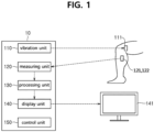
- FIG. 1 is a block diagram illustrating a concept of a device for diagnosing an abnormality by measuring a minimal change in a muscle according to an embodiment.
- a device for diagnosing an abnormality by measuring a minimal change in a muscle 10 (hereinafter, "diagnosis device") according to an embodiment is configured to include a vibration unit 110, a measuring unit 120, and a processing unit 130, a display unit 140, and a control unit 150.
- each of the components may be implemented by one processing unit or program, or may be implemented by an organic combination of two or more independent processing units or programs.
- the vibration unit 110 receives a control signal from the control unit 150 and provides the vibration of a specific frequency through a vibrator 111 attached to a body part.
- the vibration unit 110 receives the control signal, for example, an input for the intensity, frequency, and form of vibration to be provided to the user (regular intensity or periodically repeating strength and weakness) from the control unit 150, and transmit it to the vibration unit 110 by wire or wirelessly.
- the vibrator 111 is a device having a power device such as a motor to convert electrical energy into physical vibration, and is not limited to a specific shape or size.
- the vibrator 111 may be connected to the diagnosis device 10 by wire or through a wireless network (Bluetooth, WiFi, infrared communication, etc.).
- the vibrator 111 may be powered by a motor by wire, or may be powered by a built-in battery.
- the vibrator 111 may be attached to the user's body part (arm, leg, etc.) through a fixing part such as a strap or adhesive tape that can be worn on the user's body part.
- a pressure sensor (not shown) for sensing pressure according to vibration between the vibrator 111 and the user's body part may be further provided.
- the processing unit 130 to be described later may display a change in pressure according to the vibration on the display unit 140. This allows the user to know in real time whether the vibrator is providing vibration at the appropriate pressure level. Accordingly, when the pressure is too low or too high, the operations of the vibration unit 110 and vibrator 111 may be controlled through the control unit 150.
- the measuring unit 120 may measure a change in elasticity of a body part according to the vibration provided by the vibration unit 110 to detect a minimal change in a muscle. For example, when deep vein thrombosis (DVT) occurs, blood clots are formed in the vein, which causes muscle stiffness, and the measuring unit 120 measures a change in elasticity of a muscle to detect these symptoms.
- DVD deep vein thrombosis
- the measuring unit 120 detects a change in elasticity of a body part through an accelerometer 121 and/or an electromyography 122, and warns the user by calculating an abnormality or risk of thrombosis of the body part when an abnormal change occurs.
- the measuring unit 120 may sense the reflex vibration generated in the user's body part through an accelerometer 121.
- Reflex vibration refers to the tremor that occurs in the surrounding muscles when an arbitrary vibrational stimulus is applied to the muscle.
- the vibration stimulation of the same characteristic intensity, frequency, stimulation interval
- the reflex vibration of the corresponding characteristic appears at a constant level.
- the characteristic of the reflex vibration sensed in the muscle changes rapidly.
- the accelerometer 121 is a device for converting a minimal movement of a muscle into an electrical signal, and detects a change in vibration characteristics due to the muscular dystrophy or thrombosis and transmits it to the processing unit 130. After the development of muscular dystrophy or deep vein thrombosis, the measured value of measured reflex vibration changes significantly.
- the measuring unit 120 may measure an electromyograph (EMG) signal of the user's body part through an electromyograph.
- EMG electromyograph
- the electromyograph is a device that records electrical activity according to muscle contraction using the electrodes attached to or inserted into the body parts.
- the EMG 122 detects a change in the EMG signal and transmits it to the processing unit 130. After the development of deep vein thrombosis, the measured value of measured EMG signal changes significantly.
- the processing unit 130 calculates an abnormality of a body part based on a change in elasticity of the body part.
- the processing unit 130 may calculate an abnormality of a body part by using an algorithm that analyzes data distribution to detect a degree to which a specific value of a muscle moves away from the distribution or an anomaly detection algorithm that detects whether there is anomaly in a variable.
- the abnormality of the body part calculated by the processing unit 130 may mean a risk of thrombosis.
- the processing unit 130 combines and simultaneously uses the measurement results of reflex vibration and EMG signal (in this case, each measurement result may be given a weight), or uses each result independently to calculate an abnormality of muscle or a risk of thrombosis.
- the display unit 140 displays the risk of thrombosis calculated by the processing unit 130 to the user through an external device.
- the display unit 140 may display the calculation result as a numerical value on a display device 141 (refer to FIG. 1 ).
- the display unit 140 may warn the user by turning on an LED element (not shown) when the abnormality of muscle or the risk of thrombosis is greater than or equal to a threshold (e.g., if the risk of deep vein thrombosis is high, a red light is turned on, and if the risk is low, a green light is turned on).
- a threshold e.g., if the risk of deep vein thrombosis is high, a red light is turned on, and if the risk is low, a green light is turned on.
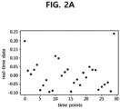
- FIGS. 2A to 2D illustrate a process in which the processing unit calculates a degree to which the reflex vibration or EMG signal of a body part moves away from data distribution by using a Bayesian algorithm according to an embodiment.
- the processing unit 130 may calculate the abnormality of the body part using an algorithm that detects the degree to which a specific value of muscle has moved away from the distribution chart by analyzing the data distribution of the measurement result of the reflex vibration of the body part and the measurement result of the EMG signal.
- the algorithm may follow the distribution of Bayesian probability values.
- the processing unit 130 obtains an approximate function for each of the measurement values of the reflex vibration and the EMG signal of the body part based on a change in the measurement values of the reflex vibration and EMG signal of the body part over time, and converts each of the measurement values of the reflex vibration and EMG signal of the body part into a value within a range of 0 to 1 using the obtained approximate function.
- the measurement value of reflex vibration or EMG signal of the body part converted to a value within the range of 0 to 1 may be a value indicating the probability that the thrombosis will be measured when a muscular dystrophy or thrombosis occurs in a patient, but the embodiment is not limited thereto.
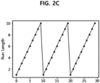
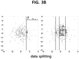
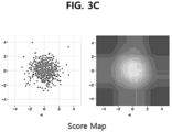
- FIGS. 3A to 3C illustrate a process in which the processing unit 130 calculates an abnormality of reflex vibration or EMG signal of a body part using an anomaly detection algorithm according to an embodiment.
- the processing unit 130 can calculate an abnormality in reflex vibrations or EMG signals of a body part by using an isolation forest algorithm that detects an anomaly by isolating anomaly data based on a tree.
- the anomaly detection refers to detecting data that is significantly different from the majority of data or unique data. Here, anomaly may be expressed as noise, deviation, or exception.
- the anomaly detection is to find outlier, which is data showing different pattern in the collected data.
- the isolation forest technique expresses the data set in the form of a decision tree, and follows the depth direction of the decision tree in the case of splitting a normal value and uses the characteristic of splitting at the top of the decision tree in the case of splitting outlier.
- the isolation forest makes it possible to split normal values and outliers based on how many times the decision tree is descended and split. If there is a sudden change in the frequency component of the reflex vibration measured by the accelerometer and/or the frequency component of the EMG signal measured by the EMG, it is split as outlier. The greater the number of splitting, the higher the possibility of muscular dystrophy or deep vein thrombosis.
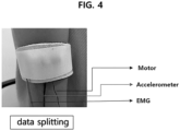
- FIG. 4 illustrates a device for diagnosing abnormality that is implemented to be detachable as a wearable patch and measures a minimal change in a muscle, according to an embodiment.
- a small device such as a wearable device is used in a form that is in direct contact with a person's body or is attached to clothes or other accessories without direct contact with the body.
- a wearable multi-biosignal measuring device such as a wearable electromyography device is a device that uses a sensor such as a patch-type electrode to form a contact point with various body parts (chest, wrist, ankle, etc.) of a subject to measure biosignals such as electromyography.
- This device is used to predict or diagnose an abnormality in body parts or the occurrence of diseases such as thrombosis by monitoring biosignals.
- the diagnostic device of the present invention By implementing the diagnostic device of the present invention in a detachable manner as a wearable patch, it is possible to easily monitor bio-signals such as electromyography in daily life.
- bio-signals such as electromyography
- medical personnel can be continuously provided with the patient's condition through the motor unit, measuring unit, and processing unit mounted on the wearable patch, and can rapidly diagnose and treat predicted disease, thereby reducing the risk of death.
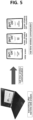
- FIG. 5 illustrates a process in which the processing unit 130 calculates an abnormality of muscle or a risk of thrombosis, and a display unit warns a user through wireless communication, according to an embodiment.
- the processing unit 130 may be connected to communicate wirelessly with the display unit 140 using one or more of radio frequency (RF), Wi-Fi, cellular, Bluetooth, Bluetooth Low Energy (BLE), personal area network (PAN), short-wavelength UHF, and a combination thereof.
- RF radio frequency
- Wi-Fi Wireless Fidelity
- BLE Bluetooth Low Energy
- PAN personal area network
- UHF short-wavelength UHF
- the display unit 140 displays the risk of thrombosis calculated by the processing unit 130 to the user through an external device.
- the display unit 140 may display the calculation result as a numerical value on the display device 141.
- the display unit 140 may warn the user by turning on an LED element (not shown) when the risk of thrombosis is greater than or equal to a threshold (e.g., if the risk of deep vein thrombosis is high, a red light is turned on, and if the risk of deep vein thrombosis is low, a green light is turned on).
- a threshold e.g., if the risk of deep vein thrombosis is high, a red light is turned on, and if the risk of deep vein thrombosis is low, a green light is turned on.
- the vibration of a specific frequency is provided using the vibrator attached to the user's body, and a muscular dystrophy or deep vein thrombosis is diagnosed by measuring the reflex vibration and EMG signal of the muscle sensed in response to the vibration.
- the diagnosis device is configured to include the vibrator attached to a body part and the sensor capable of sensing a change in the body part, so that it is easy to install and use. For example, by using the vibrator and sensor provided under an airplane seat, it is possible to automatically measure and warn an abnormality of the body part or a risk of thrombosis of a traveler. According to this, it is possible to diagnose muscular dystrophy and deep vein thrombosis at an earlier stage compared to the conventional ultrasound examination method or blood test method.
Landscapes
- Health & Medical Sciences (AREA)
- Life Sciences & Earth Sciences (AREA)
- Engineering & Computer Science (AREA)
- Physics & Mathematics (AREA)
- Biomedical Technology (AREA)
- Pathology (AREA)
- Biophysics (AREA)
- Heart & Thoracic Surgery (AREA)
- Medical Informatics (AREA)
- Molecular Biology (AREA)
- Surgery (AREA)
- Animal Behavior & Ethology (AREA)
- General Health & Medical Sciences (AREA)
- Public Health (AREA)
- Veterinary Medicine (AREA)
- Physiology (AREA)
- Dentistry (AREA)
- Oral & Maxillofacial Surgery (AREA)
- Vascular Medicine (AREA)
- Artificial Intelligence (AREA)
- Cardiology (AREA)
- Computer Vision & Pattern Recognition (AREA)
- Psychiatry (AREA)
- Signal Processing (AREA)
- Orthopedic Medicine & Surgery (AREA)
- Rheumatology (AREA)
- Bioinformatics & Cheminformatics (AREA)
- Medicinal Chemistry (AREA)
- Chemical & Material Sciences (AREA)
- Physical Education & Sports Medicine (AREA)
- Evolutionary Computation (AREA)
- Fuzzy Systems (AREA)
- Mathematical Physics (AREA)
- Computer Networks & Wireless Communication (AREA)
- Measuring And Recording Apparatus For Diagnosis (AREA)
- Measurement Of The Respiration, Hearing Ability, Form, And Blood Characteristics Of Living Organisms (AREA)
Abstract
Description
- The present invention relates to a device for diagnosing an abnormality by measuring a minimal change in muscle, and more particularly, to a device capable of providing vibration to a user's body part, and at the same time, calculating an abnormality of the body part based on a change in the muscle of the body part.
- This study was made with the support of a joint research project of KU Medicine-KIST TRC (Project title: Development of system for early diagnosis and monitoring of extremity deep vein thrombosis using sensor fusion technology, Project identification number: 2E3115J) under the supervision of the Korea Institute of Science and Technology and Korea University Ansan Hospital.
- Deep vein thrombosis (DVT) is a disease that occurs due to blood clots in veins. This is a symptom in which venous blood in body parts (mainly in the lower extremities) stagnates when a person is in an immobile position for a long time or is in various situations where blood clots are likely to occur, causing blood clots to form in deep veins. It is also called 'economy class syndrome' because it is a disease that often occurs in passengers sitting in narrow seats during long-distance flights. If left untreated, deep vein thrombosis can cause limb necrosis, pulmonary embolism, and breathing difficulties, leading to death, so it is important to diagnose these symptoms early and prevent the condition from getting worse.
- Conventionally, deep vein thrombosis was diagnosed mainly by examining a patient's body through ultrasound or through blood tests. Such a diagnosis method may be effective in a situation where a patient can receive periodic ultrasound or blood tests in a hospital, clinic, etc., but early diagnosis is difficult if it is caused by a long flight or driving. Preemptive methods such as mechanical prophylaxis (compression stockings, air compression devices, etc.) or pharmacological prophylaxis (administration of anticoagulants, etc.) exist, but in many cases, practical application is difficult, resulting in a large number of patients with deep vein thrombosis every year.
- Accordingly, an object of the present invention is to provide a diagnosis device capable of not only calculating an abnormality of a body part based on a change in muscle of a body part, but also diagnosing deep vein thrombosis (DVT) at an early stage.
- A device for diagnosing an abnormality by measuring a minimal change in a muscle according to an embodiment includes a vibration unit that provides vibration to a body part of a user; a measuring unit that detects a minimal change in a muscle by measuring a change in elasticity of the body part according to the vibration; and a processing unit that calculates an abnormality of the body part based on the change in the elasticity of the body part, wherein the processing unit calculates the abnormality of the body party by using an algorithm that detects a degree to which a specific value of the muscle is far from a distribution chart by analyzing data distribution or an anomaly detection algorithm that detects whether there is anomaly in a variable.
- According to an embodiment, the processing unit may calculate a risk of thrombosis based on the abnormality of the body part.
- According to an embodiment, the device may further include a display unit that displays the risk of thrombosis.
- According to an embodiment, the device may further include a control unit that controls an operation of the vibration unit.
- According to an embodiment, the vibration unit may receive a control signal from the control unit and provide the vibration of a specific frequency through a vibrator attached to the body part.
- According to an embodiment, the device may further include a pressure sensor positioned between the vibrator and the body part of the user to sense pressure according to the vibration, wherein the processing unit may be configured to indicate the pressure according to the vibration through the display unit.
- According to an embodiment, the measuring unit may include an accelerometer that detects reflex vibration generated in the body part in response to the vibration.
- According to an embodiment, the measuring unit may include an electromyograph that measures an electromyograph signal of the body part.
- According to an embodiment, the processing unit may obtain an approximate function of each of measurement values of reflex vibration and electromyograph signal of the body part using a distribution of Bayesian probability value based on a change in the measurement values of the reflex vibration and EMG signal of the body part over time, and may convert each of the measurement values of the reflex vibration and EMG signal of the body part into a value within a range of 0 to 1 by using the calculated approximate function.
- According to an embodiment, an anomaly detection algorithm used by the processing unit may include an isolation forest algorithm that detects anomaly by splitting anomaly data based on a tree.
- According to an embodiment, the display unit may numerically indicate the risk of thrombosis through a display device.
- According to an embodiment, the display unit may be configured to turn on an LED element when the risk of thrombosis is greater than or equal to a threshold.
- According to an embodiment, the device may be implemented to be detachable as a wearable patch.
- According to an embodiment, the processing unit may be connected to wirelessly communicate with the display unit using one or more of radio frequency (RF), Wi-Fi, cellular, Bluetooth, Bluetooth Low Energy (BLE), personal area network (PAN), short-wavelength UHF, and a combination thereof.
- According to an embodiment of the present invention, a devise for diagnosing an abnormality of a body part at an early stage by providing vibration of a specific frequency using a vibrator attached to the user's body, and measuring the reflex vibration and EMG signal of the muscle sensed in response to the vibration.
- The diagnosis device according to an embodiment determines that the occurrence of deep vein thrombosis is high when an abnormal change appears in the measurement value of reflex vibration measured by an accelerometer /or the measurement value of EMG signal measured by an electromyography, and notifies the user of abnormalities in the body parts or risk of thrombosis in real time.
- The diagnosis apparatus according to an embodiment includes a vibrator attached to a body part and a sensor (accelerometer, electromyography, etc.) capable of detecting a change in the body part, so that it is easy to install and use. For example, the risk of thrombosis may be measured at regular intervals using the vibrator and sensor provided under an airplane seat. Therefore, it is possible to diagnose deep vein thrombosis earlier than the conventional ultrasound or blood test methods.
-
-
FIG. 1 is a block diagram illustrating a concept of a devise for diagnosing an abnormality by measuring a minimal change in a muscle according to an embodiment. -
FIGS. 2A to 2D illustrate a process in which a processing unit calculates a degree to which the reflex vibration or EMG signal of a body part moves away from a data distribution by using a Bayesian algorithm according to an embodiment. -
FIGS. 3A to 3C illustrate a process in which a processing unit calculates an abnormality of the reflex vibration or EMG signal of a body part using an anomaly detection algorithm according to an embodiment. -
FIG. 4 illustrates a device for diagnosing an abnormality that is implemented as a wearable patch and measures an abnormality by measuring a minimal change in a muscle according to an embodiment. -
FIG. 5 illustrates a process in which a processing unit calculates a risk of thrombosis and a display unit warns a user through wireless communication, according to an embodiment. - The terms used in the present specification have been selected as widely used general terms as possible while considering their functions, but may vary depending on the intention or custom of those skilled in the art or the advent of new technology. In addition, in a specific case, there is a term arbitrarily selected by the applicant, and in this case, the meaning will be described in the description of the corresponding specification. Therefore, it is intended to clarify that the terms used in this specification are not simply names of terms, but should be interpreted based on the actual meaning of the terms and the contents of the entire specification.
- Hereinafter, the embodiments will be described in detail with reference to the accompanying drawings and the contents described in the accompanying drawings, but the scope of the claims is not limited or restricted by the embodiments.
-
FIG. 1 is a block diagram illustrating a concept of a device for diagnosing an abnormality by measuring a minimal change in a muscle according to an embodiment. Referring toFIG. 1 , a device for diagnosing an abnormality by measuring a minimal change in a muscle 10 (hereinafter, "diagnosis device") according to an embodiment is configured to include avibration unit 110, ameasuring unit 120, and aprocessing unit 130, adisplay unit 140, and acontrol unit 150. - Although the above components are shown separately for the convenience of conceptual description, they do not necessarily have to be implemented as independent devices or programs. For example, each of the components may be implemented by one processing unit or program, or may be implemented by an organic combination of two or more independent processing units or programs.
- The
vibration unit 110 receives a control signal from thecontrol unit 150 and provides the vibration of a specific frequency through avibrator 111 attached to a body part. Thevibration unit 110 receives the control signal, for example, an input for the intensity, frequency, and form of vibration to be provided to the user (regular intensity or periodically repeating strength and weakness) from thecontrol unit 150, and transmit it to thevibration unit 110 by wire or wirelessly. - The
vibrator 111 is a device having a power device such as a motor to convert electrical energy into physical vibration, and is not limited to a specific shape or size. Thevibrator 111 may be connected to thediagnosis device 10 by wire or through a wireless network (Bluetooth, WiFi, infrared communication, etc.). In addition, thevibrator 111 may be powered by a motor by wire, or may be powered by a built-in battery. In one embodiment, thevibrator 111 may be attached to the user's body part (arm, leg, etc.) through a fixing part such as a strap or adhesive tape that can be worn on the user's body part. - According to an embodiment, a pressure sensor (not shown) for sensing pressure according to vibration between the
vibrator 111 and the user's body part may be further provided. Theprocessing unit 130 to be described later may display a change in pressure according to the vibration on thedisplay unit 140. This allows the user to know in real time whether the vibrator is providing vibration at the appropriate pressure level. Accordingly, when the pressure is too low or too high, the operations of thevibration unit 110 andvibrator 111 may be controlled through thecontrol unit 150. - The
measuring unit 120 may measure a change in elasticity of a body part according to the vibration provided by thevibration unit 110 to detect a minimal change in a muscle. For example, when deep vein thrombosis (DVT) occurs, blood clots are formed in the vein, which causes muscle stiffness, and themeasuring unit 120 measures a change in elasticity of a muscle to detect these symptoms. - The
measuring unit 120 detects a change in elasticity of a body part through an accelerometer 121 and/or an electromyography 122, and warns the user by calculating an abnormality or risk of thrombosis of the body part when an abnormal change occurs. - According to an embodiment, the
measuring unit 120 may sense the reflex vibration generated in the user's body part through an accelerometer 121. Reflex vibration refers to the tremor that occurs in the surrounding muscles when an arbitrary vibrational stimulus is applied to the muscle. In general, in an environment with the same muscle quality or blood flow, when the vibration stimulation of the same characteristic (intensity, frequency, stimulation interval) is applied, the reflex vibration of the corresponding characteristic appears at a constant level. In contrast, when deep vein thrombosis occurs and the muscle becomes stiff or the blood flow changes, the characteristic of the reflex vibration sensed in the muscle changes rapidly. The accelerometer 121 is a device for converting a minimal movement of a muscle into an electrical signal, and detects a change in vibration characteristics due to the muscular dystrophy or thrombosis and transmits it to theprocessing unit 130. After the development of muscular dystrophy or deep vein thrombosis, the measured value of measured reflex vibration changes significantly. - According to an embodiment, the measuring
unit 120 may measure an electromyograph (EMG) signal of the user's body part through an electromyograph. The electromyograph is a device that records electrical activity according to muscle contraction using the electrodes attached to or inserted into the body parts. As with the above-mentioned reflex vibration, when deep vein thrombosis occurs and the muscle is stiff or there is a change in blood flow, the EMG signal also changes. The EMG 122 detects a change in the EMG signal and transmits it to theprocessing unit 130. After the development of deep vein thrombosis, the measured value of measured EMG signal changes significantly. - The
processing unit 130 calculates an abnormality of a body part based on a change in elasticity of the body part. Theprocessing unit 130 may calculate an abnormality of a body part by using an algorithm that analyzes data distribution to detect a degree to which a specific value of a muscle moves away from the distribution or an anomaly detection algorithm that detects whether there is anomaly in a variable. In an embodiment, the abnormality of the body part calculated by theprocessing unit 130 may mean a risk of thrombosis. - The
processing unit 130 combines and simultaneously uses the measurement results of reflex vibration and EMG signal (in this case, each measurement result may be given a weight), or uses each result independently to calculate an abnormality of muscle or a risk of thrombosis. - The
display unit 140 displays the risk of thrombosis calculated by theprocessing unit 130 to the user through an external device. According to an embodiment, thedisplay unit 140 may display the calculation result as a numerical value on a display device 141 (refer toFIG. 1 ). According to another embodiment, thedisplay unit 140 may warn the user by turning on an LED element (not shown) when the abnormality of muscle or the risk of thrombosis is greater than or equal to a threshold (e.g., if the risk of deep vein thrombosis is high, a red light is turned on, and if the risk is low, a green light is turned on). -
FIGS. 2A to 2D illustrate a process in which the processing unit calculates a degree to which the reflex vibration or EMG signal of a body part moves away from data distribution by using a Bayesian algorithm according to an embodiment. - Referring to
FIGS. 2A to 2D , theprocessing unit 130 may calculate the abnormality of the body part using an algorithm that detects the degree to which a specific value of muscle has moved away from the distribution chart by analyzing the data distribution of the measurement result of the reflex vibration of the body part and the measurement result of the EMG signal. The algorithm may follow the distribution of Bayesian probability values. Theprocessing unit 130 obtains an approximate function for each of the measurement values of the reflex vibration and the EMG signal of the body part based on a change in the measurement values of the reflex vibration and EMG signal of the body part over time, and converts each of the measurement values of the reflex vibration and EMG signal of the body part into a value within a range of 0 to 1 using the obtained approximate function. Here, the measurement value of reflex vibration or EMG signal of the body part converted to a value within the range of 0 to 1 may be a value indicating the probability that the thrombosis will be measured when a muscular dystrophy or thrombosis occurs in a patient, but the embodiment is not limited thereto. -
FIGS. 3A to 3C illustrate a process in which theprocessing unit 130 calculates an abnormality of reflex vibration or EMG signal of a body part using an anomaly detection algorithm according to an embodiment. - Referring to
FIGS. 3A to 3C , theprocessing unit 130 can calculate an abnormality in reflex vibrations or EMG signals of a body part by using an isolation forest algorithm that detects an anomaly by isolating anomaly data based on a tree. The anomaly detection refers to detecting data that is significantly different from the majority of data or unique data. Here, anomaly may be expressed as noise, deviation, or exception. The anomaly detection is to find outlier, which is data showing different pattern in the collected data. The isolation forest technique expresses the data set in the form of a decision tree, and follows the depth direction of the decision tree in the case of splitting a normal value and uses the characteristic of splitting at the top of the decision tree in the case of splitting outlier. Using these characteristics, the isolation forest makes it possible to split normal values and outliers based on how many times the decision tree is descended and split. If there is a sudden change in the frequency component of the reflex vibration measured by the accelerometer and/or the frequency component of the EMG signal measured by the EMG, it is split as outlier. The greater the number of splitting, the higher the possibility of muscular dystrophy or deep vein thrombosis. -
FIG. 4 illustrates a device for diagnosing abnormality that is implemented to be detachable as a wearable patch and measures a minimal change in a muscle, according to an embodiment. - Referring to
FIG. 4 , many wearable devices using IoT technology have recently appeared. Due to the characteristics of the wearable device, it should be easy to carry, and it should be light and capable of operating for a long time. A small device such as a wearable device is used in a form that is in direct contact with a person's body or is attached to clothes or other accessories without direct contact with the body. - In particular, among wearable devices, a wearable multi-biosignal measuring device such as a wearable electromyography device is a device that uses a sensor such as a patch-type electrode to form a contact point with various body parts (chest, wrist, ankle, etc.) of a subject to measure biosignals such as electromyography. This device is used to predict or diagnose an abnormality in body parts or the occurrence of diseases such as thrombosis by monitoring biosignals.
- By implementing the diagnostic device of the present invention in a detachable manner as a wearable patch, it is possible to easily monitor bio-signals such as electromyography in daily life. Thus, medical personnel can be continuously provided with the patient's condition through the motor unit, measuring unit, and processing unit mounted on the wearable patch, and can rapidly diagnose and treat predicted disease, thereby reducing the risk of death.
-
FIG. 5 illustrates a process in which theprocessing unit 130 calculates an abnormality of muscle or a risk of thrombosis, and a display unit warns a user through wireless communication, according to an embodiment. - The
processing unit 130 may be connected to communicate wirelessly with thedisplay unit 140 using one or more of radio frequency (RF), Wi-Fi, cellular, Bluetooth, Bluetooth Low Energy (BLE), personal area network (PAN), short-wavelength UHF, and a combination thereof. - Referring to
FIG. 5 , thedisplay unit 140 displays the risk of thrombosis calculated by theprocessing unit 130 to the user through an external device. According to an embodiment, thedisplay unit 140 may display the calculation result as a numerical value on thedisplay device 141. According to another embodiment, thedisplay unit 140 may warn the user by turning on an LED element (not shown) when the risk of thrombosis is greater than or equal to a threshold (e.g., if the risk of deep vein thrombosis is high, a red light is turned on, and if the risk of deep vein thrombosis is low, a green light is turned on). - According to the diagnostic device described above, the vibration of a specific frequency is provided using the vibrator attached to the user's body, and a muscular dystrophy or deep vein thrombosis is diagnosed by measuring the reflex vibration and EMG signal of the muscle sensed in response to the vibration. The diagnosis device according to an embodiment is configured to include the vibrator attached to a body part and the sensor capable of sensing a change in the body part, so that it is easy to install and use. For example, by using the vibrator and sensor provided under an airplane seat, it is possible to automatically measure and warn an abnormality of the body part or a risk of thrombosis of a traveler. According to this, it is possible to diagnose muscular dystrophy and deep vein thrombosis at an earlier stage compared to the conventional ultrasound examination method or blood test method.
- Although the above has been described with reference to the embodiments, it will be understood by those skilled in the art that various modifications and changes can be made in the present invention without departing from the spirit and scope of the present invention as set forth in the following claims.
Claims (14)
- A device (10) for diagnosing an abnormality by measuring a minimal change in a muscle, comprising,a vibration unit (110) that provides vibration to a body part of a user;a measuring unit (120) that detects a minimal change in a muscle by measuring a change in elasticity of the body part according to the vibration; anda processing unit (130) that calculates an abnormality of the body part based on the change in the elasticity of the body part,wherein the processing unit (130) calculates the abnormality of the body party by using an algorithm that detects a degree to which a specific value of the muscle is far from a distribution chart by analyzing data distribution or an anomaly detection algorithm that detects whether there is anomaly in a variable.
- The device according to claim 1, wherein the abnormality of the body part calculated by the processing unit (130) is a risk of thrombosis.
- The device according to claim 1 or 2, further comprising a display unit (140) that displays the abnormality of the body part.
- The device according to one of claims 1 to 3, further comprising a control unit (150) that controls an operation of the vibration unit.
- The device according to claim 4, wherein the vibration unit (110) receives a control signal from the control unit (150) and provides the vibration of a specific frequency through a vibrator (111) attached to the body part.
- The device according to claim 5, further comprising a pressure sensor positioned between the vibrator and the body part of the user to sense pressure according to the vibration,
wherein the processing unit is configured to indicate the pressure according to the vibration through the display unit (140). - The device according to one of claims 1 to 6, wherein the measuring unit (120) includes an accelerometer (121) that detects reflex vibration generated in the body part in response to the vibration.
- The device according to one of claims 1 to 7, wherein the measuring unit (120) includes an electromyography (EMG) (122) that measures an electromyograph signal of the body part.
- The device according to one of claims 1 to 8, wherein the processing unit (130) obtains an approximate function of each of measurement values of reflex vibration and electromyograph signal of the body part using a distribution of Bayesian probability value based on a change in the measurement values of the reflex vibration and EMG signal of the body part over time, and converts each of the measurement values of the reflex vibration and EMG signal of the body part into a value within a range of 0 to 1 by using the calculated approximate function.
- The device according to one of claims 1 to 9, wherein an anomaly detection algorithm used by the processing unit (130) includes an isolation forest algorithm that detects anomaly by splitting anomaly data based on a tree.
- The device according to claim 3 or one of the claims 4 to 10 in connection with claim 3, wherein the display unit (140) displays numerically indicates the abnormality of the body part through a display device.
- The device according to claim 3 or 11 or one of the claims 4 to 10 in connection with claim 3, wherein the display unit (140) is configured to turn on an LED element when the abnormality of the body part is greater than or equal to a threshold.
- The device according to one of claims 1 to 12, wherein the device is detachable as a wearable patch.
- The device according to claim 3 or 11 or 12 or one of the claims 4 to 10 and 13 in connection with claim 3, wherein the processing unit (130) is connected to wirelessly communicate with the display unit using one or more of radio frequency (RF), Wi-Fi, cellular, Bluetooth, Bluetooth Low Energy (BLE), personal area network (PAN), short-wavelength UHF, and a combination thereof.
Applications Claiming Priority (1)
| Application Number | Priority Date | Filing Date | Title |
|---|---|---|---|
| KR1020210192377A KR102784129B1 (en) | 2021-12-30 | 2021-12-30 | Device for diagnosing abnormality by measuring fine changes in muscles |
Publications (2)
| Publication Number | Publication Date |
|---|---|
| EP4205639A1 true EP4205639A1 (en) | 2023-07-05 |
| EP4205639B1 EP4205639B1 (en) | 2025-07-09 |
Family
ID=83996081
Family Applications (1)
| Application Number | Title | Priority Date | Filing Date |
|---|---|---|---|
| EP22203621.2A Active EP4205639B1 (en) | 2021-12-30 | 2022-10-25 | Device for diagnosing abnormality by measuring minimal change in muscle |
Country Status (4)
| Country | Link |
|---|---|
| US (1) | US20230210375A1 (en) |
| EP (1) | EP4205639B1 (en) |
| KR (1) | KR102784129B1 (en) |
| CN (1) | CN116407117B (en) |
Families Citing this family (2)
| Publication number | Priority date | Publication date | Assignee | Title |
|---|---|---|---|---|
| USD980431S1 (en) * | 2021-03-25 | 2023-03-07 | Eisai R&D Management Co., Ltd | Component for a health condition monitoring system |
| KR20250170865A (en) * | 2024-05-29 | 2025-12-08 | 주식회사 웨이브트리 | Sensor for diagnosing pathological phenomena including abnormal body fluids formed in abdominal and body cavities, and operation method of the same |
Citations (5)
| Publication number | Priority date | Publication date | Assignee | Title |
|---|---|---|---|---|
| US20050065426A1 (en) * | 2002-05-20 | 2005-03-24 | Vespro Ltd. | Method, system and device for tissue characterization |
| KR20100049382A (en) | 2008-11-03 | 2010-05-12 | 최상식 | Device of stimulating muscles of the legs electrically for preventing economy class syndrome |
| US20190150771A1 (en) * | 2017-11-14 | 2019-05-23 | The Regents Of The University Of Colorado, A Body Corporate | Mechano-Acoustic Sensing Devices And Methods |
| WO2020198131A1 (en) * | 2019-03-22 | 2020-10-01 | University Of Tartu | Method and device for measuring or determining at least one biomechanical parameter of soft biological tissues |
| US20200375464A1 (en) * | 2017-12-18 | 2020-12-03 | Universiteit Antwerpen | Tissue elasticity measurement |
Family Cites Families (12)
| Publication number | Priority date | Publication date | Assignee | Title |
|---|---|---|---|---|
| US6635017B1 (en) * | 2000-02-09 | 2003-10-21 | Spentech, Inc. | Method and apparatus combining diagnostic ultrasound with therapeutic ultrasound to enhance thrombolysis |
| GB0425632D0 (en) * | 2004-11-22 | 2004-12-22 | Bioaccelerate Ltd | Device |
| US7733224B2 (en) * | 2006-06-30 | 2010-06-08 | Bao Tran | Mesh network personal emergency response appliance |
| US8170656B2 (en) * | 2008-06-26 | 2012-05-01 | Microsoft Corporation | Wearable electromyography-based controllers for human-computer interface |
| KR101032798B1 (en) | 2009-10-09 | 2011-05-06 | (주)라파앤라이프 | Spinal Stereotactic System by Analyzing Muscle Bioelectrical Signals |
| KR101383059B1 (en) * | 2012-07-23 | 2014-04-08 | 한국과학기술연구원 | Wearable vibratory stimulation device and operational protocol thereof |
| KR101451023B1 (en) | 2013-07-10 | 2014-10-14 | 고려대학교 산학협력단 | Vibrator for magnetic resonance elastography |
| CA2880623A1 (en) * | 2015-02-02 | 2016-08-02 | Jian Chen | Emg diagnostic method for traumatic brain injury |
| KR20160114487A (en) * | 2015-03-24 | 2016-10-05 | 삼성메디슨 주식회사 | Elasticity measurement apparatus and method thereof |
| CN210990217U (en) * | 2019-07-08 | 2020-07-14 | 宁波磁性材料应用技术创新中心有限公司 | Wearable product |
| JP6641056B1 (en) | 2019-07-16 | 2020-02-05 | 株式会社東芝 | Device abnormality diagnosis method and device abnormality diagnosis system |
| US11237897B2 (en) * | 2019-07-25 | 2022-02-01 | International Business Machines Corporation | Detecting and responding to an anomaly in an event log |
-
2021
- 2021-12-30 KR KR1020210192377A patent/KR102784129B1/en active Active
-
2022
- 2022-10-25 US US17/973,120 patent/US20230210375A1/en active Pending
- 2022-10-25 EP EP22203621.2A patent/EP4205639B1/en active Active
- 2022-11-02 CN CN202211362404.6A patent/CN116407117B/en active Active
Patent Citations (5)
| Publication number | Priority date | Publication date | Assignee | Title |
|---|---|---|---|---|
| US20050065426A1 (en) * | 2002-05-20 | 2005-03-24 | Vespro Ltd. | Method, system and device for tissue characterization |
| KR20100049382A (en) | 2008-11-03 | 2010-05-12 | 최상식 | Device of stimulating muscles of the legs electrically for preventing economy class syndrome |
| US20190150771A1 (en) * | 2017-11-14 | 2019-05-23 | The Regents Of The University Of Colorado, A Body Corporate | Mechano-Acoustic Sensing Devices And Methods |
| US20200375464A1 (en) * | 2017-12-18 | 2020-12-03 | Universiteit Antwerpen | Tissue elasticity measurement |
| WO2020198131A1 (en) * | 2019-03-22 | 2020-10-01 | University Of Tartu | Method and device for measuring or determining at least one biomechanical parameter of soft biological tissues |
Also Published As
| Publication number | Publication date |
|---|---|
| KR102784129B1 (en) | 2025-03-19 |
| CN116407117A (en) | 2023-07-11 |
| CN116407117B (en) | 2025-10-24 |
| EP4205639B1 (en) | 2025-07-09 |
| KR20230102338A (en) | 2023-07-07 |
| US20230210375A1 (en) | 2023-07-06 |
Similar Documents
| Publication | Publication Date | Title |
|---|---|---|
| US11224385B2 (en) | Method for determining a person's sleeping phase which is favourable for waking up | |
| US10456059B2 (en) | Neuorological condition detection unit and method of using the same | |
| US6577897B1 (en) | Non-invasive monitoring of physiological parameters | |
| Klingeberg et al. | Mobile wearable device for long term monitoring of vital signs | |
| KR101448106B1 (en) | Analisys Method of Rehabilitation status using Electromyogram | |
| EP3261526B1 (en) | Apparatus and method for providing a control signal for a blood pressure measurement device | |
| US20140371545A1 (en) | Measurement of Cerebral Physiologic Parameters Using Bioimpedance | |
| US11426083B2 (en) | Equipment for monitoring blood flow and respiratory flow | |
| US20120029300A1 (en) | System and method for reducing false alarms and false negatives based on motion and position sensing | |
| KR20190121237A (en) | Systematic apparatus for motion sensor and optical sensor based cardiac arrhythmia triage | |
| KR20210104692A (en) | Apparatus and method for detecting stroke in a patient | |
| EP4205639B1 (en) | Device for diagnosing abnormality by measuring minimal change in muscle | |
| KR20190016886A (en) | System and method for real-time heartbeat events detection using low-power motion sensor | |
| AU2019271907B2 (en) | System, apparatus and method for measurement of muscle stiffness | |
| CN107028600A (en) | A kind of Intelligent bracelet for measuring blood pressure and heart rate | |
| EP3222208A1 (en) | Method and apparatus for triage and subsequent escalation based on biosignals or biometrics | |
| JP7106867B2 (en) | vital sensor and monitoring system | |
| Mohammadi-Koushki et al. | A wearable device for continuous cardiorespiratory System Monitoring | |
| US20220313137A1 (en) | A portable ecg device and an ecg system comprising the portable ecg device | |
| Postolache et al. | Unobtrusive and non-invasive sensing solutions for on-line physiological parameters monitoring | |
| US20180177410A1 (en) | Method to measure non-invasive blood pressure from patient sensor system | |
| Ribeiro et al. | A real time, wearable ECG and continous blood pressure monitoring system for first responders | |
| GB2634343A (en) | Health monitor device, system and method | |
| KR20130082878A (en) | Bio-signal transfer device, bio-signal monitoring system and method using thereof | |
| CN110022758A (en) | For determining the determination system of risk of heart failure |
Legal Events
| Date | Code | Title | Description |
|---|---|---|---|
| PUAI | Public reference made under article 153(3) epc to a published international application that has entered the european phase |
Free format text: ORIGINAL CODE: 0009012 |
|
| STAA | Information on the status of an ep patent application or granted ep patent |
Free format text: STATUS: REQUEST FOR EXAMINATION WAS MADE |
|
| 17P | Request for examination filed |
Effective date: 20221025 |
|
| AK | Designated contracting states |
Kind code of ref document: A1 Designated state(s): AL AT BE BG CH CY CZ DE DK EE ES FI FR GB GR HR HU IE IS IT LI LT LU LV MC ME MK MT NL NO PL PT RO RS SE SI SK SM TR |
|
| STAA | Information on the status of an ep patent application or granted ep patent |
Free format text: STATUS: EXAMINATION IS IN PROGRESS |
|
| 17Q | First examination report despatched |
Effective date: 20240223 |
|
| GRAP | Despatch of communication of intention to grant a patent |
Free format text: ORIGINAL CODE: EPIDOSNIGR1 |
|
| STAA | Information on the status of an ep patent application or granted ep patent |
Free format text: STATUS: GRANT OF PATENT IS INTENDED |
|
| INTG | Intention to grant announced |
Effective date: 20250217 |
|
| RIN1 | Information on inventor provided before grant (corrected) |
Inventor name: LEE, TAEHOON Inventor name: SEO, EUNYOUNG Inventor name: GEMECHU, DUGUMA TESHOME Inventor name: KIM, KEUN TAE Inventor name: KANG, JONG WOO Inventor name: LEE, SONG JOO |
|
| GRAS | Grant fee paid |
Free format text: ORIGINAL CODE: EPIDOSNIGR3 |
|
| GRAA | (expected) grant |
Free format text: ORIGINAL CODE: 0009210 |
|
| STAA | Information on the status of an ep patent application or granted ep patent |
Free format text: STATUS: THE PATENT HAS BEEN GRANTED |
|
| AK | Designated contracting states |
Kind code of ref document: B1 Designated state(s): AL AT BE BG CH CY CZ DE DK EE ES FI FR GB GR HR HU IE IS IT LI LT LU LV MC ME MK MT NL NO PL PT RO RS SE SI SK SM TR |
|
| REG | Reference to a national code |
Ref country code: GB Ref legal event code: FG4D |
|
| REG | Reference to a national code |
Ref country code: CH Ref legal event code: EP |
|
| REG | Reference to a national code |
Ref country code: IE Ref legal event code: FG4D |
|
| REG | Reference to a national code |
Ref country code: DE Ref legal event code: R096 Ref document number: 602022017246 Country of ref document: DE |
|
| P01 | Opt-out of the competence of the unified patent court (upc) registered |
Free format text: CASE NUMBER: UPC_APP_0572_4205639/2025 Effective date: 20250718 |
|
| REG | Reference to a national code |
Ref country code: NL Ref legal event code: MP Effective date: 20250709 |
|
| PG25 | Lapsed in a contracting state [announced via postgrant information from national office to epo] |
Ref country code: PT Free format text: LAPSE BECAUSE OF FAILURE TO SUBMIT A TRANSLATION OF THE DESCRIPTION OR TO PAY THE FEE WITHIN THE PRESCRIBED TIME-LIMIT Effective date: 20251110 |
|
| PG25 | Lapsed in a contracting state [announced via postgrant information from national office to epo] |
Ref country code: NL Free format text: LAPSE BECAUSE OF FAILURE TO SUBMIT A TRANSLATION OF THE DESCRIPTION OR TO PAY THE FEE WITHIN THE PRESCRIBED TIME-LIMIT Effective date: 20250709 |
|
| REG | Reference to a national code |
Ref country code: AT Ref legal event code: MK05 Ref document number: 1811019 Country of ref document: AT Kind code of ref document: T Effective date: 20250709 |
|
| PG25 | Lapsed in a contracting state [announced via postgrant information from national office to epo] |
Ref country code: IS Free format text: LAPSE BECAUSE OF FAILURE TO SUBMIT A TRANSLATION OF THE DESCRIPTION OR TO PAY THE FEE WITHIN THE PRESCRIBED TIME-LIMIT Effective date: 20251109 |
|
| PGFP | Annual fee paid to national office [announced via postgrant information from national office to epo] |
Ref country code: DE Payment date: 20251215 Year of fee payment: 4 |
|
| PG25 | Lapsed in a contracting state [announced via postgrant information from national office to epo] |
Ref country code: NO Free format text: LAPSE BECAUSE OF FAILURE TO SUBMIT A TRANSLATION OF THE DESCRIPTION OR TO PAY THE FEE WITHIN THE PRESCRIBED TIME-LIMIT Effective date: 20251009 |
|
| REG | Reference to a national code |
Ref country code: LT Ref legal event code: MG9D |
|
| PG25 | Lapsed in a contracting state [announced via postgrant information from national office to epo] |
Ref country code: AT Free format text: LAPSE BECAUSE OF FAILURE TO SUBMIT A TRANSLATION OF THE DESCRIPTION OR TO PAY THE FEE WITHIN THE PRESCRIBED TIME-LIMIT Effective date: 20250709 |
|
| PG25 | Lapsed in a contracting state [announced via postgrant information from national office to epo] |
Ref country code: FI Free format text: LAPSE BECAUSE OF FAILURE TO SUBMIT A TRANSLATION OF THE DESCRIPTION OR TO PAY THE FEE WITHIN THE PRESCRIBED TIME-LIMIT Effective date: 20250709 |
|
| PG25 | Lapsed in a contracting state [announced via postgrant information from national office to epo] |
Ref country code: HR Free format text: LAPSE BECAUSE OF FAILURE TO SUBMIT A TRANSLATION OF THE DESCRIPTION OR TO PAY THE FEE WITHIN THE PRESCRIBED TIME-LIMIT Effective date: 20250709 |
|
| PGFP | Annual fee paid to national office [announced via postgrant information from national office to epo] |
Ref country code: FR Payment date: 20251029 Year of fee payment: 4 |
|
| PG25 | Lapsed in a contracting state [announced via postgrant information from national office to epo] |
Ref country code: GR Free format text: LAPSE BECAUSE OF FAILURE TO SUBMIT A TRANSLATION OF THE DESCRIPTION OR TO PAY THE FEE WITHIN THE PRESCRIBED TIME-LIMIT Effective date: 20251010 |
|
| PG25 | Lapsed in a contracting state [announced via postgrant information from national office to epo] |
Ref country code: SE Free format text: LAPSE BECAUSE OF FAILURE TO SUBMIT A TRANSLATION OF THE DESCRIPTION OR TO PAY THE FEE WITHIN THE PRESCRIBED TIME-LIMIT Effective date: 20250709 |
|
| PG25 | Lapsed in a contracting state [announced via postgrant information from national office to epo] |
Ref country code: LV Free format text: LAPSE BECAUSE OF FAILURE TO SUBMIT A TRANSLATION OF THE DESCRIPTION OR TO PAY THE FEE WITHIN THE PRESCRIBED TIME-LIMIT Effective date: 20250709 |
|
| PG25 | Lapsed in a contracting state [announced via postgrant information from national office to epo] |
Ref country code: BG Free format text: LAPSE BECAUSE OF FAILURE TO SUBMIT A TRANSLATION OF THE DESCRIPTION OR TO PAY THE FEE WITHIN THE PRESCRIBED TIME-LIMIT Effective date: 20250709 Ref country code: PL Free format text: LAPSE BECAUSE OF FAILURE TO SUBMIT A TRANSLATION OF THE DESCRIPTION OR TO PAY THE FEE WITHIN THE PRESCRIBED TIME-LIMIT Effective date: 20250709 |
|
| PG25 | Lapsed in a contracting state [announced via postgrant information from national office to epo] |
Ref country code: RS Free format text: LAPSE BECAUSE OF FAILURE TO SUBMIT A TRANSLATION OF THE DESCRIPTION OR TO PAY THE FEE WITHIN THE PRESCRIBED TIME-LIMIT Effective date: 20251009 |
|
| PG25 | Lapsed in a contracting state [announced via postgrant information from national office to epo] |
Ref country code: ES Free format text: LAPSE BECAUSE OF FAILURE TO SUBMIT A TRANSLATION OF THE DESCRIPTION OR TO PAY THE FEE WITHIN THE PRESCRIBED TIME-LIMIT Effective date: 20250709 |
|
| PG25 | Lapsed in a contracting state [announced via postgrant information from national office to epo] |
Ref country code: RO Free format text: LAPSE BECAUSE OF FAILURE TO SUBMIT A TRANSLATION OF THE DESCRIPTION OR TO PAY THE FEE WITHIN THE PRESCRIBED TIME-LIMIT Effective date: 20250709 |
|
| PG25 | Lapsed in a contracting state [announced via postgrant information from national office to epo] |
Ref country code: SM Free format text: LAPSE BECAUSE OF FAILURE TO SUBMIT A TRANSLATION OF THE DESCRIPTION OR TO PAY THE FEE WITHIN THE PRESCRIBED TIME-LIMIT Effective date: 20250709 |
|
| PG25 | Lapsed in a contracting state [announced via postgrant information from national office to epo] |
Ref country code: DK Free format text: LAPSE BECAUSE OF FAILURE TO SUBMIT A TRANSLATION OF THE DESCRIPTION OR TO PAY THE FEE WITHIN THE PRESCRIBED TIME-LIMIT Effective date: 20250709 |
|
| PG25 | Lapsed in a contracting state [announced via postgrant information from national office to epo] |
Ref country code: IT Free format text: LAPSE BECAUSE OF FAILURE TO SUBMIT A TRANSLATION OF THE DESCRIPTION OR TO PAY THE FEE WITHIN THE PRESCRIBED TIME-LIMIT Effective date: 20250709 |
|
| PG25 | Lapsed in a contracting state [announced via postgrant information from national office to epo] |
Ref country code: CZ Free format text: LAPSE BECAUSE OF FAILURE TO SUBMIT A TRANSLATION OF THE DESCRIPTION OR TO PAY THE FEE WITHIN THE PRESCRIBED TIME-LIMIT Effective date: 20250709 |
|
| PG25 | Lapsed in a contracting state [announced via postgrant information from national office to epo] |
Ref country code: EE Free format text: LAPSE BECAUSE OF FAILURE TO SUBMIT A TRANSLATION OF THE DESCRIPTION OR TO PAY THE FEE WITHIN THE PRESCRIBED TIME-LIMIT Effective date: 20250709 Ref country code: SK Free format text: LAPSE BECAUSE OF FAILURE TO SUBMIT A TRANSLATION OF THE DESCRIPTION OR TO PAY THE FEE WITHIN THE PRESCRIBED TIME-LIMIT Effective date: 20250709 |
|
| PLBE | No opposition filed within time limit |
Free format text: ORIGINAL CODE: 0009261 |
|
| STAA | Information on the status of an ep patent application or granted ep patent |
Free format text: STATUS: NO OPPOSITION FILED WITHIN TIME LIMIT |