EP4201339A1 - Muskeltrainingsverfahren und system zur bereitstellung von visueller rückkopplung durch verwendung von ultraschallbildgebung - Google Patents
Muskeltrainingsverfahren und system zur bereitstellung von visueller rückkopplung durch verwendung von ultraschallbildgebung Download PDFInfo
- Publication number
- EP4201339A1 EP4201339A1 EP21857370.7A EP21857370A EP4201339A1 EP 4201339 A1 EP4201339 A1 EP 4201339A1 EP 21857370 A EP21857370 A EP 21857370A EP 4201339 A1 EP4201339 A1 EP 4201339A1
- Authority
- EP
- European Patent Office
- Prior art keywords
- training
- muscle
- target
- ultrasonic
- index value
- Prior art date
- Legal status (The legal status is an assumption and is not a legal conclusion. Google has not performed a legal analysis and makes no representation as to the accuracy of the status listed.)
- Pending
Links
- 238000012549 training Methods 0.000 title claims abstract description 319
- 210000003205 muscle Anatomy 0.000 title claims abstract description 268
- 230000000007 visual effect Effects 0.000 title claims abstract description 98
- 238000000034 method Methods 0.000 title claims abstract description 65
- 238000003384 imaging method Methods 0.000 title claims abstract description 35
- 230000004913 activation Effects 0.000 claims abstract description 50
- 230000008602 contraction Effects 0.000 claims abstract description 36
- 230000008569 process Effects 0.000 claims abstract description 18
- 239000000523 sample Substances 0.000 claims description 66
- 238000010586 diagram Methods 0.000 claims description 57
- 238000013507 mapping Methods 0.000 claims description 33
- 238000012545 processing Methods 0.000 claims description 27
- 230000005540 biological transmission Effects 0.000 claims description 22
- 230000000877 morphologic effect Effects 0.000 claims description 20
- 238000005070 sampling Methods 0.000 claims description 19
- 230000008859 change Effects 0.000 claims description 16
- 230000006870 function Effects 0.000 claims description 14
- 238000005516 engineering process Methods 0.000 claims description 13
- 230000017531 blood circulation Effects 0.000 claims description 12
- 238000012805 post-processing Methods 0.000 claims description 12
- 210000004872 soft tissue Anatomy 0.000 claims description 7
- 238000004364 calculation method Methods 0.000 claims description 6
- 230000003993 interaction Effects 0.000 claims description 6
- 230000008878 coupling Effects 0.000 claims description 4
- 238000010168 coupling process Methods 0.000 claims description 4
- 238000005859 coupling reaction Methods 0.000 claims description 4
- 238000006073 displacement reaction Methods 0.000 claims description 4
- 238000012067 mathematical method Methods 0.000 claims description 4
- 210000001087 myotubule Anatomy 0.000 claims description 4
- 206010033675 panniculitis Diseases 0.000 claims description 4
- 210000004304 subcutaneous tissue Anatomy 0.000 claims description 4
- 230000002457 bidirectional effect Effects 0.000 claims description 3
- 230000003252 repetitive effect Effects 0.000 claims description 3
- 230000033001 locomotion Effects 0.000 description 20
- 210000003491 skin Anatomy 0.000 description 12
- 238000011156 evaluation Methods 0.000 description 9
- 230000000694 effects Effects 0.000 description 8
- 230000009286 beneficial effect Effects 0.000 description 6
- 230000008901 benefit Effects 0.000 description 5
- 238000002567 electromyography Methods 0.000 description 4
- 238000002604 ultrasonography Methods 0.000 description 4
- 238000004891 communication Methods 0.000 description 3
- 230000007547 defect Effects 0.000 description 3
- 210000004185 liver Anatomy 0.000 description 3
- 230000007774 longterm Effects 0.000 description 3
- 238000005259 measurement Methods 0.000 description 3
- 230000009466 transformation Effects 0.000 description 3
- 208000004930 Fatty Liver Diseases 0.000 description 2
- 206010019708 Hepatic steatosis Diseases 0.000 description 2
- 230000004087 circulation Effects 0.000 description 2
- 238000003745 diagnosis Methods 0.000 description 2
- 201000010099 disease Diseases 0.000 description 2
- 208000037265 diseases, disorders, signs and symptoms Diseases 0.000 description 2
- 239000003814 drug Substances 0.000 description 2
- 208000010706 fatty liver disease Diseases 0.000 description 2
- 230000004927 fusion Effects 0.000 description 2
- 238000010191 image analysis Methods 0.000 description 2
- 230000010354 integration Effects 0.000 description 2
- 230000004118 muscle contraction Effects 0.000 description 2
- 238000007781 pre-processing Methods 0.000 description 2
- 238000003908 quality control method Methods 0.000 description 2
- 231100000240 steatosis hepatitis Toxicity 0.000 description 2
- 241001270131 Agaricus moelleri Species 0.000 description 1
- 208000023178 Musculoskeletal disease Diseases 0.000 description 1
- 230000001133 acceleration Effects 0.000 description 1
- 238000009825 accumulation Methods 0.000 description 1
- 230000009471 action Effects 0.000 description 1
- 239000000853 adhesive Substances 0.000 description 1
- 230000001070 adhesive effect Effects 0.000 description 1
- 210000000577 adipose tissue Anatomy 0.000 description 1
- 238000004458 analytical method Methods 0.000 description 1
- 210000003484 anatomy Anatomy 0.000 description 1
- 238000003491 array Methods 0.000 description 1
- 210000004204 blood vessel Anatomy 0.000 description 1
- 230000037396 body weight Effects 0.000 description 1
- 210000000988 bone and bone Anatomy 0.000 description 1
- 238000004422 calculation algorithm Methods 0.000 description 1
- 208000019425 cirrhosis of liver Diseases 0.000 description 1
- 239000003086 colorant Substances 0.000 description 1
- 238000013461 design Methods 0.000 description 1
- 238000011161 development Methods 0.000 description 1
- 230000018109 developmental process Effects 0.000 description 1
- 238000000605 extraction Methods 0.000 description 1
- 210000003195 fascia Anatomy 0.000 description 1
- 230000036541 health Effects 0.000 description 1
- 230000000004 hemodynamic effect Effects 0.000 description 1
- 230000008595 infiltration Effects 0.000 description 1
- 238000001764 infiltration Methods 0.000 description 1
- 230000003189 isokinetic effect Effects 0.000 description 1
- 210000003041 ligament Anatomy 0.000 description 1
- 208000019423 liver disease Diseases 0.000 description 1
- 238000013178 mathematical model Methods 0.000 description 1
- 239000011159 matrix material Substances 0.000 description 1
- 239000000203 mixture Substances 0.000 description 1
- 210000005036 nerve Anatomy 0.000 description 1
- 238000010606 normalization Methods 0.000 description 1
- 230000003287 optical effect Effects 0.000 description 1
- 238000004806 packaging method and process Methods 0.000 description 1
- 230000000737 periodic effect Effects 0.000 description 1
- 230000035479 physiological effects, processes and functions Effects 0.000 description 1
- 238000002360 preparation method Methods 0.000 description 1
- 238000004393 prognosis Methods 0.000 description 1
- 230000005855 radiation Effects 0.000 description 1
- 238000011160 research Methods 0.000 description 1
- 230000002441 reversible effect Effects 0.000 description 1
- 238000012552 review Methods 0.000 description 1
- 230000011218 segmentation Effects 0.000 description 1
- 230000003068 static effect Effects 0.000 description 1
- 238000007619 statistical method Methods 0.000 description 1
- 210000004003 subcutaneous fat Anatomy 0.000 description 1
- 230000001360 synchronised effect Effects 0.000 description 1
- 210000001519 tissue Anatomy 0.000 description 1
- 230000002747 voluntary effect Effects 0.000 description 1
Images


Classifications
-
- A—HUMAN NECESSITIES
- A63—SPORTS; GAMES; AMUSEMENTS
- A63B—APPARATUS FOR PHYSICAL TRAINING, GYMNASTICS, SWIMMING, CLIMBING, OR FENCING; BALL GAMES; TRAINING EQUIPMENT
- A63B71/00—Games or sports accessories not covered in groups A63B1/00 - A63B69/00
- A63B71/06—Indicating or scoring devices for games or players, or for other sports activities
- A63B71/0619—Displays, user interfaces and indicating devices, specially adapted for sport equipment, e.g. display mounted on treadmills
- A63B71/0622—Visual, audio or audio-visual systems for entertaining, instructing or motivating the user
-
- A—HUMAN NECESSITIES
- A61—MEDICAL OR VETERINARY SCIENCE; HYGIENE
- A61B—DIAGNOSIS; SURGERY; IDENTIFICATION
- A61B8/00—Diagnosis using ultrasonic, sonic or infrasonic waves
- A61B8/48—Diagnostic techniques
- A61B8/488—Diagnostic techniques involving Doppler signals
-
- A—HUMAN NECESSITIES
- A61—MEDICAL OR VETERINARY SCIENCE; HYGIENE
- A61B—DIAGNOSIS; SURGERY; IDENTIFICATION
- A61B5/00—Measuring for diagnostic purposes; Identification of persons
- A61B5/103—Measuring devices for testing the shape, pattern, colour, size or movement of the body or parts thereof, for diagnostic purposes
- A61B5/11—Measuring movement of the entire body or parts thereof, e.g. head or hand tremor or mobility of a limb
- A61B5/1124—Determining motor skills
-
- A—HUMAN NECESSITIES
- A61—MEDICAL OR VETERINARY SCIENCE; HYGIENE
- A61B—DIAGNOSIS; SURGERY; IDENTIFICATION
- A61B5/00—Measuring for diagnostic purposes; Identification of persons
- A61B5/45—For evaluating or diagnosing the musculoskeletal system or teeth
- A61B5/4519—Muscles
-
- A—HUMAN NECESSITIES
- A61—MEDICAL OR VETERINARY SCIENCE; HYGIENE
- A61B—DIAGNOSIS; SURGERY; IDENTIFICATION
- A61B5/00—Measuring for diagnostic purposes; Identification of persons
- A61B5/48—Other medical applications
- A61B5/4836—Diagnosis combined with treatment in closed-loop systems or methods
-
- A—HUMAN NECESSITIES
- A61—MEDICAL OR VETERINARY SCIENCE; HYGIENE
- A61B—DIAGNOSIS; SURGERY; IDENTIFICATION
- A61B8/00—Diagnosis using ultrasonic, sonic or infrasonic waves
- A61B8/08—Clinical applications
-
- A—HUMAN NECESSITIES
- A61—MEDICAL OR VETERINARY SCIENCE; HYGIENE
- A61B—DIAGNOSIS; SURGERY; IDENTIFICATION
- A61B8/00—Diagnosis using ultrasonic, sonic or infrasonic waves
- A61B8/08—Clinical applications
- A61B8/0858—Clinical applications involving measuring tissue layers, e.g. skin, interfaces
-
- A—HUMAN NECESSITIES
- A61—MEDICAL OR VETERINARY SCIENCE; HYGIENE
- A61B—DIAGNOSIS; SURGERY; IDENTIFICATION
- A61B8/00—Diagnosis using ultrasonic, sonic or infrasonic waves
- A61B8/44—Constructional features of the ultrasonic, sonic or infrasonic diagnostic device
- A61B8/4427—Device being portable or laptop-like
-
- A—HUMAN NECESSITIES
- A61—MEDICAL OR VETERINARY SCIENCE; HYGIENE
- A61B—DIAGNOSIS; SURGERY; IDENTIFICATION
- A61B8/00—Diagnosis using ultrasonic, sonic or infrasonic waves
- A61B8/46—Ultrasonic, sonic or infrasonic diagnostic devices with special arrangements for interfacing with the operator or the patient
- A61B8/461—Displaying means of special interest
- A61B8/463—Displaying means of special interest characterised by displaying multiple images or images and diagnostic data on one display
-
- A—HUMAN NECESSITIES
- A61—MEDICAL OR VETERINARY SCIENCE; HYGIENE
- A61B—DIAGNOSIS; SURGERY; IDENTIFICATION
- A61B8/00—Diagnosis using ultrasonic, sonic or infrasonic waves
- A61B8/52—Devices using data or image processing specially adapted for diagnosis using ultrasonic, sonic or infrasonic waves
- A61B8/5207—Devices using data or image processing specially adapted for diagnosis using ultrasonic, sonic or infrasonic waves involving processing of raw data to produce diagnostic data, e.g. for generating an image
-
- A—HUMAN NECESSITIES
- A61—MEDICAL OR VETERINARY SCIENCE; HYGIENE
- A61B—DIAGNOSIS; SURGERY; IDENTIFICATION
- A61B8/00—Diagnosis using ultrasonic, sonic or infrasonic waves
- A61B8/52—Devices using data or image processing specially adapted for diagnosis using ultrasonic, sonic or infrasonic waves
- A61B8/5215—Devices using data or image processing specially adapted for diagnosis using ultrasonic, sonic or infrasonic waves involving processing of medical diagnostic data
- A61B8/5223—Devices using data or image processing specially adapted for diagnosis using ultrasonic, sonic or infrasonic waves involving processing of medical diagnostic data for extracting a diagnostic or physiological parameter from medical diagnostic data
-
- A—HUMAN NECESSITIES
- A63—SPORTS; GAMES; AMUSEMENTS
- A63B—APPARATUS FOR PHYSICAL TRAINING, GYMNASTICS, SWIMMING, CLIMBING, OR FENCING; BALL GAMES; TRAINING EQUIPMENT
- A63B24/00—Electric or electronic controls for exercising apparatus of preceding groups; Controlling or monitoring of exercises, sportive games, training or athletic performances
- A63B24/0062—Monitoring athletic performances, e.g. for determining the work of a user on an exercise apparatus, the completed jogging or cycling distance
-
- A—HUMAN NECESSITIES
- A61—MEDICAL OR VETERINARY SCIENCE; HYGIENE
- A61B—DIAGNOSIS; SURGERY; IDENTIFICATION
- A61B2503/00—Evaluating a particular growth phase or type of persons or animals
- A61B2503/10—Athletes
-
- A—HUMAN NECESSITIES
- A61—MEDICAL OR VETERINARY SCIENCE; HYGIENE
- A61B—DIAGNOSIS; SURGERY; IDENTIFICATION
- A61B2505/00—Evaluating, monitoring or diagnosing in the context of a particular type of medical care
- A61B2505/09—Rehabilitation or training
-
- A—HUMAN NECESSITIES
- A61—MEDICAL OR VETERINARY SCIENCE; HYGIENE
- A61B—DIAGNOSIS; SURGERY; IDENTIFICATION
- A61B5/00—Measuring for diagnostic purposes; Identification of persons
- A61B5/103—Measuring devices for testing the shape, pattern, colour, size or movement of the body or parts thereof, for diagnostic purposes
- A61B5/11—Measuring movement of the entire body or parts thereof, e.g. head or hand tremor or mobility of a limb
- A61B5/1113—Local tracking of patients, e.g. in a hospital or private home
- A61B5/1114—Tracking parts of the body
-
- A—HUMAN NECESSITIES
- A61—MEDICAL OR VETERINARY SCIENCE; HYGIENE
- A61B—DIAGNOSIS; SURGERY; IDENTIFICATION
- A61B5/00—Measuring for diagnostic purposes; Identification of persons
- A61B5/103—Measuring devices for testing the shape, pattern, colour, size or movement of the body or parts thereof, for diagnostic purposes
- A61B5/11—Measuring movement of the entire body or parts thereof, e.g. head or hand tremor or mobility of a limb
- A61B5/1116—Determining posture transitions
-
- A—HUMAN NECESSITIES
- A61—MEDICAL OR VETERINARY SCIENCE; HYGIENE
- A61B—DIAGNOSIS; SURGERY; IDENTIFICATION
- A61B5/00—Measuring for diagnostic purposes; Identification of persons
- A61B5/74—Details of notification to user or communication with user or patient; User input means
- A61B5/742—Details of notification to user or communication with user or patient; User input means using visual displays
- A61B5/743—Displaying an image simultaneously with additional graphical information, e.g. symbols, charts, function plots
-
- A—HUMAN NECESSITIES
- A61—MEDICAL OR VETERINARY SCIENCE; HYGIENE
- A61B—DIAGNOSIS; SURGERY; IDENTIFICATION
- A61B8/00—Diagnosis using ultrasonic, sonic or infrasonic waves
- A61B8/02—Measuring pulse or heart rate
-
- A—HUMAN NECESSITIES
- A61—MEDICAL OR VETERINARY SCIENCE; HYGIENE
- A61B—DIAGNOSIS; SURGERY; IDENTIFICATION
- A61B8/00—Diagnosis using ultrasonic, sonic or infrasonic waves
- A61B8/06—Measuring blood flow
-
- A—HUMAN NECESSITIES
- A61—MEDICAL OR VETERINARY SCIENCE; HYGIENE
- A61B—DIAGNOSIS; SURGERY; IDENTIFICATION
- A61B8/00—Diagnosis using ultrasonic, sonic or infrasonic waves
- A61B8/42—Details of probe positioning or probe attachment to the patient
- A61B8/4209—Details of probe positioning or probe attachment to the patient by using holders, e.g. positioning frames
- A61B8/4227—Details of probe positioning or probe attachment to the patient by using holders, e.g. positioning frames characterised by straps, belts, cuffs or braces
-
- A—HUMAN NECESSITIES
- A61—MEDICAL OR VETERINARY SCIENCE; HYGIENE
- A61B—DIAGNOSIS; SURGERY; IDENTIFICATION
- A61B8/00—Diagnosis using ultrasonic, sonic or infrasonic waves
- A61B8/42—Details of probe positioning or probe attachment to the patient
- A61B8/4209—Details of probe positioning or probe attachment to the patient by using holders, e.g. positioning frames
- A61B8/4236—Details of probe positioning or probe attachment to the patient by using holders, e.g. positioning frames characterised by adhesive patches
-
- A—HUMAN NECESSITIES
- A61—MEDICAL OR VETERINARY SCIENCE; HYGIENE
- A61B—DIAGNOSIS; SURGERY; IDENTIFICATION
- A61B8/00—Diagnosis using ultrasonic, sonic or infrasonic waves
- A61B8/42—Details of probe positioning or probe attachment to the patient
- A61B8/4245—Details of probe positioning or probe attachment to the patient involving determining the position of the probe, e.g. with respect to an external reference frame or to the patient
- A61B8/4254—Details of probe positioning or probe attachment to the patient involving determining the position of the probe, e.g. with respect to an external reference frame or to the patient using sensors mounted on the probe
-
- A—HUMAN NECESSITIES
- A61—MEDICAL OR VETERINARY SCIENCE; HYGIENE
- A61B—DIAGNOSIS; SURGERY; IDENTIFICATION
- A61B8/00—Diagnosis using ultrasonic, sonic or infrasonic waves
- A61B8/42—Details of probe positioning or probe attachment to the patient
- A61B8/4272—Details of probe positioning or probe attachment to the patient involving the acoustic interface between the transducer and the tissue
- A61B8/429—Details of probe positioning or probe attachment to the patient involving the acoustic interface between the transducer and the tissue characterised by determining or monitoring the contact between the transducer and the tissue
-
- A—HUMAN NECESSITIES
- A61—MEDICAL OR VETERINARY SCIENCE; HYGIENE
- A61B—DIAGNOSIS; SURGERY; IDENTIFICATION
- A61B8/00—Diagnosis using ultrasonic, sonic or infrasonic waves
- A61B8/44—Constructional features of the ultrasonic, sonic or infrasonic diagnostic device
- A61B8/4444—Constructional features of the ultrasonic, sonic or infrasonic diagnostic device related to the probe
- A61B8/4472—Wireless probes
-
- A—HUMAN NECESSITIES
- A61—MEDICAL OR VETERINARY SCIENCE; HYGIENE
- A61B—DIAGNOSIS; SURGERY; IDENTIFICATION
- A61B8/00—Diagnosis using ultrasonic, sonic or infrasonic waves
- A61B8/46—Ultrasonic, sonic or infrasonic diagnostic devices with special arrangements for interfacing with the operator or the patient
- A61B8/467—Ultrasonic, sonic or infrasonic diagnostic devices with special arrangements for interfacing with the operator or the patient characterised by special input means
- A61B8/469—Ultrasonic, sonic or infrasonic diagnostic devices with special arrangements for interfacing with the operator or the patient characterised by special input means for selection of a region of interest
-
- A—HUMAN NECESSITIES
- A61—MEDICAL OR VETERINARY SCIENCE; HYGIENE
- A61B—DIAGNOSIS; SURGERY; IDENTIFICATION
- A61B8/00—Diagnosis using ultrasonic, sonic or infrasonic waves
- A61B8/48—Diagnostic techniques
- A61B8/485—Diagnostic techniques involving measuring strain or elastic properties
-
- A—HUMAN NECESSITIES
- A61—MEDICAL OR VETERINARY SCIENCE; HYGIENE
- A61B—DIAGNOSIS; SURGERY; IDENTIFICATION
- A61B8/00—Diagnosis using ultrasonic, sonic or infrasonic waves
- A61B8/48—Diagnostic techniques
- A61B8/486—Diagnostic techniques involving arbitrary m-mode
-
- A—HUMAN NECESSITIES
- A61—MEDICAL OR VETERINARY SCIENCE; HYGIENE
- A61B—DIAGNOSIS; SURGERY; IDENTIFICATION
- A61B8/00—Diagnosis using ultrasonic, sonic or infrasonic waves
- A61B8/54—Control of the diagnostic device
-
- A—HUMAN NECESSITIES
- A63—SPORTS; GAMES; AMUSEMENTS
- A63B—APPARATUS FOR PHYSICAL TRAINING, GYMNASTICS, SWIMMING, CLIMBING, OR FENCING; BALL GAMES; TRAINING EQUIPMENT
- A63B71/00—Games or sports accessories not covered in groups A63B1/00 - A63B69/00
- A63B71/06—Indicating or scoring devices for games or players, or for other sports activities
- A63B71/0619—Displays, user interfaces and indicating devices, specially adapted for sport equipment, e.g. display mounted on treadmills
- A63B2071/065—Visualisation of specific exercise parameters
-
- A—HUMAN NECESSITIES
- A63—SPORTS; GAMES; AMUSEMENTS
- A63B—APPARATUS FOR PHYSICAL TRAINING, GYMNASTICS, SWIMMING, CLIMBING, OR FENCING; BALL GAMES; TRAINING EQUIPMENT
- A63B2220/00—Measuring of physical parameters relating to sporting activity
- A63B2220/80—Special sensors, transducers or devices therefor
- A63B2220/802—Ultra-sound sensors
-
- G—PHYSICS
- G01—MEASURING; TESTING
- G01S—RADIO DIRECTION-FINDING; RADIO NAVIGATION; DETERMINING DISTANCE OR VELOCITY BY USE OF RADIO WAVES; LOCATING OR PRESENCE-DETECTING BY USE OF THE REFLECTION OR RERADIATION OF RADIO WAVES; ANALOGOUS ARRANGEMENTS USING OTHER WAVES
- G01S15/00—Systems using the reflection or reradiation of acoustic waves, e.g. sonar systems
- G01S15/88—Sonar systems specially adapted for specific applications
- G01S15/89—Sonar systems specially adapted for specific applications for mapping or imaging
- G01S15/8906—Short-range imaging systems; Acoustic microscope systems using pulse-echo techniques
- G01S15/899—Combination of imaging systems with ancillary equipment
Definitions
- the present application relates to the field of muscle training, and more specially relates to a muscle training method and system for providing visual feedback by using ultrasonic imaging.
- liver health and muscle training For the field of sports medicine, rehabilitation medicine and fitness science, how to effectively control the movement of target muscles to achieve specific training purposes has significance and value. For example, there is a close relationship between liver health and muscle training. Taking fatty liver as an example, studies have shown that the degree of fat accumulation in the liver is related to body weight. When the body fat of obese people is controlled, generally, the infiltration of fat in their liver is also reduced or disappeared. The management and rehabilitation of chronic liver diseases, such as liver fibrosis and fatty liver, usually includes muscle training.
- the main purpose of the present application is to provide a muscle training method and system for providing visual feedback by using ultrasonic imaging, and to solve the problem of how to intuitively feed back the training performance and training effect to users at the first time in the muscle training process to improve the user experience.
- the present application provides a muscle training method for providing visual feedback by using ultrasonic imaging, wherein, the method comprises:
- the step of converting the training index values into visual feedback signals displayed in real time comprises: constructing the visual feedback signal varying with time by using the training index values in the form of dynamic images and numerical readings, wherein, each frame of the dynamic image is used to represent the visual feedback signal of each time node, and the reading corresponding to the visual feedback signal is used to represent the degree of activation of the target muscle of the training object.
- the preset ultrasonic device comprises a probe; accordingly, in the process of determining the training object based on the preset ultrasonic device, the positioning of the probe is guided by real-time feedback information; the feedback information is obtained by extracting at least one morphological parameter from skin layer, subcutaneous tissue layer and/or muscle layer and other soft tissues according to each frame of the ultrasonic images of the training object, and the morphological parameter is used to characterize the coupling and perpendicularity between the probe and the skin to ensure the consistency of probe placement.
- the step of according to an instantaneous comparison result between the training index value and the target training data, generating prompt information corresponding to the target muscle so as to guide the training object in real time to perform target muscle training comprises:
- the ultrasonic image contains information related to the degree of muscle activation and is used to extract muscle characteristic parameters
- the ultrasonic image comprises A-mode ultrasonic image, M-mode ultrasonic image, B-mode ultrasonic image, Doppler ultrasonic image or/and elastic ultrasonic image.
- the muscle characteristic parameters comprise biological information related to the degree of muscle activation
- the biological information represents an activation state of the target muscle and characterizes the contraction function of the target muscle
- the biological information comprises at least one of morphological parameters, physiological parameters, elastic parameters and image characteristic parameters; accordingly the step of extracting muscle characteristic parameters of the target muscle from the selected regions of interest, comprises:
- the step of according to the muscle characteristic parameters, obtaining a training index value, representing a degree of activation of the target muscle of the training object during the exercise comprises:
- a carrier of the visual feedback signal is a feedback device, and the feedback device is used for outputting the visual feedback signal;
- a type of the feedback device comprises a dial plat, a scale bar, an indicator light, a curve diagram and a mapping diagram.
- mapping diagram is constructed in the following way:
- the method further comprises: at an end of the muscle training of the training object, generating a score based on an exercise performance of the target muscle of the training object in a contraction cycle, according to a deviation between a historical data of the training index value and the current target training data.
- the present application also proposes a muscle training system, wherein, the muscle training system is a visual feedback system based on wireless ultrasonic imaging, and an ultrasonic device of the system comprises a host, a mobile terminal and at least one probe; the host comprises an ultrasonic unit, a feedback signal processing unit, a feedback device generating unit and a feedback reminder unit;
- the host further comprises a wireless transmission unit, which is used to realize bidirectional data transmission between the host and the mobile terminal.
- the mobile terminal is equipped with application software, and the application software on the mobile terminal is used to control an operation of each unit of the probe and the host;
- a hardware combination of the system is an integrated structure; wherein, the probe and the ultrasonic unit, the feedback signal processing unit, the feedback device generating unit and the feedback reminder unit in the host are integrated into an all-in-one machine; the all-in-one machine performs data interaction with the mobile terminal through the wireless transmission unit.
- a hardware combination of the system is a split structure; wherein, the host is composed of the ultrasonic unit, the feedback signal processing unit, the feedback device generating unit, the feedback reminder unit, and the control unit in an integrated manner, and is wired with the probe through physical cables; the host is connected with the probe through physical cables or wirelessly through a wireless transmission unit; the host communicates data with the mobile terminal through the wireless transmission unit.
- the present method first determining a training object and target training data on the basis of a preset ultrasonic device; then acquiring ultrasonic images of a target muscle of the training object during contraction process, and respectively selecting regions of interest from ultrasonic image frames; extracting muscle characteristic parameters of the target muscle from the selected regions of interest; according to the muscle characteristic parameters, obtaining a training index value, representing the degree of activation of the target muscle, of the training object during the exercise; converting the training index value into visual feedback signals displayed in real time, and synchronously displaying the visual feedback signals and the ultrasonic images; comparing the training index value with the target training data, and according to an instantaneous comparison result between the training index value and the target training data, generating prompt information corresponding to the target muscle so as to guide the training object in real time to perform target muscle training.
- the muscle training system is a visual feedback system based on wireless ultrasonic imaging.
- the ultrasonic device of the system includes a host, a mobile terminal and at least one probe.
- This portable system requires low professional skills and is easy to be understood, and is suitable for users who are not proficient in ultrasonic technology; during muscle training, users can visually and intuitively obtain feedback training performance at the first time, thereby improving the training effect and use experience of users.
- EMG signal is vulnerable to noise, environment and other disturbances, and the measurement accuracy is poor: it only acts on superficial muscle tissue, and it is difficult to refine to a single muscle, and it is impossible to distinguish the influence of surrounding muscle and deep muscle on target muscle (crosstalk phenomenon). Therefore, the above shortcomings restrict the application of EMG signal in muscle training.
- Ultrasonic imaging has the advantages of real-time, non-invasive, non radiation, etc. It can observe the real activities of target muscle in the process of contraction and relaxation. Some studies have shown that, based on the quantitative relationship with mechanical properties, the physical characteristics of muscles (including elasticity and multiple morphological parameters) extracted from ultrasonic images can indicate the degree of muscle activation.
- the existing ultrasonic device is designed for medical purposes and operated by professional medical personnel. The movement state of muscles can only be monitored subjectively and cannot be expressed and recorded quantitatively. From the perspective of application, ultrasonic device is not only cumbersome and expensive, but also with a complex scanning process, and the ultrasonic image is obscure.
- traditional equipment includes the probe, the host, the display unit and other components, which are connected by cables; the holding mode of the probe and the lengthy cable affect the dynamic movement performance and action implementation (especially isotonic movement), which is reflected in the space restrictions on the range of motion of joints and limbs. Therefore, for businesses in the musculoskeletal direction, traditional ultrasound equipment is mainly used to qualitatively evaluate musculoskeletal diseases, and is not suitable for real-time muscle training.
- the purpose of the present application is to provide a muscle training method and system for providing visual feedback by using ultrasonic imaging against the defects of the above technical scheme, which can measure the degree of activation of the muscle, convert it into a visual feedback signal and output it to the user in reverse, and guide the contraction of target muscle in real time according to the preset target threshold or target interval, improve the ability of motion control and ensure the repeatability of training.
- the system has the advantages of wireless transmission, miniaturization and portability, low professional requirements for users, and high cost efficiency.
- the present application overcomes the defects of strong subjectivity, no quantitative reference standard and low consistency of traditional muscle training at present, and also overcomes the defects of low user friendliness of traditional ultrasonic equipment, in which only qualitative evaluation can be done.
- the first embodiment (A muscle training method for providing visual feedback by using ultrasonic imaging)
- the embodiment of the present application provides a muscle training method for providing visual feedback by using ultrasonic imaging.
- Figure 1 is the flow diagram of the muscle training method for providing visual feedback by using ultrasonic imaging.
- the muscle training method for providing visual feedback by using ultrasonic imaging in this embodiment is implemented based on the muscle training system as the hardware equipment.
- the muscle training system includes the preset ultrasonic device.
- the muscle training system is a visual feedback system based on wireless ultrasonic imaging.
- the ultrasonic device of the system at least includes a host, a mobile terminal and at least one probe.
- the probe is used to transmit ultrasonic wave to the training object and receive the echo signal of the training object.
- the training object can be understood as the human muscle tissue imaged by ultrasound, that is, the muscle tissue of the user who is using the preset ultrasonic device for muscle training, etc.
- the training object includes target muscle, non target muscle, skin, subcutaneous fat, fascia, ligament and joint and other soft tissue structures, as well as nearby blood vessels, nerves and bone and other non soft tissue structures.
- the host executes the steps related to the muscle training method for providing visual feedback by using ultrasonic imaging: Step S101: on the basis of a preset ultrasonic device, determining a training object and target training data and associating the target training data with the training object.
- the target training data in this embodiment is a quantitative scheme to describe the activation target of target muscle, which can be preset as needed according to the user's training purpose, movement and personal situation (target training data can also be set by the user), and is mainly composed of the size and expression style of the training target value. Therefore, the host needs to associate the target training data with the training object.
- the target training data can be a target threshold (i.e. single value K), and the target training data can also be a target interval (i.e. fluctuation within a certain range K ⁇ k 1 ); the target training data (including target threshold, i.e. single value k, and target interval K ⁇ k 1 ) can be expressed in any of the following ways: 1) constant, 2) periodic over time, or 3) random over time. It should be noted that the present application does not limit the numerical value and expression style of target training data.
- an implementation mode can be that in the process of determining the training object based on the preset ultrasonic device, the positioning of the probe is guided by real-time feedback information; the feedback information is obtained by extracting at least one morphological parameter from skin layer, subcutaneous tissue layer and/or muscle layer and other soft tissues according to each frame of the ultrasonic images of the training object, and the morphological parameter is used to characterize the coupling and perpendicularity between the probe and the skin to ensure a consistency of probe placement.
- the probe is placed and fixed on the skin surface of the training target (focusing on target muscle) determined by the user in step S101. Fixing methods include manual holding, tape, strap, fixator, adhesive patch, etc.
- Step S102 acquiring ultrasonic images of a target muscle of the training object during contraction process, and respectively selecting regions of interest from ultrasonic image frames.
- the preset ultrasonic device will collect the ultrasonic image sequence of the movement of the muscle.
- the ultrasonic image contains information related to the degree of muscle activation and is used to extract muscle characteristic parameters
- the ultrasonic image comprises A-mode ultrasonic image, M-mode ultrasonic image, B-mode ultrasonic image, Doppler ultrasonic image or/and elastic ultrasonic image.
- Step S103a extracting muscle characteristic parameters of the target muscle from the selected regions of interest.
- the muscle characteristic parameters comprise biological information related to the degree of muscle activation
- the biological information represents an activation state of the target muscle and characterizes the contraction function of the target muscle
- the biological information comprises at least one of morphological parameters, physiological parameters, elastic parameters and image characteristic parameters.
- ROI region of interest
- image enhancement digital, geometric transformation, image enhancement and other preprocessing will be done to the ultrasonic image sequences through image or signal processing technology.
- the boundary of the target muscle tissue is detected and identified, and the region of interest (ROI) and the sampling area within the region of interest are determined;
- ROI region of interest
- ROI is an image region selected from the image to be processed, which is the focus of image analysis. The area is delineated for further processing.
- the region of interest refers to a single muscle or an independent segment of a single muscle; from the perspective of imaging, the region of interest refers to an imaging section of the target muscle.
- each region of interest in each ultrasonic image is not limited, and each region of interest is independent of each other.
- at least one biological information describing the activation status of the target muscle and characterizing the contraction function is sampled quantitatively, which is called muscle characteristic parameters y; the sampling method can be carried out from multiple dimensions such as physiology, structure or function.
- the location, the range and number of sampling regions are not limited in this embodiment; for a single sampling region, the type and number of muscle characteristic parameter y collected are not limited in this embodiment.
- muscle characteristic parameters y include morphological parameters, physiological parameters, elastic parameters and image characteristic parameters.
- morphological parameters the classic implementation is to extract at least one of the morphological parameters related to the tissue structure by tracking and calculating the muscle displacement; Preferably, muscle thickness from A-mode ultrasonic image, M-mode ultrasonic image or B-mode ultrasonic image can be extracted in this embodiment.
- other non limiting morphological parameters include muscle cross-sectional area, pinnate angle, muscle fiber length, etc.
- Physiological parameters (referring to the hemodynamic characteristics related to muscle activation) are understood in a non restrictive way as the blood flow velocity, blood flow direction, blood flow intensity, heart rate, etc. extracted from the Doppler image.
- elastic parameters referring to the physical characteristics related to biomechanical properties, which can indicate muscle hardness
- Young's modulus shear wave velocity, shear wave attenuation coefficient, shear modulus, volume modulus, sound of speed, ultrasonic attenuation coefficient, etc. extracted from echo signals and elastic images.
- image characteristic parameters in a non restrictive way, they are understood as color features, texture features, shape features, spatial relationship features, etc. extracted from two-dimensional digital images.
- the post-processing application of ultrasonic images can assist in the selection and fixation of the placement position of the probe: according to each ultrasonic image, the boundaries of the skin layer, subcutaneous tissue layer and/or muscle layer are detected and identified, and at least one morphological parameter in the soft tissue is extracted.
- the morphological information provides real-time feedback to characterize the coupling and perpendicularity between the probe and the skin. Its function is to ensure the consistency of probe position during repeated placement. This method determines the intrinsic parameters of soft tissue through ultrasonic images, which is helpful to improve the measurement reliability and reduce the error.
- the biological information is the morphological parameter, muscle thickness, muscle cross-sectional area, pinnate angle and muscle fiber length of the target muscle are extracted from the ultrasonic image by tracking and calculating the muscle displacement of the target muscle;
- Step S103b according to the muscle characteristic parameters, obtaining a training index value, representing a degree of activation of the target muscle of the training object during the exercise.
- the muscle characteristic parameters are converted into training index values through preset mathematical methods, and the extracted muscle characteristic parameter y are converted into a quantitative training index in combination with the actual needs in the clinical or fitness fields.
- the training index value z represents the degree of the activation of the target muscle of the training object in the process of exercise, and reflects the training intensity; its function is to generate visual feedback signal.
- the training index value can be calculated by the muscle characteristic parameter y through a specific mathematical method, or the training index value can be directly represented by the muscle characteristic parameter y.
- the non restrictive description form can be the change rate, ratio, difference value, or other calculated value after simple mathematical formula or complex mathematical model transformation.
- the training index value z is the result of normalization of muscle characteristic parameter y. It should be noted that the extraction of muscle characteristic parameter y and the calculation of training index value z are based on each frame of image in real time.
- Step S104a converting the training index value into a visual feedback signal displayed in real time, and synchronously displaying the visual feedback signal and the ultrasonic images.
- the host will construct the visual feedback signal varying with time by using the training index values in the form of dynamic images and readings, wherein, each frame of the dynamic image is used to represent the visual feedback signal of each time node, and the reading corresponding to the visual feedback signal is used to represent the degree of activation of the target muscle of the training object.
- the host converts the training index value z into visual feedback signals displayed in real time.
- the visual feedback signal consists of two elements: dynamic image and numerical reading, expressed in quantitative form, are used to indicate the current (current frame) muscle activation degree as the reference standard for visual feedback training; its carrier is called a feedback device, which is an indicating device, a medium for interaction with users, and is used to output visual feedback signals.
- the imaging frame rate of the ultrasonic image matches the operating frame rate of the feedback device.
- the visual feedback signal is synchronously displayed with the traditional ultrasonic image for user observation.
- the carrier of the visual feedback signal is a feedback device, and the feedback device is used to output the visual feedback signal; this embodiment does not limit the type of the feedback device and the display form of the visual feedback signal.
- the type of the feedback device is based on one-dimensional or two-dimensional structure, and the type of the feedback device includes a dial plat, a scale bar, an indicator light, a curve diagram, a mapping diagram, etc;
- the dial plat, the scale bar and the indicator light are one-dimensional feedback devices;
- the curve diagram and the mapping diagram are two-dimensional feedback devices.
- the implementation of the display form of the visual feedback signal includes the curve amplitude change on the curve diagram, the pointer change on the dial plat, the light source brightness change on the indicator, the color or amplitude change on the scale bar, the color coding change on the mapping diagram, and so on.
- mapping diagram is constructed in the following way:
- the mapping diagram represents the spatial distribution of muscle activation degree in different colors or grayscales, and provides visual feedback signals to users by using real-time changes in color coding.
- the specific way to construct the mapping diagram is to map the training index value z containing spatial position information to a one-dimensional or two-dimensional image through image post-processing methods such as interpolation and fitting, and display it in color or gray scale.
- the mapping diagram is also equipped with a scale bar, which uses color gradient and reading range to explain the intensity value coding of the mapping diagram.
- Step S104b comparing the training index value with the target training data, and according to an instantaneous comparison result between the training index value and the target training data, generating prompt information corresponding to the target muscle so as to guide the training object in real time to perform target muscle training.
- the preset ultrasonic device determines whether the instantaneous relationship between the training index value calculated from the current frame ultrasonic image and the target training data meets a repetitive training condition, and generates the corresponding prompt information according to a determination result; wherein, a comparison calculation before generating the prompt information is carried out in real time based on each frame of the ultrasonic image; the expression of the prompt information comprises at least one of tactile, auditory and visual prompts.
- the preset ultrasonic device will compare the training index value z with the target training data K or K ⁇ k 1 in real time based on each ultrasonic image, and give an alarm prompt according to the comparison result, reminding the user to accurately control the degree of activation of the target muscle.
- the target training data is the target threshold k
- the target threshold k refer to Figure 7
- the training index value z does not exceed the target threshold (that is, z ⁇ K)
- the training index value exceeds the target threshold (that is, z>K)
- the mobile terminal will output an instant alarm prompt to remind the user that effective activation has been achieved.
- the expression of the alarm prompt includes visual, auditory and tactile forms.
- the target training data is the target interval K ⁇ k 1
- the target interval K ⁇ k 1 refers to Figure 8 and compare the training index value z with the target interval K ⁇ k 1 : if the training index value is not within the target interval (that is, z>K+k 1 or z ⁇ K-k 1 ), the continuous alarm prompt will be output to remind the user that the degree of activation is not effective; if the training index value z is within the target interval (i.e. K-k 1 ⁇ z ⁇ K+k 1 ), the alarm will stop.
- the corresponding functions of the steps S103a, S103b, S104a and S104b are performed by the mobile terminal; That is to say, the mobile terminal is equipped with application software, which is used to realize the operation control of the probe and the host of the preset ultrasonic device; moreover, the mobile terminal is also used to perform post-processing of ultrasonic and visual feedback related data, and synchronously display ultrasonic images and visual feedback signals.
- FIG. 2 is the dynamic curve diagram on the curve diagram when the type of the feedback device is two-dimensional curve diagram.
- this implementation correspondingly selects the cross-sectional area as the muscle characteristic parameter, the ratio as the training index value, and the curve amplitude as the signal value .
- the region of interest 204 contains a single sampling area X1. According to the ultrasonic image of the corresponding frame, the cross-sectional area value y1 at different times is collected.
- the curve diagram is also equipped with a constant target threshold 206 for reference, which is the target training data preset by the user in step S101.
- the visual feedback signal is displayed by the gray scale change on the mapping diagram (the gray scale change in Figure 3 is displayed by the shaded area).
- thickness is selected as the muscle characteristic parameter
- thickness growth rate as the training index
- gray scale coding is used to represent the signal value.
- the region of interest 303 includes several sampling regions arranged along the long axis of the target muscle, namely X1, X2, X3, X4.
- the muscle characteristic parameters y1, y2, y3 and y4 collected in each sampling area include the following information: activation status information (indicated by the thickness in this example) and position information (indicating one-dimensional spatial distribution), and form a single value mapping diagram 304.
- activation status information indicated by the thickness in this example
- position information indicating one-dimensional spatial distribution
- a one-dimensional gray scale mapping diagram 305 is constructed using mapping coding technology: indicate the degree of activation of different positions on the target muscle at time t 1 ; it is equipped with a scale bar 306 representing the numerical code of growth rate for interpretation and a target threshold 307 for reference.
- the technical scheme provided by the embodiment has the following beneficial effects: the present method first determining a training object and target training data on the basis of a preset ultrasonic device; then acquiring ultrasonic images of a target muscle of the training object during contraction process, and respectively selecting regions of interest from ultrasonic image frames; extracting muscle characteristic parameters of the target muscle from the selected regions of interest; according to the muscle characteristic parameters, obtaining a training index value, representing the degree of activation of the target muscle, of the training object during the exercise; converting the training index value into visual feedback signals displayed in real time, and synchronously displaying the visual feedback signal and the ultrasonic images; comparing the training index value with the target training data, and according to an instantaneous comparison result between the training index value and the target training data, generating prompt information corresponding to the target muscle so as to guide the training object in real time to perform target muscle training.
- a user can be guided to complete an assigned muscle activation task. For example, only the deep muscle is contracted and the shallow muscle is maintained at the minimum amplitude/non contraction, maximum voluntary contraction (MVC), 50% MVC, etc.
- MVC maximum voluntary contraction
- This scheme requires low professional skills, is portable and easy to understand, and is suitable for users who are not proficient in ultrasonic technology.
- users can visually and intuitively obtain feedback training performance at the first time, thereby improving the training effect and use experience of users.
- This method extends the application scenario of ultrasonic imaging technology to the field of muscle training related to personal fitness and rehabilitation, not limited to clinical disease diagnosis.
- the method also comprises: Step S105: at an end of the muscle training of the training object, generating a score based on an exercise performance of the target muscle of the training object in a contraction cycle, according to a deviation between a historical data of the training index value and the current target training data.
- the preset ultrasonic device generates a score based on the exercise performance of the target muscle of the training object in the contraction cycle.
- the completion evaluation can be divided into immediate evaluation and long-term evaluation.
- the comprehensive score is calculated and the training performance is evaluated retrospectively.
- the score is calculated by standard deviation, variance, mean square error and other statistical methods.
- the muscle characteristic parameters include morphological parameters, elastic parameters, physiological parameters and image characteristic parameters collected from ultrasonic images.
- Young's modulus is the core criterion.
- FIG 4 is an example of a specific implementation of training completion evaluation.
- this embodiment uses the curve diagram as the feedback device.
- the real-time change of dynamic curve 401 is used to represent the value of the visual feedback signal, and a periodically changing target interval 402 is provided for reference.
- the amplitude of the curve represents the current reading of the degree of activation of the muscle.
- the curve diagram 404 shows the end time of the training, and the data of the past contraction cycle is visualized. Based on the degree of coincidence between the dynamic curve and the target interval, the score is M%.
- the beneficial effect of this embodiment is to comprehensively evaluate the performance of contraction function based on the retrospective data of the muscle.
- the scores obtained can be used to formulate and adjust training programs, grade training performance, track records, monitor progress, judge prognosis, etc.
- Figure 5a is the structural block diagram of the muscle training system, which is a visual feedback system based on wireless ultrasonic imaging.
- the hardware of the ultrasonic device of the system includes a host 501, a probe 500 and a mobile terminal 502.
- the probe 500 is used for transmitting ultrasonic wave to the training object and receiving the echo signal of the training object; the probe 500 consists of a single piezoelectric transducer array element arranged in a lattice or several piezoelectric transducer arrays arranged in a linear, convex or two-dimensional matrix to form the ultrasonic probe 503.
- the ultrasound probe 503 is responsible for transmitting ultrasound to the target muscle and receiving echo signals; Generally, it can support multiple imaging modes, including A-type, M-type, B-type, Doppler and elastic ultrasonic imaging. It should be noted that this embodiment does not limit the number of probes in a single system.
- a single system is equipped with at least one probe connected, or multiple probes can be connected to the host at the same time.
- Multiple probes within a single system can be of the same/different size, type, and/or attribute. Due to the inherent limitations of the existing ultrasonic imaging technology, the scanning scope usually only covers a single muscle or a single segment of a single muscle.
- the embodiment provides an array, which relates to a multi unit probe. Its beneficial effect is that a single system can image a complete whole muscle or multiple muscles to meet the purpose of simultaneous training of the whole muscle or multiple muscles.
- the host 501 includes a control unit 504, a processing unit 505, a storage unit 506, a wireless transmission unit 507, and a power supply unit 508.
- the control unit 504 is responsible for coordinating the functions and command transmission of each unit.
- the processing unit 505 is composed of an ultrasonic unit 509, a feedback signal processing unit 510, a feedback device generating unit 511, and a feedback reminder unit 512;
- the ultrasonic unit 509 includes transmission/reception switch, beamformer, preamplifier, filter, AD converter, signal processor, image reconstruction device and other components, and is responsible for constructing traditional ultrasonic images, including A-type, M-type, B-type, Doppler image or elastic image.
- the ultrasonic unit 509 is used to construct ultrasonic images of the target muscle of the training object during contraction according to the echo signal
- the feedback signal processing unit 510 is used to select regions of interest from ultrasonic image frames, extract muscle characteristic parameters of the target muscle from the selected regions of interest, and according to the muscle characteristic parameters, obtain a training index value, representing a degree of activation of the target muscle of the training object during the exercise.
- the feedback signal processing unit 510 includes a variety of signal/image analysis technologies such as identification, focusing, matching, segmentation, feature calculation, etc. Its functions include: 1) preprocessing images based on continuous ultrasonic images. 2) determining the region of interest and the sampling region based on each frame of image. 3) based on the selected region of interest, collecting the muscle characteristic parameter y. 4) through mathematical processing, calculating the training index value z by using the muscle characteristic parameter y.
- the feedback device generating unit 511 is used to convert the training index value into visual feedback signals displayed in real time, and synchronously display the visual feedback signal and the ultrasonic images on the mobile terminal 502.
- the functions of the feedback device generating unit 511 include: 1) constructing a feedback device based on the training index value z to provide a visual feedback signal to the user in the form of a dynamic image and a reading display; 2) as for the feedback device is the embodiment of the mapping diagram, the work also includes the composition, superposition and fusion, and synchronization of the color mapping diagram.
- the feedback reminder unit 512 is used to compare the training index value z with a target training data (K or K ⁇ k 1 ) , and according to an instantaneous comparison result between the training index value and the target training data, generate prompt information corresponding to the target muscle so as to guide the training object in real time to perform target muscle training.
- the feedback signal processing unit 510, the feedback device generating unit 511 and the feedback reminder unit 512 can be either hardware based or software based, and their settings are not limited to the host 501; optionally, the corresponding functions of the feedback signal processing unit 510, the feedback device generating unit 511 and the feedback reminder unit 512 can also be performed by the mobile terminal 502. That is to say, the mobile terminal is equipped with application software, and the application software on the mobile terminal is used to realize the operation control of each unit of the probe and the host. It is also used to perform post-processing of the data related to the ultrasonic and visual feedback. It is also used to synchronously display ultrasonic images and visual feedback signals.
- Software execution code and processing algorithm are stored in the storage unit 506, which is responsible for caching and packaging ultrasonic images and data related to visual feedback.
- the wireless transmission unit 507 is used to realize bidirectional data transmission between the host and the mobile terminal.
- the wireless connection between the host 501 and the mobile terminal 502 is realized.
- the control commands include the adjustment of ultrasonic imaging parameters, the setting of training targets, the selection of sampling mode and the type of the feedback signal, etc.
- Wireless communication technologies include WiFi, Bluetooth, UWB ultra bandwidth, ZigBee, 4G technology or 5G technology. Imaging data and control commands are transmitted via wireless communication, which not only enables information interaction with cloud database, but also promotes hardware miniaturization and integration.
- the power supply unit 508 contains batteries and related circuits and is responsible for supplying power to the probe 500 and the host 501. Charging methods include wireless charging and wired charging.
- the beneficial effect of this embodiment is that compared with the existing large-scale ultrasonic imaging system, the solution of the embodiment has low profile and low-cost, especially suitable for private consumers.
- This embodiment extends the application scenario of ultrasonic equipment to the field of muscle training related to personal fitness and rehabilitation, not limited to clinical disease diagnosis.
- the mobile terminal 502 has a display unit 513 and a microprocessor 514, including a smart phone, a desktop computer, a notebook, a tablet computer, a smart TV, and the like.
- the application software (App) running on the mobile terminal 502 is a platform for interaction with the user end.
- the microprocessor 514 is responsible for post-processing data and executing user operation instructions to control the operation of each unit of the probe and the host, and the display unit 513 is responsible for displaying ultrasonic images and visual feedback signals. It should be noted that ultrasonic images and data related to visual feedback can be downloaded locally to mobile terminal 502 for reference or post-processing, or uploaded to cloud database 603.
- a motion sensor 601 and a force sensor 602 are added to the probe 500.
- the motion sensor 601 is composed of an inertial measurement unit or a plurality of integrated accelerometers and gyroscopes. It is coupled to the ultrasonic probe 503, packaged into a probe 500, and attached to a certain area of the skin surface of the trained limb/body for multi axis motion tracking: recording angular velocity, acceleration, velocity, angle and other dynamic motion data. This additional information can be used to calculate the position and movement posture of the probe 500 in the three-dimensional space, which can be used as an important reference for the standardized implementation of fitness movements.
- the at least one force sensor 602 is coupled to the ultrasonic probe 503 and can measure the pressure applied on the contact surface of the probe 500. This information can evaluate and standardize the fixation effect of the probe 500 on the skin surface of the target muscle, that is, whether it is in full contact with the appropriate pressure and whether it is placed vertically. Poor probe fixation will negatively affect the imaging quality, and even cause it to loosen and fall during muscle contraction. As the target audience of this embodiment is applicable to users lacking professional skills, not limited to medical personnel, additional mechanical information can participate in the quality control of probe fixation, improve consistency, and facilitate standardized operations.
- the quality control of the probe fixation can also be conducted under the guidance of image related feedback information; the feedback information is obtained by post-processing the ultrasonic image (as described in the first embodiment). It should be noted that the additional collected mechanical and motion information will be bundled with each ultrasonic image in chronological order and sent to the processing unit 505 for synchronous processing.
- the mobile terminal 502 can exchange data with the cloud server.
- the cloud database 603 is used for the storage, retrospective analysis and sharing of the data.
- a big data workstation can also be set up on the ECS to perform cloud computing.
- Fig. 9 is an embodiment of the software part of the present application.
- the software user interface consists of an ultrasonic image 901, a feedback device 902, a pressure indicator 903, and a feedback reminder indicator 904, which is displayed on the mobile terminal 502.
- This embodiment takes the acquisition of B-mode ultrasonic images as an example to illustrate, and selects target muscle 902 as the region of interest: extract training related parameters y and z, and convert them into visual feedback signals.
- the dial plat is selected as the feedback device 902.
- the visual feedback signal is displayed by the changes of the pointer on the dial plat.
- the reading and the pointer pointing are used to indicate the instantaneous degree of activation of the muscle, and a target threshold value 905 is provided for reference.
- the pressure indicator 903 is used to monitor the fixed instantaneous state of the probe and feed back the pressure information to the user.
- the feedback reminder indicator 904 sends out an alarm to prompt the instantaneous relationship between the degree of activation of the muscle calculated in the current frame and the target threshold.
- the feedback device and ultrasonic image are independently set on the user interface.
- Another implementation scheme is to superimpose visual feedback signals on the basis of ultrasonic images.
- the result of superposition and fusion display of mapping diagram and ultrasonic image is: in the same region of interest, the shape information and information of the degree of activation of the muscle are displayed at the same time.
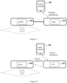
- the hardware combination of the system can be an integrated structure; wherein, the probe 500 and the ultrasonic unit, the feedback signal processing unit, the feedback device generating unit and the feedback reminder unit in the host 501 are integrated into an all-in-one machine.
- the all-in-one machine performs data interworking with the mobile terminal 502 through the wireless transmission unit. It can be understood that this integration mode has no trouble of cables, all components are connected wirelessly, and the overall structure is simple and compact, which is the optimal concept of the present application.
- This technical assumption is applicable to all dynamic and static muscle working modes (i.e. isometric contraction, isotonic contraction, isokinetic contraction), especially solving the problem of space obstruction caused by repeated isotonic movement of limbs and joints by cables.
- the hardware combination of the system can also be a separate structure; with reference to Figure 11 , the host is composed of the ultrasonic unit, the feedback signal processing unit, the feedback device generating unit, the feedback reminder unit, and the control unit in an integrated manner, and is wired with the probe through physical cables; the host communicates data with the mobile terminal through the wireless transmission unit.
- the host is composed of the ultrasonic unit, the feedback signal processing unit, the feedback device generating unit, the feedback reminder unit, and the control unit in an integrated manner, and is wirelessly connected with the probe and the mobile terminal through the wireless transmission unit; the probe is also equipped with a wireless transmission unit for data exchange. Understandably, in actual operation, the two separate ways use host to share the weight and volume of the probe, which is conducive to its fixation on the skin surface; the separate host is convenient to be carried. This technical assumption is relatively easy to implement.
- the beneficial effect of the above embodiments of Figures 10 , 11 and 12 is that the hardware combination can minimize the limitation of the cable and probe handheld mode on muscle training (especially isotonic movement); providing a basis for the design and development of wearable ultrasonic probes.
- step 1 preparation before training.
- the specific contents include setting the target training data K or K ⁇ k 1 in advance, determining the target muscle to be trained, determining the number of the contraction cycles N of the muscle, determining the training related parameters y and z, selecting the type of feedback signal, fixing the ultrasonic probe, etc.
- step 2 S102: Start the first contraction cycle of the target muscle and collect ultrasonic images.
- Step 3 S103a: extract muscle characteristic parameters y.
- step 4 (S103b): calculate the training index value z.
- the technical solution to achieve human-computer interaction is to use feedback reminder to guide training: provide feedback reminder unit 512, and compare the training index value y with the preset target threshold value K. If the training index value does not exceed the target threshold value (i.e. z ⁇ K), there is no alarm prompt; if the training index value exceeds the target threshold value (i.e. z>K), the mobile terminal 502 will output an instant alarm prompt to remind the user that the degree of activation level is effective. Unlimitedly, the expression of alarm prompt includes visual, auditory and tactile forms. After recording and completing the first contraction cycle of the target muscle, use the method of circulation until the Nth contraction cycle is completed.
- Step 6 (S104b) can also be implemented through another technical solution. In addition, other steps are similar to those in the first embodiment above, so we will not repeat them.
- the feedback reminding unit 512 is provided to compare the training index value y with the preset target interval K ⁇ k 1 : if the training index value is not within the target interval (i.e. z>K+k 1 or z ⁇ K-k 1 ), the mobile terminal 102 will output a continuous alarm prompt to remind the user that the training index value y is not in the effective degree of activation; if the training index value is within the target range (i.e. K-k 1 ⁇ z ⁇ K+k 1 ), the alarm will stop.
Landscapes
- Health & Medical Sciences (AREA)
- Life Sciences & Earth Sciences (AREA)
- Engineering & Computer Science (AREA)
- General Health & Medical Sciences (AREA)
- Veterinary Medicine (AREA)
- Pathology (AREA)
- Biomedical Technology (AREA)
- Heart & Thoracic Surgery (AREA)
- Medical Informatics (AREA)
- Molecular Biology (AREA)
- Surgery (AREA)
- Animal Behavior & Ethology (AREA)
- Biophysics (AREA)
- Public Health (AREA)
- Physics & Mathematics (AREA)
- Radiology & Medical Imaging (AREA)
- Nuclear Medicine, Radiotherapy & Molecular Imaging (AREA)
- Computer Vision & Pattern Recognition (AREA)
- Dentistry (AREA)
- Oral & Maxillofacial Surgery (AREA)
- Physiology (AREA)
- Physical Education & Sports Medicine (AREA)
- Computer Networks & Wireless Communication (AREA)
- Multimedia (AREA)
- Human Computer Interaction (AREA)
- Orthopedic Medicine & Surgery (AREA)
- Rheumatology (AREA)
- Ultra Sonic Daignosis Equipment (AREA)
Applications Claiming Priority (2)
| Application Number | Priority Date | Filing Date | Title |
|---|---|---|---|
| CN202010854798.1A CN112089442B (zh) | 2020-08-21 | 2020-08-21 | 利用超声成像提供视觉反馈的肌肉训练方法及系统 |
| PCT/CN2021/103783 WO2022037274A1 (zh) | 2020-08-21 | 2021-06-30 | 利用超声成像提供视觉反馈的肌肉训练方法及系统 |
Publications (2)
| Publication Number | Publication Date |
|---|---|
| EP4201339A1 true EP4201339A1 (de) | 2023-06-28 |
| EP4201339A4 EP4201339A4 (de) | 2024-09-11 |
Family
ID=73754186
Family Applications (1)
| Application Number | Title | Priority Date | Filing Date |
|---|---|---|---|
| EP21857370.7A Pending EP4201339A4 (de) | 2020-08-21 | 2021-06-30 | Muskeltrainingsverfahren und system zur bereitstellung von visueller rückkopplung durch verwendung von ultraschallbildgebung |
Country Status (4)
| Country | Link |
|---|---|
| US (1) | US20230330507A1 (de) |
| EP (1) | EP4201339A4 (de) |
| CN (1) | CN112089442B (de) |
| WO (1) | WO2022037274A1 (de) |
Cited By (2)
| Publication number | Priority date | Publication date | Assignee | Title |
|---|---|---|---|---|
| EP4289364A4 (de) * | 2021-02-08 | 2024-12-25 | Eieling Technology Limited | Verfahren und vorrichtung zur beurteilung des kontaktzustands einer ultraschallsonde auf der basis von weichgewebemorphologie |
| IT202300018135A1 (it) * | 2023-09-04 | 2025-03-04 | Xeos S R L | Dispositivo adattivo per il supporto alle decisioni nell'ambito dell'allenamento agonistico e della prevenzione degli infortuni sportivi, applicabile in particolare al muscolo bicipite femorale |
Families Citing this family (11)
| Publication number | Priority date | Publication date | Assignee | Title |
|---|---|---|---|---|
| CN112089442B (zh) * | 2020-08-21 | 2023-10-13 | 意领科技有限公司 | 利用超声成像提供视觉反馈的肌肉训练方法及系统 |
| CN112998759A (zh) * | 2021-04-06 | 2021-06-22 | 无锡海斯凯尔医学技术有限公司 | 组织弹性检测方法、装置和系统 |
| CN113425575A (zh) * | 2021-06-22 | 2021-09-24 | 深圳市理德铭科技股份有限公司 | 一种具有自适应功能的筋膜枪及健康管理系统 |
| CN113921108A (zh) * | 2021-10-08 | 2022-01-11 | 重庆邮电大学 | 一种弹性带抗阻训练力数据的自动分割方法 |
| JP2024041446A (ja) * | 2022-09-14 | 2024-03-27 | セイコーエプソン株式会社 | 疲労検出装置、支援システム、及び疲労検出方法 |
| CN115920333B (zh) * | 2022-10-17 | 2025-12-05 | 北京体育大学 | 一种深蹲1rm抗阻训练效果预测方法、系统和存储介质 |
| CN119423832A (zh) * | 2023-08-07 | 2025-02-14 | 中慧医学成像(深圳)有限公司 | 肌肉三维成像分析方法及系统 |
| CN117808808B (zh) * | 2024-03-01 | 2024-05-14 | 山东师范大学 | 一种矿石粒度检测方法、系统、电子设备及存储介质 |
| WO2026032236A1 (en) * | 2024-08-03 | 2026-02-12 | The Hong Kong Polytechnic University | Muscle activity data acquisition equipment, method for analyzing muscle activity, electronic device and storage medium |
| WO2026032209A1 (en) * | 2024-08-03 | 2026-02-12 | The Hong Kong Polytechnic University | Method and apparatus for monitoring neuromuscular blockade, electronic device and storage medium |
| CN119252425B (zh) * | 2024-12-06 | 2025-04-29 | 鎏玥(上海)智能科技有限公司 | 一种基于图像分析的腿部肌肉训练分析方法及训练椅 |
Family Cites Families (13)
| Publication number | Priority date | Publication date | Assignee | Title |
|---|---|---|---|---|
| US7764990B2 (en) * | 2004-07-01 | 2010-07-27 | Suunto Oy | Method and device for measuring exercise level during exercise and for measuring fatigue |
| KR101201579B1 (ko) * | 2010-06-09 | 2012-11-14 | 강원대학교산학협력단 | 3차원 영상 피드백 및 전기 자극을 이용한 근육 재교육 훈련 장치 및 방법 |
| US20150099972A1 (en) * | 2013-10-04 | 2015-04-09 | Vibrado Technologies, Inc. | Myography method and system |
| CN103584884B (zh) * | 2013-11-07 | 2015-07-01 | 中国科学院深圳先进技术研究院 | 肌力评估方法及装置、肌肉康复训练跟踪评估方法及系统 |
| US11219396B2 (en) * | 2013-11-23 | 2022-01-11 | MAD Apparel, Inc. | System and method for monitoring biometric signals |
| EP3576607A1 (de) * | 2017-02-02 | 2019-12-11 | Syddansk Universitet | Ultraschalluntersuchungsverfahren zur messung der muskelverformung |
| WO2018147384A1 (ja) * | 2017-02-10 | 2018-08-16 | 株式会社Mtg | トレーニング具およびトレーニングシステム |
| TWM542489U (zh) * | 2017-02-18 | 2017-06-01 | wei-jun Xu | 肌電回饋式運動訓練系統 |
| CN106691447B (zh) * | 2017-02-23 | 2023-09-12 | 北京纳通科技集团有限公司 | 肌肉训练辅助装置、肌肉训练评估装置及方法 |
| CN108742705A (zh) * | 2018-04-10 | 2018-11-06 | 深圳大学 | 一种实时检测肌肉形态参数的超声成像设备及方法 |
| DE112020002488T5 (de) * | 2019-05-23 | 2022-02-24 | Koninklijke Philips N.V. | Muskelbildgebungssystem |
| CN210904853U (zh) * | 2019-10-09 | 2020-07-03 | 周凤鸣 | 一种简易的肌肉训练视觉反馈装置 |
| CN112089442B (zh) * | 2020-08-21 | 2023-10-13 | 意领科技有限公司 | 利用超声成像提供视觉反馈的肌肉训练方法及系统 |
-
2020
- 2020-08-21 CN CN202010854798.1A patent/CN112089442B/zh active Active
-
2021
- 2021-06-30 WO PCT/CN2021/103783 patent/WO2022037274A1/zh not_active Ceased
- 2021-06-30 US US18/022,309 patent/US20230330507A1/en active Pending
- 2021-06-30 EP EP21857370.7A patent/EP4201339A4/de active Pending
Cited By (2)
| Publication number | Priority date | Publication date | Assignee | Title |
|---|---|---|---|---|
| EP4289364A4 (de) * | 2021-02-08 | 2024-12-25 | Eieling Technology Limited | Verfahren und vorrichtung zur beurteilung des kontaktzustands einer ultraschallsonde auf der basis von weichgewebemorphologie |
| IT202300018135A1 (it) * | 2023-09-04 | 2025-03-04 | Xeos S R L | Dispositivo adattivo per il supporto alle decisioni nell'ambito dell'allenamento agonistico e della prevenzione degli infortuni sportivi, applicabile in particolare al muscolo bicipite femorale |
Also Published As
| Publication number | Publication date |
|---|---|
| US20230330507A1 (en) | 2023-10-19 |
| CN112089442A (zh) | 2020-12-18 |
| WO2022037274A1 (zh) | 2022-02-24 |
| EP4201339A4 (de) | 2024-09-11 |
| CN112089442B (zh) | 2023-10-13 |
Similar Documents
| Publication | Publication Date | Title |
|---|---|---|
| EP4201339A1 (de) | Muskeltrainingsverfahren und system zur bereitstellung von visueller rückkopplung durch verwendung von ultraschallbildgebung | |
| EP4289364A1 (de) | Verfahren und vorrichtung zur beurteilung des kontaktzustands einer ultraschallsonde auf der basis von weichgewebemorphologie | |
| CN102551800B (zh) | 用于显示超声数据的方法和系统 | |
| CN106264573B (zh) | 便携式肌肉力学参数及肌肉力在体超声检测装置及方法 | |
| CN105997138B (zh) | 超声成像装置及其处理超声图像的方法 | |
| CN111758137A (zh) | 用于远程医疗的方法和设备 | |
| CN104905814B (zh) | 超声诊断设备和显示超声图像的方法 | |
| JP2004527266A (ja) | プログラムされた特定用途向けデータ収集装置によって得られた超音波情報の遠隔評価システム | |
| CN113160921A (zh) | 一种基于血流动力学的数字人体心血管系统的构建方法和应用 | |
| CN106562794B (zh) | 一种膀胱测容装置及其实现方法 | |
| EP2989987B1 (de) | Diagnostische Ultraschallvorrichtung und Verfahren und computerlesbares Speichermedium | |
| CN103536313A (zh) | 远程控制的超声设备和超声治疗系统 | |
| CN110279404A (zh) | 一种脉搏波采集装置及方法 | |
| Castellini et al. | Ultrasound imaging as a human-machine interface in a realistic scenario | |
| CN111938699A (zh) | 一种引导使用超声设备的系统及方法 | |
| WO2024172855A1 (en) | Systems and methods for user-assisted acquisition of ultrasound images | |
| CN116616814A (zh) | 一种实现超声成像控制和显示的装置及方法 | |
| CN202365763U (zh) | 一种基于脉象频谱分析的穿戴式无线脉诊仪系统 | |
| US20070016061A1 (en) | System for measuring and tracking human body fat | |
| HK40043764B (zh) | 利用超声成像提供视觉反馈的肌肉训练方法及系统 | |
| HK40043764A (en) | Muscle training method and system using ultrasound imaging to provide visual biofeedback | |
| Sorriento et al. | A novel approach for remote ultrasonography of knee osteoarthritis | |
| RU129681U1 (ru) | Система определения функционального состояния группы людей с обратной связью | |
| HK40057058A (en) | Method and device for evaluating contact state of ultrasound transducer based on soft tissue morphology | |
| US11918409B2 (en) | Using ultrasound and artificial intelligence to assess muscle stretch reflexes |
Legal Events
| Date | Code | Title | Description |
|---|---|---|---|
| STAA | Information on the status of an ep patent application or granted ep patent |
Free format text: STATUS: THE INTERNATIONAL PUBLICATION HAS BEEN MADE |
|
| PUAI | Public reference made under article 153(3) epc to a published international application that has entered the european phase |
Free format text: ORIGINAL CODE: 0009012 |
|
| STAA | Information on the status of an ep patent application or granted ep patent |
Free format text: STATUS: REQUEST FOR EXAMINATION WAS MADE |
|
| 17P | Request for examination filed |
Effective date: 20230320 |
|
| AK | Designated contracting states |
Kind code of ref document: A1 Designated state(s): AL AT BE BG CH CY CZ DE DK EE ES FI FR GB GR HR HU IE IS IT LI LT LU LV MC MK MT NL NO PL PT RO RS SE SI SK SM TR |
|
| DAV | Request for validation of the european patent (deleted) | ||
| DAX | Request for extension of the european patent (deleted) | ||
| A4 | Supplementary search report drawn up and despatched |
Effective date: 20240813 |
|
| RIC1 | Information provided on ipc code assigned before grant |
Ipc: A61B 8/08 20060101ALI20240807BHEP Ipc: A61B 8/00 20060101AFI20240807BHEP |
|
| RAP1 | Party data changed (applicant data changed or rights of an application transferred) |
Owner name: SONOCONNECT TECHNOLOGY LIMITED |