EP4183414A1 - Methods for quantitating individual antibodies from a mixture - Google Patents
Methods for quantitating individual antibodies from a mixture Download PDFInfo
- Publication number
- EP4183414A1 EP4183414A1 EP22208008.7A EP22208008A EP4183414A1 EP 4183414 A1 EP4183414 A1 EP 4183414A1 EP 22208008 A EP22208008 A EP 22208008A EP 4183414 A1 EP4183414 A1 EP 4183414A1
- Authority
- EP
- European Patent Office
- Prior art keywords
- mixture
- antibody
- antibodies
- hic
- hplc
- Prior art date
- Legal status (The legal status is an assumption and is not a legal conclusion. Google has not performed a legal analysis and makes no representation as to the accuracy of the status listed.)
- Pending
Links
- 239000000203 mixture Substances 0.000 title claims abstract description 321
- 238000000034 method Methods 0.000 title claims abstract description 183
- 238000004128 high performance liquid chromatography Methods 0.000 claims abstract description 21
- 238000004191 hydrophobic interaction chromatography Methods 0.000 claims abstract description 18
- 102000004169 proteins and genes Human genes 0.000 claims description 82
- 108090000623 proteins and genes Proteins 0.000 claims description 82
- 201000011001 Ebola Hemorrhagic Fever Diseases 0.000 claims description 31
- 229940126534 drug product Drugs 0.000 claims description 22
- 238000010828 elution Methods 0.000 claims description 22
- 239000000825 pharmaceutical preparation Substances 0.000 claims description 22
- 238000012216 screening Methods 0.000 claims description 22
- 208000025370 Middle East respiratory syndrome Diseases 0.000 claims description 19
- 239000000872 buffer Substances 0.000 claims description 16
- BFNBIHQBYMNNAN-UHFFFAOYSA-N ammonium sulfate Chemical compound N.N.OS(O)(=O)=O BFNBIHQBYMNNAN-UHFFFAOYSA-N 0.000 claims description 15
- 229910052921 ammonium sulfate Inorganic materials 0.000 claims description 15
- 235000011130 ammonium sulphate Nutrition 0.000 claims description 15
- 230000003993 interaction Effects 0.000 claims description 14
- 238000004611 spectroscopical analysis Methods 0.000 claims description 13
- 239000002105 nanoparticle Substances 0.000 claims description 12
- 229920002594 Polyethylene Glycol 8000 Polymers 0.000 claims description 11
- 208000035473 Communicable disease Diseases 0.000 claims description 10
- 208000030820 Ebola disease Diseases 0.000 claims description 10
- 239000000427 antigen Substances 0.000 claims description 10
- 102000036639 antigens Human genes 0.000 claims description 10
- 108091007433 antigens Proteins 0.000 claims description 10
- 208000015181 infectious disease Diseases 0.000 claims description 10
- 208000002780 macular degeneration Diseases 0.000 claims description 10
- 238000002169 hydrotherapy Methods 0.000 abstract description 10
- 238000009472 formulation Methods 0.000 description 48
- 238000002347 injection Methods 0.000 description 32
- 239000007924 injection Substances 0.000 description 32
- 239000013016 formulated drug substance Substances 0.000 description 21
- 238000011084 recovery Methods 0.000 description 20
- 239000000523 sample Substances 0.000 description 14
- 238000002835 absorbance Methods 0.000 description 12
- 208000001528 Coronaviridae Infections Diseases 0.000 description 10
- 238000000926 separation method Methods 0.000 description 10
- 241000127282 Middle East respiratory syndrome-related coronavirus Species 0.000 description 8
- 229940096437 Protein S Drugs 0.000 description 8
- 101710198474 Spike protein Proteins 0.000 description 8
- 230000002209 hydrophobic effect Effects 0.000 description 8
- 201000010099 disease Diseases 0.000 description 7
- 208000037265 diseases, disorders, signs and symptoms Diseases 0.000 description 7
- 239000008194 pharmaceutical composition Substances 0.000 description 7
- HNSDLXPSAYFUHK-UHFFFAOYSA-N 1,4-bis(2-ethylhexyl) sulfosuccinate Chemical compound CCCCC(CC)COC(=O)CC(S(O)(=O)=O)C(=O)OCC(CC)CCCC HNSDLXPSAYFUHK-UHFFFAOYSA-N 0.000 description 6
- HEMHJVSKTPXQMS-UHFFFAOYSA-M Sodium hydroxide Chemical compound [OH-].[Na+] HEMHJVSKTPXQMS-UHFFFAOYSA-M 0.000 description 6
- 238000012506 imaged capillary isoelectric focusing Methods 0.000 description 6
- 108060003951 Immunoglobulin Proteins 0.000 description 5
- 238000011088 calibration curve Methods 0.000 description 5
- 239000003814 drug Substances 0.000 description 5
- 229940088679 drug related substance Drugs 0.000 description 5
- 102000018358 immunoglobulin Human genes 0.000 description 5
- 150000003839 salts Chemical class 0.000 description 5
- 239000000243 solution Substances 0.000 description 5
- 239000002904 solvent Substances 0.000 description 5
- 241000894007 species Species 0.000 description 5
- 229910019142 PO4 Inorganic materials 0.000 description 4
- 229930006000 Sucrose Natural products 0.000 description 4
- CZMRCDWAGMRECN-UGDNZRGBSA-N Sucrose Chemical compound O[C@H]1[C@H](O)[C@@H](CO)O[C@@]1(CO)O[C@@H]1[C@H](O)[C@@H](O)[C@H](O)[C@@H](CO)O1 CZMRCDWAGMRECN-UGDNZRGBSA-N 0.000 description 4
- 239000008186 active pharmaceutical agent Substances 0.000 description 4
- 150000001413 amino acids Chemical class 0.000 description 4
- 238000012544 monitoring process Methods 0.000 description 4
- NBIIXXVUZAFLBC-UHFFFAOYSA-K phosphate Chemical compound [O-]P([O-])([O-])=O NBIIXXVUZAFLBC-UHFFFAOYSA-K 0.000 description 4
- 239000010452 phosphate Substances 0.000 description 4
- 238000002360 preparation method Methods 0.000 description 4
- 230000002441 reversible effect Effects 0.000 description 4
- 239000005720 sucrose Substances 0.000 description 4
- WEVYAHXRMPXWCK-UHFFFAOYSA-N Acetonitrile Chemical compound CC#N WEVYAHXRMPXWCK-UHFFFAOYSA-N 0.000 description 3
- 125000000539 amino acid group Chemical group 0.000 description 3
- 239000013068 control sample Substances 0.000 description 3
- 239000006185 dispersion Substances 0.000 description 3
- 238000012395 formulation development Methods 0.000 description 3
- 210000004602 germ cell Anatomy 0.000 description 3
- 239000000463 material Substances 0.000 description 3
- 238000012986 modification Methods 0.000 description 3
- 230000004048 modification Effects 0.000 description 3
- 108090000765 processed proteins & peptides Proteins 0.000 description 3
- 102000004196 processed proteins & peptides Human genes 0.000 description 3
- 239000003643 water by type Substances 0.000 description 3
- 102100035360 Cerebellar degeneration-related antigen 1 Human genes 0.000 description 2
- 102000016622 Dipeptidyl Peptidase 4 Human genes 0.000 description 2
- 108010067722 Dipeptidyl Peptidase 4 Proteins 0.000 description 2
- FAPWRFPIFSIZLT-UHFFFAOYSA-M Sodium chloride Chemical compound [Na+].[Cl-] FAPWRFPIFSIZLT-UHFFFAOYSA-M 0.000 description 2
- 238000010521 absorption reaction Methods 0.000 description 2
- LFVGISIMTYGQHF-UHFFFAOYSA-N ammonium dihydrogen phosphate Chemical compound [NH4+].OP(O)([O-])=O LFVGISIMTYGQHF-UHFFFAOYSA-N 0.000 description 2
- 238000003556 assay Methods 0.000 description 2
- 238000005341 cation exchange Methods 0.000 description 2
- 238000004587 chromatography analysis Methods 0.000 description 2
- 238000000576 coating method Methods 0.000 description 2
- 238000012217 deletion Methods 0.000 description 2
- 230000037430 deletion Effects 0.000 description 2
- 239000002612 dispersion medium Substances 0.000 description 2
- 239000003937 drug carrier Substances 0.000 description 2
- 238000005516 engineering process Methods 0.000 description 2
- 238000001914 filtration Methods 0.000 description 2
- 239000004615 ingredient Substances 0.000 description 2
- 230000005764 inhibitory process Effects 0.000 description 2
- 238000003780 insertion Methods 0.000 description 2
- 230000037431 insertion Effects 0.000 description 2
- 239000003446 ligand Substances 0.000 description 2
- 238000005259 measurement Methods 0.000 description 2
- VIKNJXKGJWUCNN-XGXHKTLJSA-N norethisterone Chemical compound O=C1CC[C@@H]2[C@H]3CC[C@](C)([C@](CC4)(O)C#C)[C@@H]4[C@@H]3CCC2=C1 VIKNJXKGJWUCNN-XGXHKTLJSA-N 0.000 description 2
- 239000000546 pharmaceutical excipient Substances 0.000 description 2
- 239000008363 phosphate buffer Substances 0.000 description 2
- 229920001184 polypeptide Polymers 0.000 description 2
- 239000011148 porous material Substances 0.000 description 2
- 108020003175 receptors Proteins 0.000 description 2
- 102000005962 receptors Human genes 0.000 description 2
- 230000001105 regulatory effect Effects 0.000 description 2
- AJPJDKMHJJGVTQ-UHFFFAOYSA-M sodium dihydrogen phosphate Chemical compound [Na+].OP(O)([O-])=O AJPJDKMHJJGVTQ-UHFFFAOYSA-M 0.000 description 2
- BBMHARZCALWXSL-UHFFFAOYSA-M sodium dihydrogenphosphate monohydrate Chemical compound O.[Na+].OP(O)([O-])=O BBMHARZCALWXSL-UHFFFAOYSA-M 0.000 description 2
- 229910000162 sodium phosphate Inorganic materials 0.000 description 2
- 238000006467 substitution reaction Methods 0.000 description 2
- 208000024891 symptom Diseases 0.000 description 2
- 230000001225 therapeutic effect Effects 0.000 description 2
- IIZPXYDJLKNOIY-JXPKJXOSSA-N 1-palmitoyl-2-arachidonoyl-sn-glycero-3-phosphocholine Chemical compound CCCCCCCCCCCCCCCC(=O)OC[C@H](COP([O-])(=O)OCC[N+](C)(C)C)OC(=O)CCC\C=C/C\C=C/C\C=C/C\C=C/CCCCC IIZPXYDJLKNOIY-JXPKJXOSSA-N 0.000 description 1
- QTBSBXVTEAMEQO-UHFFFAOYSA-M Acetate Chemical compound CC([O-])=O QTBSBXVTEAMEQO-UHFFFAOYSA-M 0.000 description 1
- 206010069754 Acquired gene mutation Diseases 0.000 description 1
- 241000599985 Beijerinckia mobilis Species 0.000 description 1
- 241000283690 Bos taurus Species 0.000 description 1
- 241000282472 Canis lupus familiaris Species 0.000 description 1
- 241000283707 Capra Species 0.000 description 1
- 241000700198 Cavia Species 0.000 description 1
- 241000699800 Cricetinae Species 0.000 description 1
- 241001115402 Ebolavirus Species 0.000 description 1
- 241000283086 Equidae Species 0.000 description 1
- 241000282326 Felis catus Species 0.000 description 1
- 108010010803 Gelatin Proteins 0.000 description 1
- 241000699694 Gerbillinae Species 0.000 description 1
- 241000282412 Homo Species 0.000 description 1
- 101000935587 Homo sapiens Flavin reductase (NADPH) Proteins 0.000 description 1
- 101150030213 Lag3 gene Proteins 0.000 description 1
- 239000007987 MES buffer Substances 0.000 description 1
- 241000124008 Mammalia Species 0.000 description 1
- 241000699666 Mus <mouse, genus> Species 0.000 description 1
- 241000699670 Mus sp. Species 0.000 description 1
- 241000283973 Oryctolagus cuniculus Species 0.000 description 1
- 241000282579 Pan Species 0.000 description 1
- 241000282520 Papio Species 0.000 description 1
- 241001494479 Pecora Species 0.000 description 1
- 241000282405 Pongo abelii Species 0.000 description 1
- 241000700159 Rattus Species 0.000 description 1
- VYPSYNLAJGMNEJ-UHFFFAOYSA-N Silicium dioxide Chemical compound O=[Si]=O VYPSYNLAJGMNEJ-UHFFFAOYSA-N 0.000 description 1
- 241000282887 Suidae Species 0.000 description 1
- XSQUKJJJFZCRTK-UHFFFAOYSA-N Urea Chemical compound NC(N)=O XSQUKJJJFZCRTK-UHFFFAOYSA-N 0.000 description 1
- 241000700605 Viruses Species 0.000 description 1
- 208000020329 Zika virus infectious disease Diseases 0.000 description 1
- 239000003070 absorption delaying agent Substances 0.000 description 1
- 239000002253 acid Substances 0.000 description 1
- 108010023082 activin A Proteins 0.000 description 1
- 108010081667 aflibercept Proteins 0.000 description 1
- 229960004539 alirocumab Drugs 0.000 description 1
- 125000003275 alpha amino acid group Chemical group 0.000 description 1
- 239000003242 anti bacterial agent Substances 0.000 description 1
- 230000000844 anti-bacterial effect Effects 0.000 description 1
- 239000003429 antifungal agent Substances 0.000 description 1
- 229940121375 antifungal agent Drugs 0.000 description 1
- 238000013459 approach Methods 0.000 description 1
- 239000004202 carbamide Substances 0.000 description 1
- 210000004027 cell Anatomy 0.000 description 1
- 230000008859 change Effects 0.000 description 1
- 239000003153 chemical reaction reagent Substances 0.000 description 1
- 239000012504 chromatography matrix Substances 0.000 description 1
- 239000011248 coating agent Substances 0.000 description 1
- -1 coatings Substances 0.000 description 1
- 238000000205 computational method Methods 0.000 description 1
- 239000013078 crystal Substances 0.000 description 1
- 230000003247 decreasing effect Effects 0.000 description 1
- 230000001934 delay Effects 0.000 description 1
- 239000003085 diluting agent Substances 0.000 description 1
- 239000000539 dimer Substances 0.000 description 1
- 238000010494 dissociation reaction Methods 0.000 description 1
- 230000005593 dissociations Effects 0.000 description 1
- 229950004341 evinacumab Drugs 0.000 description 1
- 108020001507 fusion proteins Proteins 0.000 description 1
- 102000037865 fusion proteins Human genes 0.000 description 1
- 229920000159 gelatin Polymers 0.000 description 1
- 239000008273 gelatin Substances 0.000 description 1
- 235000019322 gelatine Nutrition 0.000 description 1
- 235000011852 gelatine desserts Nutrition 0.000 description 1
- PCHJSUWPFVWCPO-UHFFFAOYSA-N gold Chemical compound [Au] PCHJSUWPFVWCPO-UHFFFAOYSA-N 0.000 description 1
- 239000010931 gold Substances 0.000 description 1
- 229910052737 gold Inorganic materials 0.000 description 1
- 125000001165 hydrophobic group Chemical group 0.000 description 1
- 229940127121 immunoconjugate Drugs 0.000 description 1
- 229940072221 immunoglobulins Drugs 0.000 description 1
- 238000000338 in vitro Methods 0.000 description 1
- 238000001727 in vivo Methods 0.000 description 1
- 239000007972 injectable composition Substances 0.000 description 1
- 230000010354 integration Effects 0.000 description 1
- 239000007951 isotonicity adjuster Substances 0.000 description 1
- 239000000787 lecithin Substances 0.000 description 1
- 229940067606 lecithin Drugs 0.000 description 1
- 235000010445 lecithin Nutrition 0.000 description 1
- 239000002502 liposome Substances 0.000 description 1
- 238000012423 maintenance Methods 0.000 description 1
- 239000004530 micro-emulsion Substances 0.000 description 1
- 238000002156 mixing Methods 0.000 description 1
- 235000019799 monosodium phosphate Nutrition 0.000 description 1
- 230000035772 mutation Effects 0.000 description 1
- 230000003287 optical effect Effects 0.000 description 1
- 239000002245 particle Substances 0.000 description 1
- VLTRZXGMWDSKGL-UHFFFAOYSA-M perchlorate Inorganic materials [O-]Cl(=O)(=O)=O VLTRZXGMWDSKGL-UHFFFAOYSA-M 0.000 description 1
- VLTRZXGMWDSKGL-UHFFFAOYSA-N perchloric acid Chemical compound OCl(=O)(=O)=O VLTRZXGMWDSKGL-UHFFFAOYSA-N 0.000 description 1
- 230000004481 post-translational protein modification Effects 0.000 description 1
- 230000002265 prevention Effects 0.000 description 1
- 238000004393 prognosis Methods 0.000 description 1
- 230000002035 prolonged effect Effects 0.000 description 1
- 238000000734 protein sequencing Methods 0.000 description 1
- 238000003908 quality control method Methods 0.000 description 1
- 238000002708 random mutagenesis Methods 0.000 description 1
- 230000009467 reduction Effects 0.000 description 1
- 238000002741 site-directed mutagenesis Methods 0.000 description 1
- 239000011780 sodium chloride Substances 0.000 description 1
- 239000007787 solid Substances 0.000 description 1
- 230000037439 somatic mutation Effects 0.000 description 1
- 230000001954 sterilising effect Effects 0.000 description 1
- 238000004659 sterilization and disinfection Methods 0.000 description 1
- 238000002198 surface plasmon resonance spectroscopy Methods 0.000 description 1
- 239000004094 surface-active agent Substances 0.000 description 1
- 229950006444 trevogrumab Drugs 0.000 description 1
- 239000013638 trimer Substances 0.000 description 1
- 238000000825 ultraviolet detection Methods 0.000 description 1
- 238000002211 ultraviolet spectrum Methods 0.000 description 1
- 239000003981 vehicle Substances 0.000 description 1
Images


Classifications
-
- G—PHYSICS
- G01—MEASURING; TESTING
- G01N—INVESTIGATING OR ANALYSING MATERIALS BY DETERMINING THEIR CHEMICAL OR PHYSICAL PROPERTIES
- G01N30/00—Investigating or analysing materials by separation into components using adsorption, absorption or similar phenomena or using ion-exchange, e.g. chromatography or field flow fractionation
- G01N30/02—Column chromatography
- G01N30/88—Integrated analysis systems specially adapted therefor, not covered by a single one of the groups G01N30/04 - G01N30/86
-
- B—PERFORMING OPERATIONS; TRANSPORTING
- B01—PHYSICAL OR CHEMICAL PROCESSES OR APPARATUS IN GENERAL
- B01D—SEPARATION
- B01D15/00—Separating processes involving the treatment of liquids with solid sorbents; Apparatus therefor
- B01D15/08—Selective adsorption, e.g. chromatography
- B01D15/26—Selective adsorption, e.g. chromatography characterised by the separation mechanism
- B01D15/30—Partition chromatography
-
- A—HUMAN NECESSITIES
- A61—MEDICAL OR VETERINARY SCIENCE; HYGIENE
- A61K—PREPARATIONS FOR MEDICAL, DENTAL OR TOILETRY PURPOSES
- A61K39/00—Medicinal preparations containing antigens or antibodies
- A61K39/395—Antibodies; Immunoglobulins; Immune serum, e.g. antilymphocytic serum
- A61K39/39591—Stabilisation, fragmentation
-
- A—HUMAN NECESSITIES
- A61—MEDICAL OR VETERINARY SCIENCE; HYGIENE
- A61P—SPECIFIC THERAPEUTIC ACTIVITY OF CHEMICAL COMPOUNDS OR MEDICINAL PREPARATIONS
- A61P11/00—Drugs for disorders of the respiratory system
-
- A—HUMAN NECESSITIES
- A61—MEDICAL OR VETERINARY SCIENCE; HYGIENE
- A61P—SPECIFIC THERAPEUTIC ACTIVITY OF CHEMICAL COMPOUNDS OR MEDICINAL PREPARATIONS
- A61P27/00—Drugs for disorders of the senses
- A61P27/02—Ophthalmic agents
-
- A—HUMAN NECESSITIES
- A61—MEDICAL OR VETERINARY SCIENCE; HYGIENE
- A61P—SPECIFIC THERAPEUTIC ACTIVITY OF CHEMICAL COMPOUNDS OR MEDICINAL PREPARATIONS
- A61P31/00—Antiinfectives, i.e. antibiotics, antiseptics, chemotherapeutics
- A61P31/12—Antivirals
-
- B—PERFORMING OPERATIONS; TRANSPORTING
- B01—PHYSICAL OR CHEMICAL PROCESSES OR APPARATUS IN GENERAL
- B01D—SEPARATION
- B01D15/00—Separating processes involving the treatment of liquids with solid sorbents; Apparatus therefor
- B01D15/08—Selective adsorption, e.g. chromatography
- B01D15/26—Selective adsorption, e.g. chromatography characterised by the separation mechanism
- B01D15/32—Bonded phase chromatography
- B01D15/325—Reversed phase
- B01D15/327—Reversed phase with hydrophobic interaction
-
- C—CHEMISTRY; METALLURGY
- C07—ORGANIC CHEMISTRY
- C07K—PEPTIDES
- C07K16/00—Immunoglobulins [IGs], e.g. monoclonal or polyclonal antibodies
-
- C—CHEMISTRY; METALLURGY
- C07—ORGANIC CHEMISTRY
- C07K—PEPTIDES
- C07K16/00—Immunoglobulins [IGs], e.g. monoclonal or polyclonal antibodies
- C07K16/06—Immunoglobulins [IGs], e.g. monoclonal or polyclonal antibodies from serum
-
- C—CHEMISTRY; METALLURGY
- C07—ORGANIC CHEMISTRY
- C07K—PEPTIDES
- C07K16/00—Immunoglobulins [IGs], e.g. monoclonal or polyclonal antibodies
- C07K16/06—Immunoglobulins [IGs], e.g. monoclonal or polyclonal antibodies from serum
- C07K16/065—Purification, fragmentation
-
- C—CHEMISTRY; METALLURGY
- C07—ORGANIC CHEMISTRY
- C07K—PEPTIDES
- C07K16/00—Immunoglobulins [IGs], e.g. monoclonal or polyclonal antibodies
- C07K16/08—Immunoglobulins [IGs], e.g. monoclonal or polyclonal antibodies against material from viruses
- C07K16/10—Immunoglobulins [IGs], e.g. monoclonal or polyclonal antibodies against material from viruses from RNA viruses
- C07K16/1002—Coronaviridae
-
- G—PHYSICS
- G01—MEASURING; TESTING
- G01N—INVESTIGATING OR ANALYSING MATERIALS BY DETERMINING THEIR CHEMICAL OR PHYSICAL PROPERTIES
- G01N30/00—Investigating or analysing materials by separation into components using adsorption, absorption or similar phenomena or using ion-exchange, e.g. chromatography or field flow fractionation
- G01N30/02—Column chromatography
-
- G—PHYSICS
- G01—MEASURING; TESTING
- G01N—INVESTIGATING OR ANALYSING MATERIALS BY DETERMINING THEIR CHEMICAL OR PHYSICAL PROPERTIES
- G01N33/00—Investigating or analysing materials by specific methods not covered by groups G01N1/00 - G01N31/00
- G01N33/48—Biological material, e.g. blood, urine; Haemocytometers
- G01N33/50—Chemical analysis of biological material, e.g. blood, urine; Testing involving biospecific ligand binding methods; Immunological testing
- G01N33/68—Chemical analysis of biological material, e.g. blood, urine; Testing involving biospecific ligand binding methods; Immunological testing involving proteins, peptides or amino acids
- G01N33/6854—Immunoglobulins
-
- G—PHYSICS
- G01—MEASURING; TESTING
- G01N—INVESTIGATING OR ANALYSING MATERIALS BY DETERMINING THEIR CHEMICAL OR PHYSICAL PROPERTIES
- G01N30/00—Investigating or analysing materials by separation into components using adsorption, absorption or similar phenomena or using ion-exchange, e.g. chromatography or field flow fractionation
- G01N30/02—Column chromatography
- G01N2030/022—Column chromatography characterised by the kind of separation mechanism
- G01N2030/027—Liquid chromatography
-
- G—PHYSICS
- G01—MEASURING; TESTING
- G01N—INVESTIGATING OR ANALYSING MATERIALS BY DETERMINING THEIR CHEMICAL OR PHYSICAL PROPERTIES
- G01N30/00—Investigating or analysing materials by separation into components using adsorption, absorption or similar phenomena or using ion-exchange, e.g. chromatography or field flow fractionation
- G01N30/02—Column chromatography
- G01N30/88—Integrated analysis systems specially adapted therefor, not covered by a single one of the groups G01N30/04 - G01N30/86
- G01N2030/8809—Integrated analysis systems specially adapted therefor, not covered by a single one of the groups G01N30/04 - G01N30/86 analysis specially adapted for the sample
- G01N2030/8813—Integrated analysis systems specially adapted therefor, not covered by a single one of the groups G01N30/04 - G01N30/86 analysis specially adapted for the sample biological materials
- G01N2030/8831—Integrated analysis systems specially adapted therefor, not covered by a single one of the groups G01N30/04 - G01N30/86 analysis specially adapted for the sample biological materials involving peptides or proteins
Definitions
- This disclosure relates to the field of assays for co-formulations of therapeutic antibodies.
- mAbs monoclonal antibodies
- DP drug product
- a method is required by regulatory agencies to quantitate the individual mAbs in a co-formulated drug substance (cFDS) to be incorporated into a DP, or a DP itself.
- cFDS co-formulated drug substance
- Developing a method to separate two or more antibody molecules and to measure the concentration of each mAb is challenging, because the antibody molecules may have similar molecular weights, protein structures, and charge properties.
- This disclosure includes a method of quantitating amounts of antibodies from a mixture comprising a plurality of antibodies.
- the method may include, among other things, separating each of the plurality of antibodies in the mixture using hydrophobic interaction chromatography high performance liquid chromatography (HIC-HPLC), and quantitating an amount of each antibody in the mixture, wherein a molecular weight of each antibody in the mixture is within 15 kDa of a molecular weight of any other antibodies in the mixture, and either a surface hydrophobicity of each antibody in the mixture is different from a surface hydrophobicity of another antibody in the mixture by more than about 0.25 units on the Kyte & Doolittle hydropathy scale, or each antibody in the mixture, when run on HIC-HPLC individually, elutes at a distinct run time from another antibody in the mixture, or both.
- HIC-HPLC hydrophobic interaction chromatography high performance liquid chromatography
- the surface hydrophobicity of each antibody in the mixture is different from the surface hydrophobicity of each other antibody in the mixture by about 0.5 to about 1.0 units on the Kyte & Doolittle hydropathy scale.
- the surface hydrophobicity of each antibody in the mixture is determined by calculating surface hydrophobicity based on protein structure or structural model, rapid screening for solubility in ammonium sulfate or PEG8000, or rapid screening for molecule interaction by affinity capture-self-interaction nanoparticle spectroscopy (AC-SINS).
- a first antibody in the mixture elutes at a first run time during a HIC-HPLC run
- a second antibody in the mixture elutes at a second run time during the HIC-HPLC run
- the first and second run times do not overlap.
- a first antibody in the mixture and a second antibody in the mixture have protein sequences that are at least 90% homologous
- the first antibody and the second antibody have protein structures that are at least 90% homologous, as determined by their protein sequences
- the first antibody and the second antibody have isoelectric points (pI) within about 0.6 of one another, as determined by their protein sequences.
- the plurality of antibodies comprises three antibodies.
- one or more of the antibodies in the mixture are monoclonal antibodies.
- one or more of the antibodies in the mixture are human monoclonal antibodies.
- two or more of the antibodies in the mixture are of the same isotype.
- two or more of the antibodies in the mixture are variants of each other.
- two or more of the antibodies in the mixture bind to the same antigen.
- the mixture is a co-formulated composition.
- the co-formulated composition is configured to treat MERS in a human patient.
- the co-formulated composition is configured to treat Ebola hemorrhagic fever in a human patient.
- the co-formulated composition is configured to treat macular degeneration in a human patient.
- the two or more antibodies in the co-formulated composition are configured to treat an infectious disease in a human patient.
- the co-formulated composition is included in a drug product.
- the HIC-HPLC is performed in a buffer at about pH 5.0 to about pH 7.0.
- the method further comprises generating a chromatograph from the HIC-HPLC, wherein for elution of each antibody in the mixture, the chromatograph shows a peak that does not overlap with other peaks in the chromatograph.
- This disclosure also includes a method of quantitating amounts of antibodies from a mixture comprising a plurality of antibodies, the method comprising: separating each of the plurality of antibodies in the mixture using hydrophobic interaction chromatography high performance liquid chromatography (HIC-HPLC), wherein a molecular weight of each antibody in the mixture is within 15 kDa of a molecular weight of each other antibody in the mixture; quantitating an amount of each antibody in the mixture; and generating a chromatograph from the HIC-HPLC, wherein for elution of each antibody in the mixture, the chromatograph shows a peak that does not overlap with other peaks in the chromatograph.
- one or more of the plurality of antibodies are human monoclonal antibodies.
- either a surface hydrophobicity of each antibody in the mixture is different from a surface hydrophobicity of another antibody in the mixture by more than about 0.25 units on the Kyte & Doolittle hydropathy scale, or each antibody in the mixture, when run on HIC-HPLC individually, elutes at a distinct run time from another antibody in the mixture, or both.
- antibody is sometimes used interchangeably with the term “immunoglobulin.” Briefly, it may refer to a whole antibody comprising two light chain polypeptides and two heavy chain polypeptides. Whole antibodies include different antibody isotypes including IgM, IgG, IgA, IgD, and IgE antibodies.
- the term “antibody” may include, for example, a polyclonal antibody, a monoclonal antibody (mAb), a chimerized or chimeric antibody, a humanized antibody, a primatized antibody, a deimmunized antibody, and a fully human antibody.
- the antibody may be made in or derived from any of a variety of species, e .
- the antibody may be a purified or a recombinant antibody.
- the antibody can also be an engineered protein or antibody-like protein containing at least one immunoglobulin domain ( e . g ., a fusion protein).
- the engineered protein or antibody-like protein may also be a bi-specific antibody or a tri-specific antibody, or a dimer, trimer, or multimer antibody, or a diabody, a DVD-Ig, a CODV-Ig, an Affibody ® , or a Nanobody ® .
- variant of an antibody refers to an antibody that varies from another antibody in that the variant antibody is a deletion variant, insertion variant, and/or substitution variant of the other antibody.
- human antibody is intended to include antibodies having variable and constant regions derived from human germline immunoglobulin sequences.
- Human mAbs may include amino acid residues not encoded by human germline immunoglobulin sequences (e . g ., mutations introduced by random or site-specific mutagenesis in vitro or by somatic mutation in vivo ), for example in the CDRs and in particular CDR3.
- the term "human antibody,” as used herein, is not intended to include mAbs in which CDR sequences derived from the germline of another mammalian species ( e . g ., mouse), have been grafted onto human FR sequences.
- the term includes antibodies recombinantly produced in a non-human mammal, or in cells of a non-human mammal. The term is not intended to include antibodies isolated from or generated in a human subject.
- the terms “treat,” “treating,” or “treatment” refer to the reduction or amelioration of the severity of at least one symptom or indication of a disease or condition due to the administration of a co-formulation of two or more antibodies to a subject in need thereof.
- the terms include inhibition of progression of disease.
- the terms also include positive prognosis of disease.
- prevent refers to inhibition of manifestation of a disease or condition any symptoms or indications of that disease or condition upon administration of a co-formulation of two or more antibodies.
- Antibody molecules such as monoclonal antibody molecules, may be co-formulated to treat one or more diseases or conditions in a patient (including a human patient).
- a patient including a human patient.
- the terms "patient” and “subject” are used interchangeably herein.
- a co-formulated drug product may include a co-formulated drug substance (cFDS) (also referred to herein as a co-formulation) containing two or more (e.g., three) human monoclonal antibody (mAb) molecules.
- cFDS co-formulated drug substance
- the cFDS is prepared by mixing purified mAbs at a predetermined ratio. A method is required by regulatory agencies to quantitate each of individual mAbs in the cFDS.
- the mAb molecules in the co-formulation may be similar to each other: they may be immunoglobulins (such as IgG1) with about the same molecular weight (e.g., ⁇ 145 kDa); with similar protein structure and charge properties.
- immunoglobulins such as IgG1
- IgG1 immunoglobulins with about the same molecular weight (e.g., ⁇ 145 kDa); with similar protein structure and charge properties.
- Methods disclosed herein for quantitating similar mAb molecules in a co-formulation are precise, accurate, reproducible, suitable for use in quality control environments, do not use expensive equipment, and do not require cumbersome sample preparation.
- This disclosure provides methods of quantitating an amount of an antibody molecule from a mixture comprising two or more antibody molecules.
- the method may comprise separating each of the two or more antibody molecules from the mixture by hydrophobic interaction chromatography high performance liquid chromatography (HIC-HPLC) and quantitating an amount of each antibody molecule, wherein the molecular weight of each antibody molecule is within 15 kDa of any other antibody molecule in the mixture and either each antibody molecule is different from another antibody molecule in the mixture by more than about 0.25 units on the Kyte & Doolittle hydropathy scale ( see, e . g ., Kyte and Doolittle, J. Mol. Biol.
- HIC-HPLC hydrophobic interaction chromatography high performance liquid chromatography
- each of the antibody molecules when run alone on HIC-HPLC, elutes at a distinct run time with little overlap from other antibody molecules in the mixture, or both.
- the relative hydrophobicity of each antibody molecule is different from each other antibody molecule in the mixture by about 0.5 to about 1.0 unit on the Kyte & Doolittle hydropathy scale.
- each of the antibody molecules, when run alone on HIC-HPLC elutes at a distinct run time with little overlap from the other antibody molecules in the mixture.
- the mixture comprises a plurality of antibody molecules (e.g., two, three, four, or five antibody molecules).
- two antibody molecules from the mixture when run on HIC-HPLC, have little to no overlap in the chromatograph monitoring elution profiles of the antibody molecules by absorbance verses time of elution, in that the antibody molecules elute at times that are not close, resulting in separation such that the antibody molecules are substantially purified, each being at least 90%, 91%, 92%, 93%, 94%, 95%, 96%, 97%, 98%, 99%, or 100% pure.
- methods disclosed herein are used to quantitate an individual population of antibodies, also referred to as "an antibody molecule," from a mixture of antibodies, which includes two or more different populations of antibodies, where the different populations have at least one amino-acid difference between them.
- a population of antibodies may have the same amino acid sequence as that of another population in the mixture of antibodies, but they may differ in post-translational modifications.
- a mixture is a co-formulated composition, such as or including a co-formulated drug substance (cFDS).
- a mixture comprises excipients.
- a mixture comprises sucrose, and may be a sucrose drug substance comprising mAb molecules.
- each antibody in the mixture of antibodies will have a similar molecular weight, within 1, 2, 3, 4, 5, 10, or 15 kilodaltons (kDa) of each other.
- the average molecular weight of all the antibodies will be about 150kDa.
- This method can also be used with two or more trap molecules that have similar molecular weights.
- the co-formulated composition comprises VEGF-Trap. ( See , e . g ., US Patent No. 9,265,827 and US patent Application No. 14/943,490 , which are incorporated by reference here in their entireties.) This method may also be used to monitor the concentration for each of the mAbs in a mixture ( e .
- this method is used to separate antibody molecules that cannot be separated by another chromatographic method, such as by reverse phase high pressure liquid chromatography (HPLC) or ultra high pressure liquid chromatography (UPLC).
- HPLC reverse phase high pressure liquid chromatography
- UPLC ultra high pressure liquid chromatography
- Hydrophobic Interaction Chromatography separates antibodies in a decreasing salt gradient, based on differences in surface hydrophobicity of the antibodies. Separation using HIC is based on the reversible interaction between an antibody and the hydrophobic ligand bound to the chromatography matrix. Though hydrophobic amino acids of proteins and peptides are usually located away from molecular surfaces, biomolecules have some hydrophobic groups that are exposed to allow interaction with hydrophobic ligands on media. The hydrophobic interaction is enhanced by buffers with high ionic strength.
- any suitable HIC-HPLC column may be employed for methods disclosed herein, including, without limitation, Dionex ProPac HIC-10, mAbPac HIC-10, mAbPac HIC-20, mAbPac HIC-Butyl (Dionex, Thermo Fisher Scientific, Sunnyville, CA).
- the buffer may be any suitable buffer.
- the HIC-HPLC is performed in a buffer pH between about pH 5.0 to about pH 7.0, or between about pH 6.0 to about pH 7.0, or between about 6.5 to about 7.5.
- the columns are Dionex MabPac HIC-10 (100 ⁇ 4.6 mm, 5 ⁇ m, Pore Size 1000A°) and ProPac HIC-10 (100 ⁇ 4.6 mm, 5 ⁇ m, Pore Size 300A°).
- a mixture comprising two or more antibodies is run on HIC-HPLC.
- the antibodies are eluted at separate times (run times).
- the antibodies are monitored by measuring their absorbance at, e . g ., 280nm on the UV spectrum.
- a chromatograph may be used to monitor and to document the run.
- buffers used in the HIC-HPLC are different gradients of 1M ammonium sulfate (from 100% to 0%) in 100 mM phosphate buffer.
- the antibody molecules are quantitated by comparing their absorbance at 280 nm to a standard curve.
- a standard curve may be constructed by determining the absorbance of a known antibody at known concentrations from a HIC-HPLC run, at an absorbance of, for example, 280nm. The greater the absorbance (or "optical density"), the higher the protein concentration.
- These data for known concentrations of an antibody are used to make the standard curve, plotting concentration on the X axis, and the absorbance obtained from the chromatograph of the elution profile of the antibody from the HIC-HPLC run on the Y axis.
- a sample comprising two or more antibodies of unknown concentrations, including the antibody from the standard curve, are run.
- the antibodies are separated by HIC-HPLC.
- the elution profiles of the antibodies are displayed on a chromatograph, with antibody concentration represented by absorbance monitored over time by a spectrophotometer.
- the absorbance of each antibody, as identified by its elution time (run time) is used to locate a corresponding absorbance value on the standard curve.
- the corresponding X-axis value for that point on the standard curve is the concentration of that antibody in the sample.
- methods for quantitating individual antibodies in a mixture disclosed herein may be useful if the relative surface hydrophobicity of each antibody in the mixture is different from that of each other antibody in the mixture by greater than about 0.25 units, such as by about 0.5 to about 1.0 units, on the Kyte & Doolittle hydropathy scale.
- the relative surface hydrophobicity of the antibodies may be determined/estimated by a number of methods.
- HIC-HPLC may be used to determine the relative surface hydrophobicity of different antibody molecules.
- Each of the antibody molecules is run alone on HIC-HPLC and the run time (elution time) for each antibody is determined.
- the difference in surface hydrophobicity between antibodies in a mixture is determined by one or more of the following methods: calculated surface hydrophobicity based on protein structure or structural model, rapid screening method for solubility in ammonium sulfate or PEG8000, and rapid screening for molecule interaction by affinity capture - self-interaction nanoparticle spectroscopy (AC-SINS).
- the relative surface hydrophobicity may be calculated based on known structure of the antibody (based on, for example, crystal structure or NMR structure) or a structural model. Such computational methods are known in the art.
- the relative surface hydrophobicity may be calculated/estimated by determining the solubility of the antibody molecules by, for example, rapid screening method for solubility in conditions that enhance hydrophobicity, such as in ammonium sulfate or PEG8000.
- the more soluble a protein the less hydrophobic. (See, e.g., Kramer et al., Biophysical Journal Volume 102 April 1907-1915 (2012 ).)
- the relative surface hydrophobicity may be calculated/estimated by rapid screening for molecule interaction by affinity capture - self-interaction nanoparticle spectroscopy (AC-SINS).
- AC-SINS affinity capture - self-interaction nanoparticle spectroscopy
- AC-SINS is an approach that coats gold nanoparticles with polyclonal anti-human antibodies, uses these conjugates to immobilize human mAbs, and evaluates mAb self-interactions by measuring the plasmon wavelengths of the antibody conjugates as a function of ammonium sulfate concentration.
- antibodies in a mixture that can be separated using the described methods will have similar size or overall charge as determined by one or more of the following methods: protein structure, or structural modeling, based on sequence and other known protein structures, calculated overall protein charge property based on protein sequences, calculated hydrophobicity based on protein sequences, and protein sequencing.
- antibodies in a mixture have at least 80%, at least 85%, at least 90%, at least 95%, at least 98%, or at least 99% homologous protein structure or structural models based on sequence and other known protein structures.
- antibodies in a mixture have calculated protein charge properties within 3, 4, or 5, 10 or 15 elementary units of one another, where protein charge properties are calculated based on protein sequence.
- two or more antibodies in a mixture have at least 80%, at least 85%, at least 90%, at least 95%, at least 98%, or at least 99% identity between their protein sequences.
- methods of quantifying antibodies in a mixture using HIC-HPLC as disclosed herein may not be as useful if there is a large difference between the antibody molecules as calculated or estimated by protein structure, sequence-based structural models, protein sequence, and other known aspects of protein structure. If there are differences in the hydrophobic profiles of the antibodies, then methods using HIC-HPLC may nevertheless facilitate separation of the antibody species.
- methods of quantifying antibodies in a mixture using HIC-HPLC as disclosed herein may not be as useful if there is a large difference between the antibody molecules calculated/estimated by the calculated overall protein charge property of each antibody molecule, based on the protein sequence of each antibody molecule. If there are differences in the hydrophobic profiles of the antibodies, then methods using HIC-HPLC may nevertheless facilitate separation of the antibody species.
- methods of quantifying antibodies in a mixture using HIC-HPLC as disclosed herein may not be as useful if there are large differences between the antibody molecules calculated/estimated by inspecting the protein charge or size. If there are differences in the hydrophobic profiles of the antibodies, then methods using HIC-HPLC may nevertheless facilitate separation of the antibody species.
- one or more of the antibodies in a mixture of antibodies are monoclonal antibodies. In certain embodiments, one or more of the monoclonal antibodies are human monoclonal antibodies. In certain embodiments, two or more of the antibody molecules are of the same isotype. In certain embodiments, two or more of the antibody molecules are variants of each other. In yet certain other embodiments, two or more of the antibody molecules bind to the same antigen.
- the mAbs may be whole antibody molecules.
- the mixture is a co-formulated composition.
- the co-formulated composition comprises two or more mAbs that are effective for treating Middle East Respiratory Syndrome (MERS) in a human patient.
- MERS Middle East Respiratory Syndrome
- MERS-CoV Antibodies to the MERS corona virus
- the co-formulated composition comprises mAbs that are effective in treating Ebola hemorrhagic fever in a human patient.
- Antibodies to the Ebola virus are disclosed in, for example, U.S. Patent Application No. 15/005,334, filed on Jan. 25, 2016 .
- the co-formulated composition comprises mAbs that are effective in treating macular degeneration in a human patient.
- the co-formulated composition comprises mAbs alirocumab and evinacumab disclosed in U.S. Provisional Application No. 62/302,907 .
- the co-formulated composition comprises PD1 antibodies and other immune-oncology antibody products, such as bispecific antibodies. ( See, e.g ., disclosure of PD-1 and CD3xCD20 in U.S. Provisional Application No. 62/270,749 , U.S. Patent Application No. 15/386,443 , and U.S. Patent Application No.
- the co-formulated composition comprises anti-Zika virus antibodies.
- the co-formulated composition comprises trevogrumab and Activin A antibodies.
- two or more antibodies in the co-formulated composition may treat an infectious disease in a human patient.
- the anti-MERS mAbs in a co-formulation may bind to, for example, the spike protein of MERS-CoV (e . g ., the spike protein of MERS-CoV isolate EMC/2012).
- the spike protein's epitope may be within the receptor binding domain of the spike protein ( e . g ., amino acids selected from the amino acids 367 to 606 of GenBank Accession No. AFS88936.1).
- the anti-MERS mAbs in the co-formulated composition may be: a fully human monoclonal antibody that binds to the MERS-CoV spike protein; one that interacts with one or more amino acid residues in the receptor binding domain of the MERS-CoV spike protein selected from amino acid residues 367 to 606 of GenBank Accession No. AFS88936.1; one that binds to MERS-CoV spike protein with a dissociation constant (Ko) of less than 18.5nM, as measured in a surface plasmon resonance assay; or one that blocks binding of MERS-CoV spike protein to dipeptidyl peptidase 4 (DPP4) by more than 90%.
- a fully human monoclonal antibody that binds to the MERS-CoV spike protein
- Ko dissociation constant
- the co-formulated composition may be any composition comprising two or more antibodies directed to the same or different target, and are effective in treating the same or different disease or condition in a patient, including a human patient.
- compositions containing two or more antibody molecules may be formulated as a pharmaceutical composition (e . g ., a DP) for administering to a subject.
- the pharmaceutical compositions can include, for example, three antibody molecules.
- Any suitable pharmaceutical compositions and formulations, as well as suitable methods for formulating and suitable routes and suitable sites of administration, are within the scope of this invention. Also, unless otherwise stated, any suitable dosage(s) and frequency of administration are contemplated.
- the mAbs in the co-formulations are purified by methods known in the art before being co-administered.
- the co-formulations of two or more antibodies may be any suitable co-formulations.
- compositions/co-formulations may include a pharmaceutically acceptable carrier (i . e ., an excipient).
- a pharmaceutically acceptable carrier refers to, and includes, any and all solvents, dispersion media, coatings, antibacterial and antifungal agents, isotonic and absorption delaying agents, diluents, glidants, etc.
- the compositions can include a pharmaceutically acceptable salt, e . g ., an acid addition salt or a base addition salt ( see e . g ., Berge et al. J Pharm Sci 66:1-19 (1977 )).
- the composition can include sucrose or can be coated when appropriate.
- the protein compositions can be stabilized and formulated as a solution, microemulsion, dispersion, liposome, lyophilized cake, solid, etc.
- Sterile injectable solutions can be prepared by incorporating two or more mAbs in the required amounts in an appropriate solvent with one or a combination of ingredients enumerated above, as required, followed by filtered sterilization.
- dispersions are prepared by incorporating two or more mAb molecules into a sterile vehicle that contains a basic dispersion medium and the required other ingredients from those enumerated above.
- the proper fluidity of a solution can be maintained, for example, by the use of a coating such as lecithin, by the maintenance of the required particle size in the case of dispersion and by the use of surfactants.
- Prolonged absorption of injectable compositions can be brought about by including in the composition a reagent that delays absorption, for example, monostearate salts, and gelatin.
- the anti-Ebola mAbs in the co-formulation used in Examples 1 and 3-7 are whole, fully human IgG 1 monoclonal antibodies.
- the three mAbs (mAb A, mAb B, and mAb C) have similar molecular weights ( e . g ., about 145 kDa), protein structure, and charge properties ( e . g ., a difference in pI of about 0.6 or less, as determined by protein sequences).
- the isoelectric point (pI) of mAb A is determined to be 9.0
- the pI of mAb B is determined to be 8.5
- the pI of mAb C is determined to be 9.1.
- the anti-MERS mAbs in the co-formulation used in Example 2 are whole, fully human anti-Ebola mAbs.
- the two mAbs have similar molecular weights (e . g ., within about 15 kDa of one another) and charge properties.
- molecular weights e . g ., within about 15 kDa of one another
- charge properties See U.S. Patent Publication No. US2015/0337029 , WO2015/179535A1 , and U.S. Patent Application No. 14/717,760 , the disclosure of each of which is hereby incorporated by reference herein.
- a HIC-HPLC method is used to quantitate 3 anti-Ebola monoclonal antibodies (mAb 1, mAb 2, and mAb 3) of similar molecular weights, protein structures, and charge properties from a co-formulation by first separating the 3 mAbs from the co-formulation and then quantitating each of them.
- mAb 1, mAb 2, and mAb 3 3 anti-Ebola monoclonal antibodies
- a Dionex ProPac HIC-10 column is used, Cat# 063655 (Dionex, Thermo Fisher Scientific, Sunnyville, CA), 4.6 ⁇ 100 mm.
- the mobile phases include a Mobile Phase A and a Mobile Phase B.
- Mobile Phase A includes 1M Ammonia phosphate and 100mM phosphate, at a pH of 7.0.
- Preparation of Mobile Phase A includes: dissolving 13.8g of sodium phosphate monobasic, monohydrate (NaH2PO4.H2O) and 132.1 g of ammonia phosphate in 800 mL Milli Q; adjusting the pH to 7.0 with 50% NaOH; bringing the volume to 1000mL; and filtering the solution through a 0.22 ⁇ M filter.
- Mobile Phase B includes 100mM phosphate at a pH of 7.0.
- Preparation of Mobile Phase B includes: Dissolving 13.8g of sodium phosphate monobasic, monohydrate (NaH 2 PO 4 .H 2 O) in 900 mL Milli Q; adjusting the pH to 7.0 with 50% NaOH; bringing the volume to 1000mL; and filtering the solution through a 0.22 ⁇ M filter.
- the HIC-HPLC is run through the aforementioned column at a flow rate of 0.5 mL/minute.
- the column temperature is kept at 30°C, and the co-formulation sample temperature is 5°C.
- the stop time for the column is 40 minutes.
- the effluent's absorbance of 280 nm ultraviolet light is monitored using a UV detector.
- Table 1 shows the mix of Mobile Phase A and Mobile Phase B as percentages of the mobile phase composition gradient to be introduced over the column run time.
- Table 1 Mobile phase gradient Time (min) % Mobile Phase A % Mobile Phase B 0 60 40 4 60 40 19 0 100 32 0 100 33 60 40 40 60 40
- a control sample is also prepared for the calibration sequence.
- the control sequence has a concentration of 50.31 mg/mL as measured by Solo VPE ® Spectroscopy ( e . g ., C Technologies, Inc.).
- One injection of the control sample is 12.0 ⁇ L (603.72 ⁇ g).
- Table 2 below lists the calibration samples used of each of the mAb FDS.
- the calibration sequence is run. During the calibration sequence, one injection of the control sample is introduced at the beginning of each sample set (e.g., the set of mAb 1 FDS injections, the set of mAb 2 FDS injections, or the set of mAb 3 FDS injections), after every 20-24 injections of mAb FDS, and at the end of each injection sequence.
- each sample set e.g., the set of mAb 1 FDS injections, the set of mAb 2 FDS injections, or the set of mAb 3 FDS injections
- one injection of each known amount of mAb is performed.
- four injections of each known amount of mAb are performed to determine repeatability of injections.
- a standard calibration curve is constructed for each mAb.
- the calibration curve for each mAb has an R 2 ⁇ 0.999, and the variability of repeated injections of the same amounts of each mAb is ⁇ 1%.
- Percent recovery for each known mAb FDS is calculated by measuring the concentration by HIC-HPLC, and then dividing the concentration measured by HIC-HPLC by the known mAb concentration in Table 2. Percent recovery is calculated to be 90 - 110% of the known concentrations for all three known mAb FDS.
- a sample co-formulation having a total of 600 ⁇ g mAb (e.g., 12 ⁇ L of a 50 mg/mL mAb sample) is run through HIC-HPLC.
- 12 ⁇ L of a co-formulation including 600 ⁇ g total of mAb may include the equivalent of three injections of 200 ⁇ g/injection of individual mAb.
- the individual mAbs in the co-formulation are mAb 1, mAb 2, and mAb 3.
- FIG. 1 is a chromatograph showing an HIC-HPLC run of this co-formulation. Each mAb is depicted by a separate line, and the cFDS as a whole is also depicted by a line. The 3 antibodies are well separated by HIC-HPLC, with little overlap in peaks.
- HIC-HPLC methods are used to quantitate 2 anti-MERS monoclonal antibodies of similar molecular weights and charge properties from a co-formulation by first separating the 2 mAbs from the co-formulation and then quantitating each of them.
- mAb 1 and mAb 2 drug substances Two mAb (mAb 1 and mAb 2) drug substances are combined.
- mAb 1 drug substance includes 52.3 mg/ml mAb 1 and 10mM His, at a pH of 5.5 (Regeneron Pharmaceuticals, Tarrytown, NY).
- mAb 2 drug substance includes 40.4 mg/ml mAb 2 and 10mM His, at a pH of 6.0, and includes 5% (w/v) sucrose (Regeneron Pharmaceuticals, Tarrytown, NY).
- Table 3.1 Method 1 gradient and flow information Time (min) Flow (mL/min) %A in mobile phase %B in mobile phase Startup 0.500 100 0 4 0.500 100 0 29 0.500 0 100 40 0.500 0 100 41 0.500 100 0 50 0.500 100 0
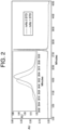
- FIG. 2 shows a chromatograph of this run. The two antibodies are somewhat separated but have significant overlap.
- Table 3.2 Method 2 gradient and flow information Time (min) Flow (mL/min) %A in mobile phase %B in mobile phase Startup 0.500 40 60 4 0.500 40 60 40 0.500 20 80 41 0.500 0 100 51 0.500 0 100 52 0.500 40 60 56 0.500 40 60
- FIG. 3 shows a chromatograph of this run. The two antibodies are somewhat separated but have significant overlap.
- Table 3.3 Method 3 gradient and flow information Time (min) Flow (mL/min) %A in mobile phase %B in mobile phase Startup 0.500 39 61 4 0.500 39 61 49 0.500 39 70 50 0.500 0 100 60 0.500 0 100 61 0.500 39 61 65 0.500 39 61
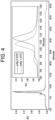
- FIG. 4 shows a chromatograph of this run. The two antibodies are somewhat separated but have significant overlap.
- Table 3.4 Method 4 gradient and flow information Time (min) Flow (mL/min) %A in mobile phase %B in mobile phase Startup 0.500 100 0 4 0.500 100 0 34 0.500 40 60 35 0.500 0 100 45 0.500 0 100 46 0.500 100 0 50 0.500 100 0
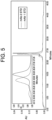
- FIG. 5 shows a chromatograph of this run. The two antibodies are somewhat separated but still have some overlap.
- Table 3.5 Method 5 gradient and flow information Time (min) Flow (mL/min) %A in mobile phase %B in mobile phase Startup 0.500 80 20 4 0.500 80 20 34 0.500 50 50 35 0.500 0 100 45 0.500 0 100 46 0.500 80 20 50 0.500 80 20
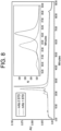
- FIG. 6 shows a chromatograph of this run. The two antibodies are somewhat separated but have some overlap.
- Table 3.6 Method 6 gradient and flow information Time (min) Flow (mL/min) %A in mobile phase %B in mobile phase Startup 0.500 90 10 4 0.500 90 10 44 0.500 50 50 45 0.500 0 100 55 0.500 0 100 56 0.500 90 10 60 0.500 90 10
- FIG. 7 shows a chromatograph of this run. The two antibodies are somewhat separated but have some overlap.
- FIG. 8 shows a chromatograph of this run. The two antibodies are somewhat separated but have some overlap.
- HIC separation of a co-formulation comprising 3 anti-Ebola mAbs of similar molecular weights, protein structures, and charge properties is developed and evaluated on Agilent 1100 HPLC system.
- the HIC-HPLC method uses a Dionex ProPac HIC-10 column with a two-part mobile phase (including Mobile Phase A (100 mM phosphate, 1 M ammonium sulfate, pH 7.0) and Mobile Phase B (100 mM phosphate, pH 7.0)) at a flow rate of 0.5 mL/minute.
- the HIC method is qualified using a five-point calibration curve for each mAb.
- the accuracy (% recovery) is determined by comparing the measured concentration to the theoretical concentration of each mAb.
- a gradient of 600 to 0 mM ammonium sulfate is used to run the column.
- FIG. 9 shows a chromatograph of the HIC-HPLC run. As can be seen by the three distinct peaks, the three antibodies are well separated and their peaks have little to no overlap.
- the repeatability is evaluated by analyzing samples of the cFDS in triplicate at 75%, 100%, 125%, 150% of the target amount (600 ⁇ g/injection of cFDS).
- the relative standard deviation (RSD) of repeatability is determined to be ⁇ 0.2%.
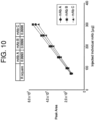
- Linearity is determined by analyzing samples of cFDS samples in triplicate at 9 levels (75 to 1200 ⁇ g of cFDS; 25 to 400 ⁇ g of individual mAb).
- a plot ( FIG. 10 ) of the peak area of individual mAb versus the injected amount results in a linear curve: At 75 to 300 ⁇ g, R 2 ⁇ 0.999; at 25 to 400 ⁇ g, R 2 ⁇ 0.98.
- the range is the interval between the lowest and highest concentration for which the method can demonstrate acceptable precision ( ⁇ 1.7%), accuracy (93-106% Recovery) and linearity (R 2 ⁇ 0.999).
- the range is determined to be 300 to 900 ⁇ g for cFDS sample (100 to 300 ⁇ g for each individual mAb).
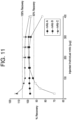
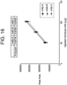
- FIG. 11 is a plot of the % recovery of the mAbs vs. amount of individual mAb.
- FIG. 12 is a plot of the % recovery of the mAbs vs. amount of individual mAb. % Recovery are within 93 - 106% at 75%, 100%, 125%, 150% of the target amount (600 ⁇ g total protein, 200 ⁇ g each mAb). The accuracy (% Recovery) is determined to be 93% - 106%.
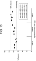
- FIG. 13 is the % recovery of the mAbs vs. each individual mAb in different cFDS ratios. % Recovery are within 94 - 106% with repeatability ⁇ 0.3%.
- HIC-HPLC to quantitate each of the mAbs in a co-formulation or a DP may have a variety of applications in co-formulation development.
- concentration of each mAb in a co-formulation or a DP may be monitored in a storage stability study.
- FIG. 14 shows a chart monitoring storage stability of three mAbs in a DP as a function of time (months).
- HIC-HPLC is used to quantitate the amount, and thus the concentration, of each of the three mAbs at six points in time over one year.
- An example set of data points from one time point in this study includes a measurement of the concentration of mAb A at 16.6 mg/mL, the concentration of mAb B at 17.5 mg/mL, and mAb C at 17.1 mg/mL.
- the storage stability shows that over the time period monitored, there is no appreciable change in concentration of each of the mAbs ( ⁇ 4%).
- the HIC-HPLC method has acceptable repeatability ( ⁇ 0.2%), intermediate precision ( ⁇ 1.7%), accuracy (93% - 106% recovery) and linearity (R 2 ⁇ 0.999) over the range of 100 to 300 ⁇ g (each individual mAb) and can be used to determine the concentration of each of three mAbs in the cFDS and DP.
- the HIC-HPLC method may be used as a release method for quantitating each of three mAbs in the co-formulated DS and DP. This method can also be used to support formulation development.
- CEX-UPLC Cation exchange ultra high-pressure liquid chromatography
- the CEX-UPLC method uses a YMC-BioPro SP-F column with mobile phases including 200 mM MES buffer and 10 to 120 mM NaCl gradient, with a pH of 6.5.
- a six-point standard calibration curve is prepared for each of three mAb molecules (mAb A, mAb B, mAb C).
- the linearity of each calibration curve is determined to have an R 2 ⁇ 0.99.
- the three mAb molecules are separated from a co-formulation ( i . e ., a mixture) using CEX-UPLC, and a chromatograph of the UPLC is generated. The chromatograph shows separation of each mAb, with some overlap between mAb A and mAb C.
- the repeatability of the method is evaluated by analyzing triplicate samples of the co-formulation run in three different amounts (150 ⁇ g, 225 ⁇ g, and 300 ⁇ g).
- the accuracy (% recovery) is determined by comparing the measured concentrations for each sample to the theoretical value.
- a linear range of quantitation of each mAb is determined to be 50 to 100 ⁇ g for each antibody, except for mAb A, with R 2 ⁇ 0.973 and accuracy of 75% -91%.
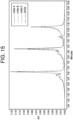
- FIG. 15 shows a chromatograph of a CEX-UPLC run of separation of 3 mAbs from a co-formulation. The three antibodies are separated, with a small overlap between mAb A and mAb C.
- the repeatability is evaluated by analyzing samples of the cFDS in triplicate at five different percentages (25%-100%) of the target amount (600 ⁇ g/injection of cFDS).
- the CEX-UPLC method separates three mAbs based on their charge properties, with some overlap.
- the overlap means that manual integration of the method is needed.
- the precision, accuracy, and range of CEX-UPLC is not as optimal as HIC-HPLC.
- SE-UPLC of the co-formulation comprising the three anti-Ebola mAbs of similar molecular weight is evaluated.
- SE-UPLC separates mAbs by size.
- a Waters Acquity H UPLC system is used. Two Waters BEH200 SEC columns are linked in series.
- the mobile phase includes 10 mM Phosphate buffer at pH 6.0, and 1 M Perchlorate.
- FIG. 17 depicts a chromatogram of the SE-UPLC. As shown, there is significant overlap between elution times of the three mAbs.
- SE-UPLC is a sub-optimal method for separating the three anti-MERS mABs of similar molecular weight.
- iCIEF of the co-formulation comprising the three anti-Ebola mAbs of similar molecular weights, protein structures, and charge properties is evaluated.
- iCIEF separates proteins by their isoelectric point (pI).
- a ProteinSimple iCE3 charge variant analyzer is used.
- 4% pH 3-10 Pharmalyte ® is used as an ampholyte, and 2M urea is used as a buffer.
- FIG. 18 depicts an iCIEF profile generated using these specifications. As shown, there is significant overlap between elution times of mAb A and mAb C. Thus, iCIEF is a sub-optimal method for separating the three anti-MERS mABs.
- RP-UPLC of the co-formulation comprising the three anti-Ebola mAbs of similar molecular weights, protein structures, and charge properties is evaluated.
- RP-UPLC separates mAbs by hydrophobicity.
- a Waters Acquity UPLC system is used.
- a ZORBAX 300SB-C8 column is used, and the column is run at 80° C.
- the mobile phase includes 60-90% acetonitrile in 0.1% TFA.
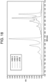
- FIG. 19 depicts a chromatogram of the RP-UPLC. As shown, there is significant overlap between elution times of mAb A and mAb B.
- RP-UPLC is a sub-optimal method for separating the three anti-MERS mAbs of similar molecular weight.
Abstract
Description
- This application claims priority to
U.S. Provisional Application No. 62/375,887, filed on August 16, 2016 - This disclosure relates to the field of assays for co-formulations of therapeutic antibodies.
- Administration of multiple, rather than single, monoclonal antibodies (mAbs) to a patient may improve their diagnostic or therapeutic indication and efficacy. These mAbs may be co-formulated in a single drug product (DP) and the DP administered to a patient.
- A method is required by regulatory agencies to quantitate the individual mAbs in a co-formulated drug substance (cFDS) to be incorporated into a DP, or a DP itself. Developing a method to separate two or more antibody molecules and to measure the concentration of each mAb is challenging, because the antibody molecules may have similar molecular weights, protein structures, and charge properties.
- This disclosure includes a method of quantitating amounts of antibodies from a mixture comprising a plurality of antibodies. In some aspects, the method may include, among other things, separating each of the plurality of antibodies in the mixture using hydrophobic interaction chromatography high performance liquid chromatography (HIC-HPLC), and quantitating an amount of each antibody in the mixture, wherein a molecular weight of each antibody in the mixture is within 15 kDa of a molecular weight of any other antibodies in the mixture, and either a surface hydrophobicity of each antibody in the mixture is different from a surface hydrophobicity of another antibody in the mixture by more than about 0.25 units on the Kyte & Doolittle hydropathy scale, or each antibody in the mixture, when run on HIC-HPLC individually, elutes at a distinct run time from another antibody in the mixture, or both.
- In some embodiments, the surface hydrophobicity of each antibody in the mixture is different from the surface hydrophobicity of each other antibody in the mixture by about 0.5 to about 1.0 units on the Kyte & Doolittle hydropathy scale. In further embodiments, the surface hydrophobicity of each antibody in the mixture is determined by calculating surface hydrophobicity based on protein structure or structural model, rapid screening for solubility in ammonium sulfate or PEG8000, or rapid screening for molecule interaction by affinity capture-self-interaction nanoparticle spectroscopy (AC-SINS).
- In additional embodiments, a first antibody in the mixture elutes at a first run time during a HIC-HPLC run, a second antibody in the mixture elutes at a second run time during the HIC-HPLC run, and the first and second run times do not overlap. In yet further embodiments, a first antibody in the mixture and a second antibody in the mixture have protein sequences that are at least 90% homologous, the first antibody and the second antibody have protein structures that are at least 90% homologous, as determined by their protein sequences, or the first antibody and the second antibody have isoelectric points (pI) within about 0.6 of one another, as determined by their protein sequences.
- In some embodiments, the plurality of antibodies comprises three antibodies. In further embodiments, one or more of the antibodies in the mixture are monoclonal antibodies. In still further embodiments, one or more of the antibodies in the mixture are human monoclonal antibodies. In other embodiments, two or more of the antibodies in the mixture are of the same isotype. In some embodiments, two or more of the antibodies in the mixture are variants of each other. In further embodiments, two or more of the antibodies in the mixture bind to the same antigen.
- In some embodiments, the mixture is a co-formulated composition. In additional embodiments, the co-formulated composition is configured to treat MERS in a human patient. In further embodiments, the co-formulated composition is configured to treat Ebola hemorrhagic fever in a human patient. In further embodiments, the co-formulated composition is configured to treat macular degeneration in a human patient. In yet further embodiments, the two or more antibodies in the co-formulated composition are configured to treat an infectious disease in a human patient. In some embodiments, the co-formulated composition is included in a drug product.
- In some embodiments, the HIC-HPLC is performed in a buffer at about pH 5.0 to about pH 7.0. In further embodiments, the method further comprises generating a chromatograph from the HIC-HPLC, wherein for elution of each antibody in the mixture, the chromatograph shows a peak that does not overlap with other peaks in the chromatograph.
- This disclosure also includes a method of quantitating amounts of antibodies from a mixture comprising a plurality of antibodies, the method comprising: separating each of the plurality of antibodies in the mixture using hydrophobic interaction chromatography high performance liquid chromatography (HIC-HPLC), wherein a molecular weight of each antibody in the mixture is within 15 kDa of a molecular weight of each other antibody in the mixture; quantitating an amount of each antibody in the mixture; and generating a chromatograph from the HIC-HPLC, wherein for elution of each antibody in the mixture, the chromatograph shows a peak that does not overlap with other peaks in the chromatograph. In some embodiments, one or more of the plurality of antibodies are human monoclonal antibodies. In further embodiments, either a surface hydrophobicity of each antibody in the mixture is different from a surface hydrophobicity of another antibody in the mixture by more than about 0.25 units on the Kyte & Doolittle hydropathy scale, or each antibody in the mixture, when run on HIC-HPLC individually, elutes at a distinct run time from another antibody in the mixture, or both.
- Numerous other aspects and embodiments are provided in accordance with these and other aspects of the disclosure. Other features and aspects of the present disclosure will become more fully apparent from the following detailed description and the appended claims.
- The accompanying drawings, which are incorporated in and constitute a part of this specification, illustrate various examples and together with the description, serve to explain the principles of the present disclosure. Any features of an embodiment or example described herein (e.g., device, method, etc.) may be combined with any other embodiment or example, and are encompassed by the present disclosure.
-
FIG. 1 shows an exemplary chromatograph of a HIC-HPLC run of a co-formulation comprising anti-Ebola mAbs. -
FIGS. 2-8 show exemplary chromatographs of HIC-HPLC runs of a co-formulation comprising anti-MERS mAbs. -
FIG. 9 shows an exemplary chromatograph of a HIC-HPLC run of a co-formulation comprising anti-Ebola mAbs. -
FIG. 10 is a plot of linearity of HIC-HPLC, plotting peak area against the amount of individual anti-Ebola mAb run. -
FIG. 11 is a plot of range, plotting percent recovery of the mAbs against the amount of individual anti-Ebola mAb run. -
FIG. 12 is a plot of the accuracy of HIC-HPLC with different mixture sample lots, plotting percent recovery of the mAbs against the amount of individual anti-Ebola mAb run. -
FIG. 13 is a plot of the accuracy of HIC-HPLC with different ratios of individual anti-Ebola mAbs, plotting percent recovery of the mAbs against the amount of individual anti-Ebola mAb run. -
FIG. 14 is a plot of storage stability of the mAbs in the cFDS, plotting amount of individual anti-Ebola mAbs against storage time in months. -
FIG. 15 shows an exemplary chromatograph of a cation exchange ultra high pressure liquid chromatography (CEX-UPLC) run of a co-formulation comprising anti-Ebola mAbs. -
FIG. 16 is a plot of linearity of CEX-UPLC, plotting peak area against amount of individual anti-Ebola mAb. -
FIG. 17 shows an exemplary chromatograph of a size-exclusion ultra high pressure liquid chromatography (SE-UPLC) run of a co-formulation comprising anti-Ebola mAbs. -
FIG. 18 shows an exemplary imaged capillary isoelectric focusing (iCIEF) profile of a co-formulation comprising anti-Ebola mAbs. -
FIG. 19 shows an exemplary chromatograph of a reverse phase ultra high pressure liquid chromatography (RP-UPLC) run of a co-formulation comprising anti-Ebola mAbs. - The term "antibody" is sometimes used interchangeably with the term "immunoglobulin." Briefly, it may refer to a whole antibody comprising two light chain polypeptides and two heavy chain polypeptides. Whole antibodies include different antibody isotypes including IgM, IgG, IgA, IgD, and IgE antibodies. The term "antibody" may include, for example, a polyclonal antibody, a monoclonal antibody (mAb), a chimerized or chimeric antibody, a humanized antibody, a primatized antibody, a deimmunized antibody, and a fully human antibody. The antibody may be made in or derived from any of a variety of species, e.g., mammals such as humans, non-human primates (e.g., orangutan, baboons, or chimpanzees), horses, cattle, pigs, sheep, goats, dogs, cats, rabbits, guinea pigs, gerbils, hamsters, rats, and mice. The antibody may be a purified or a recombinant antibody. The antibody can also be an engineered protein or antibody-like protein containing at least one immunoglobulin domain (e.g., a fusion protein). The engineered protein or antibody-like protein may also be a bi-specific antibody or a tri-specific antibody, or a dimer, trimer, or multimer antibody, or a diabody, a DVD-Ig, a CODV-Ig, an Affibody®, or a Nanobody®.
- The terms "variant of an antibody," "antibody variant," and the like, refer to an antibody that varies from another antibody in that the variant antibody is a deletion variant, insertion variant, and/or substitution variant of the other antibody.
- The term "human antibody," as used herein, is intended to include antibodies having variable and constant regions derived from human germline immunoglobulin sequences. Human mAbs may include amino acid residues not encoded by human germline immunoglobulin sequences (e.g., mutations introduced by random or site-specific mutagenesis in vitro or by somatic mutation in vivo), for example in the CDRs and in particular CDR3. However, the term "human antibody," as used herein, is not intended to include mAbs in which CDR sequences derived from the germline of another mammalian species (e.g., mouse), have been grafted onto human FR sequences. The term includes antibodies recombinantly produced in a non-human mammal, or in cells of a non-human mammal. The term is not intended to include antibodies isolated from or generated in a human subject.
- As used herein, the terms "treat," "treating," or "treatment" refer to the reduction or amelioration of the severity of at least one symptom or indication of a disease or condition due to the administration of a co-formulation of two or more antibodies to a subject in need thereof. The terms include inhibition of progression of disease. The terms also include positive prognosis of disease.
- The terms "prevent," "preventing" or "prevention" refer to inhibition of manifestation of a disease or condition any symptoms or indications of that disease or condition upon administration of a co-formulation of two or more antibodies.
- For the terms "for example" and "such as," and grammatical equivalences thereof, the phrase "and without limitation" is understood to follow unless explicitly stated otherwise. As used herein, the term "about" and the signifier "-" are meant to account for variations due to experimental error. All measurements reported herein are understood to be modified by the term "about," whether or not the term is explicitly used, unless explicitly stated otherwise. As used herein, the singular forms "a," "an," and "the" include plural referents unless the context clearly dictates otherwise.
- Unless otherwise defined, all technical and scientific terms used herein have the same meaning as commonly understood by one of ordinary skill in the art to which this invention belongs. Methods and materials are described herein for use in the present invention; other, suitable methods and materials known in the art can also be used. The materials, methods, and examples are illustrative only and not intended to be limiting. All publications, patent applications, patents, sequences, database entries, and other references mentioned herein are incorporated by reference in their entirety. In case of conflict, the present specification, including definitions, will control.
- Antibody molecules, such as monoclonal antibody molecules, may be co-formulated to treat one or more diseases or conditions in a patient (including a human patient). The terms "patient" and "subject" are used interchangeably herein.
- A co-formulated drug product (DP) may include a co-formulated drug substance (cFDS) (also referred to herein as a co-formulation) containing two or more (e.g., three) human monoclonal antibody (mAb) molecules. The cFDS is prepared by mixing purified mAbs at a predetermined ratio. A method is required by regulatory agencies to quantitate each of individual mAbs in the cFDS.
- The mAb molecules in the co-formulation may be similar to each other: they may be immunoglobulins (such as IgG1) with about the same molecular weight (e.g., ~145 kDa); with similar protein structure and charge properties.
- Methods disclosed herein for quantitating similar mAb molecules in a co-formulation are precise, accurate, reproducible, suitable for use in quality control environments, do not use expensive equipment, and do not require cumbersome sample preparation.
- This disclosure provides methods of quantitating an amount of an antibody molecule from a mixture comprising two or more antibody molecules. The method may comprise separating each of the two or more antibody molecules from the mixture by hydrophobic interaction chromatography high performance liquid chromatography (HIC-HPLC) and quantitating an amount of each antibody molecule, wherein the molecular weight of each antibody molecule is within 15 kDa of any other antibody molecule in the mixture and either each antibody molecule is different from another antibody molecule in the mixture by more than about 0.25 units on the Kyte & Doolittle hydropathy scale (see, e.g., Kyte and Doolittle, J. Mol. Biol. 157, 105-132 (1982)), or each of the antibody molecules, when run alone on HIC-HPLC, elutes at a distinct run time with little overlap from other antibody molecules in the mixture, or both. In certain embodiments, for example, the relative hydrophobicity of each antibody molecule is different from each other antibody molecule in the mixture by about 0.5 to about 1.0 unit on the Kyte & Doolittle hydropathy scale. In certain other embodiments, each of the antibody molecules, when run alone on HIC-HPLC, elutes at a distinct run time with little overlap from the other antibody molecules in the mixture. In certain embodiments, the mixture comprises a plurality of antibody molecules (e.g., two, three, four, or five antibody molecules).
- If two antibody molecules from the mixture, when run on HIC-HPLC, have significant overlap in the chromatograph monitoring elution profiles of the antibody molecules by absorbance verses time of elution, this indicates that the two antibody molecules elute at times that are close, resulting in a lack of separation within the column such that the antibody molecules are not purified (e.g., less than 50% pure). In such cases, the individual antibodies cannot be fully quantitated because of that significant overlap. In some embodiments, two antibody molecules from the mixture, when run on HIC-HPLC, have little to no overlap in the chromatograph monitoring elution profiles of the antibody molecules by absorbance verses time of elution, in that the antibody molecules elute at times that are not close, resulting in separation such that the antibody molecules are substantially purified, each being at least 90%, 91%, 92%, 93%, 94%, 95%, 96%, 97%, 98%, 99%, or 100% pure.
- In some embodiments, methods disclosed herein are used to quantitate an individual population of antibodies, also referred to as "an antibody molecule," from a mixture of antibodies, which includes two or more different populations of antibodies, where the different populations have at least one amino-acid difference between them. In some embodiments, a population of antibodies may have the same amino acid sequence as that of another population in the mixture of antibodies, but they may differ in post-translational modifications. In certain embodiments, a mixture is a co-formulated composition, such as or including a co-formulated drug substance (cFDS). In certain embodiments, a mixture comprises excipients. In certain embodiments, a mixture comprises sucrose, and may be a sucrose drug substance comprising mAb molecules. Each antibody in the mixture of antibodies will have a similar molecular weight, within 1, 2, 3, 4, 5, 10, or 15 kilodaltons (kDa) of each other. In another aspect, the average molecular weight of all the antibodies will be about 150kDa. This method can also be used with two or more trap molecules that have similar molecular weights. In certain further embodiments, the co-formulated composition comprises VEGF-Trap. (See, e.g.,
US Patent No. 9,265,827 US patent Application No. 14/943,490 , which are incorporated by reference here in their entireties.) This method may also be used to monitor the concentration for each of the mAbs in a mixture (e.g., a co-formulation) in monitoring storage stability. In certain embodiments, this method is used to separate antibody molecules that cannot be separated by another chromatographic method, such as by reverse phase high pressure liquid chromatography (HPLC) or ultra high pressure liquid chromatography (UPLC). - Hydrophobic Interaction Chromatography (HIC) separates antibodies in a decreasing salt gradient, based on differences in surface hydrophobicity of the antibodies. Separation using HIC is based on the reversible interaction between an antibody and the hydrophobic ligand bound to the chromatography matrix. Though hydrophobic amino acids of proteins and peptides are usually located away from molecular surfaces, biomolecules have some hydrophobic groups that are exposed to allow interaction with hydrophobic ligands on media. The hydrophobic interaction is enhanced by buffers with high ionic strength.
- Any suitable HIC-HPLC column may be employed for methods disclosed herein, including, without limitation, Dionex ProPac HIC-10, mAbPac HIC-10, mAbPac HIC-20, mAbPac HIC-Butyl (Dionex, Thermo Fisher Scientific, Sunnyville, CA). The buffer may be any suitable buffer. In certain embodiments, the HIC-HPLC is performed in a buffer pH between about pH 5.0 to about pH 7.0, or between about pH 6.0 to about pH 7.0, or between about 6.5 to about 7.5. In certain embodiments, the columns are Dionex MabPac HIC-10 (100 × 4.6 mm, 5 µm, Pore Size 1000A°) and ProPac HIC-10 (100 × 4.6 mm, 5 µm, Pore Size 300A°).
- A mixture comprising two or more antibodies is run on HIC-HPLC. The antibodies are eluted at separate times (run times). The antibodies are monitored by measuring their absorbance at, e.g., 280nm on the UV spectrum. A chromatograph may be used to monitor and to document the run. In certain embodiments, buffers used in the HIC-HPLC are different gradients of 1M ammonium sulfate (from 100% to 0%) in 100 mM phosphate buffer.
- In certain embodiments, the antibody molecules are quantitated by comparing their absorbance at 280 nm to a standard curve. A standard curve may be constructed by determining the absorbance of a known antibody at known concentrations from a HIC-HPLC run, at an absorbance of, for example, 280nm. The greater the absorbance (or "optical density"), the higher the protein concentration. These data for known concentrations of an antibody are used to make the standard curve, plotting concentration on the X axis, and the absorbance obtained from the chromatograph of the elution profile of the antibody from the HIC-HPLC run on the Y axis. A sample comprising two or more antibodies of unknown concentrations, including the antibody from the standard curve, are run. To analyze the data, the antibodies are separated by HIC-HPLC. The elution profiles of the antibodies are displayed on a chromatograph, with antibody concentration represented by absorbance monitored over time by a spectrophotometer. The absorbance of each antibody, as identified by its elution time (run time) is used to locate a corresponding absorbance value on the standard curve. The corresponding X-axis value for that point on the standard curve is the concentration of that antibody in the sample.
- In some aspects of the current disclosure, methods for quantitating individual antibodies in a mixture disclosed herein may be useful if the relative surface hydrophobicity of each antibody in the mixture is different from that of each other antibody in the mixture by greater than about 0.25 units, such as by about 0.5 to about 1.0 units, on the Kyte & Doolittle hydropathy scale. The relative surface hydrophobicity of the antibodies may be determined/estimated by a number of methods.
- For example, HIC-HPLC may be used to determine the relative surface hydrophobicity of different antibody molecules. Each of the antibody molecules is run alone on HIC-HPLC and the run time (elution time) for each antibody is determined.
- In certain embodiments, the difference in surface hydrophobicity between antibodies in a mixture is determined by one or more of the following methods: calculated surface hydrophobicity based on protein structure or structural model, rapid screening method for solubility in ammonium sulfate or PEG8000, and rapid screening for molecule interaction by affinity capture - self-interaction nanoparticle spectroscopy (AC-SINS).
- The relative surface hydrophobicity may be calculated based on known structure of the antibody (based on, for example, crystal structure or NMR structure) or a structural model. Such computational methods are known in the art.
- The relative surface hydrophobicity may be calculated/estimated by determining the solubility of the antibody molecules by, for example, rapid screening method for solubility in conditions that enhance hydrophobicity, such as in ammonium sulfate or PEG8000. The more soluble a protein, the less hydrophobic. (See, e.g., Kramer et al., Biophysical Journal Volume 102 April 1907-1915 (2012).)
- The relative surface hydrophobicity may be calculated/estimated by rapid screening for molecule interaction by affinity capture - self-interaction nanoparticle spectroscopy (AC-SINS). (See, e.g., Estep et al., mAbs, 7:3,553-561 (2015).) AC-SINS is an approach that coats gold nanoparticles with polyclonal anti-human antibodies, uses these conjugates to immobilize human mAbs, and evaluates mAb self-interactions by measuring the plasmon wavelengths of the antibody conjugates as a function of ammonium sulfate concentration.
- In certain other embodiments, antibodies in a mixture that can be separated using the described methods will have similar size or overall charge as determined by one or more of the following methods: protein structure, or structural modeling, based on sequence and other known protein structures, calculated overall protein charge property based on protein sequences, calculated hydrophobicity based on protein sequences, and protein sequencing.
- In certain embodiments, antibodies in a mixture have at least 80%, at least 85%, at least 90%, at least 95%, at least 98%, or at least 99% homologous protein structure or structural models based on sequence and other known protein structures. In certain embodiments, antibodies in a mixture have calculated protein charge properties within 3, 4, or 5, 10 or 15 elementary units of one another, where protein charge properties are calculated based on protein sequence. In certain embodiments, two or more antibodies in a mixture have at least 80%, at least 85%, at least 90%, at least 95%, at least 98%, or at least 99% identity between their protein sequences.
- In an aspect, methods of quantifying antibodies in a mixture using HIC-HPLC as disclosed herein may not be as useful if there is a large difference between the antibody molecules as calculated or estimated by protein structure, sequence-based structural models, protein sequence, and other known aspects of protein structure. If there are differences in the hydrophobic profiles of the antibodies, then methods using HIC-HPLC may nevertheless facilitate separation of the antibody species.
- In another aspect, methods of quantifying antibodies in a mixture using HIC-HPLC as disclosed herein may not be as useful if there is a large difference between the antibody molecules calculated/estimated by the calculated overall protein charge property of each antibody molecule, based on the protein sequence of each antibody molecule. If there are differences in the hydrophobic profiles of the antibodies, then methods using HIC-HPLC may nevertheless facilitate separation of the antibody species.
- In another aspect, methods of quantifying antibodies in a mixture using HIC-HPLC as disclosed herein may not be as useful if there are large differences between the antibody molecules calculated/estimated by inspecting the protein charge or size. If there are differences in the hydrophobic profiles of the antibodies, then methods using HIC-HPLC may nevertheless facilitate separation of the antibody species.
- In certain embodiments, one or more of the antibodies in a mixture of antibodies are monoclonal antibodies. In certain embodiments, one or more of the monoclonal antibodies are human monoclonal antibodies. In certain embodiments, two or more of the antibody molecules are of the same isotype. In certain embodiments, two or more of the antibody molecules are variants of each other. In yet certain other embodiments, two or more of the antibody molecules bind to the same antigen. The mAbs may be whole antibody molecules.
- In certain embodiments, the mixture is a co-formulated composition. In certain further embodiments, the co-formulated composition comprises two or more mAbs that are effective for treating Middle East Respiratory Syndrome (MERS) in a human patient. Antibodies to the MERS corona virus (MERS-CoV) are disclosed in, for example,
U.S. Patent Application No. 14/717,760 and International Application Publication No.WO2015/179535A1 . In certain further embodiments, the co-formulated composition comprises mAbs that are effective in treating Ebola hemorrhagic fever in a human patient. Antibodies to the Ebola virus are disclosed in, for example,U.S. Patent Application No. 15/005,334, filed on Jan. 25, 2016 . In certain further embodiments, the co-formulated composition comprises mAbs that are effective in treating macular degeneration in a human patient. In certain further embodiments, the co-formulated composition comprises mAbs alirocumab and evinacumab disclosed inU.S. Provisional Application No. 62/302,907 U.S. Provisional Application No. 62/270,749 U.S. Patent Application No. 15/386,443 , andU.S. Patent Application No. 15/386,453 ; and PD-1 and Lag3 inU.S. Provisional Application 62/365,006 U.S. Patent Application No. 15/289,032 .) In certain embodiments, the co-formulated composition comprises anti-Zika virus antibodies. (See, e.g.,U.S. Provisional Application No. 62/363,546 U.S. Patent No. 8,871,209 .) In certain embodiments, two or more antibodies in the co-formulated composition may treat an infectious disease in a human patient. The disclosure of each of the above-cited patent documents in this paragraph is hereby incorporated by reference. - The anti-MERS mAbs in a co-formulation may bind to, for example, the spike protein of MERS-CoV (e.g., the spike protein of MERS-CoV isolate EMC/2012). The spike protein's epitope may be within the receptor binding domain of the spike protein (e.g., amino acids selected from the amino acids 367 to 606 of GenBank Accession No. AFS88936.1). The anti-MERS mAbs in the co-formulated composition may be: a fully human monoclonal antibody that binds to the MERS-CoV spike protein; one that interacts with one or more amino acid residues in the receptor binding domain of the MERS-CoV spike protein selected from amino acid residues 367 to 606 of GenBank Accession No. AFS88936.1; one that binds to MERS-CoV spike protein with a dissociation constant (Ko) of less than 18.5nM, as measured in a surface plasmon resonance assay; or one that blocks binding of MERS-CoV spike protein to dipeptidyl peptidase 4 (DPP4) by more than 90%.
- The co-formulated composition may be any composition comprising two or more antibodies directed to the same or different target, and are effective in treating the same or different disease or condition in a patient, including a human patient.
- Compositions containing two or more antibody molecules may be formulated as a pharmaceutical composition (e.g., a DP) for administering to a subject. The pharmaceutical compositions can include, for example, three antibody molecules. Any suitable pharmaceutical compositions and formulations, as well as suitable methods for formulating and suitable routes and suitable sites of administration, are within the scope of this invention. Also, unless otherwise stated, any suitable dosage(s) and frequency of administration are contemplated.
- The mAbs in the co-formulations are purified by methods known in the art before being co-administered. The co-formulations of two or more antibodies may be any suitable co-formulations.
- The pharmaceutical compositions/co-formulations may include a pharmaceutically acceptable carrier (i.e., an excipient). A "pharmaceutically acceptable carrier" refers to, and includes, any and all solvents, dispersion media, coatings, antibacterial and antifungal agents, isotonic and absorption delaying agents, diluents, glidants, etc. The compositions can include a pharmaceutically acceptable salt, e.g., an acid addition salt or a base addition salt (see e.g., Berge et al. J Pharm Sci 66:1-19 (1977)). The composition can include sucrose or can be coated when appropriate.
- In certain embodiments, the protein compositions can be stabilized and formulated as a solution, microemulsion, dispersion, liposome, lyophilized cake, solid, etc. Sterile injectable solutions can be prepared by incorporating two or more mAbs in the required amounts in an appropriate solvent with one or a combination of ingredients enumerated above, as required, followed by filtered sterilization. Generally, dispersions are prepared by incorporating two or more mAb molecules into a sterile vehicle that contains a basic dispersion medium and the required other ingredients from those enumerated above. The proper fluidity of a solution can be maintained, for example, by the use of a coating such as lecithin, by the maintenance of the required particle size in the case of dispersion and by the use of surfactants. Prolonged absorption of injectable compositions can be brought about by including in the composition a reagent that delays absorption, for example, monostearate salts, and gelatin.
- For this invention to be better understood, the following examples are set forth. These examples are for purposes of illustration only and are not be construed as limiting the scope of the invention in any manner.
- The anti-Ebola mAbs in the co-formulation used in Examples 1 and 3-7 are whole, fully
human IgG 1 monoclonal antibodies. The three mAbs (mAb A, mAb B, and mAb C) have similar molecular weights (e.g., about 145 kDa), protein structure, and charge properties (e.g., a difference in pI of about 0.6 or less, as determined by protein sequences). The isoelectric point (pI) of mAb A is determined to be 9.0, the pI of mAb B is determined to be 8.5, and the pI of mAb C is determined to be 9.1. - The anti-MERS mAbs in the co-formulation used in Example 2 are whole, fully human anti-Ebola mAbs. The two mAbs have similar molecular weights (e.g., within about 15 kDa of one another) and charge properties. (See U.S. Patent Publication No.
US2015/0337029 ,WO2015/179535A1 , andU.S. Patent Application No. 14/717,760 , the disclosure of each of which is hereby incorporated by reference herein.) - A HIC-HPLC method is used to quantitate 3 anti-Ebola monoclonal antibodies (
mAb 1,mAb 2, and mAb 3) of similar molecular weights, protein structures, and charge properties from a co-formulation by first separating the 3 mAbs from the co-formulation and then quantitating each of them. - A Dionex ProPac HIC-10 column is used, Cat# 063655 (Dionex, Thermo Fisher Scientific, Sunnyville, CA), 4.6 × 100 mm.
- The mobile phases include a Mobile Phase A and a Mobile Phase B. Mobile Phase A includes 1M Ammonia phosphate and 100mM phosphate, at a pH of 7.0. Preparation of Mobile Phase A includes: dissolving 13.8g of sodium phosphate monobasic, monohydrate (NaH2PO4.H2O) and 132.1 g of ammonia phosphate in 800 mL Milli Q; adjusting the pH to 7.0 with 50% NaOH; bringing the volume to 1000mL; and filtering the solution through a 0.22 µM filter. Mobile Phase B includes 100mM phosphate at a pH of 7.0. Preparation of Mobile Phase B includes: Dissolving 13.8g of sodium phosphate monobasic, monohydrate (NaH2PO4.H2O) in 900 mL Milli Q; adjusting the pH to 7.0 with 50% NaOH; bringing the volume to 1000mL; and filtering the solution through a 0.22 µM filter.
- The HIC-HPLC is run through the aforementioned column at a flow rate of 0.5 mL/minute. The column temperature is kept at 30°C, and the co-formulation sample temperature is 5°C. The stop time for the column is 40 minutes. The effluent's absorbance of 280 nm ultraviolet light is monitored using a UV detector. Table 1 below shows the mix of Mobile Phase A and Mobile Phase B as percentages of the mobile phase composition gradient to be introduced over the column run time.
Table 1: Mobile phase gradient Time (min) % Mobile Phase A % Mobile Phase B 0 60 40 4 60 40 19 0 100 32 0 100 33 60 40 40 60 40 - In order to calibrate results for the three individual mAbs, known amounts of each mAb in FDS are run. A FDS with a known concentration of each mAb is prepared, and the concentration is measured by either reverse phase (RP) chromatography or Solo VPE® Spectroscopy (e.g., C Technologies, Inc.). The known FDS for each mAb are as follows:
Table 2: Concentration of each mAb FDS as measured by RP or Solo VPE® Formulation Known mAb amount (g) Known mAb concentration (t=0) (mg/mL) mAb 1 FDS84.08 51.31 (by Solo VPE®) mAb 2 FDS84.13 53.40 (by RP) mAb 3 FDS84.19 50.65 (by RP) - A control sample is also prepared for the calibration sequence. The control sequence has a concentration of 50.31 mg/mL as measured by Solo VPE® Spectroscopy (e.g., C Technologies, Inc.). One injection of the control sample is 12.0 µL (603.72 µg).
- Table 2 below lists the calibration samples used of each of the mAb FDS.
Table 2: Calibration samples Amount Number of injections mAb 1 FDS: 50.5 mg/mL working Standard Lot# YC10190-42A (Regeneron Pharmaceuticals, Tarrytown, NY) 2.0 µL = 101 µg 1 injection 3.0 µL = 151.5 µg 1 injection 4.0 µL = 201 µg 1 injection 5.0 µL = 252.5 µg 1 injection 6.0 µL = 303 µg 1 injection mAb 2 FDS: 51.7 mg/mL working Standard Lot# YC10190-42B (Regeneron Pharmaceuticals, Tarrytown. NY) 2.0 µL = 103.4 µg 1 injection 3.0 µL = 155.1 µg 1 injection 4.0 µL = 206.8 µg 1 injection 5.0 µL = 258.5 µg 1 injection 6.0 µL = 310.2 µg 1 injection mAb 3 FDS: 51.4 mg/mL working Standard Lot# YC10190-42C (Regeneron Pharmaceuticals, Tarrytown, NY) 2.0 µL = 102.8 µg 1 injection 3.0 µL = 154.2 µg 1 injection 4.0 µL = 205.6 µg 1 injection 5.0 µL = 257µg 1 injection 6.0 µL = 308.4 µg 1 injection - The calibration sequence is run. During the calibration sequence, one injection of the control sample is introduced at the beginning of each sample set (e.g., the set of
mAb 1 FDS injections, the set ofmAb 2 FDS injections, or the set ofmAb 3 FDS injections), after every 20-24 injections of mAb FDS, and at the end of each injection sequence. - During one calibration sequence, one injection of each known amount of mAb is performed. During another calibration sequence, four injections of each known amount of mAb are performed to determine repeatability of injections.
- A standard calibration curve is constructed for each mAb. The calibration curve for each mAb has an R2 ≥ 0.999, and the variability of repeated injections of the same amounts of each mAb is ≤ 1%.
- A control HIC-HPLC run of each known mAb FDS is performed. Percent recovery for each known mAb FDS is calculated by measuring the concentration by HIC-HPLC, and then dividing the concentration measured by HIC-HPLC by the known mAb concentration in Table 2. Percent recovery is calculated to be 90 - 110% of the known concentrations for all three known mAb FDS.
- A sample co-formulation having a total of 600 µg mAb (e.g., 12 µL of a 50 mg/mL mAb sample) is run through HIC-HPLC. For example, 12 µL of a co-formulation including 600 µg total of mAb may include the equivalent of three injections of 200 µg/injection of individual mAb. The individual mAbs in the co-formulation are
mAb 1,mAb 2, andmAb 3. - A duplicate run of the sample is performed, and the average concentration (mg/mL) of each individual mAb in the co-formulation and the ratio of each mAb to the total amount of mAb is reported.
-
FIG. 1 is a chromatograph showing an HIC-HPLC run of this co-formulation. Each mAb is depicted by a separate line, and the cFDS as a whole is also depicted by a line. The 3 antibodies are well separated by HIC-HPLC, with little overlap in peaks. - Multiple HIC-HPLC methods are used to quantitate 2 anti-MERS monoclonal antibodies of similar molecular weights and charge properties from a co-formulation by first separating the 2 mAbs from the co-formulation and then quantitating each of them.
- Two mAb (
mAb 1 and mAb 2) drug substances are combined.mAb 1 drug substance includes 52.3 mg/ml mAb 1 and 10mM His, at a pH of 5.5 (Regeneron Pharmaceuticals, Tarrytown, NY).mAb 2 drug substance includes 40.4 mg/ml mAb 2 and 10mM His, at a pH of 6.0, and includes 5% (w/v) sucrose (Regeneron Pharmaceuticals, Tarrytown, NY). - Seven HIC-HPLC methods are run. In each method, 50 µg (
mAb 1 and mAb 2) of sample is loaded. The effluent is monitored using UV detection at 280 nm. Two solvents are used, in varying proportion, in the mobile phase. Solvent A is 1M (NH4)SO4 20mM Acetate at pH 5.5, and Solvent B is 20 mM Acetatae at pH 5.5. -
Table 3.1: Method 1 gradient and flow informationTime (min) Flow (mL/min) %A in mobile phase %B in mobile phase Startup 0.500 100 0 4 0.500 100 0 29 0.500 0 100 40 0.500 0 100 41 0.500 100 0 50 0.500 100 0 -
FIG. 2 shows a chromatograph of this run. The two antibodies are somewhat separated but have significant overlap. -
Table 3.2: Method 2 gradient and flow informationTime (min) Flow (mL/min) %A in mobile phase %B in mobile phase Startup 0.500 40 60 4 0.500 40 60 40 0.500 20 80 41 0.500 0 100 51 0.500 0 100 52 0.500 40 60 56 0.500 40 60 -
FIG. 3 shows a chromatograph of this run. The two antibodies are somewhat separated but have significant overlap. -
Table 3.3: Method 3 gradient and flow informationTime (min) Flow (mL/min) %A in mobile phase %B in mobile phase Startup 0.500 39 61 4 0.500 39 61 49 0.500 39 70 50 0.500 0 100 60 0.500 0 100 61 0.500 39 61 65 0.500 39 61 -
FIG. 4 shows a chromatograph of this run. The two antibodies are somewhat separated but have significant overlap. -
Table 3.4: Method 4 gradient and flow information Time (min) Flow (mL/min) %A in mobile phase %B in mobile phase Startup 0.500 100 0 4 0.500 100 0 34 0.500 40 60 35 0.500 0 100 45 0.500 0 100 46 0.500 100 0 50 0.500 100 0 -
FIG. 5 shows a chromatograph of this run. The two antibodies are somewhat separated but still have some overlap. -
Table 3.5: Method 5 gradient and flow information Time (min) Flow (mL/min) %A in mobile phase %B in mobile phase Startup 0.500 80 20 4 0.500 80 20 34 0.500 50 50 35 0.500 0 100 45 0.500 0 100 46 0.500 80 20 50 0.500 80 20 -
FIG. 6 shows a chromatograph of this run. The two antibodies are somewhat separated but have some overlap. -
Table 3.6: Method 6 gradient and flow informationTime (min) Flow (mL/min) %A in mobile phase %B in mobile phase Startup 0.500 90 10 4 0.500 90 10 44 0.500 50 50 45 0.500 0 100 55 0.500 0 100 56 0.500 90 10 60 0.500 90 10 -
FIG. 7 shows a chromatograph of this run. The two antibodies are somewhat separated but have some overlap. -
Table 3.7 Method 7 gradient and flow information Time (min) Flow (mL/min) %A in mobile phase %B in mobile phase Startup 0.500 87 130 4 0.500 87 13 74 0.500 52 48 75 0.500 0 100 85 0.500 0 100 86 0.500 87 13 90 0.500 87 13 -
FIG. 8 shows a chromatograph of this run. The two antibodies are somewhat separated but have some overlap. - HIC separation of a co-formulation comprising 3 anti-Ebola mAbs of similar molecular weights, protein structures, and charge properties is developed and evaluated on Agilent 1100 HPLC system.
- The HIC-HPLC method uses a Dionex ProPac HIC-10 column with a two-part mobile phase (including Mobile Phase A (100 mM phosphate, 1 M ammonium sulfate, pH 7.0) and Mobile Phase B (100 mM phosphate, pH 7.0)) at a flow rate of 0.5 mL/minute. The HIC method is qualified using a five-point calibration curve for each mAb. The accuracy (% recovery) is determined by comparing the measured concentration to the theoretical concentration of each mAb. A gradient of 600 to 0 mM ammonium sulfate is used to run the column.
-
FIG. 9 shows a chromatograph of the HIC-HPLC run. As can be seen by the three distinct peaks, the three antibodies are well separated and their peaks have little to no overlap. - The repeatability is evaluated by analyzing samples of the cFDS in triplicate at 75%, 100%, 125%, 150% of the target amount (600 µg/injection of cFDS). The relative standard deviation (RSD) of repeatability is determined to be ≤0.2%.
Table 4: Repeatability data Inject amount cFDS [µg] Measured Concentration [mg/mL] mAb A mAb B mAb C Ave (n=3) RSD Ave (n=3) RSD Ave (n=3) RSD 450 17.4 0.1% 17 0.2% 17.9 0.2% 600 17.4 0.1% 17.3 0.1% 17.8 0.2% 750 17.3 0.1% 17.4 0.2% 17.7 0.1% 900 17.1 0.1% 17.5 0.1% 17.6 0.2% - The repeatability study is repeated on a different day, using a different lot of column and different batch of mobile phase. The RSD of intermediate precision is determined to be ≤1.7%.
Table 5: Intermediate precision Inject amount cFDS [µg] Measured Concentration [mg/mL] mAb A mAb B mAb C Ave (n=6) RSD Ave (n=6) RSD Ave (n=6) RSD 450 17.3 0.8% 17.2 0.9% 18.0 0.7% 600 17.4 0.1% 17.3 0.2% 18.0 1.1% 750 17.4 0.4% 17.4 0.2% 18.0 1.7% 900 17.1 0.1% 17.4 0.6% 17.8 1.6% - Linearity is determined by analyzing samples of cFDS samples in triplicate at 9 levels (75 to 1200µg of cFDS; 25 to 400µg of individual mAb). A plot (
FIG. 10 ) of the peak area of individual mAb versus the injected amount results in a linear curve: At 75 to 300µg, R2 ≥ 0.999; at 25 to 400µg, R2 ≥ 0.98. - The range is the interval between the lowest and highest concentration for which the method can demonstrate acceptable precision (≤ 1.7%), accuracy (93-106% Recovery) and linearity (R2 ≥ 0.999). The range is determined to be 300 to 900 µg for cFDS sample (100 to 300 µg for each individual mAb).
FIG. 11 is a plot of the % recovery of the mAbs vs. amount of individual mAb. - Data obtained from intra-day and inter-day precision (cFDS lot #1), and range study (cFDS lot #2) is used to evaluate the method accuracy.
FIG. 12 is a plot of the % recovery of the mAbs vs. amount of individual mAb. % Recovery are within 93 - 106% at 75%, 100%, 125%, 150% of the target amount (600 µg total protein, 200 µg each mAb). The accuracy (% Recovery) is determined to be 93% - 106%. - CFDS is formulated by 1:1:1, 2:1:1, 1:2:1, 1:1:2 mix of individual mAb. Each cFDS is analyzed at 100% of the target amount (600 µg/injection, n=3).
FIG. 13 is the % recovery of the mAbs vs. each individual mAb in different cFDS ratios. % Recovery are within 94 - 106% with repeatability ≤ 0.3%.Table 6: HIC-HPLC data summary Repeatability (450 - 900 µg cFDS) (n=3) mAb A ≤0.1% mAb B ≤0.2% mAb C ≤0.2% Intermediate Precision (450 - 900 µg cFDS) (n=6) mAb A ≤0.8% mAb B ≤0.9% mAb C ≤1.7% Accuracy (% Recovery ) (450 - 900 µg cFDS) 93 - 106 % Linearity R2 ≥ 0.999 ( a plot of the peak area of each mAb versus the injected individual mAb amount in cFDS at 75 to 300µg range) Standard Curve R2 ≥ 0.999 (50 -300 µg of individual mAb standard) Range 100 to 300 µg for each individual mAb 300 to 900 µg for cFDS sample - Use of HIC-HPLC to quantitate each of the mAbs in a co-formulation or a DP may have a variety of applications in co-formulation development. For example, the concentration of each mAb in a co-formulation or a DP may be monitored in a storage stability study.
-
FIG. 14 shows a chart monitoring storage stability of three mAbs in a DP as a function of time (months). HIC-HPLC is used to quantitate the amount, and thus the concentration, of each of the three mAbs at six points in time over one year. An example set of data points from one time point in this study includes a measurement of the concentration of mAb A at 16.6 mg/mL, the concentration of mAb B at 17.5 mg/mL, and mAb C at 17.1 mg/mL. In this case, the storage stability shows that over the time period monitored, there is no appreciable change in concentration of each of the mAbs (≤4%). - The HIC-HPLC method has acceptable repeatability (≤0.2%), intermediate precision (≤ 1.7%), accuracy (93% - 106% recovery) and linearity (R2 ≥ 0.999) over the range of 100 to 300 µg (each individual mAb) and can be used to determine the concentration of each of three mAbs in the cFDS and DP.
- The HIC-HPLC method may be used as a release method for quantitating each of three mAbs in the co-formulated DS and DP. This method can also be used to support formulation development.
- Cation exchange ultra high-pressure liquid chromatography (CEX-UPLC) separation of a co-formulation comprising 3 anti-Ebola mAbs of similar molecular weights, protein structures, and charge properties is developed and evaluated.
- The CEX-UPLC method uses a YMC-BioPro SP-F column with mobile phases including 200 mM MES buffer and 10 to 120 mM NaCl gradient, with a pH of 6.5. A six-point standard calibration curve is prepared for each of three mAb molecules (mAb A, mAb B, mAb C). The linearity of each calibration curve is determined to have an R2 ≥ 0.99. The three mAb molecules are separated from a co-formulation (i.e., a mixture) using CEX-UPLC, and a chromatograph of the UPLC is generated. The chromatograph shows separation of each mAb, with some overlap between mAb A and mAb C.
- The repeatability of the method is evaluated by analyzing triplicate samples of the co-formulation run in three different amounts (150 µg, 225 µg, and 300 µg). The accuracy (% recovery) is determined by comparing the measured concentrations for each sample to the theoretical value. A linear range of quantitation of each mAb is determined to be 50 to 100 µg for each antibody, except for mAb A, with R2 ≥ 0.973 and accuracy of 75% -91%.
-
FIG. 15 shows a chromatograph of a CEX-UPLC run of separation of 3 mAbs from a co-formulation. The three antibodies are separated, with a small overlap between mAb A and mAb C. - The repeatability is evaluated by analyzing samples of the cFDS in triplicate at five different percentages (25%-100%) of the target amount (600 µg/injection of cFDS). The RSD of repeatability is determined to be between 3.7% and 21.4%
Table 7: Repeatability and accuracy data Inject amount cFDS [µg] Measured Concentration [mg/mL] mAb A mAb B mAb C RSD (n=3) % Recovery RSD (n=3) % Recovery RSD (n=3) % Recovery 150 (50 per mAb) 9.5% 80% 9.7% 83% 8.9% 86% 225 (75 per mAb) 3.7% 91% 5.0% 89% 3.7% 91% 300 (100 per mAb) 21.4%* 75% 5.2% 91% 4.9% 90% ∗ These data points were out of analytical range of the method - Data from the repeatability study is used to evaluate the range and linearity of the CEX-UPLC method. A plot (
FIG. 16 ) of the peak area of individual mAb versus the injected amount generally results in a linear curve for each mAb. However, because the RSD for mAb is over 10%, the data point for mAb A at 100 µg is not sufficiently statistically significant to conclude that a linear curve extending to 100 µg exists for mAb A. - Some repeatability (RSD ≤ 10%), accuracy (> 80%) and linearity (R2 ≥ 0.97) is shown for the cFDS sample from 150 to 300 µg in total antibody quantity. However, a range for CEX-UPLC is not as clearly shown as for HIC-HPLC.
Table 8: HIC-HPLC data summary Repeatability (n=3) mAb A ≤0.1% mAb B ≤0.2% mAb C ≤0.2% Accuracy (% Recovery ) mAb A 80-91% mAb B 83-91% mAb C 86-91% Linearity R2 ≥ 0.97 ( a plot of the peak area of each mAb versus the injected individual mAb amount in cFDS at 50 to 100 µg range) Standard Curve R2 ≥ 0.99 (25-250 µg of individual mAb standard) Range mAb A 50 to 75 µg mAb B 50 to 100 µg mAb C 50 to 100 µg - The CEX-UPLC method separates three mAbs based on their charge properties, with some overlap. The overlap means that manual integration of the method is needed. The precision, accuracy, and range of CEX-UPLC is not as optimal as HIC-HPLC.
- SE-UPLC of the co-formulation comprising the three anti-Ebola mAbs of similar molecular weight is evaluated. SE-UPLC separates mAbs by size. A Waters Acquity H UPLC system is used. Two Waters BEH200 SEC columns are linked in series. The mobile phase includes 10 mM Phosphate buffer at pH 6.0, and 1 M Perchlorate.
FIG. 17 depicts a chromatogram of the SE-UPLC. As shown, there is significant overlap between elution times of the three mAbs. Thus, SE-UPLC is a sub-optimal method for separating the three anti-MERS mABs of similar molecular weight. - iCIEF of the co-formulation comprising the three anti-Ebola mAbs of similar molecular weights, protein structures, and charge properties is evaluated. iCIEF separates proteins by their isoelectric point (pI). A ProteinSimple iCE3 charge variant analyzer is used. 4% pH 3-10 Pharmalyte® is used as an ampholyte, and 2M urea is used as a buffer.
FIG. 18 depicts an iCIEF profile generated using these specifications. As shown, there is significant overlap between elution times of mAb A and mAb C. Thus, iCIEF is a sub-optimal method for separating the three anti-MERS mABs. - RP-UPLC of the co-formulation comprising the three anti-Ebola mAbs of similar molecular weights, protein structures, and charge properties is evaluated. RP-UPLC separates mAbs by hydrophobicity. A Waters Acquity UPLC system is used. A ZORBAX 300SB-C8 column is used, and the column is run at 80° C. The mobile phase includes 60-90% acetonitrile in 0.1% TFA.
FIG. 19 depicts a chromatogram of the RP-UPLC. As shown, there is significant overlap between elution times of mAb A and mAb B. Thus, RP-UPLC is a sub-optimal method for separating the three anti-MERS mAbs of similar molecular weight. - The foregoing description discloses only exemplary embodiments of the invention.
- It is to be understood that while the invention has been described in conjunction with the detailed description thereof, the foregoing description is intended to illustrate and not limit the scope of the invention, which is defined by the scope of the appended claims. Other aspects, advantages, and modifications are within the scope of the appended claims. Thus, while only certain features of the invention have been illustrated and described, many modifications and changes will occur to those skilled in the art. It is therefore to be understood that the appended claims are intended to cover all such modifications and changes as fall within the true spirit of the invention.
- It follows a list of further embodiments of the invention:
-
Embodiment 1. A method of quantitating amounts of antibodies from a mixture comprising a plurality of antibodies, the method comprising:- separating each of the plurality of antibodies in the mixture using hydrophobic interaction chromatography high performance liquid chromatography (HIC-HPLC); and quantitating an amount of each antibody in the mixture,
- wherein a molecular weight of each antibody in the mixture is within 15 kDa of a molecular weight of any other antibodies in the mixture, and either a surface hydrophobicity of each antibody in the mixture is different from a surface hydrophobicity of another antibody in the mixture by more than about 0.25 units on the Kyte & Doolittle hydropathy scale, or each antibody in the mixture, when run on HIC-HPLC individually, elutes at a distinct run time from another antibody in the mixture, or both.
-
Embodiment 2. The method ofembodiment 1, wherein the surface hydrophobicity of each antibody in the mixture is different from the surface hydrophobicity of each other antibody in the mixture by about 0.5 to about 1.0 units on the Kyte & Doolittle hydropathy scale. -
Embodiment 3. The method ofembodiment 1, wherein a first antibody in the mixture elutes at a first run time during a HIC-HPLC run, a second antibody in the mixture elutes at a second run time during the HIC-HPLC run, and the first and second run times do not overlap. - Embodiment 4. The method of
embodiment 1, wherein the HIC-HPLC is performed in a buffer at about pH 5.0 to about pH 7.0. - Embodiment 5. The method of
embodiment 1, wherein the plurality of antibodies comprises three antibodies. -
Embodiment 6. The method ofembodiment 1, wherein the surface hydrophobicity of each antibody in the mixture is determined by one or more methods selected from the group consisting of: calculated surface hydrophobicity based on protein structure or structural model, rapid screening for solubility in ammonium sulfate or PEG8000, or rapid screening for molecule interaction by affinity capture-self-interaction nanoparticle spectroscopy (AC-SINS). - Embodiment 7. The method of
embodiment 1, wherein:- a first antibody in the mixture and a second antibody in the mixture have protein sequences that are at least 90% homologous;
- the first antibody and the second antibody have protein structures that are at least 90% homologous, as determined by their protein sequences; or
- the first antibody and the second antibody have isoelectric points (pi) within about 0.6 of one another, as determined by their protein sequences.
- Embodiment 8. The method of
embodiment 1, wherein one or more of the antibodies in the mixture are monoclonal antibodies. - Embodiment 9. The method of embodiment 8, wherein one or more of the monoclonal antibodies in the mixture are human monoclonal antibodies.
- Embodiment 10. The method of
embodiment 1, wherein two or more of the antibodies in the mixture are of the same isotype. - Embodiment 11. The method of
embodiment 1, wherein two or more of the antibodies in the mixture are variants of each other. -
Embodiment 12. The method ofembodiment 1, wherein two or more of the antibodies in the mixture bind to the same antigen. - Embodiment 13. The method of
embodiment 1, wherein the mixture is a co-formulated composition. - Embodiment 14. The method of embodiment 13, wherein the co-formulated composition is configured to treat MERS in a human patient.
-
Embodiment 15. The method of embodiment 13, wherein the co-formulated composition is configured to treat Ebola hemorrhagic fever in a human patient. - Embodiment 16. The method of embodiment 13, wherein the co-formulated composition is configured to treat macular degeneration in a human patient.
- Embodiment 17. The method of embodiment 13, wherein the two or more antibodies in the co-formulated composition are configured to treat an infectious disease in a human patient.
- Embodiment 18. The method of embodiment 13, wherein the co-formulated composition is included in a drug product.
- Embodiment 19. The method of
embodiment 1 , further comprising generating a chromatograph from the HIC-HPLC, wherein for elution of each antibody in the mixture, the chromatograph shows a peak that does not overlap with other peaks in the chromatograph. -
Embodiment 20. A method of quantitating amounts of antibodies from a mixture comprising a plurality of antibodies, the method comprising:- separating each of the plurality of antibodies in the mixture using hydrophobic interaction chromatography high performance liquid chromatography (HIC-HPLC), wherein a molecular weight of each antibody in the mixture is within 15 kDa of a molecular weight of each other antibody in the mixture;
- quantitating an amount of each antibody in the mixture; and
- generating a chromatograph from the HIC-HPLC, wherein for elution of each antibody in the mixture, the chromatograph shows a peak that does not overlap with other peaks in the chromatograph.
- Embodiment 21. The method of
embodiment 20, wherein one or more of the plurality of antibodies are human monoclonal antibodies. - Embodiment 22. The method of
embodiment 20, wherein either a surface hydrophobicity of each antibody in the mixture is different from a surface hydrophobicity of another antibody in the mixture by more than about 0.25 units on the Kyte & Doolittle hydropathy scale, or each antibody in the mixture, when run on HIC-HPLC individually, elutes at a distinct run time from another antibody in the mixture, or both. - It follows a list of further embodiments of the invention:
-
Embodiment 1. A method of quantitating amounts of antibodies from a mixture comprising a plurality of antibodies, the method comprising:- separating each of the plurality of antibodies in the mixture using hydrophobic interaction chromatography high performance liquid chromatography (HIC-HPLC); and
- quantitating an amount of each antibody in the mixture,
- wherein a molecular weight of each antibody in the mixture is within 15 kDa of a molecular weight of any other antibodies in the mixture, and each antibody in the mixture, when run on HIC-HPLC individually, elutes at a distinct run time from another antibody in the mixture, wherein the mixture is a co-formulated composition and wherein the two or more antibodies in the co-formulated composition are configured to treat an infectious disease in a human patient and
- wherein a first antibody in the mixture elutes at a first run time during a HIC-HPLC run, a second antibody in the mixture elutes at a second run time during the HIC-HPLC run, and the first and second run times do not overlap.
-
Embodiment 2. The method ofembodiment 1, wherein the HIC-HPLC is performed in a buffer at about pH 5.0 to about pH 7.0. -
Embodiment 3. The method ofembodiment - Embodiment 4. The method of one of the preceding embodiments, wherein a surface hydrophobicity of each antibody in the mixture is determined by one or more methods selected from the group consisting of: calculated surface hydrophobicity based on protein structure or structural model, rapid screening for solubility in ammonium sulfate or PEG8000, or rapid screening for molecule interaction by affinity capture-self-interaction nanoparticle spectroscopy (AC-SINS).
- Embodiment 5. The method of one of the preceding embodiments, wherein:
- a first antibody in the mixture and a second antibody in the mixture have protein sequences that are at least 90% homologous;
- the first antibody and the second antibody have protein structures that are at least 90% homologous, as determined by their protein sequences; or
- the first antibody and the second antibody have isoelectric points (pl) within about 0.6 of one another, as determined by their protein sequences.
-
Embodiment 6. The method ofembodiment 1, wherein one or more of the antibodies in the mixture are monoclonal antibodies, in particular
human monoclonal antibodies. - Embodiment 7. The method of one of the preceding embodiments, wherein two or more of the antibodies in the mixture are of the same isotype,
- variants of each other, and/or
- bind to the same antigen.
- Embodiment 8. The method of one of the preceding embodiments,
- wherein the co-formulated composition is configured to treat MERS in a human patient,
- Ebola hemorrhagic fever in a human patient, and/or
- macular degeneration in a human patient.
- Embodiment 9. The method of embodiment 8, wherein the co-formulated composition is included in a drug product.
- Embodiment 10. The method of one of the preceding embodiments, further comprising generating a chromatograph from the HIC-HPLC, wherein for elution of each antibody in the mixture, the chromatograph shows a peak that does not overlap with other peaks in the chromatograph.
- It follows a list of further embodiments of the invention:
-
Embodiment 1. A method of quantitating amounts of antibodies from a mixture comprising a plurality of antibodies, the method comprising:- separating each of the plurality of antibodies in the mixture using hydrophobic interaction chromatography high performance liquid chromatography (HIC-HPLC); and
- quantitating an amount of each antibody in the mixture,
- wherein a molecular weight of each antibody in the mixture is within 15 kDa of a molecular weight of any other antibodies in the mixture, and each antibody in the mixture, when run on HIC-HPLC individually, elutes at a distinct run time from another antibody in the mixture, wherein the mixture is a co-formulation and wherein the two or more antibodies in the co-formulation are configured to treat an infectious disease in a human patient and
- wherein a first antibody in the mixture elutes at a first run time during a HIC-HPLC run, a second antibody in the mixture elutes at a second run time during the HIC-HPLC run, and the first and second run times do not overlap.
-
Embodiment 2. The method ofembodiment 1, wherein the HIC-HPLC is performed in a buffer at about pH 5.0 to about pH 7.0. -
Embodiment 3. The method ofembodiment - Embodiment 4. The method of one of the preceding embodiments, wherein a surface hydrophobicity of each antibody in the mixture is determined by one or more methods selected from the group consisting of: calculated surface hydrophobicity based on protein structure or structural model, rapid screening for solubility in ammonium sulfate or PEG8000, or rapid screening for molecule interaction by affinity capture-self-interaction nanoparticle spectroscopy (AC-SINS).
- Embodiment 5. The method of one of the preceding embodiments, wherein:
- a first antibody in the mixture and a second antibody in the mixture have protein sequences that are at least 90% homologous;
- the first antibody and the second antibody have protein structures that are at least 90% homologous, as determined by their protein sequences; or
- the first antibody and the second antibody have isoelectric points (pl) within about 0.6 of one another, as determined by their protein sequences.
-
Embodiment 6. The method ofembodiment 1, wherein one or more of the antibodies in the mixture are monoclonal antibodies, in particular
human monoclonal antibodies. - Embodiment 7. The method of one of the preceding embodiments, wherein two or more of the antibodies in the mixture are of the same isotype,
- variants of each other, and/or
- bind to the same antigen.
- Embodiment 8. The method of one of the preceding embodiments,
- wherein the co-formulation is configured to treat MERS in a human patient,
- Ebola hemorrhagic fever in a human patient, and/or
- macular degeneration in a human patient.
- Embodiment 9. The method of embodiment 8, wherein the co-formulation is included in a drug product.
- Embodiment 10. The method of one of the preceding embodiments, further comprising generating a chromatograph from the HIC-HPLC, wherein for elution of each antibody in the mixture, the chromatograph shows a peak that does not overlap with other peaks in the chromatograph.
- It follows a list of further embodiments of the invention:
-
Embodiment 1. A method of quantitating amounts of antibodies from a mixture comprising a plurality of antibodies, the method comprising:- separating each of the plurality of antibodies in the mixture using hydrophobic interaction chromatography high performance liquid chromatography (HIC-HPLC); and
- quantitating an amount of each antibody in the mixture,
- wherein a molecular weight of each antibody in the mixture is within 15 kDa of a molecular weight of any other antibodies in the mixture, and each antibody in the mixture,
- when run on HIC-HPLC individually, elutes at a distinct run time from another antibody in the mixture, wherein the mixture is a co-formulated composition and wherein the two or more antibodies in the co-formulated composition are configured to treat an infectious disease in a human patient,
- wherein a first antibody in the mixture elutes at a first run time during a HIC-HPLC run, a second antibody in the mixture elutes at a second run time during the HIC-HPLC run, and the first and second run times do not overlap, and
- wherein the second antibody is a deletion variant, an insertion variant, or a substitution variant of the first antibody.
-
Embodiment 2. The method ofembodiment 1, wherein the HIC-HPLC is performed in a buffer at about pH 5.0 to about pH 7.0. -
Embodiment 3. The method ofembodiment - Embodiment 4. The method of one of the preceding embodiments, wherein a surface hydrophobicity of each antibody in the mixture is determined by one or more methods selected from the group consisting of: calculated surface hydrophobicity based on protein structure or structural model, rapid screening for solubility in ammonium sulfate or PEG8000, or rapid screening for molecule interaction by affinity capture-self-interaction nanoparticle spectroscopy (AC-SINS).
- Embodiment 5. The method of one of the preceding embodiments, wherein:
- a first antibody in the mixture and a second antibody in the mixture have protein sequences that are at least 90% homologous;
- the first antibody and the second antibody have protein structures that are at least 90% homologous, as determined by their protein sequences; or
- the first antibody and the second antibody have isoelectric points (pl) within about 0.6 of one another, as determined by their protein sequences.
-
Embodiment 6. The method ofembodiment 1, wherein one or more of the antibodies in the mixture are monoclonal antibodies, in particular
human monoclonal antibodies. - Embodiment 7. The method of one of the preceding embodiments, wherein two or more of the antibodies in the mixture are of the same isotype,
and/or
bind to the same antigen. - Embodiment 8. The method of one of the preceding embodiments,
- wherein the co-formulated composition is configured to treat MERS in a human patient,
- Ebola hemorrhagic fever in a human patient, and/or
- macular degeneration in a human patient.
- Embodiment 9. The method of embodiment 8, wherein the co-formulated composition is included in a drug product.
- Embodiment 10. The method of one of the preceding embodiments, further comprising generating a chromatograph from the HIC-HPLC, wherein for elution of each antibody in the mixture, the chromatograph shows a peak that does not overlap with other peaks in the chromatograph.
- It follows a list of further embodiments of the invention:
-
Embodiment 1. A method of quantitating amounts of antibodies from a mixture comprising a plurality of antibodies, the method comprising:- separating each of the plurality of antibodies in the mixture using hydrophobic interaction chromatography high performance liquid chromatography (HIC-HPLC); and
- quantitating an amount of each antibody in the mixture,
- wherein a molecular weight of each antibody in the mixture is within 15 kDa of a molecular weight of any other antibodies in the mixture, and each antibody in the mixture, when run on HIC-HPLC individually, elutes at a distinct run time from another antibody in the mixture, wherein the mixture is a co-formulated composition and wherein the two or more antibodies in the co-formulated composition are configured to treat an infectious disease in a human patient,
- wherein a first antibody in the mixture elutes at a first run time during a HIC-HPLC run, a second antibody in the mixture elutes at a second run time during the HIC-HPLC run, and the first and second run times do not overlap, and
- wherein each antibody of the plurality of antibodies binds to the same antigen.
-
Embodiment 2. The method ofembodiment 1, wherein the HIC-HPLC is performed in a buffer at about pH 5.0 to about pH 7.0. -
Embodiment 3. The method ofembodiment - Embodiment 4. The method of one of the preceding embodiments, wherein a surface hydrophobicity of each antibody in the mixture is determined by one or more methods selected from the group consisting of: calculated surface hydrophobicity based on protein structure or structural model, rapid screening for solubility in ammonium sulfate or PEG8000, or rapid screening for molecule interaction by affinity capture-self-interaction nanoparticle spectroscopy (AC-SINS).
- Embodiment 5. The method of one of the preceding embodiments, wherein:
- a first antibody in the mixture and a second antibody in the mixture have protein sequences that are at least 90% homologous;
- the first antibody and the second antibody have protein structures that are at least 90% homologous, as determined by their protein sequences; or
- the first antibody and the second antibody have isoelectric points (pl) within about 0.6 of one another, as determined by their protein sequences.
-
Embodiment 6. The method ofembodiment 1, wherein one or more of the antibodies in the mixture are monoclonal antibodies, in particular
human monoclonal antibodies. - Embodiment 7. The method of one of the preceding embodiments, wherein two or more of the antibodies in the mixture are of the same isotype,
and/or
variants of each other. - Embodiment 8. The method of one of the preceding embodiments,
- wherein the co-formulated composition is configured to treat MERS in a human patient,
- Ebola hemorrhagic fever in a human patient, and/or
- macular degeneration in a human patient.
- Embodiment 9. The method of embodiment 8, wherein the co-formulated composition is included in a drug product.
- Embodiment 10. The method of one of the preceding embodiments, further comprising generating a chromatograph from the HIC-HPLC, wherein for elution of each antibody in the mixture, the chromatograph shows a peak that does not overlap with other peaks in the chromatograph.
- It follows a list of further embodiments of the invention:
-
Embodiment 1. A method of quantitating amounts of antibodies from a mixture comprising a plurality of antibodies, the method comprising:- separating each of the plurality of antibodies in the mixture using hydrophobic interaction chromatography high performance liquid chromatography (HIC-HPLC); and
- quantitating an amount of each antibody in the mixture,
- wherein a molecular weight of each antibody in the mixture is within 15 kDa of a molecular weight of any other antibodies in the mixture, , wherein the mixture is a co-formulated composition and wherein the two or more antibodies in the co-formulated composition are configured to treat an infectious disease in a human patient,
- wherein a first antibody in the mixture elutes at a first run time during a HIC-HPLC run, a second antibody in the mixture elutes at a second run time during the HIC-HPLC run, and the first and second run times do not overlap, and
- wherein each antibody in the mixture elutes at a distinct run time from each other antibody in the mixture, based on differences in surface hydrophobicity of the antibodies.
-
Embodiment 2. The method ofembodiment 1, wherein the HIC-HPLC is performed in a buffer at about pH 5.0 to about pH 7.0. -
Embodiment 3. The method ofembodiment - Embodiment 4. The method of one of the preceding embodiments, wherein a surface hydrophobicity of each antibody in the mixture is determined by one or more methods selected from the group consisting of: calculated surface hydrophobicity based on protein structure or structural model, rapid screening for solubility in ammonium sulfate or PEG8000, or rapid screening for molecule interaction by affinity capture-self-interaction nanoparticle spectroscopy (AC-SINS).
- Embodiment 5. The method of one of the preceding embodiments, wherein:
- a first antibody in the mixture and a second antibody in the mixture have protein sequences that are at least 90% homologous;
- the first antibody and the second antibody have protein structures that are at least 90% homologous, as determined by their protein sequences; or
- the first antibody and the second antibody have isoelectric points (pl) within about 0.6 of one another, as determined by their protein sequences.
-
Embodiment 6. The method ofembodiment 1, wherein one or more of the antibodies in the mixture are monoclonal antibodies, in particular
human monoclonal antibodies. - Embodiment 7. The method of one of the preceding embodiments, wherein two or more of the antibodies in the mixture are of the same isotype,
- variants of each other, and/or
- bind to the same antigen.
- Embodiment 8. The method of one of the preceding embodiments,
- wherein the co-formulated composition is configured to treat MERS in a human patient,
- Ebola hemorrhagic fever in a human patient, and/or
- macular degeneration in a human patient.
- Embodiment 9. The method of embodiment 8, wherein the co-formulated composition is included in a drug product.
- Embodiment 10. The method of one of the preceding embodiments, further comprising generating a chromatograph from the HIC-HPLC, wherein for elution of each antibody in the mixture, the chromatograph shows a peak that does not overlap with other peaks in the chromatograph.
- It follows a list of further embodiments of the invention:
-
Embodiment 1. A method of quantitating amounts of antibodies from a mixture comprising a plurality of antibodies, the method comprising:- separating each of the plurality of antibodies in the mixture using hydrophobic interaction chromatography high performance liquid chromatography (HIC-HPLC); and
- quantitating an amount of each antibody in the mixture,
- wherein a molecular weight of each antibody in the mixture is within 15 kDa of a molecular weight of any other antibodies in the mixture, and each antibody in the mixture, when run on HIC-HPLC individually, elutes at a distinct run time from another antibody in the mixture, wherein the mixture is a co-formulated composition and wherein the two or more antibodies in the co-formulated composition are configured to treat an infectious disease in a human patient,
- wherein a first antibody in the mixture elutes at a first run time during a HIC-HPLC run, a second antibody in the mixture elutes at a second run time during the HIC-HPLC run, and the first and second run times do not overlap, and
- wherein the co-formulated composition is included in a drug product.
-
Embodiment 2. The method ofembodiment 1, wherein the HIC-HPLC is performed in a buffer at about pH 5.0 to about pH 7.0. -
Embodiment 3. The method ofembodiment - Embodiment 4. The method of one of the preceding embodiments, wherein a surface hydrophobicity of each antibody in the mixture is determined by one or more methods selected from the group consisting of: calculated surface hydrophobicity based on protein structure or structural model, rapid screening for solubility in ammonium sulfate or PEG8000, or rapid screening for molecule interaction by affinity capture-self-interaction nanoparticle spectroscopy (AC-SINS).
- Embodiment 5. The method of one of the preceding embodiments, wherein:
- a first antibody in the mixture and a second antibody in the mixture have protein sequences that are at least 90% homologous;
- the first antibody and the second antibody have protein structures that are at least 90% homologous, as determined by their protein sequences; or
- the first antibody and the second antibody have isoelectric points (pl) within about 0.6 of one another, as determined by their protein sequences.
-
Embodiment 6. The method ofembodiment 1, wherein one or more of the antibodies in the mixture are monoclonal antibodies, in particular
human monoclonal antibodies. - Embodiment 7. The method of one of the preceding embodiments, wherein two or more of the antibodies in the mixture are of the same isotype,
- variants of each other, and/or
- bind to the same antigen.
- Embodiment 8. The method of one of the preceding embodiments,
- wherein the co-formulated composition is configured to treat MERS in a human patient,
- Ebola hemorrhagic fever in a human patient, and/or
- macular degeneration in a human patient.
- Embodiment 9. The method of one of the preceding embodiments, further comprising generating a chromatograph from the HIC-HPLC, wherein for elution of each antibody in the mixture, the chromatograph shows a peak that does not overlap with other peaks in the chromatograph.
Claims (10)
- A method of quantitating amounts of antibodies from a mixture comprising a plurality of antibodies, the method comprising:separating each of the plurality of antibodies in the mixture using hydrophobic interaction chromatography high performance liquid chromatography (HIC-HPLC); andquantitating an amount of each antibody in the mixture,wherein a molecular weight of each antibody in the mixture is within 15 kDa of a molecular weight of any other antibodies in the mixture, wherein the mixture is a co-formulated composition and wherein the two or more antibodies in the co-formulated composition are configured to treat an infectious disease in a human patient,wherein a first antibody in the mixture elutes at a first run time during a HIC-HPLC run, a second antibody in the mixture elutes at a second run time during the HIC-HPLC run, and the first and second run times do not overlap, andwherein each antibody in the mixture elutes at a distinct run time from each other antibody in the mixture, based on differences in surface hydrophobicity of the antibodies.
- The method of claim 1, wherein the HIC-HPLC is performed in a buffer at about pH 5.0 to about pH 7.0.
- The method of claim 1 or 2, wherein the plurality of antibodies comprises three anti-bodies.
- The method of one of the preceding claims, wherein a surface hydrophobicity of each antibody in the mixture is determined by one or more methods selected from the group consisting of: calculated surface hydrophobicity based on protein structure or structural model, rapid screening for solubility in ammonium sulfate or PEG8000, or rapid screening for molecule interaction by affinity capture-self-interaction nanoparticle spectroscopy (AC-SINS).
- The method of one of the preceding claims, wherein:a first antibody in the mixture and a second antibody in the mixture have protein sequences that are at least 90% homologous;the first antibody and the second antibody have protein structures that are at least 90% homologous, as determined by their protein sequences; orthe first antibody and the second antibody have isoelectric points (pl) within about 0.6 of one another, as determined by their protein sequences.
- The method of claim 1, wherein one or more of the antibodies in the mixture are monoclonal antibodies, in particular
human monoclonal antibodies. - The method of one of the preceding claims, wherein two or more of the antibodies in the mixture are of the same isotype,variants of each other, and/orbind to the same antigen.
- The method of one of the preceding claims,wherein the co-formulated composition is configured to treat MERS in a human patient,Ebola hemorrhagic fever in a human patient, and/ormacular degeneration in a human patient.
- The method of claim 8, wherein the co-formulated composition is included in a drug product.
- The method of one of the preceding claims, further comprising generating a chromatograph from the HIC-HPLC, wherein for elution of each antibody in the mixture, the chromatograph shows a peak that does not overlap with other peaks in the chromatograph.
Applications Claiming Priority (3)
| Application Number | Priority Date | Filing Date | Title |
|---|---|---|---|
| US201662375887P | 2016-08-16 | 2016-08-16 | |
| PCT/US2017/045855 WO2018034885A1 (en) | 2016-08-16 | 2017-08-08 | Methods for quantitating individual antibodies from a mixture |
| EP17757957.0A EP3500297A1 (en) | 2016-08-16 | 2017-08-08 | Methods for quantitating individual antibodies from a mixture |
Related Parent Applications (1)
| Application Number | Title | Priority Date | Filing Date |
|---|---|---|---|
| EP17757957.0A Division EP3500297A1 (en) | 2016-08-16 | 2017-08-08 | Methods for quantitating individual antibodies from a mixture |
Publications (1)
| Publication Number | Publication Date |
|---|---|
| EP4183414A1 true EP4183414A1 (en) | 2023-05-24 |
Family
ID=59702834
Family Applications (2)
| Application Number | Title | Priority Date | Filing Date |
|---|---|---|---|
| EP22208008.7A Pending EP4183414A1 (en) | 2016-08-16 | 2017-08-08 | Methods for quantitating individual antibodies from a mixture |
| EP17757957.0A Withdrawn EP3500297A1 (en) | 2016-08-16 | 2017-08-08 | Methods for quantitating individual antibodies from a mixture |
Family Applications After (1)
| Application Number | Title | Priority Date | Filing Date |
|---|---|---|---|
| EP17757957.0A Withdrawn EP3500297A1 (en) | 2016-08-16 | 2017-08-08 | Methods for quantitating individual antibodies from a mixture |
Country Status (14)
| Country | Link |
|---|---|
| US (5) | US11020686B2 (en) |
| EP (2) | EP4183414A1 (en) |
| JP (2) | JP2019529350A (en) |
| KR (3) | KR102511050B1 (en) |
| CN (1) | CN109641050A (en) |
| AU (1) | AU2017312785A1 (en) |
| BR (1) | BR112019000872A2 (en) |
| CA (1) | CA3031742A1 (en) |
| EA (1) | EA201990317A1 (en) |
| IL (2) | IL309453A (en) |
| MX (1) | MX2019000935A (en) |
| MY (1) | MY196321A (en) |
| SG (1) | SG11201900201YA (en) |
| WO (1) | WO2018034885A1 (en) |
Families Citing this family (19)
| Publication number | Priority date | Publication date | Assignee | Title |
|---|---|---|---|---|
| US10967298B2 (en) | 2012-03-15 | 2021-04-06 | Flodesign Sonics, Inc. | Driver and control for variable impedence load |
| US10704021B2 (en) | 2012-03-15 | 2020-07-07 | Flodesign Sonics, Inc. | Acoustic perfusion devices |
| US9950282B2 (en) | 2012-03-15 | 2018-04-24 | Flodesign Sonics, Inc. | Electronic configuration and control for acoustic standing wave generation |
| US9458450B2 (en) | 2012-03-15 | 2016-10-04 | Flodesign Sonics, Inc. | Acoustophoretic separation technology using multi-dimensional standing waves |
| WO2015105955A1 (en) | 2014-01-08 | 2015-07-16 | Flodesign Sonics, Inc. | Acoustophoresis device with dual acoustophoretic chamber |
| US11708572B2 (en) | 2015-04-29 | 2023-07-25 | Flodesign Sonics, Inc. | Acoustic cell separation techniques and processes |
| US11021699B2 (en) | 2015-04-29 | 2021-06-01 | FioDesign Sonics, Inc. | Separation using angled acoustic waves |
| US11377651B2 (en) | 2016-10-19 | 2022-07-05 | Flodesign Sonics, Inc. | Cell therapy processes utilizing acoustophoresis |
| US11474085B2 (en) | 2015-07-28 | 2022-10-18 | Flodesign Sonics, Inc. | Expanded bed affinity selection |
| US11459540B2 (en) | 2015-07-28 | 2022-10-04 | Flodesign Sonics, Inc. | Expanded bed affinity selection |
| US11214789B2 (en) | 2016-05-03 | 2022-01-04 | Flodesign Sonics, Inc. | Concentration and washing of particles with acoustics |
| US11085035B2 (en) | 2016-05-03 | 2021-08-10 | Flodesign Sonics, Inc. | Therapeutic cell washing, concentration, and separation utilizing acoustophoresis |
| CA3041517A1 (en) | 2016-10-19 | 2018-04-26 | Flodesign Sonics, Inc. | Affinity cell extraction by acoustics |
| JP2021507561A (en) | 2017-12-14 | 2021-02-22 | フロデザイン ソニックス, インク.Flodesign Sonics, Inc. | Acoustic Transducer Driver and Controller |
| CA3104390A1 (en) * | 2018-06-22 | 2019-12-26 | Genmab A/S | Method for producing a controlled mixture of two or more different antibodies |
| EP3886921A4 (en) * | 2018-11-30 | 2022-11-23 | Actinium Pharmaceuticals, Inc. | Antibodies conjugated with actinium-225 and actinium-227, and related compositions and methods |
| SG11202106370WA (en) * | 2019-01-16 | 2021-07-29 | Regeneron Pharma | Method and system of identifying and quantifying antibody fragmentation |
| CR20220552A (en) | 2020-04-02 | 2023-01-17 | Regeneron Pharma | Anti-sars-cov-2-spike glycoprotein antibodies and antigen-binding fragments |
| CN114577885B (en) * | 2020-12-01 | 2024-03-29 | 齐鲁制药有限公司 | Method for detecting content proportion, charge heterogeneity and/or isoelectric point of recombinant combined antibody |
Citations (4)
| Publication number | Priority date | Publication date | Assignee | Title |
|---|---|---|---|---|
| WO2011028961A2 (en) * | 2009-09-04 | 2011-03-10 | Xoma Technology Ltd. | Anti-botulism antibody coformulations |
| US8871209B2 (en) | 2011-11-14 | 2014-10-28 | Regeneron Pharmaceuticals, Inc. | Compositions and methods for increasing muscle mass and muscle strength by specifically antagonizing GDF8 and or Activin A |
| WO2015179535A1 (en) | 2014-05-23 | 2015-11-26 | Regeneron Pharmaceuticals, Inc. | Human antibodies to middle east respiratory syndrome -coronavirus spike protein |
| US9265827B2 (en) | 2013-01-09 | 2016-02-23 | Regeneron Pharmaceuticals, Inc. | Anti-PDGFR-beta antibodies and uses thereof |
Family Cites Families (117)
| Publication number | Priority date | Publication date | Assignee | Title |
|---|---|---|---|---|
| US5190864A (en) | 1986-04-15 | 1993-03-02 | Northeastern University | Enzyme amplification by using free enzyme to release enzyme from an immobilized enzyme material |
| US4937188A (en) | 1986-04-15 | 1990-06-26 | Northeastern University | Enzyme activity amplification method for increasing assay sensitivity |
| US4801726A (en) | 1986-04-15 | 1989-01-31 | Northeastern University | Repetitive hit-and-run immunoassay and stable support-analyte conjugates; applied to T-2 toxin |
| US5412083A (en) | 1992-04-16 | 1995-05-02 | Northeastern University | Carbohydrate heterobifunctional cross-linking reagent |
| US5429746A (en) | 1994-02-22 | 1995-07-04 | Smith Kline Beecham Corporation | Antibody purification |
| AU4986596A (en) | 1995-02-16 | 1996-09-04 | Massachusetts Health Research Institute, Inc. | Purified tetanus toxoid and toxin |
| US5641870A (en) | 1995-04-20 | 1997-06-24 | Genentech, Inc. | Low pH hydrophobic interaction chromatography for antibody purification |
| US5695760A (en) | 1995-04-24 | 1997-12-09 | Boehringer Inglehiem Pharmaceuticals, Inc. | Modified anti-ICAM-1 antibodies and their use in the treatment of inflammation |
| EP0964057B1 (en) | 1998-05-11 | 2004-12-22 | Tosoh Corporation | Method for separating nucleic acids by means of liquid chromatography |
| US7070959B1 (en) | 1999-06-08 | 2006-07-04 | Regeneron Pharmaceuticals, Inc. | Modified chimeric polypeptides with improved pharmacokinetic properties |
| US7306799B2 (en) | 1999-06-08 | 2007-12-11 | Regeneron Pharmaceuticals, Inc. | Use of VEGF inhibitors for treatment of eye disorders |
| US7087411B2 (en) | 1999-06-08 | 2006-08-08 | Regeneron Pharmaceuticals, Inc. | Fusion protein capable of binding VEGF |
| US7303746B2 (en) | 1999-06-08 | 2007-12-04 | Regeneron Pharmaceuticals, Inc. | Methods of treating eye disorders with modified chimeric polypeptides |
| ES2332402T5 (en) | 2000-10-12 | 2018-05-14 | Genentech, Inc. | Concentrated protein formulations of reduced viscosity |
| US20020064860A1 (en) | 2000-11-29 | 2002-05-30 | Schering Corporation | Method for purifying adenoviruses |
| US7101982B2 (en) | 2001-03-30 | 2006-09-05 | Immunex Corporation | Control of ph transitions during chromatography |
| WO2004020971A2 (en) | 2002-08-28 | 2004-03-11 | Introgen Therapeutics Inc. | Chromatographic methods for adenovirus purification |
| US7220356B2 (en) | 2002-09-13 | 2007-05-22 | Biogen Idec Inc. | Method of purifying polypeptides by simulated moving bed chromatography |
| SE0300612D0 (en) * | 2003-03-05 | 2003-03-05 | Amersham Biosciences Ab | A method of preparing ligands for hydrophobic interaction chromatography |
| EP1614693A4 (en) | 2003-03-31 | 2006-07-19 | Kirin Brewery | Purification of human monoclonal antibody and human polyclonal antibody |
| US20050163782A1 (en) | 2003-06-27 | 2005-07-28 | Biogen Idec Ma Inc. | Modified binding molecules comprising connecting peptides |
| ATE462716T1 (en) | 2003-10-24 | 2010-04-15 | Amgen Inc | METHOD FOR THE PURIFICATION OF PROTEINS IN A FLOW FRACTION FROM CHROMATOGRAPHY WITH HYDROPHOBIC INTERACTIONS |
| US8084032B2 (en) | 2004-01-21 | 2011-12-27 | Ajinomoto Co., Inc. | Purification method which prevents denaturation of an antibody |
| NZ548126A (en) | 2004-02-27 | 2009-10-30 | Ge Healthcare Bio Sciences Ab | A process for the purification of antibodies involving addition of a second resin |
| SE0400501D0 (en) | 2004-02-27 | 2004-02-27 | Amersham Biosciences Ab | Antibody purification |
| SE0400886D0 (en) | 2004-04-02 | 2004-04-02 | Amersham Biosciences Ab | Process of purification |
| US20060027454A1 (en) | 2004-08-09 | 2006-02-09 | Dinovo Augustine | Procedure for the fractionation of proteins by using sequential ion exchange and hydrophobic interaction chromatography as prefractionation steps before analysis by two dimensional electrophoresis |
| US7795405B2 (en) | 2004-08-09 | 2010-09-14 | Guild Associates, Inc. | Procedure for the fractionation of proteins by using sequential ion exchange and hydrophobic interaction chromatography as prefractionation steps before analysis by two dimensional electrophoresis |
| RU2541429C2 (en) | 2004-10-21 | 2015-02-10 | Джи-И Хелткер Байо-Сайенсиз АБ | Chromatographic ligand |
| AU2006214658A1 (en) | 2005-02-02 | 2006-08-24 | Regeneron Pharmaceuticals, Inc. | Method of treating eye injury with local administration of a VEGF inhibitor |
| WO2007064281A1 (en) | 2005-12-02 | 2007-06-07 | Ge Healthcare Bio-Sciences Ab | Hydrophobic interaction chromatography |
| KR20080104160A (en) | 2006-03-28 | 2008-12-01 | 에프. 호프만-라 로슈 아게 | Anti-igf-1r human monoclonal antibody formulation |
| EP3088410A3 (en) | 2006-04-05 | 2016-12-28 | AbbVie Biotechnology Ltd | Antibody purification |
| SI2364691T1 (en) | 2006-06-16 | 2013-08-30 | Regeneron Pharmaceuticals, Inc. | VEGF antagonist formulations suitable for intravitreal administration |
| US9766217B2 (en) | 2006-09-08 | 2017-09-19 | Novo Nordisk A/S | Methods of optimizing chromatographic separation of polypeptides |
| US20100075375A1 (en) | 2006-10-03 | 2010-03-25 | Novo Nordisk A/S | Methods for the purification of polypeptide conjugates |
| ATE529752T1 (en) * | 2006-12-21 | 2011-11-15 | Novartis Ag | ANTIBODIES QUANTIFICATION |
| WO2008109721A1 (en) | 2007-03-06 | 2008-09-12 | Introgen Therapeutics, Inc. | Chromatographic methods for assessing adenovirus purity |
| US8003364B2 (en) | 2007-05-14 | 2011-08-23 | Bavarian Nordic A/S | Purification of vaccinia viruses using hydrophobic interaction chromatography |
| WO2009058769A1 (en) | 2007-10-30 | 2009-05-07 | Schering Corporation | Purification of antibodies containing hydrophobic variants |
| CA2712732C (en) | 2008-01-25 | 2018-11-27 | Biogen Idec Ma Inc. | Automated system and method for monitoring chromatography column performance, and applications thereof |
| CA2734246C (en) | 2008-08-15 | 2021-04-27 | Biogen Idec Ma Inc. | Methods for evaluating chromatography column performance |
| US20100069617A1 (en) | 2008-09-12 | 2010-03-18 | Ge Healthcare Bio-Sciences Ab | Enhanced protein aggregate removal by mixed mode chromatography on hydrophobic interaction media in the presence of protein-excluded zwitterions |
| CN102257006A (en) | 2008-10-20 | 2011-11-23 | 雅培制药有限公司 | Isolation and purification of antibodies using protein a affinity chromatography |
| US9109010B2 (en) | 2008-10-20 | 2015-08-18 | Abbvie Inc. | Viral inactivation during purification of antibodies cross reference to related applications |
| CN102458469B (en) | 2009-05-04 | 2014-12-24 | 艾伯维生物技术有限公司 | Stable high protein concentration formulations of human anti-tnf-alpha-antibodies |
| DK3067417T3 (en) | 2009-06-16 | 2018-11-19 | Genzyme Corp | IMPROVED PROCEDURES FOR CLEANING RECOMBINANT AAV VECTORS |
| US9289699B2 (en) * | 2009-07-30 | 2016-03-22 | Hoffmann-La Roche Inc. | Moveable chromatography column separator |
| JP5914342B2 (en) | 2009-10-20 | 2016-05-11 | アッヴィ・インコーポレイテッド | Isolation and purification of anti-IL-13 antibody using protein A affinity chromatography |
| US8246833B2 (en) | 2009-12-17 | 2012-08-21 | Ge Healthcare Bio-Sciences Ab | Chromatography column and maintenance method |
| BR112012020254A2 (en) | 2010-02-12 | 2016-05-03 | Dsm Ip Assets Bv | Method for the purification of antibodies from a protein mixture and single operating unit. |
| WO2012030512A1 (en) | 2010-09-03 | 2012-03-08 | Percivia Llc. | Flow-through protein purification process |
| NZ609469A (en) | 2010-11-11 | 2015-02-27 | Abbvie Biotechnology Ltd | Improved high concentration anti-tnfα antibody liquid formulations |
| JP6088435B2 (en) | 2010-12-15 | 2017-03-01 | バクスアルタ ゲーエムベーハー | Eluate collection using conductivity gradient |
| EP2500073A1 (en) * | 2011-03-17 | 2012-09-19 | ChromaCon AG | Method for identification and purification of multi-specific polypeptides |
| TWI589299B (en) | 2011-10-11 | 2017-07-01 | 再生元醫藥公司 | Compositions for the treatment of rheumatoid arthritis and methods of using same |
| US9943594B2 (en) | 2011-10-11 | 2018-04-17 | Sanofi Biotechnology | Methods for the treatment of rheumatoid arthritis |
| PT2768857T (en) * | 2011-10-19 | 2020-01-27 | Novimmune Sa | Methods of purifying antibodies |
| EP2773439A4 (en) | 2011-10-31 | 2015-07-01 | Merck Sharp & Dohme | Chromatography process for resolving heterogeneous antibody aggregates |
| MX2014005885A (en) | 2011-11-21 | 2014-09-04 | Genentech Inc | Purification of anti-c-met antibodies. |
| ES2690213T3 (en) | 2011-12-15 | 2018-11-19 | Prestige Biopharma Pte. Ltd. | An antibody purification method |
| US9067990B2 (en) | 2013-03-14 | 2015-06-30 | Abbvie, Inc. | Protein purification using displacement chromatography |
| WO2013158279A1 (en) | 2012-04-20 | 2013-10-24 | Abbvie Inc. | Protein purification methods to reduce acidic species |
| US9181572B2 (en) | 2012-04-20 | 2015-11-10 | Abbvie, Inc. | Methods to modulate lysine variant distribution |
| US20140154270A1 (en) | 2012-05-21 | 2014-06-05 | Chen Wang | Purification of non-human antibodies using kosmotropic salt enhanced protein a affinity chromatography |
| US20140010820A1 (en) | 2012-05-21 | 2014-01-09 | Abbvie, Inc. | Novel purification of non-human antibodies using protein a affinity chromatography |
| WO2013176754A1 (en) | 2012-05-24 | 2013-11-28 | Abbvie Inc. | Novel purification of antibodies using hydrophobic interaction chromatography |
| DK2879691T3 (en) | 2012-08-06 | 2019-06-11 | Biogen Ma Inc | METHODS AND COMPOSITIONS FOR DISABLING CREATIVE WEAR |
| US9650411B2 (en) | 2012-08-07 | 2017-05-16 | Kyowa Hakko Kirin Co., Ltd. | Method of purifying protein |
| CA2895198A1 (en) | 2012-12-20 | 2014-06-26 | Medimmune, Llc | Liquid antibody formulation with improved aggregation properties |
| WO2014137903A2 (en) | 2013-03-08 | 2014-09-12 | Genzyme Corporation | Integrated continuous manufacturing of therapeutic protein drug substances |
| CA2905010A1 (en) | 2013-03-12 | 2014-09-18 | Abbvie Inc. | Human antibodies that bind human tnf-alpha and methods of preparing the same |
| US10023608B1 (en) | 2013-03-13 | 2018-07-17 | Amgen Inc. | Protein purification methods to remove impurities |
| CN105229036B (en) * | 2013-03-13 | 2022-05-24 | 伊班绰斯有限责任公司 | Protein having electrostatic interaction introduced into hydrophobic interaction site and method for producing same |
| CA2899308C (en) | 2013-03-14 | 2017-04-18 | Abbvie Inc. | Low acidic species adalimumab compositions and uses thereof |
| US9017687B1 (en) | 2013-10-18 | 2015-04-28 | Abbvie, Inc. | Low acidic species compositions and methods for producing and using the same using displacement chromatography |
| US20140286968A1 (en) * | 2013-03-15 | 2014-09-25 | Abbvie Inc. | Antibody drug conjugate (adc) purification |
| EP3754012A1 (en) | 2013-03-15 | 2020-12-23 | Alder Biopharmaceuticals, Inc. | Antibody purification and purity monitoring |
| EP2970378B1 (en) | 2013-03-15 | 2021-05-26 | Biogen MA Inc. | Hydrophobic interaction protein chromatography under no-salt conditions |
| AU2014262843B2 (en) | 2013-05-06 | 2017-06-22 | Scholar Rock, Inc. | Compositions and methods for growth factor modulation |
| TWI682780B (en) | 2013-05-30 | 2020-01-21 | 美商再生元醫藥公司 | Use of a pharmaceutical composition for the manufacture of a medicament for treating autosomal dominant hypercholesterolemia associated with pcsk9 gain-of-function mutations |
| US10111953B2 (en) | 2013-05-30 | 2018-10-30 | Regeneron Pharmaceuticals, Inc. | Methods for reducing remnant cholesterol and other lipoprotein fractions by administering an inhibitor of proprotein convertase subtilisin kexin-9 (PCSK9) |
| TWI633891B (en) | 2013-06-04 | 2018-09-01 | 再生元醫藥公司 | Methods for treating allergy and enhancing allergen-specific immunotherapy by administering an il-4r inhibitor |
| KR101569783B1 (en) | 2013-06-05 | 2015-11-19 | 한화케미칼 주식회사 | A Method of Antibody Purification |
| US9150938B2 (en) | 2013-06-12 | 2015-10-06 | Orochem Technologies, Inc. | Tagatose production from deproteinized whey and purification by continuous chromatography |
| AR096713A1 (en) | 2013-06-25 | 2016-01-27 | Cadila Healthcare Ltd | PURIFICATION PROCESS FOR MONOCLONAL ANTIBODIES |
| EP3041593A4 (en) | 2013-09-05 | 2017-08-30 | F. Hoffmann-La Roche AG | Method for chromatography reuse |
| KR102332302B1 (en) | 2013-09-13 | 2021-12-01 | 제넨테크, 인크. | Compositions and methods for detecting and quantifying host cell protein in cell lines and recombinant polypeptide products |
| CN105722532A (en) | 2013-09-13 | 2016-06-29 | 豪夫迈·罗氏有限公司 | Methods and compositions comprising purified recombinant polypeptides |
| US8946395B1 (en) | 2013-10-18 | 2015-02-03 | Abbvie Inc. | Purification of proteins using hydrophobic interaction chromatography |
| WO2015061526A1 (en) | 2013-10-25 | 2015-04-30 | Medimmune, Llc | Antibody purification |
| US10115576B2 (en) | 2013-12-12 | 2018-10-30 | Waters Technologies Corporation | Method and an apparatus for analyzing a complex sample |
| FR3025515B1 (en) | 2014-09-05 | 2016-09-09 | Lab Francais Du Fractionnement | PROCESS FOR PURIFYING MONOCLONAL ANTIBODY |
| TW201628649A (en) | 2014-10-09 | 2016-08-16 | 再生元醫藥公司 | Process for reducing subvisible particles in a pharmaceutical formulation |
| EP3226899A4 (en) | 2014-12-02 | 2018-09-05 | Regeneron Pharmaceuticals, Inc. | Methods for treating dry eye disease by administering an il-6r antagonist |
| US10696735B2 (en) | 2015-01-21 | 2020-06-30 | Outlook Therapeutics, Inc. | Modulation of charge variants in a monoclonal antibody composition |
| TWI710573B (en) | 2015-01-26 | 2020-11-21 | 美商再生元醫藥公司 | Human antibodies to ebola virus glycoprotein |
| TWI756187B (en) | 2015-10-09 | 2022-03-01 | 美商再生元醫藥公司 | Anti-lag3 antibodies and uses thereof |
| US20170174781A1 (en) | 2015-12-22 | 2017-06-22 | Regeneron Pharmaceuticals, Inc. | Bispecific Anti-CD20/Anti-CD3 Antibodies to Treat Acute Lymphoblastic Leukemia |
| MA44146B1 (en) | 2015-12-22 | 2023-10-31 | Regeneron Pharma | COMBINATION OF ANTI-PD-1 ANTIBODIES AND BISPECIFIC ANTI-CD20/ANTI-CD3 ANTIBODIES TO TREAT CANCER |
| EP3184119A1 (en) | 2015-12-23 | 2017-06-28 | Themis Bioscience GmbH | Chromatography based purification strategies for measles scaffold based viruses |
| GB201602938D0 (en) | 2016-02-19 | 2016-04-06 | Ucb Biopharma Sprl | Protein purification |
| WO2017218454A1 (en) | 2016-06-14 | 2017-12-21 | Biogen Ma Inc. | Hydrophobic interaction chromatography for purification of oligonucleotides |
| WO2018027195A1 (en) | 2016-08-05 | 2018-02-08 | Abbvie Biotherapeutics Inc. | Compositions containing reduced amounts of daclizumab acidic isoforms and methods for preparing the same |
| US10626376B2 (en) | 2016-11-14 | 2020-04-21 | St. Jude Children's Research Hospital | Method for isolating and purifying adeno-associated virus particles using salt |
| EP3672981A4 (en) | 2017-08-22 | 2021-09-15 | Biogen MA Inc. | Methods for purifying antibodies having reduced high molecular weight aggregates |
| WO2019178489A1 (en) | 2018-03-16 | 2019-09-19 | Bristol-Myers Squibb Company | Metabolic enzyme activity and disulfide bond reduction during protein production |
| WO2019178495A1 (en) | 2018-03-16 | 2019-09-19 | Biogen Ma Inc. | Methods for purifying recombinant adeno-associated viruses |
| KR20200136464A (en) | 2018-03-29 | 2020-12-07 | 브리스톨-마이어스 스큅 컴퍼니 | Method for purifying monomeric monoclonal antibodies |
| WO2019246153A1 (en) | 2018-06-19 | 2019-12-26 | Bristol-Myers Squibb Company | Methods of purifying proteins using chromatography |
| TW202005694A (en) | 2018-07-02 | 2020-02-01 | 美商里珍納龍藥品有限公司 | Systems and methods for preparing a polypeptide from a mixture |
| WO2020023566A1 (en) | 2018-07-25 | 2020-01-30 | Merck Sharp & Dohme Corp. | Methods of separating host cell lipases from a production protein in chromatographic processes |
| CN112805300A (en) | 2018-08-15 | 2021-05-14 | 百时美施贵宝公司 | Protein fragmentation control strategy based on reoxidation in downstream chromatography |
| CN113301922A (en) | 2018-11-05 | 2021-08-24 | 百时美施贵宝公司 | Method for purifying pegylated proteins |
| WO2020172658A1 (en) | 2019-02-24 | 2020-08-27 | Bristol-Myers Squibb Company | Methods of isolating a protein |
| EP3948262A1 (en) | 2019-03-29 | 2022-02-09 | Bristol-Myers Squibb Company | Methods of measuring hydrophobicity of chromatographic resins |
| US20220267796A1 (en) | 2019-06-28 | 2022-08-25 | Takeda Pharmaceutical Company Limited | Adeno-associated virus purification methods |
-
2017
- 2017-08-08 AU AU2017312785A patent/AU2017312785A1/en active Pending
- 2017-08-08 EP EP22208008.7A patent/EP4183414A1/en active Pending
- 2017-08-08 IL IL309453A patent/IL309453A/en unknown
- 2017-08-08 EA EA201990317A patent/EA201990317A1/en unknown
- 2017-08-08 IL IL264631A patent/IL264631B1/en unknown
- 2017-08-08 SG SG11201900201YA patent/SG11201900201YA/en unknown
- 2017-08-08 KR KR1020227005544A patent/KR102511050B1/en active IP Right Grant
- 2017-08-08 US US16/322,292 patent/US11020686B2/en active Active
- 2017-08-08 JP JP2019505225A patent/JP2019529350A/en active Pending
- 2017-08-08 MX MX2019000935A patent/MX2019000935A/en unknown
- 2017-08-08 MY MYPI2021007750A patent/MY196321A/en unknown
- 2017-08-08 BR BR112019000872-1A patent/BR112019000872A2/en unknown
- 2017-08-08 CA CA3031742A patent/CA3031742A1/en active Pending
- 2017-08-08 KR KR1020197004613A patent/KR102369014B1/en active IP Right Grant
- 2017-08-08 EP EP17757957.0A patent/EP3500297A1/en not_active Withdrawn
- 2017-08-08 WO PCT/US2017/045855 patent/WO2018034885A1/en unknown
- 2017-08-08 KR KR1020237008775A patent/KR20230041834A/en not_active Application Discontinuation
- 2017-08-08 CN CN201780050583.0A patent/CN109641050A/en active Pending
-
2020
- 2020-10-21 US US17/076,307 patent/US11369896B2/en active Active
-
2021
- 2021-05-17 US US17/321,663 patent/US11571636B2/en active Active
-
2022
- 2022-08-04 JP JP2022124958A patent/JP2022159388A/en active Pending
- 2022-12-19 US US18/067,803 patent/US11850535B2/en active Active
-
2023
- 2023-08-30 US US18/458,482 patent/US20230415073A1/en active Pending
Patent Citations (5)
| Publication number | Priority date | Publication date | Assignee | Title |
|---|---|---|---|---|
| WO2011028961A2 (en) * | 2009-09-04 | 2011-03-10 | Xoma Technology Ltd. | Anti-botulism antibody coformulations |
| US8871209B2 (en) | 2011-11-14 | 2014-10-28 | Regeneron Pharmaceuticals, Inc. | Compositions and methods for increasing muscle mass and muscle strength by specifically antagonizing GDF8 and or Activin A |
| US9265827B2 (en) | 2013-01-09 | 2016-02-23 | Regeneron Pharmaceuticals, Inc. | Anti-PDGFR-beta antibodies and uses thereof |
| WO2015179535A1 (en) | 2014-05-23 | 2015-11-26 | Regeneron Pharmaceuticals, Inc. | Human antibodies to middle east respiratory syndrome -coronavirus spike protein |
| US20150337029A1 (en) | 2014-05-23 | 2015-11-26 | Regeneron Pharmaceuticals, Inc. | Human Antibodies to Middle East Respiratory Syndrome - Coronavirus Spike Protein |
Non-Patent Citations (10)
| Title |
|---|
| "GenBank", Database accession no. AFS88936. 1 |
| BERGE ET AL., J PHARM SCI, vol. 66, 1977, pages 1 - 19 |
| DANIEL BOYD ET AL: "HIC resolution of an IgG1 with an oxidized Trp in a complementarity determining region", JOURNAL OF CHROMATOGRAPHY B: BIOMEDICAL SCIENCES & APPLICATIONS, ELSEVIER, AMSTERDAM, NL, vol. 879, no. 13, 2 March 2011 (2011-03-02), pages 955 - 960, XP028188689, ISSN: 1570-0232, [retrieved on 20110310], DOI: 10.1016/J.JCHROMB.2011.03.006 * |
| DAVID OUELLETTE ET AL: "Comparison of the in vitro and in vivo stability of a succinimide intermediate observed on a therapeutic IgG1 molecule", MABS, vol. 5, no. 3, 1 May 2013 (2013-05-01), US, pages 432 - 444, XP055407197, ISSN: 1942-0862, DOI: 10.4161/mabs.24458 * |
| JOHN VALLIERE-DOUGLASS ET AL: "Separation and Characterization of an IgG2 Antibody Containing a Cyclic Imide in CDR1 of Light Chain by Hydrophobic Interaction Chromatography and Mass Spectrometry", ANALYTICAL CHEMISTRY, vol. 80, no. 9, 1 May 2008 (2008-05-01), US, pages 3168 - 3174, XP055407179, ISSN: 0003-2700, DOI: 10.1021/ac702245c * |
| KRAMER, BIOPHYSICAL JOURNAL, vol. 102, no. 2012, April 1907 (1907-04-01) |
| KYTEDOOLITTLE, J. MOL. BIOL, vol. 157, 1982, pages 105 - 132 |
| MARK HAVERICK ET AL: "Separation of mAbs molecular variants by analytical hydrophobic interaction chromatography HPLC: overview and applications", MABS, vol. 6, no. 4, 1 April 2014 (2014-04-01), US, pages 852 - 858, XP055407190, ISSN: 1942-0870, DOI: 10.4161/mabs.28693 * |
| OUYANG JUN ED - DUCRY ET AL: "Drug-to-Antibody Ratio (DAR) and Drug Load Distribution by Hydrophobic Interaction Chromatography and Reversed Phase High-Performance Liquid Chromatography", 1 January 2013, ANTIBODY-DRUG CONJUG; [METHODS IN MOLECULAR BIOLOGY; ISSN 1064-3745; VOL. 1045], NEW YORK [U.A.] : HUMANA PRESS, C2013, US, PAGE(S) 275 - 283, ISBN: 978-1-62703-540-8, XP008170017 * |
| VALLIERE-DOUGLASS J ET AL: "Separation of populations of antibody variants by fine tuning of hydrophobic-interaction chromatography operating conditions", JOURNAL OF CHROMATOGRAPHY A, ELSEVIER, AMSTERDAM, NL, vol. 1214, no. 1-2, 19 December 2008 (2008-12-19), pages 81 - 89, XP025693570, ISSN: 0021-9673, [retrieved on 20081025], DOI: 10.1016/J.CHROMA.2008.10.078 * |
Also Published As
| Publication number | Publication date |
|---|---|
| US20190184312A1 (en) | 2019-06-20 |
| BR112019000872A2 (en) | 2019-04-30 |
| JP2022159388A (en) | 2022-10-17 |
| US11571636B2 (en) | 2023-02-07 |
| US20230415073A1 (en) | 2023-12-28 |
| US11369896B2 (en) | 2022-06-28 |
| US20230119592A1 (en) | 2023-04-20 |
| US20210268404A1 (en) | 2021-09-02 |
| IL264631B1 (en) | 2024-01-01 |
| KR20220025943A (en) | 2022-03-03 |
| KR20190039144A (en) | 2019-04-10 |
| KR102369014B1 (en) | 2022-03-02 |
| JP2019529350A (en) | 2019-10-17 |
| CN109641050A (en) | 2019-04-16 |
| KR20230041834A (en) | 2023-03-24 |
| WO2018034885A1 (en) | 2018-02-22 |
| US11850535B2 (en) | 2023-12-26 |
| MX2019000935A (en) | 2019-07-04 |
| IL264631A (en) | 2019-02-28 |
| US20210031123A1 (en) | 2021-02-04 |
| IL309453A (en) | 2024-02-01 |
| US11020686B2 (en) | 2021-06-01 |
| CA3031742A1 (en) | 2018-02-22 |
| AU2017312785A1 (en) | 2019-01-24 |
| EP3500297A1 (en) | 2019-06-26 |
| MY196321A (en) | 2023-03-24 |
| EA201990317A1 (en) | 2019-07-31 |
| SG11201900201YA (en) | 2019-02-27 |
| KR102511050B1 (en) | 2023-03-17 |
Similar Documents
| Publication | Publication Date | Title |
|---|---|---|
| US11850535B2 (en) | Methods for quantitating individual antibodies from a mixture | |
| Paul et al. | Structure and function of purified monoclonal antibody dimers induced by different stress conditions | |
| Williams et al. | Automated 2D-HPLC method for characterization of protein aggregation with in-line fraction collection device | |
| US11754569B2 (en) | Methods for characterizing protein complexes | |
| US20230355757A1 (en) | Formulations of anti-pd1 antibodies | |
| Verscheure et al. | 3D-LC-MS with 2D multimethod option for fully automated assessment of multiple attributes of monoclonal antibodies directly from cell culture supernatants | |
| US20100292092A1 (en) | Antibody formulations | |
| US20230272106A1 (en) | Stable formulation for recombinant anti-pd-1 monoclonal antibody | |
| WO2023120561A1 (en) | Antibody variant with reduced biological activity | |
| US20220267370A1 (en) | Process for Separating Antigen-Binding Polypeptide Monomers Comprising One or More Immunoglobulin Single Variable Domains from Aggregates of Said Monomers | |
| EP4008345A1 (en) | Novel formulations for antibodies | |
| JP2024514500A (en) | How to predict and regulate protein glycation | |
| CN117731771A (en) | Liquid antibody compositions and uses thereof | |
| WO2024054992A1 (en) | Methods of separating chelator |
Legal Events
| Date | Code | Title | Description |
|---|---|---|---|
| PUAI | Public reference made under article 153(3) epc to a published international application that has entered the european phase |
Free format text: ORIGINAL CODE: 0009012 |
|
| STAA | Information on the status of an ep patent application or granted ep patent |
Free format text: STATUS: THE APPLICATION HAS BEEN PUBLISHED |
|
| AC | Divisional application: reference to earlier application |
Ref document number: 3500297 Country of ref document: EP Kind code of ref document: P |
|
| AK | Designated contracting states |
Kind code of ref document: A1 Designated state(s): AL AT BE BG CH CY CZ DE DK EE ES FI FR GB GR HR HU IE IS IT LI LT LU LV MC MK MT NL NO PL PT RO RS SE SI SK SM TR |
|
| P01 | Opt-out of the competence of the unified patent court (upc) registered |
Effective date: 20230524 |
|
| REG | Reference to a national code |
Ref country code: HK Ref legal event code: DE Ref document number: 40085634 Country of ref document: HK |
|
| STAA | Information on the status of an ep patent application or granted ep patent |
Free format text: STATUS: REQUEST FOR EXAMINATION WAS MADE |
|
| 17P | Request for examination filed |
Effective date: 20231124 |
|
| RBV | Designated contracting states (corrected) |
Designated state(s): AL AT BE BG CH CY CZ DE DK EE ES FI FR GB GR HR HU IE IS IT LI LT LU LV MC MK MT NL NO PL PT RO RS SE SI SK SM TR |