EP4177828A1 - Procédé et système de détection de lésions universelle robuste basée sur une attention multi-têtes augmentée par la connaissance du domaine - Google Patents
Procédé et système de détection de lésions universelle robuste basée sur une attention multi-têtes augmentée par la connaissance du domaine Download PDFInfo
- Publication number
- EP4177828A1 EP4177828A1 EP22177547.1A EP22177547A EP4177828A1 EP 4177828 A1 EP4177828 A1 EP 4177828A1 EP 22177547 A EP22177547 A EP 22177547A EP 4177828 A1 EP4177828 A1 EP 4177828A1
- Authority
- EP
- European Patent Office
- Prior art keywords
- slice
- attention module
- organ
- agnostic
- feature
- Prior art date
- Legal status (The legal status is an assumption and is not a legal conclusion. Google has not performed a legal analysis and makes no representation as to the accuracy of the status listed.)
- Granted
Links
Images


Classifications
-
- G—PHYSICS
- G06—COMPUTING OR CALCULATING; COUNTING
- G06T—IMAGE DATA PROCESSING OR GENERATION, IN GENERAL
- G06T7/00—Image analysis
- G06T7/0002—Inspection of images, e.g. flaw detection
- G06T7/0012—Biomedical image inspection
-
- G—PHYSICS
- G06—COMPUTING OR CALCULATING; COUNTING
- G06V—IMAGE OR VIDEO RECOGNITION OR UNDERSTANDING
- G06V10/00—Arrangements for image or video recognition or understanding
- G06V10/20—Image preprocessing
- G06V10/25—Determination of region of interest [ROI] or a volume of interest [VOI]
-
- G—PHYSICS
- G06—COMPUTING OR CALCULATING; COUNTING
- G06V—IMAGE OR VIDEO RECOGNITION OR UNDERSTANDING
- G06V10/00—Arrangements for image or video recognition or understanding
- G06V10/20—Image preprocessing
- G06V10/26—Segmentation of patterns in the image field; Cutting or merging of image elements to establish the pattern region, e.g. clustering-based techniques; Detection of occlusion
-
- G—PHYSICS
- G06—COMPUTING OR CALCULATING; COUNTING
- G06V—IMAGE OR VIDEO RECOGNITION OR UNDERSTANDING
- G06V10/00—Arrangements for image or video recognition or understanding
- G06V10/40—Extraction of image or video features
- G06V10/44—Local feature extraction by analysis of parts of the pattern, e.g. by detecting edges, contours, loops, corners, strokes or intersections; Connectivity analysis, e.g. of connected components
- G06V10/443—Local feature extraction by analysis of parts of the pattern, e.g. by detecting edges, contours, loops, corners, strokes or intersections; Connectivity analysis, e.g. of connected components by matching or filtering
- G06V10/449—Biologically inspired filters, e.g. difference of Gaussians [DoG] or Gabor filters
- G06V10/451—Biologically inspired filters, e.g. difference of Gaussians [DoG] or Gabor filters with interaction between the filter responses, e.g. cortical complex cells
- G06V10/454—Integrating the filters into a hierarchical structure, e.g. convolutional neural networks [CNN]
-
- G—PHYSICS
- G06—COMPUTING OR CALCULATING; COUNTING
- G06V—IMAGE OR VIDEO RECOGNITION OR UNDERSTANDING
- G06V10/00—Arrangements for image or video recognition or understanding
- G06V10/70—Arrangements for image or video recognition or understanding using pattern recognition or machine learning
- G06V10/77—Processing image or video features in feature spaces; using data integration or data reduction, e.g. principal component analysis [PCA] or independent component analysis [ICA] or self-organising maps [SOM]; Blind source separation
- G06V10/778—Active pattern-learning, e.g. online learning of image or video features
-
- G—PHYSICS
- G06—COMPUTING OR CALCULATING; COUNTING
- G06T—IMAGE DATA PROCESSING OR GENERATION, IN GENERAL
- G06T2207/00—Indexing scheme for image analysis or image enhancement
- G06T2207/10—Image acquisition modality
- G06T2207/10072—Tomographic images
- G06T2207/10081—Computed x-ray tomography [CT]
-
- G—PHYSICS
- G06—COMPUTING OR CALCULATING; COUNTING
- G06T—IMAGE DATA PROCESSING OR GENERATION, IN GENERAL
- G06T2207/00—Indexing scheme for image analysis or image enhancement
- G06T2207/20—Special algorithmic details
- G06T2207/20081—Training; Learning
-
- G—PHYSICS
- G06—COMPUTING OR CALCULATING; COUNTING
- G06T—IMAGE DATA PROCESSING OR GENERATION, IN GENERAL
- G06T2207/00—Indexing scheme for image analysis or image enhancement
- G06T2207/20—Special algorithmic details
- G06T2207/20084—Artificial neural networks [ANN]
-
- G—PHYSICS
- G06—COMPUTING OR CALCULATING; COUNTING
- G06T—IMAGE DATA PROCESSING OR GENERATION, IN GENERAL
- G06T2207/00—Indexing scheme for image analysis or image enhancement
- G06T2207/30—Subject of image; Context of image processing
- G06T2207/30004—Biomedical image processing
- G06T2207/30096—Tumor; Lesion
-
- G—PHYSICS
- G06—COMPUTING OR CALCULATING; COUNTING
- G06V—IMAGE OR VIDEO RECOGNITION OR UNDERSTANDING
- G06V2201/00—Indexing scheme relating to image or video recognition or understanding
- G06V2201/03—Recognition of patterns in medical or anatomical images
- G06V2201/032—Recognition of patterns in medical or anatomical images of protuberances, polyps nodules, etc.
-
- G—PHYSICS
- G06—COMPUTING OR CALCULATING; COUNTING
- G06V—IMAGE OR VIDEO RECOGNITION OR UNDERSTANDING
- G06V2201/00—Indexing scheme relating to image or video recognition or understanding
- G06V2201/07—Target detection
Definitions
- the embodiments herein generally relate to field of lesion detection from medical images and, more particularly, to a method and system for domain knowledge augmented multi-head attention based robust universal lesion detection.
- RCNN based ULD networks that use weights pre-trained on ImageNet for detection.
- attention-based ULD networks where attention has been shown to improve the lesion detection by enabling the network to focus on important regions of CT-scans.
- MVP-Net proposed to use a position-aware attention module to aggregate features from a multi-view feature pyramid network.
- Another work on ULD by Wang et al. proposed volumetric attention which exploits 3-Dimensional (3D)-context from multi-slice image inputs and a 2.5 D network for improving the detection performance.
- the multi-task universal lesion analysis network utilizes 27 slices as input and proposes a 3D feature fusion strategy with Mask-RCNN backbone for lesion detection. In addition, they jointly train the network to perform lesion segmentation and tagging.
- LENS Lesion ENSemble
- MELD MELD network for lesion detection which learns from multiple heterogeneous diverse datasets and uses missing annotation matching (MAM) and negative region mining (NRM) for achieving state-of-the-art lesion detection performance on DeepLesion dataset.
- MAM missing annotation matching
- NPM negative region mining
- Embodiments of the present disclosure present technological improvements as solutions to one or more of the above-mentioned technical problems recognized by the inventors in conventional systems.
- a method for domain knowledge augmented multi-head attention based robust universal lesion detection receives and preprocesses a slice set, from amongst a plurality of slices of a Computed Tomography (CT) scan of a subject, the slice set comprising i) a key slice of a subject's Region of Interest (subRoI) and ii) a superior slice and an inferior slice in neighborhood of the key slice of the subRoI.
- CT Computed Tomography
- the method creates a 3-Dimensional (3D) context of the subRoI by defining a 3-channel input image based on each preprocessed slice of the slice set;
- each preprocessed slice of the slice set of the subRoI in accordance with a plurality of heuristically determined organ agnostic Hounsfield Unit (HU) windows with varying pixel intensities and highlighting varying organs, wherein a set of HU windowed images is created for each pre-processed slice.
- HU Hounsfield Unit
- the method generates a feature map block corresponding to each of the organ agnostic HU window by extracting a plurality of feature maps, using a shared feature extractor comprising a feature pyramid network (FPN) applied on HU windowed images from amongst the set of HU windowed images of each of the preprocessed slice that fall under same window range of an organ agnostic HU window.
- Each feature map block corresponding to each of the organ agnostic HU window comprises a set of sub-level feature maps at a plurality of FPN levels, with each FPN level having receptive fields a different resolution to capture features of one or more lesions having varying sizes.
- the method generates a fused feature map block (F') using a convolution augmented attention module that applies feature fusion on the feature map block corresponding to each of the organ agnostic HU window, wherein the convolution augmented attention module: i) concatenates the set of sub-level feature maps of the feature map block for each of the organ agnostic HU window to obtain a concatenated multi-view feature map block, ii) utilizes a combination of a learnable 2D convolution layer for pooling multi-view features and a multi-headed self-attention module providing channel and spatial attention, wherein the learnable 2D convolution layer is augmented in parallel to the multi-headed self-attention module to reduce computational burden of the convolution augmented attention module, and number of output channels of the convolution augmented attention module are divided between the learnable 2D convolution layer and the multi-headed self-attention module based on allowed computational memory, iii) convolutes down each of the sub-level feature maps to a lower dimension using the multi-headed
- the method predicts one or more lesions of varying sizes in the preprocessed slice set by analyzing the fused feature map block (F') using a Region proposal Network (RPN), wherein the RPN generates bounding boxes and corresponding probability values across the one or more lesions of varying sizes from amongst a set of customized lesion specific anchors sizes.
- RPN Region proposal Network
- the method trains the shared feature extractor using a self-supervised learning (SSL) technique that applies medical domain specific weights during training for accurate extraction of lesions with varying sizes.
- SSL self-supervised learning
- a system for domain knowledge augmented multi-head attention based robust universal lesion detection comprises a memory storing instructions; one or more Input/Output (I/O) interfaces; and one or more hardware processors coupled to the memory via the one or more I/O interfaces, wherein the one or more hardware processors are configured by the instructions to receive and preprocess a slice set, from amongst a plurality of slices of a Computed Tomography (CT) scan of a subject, the slice set comprising i) a key slice of a subject's Region of Interest (subRoI) and ii) a superior slice and an inferior slice in neighborhood of the key slice of the subRoI.
- CT Computed Tomography
- the system creates a 3-Dimensional (3D) context of the subRoI by defining a 3-channel input image based on each preprocessed slice of the slice set;
- each preprocessed slice of the slice set of the subRoI in accordance with a plurality of heuristically determined organ agnostic Hounsfield Unit (HU) windows with varying pixel intensities and highlighting varying organs, wherein a set of HU windowed images is created for each preprocessed slice.
- HU heuristically determined organ agnostic Hounsfield Unit
- the system generates a feature map block corresponding to each of the organ agnostic HU window by extracting a plurality of feature maps, using a shared feature extractor comprising a feature pyramid network (FPN) applied on HU windowed images from amongst the set of HU windowed images of each of the preprocessed slice that fall under same window range of an organ agnostic HU window.
- Each feature map block corresponding to each of the organ agnostic HU window comprises a set of sub-level feature maps at a plurality of FPN levels, with each FPN level having receptive fields a different resolution to capture features of one or more lesions having varying sizes.
- the system generates a fused feature map block (F') using a convolution augmented attention module that applies feature fusion on the feature map block corresponding to each of the organ agnostic HU window, wherein the convolution augmented attention module: i) concatenates the set of sub-level feature maps of the feature map block for each of the organ agnostic HU window to obtain a concatenated multi-view feature map block, ii) utilizes a combination of a learnable 2D convolution layer for pooling multi-view features and a multi-headed self-attention module providing channel and spatial attention, wherein the learnable 2D convolution layer is augmented in parallel to the multi-headed self-attention module to reduce computational burden of the convolution augmented attention module, and number of output channels of the convolution augmented attention module are divided between the learnable 2D convolution layer and the multi-headed self-attention module based on allowed computational memory, iii) convolutes down each of the sub-level feature maps to a lower dimension using the multi-headed
- the system predicts one or more lesions of varying sizes in the preprocessed slice set by analyzing the fused feature map block (F') using a Region proposal Network (RPN), wherein the RPN generates bounding boxes and corresponding probability values across the one or more lesions of varying sizes from amongst a set of customized lesion specific anchors sizes.
- RPN Region proposal Network
- the system trains the shared feature extractor using a self-supervised learning (SSL) technique that applies medical domain specific weights during training for accurate extraction of lesions with varying sizes.
- SSL self-supervised learning
- one or more non-transitory machine-readable information storage mediums comprising one or more instructions, which when executed by one or more hardware processors causes a method for domain knowledge augmented multi-head attention based robust universal lesion detection.
- the method receives and preprocesses a slice set, from amongst a plurality of slices of a Computed Tomography (CT) scan of a subject, the slice set comprising i) a key slice of a subject's Region of Interest (subRoI) and ii) a superior slice and an inferior slice in neighborhood of the key slice of the subRoI.
- CT Computed Tomography
- the method creates a 3-Dimensional (3D) context of the subRoI by defining a 3-channel input image based on each preprocessed slice of the slice set.
- each preprocessed slice of the slice set of the subRoI in accordance with a plurality of heuristically determined organ agnostic Hounsfield Unit (HU) windows with varying pixel intensities and highlighting varying organs, wherein a set of HU windowed images is created for each pre-processed slice.
- HU heuristically determined organ agnostic Hounsfield Unit
- the method generates a feature map block corresponding to each of the organ agnostic HU window by extracting a plurality of feature maps, using a shared feature extractor comprising a feature pyramid network (FPN) applied on HU windowed images from amongst the set of HU windowed images of each of the preprocessed slice that fall under same window range of an organ agnostic HU window.
- Each feature map block corresponding to each of the organ agnostic HU window comprises a set of sub-level feature maps at a plurality of FPN levels, with each FPN level having receptive fields a different resolution to capture features of one or more lesions having varying sizes.
- the method generates a fused feature map block (F') using a convolution augmented attention module that applies feature fusion on the feature map block corresponding to each of the organ agnostic HU window, wherein the convolution augmented attention module: i) concatenates the set of sub-level feature maps of the feature map block for each of the organ agnostic HU window to obtain a concatenated multi-view feature map block, ii) utilizes a combination of a learnable 2D convolution layer for pooling multi-view features and a multi-headed self-attention module providing channel and spatial attention, wherein the learnable 2D convolution layer is augmented in parallel to the multi-headed self-attention module to reduce computational burden of the convolution augmented attention module, and number of output channels of the convolution augmented attention module are divided between the learnable 2D convolution layer and the multi-headed self-attention module based on allowed computational memory, iii) convolutes down each of the sub-level feature maps to a lower dimension using the multi-headed
- the method predicts one or more lesions of varying sizes in the preprocessed slice set by analyzing the fused feature map block (F') using a Region proposal Network (RPN), wherein the RPN generates bounding boxes and corresponding probability values across the one or more lesions of varying sizes from amongst a set of customized lesion specific anchors sizes.
- RPN Region proposal Network
- the method trains the shared feature extractor using a self-supervised learning (SSL) technique that applies medical domain specific weights during training for accurate extraction of lesions with varying sizes.
- SSL self-supervised learning
- the method utilizes minimal number of Computer Tomography (CT) scan slices to extract maximum information using organ agnostic HU windows using domain knowledge associated with CT-scans and a convolution augmented attention module for a computationally efficient ULD with enhanced prediction performance providing increased accuracy for automated lesion detection.
- CT Computer Tomography
- FIGS. 1 through 6D where similar reference characters denote corresponding features consistently throughout the figures, there are shown preferred embodiments and these embodiments are described in the context of the following exemplary system and/or method.
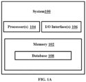
- FIG. 1A is a functional block diagram of a system 100, for domain knowledge augmented multi-head attention based robust universal lesion detection, in accordance with some embodiments of the present disclosure.
- the system 100 includes a processor(s) 104, communication interface device(s), alternatively referred as input/output (I/O) interface(s) 106, and one or more data storage devices or a memory 102 operatively coupled to the processor(s) 104.
- the system 100 with one or more hardware processors is configured to execute functions of one or more functional blocks of the system 100.
- the processor(s) 104 can be one or more hardware processors 104.
- the one or more hardware processors 104 can be implemented as one or more microprocessors, microcomputers, microcontrollers, digital signal processors, central processing units, state machines, logic circuitries, and/or any devices that manipulate signals based on operational instructions.
- the one or more hardware processors 104 are configured to fetch and execute computer-readable instructions stored in the memory 102.
- the system 100 can be implemented in a variety of computing systems including laptop computers, notebooks, hand-held devices such as mobile phones, workstations, mainframe computers, servers, and the like.
- the I/O interface(s) 106 can include a variety of software and hardware interfaces, for example, a web interface, a graphical user interface to display the generated target images and the like and can facilitate multiple communications within a wide variety of networks N/W and protocol types, including wired networks, for example, LAN, cable, etc., and wireless networks, such as WLAN, cellular and the like.
- the I/O interface (s) 106 can include one or more ports for connecting to a number of external devices or to another server or devices.
- the memory 102 may include any computer-readable medium known in the art including, for example, volatile memory, such as static random access memory (SRAM) and dynamic random access memory (DRAM), and/or non-volatile memory, such as read only memory (ROM), erasable programmable ROM, flash memories, hard disks, optical disks, and magnetic tapes.
- volatile memory such as static random access memory (SRAM) and dynamic random access memory (DRAM)
- DRAM dynamic random access memory
- non-volatile memory such as read only memory (ROM), erasable programmable ROM, flash memories, hard disks, optical disks, and magnetic tapes.
- the memory 102 includes modules (not shown) for (a) multilevel feature map extraction, (b) attention based feature fusion, (c) lesion detection with custom RPN as depicted in FIG. 1B , 1C and 1D respectively that enable domain knowledge augmented multi-head attention based robust universal lesion detection.
- the memory 102 may comprise information pertaining to input(s)/output(s) of each step performed by the processor(s) 104 of the system100 and methods of the present disclosure.
- the memory 102 includes a database 108 that stores the CT scan slices received for lesion detection, and the like.
- the database 108 may be external (not shown) to the system 100 and coupled to the system via the I/O interface 106. Functions of the components of the system 100 are explained in conjunction with architecture of the system as depicted in FIGS. 1B through 1D and steps of flow diagram of FIG. 2 .
- FIGS. 1B , 1C and 1D illustrate the architectural overview of the system 100 of FIG. 1A , in accordance with some embodiments of the present disclosure.
- the system 100 extracts as many domain-specific features as possible from minimal number of Computer Tomography (CT) scan slices for a particular patient or subject, to provide a robust, computation and resource efficient ULD with enhanced prediction performance or accuracy in prediction of lesion.
- CT Computer Tomography
- the system 100 utilizes lesser training data as compared to state of the art approaches, hence requires lesser storage or memory space.
- the system 100 utilizes minimum slices from CT scan of a patient and extracts maximum information using the disclosed architecture of the DKMA-ULD framework that enables incorporating 3-Dimensional (3D) context of input data to the system for further processing.
- the information of tissue density from CT-scans represented as Hounsfield Unit (HU)-values in terms of window width and window length.
- HU Hounsfield Unit
- radiologists adjust these windows to focus on organs/tissues of interest in a subject's Region of interest (subRoI), interchangeably referred as body part of interest such as abdomen, chest, or the like.
- ROI Region of interest
- the method and system disclosed herein, in an example implementation utilizes 5 heuristically determined HU windows for CT-slices and feed them as multi-intensity input to the detection network to make it organ-agnostic.
- the computed features are combined using a convolution augmented multi-head attention-based fusion architecture.
- transformers based self-attention mechanism for feature-fusion of multi-intensity images that effectively combines multi-organ information efficiently. This is analogous to the radiologists' way of paying attention to different organs at different HU windows of CT scan slices simultaneously while detecting lesions.
- the system since default anchor sizes and ratios used in general object detection networks do not perform satisfactorily for lesions of different sizes, particularly for very small ( ⁇ 10mm) and medium-sized (10-30mm) lesions, the system generates customized anchor sizes and ratios for a Region Proposal Network (RPN) that enable detecting lesions of varied sizes mostly found in medical imaging datasets.
- RPN Region Proposal Network
- SSL self-supervised learning
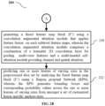
- FIG. 2A and 2B (collectively referred as FIG. 2 ) is a flow diagram illustrating a method 200 for domain knowledge augmented multi-head attention based robust universal lesion detection, using the system 100 of FIG. 1 , in accordance with some embodiments of the present disclosure.
- the system 100 comprises one or more data storage devices or the memory 102 operatively coupled to the processor(s) 104 and is configured to store instructions for execution of steps of the method 200 by the processor(s) or one or more hardware processors 104.
- the steps of the method 200 of the present disclosure will now be explained with reference to the components or blocks of the system 100 as depicted in FIG. 1A through 1D and the steps of flow diagram as depicted in FIG. 2 .
- the one or more hardware processors 104 receive and preprocess a slice set, from amongst a plurality of slices of the Computed Tomography (CT) scan of a subject.
- the slice set comprises i) a key slice of a subject's Region of Interest (subRoI) and ii) a superior slice and an inferior slice in neighborhood of the key slice of the subRoI.
- subRoI Region of Interest
- key slice number is provided as a meta data.
- An example slice set is provided later at step 206.
- CT scan slices obtained from the dataset may be stored in the database 108.
- CT scan slices obtained for a subject or patient, via the I/O interface 106, in real time are analyzed for predicting and locating lesions in the CT scans of the patient.
- preprocessing includes, first resampling CT volume to a common voxel resolution of 0.8 ⁇ 0.8 ⁇ 2mm 3 followed by the removal of black borders of the CT-slices for computational efficiency and to focus on area of interest.
- the one or more hardware processors 104 create a 3D context of the subRoI by defining a 3-channel input image based on each preprocessed slice of the slice set.
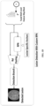
- 3 slices key slice with one superior and one inferior neighboring slice are utilized to generate 3-channel input image (I), as depicted in FIG. 1B .
- Meta-data is provided with dataset to decide which slice number is to be considered as a key slice. For neighbor slices, one slice present 2 mm above and another slice present 2 mm below the key slice are selected.
- the one or more hardware processors 104 window, using a widowing method known in art, each preprocessed slice of the slice set of the subRoI in accordance with a plurality of heuristically determined organ agnostic Hounsfield Unit (HU) windows with varying pixel intensities and highlighting varying organs, wherein a set of HU windowed images is created for each preprocessed slice.
- HU heuristically determined organ agnostic Hounsfield Unit
- the intensity of a CT-slice is rescaled using a certain HU window, U (e.g., a single and wide window of [1024, 4096], as used by prior approaches), in order to include gray-scale intensities of different organs.
- U e.g., a single and wide window of [1024, 4096], as used by prior approaches
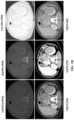
- using a single window suppresses organ-specific information resulting into a degenerated image-contrast, as shown in FIG. 3A (a), 3B (a) and 3C (a) which in turn makes it hard for the system 100 to learn to focus on various organs present in the given CT volume.
- radiologists adjust these intensity values to focus on organs/tissue of interest.
- the method 200 disclosed herein exploit this domain knowledge and feeds it to a deep network, comprising the shared feature extractor, the attention network and the region proposal network (RPN), explicitly in the form of CT-slices having multiple intensities which highlight different organs of the body or the subjects's Rol (subRoI).
- RPN region proposal network
- MVP-Net Multi-view FPN with Position-aware attention for Deep Universal Lesion Detection' by Zihao Li et al., utilizes a clustering algorithm to determine three HU windows.
- the method disclosed herein utilizes a set of 5 organ agnostic HU windows, interchangeably referred as HU windows, by utilizing domain knowledge, to capture or cover multi-organ information in input CT-slices.
- HU windows 5 organ agnostic HU windows
- domain knowledge to capture or cover multi-organ information in input CT-slices.
- '2D-Densely Connected Convolution Neural Networks for automatic Liver and Tumor Segmentation' by Krishna Chaitanya Kaluva et. al refers using multiple HU windows
- the lesion detection in the literature work is specific to liver and is an organ specific lesion detection method.
- the 5 HU windows identified by the method disclosed are based on domain knowledge which makes them more generic, data independent and unsupervised.
- the given eight organs are divided into different body parts and based on that corresponding HU windows are selected.
- the respective body parts are bones, chest region including lungs and mediastinum, abdomen including liver and kidney, and soft-tissues.
- number of windows are flexible and can be increased in accordance with additional body parts that the system 100 is expected to analyze.
- the HU widows are determined in such a way that the major body organs are covered.
- U [U 1, U w ]
- U 1 and U w are the window level/center and window width
- the intensity values of a CT-slice are first normalized using (U 1 ⁇ U w /2) as data min/max and clipped between [0,1] and then, re-scaled to values in [0,255].
- FIGS. 3A , 3B and 3C depicts CT-slices of chest-region, abdomen-region, and pelvic-region, respectively.
- the FIGS, 3A to 3C clearly demonstrates that by using a greater number of windows, different organs of a particular body-region present in a CT volume, can be highlighted more efficiently.
- the one or more hardware processors 104 generate a feature map block corresponding to each of the organ agnostic HU window by extracting a plurality of feature maps, using a shared feature extractor comprising a feature pyramid network (FPN) applied on HU windowed images from amongst the set of HU windowed images of each of the preprocessed slice that fall under same window range of an organ agnostic HU window.
- Each feature map block corresponding to each of the organ agnostic HU window comprises a set of sub-level feature maps at a plurality of FPN levels, with each FPN level having receptive fields of different resolution to capture features of one or more lesions having varying sizes.
- Convolution Feature Extraction Backbone for multilevel feature map extraction As depicted in FIG. 1B , for a given patient or subject, 5 multiple intensity images each having 3 slices/channels, are passed as input to the ResNeXt-152 shared backbone with feature pyramid network (FPN) based convolutional feature extractor.
- FPN feature pyramid network
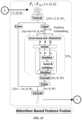
- the one or more hardware processors 104 generate a fused feature map block (F') using a convolution augmented attention module that applies feature fusion on the feature map block corresponding to each of the organ agnostic HU window.
- F' fused feature map block
- a convolution augmented attention module that applies feature fusion on the feature map block corresponding to each of the organ agnostic HU window.
- feature maps extracted from different windowed images are concatenated along channels. Aim of the feature fusion is to bring down the number of concatenated channels to a fixed lower value.
- a simple convolution layer as used in state -of-the-art approaches, can also be used for the feature fusion but convolution layers operate only on a local neighborhood, so to increase the fusion efficiency the method 200 uses a self-attention approach, which captures global information across long range dependencies.
- Number of output channels of a self-attention module are governed by its Value matrix. Directly using the self-attention module to fuse multiple feature maps will result in a higher Value matrix dimension and that would result in a computational overload and also restricts its usage for feature fusion at high resolution pyramid levels.
- a convolution layer is augmented in parallel to self-attention module, and number of output channels are divided between these two branches based on allowed computational memory.
- self-attention branch first, channels from multiple feature maps are convoluted down to a lower dimension. This compressed channel information is then used to generate Key, Query and Value matrix and then multi-headed self-attention module is applied. To match the desired output number of channels, outputs from two parallel branches are concatenated. This optimization allows the method 200 to readily apply feature fusion module on different pyramid level with different resolutions, which in-turn helps to detect lesions of different sizes.
- the convolution augmented attention module performs following functions:
- FIG. 1C The architecture of the convolution augmented multi-head attention module for attention based feature fusion is depicted in FIG. 1C .
- State of the art ULD techniques such as MULAN incorporated information from multiple slices in their network by fusing the feature maps of all 3-channel images/slices with a convolution layer to obtain a 3D-context-enhanced feature map for the central slice.
- the method disclosed utilizes self-attention technique and use of the multi-head self-attention enables the feature fusion module to attend jointly to both spatial and feature sub-spaces.
- FIG. 1C first 5 multi-view feature maps (256, H,W)j for a specific sub-level are concatenated to obtain feature-vectors of shape (256 ⁇ 5,H,W)j.
- the one or more hardware processors 104 predict one or more lesions of varying sizes in the preprocessed slice set by analyzing the fused feature map block (F') using a Region proposal Network (RPN).
- RPN Region proposal Network
- the RPN generates bounding boxes and corresponding probability values across the one or more lesions of varying sizes from amongst a set of customized lesion specific anchors sizes.
- lesion-specific anchors are used for extracting Regions of Interest (ROI) from the obtained feature maps from 5 FPN levels, anchor boxes play a very crucial role. It is observed that small lesions are hard to detect using default anchor sizes and ratios used in RPN for real-world object detection.
- the method 200 creates new custom anchors, which are well suited for detecting lesions of all sizes in CT scans. If, H and W are the image height and width, respectively, anchor boxes of different sizes centered on each image pixel are generated, such that it has maximum Intersection over Union (IoU) with lesion bounding box. If anchor sizes and ratios are in sets ⁇ s,s 2 , Vietnamese, s n ⁇ and ⁇ r 1 r 2 ,...., r m ⁇ , respectively for each r > 0, there can be a total of WH(n+m-1) anchor boxes.
- IOU Intersection over Union
- a differential evolution search algorithm known in the art is used to find, for example 5 best anchor sizes,[16,24,64,128,256] and ratios [3.27,1.78,1,0:56,0.30] for P2,P3,P4,P5, and P6 feature map sub-levels, respectively.
- These lesion-specific anchors are used in RPN for Rol extraction, which are combined with feature maps using Rol pooling layer and further used, for predicting bounding-boxes around lesions along with probability values, as shown in FIG. 1D .
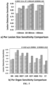
- the custom anchors allow covering varied sized lesions and more specifically, improve the detection of small-sized ( ⁇ 10mm)and medium-sized (10-30mm) lesions considerably, as evident in graphical analysis of FIG. 4 (a) .
- Self-supervision The idea behind self-supervised learning (SSL) is that the learned intermediate representations can carry better semantic and structural meanings and can prove to be beneficial for a variety of downstream tasks.
- SSL self-supervised learning
- BYOL Bootstrap your own latent
- the target network (parameterized by ⁇ ) has the same architecture as the online one (parameterized by ⁇ ), but with polyak averaged weights, ⁇ ⁇ ⁇ + +(1 - ⁇ ) ⁇ .
- the goal is to learn a representation y that can be used in downstream tasks.
- the detection networks are initialized using weights pre-trained on imagenet consisting of natural images and may not be effective for the medical imaging domain. Therefore, the domain-specific weights, obtained by training the backbone using SSL over train-split (23K images) of the DeepLesion dataset, for initializing DKMA-ULD are used to obtain enhanced performance.
- training of the DKMA-ULD framework is performed on 3 channel CT images of size 512 ⁇ 512 with a batch size of 4 on a single NVIDIA Tesla V100 having 32GB GPU-memory.
- a cross-entropy and smooth l 1 loss for classification and bounding-box regression is used, respectively.
- the system 100 is trained until convergence using SGD optimizer with a learning rate (LR) and decay-factor of 0.02 and 10, respectively.
- the SSL model is trained using cross-entropy loss with a batch size of 64, as provided by Adam optimizer, and LR of 3e-4 for 300 epochs.
- DKMA-ULD improves the detection of very small ( ⁇ 10mm) and medium-sized (10-30mm) lesions over 3DCE and improved RetinaNet.
- FIG. 4 (b) it is observed that the method disclosed herein that including domain-specific information in the lesion detection network improves the average sensitivity across all organs present in the dataset.
- cropped CT slices were used by clipping the black border region to focus on the region of interest.
- the ablation study is presented on the introduction of different modules in the disclosed lesion detection pipeline, as shown in Table 2.
- the 5 HU windows approach used by the method for organ-specific domain knowledge results in an improvement of approximately 2% in the average sensitivity (82.37%), as shown in row 3 of Table 2.
- Sensitivity 1 1 ⁇ 101 77.59 2 3 ⁇ 101 80.66 3 5 ⁇ 101 82.37 4 5 ⁇ 101 Yes (Y) 83.30 5 5 ⁇ 101 Y Y 84.23 6 5 ⁇ 152 Y Y 84.85 7 ⁇ 5 ⁇ 152 Y Y 86.88
- FIG. 5 depicts average sensitivity comparison for different lesion sizes and organs.
- FIG. 5 (b) comparison of organ-wise average sensitivity of MULAN and DKMA-ULD.
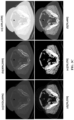
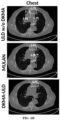
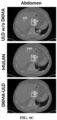
- FIGS. 6A through 6D depict detection results rate of 2 false positive (FP) of DKMA-ULD on CT-scans of different body regions (subject's Rol).
- the system 100 is compared with, the state of the art MULAN, with and without domain knowledge.
- the solid line, dotted line, and dashed line boxes represent ground-truth, true-positive (TP), and false-positive (FP) lesion detections, respectively.
- TP true-positive
- FP false-positive
- ULD w/o DKMA represents when 3 slices with only one HU window ([1024;4096]), default anchors, and without convolution augmented multi-head attention feature fusion are used.
- DKMA-ULD multi-head attention
- the method and system disclosed herein performs robust detection of lesions across all organs of the body using 5 HU windows, computed in an unsupervised manner, for highlighting the different organs of the body in CT-scans and self-attention based feature aggregation which make DKMA-ULD framework organ agnostic.
- the HU windows and the architecture does not require any modification, dedicated learning still provides lesions detection with consistent accuracy across different organs of the body.
- the explicit incorporation of domain knowledge into the deep network alleviates the need of using a large number of heterogenous datasets and enables the network to learn maximum information with limited number of slices per patient's CT-scan.
- the hardware device can be any kind of device which can be programmed including e.g. any kind of computer like a server or a personal computer, or the like, or any combination thereof.
- the device may also include means which could be e.g. hardware means like e.g. an application-specific integrated circuit (ASIC), a field-programmable gate array (FPGA), or a combination of hardware and software means, e.g.
- ASIC application-specific integrated circuit
- FPGA field-programmable gate array
- the means can include both hardware means, and software means.
- the method embodiments described herein could be implemented in hardware and software.
- the device may also include software means.
- the embodiments may be implemented on different hardware devices, e.g. using a plurality of CPUs.
- the embodiments herein can comprise hardware and software elements.
- the embodiments that are implemented in software include but are not limited to, firmware, resident software, microcode, etc.
- the functions performed by various components described herein may be implemented in other components or combinations of other components.
- a computer-usable or computer readable medium can be any apparatus that can comprise, store, communicate, propagate, or transport the program for use by or in connection with the instruction execution system, apparatus, or device.
- a computer-readable storage medium refers to any type of physical memory on which information or data readable by a processor may be stored.
- a computer-readable storage medium may store instructions for execution by one or more processors, including instructions for causing the processor(s) to perform steps or stages consistent with the embodiments described herein.
- the term "computer-readable medium” should be understood to include tangible items and exclude carrier waves and transient signals, i.e., be non-transitory. Examples include random access memory (RAM), read-only memory (ROM), volatile memory, nonvolatile memory, hard drives, CD ROMs, DVDs, flash drives, disks, and any other known physical storage media.
Landscapes
- Engineering & Computer Science (AREA)
- Theoretical Computer Science (AREA)
- General Physics & Mathematics (AREA)
- Physics & Mathematics (AREA)
- Multimedia (AREA)
- Health & Medical Sciences (AREA)
- Computer Vision & Pattern Recognition (AREA)
- General Health & Medical Sciences (AREA)
- Medical Informatics (AREA)
- Evolutionary Computation (AREA)
- Artificial Intelligence (AREA)
- Databases & Information Systems (AREA)
- Quality & Reliability (AREA)
- Nuclear Medicine, Radiotherapy & Molecular Imaging (AREA)
- Radiology & Medical Imaging (AREA)
- Computing Systems (AREA)
- Biomedical Technology (AREA)
- Molecular Biology (AREA)
- Biodiversity & Conservation Biology (AREA)
- Life Sciences & Earth Sciences (AREA)
- Software Systems (AREA)
- Apparatus For Radiation Diagnosis (AREA)
Applications Claiming Priority (1)
| Application Number | Priority Date | Filing Date | Title |
|---|---|---|---|
| IN202121050603 | 2021-11-03 |
Publications (2)
| Publication Number | Publication Date |
|---|---|
| EP4177828A1 true EP4177828A1 (fr) | 2023-05-10 |
| EP4177828B1 EP4177828B1 (fr) | 2026-01-07 |
Family
ID=82611131
Family Applications (1)
| Application Number | Title | Priority Date | Filing Date |
|---|---|---|---|
| EP22177547.1A Active EP4177828B1 (fr) | 2021-11-03 | 2022-06-07 | Procédé et système de détection de lésions universelle robuste basée sur une attention multi-têtes augmentée par la connaissance du domaine |
Country Status (2)
| Country | Link |
|---|---|
| US (1) | US12131466B2 (fr) |
| EP (1) | EP4177828B1 (fr) |
Cited By (7)
| Publication number | Priority date | Publication date | Assignee | Title |
|---|---|---|---|---|
| CN117689960A (zh) * | 2024-01-31 | 2024-03-12 | 中国地质大学(武汉) | 一种岩性场景分类模型构建方法及分类方法 |
| CN118587217A (zh) * | 2024-08-06 | 2024-09-03 | 华侨大学 | 基于特征相关性的骨肉瘤ct图像病变区域检测方法及系统 |
| CN118606671A (zh) * | 2024-05-30 | 2024-09-06 | 深圳大学 | 一种基于多模态融合的激光诱导热裂切割脆性材料过程高精度实时监测系统 |
| CN119722670A (zh) * | 2025-02-27 | 2025-03-28 | 国网安徽省电力有限公司超高压分公司 | 交流滤波器缺陷的检测方法、存储介质 |
| CN120032369A (zh) * | 2025-04-22 | 2025-05-23 | 山东大学 | 一种细胞分割方法和装置、电子设备 |
| CN120107705A (zh) * | 2025-05-09 | 2025-06-06 | 江西师范大学 | 医学影像多标签分类模型训练方法、电子设备和存储介质 |
| CN120655544A (zh) * | 2025-08-13 | 2025-09-16 | 昆明理工大学 | 基于退化原型学习的多退化医学图像统一融合方法 |
Families Citing this family (15)
| Publication number | Priority date | Publication date | Assignee | Title |
|---|---|---|---|---|
| CN117011657B (zh) * | 2023-07-05 | 2024-09-03 | 深圳市龙岗中心医院 | 基于自注意力机制融合的脑ct图像处理方法及相关设备 |
| CN116912625A (zh) * | 2023-07-25 | 2023-10-20 | 东北大学秦皇岛分校 | 一种基于先验缺陷特征与sspcab注意力机制的数据增强方法 |
| CN116863257A (zh) * | 2023-08-02 | 2023-10-10 | 中国医学科学院医学信息研究所 | 基于深度学习的ct图像上纵膈病灶的检测方法及系统 |
| CN117058448A (zh) * | 2023-08-10 | 2023-11-14 | 太原理工大学 | 基于领域知识与并行可分离卷积Swin Transformer的肺部CT图像分类系统 |
| CN117392082A (zh) * | 2023-10-13 | 2024-01-12 | 常州大学 | 基于全尺度跳跃连接的肝脏ct图像分割方法及系统 |
| CN117235119B (zh) * | 2023-11-09 | 2024-01-30 | 北京谷器数据科技有限公司 | 一种低代码平台下多表联合查询的方法 |
| CN117746463B (zh) * | 2023-12-20 | 2025-02-07 | 脉得智能科技(无锡)有限公司 | 体征信息识别方法、系统和电子设备 |
| CN117975011B (zh) * | 2024-02-07 | 2024-10-11 | 安徽大学 | 一种基于细节增强反向注意力网络的膀胱肿瘤图像分割方法及系统 |
| CN117911705B (zh) * | 2024-03-19 | 2024-05-28 | 成都理工大学 | 一种基于GAN-UNet变体网络的脑部MRI肿瘤分割方法 |
| CN118154604A (zh) * | 2024-05-11 | 2024-06-07 | 南京信息工程大学 | 一种墙体裂缝诊断方法、装置、计算机设备及存储介质 |
| CN118736282B (zh) * | 2024-06-14 | 2025-04-01 | 大医智诚高科有限公司 | 基于器官专一化特征感知与区域特征增强的病变分类方法 |
| CN118887401B (zh) * | 2024-07-09 | 2025-04-22 | 广州医科大学 | 基于多模态特征主动脉夹层预测模型的构建方法和设备 |
| CN119559341B (zh) * | 2025-01-26 | 2025-04-18 | 中日友好医院(中日友好临床医学研究所) | 基于直乙镜图像的ai分析方法及系统 |
| CN120259285B (zh) * | 2025-06-03 | 2025-08-01 | 浙江微因医学诊断技术有限公司 | 宫颈病变细胞自动检测的卷积视觉变换器设计与训练方法 |
| CN120374631B (zh) * | 2025-06-30 | 2025-08-26 | 山东未来网络研究院(紫金山实验室工业互联网创新应用基地) | 一种基于乳腺超声的多尺度自适应病变检测方法 |
Family Cites Families (8)
| Publication number | Priority date | Publication date | Assignee | Title |
|---|---|---|---|---|
| KR20150098119A (ko) * | 2014-02-19 | 2015-08-27 | 삼성전자주식회사 | 의료 영상 내 거짓양성 병변후보 제거 시스템 및 방법 |
| US11403493B2 (en) * | 2020-01-17 | 2022-08-02 | Ping An Technology (Shenzhen) Co., Ltd. | Device and method for universal lesion detection in medical images |
| US11282193B2 (en) * | 2020-03-31 | 2022-03-22 | Ping An Technology (Shenzhen) Co., Ltd. | Systems and methods for tumor characterization |
| US11256960B2 (en) * | 2020-04-15 | 2022-02-22 | Adobe Inc. | Panoptic segmentation |
| CN112241766B (zh) * | 2020-10-27 | 2023-04-18 | 西安电子科技大学 | 基于样本生成和迁移学习的肝脏ct图像多病变分类方法 |
| US11410309B2 (en) * | 2020-12-03 | 2022-08-09 | Ping An Technology (Shenzhen) Co., Ltd. | Method, device, and computer program product for deep lesion tracker for monitoring lesions in four-dimensional longitudinal imaging |
| US11900596B2 (en) * | 2021-04-14 | 2024-02-13 | Ping An Technology (Shenzhen) Co., Ltd. | Method, device, and storage medium for weakly-supervised universal lesion segmentation with regional level set loss |
| WO2022244002A1 (fr) * | 2021-05-18 | 2022-11-24 | Ramot At Tel-Aviv University Ltd. | Système et procédé d'analyse de balayage abdominal |
-
2022
- 2022-06-07 EP EP22177547.1A patent/EP4177828B1/fr active Active
- 2022-06-10 US US17/806,402 patent/US12131466B2/en active Active
Non-Patent Citations (5)
| Title |
|---|
| DAI YIN ET AL: "TransMed: Transformers Advance Multi-modal Medical Image Classification", 10 March 2021 (2021-03-10), XP093000371, Retrieved from the Internet <URL:https://arxiv.org/abs/2103.05940> [retrieved on 20221121], DOI: 10.48550/arXiv.2103.05940 * |
| LI ZIHAO ET AL: "MVP-Net: Multi-view FPN with Position-Aware Attention for Deep Universal Lesion Detection", 10 October 2019, ARXIV.ORG, CORNELL UNIVERSITY LIBRARY, 201 OLIN LIBRARY CORNELL UNIVERSITY ITHACA, NY 14853, PAGE(S) 13 - 21, XP047522311 * |
| MANU SHEORAN ET AL: "DKMA-ULD: Domain Knowledge augmented Multi-head Attention based Robust Universal Lesion Detection", ARXIVARXIV.ORG, CORNELL UNIVERSITY LIBRARY, 201 OLIN LIBRARY CORNELL UNIVERSITY ITHACA, NY 14853, 14 March 2022 (2022-03-14), XP091180769 * |
| SHEORAN MANU ET AL: "An Efficient Anchor-Free Universal Lesion Detection in Ct-Scans", 2022 IEEE 19TH INTERNATIONAL SYMPOSIUM ON BIOMEDICAL IMAGING (ISBI), IEEE, 28 March 2022 (2022-03-28), pages 1 - 4, XP034116961, DOI: 10.1109/ISBI52829.2022.9761698 * |
| XUELIN QIAN ET AL: "M3Lung-Sys: A Deep Learning System for Multi-Class Lung Pneumonia Screening from CT Imaging", ARXIV.ORG, CORNELL UNIVERSITY LIBRARY, 201 OLIN LIBRARY CORNELL UNIVERSITY ITHACA, NY 14853, 7 October 2020 (2020-10-07), XP081780520 * |
Cited By (8)
| Publication number | Priority date | Publication date | Assignee | Title |
|---|---|---|---|---|
| CN117689960A (zh) * | 2024-01-31 | 2024-03-12 | 中国地质大学(武汉) | 一种岩性场景分类模型构建方法及分类方法 |
| CN117689960B (zh) * | 2024-01-31 | 2024-04-26 | 中国地质大学(武汉) | 一种岩性场景分类模型构建方法及分类方法 |
| CN118606671A (zh) * | 2024-05-30 | 2024-09-06 | 深圳大学 | 一种基于多模态融合的激光诱导热裂切割脆性材料过程高精度实时监测系统 |
| CN118587217A (zh) * | 2024-08-06 | 2024-09-03 | 华侨大学 | 基于特征相关性的骨肉瘤ct图像病变区域检测方法及系统 |
| CN119722670A (zh) * | 2025-02-27 | 2025-03-28 | 国网安徽省电力有限公司超高压分公司 | 交流滤波器缺陷的检测方法、存储介质 |
| CN120032369A (zh) * | 2025-04-22 | 2025-05-23 | 山东大学 | 一种细胞分割方法和装置、电子设备 |
| CN120107705A (zh) * | 2025-05-09 | 2025-06-06 | 江西师范大学 | 医学影像多标签分类模型训练方法、电子设备和存储介质 |
| CN120655544A (zh) * | 2025-08-13 | 2025-09-16 | 昆明理工大学 | 基于退化原型学习的多退化医学图像统一融合方法 |
Also Published As
| Publication number | Publication date |
|---|---|
| US20230177678A1 (en) | 2023-06-08 |
| EP4177828B1 (fr) | 2026-01-07 |
| US12131466B2 (en) | 2024-10-29 |
Similar Documents
| Publication | Publication Date | Title |
|---|---|---|
| EP4177828A1 (fr) | Procédé et système de détection de lésions universelle robuste basée sur une attention multi-têtes augmentée par la connaissance du domaine | |
| Zhang et al. | Automatic pancreas segmentation based on lightweight DCNN modules and spatial prior propagation | |
| Zlocha et al. | Improving RetinaNet for CT lesion detection with dense masks from weak RECIST labels | |
| Pustokhin et al. | An effective deep residual network based class attention layer with bidirectional LSTM for diagnosis and classification of COVID-19 | |
| US11449717B2 (en) | System and method for identification and localization of images using triplet loss and predicted regions | |
| US10430946B1 (en) | Medical image segmentation and severity grading using neural network architectures with semi-supervised learning techniques | |
| US20250061682A1 (en) | Efficient segmentation of tumours from lung ct | |
| Pang et al. | CTumorGAN: a unified framework for automatic computed tomography tumor segmentation | |
| Saha et al. | YoTransViT: A transformer and CNN method for predicting and classifying skin diseases using segmentation techniques | |
| Victor Ikechukwu et al. | CX-Net: an efficient ensemble semantic deep neural network for ROI identification from chest-x-ray images for COPD diagnosis | |
| Hesamian et al. | Synthetic CT images for semi-sequential detection and segmentation of lung nodules | |
| US11282193B2 (en) | Systems and methods for tumor characterization | |
| KR20240115234A (ko) | 의료 영상 내 생물학적 객체의 머신 러닝 기반 세그먼트화 | |
| Nguyen et al. | PolyPooling: An accurate polyp segmentation from colonoscopy images | |
| Abdulwahhab et al. | A review on medical image applications based on deep learning techniques | |
| Wan et al. | A coarse-to-fine full attention guided capsule network for medical image segmentation | |
| Sheoran et al. | An efficient anchor-free universal lesion detection in CT-scans | |
| Yamasaki et al. | GrowCut-based fast tumor segmentation for 3D magnetic resonance images | |
| CN120259215A (zh) | 一种增强的可解释多模态食管癌目标检测方法和系统 | |
| Fischer et al. | Mask the unknown: Assessing different strategies to handle weak annotations in the miccai2023 mediastinal lymph node quantification challenge | |
| EP4475071A1 (fr) | Procédé de détermination d'une probabilité de présence d'au moins une lésion candidate dans au moins une image médicale | |
| Retico | Computer-aided detection for pulmonary nodule identification: improving the radiologist's performance? | |
| Armstrong et al. | Brain tumor image segmentation using Deep learning | |
| Huang et al. | Leveraging transfer learning and attention mechanisms for a computed tomography lung cancer classification model | |
| EP4510142A1 (fr) | Détection assistée par ordinateur guidée par un dossier médical |
Legal Events
| Date | Code | Title | Description |
|---|---|---|---|
| PUAI | Public reference made under article 153(3) epc to a published international application that has entered the european phase |
Free format text: ORIGINAL CODE: 0009012 |
|
| STAA | Information on the status of an ep patent application or granted ep patent |
Free format text: STATUS: REQUEST FOR EXAMINATION WAS MADE |
|
| 17P | Request for examination filed |
Effective date: 20230317 |
|
| AK | Designated contracting states |
Kind code of ref document: A1 Designated state(s): AL AT BE BG CH CY CZ DE DK EE ES FI FR GB GR HR HU IE IS IT LI LT LU LV MC MK MT NL NO PL PT RO RS SE SI SK SM TR |
|
| GRAP | Despatch of communication of intention to grant a patent |
Free format text: ORIGINAL CODE: EPIDOSNIGR1 |
|
| STAA | Information on the status of an ep patent application or granted ep patent |
Free format text: STATUS: GRANT OF PATENT IS INTENDED |
|
| INTG | Intention to grant announced |
Effective date: 20251008 |
|
| GRAS | Grant fee paid |
Free format text: ORIGINAL CODE: EPIDOSNIGR3 |
|
| GRAA | (expected) grant |
Free format text: ORIGINAL CODE: 0009210 |
|
| STAA | Information on the status of an ep patent application or granted ep patent |
Free format text: STATUS: THE PATENT HAS BEEN GRANTED |
|
| AK | Designated contracting states |
Kind code of ref document: B1 Designated state(s): AL AT BE BG CH CY CZ DE DK EE ES FI FR GB GR HR HU IE IS IT LI LT LU LV MC MK MT NL NO PL PT RO RS SE SI SK SM TR |
|
| REG | Reference to a national code |
Ref country code: CH Ref legal event code: F10 Free format text: ST27 STATUS EVENT CODE: U-0-0-F10-F00 (AS PROVIDED BY THE NATIONAL OFFICE) Effective date: 20260107 Ref country code: GB Ref legal event code: FG4D |
|
| REG | Reference to a national code |
Ref country code: CH Ref legal event code: R17 Free format text: ST27 STATUS EVENT CODE: U-0-0-R10-R17 (AS PROVIDED BY THE NATIONAL OFFICE) Effective date: 20260121 |
|
| REG | Reference to a national code |
Ref country code: DE Ref legal event code: R096 Ref document number: 602022028264 Country of ref document: DE |