EP4174490A1 - Immobilization of amino group-containing or thiol group-containing bait molecules onto iron-based particles - Google Patents
Immobilization of amino group-containing or thiol group-containing bait molecules onto iron-based particles Download PDFInfo
- Publication number
- EP4174490A1 EP4174490A1 EP21204729.4A EP21204729A EP4174490A1 EP 4174490 A1 EP4174490 A1 EP 4174490A1 EP 21204729 A EP21204729 A EP 21204729A EP 4174490 A1 EP4174490 A1 EP 4174490A1
- Authority
- EP
- European Patent Office
- Prior art keywords
- protein
- acid
- interest
- molecule
- catechol
- Prior art date
- Legal status (The legal status is an assumption and is not a legal conclusion. Google has not performed a legal analysis and makes no representation as to the accuracy of the status listed.)
- Pending
Links
- XEEYBQQBJWHFJM-UHFFFAOYSA-N Iron Chemical compound [Fe] XEEYBQQBJWHFJM-UHFFFAOYSA-N 0.000 title claims abstract description 220
- 239000002245 particle Substances 0.000 title claims abstract description 139
- 229910052742 iron Inorganic materials 0.000 title claims abstract description 106
- 125000003277 amino group Chemical group 0.000 title claims abstract description 53
- 125000003396 thiol group Chemical group [H]S* 0.000 title claims abstract description 41
- YCIMNLLNPGFGHC-UHFFFAOYSA-N catechol Chemical compound OC1=CC=CC=C1O YCIMNLLNPGFGHC-UHFFFAOYSA-N 0.000 claims abstract description 310
- 102000004169 proteins and genes Human genes 0.000 claims abstract description 233
- 108090000623 proteins and genes Proteins 0.000 claims abstract description 233
- 239000002253 acid Substances 0.000 claims abstract description 194
- 238000000034 method Methods 0.000 claims abstract description 128
- 150000005206 1,2-dihydroxybenzenes Chemical class 0.000 claims abstract description 43
- 210000001124 body fluid Anatomy 0.000 claims abstract description 39
- 239000010839 body fluid Substances 0.000 claims abstract description 38
- 208000037265 diseases, disorders, signs and symptoms Diseases 0.000 claims abstract description 24
- 230000003213 activating effect Effects 0.000 claims abstract description 22
- 201000010099 disease Diseases 0.000 claims abstract description 13
- 208000035475 disorder Diseases 0.000 claims abstract description 11
- 238000001514 detection method Methods 0.000 claims description 67
- QAIPRVGONGVQAS-DUXPYHPUSA-N trans-caffeic acid Chemical compound OC(=O)\C=C\C1=CC=C(O)C(O)=C1 QAIPRVGONGVQAS-DUXPYHPUSA-N 0.000 claims description 59
- 230000027455 binding Effects 0.000 claims description 52
- 244000052769 pathogen Species 0.000 claims description 39
- 230000001717 pathogenic effect Effects 0.000 claims description 36
- QAIPRVGONGVQAS-UHFFFAOYSA-N cis-caffeic acid Natural products OC(=O)C=CC1=CC=C(O)C(O)=C1 QAIPRVGONGVQAS-UHFFFAOYSA-N 0.000 claims description 33
- 235000004883 caffeic acid Nutrition 0.000 claims description 31
- ACEAELOMUCBPJP-UHFFFAOYSA-N (E)-3,4,5-trihydroxycinnamic acid Natural products OC(=O)C=CC1=CC(O)=C(O)C(O)=C1 ACEAELOMUCBPJP-UHFFFAOYSA-N 0.000 claims description 30
- 229940074360 caffeic acid Drugs 0.000 claims description 30
- 238000002372 labelling Methods 0.000 claims description 29
- KRKNYBCHXYNGOX-UHFFFAOYSA-K Citrate Chemical compound [O-]C(=O)CC(O)(CC([O-])=O)C([O-])=O KRKNYBCHXYNGOX-UHFFFAOYSA-K 0.000 claims description 27
- 230000005291 magnetic effect Effects 0.000 claims description 26
- 241000711573 Coronaviridae Species 0.000 claims description 23
- 210000004369 blood Anatomy 0.000 claims description 22
- 239000008280 blood Substances 0.000 claims description 22
- 210000002966 serum Anatomy 0.000 claims description 21
- 108010067390 Viral Proteins Proteins 0.000 claims description 19
- 241000725303 Human immunodeficiency virus Species 0.000 claims description 18
- 241000315672 SARS coronavirus Species 0.000 claims description 16
- -1 pentafluorophenyl caffeic acid Chemical compound 0.000 claims description 15
- 241001678559 COVID-19 virus Species 0.000 claims description 13
- UQSXHKLRYXJYBZ-UHFFFAOYSA-N Iron oxide Chemical compound [Fe]=O UQSXHKLRYXJYBZ-UHFFFAOYSA-N 0.000 claims description 12
- 108010042038 Protozoan Proteins Proteins 0.000 claims description 12
- 108010077805 Bacterial Proteins Proteins 0.000 claims description 11
- 108010058643 Fungal Proteins Proteins 0.000 claims description 11
- 229960005191 ferric oxide Drugs 0.000 claims description 11
- 235000013980 iron oxide Nutrition 0.000 claims description 11
- 239000002096 quantum dot Substances 0.000 claims description 10
- 230000004044 response Effects 0.000 claims description 10
- VCQURUZYYSOUHP-UHFFFAOYSA-N (2,3,4,5,6-pentafluorophenyl) 2,2,2-trifluoroacetate Chemical compound FC1=C(F)C(F)=C(OC(=O)C(F)(F)F)C(F)=C1F VCQURUZYYSOUHP-UHFFFAOYSA-N 0.000 claims description 9
- MUBZPKHOEPUJKR-UHFFFAOYSA-N Oxalic acid Chemical compound OC(=O)C(O)=O MUBZPKHOEPUJKR-UHFFFAOYSA-N 0.000 claims description 9
- 208000015181 infectious disease Diseases 0.000 claims description 9
- 244000052613 viral pathogen Species 0.000 claims description 9
- 229940031182 nanoparticles iron oxide Drugs 0.000 claims description 8
- 238000002255 vaccination Methods 0.000 claims description 8
- 241000008904 Betacoronavirus Species 0.000 claims description 7
- 241001678561 Sarbecovirus Species 0.000 claims description 7
- 230000003100 immobilizing effect Effects 0.000 claims description 7
- CIWBSHSKHKDKBQ-JLAZNSOCSA-N Ascorbic acid Chemical compound OC[C@H](O)[C@H]1OC(=O)C(O)=C1O CIWBSHSKHKDKBQ-JLAZNSOCSA-N 0.000 claims description 6
- 241000713666 Lentivirus Species 0.000 claims description 6
- 241000127282 Middle East respiratory syndrome-related coronavirus Species 0.000 claims description 6
- 244000052616 bacterial pathogen Species 0.000 claims description 6
- 108010003723 Single-Domain Antibodies Proteins 0.000 claims description 5
- WTFXARWRTYJXII-UHFFFAOYSA-N iron(2+);iron(3+);oxygen(2-) Chemical compound [O-2].[O-2].[O-2].[O-2].[Fe+2].[Fe+3].[Fe+3] WTFXARWRTYJXII-UHFFFAOYSA-N 0.000 claims description 5
- NMHMNPHRMNGLLB-UHFFFAOYSA-N phloretic acid Chemical compound OC(=O)CCC1=CC=C(O)C=C1 NMHMNPHRMNGLLB-UHFFFAOYSA-N 0.000 claims description 5
- 230000003612 virological effect Effects 0.000 claims description 5
- 229910000828 alnico Inorganic materials 0.000 claims description 4
- 229910000889 permalloy Inorganic materials 0.000 claims description 4
- 206010036790 Productive cough Diseases 0.000 claims description 3
- QVYYOKWPCQYKEY-UHFFFAOYSA-N [Fe].[Co] Chemical compound [Fe].[Co] QVYYOKWPCQYKEY-UHFFFAOYSA-N 0.000 claims description 3
- 235000001014 amino acid Nutrition 0.000 claims description 3
- 150000001413 amino acids Chemical class 0.000 claims description 3
- 235000010323 ascorbic acid Nutrition 0.000 claims description 3
- 229960005070 ascorbic acid Drugs 0.000 claims description 3
- 239000011668 ascorbic acid Substances 0.000 claims description 3
- 239000003651 drinking water Substances 0.000 claims description 3
- 235000020188 drinking water Nutrition 0.000 claims description 3
- 239000003673 groundwater Substances 0.000 claims description 3
- 210000002751 lymph Anatomy 0.000 claims description 3
- 235000006408 oxalic acid Nutrition 0.000 claims description 3
- 210000002381 plasma Anatomy 0.000 claims description 3
- 210000003296 saliva Anatomy 0.000 claims description 3
- 210000003802 sputum Anatomy 0.000 claims description 3
- 208000024794 sputum Diseases 0.000 claims description 3
- 210000002700 urine Anatomy 0.000 claims description 3
- 239000002351 wastewater Substances 0.000 claims description 3
- 235000018102 proteins Nutrition 0.000 description 203
- 239000000523 sample Substances 0.000 description 70
- 241000700605 Viruses Species 0.000 description 34
- 239000000427 antigen Substances 0.000 description 23
- 108091007433 antigens Proteins 0.000 description 23
- 102000036639 antigens Human genes 0.000 description 23
- 238000006243 chemical reaction Methods 0.000 description 20
- 241001430294 unidentified retrovirus Species 0.000 description 17
- 108091003079 Bovine Serum Albumin Proteins 0.000 description 16
- 229940098773 bovine serum albumin Drugs 0.000 description 16
- LOKCTEFSRHRXRJ-UHFFFAOYSA-I dipotassium trisodium dihydrogen phosphate hydrogen phosphate dichloride Chemical compound P(=O)(O)(O)[O-].[K+].P(=O)(O)([O-])[O-].[Na+].[Na+].[Cl-].[K+].[Cl-].[Na+] LOKCTEFSRHRXRJ-UHFFFAOYSA-I 0.000 description 16
- 239000002953 phosphate buffered saline Substances 0.000 description 16
- 239000000872 buffer Substances 0.000 description 12
- VLKZOEOYAKHREP-UHFFFAOYSA-N n-Hexane Chemical compound CCCCCC VLKZOEOYAKHREP-UHFFFAOYSA-N 0.000 description 12
- 239000000090 biomarker Substances 0.000 description 11
- 244000309711 non-enveloped viruses Species 0.000 description 11
- JEIPFZHSYJVQDO-UHFFFAOYSA-N ferric oxide Chemical compound O=[Fe]O[Fe]=O JEIPFZHSYJVQDO-UHFFFAOYSA-N 0.000 description 10
- 238000000684 flow cytometry Methods 0.000 description 10
- 238000001943 fluorescence-activated cell sorting Methods 0.000 description 10
- 239000003446 ligand Substances 0.000 description 10
- 229940096437 Protein S Drugs 0.000 description 9
- 239000006228 supernatant Substances 0.000 description 9
- 238000005406 washing Methods 0.000 description 9
- XEKOWRVHYACXOJ-UHFFFAOYSA-N Ethyl acetate Chemical compound CCOC(C)=O XEKOWRVHYACXOJ-UHFFFAOYSA-N 0.000 description 8
- 125000003178 carboxy group Chemical group [H]OC(*)=O 0.000 description 8
- SZVJSHCCFOBDDC-UHFFFAOYSA-N ferrosoferric oxide Chemical group O=[Fe]O[Fe]O[Fe]=O SZVJSHCCFOBDDC-UHFFFAOYSA-N 0.000 description 8
- 239000000203 mixture Substances 0.000 description 8
- 238000007306 functionalization reaction Methods 0.000 description 7
- 238000011534 incubation Methods 0.000 description 7
- 239000002105 nanoparticle Substances 0.000 description 7
- 239000000243 solution Substances 0.000 description 7
- 238000012360 testing method Methods 0.000 description 7
- WEVYAHXRMPXWCK-UHFFFAOYSA-N Acetonitrile Chemical compound CC#N WEVYAHXRMPXWCK-UHFFFAOYSA-N 0.000 description 6
- 208000007407 African swine fever Diseases 0.000 description 6
- 241000710929 Alphavirus Species 0.000 description 6
- 108091023037 Aptamer Proteins 0.000 description 6
- 241000588832 Bordetella pertussis Species 0.000 description 6
- 241000711950 Filoviridae Species 0.000 description 6
- 241000710831 Flavivirus Species 0.000 description 6
- 241000606768 Haemophilus influenzae Species 0.000 description 6
- 208000037262 Hepatitis delta Diseases 0.000 description 6
- 241000724709 Hepatitis delta virus Species 0.000 description 6
- 241000713112 Orthobunyavirus Species 0.000 description 6
- 101710198474 Spike protein Proteins 0.000 description 6
- 230000003208 anti-thyroid effect Effects 0.000 description 6
- 229940043671 antithyroid preparations Drugs 0.000 description 6
- 238000005119 centrifugation Methods 0.000 description 6
- 238000000576 coating method Methods 0.000 description 6
- 230000000295 complement effect Effects 0.000 description 6
- VYFYYTLLBUKUHU-UHFFFAOYSA-N dopamine Chemical compound NCCC1=CC=C(O)C(O)=C1 VYFYYTLLBUKUHU-UHFFFAOYSA-N 0.000 description 6
- 239000013642 negative control Substances 0.000 description 6
- 241001529453 unidentified herpesvirus Species 0.000 description 6
- XLYOFNOQVPJJNP-UHFFFAOYSA-N water Substances O XLYOFNOQVPJJNP-UHFFFAOYSA-N 0.000 description 6
- XBNGYFFABRKICK-UHFFFAOYSA-N 2,3,4,5,6-pentafluorophenol Chemical compound OC1=C(F)C(F)=C(F)C(F)=C1F XBNGYFFABRKICK-UHFFFAOYSA-N 0.000 description 5
- 241000894006 Bacteria Species 0.000 description 5
- 208000035473 Communicable disease Diseases 0.000 description 5
- 241000233866 Fungi Species 0.000 description 5
- 238000006957 Michael reaction Methods 0.000 description 5
- 230000004913 activation Effects 0.000 description 5
- 239000007864 aqueous solution Substances 0.000 description 5
- 238000003556 assay Methods 0.000 description 5
- 238000001636 atomic emission spectroscopy Methods 0.000 description 5
- 230000015572 biosynthetic process Effects 0.000 description 5
- 210000004027 cell Anatomy 0.000 description 5
- 239000006185 dispersion Substances 0.000 description 5
- IZUPBVBPLAPZRR-UHFFFAOYSA-N pentachloro-phenol Natural products OC1=C(Cl)C(Cl)=C(Cl)C(Cl)=C1Cl IZUPBVBPLAPZRR-UHFFFAOYSA-N 0.000 description 5
- 238000003786 synthesis reaction Methods 0.000 description 5
- YQUVCSBJEUQKSH-UHFFFAOYSA-N 3,4-dihydroxybenzoic acid Chemical compound OC(=O)C1=CC=C(O)C(O)=C1 YQUVCSBJEUQKSH-UHFFFAOYSA-N 0.000 description 4
- 241000186041 Actinomyces israelii Species 0.000 description 4
- 102100023635 Alpha-fetoprotein Human genes 0.000 description 4
- 241001466460 Alveolata Species 0.000 description 4
- 241000228212 Aspergillus Species 0.000 description 4
- 241000193738 Bacillus anthracis Species 0.000 description 4
- 241000606124 Bacteroides fragilis Species 0.000 description 4
- 241000589968 Borrelia Species 0.000 description 4
- 241000589562 Brucella Species 0.000 description 4
- 241000589875 Campylobacter jejuni Species 0.000 description 4
- 241000222120 Candida <Saccharomycetales> Species 0.000 description 4
- 108010022366 Carcinoembryonic Antigen Proteins 0.000 description 4
- 102100025475 Carcinoembryonic antigen-related cell adhesion molecule 5 Human genes 0.000 description 4
- 241000606161 Chlamydia Species 0.000 description 4
- 241001647378 Chlamydia psittaci Species 0.000 description 4
- 102000011022 Chorionic Gonadotropin Human genes 0.000 description 4
- 108010062540 Chorionic Gonadotropin Proteins 0.000 description 4
- 241001533384 Circovirus Species 0.000 description 4
- 208000005443 Circulating Neoplastic Cells Diseases 0.000 description 4
- 241000193403 Clostridium Species 0.000 description 4
- 241000186216 Corynebacterium Species 0.000 description 4
- 241001337994 Cryptococcus <scale insect> Species 0.000 description 4
- 108020004414 DNA Proteins 0.000 description 4
- 241000605314 Ehrlichia Species 0.000 description 4
- 241001464848 Entamoebidae Species 0.000 description 4
- 241000194033 Enterococcus Species 0.000 description 4
- 241000588722 Escherichia Species 0.000 description 4
- 241001467465 Euglenozoa Species 0.000 description 4
- 241000589602 Francisella tularensis Species 0.000 description 4
- 241000590002 Helicobacter pylori Species 0.000 description 4
- 241001112094 Hepevirus Species 0.000 description 4
- 241000228402 Histoplasma Species 0.000 description 4
- 101000947699 Homo sapiens Microfibril-associated glycoprotein 4 Proteins 0.000 description 4
- 102100034353 Integrase Human genes 0.000 description 4
- 201000008225 Klebsiella pneumonia Diseases 0.000 description 4
- 241000588747 Klebsiella pneumoniae Species 0.000 description 4
- 241000589242 Legionella pneumophila Species 0.000 description 4
- 241000589902 Leptospira Species 0.000 description 4
- 241000186779 Listeria monocytogenes Species 0.000 description 4
- 208000016604 Lyme disease Diseases 0.000 description 4
- 102100022185 Melanoma-derived growth regulatory protein Human genes 0.000 description 4
- 101710195116 Melanoma-derived growth regulatory protein Proteins 0.000 description 4
- 102100036103 Microfibril-associated glycoprotein 4 Human genes 0.000 description 4
- 241000186359 Mycobacterium Species 0.000 description 4
- 208000001572 Mycoplasma Pneumonia Diseases 0.000 description 4
- 201000008235 Mycoplasma pneumoniae pneumonia Diseases 0.000 description 4
- 241000588653 Neisseria Species 0.000 description 4
- 241000187654 Nocardia Species 0.000 description 4
- 241000709664 Picornaviridae Species 0.000 description 4
- 241000233870 Pneumocystis Species 0.000 description 4
- 206010035717 Pneumonia klebsiella Diseases 0.000 description 4
- 102000007066 Prostate-Specific Antigen Human genes 0.000 description 4
- 108010072866 Prostate-Specific Antigen Proteins 0.000 description 4
- 241000125945 Protoparvovirus Species 0.000 description 4
- 241000589517 Pseudomonas aeruginosa Species 0.000 description 4
- 241000702263 Reovirus sp. Species 0.000 description 4
- 241000119236 Rhizaria Species 0.000 description 4
- 241000606701 Rickettsia Species 0.000 description 4
- 241000607142 Salmonella Species 0.000 description 4
- 241000607768 Shigella Species 0.000 description 4
- 241001279361 Stachybotrys Species 0.000 description 4
- 241000191940 Staphylococcus Species 0.000 description 4
- 241000194017 Streptococcus Species 0.000 description 4
- 241000607626 Vibrio cholerae Species 0.000 description 4
- 241000607479 Yersinia pestis Species 0.000 description 4
- 238000007792 addition Methods 0.000 description 4
- 108010026331 alpha-Fetoproteins Proteins 0.000 description 4
- 230000002502 anti-myelin effect Effects 0.000 description 4
- 244000309743 astrovirus Species 0.000 description 4
- 229940065181 bacillus anthracis Drugs 0.000 description 4
- 230000008901 benefit Effects 0.000 description 4
- 239000011248 coating agent Substances 0.000 description 4
- ZPUCINDJVBIVPJ-LJISPDSOSA-N cocaine Chemical compound O([C@H]1C[C@@H]2CC[C@@H](N2C)[C@H]1C(=O)OC)C(=O)C1=CC=CC=C1 ZPUCINDJVBIVPJ-LJISPDSOSA-N 0.000 description 4
- 206010013023 diphtheria Diseases 0.000 description 4
- 108010078428 env Gene Products Proteins 0.000 description 4
- 235000019439 ethyl acetate Nutrition 0.000 description 4
- 230000005284 excitation Effects 0.000 description 4
- 229940118764 francisella tularensis Drugs 0.000 description 4
- 229940037467 helicobacter pylori Drugs 0.000 description 4
- 229940084986 human chorionic gonadotropin Drugs 0.000 description 4
- 229940115932 legionella pneumophila Drugs 0.000 description 4
- 239000000463 material Substances 0.000 description 4
- 150000002924 oxiranes Chemical class 0.000 description 4
- SWUARLUWKZWEBQ-VQHVLOKHSA-N phenethyl caffeate Chemical compound C1=C(O)C(O)=CC=C1\C=C\C(=O)OCCC1=CC=CC=C1 SWUARLUWKZWEBQ-VQHVLOKHSA-N 0.000 description 4
- 201000000317 pneumocystosis Diseases 0.000 description 4
- 108020003175 receptors Proteins 0.000 description 4
- 102000005962 receptors Human genes 0.000 description 4
- 150000003384 small molecules Chemical class 0.000 description 4
- 241000701161 unidentified adenovirus Species 0.000 description 4
- 208000023275 Autoimmune disease Diseases 0.000 description 3
- YMWUJEATGCHHMB-UHFFFAOYSA-N Dichloromethane Chemical compound ClCCl YMWUJEATGCHHMB-UHFFFAOYSA-N 0.000 description 3
- 101710091045 Envelope protein Proteins 0.000 description 3
- 102100034349 Integrase Human genes 0.000 description 3
- ABLZXFCXXLZCGV-UHFFFAOYSA-N Phosphorous acid Chemical class OP(O)=O ABLZXFCXXLZCGV-UHFFFAOYSA-N 0.000 description 3
- 241000288906 Primates Species 0.000 description 3
- 101710188315 Protein X Proteins 0.000 description 3
- 108091005609 SARS-CoV-2 Spike Subunit S1 Proteins 0.000 description 3
- 101000629318 Severe acute respiratory syndrome coronavirus 2 Spike glycoprotein Proteins 0.000 description 3
- PXIPVTKHYLBLMZ-UHFFFAOYSA-N Sodium azide Chemical compound [Na+].[N-]=[N+]=[N-] PXIPVTKHYLBLMZ-UHFFFAOYSA-N 0.000 description 3
- HEMHJVSKTPXQMS-UHFFFAOYSA-M Sodium hydroxide Chemical compound [OH-].[Na+] HEMHJVSKTPXQMS-UHFFFAOYSA-M 0.000 description 3
- 101710090322 Truncated surface protein Proteins 0.000 description 3
- CSCPPACGZOOCGX-WFGJKAKNSA-N acetone d6 Chemical compound [2H]C([2H])([2H])C(=O)C([2H])([2H])[2H] CSCPPACGZOOCGX-WFGJKAKNSA-N 0.000 description 3
- 235000014633 carbohydrates Nutrition 0.000 description 3
- 150000001720 carbohydrates Chemical class 0.000 description 3
- 239000000969 carrier Substances 0.000 description 3
- 235000018417 cysteine Nutrition 0.000 description 3
- 229960003638 dopamine Drugs 0.000 description 3
- 150000002148 esters Chemical class 0.000 description 3
- 235000014413 iron hydroxide Nutrition 0.000 description 3
- NCNCGGDMXMBVIA-UHFFFAOYSA-L iron(ii) hydroxide Chemical compound [OH-].[OH-].[Fe+2] NCNCGGDMXMBVIA-UHFFFAOYSA-L 0.000 description 3
- 239000011859 microparticle Substances 0.000 description 3
- 238000001556 precipitation Methods 0.000 description 3
- 239000002904 solvent Substances 0.000 description 3
- 238000010561 standard procedure Methods 0.000 description 3
- 238000002198 surface plasmon resonance spectroscopy Methods 0.000 description 3
- 108091032973 (ribonucleotides)n+m Proteins 0.000 description 2
- INZOTETZQBPBCE-NYLDSJSYSA-N 3-sialyl lewis Chemical compound O[C@H]1[C@H](O)[C@H](O)[C@H](C)O[C@H]1O[C@H]([C@H](O)CO)[C@@H]([C@@H](NC(C)=O)C=O)O[C@H]1[C@H](O)[C@@H](O[C@]2(O[C@H]([C@H](NC(C)=O)[C@@H](O)C2)[C@H](O)[C@H](O)CO)C(O)=O)[C@@H](O)[C@@H](CO)O1 INZOTETZQBPBCE-NYLDSJSYSA-N 0.000 description 2
- WLCZTRVUXYALDD-IBGZPJMESA-N 7-[[(2s)-2,6-bis(2-methoxyethoxycarbonylamino)hexanoyl]amino]heptoxy-methylphosphinic acid Chemical group COCCOC(=O)NCCCC[C@H](NC(=O)OCCOC)C(=O)NCCCCCCCOP(C)(O)=O WLCZTRVUXYALDD-IBGZPJMESA-N 0.000 description 2
- 108010011170 Ala-Trp-Arg-His-Pro-Gln-Phe-Gly-Gly Proteins 0.000 description 2
- 241000486679 Antitype Species 0.000 description 2
- 102000005666 Apolipoprotein A-I Human genes 0.000 description 2
- 108010059886 Apolipoprotein A-I Proteins 0.000 description 2
- 239000004475 Arginine Substances 0.000 description 2
- XKRFYHLGVUSROY-UHFFFAOYSA-N Argon Chemical compound [Ar] XKRFYHLGVUSROY-UHFFFAOYSA-N 0.000 description 2
- 102000013602 Cardiac Myosins Human genes 0.000 description 2
- 108010051609 Cardiac Myosins Proteins 0.000 description 2
- 101710098119 Chaperonin GroEL 2 Proteins 0.000 description 2
- 241000193449 Clostridium tetani Species 0.000 description 2
- 229910002518 CoFe2O4 Inorganic materials 0.000 description 2
- 108010035532 Collagen Proteins 0.000 description 2
- 102000008186 Collagen Human genes 0.000 description 2
- 241000186227 Corynebacterium diphtheriae Species 0.000 description 2
- 102000053602 DNA Human genes 0.000 description 2
- 241000991587 Enterovirus C Species 0.000 description 2
- 241000283074 Equus asinus Species 0.000 description 2
- 108090000288 Glycoproteins Proteins 0.000 description 2
- 102000003886 Glycoproteins Human genes 0.000 description 2
- 241000700721 Hepatitis B virus Species 0.000 description 2
- 208000007514 Herpes zoster Diseases 0.000 description 2
- 101000623901 Homo sapiens Mucin-16 Proteins 0.000 description 2
- 241000701085 Human alphaherpesvirus 3 Species 0.000 description 2
- 241000701806 Human papillomavirus Species 0.000 description 2
- 239000004472 Lysine Substances 0.000 description 2
- KDXKERNSBIXSRK-UHFFFAOYSA-N Lysine Natural products NCCCCC(N)C(O)=O KDXKERNSBIXSRK-UHFFFAOYSA-N 0.000 description 2
- CSNNHWWHGAXBCP-UHFFFAOYSA-L Magnesium sulfate Chemical compound [Mg+2].[O-][S+2]([O-])([O-])[O-] CSNNHWWHGAXBCP-UHFFFAOYSA-L 0.000 description 2
- 241000712079 Measles morbillivirus Species 0.000 description 2
- 102100023123 Mucin-16 Human genes 0.000 description 2
- 241000711386 Mumps virus Species 0.000 description 2
- 241000699666 Mus <mouse, genus> Species 0.000 description 2
- 241000588650 Neisseria meningitidis Species 0.000 description 2
- 101001028244 Onchocerca volvulus Fatty-acid and retinol-binding protein 1 Proteins 0.000 description 2
- 241000283973 Oryctolagus cuniculus Species 0.000 description 2
- 102100035220 Plastin-3 Human genes 0.000 description 2
- 101710081133 Plastin-3 Proteins 0.000 description 2
- 108700039882 Protein Glutamine gamma Glutamyltransferase 2 Proteins 0.000 description 2
- 102100021487 Protein S100-B Human genes 0.000 description 2
- 101710122255 Protein S100-B Proteins 0.000 description 2
- 102100038095 Protein-glutamine gamma-glutamyltransferase 2 Human genes 0.000 description 2
- JUJWROOIHBZHMG-UHFFFAOYSA-N Pyridine Chemical compound C1=CC=NC=C1 JUJWROOIHBZHMG-UHFFFAOYSA-N 0.000 description 2
- 241000700159 Rattus Species 0.000 description 2
- 241000702670 Rotavirus Species 0.000 description 2
- 241000710799 Rubella virus Species 0.000 description 2
- 241000193998 Streptococcus pneumoniae Species 0.000 description 2
- 102000005789 Vascular Endothelial Growth Factors Human genes 0.000 description 2
- 108010019530 Vascular Endothelial Growth Factors Proteins 0.000 description 2
- 239000008351 acetate buffer Substances 0.000 description 2
- 150000001299 aldehydes Chemical class 0.000 description 2
- 230000001004 anti-acetylcholinic effect Effects 0.000 description 2
- 230000003476 anti-centromere Effects 0.000 description 2
- 230000002583 anti-histone Effects 0.000 description 2
- 230000003598 anti-microsomal effect Effects 0.000 description 2
- 230000003460 anti-nuclear Effects 0.000 description 2
- 230000003356 anti-rheumatic effect Effects 0.000 description 2
- 239000004019 antithrombin Substances 0.000 description 2
- ODKSFYDXXFIFQN-UHFFFAOYSA-N arginine Natural products OC(=O)C(N)CCCNC(N)=N ODKSFYDXXFIFQN-UHFFFAOYSA-N 0.000 description 2
- 230000001580 bacterial effect Effects 0.000 description 2
- 230000000903 blocking effect Effects 0.000 description 2
- 150000001718 carbodiimides Chemical class 0.000 description 2
- 210000000170 cell membrane Anatomy 0.000 description 2
- 229960003920 cocaine Drugs 0.000 description 2
- 229920001436 collagen Polymers 0.000 description 2
- 239000012230 colorless oil Substances 0.000 description 2
- XUJNEKJLAYXESH-UHFFFAOYSA-N cysteine Natural products SCC(N)C(O)=O XUJNEKJLAYXESH-UHFFFAOYSA-N 0.000 description 2
- 238000004163 cytometry Methods 0.000 description 2
- 230000001086 cytosolic effect Effects 0.000 description 2
- 238000001212 derivatisation Methods 0.000 description 2
- 238000011161 development Methods 0.000 description 2
- 238000000502 dialysis Methods 0.000 description 2
- 239000000975 dye Substances 0.000 description 2
- 238000005516 engineering process Methods 0.000 description 2
- 102000052116 epidermal growth factor receptor activity proteins Human genes 0.000 description 2
- 108700015053 epidermal growth factor receptor activity proteins Proteins 0.000 description 2
- 238000002474 experimental method Methods 0.000 description 2
- 230000005294 ferromagnetic effect Effects 0.000 description 2
- 239000007850 fluorescent dye Substances 0.000 description 2
- 230000002538 fungal effect Effects 0.000 description 2
- 244000053095 fungal pathogen Species 0.000 description 2
- 239000004220 glutamic acid Substances 0.000 description 2
- 229940045808 haemophilus influenzae type b Drugs 0.000 description 2
- 125000002887 hydroxy group Chemical group [H]O* 0.000 description 2
- 150000002463 imidates Chemical class 0.000 description 2
- 238000000338 in vitro Methods 0.000 description 2
- 238000011065 in-situ storage Methods 0.000 description 2
- 230000003993 interaction Effects 0.000 description 2
- 239000012948 isocyanate Substances 0.000 description 2
- 150000002513 isocyanates Chemical class 0.000 description 2
- 229920002521 macromolecule Polymers 0.000 description 2
- 238000007885 magnetic separation Methods 0.000 description 2
- 230000014759 maintenance of location Effects 0.000 description 2
- 238000005259 measurement Methods 0.000 description 2
- 230000007246 mechanism Effects 0.000 description 2
- 230000002503 metabolic effect Effects 0.000 description 2
- RMAHPRNLQIRHIJ-UHFFFAOYSA-N methyl carbamimidate Chemical compound COC(N)=N RMAHPRNLQIRHIJ-UHFFFAOYSA-N 0.000 description 2
- YOHYSYJDKVYCJI-UHFFFAOYSA-N n-[3-[[6-[3-(trifluoromethyl)anilino]pyrimidin-4-yl]amino]phenyl]cyclopropanecarboxamide Chemical compound FC(F)(F)C1=CC=CC(NC=2N=CN=C(NC=3C=C(NC(=O)C4CC4)C=CC=3)C=2)=C1 YOHYSYJDKVYCJI-UHFFFAOYSA-N 0.000 description 2
- 230000003472 neutralizing effect Effects 0.000 description 2
- 239000012038 nucleophile Substances 0.000 description 2
- 210000004248 oligodendroglia Anatomy 0.000 description 2
- 229960003407 pegaptanib Drugs 0.000 description 2
- 239000000137 peptide hydrolase inhibitor Substances 0.000 description 2
- 102000013415 peroxidase activity proteins Human genes 0.000 description 2
- 108040007629 peroxidase activity proteins Proteins 0.000 description 2
- 229920000642 polymer Polymers 0.000 description 2
- 125000002924 primary amino group Chemical group [H]N([H])* 0.000 description 2
- 102000004196 processed proteins & peptides Human genes 0.000 description 2
- 108090000765 processed proteins & peptides Proteins 0.000 description 2
- 244000079416 protozoan pathogen Species 0.000 description 2
- 238000000746 purification Methods 0.000 description 2
- 239000007790 solid phase Substances 0.000 description 2
- 238000001179 sorption measurement Methods 0.000 description 2
- 238000003756 stirring Methods 0.000 description 2
- 229940031000 streptococcus pneumoniae Drugs 0.000 description 2
- YBBRCQOCSYXUOC-UHFFFAOYSA-N sulfuryl dichloride Chemical compound ClS(Cl)(=O)=O YBBRCQOCSYXUOC-UHFFFAOYSA-N 0.000 description 2
- 238000001847 surface plasmon resonance imaging Methods 0.000 description 2
- 208000011580 syndromic disease Diseases 0.000 description 2
- 229960002175 thyroglobulin Drugs 0.000 description 2
- 102000004217 thyroid hormone receptors Human genes 0.000 description 2
- 108090000721 thyroid hormone receptors Proteins 0.000 description 2
- 231100000588 tumorigenic Toxicity 0.000 description 2
- 230000000381 tumorigenic effect Effects 0.000 description 2
- 241000712461 unidentified influenza virus Species 0.000 description 2
- 229910006297 γ-Fe2O3 Inorganic materials 0.000 description 2
- VIYKYVYAKVNDPS-HKGPVOKGSA-N (2s)-2-azanyl-3-[3,4-bis(oxidanyl)phenyl]propanoic acid Chemical compound OC(=O)[C@@H](N)CC1=CC=C(O)C(O)=C1.OC(=O)[C@@H](N)CC1=CC=C(O)C(O)=C1 VIYKYVYAKVNDPS-HKGPVOKGSA-N 0.000 description 1
- 102000040650 (ribonucleotides)n+m Human genes 0.000 description 1
- 238000001644 13C nuclear magnetic resonance spectroscopy Methods 0.000 description 1
- 238000004293 19F NMR spectroscopy Methods 0.000 description 1
- 238000005160 1H NMR spectroscopy Methods 0.000 description 1
- WXTMDXOMEHJXQO-UHFFFAOYSA-N 2,5-dihydroxybenzoic acid Chemical compound OC(=O)C1=CC(O)=CC=C1O WXTMDXOMEHJXQO-UHFFFAOYSA-N 0.000 description 1
- WHNCXIBKVUPNHR-UHFFFAOYSA-N 3,4-dihydroxy-1h-indole-2-carboxylic acid Chemical compound C1=CC(O)=C2C(O)=C(C(=O)O)NC2=C1 WHNCXIBKVUPNHR-UHFFFAOYSA-N 0.000 description 1
- 239000012103 Alexa Fluor 488 Substances 0.000 description 1
- BTBUEUYNUDRHOZ-UHFFFAOYSA-N Borate Chemical compound [O-]B([O-])[O-] BTBUEUYNUDRHOZ-UHFFFAOYSA-N 0.000 description 1
- 241000283690 Bos taurus Species 0.000 description 1
- 238000009010 Bradford assay Methods 0.000 description 1
- 208000025721 COVID-19 Diseases 0.000 description 1
- 241000283707 Capra Species 0.000 description 1
- 102000004127 Cytokines Human genes 0.000 description 1
- 108090000695 Cytokines Proteins 0.000 description 1
- 102000004190 Enzymes Human genes 0.000 description 1
- 108090000790 Enzymes Proteins 0.000 description 1
- CWYNVVGOOAEACU-UHFFFAOYSA-N Fe2+ Chemical compound [Fe+2] CWYNVVGOOAEACU-UHFFFAOYSA-N 0.000 description 1
- VTLYFUHAOXGGBS-UHFFFAOYSA-N Fe3+ Chemical class [Fe+3] VTLYFUHAOXGGBS-UHFFFAOYSA-N 0.000 description 1
- 241000282326 Felis catus Species 0.000 description 1
- 238000012413 Fluorescence activated cell sorting analysis Methods 0.000 description 1
- 241000941423 Grom virus Species 0.000 description 1
- 241000713772 Human immunodeficiency virus 1 Species 0.000 description 1
- 101900297506 Human immunodeficiency virus type 1 group M subtype B Reverse transcriptase/ribonuclease H Proteins 0.000 description 1
- 238000012404 In vitro experiment Methods 0.000 description 1
- 108010052285 Membrane Proteins Proteins 0.000 description 1
- 102000018697 Membrane Proteins Human genes 0.000 description 1
- 241001465754 Metazoa Species 0.000 description 1
- 238000006845 Michael addition reaction Methods 0.000 description 1
- ZMXDDKWLCZADIW-UHFFFAOYSA-N N,N-Dimethylformamide Chemical compound CN(C)C=O ZMXDDKWLCZADIW-UHFFFAOYSA-N 0.000 description 1
- RINSJDWDXSWUOK-UHFFFAOYSA-N OC(=O)C1=CC=C(O)C(O)=C1.OC(=O)C1=CC=C(O)C(O)=C1 Chemical compound OC(=O)C1=CC=C(O)C(O)=C1.OC(=O)C1=CC=C(O)C(O)=C1 RINSJDWDXSWUOK-UHFFFAOYSA-N 0.000 description 1
- 229910019142 PO4 Inorganic materials 0.000 description 1
- 241001494479 Pecora Species 0.000 description 1
- 241000009328 Perro Species 0.000 description 1
- KJTLSVCANCCWHF-UHFFFAOYSA-N Ruthenium Chemical compound [Ru] KJTLSVCANCCWHF-UHFFFAOYSA-N 0.000 description 1
- 108091005634 SARS-CoV-2 receptor-binding domains Proteins 0.000 description 1
- 108020004682 Single-Stranded DNA Proteins 0.000 description 1
- 208000021386 Sjogren Syndrome Diseases 0.000 description 1
- 108010090804 Streptavidin Proteins 0.000 description 1
- 241000282898 Sus scrofa Species 0.000 description 1
- 108091023040 Transcription factor Proteins 0.000 description 1
- 102000040945 Transcription factor Human genes 0.000 description 1
- 238000002835 absorbance Methods 0.000 description 1
- 238000007259 addition reaction Methods 0.000 description 1
- 229910045601 alloy Inorganic materials 0.000 description 1
- 239000000956 alloy Substances 0.000 description 1
- 150000001408 amides Chemical class 0.000 description 1
- 150000001412 amines Chemical class 0.000 description 1
- 239000012491 analyte Substances 0.000 description 1
- 230000036436 anti-hiv Effects 0.000 description 1
- 230000009830 antibody antigen interaction Effects 0.000 description 1
- 239000012736 aqueous medium Substances 0.000 description 1
- 239000007900 aqueous suspension Substances 0.000 description 1
- 229910052786 argon Inorganic materials 0.000 description 1
- 239000011324 bead Substances 0.000 description 1
- 238000012575 bio-layer interferometry Methods 0.000 description 1
- KGBXLFKZBHKPEV-UHFFFAOYSA-N boric acid Chemical compound OB(O)O KGBXLFKZBHKPEV-UHFFFAOYSA-N 0.000 description 1
- 239000004327 boric acid Substances 0.000 description 1
- 150000001728 carbonyl compounds Chemical class 0.000 description 1
- 125000002843 carboxylic acid group Chemical group 0.000 description 1
- 235000013339 cereals Nutrition 0.000 description 1
- 230000008859 change Effects 0.000 description 1
- 239000003795 chemical substances by application Substances 0.000 description 1
- 238000011097 chromatography purification Methods 0.000 description 1
- 238000000975 co-precipitation Methods 0.000 description 1
- 239000002131 composite material Substances 0.000 description 1
- 150000001875 compounds Chemical class 0.000 description 1
- 150000001945 cysteines Chemical class 0.000 description 1
- 238000002405 diagnostic procedure Methods 0.000 description 1
- IJKVHSBPTUYDLN-UHFFFAOYSA-N dihydroxy(oxo)silane Chemical compound O[Si](O)=O IJKVHSBPTUYDLN-UHFFFAOYSA-N 0.000 description 1
- 238000012377 drug delivery Methods 0.000 description 1
- 125000006575 electron-withdrawing group Chemical group 0.000 description 1
- 239000012039 electrophile Substances 0.000 description 1
- 229940088598 enzyme Drugs 0.000 description 1
- WBJINCZRORDGAQ-UHFFFAOYSA-N ethyl formate Chemical compound CCOC=O WBJINCZRORDGAQ-UHFFFAOYSA-N 0.000 description 1
- 238000011156 evaluation Methods 0.000 description 1
- 238000013401 experimental design Methods 0.000 description 1
- 230000005293 ferrimagnetic effect Effects 0.000 description 1
- 230000005308 ferrimagnetism Effects 0.000 description 1
- 235000013312 flour Nutrition 0.000 description 1
- 239000012530 fluid Substances 0.000 description 1
- 125000000524 functional group Chemical group 0.000 description 1
- 230000001900 immune effect Effects 0.000 description 1
- 230000003053 immunization Effects 0.000 description 1
- 238000002649 immunization Methods 0.000 description 1
- 238000003018 immunoassay Methods 0.000 description 1
- 150000002500 ions Chemical class 0.000 description 1
- 150000002505 iron Chemical class 0.000 description 1
- 229910052943 magnesium sulfate Inorganic materials 0.000 description 1
- 235000019341 magnesium sulphate Nutrition 0.000 description 1
- 230000005389 magnetism Effects 0.000 description 1
- 238000004949 mass spectrometry Methods 0.000 description 1
- 239000012528 membrane Substances 0.000 description 1
- 229910044991 metal oxide Inorganic materials 0.000 description 1
- 150000004706 metal oxides Chemical class 0.000 description 1
- 229930014626 natural product Natural products 0.000 description 1
- 229910052759 nickel Inorganic materials 0.000 description 1
- 231100000252 nontoxic Toxicity 0.000 description 1
- 230000003000 nontoxic effect Effects 0.000 description 1
- 238000000655 nuclear magnetic resonance spectrum Methods 0.000 description 1
- 238000005935 nucleophilic addition reaction Methods 0.000 description 1
- 239000012074 organic phase Substances 0.000 description 1
- 239000008188 pellet Substances 0.000 description 1
- 235000021317 phosphate Nutrition 0.000 description 1
- 150000003013 phosphoric acid derivatives Chemical class 0.000 description 1
- 230000010287 polarization Effects 0.000 description 1
- 239000000047 product Substances 0.000 description 1
- 230000004850 protein–protein interaction Effects 0.000 description 1
- UMJSCPRVCHMLSP-UHFFFAOYSA-N pyridine Natural products COC1=CC=CN=C1 UMJSCPRVCHMLSP-UHFFFAOYSA-N 0.000 description 1
- 238000003908 quality control method Methods 0.000 description 1
- 229910052761 rare earth metal Inorganic materials 0.000 description 1
- 239000011541 reaction mixture Substances 0.000 description 1
- 230000009257 reactivity Effects 0.000 description 1
- 230000002829 reductive effect Effects 0.000 description 1
- 239000013074 reference sample Substances 0.000 description 1
- 229910052707 ruthenium Inorganic materials 0.000 description 1
- 239000012266 salt solution Substances 0.000 description 1
- 108010008595 sarcoma-associated antigen S1 Proteins 0.000 description 1
- 238000012216 screening Methods 0.000 description 1
- 238000000926 separation method Methods 0.000 description 1
- NLJMYIDDQXHKNR-UHFFFAOYSA-K sodium citrate Chemical compound O.O.[Na+].[Na+].[Na+].[O-]C(=O)CC(O)(CC([O-])=O)C([O-])=O NLJMYIDDQXHKNR-UHFFFAOYSA-K 0.000 description 1
- 239000001509 sodium citrate Substances 0.000 description 1
- 241000894007 species Species 0.000 description 1
- 230000009870 specific binding Effects 0.000 description 1
- 239000000126 substance Substances 0.000 description 1
- 239000013595 supernatant sample Substances 0.000 description 1
- 239000000725 suspension Substances 0.000 description 1
- 230000008685 targeting Effects 0.000 description 1
- 230000001225 therapeutic effect Effects 0.000 description 1
- 239000003053 toxin Substances 0.000 description 1
- 231100000765 toxin Toxicity 0.000 description 1
- 108700012359 toxins Proteins 0.000 description 1
Images





Classifications
-
- G—PHYSICS
- G01—MEASURING; TESTING
- G01N—INVESTIGATING OR ANALYSING MATERIALS BY DETERMINING THEIR CHEMICAL OR PHYSICAL PROPERTIES
- G01N33/00—Investigating or analysing materials by specific methods not covered by groups G01N1/00 - G01N31/00
- G01N33/48—Biological material, e.g. blood, urine; Haemocytometers
- G01N33/50—Chemical analysis of biological material, e.g. blood, urine; Testing involving biospecific ligand binding methods; Immunological testing
- G01N33/52—Use of compounds or compositions for colorimetric, spectrophotometric or fluorometric investigation, e.g. use of reagent paper and including single- and multilayer analytical elements
-
- G—PHYSICS
- G01—MEASURING; TESTING
- G01N—INVESTIGATING OR ANALYSING MATERIALS BY DETERMINING THEIR CHEMICAL OR PHYSICAL PROPERTIES
- G01N33/00—Investigating or analysing materials by specific methods not covered by groups G01N1/00 - G01N31/00
- G01N33/48—Biological material, e.g. blood, urine; Haemocytometers
- G01N33/50—Chemical analysis of biological material, e.g. blood, urine; Testing involving biospecific ligand binding methods; Immunological testing
- G01N33/53—Immunoassay; Biospecific binding assay; Materials therefor
-
- G—PHYSICS
- G01—MEASURING; TESTING
- G01N—INVESTIGATING OR ANALYSING MATERIALS BY DETERMINING THEIR CHEMICAL OR PHYSICAL PROPERTIES
- G01N33/00—Investigating or analysing materials by specific methods not covered by groups G01N1/00 - G01N31/00
- G01N33/48—Biological material, e.g. blood, urine; Haemocytometers
- G01N33/50—Chemical analysis of biological material, e.g. blood, urine; Testing involving biospecific ligand binding methods; Immunological testing
- G01N33/53—Immunoassay; Biospecific binding assay; Materials therefor
- G01N33/543—Immunoassay; Biospecific binding assay; Materials therefor with an insoluble carrier for immobilising immunochemicals
- G01N33/54313—Immunoassay; Biospecific binding assay; Materials therefor with an insoluble carrier for immobilising immunochemicals the carrier being characterised by its particulate form
- G01N33/54326—Magnetic particles
Definitions
- iron oxide nanoparticles possess a unique versatility of attachment compared to other carriers or carrier-free applications [18].
- the ability to be magnetically controllable is extremely advantageous for these particles, as washing steps and analyte concentration measures can be significantly simplified compared to conventional non-magnetic beads.
- the assessment of antibody status under the conditions of the Sars-CoV2 pandemic is of great importance for epidemiological evaluation. This is true for both: recording the extent to which infection has progressed and for surveying immunization status as the population vaccination campaign progresses. While a number of diagnostic methods exist, they are often device-specific.
- the Cobas Elecsys Anti-SARS-CoV-2 chemiluminescence immunoassay as discussed in Lippi et al. [19] is based on a sandwich reaction, wherein the test sample, biotinylated SARS-CoV-2-specific recombinant antigen, and SARS-CoV-2-specific recombinant antigen labeled with ruthenium are initially incubated altogether.
- dopamine has only one amino group for further functionalization with possible target molecules, which is homologous to the amino groups of possible targets and is therefore only suitable to a limited extent for corresponding binding to such groups.
- catechols have been described to have the ability to generate firm complexes with the iron hydroxide surface of the corresponding particles [4, 5].
- the invention relates to a method for identifying a protein of interest in an aqueous or body fluid sample by using a complex comprising (A) a superparamagnetic iron-based particle, (B) one or more catechol acid moieties, and (C) one or more bait molecules, as further defined in the claims.
- Said complex allows a simple and robust detection of proteins of interest in an aqueous or body fluid sample.
- bait proteins such as antigens (e.g. virus proteins)
- proteins of interest such as antibodies
- a versatile diagnostic platform is obtained, which can be used to detect protein of interest such as pathogens or autoantibodies, if a corresponding selective complementary binding partner, such as an antigen-antibody pair, exists.
- the method is widely applicable, as it involves stochastic binding of the (A) superparamagnetic iron-based particle with (B) catechol acid to exposed amino or thiol groups in the (C) bait protein.
- a wide variety of antigens can be covalently linked to the superparamagnetic iron-based particles as bait molecules.
- a protein of interest which is a complementary binding partner to the bait protein, can be detected.
- a detection molecule such as a fluorescent dye-labelled antibody directed against the protein of interest, the presence of the protein of interest can be reliably determined e.g. by flow cytometer.
- the main purpose of application is in vitro diagnostics.
- activated catechols have been found to be especially advantageous, particularly when the catechols contain a carboxyl group, such as those found in derivatives of 3,4-dihydroxycinnamic acid, also known by the trivial name caffeic acid.
- a carboxyl group such as those found in derivatives of 3,4-dihydroxycinnamic acid, also known by the trivial name caffeic acid.
- catechols have high affinities to metal oxide surfaces.
- catechols are derived from known natural products (such as dopamine or caffeic acid) and are therefore well characterized and mostly non-toxic.
- Another example of carboxyl catechols is protocatechuic acid (3,4-dihydroxybenzoic acid).
- Caffeic acid has an acid group that can form a stable bond with corresponding nucleophiles.
- the acid group of catechol acid is generally not reactive enough for this reaction to occur under conditions that do not affect the nature of the particle system.
- the acid groups can be activated accordingly, but then the properties of the linker may be altered in such a way that the colloidal stability of the particle is no longer guaranteed.
- the activation of the catechol acid has the particular advantage that said activation is fully compatible with the binding to superparamagnetic iron-based particles and that the activated catechol acid readily reacts with exposed amino groups or via addition reaction of the unsaturated ⁇ , ⁇ position to the carboxyl with a thiol group on bait molecules.
- activated catechol acid can be (i) readily bound to superparamagnetic iron-based particles by e.g. ligand replace reactions and (ii) covalently bound to amino or thiol groups of bait molecule, such as the Sars-CoV-2 antigen.
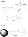
- the inventors have activated caffeic acid with pentafluorophenyl trifluoroacetate, resulting in the reactive pentafluorophenol caffeic acid ester (CAF-PFP) ( Fig. 1 ). Furthermore, the inventors have covalently bound the Sars-CoV-2 antigen (Spike protein) as well as and HIV-envelope protein as bait proteins to the activated catechol acid. As a proof of principle, the inventors show a diagnostic particle system for the selective detection of e.g. Sars-CoV2 antibodies from patient blood, which has proven its performance in samples of Covid-19 of convalescents.
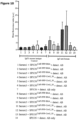
- the data revealed a correct determination of patient sera having a positive or negative Sars-CoV2 antibody status ( Fig. 10 ).
- the presence of anti-gp120 antibodies in a sample could be readily detected by using the envelope protein gp120 as a bait protein ( Fig. 11 ) .
- a method for identifying a protein of interest in an aqueous or body fluid sample comprising the steps of:
- the methods as disclosed herein are in vitro methods. In other words, said methods are performed ex vivo, in particular outside of the human body.
- the present disclosure is concerned with a particularly advantageous linkage between the superparamagnetic iron-based particle (A) and the bait molecule (C).
- A is connected to C via one or more catechol acid moieties (B).
- B has been bound to A.
- the term 'catechol acid moiety' as used herein refers to a catechol acid portion, which is bound by A and C.
- an activated catechol acid being bound to A and C is a catechol acid moiety.
- the superparamagnetic iron-based particle (A) is linked to the one or more catechol acid moieties via citrate.
- B has been covalently linked to C by activating the catechol acid (benzene-1,2-diol acid) and reacting said activated catechol acid with an amino group or thiol group of C.
- the inventors have demonstrated such a linkage in Example 1 (Table 1).
- the bait molecule has been the SARS-CoV 2 Spike protein.
- the envelope protein gp120 of HIV has been used as a bait molecule in
- the catechol acid may comprise, preferably comprises, a carboxyl group, preferably wherein the catechol acid is caffeic acid (3,4-dihydroxycinnamic acid) or protocatechuic acid, more preferably wherein the catechol acid is caffeic acid.
- Caffeic acids has also been used in Examples 1 and 2 herein. However, the skilled person is able to determine other suitable catechol acids such as L-Dopa (L-3,4-dihydroxyphenylalanine), dihydroxybenzoic acid, dihydroxyindole-2-carboxylic acid.
- the catechol acid has been activated to be a pentafluorophenyl catechol acid, terafluorophenyl catechol acid, or a catechol acid N-Hydroxysuccinimide ester, preferably wherein the catechol acid has been activated to be a pentafluorophenyl catechol acid, such as demonstrated in Example 1 herein.
- the skilled person is provided with several options to activate the catechol acid with.
- the catechol acid may have been activated with a isocyanate, acyl azides, a sulfonyl chloride, a tosylate ester, an epoxide, an oxiranes, an imido ester, a carbodiimide, a fluorophenyl ester, or a O-methyl isourea.
- the caffeic acid may have been activated to be a pentafluorophenyl caffeic acid, terafluorophenyl caffeic acid, or a caffeic acid N-Hydroxysuccinimide ester.
- the catechol acid has been activated by pentafluorophenyl trifluoroacetate.
- the catechol acid is caffeic acid and wherein pentafluorophenol caffeic acid ester has been formed by the activation of caffeic acid.
- the catechol acid may be caffeic acid and the caffeic acid may have been activated by pentafluorophenyl trifluoroacetate. Further assistance regarding the exact reaction conditions can be found in Example 1.
- the particular advantage of this linkage between the iron-based particles and the bait molecule is that a linkage via a catechol moiety provides a versatile tool for linking bait molecules to the particles reliably. This is, for example, because the bait molecule merely requires an amino group or a thiol group.
- activated catechol groups are highly reactive with bait molecules such as proteins.
- activated catechol are easy to handle and usually cheap in comparison to other alternatives. Catechols have the property that they polymerize with one another, thus adhering more stably to the particle surface and at the same time do not lose their ability to bind to the bait molecules via the activated ester.
- the described linkage can be combined with any bait molecule - protein of interest pair, as long as the bait molecule comprises an amino group or a thiol groups.
- the described linkage harbors a broad applicability making said linkage a versatile toolkit.
- the protein of interest and the bait molecule generally have a high affinity to each other, such as a nanomolar affinity.
- the skilled person is aware of standard methods to determine the strength of macromolecular interactions, such as protein-protein interactions.
- the skilled person may use SPR, electromobility shift assays, ITC, fluorescent polarization.
- the bait molecule comprises a binding site for the protein of interest, more preferably wherein the bait molecule provides an antigen for the protein of interest and wherein the protein of interest is an antibody.
- proteins of interest refers to any protein, which may be part of an aqueous or body fluid sample.
- the protein of interest may also be regarded as the 'prey', which is sought after by the bait protein.
- proteins of interest may be antibodies or pathogen proteins.
- pathogen protein may be a viral protein, a bacterial protein, a fungal protein, or a protozoan protein.
- the skilled person is aware of such proteins and which diseases such proteins of interest are indicative for.
- the viral protein may be from an enveloped or a non-enveloped virus.
- the viral protein is from an enveloped virus.
- the enveloped virus may be a coronavirus, retrovirus, herpesvirus, poxvirus, hepadnavirus, african swine fever and related viruses, flavivirus, alphavirus, togavirus, hepatitis D virus, orthomyxovirus, paramyxovirus, rhabdovirus, bunyavirus, or filovirus.
- the enveloped virus may be a coronavirus, preferably a orthocoronavirus, more preferably a betacoronavirus, even more preferably a sarbecovirus, even more preferably a SARS-coronavirus or a MERS-coronavirus, even more preferably a SARS coronavirus, and most preferably SARS-coronavirus 2.
- the virus protein may be from a retrovirus such as HIV. It is preferred that the retrovirus is an orthoretrovirus, preferably a lentivirus, more preferable human immunodeficiency viruses (HIV).
- HIV human immunodeficiency viruses
- the virus protein may be from a non-enveloped virus. Exemplary virus proteins from non-enveloped viruses are from an adenovirus, astrovirus, circovirus, hepatitis E-like virus, papovavirus, parvovirus, picornavirus, or reovirus.
- the protein of interest may be a pathogen protein, wherein the pathogen is a bacterium.
- exemplary bacterial proteins are selected from one of the bacteria Actinomyces israelii, Bacillus anthracis, Bacteroides fragilis, Bordetella pertussis, Borrelia, Brucella, Campylobacter jejuni, Chlamydia, Chlamydophila psittaci, Clostridium, Corynebacterium diphtheria, Ehrlichia, Enterococcus, Escherichia, Francisella tularensis, Haemophilus influenza, Helicobacter pylori, Klebsiella pneumonia, Legionella pneumophila, Leptospira species, Listeria monocytogenes, Mycobacterium, Mycoplasma pneumonia, Neisseria, Pseudomonas aeruginosa, Nocardia asteroids, Rickettsia rickettsia, Salmonella, Shi
- the protein of interest may be a pathogen protein, wherein the pathogen is a fungus.
- Exemplary fungal proteins are selected from one of the fungi Candida, Aspergillus, Cryptococcus, Histoplasma, Pneumocystis, or Stachybotrys.
- the protein of interest may be a pathogen protein, wherein the pathogen protein is a protozoan protein.
- Exemplary protozoan proteins are selected from one of Vaccinodas, Parabasilia, Euglenozoa, Entamoebida, Stramenopila, Alveolata, Rhizaria, or Archaeplastida.
- the protein of interest is a biomarker. This is particularly advantageous as a patient may be diagnosed or monitored for a disease by analyzing a bodily fluid samples such as a serum sample. In case the biomarker is identified, the patient may be monitored more specifically.
- Preferred biomarkers which may be proteins of interest are a tumorigenic biomarker or metabolic biomarker, more preferably wherein the biomarker is selected from a list consisting of S100 calcium-binding protein B, MIA protein (melanoma inhibitory activity), Prostate-specific antigen (PSA), human chorionic gonadotropin (hCG), Her2neu, Plastin-3, EGFR, carcinoembryonic antigen (CEA), cancer antigen 125 (CA 125), Carbohydrate antigen 19-9 (CA 19-9), Carbohydrate antigen 15-3 (CA 15-3), alpha-fetoprotein (AFP), Microfibril Associated Protein 4 (MFAP4), or ApoA-1.
- Her2neu, Plastin-3 and/or EGFR can be biomarkers for circulating tumor cells (CTCs).
- the protein of interest is an antibody.
- proteins produced in response to and counteracting a specific antigen This may, for example, occur in response to a vaccination.
- antibodies against Sars-CoV-2 may be formed in response to a vaccination.
- antibodies against the spike protein of SarS-CoV 2 may be formed in response to a vaccination.
- the antibodies may be formed in response to a natural infection.
- the antibody status allows physicians and scientist to monitor the antibody positive and negative rate in the population over time.
- the method as disclosed in this application is useful in assessing e.g. the antibody status against SarS-Cov2 as demonstrated in Example 1, Fig. 10 .
- the linkage between the iron-based particles and the bait molecule merely requires the bait molecule to have an amino group or a thiol group, said linking mechanism is widely applicable.
- the protein of interest is a neutralizing antibody.
- the protein of interest is an antibody having been formed in response to an infection by a pathogen or a vaccination against said pathogen.
- antibodies against Sars-CoV2 are formed in response to an infection or vaccination.
- said antibodies may have been formed in response to a viral or bacterial pathogen.
- a possible pathogens against which antibodies may have been formed, include Sars-CoV2, corynebacterium diphtheriae, haemophilus influenzae type b, hepatitis B virus, measles virus, meningococcus (Neisseria meningitidis), mumps virus, bordetella pertussis, pneumococcus (Streptococcus pneumoniae), poliovirus, rotavirus, rubella virus, Clostridium tetani, varicella zoster virus, human papillomavirus, herpes zoster, and influenza virus.
- Sars-CoV2 corynebacterium diphtheriae
- haemophilus influenzae type b hepatitis B virus
- measles virus measles virus
- meningococcus Neisseria meningitidis
- mumps virus mumps virus
- bordetella pertussis pneumococc
- pathogens against which antibodies may have been formed, may be enveloped or a non-enveloped viruses, preferably an enveloped virus.
- the enveloped virus, against which antibodies may have been formed may be a coronavirus, retrovirus, herpesvirus, poxvirus, hepadnavirus, african swine fever and related viruses, flavivirus, alphavirus, togavirus, hepatitis D virus, orthomyxovirus, paramyxovirus, rhabdovirus, bunyavirus, or filovirus.
- said enveloped virus may be coronavirus, preferably wherein the coronavirus is a orthocoronavirus, preferably a betacoronavirus, more preferably a sarbecovirus, even more preferably a SARS-coronavirus, and most preferably SARS-coronavirus 2.
- the coronavirus is a orthocoronavirus, preferably a betacoronavirus, more preferably a sarbecovirus, even more preferably a SARS-coronavirus, and most preferably SARS-coronavirus 2.
- the protein of interest is an auto-antibody.
- auto-antibodies which generally form in the context of an autoimmune disease.
- the auto-antibody may be selected from a list consisting of antinuclear antibodies, anti-transglutaminase antibodies, anti-ganglioside antibodies, anti-thrombin antibodies, anti-thyroid autoantibodies, anti-myelin basic protein, anti-myelin oligodendrocyte glycoprotein, anti-citrullinated protein, anti-rheumatoid factor, antimicrosomal antibodies, anti-thyroid plasma membrane antibodies, IgA endomysium antibodies, IgA tissue transglutaminase antibodies, anti-Human Cardiac Myosin antibodies, anti-thyroid peroxidase antibodies, anti-thyroglobulin antibodies, anti-Saccharomyces cerevisiae antibodies, anti-flagellin (CBir1), anti-neutrophil cytoplasmic antibodies
- the term 'bait molecule' is a macromolecule, which is used to catch a protein of interest.
- the protein of interest and the bait protein have a high affinity for each other such as nanomolar affinity.
- the bait molecule is a protein.
- the bait protein may be a pathogen protein, preferably the bait protein may a viral, bacterial, fungal or protozoan pathogen protein.
- the bait protein may be from a viral, bacterial, fungal or protozoan pathogen.
- Example 1 and 2 demonstrates that the pathogen proteins Sars-CoV-2 (spike protein) or HIV (GP120) may be used as bait proteins.
- Other possible bait proteins include viral protein from enveloped or non-enveloped viruses.
- the virus may be selected from a list consisting of coronavirus, retrovirus, herpesvirus, poxvirus, hepadnavirus, african swine fever and related viruses, flavivirus, alphavirus, togavirus, hepatitis D virus, orthomyxovirus, paramyxovirus, rhabdovirus, bunyavirus, or filovirus.
- the enveloped virus may be a coronavirus, preferably wherein the coronavirus is a orthocoronavirus, preferably a betacoronavirus, more preferably a sarbecovirus, even more preferably a SARS-coronavirus or a MERS-coronavirus, even more preferably a SARS coronavirus, and most preferably SARS-coronavirus 2.
- the coronavirus is a orthocoronavirus, preferably a betacoronavirus, more preferably a sarbecovirus, even more preferably a SARS-coronavirus or a MERS-coronavirus, even more preferably a SARS coronavirus, and most preferably SARS-coronavirus 2.
- the spike protein of SARS-CoV2 can be used as a bait protein to identify Sars-CoV-2 antibodies in serum, i.e. antibodies against the spike protein.
- the bait protein may be from a retrovirus such as an orthoretrovirus, preferably wherein the retrovirus may be a lentivirus, more preferably wherein the retrovirus is human immunodeficiency viruses (HIV).
- HIV human immunodeficiency viruses
- the bait protein may be from a non-enveloped virus such as an adenovirus, astrovirus, circovirus, hepatitis E-like virus, papovavirus, parvovirus, picornavirus, or reovirus.
- the bait protein may be a bacterial protein from a bacterial pathogen, such as a bacterial protein is selected from one of the bacteria Actinomyces israelii, Bacillus anthracis, Bacteroides fragilis, Bordetella pertussis, Borrelia, Brucella, Campylobacter jejuni, Chlamydia, Chlamydophila psittaci, Clostridium, Corynebacterium diphtheria, Ehrlichia, Enterococcus, Escherichia, Francisella tularensis, Haemophilus influenza, Helicobacter pylori, Klebsiella pneumonia, Legionella pneumophila, Leptospira species, Listeria monocytogenes, Mycobacterium, Mycoplasma pneumonia, Neisseria, Pseudomonas aeruginosa, Nocardia asteroids, Rickettsia rickettsia, Salmonella, Shigella, Staphyloc
- the bait protein may be a fungal protein, preferably wherein the fungal protein is selected from one of the fungi Candida, Aspergillus, Cryptococcus, Histoplasma, Pneumocystis, or Stachybotrys. Additionally, the bait protein may be a protozoan protein, preferably wherein the protozoan protein is from Vaccinodas, Parabasilia, Euglenozoa, Entamoebida, Stramenopila, Alveolata, Rhizaria, or Archaeplastida.
- the bait protein is an antibody, especially an antibody binding to a protein of interest as defined in the embodiments concerning the protein of interest in this application.
- the bait molecule may bind to a tag on the protein of interest.
- Exemplary tags include a Strep-Tag, SNAP-Tag, Clig-Tag, His-Tag, aldehyde Tag, FLAG-Tag, Flash-Tag, Thioreduxin-Tag or wherein the bait protein comprises protein A.
- the bait molecule can be a bait aptamere.
- an aptamere is short, single-stranded DNA or RNA (ssDNA or ssRNA) molecule that can selectively bind to a specific target, including proteins, peptides, carbohydrates, small molecules, and toxins.
- Aptamer are capable of targeting a range from small organic molecules, ions, small peptides [11,12], large proteins [13-17], and whole cells and viruses. Aptamers usually show affinities in the pM to low nM range.
- Protein targets include viral proteins, cytokines, enzymes, and transcription factors. In the context of this application, a 'bait aptamere' bind to the protein of interest.
- the bait aptamere bind to the protein of interest with an affinity lower than 100 nM.
- suitable methods to detect the affinity of an aptamere-protein interaction such as change of the refractive index in surface plasmon resonance (SPR) by e.g. Cytiva Biacore 8K; Surface plasmon resonance imaging (SPRi) e.g. by Horiba XelPlex; or Bio-Layer Interferometry e.g. by an Octet or Octet Red (ForteBio) in comparison to a reference sample.
- SPR surface plasmon resonance
- SPRi Surface plasmon resonance imaging
- Bio-Layer Interferometry e.g. by an Octet or Octet Red (ForteBio) in comparison to a reference sample.
- aptameres which can be used as bait aptameres are commercially available.
- aptameres binding to any of the proteins of interest as defined in this application.
- Exemplary bait aptameres bind to VEGF, Sars-CoV2 or cocaine or the exemplary bait aptamere is Pegaptanib or BC 007.
- the amino group of the bait molecule is an N-terminal amino group, an amino group of a lysine, or an amino group of an arginine.
- the amino group may be covalently linked to the catechol acids via an amid synthesis or a Michael reaction.
- the reaction can occur following the principles of an amid synthesis as known in the art (see also Fig. 5A for illustration).
- the acid group such as the carboxylic acid group of the catechol acid is coupled with an amine, specifically an amino group of the bait molecule.
- high temperatures are generally required.
- the catechol acid is activated by converting it to a better electrophile, as detailed in this disclosure.
- the bait molecule might be covalently linked to the activated catechol acid via a thiol group.
- the thiol group may be part of a cysteine.
- the bait molecule is a bait protein harboring one or more cysteines.
- the amino group or the thiol group may be reacting with the activated catechol acid via a Michael reaction ( Fig. 5B ).
- Michael reaction also called Michael addition is the nucleophilic addition of a nucleophile to an ⁇ , ⁇ -unsaturated carbonyl compound containing an electron withdrawing group and the skilled person is aware of the Michael reaction.
- sample is an aqueous of body fluid sample.
- an "aqueous solution” is a solution in which the solvent is water.
- an aqueous solution is water soluble and the skilled person is aware of aqueous solutions.
- Exemplary aqueous solutions are waste water, ground water, or drinking water.
- a "body fluid” is any fluid, which occurs in or is excrete from a body.
- the sample may be a body fluid sample, which may be selected from blood, serum, plasma, lymph, urine, liquor, saliva and sputum, even more preferably wherein the human body fluid is blood or serum, and even more preferably wherein the human body fluid is blood.
- the blood sample is a mammalian blood sample, more preferably a primate blood sample, and even more preferably a human blood sample.
- the sample is serum of a body, it is preferred that the serum sample is a mammalian serum sample, more preferably a primate serum sample, and even more preferably a human serum sample.
- the mammalian blood or serum samples may be from a group selected from cow, pig, rabbit, sheep, goat, mouse, rat, cat, dog, mouse, and rat.
- the method as disclosed herein can also be used on samples from farm animals as well as pets.
- “superparamagnetic iron-based particles” as used herein are particles, which are magnetically attractable.
- the skilled person is aware that the magnetic behaviour of materials can be classified into five major groups: ferro-, ferri-, para-, antiferro-, and diamagnetism.
- Ferro- and ferrimagnetism are the basic mechanisms in permanent magnets. Permanent magnets can be magnetized by external magnetic fields and remain magnetized after the removal of the fields. Materials such as Fe (iron), Co, Ni, and their alloys, and some compounds of rare earth elements are ferromagnetic.
- Superparamagnetism is a form of magnetism, which appears in small ferromagnetic or ferrimagnetic particles.
- particles as used herein are defined as particles of matter that are from 1 nm (nanometer) to 2 micrometers ( ⁇ m) in diameter.
- iron (Fe) based particles such as ironoxide based particles, which have been used in biological applications.
- the skilled person is also aware of various iron-based particles.
- the particles may be iron oxide, iron (Fe), iron-cobalt, alnico or permalloy particles.
- the superparamagnetic iron-based particles may be selected from iron oxide, iron (Fe), ironcobalt, alnico, and permalloy particles.
- the superparamagnetic iron-based particles are superparamagnetic ironoxide nanoparticles (SPIONs).
- SPIONs superparamagnetic ironoxide nanoparticles
- the superparamagnetic iron-based particles may be superparamagnetic ironoxide nanoparticles (SPIONs), more preferably wherein the iron oxide is selected from Fe 3 O 4 , ⁇ -Fe 2 O 3 , CoFe 2 O 4 or a mixture thereof, more preferably wherein the iron oxide is Fe 3 O 4 .
- the concentration of the superparamagnetic iron-based particles is determined by atomic emission spectroscopy (AES).
- the iron content in a solution may, for example, be determined with Atomic Emission Spectroscopy (AES).
- AES Atomic Emission Spectroscopy
- concentration of iron in a sample can also be determined photometrically.
- a determination of the iron concentration by AES is preferred.
- B has been bound to A (the superparamagnetic iron-based particle).
- the catechol acid moiety (B) has been bound to the superparamagnetic iron-based particle (A) by a ligand exchange reaction.
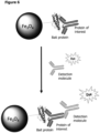
- this ligand exchange reaction may be performed with superparamagnetic iron-based particles previously coated with citrate ( Fig. 2 ).
- the superparamagnetic iron-based particles are linked to the catechol acid via the citrate.
- the superparamagnetic iron-based particles are coated with citrate following the protocol as described in Stein et al. Molecules 2020, 25(19 ) [8].
- the superparamagnetic iron-based particles may be coated with citrate, oxalic acid, ascorbic acid, amino acids, preferably citrate.
- citrate oxalic acid
- ascorbic acid amino acids
- amino acids preferably citrate.
- B has been bound to A directly in situ.
- an alkaline precipitation of the naive raw superparamagnetic iron-based particles may be performed so that the activated catechol acid may directly attach to the superparamagnetic iron-based particles.
- B has been bound to A and said bond has been described in the art such as in Zhot et al. [20].
- the complex may contacted with the sample in step b) for at least 5 minutes, preferably at least 10 minutes, more preferably at least 20 minutes, more preferably at least 30 minutes, even more preferably at least 45 minutes, even more preferably at least 1 hour, even more preferably at least 1.5 hours, and most preferably for about 2 hours.
- the complex is contacted for at most 24 hour, more preferably at most 18 hours, more preferably at most 14 hours, more preferably at most 12 hours, more preferably at most 10 hours, even more preferably at most 8 hours, even more preferably at most 6 hours, and even more preferably at most 4 hours.
- step (c) of the method the complex binding to the protein of interest of step b) is immobilized by a magnetic field.
- immobilization means that something is made immobile. In the present case, immobilization occurs by the influence of a magnetic field. This means that the magnetically attractable particles are immobilised by the magnet field.
- a suitable magnetic field can be determined by the skilled person. For example, a suitable magnetic field is capable of immobilizing the superparamagnetic iron based particles. Immobilization can be assessed e.g. by assessing the iron concentration in the supernatant sample.
- Exemplary magnetic field strengths applied in step (c) may be from 50-2000 mT, preferably wherein the magnetic field is from 80 to 1500 mT, more preferably wherein the magnetic field is from 100 mT to 1000 mT, even more preferably wherein the magnetic field is from 150 mT to 500 mT, even more preferably wherein the magnetic field is from 180 mT to 300 mT and most preferably wherein the magnetic field is around 210 mT.
- Immobilization of the superparamagnetic iron-based particles increases with increasing time periods. In light of the present disclosure, and in particular Example 1 and 2, the skilled person is able to determine suitable time periods for immobilization of the superparamagnetic iron-based particles.
- the superparamagnetic iron-based particles are immobilised in step (c) for a time period of at least 0.5 seconds, preferably at least 1 second, more preferably at least 2 seconds, even more preferably at least 3 seconds, more preferably at least 10 seconds, more preferably at least 20 seconds, more preferably at least 30 seconds, more preferably at least 45 seconds, more preferably at least 1 minute, even more preferably at least 2 minutes, most preferably at least 3 minutes preferably at least 5 minutes, more preferably at least 10 minutes, even more preferably at least 20 minutes, even more preferably at least 30 minutes, even more preferably at least 1 hour.
- the superparamagnetic iron-based particles are immobilised in step (c) for a time period of at most 180 minutes, preferably at most 120 minutes, more preferably at most 90 minutes, more preferably at most 60 minutes, and even more preferably at most 30 minutes.
- the skilled person can freely combine the preferred time ranges for immobilization as disclosed herein.
- the superparamagnetic iron-based particles are immobilised in step (c) for a time period from 15 seconds to 90 minutes, preferably from 30 seconds to 60 minutes, and more preferably from 1 minute to 10 minutes.
- the complex binding to the protein of interest of step c) is contacted with a detection molecule.
- a detection molecule may be a protein, such as an antibody, or an aptamere.
- the detection molecule is a protein, preferably an antibody, nanobody or camelid single domain antibody, preferably wherein the detection molecule is an antibody.
- the detection molecule may be an aptamere.
- the detection molecule comprises a detectable label, preferably wherein the detection molecule is covalently linked to a detectable label.
- the term 'label' as used herein refers to a molecule, which may be made detectable.
- the detectable label may be, preferably is, a fluorophore or a quantum dot, even more preferably wherein the detectable label is a fluorophore.
- the detection molecule is a fluorescently or quantumdot labelled molecule and even more preferably wherein the detectable label is a fluorescently labelled molecule.
- Fluorescently labelled molecules such as fluorescently labelled antibodies, are known to the skilled person and are also commercially available. In the context of the present invention, the detection molecule needs to be specific to an antigen of the protein of interest.
- the detection molecule may be detectable by a labelling molecule, preferably wherein the labelling molecule is linked to a detectable label, more preferably wherein the detectable label is a fluorophore or a quantum dot, even more preferably wherein the detectable label is a fluorophore, and most preferably wherein the labelling molecule is a fluorescently or quantum dot labelled molecule.
- the labelling molecule may be a protein or an aptamer. In case the labelling molecule is a protein, the labelling molecule is preferably an antibody, nanobody or camelid single domain antibody. It is even more preferred that the labelling molecule is an antibody.
- the labelling molecule is secondary antibody which is fluorescently labelled, binding to a primary antibody.
- typical detection and labelling molecules such as primary and secondary antibodies, as standardly used in mass spectrometry. Briefly, the detection molecule is directed against an antigen on the protein of interest, and the labelling molecule is directed against the detection molecule.
- Typical pairs of detection and labelling molecules are primary and secondary antibodies, wherein the secondary antibody specifically binds to the constant region of the primary antibody, which is specific to a species. For example, for a human sample, a primary antibody raised in mouse may be used and a secondary antibody against the mouse constant region raised in rabbit may be used.
- the identity of the protein of interest is determined by the presence of the label.
- the presence of the label is measured by a cell analyser, preferably by flow cytometry or by microcapillary cytometry, more preferably by flow cytometry.
- a cell analyser preferably by flow cytometry or by microcapillary cytometry, more preferably by flow cytometry.
- the skilled person is aware of standard cell analyser methods, which allow the detection of a label.
- the label is present if the signal of the label is at least 2-fold higher than the negative control, preferably 3-fold, more preferably 4-fold, even more preferably 5-fold, even more preferably 6-fold, even more preferably 7-fold, even more preferably 8-fold, even more preferably 9-fold, even more preferably 10-fold, even more preferably 12-fold, even more preferably 15-fold, even more preferably 20-fold, and at most 1000-fold.
- step a) has been obtained by following the steps of:
- step (ii) the activating group of the catechol acid is cleaved and the catechol acid is subsequently reacted with the bait molecule.
- Said order of the reaction has the advantage of controlling the steps of the reaction, since the pentafluorophenol can be readily detected by HPLC-UV.
- the iron weight amount of A is linked to one (1) weight amount of the bait molecule, preferably 7-15 times, more preferably, 8-13 times, even more preferably 9-11 times, and most preferably around 10 times.
- the iron weight amount can be determined by standard methods such as AES.
- the weight amount of the bait molecule can also be determined by standard methods.
- the bait molecule may be a bait protein and the concentration may be determined by UV absorbance, e.g. via NanoDrop, or Bradford Assay.
- 10-1000 ug iron of A is linked to 1-100 ug of the bait molecule (C), preferably wherein 20-500 ug iron of A is linked to 2-50 ug of C, more preferably wherein 40-200 ug iron of A is linked to 4-20 ug of C, even more preferably wherein 60-160 ug iron of A is linked to 6-16 ug of C, even more preferably wherein 80-130 ug iron of A is linked to 8-13 ug of C, and most preferably wherein around 100 ug iron of A is linked to around 10 ug of C.
- C the bait molecule
- the complex comprises, in particular consists of, (A) a (one) superparamagnetic iron-based particle, (B) one or more catechol acid moieties, and (C) one or more bait molecules, wherein the number of bait molecules is not greater than the number of catechol acid moieties.
- each catechol acid moiety (B) has been covalently linked to at most one bait molecule (C).
- catechol acid moieties do not form a sequential chain of catechol acid moieties to which the bait molecule is covalently linked. It is even more preferred that the number of bait molecules is equal to the number of catechol acid moieties.
- each catechol acid moiety (B) has been covalently linked to one bait molecule (C) forming a complex of the formula: A-[B-C] n , wherein n is at least 1, more preferably at most 200.
- the complex consists of a superparamagnetic iron-based particle (A), one or more catechol acid moieties (B), and one or more and bait molecules (C).
- A superparamagnetic iron-based particle
- B catechol acid moieties
- C bait molecules
- numerous complexes are required for the identification of numerous proteins of interest as not a single protein of interest can be detected in practice.
- Based on the molecular weight of the bait molecule more or less bait molecules may be bound to the particles.
- the amino group or thiol group of bovine serum albumin may be covalently bound to the complex of step (b) instead of the bait molecule and may thereby serve a negative control.
- BSA bovine serum albumin
- a complex comprising, preferably consisting of
- bait molecule the superparamagnetic iron-based particles, the catechol acid, and/or the amino group of thiol group of the protein of interest are discussed in this disclosure and apply equally to this aspect of the invention.
- a protein of interest may be bound to the bait molecule (C).
- the complex as disclosed in this aspect of the invention may comprise, preferably consist of A, one or more of B, one or more of C and additionally one or more of protein of interest (D).
- the protein of interest may be an antibody and/or the bait molecule may be an antibody.
- the complex consists of a superparamagnetic iron-based particle (A), one or more catechol acid moieties (B), and one or more bait molecules (C).
- B may have been bound to A by a ligand exchange reaction.
- B may have been bound to A in situ by alkaline precipitation, which are also discussed in this disclosure and which applies equally to this aspect of the disclosure.
- A may have been linked to a plurality of catechol acid moieties (B).
- the plurality of catechol acid moieties (B) may have been linked to a plurality of bait molecules (C).
- A may have been linked to a plurality of catechol acid moieties (B).
- B may have been linked to a plurality of bait molecules (C).
- C bait molecules
- one superparamagnetic iron-based particle (A) may be bound to one or more catechol acid moieties (B) in practice as several, i.e. a plurality of B can be bound to A.
- one bait molecule (C) can be covalently bound to one catechol acid moiety (B).
- one superparamagnetic iron-based particle (A) may be bound to one or more catechol acid moieties (B), and one or more bait molecules (C), wherein the number of B and C ideally the same.
- the complex comprises, in particular consists of, (A) a (one) superparamagnetic iron-based particle, (B) one or more catechol acid moieties, and (C) one or more bait molecules, wherein the number of bait molecules is not greater than the number of catechol acid moieties.
- each catechol acid moiety (B) has been covalently linked to at most one bait molecule (C).
- catechol acid moieties do not form a sequential chain of catechol acid moieties to which the bait molecule is covalently linked. It is even more preferred that the number of bait molecules is equal to the number of catechol acid moieties.
- each catechol acid moiety (B) has been covalently linked to one bait molecule (C) forming a complex of the formula: A-[B-C] n , wherein n is at least 1, more preferably at most 200.
- a method for diagnosing a disease or disorder, wherein a protein of interest is identified in a body fluid sample of a patient comprising the steps of:
- the presence of the protein of interest indicates that the patient has a risk of developing or suffers from said disease or disorder.
- exemplary diseases or disorders are infectious diseases or auto-immune disorders, preferably wherein the disease or disorder is an infectious disease.
- Preferred infectious diseases are caused by a virus, bacterium, fungus or protozoan.
- preferred viruses bacteria, fungi or protozoans may be selected from a pathogen as defined in this disclosure.
- preferred embodiments regarding the protein of interest, the sample, the superparamagnetic iron-based, the catechol acid, the amino group or thiol group of the protein of interest, and/or the bait molecule are defined in this disclosure and apply equally to this aspect of the invention.
- a method of preparing a complex comprising the steps of:
- a complex for identifying a protein of interest in an aqueous or body fluid sample is disclosed herein, wherein a comprises, preferably consists of,
- Preferred embodiments regarding the protein of interest, the aqueous or body fluid sample, the superparamagnetic iron-based particles, the catechol acid, the amino group or thiol group of the protein of interest, and/or the bait molecule are defined in this disclosed and apply equally to this aspect of the invention.
- the complex comprises, in particular consists of, (A) a (one) superparamagnetic iron-based particle, (B) one or more catechol acid moieties, and (C) one or more bait molecules, wherein the number of bait molecules is not greater than the number of catechol acid moieties.
- each catechol acid moiety (B) has been covalently linked to at most one bait molecule (C).
- catechol acid moieties do not form a sequential chain of catechol acid moieties to which the bait molecule is covalently linked. It is even more preferred that the number of bait molecules is equal to the number of catechol acid moieties.
- each catechol acid moiety (B) has been covalently linked to one bait molecule (C) forming a complex of the formula: A-[B-C] n , wherein n is at least 1, more preferably at most 200.
- kits for identifying a protein of interest in an aqueous or body fluid sample comprising a complex comprising, preferably consisting of:
- Preferred embodiments regarding the protein of interest, the aqueous or body fluid sample, the superparamagnetic iron-based particles, the catechol acid, the amino group or thiol group of the protein of interest, and/or the bait molecule are defined in this disclosed and apply equally to this aspect of the invention.
- the complex comprises, in particular consists of, (A) a (one) superparamagnetic iron-based particle, (B) one or more catechol acid moieties, and (C) one or more bait molecules, wherein the number of bait molecules is not greater than the number of catechol acid moieties.
- each catechol acid moiety (B) has been covalently linked to at most one bait molecule (C).
- catechol acid moieties do not form a sequential chain of catechol acid moieties to which the bait molecule is covalently linked. It is even more preferred that the number of bait molecules is equal to the number of catechol acid moieties.
- each catechol acid moiety (B) has been covalently linked to one bait molecule (C) forming a complex of the formula: A-[B-C] n , wherein n is at least 1, more preferably at most 200.
- the term 'functionalization' of a particle means the binding of e.g. the activated catechol acid or the antigen to the particle.
- the CAF-PFP functionalized particles are suitable to bind exposed amino groups in the SARS-CoV2 S1 spike or just the RBD of the spike-protein target by their activated ester function (3). The following procedure was applied. Before covalently binding the antigen, the SPION CAF-PFP was washed two times with 0,1 M borate buffer (pH 8).
- particles were magnetically separated and magnetically washed two times with PBS containing 0.1% BSA.
- virus protein exposing particles SPION-CAF-PFP-PROT v
- the virus protein exposing particles were dispersed again in 200 ⁇ l of sterile FACS-buffer containing PBS with 0.1% BSA and 0.02% sodium azide.
- Synthesis of the citrate-coated particles leads to particle sizes of 103 nm and increases to 117 nm hydrodynamic diameter after functionalization with CAF-PFP. Further immobilization with the S1 antigen or the RBD domain of the S1 protein leads to size increases of 256 and 238 nm, respectively. In contrast, the development of the zeta potential behaves inconsistently (Tab 1) (Fig., Fig. ).
- Table 1 Z-average, PDI and zetapotenial of used nanoparticles SPION CIT SPION CAF-PFP SPION CAF- PFP-S1 SPION CAF-PFP-S1 RBD Z-average in nm 103 117 ⁇ 1.0 256 ⁇ 4.5 237.6 ⁇ 3.6 PDI 0.15 0.473 0.425 ⁇ [mV] - 26 ⁇ 1.26 - 22.7 ⁇ 0.58 - 25.6 ⁇ 1.08
- Controls and samples were incubated with anti-SARS-Cov antibody (Chimeric MAb (mouse/human) SARS-CoV/Sars-CoV2 Spike, SinoBiological, Germany) with reactivity against Sars-CoV2 S1 and Sars-CoV2 S1-RBD and the dye labelled detection antibody (Donkey anti-mouse IgG (H+L) Alexa Fluor 488, Thermo Fisher Scientific, Germany) directed against the primary antibody, as indicated in Fig..
- Flow cytometry data revealed only background fluorescence in controls 1 and 2. In contrast, a high signal could be detected in samples one and two after sequential incubation with both antibodies. The signal difference between controls and samples was highly significant (p ⁇ 0.0001). The differences between the controls as well the samples themselves were not significant.
- the mouse antibody in the control panel did not induce a significant signal above the background ( Fig. 10 ).
- anti-human-IgG antibodies could be caught by the bait molecule bound particles (also called: antigen functionalized particles, column 11 and 12).
- Particles, which were only blocked with BSA (column 8,9 and 10) were not able to ligate the anti-human-IgG antibodies in a way which is significantly higher than non-specific adsorption.
- Non-specific adsorption, which was detectable for the detection antibody was also much below the level of selectively labelling of previously captured anti-human-IgG antibodies (column 14).
- the signal of the two confirmed Sars-CoV2 positive sera was significantly higher than the background and the samples with random binding, whereas the signal of the sample with known negative Sars-CoV2-status did not exceed the background signal.
- the data revealed a correct determination of patient sera having a positive or negative Sars-CoV2 status.
- the versatility of the principle becomes clear by transferring it to another complementary binding pair.
- This consists of an HIV surface protein as bait protein (ENV UG37gp140 (clade A), Polymun Scientific, Kleinneuburg, Austria) and the corresponding protein of interest (antibody, 2G12: recombinant human monoclonal antibody to HIV-1 gp120; Ab002, Polymun Scientific, Kleinneuburg, Austria) which is already labelled with Alexa TM 647 as a fluorescent dye. Therefore, no further antibody is needed in this setting.
- 175 ⁇ l SPION CAF-PFP Suspension at a concentration of 1 mg/ml was added.
- ENV conc.: 0.4 ⁇ g/ ⁇ l
- bovine serum albumin in the same concentration as negative control
- Fig. 11 shows that the envelope specific antibody readily bound to the bait protein and thus the protein of interest could be readily identified in the sample.
- the method is widely applicable, as it is a stochastic binding that addresses exposed amino groups or thiol groups in the target.
- a wide variety of antigens can be immobilized here, which can subsequently be detected by complementary antibodies.
- a fluorescent dye-labeled antibody as a detection molecule directed against the protein of interest, reliable values can be obtained e.g. by flow cytometry.
- Detection in human serum via detection antibody anti-human IgG Antibody, anti-human, VioBlue; order no: 130-119-967
- control antibody 130-113-205 Isotyp-control, mouse IgG1, VioBlue; Miltenyi Biotech GmbH, Bergisch Gladbach, Germany.
- CAF-PFP ( Fig. 1 ) is essentially based on the work of Williams et al.[7] (Supplementary Information), but slightly adapted.
- 790.2 mg caffeic acid are dissolved in 10 ml dimethylformamid. This is followed by the addition of 570 ⁇ l pyridine and then the addition of 1.21 ml pentafluorophenyl trifluoroacetate. The mixture is stirred for 2h at RT (300 rpm) and then diluted with 30 ml dichloromethane and transferred to a separation funnel. There it is washed 5 times with 25 ml 1 N HCI.
- the previously generated CAF-PFP active ester must first be bound to the particle surface. This functionalization is carried out in the form of a ligand exchange reaction with particles previously coated with citrate ( Fig. 2 ).
- Citrate-stabilized SPIONs were synthesized by a simple co-precipitation method described in our previous work [8].
- an aqueous solution of iron (II) and iron (III) salts containing 13.2 mg iron/ml is deoxygenized using argon.
- the precipitation of SPIONs is achieved by injecting 25% NH 3 (aq) into the vigorously stirring iron salt solution.
- 15 mL of a 293.3 mg/mL sodium citrate solution was added and the temperature was raised to 90 °C.
- the dispersion was further stirred for 30 min, before letting it cool to room temperature and transferring it into 10 kDa dialysis tubes.
- the dispersion was dialyzed for 6 h against H 2 O. Once the dialysis was done, the SPIONs were filtered through a 0.2 ⁇ m filter membrane (Minisart ® , Sartorius AG, Göttingen, Germany) to achieve the final citrate-stabilized SPIONs. The resulting particles are present as a stable aqueous suspension and have a hydrodynamic size of 99 nm and are ready for the subsequent CAF-PFP functionalization. 2 mg CAF-PFP are dissolved in 1 ml acetonitrile. The citrate-coated particles are dispersed in acetate buffer at pH 4 (0.1 M) at a concentration of 2 mg Fe/ ml.
- the particles functionalized with CAF-PFP are therefore stably suspended in aqueous medium, since there is still a proportion of citrate on the particle surface that has not been substituted by CAF-PFP.
Landscapes
- Health & Medical Sciences (AREA)
- Life Sciences & Earth Sciences (AREA)
- Immunology (AREA)
- Engineering & Computer Science (AREA)
- Hematology (AREA)
- Molecular Biology (AREA)
- Biomedical Technology (AREA)
- Chemical & Material Sciences (AREA)
- Urology & Nephrology (AREA)
- Biotechnology (AREA)
- Microbiology (AREA)
- Cell Biology (AREA)
- Food Science & Technology (AREA)
- Medicinal Chemistry (AREA)
- Physics & Mathematics (AREA)
- Analytical Chemistry (AREA)
- Biochemistry (AREA)
- General Health & Medical Sciences (AREA)
- General Physics & Mathematics (AREA)
- Pathology (AREA)
- Peptides Or Proteins (AREA)
Abstract
The present invention is concerned a method for identifying a protein of interest in an aqueous or body fluid sample. The identification is carried out by using a complex comprising (A) a superparamagnetic iron-based particle, (B) one or more catechol acid moieties, and (C) one or more bait molecules, wherein B has been bound to A, and wherein B has been covalently linked to C by activating the catechol acid and reacting said activated catechol acid with the amino group or the thiol group of C. The invention also relates to said complex as well as a use of said complex for identifying a protein of interest in an aqueous or body fluid sample. Finally, the invention also refers to a method for diagnosing a disease or disorder, wherein a protein of interest is identified in a body fluid sample of a patient by using said complex.
Description
- The stable binding of small molecules as well as large macromolecules to surfaces of nano-and microparticles is an important technique for therapeutic (e.g. drug delivery), diagnostic as well as other applications. For this purpose, iron oxide nanoparticles possess a unique versatility of attachment compared to other carriers or carrier-free applications [18]. In addition, the ability to be magnetically controllable is extremely advantageous for these particles, as washing steps and analyte concentration measures can be significantly simplified compared to conventional non-magnetic beads.
- For example, the assessment of antibody status under the conditions of the Sars-CoV2 pandemic is of great importance for epidemiological evaluation. This is true for both: recording the extent to which infection has progressed and for surveying immunization status as the population vaccination campaign progresses. While a number of diagnostic methods exist, they are often device-specific. For example, the Cobas Elecsys Anti-SARS-CoV-2 chemiluminescence immunoassay as discussed in Lippi et al. [19] is based on a sandwich reaction, wherein the test sample, biotinylated SARS-CoV-2-specific recombinant antigen, and SARS-CoV-2-specific recombinant antigen labeled with ruthenium are initially incubated altogether. After adding streptavidin-coated microparticles, the complex is bound to the solid phase through a biotin-streptavidin reaction and then aspirated into a measuring cell, where microparticles are magnetically captured. Hence, such an assay is limited to assay-specific equipment and assay-specific disposals and adjustments relating to the binding partners cannot be easily implemented. Moreover, it is questionable whether existing capacity is sufficient to meet the upcoming demand for antibody screening. The other category of assessing antibody status are Point of Care (POC) diagnostics, which is based on a test strip technology, which represent an immunological rapid detection of antigens immobilised on solid phases and possible antibodies in the sample material. Here, a detailed insight is even less precise and the statement can only be made according to a simple yes-no scheme. Thus, there is a need in the art for new, reliable and widely applicable methods to identify proteins of interest such as the antibody status of a patient.
- The use of flow cytometry is very suitable in terms of versatility and widespread use in clinics and, with nanoparticles as carriers for the analytes, offers excellent application potential. For this purpose, iron oxide nanoparticles have a unique versatility of attachment compared to other carriers or carrier-free applications. The surface with its specific iron-hydroxide arrangement offers the possibility that certain substances can attach very stably there [1]. Carboxyl groups, phosphates, phosphonates, or various polymers with hydroxyl groups or other functional groups are described in the literature, which contribute to stable coatings [20, 21]. In the case of OH groups, their large number in polymers is the main reason for stable additions. However, coating with small molecules is much more challenging, as it is subject to ligand exchange reactions under certain conditions. This is especially true for carboxyl groups. Stable coatings are achieved with small molecules only if several carboxyl groups act simultaneously on the particle surface [2, 3]. Phosphonates are considered as highly stable anchor groups as they form strong complexes with the iron hydroxide surface [1]. The availability and possibility of derivatization of these phosphonates is unfortunately limited, which is why further functionalization for versatile applications has been challenging so far.
- Widely used, for example, are coating strategies with dopamine, which, however, tends to auto-polymerize under certain circumstances [6] and thus complicates further functionalization. In addition, dopamine has only one amino group for further functionalization with possible target molecules, which is homologous to the amino groups of possible targets and is therefore only suitable to a limited extent for corresponding binding to such groups. For example, catechols have been described to have the ability to generate firm complexes with the iron hydroxide surface of the corresponding particles [4, 5].
- Hence, there is a need in the art for an optimized and robust linkage between the nanoparticle and a bait molecule, which can then reliable test for a protein of interest in a sample, e.g. the antibody status in a serum sample.
- The invention relates to a method for identifying a protein of interest in an aqueous or body fluid sample by using a complex comprising (A) a superparamagnetic iron-based particle, (B) one or more catechol acid moieties, and (C) one or more bait molecules, as further defined in the claims. Said complex allows a simple and robust detection of proteins of interest in an aqueous or body fluid sample. In these complexes, bait proteins, such as antigens (e.g. virus proteins), can be stably exposed on iron oxide-based nanoparticles. Proteins of interest, such as antibodies, can then be selectively detected upon binding to the bait protein and e.g. recorded in a flow cytometer. Thus, a versatile diagnostic platform is obtained, which can be used to detect protein of interest such as pathogens or autoantibodies, if a corresponding selective complementary binding partner, such as an antigen-antibody pair, exists.
- The method is widely applicable, as it involves stochastic binding of the (A) superparamagnetic iron-based particle with (B) catechol acid to exposed amino or thiol groups in the (C) bait protein. By using this method, a wide variety of antigens can be covalently linked to the superparamagnetic iron-based particles as bait molecules. Subsequently, a protein of interest, which is a complementary binding partner to the bait protein, can be detected. Using a detection molecule such as a fluorescent dye-labelled antibody directed against the protein of interest, the presence of the protein of interest can be reliably determined e.g. by flow cytometer. The main purpose of application is in vitro diagnostics.
- In this invention, activated catechols have been found to be especially advantageous, particularly when the catechols contain a carboxyl group, such as those found in derivatives of 3,4-dihydroxycinnamic acid, also known by the trivial name caffeic acid. This is because catechols have high affinities to metal oxide surfaces. Furthermore, many catechols are derived from known natural products (such as dopamine or caffeic acid) and are therefore well characterized and mostly non-toxic. Another example of carboxyl catechols is protocatechuic acid (3,4-dihydroxybenzoic acid). Caffeic acid has an acid group that can form a stable bond with corresponding nucleophiles. However, the acid group of catechol acid is generally not reactive enough for this reaction to occur under conditions that do not affect the nature of the particle system. The acid groups can be activated accordingly, but then the properties of the linker may be altered in such a way that the colloidal stability of the particle is no longer guaranteed. The activation of the catechol acid has the particular advantage that said activation is fully compatible with the binding to superparamagnetic iron-based particles and that the activated catechol acid readily reacts with exposed amino groups or via addition reaction of the unsaturated α,β position to the carboxyl with a thiol group on bait molecules. Specifically, activated catechol acid can be (i) readily bound to superparamagnetic iron-based particles by e.g. ligand replace reactions and (ii) covalently bound to amino or thiol groups of bait molecule, such as the Sars-CoV-2 antigen.
- For example, the inventors have activated caffeic acid with pentafluorophenyl trifluoroacetate, resulting in the reactive pentafluorophenol caffeic acid ester (CAF-PFP) (
Fig. 1 ). Furthermore, the inventors have covalently bound the Sars-CoV-2 antigen (Spike protein) as well as and HIV-envelope protein as bait proteins to the activated catechol acid. As a proof of principle, the inventors show a diagnostic particle system for the selective detection of e.g. Sars-CoV2 antibodies from patient blood, which has proven its performance in samples of Covid-19 of convalescents. Hence, the data revealed a correct determination of patient sera having a positive or negative Sars-CoV2 antibody status (Fig. 10 ). Similarly, the presence of anti-gp120 antibodies in a sample could be readily detected by using the envelope protein gp120 as a bait protein (Fig. 11 ) . - In a first aspect, a method for identifying a protein of interest in an aqueous or body fluid sample, the method comprising the steps of:
- a) providing a complex comprising
- (A) a superparamagnetic iron-based particle,
- (B) one or more catechol acid moieties, and
- (C) one or more bait molecules,
wherein B has been bound to A; and wherein B has been covalently linked to C by activating the catechol acid (benzene-1,2-diol acid) and reacting said activated catechol acid with an amino group or a thiol group of C;
- b) contacting the complex of step a) with the sample whereby the bait molecule (C) binds to the protein of interest;
- c) immobilizing the complex binding to the protein of interest of step b) by a magnetic field and thereby separating the protein of interest from the sample,
- d) contacting the complex binding to the protein of interest of step c) with a detection molecule, wherein the detection molecule binds to the protein of interest; wherein the detection molecule (i) comprises a detectable label or (ii) is detectable by a labelling molecule comprising the label,
- e) determining the presence of the label, thereby identifying the protein of interest.
- In general, the methods as disclosed herein are in vitro methods. In other words, said methods are performed ex vivo, in particular outside of the human body.
- As noted above, the present disclosure is concerned with a particularly advantageous linkage between the superparamagnetic iron-based particle (A) and the bait molecule (C). In general, A is connected to C via one or more catechol acid moieties (B). In particular, B has been bound to A.
- In general, the term 'catechol acid moiety' as used herein refers to a catechol acid portion, which is bound by A and C. In other words, an activated catechol acid being bound to A and C is a catechol acid moiety. In an especially preferred embodiment, the superparamagnetic iron-based particle (A) is linked to the one or more catechol acid moieties via citrate. For exemplary data, reference is made to Example 1 and Table 1. According to this disclosure, B has been covalently linked to C by activating the catechol acid (benzene-1,2-diol acid) and reacting said activated catechol acid with an amino group or thiol group of C. For example, the inventors have demonstrated such a linkage in Example 1 (Table 1). In said exemplary case, the bait molecule has been the SARS-
CoV 2 Spike protein. As an additional proof of concept, the envelope protein gp120 of HIV has been used as a bait molecule in - In particular, the catechol acid may comprise, preferably comprises, a carboxyl group, preferably wherein the catechol acid is caffeic acid (3,4-dihydroxycinnamic acid) or protocatechuic acid, more preferably wherein the catechol acid is caffeic acid. Caffeic acids has also been used in Examples 1 and 2 herein. However, the skilled person is able to determine other suitable catechol acids such as L-Dopa (L-3,4-dihydroxyphenylalanine), dihydroxybenzoic acid, dihydroxyindole-2-carboxylic acid. Preferably, the catechol acid has been activated to be a pentafluorophenyl catechol acid, terafluorophenyl catechol acid, or a catechol acid N-Hydroxysuccinimide ester, preferably wherein the catechol acid has been activated to be a pentafluorophenyl catechol acid, such as demonstrated in Example 1 herein.
- In general, the skilled person the skilled person is provided with several options to activate the catechol acid with. For example, the catechol acid may have been activated with a isocyanate, acyl azides, a sulfonyl chloride, a tosylate ester, an epoxide, an oxiranes, an imido ester, a carbodiimide, a fluorophenyl ester, or a O-methyl isourea. In case the catechol acid is caffeic acid, which is preferred, the caffeic acid may have been activated to be a pentafluorophenyl caffeic acid, terafluorophenyl caffeic acid, or a caffeic acid N-Hydroxysuccinimide ester. In an especially preferred embodiment, the catechol acid has been activated by pentafluorophenyl trifluoroacetate. It is further preferred that the catechol acid is caffeic acid and wherein pentafluorophenol caffeic acid ester has been formed by the activation of caffeic acid. In addition, the catechol acid may be caffeic acid and the caffeic acid may have been activated by pentafluorophenyl trifluoroacetate. Further assistance regarding the exact reaction conditions can be found in Example 1.
- As noted above, the particular advantage of this linkage between the iron-based particles and the bait molecule is that a linkage via a catechol moiety provides a versatile tool for linking bait molecules to the particles reliably. This is, for example, because the bait molecule merely requires an amino group or a thiol group. In addition, activated catechol groups are highly reactive with bait molecules such as proteins. Furthermore, activated catechol are easy to handle and usually cheap in comparison to other alternatives. Catechols have the property that they polymerize with one another, thus adhering more stably to the particle surface and at the same time do not lose their ability to bind to the bait molecules via the activated ester. In addition, the described linkage can be combined with any bait molecule - protein of interest pair, as long as the bait molecule comprises an amino group or a thiol groups. Thus, the described linkage harbors a broad applicability making said linkage a versatile toolkit.
- According to this disclosure, the protein of interest and the bait molecule generally have a high affinity to each other, such as a nanomolar affinity. The skilled person is aware of standard methods to determine the strength of macromolecular interactions, such as protein-protein interactions. For example, the skilled person may use SPR, electromobility shift assays, ITC, fluorescent polarization. Hence, particularly preferred are antibody-antigen interactions between the protein of interest and the bait molecule. It is preferred that the bait molecule comprises a binding site for the protein of interest, more preferably wherein the bait molecule provides an antigen for the protein of interest and wherein the protein of interest is an antibody.
- The term 'protein of interest' refers to any protein, which may be part of an aqueous or body fluid sample. In the context of the bait molecule (C), the protein of interest may also be regarded as the 'prey', which is sought after by the bait protein. In particular, proteins of interest may be antibodies or pathogen proteins. Such a pathogen protein may be a viral protein, a bacterial protein, a fungal protein, or a protozoan protein. In general, the skilled person is aware of such proteins and which diseases such proteins of interest are indicative for. For example, the viral protein may be from an enveloped or a non-enveloped virus. Preferably the viral protein is from an enveloped virus. More specifically, the enveloped virus may be a coronavirus, retrovirus, herpesvirus, poxvirus, hepadnavirus, african swine fever and related viruses, flavivirus, alphavirus, togavirus, hepatitis D virus, orthomyxovirus, paramyxovirus, rhabdovirus, bunyavirus, or filovirus. Even more in particular, the enveloped virus may be a coronavirus, preferably a orthocoronavirus, more preferably a betacoronavirus, even more preferably a sarbecovirus, even more preferably a SARS-coronavirus or a MERS-coronavirus, even more preferably a SARS coronavirus, and most preferably SARS-
coronavirus 2. Alternatively, the virus protein may be from a retrovirus such as HIV. It is preferred that the retrovirus is an orthoretrovirus, preferably a lentivirus, more preferable human immunodeficiency viruses (HIV). As a further alternative, the virus protein may be from a non-enveloped virus. Exemplary virus proteins from non-enveloped viruses are from an adenovirus, astrovirus, circovirus, hepatitis E-like virus, papovavirus, parvovirus, picornavirus, or reovirus. - As noted above, the protein of interest may be a pathogen protein, wherein the pathogen is a bacterium. Exemplary bacterial proteins are selected from one of the bacteria Actinomyces israelii, Bacillus anthracis, Bacteroides fragilis, Bordetella pertussis, Borrelia, Brucella, Campylobacter jejuni, Chlamydia, Chlamydophila psittaci, Clostridium, Corynebacterium diphtheria, Ehrlichia, Enterococcus, Escherichia, Francisella tularensis, Haemophilus influenza, Helicobacter pylori, Klebsiella pneumonia, Legionella pneumophila, Leptospira species, Listeria monocytogenes, Mycobacterium, Mycoplasma pneumonia, Neisseria, Pseudomonas aeruginosa, Nocardia asteroids, Rickettsia rickettsia, Salmonella, Shigella, Staphylococcus, Streptococcus, Vibrio cholera, or Yersinia pestis. Similarly, the protein of interest may be a pathogen protein, wherein the pathogen is a fungus. Exemplary fungal proteins are selected from one of the fungi Candida, Aspergillus, Cryptococcus, Histoplasma, Pneumocystis, or Stachybotrys. As noted above, the protein of interest may be a pathogen protein, wherein the pathogen protein is a protozoan protein. Exemplary protozoan proteins are selected from one of Diplomodas, Parabasilia, Euglenozoa, Entamoebida, Stramenopila, Alveolata, Rhizaria, or Archaeplastida.
- In another embodiment, the protein of interest is a biomarker. This is particularly advantageous as a patient may be diagnosed or monitored for a disease by analyzing a bodily fluid samples such as a serum sample. In case the biomarker is identified, the patient may be monitored more specifically. Preferred biomarkers, which may be proteins of interest are a tumorigenic biomarker or metabolic biomarker, more preferably wherein the biomarker is selected from a list consisting of S100 calcium-binding protein B, MIA protein (melanoma inhibitory activity), Prostate-specific antigen (PSA), human chorionic gonadotropin (hCG), Her2neu, Plastin-3, EGFR, carcinoembryonic antigen (CEA), cancer antigen 125 (CA 125), Carbohydrate antigen 19-9 (CA 19-9), Carbohydrate antigen 15-3 (CA 15-3), alpha-fetoprotein (AFP), Microfibril Associated Protein 4 (MFAP4), or ApoA-1. In particular, Her2neu, Plastin-3 and/or EGFR can be biomarkers for circulating tumor cells (CTCs).
- In a particularly preferred embodiment, the protein of interest is an antibody. As known in the art, are proteins produced in response to and counteracting a specific antigen. This may, for example, occur in response to a vaccination. In particular, antibodies against Sars-CoV-2 may be formed in response to a vaccination. Especially, antibodies against the spike protein of SarS-
CoV 2 may be formed in response to a vaccination. Alternatively, the antibodies may be formed in response to a natural infection. Furthermore, the antibody status allows physicians and scientist to monitor the antibody positive and negative rate in the population over time. Hence, the method as disclosed in this application is useful in assessing e.g. the antibody status against SarS-Cov2 as demonstrated in Example 1,Fig. 10 . As the linkage between the iron-based particles and the bait molecule merely requires the bait molecule to have an amino group or a thiol group, said linking mechanism is widely applicable. In a preferred embodiment, the protein of interest is a neutralizing antibody. - In a preferred embodiment, the protein of interest is an antibody having been formed in response to an infection by a pathogen or a vaccination against said pathogen. For example, antibodies against Sars-CoV2 are formed in response to an infection or vaccination. In general, said antibodies may have been formed in response to a viral or bacterial pathogen. Other examples of a possible pathogens, against which antibodies may have been formed, include Sars-CoV2, corynebacterium diphtheriae, haemophilus influenzae type b, hepatitis B virus, measles virus, meningococcus (Neisseria meningitidis), mumps virus, bordetella pertussis, pneumococcus (Streptococcus pneumoniae), poliovirus, rotavirus, rubella virus, Clostridium tetani, varicella zoster virus, human papillomavirus, herpes zoster, and influenza virus. Further example of pathogens, against which antibodies may have been formed, may be enveloped or a non-enveloped viruses, preferably an enveloped virus. For example, the enveloped virus, against which antibodies may have been formed, may be a coronavirus, retrovirus, herpesvirus, poxvirus, hepadnavirus, african swine fever and related viruses, flavivirus, alphavirus, togavirus, hepatitis D virus, orthomyxovirus, paramyxovirus, rhabdovirus, bunyavirus, or filovirus. Specifically, said enveloped virus may be coronavirus, preferably wherein the coronavirus is a orthocoronavirus, preferably a betacoronavirus, more preferably a sarbecovirus, even more preferably a SARS-coronavirus, and most preferably SARS-
coronavirus 2. - In another preferred embodiment, the protein of interest is an auto-antibody. The skilled person is aware of auto-antibodies, which generally form in the context of an autoimmune disease. Specifically, the auto-antibody may be selected from a list consisting of antinuclear antibodies, anti-transglutaminase antibodies, anti-ganglioside antibodies, anti-thrombin antibodies, anti-thyroid autoantibodies, anti-myelin basic protein, anti-myelin oligodendrocyte glycoprotein, anti-citrullinated protein, anti-rheumatoid factor, antimicrosomal antibodies, anti-thyroid plasma membrane antibodies, IgA endomysium antibodies, IgA tissue transglutaminase antibodies, anti-Human Cardiac Myosin antibodies, anti-thyroid peroxidase antibodies, anti-thyroglobulin antibodies, anti-Saccharomyces cerevisiae antibodies, anti-flagellin (CBir1), anti-neutrophil cytoplasmic antibodies, anti-glutamic acid decarboxylase antibodies, thyroid receptor autoantibodies (TRAK), anti-acetylcholine receptor, anti-double-stranded DNA antibodies, anti-Sm, anti-histone antibodies, anti-type IV collagen antibodies, anti-Scl-70 antibodies, anti-centromere antibodies, Anti-Jo-1 antibodies, anti-Proteinase 3 antibodies, anti-Plakoglobin antibodies, anti-Desmoglein III antibodies, and anti-Sjögren's-syndrome-related antigen A autoantibodies.
- The term 'bait molecule', as used herein, is a macromolecule, which is used to catch a protein of interest. As noted above, the protein of interest and the bait protein have a high affinity for each other such as nanomolar affinity. Hence, when the bait molecule is contacted with the sample containing the protein of interest so that the protein of interest binds to the bait protein. It is preferred that the bait molecule is a protein. For example, the bait protein may be a pathogen protein, preferably the bait protein may a viral, bacterial, fungal or protozoan pathogen protein. In other words, the bait protein may be from a viral, bacterial, fungal or protozoan pathogen. For example, Example 1 and 2 demonstrates that the pathogen proteins Sars-CoV-2 (spike protein) or HIV (GP120) may be used as bait proteins. Other possible bait proteins include viral protein from enveloped or non-enveloped viruses. In case the virus is an enveloped virus, the virus may be selected from a list consisting of coronavirus, retrovirus, herpesvirus, poxvirus, hepadnavirus, african swine fever and related viruses, flavivirus, alphavirus, togavirus, hepatitis D virus, orthomyxovirus, paramyxovirus, rhabdovirus, bunyavirus, or filovirus. Specifically, the enveloped virus may be a coronavirus, preferably wherein the coronavirus is a orthocoronavirus, preferably a betacoronavirus, more preferably a sarbecovirus, even more preferably a SARS-coronavirus or a MERS-coronavirus, even more preferably a SARS coronavirus, and most preferably SARS-
coronavirus 2. Particular reference is made to Example 1,Figure 10 , showing that the spike protein of SARS-CoV2 can be used as a bait protein to identify Sars-CoV-2 antibodies in serum, i.e. antibodies against the spike protein. Alternatively, the bait protein may be from a retrovirus such as an orthoretrovirus, preferably wherein the retrovirus may be a lentivirus, more preferably wherein the retrovirus is human immunodeficiency viruses (HIV). Particular reference is made to Example 2,Figure 11 , showing that the envelope protein GP120 of HIV can be used as a bait protein to identify anti-HIV antibodies in solution, i.e. antibodies against the GP120. Alternatively, the bait protein may be from a non-enveloped virus such as an adenovirus, astrovirus, circovirus, hepatitis E-like virus, papovavirus, parvovirus, picornavirus, or reovirus. In addition the bait protein may be a bacterial protein from a bacterial pathogen, such as a bacterial protein is selected from one of the bacteria Actinomyces israelii, Bacillus anthracis, Bacteroides fragilis, Bordetella pertussis, Borrelia, Brucella, Campylobacter jejuni, Chlamydia, Chlamydophila psittaci, Clostridium, Corynebacterium diphtheria, Ehrlichia, Enterococcus, Escherichia, Francisella tularensis, Haemophilus influenza, Helicobacter pylori, Klebsiella pneumonia, Legionella pneumophila, Leptospira species, Listeria monocytogenes, Mycobacterium, Mycoplasma pneumonia, Neisseria, Pseudomonas aeruginosa, Nocardia asteroids, Rickettsia rickettsia, Salmonella, Shigella, Staphylococcus, Streptococcus, Vibrio cholera, or Yersinia pestis. Similarly, the bait protein may be a fungal protein, preferably wherein the fungal protein is selected from one of the fungi Candida, Aspergillus, Cryptococcus, Histoplasma, Pneumocystis, or Stachybotrys. Additionally, the bait protein may be a protozoan protein, preferably wherein the protozoan protein is from Diplomodas, Parabasilia, Euglenozoa, Entamoebida, Stramenopila, Alveolata, Rhizaria, or Archaeplastida. - It is also contemplated that the bait protein is an antibody, especially an antibody binding to a protein of interest as defined in the embodiments concerning the protein of interest in this application. Alternatively, the bait molecule may bind to a tag on the protein of interest. Exemplary tags include a Strep-Tag, SNAP-Tag, Clig-Tag, His-Tag, aldehyde Tag, FLAG-Tag, Flash-Tag, Thioreduxin-Tag or wherein the bait protein comprises protein A.
- Furthermore, the bait molecule can be a bait aptamere. As known in the art, an aptamere is short, single-stranded DNA or RNA (ssDNA or ssRNA) molecule that can selectively bind to a specific target, including proteins, peptides, carbohydrates, small molecules, and toxins. Aptamer are capable of targeting a range from small organic molecules, ions, small peptides [11,12], large proteins [13-17], and whole cells and viruses. Aptamers usually show affinities in the pM to low nM range. Protein targets include viral proteins, cytokines, enzymes, and transcription factors. In the context of this application, a 'bait aptamere' bind to the protein of interest. Preferably, the bait aptamere bind to the protein of interest with an affinity lower than 100 nM. The skilled person is aware of suitable methods to detect the affinity of an aptamere-protein interaction such as change of the refractive index in surface plasmon resonance (SPR) by e.g. Cytiva Biacore 8K; Surface plasmon resonance imaging (SPRi) e.g. by Horiba XelPlex; or Bio-Layer Interferometry e.g. by an Octet or Octet Red (ForteBio) in comparison to a reference sample. Furthermore, aptameres which can be used as bait aptameres are commercially available. In addition, there are selective methods such as SELEX to engineer aptamers very efficiently and with high binding constants [22, 23]. Particularly preferred aptameres are bait aptameres binding to any of the proteins of interest as defined in this application. Exemplary bait aptameres bind to VEGF, Sars-CoV2 or cocaine or the exemplary bait aptamere is Pegaptanib or BC 007.
- It is further contemplated that the amino group of the bait molecule is an N-terminal amino group, an amino group of a lysine, or an amino group of an arginine. In general, the amino group may be covalently linked to the catechol acids via an amid synthesis or a Michael reaction. Specifically, the reaction can occur following the principles of an amid synthesis as known in the art (see also
Fig. 5A for illustration). Briefly, the acid group, such as the carboxylic acid group of the catechol acid is coupled with an amine, specifically an amino group of the bait molecule. For a direct reaction, e.g. between a carboxyl group and an amino group to form an amide, high temperatures are generally required. Hence, the catechol acid is activated by converting it to a better electrophile, as detailed in this disclosure. Alternatively, the bait molecule might be covalently linked to the activated catechol acid via a thiol group. For example, the thiol group may be part of a cysteine. In this case, the bait molecule is a bait protein harboring one or more cysteines. - The amino group or the thiol group may be reacting with the activated catechol acid via a Michael reaction (
Fig. 5B ). In general, the Michael reaction, also called Michael addition is the nucleophilic addition of a nucleophile to an α,β-unsaturated carbonyl compound containing an electron withdrawing group and the skilled person is aware of the Michael reaction. - The "sample", as used herein is an aqueous of body fluid sample. In general, an "aqueous solution" is a solution in which the solvent is water. In other words, an aqueous solution is water soluble and the skilled person is aware of aqueous solutions. Exemplary aqueous solutions are waste water, ground water, or drinking water. Furthermore, a "body fluid" is any fluid, which occurs in or is excrete from a body. Especially preferred are samples from a human body. In particular, the sample may be a body fluid sample, which may be selected from blood, serum, plasma, lymph, urine, liquor, saliva and sputum, even more preferably wherein the human body fluid is blood or serum, and even more preferably wherein the human body fluid is blood. If the sample is blood of a body, it is preferred that the blood sample is a mammalian blood sample, more preferably a primate blood sample, and even more preferably a human blood sample. If the sample is serum of a body, it is preferred that the serum sample is a mammalian serum sample, more preferably a primate serum sample, and even more preferably a human serum sample. Additionally, the mammalian blood or serum samples may be from a group selected from cow, pig, rabbit, sheep, goat, mouse, rat, cat, dog, mouse, and rat. Advantageously, the method as disclosed herein can also be used on samples from farm animals as well as pets.
- In general, "superparamagnetic iron-based particles" as used herein are particles, which are magnetically attractable. The skilled person is aware that the magnetic behaviour of materials can be classified into five major groups: ferro-, ferri-, para-, antiferro-, and diamagnetism. Ferro- and ferrimagnetism are the basic mechanisms in permanent magnets. Permanent magnets can be magnetized by external magnetic fields and remain magnetized after the removal of the fields. Materials such as Fe (iron), Co, Ni, and their alloys, and some compounds of rare earth elements are ferromagnetic. Superparamagnetism is a form of magnetism, which appears in small ferromagnetic or ferrimagnetic particles. "Particles" as used herein are defined as particles of matter that are from 1 nm (nanometer) to 2 micrometers (µm) in diameter. Furthermore, the skilled person is aware of iron (Fe) based particles, such as ironoxide based particles, which have been used in biological applications. The skilled person is also aware of various iron-based particles. For example, the particles may be iron oxide, iron (Fe), iron-cobalt, alnico or permalloy particles. In other words, the superparamagnetic iron-based particles may be selected from iron oxide, iron (Fe), ironcobalt, alnico, and permalloy particles. In a preferred embodiment, the superparamagnetic iron-based particles are superparamagnetic ironoxide nanoparticles (SPIONs). A "nanoparticle", as used herein, is a particle of matter that is between 1 nm and 1 µm in diameter. Furthermore, the superparamagnetic iron-based particles may be superparamagnetic ironoxide nanoparticles (SPIONs), more preferably wherein the iron oxide is selected from Fe3O4, γ-Fe2O3, CoFe2O4 or a mixture thereof, more preferably wherein the iron oxide is Fe3O4. In a preferred embodiment, the concentration of the superparamagnetic iron-based particles is determined by atomic emission spectroscopy (AES). The iron content in a solution may, for example, be determined with Atomic Emission Spectroscopy (AES). The skilled person is also aware of other methods in order to determine the iron content in a sample. For example, the concentration of iron in a sample can also be determined photometrically. However, a determination of the iron concentration by AES is preferred.
- According to this disclosure, B has been bound to A (the superparamagnetic iron-based particle). Preferably, the catechol acid moiety (B) has been bound to the superparamagnetic iron-based particle (A) by a ligand exchange reaction. For example, this ligand exchange reaction may be performed with superparamagnetic iron-based particles previously coated with citrate (
Fig. 2 ). Further reference is made to Example 1 herein. Preferably, the superparamagnetic iron-based particles are linked to the catechol acid via the citrate. In a particularly preferred embodiment, the superparamagnetic iron-based particles are coated with citrate following the protocol as described in Stein et al. Molecules 2020, 25(19) [8]. Alternatively, the superparamagnetic iron-based particles may be coated with citrate, oxalic acid, ascorbic acid, amino acids, preferably citrate. The advantage of a ligand exchange reaction is that it is easily applicable as shown in Example 1 herein. - Alternatively, B has been bound to A directly in situ. For example, an alkaline precipitation of the naive raw superparamagnetic iron-based particles may be performed so that the activated catechol acid may directly attach to the superparamagnetic iron-based particles. Furthermore, B has been bound to A and said bond has been described in the art such as in Zeiniger et al. [20].
- According to step (b) of the method, the complex may contacted with the sample in step b) for at least 5 minutes, preferably at least 10 minutes, more preferably at least 20 minutes, more preferably at least 30 minutes, even more preferably at least 45 minutes, even more preferably at least 1 hour, even more preferably at least 1.5 hours, and most preferably for about 2 hours. In a preferred embodiment, the complex is contacted for at most 24 hour, more preferably at most 18 hours, more preferably at most 14 hours, more preferably at most 12 hours, more preferably at most 10 hours, even more preferably at most 8 hours, even more preferably at most 6 hours, and even more preferably at most 4 hours.
- In step (c) of the method, the complex binding to the protein of interest of step b) is immobilized by a magnetic field. "Immobilization", as used herein, means that something is made immobile. In the present case, immobilization occurs by the influence of a magnetic field. This means that the magnetically attractable particles are immobilised by the magnet field. A suitable magnetic field can be determined by the skilled person. For example, a suitable magnetic field is capable of immobilizing the superparamagnetic iron based particles. Immobilization can be assessed e.g. by assessing the iron concentration in the supernatant sample. Exemplary magnetic field strengths applied in step (c) may be from 50-2000 mT, preferably wherein the magnetic field is from 80 to 1500 mT, more preferably wherein the magnetic field is from 100 mT to 1000 mT, even more preferably wherein the magnetic field is from 150 mT to 500 mT, even more preferably wherein the magnetic field is from 180 mT to 300 mT and most preferably wherein the magnetic field is around 210 mT. Immobilization of the superparamagnetic iron-based particles increases with increasing time periods. In light of the present disclosure, and in particular Example 1 and 2, the skilled person is able to determine suitable time periods for immobilization of the superparamagnetic iron-based particles. In a preferred embodiment, the superparamagnetic iron-based particles are immobilised in step (c) for a time period of at least 0.5 seconds, preferably at least 1 second, more preferably at least 2 seconds, even more preferably at least 3 seconds, more preferably at least 10 seconds, more preferably at least 20 seconds, more preferably at least 30 seconds, more preferably at least 45 seconds, more preferably at least 1 minute, even more preferably at least 2 minutes, most preferably at least 3 minutes preferably at least 5 minutes, more preferably at least 10 minutes, even more preferably at least 20 minutes, even more preferably at least 30 minutes, even more preferably at least 1 hour. It is also preferred that the superparamagnetic iron-based particles are immobilised in step (c) for a time period of at most 180 minutes, preferably at most 120 minutes, more preferably at most 90 minutes, more preferably at most 60 minutes, and even more preferably at most 30 minutes. Of course, the skilled person can freely combine the preferred time ranges for immobilization as disclosed herein. In a strongly preferred embodiment, the superparamagnetic iron-based particles are immobilised in step (c) for a time period from 15 seconds to 90 minutes, preferably from 30 seconds to 60 minutes, and more preferably from 1 minute to 10 minutes.
- According to step (d) of the method as disclosed herein, the complex binding to the protein of interest of step c) is contacted with a detection molecule. Such a detection molecule may be a protein, such as an antibody, or an aptamere. In a preferred embodiment, the detection molecule is a protein, preferably an antibody, nanobody or camelid single domain antibody, preferably wherein the detection molecule is an antibody. Alternatively, the detection molecule may be an aptamere. Preferably, the detection molecule comprises a detectable label, preferably wherein the detection molecule is covalently linked to a detectable label. The term 'label' as used herein refers to a molecule, which may be made detectable. Preferred are labels, which can be detected by e.g. light emission upon light excitation such as fluorescent labels. The skilled person is aware of such suitable labels in the art. For example, the detectable label may be, preferably is, a fluorophore or a quantum dot, even more preferably wherein the detectable label is a fluorophore. In a particularly preferred embodiment, the detection molecule is a fluorescently or quantumdot labelled molecule and even more preferably wherein the detectable label is a fluorescently labelled molecule. Fluorescently labelled molecules, such as fluorescently labelled antibodies, are known to the skilled person and are also commercially available. In the context of the present invention, the detection molecule needs to be specific to an antigen of the protein of interest. Thus, particularly preferred are fluorescently labelled antibodies as detection molecules. Alternatively, the detection molecule may be detectable by a labelling molecule, preferably wherein the labelling molecule is linked to a detectable label, more preferably wherein the detectable label is a fluorophore or a quantum dot, even more preferably wherein the detectable label is a fluorophore, and most preferably wherein the labelling molecule is a fluorescently or quantum dot labelled molecule. The labelling molecule may be a protein or an aptamer. In case the labelling molecule is a protein, the labelling molecule is preferably an antibody, nanobody or camelid single domain antibody. It is even more preferred that the labelling molecule is an antibody.
- In an especially preferred embodiment, the labelling molecule is secondary antibody which is fluorescently labelled, binding to a primary antibody. In practice, the skilled person is aware of typical detection and labelling molecules such as primary and secondary antibodies, as standardly used in mass spectrometry. Briefly, the detection molecule is directed against an antigen on the protein of interest, and the labelling molecule is directed against the detection molecule. Typical pairs of detection and labelling molecules are primary and secondary antibodies, wherein the secondary antibody specifically binds to the constant region of the primary antibody, which is specific to a species. For example, for a human sample, a primary antibody raised in mouse may be used and a secondary antibody against the mouse constant region raised in rabbit may be used.
- According to the present disclosure, the identity of the protein of interest is determined by the presence of the label. Ideally, the presence of the label is measured by a cell analyser, preferably by flow cytometry or by microcapillary cytometry, more preferably by flow cytometry. The skilled person is aware of standard cell analyser methods, which allow the detection of a label. Ideally, the label is present if the signal of the label is at least 2-fold higher than the negative control, preferably 3-fold, more preferably 4-fold, even more preferably 5-fold, even more preferably 6-fold, even more preferably 7-fold, even more preferably 8-fold, even more preferably 9-fold, even more preferably 10-fold, even more preferably 12-fold, even more preferably 15-fold, even more preferably 20-fold, and at most 1000-fold.
- It is further contemplated that the complex of step a) has been obtained by following the steps of:
- i. activating the catechol acid (B; benzene-1,2-diol acid);
- ii. linking the activated catechol acid (B) to the superparamagnetic iron-based particle (A), and
- iii. reacting said activated catechol acid with an amino group of the bait molecule (C).
- It is further contemplated that after step (ii), the activating group of the catechol acid is cleaved and the catechol acid is subsequently reacted with the bait molecule. Said order of the reaction has the advantage of controlling the steps of the reaction, since the pentafluorophenol can be readily detected by HPLC-UV.
- When carrying out the method as disclosed herein, it is preferred that 5-20 times of the iron weight amount of A is linked to one (1) weight amount of the bait molecule, preferably 7-15 times, more preferably, 8-13 times, even more preferably 9-11 times, and most preferably around 10 times. The iron weight amount can be determined by standard methods such as AES. Furthermore, the weight amount of the bait molecule can also be determined by standard methods. For example, the bait molecule may be a bait protein and the concentration may be determined by UV absorbance, e.g. via NanoDrop, or Bradford Assay.
- It is even more preferred that 10-1000 ug iron of A is linked to 1-100 ug of the bait molecule (C), preferably wherein 20-500 ug iron of A is linked to 2-50 ug of C, more preferably wherein 40-200 ug iron of A is linked to 4-20 ug of C, even more preferably wherein 60-160 ug iron of A is linked to 6-16 ug of C, even more preferably wherein 80-130 ug iron of A is linked to 8-13 ug of C, and most preferably wherein around 100 ug iron of A is linked to around 10 ug of C.
- Additionally, it is preferred that the complex comprises, in particular consists of, (A) a (one) superparamagnetic iron-based particle, (B) one or more catechol acid moieties, and (C) one or more bait molecules, wherein the number of bait molecules is not greater than the number of catechol acid moieties. This means that each catechol acid moiety (B) has been covalently linked to at most one bait molecule (C). In particular, catechol acid moieties do not form a sequential chain of catechol acid moieties to which the bait molecule is covalently linked. It is even more preferred that the number of bait molecules is equal to the number of catechol acid moieties. This means that each catechol acid moiety (B) has been covalently linked to one bait molecule (C) forming a complex of the formula: A-[B-C]n, wherein n is at least 1, more preferably at most 200.
- It is also preferred that the complex consists of a superparamagnetic iron-based particle (A), one or more catechol acid moieties (B), and one or more and bait molecules (C). In practice, numerous complexes are required for the identification of numerous proteins of interest as not a single protein of interest can be detected in practice. Based on the molecular weight of the bait molecule more or less bait molecules may be bound to the particles. The examples section as well as the preferred embodiments above provide guidance with regard to suitable amounts for A, B and C.
- As quality control measure, the amino group or thiol group of bovine serum albumin (BSA) may be covalently bound to the complex of step (b) instead of the bait molecule and may thereby serve a negative control. Example 1 shows that such a negative control is useful to in order to determine the background noise of the detection method.
- In a further aspect of the invention, a complex comprising, preferably consisting of
- (A) a superparamagnetic iron-based particle,
- (B) one or more catechol acid moieties, and
- (C) one or more bait molecules,
- The preferred embodiments regarding the bait molecule, the superparamagnetic iron-based particles, the catechol acid, and/or the amino group of thiol group of the protein of interest are discussed in this disclosure and apply equally to this aspect of the invention.
- Additionally, a protein of interest may be bound to the bait molecule (C). In other words, the complex as disclosed in this aspect of the invention may comprise, preferably consist of A, one or more of B, one or more of C and additionally one or more of protein of interest (D). Preferred embodiments regarding the protein of interest are further defined in this disclosure and apply equally to this aspect. In particular, the protein of interest may be an antibody and/or the bait molecule may be an antibody. In a very preferred embodiment, the complex consists of a superparamagnetic iron-based particle (A), one or more catechol acid moieties (B), and one or more bait molecules (C). For example, B may have been bound to A by a ligand exchange reaction. Preferred ligand exchange reactions are discussed in this disclosure and apply equally to this aspect of the invention. Alternatively, B may have been bound to A in situ by alkaline precipitation, which are also discussed in this disclosure and which applies equally to this aspect of the disclosure. Specifically, A may have been linked to a plurality of catechol acid moieties (B). Additionally, the plurality of catechol acid moieties (B) may have been linked to a plurality of bait molecules (C). The skilled person understands that one superparamagnetic iron-based particle (A) may be bound to one or more catechol acid moieties (B) in practice as several, i.e. a plurality of B can be bound to A. In turn, the skilled person also understands that one bait molecule (C) can be covalently bound to one catechol acid moiety (B). Hence, one superparamagnetic iron-based particle (A), may be bound to one or more catechol acid moieties (B), and one or more bait molecules (C), wherein the number of B and C ideally the same.
- Furthermore, it is preferred that the complex comprises, in particular consists of, (A) a (one) superparamagnetic iron-based particle, (B) one or more catechol acid moieties, and (C) one or more bait molecules, wherein the number of bait molecules is not greater than the number of catechol acid moieties. This means that each catechol acid moiety (B) has been covalently linked to at most one bait molecule (C). In particular, catechol acid moieties do not form a sequential chain of catechol acid moieties to which the bait molecule is covalently linked. It is even more preferred that the number of bait molecules is equal to the number of catechol acid moieties. This means that each catechol acid moiety (B) has been covalently linked to one bait molecule (C) forming a complex of the formula: A-[B-C]n, wherein n is at least 1, more preferably at most 200.
- Additionally, a method for diagnosing a disease or disorder, wherein a protein of interest is identified in a body fluid sample of a patient is disclosed herein, wherein the method comprising the steps of:
- a) providing a complex comprising, preferably consisting of,
- (A) a superparamagnetic iron-based particle,
- (B) one or more catechol acid moieties, and
- (C) one or more bait molecules,
wherein B has been bound to A; and wherein B has been covalently linked to C by activating the catechol acid (benzene-1,2-diol acid) and reacting said activated catechol acid with amino groups of C;
- b) contacting the complex of step a) with the sample whereby the bait molecule (C) binds to the protein of interest;
- c) immobilizing the complex binding to the protein of interest of step b) by a magnetic field and thereby separating the protein of interest from the sample,
- d) contacting the complex binding to the protein of interest of step c) with a detection molecule, wherein the detection molecule binds to the protein of interest; wherein the detection molecule (i) comprises a detectable label or (ii) is detectable by a labelling molecule comprising the label,
- e) determining the presence of the label, thereby identifying the protein of interest.
- Preferably, the presence of the protein of interest indicates that the patient has a risk of developing or suffers from said disease or disorder. Exemplary diseases or disorders are infectious diseases or auto-immune disorders, preferably wherein the disease or disorder is an infectious disease. Preferred infectious diseases are caused by a virus, bacterium, fungus or protozoan. In addition, preferred viruses bacteria, fungi or protozoans may be selected from a pathogen as defined in this disclosure. Moreover, preferred embodiments regarding the protein of interest, the sample, the superparamagnetic iron-based, the catechol acid, the amino group or thiol group of the protein of interest, and/or the bait molecule are defined in this disclosure and apply equally to this aspect of the invention.
- In a further aspect, a method of preparing a complex is contemplated, wherein the method comprising the steps of:
- I. providing a superparamagnetic iron-based particle (A),
- II. activating catechol acid (B; benzene-1,2-diol acid);
- III. linking the activated catechol acid (B) to A, and
- IV. reacting said activated catechol acid with an amino group or thiol group of a bait molecule (C).
- Preferred embodiments regarding the superparamagnetic iron-based particles, the catechol acid, and/or the bait molecule are defined in this disclosed and apply equally to this aspect of the invention.
- Furthermore, a use of a complex for identifying a protein of interest in an aqueous or body fluid sample is disclosed herein, wherein a comprises, preferably consists of,
- (A) a superparamagnetic iron-based particle,
- (B) one or more catechol acid moieties, and
- (C) one or more bait molecules,
- wherein B has been bound to A; and wherein B has been covalently linked to C by activating the catechol acid (benzene-1,2-diol acid) and reacting said activated catechol acid with an amino group or thiol group of C;
- and wherein the protein of interest is capable of binding to the bait molecule.
- Preferred embodiments regarding the protein of interest, the aqueous or body fluid sample, the superparamagnetic iron-based particles, the catechol acid, the amino group or thiol group of the protein of interest, and/or the bait molecule, are defined in this disclosed and apply equally to this aspect of the invention.
- Furthermore, it is preferred that the complex comprises, in particular consists of, (A) a (one) superparamagnetic iron-based particle, (B) one or more catechol acid moieties, and (C) one or more bait molecules, wherein the number of bait molecules is not greater than the number of catechol acid moieties. This means that each catechol acid moiety (B) has been covalently linked to at most one bait molecule (C). In particular, catechol acid moieties do not form a sequential chain of catechol acid moieties to which the bait molecule is covalently linked. It is even more preferred that the number of bait molecules is equal to the number of catechol acid moieties. This means that each catechol acid moiety (B) has been covalently linked to one bait molecule (C) forming a complex of the formula: A-[B-C]n, wherein n is at least 1, more preferably at most 200.
- Moreover, a kit for identifying a protein of interest in an aqueous or body fluid sample is contemplated, wherein the kit comprises a complex comprising, preferably consisting of:
- (A) a superparamagnetic iron-based particle,
- (B) one or more catechol acid moieties, and
- (C) one or more bait molecules,
- wherein B has been bound to A; and wherein B has been covalently linked to C by activating the catechol acid (benzene-1,2-diol acid) and reacting said activated catechol acid with amino groups of C;
- and wherein the protein of interest is capable of binding to the bait molecule
- Preferred embodiments regarding the protein of interest, the aqueous or body fluid sample, the superparamagnetic iron-based particles, the catechol acid, the amino group or thiol group of the protein of interest, and/or the bait molecule, are defined in this disclosed and apply equally to this aspect of the invention.
- Furthermore, it is preferred that the complex comprises, in particular consists of, (A) a (one) superparamagnetic iron-based particle, (B) one or more catechol acid moieties, and (C) one or more bait molecules, wherein the number of bait molecules is not greater than the number of catechol acid moieties. This means that each catechol acid moiety (B) has been covalently linked to at most one bait molecule (C). In particular, catechol acid moieties do not form a sequential chain of catechol acid moieties to which the bait molecule is covalently linked. It is even more preferred that the number of bait molecules is equal to the number of catechol acid moieties. This means that each catechol acid moiety (B) has been covalently linked to one bait molecule (C) forming a complex of the formula: A-[B-C]n, wherein n is at least 1, more preferably at most 200.
- The invention is further described by the following embodiments:
- 1. A method for identifying a protein of interest in an aqueous or body fluid sample, the method comprising the steps of:
- a) providing a complex comprising
- (A) a superparamagnetic iron-based particle,
- (B) one or more catechol acid moieties, and
- (C) one or more bait molecules,
- b) contacting the complex of step a) with the sample whereby the bait molecule (C) binds to the protein of interest;
- c) immobilizing the complex binding to the protein of interest of step b) by a magnetic field and thereby separating the protein of interest from the sample,
- d) contacting the complex binding to the protein of interest of step c) with a detection molecule, wherein the detection molecule binds to the protein of interest; wherein the detection molecule (i) comprises a detectable label or (ii) is detectable by a labelling molecule comprising the label,
- e) determining the presence of the label,
thereby identifying the protein of interest.
- a) providing a complex comprising
- 2. The method of
embodiment 1, wherein the protein of interest is a pathogen protein, preferably wherein the pathogen protein is a viral protein, a bacterial protein, a fungal protein, or a protozoan protein. - 3. The method of
embodiment 2, wherein the viral protein is from an enveloped or a non-enveloped virus, preferably wherein the viral protein is from an enveloped virus. - 4. The method of
embodiment 3, wherein the enveloped virus is a coronavirus, retrovirus, herpesvirus, poxvirus, hepadnavirus, african swine fever and related viruses, flavivirus, alphavirus, togavirus, hepatitis D virus, orthomyxovirus, paramyxovirus, rhabdovirus, bunyavirus, or filovirus. - 5. The method of
embodiment 4, wherein the enveloped virus is a coronavirus, preferably wherein the coronavirus is a orthocoronavirus, preferably a betacoronavirus, more preferably a sarbecovirus, even more preferably a SARS-coronavirus or a MERS-coronavirus, even more preferably a SARS coronavirus, and most preferably SARS-coronavirus 2. - 6. The method of
embodiment 4, wherein the retrovirus is an orthoretrovirus, preferably wherein the retrovirus is a lentivirus, more preferable wherein the retrovirus is human immunodeficiency viruses (HIV). - 7. The method of
embodiment 3, wherein the non-enveloped virus is an adenovirus, astrovirus, circovirus, hepatitis E-like virus, papovavirus, parvovirus, picornavirus, or reovirus. - 8. The method of
embodiment 2, wherein the pathogen protein is a bacterial protein, preferably wherein the bacterial protein is selected from one of the bacteria Actinomyces israelii, Bacillus anthracis, Bacteroides fragilis, Bordetella pertussis, Borrelia, Brucella, Campylobacter jejuni, Chlamydia, Chlamydophila psittaci, Clostridium, Corynebacterium diphtheria, Ehrlichia, Enterococcus, Escherichia, Francisella tularensis, Haemophilus influenza, Helicobacter pylori, Klebsiella pneumonia, Legionella pneumophila, Leptospira species, Listeria monocytogenes, Mycobacterium, Mycoplasma pneumonia, Neisseria, Pseudomonas aeruginosa, Nocardia asteroids, Rickettsia rickettsia, Salmonella, Shigella, Staphylococcus, Streptococcus, Vibrio cholera, or Yersinia pestis. - 9. The method of
embodiment 2, wherein the pathogen protein is a fungal protein, preferably wherein the fungal protein is selected from one of the fungi Candida, Aspergillus, Cryptococcus, Histoplasma, Pneumocystis, or Stachybotrys. - 10. The method of
embodiment 2, wherein the pathogen protein is a protozoan protein, preferably wherein the protozoan protein is from Diplomodas, Parabasilia, Euglenozoa, Entamoebida, Stramenopila, Alveolata, Rhizaria, or Archaeplastida. - 11. The method of
embodiment 1, wherein the protein of interest is a biomarker, preferably a tumorigenic biomarker or metabolic biomarker, more preferably wherein the biomarker is selected from a list consisting of S100 calcium-binding protein B, MIA (melanoma inhibitory activity), Prostate-specific antigen (PSA), human chorionic gonadotropin (hCG), circulating tumor cells (CTCs), carcinoembryonic antigen (CEA), cancer antigen 125 (CA 125), Carbohydrate antigen 19-9 (CA 19-9), Carbohydrate antigen 15-3 (CA 15-3), alpha-fetoprotein (AFP), Microfibril Associated Protein 4 (MFAP4), and ApoA-1. - 12. The method of
embodiment 1, wherein the protein of interest is an antibody, preferably wherein the antibody is a neutralizing antibody. - 13. The method of
embodiment 12, wherein the protein of interest is an antibody and wherein said antibody binds to the bait molecule. - 14. The method of any of
embodiments - 15. The method of
embodiment 14, wherein the pathogen is a viral or bacterial pathogen. - 16. The method of
embodiment 15, wherein the pathogen is selected from a list consisting of Sars-CoV2, corynebacterium diphtheriae, haemophilus influenzae type b, hepatitis B virus, measles virus, meningococcus (Neisseria meningitidis), mumps virus, bordetella pertussis, pneumococcus (Streptococcus pneumoniae), poliovirus, rotavirus, rubella virus, Clostridium tetani, varicella zoster virus, human papillomavirus, herpes zoster, and influenza virus. - 17. The method of
embodiment 15, wherein the pathogen is a viral pathogen, preferably wherein the viral pathogen is an enveloped or a non-enveloped virus, preferably wherein the viral pathogen is an enveloped virus. - 18. The method of embodiment 17, wherein the enveloped virus is a coronavirus, retrovirus, herpesvirus, poxvirus, hepadnavirus, african swine fever and related viruses, flavivirus, alphavirus, togavirus, hepatitis D virus, orthomyxovirus, paramyxovirus, rhabdovirus, bunyavirus, or filovirus.
- 19. The method of embodiment 18, wherein the enveloped virus is a coronavirus, preferably wherein the coronavirus is a orthocoronavirus, preferably a betacoronavirus, more preferably a sarbecovirus, even more preferably a SARS-coronavirus, and most preferably SARS-
coronavirus 2. - 20. The method of any of
embodiments - 21. The method of any of the preceding embodiments, wherein the amino group of the bait molecule is an N-terminal amino group, an amino group of a lysine, or an amino group of an arginine; and/or the thiol group of the bait molecule is a thiol group of a cysteine.
- 22. The method of any of the preceding embodiments, wherein the bait molecule comprises a binding site for the protein of interest, preferably wherein the bait molecule provides an antigen for the protein of interest and wherein the protein of interest is an antibody.
- 23. The method of any of the preceding embodiments, wherein the bait molecule is a protein, preferably a pathogen protein, more preferably wherein the pathogen protein is a viral protein, a bacterial protein, a fungal protein, or a protozoan protein.
- 24. The method of embodiment 23, wherein the viral protein is from an enveloped or a non-enveloped virus, preferably wherein the viral protein is from an enveloped virus.
- 25. The method of embodiment 24, wherein the enveloped virus is a coronavirus, retrovirus, herpesvirus, poxvirus, hepadnavirus, african swine fever and related viruses, flavivirus, alphavirus, togavirus, hepatitis D virus, orthomyxovirus, paramyxovirus, rhabdovirus, bunyavirus, or filovirus.
- 26. The method of
embodiment 25, wherein the enveloped virus is a coronavirus, preferably wherein the coronavirus is a orthocoronavirus, preferably a betacoronavirus, more preferably a sarbecovirus, even more preferably a SARS-coronavirus or a MERS-coronavirus, even more preferably a SARS coronavirus, and most preferably SARS-coronavirus 2 . - 27. The method of
embodiment 25, wherein the retrovirus is an orthoretrovirus, preferably wherein the retrovirus is a lentivirus, more preferable wherein the retrovirus is human immunodeficiency viruses (HIV). - 28. The method of embodiment 24, wherein the non-enveloped virus is an adenovirus, astrovirus, circovirus, hepatitis E-like virus, papovavirus, parvovirus, picornavirus, or reovirus.
- 29. The method of embodiment 23, wherein the pathogen protein is a bacterial protein, preferably wherein the bacterial protein is selected from one of the bacteria Actinomyces israelii, Bacillus anthracis, Bacteroides fragilis, Bordetella pertussis, Borrelia, Brucella, Campylobacter jejuni, Chlamydia, Chlamydophila psittaci, Clostridium, Corynebacterium diphtheria, Ehrlichia, Enterococcus, Escherichia, Francisella tularensis, Haemophilus influenza, Helicobacter pylori, Klebsiella pneumonia, Legionella pneumophila, Leptospira species, Listeria monocytogenes, Mycobacterium, Mycoplasma pneumonia, Neisseria, Pseudomonas aeruginosa, Nocardia asteroids, Rickettsia rickettsia, Salmonella, Shigella, Staphylococcus, Streptococcus, Vibrio cholera, or Yersinia pestis.
- 30. The method of embodiment 23, wherein the pathogen protein is a fungal protein, preferably wherein the fungal protein is selected from one of the fungi Candida, Aspergillus, Cryptococcus, Histoplasma, Pneumocystis, or Stachybotrys.
- 31. The method of embodiment 23, wherein the pathogen protein is a protozoan protein, preferably wherein the protozoan protein is from Diplomodas, Parabasilia, Euglenozoa, Entamoebida, Stramenopila, Alveolata, Rhizaria, or Archaeplastida.
- 32. The method of any of the preceding embodiments, wherein the bait molecule is an antibody, preferably wherein the bait molecule is an antibody binding to a protein of interest as defined in any of embodiments 2-20.
- 33. The method of any of the preceding embodiments, wherein the bait molecule binds to a tag on the protein of interest, preferably wherein the tag is a Strep-Tag, SNAP-Tag, Clig-Tag, His-Tag, Aldehyde Tag, FLAG-Tag, Flash-Tag, Thioreduxin-Tag or wherein the bait protein comprises protein A.
- 34. The method of any of the preceding embodiments, wherein the bait molecule is a bait aptamere.
- 35. The method of embodiment 34, wherein the bait aptamere bind to the protein of interest with an affinity lower than 100 nM.
- 36. The method of
embodiments 34 or 35, wherein the bait aptamere binds to the any of the protein of interest as defined in embodiments 2-20. - 37. The method of any of embodiments 33-36, wherein the bait aptamere binds to VEGF, Sars-CoV2 or cocaine; or wherein the bait aptamere is Pegaptanib or BC 007.
- 38. The method of any of the preceding embodiments, wherein the aqueous or body fluid sample is a body fluid, waste water, ground water, or drinking water, preferably wherein the body fluid sample is a human body fluid, more preferably wherein the human body fluid is selected from blood, serum, plasma, lymph, urine, liquor, saliva and sputum, even more preferably wherein the human body fluid is blood or serum, and even more preferably wherein the human body fluid is blood.
- 39. The method of any of the preceding embodiments, wherein the sample is a blood sample, preferably wherein the blood sample is a mammalian blood sample, more preferably a primate blood sample, and even more preferably a human blood sample.
- 40. The method of any of the preceding embodiments, wherein the superparamagnetic iron-based particle (A) has been bound to catechol acid moieties (B) by a ligand exchange reaction.
- 41. The method of any of the preceding embodiments, wherein the superparamagnetic iron-based particles are selected from an iron oxide, iron (Fe), iron-cobalt, alnico, permalloy particles.
- 42. The method of any of the preceding embodiments, wherein the superparamagnetic iron-based particles are superparamagnetic ironoxide nanoparticles (SPIONs).
- 43. The method of any of the preceding embodiments, wherein the superparamagnetic iron-based particles are superparamagnetic ironoxide nanoparticles (SPIONs), more preferably wherein the iron oxide is selected from Fe3O4, γ-Fe2O3, CoFe2O4 or a mixture thereof, more preferably wherein the iron oxide is Fe3O4.
- 44. The method of any of the preceding embodiments, wherein the superparamagnetic iron-based particles are coated with citrate, oxalic acid, ascorbic acid, amino acids, preferably citrate.
- 45. The method of embodiment 44, wherein the superparamagnetic iron-based particles are coated with citrate following the protocol as described in Stein et al. Molecules 2020, 25(19).
- 46. The method of embodiments 44 or 45, wherein the superparamagnetic iron-based particles are linked to the catechol acid via the citrate.
- 47. The method of any of the preceding embodiments, wherein the concentration of the superparamagnetic iron-based particles is determined by atomic emission spectroscopy.
- 48. The method of any of the preceding embodiments, wherein the catechol acid comprises a carboxyl group, preferably wherein the catechol acid is caffeic acid (3,4-dihydroxycinnamic acid) or protocatechuic acid, more preferably wherein the catechol acid is caffeic acid.
- 49. The method of any of the preceding embodiments, wherein the catechol acid has been activated to be a pentafluorophenyl catechol acid, terafluorophenyl catechol acid, or a catechol acid N-Hydroxysuccinimide ester, preferably wherein the catechol acid has been activated to be a pentafluorophenyl catechol acid.
- 50. The method of any of the preceding embodiments, wherein the catechol acid has been activated with a isocyanate, acyl azides, a sulfonyl chloride, a tosylate ester, an epoxide, an oxiranes, an imido ester, a carbodiimide, a fluorophenyl ester, or a O-methyl isourea.
- 51. The method of any of the preceding embodiments, wherein the catechol acid is caffeic acid, preferably wherein the caffeic acid has been activated to be a pentafluorophenyl caffeic acid, terafluorophenyl caffeic acid, or a caffeic acid N-Hydroxysuccinimide ester.
- 52. The method of any of the preceding embodiments, wherein the catechol acid has been activated by pentafluorophenyl trifluoroacetate.
- 53. The method of any of the preceding embodiments, wherein the catechol acid is caffeic acid and wherein pentafluorophenol caffeic acid ester has been formed by the activation of caffeic acid.
- 54. The method of any of the preceding embodiments, wherein the catechol acid is caffeic acid and wherein the caffeic acid has been activated by pentafluorophenyl trifluoroacetate.
- 55. The method of any of the preceding embodiments, wherein the complex is contacted with the sample in step b) for at least 5 minutes, preferably at least 10 minutes, more preferably at least 20 minutes, more preferably at least 30 minutes, even more preferably at least 45 minutes, even more preferably at least 1 hour, even more preferably at least 1.5 hours, and most preferably for about 2 hours.
- 56. The method of any of the preceding embodiments, wherein the complex is contacted with the sample in step b) for at most 10 hours, preferably at most 8 hours, more preferably at most 6 hours, more preferably at most 5 hours, more preferably at most 4 hours, more preferably at most 3 hours, and most preferably about 2 hours.
- 57. The method of any of the preceding embodiments, wherein the magnetic field applied in step (c) is from 50-2000 mT, preferably wherein the magnetic field is from 80 to 1500 mT, more preferably wherein the magnetic field is from 100 mT to 1000 mT, even more preferably wherein the magnetic field is from 150 mT to 500 mT, even more preferably wherein the magnetic field is from 180 mT to 300 mT and most preferably wherein the magnetic field is around 210 mT.
- 58. The method of any of the preceding embodiments, wherein the complex of step (c) is contacted with the detection molecule for at least 1 minute, preferably at least 2 minutes, preferably at least 5 minutes, more preferably at least 10 minutes, even more preferably at least 20 minutes, even more preferably at least 30 minutes, even more preferably at least 1 hour, and most preferably for about 1 hour.
- 59. The method of any of the preceding embodiments, wherein the detection molecule is a protein or an aptamer.
- 60. The method of embodiment 59, wherein the detection molecule is a protein, preferably an antibody, a nanobody or a camelid single domain antibody, preferably wherein the detection molecule is an antibody.
- 61. The method of embodiment 59, wherein the detection molecule is an aptamere.
- 62. The method of any of the preceding embodiments, wherein the detection molecule comprises a detectable label, preferably wherein the detection molecule is covalently linked to a detectable label.
- 63. The method of embodiment 62, wherein the detectable label is a fluorophore or a quantum dot, even more preferably wherein the detectable label is a fluorophore, even more preferably wherein the detection molecule is a fluorescently or quantumdot labelled molecule and even more preferably wherein the detectable label is a fluorescently labelled molecule.
- 64. The method of any of embodiments 1-61, wherein the detection molecule is detectable by a labelling molecule, preferably wherein the labelling molecule is linked to a detectable label, more preferably wherein the detectable label is a fluorophore or a quantum dot, even more preferably wherein the detectable label is a fluorophore, and most preferably wherein the labelling molecule is a fluorescently or quantum dot labelled molecule.
- 65. The method of any of the preceding embodiments, wherein the presence of the label molecule is measured by a cell analyser, preferably by flow cytometry or by microcapillary cytometry, more preferably by flow cytometry.
- 66. The method of any of the preceding embodiments, wherein the labelling molecule is a protein or an aptamer.
- 67. The method of embodiment 66, wherein the labelling molecule is a protein, preferably an antibody, nanobody or camelid single domain antibody, preferably wherein the labelling molecule is an antibody, more preferably a fluorescently labelled antibody.
- 68. The method of embodiment 66, wherein the labelling molecule is an aptamere.
- 69. The method of any of the preceding embodiments, wherein the label is present if the signal of the label is at least 2-fold higher than the negative control, preferably, preferably 3-fold, more preferably 4-fold, even more preferably 5-fold, even more preferably 6-fold, even more preferably 7-fold, even more preferably 8-fold, even more preferably 9-fold, even more preferably 10-fold, even more preferably 12-fold, even more preferably 15-fold, even more preferably 20-fold, and at most 1000-fold.
- 70. The method of any of the preceding embodiments, wherein the complex of step a) has been obtained by following the steps of:
- i. activating the catechol acid (B; benzene-1,2-diol acid);
- ii. linking the activated catechol acid (B) to the superparamagnetic iron-based particle (A), and
- iii. reacting said activated catechol acid with an amino group or the thiol group of a bait molecule (C).
- 71. The method of any of the preceding embodiments, wherein 5-20 times of the iron weight amount of A is linked to one weight amount of the bait molecule, preferably 7-15 times, more preferably, 8-13 times, even more preferably 9-11 times, and most preferably around 10 times.
- 72. The method of any of the preceding embodiments, wherein 10-1000 ug iron of A is linked to 1-100 ug of C, preferably wherein 20-500 ug iron of A is linked to 2-50 ug of C, more preferably wherein 40-200 ug iron of A is linked to 4-20 ug of C, even more preferably wherein 60-160 ug iron of A is linked to 6-16 ug of C, even more preferably wherein 80-130 ug iron of A is linked to 8-13 ug of C, and most preferably wherein around 100 ug iron of A is linked to around 10 ug of C.
- 73. The method of any of the preceding embodiments, wherein the complex consists of a superparamagnetic iron-based particle (A), catechol acid moieties (B), and bait molecules (C).
- 74. The method of any of the preceding embodiments, wherein the amino group or the thiol group of bovine serum albumin (BSA) is covalently bound to the complex of step (b) instead of the bait molecule and thereby serves a negative control.
- 75. A complex comprising,
- (A) a superparamagnetic iron-based particle,
- (B) one or more catechol acid moieties, and
- (C) one or more bait molecules,
- 76. The complex of embodiment 75, wherein the bait molecule is defined in any of embodiments 22-37, the superparamagnetic iron-based particles are defined in any of embodiments 40-47, the catechol acid is defined in any of embodiments 48-54, and/or the amino group or the thiol group of the protein of interest is defined in embodiment 21.
- 77. The complex of embodiments 75 or 76, wherein additionally a protein of interest is bound to the bait molecule, preferably wherein the protein of interest is further defined in any of embodiments 2-20.
- 78. The complex of any of embodiments 75-77, wherein the protein of interest is an antibody and/or wherein the bait molecule is an antibody.
- 79. The complex of any of embodiments 75-78, wherein the complex consists of a superparamagnetic iron-based particle (A), catechol acid moieties (B), and bait molecules (C).
- 80. The complex of any of embodiments 75-79, wherein B has been bound to A by a ligand exchange reaction.
- 81. The complex of any of embodiments 75-80, wherein A has been linked to a plurality of catechol acid moieties (B).
- 82. The complex of any of embodiments 75-81, wherein the plurality of catechol acid moieties (B) is linked to a plurality of bait molecules (C).
- 83. A method for diagnosing a disease or disorder, wherein a protein of interest is identified in a body fluid sample of a patient, the method comprising the steps of:
- a) providing a complex comprising
- (A) a superparamagnetic iron-based particle,
- (B) one or more catechol acid moieties, and
- (C) one or more bait molecules,
wherein B has been bound to A; and wherein B has been covalently linked to C by activating the catechol acid (benzene-1,2-diol acid) and reacting said activated catechol acid with the amino group or the thiol group of C;
- b) contacting the complex of step a) with the sample whereby the bait molecule (C) binds to the protein of interest;
- c) immobilizing the complex binding to the protein of interest of step b) by a magnetic field and thereby separating the protein of interest from the sample,
- d) contacting the complex binding to the protein of interest of step c) with a detection molecule, wherein the detection molecule binds to the protein of interest; wherein the detection molecule (i) comprises a detectable label or (ii) is detectable by a labelling molecule comprising the label,
- e) determining the presence of the label, thereby identifying the protein of interest.
- a) providing a complex comprising
- 84. The method of embodiment 83, wherein the presence of the protein of interest indicates that the patient has a risk of developing or suffers from said disease or disorder.
- 85. The method of embodiments 83 or 84, wherein the disease or disorder is an infectious disease or auto-immune disorder, preferably wherein the disease or disorder is an infectious disease.
- 86. The method of any of embodiments 83-85, wherein the disease or disorder is caused by a virus, bacterium, fungus or protozoan.
- 87. The method embodiment 86, wherein the virus, bacterium, fungus or protozoan is selected from a pathogen as defined in any of embodiments 3-10 and 15-19.
- 88. The method of any of embodiments 83-87, wherein the protein of interest is defined in any of embodiments 2-20, the sample is defined in any of embodiments 38-39, the superparamagnetic iron-based particles are defined in any of embodiments 40-47, the catechol acid is defined in any of embodiments 48-54, the amino group or the thiol group of the protein of interest is defined in embodiment 21, and/or the bait molecule is defined in embodiment 22-37.
- 89. A method of preparing a complex, the method comprising the steps of:
- I. providing a superparamagnetic iron-based particle (A),
- II. activating catechol acid (B; benzene-1,2-diol acid);
- III. linking the activated catechol acid (B) to A, and
- IV. reacting said activated catechol acid with an amino group or the thiol group of a bait molecule (C).
- 90. The method of embodiment 89, wherein the superparamagnetic iron-based particles are defined in any of embodiments 40-47, the catechol acid is defined in any of embodiments 48-54, and/or the bait molecule is defined in any of embodiment 22-37.
- 91. Use of a complex for identifying a protein of interest in an aqueous or body fluid sample, wherein a comprises
- (A) a superparamagnetic iron-based particle,
- (B) one or more catechol acid moieties, and
- (C) one or more bait molecules,
- wherein B has been bound to A; and wherein B has been covalently linked to C by activating the catechol acid (benzene-1,2-diol acid) and reacting said activated catechol acid with an amino group or the thiol group of C;
- and wherein the protein of interest is capable of binding to the bait molecule.
- 92. The use of embodiment 91, wherein the protein of interest is defined in any of embodiments 2-20, the sample is defined in any of embodiments 38-39, the superparamagnetic iron-based particles are defined in any of embodiments 40-47, the catechol acid is defined in any of embodiments 48-54, the amino group or the thiol group of the protein of interest is defined in embodiment 21, the bait molecule is defined in any of embodiments 22-37, and/or wherein the magnetic field is defined in embodiment 57.
- 93. Kit for identifying a protein of interest in an aqueous or body fluid sample, wherein the kit comprises a complex comprising:
- (A) a superparamagnetic iron-based particle,
- (B) one or more catechol acid moieties, and
- (C) one or more bait molecules,
- wherein B has been bound to A; and wherein B has been covalently linked to C by activating the catechol acid (benzene-1,2-diol acid) and reacting said activated catechol acid with the amino group or the thiol group of C;
- and wherein the protein of interest is capable of binding to the bait molecule
- 94. The kit of embodiment 93, wherein the protein of interest is defined in any of embodiments 2-20, the sample is defined in any of embodiments 38-39, the superparamagnetic iron-based particles are defined in any of embodiments 40-47, the catechol acid is defined in any of embodiments 48-54, and/or the amino group or the thiol group of the protein of interest is defined in embodiment 21.
-
-
Fig. 1 : Activation of caffeic acid with pentafluorophenyl trifluoroacetate (PFP-TFA). -
Fig. 2 : Iron oxide nanoparticles with a hybrid coating of citrate and pentafluorophenyl caffeate (CAF-PFP) -
Fig. 3 : Binding of viral protein via correspondingly exposed amino groups to the surface of the iron oxide particles equipped with CAF-PFP. -
Fig. 4 : Detection of the protein of interest sought in the sample material. -
Fig. 5 : Amid reaction (a) and Michael reaction (b) as cartoon representation. -
Fig. 6 : Labelling of the formed 'particle-bait protein-protein of interest' composite by a dye-labelled detection antibody that specifically binds to the protein of interest. -
Fig. 7 : Z-average in nm of the different nanoparticles. Starting with citrate coated particles SPIONCit (103 nm) the functionalisation with/binding of CAF-PFP (SPIONCAF-PFP) results in a size increase to 117 nm. Size progression to SPIONCAF-PFP to the particles that bound S1 protein or the RBD domain of Sars-CoV-2 as antigen (SPIONCAF-PFP-S1; SPIONCAF-PFP-RBD S1) yields in size increases of 256 nm and 238 nm, respectively. -
Fig. 8 : The development of the zeta potential behaves inconsistently. The zeta potential varies from - 27.4 mV to -26.5 mV and additional immobilisation of components in the particle shell are not reflected in zeta potential changes. -
Fig. 9 : Control 1 (SPIONCAF-PFP): SPIONs only modified with the linker molecule at the surface. Control 2 (SPIONCAF-PFPBSA): SPIONs with linker bound BSA; probe 1 (SPIONCAF-PFP-S1): SPIONs with linker bound Sars-CoV2-S1 (SinoBiological, Germany). Sample 2 (SPIONCAF-PFP-RBD): SPIONs with the linker bound RBD (receptor binding domain) (SinoBiological, Germany) of the Sars-CoV2-S1-protein. -
Fig. 10 : Determination of anti-Sars-IgG-antibodies in three human patient sera using FACS-analysis. Panel 1 (control-panel): SPIONCAF-PFP-particles, SPIONCAF-PFP-BSA-particles and SPIONCAF-PFP-S1-particles incubated with patient sera 1-3 and the detection antibody directed against the mouse antibody from the in vitro experiments inFig. 9 . Panel 2: Incubation of the three particle types with the three patient sera and an antibody directed against human IgG-antibodies. Positively detected sera are serum1 andserum 2.Serum 3 was correctly identified as a negative sample. -
Fig. 11 : FACS measurement: binding of ENV antigen (ENV=bright columns) or BSA as control (dark column) and labelling reaction with the 2G12 antibody. The left pair of columns shows the fluorescence base signal that is generated when no antibody is added. Addition of the antibody shows specific binding of the complementary antibody in the form of a highly significant increased fluorescence signal. The following sequence of labelling is present in the left column of the right pair of columns: SPIONCAF-PFP+ ENV antigen + antibody. In the right column, on the other hand, this labelling sequence is present: SPIONCAF-PFP+ BSA + antibody. Here, the antibody is only adsorbed non-specifically. - The following examples are intended to illustrate the invention further, but are not limited to it. The examples describe technical features, and the invention also relates to combinations of the technical features presented in this section.
- In this Example, the covalent binding of SARS-CoV-2 Antigen to SPIONCAF-PFP is described. The term 'functionalization' of a particle means the binding of e.g. the activated catechol acid or the antigen to the particle. The CAF-PFP functionalized particles are suitable to bind exposed amino groups in the SARS-CoV2 S1 spike or just the RBD of the spike-protein target by their activated ester function (3). The following procedure was applied. Before covalently binding the antigen, the SPIONCAF-PFP was washed two times with 0,1 M borate buffer (pH 8).
- Therefore, 200 µl buffer and 70 µl of a 1 mg Fe/ml dispersion of SPIONCAF-PFP was mixed and afterwards magnetically separated. For the coating with S1-spike-Protein or RBD-spike-protein, particles were redispersed in 140 µl puffer and an amount of 70 µl of the respective protein in PBS was added to reach a protein concentration of 5 µg/ml. As controls in the following tests the viral proteins were replaced by PBS or BSA with 5 µg protein/ml. The samples were then homogenized on a shaker for two hours at 700 rpm at 25 °C. After the covalent binding of the antiben, particles were magnetically separated and magnetically washed two times with PBS containing 0.1% BSA. Finally, the virus protein exposing particles (SPION-CAF-PFP-PROTv) were dispersed again in 200 µl of sterile FACS-buffer containing PBS with 0.1% BSA and 0.02% sodium azide.
- Next steps are the immobilization of the corresponding protein of interest (
Fig. 4 ) and finally detecting the protein of interest by a fluorescently labeled detection antibody. - An amount of 100 µl FACS-buffer added to 10 µl of the as prepared SPION-CAF-PFP-S1 or SPION-CAF-PFP-S1RBD was deployed for each sample in a 96-V-bottom-plate and centrifuged down for 5 minutes at 3200 rcf. Supernatants were depleted and particles were well redispersed with 25 µl of a 10 wt%. BSA containing PBS solution was added and incubated for 15 minutes at 4 °C. Without any further washing an amount of 25 µl of SARS-CoV2-Spike-Sl antibody (diluted 1:200 in PBS) was added and incubated for 1 hour at 4 °C. An amount of 100 µl of FACS-buffer was added to each well followed by a centrifugation and depletion step as described before. To each well 100 µl of FACS-buffer were added and the washing step was repeated. After washing, 25 µl of the detection antibody (diluted 1:200 in PBS) were pipette to each well redispersing the particles. The plate was than incubated for another hour at 4 °C followed by two more washing steps. After the final centrifugation, the supernatant was completely depleted and the particles were redispersed in 100 µl FACS-buffer and transferred to FACS-tubes. The samples were analyzed by flow cytometry (Gallios, Beckman Coulter). As excitation wavelength 488 nm was set. Fluorescence bleed through was eliminated by electronic compensation. The acquired data was analyzed with the Kaluza software version 2.0 (Beckman Coulter).
- To test if SPION-CAF-PFP-S1 or SPION-CAF-PFP-S1RBD are able to specifically binding antibodies from patient sera, the inventors used serum that did contain antibodies with high or low amount and without antibodies against SARS-COV-2. The amount was tested previously via Eclesis test from Roche [9].
- Similar to the experiment with commercial antibodies an amount of 100 µl FACS-buffer was added to 10 µl of the as prepared SPION-CAF-PFP-S1 or SPION-CAF-PFP-S1RBD was deployed for each sample in a 96-V-bottom-plate and centrifuged down for 5 minutes at 3200 rcf. Supernatants were depleted and particles were well redispersed with 25 µl of a 10 wt%. BSA containing PBS solution was added and incubated for 15 minutes at 4 °C. Similar to the
previous test 25 µl of Serums (diluted 1:400 in PBS) were added directly and incubated for 1 hour at 4 °C. After theincubation 100 µl of FACS-buffer was added followed by a centrifugation and depletion step as described before. To each well 100 µl of FACS-buffer were added and the washing step was repeated. After washing, 50 µl of the detection antibody (diluted 1:500 in PBS) were pipetted to each well redispersing the particles. Two detection antibodies (IgG anti-human antibodies) were tested and as a control the IgG1 mouse isotype was used to make sure that no unspecific binding did occur. The plate was than incubated for another hour at 4 °C followed by two more washing steps. After the final centrifugation, the supernatant was completely depleted and the particles were redispersed in 100 µl FACS-buffer and transferred to FACS-tubes. The samples were analyzed by flow cytometry (Gallios, Beckman Coulter). As excitation wavelength 360 nm was set for these antibodies. Fluorescence bleed through was eliminated by electronic compensation. The acquired data was analyzed with the Kaluza software version 2.0 (Beckman Coulter). - Synthesis of CAF-PFP pentafluorophenol caffeic acid ester gave yields of approx. 80% after purification, which was performed using Interchim PuriFlash 450 apparatus and deploying
CHROMABOND Flash RS 40 cartridges. Crude Caf-PFP sample was dissolved in EtOAc/hexane 4/6 mixture and separated in two portions. Each one of them was loaded on cartridge (CHROMABOND RS 40) and eluted with EtOAc/hexane mixture with total flow rate of 26 ml/min. Peak with retention time of 4 minutes was collected, solvent was removed in vacuo to give Caf-PFP as colorless oil. (Rf = 0.6). NMR spectra were acquired on aBruker Avance 300 or aBruker Avance 400 spectrometer. - 1H-NMR (300 MHz, acetone-d6): δ (ppm) = 8.93-8.11 (br. s., 1H), 7.91 (d, J = 15.8 Hz, 1H), 7.33 (d, J = 2.1 Hz, 1H), 7.24 (dd, J = 8.3 Hz, J = 2.1 Hz, 1H), 6.95 (d, J = 8.2 Hz, 1H), 6.62 (d, J = 15.8 Hz, 1H);
- 19F-NMR (282.53 MHz, acetone-d6): δ (ppm) = -155.2 (m, 2F), -160.9 (t, J = 21.3 Hz, IF), 165.1 (t, J = 20.3 Hz, 2F);
- 13C-NMR (100.68 MHz, acetone-d6): δ (ppm) = 163.7, 151.0, 150.1, 146.5, 143.5, 141.2, 140.1, 137.6, 126.9, 123.9, 116.5, 115.9, 111.0;
- Synthesis of the citrate-coated particles leads to particle sizes of 103 nm and increases to 117 nm hydrodynamic diameter after functionalization with CAF-PFP. Further immobilization with the S1 antigen or the RBD domain of the S1 protein leads to size increases of 256 and 238 nm, respectively. In contrast, the development of the zeta potential behaves inconsistently (Tab 1) (Fig., Fig. ).
Table 1: Z-average, PDI and zetapotenial of used nanoparticles SPIONCIT SPIONCAF-PFP SPIONCAF- PFP-S1 SPIONCAF-PFP-S1 RBD Z-average in nm 103 117± 1.0 256 ± 4.5 237.6 ± 3.6 PDI 0.15 0.473 0.425 ζ [mV] - 26 ± 1.26 - 22.7 ± 0.58 - 25.6 ± 1.08 - Controls and samples were incubated with anti-SARS-Cov antibody (Chimeric MAb (mouse/human) SARS-CoV/Sars-CoV2 Spike, SinoBiological, Germany) with reactivity against Sars-CoV2 S1 and Sars-CoV2 S1-RBD and the dye labelled detection antibody (Donkey anti-mouse IgG (H+L) Alexa Fluor 488, Thermo Fisher Scientific, Germany) directed against the primary antibody, as indicated in Fig.. Flow cytometry data revealed only background fluorescence in
controls - The mouse antibody in the control panel did not induce a significant signal above the background (
Fig. 10 ). In the second panel, in two (serum 1 and 2) of three patient sera anti-human-IgG antibodies could be caught by the bait molecule bound particles (also called: antigen functionalized particles,column 11 and 12). Particles, which were only blocked with BSA (column - Hence, the data revealed a correct determination of patient sera having a positive or negative Sars-CoV2 status.
- The versatility of the principle becomes clear by transferring it to another complementary binding pair. This consists of an HIV surface protein as bait protein (ENV UG37gp140 (clade A), Polymun Scientific, Klosterneuburg, Austria) and the corresponding protein of interest (antibody, 2G12: recombinant human monoclonal antibody to HIV-1 gp120; Ab002, Polymun Scientific, Klosterneuburg, Austria) which is already labelled with Alexa™ 647 as a fluorescent dye. Therefore, no further antibody is needed in this setting. 175 µl SPIONCAF-PFP Suspension at a concentration of 1 mg/ml was added. 25 µl ENV (conc.: 0.4 µg/µl), or bovine serum albumin in the same concentration as negative control were added and incubated for 2h at 700 rpm. This is followed by centrifugation and removal of 200 µl supernatant. Then adding 100 µl PBS and redispersing. After that, 100 µl supernatant are taken. The result is the antigen-functionalised SPIONCAF-PFP-ENV or the SPIONCAF-PFP-BSA control. In the next step, 80 µl of 2G12 antibody diluted in PBS is added to the redispersed SPIONSCAF-PFP-GP120 (or the SPIONCAF-PFP-BSA) and incubated for 1h at 4°C under light protection. This is followed by 10 minutes of magnetic separation and the removal of 80 µl supernatant. Then 100 µl PBS was added and the particles redispersed again. This is followed by another 10-minute magnetic separation and the removal of 100 µl supernatant. Now 175 µl of PBS was added and after further redispersion the measurement was carried out in the flow cytometry (Gallios, Beckman Coulter). As excitation wavelength 360 nm was set for these antibodies. Fluorescence bleed through was eliminated by electronic compensation. The acquired data was analyzed with the Kaluza software version 2.0 (Beckman Coulter) (Fig.). In detail, the underlying experimental design was as follows: Column 1: SPIONs with ENV protein. Column 2: SPIONs with BSA pre-blocking followed by incubation with ENV protein. Column 3: SPIONs with ENV protein followed by incubation with dye-labelled 2G12. Column 4: SPIONs with BSA pre-blocking followed by incubation with ENV protein and finally incubation with dye labelled-2G12.
- In summary,
Fig. 11 shows that the envelope specific antibody readily bound to the bait protein and thus the protein of interest could be readily identified in the sample. - In principle, the method is widely applicable, as it is a stochastic binding that addresses exposed amino groups or thiol groups in the target. In the diagnostic environment, a wide variety of antigens can be immobilized here, which can subsequently be detected by complementary antibodies. Using a fluorescent dye-labeled antibody as a detection molecule directed against the protein of interest, reliable values can be obtained e.g. by flow cytometry.
- SARS-CoV2 S1-Spike-Protein (His Tag), Sino Biological, Cat-No.: 40591-V08H; SARS-CoV2-Spike-Sl Antibody, Chimeric Mab, Sino Biological, Cat-No.: 40150-D001; Goat-anti-human IgG Alexa Flour 488 donkey anti mouse, Phosphate-buffered saline (PBS) were purchased from Sigma-Aldrich, BSA, Natriumazid, boric acid were purchased from Carl Roth GmbH + Co, KG, NaOH. Bromoacetic acid was purchased from Alfa Aesar. The water used for all experiments came from a Siemens Ultra Clear system (Evoqua Water Technologies, Guenzburg, Germany). Detection in human serum via detection antibody (anti-human IgG Antibody, anti-human, VioBlue; order no: 130-119-967); control antibody 130-113-205: Isotyp-control, mouse IgG1, VioBlue; Miltenyi Biotech GmbH, Bergisch Gladbach, Germany.
- The derivatization of CAF-PFP (
Fig. 1 ) is essentially based on the work of Williams et al.[7] (Supplementary Information), but slightly adapted. In brief: 790.2 mg caffeic acid are dissolved in 10 ml dimethylformamid. This is followed by the addition of 570 µl pyridine and then the addition of 1.21 ml pentafluorophenyl trifluoroacetate. The mixture is stirred for 2h at RT (300 rpm) and then diluted with 30 ml dichloromethane and transferred to a separation funnel. There it is washed 5 times with 25 ml 1 N HCI. The remaining organic phase is dried with anhydrous magnesium sulphate and finally reduced on a rotary evaporator. This is followed by chromatographic purification with silica gel 60 grain size 0.04-0.63 mm (Carl Roth GmbH order no. P091.2). The product has an R-f value of 0.64 and is isolated accordingly and concentrated to dryness on the rotary evaporator. An alternate route for purification is using Interchim PuriFlash 450 apparatus and deployingCHROMABOND Flash RS 40 cartridges. Crude Caf-PFP sample was dissolved in EtOAc/hexane 4/6 mixture and separated in two portions. Each one of them was loaded on cartridge (CHROMABOND RS 40) and eluted with EtOAc/hexane mixture with total flow rate of 26 ml/min. Peak with retention time of 4 minutes was collected, solvent was removed in vacuo to give Caf-PFP as colorless oil. (Rf = 0.6). - The previously generated CAF-PFP active ester must first be bound to the particle surface. This functionalization is carried out in the form of a ligand exchange reaction with particles previously coated with citrate (
Fig. 2 ). - Citrate-stabilized SPIONs were synthesized by a simple co-precipitation method described in our previous work [8]. In short, an aqueous solution of iron (II) and iron (III) salts containing 13.2 mg iron/ml is deoxygenized using argon. The precipitation of SPIONs is achieved by injecting 25% NH3 (aq) into the vigorously stirring iron salt solution. After stirring the darkened SPION dispersion at 400 rpm for 10 min at room temperature, 15 mL of a 293.3 mg/mL sodium citrate solution was added and the temperature was raised to 90 °C. The dispersion was further stirred for 30 min, before letting it cool to room temperature and transferring it into 10 kDa dialysis tubes. The dispersion was dialyzed for 6 h against H2O. Once the dialysis was done, the SPIONs were filtered through a 0.2 µm filter membrane (Minisart®, Sartorius AG, Göttingen, Germany) to achieve the final citrate-stabilized SPIONs. The resulting particles are present as a stable aqueous suspension and have a hydrodynamic size of 99 nm and are ready for the subsequent CAF-PFP functionalization. 2 mg CAF-PFP are dissolved in 1 ml acetonitrile. The citrate-coated particles are dispersed in acetate buffer at pH 4 (0.1 M) at a concentration of 2 mg Fe/ ml. To 1 ml of particle dispersion, 2 ml of acetate buffer is added and then 1 ml of CAF-PFP solution is added. The reaction mixture is stirred at RT overnight (250 rpm). To remove excess CAF-PFP, the mixture is centrifuged with 6000 rcf (Centrifuge 5430 R, Eppendorf AG, Hamburg, Germany) the supernatant is discarded and the residue is taken up again with water/acetonitrile (1:1). This washing step is repeated once, agitating the pellet in an ultrasonic bath for 5 min each time (Bandelin Sonorex Digitec, Bandelin electronic GmbH & Co KG, Berlin, Germany). After the last centrifugation the residue is redispersed again in bidest. water.
- The particles functionalized with CAF-PFP are therefore stably suspended in aqueous medium, since there is still a proportion of citrate on the particle surface that has not been substituted by CAF-PFP.
-
- 1. Sarcletti, M.; Vivod, D.; Luchs, T.; Rejek, T.; Portilla, L.; Muller, L.; Dietrich, H.; Hirsch, A.; Zahn, D.; Halik, M., Superoleophilic Magnetic Iron Oxide Nanoparticles for Effective Hydrocarbon Removal from Water. Adv Funct Mater 2019, 29(15).
- 2. Etelka, T.; Marta, S.; Angela, H.; Ildiko, Y. T.; Rita Andrea, B.; Daniel, N.; Erzsebet, I.; Istvan, Z.; Ladislau, V., Colloidal stability of carboxylated iron oxide nanomagnets for biomedical use. Period Polytech-Chem 2014, 58, 3-10.
- 3. Tombacz, E.; Toth, I. Y.; Nesztor, D.; Illes, E.; Hajdu, A.; Szekeres, M.; Vekas, L., Adsorption of organic acids on magnetite nanoparticles, pH-dependent colloidal stability and salt tolerance. Colloid Surface A 2013, 435, 91-96.
- 4. Abugazleh, M. K.; Rougeau, B.; Ali, H., Adsorption of catechol and hydroquinone on titanium oxide and iron (III) oxide. J Environ Chem Eng 2020, 8(5), 104180.
- 5. Amstad, E.; Gillich, T.; Bilecka, I.; Textor, M.; Reimhult, E., Ultrastable Iron Oxide Nanoparticle Colloidal Suspensions Using Dispersants with Catechol-Derived Anchor Groups. Nano Lett2009, 9(12), 4042-4048.
- 6. Chen, T.-P.; Liu, T.; Su, T.-L; Liang, J., Self-Polymerization of Dopamine in Acidic Environments without Oxygen. Langmuir 2017, 33(23), 5863-5871.
- 7. Williams, L. K.; Zhang, X.; Caner, S.; Tysoe, C.; Nguyen, N. T.; Wicki, J.; Williams, D. E.; Coleman, J.; McNeill, J. H.; Yuen, V.; Andersen, R. J.; Withers, S. G.; Brayer, G. D., The amylase inhibitor montbretin A reveals a new glycosidase inhibition motif. Nat Chem Biol 2015, 11 (9), 691-696.
- 8. Stein, R.; Friedrich, B.; Muhlberger, M.; Cebulla, N.; Schreiber, E.; Tietze, R.; Cicha, I.; Alexiou, C.; Dutz, S.; Boccaccini, A. R.; Unterweger, H., Synthesis and Characterization of Citrate-Stabilized Gold-Coated Superparamagnetic Iron Oxide Nanoparticles for Biomedical Applications. Molecules 2020, 25(19).
- 9. Favresse, J.; Cadrobbi, J.; Eucher, C.; Elsen, M.; Laffineur, K.; Dogne, J.-M.; Douxfils, J., Clinical performance of three fully automated anti-SARS-CoV-2 immunoassays targeting the nucleocapsid or spike proteins. Journal of Medical Virology .
- 10. Wu Z., Van Ryk D., Davis C., Abrams W.R., Chaiken I., Magnani J., Malamud D. Salivary agglutinin inhibits .
- 11. Nieuwlandt D, Wecker M, Gold L. In vitro selection of RNA ligands to substance P. Biochemistry.1995; 34:5651-5659. [PubMed: 7537093]
- 12. Williams KP, Liu XH, Schumacher TN, Lin HY, et al. Bioactive and nuclease-resistant I-DNA ligand of vasopressin. Proc. Natl. Acad. Sci. USA. 1997; 94:11285-11290. [PubMed: 9326601]
- 13. Tuerk C, MacDougal S, Gold L. RNA pseudoknots that inhibit human ]
- 14. Kubik MF, Stephens AW, Schneider D, Marlar RA, Tasset D. High-affinity RNA ligands to human α-thrombin. Nucleic Acids Res. 1994; 22:2619-2626. [PubMed: 7518917]
- 15. Somasunderam A, Ferguson MR, Rojo DR, Thiviyanathan V, et al. Combinatorial selection, inhibition, and antiviral activity of DNA thioaptamers targeting the RNase H domain of HIV-1 reverse transcriptase. Biochemistry. 2005; 44:10388-10395. [PubMed: 16042416]
- 16. Kang J, Lee MS, Watowich SJ, Gorenstein DG. Combinatorial selection of a RNA thioaptamer that binds to Venezuelan equine encephalitis virus capsid protein. FEBS Lett. 2007; 581:2497-2502. [PubMed: 17493617]
- 17. Urvil PT, Kakiuchi N, Zhou DM, Shimotohno K, et al. Selection of RNA aptamers that bind specifically to the NS3 protease of hepatitis C virus. Eur. J. Biochem. 1997; 248:130-138. [PubMed: 9310370]
- 18. Dadfar, S. M., Roemhild, K., Drude, N. I., von Stillfried, S., Knuechel, R., Kiessling, F., and Lammers, T. (2019) Iron oxide nanoparticles: Diagnostic, therapeutic and theranostic applications. Adv. Drug Delivery Rev. 138, 302-325
- 19. Lippi G, Salvagno G, Pegoraro M, Militello V, Caloi C, Peretti A, De Nitto S, Bovo C and Lo Cascio G. "Preliminary evaluation of Roche Cobas Elecsys Anti-SARS-CoV-2 chemiluminescence immunoassay" Clinical Chemistry and Laboratory Medicine (CCLM), vol. 58, no. 11, 2020, pp. e251-e253.
- 20. L. Zeininger, L. Portilla, M. Halik, A. Hirsch, (2016) Quantitative Determination and Comparison of the Surface Binding of Phosphonic Acid, Carboxylic Acid, and Catechol Ligands on TiO2 Nanoparticles. Chem. Eur. J. 22, 13506.
- 21. Mohammed, L., Ragab, D., and Gomaa, H. (2016) Bioactivity of Hybrid Polymeric Magnetic Nanoparticles and Their Applications in Drug Delivery. Curr. Pharm. Des. 22, 3332-3352
- 22. Drabik, A., Ner-Kluza, J., Mielczarek, P., Civit, L., Mayer, G., and Silberring, J. (2018) Advances in the Study of Aptamer-Protein Target Identification Using the Chromatographic Approach. J. Proteome Res. 17, 2174-2181
- 23. Yazdian-Robati, R., Ramezani, M., Khedri, M., Ansari, N., Abnous, K., and Taghdisi, S. M. (2017) An aptamer for recognizing the transmembrane protein PDL-1 (programmed death-ligand 1), and its application to fluorometric single cell detection of human ovarian carcinoma cells. Microchim. Acta 184, 4029-4035
Claims (15)
- A method for identifying a protein of interest in an aqueous or body fluid sample, the method comprising the steps of:a) providing a complex comprising, preferably consisting of,(A) a superparamagnetic iron-based particle,(B) one or more catechol acid moieties, and(C) one or more bait molecules,
wherein B has been bound to A; and wherein B has been covalently linked to C by activating the catechol acid (benzene-1,2-diol acid) and reacting said activated catechol acid with an amino group or thiol group of C;b) contacting the complex of step a) with the sample whereby the bait molecule (C) binds to the protein of interest;c) immobilizing the complex binding to the protein of interest of step b) by a magnetic field and thereby separating the protein of interest from the sample,d) contacting the complex binding to the protein of interest of step c) with a detection molecule, wherein the detection molecule binds to the protein of interest; wherein the detection molecule (i) comprises a detectable label or (ii) is detectable by a labelling molecule comprising the label,e) determining the presence of the label,
thereby identifying the protein of interest. - The method of claim 1, wherein the catechol acid is caffeic acid, preferably wherein the caffeic acid has been activated to be a pentafluorophenyl caffeic acid, terafluorophenyl caffeic acid, or a caffeic acid N-Hydroxysuccinimide ester, preferably wherein the catechol acid has been activated by pentafluorophenyl trifluoroacetate.
- The method of any of the preceding claims, wherein the protein of interest is a pathogen protein, preferably wherein the pathogen protein is a viral protein, a bacterial protein, a fungal protein, or a protozoan protein, preferably wherein the viral protein is from a coronavirus, more preferably wherein the coronavirus is a SARS-coronavirus or a MERS-coronavirus, even more preferably a SARS coronavirus, and most preferably SARS-coronavirus 2; or wherein the viral protein is from a lentivirus, more preferably from human immunodeficiency viruses (HIV); and/or
wherein the protein of interest is an antibody having been formed in response to an infection by a pathogen or a vaccination against said pathogen, preferably wherein the pathogen is a viral or bacterial pathogen, preferably wherein the viral pathogen is a coronavirus, preferably wherein the coronavirus is a orthocoronavirus, preferably a betacoronavirus, more preferably a sarbecovirus, even more preferably a SARS-coronavirus, and most preferably SARS-coronavirus 2. - The method of any of the preceding claims, wherein the bait molecule is a protein or an aptamere, preferably wherein the bait molecule is a pathogen protein, more preferably wherein the pathogen protein is a viral protein, preferably wherein the viral protein is from a coronavirus, more preferably wherein the coronavirus is a SARS-coronavirus or a MERS-coronavirus, even more preferably a SARS coronavirus, and most preferably SARS-coronavirus 2; or wherein the viral protein is from a lentivirus, more preferably from human immunodeficiency viruses (HIV); and/or
wherein the bait molecule is an antibody, preferably wherein the bait molecule is an antibody binding to a protein of interest as defined in claim 3. - The method of any of the preceding claims, wherein the aqueous or body fluid sample is a body fluid, waste water, ground water, or drinking water, preferably wherein the body fluid sample is a human body fluid, more preferably wherein the human body fluid is selected from blood, serum, plasma, lymph, urine, liquor, saliva and sputum, even more preferably wherein the human body fluid is blood or serum, and even more preferably wherein the human body fluid is blood.
- The method of any of the preceding claims, wherein the superparamagnetic iron-based particles are selected from an iron oxide, iron (Fe), iron-cobalt, alnico, permalloy particles, preferably wherein the superparamagnetic iron-based particles are superparamagnetic ironoxide nanoparticles (SPIONs).
- The method of any of the preceding claims, wherein the superparamagnetic iron-based particles are coated with citrate, oxalic acid, ascorbic acid, amino acids, preferably citrate.
- The method of any of the preceding claims, wherein the detection molecule is a protein, preferably an antibody, nanobody or camelid single domain antibody, preferably wherein the detection molecule is an antibody; and/or
wherein the detection molecule comprises a detectable label, preferably wherein the detection molecule is covalently linked to a detectable label, more preferably wherein the detectable label is a fluorophore or a quantum dot, even more preferably wherein the detectable label is a fluorophore, even more preferably wherein the detection molecule is a fluorescently or quantumdot labelled molecule and even more preferably wherein the detectable label is a fluorescently labelled molecule. - The method of any of the preceding claims, wherein the complex of step a) has been obtained by following the steps of:i. activating the catechol acid (B; benzene-1,2-diol acid);ii. linking the activated catechol acid (B) to the superparamagnetic iron-based particle (A), andiii. reacting said activated catechol acid with the amino group or the thiol group of a bait molecule (C).
- A complex comprising,(A) a superparamagnetic iron-based particle,(B) one or more catechol acid moieties, and(C) one or more bait molecules,wherein A has been bound to B; and wherein B has been covalently linked to C by activating the catechol acid (benzene-1,2-diol acid) and reacting said activated catechol acid with an amino group or thiol group of C.
- The complex of claim 10, wherein the catechol acid is defined in claim 2, the bait molecule is defined in claim 4, the sample is defined in claim 5, and/or the superparamagnetic iron-based particles are defined in claim 6.
- A method for diagnosing a disease or disorder, wherein a protein of interest is identified in a body fluid sample of a patient, the method comprising the steps of:a) providing a complex comprising(A) a superparamagnetic iron-based particle,(B) one or more catechol acid moieties, and(C) one or more bait molecules,wherein A has been bound to B; and wherein B has been covalently linked to C by activating the catechol acid (benzene-1,2-diol acid) and reacting said activated catechol acid with an amino group or thiol group of C;b) contacting the complex of step a) with the sample whereby the bait molecule (C) binds to the protein of interest;c) immobilizing the complex binding to the protein of interest of step b) by a magnetic field and thereby separating the protein of interest from the sample,d) contacting the complex binding to the protein of interest of step c) with a detection molecule, wherein the detection molecule binds to the protein of interest; wherein the detection molecule (i) comprises a detectable label or (ii) is detectable by a labelling molecule comprising the label,e) determining the presence of the label,
thereby identifying the protein of interest. - The method of claim 12, wherein the catechol acid is defined in claim 2, the protein of interest is defined claim 3, the bait molecule is defined in claim 4, the sample is defined in claim 5, the superparamagnetic iron-based particles are defined in claim 6, and/or wherein the detection molecule is defined in claim 7.
- Use of a complex for identifying a protein of interest in an aqueous or body fluid sample, wherein a comprises(A) a superparamagnetic iron-based particle,(B) one or more catechol acid moieties, and(C) one or more bait molecules,wherein A has been bound to B; and wherein B has been covalently linked to C by activating the catechol acid (benzene-1,2-diol acid) and reacting said activated catechol acid with an amino group or thiol group of C;and wherein the protein of interest is capable of binding to the bait molecule.
- The use of claim 14, wherein the catechol acid is defined in claim 2, the protein of interest is defined claim 3, the bait molecule is defined in claim 4, the sample is defined in claim 5, and/or the superparamagnetic iron-based particles are defined in claim 6.
Priority Applications (1)
| Application Number | Priority Date | Filing Date | Title |
|---|---|---|---|
| EP21204729.4A EP4174490A1 (en) | 2021-10-26 | 2021-10-26 | Immobilization of amino group-containing or thiol group-containing bait molecules onto iron-based particles |
Applications Claiming Priority (1)
| Application Number | Priority Date | Filing Date | Title |
|---|---|---|---|
| EP21204729.4A EP4174490A1 (en) | 2021-10-26 | 2021-10-26 | Immobilization of amino group-containing or thiol group-containing bait molecules onto iron-based particles |
Publications (1)
| Publication Number | Publication Date |
|---|---|
| EP4174490A1 true EP4174490A1 (en) | 2023-05-03 |
Family
ID=78413652
Family Applications (1)
| Application Number | Title | Priority Date | Filing Date |
|---|---|---|---|
| EP21204729.4A Pending EP4174490A1 (en) | 2021-10-26 | 2021-10-26 | Immobilization of amino group-containing or thiol group-containing bait molecules onto iron-based particles |
Country Status (1)
| Country | Link |
|---|---|
| EP (1) | EP4174490A1 (en) |
-
2021
- 2021-10-26 EP EP21204729.4A patent/EP4174490A1/en active Pending
Non-Patent Citations (30)
| Title |
|---|
| ABUGAZLEH, M. K.ROUGEAU, B.ALI, H: "dsorption of catechol and hydroquinone on titanium oxide and iron (III) oxide", J ENVIRON CHEM EN_O, vol. 8, no. 5, 2020, pages 104180 |
| AMSTAD, E.GILLICH, T.BILECKA, I.; TEXTOR, M.; REIMHULT, E.: "Ultrastable Iron Oxide Nanoparticle Colloidal Suspensions Using Dispersants with Catechol-Derived Anchor Groups", NANO LETT, vol. 9, no. 12, 2009, pages 4042 - 4048, XP055023825, DOI: 10.1021/nl902212q |
| BAZSEFIDPAR SHAYESTEH ET AL: "Lipid-Polymer Hybrids Encapsulating Iron-Oxide Nanoparticles as a Label for Lateral Flow Immunoassays", BIOSENSORS, vol. 11, no. 7, 1 July 2021 (2021-07-01), pages 218, XP055900670, DOI: 10.3390/bios11070218 * |
| CHEN, T.-P.LIU, T.SU, T.-LLIANG, J.: "Self-Polymerization of Dopamine in Acidic Environments without Oxygen", LANGMUIR, vol. 33, no. 23, 2017, pages 5863 - 5871 |
| CRISTOFOLINI TATIANE ET AL: "Multifunctional hybrid nanoparticles as magnetic delivery systems for siRNA targeting the HER2 gene in breast cancer cells", MATERIALS SCIENCE AND ENGINEERING C, ELSEVIER SCIENCE S.A, CH, vol. 109, 14 December 2019 (2019-12-14), XP086105825, ISSN: 0928-4931, [retrieved on 20191214], DOI: 10.1016/J.MSEC.2019.110555 * |
| DADFAR, S. M.ROEMHILD, K.DRUDE, N. I.VON STILLFRIED, S.KNUECHEL, R.KIESSLING, F.LAMMERS, T.: "Iron oxide nanoparticles: Diagnostic, therapeutic and theranostic applications", ADV. DRUG DELIVERY REV., vol. 138, 2019, pages 302 - 325 |
| DRABIK, A.NER-KLUZA, J.MIELCZAREK, P.CIVIT, L.MAYER, G.SILBERRING, J.: "Advances in the Study of Aptamer-Protein Target Identification Using the Chromatographic Approach", J. PROTEOME RES., vol. 17, 2018, pages 2174 - 2181 |
| ESTHER AMSTAD ET AL: "Ultrastable Iron Oxide Nanoparticle Colloidal Suspensions Using Dispersants with Catechol-Derived Anchor Groups", NANO LETTERS, vol. 9, no. 12, 9 December 2009 (2009-12-09), pages 4042 - 4048, XP055023825, ISSN: 1530-6984, DOI: 10.1021/nl902212q * |
| ETELKA, T.; MARTA, S.; ANGELA, H.; ILDIKO, Y. T.; RITA ANDREA, B.; DANIEL, N.; ERZSEBET, I.; ISTVAN , Z.; LADISLAU, V.: "Colloidal stability of carboxylated iron oxide nanomagnets for biomedical use", PERIOD POLYTECH-CHEM, vol. 58, 2014, pages 3 - 10 |
| FAVRESSE, J.CADROBBI, J.EUCHER, C.ELSEN, M.LAFFINEUR, K.DOGNE, J.-M.DOUXFILS, J.: "Clinical performance of three fully automated anti-SARS-CoV-2 immunoassays targeting the nucleocapsid or spike proteins", JOURNAL OF MEDICAL VIROLOGY |
| KANG JLEE MSWATOWICH SJGORENSTEIN DG: "Combinatorial selection of a RNA thioaptamer that binds to Venezuelan equine encephalitis virus capsid protein", FEBS LETT., vol. 581, 2007, pages 2497 - 2502, XP022086953, DOI: 10.1016/j.febslet.2007.04.072 |
| KAVETSKYY TARAS ET AL: "Magneto-immunoassay of cancer biomarkers: Recent progress and challenges in biomedical analysis", MICROCHEMICAL JOURNAL, NEW YORK, NY, US, vol. 167, 23 April 2021 (2021-04-23), XP086602155, ISSN: 0026-265X, [retrieved on 20210423], DOI: 10.1016/J.MICROC.2021.106320 * |
| KUBIK MFSTEPHENS AWSCHNEIDER DMARLAR RATASSET D: "High-affinity RNA ligands to human a-thrombin", NUCLEIC ACIDS RES, vol. 22, 1994, pages 2619 - 2626, XP001106555 |
| L. ZEININGERL. PORTILLAM. HALIKA. HIRSCH: "Quantitative Determination and Comparison of the Surface Binding of Phosphonic Acid, Carboxylic Acid, and Catechol Ligands on Ti02 Nanoparticles", CHEM. EUR. J, vol. 22, 2016, pages 13506 |
| LEE JUN ET AL: "Caffeic acid-coated multifunctional magnetic nanoparticles for the treatment and bimodal imaging of tumours", JOURNAL OF PHOTOCHEMISTRY AND PHOTOBIOLOGY B: BIOLOGY, ELSEVIER SCIENCE S.A., BASEL, CH, vol. 160, 13 April 2016 (2016-04-13), pages 210 - 216, XP029560413, ISSN: 1011-1344, DOI: 10.1016/J.JPHOTOBIOL.2016.03.058 * |
| LIPPI GSALVAGNO GPEGORARO MMILITELLO VCALOI CPERETTI ADE NITTO SBOVO CLO CASCIO G: "Preliminary evaluation of Roche Cobas Elecsys Anti-SARS-CoV-2 chemiluminescence immunoassay", CLINICAL CHEMISTRY AND LABORATORY MEDICINE (CCLM, vol. 58, no. 11, 2020, pages e251 - e253 |
| MOHAMMED, L.RAGAB, D.GOMAA, H: "Bioactivity of Hybrid Polymeric Magnetic Nanoparticles and Their Applications in Drug Delivery", CURR. PHARM. DES., vol. 22, 2016, pages 3332 - 3352 |
| NIEUWLANDT DWECKER MGOLD L: "In vitro selection of RNA ligands to substance P.", BIOCHEMISTRY, vol. 34, 1995, pages 5651 - 5659 |
| RAHMATI ZEINAB ET AL: "An electrochemical immunosensor using SARS-CoV-2 spike protein-nickel hydroxide nanoparticles bio-conjugate modified SPCE for ultrasensitive detection of SARS-CoV-2 antibodies", MICROCHEMICAL JOURNAL, NEW YORK, NY, US, vol. 170, 6 August 2021 (2021-08-06), XP086775725, ISSN: 0026-265X, [retrieved on 20210806], DOI: 10.1016/J.MICROC.2021.106718 * |
| SANLI SERDAR ET AL: "Application of Biofunctionalized Magnetic Nanoparticles Based-Sensing in Abused Drugs Diagnostics", ANALYTICAL CHEMISTRY, vol. 92, no. 1, 4 December 2019 (2019-12-04), US, pages 1033 - 1040, XP055900702, ISSN: 0003-2700, DOI: 10.1021/acs.analchem.9b04025 * |
| SARCLETTI, MVIVOD, D.LUCHS, T.REJEK, T.PORTILLA, L.MULLER, L.DIETRICH, H.HIRSCH, A.ZAHN, D.HALIK, M.: "Superoleophilic Magnetic Iron Oxide Nanoparticles for Effective Hydrocarbon Removal from Water", ADV FUNCT MATER, vol. 20, no. 19, pages 29 |
| SOMASUNDERAM AFERGUSON MRROJO DRTHIVIYANATHAN V ET AL.: "Combinatorial selection, inhibition, and antiviral activity of DNA thioaptamers targeting the RNase H domain of HIV-1 reverse transcriptase", BIOCHEMISTRY, vol. 44, 2005, pages 10388 - 10395 |
| STEIN, R.; FRIEDRICH, B.; MUHLBERGER, M.; CEBULLA, N.; SCHREIBER, E.; TIETZE, R.; CICHA, I.; ALEXIOU, C.; DUTZ, S.; BOCCACCINI, A.: "Synthesis and Characterization of Citrate-Stabilized Gold-Coated Superparamagnetic Iron Oxide Nanoparticles for Biomedical Applications", MOLECULES, vol. 25, no. 19 |
| TOMBACZ, E.TOTH, I. Y.NESZTOR, D.ILLES, E.HAJDU, A.SZEKERES, MVEKAS, L.: "Adsorption of organic acids on magnetite nanoparticles, pH-dependent colloidal stability and salt tolerance", COLLOID SURFACE A, vol. 435, 2013, pages 91 - 96, XP028694580, DOI: 10.1016/j.colsurfa.2013.01.023 |
| TUERK CMACDOUGAL SGOLD L: "RNA pseudoknots that inhibit human immunodeficiency virus type 1 reverse transcriptase", PROC. NATL. ACAD. SCI. USA, vol. 89, 1992, pages 6988 - 6992, XP002911411, DOI: 10.1073/pnas.89.15.6988 |
| URVIL PTKAKIUCHI NZHOU DMSHIMOTOHNO K ET AL.: "Selection of RNA aptamers that bind specifically to the NS3 protease of hepatitis C virus", EUR. J. BIOCHEM., vol. 248, 1997, pages 130 - 138, XP000981888, DOI: 10.1111/j.1432-1033.1997.t01-1-00130.x |
| WILLIAMS KPLIU XHSCHUMACHER TNLIN HY ET AL.: "Bioactive and nuclease-resistant I-DNA ligand of vasopressin", PROC. NATL. ACAD. SCI. USA., vol. 94, 1997, pages 11285 - 11290, XP002185340, DOI: 10.1073/pnas.94.21.11285 |
| WILLIAMS, L. K.; ZHANG, X.; CANER, S.; TYSOE, C.; NGUYEN, N. T.; WICKI, J.; WILLIAMS, D. E.; COLEMAN, J.; MCNEILL, J. H.; YUEN, V.: "The amylase inhibitor montbretin A reveals a new glycosidase inhibition motif", NAT CHEM BIOL, vol. 11, no. 9, 2015, pages 691 - 696, XP055665698, DOI: 10.1038/nchembio.1865 |
| WU Z.VAN RYK D.DAVIS C.ABRAMS W.R.CHAIKEN I.MAGNANI J.MALAMUD D.: "Salivary agglutinin inhibits HIV type 1 infectivity through interaction with viral glycoprotein 120", AIDS RESEARCH AND HUMAN RETROVIRUSES, vol. 19, no. 3, 2003, pages 201 - 209 |
| YAZDIAN-ROBATI, R., RAMEZANI, M., KHEDRI, M., ANSARI, N., ABNOUS, K., AND TAGHDISI, S. M.: "An aptamer for recognizing the transmembrane protein PDL-1 (programmed death-ligand 1), and its application to fluorometric single cell detection of human ovarian carcinoma cells", MICROCHIM. ACTA, vol. 184, 2017, pages 4029 - 4035, XP036318432, DOI: 10.1007/s00604-017-2436-4 |
Similar Documents
| Publication | Publication Date | Title |
|---|---|---|
| US12461095B2 (en) | Stable nanomagnetic particle dispersions | |
| EP2027468B1 (en) | Detection of target molecules in a sample by using a magnetic field | |
| US12339205B2 (en) | Method for recovering extracellular vesicles | |
| JP5692738B2 (en) | Virus concentration method and magnetic composition | |
| US20210311028A1 (en) | Antibody or aptamer conjugated-lipid vesicles and detection methods and microfluidics devices using same | |
| Pleshakova et al. | The detection of hepatitis c virus core antigen using afm chips with immobolized aptamers | |
| JPWO2003066644A1 (en) | Ferrite-binding organic substance and method for producing the same | |
| Liu et al. | Synthesis of surface-functionalized molybdenum disulfide nanomaterials for efficient adsorption and deep profiling of the human plasma proteome by data-independent acquisition | |
| WO2020077304A9 (en) | Antibody or aptamer conjugated-lipid vesicles and detection methods and microfluidics devices using same | |
| JP2018533017A (en) | Specimen detection with multiple substrates | |
| CN106030305A (en) | Reagent for detection of sugar chain-containing target substance, detection method, carrier used for detection of sugar chain-containing target substance, and production method thereof | |
| JP2009517652A (en) | Sensitive magnetic capture analysis by construction of strong binding pairs | |
| WO2008108006A1 (en) | Cell isolation method, cell testing method and reagent kit therefor | |
| US20240125782A1 (en) | Liposome-receptor-assay | |
| KR101028875B1 (en) | How to detect modulators that regulate the bioactive function of intracellular substances in cells | |
| US20240026424A1 (en) | Reagent and method for detecting target nucleic acid using same | |
| US20090203149A1 (en) | Enhanced methods for gas and/or vapor phase analysis of biological assays | |
| Jankovicova et al. | Benefits of immunomagnetic separation for epitope identification in clinically important protein antigens: A case study using ovalbumin, carbonic anhydrase I and Tau protein | |
| JP2022158921A (en) | Reagent and target nucleic acid detection method using the same | |
| CA2984506C (en) | Stable nanomagnetic particle dispersions | |
| CN118435058A (en) | Systems, methods and kits for detecting binding agents | |
| JP2024056291A (en) | Sample pretreatment method using magnetic particles |
Legal Events
| Date | Code | Title | Description |
|---|---|---|---|
| PUAI | Public reference made under article 153(3) epc to a published international application that has entered the european phase |
Free format text: ORIGINAL CODE: 0009012 |
|
| STAA | Information on the status of an ep patent application or granted ep patent |
Free format text: STATUS: THE APPLICATION HAS BEEN PUBLISHED |
|
| AK | Designated contracting states |
Kind code of ref document: A1 Designated state(s): AL AT BE BG CH CY CZ DE DK EE ES FI FR GB GR HR HU IE IS IT LI LT LU LV MC MK MT NL NO PL PT RO RS SE SI SK SM TR |