EP2027468B1 - Detection of target molecules in a sample by using a magnetic field - Google Patents
Detection of target molecules in a sample by using a magnetic field Download PDFInfo
- Publication number
- EP2027468B1 EP2027468B1 EP07735694A EP07735694A EP2027468B1 EP 2027468 B1 EP2027468 B1 EP 2027468B1 EP 07735694 A EP07735694 A EP 07735694A EP 07735694 A EP07735694 A EP 07735694A EP 2027468 B1 EP2027468 B1 EP 2027468B1
- Authority
- EP
- European Patent Office
- Prior art keywords
- binding
- binding molecule
- target
- magnetic
- molecule
- Prior art date
- Legal status (The legal status is an assumption and is not a legal conclusion. Google has not performed a legal analysis and makes no representation as to the accuracy of the status listed.)
- Active
Links
Images


Classifications
-
- G—PHYSICS
- G01—MEASURING; TESTING
- G01N—INVESTIGATING OR ANALYSING MATERIALS BY DETERMINING THEIR CHEMICAL OR PHYSICAL PROPERTIES
- G01N33/00—Investigating or analysing materials by specific methods not covered by groups G01N1/00 - G01N31/00
- G01N33/48—Biological material, e.g. blood, urine; Haemocytometers
- G01N33/50—Chemical analysis of biological material, e.g. blood, urine; Testing involving biospecific ligand binding methods; Immunological testing
- G01N33/53—Immunoassay; Biospecific binding assay; Materials therefor
- G01N33/543—Immunoassay; Biospecific binding assay; Materials therefor with an insoluble carrier for immobilising immunochemicals
- G01N33/54313—Immunoassay; Biospecific binding assay; Materials therefor with an insoluble carrier for immobilising immunochemicals the carrier being characterised by its particulate form
- G01N33/54326—Magnetic particles
- G01N33/54333—Modification of conditions of immunological binding reaction, e.g. use of more than one type of particle, use of chemical agents to improve binding, choice of incubation time or application of magnetic field during binding reaction
-
- G—PHYSICS
- G01—MEASURING; TESTING
- G01N—INVESTIGATING OR ANALYSING MATERIALS BY DETERMINING THEIR CHEMICAL OR PHYSICAL PROPERTIES
- G01N27/00—Investigating or analysing materials by the use of electric, electrochemical, or magnetic means
- G01N27/72—Investigating or analysing materials by the use of electric, electrochemical, or magnetic means by investigating magnetic variables
- G01N27/74—Investigating or analysing materials by the use of electric, electrochemical, or magnetic means by investigating magnetic variables of fluids
- G01N27/745—Investigating or analysing materials by the use of electric, electrochemical, or magnetic means by investigating magnetic variables of fluids for detecting magnetic beads used in biochemical assays
-
- G—PHYSICS
- G01—MEASURING; TESTING
- G01N—INVESTIGATING OR ANALYSING MATERIALS BY DETERMINING THEIR CHEMICAL OR PHYSICAL PROPERTIES
- G01N2446/00—Magnetic particle immunoreagent carriers
Definitions
- the invention relates to a method for detecting a target in a sample.
- the invention especially relates to competitive assay setups for detection of target molecules, particularly targets having a single epitope, in which the targets compete in solution with molecules attached to magnetic particles or to immobilized surfaces.
- Immunoassays are commonly used to determine the amount of specific proteins in body fluids to aid further diagnosis and treatment.
- the best-known principle of detection is a sandwich assay. Molecules of interest in a sample fluid are trapped ( ⁇ sandwiched') between a biologically active sensor surface and biologically active labels (e.g. magnetic particles). Therefore, a sandwich assay requires targets with at least two epitopes. However, smaller molecules, such as drugs of abuse, generally possess only one epitope and for this reason cannot be detected by a regular sandwich assay.
- a competitive or inhibition assay is the method to detect these molecules.
- a well-known competitive assay setup is to couple the target molecules of interest onto a surface and link antibodies to a detection tag (enzyme/fluorophore/ magnetic bead). This system is used to perform a competitive assay between the target molecules in the sample and the target molecules on the surface, using the tagged antibodies (see for example GB 2404022A and GB 2404023A ).
- the competition is not fair and the doses response curve will not be linear. This can make it difficult to use the assay for quantitative measurements.
- the limited mobility of the targets bound to the surface or the label results in a relatively slow reaction time.
- the invention relates to a method for detecting a target in a sample suspected of containing the target, comprising:
- the number of particles detected in step b) of the claimed method relates to the concentration of target in the sample.
- a number that is reduced, e.g., reduced compared to a reference value, indicates that the sample analyzed contains target.
- the reference value may be a known value. Alternatively, it may be obtained in a control assay that is run simultaneously or separately using a sample that does not contain any target.
- Target may be any molecule of which the concentration or presence as such is to be determined.
- suitable targets in the context of the present invention are molecular targets such as small molecules, drugs, proteins, enzymes, hormones, peptides and nucleic acids.
- Molecular targets often determine the concentration and/or presence of larger moieties, e.g. cells, viruses, or fractions of cells or viruses, tissue extract, etc.
- targets are those, which only have a single epitope, including single epitope small molecules, drugs, hormones, and peptides.
- Particularly preferred as targets are addictive drugs.
- An example of an addictive drug preferred as target is morphine.
- Other addictive drugs preferred as targets are cocaine, THC, anabolics, or drugs of the amphetamine/methamphetamine group.
- the target may exist as such in a sample that is analysed or may be formed in situ, e.g., during the step of contacting, for example via a reaction that takes place during that step. If a sensor is used to monitor a reaction, the target may for example be the starting product of the reaction or a reaction product.
- the target to be detected according to the method of the invention will be one that interferes with the binding between the first binding molecule and the second binding molecule in a competitive manner. It will furthermore be appreciated that the first and the second binding molecule will be chosen such that the target to be detected will interfere with their binding to each other in a competitive manner.
- the reaction products can be detected directly by the sensing method. As well, the reaction products can be further processed prior to detection. An example of further processing is that materials are added or that the (bio)chemical or physical properties of the target are modified to facilitate detection.
- the method according to the invention includes a step of contacting the sample and a first binding molecule that is attached to a magnetic particle with a second binding molecule bound to a solid support. The contacting will take place in solution.
- in solution means that the step, (binding) reaction or assay referred to in this context is carried out in a liquid environment.
- the liquid environment is an aqueous liquid environment.
- the reagents that take part need not actually be dissolved in the liquid environment but may also be present in a suspended or dispersed state.
- the contacting of the sample and the first binding molecule attached to the magnetic particle with the second binding molecule bound to the solid support may occur at the same time or at different times.
- the sample may be contacted with the second binding molecule on the solid support first, and shortly thereafter the first binding molecule attached to the magnetic particle may be added.
- the first binding molecule attached to the magnetic particle may be contacted with the second binding molecule on the solid support first, and shortly thereafter the sample may be added.
- the sample and the first binding molecule attached to the magnetic particle will be contacted with the second binding molecule bound to the solid support at the same time. This can be done, e.g., in that the sample and the first binding molecule attached to the magnetic particle are mixed and subsequently contacted with the second binding molecule bound to the solid support. Alternatively, the sample and the first binding molecule attached to the magnetic particle can be added separately, but simultaneously, to the second binding molecule bound to the solid support.
- the method of the invention furthermore includes applying magnetic force in step a) in order to bring the magnetic particle into close proximity with the solid support.
- This step of magnetic actuation will generally lead to a reduced reaction time regarding the binding of the components participating in the competitive binding events of the assay.
- the sensitivity of the assay can be influenced in this way.
- the magnetic force during the actuation step is preferably applied in such a way so as to reduce or eliminate the impact of a difference in mobility between the target and the first binding molecule attached to the magnetic label on the binding.
- the magnetic force is applied to concentrate the magnetic particles to the solid support, e.g., a sensor surface, in order to compensate for the decreased mobility of the first binding molecule attached to the magnetic particle.
- the magnetic force may be applied by virtue of an electromagnet.
- An air-cored coil or a permanent magnet are, however, also suitable.
- the first binding molecule is capable of selectively binding both the target in the sample and the second binding molecule.
- the contacting step furthermore comprises allowing competition of the second binding molecule with the target for said selective binding to the first binding molecule.
- first binding molecules examples include AffibodiesTM, antibodies, receptor molecules, aptamers and chelators.
- the first binding molecule will comprise nucleic acids having a base sequence that is complementary to a part of the sequence of the target.
- Particularly preferred as first binding molecules are antibodies specifically binding the target.
- the second binding molecule is identical to the target, or is a target homologue.
- Target homologue as used herein is intended to mean either a construct which contains at least a part of the target, preferably the part that distinguishes it from other related molecules or a construct that the first binding molecule binds similarly strong to as the target.
- strong binding is defined as having a K a preferably a factor of 10 3 , more preferred a factor of 10 2 , most preferred a factor of 10 or smaller in difference.
- the first binding molecule is attached to a magnetic particle.
- Suitable magnetic particles for use in the method of the invention are those which may be actuated by magnetic force.
- the magnetic particles may be of any shape or form They may be magnetic, diamagnetic, paramagnetic, superparamagnetic, ferrimagnetic or ferromagnetic, i.e., any form of magnetism which generates a magnetic dipole in an electric filed, either permanently or temporarily.
- the second binding molecule is capable of selectively binding both the target in the sample and the first binding molecule.
- the contacting step furthermore comprises allowing competition of the first binding molecule with the target for said selective binding to the second binding molecule.
- FIG. 1 This embodiment is depicted in an exemplary manner in Figure 1 .
- the Figure shows a competitive assay setup, which may be used to quantitatively detect molecules with a single epitope (although molecules with two or more epitopes may also be detected).
- the setup does not require premixing of separate reagents, i.e., it is suitable to be operated in a single-chamber competitive assay format.
- This setup allows for competition of the target (a) with the first binding molecules (d) attached to magnetic particles in solution.
- the target molecules (a) compete for the second binding molecules (b) on the solid support (c), e.g., a sensor surface, with target molecules or target homologues (d), which are directly or indirectly attached to the magnetic particles.
- the mobility difference between free target molecules (a) and the target molecules or homologues (d) bound to the magnetic particles may be overcome by application of a magnetic force (magnetic actuation).
- a magnetic force magnetic actuation
- the dose response curve may surprisingly be improved towards a linear curve.
- This assay setup is particularly suitable for quantitative measurements of target molecules in a sample.
- suitable second binding molecules are again AffibodiesTM, antibodies, receptor molecules, aptamers and chelators.
- the second binding molecule will comprise nucleic acids having a base sequence that is complementary to a part of the sequence of the target.
- Particularly preferred as second binding molecules are antibodies specifically binding the target.
- the first binding molecule is identical to the target, or is a target homologue, the term "target homologue" having the meaning as defined above.
- Suitable magnetic particles for use in the methods of the present invention may have a size from about 10 nm to a few micrometers, more preferred from about 50 nm to about 1 ⁇ m. Also preferred are particles with sizes from about 100 nm to about 500 nm, e.g., particles of about 300 nm, or particles of about 200 nm. In a preferred embodiment, the magnetic particles are larger than the individual target molecules in the sample to be analyzed in the assay of the invention.
- the attachment of the first binding molecule to the magnetic particle may be done via coating of the particle with the first binding molecule. It may also be done via covalent linkage, either directly or with the help of a spacer molecule. Suitable spacer molecules will be known to those skilled in the art, and include, for example, alkylene diamine or ethylene diamine.
- the attachment is done via a strong binding couple.
- one binding partner of a strong binding couple will be attached to the magnetic particle, and the other binding partner of the strong binding couple will be attached to the first binding molecule, or is the first binding molecule itself.
- Examples of preferred strong binding couples are avidin/biotin, hapten/antibody, protein or peptide/antibody, protein/carbohydrate, protein/protein, nucleic acid/nucleic acid, protein/nucleic acids and hapten/nucleic acids.
- the interaction between the protein avidin and the molecule biotin is widely applied to link biological molecules to other moieties.
- the affinity constant (K a ) of avidin with biotin is one of the highest known at approximately 10 15 L/mol and thus the binding is considered irreversible under normal assay conditions.
- the biotinylation or chemical labelling of proteins with biotin is facile and does not reduce the biological activity.
- Avidin can also be chemically coupled to other proteins through standard linking agents involving carbodiimide.
- There are a number of varieties of avidin commercially available including streptavidin and neutravidin, which differ in degree of glycosylation, isoelectric point and non-specific binding characteristics. Another alternative is the strep-tag II /strep-tactin couple.
- High affinity antibodies can be raised to haptens or small molecules including dyes, drugs, hormones and vitamins.
- antibodies to nearly any hapten can be created and a number of high affinity antibodies exist with K a greater than 10 11 L/mol for molecules such as digoxigenin, 2,4-dinitrophenyl (DNP) and fluorescein-5-isothiocyanate (FITC).
- DNP 2,4-dinitrophenyl
- FITC fluorescein-5-isothiocyanate
- the specificity and high binding affinity of proteins or peptides to their antibodies is the basis for many immunoassays.
- the affinity constant of such interactions can be as high as 10 13 L/mol and can vary many orders of magnitude depending on the particular peptide or protein used.
- Protein-protein binding occurs between specific types of proteins.
- Protein A and protein G are known for their high affinity to the Fc portion of immunoglobulins.
- concanavalin A is a lectin which binds to the carbohydrate fraction of glycoproteins although not as strongly as protein A and G binds to immunoglobulins.
- These interactions are less specific and could be used only in assays where the sample does not contain an Fc region or where the sample is not a glycoprotein.
- antibodies and proteins within the assay for which these binding interactions are not intended may be modified. For example, one can synthesize recombinant antibodies in which the Fc or glycosylated regions are removed.
- the attachment of the first binding molecule to the microparticle is done by coating a protein (e.g., bovine serum albumin, BSA) to which the binding molecule is linked, to the particle.
- a protein e.g., bovine serum albumin, BSA
- BSA bovine serum albumin
- the BSA-morphine, BSA-morphine homologue or BSA-addictive drug conjugate is then coated onto the particle.
- the method of the invention typically includes a washing step that is carried out prior to the detection step.
- a washing step first binding molecules which were not bound to the second binding molecule during step a) are removed.
- a preferred way of removing the unbound first binding molecules is by applying magnetic force, e.g., generated by an electromagnet, an air-cored coil, or a permanent magnet. Rinsing with a wash solution is, however, also possible.
- the second binding molecule is attached to a solid support.
- the solid support is the surface of a sensor device.
- the sensor device may contain any suitable detector for detecting a label. Suitable detectors are magnetic detectors, optical detectors, sonic detectors, radioactivity detectors, or electrical detectors.
- the detector can be any suitable sensor to detect the presence of magnetic particles on or near to a sensor surface, based on any property of the particles, e.g. it can detect via magnetic methods (e.g. magnetoresistive, Hall, coils), optical methods (e.g. imaging, fluorescence, chemiluminescence, absorption, scattering, evanescent-field techniques, surface plasmon resonance, Raman, etc.), sonic detection (e.g. surface acoustic wave, bulk acoustic wave, cantilever, quartz crystal etc), electrical detection (e.g. conduction, impedance, amperometric, redox cycling), etc.
- magnetic methods e.g. magnetoresistive, Hall,
- Particularly preferred in the context of the current invention is a magnetic detector.
- the detector can be any suitable sensor based on the detection of the magnetic properties of the particle on or near to a sensor surface, e.g. a coil, a wire, magneto-resistive sensor, magneto-restrictive sensor, Hall sensor, planar Hall sensor, flux gate sensor, SQUID, magnetic resonance sensor, etc.
- a sensor surface e.g. a coil, a wire, magneto-resistive sensor, magneto-restrictive sensor, Hall sensor, planar Hall sensor, flux gate sensor, SQUID, magnetic resonance sensor, etc.
- the other detectors e.g., the optical detector, are likewise preferred.
- the surface is a gold, glass or transparent plastic surface, particularly a gold, glass or transparent plastic surface coated with a protein to which the second binding molecule is bound.
- the attachment of the second binding molecule to the solid support may be achieved in the ways described above as suitable in connection with the attachment of the first binding molecule to the magnetic particle.
- it may be done via coating of the solid support with the second binding molecule.
- covalent linkage either directly or with the help of a spacer molecule.
- suitable spacer molecules will be known to those skilled in the art, and include, for example, alkylene diamine or ethylene diamine.
- the attachment is done via a strong binding couple.
- one binding partner of a strong binding couple will be attached to the solid support, and the other binding partner of the strong binding couple will be attached to the second binding molecule, or is the second binding molecule itself.
- examples of preferred strong binding couples for attaching the second binding molecule to the solid support are avidin/biotin, hapten/antibody, protein or peptide/antibody, protein/carbohydrate, protein/protein, nucleic acid/nucleic acid, protein/nucleic acids and hapten/nucleic acids.
- varieties of avidin suitable in this regard include streptavidin and neutravidin.
- Another alternative is the strep-tag II / strep-tactin couple.
- Suitable high affinity antibodies are those raised to haptens or small molecules including dyes, drugs, hormones and vitamins. As mentioned above, such antibodies include antibodies against digoxigenin, 2,4-dinitrophenyl (DNP) and fluorescein-5-isothiocyanate (FITC).
- DNP 2,4-dinitrophenyl
- FITC fluorescein-5-isothiocyanate
- Protein-protein binding is likewise suitable, e.g., binding of Protein A or protein G to immunoglobulins via their Fc portion.
- Lectins such as Concanavalin A which bind to the carbohydrate fraction of glycoproteins are likewise suitable.
- the attachment of the second binding molecule to the solid support is done by coating a protein (e.g., bovine serum albumin, BSA) to which the binding molecule is linked, to the solid support.
- a protein e.g., bovine serum albumin, BSA
- BSA bovine serum albumin
- BSA-morphine, BSA-morphine homologue or BSA-addictive drug conjugate is then coated onto the solid support.
- the detection of the magnetic particles which are bound to the solid support by virtue of the binding of the first to the second binding molecule may be done via binding of a third binding molecule to the first binding molecules attached to these particles, and subsequent detection of this binding.
- the third binding molecule is one that is capable of selectively binding to the first binding molecule.
- first binding molecules on the magnetic particles are detected in this step which are not involved in binding to any of the second binding molecules on the solid support.
- the detection of the binding of the third binding molecule to the first binding molecule in the afore-mentioned embodiment is done by virtue of a detectable label.
- This label may be directly linked to the third binding molecule.
- the detectable label may be attached to the third binding molecule by allowing an agent, which is linked to the detectable label and capable of selectively binding to the third binding molecule, to bind to the third binding molecule.
- the labels can be detected directly by the sensing method.
- the particles can be further processed prior to detection.
- An example of further processing is that materials are added or that the (bio)chemical or physical properties of the label are modified to facilitate detection.
- the assay, device, sensor an/or method for detection of this invention are suited for label multiplexing (i.e. the parallel use of different types of labels) and chamber multiplexing (i.e. the parallel use of different reaction chambers).
- Suitable labels in this regard are labels selected from the group consisting of a fluorescent label, a colorimetric label, a chemiluminescence label, an enzymatic label (e.g., horseradish peroxidase (HRP) or alkaline phosphatase (AP)), a radiolabel, an electrostatically charged label, and a donating/accepting label.
- a fluorescent label e.g., a colorimetric label, a chemiluminescence label, an enzymatic label (e.g., horseradish peroxidase (HRP) or alkaline phosphatase (AP)), a radiolabel, an electrostatically charged label, and a donating/accepting label.
- HRP horseradish peroxidase
- AP alkaline phosphatase
- the sensor and methods for detection described in the present invention can be used as rapid, robust, and easy to use point-of care biosensors for small sample volumes.
- the reaction chamber can be a disposable item to be used with a compact reader, containing the one or more magnetic field generating means and one or more detection means.
- the sensor and methods for detection of the present invention can be used in automated high-throughput testing.
- the reaction chamber is e.g. a well plate or cuvette, fitting into an automated instrument.
- Measurement data can be derived as an end-point measurement, as well as by recording signals kinetically or intermittently.
- the method of the present invention is applicable to various kinds of samples. For example, it can be applied to urine samples, blood samples, sweat samples, ocular fluid samples, oral fluid (saliva) samples, or hair samples. Samples derived from the above samples by processing body fluids, tissue, or cells to bring them into a state suitable for being subjected to the assay of the invention can likewise be used. Such processing may include mixing of the samples with suitable buffers or salt solutions, chaotropic agents, detergents, or solvents, agitation, extraction via chemical or mechanical means, and the like.
- MPs were collected from 100 ⁇ l of a magnetic particle solution (Ademtech, 200 nm, Protein G coated MPs, lot nr. 0433) and dissolved in 100 ⁇ l PBS + 0.65% Tween 20. After addition of 10 ⁇ l monoclonal mouse antibody (1 mg/ml stock solution), the mixture was mixed for 1 h at RT, followed by washing 2 times using the MPC in 100 ⁇ l PBS + 0.65% Tween 20 and once with 100 ⁇ l Triethanolamine (0.2 M, pH 8). MPs were dissolved in 1 ml DMP (20 mM) in Triethanolamine (0.2 M, pH 8) and the solution mixed for 30 min at RT.
- a magnetic particle solution Ademtech, 200 nm, Protein G coated MPs, lot nr. 0433
- morphine linked to BSA as a carrier was coated on a gold surface (physisorption).
- Anti-morphine antibodies (monoclonal) were bound to magnetic particles (MPs) coated with Protein G.
- MPs magnetic particles
- a dose response curve was made using free morphine to compete with the morphine on the surface for the antibody binding sites on the beads (shown here in Figure 2 ).
- the beads were attracted to the bio-active surface using permanent magnets below the gold surface, or allowed to precipitate onto the surface. Unbound beads were fished from the solution by putting a permanent magnet in the solution above the gold surface.
- Unbound beads were fished from the solution by putting a permanent magnet in the solution above the surface. Thereafter, incubation with biotinylated secondary Ab was performed (probably due to the high level of biotinylation, we observed that this Ab does not bind to Protein G) and after washing steps, incubation with streptavidin-HRP was done. Thereafter, luminol/luminescence was done to quantify the amount of HRP bound to the surface.
- the results are shown in Figure 4 .
- the first two bars represent two different dilutions of the MPs, the third bar represents a parallel assay were no Ab was bound to the surface (to check for unspecific binding of MP-morphine to the surface), the fourth bar represents the same assay done in parallel with MPs that were not linked to morphine.
Abstract
Description
- The invention relates to a method for detecting a target in a sample. The invention especially relates to competitive assay setups for detection of target molecules, particularly targets having a single epitope, in which the targets compete in solution with molecules attached to magnetic particles or to immobilized surfaces.
- Health care research involves developing diagnostic methods to determine the presence or absence of specific compounds such as DNA, RNA, hormones, metabolites, drugs etc. Immunoassays are commonly used to determine the amount of specific proteins in body fluids to aid further diagnosis and treatment. The best-known principle of detection is a sandwich assay. Molecules of interest in a sample fluid are trapped (`sandwiched') between a biologically active sensor surface and biologically active labels (e.g. magnetic particles). Therefore, a sandwich assay requires targets with at least two epitopes. However, smaller molecules, such as drugs of abuse, generally possess only one epitope and for this reason cannot be detected by a regular sandwich assay.
- A competitive or inhibition assay is the method to detect these molecules. A well-known competitive assay setup is to couple the target molecules of interest onto a surface and link antibodies to a detection tag (enzyme/fluorophore/ magnetic bead). This system is used to perform a competitive assay between the target molecules in the sample and the target molecules on the surface, using the tagged antibodies (see for example
GB 2404022A GB 2404023A - Since there is a difference in mobility of the target molecules free in solution compared to the target molecules bound to the surface or to a label, the competition is not fair and the doses response curve will not be linear. This can make it difficult to use the assay for quantitative measurements. Furthermore, the limited mobility of the targets bound to the surface or the label results in a relatively slow reaction time.
- It is an object of the present invention to overcome at least one of these drawbacks.
- It is a further object of the invention to provide a competitive assay for the detection of target molecules in a sample, which is suitable to be performed in a single-chamber format.
- It was found that at least one of these objectives is met by the methods described in
claim 1 and the claims dependent thereon. - Accordingly, in one aspect the invention relates to a method for detecting a target in a sample suspected of containing the target, comprising:
- a) contacting the sample and a first binding molecule attached to a magnetic particle, at the same time or at different times, with a second binding molecule attached to a solid support, wherein the first binding molecule is capable of binding to the second binding molecule, and wherein the target is capable of interfering with this binding; and applying a magnetic force to bring the magnetic particle into close proximity with the solid support; and
- b) detecting the number of magnetic particles bound to the solid support by virtue of the binding of the first binding molecule to the second binding molecule, wherein said number of magnetic particles is determined by allowing a third binding molecule, which is capable of selectively binding to the first binding molecule, to bind to the first binding molecules attached to said magnetic particles, and detecting the third binding molecules bound to said first binding molecules and wherein the first binding molecules to which said third binding molecule is allowed to bind, are the first binding molecules which are not involved in binding to the second binding molecule.
- The number of particles detected in step b) of the claimed method relates to the concentration of target in the sample. A number that is reduced, e.g., reduced compared to a reference value, indicates that the sample analyzed contains target. The reference value may be a known value. Alternatively, it may be obtained in a control assay that is run simultaneously or separately using a sample that does not contain any target.
- This and other aspects of the invention will be apparent from and elucidated with reference to the embodiment(s) described hereinafter.
-
Figure 1 shows a schematic depiction of a one-chamber competitive assay setup according to the invention wherein target molecules of interest (a) compete with target or target homologue molecules (d) attached to a magnetic particle for binding to antibodies attached to a solid support surface. -
Figure 2 shows a dose response curve of a competitive binding assay relating to morphine with and without the use of magnetic actuation to concentrate the magnetic beads coated with anti-OPI antibodies to a surface coated with BSA-OPI. -
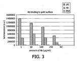
Figure 3 shows the effectiveness of coating anti-morphine antibodies to a gold surface, detected by morphine-HRP and luminol/luminescence. -
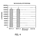
Figure 4 shows the functionality of microparticles (MPs) to which morphine was bound, detected by binding of MP-morphine to anti-morphine Abs attached to a surface. -
Figure 5 shows the results of a competitive assay wherein MP-morphine binding to anti-morphine Abs attached to a surface was detected in the presence of different amounts of morphine in solution. - "Target" may be any molecule of which the concentration or presence as such is to be determined. Examples of suitable targets in the context of the present invention are molecular targets such as small molecules, drugs, proteins, enzymes, hormones, peptides and nucleic acids. Molecular targets often determine the concentration and/or presence of larger moieties, e.g. cells, viruses, or fractions of cells or viruses, tissue extract, etc.
- Particularly preferred targets are those, which only have a single epitope, including single epitope small molecules, drugs, hormones, and peptides. Particularly preferred as targets are addictive drugs. An example of an addictive drug preferred as target is morphine. Other addictive drugs preferred as targets are cocaine, THC, anabolics, or drugs of the amphetamine/methamphetamine group. The target may exist as such in a sample that is analysed or may be formed in situ, e.g., during the step of contacting, for example via a reaction that takes place during that step. If a sensor is used to monitor a reaction, the target may for example be the starting product of the reaction or a reaction product.
- It will be appreciated that the target to be detected according to the method of the invention will be one that interferes with the binding between the first binding molecule and the second binding molecule in a competitive manner. It will furthermore be appreciated that the first and the second binding molecule will be chosen such that the target to be detected will interfere with their binding to each other in a competitive manner. The reaction products can be detected directly by the sensing method. As well, the reaction products can be further processed prior to detection. An example of further processing is that materials are added or that the (bio)chemical or physical properties of the target are modified to facilitate detection.
- As mentioned above, the method according to the invention includes a step of contacting the sample and a first binding molecule that is attached to a magnetic particle with a second binding molecule bound to a solid support. The contacting will take place in solution.
- The term "in solution" as used herein means that the step, (binding) reaction or assay referred to in this context is carried out in a liquid environment. Preferably, the liquid environment is an aqueous liquid environment. The reagents that take part need not actually be dissolved in the liquid environment but may also be present in a suspended or dispersed state.
- The contacting of the sample and the first binding molecule attached to the magnetic particle with the second binding molecule bound to the solid support may occur at the same time or at different times. For example, the sample may be contacted with the second binding molecule on the solid support first, and shortly thereafter the first binding molecule attached to the magnetic particle may be added. Alternatively, the first binding molecule attached to the magnetic particle may be contacted with the second binding molecule on the solid support first, and shortly thereafter the sample may be added.
- In a particularly preferred embodiment of the invention, the sample and the first binding molecule attached to the magnetic particle will be contacted with the second binding molecule bound to the solid support at the same time. This can be done, e.g., in that the sample and the first binding molecule attached to the magnetic particle are mixed and subsequently contacted with the second binding molecule bound to the solid support. Alternatively, the sample and the first binding molecule attached to the magnetic particle can be added separately, but simultaneously, to the second binding molecule bound to the solid support.
- The method of the invention furthermore includes applying magnetic force in step a) in order to bring the magnetic particle into close proximity with the solid support. In other words, the particles are forced onto the solid support. This step of magnetic actuation will generally lead to a reduced reaction time regarding the binding of the components participating in the competitive binding events of the assay. Furthermore, it has been surprisingly found that in certain embodiments of the invention, the sensitivity of the assay can be influenced in this way. The magnetic force during the actuation step is preferably applied in such a way so as to reduce or eliminate the impact of a difference in mobility between the target and the first binding molecule attached to the magnetic label on the binding. Thus, the magnetic force is applied to concentrate the magnetic particles to the solid support, e.g., a sensor surface, in order to compensate for the decreased mobility of the first binding molecule attached to the magnetic particle.
- The magnetic force may be applied by virtue of an electromagnet. An air-cored coil or a permanent magnet are, however, also suitable.
- In one embodiment of the method of the invention, the first binding molecule is capable of selectively binding both the target in the sample and the second binding molecule. The contacting step furthermore comprises allowing competition of the second binding molecule with the target for said selective binding to the first binding molecule.
- Examples of suitable first binding molecules are Affibodies™, antibodies, receptor molecules, aptamers and chelators.
- In cases where the target is a nucleic acid, the first binding molecule will comprise nucleic acids having a base sequence that is complementary to a part of the sequence of the target.
- Particularly preferred as first binding molecules are antibodies specifically binding the target.
- In case the first binding molecule is capable of selectively binding both the target in the sample and the second binding molecule, the second binding molecule is identical to the target, or is a target homologue. "Target homologue" as used herein is intended to mean either a construct which contains at least a part of the target, preferably the part that distinguishes it from other related molecules or a construct that the first binding molecule binds similarly strong to as the target. Similarly strong binding is defined as having a Ka preferably a factor of 103, more preferred a factor of 102, most preferred a factor of 10 or smaller in difference. Without wishing to be bound by any theory it is believed that very often a target and a target homologue share the same epitopes for binding to a binding site.
- In accordance with the method of the invention, the first binding molecule is attached to a magnetic particle. Suitable magnetic particles for use in the method of the invention are those which may be actuated by magnetic force. The magnetic particles may be of any shape or form They may be magnetic, diamagnetic, paramagnetic, superparamagnetic, ferrimagnetic or ferromagnetic, i.e., any form of magnetism which generates a magnetic dipole in an electric filed, either permanently or temporarily.
- In another embodiment of the method of the invention, the second binding molecule is capable of selectively binding both the target in the sample and the first binding molecule. The contacting step furthermore comprises allowing competition of the first binding molecule with the target for said selective binding to the second binding molecule.
- This embodiment is depicted in an exemplary manner in
Figure 1 . The Figure shows a competitive assay setup, which may be used to quantitatively detect molecules with a single epitope (although molecules with two or more epitopes may also be detected). The setup does not require premixing of separate reagents, i.e., it is suitable to be operated in a single-chamber competitive assay format. This setup allows for competition of the target (a) with the first binding molecules (d) attached to magnetic particles in solution. Specifically, the target molecules (a) compete for the second binding molecules (b) on the solid support (c), e.g., a sensor surface, with target molecules or target homologues (d), which are directly or indirectly attached to the magnetic particles. The mobility difference between free target molecules (a) and the target molecules or homologues (d) bound to the magnetic particles may be overcome by application of a magnetic force (magnetic actuation). In this way, the dose response curve may surprisingly be improved towards a linear curve. This assay setup is particularly suitable for quantitative measurements of target molecules in a sample. - Examples of suitable second binding molecules are again Affibodies™, antibodies, receptor molecules, aptamers and chelators.
- In the case where the target is a nucleic acid, the second binding molecule will comprise nucleic acids having a base sequence that is complementary to a part of the sequence of the target.
- Particularly preferred as second binding molecules are antibodies specifically binding the target.
- In case the second binding molecule is capable of selectively binding both the target in the sample and the first binding molecule, the first binding molecule is identical to the target, or is a target homologue, the term "target homologue" having the meaning as defined above.
- Suitable magnetic particles for use in the methods of the present invention may have a size from about 10 nm to a few micrometers, more preferred from about 50 nm to about 1 µm. Also preferred are particles with sizes from about 100 nm to about 500 nm, e.g., particles of about 300 nm, or particles of about 200 nm. In a preferred embodiment, the magnetic particles are larger than the individual target molecules in the sample to be analyzed in the assay of the invention.
- The attachment of the first binding molecule to the magnetic particle may be done via coating of the particle with the first binding molecule. It may also be done via covalent linkage, either directly or with the help of a spacer molecule. Suitable spacer molecules will be known to those skilled in the art, and include, for example, alkylene diamine or ethylene diamine.
- In a preferred embodiment, the attachment is done via a strong binding couple. In this embodiment, one binding partner of a strong binding couple will be attached to the magnetic particle, and the other binding partner of the strong binding couple will be attached to the first binding molecule, or is the first binding molecule itself.
- Examples of preferred strong binding couples are avidin/biotin, hapten/antibody, protein or peptide/antibody, protein/carbohydrate, protein/protein, nucleic acid/nucleic acid, protein/nucleic acids and hapten/nucleic acids.
- The interaction between the protein avidin and the molecule biotin is widely applied to link biological molecules to other moieties. The affinity constant (Ka) of avidin with biotin is one of the highest known at approximately 1015 L/mol and thus the binding is considered irreversible under normal assay conditions. In addition to the high affinity, there are four binding sites available for biotin on each avidin molecule. The biotinylation or chemical labelling of proteins with biotin is facile and does not reduce the biological activity. Avidin can also be chemically coupled to other proteins through standard linking agents involving carbodiimide. There are a number of varieties of avidin commercially available including streptavidin and neutravidin, which differ in degree of glycosylation, isoelectric point and non-specific binding characteristics. Another alternative is the strep-tag II /strep-tactin couple.
- High affinity antibodies can be raised to haptens or small molecules including dyes, drugs, hormones and vitamins. In general, antibodies to nearly any hapten can be created and a number of high affinity antibodies exist with Ka greater than 1011 L/mol for molecules such as digoxigenin, 2,4-dinitrophenyl (DNP) and fluorescein-5-isothiocyanate (FITC). There are several known simple haptenylation procedures for chemically labelling molecules with haptens. Attachment of the antibodies can be accomplished also with chemical techniques similar to that described for avidin or with recombinant techniques for coupling to proteins.
- The specificity and high binding affinity of proteins or peptides to their antibodies is the basis for many immunoassays. The affinity constant of such interactions can be as high as 1013 L/mol and can vary many orders of magnitude depending on the particular peptide or protein used.
- Protein-protein binding occurs between specific types of proteins. For example Protein A and protein G are known for their high affinity to the Fc portion of immunoglobulins. Similarly concanavalin A is a lectin which binds to the carbohydrate fraction of glycoproteins although not as strongly as protein A and G binds to immunoglobulins. These interactions are less specific and could be used only in assays where the sample does not contain an Fc region or where the sample is not a glycoprotein. In order to achieve the desired specificity and reduce cross reactivity, antibodies and proteins within the assay for which these binding interactions are not intended may be modified. For example, one can synthesize recombinant antibodies in which the Fc or glycosylated regions are removed.
- In another preferred embodiment, the attachment of the first binding molecule to the microparticle is done by coating a protein (e.g., bovine serum albumin, BSA) to which the binding molecule is linked, to the particle. For example, if the first binding molecule is morphine or a morphine homologue, or is another addictive drug, BSA is a useful protein to which these drugs can be linked. The BSA-morphine, BSA-morphine homologue or BSA-addictive drug conjugate is then coated onto the particle.
- The method of the invention typically includes a washing step that is carried out prior to the detection step. During the washing step, first binding molecules which were not bound to the second binding molecule during step a) are removed. A preferred way of removing the unbound first binding molecules is by applying magnetic force, e.g., generated by an electromagnet, an air-cored coil, or a permanent magnet. Rinsing with a wash solution is, however, also possible.
- As mentioned previously, the second binding molecule is attached to a solid support. In a preferred embodiment, the solid support is the surface of a sensor device. The sensor device may contain any suitable detector for detecting a label. Suitable detectors are magnetic detectors, optical detectors, sonic detectors, radioactivity detectors, or electrical detectors. The detector can be any suitable sensor to detect the presence of magnetic particles on or near to a sensor surface, based on any property of the particles, e.g. it can detect via magnetic methods (e.g. magnetoresistive, Hall, coils), optical methods (e.g. imaging, fluorescence, chemiluminescence, absorption, scattering, evanescent-field techniques, surface plasmon resonance, Raman, etc.), sonic detection (e.g. surface acoustic wave, bulk acoustic wave, cantilever, quartz crystal etc), electrical detection (e.g. conduction, impedance, amperometric, redox cycling), etc.
- Particularly preferred in the context of the current invention is a magnetic detector.
- The detector can be any suitable sensor based on the detection of the magnetic properties of the particle on or near to a sensor surface, e.g. a coil, a wire, magneto-resistive sensor, magneto-restrictive sensor, Hall sensor, planar Hall sensor, flux gate sensor, SQUID, magnetic resonance sensor, etc. However, the other detectors, e.g., the optical detector, are likewise preferred.
- In a particularly preferred embodiment, the surface is a gold, glass or transparent plastic surface, particularly a gold, glass or transparent plastic surface coated with a protein to which the second binding molecule is bound.
- Generally, the attachment of the second binding molecule to the solid support, e.g., the sensor surface, may be achieved in the ways described above as suitable in connection with the attachment of the first binding molecule to the magnetic particle. For example, it may be done via coating of the solid support with the second binding molecule. It may also be done via covalent linkage, either directly or with the help of a spacer molecule. Again, suitable spacer molecules will be known to those skilled in the art, and include, for example, alkylene diamine or ethylene diamine.
- In a preferred embodiment, the attachment is done via a strong binding couple. In this embodiment, one binding partner of a strong binding couple will be attached to the solid support, and the other binding partner of the strong binding couple will be attached to the second binding molecule, or is the second binding molecule itself.
- As in the case of the attachment of the first binding molecule to the magnetic particle, examples of preferred strong binding couples for attaching the second binding molecule to the solid support are avidin/biotin, hapten/antibody, protein or peptide/antibody, protein/carbohydrate, protein/protein, nucleic acid/nucleic acid, protein/nucleic acids and hapten/nucleic acids.
- As also mentioned above, varieties of avidin suitable in this regard include streptavidin and neutravidin. Another alternative is the strep-tag II / strep-tactin couple.
- Suitable high affinity antibodies are those raised to haptens or small molecules including dyes, drugs, hormones and vitamins. As mentioned above, such antibodies include antibodies against digoxigenin, 2,4-dinitrophenyl (DNP) and fluorescein-5-isothiocyanate (FITC).
- Protein-protein binding is likewise suitable, e.g., binding of Protein A or protein G to immunoglobulins via their Fc portion. Lectins such as Concanavalin A which bind to the carbohydrate fraction of glycoproteins are likewise suitable.
- In another preferred embodiment, the attachment of the second binding molecule to the solid support is done by coating a protein (e.g., bovine serum albumin, BSA) to which the binding molecule is linked, to the solid support. For example, if the second binding molecule is morphine or a morphine homologue, or is another addictive drug, BSA is a useful protein to which these drugs can be linked. The BSA-morphine, BSA-morphine homologue or BSA-addictive drug conjugate is then coated onto the solid support.
- The detection of the magnetic particles which are bound to the solid support by virtue of the binding of the first to the second binding molecule may be done via binding of a third binding molecule to the first binding molecules attached to these particles, and subsequent detection of this binding. The third binding molecule is one that is capable of selectively binding to the first binding molecule. In one embodiment, first binding molecules on the magnetic particles are detected in this step which are not involved in binding to any of the second binding molecules on the solid support.
- The detection of the binding of the third binding molecule to the first binding molecule in the afore-mentioned embodiment is done by virtue of a detectable label. This label may be directly linked to the third binding molecule. Alternatively, the detectable label may be attached to the third binding molecule by allowing an agent, which is linked to the detectable label and capable of selectively binding to the third binding molecule, to bind to the third binding molecule.
- The labels can be detected directly by the sensing method. As well, the particles can be further processed prior to detection. An example of further processing is that materials are added or that the (bio)chemical or physical properties of the label are modified to facilitate detection. Further, the assay, device, sensor an/or method for detection of this invention are suited for label multiplexing (i.e. the parallel use of different types of labels) and chamber multiplexing (i.e. the parallel use of different reaction chambers).
- Suitable labels in this regard are labels selected from the group consisting of a fluorescent label, a colorimetric label, a chemiluminescence label, an enzymatic label (e.g., horseradish peroxidase (HRP) or alkaline phosphatase (AP)), a radiolabel, an electrostatically charged label, and a donating/accepting label. A preferred label is HRP. Another preferred label is AP.
- The sensor and methods for detection described in the present invention can be used as rapid, robust, and easy to use point-of care biosensors for small sample volumes. The reaction chamber can be a disposable item to be used with a compact reader, containing the one or more magnetic field generating means and one or more detection means. Also, the sensor and methods for detection of the present invention can be used in automated high-throughput testing. In this case, the reaction chamber is e.g. a well plate or cuvette, fitting into an automated instrument. Measurement data can be derived as an end-point measurement, as well as by recording signals kinetically or intermittently.
- The method of the present invention is applicable to various kinds of samples. For example, it can be applied to urine samples, blood samples, sweat samples, ocular fluid samples, oral fluid (saliva) samples, or hair samples. Samples derived from the above samples by processing body fluids, tissue, or cells to bring them into a state suitable for being subjected to the assay of the invention can likewise be used. Such processing may include mixing of the samples with suitable buffers or salt solutions, chaotropic agents, detergents, or solvents, agitation, extraction via chemical or mechanical means, and the like.
- The figures and examples given below should illustrate the principles of the invention without limiting the scope of the invention.
- In a magnetic particle collector (MPC), MPs were collected from 100 µl of a magnetic particle solution (Ademtech, 200 nm, Protein G coated MPs, lot nr. 0433) and dissolved in 100 µl PBS + 0.65
% Tween 20. After addition of 10 µl monoclonal mouse antibody (1 mg/ml stock solution), the mixture was mixed for 1 h at RT, followed by washing 2 times using the MPC in 100 µl PBS + 0.65% Tween 20 and once with 100 µl Triethanolamine (0.2 M, pH 8). MPs were dissolved in 1 ml DMP (20 mM) in Triethanolamine (0.2 M, pH 8) and the solution mixed for 30 min at RT. The crosslink reaction was stopped by addition of 50 µl Tris (1 M, pH 7.5, end conc. = 50 mM) and mixing continued for 15 min at RT. MPs were isolated and washed 1 time using the MPC with 100 µl Tris (50 mM, pH 7.5) and 1 time with storage buffer in the MPC and finally dissolved in 100 µl of storage buffer. - Gold discs were etched and coated with BSA or BSA-OPI (3 µg/ml) in coating buffer (15 mM Sodium carbonate, 35 mM Sodium bicarbonate, 0.05% Sodium azide, pH to 9.6). The coated discs were washed 3 times with a wash buffer (0.05
% Tween 20 in PBS). A dilution series of morphine was prepared in reaction tubes in dilution buffer (PBS + 0.65% Tween 20 + 10 mg/ml BSA; 2x concentration to be tested). Premixed MP-solution (0.1 % w/v = 1 mg/ml MP in buffer) was added to each dilution in the tubes (MP-solution : morphine solution = 1:1). 50 µl of each of these solutions were each put into a well and the MPs actuated via application of magnetic force for 30 sec (6-24 fN) or allowed to precipitate for 30 min. Subsequently, unbound MPs were removed by application of magnetic force (70fN) for 30 sec and 100 µl anti-mouse IgG-HRP (1:3000 dilution of stock in dilution buffer) were added to each well, followed by incubation for 60 min at RT. Discs were washed 4 times with 200 µl wash buffer per well and transferred to white microtiter plates to wells containing wash buffer. Wash buffer was removed and 100 µl AB mix (ECL; A+B were mixed 1:1) were added and incubated for 5 min at RT. Luminescence was read. - Using this assay setup, morphine linked to BSA as a carrier was coated on a gold surface (physisorption). Anti-morphine antibodies (monoclonal) were bound to magnetic particles (MPs) coated with Protein G. Using a well plate, a dose response curve was made using free morphine to compete with the morphine on the surface for the antibody binding sites on the beads (shown here in
Figure 2 ). As explained above, the beads were attracted to the bio-active surface using permanent magnets below the gold surface, or allowed to precipitate onto the surface. Unbound beads were fished from the solution by putting a permanent magnet in the solution above the gold surface. To measure the amount of MPs bound to the surface, incubation with a secondary antibody (goat anti mouse antibody) linked to HRP (horseradish peroxidase) was performed and unbound secondary antibody was washed away afterwards. Then the amount of bound HRP was determined using luminol (a substrate for HRP) and luminescence measured. - As can be seen from
Figure 2 , magnetic actuation makes the dose response curve steeper, thus rendering the assay more sensitive. - For an alternative embodiment of the method of the present invention we first needed to put Ab on a gold surface. As is shown in
Figure 3 , this works best when we first put protein G (pG) on the surface. Here, we used 50 µg/ml protein G to coat the gold surface. Then we washed and incubated the surface with different amounts of Ab. After that, we incubated the surfaces with morphine-HRP, then washed. Following that, the amount of bound morphine was determined using luminol/luminescence. The signal obtained reflected the amount of functional Ab on the surface. The dark grey bar inFigure 3 (Ab) represents Ab binding directly on the gold surface (physisorption) when leaving out protein G. The white bar represents a gold surface coated with BSA (no Ab, no protein G) to check for unspecific binding. - For this assay format we also needed morphine bound to MPs. Here we used COOH MPs purchased from Ademtech (200 nm and 300 nm beads were used). Morphine-3-glucoronide was bound to the COOH particles by first activating the COOH groups with EDC/NHS. Then the activated groups were reacted with ethylene diamine (to obtain free NH2 binding sites). Morphine-3-glucoronide in turn was activated with EDC/NHC and reacted with the treated MPs.
Figure 4 shows a test for functionality of these beads. Anti-morphine Ab was put on the surface (via Protein G), as mentioned above, then beads bound to morphine were attracted to the surface using permanent magnets beneath the surface. Unbound beads were fished from the solution by putting a permanent magnet in the solution above the surface. Thereafter, incubation with biotinylated secondary Ab was performed (probably due to the high level of biotinylation, we observed that this Ab does not bind to Protein G) and after washing steps, incubation with streptavidin-HRP was done. Thereafter, luminol/luminescence was done to quantify the amount of HRP bound to the surface. - The results are shown in
Figure 4 . The first two bars represent two different dilutions of the MPs, the third bar represents a parallel assay were no Ab was bound to the surface (to check for unspecific binding of MP-morphine to the surface), the fourth bar represents the same assay done in parallel with MPs that were not linked to morphine. - One advantage we observed using this assay setup is the fact that beads covered with morphine do not tend to cluster (in contrast to MPs covered with Abs, which tend to cluster). Clustering of beads is often a problem when detection of single beads is intended.
- A competitive assay was performed in a well plate using the MPs and surfaces described above. The results are shown in
Figure 5 .
Claims (18)
- A method for detecting a target in a sample suspected of containing the target, comprising:a) contacting the sample and a first binding molecule attached to a magnetic particle, at the same time or at different times, with a second binding molecule attached to a solid support, wherein the first binding molecule is capable of binding to the second binding molecule, and wherein the target is capable of interfering with this binding; and applying a magnetic force to bring the magnetic particle into close proximity with the solid support; andb) detecting the number of magnetic particles bound to the solid support by virtue of the binding of the first binding molecule to the second binding molecule, wherein said number of magnetic particles is determined by allowing a third binding molecule, which is capable of selectively binding to the first binding molecule, to bind to the first binding molecules attached to said magnetic particles, and detecting the third binding molecules bound to said first binding molecules and wherein the first binding molecules to which said third binding molecule is allowed to bind, are the first binding molecules which are not involved in binding to the second binding molecule.
- The method of claim 1, wherein the first binding molecule is capable of selectively binding the target in the sample and the second binding molecule, and wherein contacting step a) comprises allowing competition of the second binding molecule with the target for said selective binding to the first binding molecule.
- The method of claim 2, wherein the second binding molecule is identical to the target, or is a target homologue.
- The method of claim 1, wherein the second binding molecule is capable of selectively binding the target in the sample and the first binding molecule, and wherein contacting step a) comprises allowing competition of the first binding molecule with the target for said selective binding to the second binding molecule.
- The method of claim 4, wherein the first binding molecule is identical to the target, or is a target homologue.
- The method of any of claims 1 to 5, wherein the target is selected from the group consisting of a small molecule, a drug, a protein, a peptide, an enzyme, a hormone and a nucleic acid.
- The method of any of claims 1 to 6, wherein the target is an addictive drug, selected from the group of morphine, cocaine, THC, anabolics, or a drug of the amphetamine/methamphetamine group.
- The method of any of claims 1 to 7, wherein the magnetic force is applied in such a way so as to reduce or eliminate the impact of a difference in mobility between the target and the first binding molecule attached to the magnetic label on the binding to the second binding molecule.
- The method of claim 8, wherein the magnetic force is applied by virtue of an electromagnet, an air-cored coil, or a permanent magnet.
- The method of any of claims 1 to 9, further comprising prior to detection step b) a step of removing first binding molecules which are not bound to the second binding molecule during step a).
- The method of any of claims 1 to 10, wherein the first binding molecule is attached to said magnetic particle via a spacer molecule, a protein, or a strong binding couple.
- The method of claim 11, wherein the spacer molecule is alkylene diamine or ethylene diamine.
- The method of any of claims 1 to 12, wherein the solid support is the surface of a sensor device, e.g., a magnetic sensor device.
- The method of claim 13, wherein the sensor surface is a gold surface.
- The method of claim 14, wherein the gold surface is coated with a protein to which the second binding molecule is bound.
- The method of any of claims 1 to 15, wherein said sample consists of, or is derived from, urine, blood, sweat, ocular fluid, oral fluid (saliva), or hair.
- The method of any one of the preceding claims, wherein the magnetic particle is selected from the group consisting of a magnetic, a diamagnetic, a paramagnetic, a superparamagnetic, a ferrimagnetic and a ferromagnetic particle.
- The method of any one of the preceding claims, wherein the magnetic particle has a size from about 10 nm to about 10 µm, from about 50 nm to about 1 µm, from about 100 nm to about 500 nm, of about 300 nm, or of about 200 nm.
Priority Applications (1)
| Application Number | Priority Date | Filing Date | Title |
|---|---|---|---|
| EP07735694A EP2027468B1 (en) | 2006-05-09 | 2007-04-27 | Detection of target molecules in a sample by using a magnetic field |
Applications Claiming Priority (3)
| Application Number | Priority Date | Filing Date | Title |
|---|---|---|---|
| EP06113698 | 2006-05-09 | ||
| EP07735694A EP2027468B1 (en) | 2006-05-09 | 2007-04-27 | Detection of target molecules in a sample by using a magnetic field |
| PCT/IB2007/051578 WO2007132373A1 (en) | 2006-05-09 | 2007-04-27 | Detection of target molecules in a sample by using a magnetic field |
Publications (2)
| Publication Number | Publication Date |
|---|---|
| EP2027468A1 EP2027468A1 (en) | 2009-02-25 |
| EP2027468B1 true EP2027468B1 (en) | 2011-06-22 |
Family
ID=38445621
Family Applications (1)
| Application Number | Title | Priority Date | Filing Date |
|---|---|---|---|
| EP07735694A Active EP2027468B1 (en) | 2006-05-09 | 2007-04-27 | Detection of target molecules in a sample by using a magnetic field |
Country Status (8)
| Country | Link |
|---|---|
| US (1) | US10488408B2 (en) |
| EP (1) | EP2027468B1 (en) |
| JP (1) | JP5200003B2 (en) |
| CN (1) | CN101438163B (en) |
| AT (1) | ATE514086T1 (en) |
| BR (1) | BRPI0711346A8 (en) |
| RU (1) | RU2444736C2 (en) |
| WO (1) | WO2007132373A1 (en) |
Families Citing this family (14)
| Publication number | Priority date | Publication date | Assignee | Title |
|---|---|---|---|---|
| US8618508B2 (en) | 2008-09-25 | 2013-12-31 | Koninklijke Philips N.V. | Detection system and method |
| BRPI0918101A2 (en) * | 2008-12-22 | 2015-11-24 | Koninkl Philips Electronics Nv | method for measuring troponin i in a sample, cartridge for use in a assay device and use |
| US20110070228A1 (en) * | 2009-09-19 | 2011-03-24 | Surgimed Biosciences, Inc. | Method and apparatus for automated cell transfer therapy and hair transplantation |
| GB201110272D0 (en) | 2011-06-17 | 2011-08-03 | Isis Innovation | Metod of detection by mass spectrometry |
| CN103620414A (en) * | 2011-06-28 | 2014-03-05 | 皇家飞利浦有限公司 | Means used for examination of body fluids |
| ES2608930T3 (en) | 2012-01-04 | 2017-04-17 | Magnomics, S.A. | Monolithic device that combines CMOS with magnetoresistive sensors |
| CN104603611B (en) * | 2012-09-04 | 2019-04-05 | 皇家飞利浦有限公司 | Sensor device and the method for sampling |
| US9841419B2 (en) | 2012-09-04 | 2017-12-12 | Koninklijke Philips N.V. | Sensor device and a method of sampling |
| EP3004882B1 (en) * | 2013-06-06 | 2018-03-21 | Koninklijke Philips N.V. | Reagents, methods and devices to prevent aggregation in particle based tests for the detection of multimeric target molecules |
| EP3210020A1 (en) * | 2014-10-20 | 2017-08-30 | Christopher Gordon Atwood | Analyzing a sample with a construct comprising a fluorescent moiety and a magnetic moiety |
| US10261076B2 (en) * | 2015-10-02 | 2019-04-16 | The Board Of Trustees Of The Leland Stanford Junior University | Method for detecting small molecule analytes using magnetoresistant sensors |
| CN108474743B (en) * | 2015-12-24 | 2021-12-10 | 西门子医疗系统荷兰有限公司 | Optical detection of substances in fluids |
| CN110462383A (en) * | 2017-01-26 | 2019-11-15 | 生命生物科学股份有限公司 | Immunoassays and its application method based on magnetic-particle |
| WO2023057823A1 (en) * | 2021-10-10 | 2023-04-13 | Alipour Elias | Detecting an antigen of a virus using electrochemical analysis |
Family Cites Families (26)
| Publication number | Priority date | Publication date | Assignee | Title |
|---|---|---|---|---|
| IL82873A0 (en) * | 1987-06-15 | 1987-12-20 | Orgenics Ltd | Reversed competitive solid phase immunoassay for detecting single epitope analytes and kit therefor |
| US5445971A (en) * | 1992-03-20 | 1995-08-29 | Abbott Laboratories | Magnetically assisted binding assays using magnetically labeled binding members |
| US5635364A (en) * | 1992-03-27 | 1997-06-03 | Abbott Laboratories | Assay verification control for an automated analytical system |
| AU3972993A (en) | 1992-04-01 | 1993-11-08 | Regents Of The University Of California, The | Non-instrumented enzyme immunoassay: general method utilizing kinetic binding effects |
| WO1996007101A1 (en) * | 1994-08-31 | 1996-03-07 | First Medical, Inc. | Quantitative assays employing magnetizable particles for rate enhancement |
| US5660990A (en) * | 1995-08-18 | 1997-08-26 | Immunivest Corporation | Surface immobilization of magnetically collected materials |
| JP2000510582A (en) * | 1996-04-25 | 2000-08-15 | ゼニコン・サイエンシーズ・コーポレーション | Analyte assays using particulate labeling |
| US6586193B2 (en) | 1996-04-25 | 2003-07-01 | Genicon Sciences Corporation | Analyte assay using particulate labels |
| FR2758884B1 (en) * | 1997-01-30 | 1999-04-02 | Bio Merieux | METHOD FOR ISOLATING, IN PARTICULAR DETECTING OR QUANTIFYING AN ANALYTE IN A MEDIUM |
| US5981297A (en) | 1997-02-05 | 1999-11-09 | The United States Of America As Represented By The Secretary Of The Navy | Biosensor using magnetically-detected label |
| DE19822123C2 (en) * | 1997-11-21 | 2003-02-06 | Meinhard Knoll | Method and device for the detection of analytes |
| CA2327816A1 (en) * | 1998-04-09 | 1999-10-21 | Nycomed Imaging As | Use of particulate contrast agents in diagnostic imaging for studying physiological parameters |
| US6287765B1 (en) * | 1998-05-20 | 2001-09-11 | Molecular Machines, Inc. | Methods for detecting and identifying single molecules |
| EP1210461A4 (en) | 1999-08-21 | 2005-11-23 | John S Fox | High sensitivity biomolecule detection with magnetic particles |
| US6897015B2 (en) * | 2000-03-07 | 2005-05-24 | Bioforce Nanosciences, Inc. | Device and method of use for detection and characterization of pathogens and biological materials |
| RU2166751C1 (en) | 2000-03-09 | 2001-05-10 | Никитин Петр Иванович | Process of analysis of mixture of biologic and/or chemical components with use of magnetic particles and device for its implementation |
| CA2342023C (en) * | 2000-04-10 | 2007-07-03 | Randox Laboratories Ltd. | Paramagnetic particle detection |
| GB0124341D0 (en) * | 2001-10-10 | 2001-11-28 | Randox Lab Ltd | Assay |
| US7432105B2 (en) | 2002-08-27 | 2008-10-07 | Kimberly-Clark Worldwide, Inc. | Self-calibration system for a magnetic binding assay |
| CN1210568C (en) * | 2002-11-18 | 2005-07-13 | 中国农业科学院原子能利用研究所 | Bt crystallin CrylAc radio-immunity test reagent box, preparing and detecting method thereof |
| GB2404022B (en) | 2004-06-14 | 2005-08-10 | Cozart Bioscience Ltd | Competitive assays for the detection of methamphetamine group drugs |
| GB2404023B (en) | 2004-07-02 | 2005-07-06 | Cozart Bioscience Ltd | Delta-9-tetrahydrocannabinol detection method |
| US20060024769A1 (en) * | 2004-07-29 | 2006-02-02 | Saladax Biomedical Inc. | Taxol immunoassay |
| US7300631B2 (en) * | 2005-05-02 | 2007-11-27 | Bioscale, Inc. | Method and apparatus for detection of analyte using a flexural plate wave device and magnetic particles |
| US7611908B2 (en) * | 2005-05-02 | 2009-11-03 | Bioscale, Inc. | Method and apparatus for therapeutic drug monitoring using an acoustic device |
| RU2415432C2 (en) * | 2005-06-17 | 2011-03-27 | Конинклейке Филипс Электроникс Н.В. | Precise magnetic bio transducer |
-
2007
- 2007-04-27 CN CN200780016721XA patent/CN101438163B/en active Active
- 2007-04-27 JP JP2009508593A patent/JP5200003B2/en active Active
- 2007-04-27 RU RU2008148311/15A patent/RU2444736C2/en active
- 2007-04-27 US US12/299,806 patent/US10488408B2/en active Active
- 2007-04-27 BR BRPI0711346A patent/BRPI0711346A8/en not_active IP Right Cessation
- 2007-04-27 EP EP07735694A patent/EP2027468B1/en active Active
- 2007-04-27 WO PCT/IB2007/051578 patent/WO2007132373A1/en active Application Filing
- 2007-04-27 AT AT07735694T patent/ATE514086T1/en not_active IP Right Cessation
Also Published As
| Publication number | Publication date |
|---|---|
| RU2008148311A (en) | 2010-06-20 |
| JP2009536344A (en) | 2009-10-08 |
| BRPI0711346A8 (en) | 2015-10-06 |
| CN101438163A (en) | 2009-05-20 |
| RU2444736C2 (en) | 2012-03-10 |
| US20090117670A1 (en) | 2009-05-07 |
| WO2007132373A1 (en) | 2007-11-22 |
| ATE514086T1 (en) | 2011-07-15 |
| US10488408B2 (en) | 2019-11-26 |
| JP5200003B2 (en) | 2013-05-15 |
| CN101438163B (en) | 2013-05-29 |
| EP2027468A1 (en) | 2009-02-25 |
| BRPI0711346A2 (en) | 2011-08-30 |
Similar Documents
| Publication | Publication Date | Title |
|---|---|---|
| EP2027468B1 (en) | Detection of target molecules in a sample by using a magnetic field | |
| US10732111B2 (en) | Automated immunoanalyzer system for performing diagnostic assays for allergies and autoimmune diseases | |
| CN103443626B (en) | Streptavidin-bonded magnetic particles and manufacturing method for same | |
| CN106771219B (en) | Multi-epitope assay | |
| RU2530716C2 (en) | Analysis of troponin i with application of magnetic labels | |
| US10151750B2 (en) | Magnetic and/or electric label assisted detection system and method | |
| US20080268481A1 (en) | Sensitive Magnetic Catch Assay By Building a Strong Binding Couple | |
| KR20130051647A (en) | Method for analysis of biomaterials using magnetic bead | |
| JPH10513551A (en) | Methods and compounds for detection of analytes in magnetic relaxation measurements and uses thereof | |
| JP2010534320A (en) | Magnetic sensor device | |
| JP2005517899A (en) | Analyte detection method using colloidal magnetic particles | |
| WO2015170844A1 (en) | Multi-fluorescence immune-assay using magnetism | |
| WO2009083856A2 (en) | Concentrated unbound magnetic particle assay for biosensors | |
| EP2073016A1 (en) | Magnetic label based detection | |
| WO2019088142A1 (en) | Detection agent for bioassay and signal amplification method using same | |
| EP1801594A1 (en) | Sensing device and method for determination of the amount of target molecule in an analyte | |
| WO2010086772A1 (en) | System and method for assay | |
| EP1936350A1 (en) | A method for quantitatively measuring agglutination parameters | |
| EP2133695A1 (en) | Parathyroid hormone assay | |
| KR20110130384A (en) | Quantitative analysis using minute tube with accumulated enzyme | |
| Zhang et al. | Development of a Novel Sensor System Based on Magnetic Microspheres to Detect Cardiac Troponin T | |
| Saito et al. | No-Wash Immunoassay for Low Affinity Target Molecule Detection Based on Giant Magneto-Resistive Bio-Sensing Techniques | |
| JP2004020344A (en) | Method for measuring substance fixed on minute particle solid phase |
Legal Events
| Date | Code | Title | Description |
|---|---|---|---|
| PUAI | Public reference made under article 153(3) epc to a published international application that has entered the european phase |
Free format text: ORIGINAL CODE: 0009012 |
|
| 17P | Request for examination filed |
Effective date: 20081209 |
|
| AK | Designated contracting states |
Kind code of ref document: A1 Designated state(s): AT BE BG CH CY CZ DE DK EE ES FI FR GB GR HU IE IS IT LI LT LU LV MC MT NL PL PT RO SE SI SK TR |
|
| AX | Request for extension of the european patent |
Extension state: AL BA HR MK RS |
|
| GRAP | Despatch of communication of intention to grant a patent |
Free format text: ORIGINAL CODE: EPIDOSNIGR1 |
|
| DAX | Request for extension of the european patent (deleted) | ||
| GRAS | Grant fee paid |
Free format text: ORIGINAL CODE: EPIDOSNIGR3 |
|
| GRAJ | Information related to disapproval of communication of intention to grant by the applicant or resumption of examination proceedings by the epo deleted |
Free format text: ORIGINAL CODE: EPIDOSDIGR1 |
|
| GRAL | Information related to payment of fee for publishing/printing deleted |
Free format text: ORIGINAL CODE: EPIDOSDIGR3 |
|
| GRAP | Despatch of communication of intention to grant a patent |
Free format text: ORIGINAL CODE: EPIDOSNIGR1 |
|
| GRAS | Grant fee paid |
Free format text: ORIGINAL CODE: EPIDOSNIGR3 |
|
| GRAA | (expected) grant |
Free format text: ORIGINAL CODE: 0009210 |
|
| AK | Designated contracting states |
Kind code of ref document: B1 Designated state(s): AT BE BG CH CY CZ DE DK EE ES FI FR GB GR HU IE IS IT LI LT LU LV MC MT NL PL PT RO SE SI SK TR |
|
| REG | Reference to a national code |
Ref country code: GB Ref legal event code: FG4D |
|
| REG | Reference to a national code |
Ref country code: CH Ref legal event code: EP |
|
| REG | Reference to a national code |
Ref country code: IE Ref legal event code: FG4D |
|
| REG | Reference to a national code |
Ref country code: DE Ref legal event code: R096 Ref document number: 602007015383 Country of ref document: DE Effective date: 20110811 |
|
| REG | Reference to a national code |
Ref country code: SE Ref legal event code: TRGR |
|
| REG | Reference to a national code |
Ref country code: NL Ref legal event code: VDEP Effective date: 20110622 |
|
| PG25 | Lapsed in a contracting state [announced via postgrant information from national office to epo] |
Ref country code: LT Free format text: LAPSE BECAUSE OF FAILURE TO SUBMIT A TRANSLATION OF THE DESCRIPTION OR TO PAY THE FEE WITHIN THE PRESCRIBED TIME-LIMIT Effective date: 20110622 |
|
| PG25 | Lapsed in a contracting state [announced via postgrant information from national office to epo] |
Ref country code: AT Free format text: LAPSE BECAUSE OF FAILURE TO SUBMIT A TRANSLATION OF THE DESCRIPTION OR TO PAY THE FEE WITHIN THE PRESCRIBED TIME-LIMIT Effective date: 20110622 Ref country code: GR Free format text: LAPSE BECAUSE OF FAILURE TO SUBMIT A TRANSLATION OF THE DESCRIPTION OR TO PAY THE FEE WITHIN THE PRESCRIBED TIME-LIMIT Effective date: 20110923 Ref country code: FI Free format text: LAPSE BECAUSE OF FAILURE TO SUBMIT A TRANSLATION OF THE DESCRIPTION OR TO PAY THE FEE WITHIN THE PRESCRIBED TIME-LIMIT Effective date: 20110622 Ref country code: SI Free format text: LAPSE BECAUSE OF FAILURE TO SUBMIT A TRANSLATION OF THE DESCRIPTION OR TO PAY THE FEE WITHIN THE PRESCRIBED TIME-LIMIT Effective date: 20110622 Ref country code: CY Free format text: LAPSE BECAUSE OF FAILURE TO SUBMIT A TRANSLATION OF THE DESCRIPTION OR TO PAY THE FEE WITHIN THE PRESCRIBED TIME-LIMIT Effective date: 20110622 Ref country code: LV Free format text: LAPSE BECAUSE OF FAILURE TO SUBMIT A TRANSLATION OF THE DESCRIPTION OR TO PAY THE FEE WITHIN THE PRESCRIBED TIME-LIMIT Effective date: 20110622 |
|
| PG25 | Lapsed in a contracting state [announced via postgrant information from national office to epo] |
Ref country code: NL Free format text: LAPSE BECAUSE OF FAILURE TO SUBMIT A TRANSLATION OF THE DESCRIPTION OR TO PAY THE FEE WITHIN THE PRESCRIBED TIME-LIMIT Effective date: 20110622 Ref country code: BE Free format text: LAPSE BECAUSE OF FAILURE TO SUBMIT A TRANSLATION OF THE DESCRIPTION OR TO PAY THE FEE WITHIN THE PRESCRIBED TIME-LIMIT Effective date: 20110622 |
|
| PG25 | Lapsed in a contracting state [announced via postgrant information from national office to epo] |
Ref country code: EE Free format text: LAPSE BECAUSE OF FAILURE TO SUBMIT A TRANSLATION OF THE DESCRIPTION OR TO PAY THE FEE WITHIN THE PRESCRIBED TIME-LIMIT Effective date: 20110622 Ref country code: IS Free format text: LAPSE BECAUSE OF FAILURE TO SUBMIT A TRANSLATION OF THE DESCRIPTION OR TO PAY THE FEE WITHIN THE PRESCRIBED TIME-LIMIT Effective date: 20111022 Ref country code: CZ Free format text: LAPSE BECAUSE OF FAILURE TO SUBMIT A TRANSLATION OF THE DESCRIPTION OR TO PAY THE FEE WITHIN THE PRESCRIBED TIME-LIMIT Effective date: 20110622 Ref country code: PT Free format text: LAPSE BECAUSE OF FAILURE TO SUBMIT A TRANSLATION OF THE DESCRIPTION OR TO PAY THE FEE WITHIN THE PRESCRIBED TIME-LIMIT Effective date: 20111024 |
|
| PG25 | Lapsed in a contracting state [announced via postgrant information from national office to epo] |
Ref country code: SK Free format text: LAPSE BECAUSE OF FAILURE TO SUBMIT A TRANSLATION OF THE DESCRIPTION OR TO PAY THE FEE WITHIN THE PRESCRIBED TIME-LIMIT Effective date: 20110622 Ref country code: RO Free format text: LAPSE BECAUSE OF FAILURE TO SUBMIT A TRANSLATION OF THE DESCRIPTION OR TO PAY THE FEE WITHIN THE PRESCRIBED TIME-LIMIT Effective date: 20110622 Ref country code: PL Free format text: LAPSE BECAUSE OF FAILURE TO SUBMIT A TRANSLATION OF THE DESCRIPTION OR TO PAY THE FEE WITHIN THE PRESCRIBED TIME-LIMIT Effective date: 20110622 |
|
| PLBE | No opposition filed within time limit |
Free format text: ORIGINAL CODE: 0009261 |
|
| STAA | Information on the status of an ep patent application or granted ep patent |
Free format text: STATUS: NO OPPOSITION FILED WITHIN TIME LIMIT |
|
| 26N | No opposition filed |
Effective date: 20120323 |
|
| PG25 | Lapsed in a contracting state [announced via postgrant information from national office to epo] |
Ref country code: DK Free format text: LAPSE BECAUSE OF FAILURE TO SUBMIT A TRANSLATION OF THE DESCRIPTION OR TO PAY THE FEE WITHIN THE PRESCRIBED TIME-LIMIT Effective date: 20110622 |
|
| REG | Reference to a national code |
Ref country code: DE Ref legal event code: R097 Ref document number: 602007015383 Country of ref document: DE Effective date: 20120323 |
|
| PG25 | Lapsed in a contracting state [announced via postgrant information from national office to epo] |
Ref country code: MC Free format text: LAPSE BECAUSE OF NON-PAYMENT OF DUE FEES Effective date: 20120430 |
|
| PG25 | Lapsed in a contracting state [announced via postgrant information from national office to epo] |
Ref country code: ES Free format text: LAPSE BECAUSE OF FAILURE TO SUBMIT A TRANSLATION OF THE DESCRIPTION OR TO PAY THE FEE WITHIN THE PRESCRIBED TIME-LIMIT Effective date: 20111003 Ref country code: IE Free format text: LAPSE BECAUSE OF NON-PAYMENT OF DUE FEES Effective date: 20120427 |
|
| PG25 | Lapsed in a contracting state [announced via postgrant information from national office to epo] |
Ref country code: BG Free format text: LAPSE BECAUSE OF FAILURE TO SUBMIT A TRANSLATION OF THE DESCRIPTION OR TO PAY THE FEE WITHIN THE PRESCRIBED TIME-LIMIT Effective date: 20110922 |
|
| PG25 | Lapsed in a contracting state [announced via postgrant information from national office to epo] |
Ref country code: MT Free format text: LAPSE BECAUSE OF FAILURE TO SUBMIT A TRANSLATION OF THE DESCRIPTION OR TO PAY THE FEE WITHIN THE PRESCRIBED TIME-LIMIT Effective date: 20110622 |
|
| REG | Reference to a national code |
Ref country code: DE Ref legal event code: R082 Ref document number: 602007015383 Country of ref document: DE Representative=s name: MEISSNER, BOLTE & PARTNER GBR, DE |
|
| REG | Reference to a national code |
Ref country code: DE Ref legal event code: R082 Ref document number: 602007015383 Country of ref document: DE Representative=s name: MEISSNER, BOLTE & PARTNER GBR, DE Effective date: 20140328 Ref country code: DE Ref legal event code: R082 Ref document number: 602007015383 Country of ref document: DE Representative=s name: MEISSNER BOLTE PATENTANWAELTE RECHTSANWAELTE P, DE Effective date: 20140328 Ref country code: DE Ref legal event code: R081 Ref document number: 602007015383 Country of ref document: DE Owner name: KONINKLIJKE PHILIPS N.V., NL Free format text: FORMER OWNER: KONINKLIJKE PHILIPS ELECTRONICS N.V., EINDHOVEN, NL Effective date: 20140328 |
|
| PG25 | Lapsed in a contracting state [announced via postgrant information from national office to epo] |
Ref country code: LU Free format text: LAPSE BECAUSE OF NON-PAYMENT OF DUE FEES Effective date: 20120427 |
|
| PG25 | Lapsed in a contracting state [announced via postgrant information from national office to epo] |
Ref country code: HU Free format text: LAPSE BECAUSE OF FAILURE TO SUBMIT A TRANSLATION OF THE DESCRIPTION OR TO PAY THE FEE WITHIN THE PRESCRIBED TIME-LIMIT Effective date: 20070427 |
|
| REG | Reference to a national code |
Ref country code: FR Ref legal event code: CA Effective date: 20141126 Ref country code: FR Ref legal event code: CD Owner name: KONINKLIJKE PHILIPS ELECTRONICS N.V., NL Effective date: 20141126 |
|
| REG | Reference to a national code |
Ref country code: FR Ref legal event code: PLFP Year of fee payment: 10 |
|
| REG | Reference to a national code |
Ref country code: FR Ref legal event code: PLFP Year of fee payment: 11 |
|
| REG | Reference to a national code |
Ref country code: FR Ref legal event code: PLFP Year of fee payment: 12 |
|
| REG | Reference to a national code |
Ref country code: DE Ref legal event code: R082 Ref document number: 602007015383 Country of ref document: DE Ref country code: DE Ref legal event code: R082 Ref document number: 602007015383 Country of ref document: DE Representative=s name: MAIER, DANIEL OLIVER, DIPL.-ING. UNIV., DE |
|
| REG | Reference to a national code |
Ref country code: DE Ref legal event code: R081 Ref document number: 602007015383 Country of ref document: DE Owner name: SIEMENS HEALTHINEERS NEDERLAND B.V., NL Free format text: FORMER OWNER: KONINKLIJKE PHILIPS N.V., EINDHOVEN, NL Ref country code: DE Ref legal event code: R082 Ref document number: 602007015383 Country of ref document: DE Representative=s name: MAIER, DANIEL OLIVER, DIPL.-ING. UNIV., DE |
|
| REG | Reference to a national code |
Ref country code: GB Ref legal event code: 732E Free format text: REGISTERED BETWEEN 20210617 AND 20210623 |
|
| PGFP | Annual fee paid to national office [announced via postgrant information from national office to epo] |
Ref country code: IT Payment date: 20230421 Year of fee payment: 17 Ref country code: FR Payment date: 20230421 Year of fee payment: 17 Ref country code: DE Payment date: 20220620 Year of fee payment: 17 |
|
| PGFP | Annual fee paid to national office [announced via postgrant information from national office to epo] |
Ref country code: TR Payment date: 20230424 Year of fee payment: 17 Ref country code: SE Payment date: 20230417 Year of fee payment: 17 |
|
| PGFP | Annual fee paid to national office [announced via postgrant information from national office to epo] |
Ref country code: GB Payment date: 20230504 Year of fee payment: 17 Ref country code: CH Payment date: 20230720 Year of fee payment: 17 |