EP4166945A1 - Novel biomarkers of membranous glomerulonephritis - Google Patents

Novel biomarkers of membranous glomerulonephritis Download PDF

Info

Publication number
EP4166945A1
EP4166945A1 EP21202650.4A EP21202650A EP4166945A1 EP 4166945 A1 EP4166945 A1 EP 4166945A1 EP 21202650 A EP21202650 A EP 21202650A EP 4166945 A1 EP4166945 A1 EP 4166945A1
Authority
EP
European Patent Office
Prior art keywords
ntng1
antibody
polypeptide
binding fragments
autoantibodies
Prior art date
Legal status (The legal status is an assumption and is not a legal conclusion. Google has not performed a legal analysis and makes no representation as to the accuracy of the status listed.)
Withdrawn
Application number
EP21202650.4A
Other languages
German (de)
French (fr)
Inventor
Rolf Stahl
Elion HOXHA
Linda REINHARD
Current Assignee (The listed assignees may be inaccurate. Google has not performed a legal analysis and makes no representation or warranty as to the accuracy of the list.)
Euroimmun Medizinische Labordiagnostika AG
Original Assignee
Euroimmun Medizinische Labordiagnostika AG
Priority date (The priority date is an assumption and is not a legal conclusion. Google has not performed a legal analysis and makes no representation as to the accuracy of the date listed.)
Filing date
Publication date
Application filed by Euroimmun Medizinische Labordiagnostika AG filed Critical Euroimmun Medizinische Labordiagnostika AG
Priority to EP21202650.4A priority Critical patent/EP4166945A1/en
Priority to US18/046,496 priority patent/US20230133318A1/en
Priority to CN202211262231.0A priority patent/CN115980358A/en
Priority to EP22201552.1A priority patent/EP4166946A1/en
Publication of EP4166945A1 publication Critical patent/EP4166945A1/en
Withdrawn legal-status Critical Current

Links

Images

Classifications

    • GPHYSICS
    • G01MEASURING; TESTING
    • G01NINVESTIGATING OR ANALYSING MATERIALS BY DETERMINING THEIR CHEMICAL OR PHYSICAL PROPERTIES
    • G01N33/00Investigating or analysing materials by specific methods not covered by groups G01N1/00 - G01N31/00
    • G01N33/48Biological material, e.g. blood, urine; Haemocytometers
    • G01N33/50Chemical analysis of biological material, e.g. blood, urine; Testing involving biospecific ligand binding methods; Immunological testing
    • G01N33/53Immunoassay; Biospecific binding assay; Materials therefor
    • G01N33/564Immunoassay; Biospecific binding assay; Materials therefor for pre-existing immune complex or autoimmune disease, i.e. systemic lupus erythematosus, rheumatoid arthritis, multiple sclerosis, rheumatoid factors or complement components C1-C9
    • GPHYSICS
    • G01MEASURING; TESTING
    • G01NINVESTIGATING OR ANALYSING MATERIALS BY DETERMINING THEIR CHEMICAL OR PHYSICAL PROPERTIES
    • G01N33/00Investigating or analysing materials by specific methods not covered by groups G01N1/00 - G01N31/00
    • G01N33/48Biological material, e.g. blood, urine; Haemocytometers
    • G01N33/50Chemical analysis of biological material, e.g. blood, urine; Testing involving biospecific ligand binding methods; Immunological testing
    • G01N33/68Chemical analysis of biological material, e.g. blood, urine; Testing involving biospecific ligand binding methods; Immunological testing involving proteins, peptides or amino acids
    • G01N33/6854Immunoglobulins
    • GPHYSICS
    • G01MEASURING; TESTING
    • G01NINVESTIGATING OR ANALYSING MATERIALS BY DETERMINING THEIR CHEMICAL OR PHYSICAL PROPERTIES
    • G01N33/00Investigating or analysing materials by specific methods not covered by groups G01N1/00 - G01N31/00
    • G01N33/48Biological material, e.g. blood, urine; Haemocytometers
    • G01N33/50Chemical analysis of biological material, e.g. blood, urine; Testing involving biospecific ligand binding methods; Immunological testing
    • G01N33/68Chemical analysis of biological material, e.g. blood, urine; Testing involving biospecific ligand binding methods; Immunological testing involving proteins, peptides or amino acids
    • G01N33/6893Chemical analysis of biological material, e.g. blood, urine; Testing involving biospecific ligand binding methods; Immunological testing involving proteins, peptides or amino acids related to diseases not provided for elsewhere
    • GPHYSICS
    • G01MEASURING; TESTING
    • G01NINVESTIGATING OR ANALYSING MATERIALS BY DETERMINING THEIR CHEMICAL OR PHYSICAL PROPERTIES
    • G01N2333/00Assays involving biological materials from specific organisms or of a specific nature
    • G01N2333/435Assays involving biological materials from specific organisms or of a specific nature from animals; from humans
    • G01N2333/705Assays involving receptors, cell surface antigens or cell surface determinants
    • G01N2333/70503Immunoglobulin superfamily, e.g. VCAMs, PECAM, LFA-3
    • GPHYSICS
    • G01MEASURING; TESTING
    • G01NINVESTIGATING OR ANALYSING MATERIALS BY DETERMINING THEIR CHEMICAL OR PHYSICAL PROPERTIES
    • G01N2800/00Detection or diagnosis of diseases
    • G01N2800/34Genitourinary disorders
    • G01N2800/347Renal failures; Glomerular diseases; Tubulointerstitial diseases, e.g. nephritic syndrome, glomerulonephritis; Renovascular diseases, e.g. renal artery occlusion, nephropathy
    • GPHYSICS
    • G01MEASURING; TESTING
    • G01NINVESTIGATING OR ANALYSING MATERIALS BY DETERMINING THEIR CHEMICAL OR PHYSICAL PROPERTIES
    • G01N2800/00Detection or diagnosis of diseases
    • G01N2800/52Predicting or monitoring the response to treatment, e.g. for selection of therapy based on assay results in personalised medicine; Prognosis

Definitions

  • the present invention relates to novel biomarkers of membranous glomerulonephritis (MGN), particularly biomarkers, such as autoantibodies and antigens present in subjects diagnosed with MGN.
  • MGN membranous glomerulonephritis
  • the invention provides in vitro methods and kits for diagnosis, prognosis and monitoring of MGN.
  • MGN Membranous glomerulonephritis
  • MGN is an autoimmune kidney disease characterized by proteinuria, the presence of excess proteins in the urine. MGN develops when circulating antibodies bind antigens on the surface of the podocyte cells of the glomerular basement membrane of the kidney, which leads to activation of the complement system, disruption of the glomerular filtration barrier and development of proteinuria.
  • the antigens that elicit the autoimmune reaction causing MGN remained unknown, and the molecular pathophysiology of the disease could not be characterized.
  • the first MGN target antigens phospholipase A2 receptor 1 (PLA2R1) and thrombospondin type 1 domain-containing protein 7A (THSD7A) were discovered in 2009 and 2014, respectively. Together, they account for about 80% of patients with MGN.
  • the discovery of these disease-causing antigens enabled the development of specific diagnostic markers, such as specific staining of renal biopsies for the respective antigens and detection of circulating antibodies in patient blood samples.
  • the level of circulating autoantibodies can not only predict the response to therapies, but also enable a prognosis with respect to renal insufficiency.
  • the therapy for these patients can be adjusted based on their individual PLA2R1 antibody level and thus therapy can be adjusted with disease progression and pathogenesis.
  • Targeted therapies or biomarkers are not available for 20% of patients with MGN. Thus, there is a corresponding need for diagnosis, prognosis and targeted therapy for these patients.
  • the development of further biomarkers aiding the therapy of MGN has been hindered by the molecular properties of the antigens.
  • the epitopes recognized by autoantibodies in PLA2R1 and THSD7A are conformation dependent and cannot be detected by conventional methods, such as standard Western Blot methods.
  • the present inventors were able to develop a new biomarker for MGN that can be used to characterize MGN in patient blood samples in order to aid diagnosis, monitor disease progression and enable targeted therapy.
  • MGN membranous glomerulonephritis
  • idiopathic membranous glomerulonephritis in a subject.
  • non-invasive methods of determining therapeutic effectiveness and disease progression in subject with MGN Preferably the MGN is an autoimmune MGN, more preferably associated with the presence of anti-NTNG1, such as anti-NTNG1 autoantibodies.
  • Another objective is the provision and preparation of devices, assays and reagents for such an assay.
  • the inventors were able to identify a novel autoantigen in membranous glomerulonephritis.
  • the invention relates to the identification of Netrin G1 (NTNG1) as a novel autoantigen in membranous glomerulonephritis and providing methods of using NTNG1 and NTNG1 autoantibodies as a biomarker.
  • NTNG1 Netrin G1
  • a first aspect of the invention relates to an in vitro method comprising detecting one or more autoantibodies recognizing a Netrin G1 (NTNG1) polypeptide or one or more anti-NTNG1 antibody-binding fragments thereof in a biological sample obtained from a subject.
  • NTNG1 Netrin G1
  • the in vitro method further comprises the step of detecting any antigen-antibody complexes formed between the NTNG1 polypeptide or one or more anti-NTNG1 antibody-binding fragments thereof and anti-NTNG1 autoantibodies in the biological sample.
  • the in vitro method of diagnosing membranous glomerulonephritis in a subject comprises the steps of (i) contacting a biological sample obtained from the subject with an NTNG1 polypeptide or one or more anti-NTNG1 antibody-binding fragments thereof, (ii) detecting any antigen-antibody complexes formed between the NTNG1 polypeptide or one or more anti-NTNG1 antibody-binding fragments thereof and anti-NTNG1 autoantibodies in the biological sample.
  • detecting the antigen-antibody complexes comprises determining the presence of anti-NTNG1 autoantibodies.
  • detecting the presence of any antigen-antibody complexes between the NTNG1 polypeptide or one or more anti-NTNG1 antibody-binding fragments thereof and anti-NTNG1 autoantibodies may result or results in a positive diagnosis for NTNG1-positive MGN.
  • NTNG1 serves as an autoantigen in MGN in a group of patients who do not produce autoantibodies to antigens commonly associated with MGN, and that levels of anti-NTNG1 autoantibodies in the blood of patients with MGN can be used to determine the effectiveness of treatment of the patient.
  • the invention also relates to an in vitro method of determining the effectiveness of a treatment for MGN in a subject, comprising (i) determining the level of anti-NTNG1 autoantibodies in a first biological sample obtained from a subject at a first time-point and (ii) determining the level of anti-NTNG1 autoantibodies in a second biological sample obtained from a subject at a second time-point, wherein a decrease in the level of anti-NTNG1 antibodies in the second time point compared to the first time point indicates that the treatment is effective, and/or an increase in the level of anti-NTNG1 autoantibodies in the second time point compared to the first time point indicates that the treatment is not effective.
  • determining the level of anti-NTNG1 autoantibodies in a first or second sample comprises contacting a biological sample obtained from the subject with an NTNG1 polypeptide or one or more anti-NTNG1 antibody-binding fragments thereof and detecting any antigen-antibody complexes formed between the NTNG1 polypeptide or one or more anti-NTNG1 antibody-binding fragments thereof and anti-NTNG1 autoantibodies in the biological sample.
  • the biological sample is a blood sample, preferably a serum sample.
  • NTNG1 polypeptides or fragments thereof in the biological sample of a subject containing at least one NTNG1 antigen that binds to anti-NTNG1 autoantibodies of subjects with MGN, can be used to make a prognosis and/or determine the therapeutic needs in said subjects.
  • a further aspect of the invention relates to an in vitro method for prognosis of MGN in a subject, comprising the steps of determining the expression level of NTNG1 polypeptide or one or more anti-NTNG1 antibody-binding fragments thereof in a biological sample obtained from said subject and comparing said expression level to a reference expression level, wherein an increased expression level of NTNG1 polypeptide or one or more anti-NTNG1 antibody-binding fragments thereof compared to said reference expression level is indicative of MGN or an increased likelihood of MGN, compared to an average subject, such as a healthy subject.
  • the biological sample is a tissue sample, more preferably a kidney biopsy.
  • the reference level is determined by determining the expression level of NTNG1 polypeptide or one or more anti-NTNG1 antibody-binding fragments thereof in a group of subjects without NTNG1-associated MGN and calculating the mean level of NTNG1 polypeptide or one or more anti-NTNG1 antibody-binding fragments thereof in subjects without NTNG1-associated MGN.
  • the expression level of NTNG1 polypeptide or one or more anti-NTNG1 antibody-binding fragments thereof in a biological sample is determined by contacting the biological sample with an anti-NTNG1 antibody or one or more NTNG1 antigen-binding fragments thereof.
  • the anti-NTNG1 antibody comprises a full-length antibody or one or more antibody fragments capable of binding to one or more NTNG1 antigens.
  • the expression level of NTNG1 polypeptide or one or more anti-NTNG1 antibody-binding fragments thereof is determined by detecting NTNG1 mRNA or fragments of NTNG1 mRNA, preferably wherein the fragments of NTNG1 mRNA are at least 12 nucleotides in length.
  • the invention also provides a kit, optionally for detecting anti-NTG1 or diagnosing and/or prognosing MGN in a subject, the kit comprising (i) an NTNG1 polypeptide or one or more anti-NTNG1 antibody-binding fragments thereof and/or (ii) a reagent for detection of an antigen-antibody complex formed between the NTNG1 polypeptide or one or more anti-NTNG1 antibody-binding fragments thereof and one or more anti-NTNG1 antibody present in the sample.
  • the NTNG1 polypeptide or one or more anti-NTNG1 antibody-binding fragments thereof are capable of binding or binding specifically to anti-NTNG1 antibodies.
  • the sample is a tissue sample, preferably a renal biopsy sample.
  • the biological sample is a blood sample or a renal biopsy sample.
  • the biological sample is a blood sample.
  • the NTNG1 polypeptide or one or more anti-NTNG1 antibody-binding fragments thereof are soluble.
  • the NTNG1 polypeptide or one or more anti-NTNG1 antibody-binding fragments thereof are isolated and/or recombinant.
  • the NTNG1 polypeptide comprises an amino acid sequence with at least 70% identity to the amino acid sequence according to SEQ ID NO: 1. In another embodiment, the NTNG1 polypeptide comprises an amino acid sequence with at least 75%, at least 80%, at least 85%, at least 90%, at least 95%, at least 98%, at least 99%, or 100% identity to the amino acid sequence according to SEQ ID NO: 1.
  • an autoantibody binding specifically to a mammalian NTNG1 is provided, preferably in a solution comprising one or more, more preferably all from the group comprising an artificial buffer, a preservative and an artificial anticoagulant.
  • An artificial buffer is a buffer which is synthetic and/or may not occur in the body of the patient or at least at concentrations well below the concentration used.
  • the buffer may be selected from the group comprising Tris, phosphate, Tricine, acetate, MOPS, MES, carbonate, citrate and HEPES.
  • the term "preservative" as used herein refers to a substance inhibiting microbial growth and/or chemical degradation in a liquid solution and may preferably be selected from the group comprising azide, lactic acid, nitrate, nitrite, antibiotics, a protease inhibitor and ethanol.
  • the term "stabilizer”, as used herein, is a reagent that stabilizes a proteinacous molecule such as an antibody, for example by decreasing degradation, by decreasing unfolding or by decreasing loss of the molecule, for example as a result of non-specific absorption to a solid phase such as reaction vessel.
  • the autoantibody is in a blood sample from a subject, preferably human, suffering from MNG.
  • the autoantibody may be used as a positive control for detection assays, as a reagent for inhibiting renal function, for example in an experimental setup, as a labeled or non-labeled reagent for competitive assays or as a calibrator.
  • the term "obtained” is considered to be a preferred embodiment of the term “obtainable”. If hereinafter e.g. an antibody is defined to be obtainable from a specific source, this is also to be understood to disclose an antibody which is obtained from this source.
  • autoantibody refers to an antibody that is produced by the immune system of a subject (or patient) and that is directed against a subject's (or patient's) own proteins. Autoantibodies may attack the body's own cells, tissues, and/or organs, causing inflammation and cell injury.
  • autoantibody recognizing NTNG1 can be used interchangeably with “anti-NTNG1 autoantibody”.
  • anti-NTNG1 antibody also includes anti-NTNG1 autoantibodies.
  • circulating autoantibody refers to autoantibodies circulating within the body, for example within the blood circulatory system or the lymph system. Specifically, circulating autoantibodies are commonly detected in the serum of a subject.
  • RNA expression refers to the process of synthesis of a gene product, preferably a functional RNA or protein. Gene expression generally comprises DNA transcription, optionally RNA processing and in the case of protein-expressing genes, RNA translation.
  • heterologous or exogenous or foreign or recombinant or non-native polypeptide is defined herein as a polypeptide that is not native to the host cell, a polypeptide native to the host cell in which structural modifications, e.g., deletions, substitutions, and/or insertions, have been made by recombinant DNA techniques to alter the native polypeptide, or a polypeptide native to the host cell whose expression is quantitatively altered or whose expression is directed from a genomic location different from the native host cell as a result of manipulation of the DNA of the host cell by recombinant DNA techniques, or whose expression is quantitatively altered as a result of manipulation of the regulatory elements of the polynucleotide by recombinant DNA techniques e.g., a stronger promoter; or a polynucleotide native to the host cell, but integrated not within its natural genetic environment as a result of genetic manipulation by recombinant DNA techniques.
  • nucleic acid or “nucleic acid molecule” or “nucleic acid sequence” or “nucleotide sequence” are used interchangeably herein to refer to a biomolecule composed of nucleotides.
  • the nucleic acid molecule can be comprised within an eukaryotic or prokaryotic organism, a eukaryotic or prokaryotic cell, a cell nucleus or a cell organelle, as part of a genome or as an individual molecule; or it can be comprised within a plasmid, a vector, an artificial chromosome; a nucleic acid can also exist outside of a cell, in vesicles, viruses or freely circulating, it can be isolated in a suitable composition, in a fixed or frozen tissue or cell culture, or dried.
  • the nucleic acid can be synthesized or naturally occurring, i.e. isolated from nature.
  • sequence identity refers to the comparison of a first nucleic acid sequence to a second nucleic acid sequence, or a comparison of a first amino acid sequence to a second amino acid sequence and is calculated as a percentage based on the comparison. The result of this calculation can be described as “percent identical” or “percent ID.”
  • a sequence identity may be determined by a program, which produces an alignment, and calculates identity counting both mismatches at a single position and gaps at a single position as non-identical positions in final sequence identity calculation. The sequence identity is determined over the entire length of the first and second nucleic acid sequence.
  • encoded protein or “encoded amino acid” refers a protein that consists of a chain of amino acids, which results from a sequence that is encoded by a nucleic acid molecule comprising three-nucleotide codons.
  • polypeptide refers to amino acid sequences of a variety of lengths.
  • polypeptide may also refer to the primary, secondary, tertiary, quartenery or quinary structure or a protein.
  • Polypeptides can be in uncharged forms or as salts, either unmodified or modified by glycosylation, side chain oxidation, phosphorylation, citrullination or transglutamination.
  • the polypeptide is a full-length native protein. In other embodiments, the polypeptide is a smaller fragment of the full-length protein.
  • the amino acid sequence is modified by additional substituents attached to the amino acid side chains, such as N- or C-terminal added protein tags such as affinity tags, linkers, which may comprise 2-40, preferably 5 to 30 amino acids with epitopes other than those from NTNG1 or not epitopes, glycosyl units, lipids, or inorganic ions such as phosphates, as well as modifications relating to chemical conversion of the chains such as oxidation of sulfhydryl groups.
  • polypeptide is intended to include the amino acid sequence of the full-length native protein, or a fragment thereof, subject to those modifications that do not significantly change its specific properties.
  • the term “polypeptide” encompasses protein isoforms, i.e., variants that are encoded by the same gene, but that differ in their amino acid sequence or in other properties.
  • antibody-binding fragment of an NTNG1 polypeptide or "anti-NTNG1 antibody-binding fragment” relates to a molecule comprising an amino acid sequence derived from a protein that is not full length and comprises an antigen sequence which enables antibody binding.
  • antigen e.g. NTNG1
  • anti-NTNG1 antibody-binding fragment refers to a fragment of the antigen that retains the ability of the antigen to bind specifically an anti-NTNG1 antibody to form an antibody-antigen complex.
  • Suitable anti-NTNG1 antibody-binding fragments of an antigen may be identified by one skilled in the art by simple trials to ascertain their ability to bind specific autoantibodies of MGN.
  • antigen relates to a molecule or a molecular structure that can be bound by an antigen-specific antibody or a binding fragment thereof.
  • An antigen can be composed of one or more epitopes, which can be linear structure.
  • An antigen can also be composed of amino acids that form structure-dependent epitopes.
  • epitope refers to the distinct surface features of an antigen that can act as points of interaction for specific antibodies.
  • Antigenic molecules can comprise one or more epitopes.
  • the minimum size of a linear epitope is about 4 amino acids and comprises one or more antibody contact residues.
  • an "autoantigen” is an endogenous molecule, a polypeptide, protein or protein complex, or a DNA or RNA molecule that is recognized by the immune system of a subject suffering from a specific autoimmune disease.
  • membranous nephropathy refers to a slowly progressive disease of the kidney which is a frequent cause of adult nephrotic syndrome.
  • Primary or Idiopathic membranous glomerulonephritis as described herein is considered to be an autoimmune disease targeting the glomerulus.
  • Secondary membranous glomerulonephritis includes nephropathies caused by secondary factors such as systemic lupus erythematosus, hepatitis B or syphilis.
  • UniProt numbers or "UniProt” or “UniProt Accession numbers” provided herein refer to the unique identifiers given to individual genes and proteins by the UniProt Consortium, which are available from their database at www.uniprot.org and commonly used as references in the field.
  • UniProtKB (UniProt Knowledgebase) is a freely accessible database of protein sequence and functional information.
  • the UniProt database includes manually annotated and reviewed entries (provided by the Swiss-Prot database) and automatically annotated and not manually reviewed entries (provided by TrEMBL database), many of which are derived from genome sequencing projects.
  • TrEMBL includes translated coding sequences from the EMBL-Bank/GenBank/DDBJ nucleotide sequence database, and others.
  • biomarker refers to a measurable indicator of the presence or severity of a disease state.
  • a biomarker can for example be used for diagnosis, to measure the progress of disease, or to evaluate effective therapeutic regimes.
  • NTNG1 protein or an NTNG1 polypeptide or any anti-NTNG1 antibody-binding fragment thereof; and anti- NTNG1 autoantibodies are biomarkers of MGN, particularly idiopathic MGN.
  • the efficacy of a candidate drug to treat MGN may be tested or monitored.
  • a decreasing level of anti-NTNG1, specifically of anti-NTNG1 autoantibody indicates an increased likelihood that the drug is effective.
  • Phospholipase A2 Receptor 1 refers a type-1 transmembrane protein, which in humans is encoded by the PLA2R1 gene and predominantly expressed in the podocytes of the renal corpuscle of the kidney. It is known as an antigen that is predominant in about 70% of patients with idiopathic MGN.
  • Thrombospondin type 1 domain-containing protein 7A (THSD7A, UniProt: Q9UPZ6) refers to a protein highly expressed on podocytes which in humans in encoded by the THSD7A gene. It is a known antigen in MGN and the second most common antigen in MGN after PLA2R1.
  • the term "patient” or “subject” is used interchangeably and refers to a human subject or another mammal subject (e.g., primate, dog, cat, goat, horse, pig, mouse, rat, rabbit, and the liked), that can be afflicted with a membranous nephropathy, particularly an idiopathic MGN.
  • the patient or subject is a human patient or subject.
  • a patient suspected to have MGN has typically been diagnosed based on a kidney biopsy or due to the presence of a biomarker associated with MGN.
  • biological sample is used herein in its broadest sense.
  • a biological sample is generally obtained for a subject.
  • Said subject is mammal, preferably human.
  • biological sample is generally obtained from a patient or subject.
  • said sample comprises a representative set of autoantibodies or autoantigen.
  • said biological sample may be a blood sample or a tissue sample.
  • the tissue sample is preferably a kidney biopsy sample.
  • a sample may be of any biological tissue or fluid with which biomarker(s) of the present invention may be assayed.
  • samples include, but are not limited to, bodily fluids which may or may not contain cells, e.g., blood (e.g., whole blood, serum or plasma) or urine.
  • samples also include biopsies (for example kidney biopsy).
  • the term biological sample also encompasses any material derived by processing a biological sample. Derived materials include, but are not limited to, cells (or their progeny) isolated from the sample or proteins extracted from the sample. Processing of a biological sample may involve one or more of: filtration, distillation, extraction, concentration, inactivation of interfering components, addition of reagents, and the like.
  • control when used to characterize a subject, refers to a subject that is healthy or to a patient that has been diagnosed with a specific disease other than renal disease.
  • control sample refers to one, or more than one sample, that has been obtained from a healthy subject or from a patient diagnosed with a disease other than renal disorder.
  • a control sample as used herein may also be a sample from a patient diagnosed with PLA2R1-positive or THSD7A-positive MGN.
  • the term “level” refers to the quantity of a molecule in a biological sample.
  • anti-NTNG1 autoantibody level refers to the number of detectable anti-NTNG1 autoantibody in a sample. The number may be a total or absolute number, or the number may be relative to a control or reference measurement. The level may be determined numerically, or it may be determined by measuring a secondary signal, such as fluorescence intensity. The level may also be expressed binary by presence or absence of the measured molecule, i.e. the level may be positive or negative.
  • Antibody “level” can also refer be called “titer”. When referring to a polypeptide or protein, "level” may also refer to the expression level of the polypeptide or protein.
  • the term “reference level” refers to a level measured in a biological sample obtained from a control or preferably to an average of several levels measured in biological samples obtained from several controls.
  • the term “diagnosis”, “diagnosing” or “diagnostic” is used on its broadest sense and encompasses diagnosis, prognosis, theragnosis and monitoring in membranous nephropathy.
  • NTNG1-negative patient refers to a patient whose serum contains no autoantibodies or at least no detectable levels of autoantibodies directed against the respective antigen.
  • NTNG1-positive patient refers to a patient whose serum contains autoantibodies directed against the respective protein.
  • the term "contacting" or “reacting” means bringing two compounds or molecules close enough to each other that they can chemically, electrically or physically interact. This interaction may be due to forces between molecules and may involve bonding and unbonding of molecules. Contacting may take place in solution or between solutions, on a solid support, between a liquid and solid phase, between a liquid and gaseous phase, between a solid and gaseous phase, within one or more gaseous phases or within a cell or organism or between cells and organism.
  • Netrin G1 (NTNG1, UniProt: Q9Y2I2), which is in humans encoded by the gene NTNG1, is a secreted glycoprotein of about 50 kDa size. It binds to the cell membrane on the extracellular side with a glycosylphosphatidylinositol (GPI) anchor. Thus, NTNG1 is exposed on the cell surface of podocytes and is able to bind circulating antibodies. Podocytes are specialized epithelial cells that cover the outer surfaces of glomerular capillaries.
  • Table 1 provides information about the different isoforms and changed or missing amino acid features compared in relation to isoform 3.
  • Table 1 Overview of NTNG1 isoforms Isoform Region MW (not processed) 3 (also 1A) M1 - F539 60541 2 (also 1F) M1 - C364 41759 363-364 NC ⁇ SK 1 (also 1C) 363-463 missing 49340 464-464 P ⁇ T 4 (also 1D) 364-464 53946 CECFGHSNRC...GNSWHYGCQP ⁇ PPKFNRIW PN...SVQVANHKRA 5 (also 1E) 364-385 51858 CECFGHSNRCSYIDLLNTVICV ⁇ PPKFNRIWPN ISSLEVSNPKQA 386-464 missing 6 (also 1G) 419-463: missing 55660 464-464 P ⁇ A
  • NTNG1 comprises domains that are removed upon maturation and processing (Table 2).
  • the unprocessed NTNG1 polypeptide has an amino acid sequence according to SEQ ID NO: 1.
  • the propeptide, comprising amino acids 511-539 is removed from the mature from.
  • the mature NTNG1 polypeptide sequence is an amino acid sequence according to SEQ ID NO: 2.
  • NTNG1 polypeptide comprises a signal peptide which comprises amino acids 1-28. Signal peptides are commonly cleaved off from presecretory polypeptides by signal peptidase during or immediately after insertion into the membrane.
  • the fully processed NTNG1 polypeptide once secreted, has an amino acid sequence according to SEQ ID NO: 3.
  • the NTNG1 polypeptide comprises an amino acid sequence with at least 70% identity to the amino acid sequence according to SEQ ID NO: 3. In another embodiment, the NTNG1 polypeptide comprises an amino acid sequence with at least 75%, at least 80%, at least 85%, at least 90%, at least 95%, at least 98%, at least 99%, or 100% identity to the amino acid sequence according to SEQ ID NO: 3.
  • the NTNG1 polypeptide comprises an amino acid sequence with at least 70% identity to the amino acid sequence according to SEQ ID NO: 1.
  • the NTNG1 polypeptide or one or more anti-NTNG1 antibody-binding fragments thereof comprise an amino acid sequence with at least 75%, at least 80%, at least 85%, at least 90%, at least 95%, at least 98%, at least 99%, or 100% identity to the amino acid sequence according to SEQ ID NO: 1.
  • the NTNG1 polypeptide comprises an amino acid sequence with at least 70% identity to an amino acid sequence selected from the group consisting of SEQ ID NO: 5, SEQ ID NO: 6, SEQ ID NO: 7 or SEQ ID NO: 8. In another embodiment, the NTNG1 polypeptide comprises an amino acid sequence with at least 80% identity to the amino acid sequence selected from the group consisting of SEQ ID NO: 5, SEQ ID NO: 6, SEQ ID NO: 7 or SEQ ID NO: 8. In another embodiment, the NTNG1 polypeptide comprises an amino acid sequence with at least 90% identity to the amino acid sequence selected from the group consisting of SEQ ID NO: 5, SEQ ID NO: 6, SEQ ID NO: 7 or SEQ ID NO: 8.. In another embodiment, the NTNG1 polypeptide comprises an amino acid sequence according to an amino acid sequence selected from the group consisting of SEQ ID NO: 5, SEQ ID NO: 6, SEQ ID NO: 7 or SEQ ID NO: 8.
  • anti-NTNG1 antibody-binding fragment of the NTNG1 polypeptide or “fragment of NTNG1” refers to a continuous amino acid sequence comprised in the full-length NTNG1 polypeptide that comprises an antigen sequence that can recognize and bind an anti-NTNG1 antibody, and is able to participate in the formation of an antigen-antibody complex with one or more anti-NTNG1 antibodies.
  • said anti-NTNG1 antibody-binding fragment of the NTNG1 polypeptide comprises at least 10 consecutive amino acids of the entire amino acid sequence of NTNG1.
  • said anti-NTNG1 antibody-binding fragment of NTNG1 comprises at least 20, at least 40, at least 60, at least 80, at least 100, at least 120, at least 140, at least 160, at least 180, at least 200, at least 250, at least 300, at least 350, at least 400, at least 450, at least 500 or 539 consecutive amino acids of the entire amino acid sequence of full-length NTNG1 (SEQ ID NO: 1).
  • the anti-NTNG1 antibody-binding fragment of NTNG1 comprises at least 5%, at least 10%, at least 20%, at least 30%, at least 40%, at least 50%, at least 60%, at least 70%, at least 75%, at least 80%, at least 85%, at least 90%, at least 95%, at least 98%, at least 99% or 100% of the amino acid sequence of full-length NTNG1, i.e. SEQ ID NO: 1.
  • the anti-NTNG1 antibody-binding fragment of NTNG1 comprises at least 10%, at least 20%, at least 30%, at least 40%, at least 50%, at least 60%, at least 70%, at least 75%, at least 80%, at least 85%, at least 90%, at least 95%, at least 98%, at least 99% or 100% of the amino acid sequence of mature NTNG1, i.e. SEQ ID NO: 3.
  • the anti-NTNG1 antibody-binding fragment of NTNG1 is recognized by an autoantibody directed against NTNG1. Determining the ability of the fragment to interact with said autoantibodies and detecting antibody-antigen complexes between the NTNG1 polypeptide or one or more anti-NTNG1 antibody-binding fragments thereof and anti-NTNG1 autoantibodies can be accomplished by one of the methods described herein or by methods known in the art for determining direct binding.
  • Such methods include co-immunoprecipitation, pull down assay, crosslinking assays, gel electrophoresis and immunoblots (Western Blot, Dotplot), ELISA (enzyme-linked immunosorbent assay), EIA (enzyme immunoassay), FRET (fluorescence resonance energy transfer), luciferase assay, immunohistochemistry, flow cytometry, co-crystallization, or mass spectrometry.
  • anti-NTNG1 antibody or a complex comprising NTNG1 is detected using a detection method selected from the group comprising immunodiffusion, immunoelectrophoresis, light scattering immunoassays, agglutination, labeled immunoassays such as those from the group comprising radiolabeled immunoassay, enzyme immunoassays, more preferably ELISA, chemiluminescence immunoassays, preferably electrochemiluminescence immunoassay, and immunofluorescence, preferably indirect immunofluorescence.
  • a detection method selected from the group comprising immunodiffusion, immunoelectrophoresis, light scattering immunoassays, agglutination, labeled immunoassays such as those from the group comprising radiolabeled immunoassay, enzyme immunoassays, more preferably ELISA, chemiluminescence immunoassays, preferably electrochemiluminescence immunoassay, and immunofluorescence, preferably indirect immunofluorescence.
  • a competitive assay, a capture bridge assay, an immunometric assay, a class-specific second antibody on the solid phase, a direct or indirect class capture assay may also be used.
  • the principle of each of these formats is detailed in The Immunoassay Handbook, 3rd edition, edited by David Wild, Elsevier, 2005 .
  • the detection of the anti-NTNG1 is preferably carried out under non-reducing conditions.
  • a first aspect of the invention relates to an in vitro method, optionally an in vitro method of diagnosing membranous glomerulonephritis (MGN) in a subject, comprising detecting one or more autoantibodies recognizing a Netrin G1 (NTNG1) polypeptide or one or more anti-NTNG1 antibody-binding fragments thereof in a biological sample obtained from a subject.
  • MGN membranous glomerulonephritis
  • the in vitro method further comprises the step of (i) contacting a biological sample obtained from the subject with an NTNG1 polypeptide or one or more anti-NTNG1 antibody-binding fragments thereof, and (ii) detecting any antigen-antibody complexes formed between the NTNG1 polypeptide or one or more anti-NTNG1 antibody-binding fragments thereof and anti-NTNG1 autoantibodies in the biological sample.
  • detecting the antigen-antibody complexes between the NTNG1 polypeptide or one or more anti-NTNG1 antibody-binding fragments thereof and anti-NTNG1 autoantibodies comprises determining the presence of anti-NTNG1 autoantibodies.
  • detection of antigen-antibody complexes between the NTNG1 polypeptide or one or more anti-NTNG1 antibody-binding fragments thereof and anti-NTNG1 autoantibodies formed in the biological sample may lead to a positive diagnosis of the subject.
  • the mere detection of the anti-NTNG1 autoantibody in other words determining whether or not detectable levels of the antibody are present in the sample, is sufficient for the diagnosis.
  • this may involve determining whether the concentration is at least 10%, preferably 20%, 50%, 100%, 200%, 500%, 1.000%, 2.000%, 2.500%, 5.000%, 1.0000%, 2.0000%, 5.0000%, 100.000%, 1.000.000% or 10.000.000% times higher than the concentration of the antibody of interest found in the average healthy subject. If the autoantibody can be detected, this will be information instrumental for the clinician's diagnosis. It may indicate an increased likelihood that the patient suffers, suffered or will suffer from MNG.
  • a kit comprising a cell expressing, preferably overexpressing the NTNG1 polypeptide or one or more anti-NTNG1 antibody-binding fragments thereof, or the device or the NTNG1 polypeptide or one or more anti-NTNG1 antibody-binding fragments thereof and further comprising one or more, preferably all reagents from the group comprising a secondary antibody, preferably labeled with a detectable label, a washing solution, a positive control, a negative control, a detergent, a cover glass, a mounting medium and a physiological salt solution, preferably PBS, or salt required to prepare it.
  • the positive control is a diluted sample, preferably blood from a patient suffering from MNG and comprising anti-NTNG1 or a monoclonal antibody binding specifically to NTNG1 polypeptide, preferably recognized by human IgG antibodies.
  • the negative control may be a diluted sample from a healthy subject, for example a blood donor.
  • the kit may comprise instructions how to carry out the assay.
  • the kit may comprise a calibrator, preferably a set of at least three calibrators.
  • the term "calibrator”, as used herein, refers to a reagent that may be used to calibrate an assay, in particular set up a reference range or a calibration curve.
  • the calibrator is an antibody which binds to the NTNG1 and/or the polypeptide comprising NTNG1 coated on a device and is recognized by a secondary antibody recognizing the antibody to be detected, more preferably IgG, most preferably human IgG.
  • a set of calibrators may comprise at least 3, 4 or 5 solutions each comprising different known concentrations of the calibrator. The concentrations between the calibrator with the lowest concentration and the calibrator with the highest concentration may be at least 1:10, 1:20, 1:50, 1:100, 1:200, 1:500 or 1:1000.
  • the secondary antibody is a secondary antibody binding specifically to IgG class antibodies, preferably human IgG class antibodies.
  • the kit comprises the NTNG1 polypeptide or one or more anti-NTNG1 antibody-binding fragments thereof, optionally comprising a detectable label.
  • the kit may be used for detecting a mammalian antibody to NTNG1 in a sample, preferably a human antibody to NTNG1 in a sample, more preferably a blood sample.
  • the kit may be used for diagnosing, aiding in the diagnosis of NTNG1 or for identifying a subject having an increased risk of suffering from NTNG1 in the past, present or future.
  • Another aspect of the invention relates to a method comprising the step of detecting the presence or absence of anti-NTNG1 antibody, in a sample.
  • the method may be for diagnosing or aiding in the diagnosis of a nephrological autoimmune disease, preferably MGN, or identifying a subject having an increased risk of suffering from the disease in the past, presence or future.
  • the method may be for distinguishing an autoimmune nephrological disease from another nephrological disease not associated with the presence of anti-NTNG1.
  • any information, result or data obtained according to the present invention may be communicated to the patient or a medical doctor treating the patient orally, preferably by telephone, in a written form, preferably fax or letter, or in an electronic form via fax or via the internet, for example as an email or text message.
  • the NTNG1 polypeptide or one or more anti-NTNG1 antibody-binding fragments thereof is from human NTNG1. In another embodiment, the NTNG1 polypeptide or one or more anti-NTNG1 antibody-binding fragments thereof is from a different mammalian species, such as non-human primate, pig, sheep, horse, donkey, rabbit, rat or mouse.
  • the NTNG1 polypeptide may be a full-length NTNG1 polypeptide according to SEQ ID NO: 1. In another embodiment, the NTNG1 polypeptide may be a mature, processed NTNG1 polypeptide according to SEQ ID NO: 3.
  • NTNG1 polypeptide or one or more anti-NTNG1 antibody-binding fragments thereof is an isolated and/or a recombinant polypeptide.
  • the NTNG1 polypeptide or one or more anti-NTNG1 antibody-binding fragments thereof may be recombinantly expressed in a cell or organism.
  • the NTNG1 polypeptide or one or more anti-NTNG1 antibody-binding fragments thereof is a recombinant polypeptide or one or more antibody-binding fragments thereof.
  • the recombinant polypeptide or one or more antibody-binding fragments is produced in recombinant mammalian cells.
  • the antibody preferably anti-NTNG1, such as anti-NTNG antibody, or the complex to be detected, binds specifically to its interaction partner, preferably NTNG1 polypeptide.
  • binding specifically preferably means that the binding reaction is stronger than a binding reaction characterized by a dissociation constant of 1 ⁇ 10 -5 M, more preferably 1 ⁇ 10 -7 M, more preferably 1 ⁇ 10 -8 M, more preferably 1 ⁇ 10 -9 M, more preferably 1 ⁇ 10 -10 M, more preferably 1 ⁇ 10 -11 M, more preferably 1 ⁇ 10 -12 M, as determined by surface plasmon resonance using Biacore equipment at 25 °C in PBS buffer at pH 7.
  • the methods and products according to the present invention may be used for determining the concentration of an antibody binding specifically to NTNG1 in a liquid, which may not be a sample from a subject or only a processed sample, for example for preparing a calibrator or set of calibrators with known relative or absolute concentrations or standardizing solutions.
  • Methods for expressing polypeptides in cells or organisms include cell/tissue culture, batch cell culture, fed-batch culture or suspension culture or in vitro cell free expression systems.
  • Cells or organisms capable of producing the NTNG1 polypeptide or one or more antibody-binding fragments include mammalian cells, insect cells, yeasts, fungal cells, bacterial cells, plant and algal cells.
  • the NTNG1 polypeptide or one or more antibody-binding fragments are produced in mammalian cells.
  • Suitable mammalian cell lines include human embryonic kindey (HEK) 293, human fibrosarcoma HT-1080, human embryonic retinal PER.C6, HeLa, Chinese hamster ovary (CHO), hamster kidney BHK 21, mouse myeloma NS0, and murine C127.
  • HEK human embryonic kindey
  • HT-1080 human fibrosarcoma HT-1080
  • human embryonic retinal PER.C6 HeLa
  • CHO Chinese hamster ovary
  • hamster kidney BHK 21 mouse myeloma NS0
  • murine C127 murine C127.
  • Mammalian expression systems can be transiently induced to produce the polypeptide of the invention by transfection, transduction or infection or a suitable vector.
  • the mammalian cell may also be a stable cell lines in which the expression construct is stably integrated into the host genome at one or more locations.
  • Suitable non-mammlian cells are insect cells optimized for protein expression such as Drosophila melanogasterS2 cells, Spodoptera frugiperda Sf9 or Sf21 cells or Trichoplusia ni BTI-TN-5B1-4 (High Five) cells.
  • Transient expression is the temporary expression of genes that are expressed for a short time after a nucleic acid has been introduced into the cell or organism. Transient expression may be achieved by transfection with a plasmid vector or viral transduction.
  • Stable integration may be achieved using methods known in the art, including virus transduction, plasmid, BAC (bacterial artificial chromosome) or YAC (yeast artificial chromosome) transfection, transposon-mediated transfer and integration, site-specific integration and gene editing methods such as zinc finger nucleases, CRISPR/Cas9 or TALENs.
  • NTNG1 polypeptide or one or more anti-NTNG1 antibody-binding fragments thereof from the cell or organism or culture medium are described herein and known in the art and include spin columns and kits, liquid chromatography, such as FPLC or HPLC, magnetic beads, filter plates, or resins.
  • the NTNG1 polypeptide or one or more anti-NTNG1 antibody-binding fragments thereof may comprise an epitope or purification tag, preferably selected from His-tag, GS-tag, FLAG-tag, c-Myc tag or HA-tag.
  • the tag may also be a fluorescent tag, such as a fluorescent protein or FRET label, or a radiolabel.
  • the NTNG1 polypeptide or one or more anti-NTNG1 antibody-binding fragments thereof may be untagged.
  • the invention also provides a vector for the expression of an NTNG1 polypeptide or one or more anti-NTNG1 antibody-binding fragments thereof, preferably wherein the vector comprises an expression cassette comprising a promoter and a nucleic acid molecule encoding the NTNG1 polypeptide or one or more anti-NTNG1 antibody-binding fragments thereof of the invention.
  • the vector may comprise a nucleic acid molecule with at least 70%, at least 75%, at least 80%, at least 85%, at least 90%, at least 95%, at least 98%, at least 99% or 100% identity with the nucleic acid according to SEQ ID NO: 4.
  • the vector is a eukaryotic expression vector or a viral vector.
  • the promoter may be any suitable promotor for expression in the chosen system, for example CMV, T7, lac operator, EF1alpha, Tet operator, EpIE2, SV40, MT, UbC, RSV, AUG1, CMV/EF1alpha, FLD1, TK, Gal4 UAS/E1b, trc, AOX1, Polyedrin or copia.
  • the NTNG1 polypeptide or one or more anti-NTNG1 antibody-binding fragments thereof is immobilized onto a solid carrier or support (e.g., a bead, microplate or array).
  • a diagnostic device on which the NTNG1 polypeptide or one or more anti-NTNG1 antibody-binding fragments thereof is immobilized.
  • the method of the invention involves the use of a diagnostic device coated with the NTNG1 polypeptide or one or more anti-NTNG1 antibody-binding fragments thereof.
  • the device is selected from the group comprising a glass slide, preferably for microscopy, a biochip, a microtiter plate, a lateral flow device, a test strip, a membrane, preferably a line blot, a chromatography column and a bead, preferably a magnetic or fluorescent bead.
  • the term "coated with” means that a polypeptide such as the NTNG1 polypeptide or an antibody or another agent is immobilized on a solid surface of a device or the polypeptide and the device are configured for such an immobilization.
  • a particularly preferred way for the configuration or the immobilization is using a carrier and/or means modified such that one of the agent or polypeptide to be immobilized comprises an affinity tag such as biotin and the other comprises a ligand to the affinity tag such as streptavidin.
  • one of the carrier or the polypeptide or agent to be immobilized may comprise reactive chemical groups such as thiol, amino, epoxide, ester and anhydride groups which form covalent or non-covalent bonds with reactive chemical groups associated with the other.
  • the device may comprise a negative control, for example a polypeptide which does not bind specifically to anti-NTNG1, preferably from human, preferably spatially separate from the NTNG1 polypeptide or one or more anti-NTNG1 antibody-binding fragments.
  • the diagnostic methods of the present invention involve detection of an antigen- antibody complex formed between the biomarker of the invention (e.g. NTNG1 polypeptide or one or more anti-NTNG1 antibody-binding fragments thereof) and an autoantibody present in the biological sample tested. This detection is indicative of the presence of autoantibodies (namely anti-NTNG1 autoantibodies or antigen-binding fragments thereof) in said sample.
  • an antigen- antibody complex formed between the biomarker of the invention e.g. NTNG1 polypeptide or one or more anti-NTNG1 antibody-binding fragments thereof
  • autoantibodies namely anti-NTNG1 autoantibodies or antigen-binding fragments thereof
  • the detection of an antibody-antigen complex between the NTNG1 polypeptide or one or more anti-NTNG1 antibody-binding fragments thereof and anti-NTNG1 autoantibodies may be performed by any suitable method.
  • the antibody-antigen complex between the NTNG1 polypeptide or one or more anti-NTNG1 antibody-binding fragments thereof and anti-NTNG1 autoantibodies may be detected using an immunoassay.
  • immunoassays include radioimmunoassay, enzyme immunoassays (EIA), enzyme-linked immunosorbent assays (ELISA), immunofluorescence or immunoprecipitation.
  • the NTNG1 polypeptide or one or more anti-NTNG1 antibody-binding fragments thereof is immobilized on a solid surface and the biological sample is brought into contact with the bound polypeptide or fragments thereof under conditions allowing formation of an antigen-antibody complex between the NTNG1 polypeptide or one or more anti-NTNG1 antibody-binding fragments thereof and anti-NTNG1 autoantibodies.
  • the antigen-antibody complex between the NTNG1 polypeptide or one or more anti-NTNG1 antibody-binding fragments thereof and anti-NTNG1 autoantibodies can then be detected using any secondary IgG antibody labelled, for example human IgG antibody for samples from human subjects, with a detectable moiety.
  • the antibody-antigen complex between the NTNG1 polypeptide or one or more anti-NTNG1 antibody-binding fragments thereof and anti-NTNG1 autoantibodies may also be detected using an immunoblot, such as the Western Blot described herein.
  • an immunoblot such as the Western Blot described herein.
  • the NTNG1 polypeptide or one or more anti-NTNG1 antibody-binding fragments thereof are immobilized in a carrier such as a polyacrylamide gel. Then, the NTNG1 polypeptide or one or more anti-NTNG1 antibody-binding fragments thereof are transferred to a blotting membrane.
  • the biological sample is added and the antigen-antibody complex between the NTNG1 polypeptide or one or more anti-NTNG1 antibody-binding fragments thereof and anti-NTNG1 autoantibodies forms on the membrane.
  • the antigen-antibody complex between the NTNG1 polypeptide or one or more anti-NTNG1 antibody-binding fragments thereof and anti-NTNG1 autoantibodies can then be detected using any secondary IgG antibody labelled, for example human IgG antibody for samples from human subjects, with a detectable moiety.
  • the formation of antigen-antibody complex between the NTNG1 polypeptide or one or more anti-NTNG1 antibody-binding fragments thereof and anti-NTNG1 autoantibodies is achieved under non-reducing conditions.
  • the secondary antibody may be labeled with any detectable moiety, i.e., any entity that, by its chemical nature, provides an analytically identifiable signal allowing detection of the ternary complex between the NTNG1 polypeptide or one or more anti-NTNG1 antibody-binding fragments thereof and anti-NTNG1 autoantibodies, and consequently detection of the biomarker-antibody complex between the NTNG1 polypeptide or one or more anti-NTNG1 antibody-binding fragments thereof and anti-NTNG1 autoantibodies.
  • any detectable moiety i.e., any entity that, by its chemical nature, provides an analytically identifiable signal allowing detection of the ternary complex between the NTNG1 polypeptide or one or more anti-NTNG1 antibody-binding fragments thereof and anti-NTNG1 autoantibodies, and consequently detection of the biomarker-antibody complex between the NTNG1 polypeptide or one or more anti-NTNG1 antibody-binding fragments thereof and anti-NTNG1 autoantibodies.
  • Detection may be either qualitative or quantitative. Methods for labeling biological molecules such as antibodies are well-known in the art. The most commonly used detectable moieties in immunoassays are enzymes and fluorophores. In the case of an enzyme immunoassay (EIA or ELISA), an enzyme such as horseradish perodixase, glucose oxidase, beta-galactosidase, alkaline phosphatase, and the like, is conjugated to the second antibody, generally by means of glutaraldehyde or periodate.
  • the substrates to be used with the specific enzymes are generally chosen for the production of a detectable color change, upon hydrolysis of the corresponding enzyme.
  • the second antibody is chemically coupled to a fluorescent moiety without alteration of its binding capacity. After binding of the fluorescently labeled antibody to the biomarker-antibody complex and removal of any unbound material, the fluorescent signal generated by the fluorescent moiety is detected, and optionally quantified.
  • the secondary antibody may be labeled with a radioisotope, a chemiluminescent moiety, or a bioluminescent moiety.
  • Another method of the invention concerns the correlation between the presence and level of anti-NTNG1 autoantibodies and effectiveness of treatment, remission or relapse of patients with MGN, more specifically idiopathic MGN.
  • glucocorticoids for example, glucocorticoids, cyclosporin, tacrolimus, azathioprine, infliximab, omalizumab, daclizumab, adalimumab, eculizumab, efalizumab, natalizumab, omalizumab and rapamycin. It also includes cyclophosphamide, chlorambucil, and rituximab.
  • the invention further relates to an in vitro method of determining the effectiveness of a treatment for MGN in a subject, comprising (i) determining the level of anti-NTNG1 autoantibodies in a first biological sample obtained from a subject at a first time-point and (ii) determining the level of anti-NTNG1 autoantibodies in a second biological sample obtained from a subject at a second time-point, wherein a decrease in the level of anti-NTNG1 autoantibodies in the second time point compared to the first time point indicates that the treatment is effective, and/or an increase in the level of anti-NTNG1 autoantibodies in the second time point compared to the first time point indicates that the treatment is not effective.
  • determining the level of anti-NTNG1 autoantibodies in a first or second sample comprises contacting a biological sample obtained from the subject with a Netrin G1 (NTNG1) polypeptide or one or more anti-NTNG1 antibody-binding fragments thereof and detecting any antigen-antibody complexes formed between the NTNG1 polypeptide or one or more anti-NTNG1 antibody-binding fragments thereof and anti-NTNG1 autoantibodies in the biological sample.
  • NTNG1 Netrin G1
  • the second time point is chosen later than the first time point, preferably at least 1 months, 2 months, 4 months, 6 months, 12 months, 24 months, 36 months later or more than 36 months later.
  • the method can be performed several times using more than one first and second time points. In one embodiment, the method is performed in intervals ranging between 2 months and 24 months.
  • the method of determining the effectiveness of a treatment for MGN in a subject is performed for subjects that have been diagnosed for MGN and that are NTNG1-positive subjects.
  • the level of the anti-NTNG1 autoantibodies can be detected by an immunoassay wherein an antigen-antibody complex is formed, as described above.
  • the treatment is considered to be effective when a decrease of at least 10%, at least 20%, at least 30%, at least 40%, at least 50%, at least 60%, at least 70%, or at least 80%, of the level of anti-NTNG1 autoantibodies is observed.
  • the treatment is considered to be effective when a decrease of at least 50% of the level of anti-NTNG1 autoantibodies is observed.
  • the treatment is considered to be effective when a decrease of between 10% to 100% of the level of anti-NTNG1 autoantibodies is observed.
  • the level of anti-NTNG1 autoantibodies after treatment is not detectable, i.e. 100% decreased.
  • the treatment is considered to be ineffective or likely ineffective when level between the first and the second time point is stable or increases.
  • the NTNG1 polypeptide or one or more anti-NTNG1 antibody-binding fragments thereof are soluble. In another embodiment, the NTNG1 polypeptide or one or more anti-NTNG1 antibody-binding fragments thereof are immobilized on a solid surface.
  • the NTNG1 polypeptide or one or more anti-NTNG1 antibody-binding fragments thereof are isolated and/or recombinant.
  • NTNG1 polypeptides or one or more fragments thereof in the biological sample of a subject can be used to make a prognosis and/or diagnosis and/or determine the therapeutic needs in said subjects.
  • NTNG1 polypeptides or one or more fragments thereof comprise at least one NTNG1 antigen that binds to circulating anti-NTNG1 autoantibodies of subjects with MGN.
  • a further aspect of the invention relates to an in vitro method for prognosis and/or diagnosis of MGN in a subject, comprising the steps of determining the expression level of NTNG1 polypeptide or one or more anti-NTNG1 antibody-binding fragments thereof in a biological sample obtained from said subject and comparing said expression level to a reference expression level, wherein an increased expression level of NTNG1 polypeptide or one or more anti-NTNG1 antibody-binding fragments thereof compared to said reference expression level is indicative of MGN.
  • the biological sample is a tissue sample, preferably a renal biopsy sample.
  • the expression level of NTNG1 polypeptide or one or more anti-NTNG1 antibody-binding fragments thereof is measured in podocytes and/or in the glomerular basement membrane (GBM).
  • GBM glomerular basement membrane
  • an increased expression level of NTNG1 polypeptide or one or more anti-NTNG1 antibody-binding fragments thereof in podocytes and/or GBM of said subject compared to said reference expression level is indicative of MGN.
  • the accumulation of the NTNG1 polypeptide along the capillary wall is indicative of MGN.
  • an increased expression level of NTNG1 polypeptide or one or more anti-NTNG1 antibody-binding fragments thereof in podocytes and/or GBM of said subject compared to said reference expression level in combination with the accumulation of the NTNG1 polypeptide along the capillary wall is indicative of MGN.
  • the expression level of NTNG1 polypeptide or one or more anti-NTNG1 antibody-binding fragments thereof in a biological sample is determined by contacting the biological sample with an anti-NTNG1 antibody or one or more antigen-binding fragments thereof.
  • the anti-NTNG1 antibody comprises a full-length antibody or one or more antibody fragments capable of binding to one or more NTNG1 antigens.
  • Antibodies or “antibody”, also called “immunoglobulins” (Ig), generally comprise four polypeptide chains, two heavy (H) chains and two light (L) chains, and are therefore multimeric proteins, or comprise an equivalent Ig homologue thereof (e.g., a camelid antibody comprising only a heavy chain, single-domain antibodies (sdAb) or nanobodies which can either be derived from a heavy or a light chain).
  • Ig immunoglobulins
  • antibodies as used herein includes antibody-based binding proteins or fragments thereof, or modified antibody formats, all of which retain their antigen target-binding capacity.
  • antibodies also includes full length functional mutants, variants, or derivatives thereof (including, but not limited to, murine, chimeric, humanized and fully human antibodies) which retain the essential epitope binding features of an Ig molecule, and includes dual specific, bispecific, multispecific, and dual variable domain Igs.
  • Ig molecules can be of any class (e.g., IgG, IgE, IgM, IgD, IgA, and IgY), or subclass (e.g., IgG1, IgG2, IgG3, IgG4, IgA1, and IgA2) and allotype. Ig molecules may also be mutated e.g. to enhance or reduce affinity for Fcy receptors or the neonatal Fc receptor (FcRn).
  • class e.g., IgG, IgE, IgM, IgD, IgA, and IgY
  • subclass e.g., IgG1, IgG2, IgG3, IgG4, IgA1, and IgA2
  • Ig molecules may also be mutated e.g. to enhance or reduce affinity for Fcy receptors or the neonatal Fc receptor (FcRn).
  • an "antibody fragment”, as used herein, relates to a molecule comprising at least one polypeptide chain derived from an antibody that is not full length and exhibits antigen target binding, including, but not limited to (i) a Fab fragment, which is a monovalent fragment consisting of the variable light (VL), variable heavy (VH), constant light (CL) and constant heavy 1 (CH1) domains; (ii) a F(ab')2 fragment, which is a bivalent fragment comprising two Fab fragments linked by a disulfide bridge at the hinge region (reduction of a F(ab')2 fragment result in two Fab' fragment with a free sulfhydryl group); (iii) a heavy chain portion of a Fab (Fa) fragment, which consists of the VH and CH1 domains; (iv) a variable fragment (Fv) fragment, which consists of the VL and VH domains of a single arm of an antibody; (v) a domain antibody (dAb)
  • Nanobodies typically comprise one variable domain of a heavy-chain (V H ). They are often derived from immunization of dromedaries, camels, llamas alpacas or sharks, who naturally produce single domain V HH antibodies.
  • V H heavy-chain
  • aptamer is an oligonucleotide or peptide molecule that can bind to a specific target molecule. Aptamers can be used instead of antibodies and exhibit high binding affinity.
  • the antigen-binding fragments of the anti-NTNG1 antibody are not full length antibodies and are selected from the group consisting of a F(ab) fragment, a F(ab')2 fragment, a heavy chain portion of a Fab (Fa) fragment, a variable fragment (Fv) fragment, a domain antibody (dAb), an isolated complementarity determining region (CDR), a single chain Fv fragment (scFv), a diabody, a single domain antibody (sdAb) or nanobody, a linear antibody, Dual-Variable Domain Immunoglobulin, and aptamer, or other non-full length portions of immunoglobulin heavy and/or light chains, or mutants, variants, or derivatives thereof, alone or in any combination.
  • the antigen-binding fragments of the anti-NTNG1 antibody are selected from F(ab) fragment, F(ab')2 fragment, nanobody and aptamer.
  • the anti-NTNG1 antibody is a monoclonal antibody, a polyclonal antibody or an aptamer. In a preferred embodiment, the anti-NTNG1 antibody is a monoclonal antibody.
  • the endogenous expression of antigen-comprising NTNG1 polypeptide serves as a biomarker.
  • the expression level of NTNG1 polypeptide or one or more anti-NTNG1 antibody-binding fragments thereof in a biological sample is increased by at least 5%, at least 10%, at least 15%, at least 20%, at least 25%, at least 30%, at least 35%, at least 40%, at least 45%, at least 50%, at least 55%, at least 60%, at least 65%, at least 70%, at least 75%, or at least 80% compared to the reference expression level.
  • the expression level of NTNG1 polypeptide or one or more anti-NTNG1 antibody-binding fragments thereof in a biological sample is increased by at least 50% compared to the reference expression level. In some embodiments, the expression level of NTNG1 polypeptide or one or more anti-NTNG1 antibody-binding fragments thereof in a biological sample is increased by more than 100%, more than 200%, more than 300%, more than 400% or more than 500% compared to the reference expression level.
  • the expression level of NTNG1 polypeptide or one or more anti-NTNG1 antibody-binding fragments thereof in a biological sample is increased by 5% to 500%, preferably by 50 % to 500%, more preferably by 50 % to 200 % compared to the reference expression level.
  • said expression level refers to the expression level in podocytes and/or GBM.
  • the reference expression level is determined by determining the expression level of NTNG1 polypeptide or one or more anti-NTNG1 antibody-binding fragments thereof in a group of subjects without NTNG1-associated MGN and calculating the mean expression level of NTNG1 polypeptide or one or more anti-NTNG1 antibody-binding fragments thereof in subjects without NTNG1-associated MGN.
  • said expression level refers to the expression level in podocytes and/or GBM.
  • the expression level of the NTNG1 polypeptide or one or more anti-NTNG1 antibody-binding fragments thereof may also be measured on the basis of RNA.
  • the level of NTNG1 polypeptide or one or more anti-NTNG1 antibody-binding fragments thereof in a biological sample is determined by measuring the level of NTNG1 mRNA or detectable fragments thereof in a biological sample from a subject.
  • the mRNA or fragments thereof are between 20 and 2000 nucleotides in length. In one embodiment, mRNA or fragments thereof are at least 10, at least 20, at least 30, at least 40, at least 50, at least 60, at least 70, at least 80, at least 90, at least 100, at least 150, at least 200, at least 250, at least 300, at least 350, at least 400, at least 450 or at least 500 nucleotides in length.
  • mRNA or fragments thereof can be detected using known methods such as polymerase chain reaction (PCR), quantitative PCR, RNA in situ hybridization, enzymatic digest, agarose gel-electrophoresis, expression profiling, RNA sequencing and next-generation sequencing, or Northern blotting.
  • PCR polymerase chain reaction
  • oligonucleotide primers are used to determine the level of NTNG1 mRNA expression in a biological sample from a subject.
  • the oligonucleotide primers comprise at least 12 consecutive nucleotides selected from the nucleotide sequence according to SEQ ID NO: 4 or its reverse compliment sequence.
  • the oligonucleotide primers comprise at least 12, at least 15, at least 18 or at least 20 consecutive nucleotides selected from the nucleotide sequence according to SEQ ID NO: 4 or its reverse compliment sequence.
  • Methods of determining expression level of mRNA are well known in the field and may include transcribing RNA from a sample to cDNA by a reverse transcriptase, followed by second strand synthesis and polymerase chain reaction. These steps may be performed in one reaction set-up or sequentially. mRNA expression levels can also be determined using sequencing methods such as RNAseq or next generation sequencing. In one embodiment, the expression level of NTNG1 polypeptide or one or more anti-NTNG1 antibody-binding fragments thereof is determined by quantitative polymerase chain reaction (qPCR).
  • qPCR quantitative polymerase chain reaction
  • the invention also provides a kit for diagnosing and/or prognosing MGN in a subject, the kit comprising (i) an NTNG1 polypeptide or one or more anti-NTNG1 antibody-binding fragments thereof and (ii) a reagent for detection of an antigen-antibody complex formed between the NTNG1 polypeptide or one or more anti-NTNG1 antibody-binding fragments thereof and one or more anti-NTNG1 autoantibody present in the sample.
  • the kit further comprises a solid surface on which the NTNG1 polypeptide or one or more anti-NTNG1 antibody-binding fragments thereof is immobilized, preferably wherein the solid surface is an array, a microchip, a microplate, a bead, a resin, a membrane or a column.
  • the beads are preferably magnetic beads or agarose beads.
  • NTNG1 polypeptide or one or more anti-NTNG1 antibody-binding fragments thereof contains at least one NTNG1 antigen that binds to circulating anti-NTNG1 autoantibodies of subjects with MGN.
  • the NTNG1 antigen comprises at least one epitope that is recognized by circulating anti-NTNG1 autoantibodies of subjects with MGN.
  • the biological sample is a blood sample or a urine sample.
  • the biological sample is a blood sample, more preferably a serum sample.
  • the sample is a tissue sample, preferably a renal biopsy sample.
  • the biological sample is a blood sample or a renal biopsy sample.
  • the biological sample is a blood sample.
  • Another aspect of the invention relates to an in vitro method of diagnosing membranous glomerulonephritis (MGN) in a subject, comprising contacting a biological sample from the subject with one or more of an PLA2R1 polypeptide or one or more anti-PLA2R1 antibody-binding fragments thereof, an THSD7A polypeptide or one or more anti-THSD7A antibody-binding fragments thereof and an NTNG1 polypeptide or one or more anti-NTNG1 antibody-binding fragments thereof.
  • the contacting is performed consecutively.
  • the contacting can also be performed simultaneously.
  • the different polypeptides or one or more antibody-binding fragments thereof are labelled with different detectable moieties. Therefore, they can be contacted simultaneously.
  • the PLA2R1 polypeptide comprises an amino acid sequence that is at least 70% identical to the amino acid sequence according to SEQ ID NO: 9. In another embodiment, the PLA2R1 polypeptide comprises an amino acid sequence that is at least 75%, at least 80%, at least 85%, at least 90%, at least 95%, at least 98%, at least 99% or 100% identical to the amino acid sequence according to SEQ ID NO: 9.
  • said anti-PLA2R1 antibody-binding fragment of the PLA2R1 polypeptide comprises at least 10 consecutive amino acids of the entire amino acid sequence of PLA2R1.
  • said anti-PLA2R1 antibody-binding fragment of PLA2R1 comprises at least 20, at least 40, at least 60, at least 80, at least 100, at least 120, at least 140, at least 160, at least 180, at least 200, at least 250, at least 300, at least 350, at least 400, at least 450, at least 500 or 539 consecutive amino acids of the entire amino acid sequence of full-length PLA2R1 (SEQ ID NO: 9).
  • the anti-PLA2R1 antibody-binding fragment of PLA2R1 comprises at least 5%, at least 10%, at least 20%, at least 30%, at least 40%, at least 50%, at least 60%, at least 70%, at least 75%, at least 80%, at least 85%, at least 90%, at least 95%, at least 98%, at least 99% or 100% of the amino acid sequence of full-length PLA2R1, i.e. SEQ ID NO: 9.
  • the anti-PLA2R1 antibody-binding fragment of PLA2R1 is recognized by an autoantibody directed against PLA2R1.
  • the THSD7A polypeptide comprises an amino acid sequence that is at least 70% identical to the amino acid sequence according to SEQ ID NO: 10. In another embodiment, the THSD7A polypeptide comprises an amino acid sequence that is at least 75%, at least 80%, at least 85%, at least 90%, at least 95%, at least 98%, at least 99% or 100% identical to the amino acid sequence according to SEQ ID NO: 10.
  • said anti- THSD7A antibody-binding fragment of the THSD7A polypeptide comprises at least 10 consecutive amino acids of the entire amino acid sequence of THSD7A.
  • said anti-THSD7A antibody-binding fragment of THSD7A comprises at least 20, at least 40, at least 60, at least 80, at least 100, at least 120, at least 140, at least 160, at least 180, at least 200, at least 250, at least 300, at least 350, at least 400, at least 450, at least 500 or 539 consecutive amino acids of the entire amino acid sequence of full-length THSD7A (SEQ ID NO: 10).
  • the anti- THSD7A antibody-binding fragment of THSD7A comprises at least 5%, at least 10%, at least 20%, at least 30%, at least 40%, at least 50%, at least 60%, at least 70%, at least 75%, at least 80%, at least 85%, at least 90%, at least 95%, at least 98%, at least 99% or 100% of the amino acid sequence of full-length THSD7A, i.e. SEQ ID NO: 10.
  • the anti-THSD7A antibody-binding fragment of THSD7A is recognized by an autoantibody directed against THSD7A.
  • Example 1 Comparison of "Gentle” Western Blot and standard Western Blot
  • the epitopes recognized by autoantibodies in MGN associated with PLA2R1 and THSD7A are conformation-dependent and can only be detected by Western Blot performed under non-reducing conditions. It was hypothesized that the same would be true for the new target antigen. Additionally, it was advantageous to perform the protein transfer after polyacrylamide gel-electrophoresis using a "Tank-Blot” method, as opposed to the more commonly used “semi-dry” transfer method. Performing a semi-dry transfer led to loss of circulating antibody binding (see Figure 1B ). SDS-PAGE and Western Blot were performed according to the specifically developed "Gentle” Western Blot protocol detailed in Table 1.
  • Table 1 also shows a comparison between the "Gentle” Western Blot protocol used for the antigens of the invention and a standard protocol published by BioRad (BioRad Bulletin# 6211 "Transfer Buffer Formulations, Ver B", BioRad “Mini-PROTEAN ® TGX TM Precast Gels Quick Start Guide”).
  • FIG. 2A shows blotting of PLA2R1-antibody positive patient serum on HGE. Recognition of PLA2R1 by the PLA2R1-antibody positive patient serum occurs in both conditions.
  • Figure 2B shows blotting of MGN index patient serum on HGE. Recognition of NTNG1 by the NTNG1-antibody positive serum of the MGN index patient is significantly improved by the "Tank-Blot" transfer method.
  • Serum samples were taken from a cohort of 406 patients with known MGN confirmed by renal biopsy. Sera were tested for serum anti-PLA2R1 and/or anti-THSD7A autoantibodies using ELISA and an indirect immunofluorescence test. 319 patients (79%) were positive for PLA2R1 and/or THSD7A antibodies. The sera of the remaining 87 patients were further investigated.
  • Step 1 Identification of IgG4-specific recognition in the membrane fraction of human glomerular extract using non-reducing Western Blot
  • Human glomeruli were collected by sieving as previously reported ( Tomas et al., NEJM, 2014 ), flash frozen in liquid nitrogen and stored at -80°C until use.
  • extraction buffer I 100 mM Tris pH 8.0, 20% glycerol, supplemented with 1X EDTA-free protease inhibitor.
  • the glomeruli were ruptured by sonication using 5x 10s pulses with 10% power.
  • Solubilization of the membrane fraction was initiated by addition of 0.5% Triton X-100 or 0.5% Lauryl Maltose Neopentyl Glycol (LMNG), followed by incubation for 1 h at 4°C on a rotator.
  • the non-solubilized fraction was removed by centrifugation for 10 min at 14,800 x g.
  • the supernatant was collected and applied to Protein G resin, which was pre-equilibrated with extraction buffer I, followed by incubation at 4°C for 2h on a rotator.
  • the supernatant was applied to a Spin-X column and centrifuged for 2 min at 16,000 x g.
  • the flow-through fraction was collected and presents the HGE fraction.
  • the centrifugation step was repeated.
  • the collected supernatant was applied to protein G resin, which was equilibrated in extraction buffer.
  • the samples were incubated for 2 h at 4°C on a rotator.
  • the supernatant was applied to a Spin-X filter and centrifuged for 2 min at 16,000 x g.
  • the flow-through fraction was collected and presents the HGE-membrane fraction. All samples were flash frozen in liquid nitrogen and stored at -80°C until use.
  • HGE proteins were separated according to size using SDS-PAGE (sodium dodecyl sulphate-polyacrylamide gel electrophoresis) and transferred to a methanol activated PVDF membrane according to the "Gentle" Western Blot protocol described herein. Since HGE comprises the proteins usually expressed in the glomeruli of the kidney, it most likely contains proteins that elicit auto-antibody responses in MGN. Hence, when the MGN patient sera are blotted against the HGE proteins, the resulting positive signals derive from circulating antibodies comprised in the patient sera binding to antigens comprised in HGE proteins.
  • HGE protein samples containing only HGE membrane fraction proteins (HGE-membrane) or only cytosolic fraction proteins (HGE-cytoplasm) were used. Furthermore, a sample of deglycosylated HGE proteins was also tested.
  • Mouse anti-human IgG4 Fc-HRP is used as a secondary antibody in order to detect binding of circulating IgG4 antibodies on the blot (Southern Biotech 9200-05).
  • Patient sera that showed specific IgG4 activity against HGE membrane proteins, but not cytosolic proteins, were selected for further study. The serum of a healthy subject was used as a control sample.
  • Step 2 Purification of IgG4 from the index-patient using affinity chromatography
  • the circulating IgG4 antibodies were extracted from the patient sera.
  • the IgG4 were purified using affinity chromatography (CaptureSelect TM IgG4 (Hu) Affinity Matrix ThermoScientific, #290005) according to the manufacturer's instructions.
  • affinity chromatography CaptureSelect TM IgG4 (Hu) Affinity Matrix ThermoScientific, #290005) according to the manufacturer's instructions.
  • the eluted antibodies were applied to a Spin-X filter and centrifuged for 2 min at 16,000 x g.
  • the flow-through fraction was neutralized using neutralization buffer (1 M Tris pH 8.5).
  • the purified IgG4 was concentrated using Spin-X R UF 500 columns at 4°C and 10,000 x g.
  • a buffer exchange was performed to the Dynabeads TM C1 buffer using the same ultrafiltration setup.
  • Step 3 Covalent coupling of IgG4 to magnetic beads
  • the purified patient IgG4 antibodies from step 2 were coupled to epoxy-coated magnetic beads (Dynabeads TM M270 Epoxy) according to the manufacturer's instructions.
  • the surface epoxy groups covalently bind primary amino and sulfhydryl groups and are commonly used for coupling antibodies to magnetic beads.
  • the resin was additionally washed with Pierce TM IgG elution buffer supplemented with 0.5% LMNG.
  • Step 4a Immunoprecipitation of membrane fraction from human glomerular extract and elution and neutralization of the precipitated target antigen
  • the polypeptides or polypeptide fragments containing the target antigen for the specific IgG4 antibody were immunoprecipitated from the HGE-membrane fraction pool of proteins using the Dynabeads TM Co-Immunoprecipitation kit with the following modifications. After incubation with the HGE-membrane, four additional washing steps were performed using binding buffer (100 mM Tris-HCI pH 8.0, 10% glycerol, 1X protease inhibitor) supplemented with 250 mM NaCl, 500 mM NaCl, 250 mM NaCl and 0 mM NaCl, respectively. Further, the Dynabeads TM elution buffer was supplemented with 0.25% LMNG and 10% glycerol.
  • binding buffer 100 mM Tris-HCI pH 8.0, 10% glycerol, 1X protease inhibitor
  • Step 5 Concentration of target antigen using ultrafiltration
  • the target antigen was concentrated by ultrafiltration using Microcon YM-10 columns following the manufactures instructions.
  • Step 6 (optional): Validation using silver staining of SDS-PACE gel
  • the purified target antigen-containing polypeptide or fragment can be visualized on an SDS-polyacrylamide gel using silver staining (Pierce TM Silver Stain Kit) following the manufacture's instructions.
  • This method makes polypeptides visible within the gel by deposition of metallic silver onto the surface of the gel at the location of protein bands.
  • Silver ions bind with protein functional groups such as carboxylic acid, imidazole, sulfhydryl and amines.
  • this step can validate the presence of one or more polypeptides in the sample.
  • Step 7 Identification of target antigen using mass spectrometry analysis
  • the isolated target antigen was identified using mass spectrometry analysis.
  • the mass spectrometry using tandem mass tagged (TMT)-based relative quantification showed a significant difference in the concentration of a specific protein in the patient serum compared to the control ( Figure 3 ).
  • TMT tandem mass tagged
  • the antigen-containing protein was identified as Netrin G1 (NTNG1).
  • Step 8 Validation of target antigen using non-reducing Western Blot
  • Immunohistochemical staining was performed on a kidney biopsy of the MGN index patient using an mouse monoclonal anti-NTNG1 antibody (SantaCruz; #sc-271774) following standard procedures ( Figure 4C ).
  • the immunohistochemical staining shows positive signal for NTNG1 along the glomerular basement membrane, which is analogous to the localization of known MGN antigens and characteristic of the disease.
  • Immunohistochemical staining with anti-PLA2R1 and anti-THSD7A antibodies did not show an increased expression of these proteins in the index patient, confirming that NTNG1, but not PLA2R1 or THSD7A acts as a disease-causing antigen.
  • Serum samples were collected from the MGN index patient at the time of diagnosis (time point 0) and at +36, +38, +45 and +48 months after diagnosis. Probing a "Gentle" Western Blot with recombinant NTNG1 polypeptide with these serum samples showed that the serum of this patient remained positive for circulating anti-NTNG1 antibodies over time.
  • the expression of NTNG1 antigen and the presence of circulating anti-NTNG1 antibodies over time suggest that the anti-NTNG1 antibody level in patient serum can be used as a biomarker and to determine the effectiveness of a therapy in the patient.
  • the therapy can be adapted to the level of anti-NTNG1 antibodies, which reflect the immunological activity of the disease.

Landscapes

  • Health & Medical Sciences (AREA)
  • Life Sciences & Earth Sciences (AREA)
  • Immunology (AREA)
  • Engineering & Computer Science (AREA)
  • Hematology (AREA)
  • Urology & Nephrology (AREA)
  • Molecular Biology (AREA)
  • Biomedical Technology (AREA)
  • Chemical & Material Sciences (AREA)
  • Food Science & Technology (AREA)
  • Physics & Mathematics (AREA)
  • Cell Biology (AREA)
  • Pathology (AREA)
  • Biotechnology (AREA)
  • General Physics & Mathematics (AREA)
  • Medicinal Chemistry (AREA)
  • Microbiology (AREA)
  • Analytical Chemistry (AREA)
  • Biochemistry (AREA)
  • General Health & Medical Sciences (AREA)
  • Rheumatology (AREA)
  • Rehabilitation Therapy (AREA)
  • Proteomics, Peptides & Aminoacids (AREA)
  • Peptides Or Proteins (AREA)

Abstract

The present invention relates to Netrin G1 as a biomarker of membranous glomerulonephritis (MGN), particularly using Netrin G1 antigen-comprising polypeptides and Netrin G1 antibodies in in vitro methods and kits for diagnosis, prognosis and monitoring of MGN of patients with circulating Netrin G1 autoantibodies.

Description

    FIELD OF THE INVENTION
  • The present invention relates to novel biomarkers of membranous glomerulonephritis (MGN), particularly biomarkers, such as autoantibodies and antigens present in subjects diagnosed with MGN. The invention provides in vitro methods and kits for diagnosis, prognosis and monitoring of MGN.
  • Background of the invention
  • Membranous glomerulonephritis (MGN) is an autoimmune kidney disease characterized by proteinuria, the presence of excess proteins in the urine. MGN develops when circulating antibodies bind antigens on the surface of the podocyte cells of the glomerular basement membrane of the kidney, which leads to activation of the complement system, disruption of the glomerular filtration barrier and development of proteinuria. For decades, the antigens that elicit the autoimmune reaction causing MGN remained unknown, and the molecular pathophysiology of the disease could not be characterized. Furthermore, it was impossible to make an antigen-specific diagnosis of MGN. Without an immunological biomarker serving as a marker for diagnostics and disease progression, the disease could only be diagnosed via biopsy. Therapeutic decisions were made based on the markers proteinuria and serum creatine, which are markers of kidney damage that occur once the disease has progressed and measurable damage has occurred. Thus, it was difficult to adapt possible therapies to disease progression, since early biomarkers and ways to characterize the disease were not available.
  • The first MGN target antigens, phospholipase A2 receptor 1 (PLA2R1) and thrombospondin type 1 domain-containing protein 7A (THSD7A) were discovered in 2009 and 2014, respectively. Together, they account for about 80% of patients with MGN. The discovery of these disease-causing antigens enabled the development of specific diagnostic markers, such as specific staining of renal biopsies for the respective antigens and detection of circulating antibodies in patient blood samples. In patients with PLA2R1 antibody-associated MGN, the level of circulating autoantibodies can not only predict the response to therapies, but also enable a prognosis with respect to renal insufficiency. Hence, the therapy for these patients can be adjusted based on their individual PLA2R1 antibody level and thus therapy can be adjusted with disease progression and pathogenesis.
  • Targeted therapies or biomarkers are not available for 20% of patients with MGN. Thus, there is a corresponding need for diagnosis, prognosis and targeted therapy for these patients. The development of further biomarkers aiding the therapy of MGN has been hindered by the molecular properties of the antigens. The epitopes recognized by autoantibodies in PLA2R1 and THSD7A are conformation dependent and cannot be detected by conventional methods, such as standard Western Blot methods.
  • The present inventors were able to develop a new biomarker for MGN that can be used to characterize MGN in patient blood samples in order to aid diagnosis, monitor disease progression and enable targeted therapy.
  • Objectives and Summary of the Invention
  • It is an objective of the invention to provide effective methods for diagnosis and prognosis of membranous glomerulonephritis (MGN), particularly idiopathic membranous glomerulonephritis in a subject. Furthermore, it is an objective of the invention to provide non-invasive methods of determining therapeutic effectiveness and disease progression in subject with MGN. Preferably the MGN is an autoimmune MGN, more preferably associated with the presence of anti-NTNG1, such as anti-NTNG1 autoantibodies. Another objective is the provision and preparation of devices, assays and reagents for such an assay.
  • Surprisingly, the inventors were able to identify a novel autoantigen in membranous glomerulonephritis. Thus, the invention relates to the identification of Netrin G1 (NTNG1) as a novel autoantigen in membranous glomerulonephritis and providing methods of using NTNG1 and NTNG1 autoantibodies as a biomarker.
  • A first aspect of the invention relates to an in vitro method comprising detecting one or more autoantibodies recognizing a Netrin G1 (NTNG1) polypeptide or one or more anti-NTNG1 antibody-binding fragments thereof in a biological sample obtained from a subject.
  • In one embodiment, the in vitro method further comprises the step of detecting any antigen-antibody complexes formed between the NTNG1 polypeptide or one or more anti-NTNG1 antibody-binding fragments thereof and anti-NTNG1 autoantibodies in the biological sample.
  • Hence, in one embodiment, the in vitro method of diagnosing membranous glomerulonephritis in a subject comprises the steps of (i) contacting a biological sample obtained from the subject with an NTNG1 polypeptide or one or more anti-NTNG1 antibody-binding fragments thereof, (ii) detecting any antigen-antibody complexes formed between the NTNG1 polypeptide or one or more anti-NTNG1 antibody-binding fragments thereof and anti-NTNG1 autoantibodies in the biological sample.
  • In one embodiment, detecting the antigen-antibody complexes comprises determining the presence of anti-NTNG1 autoantibodies.
  • In one embodiment, detecting the presence of any antigen-antibody complexes between the NTNG1 polypeptide or one or more anti-NTNG1 antibody-binding fragments thereof and anti-NTNG1 autoantibodies may result or results in a positive diagnosis for NTNG1-positive MGN.
  • Without adequate biomarkers to measure the disease progression of MGN in a patient, it is not possible to determine therapeutic effectiveness or to adjust treatments or therapies to match the extend of disease progression. The inventors have surprisingly been able to show that NTNG1 serves as an autoantigen in MGN in a group of patients who do not produce autoantibodies to antigens commonly associated with MGN, and that levels of anti-NTNG1 autoantibodies in the blood of patients with MGN can be used to determine the effectiveness of treatment of the patient.
  • Hence, the invention also relates to an in vitro method of determining the effectiveness of a treatment for MGN in a subject, comprising (i) determining the level of anti-NTNG1 autoantibodies in a first biological sample obtained from a subject at a first time-point and (ii) determining the level of anti-NTNG1 autoantibodies in a second biological sample obtained from a subject at a second time-point, wherein a decrease in the level of anti-NTNG1 antibodies in the second time point compared to the first time point indicates that the treatment is effective, and/or an increase in the level of anti-NTNG1 autoantibodies in the second time point compared to the first time point indicates that the treatment is not effective.
  • In one embodiment, determining the level of anti-NTNG1 autoantibodies in a first or second sample comprises contacting a biological sample obtained from the subject with an NTNG1 polypeptide or one or more anti-NTNG1 antibody-binding fragments thereof and detecting any antigen-antibody complexes formed between the NTNG1 polypeptide or one or more anti-NTNG1 antibody-binding fragments thereof and anti-NTNG1 autoantibodies in the biological sample.
  • In one embodiment, the biological sample is a blood sample, preferably a serum sample.
  • Furthermore, the inventors found that levels of NTNG1 polypeptides or fragments thereof in the biological sample of a subject, containing at least one NTNG1 antigen that binds to anti-NTNG1 autoantibodies of subjects with MGN, can be used to make a prognosis and/or determine the therapeutic needs in said subjects.
  • Thus, a further aspect of the invention relates to an in vitro method for prognosis of MGN in a subject, comprising the steps of determining the expression level of NTNG1 polypeptide or one or more anti-NTNG1 antibody-binding fragments thereof in a biological sample obtained from said subject and comparing said expression level to a reference expression level, wherein an increased expression level of NTNG1 polypeptide or one or more anti-NTNG1 antibody-binding fragments thereof compared to said reference expression level is indicative of MGN or an increased likelihood of MGN, compared to an average subject, such as a healthy subject. Preferably, the biological sample is a tissue sample, more preferably a kidney biopsy.
  • In one embodiment, the reference level is determined by determining the expression level of NTNG1 polypeptide or one or more anti-NTNG1 antibody-binding fragments thereof in a group of subjects without NTNG1-associated MGN and calculating the mean level of NTNG1 polypeptide or one or more anti-NTNG1 antibody-binding fragments thereof in subjects without NTNG1-associated MGN.
  • In one embodiment, the expression level of NTNG1 polypeptide or one or more anti-NTNG1 antibody-binding fragments thereof in a biological sample is determined by contacting the biological sample with an anti-NTNG1 antibody or one or more NTNG1 antigen-binding fragments thereof.
  • In one embodiment, the anti-NTNG1 antibody comprises a full-length antibody or one or more antibody fragments capable of binding to one or more NTNG1 antigens.
  • In another embodiment, the expression level of NTNG1 polypeptide or one or more anti-NTNG1 antibody-binding fragments thereof is determined by detecting NTNG1 mRNA or fragments of NTNG1 mRNA, preferably wherein the fragments of NTNG1 mRNA are at least 12 nucleotides in length.
  • In a further aspect, the invention also provides a kit, optionally for detecting anti-NTG1 or diagnosing and/or prognosing MGN in a subject, the kit comprising (i) an NTNG1 polypeptide or one or more anti-NTNG1 antibody-binding fragments thereof and/or (ii) a reagent for detection of an antigen-antibody complex formed between the NTNG1 polypeptide or one or more anti-NTNG1 antibody-binding fragments thereof and one or more anti-NTNG1 antibody present in the sample.
  • In one embodiment, the NTNG1 polypeptide or one or more anti-NTNG1 antibody-binding fragments thereof are capable of binding or binding specifically to anti-NTNG1 antibodies.
  • In one embodiment, the sample is a tissue sample, preferably a renal biopsy sample. Hence, in one embodiment, the biological sample is a blood sample or a renal biopsy sample. Preferably, the biological sample is a blood sample.
  • In one embodiment, the NTNG1 polypeptide or one or more anti-NTNG1 antibody-binding fragments thereof are soluble.
  • In one embodiment, the NTNG1 polypeptide or one or more anti-NTNG1 antibody-binding fragments thereof are isolated and/or recombinant.
  • In one embodiment, the NTNG1 polypeptide comprises an amino acid sequence with at least 70% identity to the amino acid sequence according to SEQ ID NO: 1. In another embodiment, the NTNG1 polypeptide comprises an amino acid sequence with at least 75%, at least 80%, at least 85%, at least 90%, at least 95%, at least 98%, at least 99%, or 100% identity to the amino acid sequence according to SEQ ID NO: 1.
  • According to the present invention, an autoantibody binding specifically to a mammalian NTNG1 is provided, preferably in a solution comprising one or more, more preferably all from the group comprising an artificial buffer, a preservative and an artificial anticoagulant.
  • An artificial buffer is a buffer which is synthetic and/or may not occur in the body of the patient or at least at concentrations well below the concentration used. The buffer may be selected from the group comprising Tris, phosphate, Tricine, acetate, MOPS, MES, carbonate, citrate and HEPES.
  • In a preferred embodiment, the term "preservative" as used herein, refers to a substance inhibiting microbial growth and/or chemical degradation in a liquid solution and may preferably be selected from the group comprising azide, lactic acid, nitrate, nitrite, antibiotics, a protease inhibitor and ethanol.
  • In a preferred embodiment, the term "stabilizer", as used herein, is a reagent that stabilizes a proteinacous molecule such as an antibody, for example by decreasing degradation, by decreasing unfolding or by decreasing loss of the molecule, for example as a result of non-specific absorption to a solid phase such as reaction vessel.
  • Examples of stabilizers include bovine serum albumin, casein and soybean protein. In a preferred embodiment, the autoantibody is in a blood sample from a subject, preferably human, suffering from MNG.
  • The autoantibody may be used as a positive control for detection assays, as a reagent for inhibiting renal function, for example in an experimental setup, as a labeled or non-labeled reagent for competitive assays or as a calibrator.
  • Brief Description of the Drawings
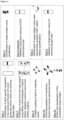
    • Figure 1: Overview of method used to identify a novel target antigen in an index patient, including isolating IgG4-antibodies from the blood of the index patient, coupling of the purified IgG4 to magnetic beats, immunoprecipitation of bound antigen targets, concentration of elution fractions and identification of target antigen using mass spectrometry.
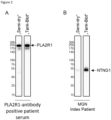
    • Figure 2: Comparison of antibody binding to target antigen after protein transfer using "Semi-dry" (25 V, 1 A, 30 min at room temperature) or "Tank-Blot" method (12 V, 25 mA, 3 h on ice). Except for the transfer method, all procedural conditions were kept the same. The protein sample is human glomerular extract (HGE) which was separated in a polyacrylamide gel by electrophoresis (SDS-PAGE). A: Recognition of PLA2R1 target antigen by PLA2R1-antibody positive patient serum. B: Recognition of NTNG1 target antigen by MGN index patient serum.
    • Figure 3: Identification of NTNG1 as target antigen in the MGN index patient by tandem mass tagged (TMT)-based relative quantification mass spectrometry. Shown is a volcano blot showing the specific accumulation of NTNG1 in the patient sample compared to the healthy control.
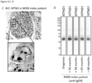
    • Figure 4: Validation of NTNG1 as target antigen in the MGN index patient. A: Gentle Western Blot of different HGE protein samples (membrane fraction, cytoplasmic fraction and deglycosylated protein) probed with serum of the index patient. B: Gentle Western Blot of recombinantly expressed NTNG1 polypeptide probed with serum of the index patient, a healthy subject, a PLA2R1-antibody positive patient and a THSD7A-antibody positive patient. C: Immunohistochemical staining of renal biopsy sample from the MGN index patient with anti-NTNG1 antibody. D: Gentle Western Blot of recombinantly expressed NTNG1 polypeptide probed with serum of the index patient at time of diagnosis, +36 months, +38 months, +45 months and +48 months after diagnosis. Secondary antibody was anti-IgG4 antibody.
    DETAILED DESCRIPTION OF THE INVENTION General definitions
  • Before the invention is described in detail with respect to some of its preferred embodiments, the following general definitions are provided.
  • The present invention as illustratively described in the following may suitably be practiced in the absence of any element or elements, limitation or limitations, not specifically disclosed herein. The present invention will be described with respect to particular embodiments and with reference to certain figures but the invention is not limited thereto but only by the claims.
  • Where the term "comprising" is used in the present description and claims, it does not exclude other elements. For the purposes of the present invention, the term "consisting of" is considered to be a preferred embodiment of the term "comprising of". If hereinafter a group is defined to comprise at least a certain number of embodiments, this is also to be understood to disclose a group which preferably consists only of these embodiments.
  • For the purposes of the present invention, the term "obtained" is considered to be a preferred embodiment of the term "obtainable". If hereinafter e.g. an antibody is defined to be obtainable from a specific source, this is also to be understood to disclose an antibody which is obtained from this source.
  • Where an indefinite or definite article is used when referring to a singular noun, e.g. "a", "an" or "the", this includes a plural of that noun unless something else is specifically stated. The terms "about" or "approximately" in the context of the present invention denote an interval of accuracy that the person skilled in the art will understand to still ensure the technical effect of the feature in question. The term typically indicates deviation from the indicated numerical value of ±10%, and preferably of ±5%.
  • Technical terms are used by their common sense. If a specific meaning is conveyed to certain terms, definitions of terms will be given in the following in the context of which the terms are used.
  • The term "autoantibody" refers to an antibody that is produced by the immune system of a subject (or patient) and that is directed against a subject's (or patient's) own proteins. Autoantibodies may attack the body's own cells, tissues, and/or organs, causing inflammation and cell injury. The terms "autoantibody recognizing NTNG1" can be used interchangeably with "anti-NTNG1 autoantibody". The term "anti-NTNG1 antibody" also includes anti-NTNG1 autoantibodies.
  • The term "circulating autoantibody" refers to autoantibodies circulating within the body, for example within the blood circulatory system or the lymph system. Specifically, circulating autoantibodies are commonly detected in the serum of a subject.
  • The term "expression" or "gene expression" as used herein refers to the process of synthesis of a gene product, preferably a functional RNA or protein. Gene expression generally comprises DNA transcription, optionally RNA processing and in the case of protein-expressing genes, RNA translation.
  • The term "heterologous" (or exogenous or foreign or recombinant or non-native) polypeptide is defined herein as a polypeptide that is not native to the host cell, a polypeptide native to the host cell in which structural modifications, e.g., deletions, substitutions, and/or insertions, have been made by recombinant DNA techniques to alter the native polypeptide, or a polypeptide native to the host cell whose expression is quantitatively altered or whose expression is directed from a genomic location different from the native host cell as a result of manipulation of the DNA of the host cell by recombinant DNA techniques, or whose expression is quantitatively altered as a result of manipulation of the regulatory elements of the polynucleotide by recombinant DNA techniques e.g., a stronger promoter; or a polynucleotide native to the host cell, but integrated not within its natural genetic environment as a result of genetic manipulation by recombinant DNA techniques.
  • The terms "nucleic acid" or "nucleic acid molecule" or "nucleic acid sequence" or "nucleotide sequence" are used interchangeably herein to refer to a biomolecule composed of nucleotides. The nucleic acid molecule can be comprised within an eukaryotic or prokaryotic organism, a eukaryotic or prokaryotic cell, a cell nucleus or a cell organelle, as part of a genome or as an individual molecule; or it can be comprised within a plasmid, a vector, an artificial chromosome; a nucleic acid can also exist outside of a cell, in vesicles, viruses or freely circulating, it can be isolated in a suitable composition, in a fixed or frozen tissue or cell culture, or dried. The nucleic acid can be synthesized or naturally occurring, i.e. isolated from nature.
  • The terms "sequence identity", "% sequence identity", "% identity", "% identical" or "sequence alignment" are used interchangeably herein and refer to the comparison of a first nucleic acid sequence to a second nucleic acid sequence, or a comparison of a first amino acid sequence to a second amino acid sequence and is calculated as a percentage based on the comparison. The result of this calculation can be described as "percent identical" or "percent ID." A sequence identity may be determined by a program, which produces an alignment, and calculates identity counting both mismatches at a single position and gaps at a single position as non-identical positions in final sequence identity calculation. The sequence identity is determined over the entire length of the first and second nucleic acid sequence.
  • The terms "encoded protein" or "encoded amino acid" refers a protein that consists of a chain of amino acids, which results from a sequence that is encoded by a nucleic acid molecule comprising three-nucleotide codons.
  • The terms "polypeptide", "protein" or "peptide" are used interchangeably herein and refer to amino acid sequences of a variety of lengths. The term polypeptide may also refer to the primary, secondary, tertiary, quartenery or quinary structure or a protein. Polypeptides can be in uncharged forms or as salts, either unmodified or modified by glycosylation, side chain oxidation, phosphorylation, citrullination or transglutamination. In certain embodiments, the polypeptide is a full-length native protein. In other embodiments, the polypeptide is a smaller fragment of the full-length protein. In still other embodiments, the amino acid sequence is modified by additional substituents attached to the amino acid side chains, such as N- or C-terminal added protein tags such as affinity tags, linkers, which may comprise 2-40, preferably 5 to 30 amino acids with epitopes other than those from NTNG1 or not epitopes, glycosyl units, lipids, or inorganic ions such as phosphates, as well as modifications relating to chemical conversion of the chains such as oxidation of sulfhydryl groups. Thus, the term "polypeptide" is intended to include the amino acid sequence of the full-length native protein, or a fragment thereof, subject to those modifications that do not significantly change its specific properties. In particular, the term "polypeptide" encompasses protein isoforms, i.e., variants that are encoded by the same gene, but that differ in their amino acid sequence or in other properties.
  • The term "antibody-binding fragment of an NTNG1 polypeptide" or "anti-NTNG1 antibody-binding fragment" relates to a molecule comprising an amino acid sequence derived from a protein that is not full length and comprises an antigen sequence which enables antibody binding. When used herein in connection with an antigen (e.g. NTNG1), "anti-NTNG1 antibody-binding fragment" refers to a fragment of the antigen that retains the ability of the antigen to bind specifically an anti-NTNG1 antibody to form an antibody-antigen complex. Suitable anti-NTNG1 antibody-binding fragments of an antigen may be identified by one skilled in the art by simple trials to ascertain their ability to bind specific autoantibodies of MGN.
  • The term "antigen" relates to a molecule or a molecular structure that can be bound by an antigen-specific antibody or a binding fragment thereof. An antigen can be composed of one or more epitopes, which can be linear structure. An antigen can also be composed of amino acids that form structure-dependent epitopes.
  • The "epitope" refers to the distinct surface features of an antigen that can act as points of interaction for specific antibodies. Antigenic molecules can comprise one or more epitopes. The minimum size of a linear epitope is about 4 amino acids and comprises one or more antibody contact residues.
  • An "autoantigen" is an endogenous molecule, a polypeptide, protein or protein complex, or a DNA or RNA molecule that is recognized by the immune system of a subject suffering from a specific autoimmune disease.
  • The terms "membranous nephropathy", "membranous glomerulopathy" or "membranous glomerulonephritis" or "MGN" refer to a slowly progressive disease of the kidney which is a frequent cause of adult nephrotic syndrome. Primary or Idiopathic membranous glomerulonephritis as described herein is considered to be an autoimmune disease targeting the glomerulus. Secondary membranous glomerulonephritis includes nephropathies caused by secondary factors such as systemic lupus erythematosus, hepatitis B or syphilis.
  • The "UniProt numbers" or "UniProt" or "UniProt Accession numbers" provided herein refer to the unique identifiers given to individual genes and proteins by the UniProt Consortium, which are available from their database at www.uniprot.org and commonly used as references in the field. UniProtKB (UniProt Knowledgebase) is a freely accessible database of protein sequence and functional information. The UniProt database includes manually annotated and reviewed entries (provided by the Swiss-Prot database) and automatically annotated and not manually reviewed entries (provided by TrEMBL database), many of which are derived from genome sequencing projects. TrEMBL includes translated coding sequences from the EMBL-Bank/GenBank/DDBJ nucleotide sequence database, and others.
  • As used herein, the term "biomarker" or "marker" refers to a measurable indicator of the presence or severity of a disease state. A biomarker can for example be used for diagnosis, to measure the progress of disease, or to evaluate effective therapeutic regimes. According to the invention, NTNG1 protein or an NTNG1 polypeptide or any anti-NTNG1 antibody-binding fragment thereof; and anti- NTNG1 autoantibodies are biomarkers of MGN, particularly idiopathic MGN. The efficacy of a candidate drug to treat MGN may be tested or monitored. A decreasing level of anti-NTNG1, specifically of anti-NTNG1 autoantibody, indicates an increased likelihood that the drug is effective.
  • The term "Phospholipase A2 Receptor 1" (PLA2R1, UniProt: Q13018) refers a type-1 transmembrane protein, which in humans is encoded by the PLA2R1 gene and predominantly expressed in the podocytes of the renal corpuscle of the kidney. It is known as an antigen that is predominant in about 70% of patients with idiopathic MGN.
  • The term "Thrombospondin type 1 domain-containing protein 7A" (THSD7A, UniProt: Q9UPZ6) refers to a protein highly expressed on podocytes which in humans in encoded by the THSD7A gene. It is a known antigen in MGN and the second most common antigen in MGN after PLA2R1.
  • As used herein, the term "patient" or "subject" is used interchangeably and refers to a human subject or another mammal subject (e.g., primate, dog, cat, goat, horse, pig, mouse, rat, rabbit, and the liked), that can be afflicted with a membranous nephropathy, particularly an idiopathic MGN. Preferably, the patient or subject is a human patient or subject. A patient suspected to have MGN has typically been diagnosed based on a kidney biopsy or due to the presence of a biomarker associated with MGN.
  • The term "biological sample" is used herein in its broadest sense. A biological sample is generally obtained for a subject. Said subject is mammal, preferably human. Typically, biological sample is generally obtained from a patient or subject. Typically, said sample comprises a representative set of autoantibodies or autoantigen. Hence, said biological sample may be a blood sample or a tissue sample. The tissue sample is preferably a kidney biopsy sample.
  • A sample may be of any biological tissue or fluid with which biomarker(s) of the present invention may be assayed. Such samples include, but are not limited to, bodily fluids which may or may not contain cells, e.g., blood (e.g., whole blood, serum or plasma) or urine. Such samples also include biopsies (for example kidney biopsy). The term biological sample also encompasses any material derived by processing a biological sample. Derived materials include, but are not limited to, cells (or their progeny) isolated from the sample or proteins extracted from the sample. Processing of a biological sample may involve one or more of: filtration, distillation, extraction, concentration, inactivation of interfering components, addition of reagents, and the like.
    In the context of the present invention, the term "control", when used to characterize a subject, refers to a subject that is healthy or to a patient that has been diagnosed with a specific disease other than renal disease. The term "control sample" refers to one, or more than one sample, that has been obtained from a healthy subject or from a patient diagnosed with a disease other than renal disorder. A control sample as used herein may also be a sample from a patient diagnosed with PLA2R1-positive or THSD7A-positive MGN.
  • As used herein, the term "level" refers to the quantity of a molecule in a biological sample. For example, "anti-NTNG1 autoantibody level" refers to the number of detectable anti-NTNG1 autoantibody in a sample. The number may be a total or absolute number, or the number may be relative to a control or reference measurement. The level may be determined numerically, or it may be determined by measuring a secondary signal, such as fluorescence intensity. The level may also be expressed binary by presence or absence of the measured molecule, i.e. the level may be positive or negative. Antibody "level" can also refer be called "titer". When referring to a polypeptide or protein, "level" may also refer to the expression level of the polypeptide or protein.
  • As used herein, the term "reference level" refers to a level measured in a biological sample obtained from a control or preferably to an average of several levels measured in biological samples obtained from several controls. As used herein, the term "diagnosis", "diagnosing" or "diagnostic" is used on its broadest sense and encompasses diagnosis, prognosis, theragnosis and monitoring in membranous nephropathy.
  • The terms "NTNG1-negative patient", "PLA2R1-negative patient or "THSD7A-negative patient" (or subject) refers to a patient whose serum contains no autoantibodies or at least no detectable levels of autoantibodies directed against the respective antigen. The terms "NTNG1-positive patient", "PLA2R1- positive patient or "THSD7A- positive patient" refer to a patient whose serum contains autoantibodies directed against the respective protein.
  • As used herein, the term "contacting" or "reacting" means bringing two compounds or molecules close enough to each other that they can chemically, electrically or physically interact. This interaction may be due to forces between molecules and may involve bonding and unbonding of molecules. Contacting may take place in solution or between solutions, on a solid support, between a liquid and solid phase, between a liquid and gaseous phase, between a solid and gaseous phase, within one or more gaseous phases or within a cell or organism or between cells and organism.
  • Novel Biomarker Netrin G1
  • Netrin G1 (NTNG1, UniProt: Q9Y2I2), which is in humans encoded by the gene NTNG1, is a secreted glycoprotein of about 50 kDa size. It binds to the cell membrane on the extracellular side with a glycosylphosphatidylinositol (GPI) anchor. Thus, NTNG1 is exposed on the cell surface of podocytes and is able to bind circulating antibodies. Podocytes are specialized epithelial cells that cover the outer surfaces of glomerular capillaries.
  • Several isoforms of NTNG1 exist, of which isoform 3 represents the full-length protein. Table 1 provides information about the different isoforms and changed or missing amino acid features compared in relation to isoform 3. Table 1: Overview of NTNG1 isoforms
    Isoform Region MW (not processed)
    3 (also 1A) M1 - F539 60541
    2 (also 1F) M1 - C364 41759
    363-364 NC → SK
    1 (also 1C) 363-463 missing 49340
    464-464 P → T
    4 (also 1D) 364-464 53946
    CECFGHSNRC...GNSWHYGCQP → PPKFNRIW PN...SVQVANHKRA
    5 (also 1E) 364-385 51858
    CECFGHSNRCSYIDLLNTVICV → PPKFNRIWPN ISSLEVSNPKQA
    386-464 missing
    6 (also 1G) 419-463: missing 55660
    464-464 P→ A
  • As a secreted protein, NTNG1 comprises domains that are removed upon maturation and processing (Table 2). The unprocessed NTNG1 polypeptide has an amino acid sequence according to SEQ ID NO: 1. The propeptide, comprising amino acids 511-539 is removed from the mature from. Hence, the mature NTNG1 polypeptide sequence is an amino acid sequence according to SEQ ID NO: 2. Furthermore, NTNG1 polypeptide comprises a signal peptide which comprises amino acids 1-28. Signal peptides are commonly cleaved off from presecretory polypeptides by signal peptidase during or immediately after insertion into the membrane. Hence, the fully processed NTNG1 polypeptide, once secreted, has an amino acid sequence according to SEQ ID NO: 3. Table 2: Overview of NTNG1 domains
    Domain amino acid
    signal peptide 1-28
    Laminin N-terminal domain 46-296
    Laminin EGF-like domain
    1 297-356
    2 364-419
    3 420-469
    GPI anchor 510 (amidated serine)
    Removed from mature form 511-539
  • In one embodiment, the NTNG1 polypeptide comprises an amino acid sequence with at least 70% identity to the amino acid sequence according to SEQ ID NO: 3. In another embodiment, the NTNG1 polypeptide comprises an amino acid sequence with at least 75%, at least 80%, at least 85%, at least 90%, at least 95%, at least 98%, at least 99%, or 100% identity to the amino acid sequence according to SEQ ID NO: 3.
  • In another embodiment, the NTNG1 polypeptide comprises an amino acid sequence with at least 70% identity to the amino acid sequence according to SEQ ID NO: 1. In another embodiment, the NTNG1 polypeptide or one or more anti-NTNG1 antibody-binding fragments thereof comprise an amino acid sequence with at least 75%, at least 80%, at least 85%, at least 90%, at least 95%, at least 98%, at least 99%, or 100% identity to the amino acid sequence according to SEQ ID NO: 1.
  • In another embodiment, the NTNG1 polypeptide comprises an amino acid sequence with at least 70% identity to an amino acid sequence selected from the group consisting of SEQ ID NO: 5, SEQ ID NO: 6, SEQ ID NO: 7 or SEQ ID NO: 8. In another embodiment, the NTNG1 polypeptide comprises an amino acid sequence with at least 80% identity to the amino acid sequence selected from the group consisting of SEQ ID NO: 5, SEQ ID NO: 6, SEQ ID NO: 7 or SEQ ID NO: 8. In another embodiment, the NTNG1 polypeptide comprises an amino acid sequence with at least 90% identity to the amino acid sequence selected from the group consisting of SEQ ID NO: 5, SEQ ID NO: 6, SEQ ID NO: 7 or SEQ ID NO: 8.. In another embodiment, the NTNG1 polypeptide comprises an amino acid sequence according to an amino acid sequence selected from the group consisting of SEQ ID NO: 5, SEQ ID NO: 6, SEQ ID NO: 7 or SEQ ID NO: 8.
  • As used herein, the expression "anti-NTNG1 antibody-binding fragment of the NTNG1 polypeptide" or "fragment of NTNG1" refers to a continuous amino acid sequence comprised in the full-length NTNG1 polypeptide that comprises an antigen sequence that can recognize and bind an anti-NTNG1 antibody, and is able to participate in the formation of an antigen-antibody complex with one or more anti-NTNG1 antibodies.
  • In one embodiment, said anti-NTNG1 antibody-binding fragment of the NTNG1 polypeptide comprises at least 10 consecutive amino acids of the entire amino acid sequence of NTNG1. In another embodiment, said anti-NTNG1 antibody-binding fragment of NTNG1 comprises at least 20, at least 40, at least 60, at least 80, at least 100, at least 120, at least 140, at least 160, at least 180, at least 200, at least 250, at least 300, at least 350, at least 400, at least 450, at least 500 or 539 consecutive amino acids of the entire amino acid sequence of full-length NTNG1 (SEQ ID NO: 1).
  • In another embodiment, the anti-NTNG1 antibody-binding fragment of NTNG1 comprises at least 5%, at least 10%, at least 20%, at least 30%, at least 40%, at least 50%, at least 60%, at least 70%, at least 75%, at least 80%, at least 85%, at least 90%, at least 95%, at least 98%, at least 99% or 100% of the amino acid sequence of full-length NTNG1, i.e. SEQ ID NO: 1. In another embodiment, the anti-NTNG1 antibody-binding fragment of NTNG1 comprises at least 10%, at least 20%, at least 30%, at least 40%, at least 50%, at least 60%, at least 70%, at least 75%, at least 80%, at least 85%, at least 90%, at least 95%, at least 98%, at least 99% or 100% of the amino acid sequence of mature NTNG1, i.e. SEQ ID NO: 3.
  • Preferably, the anti-NTNG1 antibody-binding fragment of NTNG1 is recognized by an autoantibody directed against NTNG1. Determining the ability of the fragment to interact with said autoantibodies and detecting antibody-antigen complexes between the NTNG1 polypeptide or one or more anti-NTNG1 antibody-binding fragments thereof and anti-NTNG1 autoantibodies can be accomplished by one of the methods described herein or by methods known in the art for determining direct binding. Such methods include co-immunoprecipitation, pull down assay, crosslinking assays, gel electrophoresis and immunoblots (Western Blot, Dotplot), ELISA (enzyme-linked immunosorbent assay), EIA (enzyme immunoassay), FRET (fluorescence resonance energy transfer), luciferase assay, immunohistochemistry, flow cytometry, co-crystallization, or mass spectrometry.
  • In a preferred embodiment, anti-NTNG1 antibody or a complex comprising NTNG1, such as a complex comprising NTNG1 polypeptide or one or more anti-NTNG1 antibody-binding fragments thereof or any other analyte, such as an analyte recognizing NTNG1 antibody, is detected using a detection method selected from the group comprising immunodiffusion, immunoelectrophoresis, light scattering immunoassays, agglutination, labeled immunoassays such as those from the group comprising radiolabeled immunoassay, enzyme immunoassays, more preferably ELISA, chemiluminescence immunoassays, preferably electrochemiluminescence immunoassay, and immunofluorescence, preferably indirect immunofluorescence. A competitive assay, a capture bridge assay, an immunometric assay, a class-specific second antibody on the solid phase, a direct or indirect class capture assay may also be used. The principle of each of these formats is detailed in The Immunoassay Handbook, 3rd edition, edited by David Wild, Elsevier, 2005. The detection of the anti-NTNG1 is preferably carried out under non-reducing conditions.
  • Methods of the Invention anti-NTNG1 autoantibodies in subjects with MGN
  • A first aspect of the invention relates to an in vitro method, optionally an in vitro method of diagnosing membranous glomerulonephritis (MGN) in a subject, comprising detecting one or more autoantibodies recognizing a Netrin G1 (NTNG1) polypeptide or one or more anti-NTNG1 antibody-binding fragments thereof in a biological sample obtained from a subject.
  • In one embodiment, the in vitro method further comprises the step of (i) contacting a biological sample obtained from the subject with an NTNG1 polypeptide or one or more anti-NTNG1 antibody-binding fragments thereof, and (ii) detecting any antigen-antibody complexes formed between the NTNG1 polypeptide or one or more anti-NTNG1 antibody-binding fragments thereof and anti-NTNG1 autoantibodies in the biological sample.
  • In one embodiment, detecting the antigen-antibody complexes between the NTNG1 polypeptide or one or more anti-NTNG1 antibody-binding fragments thereof and anti-NTNG1 autoantibodies comprises determining the presence of anti-NTNG1 autoantibodies.
  • In one embodiment, detection of antigen-antibody complexes between the NTNG1 polypeptide or one or more anti-NTNG1 antibody-binding fragments thereof and anti-NTNG1 autoantibodies formed in the biological sample may lead to a positive diagnosis of the subject. In many cases the mere detection of the anti-NTNG1 autoantibody, in other words determining whether or not detectable levels of the antibody are present in the sample, is sufficient for the diagnosis. In a more preferred embodiment, this may involve determining whether the concentration is at least 10%, preferably 20%, 50%, 100%, 200%, 500%, 1.000%, 2.000%, 2.500%, 5.000%, 1.0000%, 2.0000%, 5.0000%, 100.000%, 1.000.000% or 10.000.000% times higher than the concentration of the antibody of interest found in the average healthy subject. If the autoantibody can be detected, this will be information instrumental for the clinician's diagnosis. It may indicate an increased likelihood that the patient suffers, suffered or will suffer from MNG.
  • According to the present invention, a kit is provided, comprising a cell expressing, preferably overexpressing the NTNG1 polypeptide or one or more anti-NTNG1 antibody-binding fragments thereof, or the device or the NTNG1 polypeptide or one or more anti-NTNG1 antibody-binding fragments thereof and further comprising one or more, preferably all reagents from the group comprising a secondary antibody, preferably labeled with a detectable label, a washing solution, a positive control, a negative control, a detergent, a cover glass, a mounting medium and a physiological salt solution, preferably PBS, or salt required to prepare it.
  • In a preferred embodiment, the positive control is a diluted sample, preferably blood from a patient suffering from MNG and comprising anti-NTNG1 or a monoclonal antibody binding specifically to NTNG1 polypeptide, preferably recognized by human IgG antibodies. The negative control may be a diluted sample from a healthy subject, for example a blood donor. The kit may comprise instructions how to carry out the assay. The kit may comprise a calibrator, preferably a set of at least three calibrators. In a preferred embodiment, the term "calibrator", as used herein, refers to a reagent that may be used to calibrate an assay, in particular set up a reference range or a calibration curve. In a preferred embodiment, the calibrator is an antibody which binds to the NTNG1 and/or the polypeptide comprising NTNG1 coated on a device and is recognized by a secondary antibody recognizing the antibody to be detected, more preferably IgG, most preferably human IgG. A set of calibrators may comprise at least 3, 4 or 5 solutions each comprising different known concentrations of the calibrator. The concentrations between the calibrator with the lowest concentration and the calibrator with the highest concentration may be at least 1:10, 1:20, 1:50, 1:100, 1:200, 1:500 or 1:1000. Preferably, the secondary antibody is a secondary antibody binding specifically to IgG class antibodies, preferably human IgG class antibodies. In a preferred embodiment, the kit comprises the NTNG1 polypeptide or one or more anti-NTNG1 antibody-binding fragments thereof, optionally comprising a detectable label. The kit may be used for detecting a mammalian antibody to NTNG1 in a sample, preferably a human antibody to NTNG1 in a sample, more preferably a blood sample. The kit may be used for diagnosing, aiding in the diagnosis of NTNG1 or for identifying a subject having an increased risk of suffering from NTNG1 in the past, present or future.
  • Another aspect of the invention relates to a method comprising the step of detecting the presence or absence of anti-NTNG1 antibody, in a sample.
  • In a preferred embodiment, the method may be for diagnosing or aiding in the diagnosis of a nephrological autoimmune disease, preferably MGN, or identifying a subject having an increased risk of suffering from the disease in the past, presence or future. In another preferred embodiment, the method may be for distinguishing an autoimmune nephrological disease from another nephrological disease not associated with the presence of anti-NTNG1.
  • In a preferred embodiment, any information, result or data obtained according to the present invention, for example demonstrating the presence of absence of anti-NTNG1 in a sample or the expression of NTNG1, may be communicated to the patient or a medical doctor treating the patient orally, preferably by telephone, in a written form, preferably fax or letter, or in an electronic form via fax or via the internet, for example as an email or text message.
  • In one embodiment, the NTNG1 polypeptide or one or more anti-NTNG1 antibody-binding fragments thereof is from human NTNG1. In another embodiment, the NTNG1 polypeptide or one or more anti-NTNG1 antibody-binding fragments thereof is from a different mammalian species, such as non-human primate, pig, sheep, horse, donkey, rabbit, rat or mouse.
  • In one embodiment, the NTNG1 polypeptide may be a full-length NTNG1 polypeptide according to SEQ ID NO: 1. In another embodiment, the NTNG1 polypeptide may be a mature, processed NTNG1 polypeptide according to SEQ ID NO: 3.
  • In one embodiment the NTNG1 polypeptide or one or more anti-NTNG1 antibody-binding fragments thereof is an isolated and/or a recombinant polypeptide.
  • The NTNG1 polypeptide or one or more anti-NTNG1 antibody-binding fragments thereof may be recombinantly expressed in a cell or organism. Hence, In one embodiment, the NTNG1 polypeptide or one or more anti-NTNG1 antibody-binding fragments thereof is a recombinant polypeptide or one or more antibody-binding fragments thereof. In another embodiment, the recombinant polypeptide or one or more antibody-binding fragments is produced in recombinant mammalian cells.
  • In a preferred embodiment, the antibody, preferably anti-NTNG1, such as anti-NTNG antibody, or the complex to be detected, binds specifically to its interaction partner, preferably NTNG1 polypeptide. In a preferred embodiment, the term "binding specifically", as used herein, preferably means that the binding reaction is stronger than a binding reaction characterized by a dissociation constant of 1 × 10⁻⁵ M, more preferably 1 × 10⁻⁷ M, more preferably 1 × 10⁻⁸ M, more preferably 1 × 10⁻⁹ M, more preferably 1 × 10⁻¹⁰ M, more preferably 1 × 10-11 M, more preferably 1 × 10-12 M, as determined by surface plasmon resonance using Biacore equipment at 25 °C in PBS buffer at pH 7.
  • In another preferred embodiment, the methods and products according to the present invention may be used for determining the concentration of an antibody binding specifically to NTNG1 in a liquid, which may not be a sample from a subject or only a processed sample, for example for preparing a calibrator or set of calibrators with known relative or absolute concentrations or standardizing solutions.
  • Methods for expressing polypeptides in cells or organisms are known in the art and include cell/tissue culture, batch cell culture, fed-batch culture or suspension culture or in vitro cell free expression systems. Cells or organisms capable of producing the NTNG1 polypeptide or one or more antibody-binding fragments include mammalian cells, insect cells, yeasts, fungal cells, bacterial cells, plant and algal cells. Preferably, the NTNG1 polypeptide or one or more antibody-binding fragments are produced in mammalian cells. Suitable mammalian cell lines include human embryonic kindey (HEK) 293, human fibrosarcoma HT-1080, human embryonic retinal PER.C6, HeLa, Chinese hamster ovary (CHO), hamster kidney BHK 21, mouse myeloma NS0, and murine C127.
  • Mammalian expression systems can be transiently induced to produce the polypeptide of the invention by transfection, transduction or infection or a suitable vector. The mammalian cell may also be a stable cell lines in which the expression construct is stably integrated into the host genome at one or more locations.
    Suitable non-mammlian cells are insect cells optimized for protein expression such as Drosophila melanogasterS2 cells, Spodoptera frugiperda Sf9 or Sf21 cells or Trichoplusia niBTI-TN-5B1-4 (High Five) cells.
  • Transient expression is the temporary expression of genes that are expressed for a short time after a nucleic acid has been introduced into the cell or organism. Transient expression may be achieved by transfection with a plasmid vector or viral transduction.
  • Stable integration may be achieved using methods known in the art, including virus transduction, plasmid, BAC (bacterial artificial chromosome) or YAC (yeast artificial chromosome) transfection, transposon-mediated transfer and integration, site-specific integration and gene editing methods such as zinc finger nucleases, CRISPR/Cas9 or TALENs.
  • Methods of isolating or purifying NTNG1 polypeptide or one or more anti-NTNG1 antibody-binding fragments thereof from the cell or organism or culture medium are described herein and known in the art and include spin columns and kits, liquid chromatography, such as FPLC or HPLC, magnetic beads, filter plates, or resins.
  • In one embodiment, the NTNG1 polypeptide or one or more anti-NTNG1 antibody-binding fragments thereof may comprise an epitope or purification tag, preferably selected from His-tag, GS-tag, FLAG-tag, c-Myc tag or HA-tag. The tag may also be a fluorescent tag, such as a fluorescent protein or FRET label, or a radiolabel. In another embodiment, the NTNG1 polypeptide or one or more anti-NTNG1 antibody-binding fragments thereof may be untagged.
  • In one embodiment, the invention also provides a vector for the expression of an NTNG1 polypeptide or one or more anti-NTNG1 antibody-binding fragments thereof, preferably wherein the vector comprises an expression cassette comprising a promoter and a nucleic acid molecule encoding the NTNG1 polypeptide or one or more anti-NTNG1 antibody-binding fragments thereof of the invention. In one embodiment, the vector may comprise a nucleic acid molecule with at least 70%, at least 75%, at least 80%, at least 85%, at least 90%, at least 95%, at least 98%, at least 99% or 100% identity with the nucleic acid according to SEQ ID NO: 4.
  • In one embodiment, the vector is a eukaryotic expression vector or a viral vector. The promoter may be any suitable promotor for expression in the chosen system, for example CMV, T7, lac operator, EF1alpha, Tet operator, EpIE2, SV40, MT, UbC, RSV, AUG1, CMV/EF1alpha, FLD1, TK, Gal4 UAS/E1b, trc, AOX1, Polyedrin or copia.
  • In certain embodiments, the NTNG1 polypeptide or one or more anti-NTNG1 antibody-binding fragments thereof is immobilized onto a solid carrier or support (e.g., a bead, microplate or array). Hence some aspects of the invention refer to a diagnostic device on which the NTNG1 polypeptide or one or more anti-NTNG1 antibody-binding fragments thereof is immobilized. Accordingly, in some embodiments, the method of the invention involves the use of a diagnostic device coated with the NTNG1 polypeptide or one or more anti-NTNG1 antibody-binding fragments thereof. In a preferred embodiment, the device is selected from the group comprising a glass slide, preferably for microscopy, a biochip, a microtiter plate, a lateral flow device, a test strip, a membrane, preferably a line blot, a chromatography column and a bead, preferably a magnetic or fluorescent bead. In another preferred embodiment, the term "coated with" means that a polypeptide such as the NTNG1 polypeptide or an antibody or another agent is immobilized on a solid surface of a device or the polypeptide and the device are configured for such an immobilization. A particularly preferred way for the configuration or the immobilization is using a carrier and/or means modified such that one of the agent or polypeptide to be immobilized comprises an affinity tag such as biotin and the other comprises a ligand to the affinity tag such as streptavidin. Alternatively, one of the carrier or the polypeptide or agent to be immobilized may comprise reactive chemical groups such as thiol, amino, epoxide, ester and anhydride groups which form covalent or non-covalent bonds with reactive chemical groups associated with the other. The device may comprise a negative control, for example a polypeptide which does not bind specifically to anti-NTNG1, preferably from human, preferably spatially separate from the NTNG1 polypeptide or one or more anti-NTNG1 antibody-binding fragments.
  • The diagnostic methods of the present invention involve detection of an antigen- antibody complex formed between the biomarker of the invention (e.g. NTNG1 polypeptide or one or more anti-NTNG1 antibody-binding fragments thereof) and an autoantibody present in the biological sample tested. This detection is indicative of the presence of autoantibodies (namely anti-NTNG1 autoantibodies or antigen-binding fragments thereof) in said sample.
  • The detection of an antibody-antigen complex between the NTNG1 polypeptide or one or more anti-NTNG1 antibody-binding fragments thereof and anti-NTNG1 autoantibodies may be performed by any suitable method. For example, the antibody-antigen complex between the NTNG1 polypeptide or one or more anti-NTNG1 antibody-binding fragments thereof and anti-NTNG1 autoantibodies may be detected using an immunoassay. Examples of immunoassays include radioimmunoassay, enzyme immunoassays (EIA), enzyme-linked immunosorbent assays (ELISA), immunofluorescence or immunoprecipitation. To this end, the NTNG1 polypeptide or one or more anti-NTNG1 antibody-binding fragments thereof is immobilized on a solid surface and the biological sample is brought into contact with the bound polypeptide or fragments thereof under conditions allowing formation of an antigen-antibody complex between the NTNG1 polypeptide or one or more anti-NTNG1 antibody-binding fragments thereof and anti-NTNG1 autoantibodies. Following incubation, the antigen-antibody complex between the NTNG1 polypeptide or one or more anti-NTNG1 antibody-binding fragments thereof and anti-NTNG1 autoantibodies can then be detected using any secondary IgG antibody labelled, for example human IgG antibody for samples from human subjects, with a detectable moiety.
  • The antibody-antigen complex between the NTNG1 polypeptide or one or more anti-NTNG1 antibody-binding fragments thereof and anti-NTNG1 autoantibodies may also be detected using an immunoblot, such as the Western Blot described herein. In this case, the NTNG1 polypeptide or one or more anti-NTNG1 antibody-binding fragments thereof are immobilized in a carrier such as a polyacrylamide gel. Then, the NTNG1 polypeptide or one or more anti-NTNG1 antibody-binding fragments thereof are transferred to a blotting membrane. The biological sample is added and the antigen-antibody complex between the NTNG1 polypeptide or one or more anti-NTNG1 antibody-binding fragments thereof and anti-NTNG1 autoantibodies forms on the membrane. The antigen-antibody complex between the NTNG1 polypeptide or one or more anti-NTNG1 antibody-binding fragments thereof and anti-NTNG1 autoantibodies can then be detected using any secondary IgG antibody labelled, for example human IgG antibody for samples from human subjects, with a detectable moiety.
  • Preferably, the formation of antigen-antibody complex between the NTNG1 polypeptide or one or more anti-NTNG1 antibody-binding fragments thereof and anti-NTNG1 autoantibodies is achieved under non-reducing conditions.
  • The secondary antibody may be labeled with any detectable moiety, i.e., any entity that, by its chemical nature, provides an analytically identifiable signal allowing detection of the ternary complex between the NTNG1 polypeptide or one or more anti-NTNG1 antibody-binding fragments thereof and anti-NTNG1 autoantibodies, and consequently detection of the biomarker-antibody complex between the NTNG1 polypeptide or one or more anti-NTNG1 antibody-binding fragments thereof and anti-NTNG1 autoantibodies.
  • Detection may be either qualitative or quantitative. Methods for labeling biological molecules such as antibodies are well-known in the art. The most commonly used detectable moieties in immunoassays are enzymes and fluorophores. In the case of an enzyme immunoassay (EIA or ELISA), an enzyme such as horseradish perodixase, glucose oxidase, beta-galactosidase, alkaline phosphatase, and the like, is conjugated to the second antibody, generally by means of glutaraldehyde or periodate. The substrates to be used with the specific enzymes are generally chosen for the production of a detectable color change, upon hydrolysis of the corresponding enzyme. In the case of immunofluorescence, the second antibody is chemically coupled to a fluorescent moiety without alteration of its binding capacity. After binding of the fluorescently labeled antibody to the biomarker-antibody complex and removal of any unbound material, the fluorescent signal generated by the fluorescent moiety is detected, and optionally quantified. Alternatively, the secondary antibody may be labeled with a radioisotope, a chemiluminescent moiety, or a bioluminescent moiety.
  • Another method of the invention concerns the correlation between the presence and level of anti-NTNG1 autoantibodies and effectiveness of treatment, remission or relapse of patients with MGN, more specifically idiopathic MGN.
  • Current treatments used for membranous nephropathy, particularly idiopathic membranous nephropathy, are immunosuppressive therapy, for example, glucocorticoids, cyclosporin, tacrolimus, azathioprine, infliximab, omalizumab, daclizumab, adalimumab, eculizumab, efalizumab, natalizumab, omalizumab and rapamycin. It also includes cyclophosphamide, chlorambucil, and rituximab.
  • Thus, the invention further relates to an in vitro method of determining the effectiveness of a treatment for MGN in a subject, comprising (i) determining the level of anti-NTNG1 autoantibodies in a first biological sample obtained from a subject at a first time-point and (ii) determining the level of anti-NTNG1 autoantibodies in a second biological sample obtained from a subject at a second time-point, wherein a decrease in the level of anti-NTNG1 autoantibodies in the second time point compared to the first time point indicates that the treatment is effective, and/or an increase in the level of anti-NTNG1 autoantibodies in the second time point compared to the first time point indicates that the treatment is not effective.
  • In one embodiment, determining the level of anti-NTNG1 autoantibodies in a first or second sample comprises contacting a biological sample obtained from the subject with a Netrin G1 (NTNG1) polypeptide or one or more anti-NTNG1 antibody-binding fragments thereof and detecting any antigen-antibody complexes formed between the NTNG1 polypeptide or one or more anti-NTNG1 antibody-binding fragments thereof and anti-NTNG1 autoantibodies in the biological sample.
  • In one embodiment, the second time point is chosen later than the first time point, preferably at least 1 months, 2 months, 4 months, 6 months, 12 months, 24 months, 36 months later or more than 36 months later. The method can be performed several times using more than one first and second time points. In one embodiment, the method is performed in intervals ranging between 2 months and 24 months.
  • Preferably, the method of determining the effectiveness of a treatment for MGN in a subject is performed for subjects that have been diagnosed for MGN and that are NTNG1-positive subjects.
  • The level of the anti-NTNG1 autoantibodies can be detected by an immunoassay wherein an antigen-antibody complex is formed, as described above.
  • Upon treatment, for example, with immunosuppressive therapy, a decrease of the amount of detectably circulating anti-NTNG1 autoantibodies is observed or likely observed in case of effective treatment.
  • The treatment is considered to be effective when a decrease of at least 10%, at least 20%, at least 30%, at least 40%, at least 50%, at least 60%, at least 70%, or at least 80%, of the level of anti-NTNG1 autoantibodies is observed. Particularly, the treatment is considered to be effective when a decrease of at least 50% of the level of anti-NTNG1 autoantibodies is observed. In one embodiment, the treatment is considered to be effective when a decrease of between 10% to 100% of the level of anti-NTNG1 autoantibodies is observed. Hence, in some embodiments, the level of anti-NTNG1 autoantibodies after treatment is not detectable, i.e. 100% decreased.
  • Conversely, the treatment is considered to be ineffective or likely ineffective when level between the first and the second time point is stable or increases.
  • In one embodiment, the NTNG1 polypeptide or one or more anti-NTNG1 antibody-binding fragments thereof are soluble. In another embodiment, the NTNG1 polypeptide or one or more anti-NTNG1 antibody-binding fragments thereof are immobilized on a solid surface.
  • In one embodiment, the NTNG1 polypeptide or one or more anti-NTNG1 antibody-binding fragments thereof are isolated and/or recombinant.
  • NTNG1 expression in subjects with MGN
  • Furthermore, the inventors found that levels of NTNG1 polypeptides or one or more fragments thereof in the biological sample of a subject can be used to make a prognosis and/or diagnosis and/or determine the therapeutic needs in said subjects. Typically, NTNG1 polypeptides or one or more fragments thereof comprise at least one NTNG1 antigen that binds to circulating anti-NTNG1 autoantibodies of subjects with MGN.
  • Thus, a further aspect of the invention relates to an in vitro method for prognosis and/or diagnosis of MGN in a subject, comprising the steps of determining the expression level of NTNG1 polypeptide or one or more anti-NTNG1 antibody-binding fragments thereof in a biological sample obtained from said subject and comparing said expression level to a reference expression level, wherein an increased expression level of NTNG1 polypeptide or one or more anti-NTNG1 antibody-binding fragments thereof compared to said reference expression level is indicative of MGN. In one embodiment, the biological sample is a tissue sample, preferably a renal biopsy sample. In one embodiment, the expression level of NTNG1 polypeptide or one or more anti-NTNG1 antibody-binding fragments thereof is measured in podocytes and/or in the glomerular basement membrane (GBM). In a preferred embodiment, an increased expression level of NTNG1 polypeptide or one or more anti-NTNG1 antibody-binding fragments thereof in podocytes and/or GBM of said subject compared to said reference expression level is indicative of MGN.
    In one embodiment, the accumulation of the NTNG1 polypeptide along the capillary wall is indicative of MGN.
  • Thus in a specific embodiment, an increased expression level of NTNG1 polypeptide or one or more anti-NTNG1 antibody-binding fragments thereof in podocytes and/or GBM of said subject compared to said reference expression level in combination with the accumulation of the NTNG1 polypeptide along the capillary wall is indicative of MGN.
  • In one embodiment, the expression level of NTNG1 polypeptide or one or more anti-NTNG1 antibody-binding fragments thereof in a biological sample is determined by contacting the biological sample with an anti-NTNG1 antibody or one or more antigen-binding fragments thereof. In one embodiment, the anti-NTNG1 antibody comprises a full-length antibody or one or more antibody fragments capable of binding to one or more NTNG1 antigens.
  • "Antibodies" or "antibody", also called "immunoglobulins" (Ig), generally comprise four polypeptide chains, two heavy (H) chains and two light (L) chains, and are therefore multimeric proteins, or comprise an equivalent Ig homologue thereof (e.g., a camelid antibody comprising only a heavy chain, single-domain antibodies (sdAb) or nanobodies which can either be derived from a heavy or a light chain).
  • The term "antibodies" as used herein includes antibody-based binding proteins or fragments thereof, or modified antibody formats, all of which retain their antigen target-binding capacity. The term "antibodies" also includes full length functional mutants, variants, or derivatives thereof (including, but not limited to, murine, chimeric, humanized and fully human antibodies) which retain the essential epitope binding features of an Ig molecule, and includes dual specific, bispecific, multispecific, and dual variable domain Igs. Ig molecules can be of any class (e.g., IgG, IgE, IgM, IgD, IgA, and IgY), or subclass (e.g., IgG1, IgG2, IgG3, IgG4, IgA1, and IgA2) and allotype. Ig molecules may also be mutated e.g. to enhance or reduce affinity for Fcy receptors or the neonatal Fc receptor (FcRn).
  • An "antibody fragment", as used herein, relates to a molecule comprising at least one polypeptide chain derived from an antibody that is not full length and exhibits antigen target binding, including, but not limited to (i) a Fab fragment, which is a monovalent fragment consisting of the variable light (VL), variable heavy (VH), constant light (CL) and constant heavy 1 (CH1) domains; (ii) a F(ab')2 fragment, which is a bivalent fragment comprising two Fab fragments linked by a disulfide bridge at the hinge region (reduction of a F(ab')2 fragment result in two Fab' fragment with a free sulfhydryl group); (iii) a heavy chain portion of a Fab (Fa) fragment, which consists of the VH and CH1 domains; (iv) a variable fragment (Fv) fragment, which consists of the VL and VH domains of a single arm of an antibody; (v) a domain antibody (dAb) fragment, which comprises a single variable domain; (vi) an isolated complementarity determining region (CDR); (vii) a single chain Fv fragment (scFv); (viii) a diabody, which is a bivalent, bispecific antibody in which VH and VL domains are expressed on a single polypeptide chain, but using a linker that is too short to allow for pairing between the two domains on the same chain, thereby forcing the domains to pair with the complementarity domains of another chain and creating two antigen binding sites; (ix) a linear antibody, which comprises a pair of tandem Fv segments (VH-CH1-VH-CH1) which, together with complementarity light chain polypeptides, form a pair of antigen binding regions; (x) Dual-Variable Domain Immunoglobulin (xi) other non-full length portions of immunoglobulin heavy and/or light chains, or mutants, variants, or derivatives thereof, alone or in any combination.
  • A "nanobody" or "single-domain antibody" or "sdAb" is an antibody fragment consisting of a single monomeric variable antibody domain and is able to bind selectively to a specific antigen. Nanobodies typically comprise one variable domain of a heavy-chain (VH). They are often derived from immunization of dromedaries, camels, llamas alpacas or sharks, who naturally produce single domain VHH antibodies.
  • An "aptamer" is an oligonucleotide or peptide molecule that can bind to a specific target molecule. Aptamers can be used instead of antibodies and exhibit high binding affinity.
  • In one embodiment, the antigen-binding fragments of the anti-NTNG1 antibody are not full length antibodies and are selected from the group consisting of a F(ab) fragment, a F(ab')2 fragment, a heavy chain portion of a Fab (Fa) fragment, a variable fragment (Fv) fragment, a domain antibody (dAb), an isolated complementarity determining region (CDR), a single chain Fv fragment (scFv), a diabody, a single domain antibody (sdAb) or nanobody, a linear antibody, Dual-Variable Domain Immunoglobulin, and aptamer, or other non-full length portions of immunoglobulin heavy and/or light chains, or mutants, variants, or derivatives thereof, alone or in any combination. In a preferred embodiment, the antigen-binding fragments of the anti-NTNG1 antibody are selected from F(ab) fragment, F(ab')2 fragment, nanobody and aptamer.
  • In one embodiment, the anti-NTNG1 antibody is a monoclonal antibody, a polyclonal antibody or an aptamer. In a preferred embodiment, the anti-NTNG1 antibody is a monoclonal antibody.
  • In this case, the endogenous expression of antigen-comprising NTNG1 polypeptide serves as a biomarker. In one embodiment, the expression level of NTNG1 polypeptide or one or more anti-NTNG1 antibody-binding fragments thereof in a biological sample is increased by at least 5%, at least 10%, at least 15%, at least 20%, at least 25%, at least 30%, at least 35%, at least 40%, at least 45%, at least 50%, at least 55%, at least 60%, at least 65%, at least 70%, at least 75%, or at least 80% compared to the reference expression level. In a preferred embodiment, the expression level of NTNG1 polypeptide or one or more anti-NTNG1 antibody-binding fragments thereof in a biological sample is increased by at least 50% compared to the reference expression level. In some embodiments, the expression level of NTNG1 polypeptide or one or more anti-NTNG1 antibody-binding fragments thereof in a biological sample is increased by more than 100%, more than 200%, more than 300%, more than 400% or more than 500% compared to the reference expression level. In some embodiments, the expression level of NTNG1 polypeptide or one or more anti-NTNG1 antibody-binding fragments thereof in a biological sample is increased by 5% to 500%, preferably by 50 % to 500%, more preferably by 50 % to 200 % compared to the reference expression level. In one embodiment, said expression level refers to the expression level in podocytes and/or GBM.
  • In one embodiment, the reference expression level is determined by determining the expression level of NTNG1 polypeptide or one or more anti-NTNG1 antibody-binding fragments thereof in a group of subjects without NTNG1-associated MGN and calculating the mean expression level of NTNG1 polypeptide or one or more anti-NTNG1 antibody-binding fragments thereof in subjects without NTNG1-associated MGN. In one embodiment, said expression level refers to the expression level in podocytes and/or GBM.
  • Additionally, the expression level of the NTNG1 polypeptide or one or more anti-NTNG1 antibody-binding fragments thereof may also be measured on the basis of RNA. Hence, in one embodiment, the level of NTNG1 polypeptide or one or more anti-NTNG1 antibody-binding fragments thereof in a biological sample is determined by measuring the level of NTNG1 mRNA or detectable fragments thereof in a biological sample from a subject.
  • In one embodiment, the mRNA or fragments thereof are between 20 and 2000 nucleotides in length. In one embodiment, mRNA or fragments thereof are at least 10, at least 20, at least 30, at least 40, at least 50, at least 60, at least 70, at least 80, at least 90, at least 100, at least 150, at least 200, at least 250, at least 300, at least 350, at least 400, at least 450 or at least 500 nucleotides in length.
  • mRNA or fragments thereof can be detected using known methods such as polymerase chain reaction (PCR), quantitative PCR, RNA in situ hybridization, enzymatic digest, agarose gel-electrophoresis, expression profiling, RNA sequencing and next-generation sequencing, or Northern blotting.
  • In one embodiment, oligonucleotide primers are used to determine the level of NTNG1 mRNA expression in a biological sample from a subject. The oligonucleotide primers comprise at least 12 consecutive nucleotides selected from the nucleotide sequence according to SEQ ID NO: 4 or its reverse compliment sequence. In one embodiment, the oligonucleotide primers comprise at least 12, at least 15, at least 18 or at least 20 consecutive nucleotides selected from the nucleotide sequence according to SEQ ID NO: 4 or its reverse compliment sequence.
  • Methods of determining expression level of mRNA are well known in the field and may include transcribing RNA from a sample to cDNA by a reverse transcriptase, followed by second strand synthesis and polymerase chain reaction. These steps may be performed in one reaction set-up or sequentially. mRNA expression levels can also be determined using sequencing methods such as RNAseq or next generation sequencing. In one embodiment, the expression level of NTNG1 polypeptide or one or more anti-NTNG1 antibody-binding fragments thereof is determined by quantitative polymerase chain reaction (qPCR).
  • In a further aspect, the invention also provides a kit for diagnosing and/or prognosing MGN in a subject, the kit comprising (i) an NTNG1 polypeptide or one or more anti-NTNG1 antibody-binding fragments thereof and (ii) a reagent for detection of an antigen-antibody complex formed between the NTNG1 polypeptide or one or more anti-NTNG1 antibody-binding fragments thereof and one or more anti-NTNG1 autoantibody present in the sample.
  • In one embodiment, the kit further comprises a solid surface on which the NTNG1 polypeptide or one or more anti-NTNG1 antibody-binding fragments thereof is immobilized, preferably wherein the solid surface is an array, a microchip, a microplate, a bead, a resin, a membrane or a column. The beads are preferably magnetic beads or agarose beads.
  • In one embodiment, NTNG1 polypeptide or one or more anti-NTNG1 antibody-binding fragments thereof contains at least one NTNG1 antigen that binds to circulating anti-NTNG1 autoantibodies of subjects with MGN. In another embodiment, the NTNG1 antigen comprises at least one epitope that is recognized by circulating anti-NTNG1 autoantibodies of subjects with MGN.
  • In one embodiment, the biological sample is a blood sample or a urine sample. Preferably, the biological sample is a blood sample, more preferably a serum sample. In another embodiment, the sample is a tissue sample, preferably a renal biopsy sample. Hence, in one embodiment, the biological sample is a blood sample or a renal biopsy sample. Preferably, the biological sample is a blood sample.
  • Another aspect of the invention relates to an in vitro method of diagnosing membranous glomerulonephritis (MGN) in a subject, comprising contacting a biological sample from the subject with one or more of an PLA2R1 polypeptide or one or more anti-PLA2R1 antibody-binding fragments thereof, an THSD7A polypeptide or one or more anti-THSD7A antibody-binding fragments thereof and an NTNG1 polypeptide or one or more anti-NTNG1 antibody-binding fragments thereof. In one embodiment, the contacting is performed consecutively.
  • The contacting can also be performed simultaneously. In a particular embodiment, the different polypeptides or one or more antibody-binding fragments thereof are labelled with different detectable moieties. Therefore, they can be contacted simultaneously.
  • In one embodiment, the PLA2R1 polypeptide comprises an amino acid sequence that is at least 70% identical to the amino acid sequence according to SEQ ID NO: 9. In another embodiment, the PLA2R1 polypeptide comprises an amino acid sequence that is at least 75%, at least 80%, at least 85%, at least 90%, at least 95%, at least 98%, at least 99% or 100% identical to the amino acid sequence according to SEQ ID NO: 9.
  • In one embodiment, said anti-PLA2R1 antibody-binding fragment of the PLA2R1 polypeptide comprises at least 10 consecutive amino acids of the entire amino acid sequence of PLA2R1. In another embodiment, said anti-PLA2R1 antibody-binding fragment of PLA2R1comprises at least 20, at least 40, at least 60, at least 80, at least 100, at least 120, at least 140, at least 160, at least 180, at least 200, at least 250, at least 300, at least 350, at least 400, at least 450, at least 500 or 539 consecutive amino acids of the entire amino acid sequence of full-length PLA2R1 (SEQ ID NO: 9).
  • In another embodiment, the anti-PLA2R1 antibody-binding fragment of PLA2R1 comprises at least 5%, at least 10%, at least 20%, at least 30%, at least 40%, at least 50%, at least 60%, at least 70%, at least 75%, at least 80%, at least 85%, at least 90%, at least 95%, at least 98%, at least 99% or 100% of the amino acid sequence of full-length PLA2R1, i.e. SEQ ID NO: 9.
  • Preferably, the anti-PLA2R1 antibody-binding fragment of PLA2R1 is recognized by an autoantibody directed against PLA2R1.
  • In one embodiment, the THSD7A polypeptide comprises an amino acid sequence that is at least 70% identical to the amino acid sequence according to SEQ ID NO: 10. In another embodiment, the THSD7A polypeptide comprises an amino acid sequence that is at least 75%, at least 80%, at least 85%, at least 90%, at least 95%, at least 98%, at least 99% or 100% identical to the amino acid sequence according to SEQ ID NO: 10.
  • In one embodiment, said anti- THSD7A antibody-binding fragment of the THSD7A polypeptide comprises at least 10 consecutive amino acids of the entire amino acid sequence of THSD7A. In another embodiment, said anti-THSD7A antibody-binding fragment of THSD7A comprises at least 20, at least 40, at least 60, at least 80, at least 100, at least 120, at least 140, at least 160, at least 180, at least 200, at least 250, at least 300, at least 350, at least 400, at least 450, at least 500 or 539 consecutive amino acids of the entire amino acid sequence of full-length THSD7A (SEQ ID NO: 10).
  • In another embodiment, the anti- THSD7A antibody-binding fragment of THSD7Acomprises at least 5%, at least 10%, at least 20%, at least 30%, at least 40%, at least 50%, at least 60%, at least 70%, at least 75%, at least 80%, at least 85%, at least 90%, at least 95%, at least 98%, at least 99% or 100% of the amino acid sequence of full-length THSD7A, i.e. SEQ ID NO: 10.
  • Preferably, the anti-THSD7A antibody-binding fragment of THSD7A is recognized by an autoantibody directed against THSD7A.
  • Examples
  • Example 1: Comparison of "Gentle" Western Blot and standard Western Blot The epitopes recognized by autoantibodies in MGN associated with PLA2R1 and THSD7A are conformation-dependent and can only be detected by Western Blot performed under non-reducing conditions. It was hypothesized that the same would be true for the new target antigen. Additionally, it was advantageous to perform the protein transfer after polyacrylamide gel-electrophoresis using a "Tank-Blot" method, as opposed to the more commonly used "semi-dry" transfer method. Performing a semi-dry transfer led to loss of circulating antibody binding (see Figure 1B). SDS-PAGE and Western Blot were performed according to the specifically developed "Gentle" Western Blot protocol detailed in Table 1. Table 1 also shows a comparison between the "Gentle" Western Blot protocol used for the antigens of the invention and a standard protocol published by BioRad (BioRad Bulletin# 6211 "Transfer Buffer Formulations, Ver B", BioRad "Mini-PROTEAN® TGX Precast Gels Quick Start Guide"). Table 1: Comparison of "Gentle" Western Blot and standard Western Blot
    "Gentle" Western Blot Standard Western Blot protocol according to BioRad
    SDS-PAGE:
    Sample buffer 1 × Laemmli sample buffer without reducing agent 1 × Laemmli sample buffer with or without reducing agent
    Boiling of sample No Yes, 90-100°C, 5 min
    Gel Mini-Protean® TGX Precast Gel
    Running buffer
    25 mM Tris-Base 25 mM Tris-Base
    192 mM glycine 192 mM glycine
    0.025 % SDS 0.1 % SDS
    pH 8.3 pH 8.3
    Voltage 80 - 100 V 200 V
    Running time 60 - 80 min 30 - 40 min
    Transfer:
    System Biorad Mini Trans-Blot® cell on PVDF membrane
    Transfer buffer
    25 mM Tris-Base 25 mM Tris-Base
    192 mM glycine 192 mM glycine
    5 % methanol 20 % methanol
    pH 8.3 pH 8.3
    Equilibration time of gel in transfer buffer 5 min 15 min
    Temperature control Trans-Blot® cell placed in ice bath ("Tank-Blot") cold pack placed in Trans-Blot® cell ("semi-dry")
    Voltage 12 V at maximum 25 mA 100 V
    Transfer time 3 h 30 - 60 min
    Immunodetection all incubation and washing
    steps performed at 4°C
    SDS = sodium dodecyl sulphate, PVDF = Polyvinylidene fluoride
  • SDS-PAGE of human glomerular extract (HGE) proteins followed by Western blot was performed using either the "semi-dry" or "Tank-Blot" method of transfer. Figure 2A shows blotting of PLA2R1-antibody positive patient serum on HGE. Recognition of PLA2R1 by the PLA2R1-antibody positive patient serum occurs in both conditions. Figure 2B shows blotting of MGN index patient serum on HGE. Recognition of NTNG1 by the NTNG1-antibody positive serum of the MGN index patient is significantly improved by the "Tank-Blot" transfer method.
  • Example 2: Identification of novel target antigen
  • Serum samples were taken from a cohort of 406 patients with known MGN confirmed by renal biopsy. Sera were tested for serum anti-PLA2R1 and/or anti-THSD7A autoantibodies using ELISA and an indirect immunofluorescence test. 319 patients (79%) were positive for PLA2R1 and/or THSD7A antibodies. The sera of the remaining 87 patients were further investigated.
  • The procedure used to identify novel target antigens for MGN is summarized in Figure 1. The procedure to identify novel target antigens in these sera comprises the following steps:
  • Step 1: Identification of IgG4-specific recognition in the membrane fraction of human glomerular extract using non-reducing Western Blot a) Sample preparation
  • Human glomeruli were collected by sieving as previously reported (Tomas et al., NEJM, 2014), flash frozen in liquid nitrogen and stored at -80°C until use. For the preparation of human glomeruli extracts a glomeruli pellet was resuspended in extraction buffer I (100 mM Tris pH 8.0, 20% glycerol, supplemented with 1X EDTA-free protease inhibitor). The glomeruli were ruptured by sonication using 5x 10s pulses with 10% power. Solubilization of the membrane fraction was initiated by addition of 0.5% Triton X-100 or 0.5% Lauryl Maltose Neopentyl Glycol (LMNG), followed by incubation for 1 h at 4°C on a rotator. The non-solubilized fraction was removed by centrifugation for 10 min at 14,800 x g. The supernatant was collected and applied to Protein G resin, which was pre-equilibrated with extraction buffer I, followed by incubation at 4°C for 2h on a rotator. The supernatant was applied to a Spin-X column and centrifuged for 2 min at 16,000 x g. The flow-through fraction was collected and presents the HGE fraction.
  • For preparation of the HGE-membrane and HGE-cytoplasm fraction a similar procedure was applied, with the following modifications. Directly after sonication the sample was centrifuged for 15 min at 20,000 x g at 4°C. The supernatant was collected and presents the HGE-cytoplasm fraction. The pellet was resuspended in extraction buffer II (100 mM Tris pH 8.0, 10% glycerol, supplemented with 1X EDTA-free protease inhibitor) and the centrifugation step was repeated. The supernatant was discarded. The pellet was resuspended in extraction buffer I supplemented with 0.5% LMNG and was incubated for 30 min at 4°C on a rotator. The centrifugation step was repeated. The collected supernatant was applied to protein G resin, which was equilibrated in extraction buffer. The samples were incubated for 2 h at 4°C on a rotator. The supernatant was applied to a Spin-X filter and centrifuged for 2 min at 16,000 x g. The flow-through fraction was collected and presents the HGE-membrane fraction. All samples were flash frozen in liquid nitrogen and stored at -80°C until use.
  • b) Western Blot
  • HGE proteins were separated according to size using SDS-PAGE (sodium dodecyl sulphate-polyacrylamide gel electrophoresis) and transferred to a methanol activated PVDF membrane according to the "Gentle" Western Blot protocol described herein. Since HGE comprises the proteins usually expressed in the glomeruli of the kidney, it most likely contains proteins that elicit auto-antibody responses in MGN. Hence, when the MGN patient sera are blotted against the HGE proteins, the resulting positive signals derive from circulating antibodies comprised in the patient sera binding to antigens comprised in HGE proteins.
  • HGE protein samples containing only HGE membrane fraction proteins (HGE-membrane) or only cytosolic fraction proteins (HGE-cytoplasm) were used. Furthermore, a sample of deglycosylated HGE proteins was also tested. Mouse anti-human IgG4 Fc-HRP is used as a secondary antibody in order to detect binding of circulating IgG4 antibodies on the blot (Southern Biotech 9200-05). Patient sera that showed specific IgG4 activity against HGE membrane proteins, but not cytosolic proteins, were selected for further study. The serum of a healthy subject was used as a control sample.
  • Step 2: Purification of IgG4 from the index-patient using affinity chromatography
  • Next, the circulating IgG4 antibodies were extracted from the patient sera. To this end, the IgG4 were purified using affinity chromatography (CaptureSelect IgG4 (Hu) Affinity Matrix ThermoScientific, #290005) according to the manufacturer's instructions. Before neutralization, the eluted antibodies were applied to a Spin-X filter and centrifuged for 2 min at 16,000 x g. The flow-through fraction was neutralized using neutralization buffer (1 M Tris pH 8.5). The purified IgG4 was concentrated using Spin-XR UF 500 columns at 4°C and 10,000 x g. A buffer exchange was performed to the Dynabeads C1 buffer using the same ultrafiltration setup.
  • Step 3: Covalent coupling of IgG4 to magnetic beads
  • In this step, the purified patient IgG4 antibodies from step 2 were coupled to epoxy-coated magnetic beads (Dynabeads M270 Epoxy) according to the manufacturer's instructions. The surface epoxy groups covalently bind primary amino and sulfhydryl groups and are commonly used for coupling antibodies to magnetic beads. To remove access of uncoupled antibody, the resin was additionally washed with Pierce IgG elution buffer supplemented with 0.5% LMNG.
  • Step 4a: Immunoprecipitation of membrane fraction from human glomerular extract and elution and neutralization of the precipitated target antigen
  • Using the antibody-coupled magnetic beads as a capture ligand, the polypeptides or polypeptide fragments containing the target antigen for the specific IgG4 antibody were immunoprecipitated from the HGE-membrane fraction pool of proteins using the Dynabeads Co-Immunoprecipitation kit with the following modifications. After incubation with the HGE-membrane, four additional washing steps were performed using binding buffer (100 mM Tris-HCI pH 8.0, 10% glycerol, 1X protease inhibitor) supplemented with 250 mM NaCl, 500 mM NaCl, 250 mM NaCl and 0 mM NaCl, respectively. Further, the Dynabeads elution buffer was supplemented with 0.25% LMNG and 10% glycerol.
  • Step 5: Concentration of target antigen using ultrafiltration
  • The target antigen was concentrated by ultrafiltration using Microcon YM-10 columns following the manufactures instructions.
  • Step 6 (optional): Validation using silver staining of SDS-PACE gel
  • Optionally, the purified target antigen-containing polypeptide or fragment can be visualized on an SDS-polyacrylamide gel using silver staining (Pierce Silver Stain Kit) following the manufacture's instructions. This method makes polypeptides visible within the gel by deposition of metallic silver onto the surface of the gel at the location of protein bands. Silver ions bind with protein functional groups such as carboxylic acid, imidazole, sulfhydryl and amines. Thus, this step can validate the presence of one or more polypeptides in the sample.
  • Step 7: Identification of target antigen using mass spectrometry analysis
  • The isolated target antigen was identified using mass spectrometry analysis. The mass spectrometry using tandem mass tagged (TMT)-based relative quantification showed a significant difference in the concentration of a specific protein in the patient serum compared to the control (Figure 3). Hence, the antigen-containing protein was identified as Netrin G1 (NTNG1).
  • Step 8: Validation of target antigen using non-reducing Western Blot
  • In order to confirm the presence of anti-NTNG1 antibodies in the serum of the index patient, a "Gentle" Western Blot was performed using HGE protein samples of HGE membrane fraction, HGE cytoplasmic fraction and deglycosylated HGE proteins. When probed with patient serum from the selected MGN index patient and a secondary anti-IgG4 antibody, a band was detected at about 50kDa in the HGE membrane fraction, but not the HGE cytosolic fraction, which was also present after deglycosylation of proteins (Figure 4A).
  • To validate the presence of anti-NTNG1 antibodies in the serum of the index patient, a "Gentle" Western Blot was performed using recombinantly expressed NTNG1 polypeptide (Figure 4B). The serum of the MGN index patient reacted with the recombinant NTNG1, but not the sera of a healthy subject, PLA2R1-antibody positive patient or THSD7A-antibody positive patient. This shows that the serum of the MGN index patient comprises anti-NTNG1 antibodies.
  • Example 3: Immunohistochemical characterization of MGN index patient
  • Immunohistochemical staining was performed on a kidney biopsy of the MGN index patient using an mouse monoclonal anti-NTNG1 antibody (SantaCruz; #sc-271774) following standard procedures (Figure 4C). The immunohistochemical staining shows positive signal for NTNG1 along the glomerular basement membrane, which is analogous to the localization of known MGN antigens and characteristic of the disease. Immunohistochemical staining with anti-PLA2R1 and anti-THSD7A antibodies did not show an increased expression of these proteins in the index patient, confirming that NTNG1, but not PLA2R1 or THSD7A acts as a disease-causing antigen.
  • Example 4: Presence of anti-NTNG1 antibodies in patient blood over time
  • Serum samples were collected from the MGN index patient at the time of diagnosis (time point 0) and at +36, +38, +45 and +48 months after diagnosis. Probing a "Gentle" Western Blot with recombinant NTNG1 polypeptide with these serum samples showed that the serum of this patient remained positive for circulating anti-NTNG1 antibodies over time. The expression of NTNG1 antigen and the presence of circulating anti-NTNG1 antibodies over time suggest that the anti-NTNG1 antibody level in patient serum can be used as a biomarker and to determine the effectiveness of a therapy in the patient. Furthermore, the therapy can be adapted to the level of anti-NTNG1 antibodies, which reflect the immunological activity of the disease.
  • The invention is further described by the following items:
    1. 1. In vitro method, comprising detecting one or more autoantibodies recognizing a Netrin G1 (NTNG1) polypeptide or one or more antibody-binding fragments thereof in a biological sample obtained from a subject.
    2. 2. In vitro method according to item 1, wherein the in vitro method comprises the steps of
      1. (i) contacting a biological sample obtained from the subject with a Netrin G1 (NTNG1) polypeptide or one or more anti-NTNG1 antibody-binding fragments thereof, and
      2. (ii) detecting any antigen-antibody complexes formed between the NTNG1 polypeptide or one or more anti-NTNG1 antibody-binding fragments thereof and anti-NTNG1 autoantibodies.
    3. 3. In vitro method according to any of the preceding items, wherein detecting the antigen-antibody complexes between the NTNG1 polypeptide or one or more anti-NTNG1 antibody-binding fragments thereof and anti-NTNG1 autoantibodies comprises determining the presence of anti-NTNG1 autoantibodies, optionally wherein determining the presence of anti-NTNG1 autoantibodies comprises determining the level of anti-NTNG1 autoantibodies.
    4. 4. In vitro method of determining the effectiveness of a treatment for MGN in a subject, comprising
      1. (i) determining the level of anti-NTNG1 autoantibodies es in a first biological sample obtained from a subject at a first time-point; and
      2. (ii) determining the level of anti-NTNG1 autoantibodies in a second biological sample obtained from a subject at a second time-point;
        wherein a decrease in the level of anti-NTNG1 autoantibodies in the second time point compared to the first time point indicates that the treatment is effective; and/or an increase in the level of anti-NTNG1 autoantibodies in the second time point compared to the first time point indicates that the treatment is not effective.
    5. 5. In vitro method according to item 4, wherein determining the level of anti-NTNG1 autoantibodies in a first or second sample comprises contacting a biological sample obtained from the subject with a Netrin G1 (NTNG1) polypeptide or one or more anti-NTNG1 antibody-binding fragments thereof and detecting any antigen-antibody complexes formed between the NTNG1 polypeptide or one or more anti-NTNG1 antibody-binding fragments thereof and anti-NTNG1 autoantibodies in the biological sample.
    6. 6. In vitro method according to any one of the preceding items, wherein the biological sample is a blood sample.
    7. 7. In vitro method for prognosis and/or diagnosis of MGN in a subject, comprising the steps of determining the expression level of NTNG1 polypeptide or one or more anti-NTNG1 antibody-binding fragments thereof in a biological sample obtained from said subject and comparing said expression level to a reference expression level, wherein an increased expression level of NTNG1 polypeptide or one or more anti-NTNG1 antibody-binding fragments thereof compared to said reference expression level is indicative of MGN.
    8. 8. In vitro method according to item 7, wherein the reference expression level is determined by
      1. (i) determining the expression level of NTNG1 polypeptide or one or more anti-NTNG1 antibody-binding fragments thereof in biological samples obtained from a group of subjects without NTNG1-associated MGN and/or a group of healthy subjects, and
      2. (ii) calculating the mean expression level of NTNG1 polypeptide or one or more anti-NTNG1 antibody-binding fragments thereof in said biological samples.
    9. 9. In vitro method according to items 7 or 8, wherein the biological sample is a tissue sample, preferably a renal biopsy sample.
    10. 10. Kit, optionally for detecting, diagnosing and/or prognosing MGN in a subject, the kit comprising
      (i) a protein including an NTNG1 polypeptide or one or more anti-NTNG1 antibody-binding fragments thereof and/or (ii) a reagent for detection of an antigen-antibody complex formed between the NTNG1 polypeptide or one or more anti-NTNG1 antibody-binding fragments thereof and one or more anti-NTNG1 autoantibodies present in the sample.
    11. 11. In vitro method according to any one of items 1-9 or kit according to item 10, wherein the NTNG1 polypeptide or one or more anti-NTNG1 antibody-binding fragments thereof are soluble.
    12. 12. In vitro method according to any one of items 1-9 or kit according to item 10, wherein the NTNG1 polypeptide or one or more anti-NTNG1 antibody-binding fragments thereof are isolated and/or recombinant.
    13. 13. In vitro method according to any one of items 1-9 or kit according to item 10, wherein the NTNG1 polypeptide or one or more anti-NTNG1 antibody-binding fragments thereof are immobilized on a solid surface, optionally wherein the solid surface is an array, a microchip, a microplate, a bead, a resin, a membrane or a column.
    14. 14. In vitro method according to any one of items 1-9 or kit according to item 10, wherein the NTNG1 polypeptide or one or more anti-NTNG1 antibody-binding fragments thereof are capable of binding to anti-NTNG1 antibodies.
    15. 15. A diagnostic device coated with an NTNG1 polypeptide or one or more anti-NTNG1 antibody-binding fragments thereof.
    16. 16. Use of an NTNG1 polypeptide or one or more antibody-binding fragments thereof for in vitro diagnosis of MGN.
    Sequences
    • SEQ ID NO: 1 NTNG1-isoform-3
      Figure imgb0001
    • SEQ ID NO: 2 NTNG1-isoform-3 aa 1-510
      Figure imgb0002
    • SEQ ID NO: 3 NTNG1-isoform-3 aa 29-510
      Figure imgb0003
    • SEQ ID NO: 4 NTNG1 nucleic acid
      Figure imgb0004
      Figure imgb0005
    • SEQ ID NO: 5 NTNG1-isoform-1 amino acid
      Figure imgb0006
    • SEQ ID NO: 6 NTNG1-isoform-2 amino acid
      Figure imgb0007
    • SEQ ID NO: 7 NTNG1-isoform-4 amino acid
      Figure imgb0008
    • SEQ ID NO: 8 NTNG1-isoform- 5 amino acid
      Figure imgb0009
    • SEQ ID NO: 9 PLA2R1 amino acid
      Figure imgb0010
      Figure imgb0011
    • SEQ ID NO: 10 THSD7A amino acid
      Figure imgb0012

Claims (15)

  1. In vitro method comprising detecting one or more autoantibodies recognizing a Netrin G1 (NTNG1) polypeptide or one or more anti-NTNG1 antibody-binding fragments thereof in a biological sample obtained from a subject..
  2. In vitro method according to claim 1, wherein the in vitro method comprises the steps of:
    (i) contacting a biological sample obtained from the subject with a Netrin G1 (NTNG1) polypeptide or one or more anti-NTNG1 antibody-binding fragments thereof, and
    (ii) detecting any antigen-antibody complexes formed between the NTNG1 polypeptide or one or more anti-NTNG1 antibody-binding fragments thereof and anti-NTNG1 autoantibodies in the biological sample.
  3. In vitro method according to any of the preceding claims, wherein detecting the antigen-antibody complexes between the NTNG1 polypeptide or one or more anti-NTNG1 antibody-binding fragments thereof and anti-NTNG1 autoantibodies comprises determining the presence of anti-NTNG1 autoantibodies.
  4. In vitro method of determining the effectiveness of a treatment for MGN in a subject, comprising
    (i) determining the level of anti-NTNG1 autoantibodies in a first biological sample obtained from a subject at a first time-point; and
    (ii) determining the level of anti-NTNG1 autoantibodies in a second biological sample obtained from a subject at a second time-point;
    wherein a decrease in the level of anti-NTNG1 autoantibodies in the second time point compared to the first time point indicates that the treatment is effective; and/or an increase in the level of anti-NTNG1 autoantibodies in the second time point compared to the first time point indicates that the treatment is not effective.
  5. In vitro method according to claim 4, wherein determining the level of anti-NTNG1 autoantibodies in a first or second sample comprises contacting a biological sample obtained from the subject with a Netrin G1 (NTNG1) polypeptide or one or more anti-NTNG1 antibody-binding fragments thereof and detecting any antigen-antibody complexes formed between the NTNG1 polypeptide or one or more anti-NTNG1 antibody-binding fragments thereof and anti-NTNG1 autoantibodies in the biological sample.
  6. In vitro method according to any one of the preceding claims, wherein the biological sample is a blood sample, preferably from the group comprising whole blood, plasma, serum and capillary blood.
  7. In vitro method for prognosis of MGN in a subject, comprising the steps of determining the expression level of NTNG1 polypeptide or one or more anti-NTNG1 antibody-binding fragments thereof in a biological sample obtained from said subject and comparing said expression level to a reference expression level, wherein an increased expression level of NTNG1 polypeptide or one or more anti-NTNG1 antibody-binding fragments thereof compared to said reference expression level is indicative of MGN.
  8. In vitro method according to claim 7, wherein the reference expression level is determined by
    (i) determining the expression level of NTNG1 polypeptide or one or more anti-NTNG1 antibody-binding fragments thereof in biological samples obtained from a group of subjects without NTNG1-associated MGN and/or a group of healthy subjects, and
    (ii) calculating the mean expression level of NTNG1 polypeptide or one or more anti-NTNG1 antibody-binding fragments thereof in said biological samples.
  9. In vitro method according to claims 7 or 8, wherein the biological sample is a renal biopsy sample.
  10. Kit, optionally for detecting, diagnosing and/or prognosing MGN in a subject, the kit comprising (i) an NTNG1 polypeptide or one or more anti-NTNG1 antibody-binding fragments thereof and/or (ii) a reagent for detection of an antigen-antibody complex formed between the NTNG1 polypeptide or one or more anti-NTNG1 antibody-binding fragments thereof and one or more anti-NTNG1 autoantibodies present in the sample.
  11. In vitro method according to any one of claims 1-9or kit according to claim 10, wherein the NTNG1 polypeptide or one or more NTNG1 antibody-binding fragments thereof are isolated and/or recombinant.
  12. In vitro method according to any one of claims 1-9 or kit according to claim 10, wherein the NTNG1 polypeptide or one or more anti-NTNG1 antibody-binding fragments thereof are capable of binding to anti-NTNG1 autoantibodies.
  13. In vitro method according to any one of claims 1-9 or kit according to claim10, wherein the NTNG1 polypeptide comprises an amino acid sequence with at least 70% identity to the amino acid sequence according to SEQ ID NO: 1.
  14. In vitro method according to any one of claims 1-9 or kit according to claim 10, wherein the anti-NTNG1 antibody-binding fragment of the NTNG1 polypeptide comprises at least 10 consecutive amino acids of the entire amino acid sequence of NTNG1 according to SEQ ID NO: 1.
  15. A device coated with an NTNG1 polypeptide or one or more anti-NTNG1 antibody-binding fragments thereof.
EP21202650.4A 2021-10-14 2021-10-14 Novel biomarkers of membranous glomerulonephritis Withdrawn EP4166945A1 (en)

Priority Applications (4)

Application Number Priority Date Filing Date Title
EP21202650.4A EP4166945A1 (en) 2021-10-14 2021-10-14 Novel biomarkers of membranous glomerulonephritis
US18/046,496 US20230133318A1 (en) 2021-10-14 2022-10-13 Novel biomarkers of membranous glomerulonephritis
CN202211262231.0A CN115980358A (en) 2021-10-14 2022-10-14 Novel biomarker for membranous glomerulonephritis
EP22201552.1A EP4166946A1 (en) 2021-10-14 2022-10-14 Biomarkers of membranous glomerulonephritis

Applications Claiming Priority (1)

Application Number Priority Date Filing Date Title
EP21202650.4A EP4166945A1 (en) 2021-10-14 2021-10-14 Novel biomarkers of membranous glomerulonephritis

Publications (1)

Publication Number Publication Date
EP4166945A1 true EP4166945A1 (en) 2023-04-19

Family

ID=78211949

Family Applications (2)

Application Number Title Priority Date Filing Date
EP21202650.4A Withdrawn EP4166945A1 (en) 2021-10-14 2021-10-14 Novel biomarkers of membranous glomerulonephritis
EP22201552.1A Pending EP4166946A1 (en) 2021-10-14 2022-10-14 Biomarkers of membranous glomerulonephritis

Family Applications After (1)

Application Number Title Priority Date Filing Date
EP22201552.1A Pending EP4166946A1 (en) 2021-10-14 2022-10-14 Biomarkers of membranous glomerulonephritis

Country Status (3)

Country Link
US (1) US20230133318A1 (en)
EP (2) EP4166945A1 (en)
CN (1) CN115980358A (en)

Citations (3)

* Cited by examiner, † Cited by third party
Publication number Priority date Publication date Assignee Title
US20060035826A1 (en) * 2004-08-13 2006-02-16 John Lin Promotion of axonal regeneration
EP2977758A1 (en) * 2014-07-24 2016-01-27 Université De Nice Sophia Antipolis Methods and kits for monitoring membranous nephropathy
WO2017009245A1 (en) * 2015-07-10 2017-01-19 Chu De Nice Prognosis and monitoring of membranous nephropathy based on the analysis of pla2r1 epitope profile and spreading

Patent Citations (3)

* Cited by examiner, † Cited by third party
Publication number Priority date Publication date Assignee Title
US20060035826A1 (en) * 2004-08-13 2006-02-16 John Lin Promotion of axonal regeneration
EP2977758A1 (en) * 2014-07-24 2016-01-27 Université De Nice Sophia Antipolis Methods and kits for monitoring membranous nephropathy
WO2017009245A1 (en) * 2015-07-10 2017-01-19 Chu De Nice Prognosis and monitoring of membranous nephropathy based on the analysis of pla2r1 epitope profile and spreading

Non-Patent Citations (2)

* Cited by examiner, † Cited by third party
Title
"The Immunoassay Handbook", 2005, ELSEVIER
NIIMI ET AL: "Monoclonal antibodies discriminating netrin-G1 and netrin-G2 neuronal pathways", JOURNAL OF NEUROIMMUNOLOGY, ELSEVIER SCIENCE PUBLISHERS BV, NL, vol. 192, no. 1-2, 22 October 2007 (2007-10-22), pages 99 - 104, XP022378488, ISSN: 0165-5728, DOI: 10.1016/J.JNEUROIM.2007.09.026 *

Also Published As

Publication number Publication date
US20230133318A1 (en) 2023-05-04
CN115980358A (en) 2023-04-18
EP4166946A1 (en) 2023-04-19

Similar Documents

Publication Publication Date Title
JP5897638B2 (en) Clinical diagnosis of liver fibrosis using a novel panel of trace human plasma protein biomarkers
US9244079B2 (en) Testing method and testing reagent for angiitis
US20150044695A1 (en) Method and a Kit To Detect Malignant Tumors and Provide a Prognosis
EP2673646B1 (en) Diagnostic method for urinary tract infection
WO2015025975A1 (en) Method and kit for detecting onset or onset risk of diabetic nephropathy
US8053196B2 (en) Clinical assays for thrombospondin fragments in the detection, diagnosis, and evaluation of cancer
JP5137015B2 (en) PTX3 high sensitivity measurement method
JP5484549B2 (en) Mesothelioma diagnostic kit
US8399210B2 (en) Autoantigenes for improved diagnosis, prognosis and treatment of inflammatory neurological diseases
EP4166945A1 (en) Novel biomarkers of membranous glomerulonephritis
US11174310B2 (en) Disulfide-type HMGB1-specific antibody, method for measuring disulfide-type HMGB1 and kit for said measurement, and measurement method capable of quantitating all of HMGB1 molecules including reduced HMGB1, disulfide-type HMGB1 and thrombin-cleavable HMGB1 and kit for said measurement
US20140294850A1 (en) Early childhood membranous nephropathy due to cationic bovine serum albumin
KR102344498B1 (en) Biomarker Composition For Diagnosing Pre-eclampsia And Use Thereof
US9127054B2 (en) Immunoassay of cofilin 1 protein
JP2013096783A (en) Data detection method, diagnostic drug and diagnostic kit for determining pulmonary adenocarcinoma
WO2022080305A1 (en) Anti-ptdss2 antibody
JPWO2007063664A1 (en) Predictive factors of vascular aging and their use
WO2014125879A1 (en) METHOD FOR DETECTING c-MET GENE-AMPLIFIED CELL
EP3346271A1 (en) Methods and kits for detecting cancer
JP2021526639A (en) Complement C4d assay
US7141435B2 (en) Monoclonal antibodies against human apoptosis inhibitory protein NAIP and method for assaying the NAIP
JP2008503718A (en) JM-27 as a marker of benign prostatic hyperplasia

Legal Events

Date Code Title Description
PUAI Public reference made under article 153(3) epc to a published international application that has entered the european phase

Free format text: ORIGINAL CODE: 0009012

STAA Information on the status of an ep patent application or granted ep patent

Free format text: STATUS: THE APPLICATION HAS BEEN PUBLISHED

AK Designated contracting states

Kind code of ref document: A1

Designated state(s): AL AT BE BG CH CY CZ DE DK EE ES FI FR GB GR HR HU IE IS IT LI LT LU LV MC MK MT NL NO PL PT RO RS SE SI SK SM TR

STAA Information on the status of an ep patent application or granted ep patent

Free format text: STATUS: THE APPLICATION HAS BEEN WITHDRAWN

18W Application withdrawn

Effective date: 20230510