EP4124663A1 - Methods for predicting a response to cancer treatment - Google Patents
Methods for predicting a response to cancer treatment Download PDFInfo
- Publication number
- EP4124663A1 EP4124663A1 EP21306062.7A EP21306062A EP4124663A1 EP 4124663 A1 EP4124663 A1 EP 4124663A1 EP 21306062 A EP21306062 A EP 21306062A EP 4124663 A1 EP4124663 A1 EP 4124663A1
- Authority
- EP
- European Patent Office
- Prior art keywords
- cancer
- anticancer agent
- mutation
- patient
- tumor
- Prior art date
- Legal status (The legal status is an assumption and is not a legal conclusion. Google has not performed a legal analysis and makes no representation as to the accuracy of the status listed.)
- Withdrawn
Links
- 206010028980 Neoplasm Diseases 0.000 title claims abstract description 126
- 201000011510 cancer Diseases 0.000 title claims abstract description 60
- 238000000034 method Methods 0.000 title claims abstract description 34
- 230000004044 response Effects 0.000 title claims abstract description 14
- 238000011282 treatment Methods 0.000 title claims description 36
- 230000035772 mutation Effects 0.000 claims abstract description 71
- 108010078814 Tumor Suppressor Protein p53 Proteins 0.000 claims abstract description 50
- 101000741885 Homo sapiens Protection of telomeres protein 1 Proteins 0.000 claims abstract description 44
- 239000002246 antineoplastic agent Substances 0.000 claims abstract description 39
- 239000002955 immunomodulating agent Substances 0.000 claims abstract description 19
- 239000012472 biological sample Substances 0.000 claims abstract description 13
- 229940127089 cytotoxic agent Drugs 0.000 claims abstract description 10
- 102000015098 Tumor Suppressor Protein p53 Human genes 0.000 claims abstract 5
- 108090000623 proteins and genes Proteins 0.000 claims description 64
- 102000004169 proteins and genes Human genes 0.000 claims description 44
- 239000000427 antigen Substances 0.000 claims description 32
- 108091007433 antigens Proteins 0.000 claims description 31
- 102000036639 antigens Human genes 0.000 claims description 31
- 229910052588 hydroxylapatite Inorganic materials 0.000 claims description 26
- XYJRXVWERLGGKC-UHFFFAOYSA-D pentacalcium;hydroxide;triphosphate Chemical compound [OH-].[Ca+2].[Ca+2].[Ca+2].[Ca+2].[Ca+2].[O-]P([O-])([O-])=O.[O-]P([O-])([O-])=O.[O-]P([O-])([O-])=O XYJRXVWERLGGKC-UHFFFAOYSA-D 0.000 claims description 26
- 239000002245 particle Substances 0.000 claims description 19
- 239000000523 sample Substances 0.000 claims description 19
- 229940022399 cancer vaccine Drugs 0.000 claims description 15
- 238000009566 cancer vaccine Methods 0.000 claims description 14
- 239000001506 calcium phosphate Substances 0.000 claims description 11
- 210000002966 serum Anatomy 0.000 claims description 11
- QORWJWZARLRLPR-UHFFFAOYSA-H tricalcium bis(phosphate) Chemical compound [Ca+2].[Ca+2].[Ca+2].[O-]P([O-])([O-])=O.[O-]P([O-])([O-])=O QORWJWZARLRLPR-UHFFFAOYSA-H 0.000 claims description 11
- AOJJSUZBOXZQNB-TZSSRYMLSA-N Doxorubicin Chemical compound O([C@H]1C[C@@](O)(CC=2C(O)=C3C(=O)C=4C=CC=C(C=4C(=O)C3=C(O)C=21)OC)C(=O)CO)[C@H]1C[C@H](N)[C@H](O)[C@H](C)O1 AOJJSUZBOXZQNB-TZSSRYMLSA-N 0.000 claims description 10
- 208000031671 Large B-Cell Diffuse Lymphoma Diseases 0.000 claims description 10
- 229940037642 autologous vaccine Drugs 0.000 claims description 10
- 206010012818 diffuse large B-cell lymphoma Diseases 0.000 claims description 10
- 201000001441 melanoma Diseases 0.000 claims description 10
- 210000004881 tumor cell Anatomy 0.000 claims description 10
- 229910000391 tricalcium phosphate Inorganic materials 0.000 claims description 9
- 235000019731 tricalcium phosphate Nutrition 0.000 claims description 9
- 229940078499 tricalcium phosphate Drugs 0.000 claims description 9
- 206010025323 Lymphomas Diseases 0.000 claims description 8
- CMSMOCZEIVJLDB-UHFFFAOYSA-N Cyclophosphamide Chemical compound ClCCN(CCCl)P1(=O)NCCCO1 CMSMOCZEIVJLDB-UHFFFAOYSA-N 0.000 claims description 7
- 229960004397 cyclophosphamide Drugs 0.000 claims description 7
- 206010069754 Acquired gene mutation Diseases 0.000 claims description 6
- 229960004679 doxorubicin Drugs 0.000 claims description 6
- 230000037439 somatic mutation Effects 0.000 claims description 6
- 229960004528 vincristine Drugs 0.000 claims description 6
- OGWKCGZFUXNPDA-XQKSVPLYSA-N vincristine Chemical compound C([N@]1C[C@@H](C[C@]2(C(=O)OC)C=3C(=CC4=C([C@]56[C@H]([C@@]([C@H](OC(C)=O)[C@]7(CC)C=CCN([C@H]67)CC5)(O)C(=O)OC)N4C=O)C=3)OC)C[C@@](C1)(O)CC)CC1=C2NC2=CC=CC=C12 OGWKCGZFUXNPDA-XQKSVPLYSA-N 0.000 claims description 6
- OGWKCGZFUXNPDA-UHFFFAOYSA-N vincristine Natural products C1C(CC)(O)CC(CC2(C(=O)OC)C=3C(=CC4=C(C56C(C(C(OC(C)=O)C7(CC)C=CCN(C67)CC5)(O)C(=O)OC)N4C=O)C=3)OC)CN1CCC1=C2NC2=CC=CC=C12 OGWKCGZFUXNPDA-UHFFFAOYSA-N 0.000 claims description 6
- 206010006187 Breast cancer Diseases 0.000 claims description 4
- 201000008968 osteosarcoma Diseases 0.000 claims description 4
- 229960004618 prednisone Drugs 0.000 claims description 4
- XOFYZVNMUHMLCC-ZPOLXVRWSA-N prednisone Chemical compound O=C1C=C[C@]2(C)[C@H]3C(=O)C[C@](C)([C@@](CC4)(O)C(=O)CO)[C@@H]4[C@@H]3CCC2=C1 XOFYZVNMUHMLCC-ZPOLXVRWSA-N 0.000 claims description 4
- 201000008808 Fibrosarcoma Diseases 0.000 claims description 3
- 208000015914 Non-Hodgkin lymphomas Diseases 0.000 claims description 3
- 201000006512 mast cell neoplasm Diseases 0.000 claims description 3
- 208000006274 Brain Stem Neoplasms Diseases 0.000 claims description 2
- 208000001258 Hemangiosarcoma Diseases 0.000 claims description 2
- 206010027406 Mesothelioma Diseases 0.000 claims description 2
- 201000010208 Seminoma Diseases 0.000 claims description 2
- 206010043276 Teratoma Diseases 0.000 claims description 2
- 201000000053 blastoma Diseases 0.000 claims description 2
- 201000007455 central nervous system cancer Diseases 0.000 claims description 2
- 208000025997 central nervous system neoplasm Diseases 0.000 claims description 2
- 201000008184 embryoma Diseases 0.000 claims description 2
- 208000007538 neurilemmoma Diseases 0.000 claims description 2
- 229960005205 prednisolone Drugs 0.000 claims description 2
- OIGNJSKKLXVSLS-VWUMJDOOSA-N prednisolone Chemical compound O=C1C=C[C@]2(C)[C@H]3[C@@H](O)C[C@](C)([C@@](CC4)(O)C(=O)CO)[C@@H]4[C@@H]3CCC2=C1 OIGNJSKKLXVSLS-VWUMJDOOSA-N 0.000 claims description 2
- 102100038745 Protection of telomeres protein 1 Human genes 0.000 abstract description 29
- 238000002512 chemotherapy Methods 0.000 abstract description 28
- 238000009169 immunotherapy Methods 0.000 abstract description 25
- 102100025064 Cellular tumor antigen p53 Human genes 0.000 description 47
- 241000282472 Canis lupus familiaris Species 0.000 description 39
- 210000004027 cell Anatomy 0.000 description 36
- 230000004083 survival effect Effects 0.000 description 21
- 210000000987 immune system Anatomy 0.000 description 17
- 238000002560 therapeutic procedure Methods 0.000 description 11
- 108020004414 DNA Proteins 0.000 description 10
- 229960005486 vaccine Drugs 0.000 description 10
- 201000010099 disease Diseases 0.000 description 9
- 208000037265 diseases, disorders, signs and symptoms Diseases 0.000 description 9
- 238000002347 injection Methods 0.000 description 9
- 239000007924 injection Substances 0.000 description 9
- 239000000843 powder Substances 0.000 description 9
- 108020004707 nucleic acids Proteins 0.000 description 8
- 102000039446 nucleic acids Human genes 0.000 description 8
- 150000007523 nucleic acids Chemical class 0.000 description 8
- 102000004127 Cytokines Human genes 0.000 description 7
- 108090000695 Cytokines Proteins 0.000 description 7
- 230000003321 amplification Effects 0.000 description 7
- -1 dinitrophenyl Chemical group 0.000 description 7
- 238000003199 nucleic acid amplification method Methods 0.000 description 7
- 239000002773 nucleotide Substances 0.000 description 7
- 125000003729 nucleotide group Chemical group 0.000 description 7
- 210000002381 plasma Anatomy 0.000 description 7
- 210000001519 tissue Anatomy 0.000 description 7
- 108091032973 (ribonucleotides)n+m Proteins 0.000 description 6
- 241000282465 Canis Species 0.000 description 6
- 108010050904 Interferons Proteins 0.000 description 6
- 102000014150 Interferons Human genes 0.000 description 6
- 206010038389 Renal cancer Diseases 0.000 description 6
- 239000002671 adjuvant Substances 0.000 description 6
- 239000003814 drug Substances 0.000 description 6
- 230000000694 effects Effects 0.000 description 6
- 230000008595 infiltration Effects 0.000 description 6
- 238000001764 infiltration Methods 0.000 description 6
- 229940047124 interferons Drugs 0.000 description 6
- 102000004196 processed proteins & peptides Human genes 0.000 description 6
- 108090000765 processed proteins & peptides Proteins 0.000 description 6
- 238000007482 whole exome sequencing Methods 0.000 description 6
- 102000007644 Colony-Stimulating Factors Human genes 0.000 description 5
- 108010071942 Colony-Stimulating Factors Proteins 0.000 description 5
- 241000282412 Homo Species 0.000 description 5
- 108010047761 Interferon-alpha Proteins 0.000 description 5
- 102000006992 Interferon-alpha Human genes 0.000 description 5
- 108010063738 Interleukins Proteins 0.000 description 5
- 102000015696 Interleukins Human genes 0.000 description 5
- 208000008839 Kidney Neoplasms Diseases 0.000 description 5
- 210000001744 T-lymphocyte Anatomy 0.000 description 5
- 238000004458 analytical method Methods 0.000 description 5
- 210000001185 bone marrow Anatomy 0.000 description 5
- 239000003795 chemical substances by application Substances 0.000 description 5
- 238000011342 chemoimmunotherapy Methods 0.000 description 5
- 229940047120 colony stimulating factors Drugs 0.000 description 5
- 238000001514 detection method Methods 0.000 description 5
- 230000028993 immune response Effects 0.000 description 5
- 229940047122 interleukins Drugs 0.000 description 5
- 201000010982 kidney cancer Diseases 0.000 description 5
- OHDXDNUPVVYWOV-UHFFFAOYSA-N n-methyl-1-(2-naphthalen-1-ylsulfanylphenyl)methanamine Chemical compound CNCC1=CC=CC=C1SC1=CC=CC2=CC=CC=C12 OHDXDNUPVVYWOV-UHFFFAOYSA-N 0.000 description 5
- 206010008342 Cervix carcinoma Diseases 0.000 description 4
- 206010009944 Colon cancer Diseases 0.000 description 4
- 102100039620 Granulocyte-macrophage colony-stimulating factor Human genes 0.000 description 4
- 101150080074 TP53 gene Proteins 0.000 description 4
- 208000006105 Uterine Cervical Neoplasms Diseases 0.000 description 4
- 150000001413 amino acids Chemical group 0.000 description 4
- 230000008901 benefit Effects 0.000 description 4
- 201000010881 cervical cancer Diseases 0.000 description 4
- 238000012217 deletion Methods 0.000 description 4
- 230000037430 deletion Effects 0.000 description 4
- 230000037433 frameshift Effects 0.000 description 4
- 230000006870 function Effects 0.000 description 4
- 210000002865 immune cell Anatomy 0.000 description 4
- 230000001965 increasing effect Effects 0.000 description 4
- 108020004999 messenger RNA Proteins 0.000 description 4
- 230000001394 metastastic effect Effects 0.000 description 4
- 206010061289 metastatic neoplasm Diseases 0.000 description 4
- 108700025694 p53 Genes Proteins 0.000 description 4
- 229920001184 polypeptide Polymers 0.000 description 4
- 238000012552 review Methods 0.000 description 4
- 238000005245 sintering Methods 0.000 description 4
- 230000001225 therapeutic effect Effects 0.000 description 4
- WYWHKKSPHMUBEB-UHFFFAOYSA-N tioguanine Chemical compound N1C(N)=NC(=S)C2=C1N=CN2 WYWHKKSPHMUBEB-UHFFFAOYSA-N 0.000 description 4
- 108091023037 Aptamer Proteins 0.000 description 3
- 208000001333 Colorectal Neoplasms Diseases 0.000 description 3
- 206010061818 Disease progression Diseases 0.000 description 3
- 108010074604 Epoetin Alfa Proteins 0.000 description 3
- 102000003951 Erythropoietin Human genes 0.000 description 3
- 108090000394 Erythropoietin Proteins 0.000 description 3
- GHASVSINZRGABV-UHFFFAOYSA-N Fluorouracil Chemical compound FC1=CNC(=O)NC1=O GHASVSINZRGABV-UHFFFAOYSA-N 0.000 description 3
- 108010017080 Granulocyte Colony-Stimulating Factor Proteins 0.000 description 3
- 102000004269 Granulocyte Colony-Stimulating Factor Human genes 0.000 description 3
- 108010017213 Granulocyte-Macrophage Colony-Stimulating Factor Proteins 0.000 description 3
- 229940076838 Immune checkpoint inhibitor Drugs 0.000 description 3
- 102000037984 Inhibitory immune checkpoint proteins Human genes 0.000 description 3
- 108091008026 Inhibitory immune checkpoint proteins Proteins 0.000 description 3
- FBOZXECLQNJBKD-ZDUSSCGKSA-N L-methotrexate Chemical compound C=1N=C2N=C(N)N=C(N)C2=NC=1CN(C)C1=CC=C(C(=O)N[C@@H](CCC(O)=O)C(O)=O)C=C1 FBOZXECLQNJBKD-ZDUSSCGKSA-N 0.000 description 3
- 241001465754 Metazoa Species 0.000 description 3
- 108020004485 Nonsense Codon Proteins 0.000 description 3
- 239000002253 acid Substances 0.000 description 3
- 238000000137 annealing Methods 0.000 description 3
- 230000000259 anti-tumor effect Effects 0.000 description 3
- 238000011394 anticancer treatment Methods 0.000 description 3
- 238000001574 biopsy Methods 0.000 description 3
- 210000004369 blood Anatomy 0.000 description 3
- 239000008280 blood Substances 0.000 description 3
- 229930195731 calicheamicin Natural products 0.000 description 3
- HXCHCVDVKSCDHU-LULTVBGHSA-N calicheamicin Chemical compound C1[C@H](OC)[C@@H](NCC)CO[C@H]1O[C@H]1[C@H](O[C@@H]2C\3=C(NC(=O)OC)C(=O)C[C@](C/3=C/CSSSC)(O)C#C\C=C/C#C2)O[C@H](C)[C@@H](NO[C@@H]2O[C@H](C)[C@@H](SC(=O)C=3C(=C(OC)C(O[C@H]4[C@@H]([C@H](OC)[C@@H](O)[C@H](C)O4)O)=C(I)C=3C)OC)[C@@H](O)C2)[C@@H]1O HXCHCVDVKSCDHU-LULTVBGHSA-N 0.000 description 3
- 239000002299 complementary DNA Substances 0.000 description 3
- 150000001875 compounds Chemical class 0.000 description 3
- 230000002596 correlated effect Effects 0.000 description 3
- 230000005750 disease progression Effects 0.000 description 3
- 229940105423 erythropoietin Drugs 0.000 description 3
- 229960002949 fluorouracil Drugs 0.000 description 3
- 239000012274 immune-checkpoint protein inhibitor Substances 0.000 description 3
- 230000001024 immunotherapeutic effect Effects 0.000 description 3
- 238000000338 in vitro Methods 0.000 description 3
- 230000002401 inhibitory effect Effects 0.000 description 3
- 238000003780 insertion Methods 0.000 description 3
- 230000037431 insertion Effects 0.000 description 3
- 210000004698 lymphocyte Anatomy 0.000 description 3
- 238000004519 manufacturing process Methods 0.000 description 3
- GLVAUDGFNGKCSF-UHFFFAOYSA-N mercaptopurine Chemical compound S=C1NC=NC2=C1NC=N2 GLVAUDGFNGKCSF-UHFFFAOYSA-N 0.000 description 3
- 229960000485 methotrexate Drugs 0.000 description 3
- KKZJGLLVHKMTCM-UHFFFAOYSA-N mitoxantrone Chemical compound O=C1C2=C(O)C=CC(O)=C2C(=O)C2=C1C(NCCNCCO)=CC=C2NCCNCCO KKZJGLLVHKMTCM-UHFFFAOYSA-N 0.000 description 3
- OXCMYAYHXIHQOA-UHFFFAOYSA-N potassium;[2-butyl-5-chloro-3-[[4-[2-(1,2,4-triaza-3-azanidacyclopenta-1,4-dien-5-yl)phenyl]phenyl]methyl]imidazol-4-yl]methanol Chemical compound [K+].CCCCC1=NC(Cl)=C(CO)N1CC1=CC=C(C=2C(=CC=CC=2)C2=N[N-]N=N2)C=C1 OXCMYAYHXIHQOA-UHFFFAOYSA-N 0.000 description 3
- 239000013615 primer Substances 0.000 description 3
- 239000000047 product Substances 0.000 description 3
- 230000002829 reductive effect Effects 0.000 description 3
- 238000010839 reverse transcription Methods 0.000 description 3
- 238000007480 sanger sequencing Methods 0.000 description 3
- 239000000243 solution Substances 0.000 description 3
- 230000004936 stimulating effect Effects 0.000 description 3
- STQGQHZAVUOBTE-UHFFFAOYSA-N 7-Cyan-hept-2t-en-4,6-diinsaeure Natural products C1=2C(O)=C3C(=O)C=4C(OC)=CC=CC=4C(=O)C3=C(O)C=2CC(O)(C(C)=O)CC1OC1CC(N)C(O)C(C)O1 STQGQHZAVUOBTE-UHFFFAOYSA-N 0.000 description 2
- IJGRMHOSHXDMSA-UHFFFAOYSA-N Atomic nitrogen Chemical compound N#N IJGRMHOSHXDMSA-UHFFFAOYSA-N 0.000 description 2
- 208000026310 Breast neoplasm Diseases 0.000 description 2
- 229910014497 Ca10(PO4)6(OH)2 Inorganic materials 0.000 description 2
- GAGWJHPBXLXJQN-UORFTKCHSA-N Capecitabine Chemical compound C1=C(F)C(NC(=O)OCCCCC)=NC(=O)N1[C@H]1[C@H](O)[C@H](O)[C@@H](C)O1 GAGWJHPBXLXJQN-UORFTKCHSA-N 0.000 description 2
- 108020004705 Codon Proteins 0.000 description 2
- UHDGCWIWMRVCDJ-CCXZUQQUSA-N Cytarabine Chemical compound O=C1N=C(N)C=CN1[C@H]1[C@@H](O)[C@H](O)[C@@H](CO)O1 UHDGCWIWMRVCDJ-CCXZUQQUSA-N 0.000 description 2
- 108010092160 Dactinomycin Proteins 0.000 description 2
- 108700024394 Exon Proteins 0.000 description 2
- 108010029961 Filgrastim Proteins 0.000 description 2
- 206010064571 Gene mutation Diseases 0.000 description 2
- 101710113864 Heat shock protein 90 Proteins 0.000 description 2
- 108060003951 Immunoglobulin Proteins 0.000 description 2
- 102100026720 Interferon beta Human genes 0.000 description 2
- 108090000467 Interferon-beta Proteins 0.000 description 2
- 108010002350 Interleukin-2 Proteins 0.000 description 2
- 206010058467 Lung neoplasm malignant Diseases 0.000 description 2
- 206010027480 Metastatic malignant melanoma Diseases 0.000 description 2
- NWIBSHFKIJFRCO-WUDYKRTCSA-N Mytomycin Chemical compound C1N2C(C(C(C)=C(N)C3=O)=O)=C3[C@@H](COC(N)=O)[C@@]2(OC)[C@@H]2[C@H]1N2 NWIBSHFKIJFRCO-WUDYKRTCSA-N 0.000 description 2
- 108091028043 Nucleic acid sequence Proteins 0.000 description 2
- 206010033128 Ovarian cancer Diseases 0.000 description 2
- 206010061535 Ovarian neoplasm Diseases 0.000 description 2
- 101150014925 POT1 gene Proteins 0.000 description 2
- 206010060862 Prostate cancer Diseases 0.000 description 2
- 208000000236 Prostatic Neoplasms Diseases 0.000 description 2
- 208000006265 Renal cell carcinoma Diseases 0.000 description 2
- NKANXQFJJICGDU-QPLCGJKRSA-N Tamoxifen Chemical compound C=1C=CC=CC=1C(/CC)=C(C=1C=CC(OCCN(C)C)=CC=1)/C1=CC=CC=C1 NKANXQFJJICGDU-QPLCGJKRSA-N 0.000 description 2
- FOCVUCIESVLUNU-UHFFFAOYSA-N Thiotepa Chemical compound C1CN1P(N1CC1)(=S)N1CC1 FOCVUCIESVLUNU-UHFFFAOYSA-N 0.000 description 2
- 102000001742 Tumor Suppressor Proteins Human genes 0.000 description 2
- 108010040002 Tumor Suppressor Proteins Proteins 0.000 description 2
- 230000002159 abnormal effect Effects 0.000 description 2
- 150000007513 acids Chemical class 0.000 description 2
- RJURFGZVJUQBHK-UHFFFAOYSA-N actinomycin D Natural products CC1OC(=O)C(C(C)C)N(C)C(=O)CN(C)C(=O)C2CCCN2C(=O)C(C(C)C)NC(=O)C1NC(=O)C1=C(N)C(=O)C(C)=C2OC(C(C)=CC=C3C(=O)NC4C(=O)NC(C(N5CCCC5C(=O)N(C)CC(=O)N(C)C(C(C)C)C(=O)OC4C)=O)C(C)C)=C3N=C21 RJURFGZVJUQBHK-UHFFFAOYSA-N 0.000 description 2
- 208000009956 adenocarcinoma Diseases 0.000 description 2
- 229940037003 alum Drugs 0.000 description 2
- 239000003242 anti bacterial agent Substances 0.000 description 2
- 229940088710 antibiotic agent Drugs 0.000 description 2
- 210000000612 antigen-presenting cell Anatomy 0.000 description 2
- 230000006399 behavior Effects 0.000 description 2
- 238000003339 best practice Methods 0.000 description 2
- 230000027455 binding Effects 0.000 description 2
- 239000000090 biomarker Substances 0.000 description 2
- 229910000389 calcium phosphate Inorganic materials 0.000 description 2
- 235000011010 calcium phosphates Nutrition 0.000 description 2
- 238000002619 cancer immunotherapy Methods 0.000 description 2
- 230000008859 change Effects 0.000 description 2
- 238000006243 chemical reaction Methods 0.000 description 2
- 239000003153 chemical reaction reagent Substances 0.000 description 2
- 229960004630 chlorambucil Drugs 0.000 description 2
- JCKYGMPEJWAADB-UHFFFAOYSA-N chlorambucil Chemical compound OC(=O)CCCC1=CC=C(N(CCCl)CCCl)C=C1 JCKYGMPEJWAADB-UHFFFAOYSA-N 0.000 description 2
- 238000004587 chromatography analysis Methods 0.000 description 2
- 230000000875 corresponding effect Effects 0.000 description 2
- STQGQHZAVUOBTE-VGBVRHCVSA-N daunorubicin Chemical compound O([C@H]1C[C@@](O)(CC=2C(O)=C3C(=O)C=4C=CC=C(C=4C(=O)C3=C(O)C=21)OC)C(C)=O)[C@H]1C[C@H](N)[C@H](O)[C@H](C)O1 STQGQHZAVUOBTE-VGBVRHCVSA-N 0.000 description 2
- 230000034994 death Effects 0.000 description 2
- 231100000517 death Toxicity 0.000 description 2
- 238000011161 development Methods 0.000 description 2
- 230000018109 developmental process Effects 0.000 description 2
- 238000003745 diagnosis Methods 0.000 description 2
- 238000011833 dog model Methods 0.000 description 2
- 229940079593 drug Drugs 0.000 description 2
- VLCYCQAOQCDTCN-UHFFFAOYSA-N eflornithine Chemical compound NCCCC(N)(C(F)F)C(O)=O VLCYCQAOQCDTCN-UHFFFAOYSA-N 0.000 description 2
- OVBPIULPVIDEAO-LBPRGKRZSA-N folic acid Chemical compound C=1N=C2NC(N)=NC(=O)C2=NC=1CNC1=CC=C(C(=O)N[C@@H](CCC(O)=O)C(O)=O)C=C1 OVBPIULPVIDEAO-LBPRGKRZSA-N 0.000 description 2
- 239000012634 fragment Substances 0.000 description 2
- 231100000221 frame shift mutation induction Toxicity 0.000 description 2
- CHPZKNULDCNCBW-UHFFFAOYSA-N gallium nitrate Chemical compound [Ga+3].[O-][N+]([O-])=O.[O-][N+]([O-])=O.[O-][N+]([O-])=O CHPZKNULDCNCBW-UHFFFAOYSA-N 0.000 description 2
- 230000002068 genetic effect Effects 0.000 description 2
- 230000012010 growth Effects 0.000 description 2
- 238000009396 hybridization Methods 0.000 description 2
- 229960001101 ifosfamide Drugs 0.000 description 2
- HOMGKSMUEGBAAB-UHFFFAOYSA-N ifosfamide Chemical compound ClCCNP1(=O)OCCCN1CCCl HOMGKSMUEGBAAB-UHFFFAOYSA-N 0.000 description 2
- 102000018358 immunoglobulin Human genes 0.000 description 2
- 238000001727 in vivo Methods 0.000 description 2
- 239000003112 inhibitor Substances 0.000 description 2
- 229910052500 inorganic mineral Inorganic materials 0.000 description 2
- 150000002500 ions Chemical class 0.000 description 2
- 108010045069 keyhole-limpet hemocyanin Proteins 0.000 description 2
- 239000003446 ligand Substances 0.000 description 2
- 230000000670 limiting effect Effects 0.000 description 2
- 239000007788 liquid Substances 0.000 description 2
- 201000005202 lung cancer Diseases 0.000 description 2
- 208000020816 lung neoplasm Diseases 0.000 description 2
- 230000007246 mechanism Effects 0.000 description 2
- 229960001428 mercaptopurine Drugs 0.000 description 2
- 208000021039 metastatic melanoma Diseases 0.000 description 2
- 239000011707 mineral Substances 0.000 description 2
- 229960001156 mitoxantrone Drugs 0.000 description 2
- 239000000203 mixture Substances 0.000 description 2
- 238000012544 monitoring process Methods 0.000 description 2
- QZGIWPZCWHMVQL-UIYAJPBUSA-N neocarzinostatin chromophore Chemical compound O1[C@H](C)[C@H](O)[C@H](O)[C@@H](NC)[C@H]1O[C@@H]1C/2=C/C#C[C@H]3O[C@@]3([C@@H]3OC(=O)OC3)C#CC\2=C[C@H]1OC(=O)C1=C(O)C=CC2=C(C)C=C(OC)C=C12 QZGIWPZCWHMVQL-UIYAJPBUSA-N 0.000 description 2
- 210000005170 neoplastic cell Anatomy 0.000 description 2
- 230000037434 nonsense mutation Effects 0.000 description 2
- 210000005259 peripheral blood Anatomy 0.000 description 2
- 239000011886 peripheral blood Substances 0.000 description 2
- BASFCYQUMIYNBI-UHFFFAOYSA-N platinum Chemical compound [Pt] BASFCYQUMIYNBI-UHFFFAOYSA-N 0.000 description 2
- RXWNCPJZOCPEPQ-NVWDDTSBSA-N puromycin Chemical compound C1=CC(OC)=CC=C1C[C@H](N)C(=O)N[C@H]1[C@@H](O)[C@H](N2C3=NC=NC(=C3N=C2)N(C)C)O[C@@H]1CO RXWNCPJZOCPEPQ-NVWDDTSBSA-N 0.000 description 2
- 238000001959 radiotherapy Methods 0.000 description 2
- 229960004622 raloxifene Drugs 0.000 description 2
- GZUITABIAKMVPG-UHFFFAOYSA-N raloxifene Chemical compound C1=CC(O)=CC=C1C1=C(C(=O)C=2C=CC(OCCN3CCCCC3)=CC=2)C2=CC=C(O)C=C2S1 GZUITABIAKMVPG-UHFFFAOYSA-N 0.000 description 2
- 230000001105 regulatory effect Effects 0.000 description 2
- 238000011160 research Methods 0.000 description 2
- 238000002271 resection Methods 0.000 description 2
- 150000003839 salts Chemical class 0.000 description 2
- 108010038379 sargramostim Proteins 0.000 description 2
- 238000012216 screening Methods 0.000 description 2
- 238000000926 separation method Methods 0.000 description 2
- 238000012163 sequencing technique Methods 0.000 description 2
- 150000003384 small molecules Chemical class 0.000 description 2
- 239000007787 solid Substances 0.000 description 2
- 241000894007 species Species 0.000 description 2
- 230000002269 spontaneous effect Effects 0.000 description 2
- 150000003431 steroids Chemical class 0.000 description 2
- 230000000638 stimulation Effects 0.000 description 2
- 239000000126 substance Substances 0.000 description 2
- 238000001356 surgical procedure Methods 0.000 description 2
- 108091035539 telomere Proteins 0.000 description 2
- 210000003411 telomere Anatomy 0.000 description 2
- 102000055501 telomere Human genes 0.000 description 2
- 238000012360 testing method Methods 0.000 description 2
- 229940022511 therapeutic cancer vaccine Drugs 0.000 description 2
- 229960001196 thiotepa Drugs 0.000 description 2
- 229960003087 tioguanine Drugs 0.000 description 2
- 230000009466 transformation Effects 0.000 description 2
- 238000002255 vaccination Methods 0.000 description 2
- 238000010200 validation analysis Methods 0.000 description 2
- NNJPGOLRFBJNIW-HNNXBMFYSA-N (-)-demecolcine Chemical compound C1=C(OC)C(=O)C=C2[C@@H](NC)CCC3=CC(OC)=C(OC)C(OC)=C3C2=C1 NNJPGOLRFBJNIW-HNNXBMFYSA-N 0.000 description 1
- WDQLRUYAYXDIFW-RWKIJVEZSA-N (2r,3r,4s,5r,6r)-4-[(2s,3r,4s,5r,6r)-3,5-dihydroxy-4-[(2r,3r,4s,5s,6r)-3,4,5-trihydroxy-6-(hydroxymethyl)oxan-2-yl]oxy-6-[[(2r,3r,4s,5s,6r)-3,4,5-trihydroxy-6-(hydroxymethyl)oxan-2-yl]oxymethyl]oxan-2-yl]oxy-6-(hydroxymethyl)oxane-2,3,5-triol Chemical compound O[C@@H]1[C@@H](CO)O[C@@H](O)[C@H](O)[C@H]1O[C@H]1[C@H](O)[C@@H](O[C@H]2[C@@H]([C@@H](O)[C@H](O)[C@@H](CO)O2)O)[C@H](O)[C@@H](CO[C@H]2[C@@H]([C@@H](O)[C@H](O)[C@@H](CO)O2)O)O1 WDQLRUYAYXDIFW-RWKIJVEZSA-N 0.000 description 1
- FLWWDYNPWOSLEO-HQVZTVAUSA-N (2s)-2-[[4-[1-(2-amino-4-oxo-1h-pteridin-6-yl)ethyl-methylamino]benzoyl]amino]pentanedioic acid Chemical compound C=1N=C2NC(N)=NC(=O)C2=NC=1C(C)N(C)C1=CC=C(C(=O)N[C@@H](CCC(O)=O)C(O)=O)C=C1 FLWWDYNPWOSLEO-HQVZTVAUSA-N 0.000 description 1
- CGMTUJFWROPELF-YPAAEMCBSA-N (3E,5S)-5-[(2S)-butan-2-yl]-3-(1-hydroxyethylidene)pyrrolidine-2,4-dione Chemical compound CC[C@H](C)[C@@H]1NC(=O)\C(=C(/C)O)C1=O CGMTUJFWROPELF-YPAAEMCBSA-N 0.000 description 1
- TVIRNGFXQVMMGB-OFWIHYRESA-N (3s,6r,10r,13e,16s)-16-[(2r,3r,4s)-4-chloro-3-hydroxy-4-phenylbutan-2-yl]-10-[(3-chloro-4-methoxyphenyl)methyl]-6-methyl-3-(2-methylpropyl)-1,4-dioxa-8,11-diazacyclohexadec-13-ene-2,5,9,12-tetrone Chemical compound C1=C(Cl)C(OC)=CC=C1C[C@@H]1C(=O)NC[C@@H](C)C(=O)O[C@@H](CC(C)C)C(=O)O[C@H]([C@H](C)[C@@H](O)[C@@H](Cl)C=2C=CC=CC=2)C/C=C/C(=O)N1 TVIRNGFXQVMMGB-OFWIHYRESA-N 0.000 description 1
- XRBSKUSTLXISAB-XVVDYKMHSA-N (5r,6r,7r,8r)-8-hydroxy-7-(hydroxymethyl)-5-(3,4,5-trimethoxyphenyl)-5,6,7,8-tetrahydrobenzo[f][1,3]benzodioxole-6-carboxylic acid Chemical compound COC1=C(OC)C(OC)=CC([C@@H]2C3=CC=4OCOC=4C=C3[C@H](O)[C@@H](CO)[C@@H]2C(O)=O)=C1 XRBSKUSTLXISAB-XVVDYKMHSA-N 0.000 description 1
- MWTUOSWPJOUADP-XDJHFCHBSA-N (5z)-5-(4-hydroxy-6-oxo-3-propan-2-ylcyclohexa-2,4-dien-1-ylidene)-4-(1-methylindol-5-yl)-1,2,4-triazolidin-3-one Chemical compound O=C1C=C(O)C(C(C)C)=C\C1=C\1N(C=2C=C3C=CN(C)C3=CC=2)C(=O)NN/1 MWTUOSWPJOUADP-XDJHFCHBSA-N 0.000 description 1
- XRBSKUSTLXISAB-UHFFFAOYSA-N (7R,7'R,8R,8'R)-form-Podophyllic acid Natural products COC1=C(OC)C(OC)=CC(C2C3=CC=4OCOC=4C=C3C(O)C(CO)C2C(O)=O)=C1 XRBSKUSTLXISAB-UHFFFAOYSA-N 0.000 description 1
- AESVUZLWRXEGEX-DKCAWCKPSA-N (7S,9R)-7-[(2S,4R,5R,6R)-4-amino-5-hydroxy-6-methyloxan-2-yl]oxy-6,9,11-trihydroxy-9-(2-hydroxyacetyl)-4-methoxy-8,10-dihydro-7H-tetracene-5,12-dione iron(3+) Chemical compound [Fe+3].COc1cccc2C(=O)c3c(O)c4C[C@@](O)(C[C@H](O[C@@H]5C[C@@H](N)[C@@H](O)[C@@H](C)O5)c4c(O)c3C(=O)c12)C(=O)CO AESVUZLWRXEGEX-DKCAWCKPSA-N 0.000 description 1
- JXVAMODRWBNUSF-KZQKBALLSA-N (7s,9r,10r)-7-[(2r,4s,5s,6s)-5-[[(2s,4as,5as,7s,9s,9ar,10ar)-2,9-dimethyl-3-oxo-4,4a,5a,6,7,9,9a,10a-octahydrodipyrano[4,2-a:4',3'-e][1,4]dioxin-7-yl]oxy]-4-(dimethylamino)-6-methyloxan-2-yl]oxy-10-[(2s,4s,5s,6s)-4-(dimethylamino)-5-hydroxy-6-methyloxan-2 Chemical compound O([C@@H]1C2=C(O)C=3C(=O)C4=CC=CC(O)=C4C(=O)C=3C(O)=C2[C@@H](O[C@@H]2O[C@@H](C)[C@@H](O[C@@H]3O[C@@H](C)[C@H]4O[C@@H]5O[C@@H](C)C(=O)C[C@@H]5O[C@H]4C3)[C@H](C2)N(C)C)C[C@]1(O)CC)[C@H]1C[C@H](N(C)C)[C@H](O)[C@H](C)O1 JXVAMODRWBNUSF-KZQKBALLSA-N 0.000 description 1
- INAUWOVKEZHHDM-PEDBPRJASA-N (7s,9s)-6,9,11-trihydroxy-9-(2-hydroxyacetyl)-7-[(2r,4s,5s,6s)-5-hydroxy-6-methyl-4-morpholin-4-yloxan-2-yl]oxy-4-methoxy-8,10-dihydro-7h-tetracene-5,12-dione;hydrochloride Chemical compound Cl.N1([C@H]2C[C@@H](O[C@@H](C)[C@H]2O)O[C@H]2C[C@@](O)(CC=3C(O)=C4C(=O)C=5C=CC=C(C=5C(=O)C4=C(O)C=32)OC)C(=O)CO)CCOCC1 INAUWOVKEZHHDM-PEDBPRJASA-N 0.000 description 1
- RCFNNLSZHVHCEK-IMHLAKCZSA-N (7s,9s)-7-(4-amino-6-methyloxan-2-yl)oxy-6,9,11-trihydroxy-9-(2-hydroxyacetyl)-4-methoxy-8,10-dihydro-7h-tetracene-5,12-dione;hydrochloride Chemical compound [Cl-].O([C@H]1C[C@@](O)(CC=2C(O)=C3C(=O)C=4C=CC=C(C=4C(=O)C3=C(O)C=21)OC)C(=O)CO)C1CC([NH3+])CC(C)O1 RCFNNLSZHVHCEK-IMHLAKCZSA-N 0.000 description 1
- NOPNWHSMQOXAEI-PUCKCBAPSA-N (7s,9s)-7-[(2r,4s,5s,6s)-4-(2,3-dihydropyrrol-1-yl)-5-hydroxy-6-methyloxan-2-yl]oxy-6,9,11-trihydroxy-9-(2-hydroxyacetyl)-4-methoxy-8,10-dihydro-7h-tetracene-5,12-dione Chemical compound N1([C@H]2C[C@@H](O[C@@H](C)[C@H]2O)O[C@H]2C[C@@](O)(CC=3C(O)=C4C(=O)C=5C=CC=C(C=5C(=O)C4=C(O)C=32)OC)C(=O)CO)CCC=C1 NOPNWHSMQOXAEI-PUCKCBAPSA-N 0.000 description 1
- FPVKHBSQESCIEP-UHFFFAOYSA-N (8S)-3-(2-deoxy-beta-D-erythro-pentofuranosyl)-3,6,7,8-tetrahydroimidazo[4,5-d][1,3]diazepin-8-ol Natural products C1C(O)C(CO)OC1N1C(NC=NCC2O)=C2N=C1 FPVKHBSQESCIEP-UHFFFAOYSA-N 0.000 description 1
- IEXUMDBQLIVNHZ-YOUGDJEHSA-N (8s,11r,13r,14s,17s)-11-[4-(dimethylamino)phenyl]-17-hydroxy-17-(3-hydroxypropyl)-13-methyl-1,2,6,7,8,11,12,14,15,16-decahydrocyclopenta[a]phenanthren-3-one Chemical compound C1=CC(N(C)C)=CC=C1[C@@H]1C2=C3CCC(=O)C=C3CC[C@H]2[C@H](CC[C@]2(O)CCCO)[C@@]2(C)C1 IEXUMDBQLIVNHZ-YOUGDJEHSA-N 0.000 description 1
- FDKXTQMXEQVLRF-ZHACJKMWSA-N (E)-dacarbazine Chemical compound CN(C)\N=N\c1[nH]cnc1C(N)=O FDKXTQMXEQVLRF-ZHACJKMWSA-N 0.000 description 1
- LKJPYSCBVHEWIU-KRWDZBQOSA-N (R)-bicalutamide Chemical compound C([C@@](O)(C)C(=O)NC=1C=C(C(C#N)=CC=1)C(F)(F)F)S(=O)(=O)C1=CC=C(F)C=C1 LKJPYSCBVHEWIU-KRWDZBQOSA-N 0.000 description 1
- AGNGYMCLFWQVGX-AGFFZDDWSA-N (e)-1-[(2s)-2-amino-2-carboxyethoxy]-2-diazonioethenolate Chemical compound OC(=O)[C@@H](N)CO\C([O-])=C\[N+]#N AGNGYMCLFWQVGX-AGFFZDDWSA-N 0.000 description 1
- FONKWHRXTPJODV-DNQXCXABSA-N 1,3-bis[2-[(8s)-8-(chloromethyl)-4-hydroxy-1-methyl-7,8-dihydro-3h-pyrrolo[3,2-e]indole-6-carbonyl]-1h-indol-5-yl]urea Chemical compound C1([C@H](CCl)CN2C(=O)C=3NC4=CC=C(C=C4C=3)NC(=O)NC=3C=C4C=C(NC4=CC=3)C(=O)N3C4=CC(O)=C5NC=C(C5=C4[C@H](CCl)C3)C)=C2C=C(O)C2=C1C(C)=CN2 FONKWHRXTPJODV-DNQXCXABSA-N 0.000 description 1
- BTOTXLJHDSNXMW-POYBYMJQSA-N 2,3-dideoxyuridine Chemical compound O1[C@H](CO)CC[C@@H]1N1C(=O)NC(=O)C=C1 BTOTXLJHDSNXMW-POYBYMJQSA-N 0.000 description 1
- BOMZMNZEXMAQQW-UHFFFAOYSA-N 2,5,11-trimethyl-6h-pyrido[4,3-b]carbazol-2-ium-9-ol;acetate Chemical compound CC([O-])=O.C[N+]1=CC=C2C(C)=C(NC=3C4=CC(O)=CC=3)C4=C(C)C2=C1 BOMZMNZEXMAQQW-UHFFFAOYSA-N 0.000 description 1
- QCXJFISCRQIYID-IAEPZHFASA-N 2-amino-1-n-[(3s,6s,7r,10s,16s)-3-[(2s)-butan-2-yl]-7,11,14-trimethyl-2,5,9,12,15-pentaoxo-10-propan-2-yl-8-oxa-1,4,11,14-tetrazabicyclo[14.3.0]nonadecan-6-yl]-4,6-dimethyl-3-oxo-9-n-[(3s,6s,7r,10s,16s)-7,11,14-trimethyl-2,5,9,12,15-pentaoxo-3,10-di(propa Chemical compound C[C@H]1OC(=O)[C@H](C(C)C)N(C)C(=O)CN(C)C(=O)[C@@H]2CCCN2C(=O)[C@H](C(C)C)NC(=O)[C@H]1NC(=O)C1=C(N=C2C(C(=O)N[C@@H]3C(=O)N[C@H](C(N4CCC[C@H]4C(=O)N(C)CC(=O)N(C)[C@@H](C(C)C)C(=O)O[C@@H]3C)=O)[C@@H](C)CC)=C(N)C(=O)C(C)=C2O2)C2=C(C)C=C1 QCXJFISCRQIYID-IAEPZHFASA-N 0.000 description 1
- VNBAOSVONFJBKP-UHFFFAOYSA-N 2-chloro-n,n-bis(2-chloroethyl)propan-1-amine;hydrochloride Chemical compound Cl.CC(Cl)CN(CCCl)CCCl VNBAOSVONFJBKP-UHFFFAOYSA-N 0.000 description 1
- YIMDLWDNDGKDTJ-QLKYHASDSA-N 3'-deamino-3'-(3-cyanomorpholin-4-yl)doxorubicin Chemical compound N1([C@H]2C[C@@H](O[C@@H](C)[C@H]2O)O[C@H]2C[C@@](O)(CC=3C(O)=C4C(=O)C=5C=CC=C(C=5C(=O)C4=C(O)C=32)OC)C(=O)CO)CCOCC1C#N YIMDLWDNDGKDTJ-QLKYHASDSA-N 0.000 description 1
- NDMPLJNOPCLANR-UHFFFAOYSA-N 3,4-dihydroxy-15-(4-hydroxy-18-methoxycarbonyl-5,18-seco-ibogamin-18-yl)-16-methoxy-1-methyl-6,7-didehydro-aspidospermidine-3-carboxylic acid methyl ester Natural products C1C(CC)(O)CC(CC2(C(=O)OC)C=3C(=CC4=C(C56C(C(C(O)C7(CC)C=CCN(C67)CC5)(O)C(=O)OC)N4C)C=3)OC)CN1CCC1=C2NC2=CC=CC=C12 NDMPLJNOPCLANR-UHFFFAOYSA-N 0.000 description 1
- AOJJSUZBOXZQNB-VTZDEGQISA-N 4'-epidoxorubicin Chemical compound O([C@H]1C[C@@](O)(CC=2C(O)=C3C(=O)C=4C=CC=C(C=4C(=O)C3=C(O)C=21)OC)C(=O)CO)[C@H]1C[C@H](N)[C@@H](O)[C@H](C)O1 AOJJSUZBOXZQNB-VTZDEGQISA-N 0.000 description 1
- TVZGACDUOSZQKY-LBPRGKRZSA-N 4-aminofolic acid Chemical compound C1=NC2=NC(N)=NC(N)=C2N=C1CNC1=CC=C(C(=O)N[C@@H](CCC(O)=O)C(O)=O)C=C1 TVZGACDUOSZQKY-LBPRGKRZSA-N 0.000 description 1
- IDPUKCWIGUEADI-UHFFFAOYSA-N 5-[bis(2-chloroethyl)amino]uracil Chemical compound ClCCN(CCCl)C1=CNC(=O)NC1=O IDPUKCWIGUEADI-UHFFFAOYSA-N 0.000 description 1
- NMUSYJAQQFHJEW-KVTDHHQDSA-N 5-azacytidine Chemical compound O=C1N=C(N)N=CN1[C@H]1[C@H](O)[C@H](O)[C@@H](CO)O1 NMUSYJAQQFHJEW-KVTDHHQDSA-N 0.000 description 1
- WYXSYVWAUAUWLD-SHUUEZRQSA-N 6-azauridine Chemical compound O[C@@H]1[C@H](O)[C@@H](CO)O[C@H]1N1C(=O)NC(=O)C=N1 WYXSYVWAUAUWLD-SHUUEZRQSA-N 0.000 description 1
- 229960005538 6-diazo-5-oxo-L-norleucine Drugs 0.000 description 1
- YCWQAMGASJSUIP-YFKPBYRVSA-N 6-diazo-5-oxo-L-norleucine Chemical compound OC(=O)[C@@H](N)CCC(=O)C=[N+]=[N-] YCWQAMGASJSUIP-YFKPBYRVSA-N 0.000 description 1
- UFGQWTWQNIGAEB-UHFFFAOYSA-N 7-chloroquinoline-3-carboxylic acid Chemical compound C1=C(Cl)C=CC2=CC(C(=O)O)=CN=C21 UFGQWTWQNIGAEB-UHFFFAOYSA-N 0.000 description 1
- ZGXJTSGNIOSYLO-UHFFFAOYSA-N 88755TAZ87 Chemical compound NCC(=O)CCC(O)=O ZGXJTSGNIOSYLO-UHFFFAOYSA-N 0.000 description 1
- HDZZVAMISRMYHH-UHFFFAOYSA-N 9beta-Ribofuranosyl-7-deazaadenin Natural products C1=CC=2C(N)=NC=NC=2N1C1OC(CO)C(O)C1O HDZZVAMISRMYHH-UHFFFAOYSA-N 0.000 description 1
- 206010067484 Adverse reaction Diseases 0.000 description 1
- 108700028369 Alleles Proteins 0.000 description 1
- CEIZFXOZIQNICU-UHFFFAOYSA-N Alternaria alternata Crofton-weed toxin Natural products CCC(C)C1NC(=O)C(C(C)=O)=C1O CEIZFXOZIQNICU-UHFFFAOYSA-N 0.000 description 1
- 102000014654 Aromatase Human genes 0.000 description 1
- 108010078554 Aromatase Proteins 0.000 description 1
- NOWKCMXCCJGMRR-UHFFFAOYSA-N Aziridine Chemical class C1CN1 NOWKCMXCCJGMRR-UHFFFAOYSA-N 0.000 description 1
- 208000003950 B-cell lymphoma Diseases 0.000 description 1
- 241000193830 Bacillus <bacterium> Species 0.000 description 1
- 241000894006 Bacteria Species 0.000 description 1
- VGGGPCQERPFHOB-MCIONIFRSA-N Bestatin Chemical compound CC(C)C[C@H](C(O)=O)NC(=O)[C@@H](O)[C@H](N)CC1=CC=CC=C1 VGGGPCQERPFHOB-MCIONIFRSA-N 0.000 description 1
- 206010005003 Bladder cancer Diseases 0.000 description 1
- 108010006654 Bleomycin Proteins 0.000 description 1
- 206010005949 Bone cancer Diseases 0.000 description 1
- 208000018084 Bone neoplasm Diseases 0.000 description 1
- 208000003174 Brain Neoplasms Diseases 0.000 description 1
- MBABCNBNDNGODA-LTGLSHGVSA-N Bullatacin Natural products O=C1C(C[C@H](O)CCCCCCCCCC[C@@H](O)[C@@H]2O[C@@H]([C@@H]3O[C@H]([C@@H](O)CCCCCCCCCC)CC3)CC2)=C[C@H](C)O1 MBABCNBNDNGODA-LTGLSHGVSA-N 0.000 description 1
- KGGVWMAPBXIMEM-ZRTAFWODSA-N Bullatacinone Chemical compound O1[C@@H]([C@@H](O)CCCCCCCCCC)CC[C@@H]1[C@@H]1O[C@@H]([C@H](O)CCCCCCCCCC[C@H]2OC(=O)[C@H](CC(C)=O)C2)CC1 KGGVWMAPBXIMEM-ZRTAFWODSA-N 0.000 description 1
- KGGVWMAPBXIMEM-JQFCFGFHSA-N Bullatacinone Natural products O=C(C[C@H]1C(=O)O[C@H](CCCCCCCCCC[C@H](O)[C@@H]2O[C@@H]([C@@H]3O[C@@H]([C@@H](O)CCCCCCCCCC)CC3)CC2)C1)C KGGVWMAPBXIMEM-JQFCFGFHSA-N 0.000 description 1
- COVZYZSDYWQREU-UHFFFAOYSA-N Busulfan Chemical compound CS(=O)(=O)OCCCCOS(C)(=O)=O COVZYZSDYWQREU-UHFFFAOYSA-N 0.000 description 1
- GAGWJHPBXLXJQN-UHFFFAOYSA-N Capecitabine Natural products C1=C(F)C(NC(=O)OCCCCC)=NC(=O)N1C1C(O)C(O)C(C)O1 GAGWJHPBXLXJQN-UHFFFAOYSA-N 0.000 description 1
- SHHKQEUPHAENFK-UHFFFAOYSA-N Carboquone Chemical compound O=C1C(C)=C(N2CC2)C(=O)C(C(COC(N)=O)OC)=C1N1CC1 SHHKQEUPHAENFK-UHFFFAOYSA-N 0.000 description 1
- 229920002134 Carboxymethyl cellulose Polymers 0.000 description 1
- 208000005623 Carcinogenesis Diseases 0.000 description 1
- 201000009030 Carcinoma Diseases 0.000 description 1
- AOCCBINRVIKJHY-UHFFFAOYSA-N Carmofur Chemical compound CCCCCCNC(=O)N1C=C(F)C(=O)NC1=O AOCCBINRVIKJHY-UHFFFAOYSA-N 0.000 description 1
- DLGOEMSEDOSKAD-UHFFFAOYSA-N Carmustine Chemical compound ClCCNC(=O)N(N=O)CCCl DLGOEMSEDOSKAD-UHFFFAOYSA-N 0.000 description 1
- 241001227713 Chiron Species 0.000 description 1
- JWBOIMRXGHLCPP-UHFFFAOYSA-N Chloditan Chemical compound C=1C=CC=C(Cl)C=1C(C(Cl)Cl)C1=CC=C(Cl)C=C1 JWBOIMRXGHLCPP-UHFFFAOYSA-N 0.000 description 1
- XCDXSSFOJZZGQC-UHFFFAOYSA-N Chlornaphazine Chemical compound C1=CC=CC2=CC(N(CCCl)CCCl)=CC=C21 XCDXSSFOJZZGQC-UHFFFAOYSA-N 0.000 description 1
- MKQWTWSXVILIKJ-LXGUWJNJSA-N Chlorozotocin Chemical compound OC[C@@H](O)[C@@H](O)[C@H](O)[C@H](C=O)NC(=O)N(N=O)CCCl MKQWTWSXVILIKJ-LXGUWJNJSA-N 0.000 description 1
- 208000037051 Chromosomal Instability Diseases 0.000 description 1
- 206010052358 Colorectal cancer metastatic Diseases 0.000 description 1
- 101150073133 Cpt1a gene Proteins 0.000 description 1
- 229930188224 Cryptophycin Natural products 0.000 description 1
- YVGGHNCTFXOJCH-UHFFFAOYSA-N DDT Chemical compound C1=CC(Cl)=CC=C1C(C(Cl)(Cl)Cl)C1=CC=C(Cl)C=C1 YVGGHNCTFXOJCH-UHFFFAOYSA-N 0.000 description 1
- 239000003155 DNA primer Substances 0.000 description 1
- 230000033616 DNA repair Effects 0.000 description 1
- 230000004568 DNA-binding Effects 0.000 description 1
- 102000016928 DNA-directed DNA polymerase Human genes 0.000 description 1
- 108010014303 DNA-directed DNA polymerase Proteins 0.000 description 1
- WEAHRLBPCANXCN-UHFFFAOYSA-N Daunomycin Natural products CCC1(O)CC(OC2CC(N)C(O)C(C)O2)c3cc4C(=O)c5c(OC)cccc5C(=O)c4c(O)c3C1 WEAHRLBPCANXCN-UHFFFAOYSA-N 0.000 description 1
- NNJPGOLRFBJNIW-UHFFFAOYSA-N Demecolcine Natural products C1=C(OC)C(=O)C=C2C(NC)CCC3=CC(OC)=C(OC)C(OC)=C3C2=C1 NNJPGOLRFBJNIW-UHFFFAOYSA-N 0.000 description 1
- 108010002156 Depsipeptides Proteins 0.000 description 1
- AUGQEEXBDZWUJY-ZLJUKNTDSA-N Diacetoxyscirpenol Chemical compound C([C@]12[C@]3(C)[C@H](OC(C)=O)[C@@H](O)[C@H]1O[C@@H]1C=C(C)CC[C@@]13COC(=O)C)O2 AUGQEEXBDZWUJY-ZLJUKNTDSA-N 0.000 description 1
- AUGQEEXBDZWUJY-UHFFFAOYSA-N Diacetoxyscirpenol Natural products CC(=O)OCC12CCC(C)=CC1OC1C(O)C(OC(C)=O)C2(C)C11CO1 AUGQEEXBDZWUJY-UHFFFAOYSA-N 0.000 description 1
- 206010059866 Drug resistance Diseases 0.000 description 1
- 229930193152 Dynemicin Natural products 0.000 description 1
- AFMYMMXSQGUCBK-UHFFFAOYSA-N Endynamicin A Natural products C1#CC=CC#CC2NC(C=3C(=O)C4=C(O)C=CC(O)=C4C(=O)C=3C(O)=C3)=C3C34OC32C(C)C(C(O)=O)=C(OC)C41 AFMYMMXSQGUCBK-UHFFFAOYSA-N 0.000 description 1
- SAMRUMKYXPVKPA-VFKOLLTISA-N Enocitabine Chemical compound O=C1N=C(NC(=O)CCCCCCCCCCCCCCCCCCCCC)C=CN1[C@H]1[C@@H](O)[C@H](O)[C@@H](CO)O1 SAMRUMKYXPVKPA-VFKOLLTISA-N 0.000 description 1
- HTIJFSOGRVMCQR-UHFFFAOYSA-N Epirubicin Natural products COc1cccc2C(=O)c3c(O)c4CC(O)(CC(OC5CC(N)C(=O)C(C)O5)c4c(O)c3C(=O)c12)C(=O)CO HTIJFSOGRVMCQR-UHFFFAOYSA-N 0.000 description 1
- OBMLHUPNRURLOK-XGRAFVIBSA-N Epitiostanol Chemical compound C1[C@@H]2S[C@@H]2C[C@]2(C)[C@H]3CC[C@](C)([C@H](CC4)O)[C@@H]4[C@@H]3CC[C@H]21 OBMLHUPNRURLOK-XGRAFVIBSA-N 0.000 description 1
- 241000283086 Equidae Species 0.000 description 1
- 241000588724 Escherichia coli Species 0.000 description 1
- 229930189413 Esperamicin Natural products 0.000 description 1
- JOYRKODLDBILNP-UHFFFAOYSA-N Ethyl urethane Chemical compound CCOC(N)=O JOYRKODLDBILNP-UHFFFAOYSA-N 0.000 description 1
- 101710105178 F-box/WD repeat-containing protein 7 Proteins 0.000 description 1
- 102100028138 F-box/WD repeat-containing protein 7 Human genes 0.000 description 1
- 241000282326 Felis catus Species 0.000 description 1
- 206010051066 Gastrointestinal stromal tumour Diseases 0.000 description 1
- BLCLNMBMMGCOAS-URPVMXJPSA-N Goserelin Chemical compound C([C@@H](C(=O)N[C@H](COC(C)(C)C)C(=O)N[C@@H](CC(C)C)C(=O)N[C@@H](CCCN=C(N)N)C(=O)N1[C@@H](CCC1)C(=O)NNC(N)=O)NC(=O)[C@H](CO)NC(=O)[C@H](CC=1C2=CC=CC=C2NC=1)NC(=O)[C@H](CC=1NC=NC=1)NC(=O)[C@H]1NC(=O)CC1)C1=CC=C(O)C=C1 BLCLNMBMMGCOAS-URPVMXJPSA-N 0.000 description 1
- 108010069236 Goserelin Proteins 0.000 description 1
- 102100039619 Granulocyte colony-stimulating factor Human genes 0.000 description 1
- 208000002250 Hematologic Neoplasms Diseases 0.000 description 1
- 208000017604 Hodgkin disease Diseases 0.000 description 1
- 208000021519 Hodgkin lymphoma Diseases 0.000 description 1
- 208000010747 Hodgkins lymphoma Diseases 0.000 description 1
- 101000721661 Homo sapiens Cellular tumor antigen p53 Proteins 0.000 description 1
- 101001005550 Homo sapiens Mitogen-activated protein kinase kinase kinase 14 Proteins 0.000 description 1
- VSNHCAURESNICA-UHFFFAOYSA-N Hydroxyurea Chemical compound NC(=O)NO VSNHCAURESNICA-UHFFFAOYSA-N 0.000 description 1
- MPBVHIBUJCELCL-UHFFFAOYSA-N Ibandronate Chemical compound CCCCCN(C)CCC(O)(P(O)(O)=O)P(O)(O)=O MPBVHIBUJCELCL-UHFFFAOYSA-N 0.000 description 1
- 108091008036 Immune checkpoint proteins Proteins 0.000 description 1
- 102000037982 Immune checkpoint proteins Human genes 0.000 description 1
- 102100037850 Interferon gamma Human genes 0.000 description 1
- 108010074328 Interferon-gamma Proteins 0.000 description 1
- 108090000177 Interleukin-11 Proteins 0.000 description 1
- 108091092195 Intron Proteins 0.000 description 1
- 239000002147 L01XE04 - Sunitinib Substances 0.000 description 1
- JLERVPBPJHKRBJ-UHFFFAOYSA-N LY 117018 Chemical compound C1=CC(O)=CC=C1C1=C(C(=O)C=2C=CC(OCCN3CCCC3)=CC=2)C2=CC=C(O)C=C2S1 JLERVPBPJHKRBJ-UHFFFAOYSA-N 0.000 description 1
- 229920001491 Lentinan Polymers 0.000 description 1
- 108010000817 Leuprolide Proteins 0.000 description 1
- HLFSDGLLUJUHTE-SNVBAGLBSA-N Levamisole Chemical compound C1([C@H]2CN3CCSC3=N2)=CC=CC=C1 HLFSDGLLUJUHTE-SNVBAGLBSA-N 0.000 description 1
- GQYIWUVLTXOXAJ-UHFFFAOYSA-N Lomustine Chemical compound ClCCN(N=O)C(=O)NC1CCCCC1 GQYIWUVLTXOXAJ-UHFFFAOYSA-N 0.000 description 1
- 108010074338 Lymphokines Proteins 0.000 description 1
- 102000008072 Lymphokines Human genes 0.000 description 1
- 241000124008 Mammalia Species 0.000 description 1
- VJRAUFKOOPNFIQ-UHFFFAOYSA-N Marcellomycin Natural products C12=C(O)C=3C(=O)C4=C(O)C=CC(O)=C4C(=O)C=3C=C2C(C(=O)OC)C(CC)(O)CC1OC(OC1C)CC(N(C)C)C1OC(OC1C)CC(O)C1OC1CC(O)C(O)C(C)O1 VJRAUFKOOPNFIQ-UHFFFAOYSA-N 0.000 description 1
- 229930126263 Maytansine Natural products 0.000 description 1
- IVDYZAAPOLNZKG-KWHRADDSSA-N Mepitiostane Chemical compound O([C@@H]1[C@]2(CC[C@@H]3[C@@]4(C)C[C@H]5S[C@H]5C[C@@H]4CC[C@H]3[C@@H]2CC1)C)C1(OC)CCCC1 IVDYZAAPOLNZKG-KWHRADDSSA-N 0.000 description 1
- 206010027476 Metastases Diseases 0.000 description 1
- 102100025211 Mitogen-activated protein kinase kinase kinase 14 Human genes 0.000 description 1
- 229930192392 Mitomycin Natural products 0.000 description 1
- 241000699670 Mus sp. Species 0.000 description 1
- OVBPIULPVIDEAO-UHFFFAOYSA-N N-Pteroyl-L-glutaminsaeure Natural products C=1N=C2NC(N)=NC(=O)C2=NC=1CNC1=CC=C(C(=O)NC(CCC(O)=O)C(O)=O)C=C1 OVBPIULPVIDEAO-UHFFFAOYSA-N 0.000 description 1
- ZDZOTLJHXYCWBA-VCVYQWHSSA-N N-debenzoyl-N-(tert-butoxycarbonyl)-10-deacetyltaxol Chemical compound O([C@H]1[C@H]2[C@@](C([C@H](O)C3=C(C)[C@@H](OC(=O)[C@H](O)[C@@H](NC(=O)OC(C)(C)C)C=4C=CC=CC=4)C[C@]1(O)C3(C)C)=O)(C)[C@@H](O)C[C@H]1OC[C@]12OC(=O)C)C(=O)C1=CC=CC=C1 ZDZOTLJHXYCWBA-VCVYQWHSSA-N 0.000 description 1
- SYNHCENRCUAUNM-UHFFFAOYSA-N Nitrogen mustard N-oxide hydrochloride Chemical compound Cl.ClCC[N+]([O-])(C)CCCl SYNHCENRCUAUNM-UHFFFAOYSA-N 0.000 description 1
- 206010029719 Nonspecific reaction Diseases 0.000 description 1
- 102000007999 Nuclear Proteins Human genes 0.000 description 1
- 108010089610 Nuclear Proteins Proteins 0.000 description 1
- 108091034117 Oligonucleotide Proteins 0.000 description 1
- 108010038807 Oligopeptides Proteins 0.000 description 1
- 102000015636 Oligopeptides Human genes 0.000 description 1
- 229930187135 Olivomycin Natural products 0.000 description 1
- 229930012538 Paclitaxel Natural products 0.000 description 1
- VREZDOWOLGNDPW-ALTGWBOUSA-N Pancratistatin Chemical compound C1=C2[C@H]3[C@@H](O)[C@H](O)[C@@H](O)[C@@H](O)[C@@H]3NC(=O)C2=C(O)C2=C1OCO2 VREZDOWOLGNDPW-ALTGWBOUSA-N 0.000 description 1
- VREZDOWOLGNDPW-MYVCAWNPSA-N Pancratistatin Natural products O=C1N[C@H]2[C@H](O)[C@H](O)[C@H](O)[C@H](O)[C@@H]2c2c1c(O)c1OCOc1c2 VREZDOWOLGNDPW-MYVCAWNPSA-N 0.000 description 1
- 108010057150 Peplomycin Proteins 0.000 description 1
- 108010079855 Peptide Aptamers Proteins 0.000 description 1
- KMSKQZKKOZQFFG-HSUXVGOQSA-N Pirarubicin Chemical compound O([C@H]1[C@@H](N)C[C@@H](O[C@H]1C)O[C@H]1C[C@@](O)(CC=2C(O)=C3C(=O)C=4C=CC=C(C=4C(=O)C3=C(O)C=21)OC)C(=O)CO)[C@H]1CCCCO1 KMSKQZKKOZQFFG-HSUXVGOQSA-N 0.000 description 1
- HFVNWDWLWUCIHC-GUPDPFMOSA-N Prednimustine Chemical compound O=C([C@@]1(O)CC[C@H]2[C@H]3[C@@H]([C@]4(C=CC(=O)C=C4CC3)C)[C@@H](O)C[C@@]21C)COC(=O)CCCC1=CC=C(N(CCCl)CCCl)C=C1 HFVNWDWLWUCIHC-GUPDPFMOSA-N 0.000 description 1
- AHHFEZNOXOZZQA-ZEBDFXRSSA-N Ranimustine Chemical compound CO[C@H]1O[C@H](CNC(=O)N(CCCl)N=O)[C@@H](O)[C@H](O)[C@H]1O AHHFEZNOXOZZQA-ZEBDFXRSSA-N 0.000 description 1
- 208000007660 Residual Neoplasm Diseases 0.000 description 1
- OWPCHSCAPHNHAV-UHFFFAOYSA-N Rhizoxin Natural products C1C(O)C2(C)OC2C=CC(C)C(OC(=O)C2)CC2CC2OC2C(=O)OC1C(C)C(OC)C(C)=CC=CC(C)=CC1=COC(C)=N1 OWPCHSCAPHNHAV-UHFFFAOYSA-N 0.000 description 1
- 206010039491 Sarcoma Diseases 0.000 description 1
- 229920000519 Sizofiran Polymers 0.000 description 1
- FAPWRFPIFSIZLT-UHFFFAOYSA-M Sodium chloride Chemical compound [Na+].[Cl-] FAPWRFPIFSIZLT-UHFFFAOYSA-M 0.000 description 1
- 230000005867 T cell response Effects 0.000 description 1
- BXFOFFBJRFZBQZ-QYWOHJEZSA-N T-2 toxin Chemical compound C([C@@]12[C@]3(C)[C@H](OC(C)=O)[C@@H](O)[C@H]1O[C@H]1[C@]3(COC(C)=O)C[C@@H](C(=C1)C)OC(=O)CC(C)C)O2 BXFOFFBJRFZBQZ-QYWOHJEZSA-N 0.000 description 1
- 229940126547 T-cell immunoglobulin mucin-3 Drugs 0.000 description 1
- 102000004399 TNF receptor-associated factor 3 Human genes 0.000 description 1
- 108090000922 TNF receptor-associated factor 3 Proteins 0.000 description 1
- CGMTUJFWROPELF-UHFFFAOYSA-N Tenuazonic acid Natural products CCC(C)C1NC(=O)C(=C(C)/O)C1=O CGMTUJFWROPELF-UHFFFAOYSA-N 0.000 description 1
- 102000002933 Thioredoxin Human genes 0.000 description 1
- IWEQQRMGNVVKQW-OQKDUQJOSA-N Toremifene citrate Chemical compound OC(=O)CC(O)(C(O)=O)CC(O)=O.C1=CC(OCCN(C)C)=CC=C1C(\C=1C=CC=CC=1)=C(\CCCl)C1=CC=CC=C1 IWEQQRMGNVVKQW-OQKDUQJOSA-N 0.000 description 1
- UMILHIMHKXVDGH-UHFFFAOYSA-N Triethylene glycol diglycidyl ether Chemical compound C1OC1COCCOCCOCCOCC1CO1 UMILHIMHKXVDGH-UHFFFAOYSA-N 0.000 description 1
- 206010054094 Tumour necrosis Diseases 0.000 description 1
- 208000007097 Urinary Bladder Neoplasms Diseases 0.000 description 1
- JXLYSJRDGCGARV-WWYNWVTFSA-N Vinblastine Natural products O=C(O[C@H]1[C@](O)(C(=O)OC)[C@@H]2N(C)c3c(cc(c(OC)c3)[C@]3(C(=O)OC)c4[nH]c5c(c4CCN4C[C@](O)(CC)C[C@H](C3)C4)cccc5)[C@@]32[C@H]2[C@@]1(CC)C=CCN2CC3)C JXLYSJRDGCGARV-WWYNWVTFSA-N 0.000 description 1
- 241000700605 Viruses Species 0.000 description 1
- ZYVSOIYQKUDENJ-ASUJBHBQSA-N [(2R,3R,4R,6R)-6-[[(6S,7S)-6-[(2S,4R,5R,6R)-4-[(2R,4R,5R,6R)-4-[(2S,4S,5S,6S)-5-acetyloxy-4-hydroxy-4,6-dimethyloxan-2-yl]oxy-5-hydroxy-6-methyloxan-2-yl]oxy-5-hydroxy-6-methyloxan-2-yl]oxy-7-[(3S,4R)-3,4-dihydroxy-1-methoxy-2-oxopentyl]-4,10-dihydroxy-3-methyl-5-oxo-7,8-dihydro-6H-anthracen-2-yl]oxy]-4-[(2R,4R,5R,6R)-4-hydroxy-5-methoxy-6-methyloxan-2-yl]oxy-2-methyloxan-3-yl] acetate Chemical class COC([C@@H]1Cc2cc3cc(O[C@@H]4C[C@@H](O[C@@H]5C[C@@H](O)[C@@H](OC)[C@@H](C)O5)[C@H](OC(C)=O)[C@@H](C)O4)c(C)c(O)c3c(O)c2C(=O)[C@H]1O[C@H]1C[C@@H](O[C@@H]2C[C@@H](O[C@H]3C[C@](C)(O)[C@@H](OC(C)=O)[C@H](C)O3)[C@H](O)[C@@H](C)O2)[C@H](O)[C@@H](C)O1)C(=O)[C@@H](O)[C@@H](C)O ZYVSOIYQKUDENJ-ASUJBHBQSA-N 0.000 description 1
- SPJCRMJCFSJKDE-ZWBUGVOYSA-N [(3s,8s,9s,10r,13r,14s,17r)-10,13-dimethyl-17-[(2r)-6-methylheptan-2-yl]-2,3,4,7,8,9,11,12,14,15,16,17-dodecahydro-1h-cyclopenta[a]phenanthren-3-yl] 2-[4-[bis(2-chloroethyl)amino]phenyl]acetate Chemical compound O([C@@H]1CC2=CC[C@H]3[C@@H]4CC[C@@H]([C@]4(CC[C@@H]3[C@@]2(C)CC1)C)[C@H](C)CCCC(C)C)C(=O)CC1=CC=C(N(CCCl)CCCl)C=C1 SPJCRMJCFSJKDE-ZWBUGVOYSA-N 0.000 description 1
- IFJUINDAXYAPTO-UUBSBJJBSA-N [(8r,9s,13s,14s,17s)-17-[2-[4-[4-[bis(2-chloroethyl)amino]phenyl]butanoyloxy]acetyl]oxy-13-methyl-6,7,8,9,11,12,14,15,16,17-decahydrocyclopenta[a]phenanthren-3-yl] benzoate Chemical compound C([C@@H]1[C@@H](C2=CC=3)CC[C@]4([C@H]1CC[C@@H]4OC(=O)COC(=O)CCCC=1C=CC(=CC=1)N(CCCl)CCCl)C)CC2=CC=3OC(=O)C1=CC=CC=C1 IFJUINDAXYAPTO-UUBSBJJBSA-N 0.000 description 1
- IHGLINDYFMDHJG-UHFFFAOYSA-N [2-(4-methoxyphenyl)-3,4-dihydronaphthalen-1-yl]-[4-(2-pyrrolidin-1-ylethoxy)phenyl]methanone Chemical compound C1=CC(OC)=CC=C1C(CCC1=CC=CC=C11)=C1C(=O)C(C=C1)=CC=C1OCCN1CCCC1 IHGLINDYFMDHJG-UHFFFAOYSA-N 0.000 description 1
- XZSRRNFBEIOBDA-CFNBKWCHSA-N [2-[(2s,4s)-4-[(2r,4s,5s,6s)-4-amino-5-hydroxy-6-methyloxan-2-yl]oxy-2,5,12-trihydroxy-7-methoxy-6,11-dioxo-3,4-dihydro-1h-tetracen-2-yl]-2-oxoethyl] 2,2-diethoxyacetate Chemical compound O([C@H]1C[C@](CC2=C(O)C=3C(=O)C4=CC=CC(OC)=C4C(=O)C=3C(O)=C21)(O)C(=O)COC(=O)C(OCC)OCC)[C@H]1C[C@H](N)[C@H](O)[C@H](C)O1 XZSRRNFBEIOBDA-CFNBKWCHSA-N 0.000 description 1
- 230000005856 abnormality Effects 0.000 description 1
- ZOZKYEHVNDEUCO-XUTVFYLZSA-N aceglatone Chemical compound O1C(=O)[C@H](OC(C)=O)[C@@H]2OC(=O)[C@@H](OC(=O)C)[C@@H]21 ZOZKYEHVNDEUCO-XUTVFYLZSA-N 0.000 description 1
- 229950002684 aceglatone Drugs 0.000 description 1
- 229930183665 actinomycin Natural products 0.000 description 1
- RJURFGZVJUQBHK-IIXSONLDSA-N actinomycin D Chemical compound C[C@H]1OC(=O)[C@H](C(C)C)N(C)C(=O)CN(C)C(=O)[C@@H]2CCCN2C(=O)[C@@H](C(C)C)NC(=O)[C@H]1NC(=O)C1=C(N)C(=O)C(C)=C2OC(C(C)=CC=C3C(=O)N[C@@H]4C(=O)N[C@@H](C(N5CCC[C@H]5C(=O)N(C)CC(=O)N(C)[C@@H](C(C)C)C(=O)O[C@@H]4C)=O)C(C)C)=C3N=C21 RJURFGZVJUQBHK-IIXSONLDSA-N 0.000 description 1
- 230000009471 action Effects 0.000 description 1
- 230000003213 activating effect Effects 0.000 description 1
- 238000009098 adjuvant therapy Methods 0.000 description 1
- 229950004955 adozelesin Drugs 0.000 description 1
- BYRVKDUQDLJUBX-JJCDCTGGSA-N adozelesin Chemical compound C1=CC=C2OC(C(=O)NC=3C=C4C=C(NC4=CC=3)C(=O)N3C[C@H]4C[C@]44C5=C(C(C=C43)=O)NC=C5C)=CC2=C1 BYRVKDUQDLJUBX-JJCDCTGGSA-N 0.000 description 1
- 238000012382 advanced drug delivery Methods 0.000 description 1
- 230000006838 adverse reaction Effects 0.000 description 1
- 108700025316 aldesleukin Proteins 0.000 description 1
- 229940045714 alkyl sulfonate alkylating agent Drugs 0.000 description 1
- 150000008052 alkyl sulfonates Chemical class 0.000 description 1
- 229940100198 alkylating agent Drugs 0.000 description 1
- 239000002168 alkylating agent Substances 0.000 description 1
- SHGAZHPCJJPHSC-YCNIQYBTSA-N all-trans-retinoic acid Chemical compound OC(=O)\C=C(/C)\C=C\C=C(/C)\C=C\C1=C(C)CCCC1(C)C SHGAZHPCJJPHSC-YCNIQYBTSA-N 0.000 description 1
- 230000004075 alteration Effects 0.000 description 1
- 229960000473 altretamine Drugs 0.000 description 1
- 229960003437 aminoglutethimide Drugs 0.000 description 1
- ROBVIMPUHSLWNV-UHFFFAOYSA-N aminoglutethimide Chemical compound C=1C=C(N)C=CC=1C1(CC)CCC(=O)NC1=O ROBVIMPUHSLWNV-UHFFFAOYSA-N 0.000 description 1
- 229960002749 aminolevulinic acid Drugs 0.000 description 1
- 229960003896 aminopterin Drugs 0.000 description 1
- 229960001220 amsacrine Drugs 0.000 description 1
- XCPGHVQEEXUHNC-UHFFFAOYSA-N amsacrine Chemical compound COC1=CC(NS(C)(=O)=O)=CC=C1NC1=C(C=CC=C2)C2=NC2=CC=CC=C12 XCPGHVQEEXUHNC-UHFFFAOYSA-N 0.000 description 1
- BBDAGFIXKZCXAH-CCXZUQQUSA-N ancitabine Chemical compound N=C1C=CN2[C@@H]3O[C@H](CO)[C@@H](O)[C@@H]3OC2=N1 BBDAGFIXKZCXAH-CCXZUQQUSA-N 0.000 description 1
- 229950000242 ancitabine Drugs 0.000 description 1
- 239000003098 androgen Substances 0.000 description 1
- 229940030486 androgens Drugs 0.000 description 1
- 230000033115 angiogenesis Effects 0.000 description 1
- 230000002280 anti-androgenic effect Effects 0.000 description 1
- 230000001093 anti-cancer Effects 0.000 description 1
- 229940046836 anti-estrogen Drugs 0.000 description 1
- 230000001833 anti-estrogenic effect Effects 0.000 description 1
- 230000000340 anti-metabolite Effects 0.000 description 1
- 239000000051 antiandrogen Substances 0.000 description 1
- 229940030495 antiandrogen sex hormone and modulator of the genital system Drugs 0.000 description 1
- 239000013059 antihormonal agent Substances 0.000 description 1
- 229940100197 antimetabolite Drugs 0.000 description 1
- 239000002256 antimetabolite Substances 0.000 description 1
- 229940045687 antimetabolites folic acid analogs Drugs 0.000 description 1
- 230000006907 apoptotic process Effects 0.000 description 1
- 150000008209 arabinosides Chemical class 0.000 description 1
- 229960003852 atezolizumab Drugs 0.000 description 1
- 229950002916 avelumab Drugs 0.000 description 1
- 229960002756 azacitidine Drugs 0.000 description 1
- VSRXQHXAPYXROS-UHFFFAOYSA-N azanide;cyclobutane-1,1-dicarboxylic acid;platinum(2+) Chemical compound [NH2-].[NH2-].[Pt+2].OC(=O)C1(C(O)=O)CCC1 VSRXQHXAPYXROS-UHFFFAOYSA-N 0.000 description 1
- 229950011321 azaserine Drugs 0.000 description 1
- 150000001541 aziridines Chemical class 0.000 description 1
- 210000003719 b-lymphocyte Anatomy 0.000 description 1
- 229960000997 bicalutamide Drugs 0.000 description 1
- 229950008548 bisantrene Drugs 0.000 description 1
- 229950006844 bizelesin Drugs 0.000 description 1
- OYVAGSVQBOHSSS-UAPAGMARSA-O bleomycin A2 Chemical class N([C@H](C(=O)N[C@H](C)[C@@H](O)[C@H](C)C(=O)N[C@@H]([C@H](O)C)C(=O)NCCC=1SC=C(N=1)C=1SC=C(N=1)C(=O)NCCC[S+](C)C)[C@@H](O[C@H]1[C@H]([C@@H](O)[C@H](O)[C@H](CO)O1)O[C@@H]1[C@H]([C@@H](OC(N)=O)[C@H](O)[C@@H](CO)O1)O)C=1N=CNC=1)C(=O)C1=NC([C@H](CC(N)=O)NC[C@H](N)C(N)=O)=NC(N)=C1C OYVAGSVQBOHSSS-UAPAGMARSA-O 0.000 description 1
- 210000000481 breast Anatomy 0.000 description 1
- 229960005520 bryostatin Drugs 0.000 description 1
- MJQUEDHRCUIRLF-TVIXENOKSA-N bryostatin 1 Chemical compound C([C@@H]1CC(/[C@@H]([C@@](C(C)(C)/C=C/2)(O)O1)OC(=O)/C=C/C=C/CCC)=C\C(=O)OC)[C@H]([C@@H](C)O)OC(=O)C[C@H](O)C[C@@H](O1)C[C@H](OC(C)=O)C(C)(C)[C@]1(O)C[C@@H]1C\C(=C\C(=O)OC)C[C@H]\2O1 MJQUEDHRCUIRLF-TVIXENOKSA-N 0.000 description 1
- MUIWQCKLQMOUAT-AKUNNTHJSA-N bryostatin 20 Natural products COC(=O)C=C1C[C@@]2(C)C[C@]3(O)O[C@](C)(C[C@@H](O)CC(=O)O[C@](C)(C[C@@]4(C)O[C@](O)(CC5=CC(=O)O[C@]45C)C(C)(C)C=C[C@@](C)(C1)O2)[C@@H](C)O)C[C@H](OC(=O)C(C)(C)C)C3(C)C MUIWQCKLQMOUAT-AKUNNTHJSA-N 0.000 description 1
- MBABCNBNDNGODA-LUVUIASKSA-N bullatacin Chemical compound O1[C@@H]([C@@H](O)CCCCCCCCCC)CC[C@@H]1[C@@H]1O[C@@H]([C@H](O)CCCCCCCCCC[C@@H](O)CC=2C(O[C@@H](C)C=2)=O)CC1 MBABCNBNDNGODA-LUVUIASKSA-N 0.000 description 1
- 229960002092 busulfan Drugs 0.000 description 1
- 108700002839 cactinomycin Proteins 0.000 description 1
- 229950009908 cactinomycin Drugs 0.000 description 1
- 159000000007 calcium salts Chemical class 0.000 description 1
- IVFYLRMMHVYGJH-PVPPCFLZSA-N calusterone Chemical compound C1C[C@]2(C)[C@](O)(C)CC[C@H]2[C@@H]2[C@@H](C)CC3=CC(=O)CC[C@]3(C)[C@H]21 IVFYLRMMHVYGJH-PVPPCFLZSA-N 0.000 description 1
- 229950009823 calusterone Drugs 0.000 description 1
- 230000036952 cancer formation Effects 0.000 description 1
- 229960004117 capecitabine Drugs 0.000 description 1
- 229960004562 carboplatin Drugs 0.000 description 1
- 229960002115 carboquone Drugs 0.000 description 1
- 239000001768 carboxy methyl cellulose Substances 0.000 description 1
- 235000010948 carboxy methyl cellulose Nutrition 0.000 description 1
- 239000008112 carboxymethyl-cellulose Substances 0.000 description 1
- 231100000504 carcinogenesis Toxicity 0.000 description 1
- 229960003261 carmofur Drugs 0.000 description 1
- 229960005243 carmustine Drugs 0.000 description 1
- 229950007509 carzelesin Drugs 0.000 description 1
- BBZDXMBRAFTCAA-AREMUKBSSA-N carzelesin Chemical compound C1=2NC=C(C)C=2C([C@H](CCl)CN2C(=O)C=3NC4=CC=C(C=C4C=3)NC(=O)C3=CC4=CC=C(C=C4O3)N(CC)CC)=C2C=C1OC(=O)NC1=CC=CC=C1 BBZDXMBRAFTCAA-AREMUKBSSA-N 0.000 description 1
- 108010047060 carzinophilin Proteins 0.000 description 1
- 230000025084 cell cycle arrest Effects 0.000 description 1
- 230000032823 cell division Effects 0.000 description 1
- 230000010261 cell growth Effects 0.000 description 1
- 238000005119 centrifugation Methods 0.000 description 1
- 150000005829 chemical entities Chemical class 0.000 description 1
- 229950008249 chlornaphazine Drugs 0.000 description 1
- 229960001480 chlorozotocin Drugs 0.000 description 1
- 210000000349 chromosome Anatomy 0.000 description 1
- 230000024321 chromosome segregation Effects 0.000 description 1
- DQLATGHUWYMOKM-UHFFFAOYSA-L cisplatin Chemical compound N[Pt](N)(Cl)Cl DQLATGHUWYMOKM-UHFFFAOYSA-L 0.000 description 1
- 229960004316 cisplatin Drugs 0.000 description 1
- 210000001072 colon Anatomy 0.000 description 1
- 208000029742 colonic neoplasm Diseases 0.000 description 1
- 108010089438 cryptophycin 1 Proteins 0.000 description 1
- PSNOPSMXOBPNNV-VVCTWANISA-N cryptophycin 1 Chemical compound C1=C(Cl)C(OC)=CC=C1C[C@@H]1C(=O)NC[C@@H](C)C(=O)O[C@@H](CC(C)C)C(=O)O[C@H]([C@H](C)[C@@H]2[C@H](O2)C=2C=CC=CC=2)C/C=C/C(=O)N1 PSNOPSMXOBPNNV-VVCTWANISA-N 0.000 description 1
- 108010090203 cryptophycin 8 Proteins 0.000 description 1
- PSNOPSMXOBPNNV-UHFFFAOYSA-N cryptophycin-327 Natural products C1=C(Cl)C(OC)=CC=C1CC1C(=O)NCC(C)C(=O)OC(CC(C)C)C(=O)OC(C(C)C2C(O2)C=2C=CC=CC=2)CC=CC(=O)N1 PSNOPSMXOBPNNV-UHFFFAOYSA-N 0.000 description 1
- 239000013078 crystal Substances 0.000 description 1
- 230000001351 cycling effect Effects 0.000 description 1
- 229960000684 cytarabine Drugs 0.000 description 1
- 231100000135 cytotoxicity Toxicity 0.000 description 1
- 230000003013 cytotoxicity Effects 0.000 description 1
- 229960003901 dacarbazine Drugs 0.000 description 1
- 229960000640 dactinomycin Drugs 0.000 description 1
- 229960000975 daunorubicin Drugs 0.000 description 1
- 230000002498 deadly effect Effects 0.000 description 1
- 238000000354 decomposition reaction Methods 0.000 description 1
- 230000003247 decreasing effect Effects 0.000 description 1
- 229960005052 demecolcine Drugs 0.000 description 1
- 238000004925 denaturation Methods 0.000 description 1
- 230000036425 denaturation Effects 0.000 description 1
- 229950003913 detorubicin Drugs 0.000 description 1
- WVYXNIXAMZOZFK-UHFFFAOYSA-N diaziquone Chemical compound O=C1C(NC(=O)OCC)=C(N2CC2)C(=O)C(NC(=O)OCC)=C1N1CC1 WVYXNIXAMZOZFK-UHFFFAOYSA-N 0.000 description 1
- 229950002389 diaziquone Drugs 0.000 description 1
- 230000003292 diminished effect Effects 0.000 description 1
- 239000003534 dna topoisomerase inhibitor Substances 0.000 description 1
- AMRJKAQTDDKMCE-UHFFFAOYSA-N dolastatin Chemical compound CC(C)C(N(C)C)C(=O)NC(C(C)C)C(=O)N(C)C(C(C)C)C(OC)CC(=O)N1CCCC1C(OC)C(C)C(=O)NC(C=1SC=CN=1)CC1=CC=CC=C1 AMRJKAQTDDKMCE-UHFFFAOYSA-N 0.000 description 1
- 229930188854 dolastatin Natural products 0.000 description 1
- ZWAOHEXOSAUJHY-ZIYNGMLESA-N doxifluridine Chemical compound O[C@@H]1[C@H](O)[C@@H](C)O[C@H]1N1C(=O)NC(=O)C(F)=C1 ZWAOHEXOSAUJHY-ZIYNGMLESA-N 0.000 description 1
- 229950005454 doxifluridine Drugs 0.000 description 1
- NOTIQUSPUUHHEH-UXOVVSIBSA-N dromostanolone propionate Chemical compound C([C@@H]1CC2)C(=O)[C@H](C)C[C@]1(C)[C@@H]1[C@@H]2[C@@H]2CC[C@H](OC(=O)CC)[C@@]2(C)CC1 NOTIQUSPUUHHEH-UXOVVSIBSA-N 0.000 description 1
- 229950004683 drostanolone propionate Drugs 0.000 description 1
- 238000001035 drying Methods 0.000 description 1
- 229960005501 duocarmycin Drugs 0.000 description 1
- VQNATVDKACXKTF-XELLLNAOSA-N duocarmycin Chemical compound COC1=C(OC)C(OC)=C2NC(C(=O)N3C4=CC(=O)C5=C([C@@]64C[C@@H]6C3)C=C(N5)C(=O)OC)=CC2=C1 VQNATVDKACXKTF-XELLLNAOSA-N 0.000 description 1
- 229930184221 duocarmycin Natural products 0.000 description 1
- 229950009791 durvalumab Drugs 0.000 description 1
- AFMYMMXSQGUCBK-AKMKHHNQSA-N dynemicin a Chemical compound C1#C\C=C/C#C[C@@H]2NC(C=3C(=O)C4=C(O)C=CC(O)=C4C(=O)C=3C(O)=C3)=C3[C@@]34O[C@]32[C@@H](C)C(C(O)=O)=C(OC)[C@H]41 AFMYMMXSQGUCBK-AKMKHHNQSA-N 0.000 description 1
- 229960002759 eflornithine Drugs 0.000 description 1
- XOPYFXBZMVTEJF-PDACKIITSA-N eleutherobin Chemical compound C(/[C@H]1[C@H](C(=CC[C@@H]1C(C)C)C)C[C@@H]([C@@]1(C)O[C@@]2(C=C1)OC)OC(=O)\C=C\C=1N=CN(C)C=1)=C2\CO[C@@H]1OC[C@@H](O)[C@@H](O)[C@@H]1OC(C)=O XOPYFXBZMVTEJF-PDACKIITSA-N 0.000 description 1
- XOPYFXBZMVTEJF-UHFFFAOYSA-N eleutherobin Natural products C1=CC2(OC)OC1(C)C(OC(=O)C=CC=1N=CN(C)C=1)CC(C(=CCC1C(C)C)C)C1C=C2COC1OCC(O)C(O)C1OC(C)=O XOPYFXBZMVTEJF-UHFFFAOYSA-N 0.000 description 1
- 229950000549 elliptinium acetate Drugs 0.000 description 1
- 229950011487 enocitabine Drugs 0.000 description 1
- 229960001904 epirubicin Drugs 0.000 description 1
- 229950002973 epitiostanol Drugs 0.000 description 1
- 229960003388 epoetin alfa Drugs 0.000 description 1
- 229940089118 epogen Drugs 0.000 description 1
- 229930013356 epothilone Natural products 0.000 description 1
- 150000003883 epothilone derivatives Chemical class 0.000 description 1
- 229950002017 esorubicin Drugs 0.000 description 1
- ITSGNOIFAJAQHJ-BMFNZSJVSA-N esorubicin Chemical compound O([C@H]1C[C@@](O)(CC=2C(O)=C3C(=O)C=4C=CC=C(C=4C(=O)C3=C(O)C=21)OC)C(=O)CO)[C@H]1C[C@H](N)C[C@H](C)O1 ITSGNOIFAJAQHJ-BMFNZSJVSA-N 0.000 description 1
- LJQQFQHBKUKHIS-WJHRIEJJSA-N esperamicin Chemical compound O1CC(NC(C)C)C(OC)CC1OC1C(O)C(NOC2OC(C)C(SC)C(O)C2)C(C)OC1OC1C(\C2=C/CSSSC)=C(NC(=O)OC)C(=O)C(OC3OC(C)C(O)C(OC(=O)C=4C(=CC(OC)=C(OC)C=4)NC(=O)C(=C)OC)C3)C2(O)C#C\C=C/C#C1 LJQQFQHBKUKHIS-WJHRIEJJSA-N 0.000 description 1
- 239000000328 estrogen antagonist Substances 0.000 description 1
- QSRLNKCNOLVZIR-KRWDZBQOSA-N ethyl (2s)-2-[[2-[4-[bis(2-chloroethyl)amino]phenyl]acetyl]amino]-4-methylsulfanylbutanoate Chemical compound CCOC(=O)[C@H](CCSC)NC(=O)CC1=CC=C(N(CCCl)CCCl)C=C1 QSRLNKCNOLVZIR-KRWDZBQOSA-N 0.000 description 1
- 229960005237 etoglucid Drugs 0.000 description 1
- VJJPUSNTGOMMGY-MRVIYFEKSA-N etoposide Chemical compound COC1=C(O)C(OC)=CC([C@@H]2C3=CC=4OCOC=4C=C3[C@@H](O[C@H]3[C@@H]([C@@H](O)[C@@H]4O[C@H](C)OC[C@H]4O3)O)[C@@H]3[C@@H]2C(OC3)=O)=C1 VJJPUSNTGOMMGY-MRVIYFEKSA-N 0.000 description 1
- 238000011156 evaluation Methods 0.000 description 1
- 239000000284 extract Substances 0.000 description 1
- 229940043168 fareston Drugs 0.000 description 1
- 230000002349 favourable effect Effects 0.000 description 1
- 229960004177 filgrastim Drugs 0.000 description 1
- 238000001914 filtration Methods 0.000 description 1
- 229960000961 floxuridine Drugs 0.000 description 1
- ODKNJVUHOIMIIZ-RRKCRQDMSA-N floxuridine Chemical compound C1[C@H](O)[C@@H](CO)O[C@H]1N1C(=O)NC(=O)C(F)=C1 ODKNJVUHOIMIIZ-RRKCRQDMSA-N 0.000 description 1
- 229960000390 fludarabine Drugs 0.000 description 1
- GIUYCYHIANZCFB-FJFJXFQQSA-N fludarabine phosphate Chemical compound C1=NC=2C(N)=NC(F)=NC=2N1[C@@H]1O[C@H](COP(O)(O)=O)[C@@H](O)[C@@H]1O GIUYCYHIANZCFB-FJFJXFQQSA-N 0.000 description 1
- 239000012530 fluid Substances 0.000 description 1
- 229960002074 flutamide Drugs 0.000 description 1
- MKXKFYHWDHIYRV-UHFFFAOYSA-N flutamide Chemical compound CC(C)C(=O)NC1=CC=C([N+]([O-])=O)C(C(F)(F)F)=C1 MKXKFYHWDHIYRV-UHFFFAOYSA-N 0.000 description 1
- 229960000304 folic acid Drugs 0.000 description 1
- 235000019152 folic acid Nutrition 0.000 description 1
- 239000011724 folic acid Substances 0.000 description 1
- 150000002224 folic acids Chemical class 0.000 description 1
- 229960004783 fotemustine Drugs 0.000 description 1
- YAKWPXVTIGTRJH-UHFFFAOYSA-N fotemustine Chemical compound CCOP(=O)(OCC)C(C)NC(=O)N(CCCl)N=O YAKWPXVTIGTRJH-UHFFFAOYSA-N 0.000 description 1
- 238000013467 fragmentation Methods 0.000 description 1
- 238000006062 fragmentation reaction Methods 0.000 description 1
- 230000004927 fusion Effects 0.000 description 1
- 229940044658 gallium nitrate Drugs 0.000 description 1
- 229950004161 ganetespib Drugs 0.000 description 1
- 201000011243 gastrointestinal stromal tumor Diseases 0.000 description 1
- 229960005277 gemcitabine Drugs 0.000 description 1
- SDUQYLNIPVEERB-QPPQHZFASA-N gemcitabine Chemical compound O=C1N=C(N)C=CN1[C@H]1C(F)(F)[C@H](O)[C@@H](CO)O1 SDUQYLNIPVEERB-QPPQHZFASA-N 0.000 description 1
- 238000010353 genetic engineering Methods 0.000 description 1
- 210000004602 germ cell Anatomy 0.000 description 1
- 208000005017 glioblastoma Diseases 0.000 description 1
- 229930182470 glycoside Natural products 0.000 description 1
- 150000002338 glycosides Chemical class 0.000 description 1
- 229960002913 goserelin Drugs 0.000 description 1
- 210000003128 head Anatomy 0.000 description 1
- 238000010438 heat treatment Methods 0.000 description 1
- 230000002489 hematologic effect Effects 0.000 description 1
- 208000019691 hematopoietic and lymphoid cell neoplasm Diseases 0.000 description 1
- UUVWYPNAQBNQJQ-UHFFFAOYSA-N hexamethylmelamine Chemical compound CN(C)C1=NC(N(C)C)=NC(N(C)C)=N1 UUVWYPNAQBNQJQ-UHFFFAOYSA-N 0.000 description 1
- 230000005745 host immune response Effects 0.000 description 1
- XLYOFNOQVPJJNP-UHFFFAOYSA-M hydroxide Chemical compound [OH-] XLYOFNOQVPJJNP-UHFFFAOYSA-M 0.000 description 1
- 229960001330 hydroxycarbamide Drugs 0.000 description 1
- 229940015872 ibandronate Drugs 0.000 description 1
- 230000002519 immonomodulatory effect Effects 0.000 description 1
- 230000005746 immune checkpoint blockade Effects 0.000 description 1
- 230000036039 immunity Effects 0.000 description 1
- 229940072221 immunoglobulins Drugs 0.000 description 1
- DBIGHPPNXATHOF-UHFFFAOYSA-N improsulfan Chemical compound CS(=O)(=O)OCCCNCCCOS(C)(=O)=O DBIGHPPNXATHOF-UHFFFAOYSA-N 0.000 description 1
- 229950008097 improsulfan Drugs 0.000 description 1
- 230000006872 improvement Effects 0.000 description 1
- 231100000253 induce tumour Toxicity 0.000 description 1
- 230000001939 inductive effect Effects 0.000 description 1
- 230000005764 inhibitory process Effects 0.000 description 1
- 230000003993 interaction Effects 0.000 description 1
- 229940065638 intron a Drugs 0.000 description 1
- 229960005386 ipilimumab Drugs 0.000 description 1
- 238000011005 laboratory method Methods 0.000 description 1
- 230000002045 lasting effect Effects 0.000 description 1
- 229940115286 lentinan Drugs 0.000 description 1
- 229940087875 leukine Drugs 0.000 description 1
- GFIJNRVAKGFPGQ-LIJARHBVSA-N leuprolide Chemical compound CCNC(=O)[C@@H]1CCCN1C(=O)[C@H](CCCNC(N)=N)NC(=O)[C@H](CC(C)C)NC(=O)[C@@H](CC(C)C)NC(=O)[C@@H](NC(=O)[C@H](CO)NC(=O)[C@H](CC=1C2=CC=CC=C2NC=1)NC(=O)[C@H](CC=1N=CNC=1)NC(=O)[C@H]1NC(=O)CC1)CC1=CC=C(O)C=C1 GFIJNRVAKGFPGQ-LIJARHBVSA-N 0.000 description 1
- 229960004338 leuprorelin Drugs 0.000 description 1
- 229960001614 levamisole Drugs 0.000 description 1
- 229960002247 lomustine Drugs 0.000 description 1
- 230000007774 longterm Effects 0.000 description 1
- 229960003538 lonidamine Drugs 0.000 description 1
- WDRYRZXSPDWGEB-UHFFFAOYSA-N lonidamine Chemical compound C12=CC=CC=C2C(C(=O)O)=NN1CC1=CC=C(Cl)C=C1Cl WDRYRZXSPDWGEB-UHFFFAOYSA-N 0.000 description 1
- 210000004072 lung Anatomy 0.000 description 1
- 210000002540 macrophage Anatomy 0.000 description 1
- MQXVYODZCMMZEM-ZYUZMQFOSA-N mannomustine Chemical compound ClCCNC[C@@H](O)[C@@H](O)[C@H](O)[C@H](O)CNCCCl MQXVYODZCMMZEM-ZYUZMQFOSA-N 0.000 description 1
- 229950008612 mannomustine Drugs 0.000 description 1
- 208000006971 mastocytoma Diseases 0.000 description 1
- 230000035800 maturation Effects 0.000 description 1
- WKPWGQKGSOKKOO-RSFHAFMBSA-N maytansine Chemical compound CO[C@@H]([C@@]1(O)C[C@](OC(=O)N1)([C@H]([C@@H]1O[C@@]1(C)[C@@H](OC(=O)[C@H](C)N(C)C(C)=O)CC(=O)N1C)C)[H])\C=C\C=C(C)\CC2=CC(OC)=C(Cl)C1=C2 WKPWGQKGSOKKOO-RSFHAFMBSA-N 0.000 description 1
- 229960004961 mechlorethamine Drugs 0.000 description 1
- HAWPXGHAZFHHAD-UHFFFAOYSA-N mechlorethamine Chemical compound ClCCN(C)CCCl HAWPXGHAZFHHAD-UHFFFAOYSA-N 0.000 description 1
- 229960001924 melphalan Drugs 0.000 description 1
- SGDBTWWWUNNDEQ-LBPRGKRZSA-N melphalan Chemical compound OC(=O)[C@@H](N)CC1=CC=C(N(CCCl)CCCl)C=C1 SGDBTWWWUNNDEQ-LBPRGKRZSA-N 0.000 description 1
- 238000002844 melting Methods 0.000 description 1
- 230000008018 melting Effects 0.000 description 1
- 229950009246 mepitiostane Drugs 0.000 description 1
- 230000004060 metabolic process Effects 0.000 description 1
- VJRAUFKOOPNFIQ-TVEKBUMESA-N methyl (1r,2r,4s)-4-[(2r,4s,5s,6s)-5-[(2s,4s,5s,6s)-5-[(2s,4s,5s,6s)-4,5-dihydroxy-6-methyloxan-2-yl]oxy-4-hydroxy-6-methyloxan-2-yl]oxy-4-(dimethylamino)-6-methyloxan-2-yl]oxy-2-ethyl-2,5,7,10-tetrahydroxy-6,11-dioxo-3,4-dihydro-1h-tetracene-1-carboxylat Chemical compound O([C@H]1[C@@H](O)C[C@@H](O[C@H]1C)O[C@H]1[C@H](C[C@@H](O[C@H]1C)O[C@H]1C[C@]([C@@H](C2=CC=3C(=O)C4=C(O)C=CC(O)=C4C(=O)C=3C(O)=C21)C(=O)OC)(O)CC)N(C)C)[C@H]1C[C@H](O)[C@H](O)[C@H](C)O1 VJRAUFKOOPNFIQ-TVEKBUMESA-N 0.000 description 1
- HPNSFSBZBAHARI-UHFFFAOYSA-N micophenolic acid Natural products OC1=C(CC=C(C)CCC(O)=O)C(OC)=C(C)C2=C1C(=O)OC2 HPNSFSBZBAHARI-UHFFFAOYSA-N 0.000 description 1
- 229960003539 mitoguazone Drugs 0.000 description 1
- MXWHMTNPTTVWDM-NXOFHUPFSA-N mitoguazone Chemical compound NC(N)=N\N=C(/C)\C=N\N=C(N)N MXWHMTNPTTVWDM-NXOFHUPFSA-N 0.000 description 1
- VFKZTMPDYBFSTM-GUCUJZIJSA-N mitolactol Chemical compound BrC[C@H](O)[C@@H](O)[C@@H](O)[C@H](O)CBr VFKZTMPDYBFSTM-GUCUJZIJSA-N 0.000 description 1
- 229950010913 mitolactol Drugs 0.000 description 1
- 229960004857 mitomycin Drugs 0.000 description 1
- 229960000350 mitotane Drugs 0.000 description 1
- 230000000394 mitotic effect Effects 0.000 description 1
- 238000012986 modification Methods 0.000 description 1
- 230000004048 modification Effects 0.000 description 1
- FOYWNSCCNCUEPU-UHFFFAOYSA-N mopidamol Chemical compound C12=NC(N(CCO)CCO)=NC=C2N=C(N(CCO)CCO)N=C1N1CCCCC1 FOYWNSCCNCUEPU-UHFFFAOYSA-N 0.000 description 1
- 229950010718 mopidamol Drugs 0.000 description 1
- 230000000869 mutational effect Effects 0.000 description 1
- 229960000951 mycophenolic acid Drugs 0.000 description 1
- HPNSFSBZBAHARI-RUDMXATFSA-N mycophenolic acid Chemical compound OC1=C(C\C=C(/C)CCC(O)=O)C(OC)=C(C)C2=C1C(=O)OC2 HPNSFSBZBAHARI-RUDMXATFSA-N 0.000 description 1
- NJSMWLQOCQIOPE-OCHFTUDZSA-N n-[(e)-[10-[(e)-(4,5-dihydro-1h-imidazol-2-ylhydrazinylidene)methyl]anthracen-9-yl]methylideneamino]-4,5-dihydro-1h-imidazol-2-amine Chemical compound N1CCN=C1N\N=C\C(C1=CC=CC=C11)=C(C=CC=C2)C2=C1\C=N\NC1=NCCN1 NJSMWLQOCQIOPE-OCHFTUDZSA-N 0.000 description 1
- 229940086322 navelbine Drugs 0.000 description 1
- 210000003739 neck Anatomy 0.000 description 1
- 238000009099 neoadjuvant therapy Methods 0.000 description 1
- 229940071846 neulasta Drugs 0.000 description 1
- 229940082926 neumega Drugs 0.000 description 1
- 229940029345 neupogen Drugs 0.000 description 1
- 229960002653 nilutamide Drugs 0.000 description 1
- XWXYUMMDTVBTOU-UHFFFAOYSA-N nilutamide Chemical compound O=C1C(C)(C)NC(=O)N1C1=CC=C([N+]([O-])=O)C(C(F)(F)F)=C1 XWXYUMMDTVBTOU-UHFFFAOYSA-N 0.000 description 1
- 229960001420 nimustine Drugs 0.000 description 1
- VFEDRRNHLBGPNN-UHFFFAOYSA-N nimustine Chemical compound CC1=NC=C(CNC(=O)N(CCCl)N=O)C(N)=N1 VFEDRRNHLBGPNN-UHFFFAOYSA-N 0.000 description 1
- YMVWGSQGCWCDGW-UHFFFAOYSA-N nitracrine Chemical compound C1=CC([N+]([O-])=O)=C2C(NCCCN(C)C)=C(C=CC=C3)C3=NC2=C1 YMVWGSQGCWCDGW-UHFFFAOYSA-N 0.000 description 1
- 229950008607 nitracrine Drugs 0.000 description 1
- 229910052757 nitrogen Inorganic materials 0.000 description 1
- 229960003301 nivolumab Drugs 0.000 description 1
- 238000006384 oligomerization reaction Methods 0.000 description 1
- CZDBNBLGZNWKMC-MWQNXGTOSA-N olivomycin Chemical class O([C@@H]1C[C@@H](O[C@H](C)[C@@H]1O)OC=1C=C2C=C3C[C@H]([C@@H](C(=O)C3=C(O)C2=C(O)C=1)O[C@H]1O[C@@H](C)[C@H](O)[C@@H](OC2O[C@@H](C)[C@H](O)[C@@H](O)C2)C1)[C@H](OC)C(=O)[C@@H](O)[C@@H](C)O)[C@H]1C[C@H](O)[C@H](OC)[C@H](C)O1 CZDBNBLGZNWKMC-MWQNXGTOSA-N 0.000 description 1
- 229950011093 onapristone Drugs 0.000 description 1
- 108010046821 oprelvekin Proteins 0.000 description 1
- 210000000056 organ Anatomy 0.000 description 1
- 210000001672 ovary Anatomy 0.000 description 1
- 229960001592 paclitaxel Drugs 0.000 description 1
- VREZDOWOLGNDPW-UHFFFAOYSA-N pancratistatine Natural products C1=C2C3C(O)C(O)C(O)C(O)C3NC(=O)C2=C(O)C2=C1OCO2 VREZDOWOLGNDPW-UHFFFAOYSA-N 0.000 description 1
- 230000037361 pathway Effects 0.000 description 1
- 108010044644 pegfilgrastim Proteins 0.000 description 1
- 239000008188 pellet Substances 0.000 description 1
- 229960002621 pembrolizumab Drugs 0.000 description 1
- VSIIXMUUUJUKCM-UHFFFAOYSA-D pentacalcium;fluoride;triphosphate Chemical group [F-].[Ca+2].[Ca+2].[Ca+2].[Ca+2].[Ca+2].[O-]P([O-])([O-])=O.[O-]P([O-])([O-])=O.[O-]P([O-])([O-])=O VSIIXMUUUJUKCM-UHFFFAOYSA-D 0.000 description 1
- 229960002340 pentostatin Drugs 0.000 description 1
- FPVKHBSQESCIEP-JQCXWYLXSA-N pentostatin Chemical compound C1[C@H](O)[C@@H](CO)O[C@H]1N1C(N=CNC[C@H]2O)=C2N=C1 FPVKHBSQESCIEP-JQCXWYLXSA-N 0.000 description 1
- QIMGFXOHTOXMQP-GFAGFCTOSA-N peplomycin Chemical compound N([C@H](C(=O)N[C@H](C)[C@@H](O)[C@H](C)C(=O)N[C@@H]([C@H](O)C)C(=O)NCCC=1SC=C(N=1)C=1SC=C(N=1)C(=O)NCCCN[C@@H](C)C=1C=CC=CC=1)[C@@H](O[C@H]1[C@H]([C@@H](O)[C@H](O)[C@H](CO)O1)O[C@@H]1[C@H]([C@@H](OC(N)=O)[C@H](O)[C@@H](CO)O1)O)C=1NC=NC=1)C(=O)C1=NC([C@H](CC(N)=O)NC[C@H](N)C(N)=O)=NC(N)=C1C QIMGFXOHTOXMQP-GFAGFCTOSA-N 0.000 description 1
- 229950003180 peplomycin Drugs 0.000 description 1
- 150000003017 phosphorus Chemical class 0.000 description 1
- 229960000952 pipobroman Drugs 0.000 description 1
- NJBFOOCLYDNZJN-UHFFFAOYSA-N pipobroman Chemical compound BrCCC(=O)N1CCN(C(=O)CCBr)CC1 NJBFOOCLYDNZJN-UHFFFAOYSA-N 0.000 description 1
- NUKCGLDCWQXYOQ-UHFFFAOYSA-N piposulfan Chemical compound CS(=O)(=O)OCCC(=O)N1CCN(C(=O)CCOS(C)(=O)=O)CC1 NUKCGLDCWQXYOQ-UHFFFAOYSA-N 0.000 description 1
- 229950001100 piposulfan Drugs 0.000 description 1
- 229960001221 pirarubicin Drugs 0.000 description 1
- 229910052697 platinum Inorganic materials 0.000 description 1
- 150000003057 platinum Chemical class 0.000 description 1
- 238000003752 polymerase chain reaction Methods 0.000 description 1
- 238000010837 poor prognosis Methods 0.000 description 1
- 230000029279 positive regulation of transcription, DNA-dependent Effects 0.000 description 1
- 230000003389 potentiating effect Effects 0.000 description 1
- 239000002244 precipitate Substances 0.000 description 1
- 230000001376 precipitating effect Effects 0.000 description 1
- 238000001556 precipitation Methods 0.000 description 1
- 229960004694 prednimustine Drugs 0.000 description 1
- 239000002987 primer (paints) Substances 0.000 description 1
- CPTBDICYNRMXFX-UHFFFAOYSA-N procarbazine Chemical compound CNNCC1=CC=C(C(=O)NC(C)C)C=C1 CPTBDICYNRMXFX-UHFFFAOYSA-N 0.000 description 1
- 229960000624 procarbazine Drugs 0.000 description 1
- 230000008569 process Effects 0.000 description 1
- 229940029359 procrit Drugs 0.000 description 1
- 238000004393 prognosis Methods 0.000 description 1
- 229940087463 proleukin Drugs 0.000 description 1
- 230000001737 promoting effect Effects 0.000 description 1
- 210000002307 prostate Anatomy 0.000 description 1
- 230000004853 protein function Effects 0.000 description 1
- WOLQREOUPKZMEX-UHFFFAOYSA-N pteroyltriglutamic acid Chemical compound C=1N=C2NC(N)=NC(=O)C2=NC=1CNC1=CC=C(C(=O)NC(CCC(=O)NC(CCC(=O)NC(CCC(O)=O)C(O)=O)C(O)=O)C(O)=O)C=C1 WOLQREOUPKZMEX-UHFFFAOYSA-N 0.000 description 1
- 238000007388 punch biopsy Methods 0.000 description 1
- 239000012264 purified product Substances 0.000 description 1
- 150000003212 purines Chemical class 0.000 description 1
- 229950010131 puromycin Drugs 0.000 description 1
- 150000003230 pyrimidines Chemical class 0.000 description 1
- 230000002285 radioactive effect Effects 0.000 description 1
- 229960002185 ranimustine Drugs 0.000 description 1
- BMKDZUISNHGIBY-UHFFFAOYSA-N razoxane Chemical compound C1C(=O)NC(=O)CN1C(C)CN1CC(=O)NC(=O)C1 BMKDZUISNHGIBY-UHFFFAOYSA-N 0.000 description 1
- 229960000460 razoxane Drugs 0.000 description 1
- 230000006798 recombination Effects 0.000 description 1
- 238000005215 recombination Methods 0.000 description 1
- 210000000664 rectum Anatomy 0.000 description 1
- 230000000306 recurrent effect Effects 0.000 description 1
- 230000009467 reduction Effects 0.000 description 1
- 201000010174 renal carcinoma Diseases 0.000 description 1
- 229930002330 retinoic acid Natural products 0.000 description 1
- 230000002441 reversible effect Effects 0.000 description 1
- OWPCHSCAPHNHAV-LMONGJCWSA-N rhizoxin Chemical compound C/C([C@H](OC)[C@@H](C)[C@@H]1C[C@H](O)[C@]2(C)O[C@@H]2/C=C/[C@@H](C)[C@]2([H])OC(=O)C[C@@](C2)(C[C@@H]2O[C@H]2C(=O)O1)[H])=C\C=C\C(\C)=C\C1=COC(C)=N1 OWPCHSCAPHNHAV-LMONGJCWSA-N 0.000 description 1
- 229950004892 rodorubicin Drugs 0.000 description 1
- MBABCNBNDNGODA-WPZDJQSSSA-N rolliniastatin 1 Natural products O1[C@@H]([C@@H](O)CCCCCCCCCC)CC[C@H]1[C@H]1O[C@@H]([C@H](O)CCCCCCCCCC[C@@H](O)CC=2C(O[C@@H](C)C=2)=O)CC1 MBABCNBNDNGODA-WPZDJQSSSA-N 0.000 description 1
- VHXNKPBCCMUMSW-FQEVSTJZSA-N rubitecan Chemical compound C1=CC([N+]([O-])=O)=C2C=C(CN3C4=CC5=C(C3=O)COC(=O)[C@]5(O)CC)C4=NC2=C1 VHXNKPBCCMUMSW-FQEVSTJZSA-N 0.000 description 1
- 229930182947 sarcodictyin Natural products 0.000 description 1
- 229960002530 sargramostim Drugs 0.000 description 1
- 230000009758 senescence Effects 0.000 description 1
- 230000037432 silent mutation Effects 0.000 description 1
- 229960000714 sipuleucel-t Drugs 0.000 description 1
- 229950001403 sizofiran Drugs 0.000 description 1
- 210000003491 skin Anatomy 0.000 description 1
- 238000001179 sorption measurement Methods 0.000 description 1
- VIDRYROWYFWGSY-UHFFFAOYSA-N sotalol hydrochloride Chemical compound Cl.CC(C)NCC(O)C1=CC=C(NS(C)(=O)=O)C=C1 VIDRYROWYFWGSY-UHFFFAOYSA-N 0.000 description 1
- ICXJVZHDZFXYQC-UHFFFAOYSA-N spongistatin 1 Natural products OC1C(O2)(O)CC(O)C(C)C2CCCC=CC(O2)CC(O)CC2(O2)CC(OC)CC2CC(=O)C(C)C(OC(C)=O)C(C)C(=C)CC(O2)CC(C)(O)CC2(O2)CC(OC(C)=O)CC2CC(=O)OC2C(O)C(CC(=C)CC(O)C=CC(Cl)=C)OC1C2C ICXJVZHDZFXYQC-UHFFFAOYSA-N 0.000 description 1
- 238000001694 spray drying Methods 0.000 description 1
- 206010041823 squamous cell carcinoma Diseases 0.000 description 1
- 238000010561 standard procedure Methods 0.000 description 1
- 229960001052 streptozocin Drugs 0.000 description 1
- ZSJLQEPLLKMAKR-GKHCUFPYSA-N streptozocin Chemical compound O=NN(C)C(=O)N[C@H]1[C@@H](O)O[C@H](CO)[C@@H](O)[C@@H]1O ZSJLQEPLLKMAKR-GKHCUFPYSA-N 0.000 description 1
- 238000007920 subcutaneous administration Methods 0.000 description 1
- 238000006467 substitution reaction Methods 0.000 description 1
- WINHZLLDWRZWRT-ATVHPVEESA-N sunitinib Chemical compound CCN(CC)CCNC(=O)C1=C(C)NC(\C=C/2C3=CC(F)=CC=C3NC\2=O)=C1C WINHZLLDWRZWRT-ATVHPVEESA-N 0.000 description 1
- 229960001796 sunitinib Drugs 0.000 description 1
- 230000001629 suppression Effects 0.000 description 1
- 239000000725 suspension Substances 0.000 description 1
- 208000024891 symptom Diseases 0.000 description 1
- 238000003786 synthesis reaction Methods 0.000 description 1
- 230000009897 systematic effect Effects 0.000 description 1
- 229960001603 tamoxifen Drugs 0.000 description 1
- 230000008685 targeting Effects 0.000 description 1
- RCINICONZNJXQF-MZXODVADSA-N taxol Chemical compound O([C@@H]1[C@@]2(C[C@@H](C(C)=C(C2(C)C)[C@H](C([C@]2(C)[C@@H](O)C[C@H]3OC[C@]3([C@H]21)OC(C)=O)=O)OC(=O)C)OC(=O)[C@H](O)[C@@H](NC(=O)C=1C=CC=CC=1)C=1C=CC=CC=1)O)C(=O)C1=CC=CC=C1 RCINICONZNJXQF-MZXODVADSA-N 0.000 description 1
- RCINICONZNJXQF-XAZOAEDWSA-N taxol® Chemical compound O([C@@H]1[C@@]2(CC(C(C)=C(C2(C)C)[C@H](C([C@]2(C)[C@@H](O)C[C@H]3OC[C@]3(C21)OC(C)=O)=O)OC(=O)C)OC(=O)[C@H](O)[C@@H](NC(=O)C=1C=CC=CC=1)C=1C=CC=CC=1)O)C(=O)C1=CC=CC=C1 RCINICONZNJXQF-XAZOAEDWSA-N 0.000 description 1
- 229940063683 taxotere Drugs 0.000 description 1
- 102000043110 telombin family Human genes 0.000 description 1
- 108091084825 telombin family Proteins 0.000 description 1
- 230000033863 telomere maintenance Effects 0.000 description 1
- NRUKOCRGYNPUPR-QBPJDGROSA-N teniposide Chemical compound COC1=C(O)C(OC)=CC([C@@H]2C3=CC=4OCOC=4C=C3[C@@H](O[C@H]3[C@@H]([C@@H](O)[C@@H]4O[C@@H](OC[C@H]4O3)C=3SC=CC=3)O)[C@@H]3[C@@H]2C(OC3)=O)=C1 NRUKOCRGYNPUPR-QBPJDGROSA-N 0.000 description 1
- 229960001278 teniposide Drugs 0.000 description 1
- BPEWUONYVDABNZ-DZBHQSCQSA-N testolactone Chemical compound O=C1C=C[C@]2(C)[C@H]3CC[C@](C)(OC(=O)CC4)[C@@H]4[C@@H]3CCC2=C1 BPEWUONYVDABNZ-DZBHQSCQSA-N 0.000 description 1
- 229960005353 testolactone Drugs 0.000 description 1
- 108060008226 thioredoxin Proteins 0.000 description 1
- 229940094937 thioredoxin Drugs 0.000 description 1
- YFTWHEBLORWGNI-UHFFFAOYSA-N tiamiprine Chemical compound CN1C=NC([N+]([O-])=O)=C1SC1=NC(N)=NC2=C1NC=N2 YFTWHEBLORWGNI-UHFFFAOYSA-N 0.000 description 1
- 229950011457 tiamiprine Drugs 0.000 description 1
- 229940044693 topoisomerase inhibitor Drugs 0.000 description 1
- 229960000303 topotecan Drugs 0.000 description 1
- UCFGDBYHRUNTLO-QHCPKHFHSA-N topotecan Chemical compound C1=C(O)C(CN(C)C)=C2C=C(CN3C4=CC5=C(C3=O)COC(=O)[C@]5(O)CC)C4=NC2=C1 UCFGDBYHRUNTLO-QHCPKHFHSA-N 0.000 description 1
- 229960005026 toremifene Drugs 0.000 description 1
- XFCLJVABOIYOMF-QPLCGJKRSA-N toremifene Chemical compound C1=CC(OCCN(C)C)=CC=C1C(\C=1C=CC=CC=1)=C(\CCCl)C1=CC=CC=C1 XFCLJVABOIYOMF-QPLCGJKRSA-N 0.000 description 1
- 238000007862 touchdown PCR Methods 0.000 description 1
- 231100000331 toxic Toxicity 0.000 description 1
- 230000002588 toxic effect Effects 0.000 description 1
- 231100000419 toxicity Toxicity 0.000 description 1
- 230000001988 toxicity Effects 0.000 description 1
- 239000003053 toxin Substances 0.000 description 1
- 231100000765 toxin Toxicity 0.000 description 1
- 238000013519 translation Methods 0.000 description 1
- 238000011269 treatment regimen Methods 0.000 description 1
- 229950007217 tremelimumab Drugs 0.000 description 1
- 229950001353 tretamine Drugs 0.000 description 1
- IUCJMVBFZDHPDX-UHFFFAOYSA-N tretamine Chemical compound C1CN1C1=NC(N2CC2)=NC(N2CC2)=N1 IUCJMVBFZDHPDX-UHFFFAOYSA-N 0.000 description 1
- 229960001727 tretinoin Drugs 0.000 description 1
- 229960004560 triaziquone Drugs 0.000 description 1
- PXSOHRWMIRDKMP-UHFFFAOYSA-N triaziquone Chemical compound O=C1C(N2CC2)=C(N2CC2)C(=O)C=C1N1CC1 PXSOHRWMIRDKMP-UHFFFAOYSA-N 0.000 description 1
- 229930013292 trichothecene Natural products 0.000 description 1
- 150000003327 trichothecene derivatives Chemical class 0.000 description 1
- 229960001670 trilostane Drugs 0.000 description 1
- KVJXBPDAXMEYOA-CXANFOAXSA-N trilostane Chemical compound OC1=C(C#N)C[C@]2(C)[C@H]3CC[C@](C)([C@H](CC4)O)[C@@H]4[C@@H]3CC[C@@]32O[C@@H]31 KVJXBPDAXMEYOA-CXANFOAXSA-N 0.000 description 1
- NOYPYLRCIDNJJB-UHFFFAOYSA-N trimetrexate Chemical compound COC1=C(OC)C(OC)=CC(NCC=2C(=C3C(N)=NC(N)=NC3=CC=2)C)=C1 NOYPYLRCIDNJJB-UHFFFAOYSA-N 0.000 description 1
- 229960001099 trimetrexate Drugs 0.000 description 1
- 229950000212 trioxifene Drugs 0.000 description 1
- 229960000875 trofosfamide Drugs 0.000 description 1
- UMKFEPPTGMDVMI-UHFFFAOYSA-N trofosfamide Chemical compound ClCCN(CCCl)P1(=O)OCCCN1CCCl UMKFEPPTGMDVMI-UHFFFAOYSA-N 0.000 description 1
- HDZZVAMISRMYHH-LITAXDCLSA-N tubercidin Chemical compound C1=CC=2C(N)=NC=NC=2N1[C@@H]1O[C@@H](CO)[C@H](O)[C@H]1O HDZZVAMISRMYHH-LITAXDCLSA-N 0.000 description 1
- 230000004614 tumor growth Effects 0.000 description 1
- 239000000225 tumor suppressor protein Substances 0.000 description 1
- 238000010396 two-hybrid screening Methods 0.000 description 1
- 229950009811 ubenimex Drugs 0.000 description 1
- 229960001055 uracil mustard Drugs 0.000 description 1
- 210000003932 urinary bladder Anatomy 0.000 description 1
- 201000005112 urinary bladder cancer Diseases 0.000 description 1
- 229960003048 vinblastine Drugs 0.000 description 1
- JXLYSJRDGCGARV-XQKSVPLYSA-N vincaleukoblastine Chemical compound C([C@@H](C[C@]1(C(=O)OC)C=2C(=CC3=C([C@]45[C@H]([C@@]([C@H](OC(C)=O)[C@]6(CC)C=CCN([C@H]56)CC4)(O)C(=O)OC)N3C)C=2)OC)C[C@@](C2)(O)CC)N2CCC2=C1NC1=CC=CC=C21 JXLYSJRDGCGARV-XQKSVPLYSA-N 0.000 description 1
- 229960004355 vindesine Drugs 0.000 description 1
- UGGWPQSBPIFKDZ-KOTLKJBCSA-N vindesine Chemical compound C([C@@H](C[C@]1(C(=O)OC)C=2C(=CC3=C([C@]45[C@H]([C@@]([C@H](O)[C@]6(CC)C=CCN([C@H]56)CC4)(O)C(N)=O)N3C)C=2)OC)C[C@@](C2)(O)CC)N2CCC2=C1N=C1[C]2C=CC=C1 UGGWPQSBPIFKDZ-KOTLKJBCSA-N 0.000 description 1
- GBABOYUKABKIAF-IELIFDKJSA-N vinorelbine Chemical compound C1N(CC=2C3=CC=CC=C3NC=22)CC(CC)=C[C@H]1C[C@]2(C(=O)OC)C1=CC([C@]23[C@H]([C@@]([C@H](OC(C)=O)[C@]4(CC)C=CCN([C@H]34)CC2)(O)C(=O)OC)N2C)=C2C=C1OC GBABOYUKABKIAF-IELIFDKJSA-N 0.000 description 1
- 229960002066 vinorelbine Drugs 0.000 description 1
- CILBMBUYJCWATM-PYGJLNRPSA-N vinorelbine ditartrate Chemical compound OC(=O)[C@H](O)[C@@H](O)C(O)=O.OC(=O)[C@H](O)[C@@H](O)C(O)=O.C1N(CC=2C3=CC=CC=C3NC=22)CC(CC)=C[C@H]1C[C@]2(C(=O)OC)C1=CC([C@]23[C@H]([C@@]([C@H](OC(C)=O)[C@]4(CC)C=CCN([C@H]34)CC2)(O)C(=O)OC)N2C)=C2C=C1OC CILBMBUYJCWATM-PYGJLNRPSA-N 0.000 description 1
- 238000005406 washing Methods 0.000 description 1
- XLYOFNOQVPJJNP-UHFFFAOYSA-N water Substances O XLYOFNOQVPJJNP-UHFFFAOYSA-N 0.000 description 1
- 230000003442 weekly effect Effects 0.000 description 1
- 229940053867 xeloda Drugs 0.000 description 1
- 229950009268 zinostatin Drugs 0.000 description 1
- 229960000641 zorubicin Drugs 0.000 description 1
- FBTUMDXHSRTGRV-ALTNURHMSA-N zorubicin Chemical compound O([C@H]1C[C@@](O)(CC=2C(O)=C3C(=O)C=4C=CC=C(C=4C(=O)C3=C(O)C=21)OC)C(\C)=N\NC(=O)C=1C=CC=CC=1)[C@H]1C[C@H](N)[C@H](O)[C@H](C)O1 FBTUMDXHSRTGRV-ALTNURHMSA-N 0.000 description 1
Images


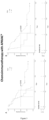

Classifications
-
- C—CHEMISTRY; METALLURGY
- C12—BIOCHEMISTRY; BEER; SPIRITS; WINE; VINEGAR; MICROBIOLOGY; ENZYMOLOGY; MUTATION OR GENETIC ENGINEERING
- C12Q—MEASURING OR TESTING PROCESSES INVOLVING ENZYMES, NUCLEIC ACIDS OR MICROORGANISMS; COMPOSITIONS OR TEST PAPERS THEREFOR; PROCESSES OF PREPARING SUCH COMPOSITIONS; CONDITION-RESPONSIVE CONTROL IN MICROBIOLOGICAL OR ENZYMOLOGICAL PROCESSES
- C12Q1/00—Measuring or testing processes involving enzymes, nucleic acids or microorganisms; Compositions therefor; Processes of preparing such compositions
- C12Q1/68—Measuring or testing processes involving enzymes, nucleic acids or microorganisms; Compositions therefor; Processes of preparing such compositions involving nucleic acids
- C12Q1/6876—Nucleic acid products used in the analysis of nucleic acids, e.g. primers or probes
- C12Q1/6883—Nucleic acid products used in the analysis of nucleic acids, e.g. primers or probes for diseases caused by alterations of genetic material
- C12Q1/6886—Nucleic acid products used in the analysis of nucleic acids, e.g. primers or probes for diseases caused by alterations of genetic material for cancer
-
- C—CHEMISTRY; METALLURGY
- C12—BIOCHEMISTRY; BEER; SPIRITS; WINE; VINEGAR; MICROBIOLOGY; ENZYMOLOGY; MUTATION OR GENETIC ENGINEERING
- C12Q—MEASURING OR TESTING PROCESSES INVOLVING ENZYMES, NUCLEIC ACIDS OR MICROORGANISMS; COMPOSITIONS OR TEST PAPERS THEREFOR; PROCESSES OF PREPARING SUCH COMPOSITIONS; CONDITION-RESPONSIVE CONTROL IN MICROBIOLOGICAL OR ENZYMOLOGICAL PROCESSES
- C12Q2600/00—Oligonucleotides characterized by their use
- C12Q2600/106—Pharmacogenomics, i.e. genetic variability in individual responses to drugs and drug metabolism
-
- C—CHEMISTRY; METALLURGY
- C12—BIOCHEMISTRY; BEER; SPIRITS; WINE; VINEGAR; MICROBIOLOGY; ENZYMOLOGY; MUTATION OR GENETIC ENGINEERING
- C12Q—MEASURING OR TESTING PROCESSES INVOLVING ENZYMES, NUCLEIC ACIDS OR MICROORGANISMS; COMPOSITIONS OR TEST PAPERS THEREFOR; PROCESSES OF PREPARING SUCH COMPOSITIONS; CONDITION-RESPONSIVE CONTROL IN MICROBIOLOGICAL OR ENZYMOLOGICAL PROCESSES
- C12Q2600/00—Oligonucleotides characterized by their use
- C12Q2600/156—Polymorphic or mutational markers
Definitions
- the present invention pertains to methods for predicting the response of a patient to an anticancer agent based on the detection of mutations in TP53 and/or POT1 genes.
- Cancer encompasses a large family of diseases characterized by uncontrolled cell growth and division. It is a leading cause of death worldwide and accounted for nearly 10 million deaths in 2020 ( Ferlay et al. Global Cancer Observatory: Cancer Today. Lyon: International Agency for Research on Cancer; 2020 ). Although significant improvements have been made in diagnosis, screening and treatment, cancer still inflicts a serious social burden in terms of loss of life, diminished quality of life, healthcare costs and reduced productivity.
- Immunotherapy is now a recognized and well-established therapeutic strategy in the fight against cancer. It aims at instructing or reactivating the immune system to recognize cancer cells as foreign, ultimately eliminating them.
- the cells of the immune system are normally capable of monitoring and detecting the presence of abnormalities within the cells of the body.
- most tumors develop several mechanisms in order to escape this monitoring, leading to tolerance of the immune system toward the tumor.
- Stimulating the immune cells, such as T lymphocytes via immunotherapy so that they specifically recognize the tumor cells makes it possible to efficiently overcome this tolerance.
- This stimulation can, for example, be carried out by directly bringing tumor antigens into contact ( in vitro or in vivo ) with antigen-presenting cells (APCs), thereby stimulating T lymphocytes.
- APCs antigen-presenting cells
- This is the concept of a therapeutic cancer vaccine.
- the principle is to isolate abnormal proteins from the tumor and to reinject them in the patient in a form that is visible to the immune system.
- HAP hydroxyapatite
- Promising cancer vaccines based on the use of hydroxyapatite (HAP) particles have been developed.
- HAP can advantageously be used for purifying vaccinating proteins from tissue extracts or directly from blood samples and, once the HAP powder has adsorbed the tumor-specific tumor antigens, be directly administered as a cancer vaccine (see e.g. patent applications published under references WO 2006/122914 , WO 2014/184453 or WO2019/175500 ).
- HAP powder is brought into contact with tumor proteins in order to produce a therapeutic antitumor vaccine which increases the level of immunity without any toxicity or side effect. This technique is safe, can be combined with chemotherapy and produces no toxic residue.
- This type of immunotherapy can make the difference between short- and long-term remission in certain cancers with a poor prognosis.
- DLBCL diffuse large B-cell lymphoma
- the present inventor has demonstrated that the presence of mutations in the TP53 and the POT1 genes is highly predictive of a patient's ability to respond to anticancer treatments such as chemotherapy and immunotherapy.
- the present invention thus pertains to a method for predicting the response of a patient suffering from cancer to an anticancer agent, said method comprising a step of detecting, in a biological sample obtained from said patient, a mutation in the TP53 and/or POT1 genes.
- the invention also relates to an anticancer agent for use in the treatment of cancer in a patient, wherein said patient has been selected as not displaying a mutation in the TP53 and POT1 genes.
- the anticancer agent is immunotherapy and/or chemotherapy.
- a preferred immunotherapy is an autologous anticancer vaccine such as that described in patent applications published under references WO 2014/184453 and WO2019/175500 , i.e. comprising HAP powders loaded with tumor antigens.
- TP53 and/or in POT1 are associated with a shorter survival (overall survival or progression-free survival) in patients either treated with chemotherapy or with a combination of chemotherapy and immunotherapy.
- Dogs are considered as an excellent model for human cancer research: they spontaneously develop the same cancers as humans and have high similarities in terms of histological appearance, tumor genetics, physio-pathological pathways and treatment response (see e.g. Rowell et al. Trends in molecular medicine vol. 17,7 (2011): 380-388 ).
- the canine immune system is physiologically similar to that of humans and more closely related to humans than the immune system of mice.
- Cancer treatments used in dogs are similar to those used in humans (such as the "CHOP” chemotherapy which comprises a combination of cyclophosphamide, doxorubicin, vincristine and prednisone), and spontaneous cancers in dogs favored the development of several human-applied therapies such as sunitinib, a cKit inhibitor which is used for the treatment of renal cell carcinoma and gastrointestinal stromal tumors, or ganetespib, a heat shock protein 90 (HSP90) inhibitor which exhibits potent cytotoxicity in a wide variety of hematological and solid tumor cell lines ( Mestrinho, L. A. & Santos, R. R. Advanced Drug Delivery Reviews 172, (2021): 296-313 ).
- HSP90 heat shock protein 90
- TP53 and POT1 can be efficiently used as biomarkers for predicting the response to anticancer agents in dogs, but also in other animals or humans.
- the present inventor has thus shown that the detection of mutations in the TP53 and/or in the POT1 genes allows predicting whether a patient will successfully respond to an anticancer agent such as chemo and/or immunotherapy.
- the present invention relates to a method for predicting the response of a patient suffering from cancer to an anticancer agent, said method comprising a step of detecting, in a biological sample obtained from said patient, a mutation in TP53 and/or POT1 genes.
- the presence of a mutation in TP53 and/or POT1 is indicative of a reduced ability to respond to an anticancer agent, i.e. is indicative of a non- or poor- responder to the anticancer agent.
- patients who do not display any mutation in TP53 and POT1 will be more likely to respond to a treatment with an anticancer agent, i.e. be a responder to the anticancer agent.
- TP53 Tumor Protein p53 gene is a gene which encodes a tumor suppressor protein containing transcriptional activation, DNA binding, and oligomerization domains.
- the encoded protein acts as a tumor suppressor in many tumor types by regulating expression of target genes, thereby inducing cell cycle arrest, apoptosis, senescence, DNA repair, or changes in metabolism.
- TP53 mutations are associated with a variety of cancers.
- the sequence of TP53 is well known by the skilled person and is available under the reference ENSG00000141510 in the Ensembl Gene Database or Gene ID: 7157 in the NCBI Entrez Gene databank.
- POT1 or "protection of telomeres 1” is a gene member of the telombin family and encodes a nuclear protein involved in telomere maintenance. This protein functions as a member of a multi-protein complex that binds to telomeres thereby regulating telomere length and protecting chromosome ends from illegitimate recombination, chromosome instability, and abnormal chromosome segregation.
- the sequence of POT1 is well known by the skilled person and is available under the reference ENSG00000128513 in the Ensembl Gene Database or Gene ID: 25913 in the NCBI Entrez Gene databank.
- a “mutation” refers to any detectable change in genetic material, e.g. DNA, RNA, cDNA, or in an amino acid sequence encoded by such a genetic material. This includes gene mutations, in which the structure (e.g. DNA sequence) of a gene is altered, as well as protein mutations, in which the amino-acid structure of the protein is altered. Generally, a mutation is identified in a subject by comparing the sequence of a nucleic acid or of a polypeptide expressed in said subject with the corresponding nucleic acid or polypeptide expressed in a control population. When the mutation to be detected is a somatic mutation, i.e.
- a mutation that occurs in a specific cell line other than the germline it can also be detected by comparing the sequence of a nucleic acid or of a polypeptide expressed by said cell line (e.g. tumor cells) with the corresponding nucleic acid or polypeptide expressed by another cell line (e.g. a non-cancerous cell line).
- said cell line e.g. tumor cells
- another cell line e.g. a non-cancerous cell line
- the mutation in TP53 or POT1 refers to a mutation of one or more nucleotides in the TP53 or POT1 gene/mRNA sequence.
- a mutation can be a deletion (i.e. removal of at least one nucleotide in the gene sequence), an insertion (i.e. addition of at least one nucleotide in the gene sequence) or a substitution (i.e. replacement of one nucleotide with another).
- the mutation is preferably located in the exons (i.e. in the coding parts of the gene). Mutations can also occur in the introns (i.e. the noncoding regions of the gene).
- Mutations can have an effect on the protein encoded by the gene such as missense mutations (which result in a codon that codes for a different amino acid), nonsense mutations (which result in a premature stop codon or a nonsense codon in the transcribed mRNA and thus in a truncated, and usually non-functional protein) or frameshift mutations (which result from the insertion or deletion of a number of nucleotides in a DNA sequence that is not divisible by three and induce a change the reading frame resulting in a completely different translation from the original). Mutations can also have no effect on the protein sequence (silent mutations).
- the mutation of TP53 or POT1 detected in the context of the present invention is a missense mutation.
- nucleic acid molecules include mRNA, genomic DNA and cDNA derived from mRNA. DNA or RNA can be single stranded or double stranded. These may be utilized for detection by amplification and/or hybridization with a probe, for instance.
- the nucleotide sequence can be obtained from a genomic DNA sample isolated from the biological sample. In this case, any biological sample containing genomic DNA can be used.
- TP53 and POT1 mutations may be detected in RNA or DNA samples, preferably after amplification.
- the isolated RNA may be subjected to coupled reverse transcription and amplification, such as reverse transcription and amplification by polymerase chain reaction (RT-PCR), using specific oligonucleotide primers that are specific for the mutated site or that enable amplification of a the region containing the mutated site.
- RT-PCR polymerase chain reaction
- Conditions for primer annealing may be chosen to ensure specific reverse transcription (where appropriate) and amplification; so that the appearance of an amplification product be a diagnostic of the presence of a mutation according to the invention.
- RNA may be reverse-transcribed and amplified, or DNA may be amplified, after which the mutated site may be detected in the amplified sequence by hybridization with a suitable probe or by direct sequencing, or any other appropriate method known in the art.
- a cDNA obtained from RNA may be cloned and sequenced to identify the mutated sequence of TP53 and POT1.
- the mutation detected in the TP53 and POT1 genes can result in mutated mature TP53 and POT1 proteins.
- the mutation of TP53 or POT1 may be detected at the protein level by detecting the mutated form of the TP53 and POT1 proteins.
- any biological sample wherein the mature protein is present may be used for detecting mutated forms of this protein.
- a sample such as a tissue biopsy, obtained from a patient may be analysed in presence of antibodies specific of the mutated form or the protein, i.e. antibodies that are capable of distinguishing between the mutated form and the wild-type protein (or any other protein), to determine the presence or absence of the mutation specified by the antibody.
- the antibodies may be monoclonal or polyclonal antibodies, single chain or double chain, chimeric antibodies, humanized antibodies, or portions of an immunoglobulin molecule, including those portions known in the art as antigen binding fragments Fab, Fab', F(ab')2 and F(v). They can also be immunoconjugated, e.g. with a toxin, or labelled antibodies.
- polyclonal antibodies may be used, monoclonal antibodies are preferred because they are more reproducible in the long run.
- Polyclonal antibodies can be obtained from serum of an animal immunized against the appropriate antigen, which may be produced by genetic engineering for example according to standard methods well-known by one skilled in the art (see e.g. Harlow et al. (1988)).
- Monoclonal antibodies refer to a population of antibody molecules that contains only one species of antibody combining site capable of immunoreacting with a particular epitope.
- a monoclonal antibody thus typically displays a single binding affinity for any epitope with which it immunoreacts.
- a monoclonal antibody may therefore contain an antibody molecule having a plurality of antibody combining sites, each immunospecific for a different epitope, e.g. a bispecific monoclonal antibody.
- Laboratory methods for preparing monoclonal antibodies are well known in the art (see, for example, Harlow et al., 1988).
- Aptamers which are a class of molecule that represents an alternative to antibodies in term of molecular recognition, can also be used for detecting the mutated form of TP53 or POT1 in the context of the present invention.
- Aptamers are oligonucleotide or oligopeptide sequences with the capacity to recognize virtually any class of target molecules with high affinity and specificity.
- Such ligands may be isolated through Systematic Evolution of Ligands by EXponential enrichment (SELEX) of a random sequence library, as described in Tuerk C. and Gold L., 1990.
- the random sequence library is obtainable by combinatorial chemical synthesis of DNA. In this library, each member is a linear oligomer, eventually chemically modified, of a unique sequence.
- Peptide aptamers consists of a conformationally constrained antibody variable region displayed by a platform protein, such as E. coli Thioredoxin A that are selected from combinatorial libraries by two hybrid methods (Colas et al., 1996).
- All the probes, primers, aptamers or antibodies used in the context of the present invention may be labelled with a detectable molecule or substance, such as a fluorescent molecule, a radioactive molecule or any others labels known in the art. Labels are known in the art that generally provide (either directly or indirectly) a signal.
- the "biological sample” can be any sample allowing for the detection of a mutation in TP53 or POT1.
- TP53 and POT1 mutations according to the present invention are advantageously detected in the tumor genome, i.e. in genetic material derived from tumor cells.
- the mutations detected in the context of the present invention are thus preferably somatic mutations displayed by the tumor cells, i.e. mutations that occur only in cancer cells. Such mutations can thus be detected by comparing the sequence of TP53 or POT1 (genes or protein) expressed in cancer cells with that of TP53 or POT1 expressed in other cell lines, in particular cells derived from healthy tissues.
- the biological sample according to the present invention is thus a sample comprising tumor-derived genetic material and allowing for the detection of mutations comprised in the tumor genome.
- samples include fluids, tissues, cell samples, organs, biopsies, etc.
- Preferred biological samples are a cell or tissue sample.
- the biological sample can be whole blood, serum or plasma obtained from the patient.
- the biological sample is a tumor sample obtained from the patient.
- the method according to the present invention allows predicting a patient's response to a treatment with an anticancer agent, i.e. whether the patient will benefit (respond) or not from receiving the treatment.
- Patients who will benefit from receiving the treatment are also referred to as "responders”.
- Administering the treatment to these patients will allow treating their disease and/or improving their survival (either overall survival or disease-free survival).
- Responders will achieve a response, i.e. the cancer is eradicated, reduced or stabilized after treatment, and show a longer survival time as compared to non-responders.
- patients who will not benefit from receiving a known treatment are referred to "non-responders" or “poor-responders”.
- Administering the treatment to these patients will not allow treating their disease or at least less efficiently than in responders, and/or the treatment will not significantly improve their survival (either overall survival or disease-free survival).
- Non-responders and poor-responders have a shorter survival time than responders after treatment. For these patients, the selection of an alternative treatment strategy would thus be advantageous.
- the method according to the present invention allows for the correct and efficient identification of responders to anticancer agents. Such patients do not display a mutation in the TP53 and/or POT1 genes.
- the present invention pertains to an anticancer agent for use in the treatment of cancer in a patient, wherein said patient has been selected as not displaying a mutation in the TP53 and POT1 genes
- the present disclosure also provides a method for treating a patient suffering from cancer comprising administering to said patient a therapeutically effective amount of an anticancer agent, wherein said patient has been selected as not displaying a mutation in the TP53 and POT1 genes.
- a “therapeutically effective amount” is meant a sufficient amount to treat cancer.
- the "patient” or “subject” is a human being or an animal, for example a mammal, and in particular dogs, horses or cats.
- the “cancer” according to the present invention can be of various origin (such as breast, colon, rectum, lung, head and neck, bladder, ovary, prostate), and involve various cancer cell types (adenocarcinoma, squamous cell carcinoma, large cell cancer, melanoma, etc).
- the cancer is chosen from the group consisting of lymphomas, osteosarcomas, mammary tumors, melanomas, hemangiosarcomas, mast cell tumors, fibrosarcomas, brain or central nervous system tumors, schwannomas, mesotheliomas, seminomas, teratomas and blastomas.
- the cancer can particularly be chosen from a glioblastoma, a sarcoma (such as osteosarcoma or fibrosarcoma), a melanoma, a carcinoma or an adenocarcinoma.
- the cancer is a lymphoma (either a non-Hodgkin lymphoma or a Hodgkin lymphoma).
- said cancer is a non-Hodgkin lymphoma such as diffuse large B-cell lymphoma (DLBCL).
- DLBCL diffuse large B-cell lymphoma
- the cancer can be a disseminated cancer, that is to say one which has metastases (patients classified in the NxMx category according to the TNM classification).
- An anti-cancer treatment generally consists of radiotherapy, surgery, chemotherapy (with a chemotherapeutic agent) or immunotherapy (with an immunotherapeutic agent).
- the treatment may consist of an adjuvant therapy (i.e. treatment after surgical resection of the primary tumor) or of a neoadjuvant therapy (i.e. treatment before surgical resection of the primary tumor).
- the method according to the present invention particularly allows predicting the response of a patient suffering from cancer to chemotherapy, immunotherapy or a combination thereof.
- the "anticancer agent" according to the present invention is an immunotherapeutic agent, a chemotherapeutic agent or a combination of an immunotherapeutic agent with a chemotherapeutic agent.
- Immunotherapy aims at activating the patient's own immune system to fight against the disease.
- An “immunotherapeutic agent” refers to a compound, composition or treatment that indirectly or directly enhances, stimulates or increases the body's immune response against cancer cells.
- Cancer immunotherapy agents include nucleic acids, cytokines, peptides, proteins, immune cells (endogenous, or conferred with anti-cancer activity ex vivo), fragments of bacteria or viruses, and synthetic drugs. They can be used to elicit a specific immune response against a particular cancer cell type, or to trigger a general immune response that indirectly targets cancer cells or their effects. The former is typically achieved with antibodies or vaccines that target one or more antigens on or in cancer cells.
- Immunotherapy is usually done with immunomodulatory agents and/or chemical entities that simultaneously activate one or more types of immune cells to fight against cancer cells.
- Immunotherapeutic agents can be non-specific, i.e. boost the immune system generally so that the human body becomes more effective in fighting the growth and/or spread of cancer cells, or they can be specific, i.e. targeted to the cancer cells themselves. Immunotherapy regimens may combine the use of non-specific and specific immunotherapeutic agents.
- Non-specific immunotherapeutic agents are substances that stimulate or indirectly improve the immune system.
- Non-specific immunotherapeutic agents have been used alone as a main therapy for the treatment of cancer, as well as in addition to a main therapy, in which case the non-specific immunotherapeutic agent functions as an adjuvant to enhance the effectiveness of other therapies (e.g. cancer vaccines).
- Non-specific immunotherapeutic agents can also function in this latter context to reduce the side effects of other therapies, for example, bone marrow suppression induced by certain chemotherapeutic agents.
- Non-specific immunotherapeutic agents can act on key immune system cells and cause secondary responses, such as increased production of cytokines and immunoglobulins. Alternatively, the agents can themselves comprise cytokines.
- Non-specific immunotherapeutic agents are generally classified as cytokines or non-cytokine adjuvants.
- cytokines have found application in the treatment of cancer either as general non-specific immunotherapies designed to boost the immune system, or as adjuvants provided with other therapies.
- Suitable cytokines include, but are not limited to, interferons, interleukins and colony-stimulating factors.
- Interferons include the common types of IFNs, IFN-alpha (IFN-a), IFN-beta (IFN-beta) and IFN-gamma (IFN-y).
- IFNs can act directly on cancer cells, for example, by slowing their growth, promoting their development into cells with more normal behavior and/or increasing their production of antigens thus making the cancer cells easier for the immune system to be recognized and destroyed.
- IFNs can also act indirectly on cancer cells, for example, by slowing down angiogenesis, boosting the immune system and/or stimulating natural killer (NK) cells, T cells and macrophages.
- Recombinant IFN-alpha is available commercially as Roferon (Roche Pharmaceuticals) and Intron A (Schering Corporation).
- IFN-alpha has shown efficacy in the treatment of various cancers such as melanoma (including metastatic melanoma), renal cancer (including metastatic renal cancer), breast cancer, prostate cancer, and cervical cancer (including metastatic cervical cancer).
- melanoma including metastatic melanoma
- renal cancer including metastatic renal cancer
- breast cancer including prostate cancer
- cervical cancer including metastatic cervical cancer
- Interleukins include e.g. IL-2and IL-11.
- Examples of commercially available recombinant interleukins include Proleukin ® (IL-2; Chiron Corporation) and Neumega ® (IL-12; Wyeth Pharmaceuticals).
- Zymogenetics, Inc. (Seattle, Wash.) is currently testing a recombinant form of IL-21, which is also contemplated for use in the combinations of the present invention.
- Interleukins alone or in combination with other immunotherapeutics or with chemotherapeutics, have shown efficacy in the treatment of various cancers such as renal cancer (including metastatic renal cancer), melanoma (including metastatic melanoma), ovarian cancer (including recurrent ovarian cancer), cervical cancer (including metastatic cervical cancer), breast cancer, colorectal cancer, lung cancer, brain cancer, and prostate cancer.
- renal cancer including metastatic renal cancer
- melanoma including metastatic melanoma
- ovarian cancer including recurrent ovarian cancer
- cervical cancer including metastatic cervical cancer
- breast cancer colorectal cancer
- lung cancer brain cancer
- prostate cancer prostate cancer
- Interleukins have also shown good activity in combination with IFN-alpha in the treatment of various cancers ( Negrier et al., Ann Oncol. 2002 13(9): 1460-8 ; Touranietal, J. Clin. Oncol. 2003 21(21):398794 ).
- Colony-stimulating factors include granulocyte colony stimulating factor (G-CSF or filgrastim), granulocyte-macrophage colony stimulating factor (GM-CSF or sargramostim) and erythropoietin (epoetin alfa, darbepoietin).
- G-CSF granulocyte colony stimulating factor
- GM-CSF or sargramostim granulocyte-macrophage colony stimulating factor
- erythropoietin epoetin alfa, darbepoietin.
- Various-recombinant colony stimulating factors are available commercially, for example, Neupogen ® (G-CSF; Amgen), Neulasta (pelfilgrastim; Amgen), Leukine (GM-CSF; Berlex), Procrit (erythropoietin; Ortho Biotech), Epogen (erythropoietin; Amgen), Arnesp (
- Non-cytokine adjuvants include Levamisole, alum hydroxide (alum), Calmette-Guerin bacillus (ACG), incomplete Freund's Adjuvant (IFA), QS-21, DETOX, Keyhole limpet hemocyanin (KLH) and dinitrophenyl (DNP).
- Non-cytokine adjuvants in combination with other immuno- and/or chemotherapeutics have demonstrated efficacy against various cancers including, for example, colon cancer and colorectal cancer (Levimasole); melanoma (BCG and QS-21); renal cancer and bladder cancer (BCG).
- immunotherapeutic agents can be active, i.e. stimulate the body's own immune response (such as cancer vaccines), or they can be passive, i.e. comprise immune system components that were generated external to the body.
- the immunotherapeutic treatment may consist of an adoptive immunotherapy as described by Nicholas P. Restifo, Mark E. Dudley and Steven A. Rosenberg "Adoptive immunotherapy for cancer: harnessing the T cell response, Nature Reviews Immunology, Volume 12, April 2012 ).
- adoptive immunotherapy the patient's circulating lymphocytes, or tumor infiltrated lymphocytes, are isolated in vitro, activated by lymphokines such as IL-2 or transuded with genes for tumor necrosis, and readministered (Rosenberg et al., 1988; 1989).
- the activated lymphocytes are most preferably the patient's own cells that were earlier isolated from a blood or tumor sample and activated (or "expanded") in vitro.
- This form of immunotherapy has produced several cases of regression of melanoma and renal carcinoma in human patients.
- Immune checkpoint inhibitor or checkpoint blockade cancer immunotherapy agent can also be used as immunotherapeutic agents. These compounds inhibit the function of an immune inhibitory checkpoint protein. Inhibition includes reduction of function and full blockade.
- Immune checkpoint inhibitors include peptides, antibodies, nucleic acid molecules and small molecules.
- Preferred immune checkpoint inhibitors are antibodies that specifically recognize immune checkpoint proteins. Such antibodies can e.g. be selected from the group consisting of anti-PD1 antibodies (such as e.g. nivolumab or lambrolizumab), anti-PDL1 antibodies (such as e.g.
- Atezolizumab Avelumab or Durvalumab
- anti-PDL2 antibodies anti-CTLA4 antibodies (such as tremelimumab or ipilimumab)
- anti-TIM-3 antibodies anti-LAG3 antibodies, anti-IDO1 antibodies, anti-TIGIT antibodies, anti-B7H3 antibodies, anti-B7H4 antibodies, anti-BTLA antibodies and anti-B7H6 antibodies.
- Cancer vaccines are preferred immunotherapeutic agents in the context of the present invention.
- the goal of therapeutic cancer vaccines is to induce tumour regression, eradicate minimal residual disease, establish lasting antitumour memory and avoid non-specific or adverse reactions ( Saxena, Mansi, et al. Nature Reviews Cancer 21.6 (2021): 360-378 ).
- Cancer vaccines particularly aim at instructing the cancer patient's own immune system to directly attack and destroy cancer cells. They thus exert an antitumor effect by engaging the host immune response, particularly T cells, and have the great potential of circumventing the intrinsic drug resistance that leads to chemotherapy failure.
- Tumor vaccines generally involve two classes of proteins: tumor-associated antigens (TAAs) and tumor-specific antigens (TSAs).
- TAAs are self-antigens that may be expressed in normal cells but that are either preferentially or abnormally expressed in tumor cells.
- TSAs encompass antigens expressed by oncoviruses and neoantigens encoded by cancer mutations, and are truly tumor-specific (see for review Hollingsworth, Robert E., and Kathrin Jansen. npj Vaccines 4.1 (2019): 1-10 ; Esfahani, K., et al. Current Oncology 27.s2 (2020): 87-97 ; or Emens, Leisha A. Expert opinion on emerging drugs 13.2 (2008): 295-308 ).
- cancer vaccines that have be approved for the treatment of various cancers include cancer vaccine HSPPC-96 or Sipuleucel-t.
- the cancer vaccine used in the context of the present invention is an autologous cancer vaccine.
- An "autologous" vaccine denotes a vaccine in which the tumor proteins/antigens are obtained from the patient intended to be vaccinated. Such vaccines are typically produced by obtaining tumor cells/tumors antigens from the patient and manipulating them to make them more recognizable by the patient's immune system. The treated tumor cells/tumors antigens are then injected into the patient's body to elicit an immune response directed against this population of cells/antigens, i.e. against the tumor. Ideally, memory immune cells persist in the body and allow targeting new cancer cells, should the cancer return.
- the cancer vaccine according to the present invention is an autologous vaccine comprising particles of hydroxyapatite and/or of tricalcium phosphate loaded with tumor antigens such as those disclosed in patent applications published under references WO 2014/184453 or WO2019/175500 (such as the Apavac ® vaccine) whose contents are incorporated herein by way of reference.
- tumor proteins/antigens are proteins and/or molecules expressed specifically by the tumor cells and which can be recognized by T and B lymphocytes.
- Tumor proteins/antigens can be extracted from tumor tissues (i.e. purified from tumor biopsies as shown in WO 2014/184453 ) or directly extracted from serum or plasma samples obtained from the cancer patient (as shown in WO2019/175500 ).
- the weight of the tumor proteins/antigens extracted from the serum or plasma sample is between 60 kDa and 130 kDa, in particular between 70 kDa and 110 kDa.
- the tumor proteins/antigens are extracted by precipitating the serum sample in a saline solution, the mixture thus obtained is then centrifuged, and the pellet obtained represents the extracted proteins.
- the proteins/antigens extracted are brought into contact with the hydroxyapatite particles to produce an autologous vaccine comprising particles of hydroxyapatite and/or of tricalcium phosphate loaded with tumor antigens.
- Hydroxyapatite is a mineral of the calcium phosphate family, of formula Ca 10 (PO4) 6 (OH) 2 , and the unit cell of the crystalline structure is hexagonal. Hydroxyapatite is the hydroxylated member of the apatite group.
- the ions of the crystal unit cell can be replaced with other ions of similar charges and sizes; tunnels also exist in the unit cell which can receive small molecules such as certain amino acids.
- the hydroxyapatite particles according to the present invention are obtained via the same method as that described in application WO 2014/184453 .
- This document describes in particular a method for producing calcium phosphate hydroxyapatite Ca 10 (PO4) 6 (OH) 2 particles, consisting of a slow precipitation at high temperature, obtained by double decomposition of a calcium salt and of a phosphorus salt in a basic medium.
- the reaction is carried out at constant temperature in a large reaction volume and is followed by a maturation phase and a phase of washing with water.
- the precipitate thus obtained undergoes a further transformation step: solid/solution separation.
- the latter step is carried out either by filtration, drying by stoving and crushing, or by spray-drying using a fluidized bed.
- the powder will undergo two transformation steps specific to the application of the invention: the step of particle size selection by dry screening so as to retain only the particle size band of interest, less than 25 ⁇ m or between 25 and 45 ⁇ m, then a final step during which the powder will be sintered at an optimal temperature to ensure fusion of the grains resulting in quite particular surface finishes of the powders (preferentially greater than or equal to 30 m 2 /g).
- selection then sintering or sintering then selection selection.
- “Sintering” is a process consisting in heating a powder without bringing it to melting. In the context of the present invention, the sintering is for example carried out at a temperature between 400°C and 600°C.
- the HAP thus obtained may be in powder form and may undergo one or more washes.
- the tumor proteins/antigens are brought into contact with hydroxyapatite particles by passing said tumor proteins/antigens over a column of hydroxyapatite particles, such as a chromatography column.
- the tumor antigens may optionally be brought into contact in solution and then washed by centrifugation.
- the tumor proteins/antigens are then adsorbed onto the surface of the hydroxyapatite particles.
- the hydroxyapatite particles may particularly be in powder form.
- a chromatography column When a chromatography column is used, it can for example be placed under pressure.
- the hydroxyapatite particles Once the hydroxyapatite particles have been loaded with tumor proteins/antigens, they are suspended in an injection liquid and can be injected into the patient from whom the serum/plasma sample was obtained.
- the injection liquid comprises an organic agent which facilitates the injection, such as carboxymethylcellulose.
- the autologous vaccine comprising particles of hydroxyapatite and/or of tricalcium phosphate loaded with tumor antigens obtained as disclosed above is a preferred immunotherapeutic agent according to the present invention.
- Said autologous vaccine is typically obtained by:
- the autologous vaccine is in the form of a suspension.
- the weight of the tumor antigens extracted from the serum or plasma sample is between 60 kDa and 130 kDa, in particular between 70 kDa and 110 kDa.
- This autologous vaccine is typically administered by injection, for example by subcutaneous or intradermal injection.
- the vaccine can be administered orally or by any other means allowing transmucosal vaccination.
- the patient may receive one or more injections, doses of said autologous vaccine.
- a dose generally comprises between 30 and 50 mg of hydroxyapatite and/or of tricalcium phosphate and between 1000 and 2000 ⁇ g of proteins.
- the patient may receive several injections spaced out over time, for example several days, weeks or months. Injections are typically given on a weekly basis during the first month and then monthly for 4 months. Each injection can be ideally prepared from a new sample of tumor proteins from the patient in the event of escape from the vaccine or of inefficacy, in particular when there is an increase in the level of tumor markers.
- chemotherapeutic agent refers to chemical compounds that are effective in inhibiting tumor growth.
- examples of chemotherapeutic agents include cyclophosphamide; alkylating agents such as thiotepa and cyclophosphamide; alkyl sulfonates such as busulfan, improsulfan and piposulfan; aziridines such as benzodopa, carboquone, meturedopa, and uredopa; ethylenimines and methylamelamines including altretamine, triethylenemelamine, trietylenephosphoramide, triethylenethiophosphaorarnide and trimethylolomelamine; acetogenins (especially bullatacin and bullatacinone); a carnptothecin (including the synthetic analogue topotecan); bryostatin; callystatin; CC-1065 (including its adozelesin, carzelesin and bizelesin synthetic ana
- calicheamicin especially calicheamicin (11 and calicheamicin 211, see, e.g., Agnew Chem Intl. Ed. Engl. 33:183-186 (1994 ); dynemicin, including dynemicin A; an esperamicin; as well as neocarzinostatin chromophore and related chromoprotein enediyne antiobiotic chromomophores), aclacinomysins, actinomycin, authramycin, azaserine, bleomycins, cactinomycin, carabicin, canninomycin, carzinophilin, chromomycins, dactinomycin, daunorubicin, detorubicin, 6-diazo-5-oxo-L-norleucine, doxorubicin (including morpholino-doxorubicin, cyanomorpholino-doxorubicin, 2-pyrrolin
- paclitaxel TAXOL ® , Bristol-Myers Squibb Oncology, Princeton, N.].
- doxetaxel TAXOTERE ® , Rhone-Poulenc Rorer, Antony, France
- chlorambucil gemcitabine
- 6-thioguanine mercaptopurine
- methotrexate platinum analogs such as cisplatin and carboplatin; vinblastine; platinum; etoposide (VP-16); ifosfamide; mitomycin C; mitoxantrone; vincristine; vinorelbine; navelbine; novantrone; teniposide; daunomycin; aminopterin; xeloda; ibandronate; CPT-1 1 ; topoisomerase inhibitor RFS 2000; difluoromethylornithine (DMFO); retinoic acid; capecitabine; and pharmaceutically acceptable salts, acids or derivatives of any of the above.
- antihormonal agents that act to regulate or inhibit honnone action on tumors
- anti-estrogens including for example tamoxifen, raloxifene, aromatase inhibiting 4(5)-imidazoles, 4-mycinetamoxifen, trioxifene, keoxifene, LY117018, onapristone, and toremifene (Fareston); and anti-androgens such as flutamide, nilutamide, bicalutamide, leuprolide, and goserelin; and pharmaceutically acceptable salts, acids or derivatives of any of the above.
- the chemotherapeutic agent according to the present invention is a combination of cyclophosphamide, doxorubicin (such as hydroxydaunorubicin), vincristine (marketed under the brand name Oncovin ® ) and a steroid selected from prednisone and prednisolone.
- This specific combination is also known as "CHOP” chemotherapy (acronym for " C yclophosphamide”, “ H ydroxydaunorubicin”, “ O ncovin ® " and " P rednisone”).
- the method according to the present invention thus specifically allows determining whether a patient suffering from cancer will respond to a chemotherapy consisting of a CHOP chemotherapy, to an immunotherapy consisting of an autologous cancer vaccine comprising particles of hydroxyapatite and/or tricalcium phosphate loaded with tumor antigens, or to a combination of these specific therapies.
- Lymphoma in domestic dogs is considered a representative and highly predictive spontaneous model for human disease.
- the complex genetics interplay, the intact immune system, the environmental exposures and the increasing incidence represent powerful elements for translational studies.
- canine diffuse large B-cell lymphoma (cDLBCL) is the most common, accounting for approximately 60-70% of hematological malignancies in this species.
- Current survival rates for cDLBCL after chemotherapy or immune-chemotherapy are usually disappointing and dogs show markedly different clinical courses and treatment responses, demonstrating a heterogeneous clinical behavior and a poor chance to anticipate the outcome.
- Proposed cDLBCL prognostic classification systems are based on bone marrow infiltration, substage, mitotic rate and histological features (centroblastic and immunoblastic) and do not take in consideration the mechanisms underlying the tumorigenesis.
- WES whole exome sequencing
- FBXW7 mutations were identified occurring in a specific codon recurrently mutated in several human cancers.
- TTP time to progression
- LSS overall survival
- gDNA Tumor genomic DNA
- SPRIworks HT Reagent Kit Bovine Coulter, Inc., Indianapolis, Indiana, Cat. No. B06938
- the resulting libraries were subjected to exome enrichment using the SureSelectXT Canine All Exon Kit (Agilent, Cat. No. 5190-5452), following the manufacturer's instructions.
- POT1 and TP53 were two of most interesting. POT1 mutations were identified in 28.6% of the cases. Missense mutations represented 54.5% of variants, while nonsense and frameshift mutations were 27.3% and 18.2%, respectively. A total of twenty-four mutations in TP53 gene were reported in 19 dogs (24.7%). We identified 19 missense (79.2%), three frameshift (12.5%) and two nonsense variants (8.3%). Five dogs presented two TP53 variants and one of them also carried a frameshift deletion and a nonsense variant on the same allele.
- Purebred dogs were more frequently affected by POT1 mutations compared to mixed breed dogs.
- the touchdown phase was conducted with 20 cycles at annealing temperature of 65°C decreasing 0.5°C at each cycle, while the second phase was conducted with 25 cycles at annealing temperature of 55°C.
- PCR products were purified using the ExoSAP-IT PCR Product Cleanup Reagent (Applied Biosystems, Foster City, CA, USA). Purified products were sequenced in the forward or reverse direction using the BigDye Terminator v1.1 Cycle Sequencing Kit (Applied Biosystems) following manufacturer's instructions and analyzed on a SeqStudio Genetic Analyzer (Applied Biosystems). In total, 47 dogs were investigated for the set of mutations identified in the six exons.
- the median TTP for TP53 mutant and wt dogs was 60 and 217 days, respectively (see Figure 4A ), while the median LSS for TP53 mutant and wt dogs was 105 and 361 days, respectively (see Figure 4B ).
Abstract
The present invention pertains to methods for predicting the response of a patient to an anticancer agent such as a chemotherapeutic agent or an immunotherapeutic agent. The methods according to the invention comprise a step of detecting, in a biological sample obtained from a cancer patient, a mutation in TP53 and/or POT1 genes. The present inventor has indeed demonstrated that the presence of a mutation in TP53 or POT1 is associated with a poor response to immunotherapy or chemotherapy.
Description
- The present invention pertains to methods for predicting the response of a patient to an anticancer agent based on the detection of mutations in TP53 and/or POT1 genes.
- Cancer encompasses a large family of diseases characterized by uncontrolled cell growth and division. It is a leading cause of death worldwide and accounted for nearly 10 million deaths in 2020 (Ferlay et al. Global Cancer Observatory: Cancer Today. Lyon: International Agency for Research on Cancer; 2020). Although significant improvements have been made in diagnosis, screening and treatment, cancer still inflicts a terrible social burden in terms of loss of life, diminished quality of life, healthcare costs and reduced productivity.
- Many types of cancer treatments have been developed and are available such as chemotherapy, radiotherapy, surgery and immunotherapy. Immunotherapy is now a recognized and well-established therapeutic strategy in the fight against cancer. It aims at instructing or reactivating the immune system to recognize cancer cells as foreign, ultimately eliminating them. The cells of the immune system are normally capable of monitoring and detecting the presence of abnormalities within the cells of the body. However, most tumors develop several mechanisms in order to escape this monitoring, leading to tolerance of the immune system toward the tumor. Stimulating the immune cells, such as T lymphocytes, via immunotherapy so that they specifically recognize the tumor cells makes it possible to efficiently overcome this tolerance. This stimulation can, for example, be carried out by directly bringing tumor antigens into contact (in vitro or in vivo) with antigen-presenting cells (APCs), thereby stimulating T lymphocytes. This is the concept of a therapeutic cancer vaccine. For an in vivo stimulation, the principle is to isolate abnormal proteins from the tumor and to reinject them in the patient in a form that is visible to the immune system.
- Promising cancer vaccines based on the use of hydroxyapatite (HAP) particles have been developed. HAP can advantageously be used for purifying vaccinating proteins from tissue extracts or directly from blood samples and, once the HAP powder has adsorbed the tumor-specific tumor antigens, be directly administered as a cancer vaccine (see e.g. patent applications published under references
WO 2006/122914 ,WO 2014/184453 orWO2019/175500 ). HAP powder is brought into contact with tumor proteins in order to produce a therapeutic antitumor vaccine which increases the level of immunity without any toxicity or side effect. This technique is safe, can be combined with chemotherapy and produces no toxic residue. This type of immunotherapy can make the difference between short- and long-term remission in certain cancers with a poor prognosis. This has for example been demonstrated in veterinary medicine, where this vaccination increases the survival of dogs with a deadly cancer such as diffuse large B-cell lymphoma (DLBCL) compared to chemotherapy alone. Excellent results have also been obtained for solid tumors such as bone cancers (osteosarcomas), mastocytomas or melanomas. - Unfortunately, some patients do not respond to first-line anticancer treatments such as immunotherapy or chemotherapy. Finding therapeutic alternatives for these patients is difficult and the efficiency of such treatments is generally hindered by the delay resulting from the elaboration of a new therapeutic strategy.
- Thus, there is a continuous need for new biomarkers allowing for an accurate evaluation of the likeliness of a patient to respond to a known treatment, in particular to first-line treatments such as immunotherapy and chemotherapy.
- The invention is defined by the claims.
- Using a dog model, the present inventor has demonstrated that the presence of mutations in the TP53 and the POT1 genes is highly predictive of a patient's ability to respond to anticancer treatments such as chemotherapy and immunotherapy.
- The present invention thus pertains to a method for predicting the response of a patient suffering from cancer to an anticancer agent, said method comprising a step of detecting, in a biological sample obtained from said patient, a mutation in the TP53 and/or POT1 genes. The invention also relates to an anticancer agent for use in the treatment of cancer in a patient, wherein said patient has been selected as not displaying a mutation in the TP53 and POT1 genes.
- The anticancer agent is immunotherapy and/or chemotherapy. A preferred immunotherapy is an autologous anticancer vaccine such as that described in patent applications published under references
WO 2014/184453 andWO2019/175500 , i.e. comprising HAP powders loaded with tumor antigens. - Using a canine model of diffuse large B-cell lymphoma (DLBCL), the present inventor has demonstrated that mutations in TP53 and/or in POT1 are associated with a shorter survival (overall survival or progression-free survival) in patients either treated with chemotherapy or with a combination of chemotherapy and immunotherapy.
- Dogs are considered as an excellent model for human cancer research: they spontaneously develop the same cancers as humans and have high similarities in terms of histological appearance, tumor genetics, physio-pathological pathways and treatment response (see e.g. Rowell et al. Trends in molecular medicine vol. 17,7 (2011): 380-388). The canine immune system is physiologically similar to that of humans and more closely related to humans than the immune system of mice. Cancer treatments used in dogs are similar to those used in humans (such as the "CHOP" chemotherapy which comprises a combination of cyclophosphamide, doxorubicin, vincristine and prednisone), and spontaneous cancers in dogs favored the development of several human-applied therapies such as sunitinib, a cKit inhibitor which is used for the treatment of renal cell carcinoma and gastrointestinal stromal tumors, or ganetespib, a heat shock protein 90 (HSP90) inhibitor which exhibits potent cytotoxicity in a wide variety of hematological and solid tumor cell lines (Mestrinho, L. A. & Santos, R. R. Advanced Drug Delivery Reviews 172, (2021): 296-313).
- The data provided therein thus demonstrate that TP53 and POT1 can be efficiently used as biomarkers for predicting the response to anticancer agents in dogs, but also in other animals or humans. The present inventor has thus shown that the detection of mutations in the TP53 and/or in the POT1 genes allows predicting whether a patient will successfully respond to an anticancer agent such as chemo and/or immunotherapy.
- Accordingly, the present invention relates to a method for predicting the response of a patient suffering from cancer to an anticancer agent, said method comprising a step of detecting, in a biological sample obtained from said patient, a mutation in TP53 and/or POT1 genes.
- In the context of the present invention, the presence of a mutation in TP53 and/or POT1 is indicative of a reduced ability to respond to an anticancer agent, i.e. is indicative of a non- or poor- responder to the anticancer agent. Alternatively, in the context of the present invention, patients who do not display any mutation in TP53 and POT1 will be more likely to respond to a treatment with an anticancer agent, i.e. be a responder to the anticancer agent.
- "TP53" or "Tumor Protein p53" gene is a gene which encodes a tumor suppressor protein containing transcriptional activation, DNA binding, and oligomerization domains. The encoded protein acts as a tumor suppressor in many tumor types by regulating expression of target genes, thereby inducing cell cycle arrest, apoptosis, senescence, DNA repair, or changes in metabolism. TP53 mutations are associated with a variety of cancers. The sequence of TP53 is well known by the skilled person and is available under the reference ENSG00000141510 in the Ensembl Gene Database or Gene ID: 7157 in the NCBI Entrez Gene databank.
- "POT1" or "protection of
telomeres 1" is a gene member of the telombin family and encodes a nuclear protein involved in telomere maintenance. This protein functions as a member of a multi-protein complex that binds to telomeres thereby regulating telomere length and protecting chromosome ends from illegitimate recombination, chromosome instability, and abnormal chromosome segregation. The sequence of POT1 is well known by the skilled person and is available under the reference ENSG00000128513 in the Ensembl Gene Database or Gene ID: 25913 in the NCBI Entrez Gene databank. - A "mutation" refers to any detectable change in genetic material, e.g. DNA, RNA, cDNA, or in an amino acid sequence encoded by such a genetic material. This includes gene mutations, in which the structure (e.g. DNA sequence) of a gene is altered, as well as protein mutations, in which the amino-acid structure of the protein is altered. Generally, a mutation is identified in a subject by comparing the sequence of a nucleic acid or of a polypeptide expressed in said subject with the corresponding nucleic acid or polypeptide expressed in a control population. When the mutation to be detected is a somatic mutation, i.e. a mutation that occurs in a specific cell line other than the germline, it can also be detected by comparing the sequence of a nucleic acid or of a polypeptide expressed by said cell line (e.g. tumor cells) with the corresponding nucleic acid or polypeptide expressed by another cell line (e.g. a non-cancerous cell line).
- In the context of the present invention, the mutation in TP53 or POT1 refers to a mutation of one or more nucleotides in the TP53 or POT1 gene/mRNA sequence. Such a mutation can be a deletion (i.e. removal of at least one nucleotide in the gene sequence), an insertion (i.e. addition of at least one nucleotide in the gene sequence) or a substitution (i.e. replacement of one nucleotide with another). In the context of the present invention, the mutation is preferably located in the exons (i.e. in the coding parts of the gene). Mutations can also occur in the introns (i.e. the noncoding regions of the gene). Mutations can have an effect on the protein encoded by the gene such as missense mutations (which result in a codon that codes for a different amino acid), nonsense mutations (which result in a premature stop codon or a nonsense codon in the transcribed mRNA and thus in a truncated, and usually non-functional protein) or frameshift mutations (which result from the insertion or deletion of a number of nucleotides in a DNA sequence that is not divisible by three and induce a change the reading frame resulting in a completely different translation from the original). Mutations can also have no effect on the protein sequence (silent mutations).
- According to a preferred embodiment, the mutation of TP53 or POT1 detected in the context of the present invention is a missense mutation.
- A person skilled in the art knows many techniques allowing to detect a mutation within a gene.
- The presence of a mutation in TP53 and POT1 genes according to the present invention may be detected by analysing TP53 and POT1 nucleic acid molecules. In the context of the invention, nucleic acid molecules include mRNA, genomic DNA and cDNA derived from mRNA. DNA or RNA can be single stranded or double stranded. These may be utilized for detection by amplification and/or hybridization with a probe, for instance. The nucleotide sequence can be obtained from a genomic DNA sample isolated from the biological sample. In this case, any biological sample containing genomic DNA can be used.
- TP53 and POT1 mutations may be detected in RNA or DNA samples, preferably after amplification. For instance, the isolated RNA may be subjected to coupled reverse transcription and amplification, such as reverse transcription and amplification by polymerase chain reaction (RT-PCR), using specific oligonucleotide primers that are specific for the mutated site or that enable amplification of a the region containing the mutated site. Conditions for primer annealing may be chosen to ensure specific reverse transcription (where appropriate) and amplification; so that the appearance of an amplification product be a diagnostic of the presence of a mutation according to the invention. Otherwise, RNA may be reverse-transcribed and amplified, or DNA may be amplified, after which the mutated site may be detected in the amplified sequence by hybridization with a suitable probe or by direct sequencing, or any other appropriate method known in the art. For instance, a cDNA obtained from RNA may be cloned and sequenced to identify the mutated sequence of TP53 and POT1.
- As discussed above, the mutation detected in the TP53 and POT1 genes can result in mutated mature TP53 and POT1 proteins. Thus, the mutation of TP53 or POT1 may be detected at the protein level by detecting the mutated form of the TP53 and POT1 proteins. In this case, any biological sample wherein the mature protein is present may be used for detecting mutated forms of this protein. In this context, a sample, such as a tissue biopsy, obtained from a patient may be analysed in presence of antibodies specific of the mutated form or the protein, i.e. antibodies that are capable of distinguishing between the mutated form and the wild-type protein (or any other protein), to determine the presence or absence of the mutation specified by the antibody.
- The antibodies may be monoclonal or polyclonal antibodies, single chain or double chain, chimeric antibodies, humanized antibodies, or portions of an immunoglobulin molecule, including those portions known in the art as antigen binding fragments Fab, Fab', F(ab')2 and F(v). They can also be immunoconjugated, e.g. with a toxin, or labelled antibodies.
- Whereas polyclonal antibodies may be used, monoclonal antibodies are preferred because they are more reproducible in the long run.
- Polyclonal antibodies can be obtained from serum of an animal immunized against the appropriate antigen, which may be produced by genetic engineering for example according to standard methods well-known by one skilled in the art (see e.g. Harlow et al. (1988)).
- Monoclonal antibodies refer to a population of antibody molecules that contains only one species of antibody combining site capable of immunoreacting with a particular epitope. A monoclonal antibody thus typically displays a single binding affinity for any epitope with which it immunoreacts. A monoclonal antibody may therefore contain an antibody molecule having a plurality of antibody combining sites, each immunospecific for a different epitope, e.g. a bispecific monoclonal antibody. Laboratory methods for preparing monoclonal antibodies are well known in the art (see, for example, Harlow et al., 1988).
- Aptamers, which are a class of molecule that represents an alternative to antibodies in term of molecular recognition, can also be used for detecting the mutated form of TP53 or POT1 in the context of the present invention. Aptamers are oligonucleotide or oligopeptide sequences with the capacity to recognize virtually any class of target molecules with high affinity and specificity. Such ligands may be isolated through Systematic Evolution of Ligands by EXponential enrichment (SELEX) of a random sequence library, as described in Tuerk C. and Gold L., 1990. The random sequence library is obtainable by combinatorial chemical synthesis of DNA. In this library, each member is a linear oligomer, eventually chemically modified, of a unique sequence. Possible modifications, uses and advantages of this class of molecules have been reviewed in Jayasena S.D., 1999. Peptide aptamers consists of a conformationally constrained antibody variable region displayed by a platform protein, such as E. coli Thioredoxin A that are selected from combinatorial libraries by two hybrid methods (Colas et al., 1996).
- All the probes, primers, aptamers or antibodies used in the context of the present invention may be labelled with a detectable molecule or substance, such as a fluorescent molecule, a radioactive molecule or any others labels known in the art. Labels are known in the art that generally provide (either directly or indirectly) a signal.
- In the context of the present invention, the "biological sample" can be any sample allowing for the detection of a mutation in TP53 or POT1. TP53 and POT1 mutations according to the present invention are advantageously detected in the tumor genome, i.e. in genetic material derived from tumor cells. The mutations detected in the context of the present invention are thus preferably somatic mutations displayed by the tumor cells, i.e. mutations that occur only in cancer cells. Such mutations can thus be detected by comparing the sequence of TP53 or POT1 (genes or protein) expressed in cancer cells with that of TP53 or POT1 expressed in other cell lines, in particular cells derived from healthy tissues. The biological sample according to the present invention is thus a sample comprising tumor-derived genetic material and allowing for the detection of mutations comprised in the tumor genome. Examples of such samples include fluids, tissues, cell samples, organs, biopsies, etc. Preferred biological samples are a cell or tissue sample. The biological sample can be whole blood, serum or plasma obtained from the patient. In a preferred embodiment according to the present invention, the biological sample is a tumor sample obtained from the patient.
- The method according to the present invention allows predicting a patient's response to a treatment with an anticancer agent, i.e. whether the patient will benefit (respond) or not from receiving the treatment. Patients who will benefit from receiving the treatment are also referred to as "responders". Administering the treatment to these patients will allow treating their disease and/or improving their survival (either overall survival or disease-free survival). Responders will achieve a response, i.e. the cancer is eradicated, reduced or stabilized after treatment, and show a longer survival time as compared to non-responders. On the contrary, patients who will not benefit from receiving a known treatment are referred to "non-responders" or "poor-responders". Administering the treatment to these patients will not allow treating their disease or at least less efficiently than in responders, and/or the treatment will not significantly improve their survival (either overall survival or disease-free survival). Non-responders and poor-responders have a shorter survival time than responders after treatment. For these patients, the selection of an alternative treatment strategy would thus be advantageous.
- The method according to the present invention allows for the correct and efficient identification of responders to anticancer agents. Such patients do not display a mutation in the TP53 and/or POT1 genes.
- Accordingly, in a further embodiment, the present invention pertains to an anticancer agent for use in the treatment of cancer in a patient, wherein said patient has been selected as not displaying a mutation in the TP53 and POT1 genes
- The present disclosure also provides a method for treating a patient suffering from cancer comprising administering to said patient a therapeutically effective amount of an anticancer agent, wherein said patient has been selected as not displaying a mutation in the TP53 and POT1 genes.
- By a "therapeutically effective amount" is meant a sufficient amount to treat cancer.
- The "patient" or "subject" is a human being or an animal, for example a mammal, and in particular dogs, horses or cats.
- The "cancer" according to the present invention can be of various origin (such as breast, colon, rectum, lung, head and neck, bladder, ovary, prostate), and involve various cancer cell types (adenocarcinoma, squamous cell carcinoma, large cell cancer, melanoma, etc).
- In one particular embodiment, the cancer is chosen from the group consisting of lymphomas, osteosarcomas, mammary tumors, melanomas, hemangiosarcomas, mast cell tumors, fibrosarcomas, brain or central nervous system tumors, schwannomas, mesotheliomas, seminomas, teratomas and blastomas. The cancer can particularly be chosen from a glioblastoma, a sarcoma (such as osteosarcoma or fibrosarcoma), a melanoma, a carcinoma or an adenocarcinoma.
- According to a preferred embodiment, the cancer is a lymphoma (either a non-Hodgkin lymphoma or a Hodgkin lymphoma). According to a more preferred embodiment, said cancer is a non-Hodgkin lymphoma such as diffuse large B-cell lymphoma (DLBCL).
- The cancer can be a disseminated cancer, that is to say one which has metastases (patients classified in the NxMx category according to the TNM classification).
- An anti-cancer treatment generally consists of radiotherapy, surgery, chemotherapy (with a chemotherapeutic agent) or immunotherapy (with an immunotherapeutic agent). The treatment may consist of an adjuvant therapy (i.e. treatment after surgical resection of the primary tumor) or of a neoadjuvant therapy (i.e. treatment before surgical resection of the primary tumor).
- As shown in the examples below, the method according to the present invention particularly allows predicting the response of a patient suffering from cancer to chemotherapy, immunotherapy or a combination thereof. Thus, according to a preferred embodiment, the "anticancer agent" according to the present invention is an immunotherapeutic agent, a chemotherapeutic agent or a combination of an immunotherapeutic agent with a chemotherapeutic agent.
- "Immunotherapy" aims at activating the patient's own immune system to fight against the disease. An "immunotherapeutic agent" refers to a compound, composition or treatment that indirectly or directly enhances, stimulates or increases the body's immune response against cancer cells. Cancer immunotherapy agents include nucleic acids, cytokines, peptides, proteins, immune cells (endogenous, or conferred with anti-cancer activity ex vivo), fragments of bacteria or viruses, and synthetic drugs. They can be used to elicit a specific immune response against a particular cancer cell type, or to trigger a general immune response that indirectly targets cancer cells or their effects. The former is typically achieved with antibodies or vaccines that target one or more antigens on or in cancer cells. Immunotherapy is usually done with immunomodulatory agents and/or chemical entities that simultaneously activate one or more types of immune cells to fight against cancer cells.
- Immunotherapeutic agents can be non-specific, i.e. boost the immune system generally so that the human body becomes more effective in fighting the growth and/or spread of cancer cells, or they can be specific, i.e. targeted to the cancer cells themselves. Immunotherapy regimens may combine the use of non-specific and specific immunotherapeutic agents.
- Non-specific immunotherapeutic agents are substances that stimulate or indirectly improve the immune system. Non-specific immunotherapeutic agents have been used alone as a main therapy for the treatment of cancer, as well as in addition to a main therapy, in which case the non-specific immunotherapeutic agent functions as an adjuvant to enhance the effectiveness of other therapies (e.g. cancer vaccines). Non-specific immunotherapeutic agents can also function in this latter context to reduce the side effects of other therapies, for example, bone marrow suppression induced by certain chemotherapeutic agents. Non-specific immunotherapeutic agents can act on key immune system cells and cause secondary responses, such as increased production of cytokines and immunoglobulins. Alternatively, the agents can themselves comprise cytokines. Non-specific immunotherapeutic agents are generally classified as cytokines or non-cytokine adjuvants.
- A number of cytokines have found application in the treatment of cancer either as general non-specific immunotherapies designed to boost the immune system, or as adjuvants provided with other therapies. Suitable cytokines include, but are not limited to, interferons, interleukins and colony-stimulating factors.
- Interferons (IFNs) include the common types of IFNs, IFN-alpha (IFN-a), IFN-beta (IFN-beta) and IFN-gamma (IFN-y). IFNs can act directly on cancer cells, for example, by slowing their growth, promoting their development into cells with more normal behavior and/or increasing their production of antigens thus making the cancer cells easier for the immune system to be recognized and destroyed. IFNs can also act indirectly on cancer cells, for example, by slowing down angiogenesis, boosting the immune system and/or stimulating natural killer (NK) cells, T cells and macrophages. Recombinant IFN-alpha is available commercially as Roferon (Roche Pharmaceuticals) and Intron A (Schering Corporation). The use of IFN-alpha, alone or in combination with other immunotherapeutics or with chemotherapeutics, has shown efficacy in the treatment of various cancers such as melanoma (including metastatic melanoma), renal cancer (including metastatic renal cancer), breast cancer, prostate cancer, and cervical cancer (including metastatic cervical cancer).
- Interleukins include e.g. IL-2and IL-11. Examples of commercially available recombinant interleukins include Proleukin® (IL-2; Chiron Corporation) and Neumega® (IL-12; Wyeth Pharmaceuticals). Zymogenetics, Inc. (Seattle, Wash.) is currently testing a recombinant form of IL-21, which is also contemplated for use in the combinations of the present invention. Interleukins, alone or in combination with other immunotherapeutics or with chemotherapeutics, have shown efficacy in the treatment of various cancers such as renal cancer (including metastatic renal cancer), melanoma (including metastatic melanoma), ovarian cancer (including recurrent ovarian cancer), cervical cancer (including metastatic cervical cancer), breast cancer, colorectal cancer, lung cancer, brain cancer, and prostate cancer.
- Interleukins have also shown good activity in combination with IFN-alpha in the treatment of various cancers (Negrier et al., Ann Oncol. 2002 13(9): 1460-8 ; Touranietal, J. Clin. Oncol. 2003 21(21):398794).
- Colony-stimulating factors (CSFs) include granulocyte colony stimulating factor (G-CSF or filgrastim), granulocyte-macrophage colony stimulating factor (GM-CSF or sargramostim) and erythropoietin (epoetin alfa, darbepoietin). Various-recombinant colony stimulating factors are available commercially, for example, Neupogen® (G-CSF; Amgen), Neulasta (pelfilgrastim; Amgen), Leukine (GM-CSF; Berlex), Procrit (erythropoietin; Ortho Biotech), Epogen (erythropoietin; Amgen), Arnesp (erytropoietin). Colony stimulating factors have shown efficacy in the treatment of cancer, including melanoma, colorectal cancer (including metastatic colorectal cancer), and lung cancer.
- Non-cytokine adjuvants include Levamisole, alum hydroxide (alum), Calmette-Guerin bacillus (ACG), incomplete Freund's Adjuvant (IFA), QS-21, DETOX, Keyhole limpet hemocyanin (KLH) and dinitrophenyl (DNP). Non-cytokine adjuvants in combination with other immuno- and/or chemotherapeutics have demonstrated efficacy against various cancers including, for example, colon cancer and colorectal cancer (Levimasole); melanoma (BCG and QS-21); renal cancer and bladder cancer (BCG).
- In addition to having specific or non-specific targets, immunotherapeutic agents can be active, i.e. stimulate the body's own immune response (such as cancer vaccines), or they can be passive, i.e. comprise immune system components that were generated external to the body.
- The immunotherapeutic treatment may consist of an adoptive immunotherapy as described by Nicholas P. Restifo, Mark E. Dudley and Steven A. Rosenberg "Adoptive immunotherapy for cancer: harnessing the T cell response, Nature Reviews Immunology, Volume 12, April 2012). In adoptive immunotherapy, the patient's circulating lymphocytes, or tumor infiltrated lymphocytes, are isolated in vitro, activated by lymphokines such as IL-2 or transuded with genes for tumor necrosis, and readministered (Rosenberg et al., 1988; 1989). The activated lymphocytes are most preferably the patient's own cells that were earlier isolated from a blood or tumor sample and activated (or "expanded") in vitro. This form of immunotherapy has produced several cases of regression of melanoma and renal carcinoma in human patients.
- Immune checkpoint inhibitor or checkpoint blockade cancer immunotherapy agent can also be used as immunotherapeutic agents. These compounds inhibit the function of an immune inhibitory checkpoint protein. Inhibition includes reduction of function and full blockade. Immune checkpoint inhibitors include peptides, antibodies, nucleic acid molecules and small molecules. Preferred immune checkpoint inhibitors are antibodies that specifically recognize immune checkpoint proteins. Such antibodies can e.g. be selected from the group consisting of anti-PD1 antibodies (such as e.g. nivolumab or lambrolizumab), anti-PDL1 antibodies (such as e.g. Atezolizumab, Avelumab or Durvalumab), anti-PDL2 antibodies, anti-CTLA4 antibodies (such as tremelimumab or ipilimumab), anti-TIM-3 antibodies, anti-LAG3 antibodies, anti-IDO1 antibodies, anti-TIGIT antibodies, anti-B7H3 antibodies, anti-B7H4 antibodies, anti-BTLA antibodies and anti-B7H6 antibodies.
- Cancer vaccines are preferred immunotherapeutic agents in the context of the present invention. The goal of therapeutic cancer vaccines is to induce tumour regression, eradicate minimal residual disease, establish lasting antitumour memory and avoid non-specific or adverse reactions (Saxena, Mansi, et al. Nature Reviews Cancer 21.6 (2021): 360-378). Cancer vaccines particularly aim at instructing the cancer patient's own immune system to directly attack and destroy cancer cells. They thus exert an antitumor effect by engaging the host immune response, particularly T cells, and have the great potential of circumventing the intrinsic drug resistance that leads to chemotherapy failure. Tumor vaccines generally involve two classes of proteins: tumor-associated antigens (TAAs) and tumor-specific antigens (TSAs). TAAs are self-antigens that may be expressed in normal cells but that are either preferentially or abnormally expressed in tumor cells. TSAs encompass antigens expressed by oncoviruses and neoantigens encoded by cancer mutations, and are truly tumor-specific (see for review Hollingsworth, Robert E., and Kathrin Jansen. npj Vaccines 4.1 (2019): 1-10; Esfahani, K., et al. Current Oncology 27.s2 (2020): 87-97; or Emens, Leisha A. Expert opinion on emerging drugs 13.2 (2008): 295-308). Examples of cancer vaccines that have be approved for the treatment of various cancers include cancer vaccine HSPPC-96 or Sipuleucel-t.
- According to a preferred embodiment, the cancer vaccine used in the context of the present invention is an autologous cancer vaccine. An "autologous" vaccine denotes a vaccine in which the tumor proteins/antigens are obtained from the patient intended to be vaccinated. Such vaccines are typically produced by obtaining tumor cells/tumors antigens from the patient and manipulating them to make them more recognizable by the patient's immune system. The treated tumor cells/tumors antigens are then injected into the patient's body to elicit an immune response directed against this population of cells/antigens, i.e. against the tumor. Ideally, memory immune cells persist in the body and allow targeting new cancer cells, should the cancer return.
- According to a preferred embodiment, the cancer vaccine according to the present invention is an autologous vaccine comprising particles of hydroxyapatite and/or of tricalcium phosphate loaded with tumor antigens such as those disclosed in patent applications published under references
WO 2014/184453 orWO2019/175500 (such as the Apavac® vaccine) whose contents are incorporated herein by way of reference. - The notion of "tumor proteins/antigens" is well known to those skilled in the art. They are proteins and/or molecules expressed specifically by the tumor cells and which can be recognized by T and B lymphocytes.
- Tumor proteins/antigens can be extracted from tumor tissues (i.e. purified from tumor biopsies as shown in
WO 2014/184453 ) or directly extracted from serum or plasma samples obtained from the cancer patient (as shown inWO2019/175500 ). - Typically, the weight of the tumor proteins/antigens extracted from the serum or plasma sample is between 60 kDa and 130 kDa, in particular between 70 kDa and 110 kDa. Those skilled in the art are aware of a large number of techniques for extracting proteins from a serum or plasma sample. Typically, the tumor proteins/antigens are extracted by precipitating the serum sample in a saline solution, the mixture thus obtained is then centrifuged, and the pellet obtained represents the extracted proteins.
- The proteins/antigens extracted are brought into contact with the hydroxyapatite particles to produce an autologous vaccine comprising particles of hydroxyapatite and/or of tricalcium phosphate loaded with tumor antigens.
- "Hydroxyapatite" is a mineral of the calcium phosphate family, of formula Ca10(PO4)6(OH)2, and the unit cell of the crystalline structure is hexagonal. Hydroxyapatite is the hydroxylated member of the apatite group. The ions of the crystal unit cell can be replaced with other ions of similar charges and sizes; tunnels also exist in the unit cell which can receive small molecules such as certain amino acids. These are those particular features which give this mineral very specific adsorption properties.
- The hydroxyapatite particles according to the present invention are obtained via the same method as that described in application
WO 2014/184453 . This document describes in particular a method for producing calcium phosphate hydroxyapatite Ca10(PO4)6(OH)2 particles, consisting of a slow precipitation at high temperature, obtained by double decomposition of a calcium salt and of a phosphorus salt in a basic medium. The reaction is carried out at constant temperature in a large reaction volume and is followed by a maturation phase and a phase of washing with water. The precipitate thus obtained undergoes a further transformation step: solid/solution separation. The latter step is carried out either by filtration, drying by stoving and crushing, or by spray-drying using a fluidized bed. Regardless of the solid/solution separation technique chosen, the powder will undergo two transformation steps specific to the application of the invention: the step of particle size selection by dry screening so as to retain only the particle size band of interest, less than 25 µm or between 25 and 45 µm, then a final step during which the powder will be sintered at an optimal temperature to ensure fusion of the grains resulting in quite particular surface finishes of the powders (preferentially greater than or equal to 30 m2/g). These two final steps can be reversed: selection then sintering or sintering then selection. "Sintering" is a process consisting in heating a powder without bringing it to melting. In the context of the present invention, the sintering is for example carried out at a temperature between 400°C and 600°C. The HAP thus obtained may be in powder form and may undergo one or more washes. - Typically, the tumor proteins/antigens are brought into contact with hydroxyapatite particles by passing said tumor proteins/antigens over a column of hydroxyapatite particles, such as a chromatography column. The tumor antigens may optionally be brought into contact in solution and then washed by centrifugation. The tumor proteins/antigens are then adsorbed onto the surface of the hydroxyapatite particles. The hydroxyapatite particles may particularly be in powder form.
- When a chromatography column is used, it can for example be placed under pressure.
- Once the hydroxyapatite particles have been loaded with tumor proteins/antigens, they are suspended in an injection liquid and can be injected into the patient from whom the serum/plasma sample was obtained.
- Typically, the injection liquid comprises an organic agent which facilitates the injection, such as carboxymethylcellulose.
- The autologous vaccine comprising particles of hydroxyapatite and/or of tricalcium phosphate loaded with tumor antigens obtained as disclosed above is a preferred immunotherapeutic agent according to the present invention. Said autologous vaccine is typically obtained by:
- a) extracting the proteins contained in a tumor, serum or plasma sample obtained from a patient suffering from cancer; and
- b) bringing the proteins extracted in step a) into contact with particles of hydroxyapatite and/or of tricalcium phosphate in order to obtain the autologous vaccine.
- Typically, the autologous vaccine is in the form of a suspension.
- Typically, the weight of the tumor antigens extracted from the serum or plasma sample is between 60 kDa and 130 kDa, in particular between 70 kDa and 110 kDa.
- This autologous vaccine is typically administered by injection, for example by subcutaneous or intradermal injection. The vaccine can be administered orally or by any other means allowing transmucosal vaccination.
- The patient may receive one or more injections, doses of said autologous vaccine. A dose generally comprises between 30 and 50 mg of hydroxyapatite and/or of tricalcium phosphate and between 1000 and 2000 µg of proteins. The patient may receive several injections spaced out over time, for example several days, weeks or months. Injections are typically given on a weekly basis during the first month and then monthly for 4 months. Each injection can be ideally prepared from a new sample of tumor proteins from the patient in the event of escape from the vaccine or of inefficacy, in particular when there is an increase in the level of tumor markers.
- The term "chemotherapeutic agent" refers to chemical compounds that are effective in inhibiting tumor growth. Examples of chemotherapeutic agents include cyclophosphamide; alkylating agents such as thiotepa and cyclophosphamide; alkyl sulfonates such as busulfan, improsulfan and piposulfan; aziridines such as benzodopa, carboquone, meturedopa, and uredopa; ethylenimines and methylamelamines including altretamine, triethylenemelamine, trietylenephosphoramide, triethylenethiophosphaorarnide and trimethylolomelamine; acetogenins (especially bullatacin and bullatacinone); a carnptothecin (including the synthetic analogue topotecan); bryostatin; callystatin; CC-1065 (including its adozelesin, carzelesin and bizelesin synthetic analogues); cryptophycins (particularly cryptophycin 1 and cryptophycin 8); dolastatin; duocarmycin (including the synthetic analogues, KW-2189 and CBI-TMI); eleutherobin; pancratistatin; a sarcodictyin; spongistatin; nitrogen mustards such as chlorambucil, chlornaphazine, cholophosphamide, estrarnustine, ifosfamide, mechlorethamine, mechlorethamine oxide hydrochloride, melphalan, novembichin, phenesterine, prednimus tine, trofosfamide, uracil mustard; nitrosureas such as carmustine, chlorozotocin, fotemustine, lomustine, nimustine, ranimustine; antibiotics such as the enediyne antibiotics (e.g. calicheamicin, especially calicheamicin (11 and calicheamicin 211, see, e.g., Agnew Chem Intl. Ed. Engl. 33:183-186 (1994); dynemicin, including dynemicin A; an esperamicin; as well as neocarzinostatin chromophore and related chromoprotein enediyne antiobiotic chromomophores), aclacinomysins, actinomycin, authramycin, azaserine, bleomycins, cactinomycin, carabicin, canninomycin, carzinophilin, chromomycins, dactinomycin, daunorubicin, detorubicin, 6-diazo-5-oxo-L-norleucine, doxorubicin (including morpholino-doxorubicin, cyanomorpholino-doxorubicin, 2-pyrrolino-doxorubicin and deoxydoxorubicin), epirubicin, esorubicin, idanrbicin, marcellomycin, mitomycins, mycophenolic acid, nogalarnycin, olivomycins, peplomycin, potfiromycin, puromycin, quelamycin, rodorubicin, streptomgrin, streptozocin, tubercidin, ubenimex, zinostatin, zorubicin; anti-metabolites such as methotrexate and 5-fluorouracil (5-FU); folic acid analogues such as denopterin, methotrexate, pteropterin, trimetrexate; purine analogs such as fludarabine, 6-mercaptopurine, thiamiprine, thioguanine; pyrimidine analogs such as ancitabine, azacitidine, 6-azauridine, carmofur, cytarabine, dideoxyuridine, doxifluridine, enocitabine, floxuridine, 5-FU; androgens such as calusterone, dromostanolone propionate, epitiostanol, mepitiostane, testolactone; anti-adrenals such as aminoglutethimide, mitotane, trilostane; folic acid replenisher such as frolinic acid; aceglatone; aldophospharnide glycoside; aminolevulinic acid; amsacrine; bestrabucil; bisantrene; edatraxate; defofamine; demecolcine; diaziquone; elfornithine; elliptinium acetate; an epothilone; etoglucid; gallium nitrate; hydroxyurea; lentinan; lonidamine; maytansinoids such as maytansine and ansamitocins; mitoguazone; mitoxantrone; mopidamol; nitracrine; pento statin; phenamet; pirarubicin; podophyllinic acid; 2-ethylhydrazide; procarbazine; PSK®; razoxane; rhizoxin; sizofiran; spirogennanium; tenuazonic acid; triaziquone; 2,2',2"-trichlorotriethylarnine; trichothecenes (especially T-2 toxin, verracurin A, roridinA and anguidine); urethan; vindesine; dacarbazine; mannomustine; mitobromtol; mitolactol; pipobroman; gacytosine; arabinoside ("Ara-C"); cyclophosphamide; thiotepa; taxoids, e.g. paclitaxel (TAXOL®, Bristol-Myers Squibb Oncology, Princeton, N.].) and doxetaxel (TAXOTERE®, Rhone-Poulenc Rorer, Antony, France); chlorambucil; gemcitabine; 6-thioguanine; mercaptopurine; methotrexate; platinum analogs such as cisplatin and carboplatin; vinblastine; platinum; etoposide (VP-16); ifosfamide; mitomycin C; mitoxantrone; vincristine; vinorelbine; navelbine; novantrone; teniposide; daunomycin; aminopterin; xeloda; ibandronate; CPT-1 1 ; topoisomerase inhibitor RFS 2000; difluoromethylornithine (DMFO); retinoic acid; capecitabine; and pharmaceutically acceptable salts, acids or derivatives of any of the above. Also included in this definition are antihormonal agents that act to regulate or inhibit honnone action on tumors such as anti-estrogens including for example tamoxifen, raloxifene, aromatase inhibiting 4(5)-imidazoles, 4-mycinetamoxifen, trioxifene, keoxifene, LY117018, onapristone, and toremifene (Fareston); and anti-androgens such as flutamide, nilutamide, bicalutamide, leuprolide, and goserelin; and pharmaceutically acceptable salts, acids or derivatives of any of the above.
- According to a preferred embodiment, the chemotherapeutic agent according to the present invention is a combination of cyclophosphamide, doxorubicin (such as hydroxydaunorubicin), vincristine (marketed under the brand name Oncovin®) and a steroid selected from prednisone and prednisolone. This specific combination is also known as "CHOP" chemotherapy (acronym for "Cyclophosphamide", "Hydroxydaunorubicin", "Oncovin®" and "Prednisone").
- The method according to the present invention thus specifically allows determining whether a patient suffering from cancer will respond to a chemotherapy consisting of a CHOP chemotherapy, to an immunotherapy consisting of an autologous cancer vaccine comprising particles of hydroxyapatite and/or tricalcium phosphate loaded with tumor antigens, or to a combination of these specific therapies.
- The present invention is presented in greater detail in the examples below. These examples are provided only for illustrative purposes and cannot be interpreted as limiting the scope of the present invention.
-
-
Figure 1 : Dogs were categorized into 2 groups based on whether they had received chemotherapy (such as CHOP therapy) or a combination of chemotherapy (such as CHOP therapy) plus immunotherapy (such as Apavac®). Curves of disease progression (A) and survival (B) over time are represented. -
Figure 2 : Dogs treated with chemotherapy were categorized into 2 groups based on the presence of mutations in TP53. Curves of disease progression (A) and survival (B) over time are represented. -
Figure 3 : Dogs treated with chemo-immunotherapy (CHOP + Apavac®) were categorized into 2 groups based on the presence of mutations in POT1 (A) or TP53 (B). Curves of survival over time are represented. -
Figure 4 : Clinical validation of TP53 mutations by Sanger sequencing analysis: Dogs treated with chemo-immunotherapy (CHOP + Apavac®) were categorized into 2 groups based on the presence of mutations in TP53. Curves of disease progression (A) and survival (B) over time are represented. - Lymphoma in domestic dogs is considered a representative and highly predictive spontaneous model for human disease. In particular, the complex genetics interplay, the intact immune system, the environmental exposures and the increasing incidence represent powerful elements for translational studies. Among the several lymphoma subtypes, canine diffuse large B-cell lymphoma (cDLBCL) is the most common, accounting for approximately 60-70% of hematological malignancies in this species. Current survival rates for cDLBCL after chemotherapy or immune-chemotherapy are usually disappointing and dogs show markedly different clinical courses and treatment responses, demonstrating a heterogeneous clinical behavior and a poor chance to anticipate the outcome. Proposed cDLBCL prognostic classification systems are based on bone marrow infiltration, substage, mitotic rate and histological features (centroblastic and immunoblastic) and do not take in consideration the mechanisms underlying the tumorigenesis. One large study based on whole exome sequencing (WES) investigated canine B-cell lymphomas obtained from three predisposed breeds (Boxer, Golden Retriever and Cocker Spaniel) and both TRAF3 and MAP3K14 were found frequently mutated. Notably, FBXW7 mutations were identified occurring in a specific codon recurrently mutated in several human cancers. Despite the large number of cases included in this study the mutations described may be breed-specific, thereby limiting the more heterogeneous mutational landscape of canine lymphoma. Furthermore, tumors were not appropriately classified by WHO criteria and clinico-pathological features were not reported. Thus, the clinical significance of these results remains unknown.
- In the present study an integrated model including mutations, copy number aberrations was designed to predict overall survival and tumor free interval. Clinically significant mutations were further identified and clinically validated.
- Study was conducted using DNA from 77 de novo cDLBCL cases with matched normal tissues for WES. Briefly, mixed-breed dogs (19/77), German shepherds (9/77), Rottweilers (7/77) and Golden retrievers (4/77) were the most common breeds. There were 40 females and 37 males. At diagnosis, median age was 7 years (range 3-15 years) and median weight was 30.8 kg (range, 4.5-81.3 kg). Regarding clinical stage, 48 dogs had stage V disease, 28 had stage IV and only one dog had stage III disease. Forty-nine (63.6%) dogs were asymptomatic at presentation (substage a), while 28 (36.4%) showed symptoms (substage b). Overall, 22 dogs had bone marrow infiltration, with a median of 3% neoplastic cells (range, 1-50%). Peripheral blood infiltration was present in 43 dogs, with a median of 4% neoplastic cells (range, 1-74%). At presentation, 33 (42.9%) dogs had an increased level of serum LDH and 23 (29.9%) dogs had received steroids before being diagnosed with lymphoma.
- Dogs received either chemotherapy (including CHOP therapy) alone or a combination of chemotherapy (CHOP therapy) plus immunotherapy (an autologous cancer vaccine: Apavac®).
- Treatment affected significantly both the time to progression (TTP) and the overall survival (LSS for "lymphoma-specific survival" - see
Figure 1 ). Indeed, dogs treated with chemo-immunotherapy (45/77) showed a favorable prognosis compared to dogs receiving chemotherapy alone (32/77) and also LSS in these dogs was significantly associated with substage and bone marrow infiltration. In dogs receiving chemotherapy only, peripheral blood infiltration correlated with both TTP and LSS, whereas bone marrow infiltration with LSS only POT1 and TP53 mutations in cDLBCL - Mutations were detected in tumor samples obtained from each dog included in the study.
- Tumor genomic DNA (gDNA) was isolated to prepare libraries for WES. For each dog, gDNA from skin punch biopsies was also extracted and employed as matched-control for subsequent analyses. The exome was captured and then sequenced by Illumina NovaSeq (150 PE) with target coverage of 250×. Briefly, gDNA was tested for concentration and purity and then subjected to fragmentation. Illumina-compatible whole exome libraries were prepared using the SPRIworks HT Reagent Kit (Beckman Coulter, Inc., Indianapolis, Indiana, Cat. No. B06938) and the resulting libraries were subjected to exome enrichment using the SureSelectXT Canine All Exon Kit (Agilent, Cat. No. 5190-5452), following the manufacturer's instructions. Final concentration and size distribution were tested with Agilent Bioanalyzer 2100. Somatic mutations discovery across all samples was performed following the Genome Analysis Toolkit16 v.3.7 Best Practices (accessible via the Broad Institute website: Genome Analysis Toolkit/Getting Started/Best practices webpage). Single nucleotide variants (SNVs) and small insertion and deletions (indels) were identified with Mutect217 and Varscan18 v.2.4.1. Loss of heterozygosity (LOH) was further investigated by Varscan. SNVs, indels and LOH loci were considered as the union of Mutect2 and Varscan filtered somatic mutations.
- We found several genes with mutations but POT1 and TP53 were two of most interesting. POT1 mutations were identified in 28.6% of the cases. Missense mutations represented 54.5% of variants, while nonsense and frameshift mutations were 27.3% and 18.2%, respectively. A total of twenty-four mutations in TP53 gene were reported in 19 dogs (24.7%). We identified 19 missense (79.2%), three frameshift (12.5%) and two nonsense variants (8.3%). Five dogs presented two TP53 variants and one of them also carried a frameshift deletion and a nonsense variant on the same allele.
- Purebred dogs were more frequently affected by POT1 mutations compared to mixed breed dogs.
- In dogs treated with chemotherapy alone TP53 mutation was correlated with a shorter TTP and overall survival. The median TTP for TP53 mutant and wt dogs was 32 and 98 days, respectively, while the median LSS for TP53 mutant and wt dogs was 60 and 176 days, respectively (see
Figure 2 ). - In dogs treated with chemo-immunotherapy POT1 mutation was correlated with a shorter LSS. The median LSS for patients with mutant and wt POT1 was 330 and 547 days, respectively (see
Figure 3A ). Likewise, dogs carrying TP53 mutation were associated with a shorter median LSS (seeFigure 3B ). Also, mutations in both genes were not associated with any other clinical parameters. - The clinical validation of TP53 mutations by Sanger sequencing analysis in cDLBCL treated with chemoimmunotherapy with APAVAC showed similar results to WES.
- Briefly, Sanger sequencing was performed to validate the protein coding somatic mutations identified by WES in TP53 gene. Primer pairs were specifically designed using Primer3 and Primer-BLAST tools. PCR was performed in a final volume of 20 µl using HotStarTaq DNA Polymerase kit (QIAGEN, Hilden, Germany) and 50 ng of gDNA with the following cycling conditions: initial denaturation at 95°C for 5 min, 33 cycles at 95°C for 30 s, at 58°C for 30 s and at 72°C for 40 s, with final extension at 72°C for 10 min. Exon 18 of POT1 was amplified using Touchdown PCR as previously described (Nat. Protoc, 2008). Briefly, the touchdown phase was conducted with 20 cycles at annealing temperature of 65°C decreasing 0.5°C at each cycle, while the second phase was conducted with 25 cycles at annealing temperature of 55°C. PCR products were purified using the ExoSAP-IT PCR Product Cleanup Reagent (Applied Biosystems, Foster City, CA, USA). Purified products were sequenced in the forward or reverse direction using the BigDye Terminator v1.1 Cycle Sequencing Kit (Applied Biosystems) following manufacturer's instructions and analyzed on a SeqStudio Genetic Analyzer (Applied Biosystems). In total, 47 dogs were investigated for the set of mutations identified in the six exons.
- The median TTP for TP53 mutant and wt dogs was 60 and 217 days, respectively (see
Figure 4A ), while the median LSS for TP53 mutant and wt dogs was 105 and 361 days, respectively (seeFigure 4B ).
Claims (14)
- A method for predicting the response of a patient suffering from cancer to an anticancer agent, said method comprising a step of detecting, in a biological sample obtained from said patient, a mutation in TP53 and/or POT1 genes.
- The method according to claim 1, wherein the presence of a mutation in TP53 and/or POT1 genes is indicative of a non- or poor- responder to the anticancer agent.
- The method according to claim 1 or 2, wherein said mutation is a missense mutation.
- The method according to any one of claims 1 to 3, wherein said biological sample is a tumor sample.
- The method according to claim 4, wherein said mutation is a somatic mutation displayed by tumor cells.
- An anticancer agent for use in the treatment of cancer in a patient, wherein said patient has been selected as not displaying a mutation in TP53 and POT1 genes.
- The method or the anticancer agent for use according to any one of claims 1 to 6, wherein said patient is a human.
- The method or the anticancer agent for use according to any one of claims 1 to 6, wherein said patient is a dog, a horse or a cat.
- The method or the anticancer agent for use according to any one of claims 1 to 8, wherein said cancer is chosen from the group consisting of lymphomas, osteosarcomas, mammary tumors, melanomas, hemangiosarcomas, mast cell tumors, fibrosarcomas, brain or central nervous system tumors, schwannomas, mesotheliomas, seminomas, teratomas and blastomas.
- The method or the anticancer agent for use according to any one of claims 1 to 9, wherein said cancer is a non-Hodgkin lymphoma, preferably diffuse large B-cell lymphoma (DLBCL).
- The method or the anticancer agent for use according to any one of claims 1 to 10, wherein said anticancer agent is an immunotherapeutic agent, a chemotherapeutic agent or a combination thereof.
- The method or the anticancer agent for use according to claim 11, wherein said immunotherapeutic agent is an autologous vaccine comprising particles of hydroxyapatite and/or of tricalcium phosphate loaded with tumor antigens.
- The method or the anticancer agent for use according to claim 12, wherein said autologous cancer vaccine comprising particles of hydroxyapatite and/or tricalcium phosphate loaded with tumor antigens, is obtained by:a) extracting the proteins contained in a tumor, serum or plasma sample obtained from a patient suffering from cancer; andb) bringing the proteins extracted in step a) into contact with particles of hydroxyapatite and/or of tricalcium phosphate in order to obtain the autologous vaccine.
- The method or the anticancer agent for use according to claim 11, wherein said chemotherapeutic agent is a combination of cyclophosphamide, doxorubicin, vincristine and prednisone or prednisolone.
Priority Applications (1)
| Application Number | Priority Date | Filing Date | Title |
|---|---|---|---|
| EP21306062.7A EP4124663A1 (en) | 2021-07-29 | 2021-07-29 | Methods for predicting a response to cancer treatment |
Applications Claiming Priority (1)
| Application Number | Priority Date | Filing Date | Title |
|---|---|---|---|
| EP21306062.7A EP4124663A1 (en) | 2021-07-29 | 2021-07-29 | Methods for predicting a response to cancer treatment |
Publications (1)
| Publication Number | Publication Date |
|---|---|
| EP4124663A1 true EP4124663A1 (en) | 2023-02-01 |
Family
ID=77465919
Family Applications (1)
| Application Number | Title | Priority Date | Filing Date |
|---|---|---|---|
| EP21306062.7A Withdrawn EP4124663A1 (en) | 2021-07-29 | 2021-07-29 | Methods for predicting a response to cancer treatment |
Country Status (1)
| Country | Link |
|---|---|
| EP (1) | EP4124663A1 (en) |
Citations (7)
| Publication number | Priority date | Publication date | Assignee | Title |
|---|---|---|---|---|
| WO2006122914A2 (en) | 2005-05-13 | 2006-11-23 | Urodelia | Medicament, particularly an anti-cancer medicament, for treatment using immunotherapy, particularly autologous |
| WO2014184453A1 (en) | 2013-05-15 | 2014-11-20 | Urodelia | Hydroxyapatite powder and process for producing same, composition based on this powder and process for preparing same and kit comprising this powder |
| WO2016057852A1 (en) * | 2014-10-08 | 2016-04-14 | The Broad Institute, Inc. | Markers for hematological cancers |
| WO2017100362A2 (en) * | 2015-12-07 | 2017-06-15 | Epizyme, Inc. | Inhibitors of ezh2 and methods of use thereof |
| US20180045727A1 (en) * | 2015-03-03 | 2018-02-15 | Caris Mpi, Inc. | Molecular profiling for cancer |
| WO2019046619A1 (en) * | 2017-08-30 | 2019-03-07 | Sanford Burnham Prebys Medical Discovery Institute | Tp53 as biomarker for responsiveness to immunotherapy |
| WO2019175500A1 (en) | 2018-03-14 | 2019-09-19 | Urodelia | Autologous cancer vaccines |
-
2021
- 2021-07-29 EP EP21306062.7A patent/EP4124663A1/en not_active Withdrawn
Patent Citations (7)
| Publication number | Priority date | Publication date | Assignee | Title |
|---|---|---|---|---|
| WO2006122914A2 (en) | 2005-05-13 | 2006-11-23 | Urodelia | Medicament, particularly an anti-cancer medicament, for treatment using immunotherapy, particularly autologous |
| WO2014184453A1 (en) | 2013-05-15 | 2014-11-20 | Urodelia | Hydroxyapatite powder and process for producing same, composition based on this powder and process for preparing same and kit comprising this powder |
| WO2016057852A1 (en) * | 2014-10-08 | 2016-04-14 | The Broad Institute, Inc. | Markers for hematological cancers |
| US20180045727A1 (en) * | 2015-03-03 | 2018-02-15 | Caris Mpi, Inc. | Molecular profiling for cancer |
| WO2017100362A2 (en) * | 2015-12-07 | 2017-06-15 | Epizyme, Inc. | Inhibitors of ezh2 and methods of use thereof |
| WO2019046619A1 (en) * | 2017-08-30 | 2019-03-07 | Sanford Burnham Prebys Medical Discovery Institute | Tp53 as biomarker for responsiveness to immunotherapy |
| WO2019175500A1 (en) | 2018-03-14 | 2019-09-19 | Urodelia | Autologous cancer vaccines |
Non-Patent Citations (13)
| Title |
|---|
| "Touranietal", J. CLIN. ONCOL., vol. 21, no. 21, 2003, pages 398794 |
| AGNEW CHEM INTL. ED. ENGL., vol. 33, 1994, pages 183 - 186 |
| ESFAHANI, K. ET AL., CURRENT ONCOLOGY, vol. 27, 2020, pages 87 - 97 |
| FERLAY ET AL.: "Global Cancer Observatory: Cancer Today", INTERNATIONAL AGENCY FOR RESEARCH ON CANCER, 2020 |
| HERLING CARMEN DIANA ET AL: "Complex karyotypes and KRAS and POT1 mutations impact outcome in CLL after chlorambucil-based chemotherapy or chemoimmunotherapy", BLOOD, AMERICAN SOCIETY OF HEMATOLOGY, US, vol. 128, no. 3, 21 July 2016 (2016-07-21), pages 395 - 404, XP086685918, ISSN: 0006-4971, [retrieved on 20201124], DOI: 10.1182/BLOOD-2016-01-691550 * |
| HOLLINGSWORTH, ROBERT E.KATHRIN JANSEN, VACCINES, vol. 4, no. 1, 2019, pages 1 - 10 |
| LEISHA A., EXPERT OPINION ON EMERGING DRUGS, vol. 13, no. 2, 2008, pages 295 - 308 |
| MESTRINHO, L. A.SANTOS, R. R., ADVANCED DRUG DELIVERY REVIEWS, vol. 172, 2021, pages 296 - 313 |
| NEGRIER ET AL., ANN ONCOL., vol. 13, no. 9, 2002, pages 1460 - 8 |
| NICHOLAS P. RESTIFOMARK E. DUDLEYSTEVEN A. ROSENBERG: "Adoptive immunotherapy for cancer: harnessing the T cell response", NATURE REVIEWS IMMUNOLOGY, vol. 12, April 2012 (2012-04-01), XP055034896, DOI: 10.1038/nri3191 |
| ROWELL ET AL., TRENDS IN MOLECULAR MEDICINE, vol. 17, no. 7, 2011, pages 380 - 388 |
| SAXENAMANSI ET AL., NATURE REVIEWS CANCER, vol. 21, no. 6, 2021, pages 360 - 378 |
| SMITH P A D ET AL: "The prevalence and characterisation of TRAF3 and POT1 mutations in canine B-cell lymphoma", VETERINARY JOURNAL, BAILLIERE TINDALL, LONDON, GB, vol. 266, 21 November 2020 (2020-11-21), XP086408237, ISSN: 1090-0233, [retrieved on 20201121], DOI: 10.1016/J.TVJL.2020.105575 * |
Similar Documents
| Publication | Publication Date | Title |
|---|---|---|
| AU2018203045B2 (en) | Methods of treating cancer patients with farnesyltransferase inhibitors | |
| ES2702722T3 (en) | Methods and kits for screening cancer patients | |
| TW201805000A (en) | Treatment of squamous cell carcinomas with inhibitors of ERK | |
| US20200330467A1 (en) | Method and pharmaceutical compositions for the treatment of multiple myeloma | |
| EP4124663A1 (en) | Methods for predicting a response to cancer treatment | |
| WO2017184968A1 (en) | Methods of selecting cancer patients for treatment with farnesyltransferase inhibitors | |
| TWI663974B (en) | Methods of treating cancer with farnesyltransferase inhibitors | |
| JP2022174309A (en) | Tim-3 for assessing cancer seriousness | |
| JP2022523544A (en) | Diagnostic method of CMMRD | |
| US20220364179A1 (en) | Methods and compositions for assessing and predicting therapeutic response | |
| JP2024508633A (en) | How to diagnose MSI cancer | |
| TW202102218A (en) | Methods of treating cancer with farnesyltransferase inhibitors | |
| CA3157889A1 (en) | Methods for diagnosing nasal intestinal type adenocarcinomas | |
| US20170326133A1 (en) | Methods of treating myelodysplastic syndrome with farnesyltransferase inhibitors |
Legal Events
| Date | Code | Title | Description |
|---|---|---|---|
| PUAI | Public reference made under article 153(3) epc to a published international application that has entered the european phase |
Free format text: ORIGINAL CODE: 0009012 |
|
| STAA | Information on the status of an ep patent application or granted ep patent |
Free format text: STATUS: THE APPLICATION HAS BEEN PUBLISHED |
|
| AK | Designated contracting states |
Kind code of ref document: A1 Designated state(s): AL AT BE BG CH CY CZ DE DK EE ES FI FR GB GR HR HU IE IS IT LI LT LU LV MC MK MT NL NO PL PT RO RS SE SI SK SM TR |
|
| STAA | Information on the status of an ep patent application or granted ep patent |
Free format text: STATUS: THE APPLICATION IS DEEMED TO BE WITHDRAWN |
|
| 18D | Application deemed to be withdrawn |
Effective date: 20230802 |