EP4115915A1 - Vein cover - Google Patents
Vein cover Download PDFInfo
- Publication number
- EP4115915A1 EP4115915A1 EP21765441.7A EP21765441A EP4115915A1 EP 4115915 A1 EP4115915 A1 EP 4115915A1 EP 21765441 A EP21765441 A EP 21765441A EP 4115915 A1 EP4115915 A1 EP 4115915A1
- Authority
- EP
- European Patent Office
- Prior art keywords
- vein
- cover
- pin
- vein cover
- axial direction
- Prior art date
- Legal status (The legal status is an assumption and is not a legal conclusion. Google has not performed a legal analysis and makes no representation as to the accuracy of the status listed.)
- Withdrawn
Links
- 210000003462 vein Anatomy 0.000 title claims abstract description 323
- 210000001367 artery Anatomy 0.000 claims abstract description 54
- 239000000463 material Substances 0.000 claims description 22
- 239000004744 fabric Substances 0.000 claims description 15
- 239000002759 woven fabric Substances 0.000 claims description 9
- 239000004745 nonwoven fabric Substances 0.000 claims description 8
- 238000005520 cutting process Methods 0.000 claims description 6
- 230000036961 partial effect Effects 0.000 claims description 2
- 230000017531 blood circulation Effects 0.000 abstract description 47
- 206010072810 Vascular wall hypertrophy Diseases 0.000 abstract description 15
- 239000008280 blood Substances 0.000 abstract description 12
- 210000004369 blood Anatomy 0.000 abstract description 12
- 230000003139 buffering effect Effects 0.000 abstract description 12
- 230000036772 blood pressure Effects 0.000 abstract description 9
- 230000035485 pulse pressure Effects 0.000 abstract description 2
- 210000002460 smooth muscle Anatomy 0.000 description 43
- 210000004177 elastic tissue Anatomy 0.000 description 37
- 230000003872 anastomosis Effects 0.000 description 35
- 239000000835 fiber Substances 0.000 description 26
- 238000011144 upstream manufacturing Methods 0.000 description 25
- 238000010276 construction Methods 0.000 description 23
- 238000011156 evaluation Methods 0.000 description 19
- 102000008186 Collagen Human genes 0.000 description 18
- 108010035532 Collagen Proteins 0.000 description 18
- 229920001436 collagen Polymers 0.000 description 18
- 238000009940 knitting Methods 0.000 description 18
- 239000005020 polyethylene terephthalate Substances 0.000 description 16
- 241000282472 Canis lupus familiaris Species 0.000 description 15
- 229920004934 Dacron® Polymers 0.000 description 15
- 238000005259 measurement Methods 0.000 description 12
- 229920005989 resin Polymers 0.000 description 12
- 239000011347 resin Substances 0.000 description 12
- 230000000541 pulsatile effect Effects 0.000 description 10
- 230000008859 change Effects 0.000 description 9
- 238000007634 remodeling Methods 0.000 description 8
- 230000002792 vascular Effects 0.000 description 8
- 210000004204 blood vessel Anatomy 0.000 description 7
- 230000004872 arterial blood pressure Effects 0.000 description 6
- 230000004323 axial length Effects 0.000 description 6
- 230000015572 biosynthetic process Effects 0.000 description 6
- 230000000052 comparative effect Effects 0.000 description 6
- -1 for example Substances 0.000 description 6
- 208000007536 Thrombosis Diseases 0.000 description 5
- 239000002473 artificial blood Substances 0.000 description 5
- 229920000295 expanded polytetrafluoroethylene Polymers 0.000 description 5
- 229920000747 poly(lactic acid) Polymers 0.000 description 5
- 229920001610 polycaprolactone Polymers 0.000 description 5
- 239000004632 polycaprolactone Substances 0.000 description 5
- 239000004626 polylactic acid Substances 0.000 description 5
- 210000004231 tunica media Anatomy 0.000 description 5
- 208000031481 Pathologic Constriction Diseases 0.000 description 4
- 230000003902 lesion Effects 0.000 description 4
- 238000004519 manufacturing process Methods 0.000 description 4
- 238000000034 method Methods 0.000 description 4
- 229920000728 polyester Polymers 0.000 description 4
- 230000010349 pulsation Effects 0.000 description 4
- 230000036262 stenosis Effects 0.000 description 4
- 208000037804 stenosis Diseases 0.000 description 4
- 238000012285 ultrasound imaging Methods 0.000 description 4
- 239000004677 Nylon Substances 0.000 description 3
- 229920000954 Polyglycolide Polymers 0.000 description 3
- 210000002808 connective tissue Anatomy 0.000 description 3
- 230000007423 decrease Effects 0.000 description 3
- 238000000502 dialysis Methods 0.000 description 3
- 208000037265 diseases, disorders, signs and symptoms Diseases 0.000 description 3
- 208000035475 disorder Diseases 0.000 description 3
- 238000000691 measurement method Methods 0.000 description 3
- 229920001778 nylon Polymers 0.000 description 3
- 230000001575 pathological effect Effects 0.000 description 3
- 239000004633 polyglycolic acid Substances 0.000 description 3
- 229920001343 polytetrafluoroethylene Polymers 0.000 description 3
- 239000004810 polytetrafluoroethylene Substances 0.000 description 3
- 239000011148 porous material Substances 0.000 description 3
- 210000000329 smooth muscle myocyte Anatomy 0.000 description 3
- 230000009885 systemic effect Effects 0.000 description 3
- 230000007704 transition Effects 0.000 description 3
- 229920000544 Gore-Tex Polymers 0.000 description 2
- 206010020772 Hypertension Diseases 0.000 description 2
- 206010029113 Neovascularisation Diseases 0.000 description 2
- 239000004721 Polyphenylene oxide Substances 0.000 description 2
- 230000008649 adaptation response Effects 0.000 description 2
- 238000010009 beating Methods 0.000 description 2
- 210000001715 carotid artery Anatomy 0.000 description 2
- 238000003745 diagnosis Methods 0.000 description 2
- 230000000694 effects Effects 0.000 description 2
- 238000001631 haemodialysis Methods 0.000 description 2
- 230000000322 hemodialysis Effects 0.000 description 2
- 238000001000 micrograph Methods 0.000 description 2
- 239000000203 mixture Substances 0.000 description 2
- 238000000879 optical micrograph Methods 0.000 description 2
- 231100000915 pathological change Toxicity 0.000 description 2
- 230000036285 pathological change Effects 0.000 description 2
- 229920002647 polyamide Polymers 0.000 description 2
- 229920000570 polyether Polymers 0.000 description 2
- 229920002635 polyurethane Polymers 0.000 description 2
- 239000004814 polyurethane Substances 0.000 description 2
- 230000001681 protective effect Effects 0.000 description 2
- 206010003210 Arteriosclerosis Diseases 0.000 description 1
- 229920000742 Cotton Polymers 0.000 description 1
- YCKRFDGAMUMZLT-UHFFFAOYSA-N Fluorine atom Chemical compound [F] YCKRFDGAMUMZLT-UHFFFAOYSA-N 0.000 description 1
- 206010019280 Heart failures Diseases 0.000 description 1
- 241001465754 Metazoa Species 0.000 description 1
- 206010067482 No adverse event Diseases 0.000 description 1
- 206010061876 Obstruction Diseases 0.000 description 1
- 239000004696 Poly ether ether ketone Substances 0.000 description 1
- 239000004698 Polyethylene Substances 0.000 description 1
- 239000002202 Polyethylene glycol Substances 0.000 description 1
- 239000004642 Polyimide Substances 0.000 description 1
- 239000004743 Polypropylene Substances 0.000 description 1
- 239000004372 Polyvinyl alcohol Substances 0.000 description 1
- 208000001647 Renal Insufficiency Diseases 0.000 description 1
- 208000017932 Steel syndrome Diseases 0.000 description 1
- 206010046996 Varicose vein Diseases 0.000 description 1
- 238000005299 abrasion Methods 0.000 description 1
- 239000002253 acid Substances 0.000 description 1
- 125000001931 aliphatic group Chemical group 0.000 description 1
- 229920003232 aliphatic polyester Polymers 0.000 description 1
- 230000008321 arterial blood flow Effects 0.000 description 1
- 208000011775 arteriosclerosis disease Diseases 0.000 description 1
- 125000003118 aryl group Chemical group 0.000 description 1
- 238000011888 autopsy Methods 0.000 description 1
- JUPQTSLXMOCDHR-UHFFFAOYSA-N benzene-1,4-diol;bis(4-fluorophenyl)methanone Chemical compound OC1=CC=C(O)C=C1.C1=CC(F)=CC=C1C(=O)C1=CC=C(F)C=C1 JUPQTSLXMOCDHR-UHFFFAOYSA-N 0.000 description 1
- 229920002988 biodegradable polymer Polymers 0.000 description 1
- 239000004621 biodegradable polymer Substances 0.000 description 1
- 230000008512 biological response Effects 0.000 description 1
- 230000003247 decreasing effect Effects 0.000 description 1
- 238000013461 design Methods 0.000 description 1
- 206010012601 diabetes mellitus Diseases 0.000 description 1
- 230000002526 effect on cardiovascular system Effects 0.000 description 1
- 229920001971 elastomer Polymers 0.000 description 1
- 239000000806 elastomer Substances 0.000 description 1
- 229920000840 ethylene tetrafluoroethylene copolymer Polymers 0.000 description 1
- 238000002474 experimental method Methods 0.000 description 1
- 230000002349 favourable effect Effects 0.000 description 1
- 239000011737 fluorine Substances 0.000 description 1
- 229910052731 fluorine Inorganic materials 0.000 description 1
- PCHJSUWPFVWCPO-UHFFFAOYSA-N gold Chemical compound [Au] PCHJSUWPFVWCPO-UHFFFAOYSA-N 0.000 description 1
- 239000010931 gold Substances 0.000 description 1
- 229910052737 gold Inorganic materials 0.000 description 1
- 230000012447 hatching Effects 0.000 description 1
- 239000007943 implant Substances 0.000 description 1
- 229910052500 inorganic mineral Inorganic materials 0.000 description 1
- 210000004731 jugular vein Anatomy 0.000 description 1
- 208000017169 kidney disease Diseases 0.000 description 1
- 201000006370 kidney failure Diseases 0.000 description 1
- 239000011159 matrix material Substances 0.000 description 1
- 230000005226 mechanical processes and functions Effects 0.000 description 1
- 239000011707 mineral Substances 0.000 description 1
- 230000004660 morphological change Effects 0.000 description 1
- 210000003205 muscle Anatomy 0.000 description 1
- 230000003287 optical effect Effects 0.000 description 1
- 230000037361 pathway Effects 0.000 description 1
- 229920011301 perfluoro alkoxyl alkane Polymers 0.000 description 1
- 230000002093 peripheral effect Effects 0.000 description 1
- 230000036581 peripheral resistance Effects 0.000 description 1
- 229920001643 poly(ether ketone) Polymers 0.000 description 1
- 229920002961 polybutylene succinate Polymers 0.000 description 1
- 239000004631 polybutylene succinate Substances 0.000 description 1
- 229920001707 polybutylene terephthalate Polymers 0.000 description 1
- 229920005906 polyester polyol Polymers 0.000 description 1
- 229920001225 polyester resin Polymers 0.000 description 1
- 229920002530 polyetherether ketone Polymers 0.000 description 1
- 229920000573 polyethylene Polymers 0.000 description 1
- 229920001223 polyethylene glycol Polymers 0.000 description 1
- 229920000139 polyethylene terephthalate Polymers 0.000 description 1
- 229920001721 polyimide Polymers 0.000 description 1
- 229920005672 polyolefin resin Polymers 0.000 description 1
- 229920001155 polypropylene Polymers 0.000 description 1
- 229920001296 polysiloxane Polymers 0.000 description 1
- 229920002451 polyvinyl alcohol Polymers 0.000 description 1
- 239000004800 polyvinyl chloride Substances 0.000 description 1
- 229920000915 polyvinyl chloride Polymers 0.000 description 1
- 230000002980 postoperative effect Effects 0.000 description 1
- 230000002829 reductive effect Effects 0.000 description 1
- 230000001105 regulatory effect Effects 0.000 description 1
- 230000002787 reinforcement Effects 0.000 description 1
- 230000003014 reinforcing effect Effects 0.000 description 1
- 230000002040 relaxant effect Effects 0.000 description 1
- 230000002441 reversible effect Effects 0.000 description 1
- 238000009751 slip forming Methods 0.000 description 1
- 238000010186 staining Methods 0.000 description 1
- 230000003878 venous anastomosis Effects 0.000 description 1
- 230000008320 venous blood flow Effects 0.000 description 1
- 239000002699 waste material Substances 0.000 description 1
- XLYOFNOQVPJJNP-UHFFFAOYSA-N water Substances O XLYOFNOQVPJJNP-UHFFFAOYSA-N 0.000 description 1
- 238000005303 weighing Methods 0.000 description 1
Images



Classifications
-
- A—HUMAN NECESSITIES
- A61—MEDICAL OR VETERINARY SCIENCE; HYGIENE
- A61M—DEVICES FOR INTRODUCING MEDIA INTO, OR ONTO, THE BODY; DEVICES FOR TRANSDUCING BODY MEDIA OR FOR TAKING MEDIA FROM THE BODY; DEVICES FOR PRODUCING OR ENDING SLEEP OR STUPOR
- A61M1/00—Suction or pumping devices for medical purposes; Devices for carrying-off, for treatment of, or for carrying-over, body-liquids; Drainage systems
- A61M1/36—Other treatment of blood in a by-pass of the natural circulatory system, e.g. temperature adaptation, irradiation ; Extra-corporeal blood circuits
- A61M1/3621—Extra-corporeal blood circuits
- A61M1/3653—Interfaces between patient blood circulation and extra-corporal blood circuit
- A61M1/3655—Arterio-venous shunts or fistulae
-
- A—HUMAN NECESSITIES
- A61—MEDICAL OR VETERINARY SCIENCE; HYGIENE
- A61B—DIAGNOSIS; SURGERY; IDENTIFICATION
- A61B17/00—Surgical instruments, devices or methods
- A61B17/11—Surgical instruments, devices or methods for performing anastomosis; Buttons for anastomosis
-
- A—HUMAN NECESSITIES
- A61—MEDICAL OR VETERINARY SCIENCE; HYGIENE
- A61F—FILTERS IMPLANTABLE INTO BLOOD VESSELS; PROSTHESES; DEVICES PROVIDING PATENCY TO, OR PREVENTING COLLAPSING OF, TUBULAR STRUCTURES OF THE BODY, e.g. STENTS; ORTHOPAEDIC, NURSING OR CONTRACEPTIVE DEVICES; FOMENTATION; TREATMENT OR PROTECTION OF EYES OR EARS; BANDAGES, DRESSINGS OR ABSORBENT PADS; FIRST-AID KITS
- A61F2/00—Filters implantable into blood vessels; Prostheses, i.e. artificial substitutes or replacements for parts of the body; Appliances for connecting them with the body; Devices providing patency to, or preventing collapsing of, tubular structures of the body, e.g. stents
- A61F2/02—Prostheses implantable into the body
- A61F2/04—Hollow or tubular parts of organs, e.g. bladders, tracheae, bronchi or bile ducts
- A61F2/06—Blood vessels
- A61F2/064—Blood vessels with special features to facilitate anastomotic coupling
-
- A—HUMAN NECESSITIES
- A61—MEDICAL OR VETERINARY SCIENCE; HYGIENE
- A61B—DIAGNOSIS; SURGERY; IDENTIFICATION
- A61B17/00—Surgical instruments, devices or methods
- A61B17/11—Surgical instruments, devices or methods for performing anastomosis; Buttons for anastomosis
- A61B2017/1107—Surgical instruments, devices or methods for performing anastomosis; Buttons for anastomosis for blood vessels
-
- A—HUMAN NECESSITIES
- A61—MEDICAL OR VETERINARY SCIENCE; HYGIENE
- A61B—DIAGNOSIS; SURGERY; IDENTIFICATION
- A61B17/00—Surgical instruments, devices or methods
- A61B17/11—Surgical instruments, devices or methods for performing anastomosis; Buttons for anastomosis
- A61B2017/1132—End-to-end connections
-
- A—HUMAN NECESSITIES
- A61—MEDICAL OR VETERINARY SCIENCE; HYGIENE
- A61B—DIAGNOSIS; SURGERY; IDENTIFICATION
- A61B17/00—Surgical instruments, devices or methods
- A61B17/11—Surgical instruments, devices or methods for performing anastomosis; Buttons for anastomosis
- A61B2017/1135—End-to-side connections, e.g. T- or Y-connections
-
- A—HUMAN NECESSITIES
- A61—MEDICAL OR VETERINARY SCIENCE; HYGIENE
- A61M—DEVICES FOR INTRODUCING MEDIA INTO, OR ONTO, THE BODY; DEVICES FOR TRANSDUCING BODY MEDIA OR FOR TAKING MEDIA FROM THE BODY; DEVICES FOR PRODUCING OR ENDING SLEEP OR STUPOR
- A61M2205/00—General characteristics of the apparatus
- A61M2205/02—General characteristics of the apparatus characterised by a particular materials
- A61M2205/0216—Materials providing elastic properties, e.g. for facilitating deformation and avoid breaking
Definitions
- the present invention relates to a vein cover used for an anastomosis site where blood vessels are anastomosed, and for example, the vein cover can be used for an anastomosis site where an artery and a vein are anastomosed to form a shunt, or for an anastomosis site where a vein and an artificial vessel that is anastomosed to an artery are anastomosed.
- hemodialysis treatment is regularly performed, in which blood is taken from the patient's body, waste products, excess water and minerals are removed with a dialyzer, and then the blood is returned to the patient's body.
- a special needle is usually inserted into a vein.
- an artery is anastomosed to the vein, which significantly reduces peripheral resistance and significantly increases the blood flow. Accordingly, the vein must be made into a venous vessel that can be used for dialysis.
- Such a vessel is called a shunt and is usually formed by making an incision in the skin of the arm to expose the artery and vein, making a small incision in the artery to which the vein is anastomosed, and diverting some of the blood flow from the artery to the vein.
- the vein may be directly anastomosed to the artery as shown in FIG. 1 , or an artificial vessel may be placed between the artery and the vein by anastomosing one end of the artificial vessel to the small incision of the artery and anastomosing the other end of the artificial vessel to the vein as shown in FIG. 2 .
- shunt blood flow may be self-regulated to a state that is not burdensome to the body.
- local conditions such as shunt blood flow and anastomotic geometry, or systemic conditions (e.g., diabetes, hypertension, arteriosclerosis, or blood conditions) are poor, the pathological biological response may exceed the extent to which an appropriate protective adaptive response occurs, leading to local or systemic disorder.
- Patent document 1 discloses a covering for reinforcing natural veins for use as surgical implants, which is a mesh fabric net covering made by forming a knitted fabric that is seamless, tubular, and substantially pile-less.
- Patent documents 2 and 3 disclose that arteriovenous grafts (AVGs) wrapped by a constrictive fiber matrix of biodegradable polymers show a throbbing radial deviation similar to the carotid artery.
- Non-patent Document 1 " I Blood Access Problems associated with blood flow failure" by Hiroaki Haruguchi, Nihon Toseki Igakkai Zasshi Vol. 15, No. 1, 68-70, 2000
- the reinforced vein wall is modified (arterialized) into an arterial wall-like structure only under certain conditions, however, the blood pressure and pulsation are not buffered and delivered downstream as blood flows from the reinforced site to the non-reinforced vein, which is not a fundamental solution to the cause of intimal thickening.
- the blood pressure and pulsation should be gradually reduced downstream from the anastomosis site, so that the most downstream vein is only subjected to low, non-pulsatile pressure.
- the present invention was made in view of the above circumstances, and the objective thereof is to provide a vein cover that can reduce or prevent intimal thickening by delivering blood to the downstream veins while gradually reducing the blood pressure, pulse pressure, and blood flow rate of the blood flowing through the lumen.
- a vein cover that can solve the above problems is as follows.
- the vein cover of the present invention due to the above configuration, can suppress incompatibility of vessel wall elasticity, turbulent flow, and high flow rate to reduce or prevent intimal thickening by gradually changing the wall structure from the anastomosis site to the downstream vein to gradually change shear stress, pressure orthogonal to the wall, blood flow, flow velocity, and range of change with beating in the covered interior.
- the reason why the vein cover of the present invention achieves this effect may be due to the following.
- Both arteries and veins consist of an intima, a tunica media, and an adventitia
- the arteries have the tunica media consisting of smooth muscle cell-rich smooth muscle layer and an elastic fiber layer including collagen fibers.
- the arteries have thick smooth muscle and elastic fiber layers so that pulsation change in the vessel wall is little to prevent turbulence generation and fluctuations in abrasion stress, even under the pressure of pulsating luminal blood flow.
- the veins have thin vessel walls and do not have the thick smooth muscle and elastic fiber layers as the arteries do.
- the vein at the shunt construction needs to be remodeled into a buffer vessel, which gradually decreases the blood pressure, pulsatility, blood flow, and maximum flow velocity towards the downstream of the vein to make the most downstream vessel have low non-pulsatile pressure.
- the vein cover of the present invention can make the vein in the shunt construction remodeled into a buffer vessel by gradually form a two-layer structure, in the venous wall at the shunt construction, comprising a smooth muscle layer containing elastic fiber thicker than the smooth muscle layer of normal veins and an elastic fiber layer containing collagen fibers thicker than the smooth muscle layer on the outside of the smooth muscle layer.
- This remodeling of the vein at the shunt construction into a buffer vessel with the elastic fiber layer containing collagen fibers thicker than the smooth muscle layer allows the pulsatile blood flow with high arterial pressure at the arteriovenous and prosthetic venous anastomoses to be gradually buffered towards the downstream of the vein and finally transformed to venous blood flow.
- blood turbulence and pulsatile changes in the venous wall are suppressed to reduce or prevent intimal thickening.
- intima contributes significantly to the anti-coagulability, but its mechanical contribution is very small.
- the composition of the mechanical elements of the arteries of the extremities which are the normal arteries used for dialysis, consists largely of the tunica media containing some elastic fibers and abundant smooth muscle and the adventitia consisting of elastic fibers and collagen fibers etc. outside the tunica media. In other words, these arteries have very abundant smooth muscle and relatively few elastic fibers (the composition of "smooth muscle > elastic fibers").
- the elastic fibers due to their elasticity, have a buffering function like a lubber tube, resisting and relaxing the high pulsatile arterial blood pressure.
- the smooth muscle which is muscle, has a more active mechanical function, resisting arterial blood pressure, and at the same time, has the active and proactive function of delivering high pulsatile arterial blood pressure to the periphery without attenuation. Because of this pressure delivering function of the smooth muscle of arteries, the blood pressure of large arteries with an inner diameter of centimeters and small arteries with an inner diameter of fractions of a millimeter remain almost unchanged. In other words, normal arteries (with the configuration of "smooth muscle > elastic fibers”) do not have the ability to buffer high pulsatile arterial pressure due to the function of the abundant smooth muscle.
- the vein cover of the present invention is a cylindrical vein cover to be placed outside a vein that is anastomosed to an artery or to an artificial vessel, having a portion (A), wherein the portion (A) has a 10% elastic index of 25 N or less when an inner diameter of the vein cover is expanded by 10% from a natural state.
- the vein cover of the present invention can prevent or reduce lesions such as intimal thickening.
- FIG. 1 shows a schematic view of a case where an autologous vein is anastomosed to a small incision in an artery at a shunt construction site
- FIG. 2 shows a schematic view of a case where one end of an artificial vessel is anastomosed to a small incision in an artery and the other end of the artificial vessel is anastomosed to a vein
- FIG. 3 to FIG. 6 show a perspective view of the vein cover according to different embodiments.
- a vein cover 10 is a cylindrical vein cover to be placed outside a vein 4 that is anastomosed to an artery 3 or to an artificial vessel 5, and has a portion (A) having a 10% elastic index of 25 N or less when an inner diameter of the vein cover 10 is expanded by 10% from its natural state.
- vein 4 which is an autologous vein
- the vein cover 10 is preferably placed from the most upstream part of the vein 4 on the anastomosis site 6 side.
- the vein cover 10 is preferably placed from the most upstream part of the vein 4 on the anastomosis site 6 side.
- the vein cover 10 when anastomosed to either the artery 3 or artificial vessel 5, may be placed to cover not only the vein 4 but also a part of the artery 3 and artificial vessel 5 on the side of the anastomosis site 6.
- the vein cover 10 is cylindrical, and may have a joint, for example formed by rounding and stitching a flat member into a tubular shape. In such a case, the joint such as stitches is preferably formed on the outer surface of the vein cover.
- the vein cover 10 may be a seamless cylindrical member by using a molded or knitted member.
- the vein cover 10 is formed in a continuous tubular shape around its entire circumference, and may be a knitted fabric, woven fabric, or a net structure that is continuously formed around its entire circumference.
- the vein cover 10 have an axial direction X, a first end 10a, and a second end 10b.
- first end 10a is placed at the upstream side of the blood flow direction B, i.e., at the anastomosis site 6 side of the vein 4
- second end 10b is placed at the downstream side in the blood flow direction B.
- the 10% elastic index is determined for a sample of the vein cover 10 having a length of 5 mm in the axial direction X when the inner diameter is expanded by 10% from its natural state.
- the measurement method of the elastic index is explained with reference to FIG. 7 and FIG. 8.
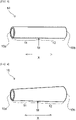
- FIG. 7 shows a perspective view of a cylindrical sample 100 for measurement of the elastic index
- FIG. 8 shows a plan view of the cylindrical sample 100 shown in FIG. 7 viewed from above.
- the cylindrical sample 100 having a length of 5 mm in the axial direction X is prepared by cutting the vein cover 10 along a circumferential cut line perpendicular to the axial direction to obtain a section having a length of 5 mm in the axial direction X.
- the cylindrical sample 100 is cut out all around along a cut plane perpendicular to the axial direction, i.e., a circumferential cut line, so that the cylindrical sample 100 is 5 mm long in the axial direction and continuous over the whole circumference.
- a first pin 101 and a second pin 102 having a diameter d of 0.75 mm are inserted into a lumen of the cylindrical sample 100 parallel to the axial direction X of the cylindrical sample 100.
- the first pin 101 is fixed, and the second pin 102 is pulled towards outside of a radial direction of the cylindrical sample 100 with a pulling force F so that a distance between the first pin 101 and the second pin 102 becomes L, and the 10% elastic index is obtained by measuring a pulling force F 1.1 that is a pulling force when ⁇ d + 2L becomes 1.1 times a perimeter of the cylindrical sample 100 in its natural state, and dividing the pulling force F 1.1 by a strain [(1.1 - 1.0)/1.0].
- the distance L between the first pin 101 and the second pin is, as shown in FIG. 8 , a distance between the center of the first pin 101 and the center of the second pin 102.
- the inner diameter of the cylindrical sample 100 is equal to the sum of 1/2 of the circumference ⁇ d of the first pin 101, 1/2 of the circumference ⁇ d of the second pin 102, and twice the distance L between the first pin 101 and the second pin 102, i.e., ⁇ d + 2L.
- the 10% elastic index can be obtained by dividing the pulling force F 1.1 that pulls the second pin 102 when the inner diameter of the cylindrical sample 100 is expanded by 10% from its natural state, i.e., when ⁇ d + 2L becomes 1.1 times the circumference of the cylindrical sample 100 in its natural state by strain [(1.1 - 1.0)/1.0].
- the length in the axial direction X of the vein cover 10 that is to be measured is less than 5 mm, multiple vein covers with similar elastic index are prepared in the same manner, and the vein covers can be arranged in the axial direction X so that the total length in the axial direction is 5 mm to obtain the elastic index in the above-described manner.
- the portion (A) having the 10% elastic index of 25 N or less may be provided continuously or separated.
- the 10% elastic index of the portion (A) is preferably 24 N or less, more preferably 21 N or less, even more preferably 17 N or less, and may be 14 N or less.
- the vein cover 10 When the vein cover 10 is placed in the shunt construction 1, it may be placed to cover not only the vein 4 but also a part of the artery 3 or the artificial vessel 5 on the side of the anastomosis site 6, however, the portion (A) is preferably placed over the part covering the vein 4.
- the inner diameter of the vein cover 10 can be appropriately determined depending on the vessel diameter to be applied, and for example, preferably 1 mm or more, more preferably 2 mm or more, even more preferably 3 mm or more, and may be 4 mm or more.
- the inner diameter of the vein cover 10 is, for example, preferably 10 mm or less, more preferably 9 mm or less, and even more preferably 8 mm or less.
- the vein cover 10 may have different inner diameter depending on the axial direction X.
- the 10% elastic index of the portion (A) is preferably 0.1 mN or more.
- the 10% elastic index of the portion (A) may be 0.2 mN or more, 0.5 mN or more, and 1 mN or more.
- the vein cover 10 has a vein cover first part 11 from the first end 10a to a midpoint M between the first end 10a and the second end 10b of the vein cover 10, and has a vein cover second part 12 from the midpoint M to the second end 10b, and the portion (A) is preferably located in the vein cover first part 11.
- portion (A) is located in the vein cover first part 11, when the first end 10a of the vein cover 10 is placed in the upstream side of the vein 4 in the blood flow direction B, the vein is gradually remodeled into a buffer vessel from the upstream side, buffering the transmural pressure, shear stress, pulsating pressure of the beat, flow velocity, and blood flow rate, resulting in the change from the upstream to the downstream part.
- the length of the portion (A) in the axial direction X is preferably 50% or longer of the outer diameter of the anastomosed artery 3 or the artificial vessel 5.
- the length of the portion (A) in the axial direction X is more preferably 60% or longer of the outer diameter of the anastomosed artery 3 or the artificial vessel 5, even more preferably 80% or longer, and may be 100% or longer.
- the upper limit of the length of the portion (A) in the axial direction X is not particularly limited, and may be 2000% or shorter of the outer diameter of the anastomosed artery 3 or the artificial vessel 5, 1750% or shorter, or may be 1500% or shorter.
- the length of the portion (A) in the axial direction X in the above range makes it easier for the vein 4 to be remodeled into a buffer vessel.
- the 10% elastic index of the vein cover second part 12 is preferably smaller than the 10% elastic index of the vein cover first part 11.
- the 10% elastic index that is larger upstream and smaller downstream allows the vein 4 to be gradually remodeled into a buffer vessel, buffering the transmural pressure, shear stress, pulsating pressure of the beat, flow velocity, and blood flow rate, resulting in the change from the upstream to the downstream part.
- the 10% elastic index of the part having a length of 5 mm from the midpoint M towards the side of the second end 10b is preferably 0.1 times or more and 0.98 times or less, or 0.96 times or less the 10% elastic index of the part having a length of 5 mm from the first end 10a, or it is preferably 0.1 times or more and 1.9 times or less.
- the 10% elastic index of the part having a length of 5 mm from the midpoint M towards the side of the second end 10b may be 0.2 times or more, or 0.3 times or more the 10% elastic index of the part having a length of 5 mm from the first end 10a.
- the 10% elastic index of the part having a length of 5 mm from the midpoint M towards the second end 10b may be 0.8 times or less, or 0.7 times or less the 10% elastic index of the part having a length of 5 mm from the first end 10a.
- the part having a length of 5 mm from the midpoint M towards the second end 10b, i.e., the part of the vein cover 10 covering the downstream of the vein 4 has the 10% elastic index in the above range in comparison to the 10% elastic index of the part having a length of 5 mm from the first end 10a, i.e., the part of the vein cover 10 covering the most upstream part of the vein 4, so that when the first end 10a of the vein cover 10 is placed in the upstream side of the vein in the blood flow direction B, the vein 4 is gradually remodeled into a buffer vessel from the upstream side, buffering the transmural pressure, shear stress, pulsating pressure of the beat, flow velocity, and blood flow rate, resulting in the change from the upstream to the downstream part
- the 10% elastic index of the part having a length of 5 mm from the second end 10b in the axial direction X is preferably 0.1 times or more and 0.98 times or less, 0.96 times or less, or 0.1 times or more and 0.9 times or less the 10% elastic index of the part having a length of 5 mm from the first end 10a in the axial direction X.
- the 10% elastic index of the part having a length of 5 mm from the second end 10b in the axial direction X may be 0.2 times or more, or 0.3 times or more the 10% elastic index of the part having a length of 5 mm from the first end 10a in the axial direction X.
- the 10% elastic index of the part having a length of 5 mm from the second end 10b in the axial direction X may be 0.8 times or less, 0.7 times or less, or 0.6 times or less the 10% elastic index of the part having a length of 5 mm from the first end 10a in the axial direction X.
- the part having a length of 5 mm from the second end 10b, i.e., the part of the vein cover 10 covering the most downstream of the vein 4 has the small 10% elastic index in the above range in comparison to the 10% elastic index of the part having a length of 5 mm from the first end 10a, i.e., the part of the vein cover 10 covering the most upstream part of the vein 4, so that when the first end 10a of the vein cover 10 is placed in the upstream side of the vein in the blood flow direction B, the vein 4 is gradually remodeled into a buffer vessel from the upstream side, buffering the transmural pressure, shear stress, pulsating pressure of the beat, flow velocity, and blood flow rate, resulting in the change from the upstream to the downstream part.
- the portion (A) preferably has a 20% elastic index of 32 N or less when the inner diameter is expanded by 20% from its natural state.
- the 20% elastic index of the portion (A) is more preferably 29 N or less, even more preferably 27 N or less, and may be 25 N or less.
- the measurement method of the 20% elastic index is the same as the method for measuring the 10% elastic index except measuring force F 1.2 that is a force when ⁇ d + 2L becomes 1.2 times the circumference of the cylindrical sample 100 in its natural state.
- the inner diameter of the part having a length of 5 mm in the axial direction X from the second end 10b of the vein cover 10 is preferably expandable by 40% or more from its natural state, more preferably expandable by 50% or more, and even more preferably expandable by 60% or more. If the inner diameter of the part having a length of 5 mm in the axial direction X from the second end 10b of the vein cover 10 is expandable in the above range, the vein 4 covered by the vein cover 10 can be easily remodeled into a buffer vessel.
- the vein cover 10 for example as shown in FIG. 3 , may be formed in a straight shape with an approximately equal inner diameter on the side of the first end 10a and the side of the second end 10b.
- the 10% elastic index and the position of the portion (A) can be adjusted by, for example, varying the density of knitted fabric or the like constituting the vein cover 10. If the vein cover 10 have a straight shape, the vein 4 can be covered by the vein cover 10 over the entire length of the axial direction X without being affected by the shape of the vein cover 10.
- the vein cover 10 for example as shown in FIG. 4 , may be formed in a tapered shape with an inner diameter increasing from the side of the first end 10a to the side of the second end 10b. If the vein cover 10 has a tapered shape, the 10% elastic index and the position of the portion (A) can be easily adjusted.
- the vein cover second part 12 can cover the vein looser, and when the side of the first end 10a of the vein cover 10 is placed in the upstream side of the vein 4 in the blood flow direction B, the vein 4 is gradually remodeled into a buffer vessel from the upstream side buffering the transmural pressure, shear stress, pulsating pressure of the beat, flow velocity, and flood flow rate, resulting in the change from the upstream to the downstream part.
- a bellows structure may be formed on the side of the first end 10a. Since the bellows structure allows the elastic index to be adjusted by the density of the bellows, the 10% elastic index and the position of the portion (A) can be easily adjusted. For example, by providing portion with high-density bellows, a portion with low-density bellows, and a portion with no bellows structure in this order, the elastic index can be gradually changed.
- one end of the bellows portion may be placed to cover not only the vein 4 but also a part of the artery 3 or the artificial vessel 5 on the side of the anastomosis site 6
- the vein cover 10 for example as shown in FIG. 6 , may be formed entirely in a bellows structure with the density and amplitude of the bellows varying in the axial direction X.
- a high-density bellows is formed at the side of the first end 10a and the density of the bellows decreases towards the side of the second end 10b.
- This configuration makes it easier to make the vein cover 10 with the 10% elastic index of the vein cover second part 12 smaller than the 10% elastic index of the vein cover first part 11, and when the side of the first end 10a of the vein cover 10 is placed in the upstream side of the vein 4 in the blood flow direction B, the vein 4 is gradually remodeled into a buffer vessel from the upstream side, buffering the transmural pressure, shear stress, pulsating pressure of the beat, flow velocity, and blood flow rate, resulting in the change from the upstream to the downstream part.
- the vein cover 10 preferably has a length in the axial direction of 5 mm or longer.
- the length in the axial direction X is more preferably 10 mm or longer, even more preferably 20 mm or longer, particularly preferably 30 mm or longer, and may be 40 mm or longer.
- the length in the axial direction X is preferably 120 mm or shorter, more preferably 100 mm or shorter, and even more preferably 90 mm or shorter.
- the length in the axial direction X within the above range makes it easier for the vein 4 to be remodeled into a buffer vessel.
- the vein cover 10 is preferably made of knitted fabric. Knitted fabrics have excellent elasticity and flexibility, and therefore are suitable for obtaining the desired elastic index.
- the type of knitting fabric is not limited, and may be warp or weft knitting. Examples of knitting texture of the warp knitting includes half knitting, back half knitting, quinz coat knitting, and satin knitting.
- the weft knitting includes circular knitting and flat knitting, and examples of knitting texture of the weft knitting include plain knitting, rib knitting, double side knitting, milan-rib knitting, and jacquard knitting.
- the knitted fabric is preferably composed of a weft knitted fabric.
- the vein cover 10 may be composed of woven fabric such as a plain weave.
- the vein cover 10 may be composed of porous materials or nonwoven fabric made by any method, such as melt-blown, needle-punched, or spun-laced method.
- the vein cover 10 may have at least one type of knitted fabric, woven fabric, nonwoven fabric, and porous materials as a component comprising at least part or the whole of the structure of the vein cover.
- the vein cover 10 may be composed of a combination of two or more different materials, for example, some parts may be made of knitted fabric and other parts may be made of other materials, for example, porous materials.
- the elasticity of the vein cover 10 may due to the material, the structure, or both.
- the desired elastic index can be obtained simply by making the material such as woven fabric or nonwoven fabric into a sheet and then into a cylindrical structure that does not impart elasticity as the characteristics of the structure.
- the desired elastic index can be obtained by making the material into a structure that can impart elasticity, such as a knitted structure.
- the yarns forming the knitted, woven, or nonwoven fabric are preferably composed of biocompatible resin materials, and examples the resin materials include polyolefin-based resin such as polyethylene and polypropylene; polyamide-based resin such as nylon; polyester-based resin such as polyethylene terephthalate and polybutylene terephthalate; aromatic polyether ketone-based resin such as PEEK; polyether polyamide-based resin; polyester-based elastomer such as polyester polyol; polyurethane-based resin; polyimide-based resin; fluorine-based resin such as PTFE, PFA, and ETFE; polyvinyl chloride-based resin; silicone-based resin.
- the resin materials include polyolefin-based resin such as polyethylene and polypropylene; polyamide-based resin such as nylon; polyester-based resin such as polyethylene terephthalate and polybutylene terephthalate; aromatic polyether ketone-based resin such as PEEK; polyether polyamide-based resin; polyester-based
- the yarns also may be made of resin materials used in artificial blood vessels (for example, polyester, PTFE, polyurethane), and specific examples include ePTFE, which is a stretched PTFE, and DACRON (registered trademark), which is polyester fiber from Dupont.
- the vein cover 10 may be composed of biodegradable materials, and examples of the materials include aliphatic polyester such as polylactic acid, polycaprolactone, polylactic acid/polycaprolactone copolymer, polyglycolic acid, polybutylene succinate, and polyhydroxyalkanoic acid; aliphatic polyether such as polyethylene glycol; and polyvinyl alcohol.
- the yarns may be made of natural fiber such as silk and cotton, or a combination of the resin materials, biodegradable materials, and natural fiber.
- the vein cover 10 When the vein cover 10 is anastomosed to the artificial vessel 5, the vein cover 10 is preferably integrated with the artificial vessel 5.
- the vein cover 10 is configured so as to be integrated with the artificial vessel 5, which makes it easy for the vein cover 10 to cover the part from the anastomosis site 6 to downstream and avoid such inconveniences as the vein cover 10 being displaced or coming off.
- the 10% elastic index of the vein cover 10 can be adjusted by varying the density of the above knitted or woven fabrics, by partially overlapping the knitted or woven fabrics, or by devising the shape of the vein cover 10.
- Assessment of whether the vein 4 has been remodeled into a buffer vessel can be done by checking whether a two-layer structure is formed, consisting of the smooth muscle layer containing elastic fibers and the elastic fiber layer containing collagen fibers thicker than the smooth muscle layer outside thereof. Specifically, the vein 4 at the shunt construction 1 is cut out and special stains such as hematoxylin-eosin (HE) stain and Elastica van Gieson (EvG) stain are applied, and the vein wall cross section is observed under a microscope.
- HE hematoxylin-eosin
- EvG Elastica van Gieson
- the smooth muscle is stained turbid yellow, the elastic fibers are stained dark purple, and the collagen fibers are stained dark red, so that the smooth muscle layer containing the elastic fibers and the elastic fiber layer containing the collage fibers can be observed and the thickness of each layer can also be determined.
- a cylindrical sample that had a length of 5 mm in the axial direction and was continuous over the whole circumference by cutting out a portion having a length of 5 mm from one end of a vein cover all around a cut plane perpendicular to the axial direction, i.e., a circumferential cut line.
- a first pin and a second pin having a diameter of 0.75 mm were inserted into a lumen of the cylindrical sample parallel to the axial direction of the cylindrical sample. The first pin was fixed and the second pin was pulled towards outside of the radial direction of the cylindrical sample.
- a pulling force was measured when the sum of twice the distance between the first pin and the second pin and ( ⁇ ⁇ 0.75 mm) became 1.1 times the perimeter of the cylindrical sample in its natural state, and the pulling force was divided by the strain [(1.1 - 1.0)/1.0] to obtain a 10% elastic index.
- Beagle dogs male and female, weighing 9-14 kg were used.
- a shunt with the anastomotic configuration shown in Table 1 was constructed between the carotid artery and jugular vein, and a vein cover with the shape, material, inner diameter, axial length and elastic index shown in Table 1 was placed over the vein at the shunt construction for follow-up observation.
- the follow-up observation was made for at least 12 weeks to ensure that no adverse events occurred over a long period of time.
- the beagle dog was euthanized and the vein at the shunt construction was removed.
- the removed vein was stained with Elastica van Gieson stain, and the cross sections contained in a section having a length of 5 mm in the direction of blood flow from just below the anastomosis site, a section having a length of 5 mm in the direction of blood flow near the midpoint of the vein cover, and a section having a length of 5 mm from the other end of the vein cover were observed under optical microscope.
- the blood flow condition was measured by Doppler blood flow measurement and further diagnosed by color Doppler ultrasound imaging diagnostic unit.
- the results of the above (4) and (5) were combined to evaluate whether the vein at the shunt construction site was remodeled into a buffer vessel according to the following criteria.
- a vein cover having a straight shape, 50 mm in axial length, and 5 mm in inner diameter was manufactured using a polyester artificial blood vessel (hereinafter, described as "DACRON ® artificial blood vessel") (Hemashield Gold Knitted Vascular Graft manufactured by Maquet Cardiovascular). The 10% elastic index and 20% elastic index of the manufactured vein cover were measured.
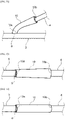
- a shunt was constructed in the neck of a beagle dog by anastomosing an artery and vein, and the vein was covered with the vein cover so that one end of the vein cover was placed at the anastomosis site as shown in FIG. 9 , and the dog was monitored for 105 days. Subsequently, the vein was evaluated by Doppler blood flow measurement and color Doppler ultrasound imaging diagnostic unit.
- the beagle dog was euthanized and the vein at the shunt construction site was removed.
- the smooth muscle layer and elastic fiber layer containing collagen fibers of the removed vein were observed, their thickness was measured, and remodeling to a buffer vessel was evaluated.
- the manufacturing condition and elastic indices of the vein cover and the results of the evaluation are shown in Table 1.
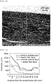
- the optical micrograph of a section of the vein wall parallel to the direction of blood flow at a distance of 2.5 mm from the anastomosis site is shown in FIG. 14
- the thickness of the smooth muscle layer and the elastic fiber containing collagen fibers are shown in Table 2 and FIG. 15 .
- the lower side is the outer wall of the vein wall and the upper side is the inner wall of the vein wall.
- a vein cover having a bellows shape, 40 mm in axial length, and 6 mm in inner diameter was manufactured using fibers that was made by bundling several single fibers ( ⁇ 3 ⁇ m) of nylon material and woolly processed, instead of DACRON ® artificial blood vessel. The 10% elastic index and 20% elastic index of the manufactured vein cover were measured.
- a shunt was constructed in the neck of a beagle dog by anastomosing an artery and vein, and the vein was covered with the vein cover so that one end of the vein cover was placed at the anastomosis site as shown as FIG. 10 , and the dog was monitored for 154 days. Subsequently, the vein was evaluated by Doppler blood flow measurement and color Doppler ultrasound imaging diagnostic unit.
- the beagle dog was euthanized and the vein at the shunt construction site was removed.
- the smooth muscle layer and elastic fiber layer containing collagen fibers of the removed vein were observed, their thickness was measured, and remodeling to a buffer vessel was evaluated.
- the manufacturing condition and elastic indices of the vein cover and the results of the evaluation are shown in Table 1. The results of the evaluation based on the above criteria (a) to (e) were good.
- Vein covers were manufactured under the manufacturing condition shown in Table 1 and the 10% and 20% elastic indices were measured, shunts were constructed in the anastomotic configuration shown in FIG. 10 to FIG. 12 , respectively, as shown in Table 1, and the veins were observed and evaluated as in Example 1.
- the values before and after the arrows for the inner diameter in Examples 5, 7, and 8 in Table 1 indicate the minimum and maximum inner diameter of the tapered vein covers.
- the elastic indices and evaluation results are shown in Table 1. All the results of the evaluation based on the above criteria (a) to (e) were good.
- a vein cover having a straight shape, 28 mm in axial length, and 5.2 mm in inner diameter was manufactured using a knitted fabric of polyglycolic acid (NEOVEIL ® manufactured by Gunze Limited). The 10% elastic index and 20% elastic index of the manufactured vein cover were measured.
- a shunt was constructed in the neck of a beagle dog by end-to end anastomosing a vein to an artificial vessel made of ePTFE (Gore-Tex ® Stretch Vascular Graft manufactured by W. L. Gore & Associates) that was side-to-end anastomosed to an artery, and the vein was covered with the vein cover so that one end of the vein cover was placed at the anastomosis site as shown in FIG. 13 , and the dog was monitored for 56 days.
- the elastic indices and the evaluation results obtained in the same way as in Example 1 are shown in Table 1. All the results of the evaluation based on the above (a) to (e) were good.
- a vein cover having a tapered shape, 25 mm in axial length, an inner diameter of one end of 5 mm, and an inner diameter of the other end of 8 mm was manufactured using a nonwoven fabric made of polylactic acid/polycaprolactone copolymer (polylactic acid 25%/polycaprolactone 75%). The 10% elastic index and 20% elastic index of the manufactured vein cover were measured.
- a shunt was constructed in the neck of a beagle dog by end-to-end anastomosing a vein to an artificial vessel made of ePTFE (Gore-Tex ® Stretch Vascular Graft manufactured by W. L.
- a vein cover having a straight shape, 30 mm in axial length, and 4.5 mm in inner diameter was manufactured using DACRON ® artificial blood vessel (J-Graft, Shield Neo S manufactured by Japan Lifeline Co., Ltd.).
- a shunt was constructed in the neck of a beagle dog by anastomosing a vein to an artificial vessel made of ePTFE (Distaflo ® Bypass Grafts manufactured by Bard Peripheral Vascular Inc.) that was anastomosed to an artery, and the vein was covered with the vein cover so that one end of the vein cover was placed at the anastomosis site, and the dog was monitored for 84 days.
- the vein was evaluated in the same way as in Example 1. The elastic indices and evaluation results are shown in Table 1.
- the smooth muscle layer and elastic fiber layer that was thicker than the smooth muscle layer and contained collagen fibers were not observed, and intimal thickening at the anastomosis site with the vein occurred to form thrombus formation, which extended into the artificial vessel made of ePTFE to cause obstruction.
- the results of the evaluation based on the above criteria (a) to (e) were bad.
- Vein covers were manufactured in the manufacturing condition shown in Table 1, and shunts were constructed in the anastomotic configuration shown in FIG. 9 and FIG. 13 as shown in Table 1, and the veins were evaluated in the same way as in Example 1.
- the elastic indices and evaluation results are shown in Table 1. All the results of the evaluation based on the above criteria (a) to (e) were bad.
Landscapes
- Health & Medical Sciences (AREA)
- Heart & Thoracic Surgery (AREA)
- Life Sciences & Earth Sciences (AREA)
- General Health & Medical Sciences (AREA)
- Veterinary Medicine (AREA)
- Public Health (AREA)
- Engineering & Computer Science (AREA)
- Biomedical Technology (AREA)
- Vascular Medicine (AREA)
- Animal Behavior & Ethology (AREA)
- Surgery (AREA)
- Cardiology (AREA)
- Transplantation (AREA)
- Oral & Maxillofacial Surgery (AREA)
- Pulmonology (AREA)
- Nuclear Medicine, Radiotherapy & Molecular Imaging (AREA)
- Gastroenterology & Hepatology (AREA)
- Medical Informatics (AREA)
- Molecular Biology (AREA)
- Anesthesiology (AREA)
- Hematology (AREA)
- Prostheses (AREA)
Abstract
Description
- The present invention relates to a vein cover used for an anastomosis site where blood vessels are anastomosed, and for example, the vein cover can be used for an anastomosis site where an artery and a vein are anastomosed to form a shunt, or for an anastomosis site where a vein and an artificial vessel that is anastomosed to an artery are anastomosed.
- For patients with serious kidney diseases including renal failure, hemodialysis treatment is regularly performed, in which blood is taken from the patient's body, waste products, excess water and minerals are removed with a dialyzer, and then the blood is returned to the patient's body. When hemodialysis is performed, a special needle is usually inserted into a vein. At this time, since the blood flow in the vein is not sufficient to carry out dialysis, an artery is anastomosed to the vein, which significantly reduces peripheral resistance and significantly increases the blood flow. Accordingly, the vein must be made into a venous vessel that can be used for dialysis. Such a vessel is called a shunt and is usually formed by making an incision in the skin of the arm to expose the artery and vein, making a small incision in the artery to which the vein is anastomosed, and diverting some of the blood flow from the artery to the vein. The vein may be directly anastomosed to the artery as shown in
FIG. 1 , or an artificial vessel may be placed between the artery and the vein by anastomosing one end of the artificial vessel to the small incision of the artery and anastomosing the other end of the artificial vessel to the vein as shown inFIG. 2 . - At an anastomosis site, since there is a significant difference in elasticity between the artery and vein, when beating blood flows with high blood pressure flow into the vein that has markedly high extensibility at low pressure and has low elasticity at high pressure, blood turbulence and stress changes to the vein wall occur. As a result, intimal thickening occurs in the anastomosis site and the vein, which is an outflow pathway, easily causing pathological changes such as stenosis, obstruction, thrombus formation, and the like. Failure to regulate shunt blood flow conditions that impose a high strain on the patient's body can lead to more extensive local disorder (e.g., varicose formation or stenosis of downstream veins, Steel's syndrome due to excessive shunt blood flow) or systemic disorder (e.g., heart failure due to markedly increased venous annular flow).
- If conditions are favorable, appropriate remodeling may occur as a protective adaptive response of the body, e.g. by elastic changes in the venous wall, and stenosis or obstruction due to intimal thickening may be avoided, or the shunt blood flow may be self-regulated to a state that is not burdensome to the body. However, if local conditions such as shunt blood flow and anastomotic geometry, or systemic conditions (e.g., diabetes, hypertension, arteriosclerosis, or blood conditions) are poor, the pathological biological response may exceed the extent to which an appropriate protective adaptive response occurs, leading to local or systemic disorder.
- To control the rapid increase in blood flow in the immediate and early postoperative period, vascular banding is used to reinforce the vein wall from the outside to prevent excessive blood pressure and consequent hyperextension and blood turbulence in the veins (Non-patent document 1).
Patent document 1 discloses a covering for reinforcing natural veins for use as surgical implants, which is a mesh fabric net covering made by forming a knitted fabric that is seamless, tubular, and substantially pile-less.Patent documents - However, the above-described vascular banding did not sufficiently prevent lesions such as intimal thickening.
-
- Patent Document 1:
JP 2004-535896 T - Patent Document 2:
JP 2010-516437 T - Patent Document 3:
JP 2013-509258 T - Non-patent Document 1: "I Blood Access Problems associated with blood flow failure" by Hiroaki Haruguchi, Nihon Toseki Igakkai Zasshi Vol. 15, No. 1, 68-70, 2000
- In the conventional vascular banding, the reinforced vein wall is modified (arterialized) into an arterial wall-like structure only under certain conditions, however, the blood pressure and pulsation are not buffered and delivered downstream as blood flows from the reinforced site to the non-reinforced vein, which is not a fundamental solution to the cause of intimal thickening. To solve this, the blood pressure and pulsation should be gradually reduced downstream from the anastomosis site, so that the most downstream vein is only subjected to low, non-pulsatile pressure.
- The present invention was made in view of the above circumstances, and the objective thereof is to provide a vein cover that can reduce or prevent intimal thickening by delivering blood to the downstream veins while gradually reducing the blood pressure, pulse pressure, and blood flow rate of the blood flowing through the lumen.
- A vein cover that can solve the above problems is as follows.
- [1] A cylindrical vein cover to be placed outside a vein that is anastomosed to an artery or to an artificial vessel, comprising a portion (A), wherein
- the portion (A) has a 10% elastic index of 25 N or less when an inner diameter of the vein cover is expanded by 10% from a natural state, and
- the 10% elastic index is measured by
- preparing a cylindrical sample having a length of 5 mm in an axial direction by cutting the vein cover along a circumferential cut line perpendicular to the axial direction to obtain a section having a length of 5 mm in the axial direction;
- inserting a first pin and a second pin having a diameter d of 0.75 mm into a lumen of the cylindrical sample parallel to an axial direction of the cylindrical sample;
- fixing the first pin;
- pulling the second pin towards outside of a radial direction of the cylindrical sample with a pulling force F so that a distance between the first pin and the second pin becomes L;
- measuring a pulling force F1.1 that is a pulling force when πd + 2L becomes 1.1 times a perimeter of the cylindrical sample in a natural state; and
- dividing the pulling force F1.1 by a strain [(1.1 - 1.0)/1.0] to obtain the 10% elastic index.
- [2] The vein cover according to [1], wherein the 10% elastic index of the portion (A) is 0.1 mN or more.
- [3] The vein cover according to [1] or [2], wherein
- the vein cover has a first part that is from a first end of the vein cover to a midpoint between the first end and a second end of the vein cover, and has a second part that is from the midpoint to the second end of the vein cover; and
- the portion (A) is located in the first part of the vein cover.
- [4] The vein cover according to any one of [1] to [3], wherein a length of the portion (A) in the axial direction is 50% of an outer diameter of the anastomosed artery or the artificial vein or longer.
- [5] The vein cover according to any one of [1] to [4], wherein
- the vein cover has a first part that is from a first end of the vein cover to a midpoint between the first end and a second end of the vein cover, and has a second part that is from the midpoint to the second end of the vein cover; and
- the 10% elastic index in the second part of the vein cover is smaller than the 10% elastic index in the first part of the vein cover.
- [6] The vein cover according to any one of [1] to [5], wherein
- the portion (A) has a 20% elastic index of 32 N or less when an inner diameter of the vein cover is expanded by 20% from a natural state, and
- the 20% elastic index is measured by
- preparing a cylindrical sample having a length of 5 mm in an axial direction by cutting the vein cover along a circumferential cut line perpendicular to the axial direction to obtain a section having a length of 5 mm in the axial direction;
- inserting a first pin and a second pin having a diameter d of 0.75 mm into a lumen of the cylindrical sample parallel to an axial direction of the cylindrical sample;
- fixing the first pin;
- pulling the second pin towards outside of a radial direction of the cylindrical sample with a pulling force F so that a distance between the first pin and the second pin becomes L;
- measuring a pulling force F1.2 that is a pulling force when πd + 2L becomes 1.2 times a perimeter of the cylindrical sample in a natural state; and
- dividing the pulling force F1.2 by a strain [(1.2 - 1.0)/1.0] to obtain the 20% elastic index.
- [7] The vein cover according to any one of [1] to [6], having a length in the axial direction of 5 mm or longer.
- [8] The vein cover according to any one of [1] to [7], made from a biodegradable material.
- [9] The vein cover according to any one of [1] to [8], comprising at least one of knit fabric, woven fabric, and nonwoven fabric as a partial or whole component.
- [10] The vein cover according to any one of [1] to [9], integrated with the artificial vessel when the vein is anastomosed to the artificial vessel.
- The vein cover of the present invention, due to the above configuration, can suppress incompatibility of vessel wall elasticity, turbulent flow, and high flow rate to reduce or prevent intimal thickening by gradually changing the wall structure from the anastomosis site to the downstream vein to gradually change shear stress, pressure orthogonal to the wall, blood flow, flow velocity, and range of change with beating in the covered interior. The reason why the vein cover of the present invention achieves this effect may be due to the following.
- Both arteries and veins consist of an intima, a tunica media, and an adventitia, and the arteries have the tunica media consisting of smooth muscle cell-rich smooth muscle layer and an elastic fiber layer including collagen fibers. The arteries have thick smooth muscle and elastic fiber layers so that pulsation change in the vessel wall is little to prevent turbulence generation and fluctuations in abrasion stress, even under the pressure of pulsating luminal blood flow. On the other hand, the veins have thin vessel walls and do not have the thick smooth muscle and elastic fiber layers as the arteries do. When arterial blood flows directly into such veins via arteriovenous shunts, lesions such as intimal thickening occur due to the substantial difference in elasticity between arteries and veins as described above. To prevent this, while the most upstream part of the vein at a shunt construction, i.e., the anastomosis site, is subjected to 100% pulsatile arterial pressure, the vein at the shunt construction needs to be remodeled into a buffer vessel, which gradually decreases the blood pressure, pulsatility, blood flow, and maximum flow velocity towards the downstream of the vein to make the most downstream vessel have low non-pulsatile pressure.
- The vein cover of the present invention, having the above configuration, can make the vein in the shunt construction remodeled into a buffer vessel by gradually form a two-layer structure, in the venous wall at the shunt construction, comprising a smooth muscle layer containing elastic fiber thicker than the smooth muscle layer of normal veins and an elastic fiber layer containing collagen fibers thicker than the smooth muscle layer on the outside of the smooth muscle layer. This remodeling of the vein at the shunt construction into a buffer vessel with the elastic fiber layer containing collagen fibers thicker than the smooth muscle layer allows the pulsatile blood flow with high arterial pressure at the arteriovenous and prosthetic venous anastomoses to be gradually buffered towards the downstream of the vein and finally transformed to venous blood flow. As a result, blood turbulence and pulsatile changes in the venous wall are suppressed to reduce or prevent intimal thickening.
- The difference between the buffer vessel described above and normal arteries are explained below. Blood vessels are composed of three layers: intima, tunica media, and adventitia. Of these, the intima contributes significantly to the anti-coagulability, but its mechanical contribution is very small. The composition of the mechanical elements of the arteries of the extremities, which are the normal arteries used for dialysis, consists largely of the tunica media containing some elastic fibers and abundant smooth muscle and the adventitia consisting of elastic fibers and collagen fibers etc. outside the tunica media. In other words, these arteries have very abundant smooth muscle and relatively few elastic fibers (the composition of "smooth muscle > elastic fibers"). The elastic fibers, due to their elasticity, have a buffering function like a lubber tube, resisting and relaxing the high pulsatile arterial blood pressure. On the other hand, the smooth muscle, which is muscle, has a more active mechanical function, resisting arterial blood pressure, and at the same time, has the active and proactive function of delivering high pulsatile arterial blood pressure to the periphery without attenuation. Because of this pressure delivering function of the smooth muscle of arteries, the blood pressure of large arteries with an inner diameter of centimeters and small arteries with an inner diameter of fractions of a millimeter remain almost unchanged. In other words, normal arteries (with the configuration of "smooth muscle > elastic fibers") do not have the ability to buffer high pulsatile arterial pressure due to the function of the abundant smooth muscle.
- On the other hand, when veins in the shunt area are remodeled into buffer vessels, the ratio of elastic fibers and smooth muscle is the reverse of that in normal arteries, with elastic fibers abundant and smooth muscle relatively thin (the configuration of "elastic fibers > smooth muscle"). Accordingly, in buffer vessels, the pressure buffering function is dominant over the pressure delivery function. In the buffer vessels, while maintaining the above configuration "elastic fibers > smooth muscle", i.e., the buffering function, the whole system gradually becomes thin and transformed to the vein, i.e., gradually transformed to the normal vein as the buffered pressure decreases.
- If, hypothetically, the vein wall changes to normal arterial-like, this is arterialization (remodeling into an artery) and not remodeling into the buffer vessel. If that arterialization gradually weakens and spontaneously transitions to completely normal veins at the downstream part, it means that the vessel having the configuration of "smooth muscle > elastic fiber" gradually becomes thin and transformed to veins, causing pathological changes in the downstream veins since it has no buffering function and high pulsatile blood pressure acts on the venous wall. This is the clear functional difference between the gradual thinning of the buffer vessels and their transition to normal veins and the gradual thinning of normal arterial-like vessel and their transition to normal veins.
- Furthermore, it should be noted that the morphological change of "elastic fiber > smooth muscle" configuration described above is a necessary but not sufficient condition for the definition of the buffer vessels. In order to be able to be defined as the buffer vessels, buffering of blood pressure and pulsation must be demonstrated by actual observation of blood flow measurements.
-
-
FIG. 1 shows a schematic view of an example of a shunt construction. -
FIG. 2 shows a schematic view of another example of a shunt construction. -
FIG. 3 shows a perspective view of a vein cover according to an embodiment of the present invention. -
FIG. 4 shows a perspective view of a vein cover according to another embodiment of the present invention. -
FIG. 5 shows a perspective view of a vein cover according to even another embodiment of the present invention. -
FIG. 6 shows a perspective view of a vein cover according to even another embodiment of the present invention. -
FIG. 7 shows a perspective view of a sample for measurement of the elastic index. -
FIG. 8 shows a plan view of the measurement sample shown inFIG. 7 viewed from above. -
FIG. 9 shows a perspective view of a vein cover according to an embodiment of the present invention placed at an anastomosis site. -
FIG. 10 shows a perspective view of a vein cover according to another embodiment of the present invention placed at an anastomosis site. -
FIG. 11 shows a perspective view of a vein cover according to even another embodiment of the present invention placed at an anastomosis site. -
FIG. 12 shows a perspective view of a vein cover according to even another embodiment of the present invention placed at an anastomosis site. -
FIG. 13 shows a perspective view of a vein cover according to even another embodiment of the present invention placed at an anastomosis site. -
FIG. 14 shows a micrograph of a vein wall section with Elastica van Gieson stain. -
FIG. 15 shows a graph representing the relationship between the distance from the anastomosis site in the axial direction and the thickness of the smooth muscle layer and elastic fiber layer containing collagen fibers in the vein wall cross-section. - Hereinafter, the present invention will be described based on the following embodiments, however, the present invention is not limited by the following embodiments and can be altered in design within a scope in compliance with the intent described above and below, and all the changes are to be encompassed within a technical scope of the present invention. Note that, in each drawing, hatching, reference signs for components, and the like may be omitted for convenience of description, and in such a case, the specification and other drawings are to be referred to. Furthermore, since the dimensions of the various components in the drawings are provided for the purpose of facilitating the understanding of the feature of the present invention, the dimensions may differ from the actual dimensions in some cases.
- The vein cover of the present invention is a cylindrical vein cover to be placed outside a vein that is anastomosed to an artery or to an artificial vessel, having a portion (A), wherein the portion (A) has a 10% elastic index of 25 N or less when an inner diameter of the vein cover is expanded by 10% from a natural state. Unlike the conventional concept of vein wall reinforcement by vascular banding, in the present invention, by loosely covering the vein with the vein cover including the portion (A) having the 10% elastic index of 25 N or less, a two-layer structure consisting of a smooth muscle layer containing elastic fibers and elastic fiber layer containing collagen fibers thicker than the smooth muscle layer outside the smooth muscle layer is formed in the covered vein, which allows the vein to be remodeled into a buffer vessel to prevent or reduce lesions such as intimal thickening. In both cases where the vein is anastomosed to an artery and where the vein is anastomosed to an artificial vessel connected to an artery, the vein cover of the present invention can prevent or reduce lesions such as intimal thickening.
- Hereinafter, the vein cover according to embodiments of the present invention will be described, referring to
FIG. 1 to FIG. 6 .FIG. 1 shows a schematic view of a case where an autologous vein is anastomosed to a small incision in an artery at a shunt construction site, andFIG. 2 shows a schematic view of a case where one end of an artificial vessel is anastomosed to a small incision in an artery and the other end of the artificial vessel is anastomosed to a vein.FIG. 3 to FIG. 6 show a perspective view of the vein cover according to different embodiments. - As shown in
FIG. 1 to FIG. 6 , avein cover 10 is a cylindrical vein cover to be placed outside avein 4 that is anastomosed to anartery 3 or to anartificial vessel 5, and has a portion (A) having a 10% elastic index of 25 N or less when an inner diameter of the vein cover 10 is expanded by 10% from its natural state. - As shown in
FIG. 1 , in the case where thevein 4, which is an autologous vein, is anastomosed to a small incision in theartery 3 at ashunt construction site 1, blood flows from theartery 3 to thevein 4 via ananastomosis site 6 in a blood flow direction indicated by an arrow B. The vein cover 10 is preferably placed from the most upstream part of thevein 4 on theanastomosis site 6 side. - As shown in
FIG. 2 , in the case where one end of theartificial vessel 5 is anastomosed to a small incision in theartery 3 at theshunt construction site 1 and the other end of theartificial vessel 5 is anastomosed to thevein 4, blood flows from theartery 3 to theartificial vessel 5, and further flows to thevein 4 via theanastomosis site 6 in the blood flow direction indicated by the arrow B. The vein cover 10 is preferably placed from the most upstream part of thevein 4 on theanastomosis site 6 side. - Although not shown in the figures, the
vein cover 10, when anastomosed to either theartery 3 orartificial vessel 5, may be placed to cover not only thevein 4 but also a part of theartery 3 andartificial vessel 5 on the side of theanastomosis site 6. - The vein cover 10 is cylindrical, and may have a joint, for example formed by rounding and stitching a flat member into a tubular shape. In such a case, the joint such as stitches is preferably formed on the outer surface of the vein cover. Alternatively, the vein cover 10 may be a seamless cylindrical member by using a molded or knitted member. The vein cover 10 is formed in a continuous tubular shape around its entire circumference, and may be a knitted fabric, woven fabric, or a net structure that is continuously formed around its entire circumference.
- The vein cover 10 have an axial direction X, a
first end 10a, and asecond end 10b. In this specification, as shown inFIG. 1 and FIG. 2 , when the vein cover 10 is placed on theshunt construction 1, thefirst end 10a is placed at the upstream side of the blood flow direction B, i.e., at theanastomosis site 6 side of thevein 4, and thesecond end 10b is placed at the downstream side in the blood flow direction B. - The 10% elastic index is determined for a sample of the vein cover 10 having a length of 5 mm in the axial direction X when the inner diameter is expanded by 10% from its natural state. The measurement method of the elastic index is explained with reference to
FIG. 7 and FIG. 8. FIG. 7 shows a perspective view of acylindrical sample 100 for measurement of the elastic index, andFIG. 8 shows a plan view of thecylindrical sample 100 shown inFIG. 7 viewed from above. - The
cylindrical sample 100 having a length of 5 mm in the axial direction X is prepared by cutting the vein cover 10 along a circumferential cut line perpendicular to the axial direction to obtain a section having a length of 5 mm in the axial direction X. Thecylindrical sample 100 is cut out all around along a cut plane perpendicular to the axial direction, i.e., a circumferential cut line, so that thecylindrical sample 100 is 5 mm long in the axial direction and continuous over the whole circumference. Afirst pin 101 and asecond pin 102 having a diameter d of 0.75 mm are inserted into a lumen of thecylindrical sample 100 parallel to the axial direction X of thecylindrical sample 100. Thefirst pin 101 is fixed, and thesecond pin 102 is pulled towards outside of a radial direction of thecylindrical sample 100 with a pulling force F so that a distance between thefirst pin 101 and thesecond pin 102 becomes L, and the 10% elastic index is obtained by measuring a pulling force F1.1 that is a pulling force when πd + 2L becomes 1.1 times a perimeter of thecylindrical sample 100 in its natural state, and dividing the pulling force F1.1 by a strain [(1.1 - 1.0)/1.0]. - The distance L between the
first pin 101 and the second pin is, as shown inFIG. 8 , a distance between the center of thefirst pin 101 and the center of thesecond pin 102. The inner diameter of thecylindrical sample 100 is equal to the sum of 1/2 of the circumference πd of thefirst pin second pin 102, and twice the distance L between thefirst pin 101 and thesecond pin 102, i.e., πd + 2L. Accordingly, the 10% elastic index can be obtained by dividing the pulling force F1.1 that pulls thesecond pin 102 when the inner diameter of thecylindrical sample 100 is expanded by 10% from its natural state, i.e., when πd + 2L becomes 1.1 times the circumference of thecylindrical sample 100 in its natural state by strain [(1.1 - 1.0)/1.0]. - If the length in the axial direction X of the vein cover 10 that is to be measured is less than 5 mm, multiple vein covers with similar elastic index are prepared in the same manner, and the vein covers can be arranged in the axial direction X so that the total length in the axial direction is 5 mm to obtain the elastic index in the above-described manner.
- The portion (A) having the 10% elastic index of 25 N or less may be provided continuously or separated. The 10% elastic index of the portion (A) is preferably 24 N or less, more preferably 21 N or less, even more preferably 17 N or less, and may be 14 N or less. By having the portion (A) having the 10% elastic index in the above range, the
vein 4 covered by the vein cover 10 can be remodeled into a buffer vessel. - When the vein cover 10 is placed in the
shunt construction 1, it may be placed to cover not only thevein 4 but also a part of theartery 3 or theartificial vessel 5 on the side of theanastomosis site 6, however, the portion (A) is preferably placed over the part covering thevein 4. - The inner diameter of the vein cover 10 can be appropriately determined depending on the vessel diameter to be applied, and for example, preferably 1 mm or more, more preferably 2 mm or more, even more preferably 3 mm or more, and may be 4 mm or more. The inner diameter of the vein cover 10 is, for example, preferably 10 mm or less, more preferably 9 mm or less, and even more preferably 8 mm or less. The vein cover 10 may have different inner diameter depending on the axial direction X.
- The 10% elastic index of the portion (A) is preferably 0.1 mN or more. The 10% elastic index of the portion (A) may be 0.2 mN or more, 0.5 mN or more, and 1 mN or more. By having the portion (A) having the 10% elastic index in the above range, the
vein 4 covered by the vein cover 10 can be remodeled into a buffer vessel. - As show in
FIG. 3 to FIG. 6 , the vein cover 10 has a vein coverfirst part 11 from thefirst end 10a to a midpoint M between thefirst end 10a and thesecond end 10b of thevein cover 10, and has a vein coversecond part 12 from the midpoint M to thesecond end 10b, and the portion (A) is preferably located in the vein coverfirst part 11. If the portion (A) is located in the vein coverfirst part 11, when thefirst end 10a of the vein cover 10 is placed in the upstream side of thevein 4 in the blood flow direction B, the vein is gradually remodeled into a buffer vessel from the upstream side, buffering the transmural pressure, shear stress, pulsating pressure of the beat, flow velocity, and blood flow rate, resulting in the change from the upstream to the downstream part. - The length of the portion (A) in the axial direction X is preferably 50% or longer of the outer diameter of the
anastomosed artery 3 or theartificial vessel 5. The length of the portion (A) in the axial direction X is more preferably 60% or longer of the outer diameter of theanastomosed artery 3 or theartificial vessel 5, even more preferably 80% or longer, and may be 100% or longer. The upper limit of the length of the portion (A) in the axial direction X is not particularly limited, and may be 2000% or shorter of the outer diameter of theanastomosed artery 3 or theartificial vessel 5, 1750% or shorter, or may be 1500% or shorter. The length of the portion (A) in the axial direction X in the above range makes it easier for thevein 4 to be remodeled into a buffer vessel. - The 10% elastic index of the vein cover
second part 12 is preferably smaller than the 10% elastic index of the vein coverfirst part 11. When the vein coverfirst part 11 is placed upstream of thevein 4 in the blood flow direction B, the 10% elastic index that is larger upstream and smaller downstream allows thevein 4 to be gradually remodeled into a buffer vessel, buffering the transmural pressure, shear stress, pulsating pressure of the beat, flow velocity, and blood flow rate, resulting in the change from the upstream to the downstream part. - The 10% elastic index of the part having a length of 5 mm from the midpoint M towards the side of the
second end 10b is preferably 0.1 times or more and 0.98 times or less, or 0.96 times or less the 10% elastic index of the part having a length of 5 mm from thefirst end 10a, or it is preferably 0.1 times or more and 1.9 times or less. The 10% elastic index of the part having a length of 5 mm from the midpoint M towards the side of thesecond end 10b may be 0.2 times or more, or 0.3 times or more the 10% elastic index of the part having a length of 5 mm from thefirst end 10a. The 10% elastic index of the part having a length of 5 mm from the midpoint M towards thesecond end 10b may be 0.8 times or less, or 0.7 times or less the 10% elastic index of the part having a length of 5 mm from thefirst end 10a. The part having a length of 5 mm from the midpoint M towards thesecond end 10b, i.e., the part of the vein cover 10 covering the downstream of thevein 4 has the 10% elastic index in the above range in comparison to the 10% elastic index of the part having a length of 5 mm from thefirst end 10a, i.e., the part of the vein cover 10 covering the most upstream part of thevein 4, so that when thefirst end 10a of the vein cover 10 is placed in the upstream side of the vein in the blood flow direction B, thevein 4 is gradually remodeled into a buffer vessel from the upstream side, buffering the transmural pressure, shear stress, pulsating pressure of the beat, flow velocity, and blood flow rate, resulting in the change from the upstream to the downstream part. - The 10% elastic index of the part having a length of 5 mm from the
second end 10b in the axial direction X is preferably 0.1 times or more and 0.98 times or less, 0.96 times or less, or 0.1 times or more and 0.9 times or less the 10% elastic index of the part having a length of 5 mm from thefirst end 10a in the axial direction X. The 10% elastic index of the part having a length of 5 mm from thesecond end 10b in the axial direction X may be 0.2 times or more, or 0.3 times or more the 10% elastic index of the part having a length of 5 mm from thefirst end 10a in the axial direction X. The 10% elastic index of the part having a length of 5 mm from thesecond end 10b in the axial direction X may be 0.8 times or less, 0.7 times or less, or 0.6 times or less the 10% elastic index of the part having a length of 5 mm from thefirst end 10a in the axial direction X. The part having a length of 5 mm from thesecond end 10b, i.e., the part of the vein cover 10 covering the most downstream of thevein 4 has the small 10% elastic index in the above range in comparison to the 10% elastic index of the part having a length of 5 mm from thefirst end 10a, i.e., the part of the vein cover 10 covering the most upstream part of thevein 4, so that when thefirst end 10a of the vein cover 10 is placed in the upstream side of the vein in the blood flow direction B, thevein 4 is gradually remodeled into a buffer vessel from the upstream side, buffering the transmural pressure, shear stress, pulsating pressure of the beat, flow velocity, and blood flow rate, resulting in the change from the upstream to the downstream part. - The portion (A) preferably has a 20% elastic index of 32 N or less when the inner diameter is expanded by 20% from its natural state. The 20% elastic index of the portion (A) is more preferably 29 N or less, even more preferably 27 N or less, and may be 25 N or less. By having the portion (A) having the 20% elastic index in the above range, the
vein 4 covered by the vein cover 10 can be easily remodeled into a buffer vessel. The measurement method of the 20% elastic index is the same as the method for measuring the 10% elastic index except measuring force F1.2 that is a force when πd + 2L becomes 1.2 times the circumference of thecylindrical sample 100 in its natural state. - The inner diameter of the part having a length of 5 mm in the axial direction X from the
second end 10b of the vein cover 10 is preferably expandable by 40% or more from its natural state, more preferably expandable by 50% or more, and even more preferably expandable by 60% or more. If the inner diameter of the part having a length of 5 mm in the axial direction X from thesecond end 10b of the vein cover 10 is expandable in the above range, thevein 4 covered by the vein cover 10 can be easily remodeled into a buffer vessel. - The
vein cover 10, for example as shown inFIG. 3 , may be formed in a straight shape with an approximately equal inner diameter on the side of thefirst end 10a and the side of thesecond end 10b. The 10% elastic index and the position of the portion (A) can be adjusted by, for example, varying the density of knitted fabric or the like constituting thevein cover 10. If the vein cover 10 have a straight shape, thevein 4 can be covered by the vein cover 10 over the entire length of the axial direction X without being affected by the shape of thevein cover 10. - The
vein cover 10, for example as shown inFIG. 4 , may be formed in a tapered shape with an inner diameter increasing from the side of thefirst end 10a to the side of thesecond end 10b. If the vein cover 10 has a tapered shape, the 10% elastic index and the position of the portion (A) can be easily adjusted. In addition, since the inner diameter at thesecond end 10b is larger than the inner diameter of thefirst end 10a, the vein coversecond part 12 can cover the vein looser, and when the side of thefirst end 10a of the vein cover 10 is placed in the upstream side of thevein 4 in the blood flow direction B, thevein 4 is gradually remodeled into a buffer vessel from the upstream side buffering the transmural pressure, shear stress, pulsating pressure of the beat, flow velocity, and flood flow rate, resulting in the change from the upstream to the downstream part. - As shown in
FIG. 5 , a bellows structure may be formed on the side of thefirst end 10a. Since the bellows structure allows the elastic index to be adjusted by the density of the bellows, the 10% elastic index and the position of the portion (A) can be easily adjusted. For example, by providing portion with high-density bellows, a portion with low-density bellows, and a portion with no bellows structure in this order, the elastic index can be gradually changed. When the vein cover 10 is placed in theshunt construction 1, one end of the bellows portion may be placed to cover not only thevein 4 but also a part of theartery 3 or theartificial vessel 5 on the side of theanastomosis site 6 - The
vein cover 10, for example as shown inFIG. 6 , may be formed entirely in a bellows structure with the density and amplitude of the bellows varying in the axial direction X. In this case, preferably, a high-density bellows is formed at the side of thefirst end 10a and the density of the bellows decreases towards the side of thesecond end 10b. This configuration makes it easier to make the vein cover 10 with the 10% elastic index of the vein coversecond part 12 smaller than the 10% elastic index of the vein coverfirst part 11, and when the side of thefirst end 10a of the vein cover 10 is placed in the upstream side of thevein 4 in the blood flow direction B, thevein 4 is gradually remodeled into a buffer vessel from the upstream side, buffering the transmural pressure, shear stress, pulsating pressure of the beat, flow velocity, and blood flow rate, resulting in the change from the upstream to the downstream part. - The vein cover 10 preferably has a length in the axial direction of 5 mm or longer. The length in the axial direction X is more preferably 10 mm or longer, even more preferably 20 mm or longer, particularly preferably 30 mm or longer, and may be 40 mm or longer. The length in the axial direction X is preferably 120 mm or shorter, more preferably 100 mm or shorter, and even more preferably 90 mm or shorter. The length in the axial direction X within the above range makes it easier for the
vein 4 to be remodeled into a buffer vessel. - The vein cover 10 is preferably made of knitted fabric. Knitted fabrics have excellent elasticity and flexibility, and therefore are suitable for obtaining the desired elastic index. The type of knitting fabric is not limited, and may be warp or weft knitting. Examples of knitting texture of the warp knitting includes half knitting, back half knitting, quinz coat knitting, and satin knitting. The weft knitting includes circular knitting and flat knitting, and examples of knitting texture of the weft knitting include plain knitting, rib knitting, double side knitting, milan-rib knitting, and jacquard knitting. In view of excellence in elasticity, the knitted fabric is preferably composed of a weft knitted fabric.
- The vein cover 10 may be composed of woven fabric such as a plain weave. Alternatively the vein cover 10 may be composed of porous materials or nonwoven fabric made by any method, such as melt-blown, needle-punched, or spun-laced method. In other words, the vein cover 10 may have at least one type of knitted fabric, woven fabric, nonwoven fabric, and porous materials as a component comprising at least part or the whole of the structure of the vein cover. Alternatively, the vein cover 10 may be composed of a combination of two or more different materials, for example, some parts may be made of knitted fabric and other parts may be made of other materials, for example, porous materials.
- The elasticity of the vein cover 10 may due to the material, the structure, or both. For example, when the material has high elasticity, the desired elastic index can be obtained simply by making the material such as woven fabric or nonwoven fabric into a sheet and then into a cylindrical structure that does not impart elasticity as the characteristics of the structure. On the other hand, when the material has low elasticity, the desired elastic index can be obtained by making the material into a structure that can impart elasticity, such as a knitted structure.
- The yarns forming the knitted, woven, or nonwoven fabric are preferably composed of biocompatible resin materials, and examples the resin materials include polyolefin-based resin such as polyethylene and polypropylene; polyamide-based resin such as nylon; polyester-based resin such as polyethylene terephthalate and polybutylene terephthalate; aromatic polyether ketone-based resin such as PEEK; polyether polyamide-based resin; polyester-based elastomer such as polyester polyol; polyurethane-based resin; polyimide-based resin; fluorine-based resin such as PTFE, PFA, and ETFE; polyvinyl chloride-based resin; silicone-based resin. The yarns also may be made of resin materials used in artificial blood vessels (for example, polyester, PTFE, polyurethane), and specific examples include ePTFE, which is a stretched PTFE, and DACRON (registered trademark), which is polyester fiber from Dupont. The vein cover 10 may be composed of biodegradable materials, and examples of the materials include aliphatic polyester such as polylactic acid, polycaprolactone, polylactic acid/polycaprolactone copolymer, polyglycolic acid, polybutylene succinate, and polyhydroxyalkanoic acid; aliphatic polyether such as polyethylene glycol; and polyvinyl alcohol. The yarns may be made of natural fiber such as silk and cotton, or a combination of the resin materials, biodegradable materials, and natural fiber.
- When the vein cover 10 is anastomosed to the
artificial vessel 5, the vein cover 10 is preferably integrated with theartificial vessel 5. For example, in theshunt construction 1 shown inFIG. 2 , while theautologous vein 4 is end-to-end anastomosed to theartificial vessel 5 that is side-to-end anastomosed to theautologous artery 3, the vein cover 10 is configured so as to be integrated with theartificial vessel 5, which makes it easy for the vein cover 10 to cover the part from theanastomosis site 6 to downstream and avoid such inconveniences as the vein cover 10 being displaced or coming off. - The 10% elastic index of the vein cover 10 can be adjusted by varying the density of the above knitted or woven fabrics, by partially overlapping the knitted or woven fabrics, or by devising the shape of the
vein cover 10. - Assessment of whether the
vein 4 has been remodeled into a buffer vessel can be done by checking whether a two-layer structure is formed, consisting of the smooth muscle layer containing elastic fibers and the elastic fiber layer containing collagen fibers thicker than the smooth muscle layer outside thereof. Specifically, thevein 4 at theshunt construction 1 is cut out and special stains such as hematoxylin-eosin (HE) stain and Elastica van Gieson (EvG) stain are applied, and the vein wall cross section is observed under a microscope. For example, with EvG staining, the smooth muscle is stained turbid yellow, the elastic fibers are stained dark purple, and the collagen fibers are stained dark red, so that the smooth muscle layer containing the elastic fibers and the elastic fiber layer containing the collage fibers can be observed and the thickness of each layer can also be determined. - The present application claims priority based on
Japanese Patent Application No. 2020-36211 filed on March 3, 2020 Japanese Patent Application No. 2020-36211 filed on March 3, 2020 - Hereinafter, the present invention is described with reference to examples. The present invention is not limited by the following examples, can be absolutely carried out with appropriate changes to the examples within a scope in compliance with the intent described above and later, and all the changes are to be encompassed within a technical scope of the present invention.
- Measurements and evaluation in the examples are as follows.
- A cylindrical sample that had a length of 5 mm in the axial direction and was continuous over the whole circumference by cutting out a portion having a length of 5 mm from one end of a vein cover all around a cut plane perpendicular to the axial direction, i.e., a circumferential cut line. A first pin and a second pin having a diameter of 0.75 mm were inserted into a lumen of the cylindrical sample parallel to the axial direction of the cylindrical sample. The first pin was fixed and the second pin was pulled towards outside of the radial direction of the cylindrical sample. A pulling force was measured when the sum of twice the distance between the first pin and the second pin and (π × 0.75 mm) became 1.1 times the perimeter of the cylindrical sample in its natural state, and the pulling force was divided by the strain [(1.1 - 1.0)/1.0] to obtain a 10% elastic index.
- Using the same method as the above (1), a pulling force was measured when the sum of twice the distance between the first pin and the second pin and (π × 0.75 mm) became 1.2 times the perimeter of the cylindrical sample in its natural state, and the pulling force was divided by the strain [(1.2 - 1.0)/1.0] to obtain a 20% elastic index, which is an elastic index required to expand the sample by 20% from its natural state.
- Beagle dogs (male and female, weighing 9-14 kg) were used. A shunt with the anastomotic configuration shown in Table 1 was constructed between the carotid artery and jugular vein, and a vein cover with the shape, material, inner diameter, axial length and elastic index shown in Table 1 was placed over the vein at the shunt construction for follow-up observation. In the case non-absorbable materials were used, the follow-up observation was made for at least 12 weeks to ensure that no adverse events occurred over a long period of time. On the other hand, in the case bioabsorbable materials were used, the dogs were sacrificed after 28 days and 56 days for evaluation to confirm the completion of remodeling to a buffer vessel by grossly and microscopically morphology before the mechanical strength of the bioabsorbable materials deteriorated.
- After the follow-up observation described in the above (3), the beagle dog was euthanized and the vein at the shunt construction was removed. The removed vein was stained with Elastica van Gieson stain, and the cross sections contained in a section having a length of 5 mm in the direction of blood flow from just below the anastomosis site, a section having a length of 5 mm in the direction of blood flow near the midpoint of the vein cover, and a section having a length of 5 mm from the other end of the vein cover were observed under optical microscope.
- From the micrographs obtained in the above (4), the thickness of each layer of the vein at each distance from the anastomosis site shown in Table 2 was determined.
- After the follow-up observation in the above (3), the blood flow condition was measured by Doppler blood flow measurement and further diagnosed by color Doppler ultrasound imaging diagnostic unit. In addition to this measurement and diagnosis, the results of the above (4) and (5) were combined to evaluate whether the vein at the shunt construction site was remodeled into a buffer vessel according to the following criteria.
- When all of the following criteria (a) to (e) were met, the vein at the shunt construction was evaluated as remodeled into a buffer vessel ("good" in Table 1); otherwise, the vein was evaluated as not remodeled ("bad" in Table 1),
- (a) The gross findings at autopsy were that the vessel lumen was open and smooth without varicosity or unnatural irregularities in the vessel wall, and that there was no intimal thickening, stenosis, or thrombus formation as pathological findings that affected blood flow.
- (b) In the observation in the above (4), a two-layered structure with an inner smooth muscle layer containing elastic fibers that was clearly thicker than the smooth muscle and an outer elastic fiber layer containing collagen fibers that is thicker than the smooth muscle layer was observed, and there was no intimal thickening or thrombus formation.
- (c) In the measurement in the above (5), the outer elastic fiber layer containing collagen fibers was thicker than the inner smooth muscle layer.
- (d) In the Doppler blood flow measurement above, the blood flowed antegradely and the arterial pulsatile blood flow was buffered (pulsatility was gradually lost from upstream to downstream position).
- (e) In the diagnosis using the above color Doppler ultrasound imaging diagnostic unit, the vessel lumen was open, the vessel wall was smooth, and there was no pathological intimal thickening or thrombus formation that affected blood flow.
- A vein cover having a straight shape, 50 mm in axial length, and 5 mm in inner diameter was manufactured using a polyester artificial blood vessel (hereinafter, described as "DACRON ® artificial blood vessel") (Hemashield Gold Knitted Vascular Graft manufactured by Maquet Cardiovascular). The 10% elastic index and 20% elastic index of the manufactured vein cover were measured. A shunt was constructed in the neck of a beagle dog by anastomosing an artery and vein, and the vein was covered with the vein cover so that one end of the vein cover was placed at the anastomosis site as shown in
FIG. 9 , and the dog was monitored for 105 days. Subsequently, the vein was evaluated by Doppler blood flow measurement and color Doppler ultrasound imaging diagnostic unit. The beagle dog was euthanized and the vein at the shunt construction site was removed. The smooth muscle layer and elastic fiber layer containing collagen fibers of the removed vein were observed, their thickness was measured, and remodeling to a buffer vessel was evaluated. The manufacturing condition and elastic indices of the vein cover and the results of the evaluation are shown in Table 1. The optical micrograph of a section of the vein wall parallel to the direction of blood flow at a distance of 2.5 mm from the anastomosis site is shown inFIG. 14 , and the thickness of the smooth muscle layer and the elastic fiber containing collagen fibers are shown in Table 2 andFIG. 15 . In the optical micrograph inFIG. 14 , the lower side is the outer wall of the vein wall and the upper side is the inner wall of the vein wall. The arrow on the inner wall side indicates the thickness of the smooth muscle layer, and the arrow on the outer wall side indicates the thickness of the elastic fiber layer containing collagen fibers.[Table 1] Shape Material Anastomotic structure Inner diameter (mm) Lentgh in axial directin (mm) 10% elastic index (N) 20% elastic index (N) Evaluation Eample 1 straight DACRON artificial vessel FIG. 9 5 50 21 29 good Eample 2 bellows woolly nylon FIG. 10 6 40 14 16 good Eample 3 straight DACRON artificial vessel FIG. 9 4.5 60 17 32 good Eample 4 straight DACRON artificial vessel FIG. 9 5 45 15 27 good Eample 5 tapered DACRON artificial vessel FIG. 12 6 --> 9 60 17 23 good Eample 6 straight DACRON artificial vessel FIG. 9 4.5 55 13 20 good Eample 7 tapered DACRON artificial vessel FIG. 12 6 --> 9 75 24 30 good Eample 8 tapered DACRON artificial vessel FIG. 11 4 --> 7 65 25 29 good Eample 9 straight knitted fabric of polyglycolic acid FIG. 13 5.2 28 1.13 1.18 good Eample 10 tapered nonwoven fabric of polylactic acid /polycaprolactone copolymer FIG. 12 5 --> 8 25 5.55 4.48 good Comparative Example 1 straight DACRON artificial vessel FIG. 13 4.5 30 30 40 bad Comparative Example 2 straight DACRON artificial vessel FIG. 9 8 40 27 36 bad Comparative Example 3 straight DACRON artificial vessel FIG. 9 5 70 27 36 bad Comparative Example 4 straight DACRON artificial vessel FIG. 13 6 50 30 40 bad [Table 2] Distance from anastomosis site (1)Thickness of smooth muscle layer (µm) (2)Thickness of elasic fiber layer (µm) Thickness of (1) + (2) (µm) 0 1267 900 2167 0.5 733 1034 1767 1 400 1067 1467 2.5 267 966 1233 10 200 466 666 15 233 500 733 20 100 566 666 25 100 400 500 30 67 433 500 35 33 434 467 40 200 600 800 45 100 730 830 50 67 633 700 - As shown in
FIG. 14 , the smooth muscle layer and elastic fiber layer containing collagen fibers thicker than the smooth muscle layer were observed. In addition, as shown in Table 2 andFIG. 15 , the thickness of the smooth muscle layer decreased downstream from the anastomosis site. The results of the evaluation based on the above criteria (a) to (e) were good. - A vein cover having a bellows shape, 40 mm in axial length, and 6 mm in inner diameter was manufactured using fibers that was made by bundling several single fibers (
ϕ 3 µm) of nylon material and woolly processed, instead of DACRON ® artificial blood vessel. The 10% elastic index and 20% elastic index of the manufactured vein cover were measured. A shunt was constructed in the neck of a beagle dog by anastomosing an artery and vein, and the vein was covered with the vein cover so that one end of the vein cover was placed at the anastomosis site as shown asFIG. 10 , and the dog was monitored for 154 days. Subsequently, the vein was evaluated by Doppler blood flow measurement and color Doppler ultrasound imaging diagnostic unit. The beagle dog was euthanized and the vein at the shunt construction site was removed. The smooth muscle layer and elastic fiber layer containing collagen fibers of the removed vein were observed, their thickness was measured, and remodeling to a buffer vessel was evaluated. The manufacturing condition and elastic indices of the vein cover and the results of the evaluation are shown in Table 1. The results of the evaluation based on the above criteria (a) to (e) were good. - Vein covers were manufactured under the manufacturing condition shown in Table 1 and the 10% and 20% elastic indices were measured, shunts were constructed in the anastomotic configuration shown in
FIG. 10 to FIG. 12 , respectively, as shown in Table 1, and the veins were observed and evaluated as in Example 1. The values before and after the arrows for the inner diameter in Examples 5, 7, and 8 in Table 1 indicate the minimum and maximum inner diameter of the tapered vein covers. The elastic indices and evaluation results are shown in Table 1. All the results of the evaluation based on the above criteria (a) to (e) were good. - A vein cover having a straight shape, 28 mm in axial length, and 5.2 mm in inner diameter was manufactured using a knitted fabric of polyglycolic acid (NEOVEIL ® manufactured by Gunze Limited). The 10% elastic index and 20% elastic index of the manufactured vein cover were measured. A shunt was constructed in the neck of a beagle dog by end-to end anastomosing a vein to an artificial vessel made of ePTFE (Gore-Tex ® Stretch Vascular Graft manufactured by W. L. Gore & Associates) that was side-to-end anastomosed to an artery, and the vein was covered with the vein cover so that one end of the vein cover was placed at the anastomosis site as shown in
FIG. 13 , and the dog was monitored for 56 days. The elastic indices and the evaluation results obtained in the same way as in Example 1 are shown in Table 1. All the results of the evaluation based on the above (a) to (e) were good. - A vein cover having a tapered shape, 25 mm in axial length, an inner diameter of one end of 5 mm, and an inner diameter of the other end of 8 mm was manufactured using a nonwoven fabric made of polylactic acid/polycaprolactone copolymer (polylactic acid 25%/polycaprolactone 75%). The 10% elastic index and 20% elastic index of the manufactured vein cover were measured. A shunt was constructed in the neck of a beagle dog by end-to-end anastomosing a vein to an artificial vessel made of ePTFE (Gore-Tex ® Stretch Vascular Graft manufactured by W. L. Gore & Associates) that was side-to-end anastomosed to an artery, and the vein was covered with the vein cover so that one end of the vein cover was placed at the anastomosis site as shown in
FIG. 12 , and the dog was monitored for 28 days. The elastic indices and the evaluation results obtained in the same way as in Example 1 are shown in Table 1. All the results of the evaluation based on the above (a) to (e) were good. - A vein cover having a straight shape, 30 mm in axial length, and 4.5 mm in inner diameter was manufactured using DACRON ® artificial blood vessel (J-Graft, Shield Neo S manufactured by Japan Lifeline Co., Ltd.). A shunt was constructed in the neck of a beagle dog by anastomosing a vein to an artificial vessel made of ePTFE (Distaflo ® Bypass Grafts manufactured by Bard Peripheral Vascular Inc.) that was anastomosed to an artery, and the vein was covered with the vein cover so that one end of the vein cover was placed at the anastomosis site, and the dog was monitored for 84 days. The vein was evaluated in the same way as in Example 1. The elastic indices and evaluation results are shown in Table 1.
- The smooth muscle layer and elastic fiber layer that was thicker than the smooth muscle layer and contained collagen fibers were not observed, and intimal thickening at the anastomosis site with the vein occurred to form thrombus formation, which extended into the artificial vessel made of ePTFE to cause obstruction. The results of the evaluation based on the above criteria (a) to (e) were bad.
- Vein covers were manufactured in the manufacturing condition shown in Table 1, and shunts were constructed in the anastomotic configuration shown in
FIG. 9 andFIG. 13 as shown in Table 1, and the veins were evaluated in the same way as in Example 1. The elastic indices and evaluation results are shown in Table 1. All the results of the evaluation based on the above criteria (a) to (e) were bad. -
- 1: shunt construction
- 2: arm
- 3: artery
- 4: vein
- 5: artificial vessel
- 6: anastomosis site
- 10: vein cover
- 10a: first end of the vein cover
- 10b: second end of the vein cover
- 11: vein cover first part
- 12: vein cover second part
- 100: cylindrical sample
- 101: first pin
- 102: second pin
- M: midpoint in the axial direction of the vein cover
- B: blood flow direction
- F: force to pull the second pin
Claims (10)
- A cylindrical vein cover to be placed outside a vein that is anastomosed to an artery or to an artificial vessel, comprising a portion (A), whereinthe portion (A) has a 10% elastic index of 25 N or less when an inner diameter of the vein cover is expanded by 10% from a natural state, andthe 10% elastic index is measured bypreparing a cylindrical sample having a length of 5 mm in an axial direction by cutting the vein cover along a circumferential cut line perpendicular to the axial direction to obtain a section having a length of 5 mm in the axial direction;inserting a first pin and a second pin each having a diameter d of 0.75 mm into a lumen of the cylindrical sample so that each of the first pin and the second pin is parallel to an axial direction of the cylindrical sample;fixing the first pin;pulling the second pin towards outside of a radial direction of the cylindrical sample with a pulling force F so that a distance between the first pin and the second pin becomes L;measuring a pulling force F1.1 that is a pulling force when πd + 2L becomes 1.1 times a perimeter of the cylindrical sample in a natural state; anddividing the pulling force F1.1 by a strain [(1.1 - 1.0)/1.0] to obtain the 10% elastic index.
- The vein cover according to claim 1, wherein the 10% elastic index of the portion (A) is 0.1 mN or more.
- The vein cover according to claim 1 or 2, whereinthe vein cover has a first part that extends from a first end of the vein cover to a midpoint between the first end and a second end of the vein cover, and has a second part that extends from the midpoint to the second end of the vein cover; andthe portion (A) is located in the first part of the vein cover.
- The vein cover according to any one of claims 1 to 3, wherein a length of the portion (A) in the axial direction is 50% or greater of an outer diameter of the artery or artificial vein to which the vein is anastomosed.
- The vein cover according to any one of claims 1 to 4, whereinthe vein cover has a first part that extends from a first end of the vein cover to a midpoint between the first end and a second end of the vein cover, and has a second part that extends from the midpoint to the second end of the vein cover; andthe 10% elastic index in the second part of the vein cover is smaller than the 10% elastic index in the first part of the vein cover.
- The vein cover according to any one of claims 1 to 5, whereinthe portion (A) further has a 20% elastic index of 32 N or less when an inner diameter of the vein cover is expanded by 20% from a natural state, andthe 20% elastic index is measured bypreparing a cylindrical sample having a length of 5 mm in an axial direction by cutting the vein cover along a circumferential cut line perpendicular to the axial direction to obtain a section having a length of 5 mm in the axial direction;inserting a first pin and a second pin each having a diameter d of 0.75 mm into a lumen of the cylindrical sample so that each of the first pin and the second pin is parallel to an axial direction of the cylindrical sample;fixing the first pin;pulling the second pin towards outside of a radial direction of the cylindrical sample with a pulling force F so that a distance between the first pin and the second pin becomes L;measuring a pulling force F1.2 that is a pulling force when πd + 2L becomes 1.2 times a perimeter of the cylindrical sample in a natural state; anddividing the pulling force F1.2 by a strain [(1.2 - 1.0)/1.0] to obtain the 20% elastic index.
- The vein cover according to any one of claims 1 to 6, having a length in the axial direction of 5 mm or longer.
- The vein cover according to any one of claims 1 to 7, made from a biodegradable material.
- The vein cover according to any one of claims 1 to 8, comprising at least one of knit fabric, woven fabric, and nonwoven fabric as a partial or whole component.
- The vein cover according to any one of claims 1 to 9, integrated with the artificial vessel when the vein is anastomosed to the artificial vessel
Applications Claiming Priority (2)
| Application Number | Priority Date | Filing Date | Title |
|---|---|---|---|
| JP2020036211 | 2020-03-03 | ||
| PCT/JP2021/007839 WO2021177273A1 (en) | 2020-03-03 | 2021-03-02 | Vein cover |
Publications (3)
| Publication Number | Publication Date |
|---|---|
| EP4115915A1 true EP4115915A1 (en) | 2023-01-11 |
| EP4115915A8 EP4115915A8 (en) | 2023-03-01 |
| EP4115915A4 EP4115915A4 (en) | 2024-04-03 |
Family
ID=77614330
Family Applications (1)
| Application Number | Title | Priority Date | Filing Date |
|---|---|---|---|
| EP21765441.7A Withdrawn EP4115915A4 (en) | 2020-03-03 | 2021-03-02 | VEIN COVERAGE |
Country Status (4)
| Country | Link |
|---|---|
| EP (1) | EP4115915A4 (en) |
| JP (1) | JP7624235B2 (en) |
| CN (1) | CN115279428A (en) |
| WO (1) | WO2021177273A1 (en) |
Families Citing this family (5)
| Publication number | Priority date | Publication date | Assignee | Title |
|---|---|---|---|---|
| US12310836B2 (en) | 2020-03-03 | 2025-05-27 | Akeo Hagiwara | Vein cover |
| US20240366361A1 (en) * | 2021-09-08 | 2024-11-07 | Akeo Hagiwara | Blood vessel cover |
| WO2023037861A1 (en) * | 2021-09-08 | 2023-03-16 | 明郎 萩原 | Blood vessel cover |
| US20240366362A1 (en) * | 2021-09-08 | 2024-11-07 | Akeo Hagiwara | Blood vessel cover |
| WO2025084409A1 (en) * | 2023-10-20 | 2025-04-24 | 株式会社彩 | Blood vessel cover |
Family Cites Families (14)
| Publication number | Priority date | Publication date | Assignee | Title |
|---|---|---|---|---|
| US6019788A (en) * | 1996-11-08 | 2000-02-01 | Gore Enterprise Holdings, Inc. | Vascular shunt graft and junction for same |
| DE10137414B4 (en) | 2001-07-31 | 2005-12-29 | Aesculap Ag & Co. Kg | Sheath for veins and use in surgery |
| US7998188B2 (en) * | 2003-04-28 | 2011-08-16 | Kips Bay Medical, Inc. | Compliant blood vessel graft |
| ATE450276T1 (en) * | 2004-12-08 | 2009-12-15 | Pervasis Therapeutics Inc | MATERIALS AND METHODS FOR MINIMALLY INVASIVE DELIVERY OF A CELL-CONTAINING FLOWABLE COMPOSITION |
| JP5179707B2 (en) * | 2005-06-17 | 2013-04-10 | フクダ電子株式会社 | Report of vascular elasticity index over time, generation method thereof, and biological information output device |
| GB0610553D0 (en) * | 2006-05-26 | 2006-07-05 | Bakhtyari Nejad Esfahani Arash | Dressing |
| US9237945B2 (en) | 2007-01-30 | 2016-01-19 | University of Pittsburgh—of the Commonwealth System of Higher Education | Bioerodible wraps and uses therefor |
| US20100121247A1 (en) * | 2008-11-13 | 2010-05-13 | Chen Yang | Dialysis Graft with Thromboses Prevention Arrangement |
| BR112012009626A2 (en) | 2009-10-28 | 2017-08-08 | Univ Of Pittsburgh Of The Commonwealth System Of Higher Education Neograft Technologies Inc | tubular tissue graft device |
| US20150119908A1 (en) * | 2013-10-25 | 2015-04-30 | Abbott Cardiovascular Systems Inc. | Extravascular devices supporting an arteriovenous fistula |
| US9918719B2 (en) * | 2014-06-08 | 2018-03-20 | Sano V Pte Ltd | Devices and methods for reshaping blood vessels |
| US9629560B2 (en) * | 2015-04-06 | 2017-04-25 | Thomas Jefferson University | Implantable vital sign sensor |
| WO2017073624A1 (en) * | 2015-10-30 | 2017-05-04 | 学校法人同志社 | Medical base material |
| JP7114407B2 (en) | 2018-08-30 | 2022-08-08 | 株式会社東芝 | Matching system |
-
2021
- 2021-03-02 EP EP21765441.7A patent/EP4115915A4/en not_active Withdrawn
- 2021-03-02 CN CN202180015656.9A patent/CN115279428A/en active Pending
- 2021-03-02 JP JP2022504381A patent/JP7624235B2/en active Active
- 2021-03-02 WO PCT/JP2021/007839 patent/WO2021177273A1/en not_active Ceased
Also Published As
| Publication number | Publication date |
|---|---|
| CN115279428A (en) | 2022-11-01 |
| WO2021177273A1 (en) | 2021-09-10 |
| EP4115915A4 (en) | 2024-04-03 |
| JP7624235B2 (en) | 2025-01-30 |
| EP4115915A8 (en) | 2023-03-01 |
| JPWO2021177273A1 (en) | 2021-09-10 |
Similar Documents
| Publication | Publication Date | Title |
|---|---|---|
| EP4115915A1 (en) | Vein cover | |
| US7905915B2 (en) | Z-stent with incorporated barbs | |
| JP5490794B2 (en) | Endovascular device with side branch | |
| JP4312543B2 (en) | Abrasion resistant vascular graft | |
| AU2015211083B2 (en) | Pararenal and thoracic arch stent graft and methods for use | |
| JP6230212B2 (en) | Apparatus and method for promoting bonding in endoluminal prostheses | |
| US20080288044A1 (en) | Composite Stent Graft | |
| US10695464B2 (en) | Medical base material | |
| WO2021161884A1 (en) | Protective cover for vascular anastomotic site | |
| CN109833112A (en) | Bracket and its application method in a kind of art | |
| US12310836B2 (en) | Vein cover | |
| IL294041B2 (en) | Multiple stent with membrane | |
| US10925714B2 (en) | Variable permeability layered structure and method | |
| US20240366362A1 (en) | Blood vessel cover | |
| RU187447U1 (en) | Biological prosthesis of arteries with an external mesh tubular coating of the external wall | |
| JP7800895B2 (en) | Buffer system artificial blood vessel | |
| CN102258391B (en) | External hoop for blood vessels | |
| WO2023037861A1 (en) | Blood vessel cover | |
| US20240366361A1 (en) | Blood vessel cover | |
| DETERLING et al. | An appraisal of woven synthetic prostheses in the vascular system | |
| CN216090928U (en) | Modular branch covered stent | |
| WO2025084409A1 (en) | Blood vessel cover | |
| US20250134645A1 (en) | Adjustable stent graft systems with branch extensions | |
| Szycher | End-stage renal disease (ESRD) and vascular access grafting: a critical review | |
| US9107740B2 (en) | Vessel connector and kit having an applicator for surgery |
Legal Events
| Date | Code | Title | Description |
|---|---|---|---|
| STAA | Information on the status of an ep patent application or granted ep patent |
Free format text: STATUS: THE INTERNATIONAL PUBLICATION HAS BEEN MADE |
|
| PUAI | Public reference made under article 153(3) epc to a published international application that has entered the european phase |
Free format text: ORIGINAL CODE: 0009012 |
|
| STAA | Information on the status of an ep patent application or granted ep patent |
Free format text: STATUS: REQUEST FOR EXAMINATION WAS MADE |
|
| 17P | Request for examination filed |
Effective date: 20220921 |
|
| AK | Designated contracting states |
Kind code of ref document: A1 Designated state(s): AL AT BE BG CH CY CZ DE DK EE ES FI FR GB GR HR HU IE IS IT LI LT LU LV MC MK MT NL NO PL PT RO RS SE SI SK SM TR |
|
| RAP3 | Party data changed (applicant data changed or rights of an application transferred) |
Owner name: KANEKA CORPORATION Owner name: HAGIWARA, AKEO |
|
| DAV | Request for validation of the european patent (deleted) | ||
| DAX | Request for extension of the european patent (deleted) | ||
| RAP1 | Party data changed (applicant data changed or rights of an application transferred) |
Owner name: HAGIWARA, AKEO |
|
| RIN1 | Information on inventor provided before grant (corrected) |
Inventor name: HAGIWARA, AKEO |
|
| A4 | Supplementary search report drawn up and despatched |
Effective date: 20240305 |
|
| RIC1 | Information provided on ipc code assigned before grant |
Ipc: A61M 1/36 20060101ALI20240228BHEP Ipc: A61F 2/06 20130101ALI20240228BHEP Ipc: A61L 27/58 20060101ALI20240228BHEP Ipc: A61L 27/18 20060101AFI20240228BHEP |
|
| STAA | Information on the status of an ep patent application or granted ep patent |
Free format text: STATUS: THE APPLICATION IS DEEMED TO BE WITHDRAWN |
|
| 18D | Application deemed to be withdrawn |
Effective date: 20240924 |