EP4088783A1 - Bispecific antibodies and uses thereof - Google Patents
Bispecific antibodies and uses thereof Download PDFInfo
- Publication number
- EP4088783A1 EP4088783A1 EP22160823.5A EP22160823A EP4088783A1 EP 4088783 A1 EP4088783 A1 EP 4088783A1 EP 22160823 A EP22160823 A EP 22160823A EP 4088783 A1 EP4088783 A1 EP 4088783A1
- Authority
- EP
- European Patent Office
- Prior art keywords
- antigen
- chain variable
- antibody
- binding fragment
- light chain
- Prior art date
- Legal status (The legal status is an assumption and is not a legal conclusion. Google has not performed a legal analysis and makes no representation as to the accuracy of the status listed.)
- Pending
Links
- 239000000427 antigen Substances 0.000 claims abstract description 653
- 108091007433 antigens Proteins 0.000 claims abstract description 653
- 102000036639 antigens Human genes 0.000 claims abstract description 653
- 230000027455 binding Effects 0.000 claims abstract description 472
- 239000012634 fragment Substances 0.000 claims abstract description 317
- 125000003275 alpha amino acid group Chemical group 0.000 claims description 257
- 108090000765 processed proteins & peptides Proteins 0.000 claims description 137
- 238000000034 method Methods 0.000 claims description 130
- 102000004196 processed proteins & peptides Human genes 0.000 claims description 125
- 229920001184 polypeptide Polymers 0.000 claims description 124
- 206010028980 Neoplasm Diseases 0.000 claims description 117
- 201000011510 cancer Diseases 0.000 claims description 101
- 102100022005 B-lymphocyte antigen CD20 Human genes 0.000 claims description 98
- 101000897405 Homo sapiens B-lymphocyte antigen CD20 Proteins 0.000 claims description 98
- 108010074708 B7-H1 Antigen Proteins 0.000 claims description 73
- 108090000623 proteins and genes Proteins 0.000 claims description 64
- 102000004169 proteins and genes Human genes 0.000 claims description 61
- 238000004519 manufacturing process Methods 0.000 claims description 18
- 238000004091 panning Methods 0.000 claims description 18
- 238000002823 phage display Methods 0.000 claims description 16
- 239000007853 buffer solution Substances 0.000 claims description 10
- 230000007423 decrease Effects 0.000 claims description 8
- 102000008096 B7-H1 Antigen Human genes 0.000 claims 1
- 102100025680 Complement decay-accelerating factor Human genes 0.000 claims 1
- 101000856022 Homo sapiens Complement decay-accelerating factor Proteins 0.000 claims 1
- 210000004027 cell Anatomy 0.000 description 172
- 102000017420 CD3 protein, epsilon/gamma/delta subunit Human genes 0.000 description 115
- 108050005493 CD3 protein, epsilon/gamma/delta subunit Proteins 0.000 description 115
- 108010047041 Complementarity Determining Regions Proteins 0.000 description 98
- 102100024216 Programmed cell death 1 ligand 1 Human genes 0.000 description 77
- 241000282414 Homo sapiens Species 0.000 description 57
- 210000001744 T-lymphocyte Anatomy 0.000 description 53
- 235000018102 proteins Nutrition 0.000 description 52
- 235000001014 amino acid Nutrition 0.000 description 36
- 229940024606 amino acid Drugs 0.000 description 34
- 150000001413 amino acids Chemical class 0.000 description 34
- 239000000611 antibody drug conjugate Substances 0.000 description 32
- 229940049595 antibody-drug conjugate Drugs 0.000 description 32
- 239000003814 drug Substances 0.000 description 31
- 210000003819 peripheral blood mononuclear cell Anatomy 0.000 description 31
- 230000004540 complement-dependent cytotoxicity Effects 0.000 description 30
- 102100035360 Cerebellar degeneration-related antigen 1 Human genes 0.000 description 28
- 229940124597 therapeutic agent Drugs 0.000 description 28
- 238000011282 treatment Methods 0.000 description 28
- 150000007523 nucleic acids Chemical class 0.000 description 26
- NFGXHKASABOEEW-UHFFFAOYSA-N 1-methylethyl 11-methoxy-3,7,11-trimethyl-2,4-dodecadienoate Chemical compound COC(C)(C)CCCC(C)CC=CC(C)=CC(=O)OC(C)C NFGXHKASABOEEW-UHFFFAOYSA-N 0.000 description 25
- 239000000203 mixture Substances 0.000 description 25
- 229960004641 rituximab Drugs 0.000 description 25
- 239000000710 homodimer Substances 0.000 description 24
- 239000013598 vector Substances 0.000 description 24
- 108091033319 polynucleotide Proteins 0.000 description 21
- 239000002157 polynucleotide Substances 0.000 description 21
- 102000040430 polynucleotide Human genes 0.000 description 21
- LOKCTEFSRHRXRJ-UHFFFAOYSA-I dipotassium trisodium dihydrogen phosphate hydrogen phosphate dichloride Chemical compound P(=O)(O)(O)[O-].[K+].P(=O)(O)([O-])[O-].[Na+].[Na+].[Cl-].[K+].[Cl-].[Na+] LOKCTEFSRHRXRJ-UHFFFAOYSA-I 0.000 description 20
- 230000006870 function Effects 0.000 description 20
- 239000003112 inhibitor Substances 0.000 description 20
- 239000002953 phosphate buffered saline Substances 0.000 description 20
- 238000003556 assay Methods 0.000 description 19
- 239000008194 pharmaceutical composition Substances 0.000 description 19
- 230000035772 mutation Effects 0.000 description 18
- 101710117290 Aldo-keto reductase family 1 member C4 Proteins 0.000 description 17
- 102100024952 Protein CBFA2T1 Human genes 0.000 description 17
- 230000010056 antibody-dependent cellular cytotoxicity Effects 0.000 description 17
- 102000039446 nucleic acids Human genes 0.000 description 17
- 108020004707 nucleic acids Proteins 0.000 description 17
- 210000004881 tumor cell Anatomy 0.000 description 17
- 125000000539 amino acid group Chemical group 0.000 description 16
- 238000002474 experimental method Methods 0.000 description 16
- 239000012636 effector Substances 0.000 description 15
- 230000001404 mediated effect Effects 0.000 description 15
- 108010021625 Immunoglobulin Fragments Proteins 0.000 description 14
- 102000008394 Immunoglobulin Fragments Human genes 0.000 description 14
- 230000006044 T cell activation Effects 0.000 description 14
- 230000001965 increasing effect Effects 0.000 description 14
- 108060003951 Immunoglobulin Proteins 0.000 description 12
- 230000022534 cell killing Effects 0.000 description 12
- 238000001943 fluorescence-activated cell sorting Methods 0.000 description 12
- 102000018358 immunoglobulin Human genes 0.000 description 12
- -1 Her3 Proteins 0.000 description 11
- 102000008070 Interferon-gamma Human genes 0.000 description 11
- 108010074328 Interferon-gamma Proteins 0.000 description 11
- 239000003795 chemical substances by application Substances 0.000 description 11
- 208000037265 diseases, disorders, signs and symptoms Diseases 0.000 description 11
- 229940044627 gamma-interferon Drugs 0.000 description 11
- 238000006467 substitution reaction Methods 0.000 description 11
- 108091028043 Nucleic acid sequence Proteins 0.000 description 10
- DEGAKNSWVGKMLS-UHFFFAOYSA-N calcein Chemical compound O1C(=O)C2=CC=CC=C2C21C1=CC(CN(CC(O)=O)CC(O)=O)=C(O)C=C1OC1=C2C=C(CN(CC(O)=O)CC(=O)O)C(O)=C1 DEGAKNSWVGKMLS-UHFFFAOYSA-N 0.000 description 10
- 230000000694 effects Effects 0.000 description 10
- 229960002378 oftasceine Drugs 0.000 description 10
- FWMNVWWHGCHHJJ-SKKKGAJSSA-N 4-amino-1-[(2r)-6-amino-2-[[(2r)-2-[[(2r)-2-[[(2r)-2-amino-3-phenylpropanoyl]amino]-3-phenylpropanoyl]amino]-4-methylpentanoyl]amino]hexanoyl]piperidine-4-carboxylic acid Chemical compound C([C@H](C(=O)N[C@H](CC(C)C)C(=O)N[C@H](CCCCN)C(=O)N1CCC(N)(CC1)C(O)=O)NC(=O)[C@H](N)CC=1C=CC=CC=1)C1=CC=CC=C1 FWMNVWWHGCHHJJ-SKKKGAJSSA-N 0.000 description 9
- 108010002350 Interleukin-2 Proteins 0.000 description 9
- 102000000588 Interleukin-2 Human genes 0.000 description 9
- 241001465754 Metazoa Species 0.000 description 9
- 239000000872 buffer Substances 0.000 description 9
- 238000012360 testing method Methods 0.000 description 9
- 230000001225 therapeutic effect Effects 0.000 description 9
- 201000009030 Carcinoma Diseases 0.000 description 8
- 108020004414 DNA Proteins 0.000 description 8
- 101000914514 Homo sapiens T-cell-specific surface glycoprotein CD28 Proteins 0.000 description 8
- 102100027213 T-cell-specific surface glycoprotein CD28 Human genes 0.000 description 8
- 239000011324 bead Substances 0.000 description 8
- 238000013461 design Methods 0.000 description 8
- 201000010099 disease Diseases 0.000 description 8
- 210000003162 effector t lymphocyte Anatomy 0.000 description 8
- 230000002601 intratumoral effect Effects 0.000 description 8
- 230000002147 killing effect Effects 0.000 description 8
- 201000001441 melanoma Diseases 0.000 description 8
- 230000004048 modification Effects 0.000 description 8
- 238000012986 modification Methods 0.000 description 8
- 230000008569 process Effects 0.000 description 8
- 210000002966 serum Anatomy 0.000 description 8
- 238000001370 static light scattering Methods 0.000 description 8
- 241000699670 Mus sp. Species 0.000 description 7
- 208000015914 Non-Hodgkin lymphomas Diseases 0.000 description 7
- 238000004458 analytical method Methods 0.000 description 7
- 230000000890 antigenic effect Effects 0.000 description 7
- 230000000295 complement effect Effects 0.000 description 7
- 239000003623 enhancer Substances 0.000 description 7
- 239000013604 expression vector Substances 0.000 description 7
- 239000002773 nucleotide Substances 0.000 description 7
- 125000003729 nucleotide group Chemical group 0.000 description 7
- 238000002360 preparation method Methods 0.000 description 7
- 230000002829 reductive effect Effects 0.000 description 7
- 208000024891 symptom Diseases 0.000 description 7
- 210000001519 tissue Anatomy 0.000 description 7
- 108010019670 Chimeric Antigen Receptors Proteins 0.000 description 6
- 241000282412 Homo Species 0.000 description 6
- 238000002022 differential scanning fluorescence spectroscopy Methods 0.000 description 6
- 230000028993 immune response Effects 0.000 description 6
- 238000000338 in vitro Methods 0.000 description 6
- 125000002496 methyl group Chemical group [H]C([H])([H])* 0.000 description 6
- 239000000126 substance Substances 0.000 description 6
- 108091003079 Bovine Serum Albumin Proteins 0.000 description 5
- 101001117317 Homo sapiens Programmed cell death 1 ligand 1 Proteins 0.000 description 5
- 102100040678 Programmed cell death protein 1 Human genes 0.000 description 5
- 241000283984 Rodentia Species 0.000 description 5
- 208000003721 Triple Negative Breast Neoplasms Diseases 0.000 description 5
- 241000700605 Viruses Species 0.000 description 5
- 230000002776 aggregation Effects 0.000 description 5
- 238000004220 aggregation Methods 0.000 description 5
- 239000005557 antagonist Substances 0.000 description 5
- 230000008901 benefit Effects 0.000 description 5
- 229940098773 bovine serum albumin Drugs 0.000 description 5
- 230000020411 cell activation Effects 0.000 description 5
- 238000012217 deletion Methods 0.000 description 5
- 230000037430 deletion Effects 0.000 description 5
- 239000003937 drug carrier Substances 0.000 description 5
- 238000010828 elution Methods 0.000 description 5
- 210000004408 hybridoma Anatomy 0.000 description 5
- 230000002163 immunogen Effects 0.000 description 5
- 238000001727 in vivo Methods 0.000 description 5
- 238000011534 incubation Methods 0.000 description 5
- 238000003780 insertion Methods 0.000 description 5
- 230000037431 insertion Effects 0.000 description 5
- 230000003993 interaction Effects 0.000 description 5
- 238000000746 purification Methods 0.000 description 5
- 230000011664 signaling Effects 0.000 description 5
- 230000008685 targeting Effects 0.000 description 5
- 238000001890 transfection Methods 0.000 description 5
- 238000012546 transfer Methods 0.000 description 5
- 208000022679 triple-negative breast carcinoma Diseases 0.000 description 5
- 230000004614 tumor growth Effects 0.000 description 5
- GANZODCWZFAEGN-UHFFFAOYSA-N 5-mercapto-2-nitro-benzoic acid Chemical compound OC(=O)C1=CC(S)=CC=C1[N+]([O-])=O GANZODCWZFAEGN-UHFFFAOYSA-N 0.000 description 4
- YXHLJMWYDTXDHS-IRFLANFNSA-N 7-aminoactinomycin D Chemical compound C[C@H]1OC(=O)[C@H](C(C)C)N(C)C(=O)CN(C)C(=O)[C@@H]2CCCN2C(=O)[C@@H](C(C)C)NC(=O)[C@H]1NC(=O)C1=C(N)C(=O)C(C)=C2OC(C(C)=C(N)C=C3C(=O)N[C@@H]4C(=O)N[C@@H](C(N5CCC[C@H]5C(=O)N(C)CC(=O)N(C)[C@@H](C(C)C)C(=O)O[C@@H]4C)=O)C(C)C)=C3N=C21 YXHLJMWYDTXDHS-IRFLANFNSA-N 0.000 description 4
- 108700012813 7-aminoactinomycin D Proteins 0.000 description 4
- CIWBSHSKHKDKBQ-JLAZNSOCSA-N Ascorbic acid Chemical compound OC[C@H](O)[C@H]1OC(=O)C(O)=C1O CIWBSHSKHKDKBQ-JLAZNSOCSA-N 0.000 description 4
- 241000282693 Cercopithecidae Species 0.000 description 4
- SHZGCJCMOBCMKK-UHFFFAOYSA-N D-mannomethylose Natural products CC1OC(O)C(O)C(O)C1O SHZGCJCMOBCMKK-UHFFFAOYSA-N 0.000 description 4
- 241000588724 Escherichia coli Species 0.000 description 4
- PNNNRSAQSRJVSB-SLPGGIOYSA-N Fucose Natural products C[C@H](O)[C@@H](O)[C@H](O)[C@H](O)C=O PNNNRSAQSRJVSB-SLPGGIOYSA-N 0.000 description 4
- DHMQDGOQFOQNFH-UHFFFAOYSA-N Glycine Chemical compound NCC(O)=O DHMQDGOQFOQNFH-UHFFFAOYSA-N 0.000 description 4
- 208000002250 Hematologic Neoplasms Diseases 0.000 description 4
- 102000013463 Immunoglobulin Light Chains Human genes 0.000 description 4
- 108010065825 Immunoglobulin Light Chains Proteins 0.000 description 4
- SHZGCJCMOBCMKK-DHVFOXMCSA-N L-fucopyranose Chemical compound C[C@@H]1OC(O)[C@@H](O)[C@H](O)[C@@H]1O SHZGCJCMOBCMKK-DHVFOXMCSA-N 0.000 description 4
- HNDVDQJCIGZPNO-YFKPBYRVSA-N L-histidine Chemical compound OC(=O)[C@@H](N)CC1=CN=CN1 HNDVDQJCIGZPNO-YFKPBYRVSA-N 0.000 description 4
- 206010025323 Lymphomas Diseases 0.000 description 4
- 241000124008 Mammalia Species 0.000 description 4
- 206010060862 Prostate cancer Diseases 0.000 description 4
- 241000700159 Rattus Species 0.000 description 4
- 208000006265 Renal cell carcinoma Diseases 0.000 description 4
- 239000002671 adjuvant Substances 0.000 description 4
- 239000000556 agonist Substances 0.000 description 4
- 238000013459 approach Methods 0.000 description 4
- 229950002916 avelumab Drugs 0.000 description 4
- 230000015572 biosynthetic process Effects 0.000 description 4
- BQRGNLJZBFXNCZ-UHFFFAOYSA-N calcein am Chemical compound O1C(=O)C2=CC=CC=C2C21C1=CC(CN(CC(=O)OCOC(C)=O)CC(=O)OCOC(C)=O)=C(OC(C)=O)C=C1OC1=C2C=C(CN(CC(=O)OCOC(C)=O)CC(=O)OCOC(=O)C)C(OC(C)=O)=C1 BQRGNLJZBFXNCZ-UHFFFAOYSA-N 0.000 description 4
- 238000004113 cell culture Methods 0.000 description 4
- 230000030833 cell death Effects 0.000 description 4
- 230000006037 cell lysis Effects 0.000 description 4
- 230000016396 cytokine production Effects 0.000 description 4
- 231100000433 cytotoxic Toxicity 0.000 description 4
- 230000001472 cytotoxic effect Effects 0.000 description 4
- 230000003247 decreasing effect Effects 0.000 description 4
- 239000000539 dimer Substances 0.000 description 4
- 230000036541 health Effects 0.000 description 4
- HNDVDQJCIGZPNO-UHFFFAOYSA-N histidine Natural products OC(=O)C(N)CC1=CN=CN1 HNDVDQJCIGZPNO-UHFFFAOYSA-N 0.000 description 4
- 238000001990 intravenous administration Methods 0.000 description 4
- 239000000463 material Substances 0.000 description 4
- 238000011275 oncology therapy Methods 0.000 description 4
- 208000008443 pancreatic carcinoma Diseases 0.000 description 4
- 238000000159 protein binding assay Methods 0.000 description 4
- 230000028327 secretion Effects 0.000 description 4
- 238000002560 therapeutic procedure Methods 0.000 description 4
- 239000004475 Arginine Substances 0.000 description 3
- 208000010839 B-cell chronic lymphocytic leukemia Diseases 0.000 description 3
- WVDDGKGOMKODPV-UHFFFAOYSA-N Benzyl alcohol Chemical compound OCC1=CC=CC=C1 WVDDGKGOMKODPV-UHFFFAOYSA-N 0.000 description 3
- 208000026310 Breast neoplasm Diseases 0.000 description 3
- 210000005236 CD8+ effector T cell Anatomy 0.000 description 3
- 210000001266 CD8-positive T-lymphocyte Anatomy 0.000 description 3
- 241000283707 Capra Species 0.000 description 3
- 241000699800 Cricetinae Species 0.000 description 3
- 238000002965 ELISA Methods 0.000 description 3
- WHUUTDBJXJRKMK-UHFFFAOYSA-N Glutamic acid Natural products OC(=O)C(N)CCC(O)=O WHUUTDBJXJRKMK-UHFFFAOYSA-N 0.000 description 3
- QNAYBMKLOCPYGJ-REOHCLBHSA-N L-alanine Chemical compound C[C@H](N)C(O)=O QNAYBMKLOCPYGJ-REOHCLBHSA-N 0.000 description 3
- ODKSFYDXXFIFQN-BYPYZUCNSA-P L-argininium(2+) Chemical compound NC(=[NH2+])NCCC[C@H]([NH3+])C(O)=O ODKSFYDXXFIFQN-BYPYZUCNSA-P 0.000 description 3
- CKLJMWTZIZZHCS-REOHCLBHSA-N L-aspartic acid Chemical compound OC(=O)[C@@H](N)CC(O)=O CKLJMWTZIZZHCS-REOHCLBHSA-N 0.000 description 3
- WHUUTDBJXJRKMK-VKHMYHEASA-N L-glutamic acid Chemical compound OC(=O)[C@@H](N)CCC(O)=O WHUUTDBJXJRKMK-VKHMYHEASA-N 0.000 description 3
- AGPKZVBTJJNPAG-WHFBIAKZSA-N L-isoleucine Chemical compound CC[C@H](C)[C@H](N)C(O)=O AGPKZVBTJJNPAG-WHFBIAKZSA-N 0.000 description 3
- KDXKERNSBIXSRK-YFKPBYRVSA-N L-lysine Chemical compound NCCCC[C@H](N)C(O)=O KDXKERNSBIXSRK-YFKPBYRVSA-N 0.000 description 3
- AYFVYJQAPQTCCC-GBXIJSLDSA-N L-threonine Chemical compound C[C@@H](O)[C@H](N)C(O)=O AYFVYJQAPQTCCC-GBXIJSLDSA-N 0.000 description 3
- QIVBCDIJIAJPQS-VIFPVBQESA-N L-tryptophane Chemical compound C1=CC=C2C(C[C@H](N)C(O)=O)=CNC2=C1 QIVBCDIJIAJPQS-VIFPVBQESA-N 0.000 description 3
- OUYCCCASQSFEME-QMMMGPOBSA-N L-tyrosine Chemical compound OC(=O)[C@@H](N)CC1=CC=C(O)C=C1 OUYCCCASQSFEME-QMMMGPOBSA-N 0.000 description 3
- 239000003798 L01XE11 - Pazopanib Substances 0.000 description 3
- 108060001084 Luciferase Proteins 0.000 description 3
- 239000005089 Luciferase Substances 0.000 description 3
- 208000031422 Lymphocytic Chronic B-Cell Leukemia Diseases 0.000 description 3
- KDXKERNSBIXSRK-UHFFFAOYSA-N Lysine Natural products NCCCCC(N)C(O)=O KDXKERNSBIXSRK-UHFFFAOYSA-N 0.000 description 3
- 239000004472 Lysine Substances 0.000 description 3
- 241000699666 Mus <mouse, genus> Species 0.000 description 3
- 101100407308 Mus musculus Pdcd1lg2 gene Proteins 0.000 description 3
- 241000283973 Oryctolagus cuniculus Species 0.000 description 3
- 206010057249 Phagocytosis Diseases 0.000 description 3
- 108700030875 Programmed Cell Death 1 Ligand 2 Proteins 0.000 description 3
- 102100024213 Programmed cell death 1 ligand 2 Human genes 0.000 description 3
- 101710089372 Programmed cell death protein 1 Proteins 0.000 description 3
- DNIAPMSPPWPWGF-UHFFFAOYSA-N Propylene glycol Chemical compound CC(O)CO DNIAPMSPPWPWGF-UHFFFAOYSA-N 0.000 description 3
- 208000000236 Prostatic Neoplasms Diseases 0.000 description 3
- 206010041067 Small cell lung cancer Diseases 0.000 description 3
- FAPWRFPIFSIZLT-UHFFFAOYSA-M Sodium chloride Chemical compound [Na+].[Cl-] FAPWRFPIFSIZLT-UHFFFAOYSA-M 0.000 description 3
- 230000006052 T cell proliferation Effects 0.000 description 3
- AYFVYJQAPQTCCC-UHFFFAOYSA-N Threonine Natural products CC(O)C(N)C(O)=O AYFVYJQAPQTCCC-UHFFFAOYSA-N 0.000 description 3
- 239000004473 Threonine Substances 0.000 description 3
- QIVBCDIJIAJPQS-UHFFFAOYSA-N Tryptophan Natural products C1=CC=C2C(CC(N)C(O)=O)=CNC2=C1 QIVBCDIJIAJPQS-UHFFFAOYSA-N 0.000 description 3
- 235000004279 alanine Nutrition 0.000 description 3
- 238000005571 anion exchange chromatography Methods 0.000 description 3
- ODKSFYDXXFIFQN-UHFFFAOYSA-N arginine Natural products OC(=O)C(N)CCCNC(N)=N ODKSFYDXXFIFQN-UHFFFAOYSA-N 0.000 description 3
- 235000003704 aspartic acid Nutrition 0.000 description 3
- OQFSQFPPLPISGP-UHFFFAOYSA-N beta-carboxyaspartic acid Natural products OC(=O)C(N)C(C(O)=O)C(O)=O OQFSQFPPLPISGP-UHFFFAOYSA-N 0.000 description 3
- 230000003915 cell function Effects 0.000 description 3
- 238000012512 characterization method Methods 0.000 description 3
- 208000032852 chronic lymphocytic leukemia Diseases 0.000 description 3
- 239000000824 cytostatic agent Substances 0.000 description 3
- 239000002254 cytotoxic agent Substances 0.000 description 3
- 238000004925 denaturation Methods 0.000 description 3
- 230000036425 denaturation Effects 0.000 description 3
- 208000035475 disorder Diseases 0.000 description 3
- 238000010494 dissociation reaction Methods 0.000 description 3
- 230000005593 dissociations Effects 0.000 description 3
- 230000009977 dual effect Effects 0.000 description 3
- 238000005516 engineering process Methods 0.000 description 3
- 208000021045 exocrine pancreatic carcinoma Diseases 0.000 description 3
- 235000013922 glutamic acid Nutrition 0.000 description 3
- 239000004220 glutamic acid Substances 0.000 description 3
- 230000013595 glycosylation Effects 0.000 description 3
- 238000006206 glycosylation reaction Methods 0.000 description 3
- 230000003053 immunization Effects 0.000 description 3
- 230000008676 import Effects 0.000 description 3
- 208000015181 infectious disease Diseases 0.000 description 3
- 230000005764 inhibitory process Effects 0.000 description 3
- 239000007924 injection Substances 0.000 description 3
- 238000002347 injection Methods 0.000 description 3
- AGPKZVBTJJNPAG-UHFFFAOYSA-N isoleucine Natural products CCC(C)C(N)C(O)=O AGPKZVBTJJNPAG-UHFFFAOYSA-N 0.000 description 3
- 229960000310 isoleucine Drugs 0.000 description 3
- 230000000670 limiting effect Effects 0.000 description 3
- 239000007788 liquid Substances 0.000 description 3
- 230000036210 malignancy Effects 0.000 description 3
- 238000002844 melting Methods 0.000 description 3
- 230000008018 melting Effects 0.000 description 3
- 208000002154 non-small cell lung carcinoma Diseases 0.000 description 3
- 229960001592 paclitaxel Drugs 0.000 description 3
- 239000002245 particle Substances 0.000 description 3
- CUIHSIWYWATEQL-UHFFFAOYSA-N pazopanib Chemical compound C1=CC2=C(C)N(C)N=C2C=C1N(C)C(N=1)=CC=NC=1NC1=CC=C(C)C(S(N)(=O)=O)=C1 CUIHSIWYWATEQL-UHFFFAOYSA-N 0.000 description 3
- 239000008188 pellet Substances 0.000 description 3
- 230000035755 proliferation Effects 0.000 description 3
- 102000005962 receptors Human genes 0.000 description 3
- 108020003175 receptors Proteins 0.000 description 3
- 208000000587 small cell lung carcinoma Diseases 0.000 description 3
- 239000000243 solution Substances 0.000 description 3
- 241000894007 species Species 0.000 description 3
- 230000000087 stabilizing effect Effects 0.000 description 3
- 238000010561 standard procedure Methods 0.000 description 3
- 238000007920 subcutaneous administration Methods 0.000 description 3
- 235000000346 sugar Nutrition 0.000 description 3
- RCINICONZNJXQF-MZXODVADSA-N taxol Chemical compound O([C@@H]1[C@@]2(C[C@@H](C(C)=C(C2(C)C)[C@H](C([C@]2(C)[C@@H](O)C[C@H]3OC[C@]3([C@H]21)OC(C)=O)=O)OC(=O)C)OC(=O)[C@H](O)[C@@H](NC(=O)C=1C=CC=CC=1)C=1C=CC=CC=1)O)C(=O)C1=CC=CC=C1 RCINICONZNJXQF-MZXODVADSA-N 0.000 description 3
- 230000007704 transition Effects 0.000 description 3
- OUYCCCASQSFEME-UHFFFAOYSA-N tyrosine Natural products OC(=O)C(N)CC1=CC=C(O)C=C1 OUYCCCASQSFEME-UHFFFAOYSA-N 0.000 description 3
- YBJHBAHKTGYVGT-ZKWXMUAHSA-N (+)-Biotin Chemical compound N1C(=O)N[C@@H]2[C@H](CCCCC(=O)O)SC[C@@H]21 YBJHBAHKTGYVGT-ZKWXMUAHSA-N 0.000 description 2
- 108091032973 (ribonucleotides)n+m Proteins 0.000 description 2
- 108010058566 130-nm albumin-bound paclitaxel Proteins 0.000 description 2
- 102100033793 ALK tyrosine kinase receptor Human genes 0.000 description 2
- 101710168331 ALK tyrosine kinase receptor Proteins 0.000 description 2
- 108010029445 Agammaglobulinaemia Tyrosine Kinase Proteins 0.000 description 2
- DCXYFEDJOCDNAF-UHFFFAOYSA-N Asparagine Natural products OC(=O)C(N)CC(N)=O DCXYFEDJOCDNAF-UHFFFAOYSA-N 0.000 description 2
- 241000894006 Bacteria Species 0.000 description 2
- 206010005003 Bladder cancer Diseases 0.000 description 2
- 241000283690 Bos taurus Species 0.000 description 2
- 206010006187 Breast cancer Diseases 0.000 description 2
- 206010009944 Colon cancer Diseases 0.000 description 2
- 208000001333 Colorectal Neoplasms Diseases 0.000 description 2
- CMSMOCZEIVJLDB-UHFFFAOYSA-N Cyclophosphamide Chemical compound ClCCN(CCCl)P1(=O)NCCCO1 CMSMOCZEIVJLDB-UHFFFAOYSA-N 0.000 description 2
- 102000004127 Cytokines Human genes 0.000 description 2
- 108090000695 Cytokines Proteins 0.000 description 2
- 229920002307 Dextran Polymers 0.000 description 2
- BWGNESOTFCXPMA-UHFFFAOYSA-N Dihydrogen disulfide Chemical compound SS BWGNESOTFCXPMA-UHFFFAOYSA-N 0.000 description 2
- AOJJSUZBOXZQNB-TZSSRYMLSA-N Doxorubicin Chemical compound O([C@H]1C[C@@](O)(CC=2C(O)=C3C(=O)C=4C=CC=C(C=4C(=O)C3=C(O)C=21)OC)C(=O)CO)[C@H]1C[C@H](N)[C@H](O)[C@H](C)O1 AOJJSUZBOXZQNB-TZSSRYMLSA-N 0.000 description 2
- 101150029707 ERBB2 gene Proteins 0.000 description 2
- 102100025137 Early activation antigen CD69 Human genes 0.000 description 2
- HKVAMNSJSFKALM-GKUWKFKPSA-N Everolimus Chemical compound C1C[C@@H](OCCO)[C@H](OC)C[C@@H]1C[C@@H](C)[C@H]1OC(=O)[C@@H]2CCCCN2C(=O)C(=O)[C@](O)(O2)[C@H](C)CC[C@H]2C[C@H](OC)/C(C)=C/C=C/C=C/[C@@H](C)C[C@@H](C)C(=O)[C@H](OC)[C@H](O)/C(C)=C/[C@@H](C)C(=O)C1 HKVAMNSJSFKALM-GKUWKFKPSA-N 0.000 description 2
- 239000004230 Fast Yellow AB Substances 0.000 description 2
- 238000012413 Fluorescence activated cell sorting analysis Methods 0.000 description 2
- PEDCQBHIVMGVHV-UHFFFAOYSA-N Glycerine Chemical compound OCC(O)CO PEDCQBHIVMGVHV-UHFFFAOYSA-N 0.000 description 2
- 239000004471 Glycine Substances 0.000 description 2
- 206010066476 Haematological malignancy Diseases 0.000 description 2
- 101000934374 Homo sapiens Early activation antigen CD69 Proteins 0.000 description 2
- 101000611936 Homo sapiens Programmed cell death protein 1 Proteins 0.000 description 2
- 101000738771 Homo sapiens Receptor-type tyrosine-protein phosphatase C Proteins 0.000 description 2
- 108090000144 Human Proteins Proteins 0.000 description 2
- 102000003839 Human Proteins Human genes 0.000 description 2
- 102100040061 Indoleamine 2,3-dioxygenase 1 Human genes 0.000 description 2
- 102000012011 Isocitrate Dehydrogenase Human genes 0.000 description 2
- 108010075869 Isocitrate Dehydrogenase Proteins 0.000 description 2
- DCXYFEDJOCDNAF-REOHCLBHSA-N L-asparagine Chemical compound OC(=O)[C@@H](N)CC(N)=O DCXYFEDJOCDNAF-REOHCLBHSA-N 0.000 description 2
- ROHFNLRQFUQHCH-YFKPBYRVSA-N L-leucine Chemical compound CC(C)C[C@H](N)C(O)=O ROHFNLRQFUQHCH-YFKPBYRVSA-N 0.000 description 2
- COLNVLDHVKWLRT-QMMMGPOBSA-N L-phenylalanine Chemical compound OC(=O)[C@@H](N)CC1=CC=CC=C1 COLNVLDHVKWLRT-QMMMGPOBSA-N 0.000 description 2
- KZSNJWFQEVHDMF-BYPYZUCNSA-N L-valine Chemical compound CC(C)[C@H](N)C(O)=O KZSNJWFQEVHDMF-BYPYZUCNSA-N 0.000 description 2
- ROHFNLRQFUQHCH-UHFFFAOYSA-N Leucine Natural products CC(C)CC(N)C(O)=O ROHFNLRQFUQHCH-UHFFFAOYSA-N 0.000 description 2
- 206010027476 Metastases Diseases 0.000 description 2
- 241000282339 Mustela Species 0.000 description 2
- 108091061960 Naked DNA Proteins 0.000 description 2
- 108091007960 PI3Ks Proteins 0.000 description 2
- ISWSIDIOOBJBQZ-UHFFFAOYSA-N Phenol Chemical compound OC1=CC=CC=C1 ISWSIDIOOBJBQZ-UHFFFAOYSA-N 0.000 description 2
- 102000003993 Phosphatidylinositol 3-kinases Human genes 0.000 description 2
- 108090000430 Phosphatidylinositol 3-kinases Proteins 0.000 description 2
- 239000002202 Polyethylene glycol Substances 0.000 description 2
- 241000288906 Primates Species 0.000 description 2
- ONIBWKKTOPOVIA-UHFFFAOYSA-N Proline Natural products OC(=O)C1CCCN1 ONIBWKKTOPOVIA-UHFFFAOYSA-N 0.000 description 2
- 102100037422 Receptor-type tyrosine-protein phosphatase C Human genes 0.000 description 2
- 206010038389 Renal cancer Diseases 0.000 description 2
- 108020005091 Replication Origin Proteins 0.000 description 2
- 241000714474 Rous sarcoma virus Species 0.000 description 2
- MTCFGRXMJLQNBG-UHFFFAOYSA-N Serine Natural products OCC(N)C(O)=O MTCFGRXMJLQNBG-UHFFFAOYSA-N 0.000 description 2
- DBMJMQXJHONAFJ-UHFFFAOYSA-M Sodium laurylsulphate Chemical compound [Na+].CCCCCCCCCCCCOS([O-])(=O)=O DBMJMQXJHONAFJ-UHFFFAOYSA-M 0.000 description 2
- 208000000102 Squamous Cell Carcinoma of Head and Neck Diseases 0.000 description 2
- 208000005718 Stomach Neoplasms Diseases 0.000 description 2
- 241000282898 Sus scrofa Species 0.000 description 2
- 208000024313 Testicular Neoplasms Diseases 0.000 description 2
- 108060008682 Tumor Necrosis Factor Proteins 0.000 description 2
- 102100029823 Tyrosine-protein kinase BTK Human genes 0.000 description 2
- 208000007097 Urinary Bladder Neoplasms Diseases 0.000 description 2
- KZSNJWFQEVHDMF-UHFFFAOYSA-N Valine Natural products CC(C)C(N)C(O)=O KZSNJWFQEVHDMF-UHFFFAOYSA-N 0.000 description 2
- JXLYSJRDGCGARV-WWYNWVTFSA-N Vinblastine Natural products O=C(O[C@H]1[C@](O)(C(=O)OC)[C@@H]2N(C)c3c(cc(c(OC)c3)[C@]3(C(=O)OC)c4[nH]c5c(c4CCN4C[C@](O)(CC)C[C@H](C3)C4)cccc5)[C@@]32[C@H]2[C@@]1(CC)C=CCN2CC3)C JXLYSJRDGCGARV-WWYNWVTFSA-N 0.000 description 2
- 238000010521 absorption reaction Methods 0.000 description 2
- RJURFGZVJUQBHK-UHFFFAOYSA-N actinomycin D Natural products CC1OC(=O)C(C(C)C)N(C)C(=O)CN(C)C(=O)C2CCCN2C(=O)C(C(C)C)NC(=O)C1NC(=O)C1=C(N)C(=O)C(C)=C2OC(C(C)=CC=C3C(=O)NC4C(=O)NC(C(N5CCCC5C(=O)N(C)CC(=O)N(C)C(C(C)C)C(=O)OC4C)=O)C(C)C)=C3N=C21 RJURFGZVJUQBHK-UHFFFAOYSA-N 0.000 description 2
- 208000009956 adenocarcinoma Diseases 0.000 description 2
- 235000010323 ascorbic acid Nutrition 0.000 description 2
- 229960005070 ascorbic acid Drugs 0.000 description 2
- 239000011668 ascorbic acid Substances 0.000 description 2
- 235000009582 asparagine Nutrition 0.000 description 2
- 229960001230 asparagine Drugs 0.000 description 2
- 125000000613 asparagine group Chemical group N[C@@H](CC(N)=O)C(=O)* 0.000 description 2
- 230000001580 bacterial effect Effects 0.000 description 2
- 230000004071 biological effect Effects 0.000 description 2
- 230000000903 blocking effect Effects 0.000 description 2
- 230000037396 body weight Effects 0.000 description 2
- 210000000481 breast Anatomy 0.000 description 2
- 238000005251 capillar electrophoresis Methods 0.000 description 2
- 238000013368 capillary electrophoresis sodium dodecyl sulfate analysis Methods 0.000 description 2
- 230000008859 change Effects 0.000 description 2
- 210000004978 chinese hamster ovary cell Anatomy 0.000 description 2
- OSASVXMJTNOKOY-UHFFFAOYSA-N chlorobutanol Chemical compound CC(C)(O)C(Cl)(Cl)Cl OSASVXMJTNOKOY-UHFFFAOYSA-N 0.000 description 2
- 239000011248 coating agent Substances 0.000 description 2
- 238000000576 coating method Methods 0.000 description 2
- 239000002299 complementary DNA Substances 0.000 description 2
- 150000001875 compounds Chemical class 0.000 description 2
- 238000005094 computer simulation Methods 0.000 description 2
- 238000004132 cross linking Methods 0.000 description 2
- 229960004397 cyclophosphamide Drugs 0.000 description 2
- 238000001514 detection method Methods 0.000 description 2
- 238000011161 development Methods 0.000 description 2
- 238000010586 diagram Methods 0.000 description 2
- 238000010790 dilution Methods 0.000 description 2
- 239000012895 dilution Substances 0.000 description 2
- 150000004662 dithiols Chemical class 0.000 description 2
- 229940079593 drug Drugs 0.000 description 2
- 238000002296 dynamic light scattering Methods 0.000 description 2
- 238000004520 electroporation Methods 0.000 description 2
- 230000002255 enzymatic effect Effects 0.000 description 2
- 229960005167 everolimus Drugs 0.000 description 2
- 230000029142 excretion Effects 0.000 description 2
- 206010017758 gastric cancer Diseases 0.000 description 2
- 210000004602 germ cell Anatomy 0.000 description 2
- 230000000762 glandular Effects 0.000 description 2
- 230000012010 growth Effects 0.000 description 2
- 201000010536 head and neck cancer Diseases 0.000 description 2
- 208000014829 head and neck neoplasm Diseases 0.000 description 2
- 201000000459 head and neck squamous cell carcinoma Diseases 0.000 description 2
- 238000001361 intraarterial administration Methods 0.000 description 2
- 238000007918 intramuscular administration Methods 0.000 description 2
- 238000007912 intraperitoneal administration Methods 0.000 description 2
- GOTYRUGSSMKFNF-UHFFFAOYSA-N lenalidomide Chemical compound C1C=2C(N)=CC=CC=2C(=O)N1C1CCC(=O)NC1=O GOTYRUGSSMKFNF-UHFFFAOYSA-N 0.000 description 2
- 229960004942 lenalidomide Drugs 0.000 description 2
- 210000004072 lung Anatomy 0.000 description 2
- 239000011159 matrix material Substances 0.000 description 2
- 230000007246 mechanism Effects 0.000 description 2
- 210000003071 memory t lymphocyte Anatomy 0.000 description 2
- 230000009401 metastasis Effects 0.000 description 2
- 230000001394 metastastic effect Effects 0.000 description 2
- 206010061289 metastatic neoplasm Diseases 0.000 description 2
- 210000004882 non-tumor cell Anatomy 0.000 description 2
- 239000006174 pH buffer Substances 0.000 description 2
- 229960000639 pazopanib Drugs 0.000 description 2
- WBXPDJSOTKVWSJ-ZDUSSCGKSA-L pemetrexed(2-) Chemical compound C=1NC=2NC(N)=NC(=O)C=2C=1CCC1=CC=C(C(=O)N[C@@H](CCC([O-])=O)C([O-])=O)C=C1 WBXPDJSOTKVWSJ-ZDUSSCGKSA-L 0.000 description 2
- 230000008782 phagocytosis Effects 0.000 description 2
- COLNVLDHVKWLRT-UHFFFAOYSA-N phenylalanine Natural products OC(=O)C(N)CC1=CC=CC=C1 COLNVLDHVKWLRT-UHFFFAOYSA-N 0.000 description 2
- 229920001223 polyethylene glycol Polymers 0.000 description 2
- AQHHHDLHHXJYJD-UHFFFAOYSA-N propranolol Chemical compound C1=CC=C2C(OCC(O)CNC(C)C)=CC=CC2=C1 AQHHHDLHHXJYJD-UHFFFAOYSA-N 0.000 description 2
- RXWNCPJZOCPEPQ-NVWDDTSBSA-N puromycin Chemical compound C1=CC(OC)=CC=C1C[C@H](N)C(=O)N[C@H]1[C@@H](O)[C@H](N2C3=NC=NC(=C3N=C2)N(C)C)O[C@@H]1CO RXWNCPJZOCPEPQ-NVWDDTSBSA-N 0.000 description 2
- 230000009467 reduction Effects 0.000 description 2
- 230000001177 retroviral effect Effects 0.000 description 2
- 230000002441 reversible effect Effects 0.000 description 2
- 230000019491 signal transduction Effects 0.000 description 2
- 239000011780 sodium chloride Substances 0.000 description 2
- 230000009870 specific binding Effects 0.000 description 2
- 201000011549 stomach cancer Diseases 0.000 description 2
- 239000006228 supernatant Substances 0.000 description 2
- 238000002198 surface plasmon resonance spectroscopy Methods 0.000 description 2
- 230000001988 toxicity Effects 0.000 description 2
- 231100000419 toxicity Toxicity 0.000 description 2
- 238000010361 transduction Methods 0.000 description 2
- 230000026683 transduction Effects 0.000 description 2
- 230000009261 transgenic effect Effects 0.000 description 2
- 238000013519 translation Methods 0.000 description 2
- 102000003390 tumor necrosis factor Human genes 0.000 description 2
- 230000001875 tumorinhibitory effect Effects 0.000 description 2
- 241000701161 unidentified adenovirus Species 0.000 description 2
- 201000005112 urinary bladder cancer Diseases 0.000 description 2
- 239000004474 valine Substances 0.000 description 2
- 229960003048 vinblastine Drugs 0.000 description 2
- JXLYSJRDGCGARV-XQKSVPLYSA-N vincaleukoblastine Chemical compound C([C@@H](C[C@]1(C(=O)OC)C=2C(=CC3=C([C@]45[C@H]([C@@]([C@H](OC(C)=O)[C@]6(CC)C=CCN([C@H]56)CC4)(O)C(=O)OC)N3C)C=2)OC)C[C@@](C2)(O)CC)N2CCC2=C1NC1=CC=CC=C21 JXLYSJRDGCGARV-XQKSVPLYSA-N 0.000 description 2
- 230000003612 virological effect Effects 0.000 description 2
- DIGQNXIGRZPYDK-WKSCXVIASA-N (2R)-6-amino-2-[[2-[[(2S)-2-[[2-[[(2R)-2-[[(2S)-2-[[(2R,3S)-2-[[2-[[(2S)-2-[[2-[[(2S)-2-[[(2S)-2-[[(2R)-2-[[(2S,3S)-2-[[(2R)-2-[[(2S)-2-[[(2S)-2-[[(2S)-2-[[2-[[(2S)-2-[[(2R)-2-[[2-[[2-[[2-[(2-amino-1-hydroxyethylidene)amino]-3-carboxy-1-hydroxypropylidene]amino]-1-hydroxy-3-sulfanylpropylidene]amino]-1-hydroxyethylidene]amino]-1-hydroxy-3-sulfanylpropylidene]amino]-1,3-dihydroxypropylidene]amino]-1-hydroxyethylidene]amino]-1-hydroxypropylidene]amino]-1,3-dihydroxypropylidene]amino]-1,3-dihydroxypropylidene]amino]-1-hydroxy-3-sulfanylpropylidene]amino]-1,3-dihydroxybutylidene]amino]-1-hydroxy-3-sulfanylpropylidene]amino]-1-hydroxypropylidene]amino]-1,3-dihydroxypropylidene]amino]-1-hydroxyethylidene]amino]-1,5-dihydroxy-5-iminopentylidene]amino]-1-hydroxy-3-sulfanylpropylidene]amino]-1,3-dihydroxybutylidene]amino]-1-hydroxy-3-sulfanylpropylidene]amino]-1,3-dihydroxypropylidene]amino]-1-hydroxyethylidene]amino]-1-hydroxy-3-sulfanylpropylidene]amino]-1-hydroxyethylidene]amino]hexanoic acid Chemical compound C[C@@H]([C@@H](C(=N[C@@H](CS)C(=N[C@@H](C)C(=N[C@@H](CO)C(=NCC(=N[C@@H](CCC(=N)O)C(=NC(CS)C(=N[C@H]([C@H](C)O)C(=N[C@H](CS)C(=N[C@H](CO)C(=NCC(=N[C@H](CS)C(=NCC(=N[C@H](CCCCN)C(=O)O)O)O)O)O)O)O)O)O)O)O)O)O)O)N=C([C@H](CS)N=C([C@H](CO)N=C([C@H](CO)N=C([C@H](C)N=C(CN=C([C@H](CO)N=C([C@H](CS)N=C(CN=C(C(CS)N=C(C(CC(=O)O)N=C(CN)O)O)O)O)O)O)O)O)O)O)O)O DIGQNXIGRZPYDK-WKSCXVIASA-N 0.000 description 1
- MTCFGRXMJLQNBG-REOHCLBHSA-N (2S)-2-Amino-3-hydroxypropansäure Chemical compound OC[C@H](N)C(O)=O MTCFGRXMJLQNBG-REOHCLBHSA-N 0.000 description 1
- YXTKHLHCVFUPPT-YYFJYKOTSA-N (2s)-2-[[4-[(2-amino-5-formyl-4-oxo-1,6,7,8-tetrahydropteridin-6-yl)methylamino]benzoyl]amino]pentanedioic acid;(1r,2r)-1,2-dimethanidylcyclohexane;5-fluoro-1h-pyrimidine-2,4-dione;oxalic acid;platinum(2+) Chemical compound [Pt+2].OC(=O)C(O)=O.[CH2-][C@@H]1CCCC[C@H]1[CH2-].FC1=CNC(=O)NC1=O.C1NC=2NC(N)=NC(=O)C=2N(C=O)C1CNC1=CC=C(C(=O)N[C@@H](CCC(O)=O)C(O)=O)C=C1 YXTKHLHCVFUPPT-YYFJYKOTSA-N 0.000 description 1
- YPBKTZBXSBLTDK-PKNBQFBNSA-N (3e)-3-[(3-bromo-4-fluoroanilino)-nitrosomethylidene]-4-[2-(sulfamoylamino)ethylamino]-1,2,5-oxadiazole Chemical compound NS(=O)(=O)NCCNC1=NON\C1=C(N=O)/NC1=CC=C(F)C(Br)=C1 YPBKTZBXSBLTDK-PKNBQFBNSA-N 0.000 description 1
- ARBXEMIAJIJEQI-WDCZJNDASA-N (3s,4r,5r)-3,4-dihydroxy-5-(hydroxymethyl)piperidin-2-one Chemical compound OC[C@H]1CNC(=O)[C@@H](O)[C@@H]1O ARBXEMIAJIJEQI-WDCZJNDASA-N 0.000 description 1
- FDKXTQMXEQVLRF-ZHACJKMWSA-N (E)-dacarbazine Chemical compound CN(C)\N=N\c1[nH]cnc1C(N)=O FDKXTQMXEQVLRF-ZHACJKMWSA-N 0.000 description 1
- IAKHMKGGTNLKSZ-INIZCTEOSA-N (S)-colchicine Chemical compound C1([C@@H](NC(C)=O)CC2)=CC(=O)C(OC)=CC=C1C1=C2C=C(OC)C(OC)=C1OC IAKHMKGGTNLKSZ-INIZCTEOSA-N 0.000 description 1
- PJOHVEQSYPOERL-SHEAVXILSA-N (e)-n-[(4r,4as,7ar,12br)-3-(cyclopropylmethyl)-9-hydroxy-7-oxo-2,4,5,6,7a,13-hexahydro-1h-4,12-methanobenzofuro[3,2-e]isoquinoline-4a-yl]-3-(4-methylphenyl)prop-2-enamide Chemical compound C1=CC(C)=CC=C1\C=C\C(=O)N[C@]1(CCC(=O)[C@@H]2O3)[C@H]4CC5=CC=C(O)C3=C5[C@]12CCN4CC1CC1 PJOHVEQSYPOERL-SHEAVXILSA-N 0.000 description 1
- NWUYHJFMYQTDRP-UHFFFAOYSA-N 1,2-bis(ethenyl)benzene;1-ethenyl-2-ethylbenzene;styrene Chemical compound C=CC1=CC=CC=C1.CCC1=CC=CC=C1C=C.C=CC1=CC=CC=C1C=C NWUYHJFMYQTDRP-UHFFFAOYSA-N 0.000 description 1
- KKVYYGGCHJGEFJ-UHFFFAOYSA-N 1-n-(4-chlorophenyl)-6-methyl-5-n-[3-(7h-purin-6-yl)pyridin-2-yl]isoquinoline-1,5-diamine Chemical compound N=1C=CC2=C(NC=3C(=CC=CN=3)C=3C=4N=CNC=4N=CN=3)C(C)=CC=C2C=1NC1=CC=C(Cl)C=C1 KKVYYGGCHJGEFJ-UHFFFAOYSA-N 0.000 description 1
- IIZPXYDJLKNOIY-JXPKJXOSSA-N 1-palmitoyl-2-arachidonoyl-sn-glycero-3-phosphocholine Chemical compound CCCCCCCCCCCCCCCC(=O)OC[C@H](COP([O-])(=O)OCC[N+](C)(C)C)OC(=O)CCC\C=C/C\C=C/C\C=C/C\C=C/CCCCC IIZPXYDJLKNOIY-JXPKJXOSSA-N 0.000 description 1
- UEJJHQNACJXSKW-UHFFFAOYSA-N 2-(2,6-dioxopiperidin-3-yl)-1H-isoindole-1,3(2H)-dione Chemical compound O=C1C2=CC=CC=C2C(=O)N1C1CCC(=O)NC1=O UEJJHQNACJXSKW-UHFFFAOYSA-N 0.000 description 1
- WAVYAFBQOXCGSZ-UHFFFAOYSA-N 2-fluoropyrimidine Chemical compound FC1=NC=CC=N1 WAVYAFBQOXCGSZ-UHFFFAOYSA-N 0.000 description 1
- AXRCEOKUDYDWLF-UHFFFAOYSA-N 3-(1-methyl-3-indolyl)-4-[1-[1-(2-pyridinylmethyl)-4-piperidinyl]-3-indolyl]pyrrole-2,5-dione Chemical compound C12=CC=CC=C2N(C)C=C1C(C(NC1=O)=O)=C1C(C1=CC=CC=C11)=CN1C(CC1)CCN1CC1=CC=CC=N1 AXRCEOKUDYDWLF-UHFFFAOYSA-N 0.000 description 1
- AOJJSUZBOXZQNB-VTZDEGQISA-N 4'-epidoxorubicin Chemical compound O([C@H]1C[C@@](O)(CC=2C(O)=C3C(=O)C=4C=CC=C(C=4C(=O)C3=C(O)C=21)OC)C(=O)CO)[C@H]1C[C@H](N)[C@@H](O)[C@H](C)O1 AOJJSUZBOXZQNB-VTZDEGQISA-N 0.000 description 1
- XXJWYDDUDKYVKI-UHFFFAOYSA-N 4-[(4-fluoro-2-methyl-1H-indol-5-yl)oxy]-6-methoxy-7-[3-(1-pyrrolidinyl)propoxy]quinazoline Chemical compound COC1=CC2=C(OC=3C(=C4C=C(C)NC4=CC=3)F)N=CN=C2C=C1OCCCN1CCCC1 XXJWYDDUDKYVKI-UHFFFAOYSA-N 0.000 description 1
- NMUSYJAQQFHJEW-KVTDHHQDSA-N 5-azacytidine Chemical compound O=C1N=C(N)N=CN1[C@H]1[C@H](O)[C@H](O)[C@@H](CO)O1 NMUSYJAQQFHJEW-KVTDHHQDSA-N 0.000 description 1
- STQGQHZAVUOBTE-UHFFFAOYSA-N 7-Cyan-hept-2t-en-4,6-diinsaeure Natural products C1=2C(O)=C3C(=O)C=4C(OC)=CC=CC=4C(=O)C3=C(O)C=2CC(O)(C(C)=O)CC1OC1CC(N)C(O)C(C)O1 STQGQHZAVUOBTE-UHFFFAOYSA-N 0.000 description 1
- 206010069754 Acquired gene mutation Diseases 0.000 description 1
- 108010075348 Activated-Leukocyte Cell Adhesion Molecule Proteins 0.000 description 1
- 108010025188 Alcohol oxidase Proteins 0.000 description 1
- 235000002198 Annona diversifolia Nutrition 0.000 description 1
- 102100021569 Apoptosis regulator Bcl-2 Human genes 0.000 description 1
- 101100339431 Arabidopsis thaliana HMGB2 gene Proteins 0.000 description 1
- 208000023275 Autoimmune disease Diseases 0.000 description 1
- 108090001008 Avidin Proteins 0.000 description 1
- 102100024222 B-lymphocyte antigen CD19 Human genes 0.000 description 1
- MLDQJTXFUGDVEO-UHFFFAOYSA-N BAY-43-9006 Chemical compound C1=NC(C(=O)NC)=CC(OC=2C=CC(NC(=O)NC=3C=C(C(Cl)=CC=3)C(F)(F)F)=CC=2)=C1 MLDQJTXFUGDVEO-UHFFFAOYSA-N 0.000 description 1
- 108091012583 BCL2 Proteins 0.000 description 1
- 108060000903 Beta-catenin Proteins 0.000 description 1
- 102000015735 Beta-catenin Human genes 0.000 description 1
- 102100032367 C-C motif chemokine 5 Human genes 0.000 description 1
- 102100025248 C-X-C motif chemokine 10 Human genes 0.000 description 1
- 102100036170 C-X-C motif chemokine 9 Human genes 0.000 description 1
- 125000001433 C-terminal amino-acid group Chemical group 0.000 description 1
- 102100024210 CD166 antigen Human genes 0.000 description 1
- 101150013553 CD40 gene Proteins 0.000 description 1
- 102100022002 CD59 glycoprotein Human genes 0.000 description 1
- 229940045513 CTLA4 antagonist Drugs 0.000 description 1
- 108050007957 Cadherin Proteins 0.000 description 1
- 102000000905 Cadherin Human genes 0.000 description 1
- 101100123850 Caenorhabditis elegans her-1 gene Proteins 0.000 description 1
- 241000282832 Camelidae Species 0.000 description 1
- 241000282465 Canis Species 0.000 description 1
- 241000282472 Canis lupus familiaris Species 0.000 description 1
- 101710132601 Capsid protein Proteins 0.000 description 1
- 102100025466 Carcinoembryonic antigen-related cell adhesion molecule 3 Human genes 0.000 description 1
- 208000017897 Carcinoma of esophagus Diseases 0.000 description 1
- 201000000274 Carcinosarcoma Diseases 0.000 description 1
- 241000700198 Cavia Species 0.000 description 1
- 102100023126 Cell surface glycoprotein MUC18 Human genes 0.000 description 1
- 206010008342 Cervix carcinoma Diseases 0.000 description 1
- 108091062157 Cis-regulatory element Proteins 0.000 description 1
- 101710094648 Coat protein Proteins 0.000 description 1
- 108010035532 Collagen Proteins 0.000 description 1
- 102000008186 Collagen Human genes 0.000 description 1
- 208000035473 Communicable disease Diseases 0.000 description 1
- 241000557626 Corvus corax Species 0.000 description 1
- 241000701022 Cytomegalovirus Species 0.000 description 1
- FBPFZTCFMRRESA-FSIIMWSLSA-N D-Glucitol Natural products OC[C@H](O)[C@H](O)[C@@H](O)[C@H](O)CO FBPFZTCFMRRESA-FSIIMWSLSA-N 0.000 description 1
- FBPFZTCFMRRESA-KVTDHHQDSA-N D-Mannitol Chemical compound OC[C@@H](O)[C@@H](O)[C@H](O)[C@H](O)CO FBPFZTCFMRRESA-KVTDHHQDSA-N 0.000 description 1
- FBPFZTCFMRRESA-JGWLITMVSA-N D-glucitol Chemical compound OC[C@H](O)[C@@H](O)[C@H](O)[C@H](O)CO FBPFZTCFMRRESA-JGWLITMVSA-N 0.000 description 1
- WQZGKKKJIJFFOK-QTVWNMPRSA-N D-mannopyranose Chemical group OC[C@H]1OC(O)[C@@H](O)[C@@H](O)[C@@H]1O WQZGKKKJIJFFOK-QTVWNMPRSA-N 0.000 description 1
- 108010092160 Dactinomycin Proteins 0.000 description 1
- 241000255581 Drosophila <fruit fly, genus> Species 0.000 description 1
- 108050002772 E3 ubiquitin-protein ligase Mdm2 Proteins 0.000 description 1
- 102000012199 E3 ubiquitin-protein ligase Mdm2 Human genes 0.000 description 1
- KCXVZYZYPLLWCC-UHFFFAOYSA-N EDTA Chemical compound OC(=O)CN(CC(O)=O)CCN(CC(O)=O)CC(O)=O KCXVZYZYPLLWCC-UHFFFAOYSA-N 0.000 description 1
- 238000012286 ELISA Assay Methods 0.000 description 1
- 102000012804 EPCAM Human genes 0.000 description 1
- 101150084967 EPCAM gene Proteins 0.000 description 1
- 241001466953 Echovirus Species 0.000 description 1
- XXPXYPLPSDPERN-UHFFFAOYSA-N Ecteinascidin 743 Natural products COc1cc2C(NCCc2cc1O)C(=O)OCC3N4C(O)C5Cc6cc(C)c(OC)c(O)c6C(C4C(S)c7c(OC(=O)C)c(C)c8OCOc8c37)N5C XXPXYPLPSDPERN-UHFFFAOYSA-N 0.000 description 1
- 241000196324 Embryophyta Species 0.000 description 1
- MBYXEBXZARTUSS-QLWBXOBMSA-N Emetamine Natural products O(C)c1c(OC)cc2c(c(C[C@@H]3[C@H](CC)CN4[C@H](c5c(cc(OC)c(OC)c5)CC4)C3)ncc2)c1 MBYXEBXZARTUSS-QLWBXOBMSA-N 0.000 description 1
- 206010014733 Endometrial cancer Diseases 0.000 description 1
- 206010014759 Endometrial neoplasm Diseases 0.000 description 1
- 102000004190 Enzymes Human genes 0.000 description 1
- 108090000790 Enzymes Proteins 0.000 description 1
- HTIJFSOGRVMCQR-UHFFFAOYSA-N Epirubicin Natural products COc1cccc2C(=O)c3c(O)c4CC(O)(CC(OC5CC(N)C(=O)C(C)O5)c4c(O)c3C(=O)c12)C(=O)CO HTIJFSOGRVMCQR-UHFFFAOYSA-N 0.000 description 1
- 241000283086 Equidae Species 0.000 description 1
- 241000283073 Equus caballus Species 0.000 description 1
- 241000701959 Escherichia virus Lambda Species 0.000 description 1
- 208000000461 Esophageal Neoplasms Diseases 0.000 description 1
- 102100038595 Estrogen receptor Human genes 0.000 description 1
- 241000206602 Eukaryota Species 0.000 description 1
- 241000282324 Felis Species 0.000 description 1
- 241000282326 Felis catus Species 0.000 description 1
- 102100020997 Fractalkine Human genes 0.000 description 1
- 101710113436 GTPase KRas Proteins 0.000 description 1
- 241000287828 Gallus gallus Species 0.000 description 1
- 108010010803 Gelatin Proteins 0.000 description 1
- 108700028146 Genetic Enhancer Elements Proteins 0.000 description 1
- 241000699694 Gerbillinae Species 0.000 description 1
- 208000032612 Glial tumor Diseases 0.000 description 1
- 206010018338 Glioma Diseases 0.000 description 1
- WQZGKKKJIJFFOK-GASJEMHNSA-N Glucose Natural products OC[C@H]1OC(O)[C@H](O)[C@@H](O)[C@@H]1O WQZGKKKJIJFFOK-GASJEMHNSA-N 0.000 description 1
- 102100041003 Glutamate carboxypeptidase 2 Human genes 0.000 description 1
- 241000282575 Gorilla Species 0.000 description 1
- 108010026389 Gramicidin Proteins 0.000 description 1
- 108700010013 HMGB1 Proteins 0.000 description 1
- 101150021904 HMGB1 gene Proteins 0.000 description 1
- 241000238631 Hexapoda Species 0.000 description 1
- 102100037907 High mobility group protein B1 Human genes 0.000 description 1
- 208000017604 Hodgkin disease Diseases 0.000 description 1
- 208000021519 Hodgkin lymphoma Diseases 0.000 description 1
- 208000010747 Hodgkins lymphoma Diseases 0.000 description 1
- 241001272567 Hominoidea Species 0.000 description 1
- 101000690301 Homo sapiens Aldo-keto reductase family 1 member C4 Proteins 0.000 description 1
- 101000980825 Homo sapiens B-lymphocyte antigen CD19 Proteins 0.000 description 1
- 101000797762 Homo sapiens C-C motif chemokine 5 Proteins 0.000 description 1
- 101000858088 Homo sapiens C-X-C motif chemokine 10 Proteins 0.000 description 1
- 101000947172 Homo sapiens C-X-C motif chemokine 9 Proteins 0.000 description 1
- 101000897400 Homo sapiens CD59 glycoprotein Proteins 0.000 description 1
- 101000914337 Homo sapiens Carcinoembryonic antigen-related cell adhesion molecule 3 Proteins 0.000 description 1
- 101000623903 Homo sapiens Cell surface glycoprotein MUC18 Proteins 0.000 description 1
- 101000854520 Homo sapiens Fractalkine Proteins 0.000 description 1
- 101000892862 Homo sapiens Glutamate carboxypeptidase 2 Proteins 0.000 description 1
- 101001034652 Homo sapiens Insulin-like growth factor 1 receptor Proteins 0.000 description 1
- 101000599852 Homo sapiens Intercellular adhesion molecule 1 Proteins 0.000 description 1
- 101001050886 Homo sapiens Lysine-specific histone demethylase 1A Proteins 0.000 description 1
- 101000961414 Homo sapiens Membrane cofactor protein Proteins 0.000 description 1
- 101001133056 Homo sapiens Mucin-1 Proteins 0.000 description 1
- 101001136592 Homo sapiens Prostate stem cell antigen Proteins 0.000 description 1
- 101001116548 Homo sapiens Protein CBFA2T1 Proteins 0.000 description 1
- 101000777293 Homo sapiens Serine/threonine-protein kinase Chk1 Proteins 0.000 description 1
- 101000622304 Homo sapiens Vascular cell adhesion protein 1 Proteins 0.000 description 1
- 206010062904 Hormone-refractory prostate cancer Diseases 0.000 description 1
- 108091006905 Human Serum Albumin Proteins 0.000 description 1
- 102000008100 Human Serum Albumin Human genes 0.000 description 1
- 108010067060 Immunoglobulin Variable Region Proteins 0.000 description 1
- 102000017727 Immunoglobulin Variable Region Human genes 0.000 description 1
- 101710120843 Indoleamine 2,3-dioxygenase 1 Proteins 0.000 description 1
- 102100039688 Insulin-like growth factor 1 receptor Human genes 0.000 description 1
- 102100025390 Integrin beta-2 Human genes 0.000 description 1
- 102100037877 Intercellular adhesion molecule 1 Human genes 0.000 description 1
- 108010002352 Interleukin-1 Proteins 0.000 description 1
- 108090000978 Interleukin-4 Proteins 0.000 description 1
- 208000008839 Kidney Neoplasms Diseases 0.000 description 1
- XUJNEKJLAYXESH-REOHCLBHSA-N L-Cysteine Chemical compound SC[C@H](N)C(O)=O XUJNEKJLAYXESH-REOHCLBHSA-N 0.000 description 1
- ONIBWKKTOPOVIA-BYPYZUCNSA-N L-Proline Chemical compound OC(=O)[C@@H]1CCCN1 ONIBWKKTOPOVIA-BYPYZUCNSA-N 0.000 description 1
- ZDXPYRJPNDTMRX-VKHMYHEASA-N L-glutamine Chemical compound OC(=O)[C@@H](N)CCC(N)=O ZDXPYRJPNDTMRX-VKHMYHEASA-N 0.000 description 1
- FFEARJCKVFRZRR-BYPYZUCNSA-N L-methionine Chemical compound CSCC[C@H](N)C(O)=O FFEARJCKVFRZRR-BYPYZUCNSA-N 0.000 description 1
- 239000002147 L01XE04 - Sunitinib Substances 0.000 description 1
- 239000005511 L01XE05 - Sorafenib Substances 0.000 description 1
- 239000002138 L01XE21 - Regorafenib Substances 0.000 description 1
- 239000002176 L01XE26 - Cabozantinib Substances 0.000 description 1
- 241000283953 Lagomorpha Species 0.000 description 1
- 241000282838 Lama Species 0.000 description 1
- NNJVILVZKWQKPM-UHFFFAOYSA-N Lidocaine Chemical compound CCN(CC)CC(=O)NC1=C(C)C=CC=C1C NNJVILVZKWQKPM-UHFFFAOYSA-N 0.000 description 1
- 206010058467 Lung neoplasm malignant Diseases 0.000 description 1
- 108010064548 Lymphocyte Function-Associated Antigen-1 Proteins 0.000 description 1
- 102100024985 Lysine-specific histone demethylase 1A Human genes 0.000 description 1
- 101710125418 Major capsid protein Proteins 0.000 description 1
- 229930195725 Mannitol Natural products 0.000 description 1
- 108010061593 Member 14 Tumor Necrosis Factor Receptors Proteins 0.000 description 1
- 102100039373 Membrane cofactor protein Human genes 0.000 description 1
- 208000002030 Merkel cell carcinoma Diseases 0.000 description 1
- 206010027406 Mesothelioma Diseases 0.000 description 1
- 102000003792 Metallothionein Human genes 0.000 description 1
- 108090000157 Metallothionein Proteins 0.000 description 1
- 206010027480 Metastatic malignant melanoma Diseases 0.000 description 1
- 229930192392 Mitomycin Natural products 0.000 description 1
- 108090000143 Mouse Proteins Proteins 0.000 description 1
- 102100034256 Mucin-1 Human genes 0.000 description 1
- 101100381978 Mus musculus Braf gene Proteins 0.000 description 1
- 101000969137 Mus musculus Metallothionein-1 Proteins 0.000 description 1
- NWIBSHFKIJFRCO-WUDYKRTCSA-N Mytomycin Chemical compound C1N2C(C(C(C)=C(N)C3=O)=O)=C3[C@@H](COC(N)=O)[C@@]2(OC)[C@@H]2[C@H]1N2 NWIBSHFKIJFRCO-WUDYKRTCSA-N 0.000 description 1
- 108050000637 N-cadherin Proteins 0.000 description 1
- 101150031836 NRCAM gene Proteins 0.000 description 1
- 229930193140 Neomycin Natural products 0.000 description 1
- 108010069196 Neural Cell Adhesion Molecules Proteins 0.000 description 1
- 102100027347 Neural cell adhesion molecule 1 Human genes 0.000 description 1
- 206010029266 Neuroendocrine carcinoma of the skin Diseases 0.000 description 1
- 101710141454 Nucleoprotein Proteins 0.000 description 1
- 206010033128 Ovarian cancer Diseases 0.000 description 1
- 206010061535 Ovarian neoplasm Diseases 0.000 description 1
- 229940122544 PD-1 agonist Drugs 0.000 description 1
- 229940124060 PD-1 antagonist Drugs 0.000 description 1
- 239000012823 PI3K/mTOR inhibitor Substances 0.000 description 1
- 229910019142 PO4 Inorganic materials 0.000 description 1
- 229930012538 Paclitaxel Natural products 0.000 description 1
- 241000282577 Pan troglodytes Species 0.000 description 1
- 206010061902 Pancreatic neoplasm Diseases 0.000 description 1
- 241001494479 Pecora Species 0.000 description 1
- 102000007079 Peptide Fragments Human genes 0.000 description 1
- 108010033276 Peptide Fragments Proteins 0.000 description 1
- 102100021768 Phosphoserine aminotransferase Human genes 0.000 description 1
- 229920002732 Polyanhydride Polymers 0.000 description 1
- 239000004698 Polyethylene Substances 0.000 description 1
- 229920000954 Polyglycolide Polymers 0.000 description 1
- 229920001710 Polyorthoester Polymers 0.000 description 1
- 101710083689 Probable capsid protein Proteins 0.000 description 1
- 102100036735 Prostate stem cell antigen Human genes 0.000 description 1
- 108010072866 Prostate-Specific Antigen Proteins 0.000 description 1
- 101710100969 Receptor tyrosine-protein kinase erbB-3 Proteins 0.000 description 1
- 102100029986 Receptor tyrosine-protein kinase erbB-3 Human genes 0.000 description 1
- 241001068263 Replication competent viruses Species 0.000 description 1
- 241001068295 Replication defective viruses Species 0.000 description 1
- AUVVAXYIELKVAI-UHFFFAOYSA-N SJ000285215 Natural products N1CCC2=CC(OC)=C(OC)C=C2C1CC1CC2C3=CC(OC)=C(OC)C=C3CCN2CC1CC AUVVAXYIELKVAI-UHFFFAOYSA-N 0.000 description 1
- 240000004808 Saccharomyces cerevisiae Species 0.000 description 1
- 241000293869 Salmonella enterica subsp. enterica serovar Typhimurium Species 0.000 description 1
- 206010039491 Sarcoma Diseases 0.000 description 1
- 229940119182 Selectin agonist Drugs 0.000 description 1
- 102100031081 Serine/threonine-protein kinase Chk1 Human genes 0.000 description 1
- 102100023085 Serine/threonine-protein kinase mTOR Human genes 0.000 description 1
- 108010071390 Serum Albumin Proteins 0.000 description 1
- 102000007562 Serum Albumin Human genes 0.000 description 1
- DWAQJAXMDSEUJJ-UHFFFAOYSA-M Sodium bisulfite Chemical compound [Na+].OS([O-])=O DWAQJAXMDSEUJJ-UHFFFAOYSA-M 0.000 description 1
- 241000256248 Spodoptera Species 0.000 description 1
- 241000187747 Streptomyces Species 0.000 description 1
- 241000282887 Suidae Species 0.000 description 1
- 101150057140 TACSTD1 gene Proteins 0.000 description 1
- 108010065917 TOR Serine-Threonine Kinases Proteins 0.000 description 1
- CBPNZQVSJQDFBE-FUXHJELOSA-N Temsirolimus Chemical compound C1C[C@@H](OC(=O)C(C)(CO)CO)[C@H](OC)C[C@@H]1C[C@@H](C)[C@H]1OC(=O)[C@@H]2CCCCN2C(=O)C(=O)[C@](O)(O2)[C@H](C)CC[C@H]2C[C@H](OC)/C(C)=C/C=C/C=C/[C@@H](C)C[C@@H](C)C(=O)[C@H](OC)[C@H](O)/C(C)=C/[C@@H](C)C(=O)C1 CBPNZQVSJQDFBE-FUXHJELOSA-N 0.000 description 1
- 108020005038 Terminator Codon Proteins 0.000 description 1
- 206010057644 Testis cancer Diseases 0.000 description 1
- 239000004098 Tetracycline Substances 0.000 description 1
- 108010022394 Threonine synthase Proteins 0.000 description 1
- 102000006601 Thymidine Kinase Human genes 0.000 description 1
- 108020004440 Thymidine kinase Proteins 0.000 description 1
- 208000024770 Thyroid neoplasm Diseases 0.000 description 1
- 101710120037 Toxin CcdB Proteins 0.000 description 1
- 102000004142 Trypsin Human genes 0.000 description 1
- 108090000631 Trypsin Proteins 0.000 description 1
- 102100028785 Tumor necrosis factor receptor superfamily member 14 Human genes 0.000 description 1
- 102100040245 Tumor necrosis factor receptor superfamily member 5 Human genes 0.000 description 1
- GBOGMAARMMDZGR-UHFFFAOYSA-N UNPD149280 Natural products N1C(=O)C23OC(=O)C=CC(O)CCCC(C)CC=CC3C(O)C(=C)C(C)C2C1CC1=CC=CC=C1 GBOGMAARMMDZGR-UHFFFAOYSA-N 0.000 description 1
- 206010046431 Urethral cancer Diseases 0.000 description 1
- 206010046458 Urethral neoplasms Diseases 0.000 description 1
- 208000006105 Uterine Cervical Neoplasms Diseases 0.000 description 1
- 102000008790 VE-cadherin Human genes 0.000 description 1
- 206010046865 Vaccinia virus infection Diseases 0.000 description 1
- 102100023543 Vascular cell adhesion protein 1 Human genes 0.000 description 1
- IXKSXJFAGXLQOQ-XISFHERQSA-N WHWLQLKPGQPMY Chemical compound C([C@@H](C(=O)N[C@@H](CC=1C2=CC=CC=C2NC=1)C(=O)N[C@@H](CC(C)C)C(=O)N[C@@H](CCC(N)=O)C(=O)N[C@@H](CC(C)C)C(=O)N1CCC[C@H]1C(=O)NCC(=O)N[C@@H](CCC(N)=O)C(=O)N[C@@H](CC(O)=O)C(=O)N1CCC[C@H]1C(=O)N[C@@H](CCSC)C(=O)N[C@@H](CC=1C=CC(O)=CC=1)C(O)=O)NC(=O)[C@@H](N)CC=1C2=CC=CC=C2NC=1)C1=CNC=N1 IXKSXJFAGXLQOQ-XISFHERQSA-N 0.000 description 1
- 230000002159 abnormal effect Effects 0.000 description 1
- 150000001242 acetic acid derivatives Chemical class 0.000 description 1
- 230000002378 acidificating effect Effects 0.000 description 1
- RJURFGZVJUQBHK-IIXSONLDSA-N actinomycin D Chemical compound C[C@H]1OC(=O)[C@H](C(C)C)N(C)C(=O)CN(C)C(=O)[C@@H]2CCCN2C(=O)[C@@H](C(C)C)NC(=O)[C@H]1NC(=O)C1=C(N)C(=O)C(C)=C2OC(C(C)=CC=C3C(=O)N[C@@H]4C(=O)N[C@@H](C(N5CCC[C@H]5C(=O)N(C)CC(=O)N(C)[C@@H](C(C)C)C(=O)O[C@@H]4C)=O)C(C)C)=C3N=C21 RJURFGZVJUQBHK-IIXSONLDSA-N 0.000 description 1
- 230000009471 action Effects 0.000 description 1
- 230000004913 activation Effects 0.000 description 1
- 239000012190 activator Substances 0.000 description 1
- 208000037844 advanced solid tumor Diseases 0.000 description 1
- 238000001261 affinity purification Methods 0.000 description 1
- 229940110282 alimta Drugs 0.000 description 1
- 229960000723 ampicillin Drugs 0.000 description 1
- AVKUERGKIZMTKX-NJBDSQKTSA-N ampicillin Chemical compound C1([C@@H](N)C(=O)N[C@H]2[C@H]3SC([C@@H](N3C2=O)C(O)=O)(C)C)=CC=CC=C1 AVKUERGKIZMTKX-NJBDSQKTSA-N 0.000 description 1
- 239000003708 ampul Substances 0.000 description 1
- VJZITPJGSQKZMX-XDPRQOKASA-N amrubicin Chemical compound O([C@H]1C[C@](CC2=C(O)C=3C(=O)C4=CC=CC=C4C(=O)C=3C(O)=C21)(N)C(=O)C)[C@H]1C[C@H](O)[C@H](O)CO1 VJZITPJGSQKZMX-XDPRQOKASA-N 0.000 description 1
- 210000004102 animal cell Anatomy 0.000 description 1
- 238000010171 animal model Methods 0.000 description 1
- 238000005349 anion exchange Methods 0.000 description 1
- 239000003957 anion exchange resin Substances 0.000 description 1
- MWPLVEDNUUSJAV-UHFFFAOYSA-N anthracene Chemical compound C1=CC=CC2=CC3=CC=CC=C3C=C21 MWPLVEDNUUSJAV-UHFFFAOYSA-N 0.000 description 1
- 239000003242 anti bacterial agent Substances 0.000 description 1
- 230000000844 anti-bacterial effect Effects 0.000 description 1
- 230000005888 antibody-dependent cellular phagocytosis Effects 0.000 description 1
- 210000000628 antibody-producing cell Anatomy 0.000 description 1
- 229940121375 antifungal agent Drugs 0.000 description 1
- 239000003429 antifungal agent Substances 0.000 description 1
- 239000003963 antioxidant agent Substances 0.000 description 1
- 235000006708 antioxidants Nutrition 0.000 description 1
- 230000006907 apoptotic process Effects 0.000 description 1
- 230000009118 appropriate response Effects 0.000 description 1
- 125000003118 aryl group Chemical group 0.000 description 1
- 229960003005 axitinib Drugs 0.000 description 1
- RITAVMQDGBJQJZ-FMIVXFBMSA-N axitinib Chemical compound CNC(=O)C1=CC=CC=C1SC1=CC=C(C(\C=C\C=2N=CC=CC=2)=NN2)C2=C1 RITAVMQDGBJQJZ-FMIVXFBMSA-N 0.000 description 1
- 229960002756 azacitidine Drugs 0.000 description 1
- VSRXQHXAPYXROS-UHFFFAOYSA-N azanide;cyclobutane-1,1-dicarboxylic acid;platinum(2+) Chemical compound [NH2-].[NH2-].[Pt+2].OC(=O)C1(C(O)=O)CCC1 VSRXQHXAPYXROS-UHFFFAOYSA-N 0.000 description 1
- 210000003719 b-lymphocyte Anatomy 0.000 description 1
- 230000009286 beneficial effect Effects 0.000 description 1
- 235000019445 benzyl alcohol Nutrition 0.000 description 1
- 230000000975 bioactive effect Effects 0.000 description 1
- 229920000249 biocompatible polymer Polymers 0.000 description 1
- 230000033228 biological regulation Effects 0.000 description 1
- 229960002685 biotin Drugs 0.000 description 1
- 235000020958 biotin Nutrition 0.000 description 1
- 239000011616 biotin Substances 0.000 description 1
- 229960003008 blinatumomab Drugs 0.000 description 1
- 210000004369 blood Anatomy 0.000 description 1
- 239000008280 blood Substances 0.000 description 1
- RSIHSRDYCUFFLA-DYKIIFRCSA-N boldenone Chemical compound O=C1C=C[C@]2(C)[C@H]3CC[C@](C)([C@H](CC4)O)[C@@H]4[C@@H]3CCC2=C1 RSIHSRDYCUFFLA-DYKIIFRCSA-N 0.000 description 1
- DQXBYHZEEUGOBF-UHFFFAOYSA-N but-3-enoic acid;ethene Chemical compound C=C.OC(=O)CC=C DQXBYHZEEUGOBF-UHFFFAOYSA-N 0.000 description 1
- ONIQOQHATWINJY-UHFFFAOYSA-N cabozantinib Chemical compound C=12C=C(OC)C(OC)=CC2=NC=CC=1OC(C=C1)=CC=C1NC(=O)C1(C(=O)NC=2C=CC(F)=CC=2)CC1 ONIQOQHATWINJY-UHFFFAOYSA-N 0.000 description 1
- 229960001292 cabozantinib Drugs 0.000 description 1
- 108010018828 cadherin 5 Proteins 0.000 description 1
- 239000001506 calcium phosphate Substances 0.000 description 1
- 229910000389 calcium phosphate Inorganic materials 0.000 description 1
- 235000011010 calcium phosphates Nutrition 0.000 description 1
- 230000004611 cancer cell death Effects 0.000 description 1
- 230000005880 cancer cell killing Effects 0.000 description 1
- 239000002775 capsule Substances 0.000 description 1
- 150000001720 carbohydrates Chemical group 0.000 description 1
- 229960004562 carboplatin Drugs 0.000 description 1
- 208000002458 carcinoid tumor Diseases 0.000 description 1
- 108010021331 carfilzomib Proteins 0.000 description 1
- BLMPQMFVWMYDKT-NZTKNTHTSA-N carfilzomib Chemical compound C([C@@H](C(=O)N[C@@H](CC(C)C)C(=O)N[C@@H](CC=1C=CC=CC=1)C(=O)N[C@@H](CC(C)C)C(=O)[C@]1(C)OC1)NC(=O)CN1CCOCC1)CC1=CC=CC=C1 BLMPQMFVWMYDKT-NZTKNTHTSA-N 0.000 description 1
- 229960002438 carfilzomib Drugs 0.000 description 1
- 125000002091 cationic group Chemical group 0.000 description 1
- 150000001768 cations Chemical class 0.000 description 1
- 229960000419 catumaxomab Drugs 0.000 description 1
- 229960002412 cediranib Drugs 0.000 description 1
- 230000010261 cell growth Effects 0.000 description 1
- 239000006285 cell suspension Substances 0.000 description 1
- VERWOWGGCGHDQE-UHFFFAOYSA-N ceritinib Chemical compound CC=1C=C(NC=2N=C(NC=3C(=CC=CC=3)S(=O)(=O)C(C)C)C(Cl)=CN=2)C(OC(C)C)=CC=1C1CCNCC1 VERWOWGGCGHDQE-UHFFFAOYSA-N 0.000 description 1
- 201000010881 cervical cancer Diseases 0.000 description 1
- 210000003679 cervix uteri Anatomy 0.000 description 1
- 239000002738 chelating agent Substances 0.000 description 1
- NDAYQJDHGXTBJL-MWWSRJDJSA-N chembl557217 Chemical compound C1=CC=C2C(C[C@H](NC(=O)[C@@H](CC(C)C)NC(=O)[C@H](CC=3C4=CC=CC=C4NC=3)NC(=O)[C@@H](CC(C)C)NC(=O)[C@H](CC=3C4=CC=CC=C4NC=3)NC(=O)[C@@H](CC(C)C)NC(=O)[C@H](CC=3C4=CC=CC=C4NC=3)NC(=O)[C@@H](C(C)C)NC(=O)[C@H](C(C)C)NC(=O)[C@@H](C(C)C)NC(=O)[C@H](C)NC(=O)[C@H](NC(=O)CNC(=O)[C@@H](NC=O)C(C)C)CC(C)C)C(=O)NCCO)=CNC2=C1 NDAYQJDHGXTBJL-MWWSRJDJSA-N 0.000 description 1
- 239000007795 chemical reaction product Substances 0.000 description 1
- 229960004926 chlorobutanol Drugs 0.000 description 1
- 238000004587 chromatography analysis Methods 0.000 description 1
- DQLATGHUWYMOKM-UHFFFAOYSA-L cisplatin Chemical compound N[Pt](N)(Cl)Cl DQLATGHUWYMOKM-UHFFFAOYSA-L 0.000 description 1
- 229960004316 cisplatin Drugs 0.000 description 1
- 150000001860 citric acid derivatives Chemical class 0.000 description 1
- 238000003776 cleavage reaction Methods 0.000 description 1
- 229920001436 collagen Polymers 0.000 description 1
- 210000001072 colon Anatomy 0.000 description 1
- 208000029742 colonic neoplasm Diseases 0.000 description 1
- 201000010989 colorectal carcinoma Diseases 0.000 description 1
- 230000001447 compensatory effect Effects 0.000 description 1
- 239000008139 complexing agent Substances 0.000 description 1
- 238000004590 computer program Methods 0.000 description 1
- 230000021615 conjugation Effects 0.000 description 1
- 238000010276 construction Methods 0.000 description 1
- 238000013270 controlled release Methods 0.000 description 1
- 230000000139 costimulatory effect Effects 0.000 description 1
- 239000003431 cross linking reagent Substances 0.000 description 1
- 239000013078 crystal Substances 0.000 description 1
- 239000012228 culture supernatant Substances 0.000 description 1
- 238000012258 culturing Methods 0.000 description 1
- 208000017763 cutaneous neuroendocrine carcinoma Diseases 0.000 description 1
- UFULAYFCSOUIOV-UHFFFAOYSA-N cysteamine Chemical compound NCCS UFULAYFCSOUIOV-UHFFFAOYSA-N 0.000 description 1
- XUJNEKJLAYXESH-UHFFFAOYSA-N cysteine Natural products SCC(N)C(O)=O XUJNEKJLAYXESH-UHFFFAOYSA-N 0.000 description 1
- 235000018417 cysteine Nutrition 0.000 description 1
- GBOGMAARMMDZGR-TYHYBEHESA-N cytochalasin B Chemical compound C([C@H]1[C@@H]2[C@@H](C([C@@H](O)[C@@H]3/C=C/C[C@H](C)CCC[C@@H](O)/C=C/C(=O)O[C@@]23C(=O)N1)=C)C)C1=CC=CC=C1 GBOGMAARMMDZGR-TYHYBEHESA-N 0.000 description 1
- GBOGMAARMMDZGR-JREHFAHYSA-N cytochalasin B Natural products C[C@H]1CCC[C@@H](O)C=CC(=O)O[C@@]23[C@H](C=CC1)[C@H](O)C(=C)[C@@H](C)[C@@H]2[C@H](Cc4ccccc4)NC3=O GBOGMAARMMDZGR-JREHFAHYSA-N 0.000 description 1
- 229960003901 dacarbazine Drugs 0.000 description 1
- 229960000640 dactinomycin Drugs 0.000 description 1
- 230000006378 damage Effects 0.000 description 1
- STQGQHZAVUOBTE-VGBVRHCVSA-N daunorubicin Chemical compound O([C@H]1C[C@@](O)(CC=2C(O)=C3C(=O)C=4C=CC=C(C=4C(=O)C3=C(O)C=21)OC)C(C)=O)[C@H]1C[C@H](N)[C@H](O)[C@H](C)O1 STQGQHZAVUOBTE-VGBVRHCVSA-N 0.000 description 1
- 229960000975 daunorubicin Drugs 0.000 description 1
- 230000034994 death Effects 0.000 description 1
- 230000002950 deficient Effects 0.000 description 1
- RSIHSRDYCUFFLA-UHFFFAOYSA-N dehydrotestosterone Natural products O=C1C=CC2(C)C3CCC(C)(C(CC4)O)C4C3CCC2=C1 RSIHSRDYCUFFLA-UHFFFAOYSA-N 0.000 description 1
- 230000001934 delay Effects 0.000 description 1
- CFCUWKMKBJTWLW-UHFFFAOYSA-N deoliosyl-3C-alpha-L-digitoxosyl-MTM Natural products CC=1C(O)=C2C(O)=C3C(=O)C(OC4OC(C)C(O)C(OC5OC(C)C(O)C(OC6OC(C)C(O)C(C)(O)C6)C5)C4)C(C(OC)C(=O)C(O)C(C)O)CC3=CC2=CC=1OC(OC(C)C1O)CC1OC1CC(O)C(O)C(C)O1 CFCUWKMKBJTWLW-UHFFFAOYSA-N 0.000 description 1
- 230000001419 dependent effect Effects 0.000 description 1
- 238000009795 derivation Methods 0.000 description 1
- 238000001212 derivatisation Methods 0.000 description 1
- 238000012938 design process Methods 0.000 description 1
- 239000008121 dextrose Substances 0.000 description 1
- 238000003745 diagnosis Methods 0.000 description 1
- 235000005911 diet Nutrition 0.000 description 1
- 230000037213 diet Effects 0.000 description 1
- 208000018554 digestive system carcinoma Diseases 0.000 description 1
- 102000004419 dihydrofolate reductase Human genes 0.000 description 1
- UGMCXQCYOVCMTB-UHFFFAOYSA-K dihydroxy(stearato)aluminium Chemical compound CCCCCCCCCCCCCCCCCC(=O)O[Al](O)O UGMCXQCYOVCMTB-UHFFFAOYSA-K 0.000 description 1
- 239000003085 diluting agent Substances 0.000 description 1
- 230000006334 disulfide bridging Effects 0.000 description 1
- 238000012444 downstream purification process Methods 0.000 description 1
- 229960004679 doxorubicin Drugs 0.000 description 1
- 229940121647 egfr inhibitor Drugs 0.000 description 1
- AUVVAXYIELKVAI-CKBKHPSWSA-N emetine Chemical compound N1CCC2=CC(OC)=C(OC)C=C2[C@H]1C[C@H]1C[C@H]2C3=CC(OC)=C(OC)C=C3CCN2C[C@@H]1CC AUVVAXYIELKVAI-CKBKHPSWSA-N 0.000 description 1
- 229960002694 emetine Drugs 0.000 description 1
- AUVVAXYIELKVAI-UWBTVBNJSA-N emetine Natural products N1CCC2=CC(OC)=C(OC)C=C2[C@H]1C[C@H]1C[C@H]2C3=CC(OC)=C(OC)C=C3CCN2C[C@H]1CC AUVVAXYIELKVAI-UWBTVBNJSA-N 0.000 description 1
- 230000002124 endocrine Effects 0.000 description 1
- 210000000750 endocrine system Anatomy 0.000 description 1
- 210000002472 endoplasmic reticulum Anatomy 0.000 description 1
- 229950002189 enzastaurin Drugs 0.000 description 1
- 229950006370 epacadostat Drugs 0.000 description 1
- 102000052116 epidermal growth factor receptor activity proteins Human genes 0.000 description 1
- 108700015053 epidermal growth factor receptor activity proteins Proteins 0.000 description 1
- 229960001904 epirubicin Drugs 0.000 description 1
- 108010038795 estrogen receptors Proteins 0.000 description 1
- ZMMJGEGLRURXTF-UHFFFAOYSA-N ethidium bromide Chemical compound [Br-].C12=CC(N)=CC=C2C2=CC=C(N)C=C2[N+](CC)=C1C1=CC=CC=C1 ZMMJGEGLRURXTF-UHFFFAOYSA-N 0.000 description 1
- 229960005542 ethidium bromide Drugs 0.000 description 1
- 239000005038 ethylene vinyl acetate Substances 0.000 description 1
- VJJPUSNTGOMMGY-MRVIYFEKSA-N etoposide Chemical compound COC1=C(O)C(OC)=CC([C@@H]2C3=CC=4OCOC=4C=C3[C@@H](O[C@H]3[C@@H]([C@@H](O)[C@@H]4O[C@H](C)OC[C@H]4O3)O)[C@@H]3[C@@H]2C(OC3)=O)=C1 VJJPUSNTGOMMGY-MRVIYFEKSA-N 0.000 description 1
- 229960005420 etoposide Drugs 0.000 description 1
- 210000003527 eukaryotic cell Anatomy 0.000 description 1
- 238000011156 evaluation Methods 0.000 description 1
- 230000005284 excitation Effects 0.000 description 1
- 230000002349 favourable effect Effects 0.000 description 1
- JYEFSHLLTQIXIO-SMNQTINBSA-N folfiri regimen Chemical compound FC1=CNC(=O)NC1=O.C1NC=2NC(N)=NC(=O)C=2N(C=O)C1CNC1=CC=C(C(=O)N[C@@H](CCC(O)=O)C(O)=O)C=C1.C1=C2C(CC)=C3CN(C(C4=C([C@@](C(=O)OC4)(O)CC)C=4)=O)C=4C3=NC2=CC=C1OC(=O)N(CC1)CCC1N1CCCCC1 JYEFSHLLTQIXIO-SMNQTINBSA-N 0.000 description 1
- 238000009472 formulation Methods 0.000 description 1
- 230000037433 frameshift Effects 0.000 description 1
- 230000033581 fucosylation Effects 0.000 description 1
- 230000002538 fungal effect Effects 0.000 description 1
- 230000004927 fusion Effects 0.000 description 1
- 108020001507 fusion proteins Proteins 0.000 description 1
- 102000037865 fusion proteins Human genes 0.000 description 1
- 230000002496 gastric effect Effects 0.000 description 1
- 229920000159 gelatin Polymers 0.000 description 1
- 239000008273 gelatin Substances 0.000 description 1
- 235000019322 gelatine Nutrition 0.000 description 1
- 235000011852 gelatine desserts Nutrition 0.000 description 1
- SDUQYLNIPVEERB-QPPQHZFASA-N gemcitabine Chemical compound O=C1N=C(N)C=CN1[C@H]1C(F)(F)[C@H](O)[C@@H](CO)O1 SDUQYLNIPVEERB-QPPQHZFASA-N 0.000 description 1
- 229960005277 gemcitabine Drugs 0.000 description 1
- 239000003862 glucocorticoid Substances 0.000 description 1
- ZDXPYRJPNDTMRX-UHFFFAOYSA-N glutamine Natural products OC(=O)C(N)CCC(N)=O ZDXPYRJPNDTMRX-UHFFFAOYSA-N 0.000 description 1
- 150000004676 glycans Chemical class 0.000 description 1
- 235000011187 glycerol Nutrition 0.000 description 1
- 238000003306 harvesting Methods 0.000 description 1
- 210000003128 head Anatomy 0.000 description 1
- 239000003481 heat shock protein 90 inhibitor Substances 0.000 description 1
- 201000005787 hematologic cancer Diseases 0.000 description 1
- 208000024200 hematopoietic and lymphoid system neoplasm Diseases 0.000 description 1
- 239000000833 heterodimer Substances 0.000 description 1
- 238000005734 heterodimerization reaction Methods 0.000 description 1
- 230000003118 histopathologic effect Effects 0.000 description 1
- 102000054751 human RUNX1T1 Human genes 0.000 description 1
- 238000003384 imaging method Methods 0.000 description 1
- 230000005965 immune activity Effects 0.000 description 1
- 238000002649 immunization Methods 0.000 description 1
- 238000003018 immunoassay Methods 0.000 description 1
- 230000002998 immunogenetic effect Effects 0.000 description 1
- 229940072221 immunoglobulins Drugs 0.000 description 1
- 230000007365 immunoregulation Effects 0.000 description 1
- 230000003308 immunostimulating effect Effects 0.000 description 1
- 239000007943 implant Substances 0.000 description 1
- 230000001976 improved effect Effects 0.000 description 1
- 230000006872 improvement Effects 0.000 description 1
- 230000006698 induction Effects 0.000 description 1
- 230000001939 inductive effect Effects 0.000 description 1
- 230000002401 inhibitory effect Effects 0.000 description 1
- 230000000977 initiatory effect Effects 0.000 description 1
- 239000007972 injectable composition Substances 0.000 description 1
- 238000007689 inspection Methods 0.000 description 1
- 230000010354 integration Effects 0.000 description 1
- 230000004068 intracellular signaling Effects 0.000 description 1
- 239000007928 intraperitoneal injection Substances 0.000 description 1
- 239000003456 ion exchange resin Substances 0.000 description 1
- 229920003303 ion-exchange polymer Polymers 0.000 description 1
- 238000002955 isolation Methods 0.000 description 1
- 239000007951 isotonicity adjuster Substances 0.000 description 1
- 230000000366 juvenile effect Effects 0.000 description 1
- 201000010982 kidney cancer Diseases 0.000 description 1
- 238000002372 labelling Methods 0.000 description 1
- 101150109249 lacI gene Proteins 0.000 description 1
- 101150066555 lacZ gene Proteins 0.000 description 1
- 239000000787 lecithin Substances 0.000 description 1
- 229940067606 lecithin Drugs 0.000 description 1
- 235000010445 lecithin Nutrition 0.000 description 1
- 231100000518 lethal Toxicity 0.000 description 1
- 230000001665 lethal effect Effects 0.000 description 1
- 229960004194 lidocaine Drugs 0.000 description 1
- 201000007270 liver cancer Diseases 0.000 description 1
- 208000014018 liver neoplasm Diseases 0.000 description 1
- 201000005202 lung cancer Diseases 0.000 description 1
- 208000020816 lung neoplasm Diseases 0.000 description 1
- 230000014759 maintenance of location Effects 0.000 description 1
- 208000015486 malignant pancreatic neoplasm Diseases 0.000 description 1
- 239000000594 mannitol Substances 0.000 description 1
- 235000010355 mannitol Nutrition 0.000 description 1
- 239000003550 marker Substances 0.000 description 1
- 238000001840 matrix-assisted laser desorption--ionisation time-of-flight mass spectrometry Methods 0.000 description 1
- 238000005259 measurement Methods 0.000 description 1
- 230000010534 mechanism of action Effects 0.000 description 1
- 229960003151 mercaptamine Drugs 0.000 description 1
- 208000021039 metastatic melanoma Diseases 0.000 description 1
- 229930182817 methionine Natural products 0.000 description 1
- 235000010270 methyl p-hydroxybenzoate Nutrition 0.000 description 1
- 244000309715 mini pig Species 0.000 description 1
- CFCUWKMKBJTWLW-BKHRDMLASA-N mithramycin Chemical compound O([C@@H]1C[C@@H](O[C@H](C)[C@H]1O)OC=1C=C2C=C3C[C@H]([C@@H](C(=O)C3=C(O)C2=C(O)C=1C)O[C@@H]1O[C@H](C)[C@@H](O)[C@H](O[C@@H]2O[C@H](C)[C@H](O)[C@H](O[C@@H]3O[C@H](C)[C@@H](O)[C@@](C)(O)C3)C2)C1)[C@H](OC)C(=O)[C@@H](O)[C@@H](C)O)[C@H]1C[C@@H](O)[C@H](O)[C@@H](C)O1 CFCUWKMKBJTWLW-BKHRDMLASA-N 0.000 description 1
- 229960004857 mitomycin Drugs 0.000 description 1
- 229960001156 mitoxantrone Drugs 0.000 description 1
- KKZJGLLVHKMTCM-UHFFFAOYSA-N mitoxantrone Chemical compound O=C1C2=C(O)C=CC(O)=C2C(=O)C2=C1C(NCCNCCO)=CC=C2NCCNCCO KKZJGLLVHKMTCM-UHFFFAOYSA-N 0.000 description 1
- 238000007479 molecular analysis Methods 0.000 description 1
- 238000012544 monitoring process Methods 0.000 description 1
- 108091005763 multidomain proteins Proteins 0.000 description 1
- LBWFXVZLPYTWQI-IPOVEDGCSA-N n-[2-(diethylamino)ethyl]-5-[(z)-(5-fluoro-2-oxo-1h-indol-3-ylidene)methyl]-2,4-dimethyl-1h-pyrrole-3-carboxamide;(2s)-2-hydroxybutanedioic acid Chemical compound OC(=O)[C@@H](O)CC(O)=O.CCN(CC)CCNC(=O)C1=C(C)NC(\C=C/2C3=CC(F)=CC=C3NC\2=O)=C1C LBWFXVZLPYTWQI-IPOVEDGCSA-N 0.000 description 1
- YOHYSYJDKVYCJI-UHFFFAOYSA-N n-[3-[[6-[3-(trifluoromethyl)anilino]pyrimidin-4-yl]amino]phenyl]cyclopropanecarboxamide Chemical compound FC(F)(F)C1=CC=CC(NC=2N=CN=C(NC=3C=C(NC(=O)C4CC4)C=CC=3)C=2)=C1 YOHYSYJDKVYCJI-UHFFFAOYSA-N 0.000 description 1
- 210000003739 neck Anatomy 0.000 description 1
- 239000013642 negative control Substances 0.000 description 1
- 229960004927 neomycin Drugs 0.000 description 1
- 230000007935 neutral effect Effects 0.000 description 1
- 239000000346 nonvolatile oil Substances 0.000 description 1
- 231100000590 oncogenic Toxicity 0.000 description 1
- 230000002246 oncogenic effect Effects 0.000 description 1
- 210000000056 organ Anatomy 0.000 description 1
- 210000004789 organ system Anatomy 0.000 description 1
- 210000001672 ovary Anatomy 0.000 description 1
- 238000004806 packaging method and process Methods 0.000 description 1
- 229960004390 palbociclib Drugs 0.000 description 1
- AHJRHEGDXFFMBM-UHFFFAOYSA-N palbociclib Chemical compound N1=C2N(C3CCCC3)C(=O)C(C(=O)C)=C(C)C2=CN=C1NC(N=C1)=CC=C1N1CCNCC1 AHJRHEGDXFFMBM-UHFFFAOYSA-N 0.000 description 1
- 201000002528 pancreatic cancer Diseases 0.000 description 1
- 230000001717 pathogenic effect Effects 0.000 description 1
- 229960005547 pelareorep Drugs 0.000 description 1
- 229960005079 pemetrexed Drugs 0.000 description 1
- 238000010647 peptide synthesis reaction Methods 0.000 description 1
- 210000001322 periplasm Anatomy 0.000 description 1
- 230000002688 persistence Effects 0.000 description 1
- 229960003742 phenol Drugs 0.000 description 1
- 235000021317 phosphate Nutrition 0.000 description 1
- 150000003013 phosphoric acid derivatives Chemical class 0.000 description 1
- 239000006187 pill Substances 0.000 description 1
- 239000013612 plasmid Substances 0.000 description 1
- 229960003171 plicamycin Drugs 0.000 description 1
- 229920001200 poly(ethylene-vinyl acetate) Polymers 0.000 description 1
- 229920000747 poly(lactic acid) Polymers 0.000 description 1
- 229920001481 poly(stearyl methacrylate) Polymers 0.000 description 1
- 239000004633 polyglycolic acid Substances 0.000 description 1
- 239000004626 polylactic acid Substances 0.000 description 1
- 229920000642 polymer Polymers 0.000 description 1
- 230000004481 post-translational protein modification Effects 0.000 description 1
- 229960000214 pralatrexate Drugs 0.000 description 1
- OGSBUKJUDHAQEA-WMCAAGNKSA-N pralatrexate Chemical compound C1=NC2=NC(N)=NC(N)=C2N=C1CC(CC#C)C1=CC=C(C(=O)N[C@@H](CCC(O)=O)C(O)=O)C=C1 OGSBUKJUDHAQEA-WMCAAGNKSA-N 0.000 description 1
- 229960004919 procaine Drugs 0.000 description 1
- MFDFERRIHVXMIY-UHFFFAOYSA-N procaine Chemical compound CCN(CC)CCOC(=O)C1=CC=C(N)C=C1 MFDFERRIHVXMIY-UHFFFAOYSA-N 0.000 description 1
- 239000000047 product Substances 0.000 description 1
- 230000002062 proliferating effect Effects 0.000 description 1
- 230000002035 prolonged effect Effects 0.000 description 1
- 229960003712 propranolol Drugs 0.000 description 1
- 210000002307 prostate Anatomy 0.000 description 1
- 229950010131 puromycin Drugs 0.000 description 1
- 238000003127 radioimmunoassay Methods 0.000 description 1
- 238000002708 random mutagenesis Methods 0.000 description 1
- 238000010188 recombinant method Methods 0.000 description 1
- 208000016691 refractory malignant neoplasm Diseases 0.000 description 1
- 229960004836 regorafenib Drugs 0.000 description 1
- FNHKPVJBJVTLMP-UHFFFAOYSA-N regorafenib Chemical compound C1=NC(C(=O)NC)=CC(OC=2C=C(F)C(NC(=O)NC=3C=C(C(Cl)=CC=3)C(F)(F)F)=CC=2)=C1 FNHKPVJBJVTLMP-UHFFFAOYSA-N 0.000 description 1
- 230000001105 regulatory effect Effects 0.000 description 1
- 201000010174 renal carcinoma Diseases 0.000 description 1
- 239000011347 resin Substances 0.000 description 1
- 229920005989 resin Polymers 0.000 description 1
- 210000002345 respiratory system Anatomy 0.000 description 1
- 230000000284 resting effect Effects 0.000 description 1
- 230000000979 retarding effect Effects 0.000 description 1
- 238000012552 review Methods 0.000 description 1
- 150000003839 salts Chemical class 0.000 description 1
- 230000007017 scission Effects 0.000 description 1
- 238000012216 screening Methods 0.000 description 1
- 125000003607 serino group Chemical group [H]N([H])[C@]([H])(C(=O)[*])C(O[H])([H])[H] 0.000 description 1
- 238000002741 site-directed mutagenesis Methods 0.000 description 1
- 210000000813 small intestine Anatomy 0.000 description 1
- 230000008410 smoothened signaling pathway Effects 0.000 description 1
- PTLRDCMBXHILCL-UHFFFAOYSA-M sodium arsenite Chemical compound [Na+].[O-][As]=O PTLRDCMBXHILCL-UHFFFAOYSA-M 0.000 description 1
- 235000010267 sodium hydrogen sulphite Nutrition 0.000 description 1
- 239000007787 solid Substances 0.000 description 1
- 239000002904 solvent Substances 0.000 description 1
- 230000037439 somatic mutation Effects 0.000 description 1
- 229960003787 sorafenib Drugs 0.000 description 1
- 239000000600 sorbitol Substances 0.000 description 1
- 238000010186 staining Methods 0.000 description 1
- 239000008223 sterile water Substances 0.000 description 1
- 238000003860 storage Methods 0.000 description 1
- 239000007929 subcutaneous injection Substances 0.000 description 1
- 150000005846 sugar alcohols Polymers 0.000 description 1
- 150000008163 sugars Chemical class 0.000 description 1
- 239000004094 surface-active agent Substances 0.000 description 1
- 239000000725 suspension Substances 0.000 description 1
- 238000013268 sustained release Methods 0.000 description 1
- 239000012730 sustained-release form Substances 0.000 description 1
- 229940034785 sutent Drugs 0.000 description 1
- 238000003786 synthesis reaction Methods 0.000 description 1
- 229940037128 systemic glucocorticoids Drugs 0.000 description 1
- 239000003826 tablet Substances 0.000 description 1
- 229960000235 temsirolimus Drugs 0.000 description 1
- QFJCIRLUMZQUOT-UHFFFAOYSA-N temsirolimus Natural products C1CC(O)C(OC)CC1CC(C)C1OC(=O)C2CCCCN2C(=O)C(=O)C(O)(O2)C(C)CCC2CC(OC)C(C)=CC=CC=CC(C)CC(C)C(=O)C(OC)C(O)C(C)=CC(C)C(=O)C1 QFJCIRLUMZQUOT-UHFFFAOYSA-N 0.000 description 1
- NRUKOCRGYNPUPR-QBPJDGROSA-N teniposide Chemical compound COC1=C(O)C(OC)=CC([C@@H]2C3=CC=4OCOC=4C=C3[C@@H](O[C@H]3[C@@H]([C@@H](O)[C@@H]4O[C@@H](OC[C@H]4O3)C=3SC=CC=3)O)[C@@H]3[C@@H]2C(OC3)=O)=C1 NRUKOCRGYNPUPR-QBPJDGROSA-N 0.000 description 1
- 229960001278 teniposide Drugs 0.000 description 1
- 230000002381 testicular Effects 0.000 description 1
- 201000003120 testicular cancer Diseases 0.000 description 1
- 229960002372 tetracaine Drugs 0.000 description 1
- GKCBAIGFKIBETG-UHFFFAOYSA-N tetracaine Chemical compound CCCCNC1=CC=C(C(=O)OCCN(C)C)C=C1 GKCBAIGFKIBETG-UHFFFAOYSA-N 0.000 description 1
- 229960002180 tetracycline Drugs 0.000 description 1
- 229930101283 tetracycline Natural products 0.000 description 1
- 235000019364 tetracycline Nutrition 0.000 description 1
- 150000003522 tetracyclines Chemical class 0.000 description 1
- 229940034915 thalomid Drugs 0.000 description 1
- 231100001274 therapeutic index Toxicity 0.000 description 1
- 229940126622 therapeutic monoclonal antibody Drugs 0.000 description 1
- RTKIYNMVFMVABJ-UHFFFAOYSA-L thimerosal Chemical compound [Na+].CC[Hg]SC1=CC=CC=C1C([O-])=O RTKIYNMVFMVABJ-UHFFFAOYSA-L 0.000 description 1
- 229940033663 thimerosal Drugs 0.000 description 1
- 150000003573 thiols Chemical class 0.000 description 1
- 201000002510 thyroid cancer Diseases 0.000 description 1
- 210000001685 thyroid gland Anatomy 0.000 description 1
- 238000000954 titration curve Methods 0.000 description 1
- 231100000331 toxic Toxicity 0.000 description 1
- 230000002588 toxic effect Effects 0.000 description 1
- 229960000977 trabectedin Drugs 0.000 description 1
- PKVRCIRHQMSYJX-AIFWHQITSA-N trabectedin Chemical compound C([C@@]1(C(OC2)=O)NCCC3=C1C=C(C(=C3)O)OC)S[C@@H]1C3=C(OC(C)=O)C(C)=C4OCOC4=C3[C@H]2N2[C@@H](O)[C@H](CC=3C4=C(O)C(OC)=C(C)C=3)N(C)[C@H]4[C@@H]21 PKVRCIRHQMSYJX-AIFWHQITSA-N 0.000 description 1
- 238000013518 transcription Methods 0.000 description 1
- 230000035897 transcription Effects 0.000 description 1
- 230000005026 transcription initiation Effects 0.000 description 1
- 230000005030 transcription termination Effects 0.000 description 1
- 230000002103 transcriptional effect Effects 0.000 description 1
- 230000009466 transformation Effects 0.000 description 1
- 206010044412 transitional cell carcinoma Diseases 0.000 description 1
- 229950001210 trebananib Drugs 0.000 description 1
- 108010075758 trebananib Proteins 0.000 description 1
- QORWJWZARLRLPR-UHFFFAOYSA-H tricalcium bis(phosphate) Chemical compound [Ca+2].[Ca+2].[Ca+2].[O-]P([O-])([O-])=O.[O-]P([O-])([O-])=O QORWJWZARLRLPR-UHFFFAOYSA-H 0.000 description 1
- 230000001960 triggered effect Effects 0.000 description 1
- 239000012588 trypsin Substances 0.000 description 1
- 241001430294 unidentified retrovirus Species 0.000 description 1
- 238000011144 upstream manufacturing Methods 0.000 description 1
- 210000002229 urogenital system Anatomy 0.000 description 1
- 229960005486 vaccine Drugs 0.000 description 1
- 208000007089 vaccinia Diseases 0.000 description 1
- 229960004528 vincristine Drugs 0.000 description 1
- OGWKCGZFUXNPDA-XQKSVPLYSA-N vincristine Chemical compound C([N@]1C[C@@H](C[C@]2(C(=O)OC)C=3C(=CC4=C([C@]56[C@H]([C@@]([C@H](OC(C)=O)[C@]7(CC)C=CCN([C@H]67)CC5)(O)C(=O)OC)N4C=O)C=3)OC)C[C@@](C1)(O)CC)CC1=C2NC2=CC=CC=C12 OGWKCGZFUXNPDA-XQKSVPLYSA-N 0.000 description 1
- OGWKCGZFUXNPDA-UHFFFAOYSA-N vincristine Natural products C1C(CC)(O)CC(CC2(C(=O)OC)C=3C(=CC4=C(C56C(C(C(OC(C)=O)C7(CC)C=CCN(C67)CC5)(O)C(=O)OC)N4C=O)C=3)OC)CN1CCC1=C2NC2=CC=CC=C12 OGWKCGZFUXNPDA-UHFFFAOYSA-N 0.000 description 1
- NMDYYWFGPIMTKO-HBVLKOHWSA-N vinflunine Chemical compound C([C@@](C1=C(C2=CC=CC=C2N1)C1)(C2=C(OC)C=C3N(C)[C@@H]4[C@@]5(C3=C2)CCN2CC=C[C@]([C@@H]52)([C@H]([C@]4(O)C(=O)OC)OC(C)=O)CC)C(=O)OC)[C@H]2C[C@@H](C(C)(F)F)CN1C2 NMDYYWFGPIMTKO-HBVLKOHWSA-N 0.000 description 1
- 229960000922 vinflunine Drugs 0.000 description 1
- 239000013603 viral vector Substances 0.000 description 1
- 229940069559 votrient Drugs 0.000 description 1
- XLYOFNOQVPJJNP-UHFFFAOYSA-N water Chemical compound O XLYOFNOQVPJJNP-UHFFFAOYSA-N 0.000 description 1
- 210000005253 yeast cell Anatomy 0.000 description 1
- 229940052129 zykadia Drugs 0.000 description 1
Images

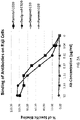


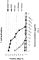

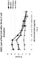

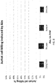
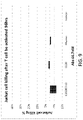

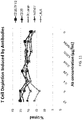
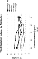
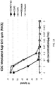


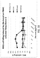
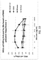


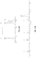

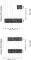

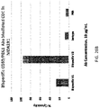
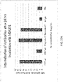

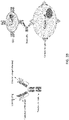
Classifications
-
- C—CHEMISTRY; METALLURGY
- C07—ORGANIC CHEMISTRY
- C07K—PEPTIDES
- C07K16/00—Immunoglobulins [IGs], e.g. monoclonal or polyclonal antibodies
- C07K16/46—Hybrid immunoglobulins
-
- C—CHEMISTRY; METALLURGY
- C07—ORGANIC CHEMISTRY
- C07K—PEPTIDES
- C07K16/00—Immunoglobulins [IGs], e.g. monoclonal or polyclonal antibodies
- C07K16/18—Immunoglobulins [IGs], e.g. monoclonal or polyclonal antibodies against material from animals or humans
- C07K16/28—Immunoglobulins [IGs], e.g. monoclonal or polyclonal antibodies against material from animals or humans against receptors, cell surface antigens or cell surface determinants
- C07K16/2803—Immunoglobulins [IGs], e.g. monoclonal or polyclonal antibodies against material from animals or humans against receptors, cell surface antigens or cell surface determinants against the immunoglobulin superfamily
- C07K16/2809—Immunoglobulins [IGs], e.g. monoclonal or polyclonal antibodies against material from animals or humans against receptors, cell surface antigens or cell surface determinants against the immunoglobulin superfamily against the T-cell receptor (TcR)-CD3 complex
-
- C—CHEMISTRY; METALLURGY
- C07—ORGANIC CHEMISTRY
- C07K—PEPTIDES
- C07K16/00—Immunoglobulins [IGs], e.g. monoclonal or polyclonal antibodies
- C07K16/46—Hybrid immunoglobulins
- C07K16/468—Immunoglobulins having two or more different antigen binding sites, e.g. multifunctional antibodies
-
- A—HUMAN NECESSITIES
- A61—MEDICAL OR VETERINARY SCIENCE; HYGIENE
- A61K—PREPARATIONS FOR MEDICAL, DENTAL OR TOILETRY PURPOSES
- A61K39/00—Medicinal preparations containing antigens or antibodies
- A61K39/395—Antibodies; Immunoglobulins; Immune serum, e.g. antilymphocytic serum
-
- A—HUMAN NECESSITIES
- A61—MEDICAL OR VETERINARY SCIENCE; HYGIENE
- A61P—SPECIFIC THERAPEUTIC ACTIVITY OF CHEMICAL COMPOUNDS OR MEDICINAL PREPARATIONS
- A61P35/00—Antineoplastic agents
-
- A—HUMAN NECESSITIES
- A61—MEDICAL OR VETERINARY SCIENCE; HYGIENE
- A61P—SPECIFIC THERAPEUTIC ACTIVITY OF CHEMICAL COMPOUNDS OR MEDICINAL PREPARATIONS
- A61P35/00—Antineoplastic agents
- A61P35/02—Antineoplastic agents specific for leukemia
-
- C—CHEMISTRY; METALLURGY
- C07—ORGANIC CHEMISTRY
- C07K—PEPTIDES
- C07K16/00—Immunoglobulins [IGs], e.g. monoclonal or polyclonal antibodies
- C07K16/18—Immunoglobulins [IGs], e.g. monoclonal or polyclonal antibodies against material from animals or humans
- C07K16/28—Immunoglobulins [IGs], e.g. monoclonal or polyclonal antibodies against material from animals or humans against receptors, cell surface antigens or cell surface determinants
- C07K16/2803—Immunoglobulins [IGs], e.g. monoclonal or polyclonal antibodies against material from animals or humans against receptors, cell surface antigens or cell surface determinants against the immunoglobulin superfamily
- C07K16/2827—Immunoglobulins [IGs], e.g. monoclonal or polyclonal antibodies against material from animals or humans against receptors, cell surface antigens or cell surface determinants against the immunoglobulin superfamily against B7 molecules, e.g. CD80, CD86
-
- C—CHEMISTRY; METALLURGY
- C07—ORGANIC CHEMISTRY
- C07K—PEPTIDES
- C07K16/00—Immunoglobulins [IGs], e.g. monoclonal or polyclonal antibodies
- C07K16/18—Immunoglobulins [IGs], e.g. monoclonal or polyclonal antibodies against material from animals or humans
- C07K16/28—Immunoglobulins [IGs], e.g. monoclonal or polyclonal antibodies against material from animals or humans against receptors, cell surface antigens or cell surface determinants
- C07K16/2887—Immunoglobulins [IGs], e.g. monoclonal or polyclonal antibodies against material from animals or humans against receptors, cell surface antigens or cell surface determinants against CD20
-
- C—CHEMISTRY; METALLURGY
- C07—ORGANIC CHEMISTRY
- C07K—PEPTIDES
- C07K16/00—Immunoglobulins [IGs], e.g. monoclonal or polyclonal antibodies
- C07K16/18—Immunoglobulins [IGs], e.g. monoclonal or polyclonal antibodies against material from animals or humans
- C07K16/28—Immunoglobulins [IGs], e.g. monoclonal or polyclonal antibodies against material from animals or humans against receptors, cell surface antigens or cell surface determinants
- C07K16/2896—Immunoglobulins [IGs], e.g. monoclonal or polyclonal antibodies against material from animals or humans against receptors, cell surface antigens or cell surface determinants against molecules with a "CD"-designation, not provided for elsewhere
-
- C—CHEMISTRY; METALLURGY
- C07—ORGANIC CHEMISTRY
- C07K—PEPTIDES
- C07K19/00—Hybrid peptides, i.e. peptides covalently bound to nucleic acids, or non-covalently bound protein-protein complexes
-
- C—CHEMISTRY; METALLURGY
- C12—BIOCHEMISTRY; BEER; SPIRITS; WINE; VINEGAR; MICROBIOLOGY; ENZYMOLOGY; MUTATION OR GENETIC ENGINEERING
- C12N—MICROORGANISMS OR ENZYMES; COMPOSITIONS THEREOF; PROPAGATING, PRESERVING, OR MAINTAINING MICROORGANISMS; MUTATION OR GENETIC ENGINEERING; CULTURE MEDIA
- C12N15/00—Mutation or genetic engineering; DNA or RNA concerning genetic engineering, vectors, e.g. plasmids, or their isolation, preparation or purification; Use of hosts therefor
- C12N15/09—Recombinant DNA-technology
- C12N15/11—DNA or RNA fragments; Modified forms thereof; Non-coding nucleic acids having a biological activity
- C12N15/62—DNA sequences coding for fusion proteins
-
- A—HUMAN NECESSITIES
- A61—MEDICAL OR VETERINARY SCIENCE; HYGIENE
- A61K—PREPARATIONS FOR MEDICAL, DENTAL OR TOILETRY PURPOSES
- A61K39/00—Medicinal preparations containing antigens or antibodies
- A61K2039/505—Medicinal preparations containing antigens or antibodies comprising antibodies
-
- C—CHEMISTRY; METALLURGY
- C07—ORGANIC CHEMISTRY
- C07K—PEPTIDES
- C07K2317/00—Immunoglobulins specific features
- C07K2317/20—Immunoglobulins specific features characterized by taxonomic origin
- C07K2317/24—Immunoglobulins specific features characterized by taxonomic origin containing regions, domains or residues from different species, e.g. chimeric, humanized or veneered
-
- C—CHEMISTRY; METALLURGY
- C07—ORGANIC CHEMISTRY
- C07K—PEPTIDES
- C07K2317/00—Immunoglobulins specific features
- C07K2317/30—Immunoglobulins specific features characterized by aspects of specificity or valency
- C07K2317/31—Immunoglobulins specific features characterized by aspects of specificity or valency multispecific
-
- C—CHEMISTRY; METALLURGY
- C07—ORGANIC CHEMISTRY
- C07K—PEPTIDES
- C07K2317/00—Immunoglobulins specific features
- C07K2317/50—Immunoglobulins specific features characterized by immunoglobulin fragments
- C07K2317/515—Complete light chain, i.e. VL + CL
-
- C—CHEMISTRY; METALLURGY
- C07—ORGANIC CHEMISTRY
- C07K—PEPTIDES
- C07K2317/00—Immunoglobulins specific features
- C07K2317/50—Immunoglobulins specific features characterized by immunoglobulin fragments
- C07K2317/52—Constant or Fc region; Isotype
- C07K2317/524—CH2 domain
-
- C—CHEMISTRY; METALLURGY
- C07—ORGANIC CHEMISTRY
- C07K—PEPTIDES
- C07K2317/00—Immunoglobulins specific features
- C07K2317/50—Immunoglobulins specific features characterized by immunoglobulin fragments
- C07K2317/52—Constant or Fc region; Isotype
- C07K2317/526—CH3 domain
-
- C—CHEMISTRY; METALLURGY
- C07—ORGANIC CHEMISTRY
- C07K—PEPTIDES
- C07K2317/00—Immunoglobulins specific features
- C07K2317/50—Immunoglobulins specific features characterized by immunoglobulin fragments
- C07K2317/56—Immunoglobulins specific features characterized by immunoglobulin fragments variable (Fv) region, i.e. VH and/or VL
- C07K2317/567—Framework region [FR]
-
- C—CHEMISTRY; METALLURGY
- C07—ORGANIC CHEMISTRY
- C07K—PEPTIDES
- C07K2317/00—Immunoglobulins specific features
- C07K2317/70—Immunoglobulins specific features characterized by effect upon binding to a cell or to an antigen
- C07K2317/71—Decreased effector function due to an Fc-modification
-
- C—CHEMISTRY; METALLURGY
- C07—ORGANIC CHEMISTRY
- C07K—PEPTIDES
- C07K2317/00—Immunoglobulins specific features
- C07K2317/70—Immunoglobulins specific features characterized by effect upon binding to a cell or to an antigen
- C07K2317/72—Increased effector function due to an Fc-modification
-
- C—CHEMISTRY; METALLURGY
- C07—ORGANIC CHEMISTRY
- C07K—PEPTIDES
- C07K2317/00—Immunoglobulins specific features
- C07K2317/70—Immunoglobulins specific features characterized by effect upon binding to a cell or to an antigen
- C07K2317/73—Inducing cell death, e.g. apoptosis, necrosis or inhibition of cell proliferation
- C07K2317/732—Antibody-dependent cellular cytotoxicity [ADCC]
-
- C—CHEMISTRY; METALLURGY
- C07—ORGANIC CHEMISTRY
- C07K—PEPTIDES
- C07K2317/00—Immunoglobulins specific features
- C07K2317/70—Immunoglobulins specific features characterized by effect upon binding to a cell or to an antigen
- C07K2317/73—Inducing cell death, e.g. apoptosis, necrosis or inhibition of cell proliferation
- C07K2317/734—Complement-dependent cytotoxicity [CDC]
-
- C—CHEMISTRY; METALLURGY
- C07—ORGANIC CHEMISTRY
- C07K—PEPTIDES
- C07K2317/00—Immunoglobulins specific features
- C07K2317/70—Immunoglobulins specific features characterized by effect upon binding to a cell or to an antigen
- C07K2317/77—Internalization into the cell
-
- C—CHEMISTRY; METALLURGY
- C07—ORGANIC CHEMISTRY
- C07K—PEPTIDES
- C07K2317/00—Immunoglobulins specific features
- C07K2317/90—Immunoglobulins specific features characterized by (pharmaco)kinetic aspects or by stability of the immunoglobulin
- C07K2317/92—Affinity (KD), association rate (Ka), dissociation rate (Kd) or EC50 value
-
- C—CHEMISTRY; METALLURGY
- C07—ORGANIC CHEMISTRY
- C07K—PEPTIDES
- C07K2317/00—Immunoglobulins specific features
- C07K2317/90—Immunoglobulins specific features characterized by (pharmaco)kinetic aspects or by stability of the immunoglobulin
- C07K2317/94—Stability, e.g. half-life, pH, temperature or enzyme-resistance
Definitions
- This disclosure relates to bispecific antibodies or antigen-binding fragments thereof.
- a bispecific antibody is an artificial protein that can simultaneously bind to two different types of antigens or two different epitopes. This dual specificity opens up a wide range of applications, including redirecting T cells to tumor cells, blocking two different signaling pathways simultaneously, dual targeting of different disease mediators, and delivering payloads to targeted sites.
- catumaxomab anti-EpCAM and anti-CD3
- blinatumomab anti-CD19 and anti-CD3
- bispecific antibodies have various applications, there is a need to continue to develop various therapeutics based on bispecific antibodies.
- This disclosure relates to imbalanced bispecific antibodies or antigen-binding fragments, wherein the bispecific antibodies or antigen-binding fragments specifically bind to two different antigens with different binding affinities.
- the disclosure relates to a bispecific antibody or antigen-binding fragment including a first heavy chain variable region, a second heavy chain variable region, a first light chain variable region, and a second light chain variable region, wherein the first heavy chain variable region and the first light chain variable region associate with each other, forming a first antigen binding region that specifically binds to a first antigen with a binding affinity greater than 10 7 M -1 , 10 8 M -1 , 10 9 M -1 , 10 10 M -1 , 10 11 M -1 , or 10 12 M -1 , and the second heavy chain variable region and the second light chain variable region associate with each other, forming a second antigen binding region that specifically binds to a second antigen with a binding affinity less than 10 9 M -1 , 10 8 M -1 , 10 7 M -1 , 10 6 M -1 , 10 5 M -1 , or 10 4 M -1 .
- the second antigen binding region specifically binds to the second antigen with a binding affinity greater than 10 7 M -1 , 10 6 M -1 , 10 5 M -1 , or 10 4 M -1 .
- the binding affinity of the first antigen binding region when it binds to the first antigen is at least 100, 1000, or 10000 times greater than the binding affinity of the second antigen binding region when it binds to the second antigen.
- the first light chain variable region and the second light chain variable region are at least 90%, 95%, 99%, or 100% identical.
- the disclosure relates to a bispecific antibody or antigen-binding fragment including a first arm comprising a first heavy chain variable region, and a first light chain variable region; and a second arm comprising a second heavy chain variable region, and a second light chain variable region, wherein the first arm specifically binds to a first antigen with a binding affinity greater than 10 7 M -1 , 10 8 M -1 , 10 9 M -1 , 10 10 M -1 , 10 11 M -1 , 10 12 M -1 , and the second arm specifically binds to a second antigen with a binding affinity less than 10 9 M -1 , 10 8 M -1 , 10 7 M -1 , 10 6 M -1 , 10 5 M -1 , or 10 4 M -1 .
- the second arm specifically binds to the second antigen with a binding affinity greater than 10 7 M -1 , 10 6 M -1 , 10 5 M -1 , or 10 4 M -1 .
- the binding affinity of the first arm when it binds to the first antigen is at least 100, 1000, or 10000 times greater than the binding affinity of the second arm when it binds to the second antigen.
- the first light chain variable region and the second light chain variable region are at least 90%, 95%, 99%, or 100% identical.
- the disclosure relates to a bispecific antibody or antigen-binding fragment including a first heavy chain comprising a first heavy chain variable region, a second heavy chain comprising a second heavy chain variable region, a first light chain comprising a first light chain variable region, and a second light chain comprising a second light chain variable region, wherein the first heavy chain variable region and the first light chain variable region associate with each other, forming a first antigen binding region that specifically binds to a first antigen with a binding affinity greater than 10 7 M -1 , 10 8 M -1 , 10 9 M -1 , 10 10 M -1 , 10 11 M -1 , 10 12 M -1 , and the second heavy chain variable region and the second light chain variable region associate with each other, forming a second antigen binding region that specifically binds to a second antigen with a binding affinity less than 10 9 M -1 , 10 8 M -1 , 10 7 M -1 , 10 6 M -1 , 10 5 M -1 , or 10
- the second antigen binding region specifically binds to the second antigen with a binding affinity greater than 10 7 M -1 , 10 6 M -1 , 10 5 M -1 , or 10 4 M -1 .
- the binding affinity of the first antigen binding region when it binds to the first antigen is at least 100, 1000, or 10000 times greater than the binding affinity of the second antigen binding region when it binds to the second antigen.
- the first light chain and the second light chain are at least 90%, 95%, 99%, or 100% identical.
- the first heavy chain and the second chain associate with each other by the knobs into holes approach.
- the first antigen is a cancer specific antigen
- the second antigen is CD3.
- the first antigen is CD20
- the second antigen is CD3.
- the first heavy chain variable region comprises a sequence that is at least 80%, 85%, 90%, or 95% identical to SEQ ID NO: 1
- the second heavy chain variable region comprises a sequence that is at least 80%, 85%, 90%, or 95% identical to SEQ ID NO: 2
- the first and the second light chain variable regions comprise a sequence that is at least 80%, 85%, 90%, or 95% identical to SEQ ID NO: 3.
- the first antigen is a cancer specific antigen
- the second antigen is a cancer-associated antigen
- the first antigen is PD-L1
- the second antigen is CD55
- the first heavy chain variable region comprises a sequence that is at least 80%, 85%, 90%, or 95% identical to SEQ ID NO: 4
- the second heavy chain variable region comprises a sequence that is at least 80%, 85%, 90%, or 95% identical to SEQ ID NO: 5
- the first and the second light chain variable regions comprise a sequence that is at least 80%, 85%, 90%, or 95% identical to SEQ ID NO: 6 or SEQ ID NO: 7.
- the disclosure relates to a method of making bispecific antibody or antigen-binding fragment, the method including selecting a first antigen and a second antigen, and identifying a first antibody or antigen-binding fragment that binds to the first antigen and a second antibody or antigen-binding fragment that binds to the second antigen, wherein the first antibody or antigen-binding fragment comprises a first heavy chain variable region (VHa) and a first light chain variable region (VLa), and the second antibody or antigen-binding fragment comprises a second heavy chain variable region (VHb) and a second light chain variable region (VLb); determining the amino acid sequence of VHa, VLa, VHb, and VLb; aligning the amino acid sequences of VLa and VLb and determining that the sequence homology between VLa and VLb is greater than 80%; designing a common light chain variable region (VLc), wherein the VLc, when it associates with VHa, maintains the affinity to the first antigen
- step (d) the binding affinity of the VLc -VHb to the second antigen can decrease.
- the method further includes developing a buffer system to purify the bispecific antibody or antigen-binding fragment.
- the disclosure relates to a method of making bispecific antibody or antigen-binding fragment, the method including selecting a first antigen and a second antigen, and identifying a first antibody or antigen-binding fragment that binds to the first antigen and a second antibody or antigen-binding fragment that binds to the second antigen, wherein the first antibody or antigen-binding fragment comprises a first heavy chain variable region (VHa) and a first light chain variable region (VLa), and the second antibody or antigen-binding fragment comprises a second heavy chain variable region (VHb) and a second light chain variable region (VLb); determining the amino acid sequence of VHa, VLa, and VLb; aligning the amino acid sequences of VLa and VLb and determining the sequence homology between VLa and VLb is less than 80%; replacing all light chain variable regions in a phage display antibody library with the VLa, and panning against the second antigen to obtain a third heavy chain variable region (VHc);
- the method further includes developing a buffer system to purify the bispecific antibody or antigen-binding fragment.
- the disclosure relates to a method of making bispecific antibody or antigen-binding fragment, the method including selecting a first antigen and a second antigen, and identifying a first antibody or antigen-binding fragment that binds to the first antigen and a second antibody or antigen-binding fragment that binds to the second antigen, wherein the first antibody or antigen-binding fragment comprises a first heavy chain variable region (VHa) and a first light chain variable region (VLa), and the second antibody or antigen-binding fragment comprises a second heavy chain variable region (VHb) and a second light chain variable region (VLb); determining the amino acid sequence of VHa, VLa, VHb, and VLb; aligning the amino acid sequences of VLa and VLb and determining the sequence homology between VLa and VLb is less than 80%; replacing all light chain variable regions in a phage display antibody library with a plurality of light chain variable regions, wherein the light chain variables regions are at least 80%
- step (d) the plurality of light chain variable regions are produced by error-prone PCR.
- the method further includes developing a buffer system to purify the bispecific antibody or antigen-binding fragment.
- the disclosure provides methods of making bispecific antibody or antigen-binding fragment thereof.
- the methods involve one or more of the following steps:
- the disclosure provides methods of making bispecific antibody or antigen-binding fragment thereof.
- the methods involve one or more of the following steps:
- the disclosure also provides methods of making bispecific antibody or antigen-binding fragment thereof.
- the methods involve one or more of the following steps:
- the disclosure further provides methods of making bispecific antibody or antigen-binding fragment thereof.
- the methods involve one or more of the following steps:
- the disclosure provides an antibody or antigen-binding fragment thereof that binds to CD3 comprising: a heavy chain variable region (VH) comprising complementarity determining regions (CDRs) 1, 2, and 3, wherein the VH CDR1 region comprises an amino acid sequence that is at least 80% identical to a selected VH CDR1 amino acid sequence, the VH CDR2 region comprises an amino acid sequence that is at least 80% identical to a selected VH CDR2 amino acid sequence, and the VH CDR3 region comprises an amino acid sequence that is at least 80% identical to a selected VH CDR3 amino acid sequence; and a light chain variable region (VL) comprising CDRs 1, 2, and 3, wherein the VL CDR1 region comprises an amino acid sequence that is at least 80% identical to a selected VL CDR1 amino acid sequence, the VL CDR2 region comprises an amino acid sequence that is at least 80% identical to a selected VL CDR2 amino acid sequence, and the VL CDR3 region comprises an amino acid sequence that is at
- the VH comprises CDRs 1, 2, 3 with the amino acid sequences set forth in SEQ ID NOs: 22, 23, 24 respectively
- the VL comprises CDRs 1, 2, 3 with the amino acid sequences set forth in SEQ ID NOs: 28, 29, 30, respectively.
- the antibody or antigen-binding fragment specifically binds to human CD3.
- the antibody or antigen-binding fragment is a bispecific antibody.
- the present disclosure also provides an antibody or antigen-binding fragment thereof that binds to PD-L1 comprising: a heavy chain variable region (VH) comprising complementarity determining regions (CDRs) 1, 2, and 3, wherein the VH CDR1 region comprises an amino acid sequence that is at least 80% identical to a selected VH CDR1 amino acid sequence, the VH CDR2 region comprises an amino acid sequence that is at least 80% identical to a selected VH CDR2 amino acid sequence, and the VH CDR3 region comprises an amino acid sequence that is at least 80% identical to a selected VH CDR3 amino acid sequence; and a light chain variable region (VL) comprising CDRs 1, 2, and 3, wherein the VL CDR1 region comprises an amino acid sequence that is at least 80% identical to a selected VL CDR1 amino acid sequence, the VL CDR2 region comprises an amino acid sequence that is at least 80% identical to a selected VL CDR2 amino acid sequence, and the VL CDR3 region comprises an amino amino acid sequence
- the VH comprises CDRs 1, 2, 3 with the amino acid sequences set forth in SEQ ID NOs: 41-43 respectively
- the VL comprises CDRs 1, 2, 3 with the amino acid sequences set forth in SEQ ID NOs: 59-61, respectively.
- the antibody or antigen-binding fragment specifically binds to human CD3. In some embodiments, the antibody or antigen-binding fragment is a bispecific antibody.
- the disclosure provides an antibody or antigen-binding fragment thereof that binds to CD55 comprising: a heavy chain variable region (VH) comprising complementarity determining regions (CDRs) 1, 2, and 3, wherein the VH CDR1 region comprises an amino acid sequence that is at least 80% identical to a selected VH CDR1 amino acid sequence, the VH CDR2 region comprises an amino acid sequence that is at least 80% identical to a selected VH CDR2 amino acid sequence, and the VH CDR3 region comprises an amino acid sequence that is at least 80% identical to a selected VH CDR3 amino acid sequence; and a light chain variable region (VL) comprising CDRs 1, 2, and 3, wherein the VL CDR1 region comprises an amino acid sequence that is at least 80% identical to a selected VL CDR1 amino acid sequence, the VL CDR2 region comprises an amino acid sequence that is at least 80% identical to a selected VL CDR2 amino acid sequence, and the VL CDR3 region comprises an amino acid sequence that is at
- the VH comprises CDRs 1, 2, 3 with the amino acid sequences set forth in SEQ ID NOs: 47-49 respectively
- the VL comprises CDRs 1, 2, 3 with the amino acid sequences set forth in SEQ ID NOs: 59-61, respectively.
- the antibody or antigen-binding fragment specifically binds to human CD3.
- the antibody or antigen-binding fragment is a bispecific antibody.
- the disclosure provides a nucleic acid comprising a polynucleotide encoding a polypeptide comprising: an immunoglobulin light chain or a fragment thereof comprising a VL comprising CDRs 1, 2, and 3 comprising the amino acid sequences set forth in SEQ ID NOs: 53-55, respectively.
- the VL when paired with a VH comprising the amino acid sequence set forth in SEQ ID NO: 4 binds to PD-L1, and/or when paired with a VH comprising the amino acid sequence set forth in SEQ ID NO: 5 binds to CD55.
- the disclosure provides a nucleic acid comprising a polynucleotide encoding a polypeptide comprising: an immunoglobulin light chain or a fragment thereof comprising a VL comprising CDRs 1, 2, and 3 comprising the amino acid sequences set forth in SEQ ID NOs: 59-61, respectively.
- the VL when paired with a VH comprising the amino acid sequence set forth in SEQ ID NO: 4 binds to PD-L1, and/or when paired with a VH comprising the amino acid sequence set forth in SEQ ID NO: 5 binds to CD55.
- the nucleic acid encodes a bispecific antibody. In some embodiments, the nucleic acid is cDNA.
- the disclosure provides a vector comprising one or more of the nucleic acids described herein.
- the disclosure provides a cell comprising the vector described herein.
- the cell is a CHO cell.
- the disclosure provides a cell comprising one or more of the nucleic acids described herein.
- the disclosure provides a bispecific antibody or antigen-binding fragment thereof that binds to CD20 and CD3 comprising a first polypeptide comprising a first heavy chain variable region (VH) comprising an amino acid sequence that is at least 90%, 95%, 96%, 97%, 98%, or 99% identical to SEQ ID NO: 1; a second polypeptide comprising a second heavy chain variable region (VH) comprising an amino acid sequence that is at least 90%, 95%, 96%, 97%, 98%, or 99% identical to SEQ ID NO: 2; a third polypeptide comprising a first light chain variable region (VL) comprising an amino acid sequence that is at least 90%, 95%, 96%, 97%, 98%, or 99% identical to SEQ ID NO: 3; a fourth polypeptide comprising a second light chain variable region (VL) comprising an amino acid sequence that is at least 90%, 95%, 96%, 97%, 98%, or 99% identical to SEQ ID NO: 3.
- VH first heavy chain
- the first heavy chain variable region (VH) comprises SEQ ID NO: 1; the second heavy chain variable region (VH) comprise SEQ ID NO: 2; the first light chain variable region (VL) comprise SEQ ID NO: 3; and the second light chain variable region (VL) comprises SEQ ID NO: 3.
- the first polypeptide comprises an amino acid sequence that is at least 90%, 95%, 96%, 97%, 98%, or 99% identical to SEQ ID NO: 34, 35, or 36;
- the second polypeptide comprises an amino acid sequence that is at least 90%, 95%, 96%, 97%, 98%, or 99% identical to SEQ ID NO: 37, 38, or 39;
- the third polypeptide comprises an amino acid sequence that is at least 90%, 95%, 96%, 97%, 98%, or 99% identical to SEQ ID NO: 40;
- the fourth polypeptide comprises an amino acid sequence that is at least 90%, 95%, 96%, 97%, 98%, or 99% identical to SEQ ID NO: 40.
- the first polypeptide comprises an amino acid sequence that is set forth in SEQ ID NO: 35; and the second polypeptide comprises an amino acid sequence that is set forth in SEQ ID NO: 38.
- the disclosure provides a bispecific antibody or antigen-binding fragment thereof that binds to PD-L1 and CD55 comprising a first polypeptide comprising a first heavy chain variable region (VH) comprising an amino acid sequence that is at least 90%, 95%, 96%, 97%, 98%, or 99% identical to SEQ ID NO: 4; a second polypeptide comprising a second heavy chain variable region (VH) comprising an amino acid sequence that is at least 90%, 95%, 96%, 97%, 98%, or 99% identical to SEQ ID NO: 5; a third polypeptide comprising a first light chain variable region (VL) comprising an amino acid sequence that is at least 90%, 95%, 96%, 97%, 98%, or 99% identical to SEQ ID NO: 6 or 7; a fourth polypeptide comprising a second light chain variable region (VL) comprising an amino acid sequence that is at least 90%, 95%, 96%, 97%, 98%, or 99% identical to SEQ ID NO:
- the first heavy chain variable region (VH) comprises SEQ ID NO: 4; the second heavy chain variable region (VH) comprise SEQ ID NO: 5; the first light chain variable region (VL) comprise SEQ ID NO: 7; and the second light chain variable region (VL) comprises SEQ ID NO: 7.
- the first polypeptide comprises an amino acid sequence that is at least 90%, 95%, 96%, 97%, 98%, or 99% identical to SEQ ID NO: 65;
- the second polypeptide comprises an amino acid sequence that is at least 90%, 95%, 96%, 97%, 98%, or 99% identical to SEQ ID NO: 66;
- the third polypeptide comprises an amino acid sequence that is at least 90%, 95%, 96%, 97%, 98%, or 99% identical to SEQ ID NO: 67 or 68;
- the fourth polypeptide comprises an amino acid sequence that is at least 90%, 95%, 96%, 97%, 98%, or 99% identical to SEQ ID NO: 67 or 68.
- the first polypeptide comprises an amino acid sequence that is set forth in SEQ ID NO: 65; the second polypeptide comprises an amino acid sequence that is set forth in SEQ ID NO: 66; the third polypeptide comprises an amino acid sequence that is set forth in SEQ ID NO: 68; and the fourth polypeptide comprises an amino acid sequence that is set forth in SEQ ID NO: 68.
- the disclosure provides an antibody-drug conjugate comprising the antibody or antigen-binding fragment thereof described herein covalently bound to a therapeutic agent.
- the therapeutic agent is a cytotoxic or cytostatic agent.
- the disclosure provides methods of treating a subject having cancer.
- the methods involve administering a therapeutically effective amount of a composition comprising the antibody or antigen-binding fragment thereof described herein, or the antibody-drug conjugate described herein, to the subject.
- the subject has a solid tumor.
- the cancer is melanoma, pancreatic carcinoma, or a hematological malignancy.
- the cancer is Non-Hodgkin's lymphoma, lymphoma, or chronic lymphocytic leukemia.
- the disclosure provides methods of decreasing the rate of tumor growth.
- the methods involve contacting a tumor cell with an effective amount of a composition comprising an antibody or antigen-binding fragment thereof described herein, or the antibody-drug conjugate described herein, to the subject.
- the disclosure provides methods of killing a tumor cell.
- the methods involve contacting a tumor cell with an effective amount of a composition comprising the antibody or antigen-binding fragment thereof described herein, or the antibody-drug conjugate described herein, to the subject.
- the disclosure provides a pharmaceutical composition
- a pharmaceutical composition comprising the antibody or antigen-binding fragment thereof described herein, and a pharmaceutically acceptable carrier.
- the disclosure provides a pharmaceutical composition
- a pharmaceutical composition comprising the antibody drug conjugate described herein, and a pharmaceutically acceptable carrier.
- a bispecific antibody or antigen-binding fragment thereof is an artificial protein that can simultaneously bind to two different types of antigens.
- a bispecific antibody or antigen-binding fragment thereof can have two arms (Arms A and B). Each arm has one heavy chain variable region and one light chain variable region.
- the bispecific antibody or antigen-binding fragment thereof can be IgG-like and non-IgG-like.
- the IgG-like bispecific antibody can have two Fab arms and one Fc region, and the two Fab arms bind to different antigens.
- the non-IgG-like bispecific antibody or antigen-binding fragment can be e.g., chemically linked Fabs (e.g., two Fab regions are chemically linked), and single-chain variable fragments (scFVs).
- a scFV can have two heavy chain variable regions and two light chain variable regions.
- the two arms can bind to the respective target antigens with different affinities.
- Ka for one arm or one antigen binding region can be greater than 10 5 M -1 , 10 6 M -1 , 10 7 M -1 , 10 8 M -1 , 10 9 M -1 , 10 10 M -1 , 10 11 M -1 , or 10 12 M -1 .
- the Ka can be less than 10 5 M -1 , 10 6 M -1 , 10 7 M -1 , 10 8 M -1 , 10 9 M -1 , 10 10 M -1 , 10 11 M -1 , or 10 12 M -1 .
- the binding affinity of the first arm or the first antigen binding region (A) can be greater than the binding affinity of the second arm or the second antigen binding region (B).
- Bispecific antibodies with imbalanced affinities can have various advantages. For example, bispecific antibodies with imbalanced affinities can be used to target a cancer specific antigen on cancer cells and CD3 on T cell. In this case, high affinity to the cancer specific antigen can lead to better capturing of cancer cells by T cells, and low affinity to CD3 can avoid triggering T-cell signaling by CD3 ( FIG. 28 ). Only when the bispecific antibody is presented to the T cell in a multivalent fashion by a target cancer cell, can the T cell be activated and kill the target cancer cell.
- the bispecific antibodies with imbalanced affinities can also be used to target a cancer specific antigen and a cancer-associated antigen ( FIG. 29 ).
- the bispecific antibody only weakly binds to non-cancer cells expressing low level of cancer-associated antigens, but strongly binds to cancer cells expressing both cancer specific antigens and high level of cancer-associated antigens.
- the Ka for the first arm or the first antigen binding region (A) can be greater than 10 7 M -1 , 10 8 M -1 , 10 9 M -1 , 10 10 M -1 , 10 11 M -1 , or 10 12 M -1 .
- the Ka for the first arm or the first antigen binding region (A) can be 10, 100, 1000, 10000, or 100000 times greater than the Ka for the second arm or the second antigen binding region (B).
- the Ka for the second arm or the second antigen binding region (B) can be less than 10 5 M -1 , 10 6 M -1 , 10 7 M -1 , 10 8 M -1 , or 10 9 M -1 .
- the Ka for the second arm or the second antigen binding region (B) still specifically binds to the target antigen with a reasonable affinity, e.g., greater than 10 4 M -1 , 10 5 M -1 or 10 6 M -1 .
- the binding affinity can also be expressed by the dissociation constant (Kd).
- Kd Antibody Antigen / Antibody ⁇ Antigen
- the Kd can be less than 10 -5 M, 10 -6 M, 10 -7 M, 10 -8 M, 10 -9 M, 10 -10 M, 10 -11 M, or 10 -12 M. In some embodiments, the Kd can be greater than 10 -5 M, 10 -6 M, 10 -7 M, 10 -8 M, 10 -9 M, 10 -10 M, 10 -11 M, or 10 -12 M.
- the binding affinity of the first arm or the first antigen binding region (A) is greater than the binding affinity of the second arm or the second antigen binding region (B).
- the Kd for the second arm or the second antigen binding region (B) can be 10, 100, 1000, 10000, or 100000 times greater (thus with less affinity) than the Kd for the first arm or the first antigen binding region (A).
- the Kd for the first arm or the first antigen binding region (A) can be less than 10 -7 M, 10 -8 M, 10 -9 M, 10 -10 M, 10 -11 M, or 10 -12 M; and the Kd for the second arm or the second antigen binding region (B) can be greater than 10 -5 M, 10 -6 M, 10 -7 M, 10 -8 M, or 10 -9 M.
- the bispecific antibody or antigen-binding fragment thereof comprises two light chains and two heavy chains.
- Each of the two light chains has one light chain variable region (VL) and one light chain constant region (CL).
- Each of the two heavy chains has one heavy chain variable region (VH) and three heavy chain constant regions (CH1, CH2, and CH3).
- the two light chains for Arm A and Arm B are the same.
- the CDRs in the VL of two light chains can be the same.
- the two heavy chains in the bispecific antibody or antigen-binding fragment thereof are different.
- the CDRs in the VH of two heavy chains are different.
- the "knobs into holes” approach introduces a mutation for an amino acid with a large sidechain in one heavy chain, and a mutation for an amino acid with a small sidechain in the other heavy chain.
- the "knobs into holes” approaches are described, e.g., in Ridgway, John BB, Leonard G. Presta, and Paul Carter. "'Knobs-into-holes' engineering of antibody CH3 domains for heavy chain heterodimerization.” Protein Engineering, Design and Selection 9.7 (1996), which is incorporated herein by reference in its entirety.
- Bispecific antibodies with a T cell specific antigen (e.g., CD3, CD4, or CD8) binding arm that can recruit and activate T cells have been widely studied for cancer therapy.
- T cell specific antigen e.g., CD3, CD4, or CD8 binding arm
- an antibody's effector function such as ADCC and CDC have been shown to play a critical role in cancer cell killing
- "safely" maintaining an antibody's effector function would expand the mechanisms of action of an therapeutic antibody as well as improve the antibody's cancer killing function.
- an imbalanced bispecific antibody technology platform has been developed based on computational antibody design.
- the first antigen binding region targets a cancer specific antigen
- the second antigen binding region targets a T cell specific antigen (e.g., CD3, CD4, or CD8) to recruit T cell to attack cancer with the cancer specific antigen ( FIG. 28 ).
- a T cell specific antigen e.g., CD3, CD4, or CD8
- cancer specific antigen refers to antigens that are specifically expressed on cancer cell surfaces. These antigens can be used to identify tumor cells. Normal cells rarely express cancer specific antigens.
- Some exemplary cancer specific antigens include, e.g., CD20, PSA, PSCA, PD-L1, Her2, Her3, Her1, ⁇ -Catenin, CD19, CEACAM3, EGFR, c-Met, EPCAM, PSMA, CD40, MUC1, and IGF1R, etc.
- PSA are primarily expressed on prostate cancer cells
- Her2 are primarily expressed on breast cancer cells.
- a bispecific antibody that binds to CD20 and CD3 is provided in this disclosure.
- This bispecific antibody can be applied to target multiple CD20 positive cancers such as CD20-positive non-Hodgkin's lymphoma (NHL), thus can be used to treat non-Hodgkin's lymphoma in a subject.
- CD20-positive non-Hodgkin's lymphoma NBL
- the bispecific antibody applies different mechanism of action to treat cancer compared to therapeutic antibodies target CD20 alone, it can be applied as a complementary therapy for CD20 positive cancers, especially for those CD20 positive cancers which don not respond well to current CD20 therapies (such has rituximab-resistant NHL).
- An antibody with high affinity to CD3 can trigger T-cell signaling, and cause undesirable immune response.
- a low affinity e.g., Ka can be less than 10 5 M -1 , 10 6 M -1 , or 10 7 M -1
- the term "safely maintaining the antibody's effector function” means that the antibody does not induce ADCC or CDC on normal cells (e.g., non-cancer cells).
- bispecific antibodies When multiple bispecific antibodies are presented on a target cancer cells (e.g., in a cluster) and bridge the interaction between cancer cell and T cell, these bispecific antibodies can trigger T-cell signaling though CD3 in a multivalent fashion, and the activated T cells will then kill the target cancer cells.
- the disclosure provides bispecific antibody or antigen-binding fragment thereof comprising two heavy chain variable regions and two light chain variable regions, wherein the first heavy chain variable region comprises a sequence that is at least 80%, 85%, 90%, 91%, 92%, 93%, 94%, 95%, 96%, 97%, 98%,or 99% identical to SEQ ID NO: 1, the second heavy chain variable region comprises a sequence that is at least 80%, 85%, 90%, 91%, 92%, 93%, 94%, 95%, 96%, 97%, 98%, or 99% identical to SEQ ID NO: 2, and the first and the second light chain variable regions comprise a sequence that is at least 80%, 85%, 90%, 91%, 92%, 93%, 94%, 95%, 96%, 97%, 98%, or 99% identical to SEQ ID NO: 3.
- the CDR sequences for binding to CD20 include CDRs of the heavy chain variable domain, SEQ ID NOs: 16-18, and CDRs of the light chain variable domain, SEQ ID NOs: 28-30, as defined by Kabat numbering. Under Chothia numbering, the CDR sequences of the heavy chain variable domain are set forth in SEQ ID NOs: 19-21, and CDRs of the light chain variable domain are set forth in SEQ ID NOs: 31-33.
- the CDR sequences for binding to CD3 include CDRs of the heavy chain variable domain, SEQ ID NOs: 22-24, and CDRs of the light chain variable domain, SEQ ID NOs: 28-30, as defined by Kabat numbering. Under Chothia numbering, the CDR sequences of the heavy chain variable domain are set forth in SEQ ID NOs: 25-27, and CDRs of the light chain variable domain are set forth in SEQ ID NOs: 31-33.
- the bispecific antibody or antigen-binding fragment thereof comprises a first heavy chain amino acid sequence that is at least 80%, 85%, 90%, 91%, 92%, 93%, 94%, 95%, 96%, 97%, 98%,or 99% identical to SEQ ID NO: 34, 35, or 36; a second heavy chain amino acid sequence that is at least 80%, 85%, 90%, 91%, 92%, 93%, 94%, 95%, 96%, 97%, 98%,or 99% identical to SEQ ID NO: 37, 38, or 39; a first light chain amino acid sequence that is at least 80%, 85%, 90%, 91%, 92%, 93%, 94%, 95%, 96%, 97%, 98%,or 99% identical to SEQ ID NO: 40; and a second light chain amino acid sequence that is at least 80%, 85%, 90%, 91%, 92%, 93%, 94%, 95%, 96%, 97%, 98%,or 99% identical to SEQ ID NO: 40.
- Imbalanced bispecific antibodies that bind to cancer specific antigens and cancer-associated antigen.
- the present disclosure also provides imbalanced bispecific antibodies that have the first antigen binding region targets a cancer specific antigen, and the second antigen binding region targets a cancer-associated antigen.
- cancer-associated antigen refers to antigens that are expressed at a relatively high level on cancer cells but may be also expressed at a relatively low level on normal cells.
- CD55, CD59, CD46 and many adhesion molecules such as N-cadherin, VE-cadherin, NCAM, Mel-CAM, ICAM, NrCAM, VCAM1, ALCAM, MCAM, etc., are cancer-associated antigens. While both cancer specific antigen and cancer-associated antigen are expressed on cancer cell surface, the difference between a cancer specific antigen and a cancer-associated antigen is that the cancer-associated antigen is also expressed on normal cells, but at a relative low level as compared to the level on cancer cells.
- a cancer specific antigen is rarely expressed on normal cells, and even if it is expressed on normal cells, the amount is extremely low.
- An antibody that targets cancer specific antigen usually will not induce Antibody-dependent Cellular Cytotoxicity (ADCC) or Complement-dependent cytotoxicity (CDC) on normal cells.
- ADCC Antibody-dependent Cellular Cytotoxicity
- CDC Complement-dependent cytotoxicity
- an antibody that targets a cancer-associated antigen with a high affinity may cause cytotoxic effects among normal cells.
- it is important that the bispecific antibody binds to a cancer-associated antigen with a relatively low affinity ( FIG. 29 ).
- a bispecific antibody that binds to PD-L1 and CD55 is provided in the examples.
- This antibody can be used for treating a subject with PD-L1 and CD55 positive cancers though ADCC or CDC as well as blocking the PD-L1/PD1 interaction to activate T cell dependent immune response and to decrease CD55's repression on CDC.
- the binding between the second arm and CD55 on the cancer cells can provide additional therapeutic effects.
- the disclosure provides bispecific antibody or antigen-binding fragment thereof comprising two heavy chain variable regions and two light chain variable regions, wherein the first heavy chain variable region comprises a sequence that is at least 80%, 85%, 90%, 91%, 92%, 93%, 94%, 95%, 96%, 97%, 98%, or 99% identical to SEQ ID NO: 4, the second heavy chain variable region comprises a sequence that is at least 80%, 85%, 90%, 91%, 92%, 93%, 94%, 95%, 96%, 97%, 98%, or 99% identical to SEQ ID NO: 5, and the first and the second light chain variable regions comprise a sequence that is at least 80%, 85%, 90%, 91%, 92%, 93%, 94%, 95%, 96%, 97%, 98%, or 99% identical to SEQ ID NO: 6 or 7.
- the CDR sequences for binding to PD-L1 include CDRs of the heavy chain variable domain, SEQ ID NOs: 41-43, and CDRs of the light chain variable domain, SEQ ID NOs: 53-55 or 59-61, as defined by Kabat numbering. Under Chothia numbering, the CDR sequences of the heavy chain variable domain are set forth in SEQ ID NOs: 44-46, and CDRs of the light chain variable domain are set forth in SEQ ID NOs: 56-58 or 62-64.
- the CDR sequences for binding to CD55 include CDRs of the heavy chain variable domain, SEQ ID NOs: 47-49, and CDRs of the light chain variable domain, SEQ ID NOs: 53-55 or 59-61, as defined by Kabat numbering. Under Chothia numbering, the CDR sequences of the heavy chain variable domain are set forth in SEQ ID NOs: 50-52, and CDRs of the light chain variable domain are set forth in SEQ ID NOs: 56-58 or 62-64.
- the bispecific antibody or antigen-binding fragment thereof comprises a first heavy chain amino acid sequence that is at least 80%, 85%, 90%, 91%, 92%, 93%, 94%, 95%, 96%, 97%, 98%,or 99% identical to SEQ ID NO: 65; a second heavy chain amino acid sequence that is at least 80%, 85%, 90%, 91%, 92%, 93%, 94%, 95%, 96%, 97%, 98%,or 99% identical to SEQ ID NO: 66; a first light chain amino acid sequence that is at least 80%, 85%, 90%, 91%, 92%, 93%, 94%, 95%, 96%, 97%, 98%,or 99% identical to SEQ ID NO: 67 or 68; and a second light chain amino acid sequence that is at least 80%, 85%, 90%, 91%, 92%, 93%, 94%, 95%, 96%, 97%, 98%,or 99% identical to SEQ ID NO: 67 or 68.
- the bispecific antibody or antigen or antigen-binding fragment thereof can be made by the following methods:
- the isoelectric point (PI) of a peptide is the pH at which a particular molecule carries no net electrical charge in the statistical mean.
- Amino acids that make up a peptide may be positive, negative, neutral, or polar in nature, and together give a protein its overall charge. However, certain amino acids in a protein are buried in the protein and will have no interaction with the solution surrounding it.
- the 3D PI takes the 3D structure of the protein into account, and provides a better estimate of the pH value at which a protein, when it is properly folded, carries no net electrical charge in the statistical mean. (We used a gradient pH buffer from a publication, so the buffer is not our invention. But we still need to optimize purification process).
- the bispecific antibody or antigen or antigen-binding fragment thereof can also be made by the following methods:
- the bispecific antibody or antigen or antigen-binding fragment thereof can also be made by the following methods:
- VHa-VLc cannot bind to the first antigen with a desired affinity, additional steps can be performed. For example, if VLc is at least 80% identical to VLa, a new common light chain can be designed. In some embodiments, the process starts with VLa, and the amino acids can be mutated to the amino acid in VLc based on the methods described herein (e.g., based on the 3D structure of VLa and VLc).
- to design a common light chain variable region involves aligning VLa and VLb, and studying the different residues between VLa and VLb on the same kabat position. If the different residue on VLb does not contact CDRs, interface residues, canonical residues or vernier zone residues on B Fv structure, the residues on VLb are mutated to the residue at the same kabat position on VLa. Otherwise, the residues on VLb are kept.
- to redesign a heavy chain variable region involves using BioLuminate to calculate 3D PI of Fv A and Fv B, and mutating non-CDR, non-canonical, non-interface and non-vernier zone residues to make the Fv which has high 3D PI even higher and the one which has low 3D PI even lower.
- BioLuminate How to use BioLuminate can be found, e.g., in BioLuminate's user guide for reference, which is incorporated herein by reference in its entirety.
- the present disclosure provides antibodies and antigen-binding fragments thereof that comprise complementary determining regions (CDRs), heavy chain variable regions, light chain variable regions, heavy chains, or light chains described herein.
- CDRs complementary determining regions
- the antibodies and antigen-binding fragments thereof are imbalanced bispecific antibodies and antigen-binding fragments thereof.
- antibodies are made up of two classes of polypeptide chains, light chains and heavy chains.
- a non-limiting antibody of the present disclosure can be an intact, four immunoglobulin chain antibody comprising two heavy chains and two light chains.
- the heavy chain of the antibody can be of any isotype including IgM, IgG, IgE, IgA, or IgD or sub-isotype including IgG1, IgG2, IgG2a, IgG2b, IgG3, IgG4, IgE1, IgE2, etc.
- the light chain can be a kappa light chain or a lambda light chain.
- An antibody can comprise two identical copies of a light chain and/or two identical copies of a heavy chain.
- the heavy chains which each contain one variable domain (or variable region, VH) and multiple constant domains (or constant regions), bind to one another via disulfide bonding within their constant domains to form the "stem" of the antibody.
- the light chains which each contain one variable domain (or variable region, VL) and one constant domain (or constant region), each bind to one heavy chain via disulfide binding.
- the variable region of each light chain is aligned with the variable region of the heavy chain to which it is bound.
- the variable regions of both the light chains and heavy chains contain three hypervariable regions sandwiched between more conserved framework regions (FR).
- CDRs complementary determining regions
- the four framework regions largely adopt a beta-sheet conformation and the CDRs form loops connecting, and in some cases forming part of, the beta-sheet structure.
- the CDRs in each chain are held in close proximity by the framework regions and, with the CDRs from the other chain, contribute to the formation of the antigen-binding region.
- the CDRs are important for recognizing an epitope of an antigen.
- an "epitope" is the smallest portion of a target molecule capable of being specifically bound by the antigen binding domain of an antibody.
- the minimal size of an epitope may be about three, four, five, six, or seven amino acids, but these amino acids need not be in a consecutive linear sequence of the antigen's primary structure, as the epitope may depend on an antigen's three-dimensional configuration based on the antigen's secondary and tertiary structure.
- the antibody is an intact immunoglobulin molecule (e.g., IgG1, IgG2a, IgG2b, IgG3, IgM, IgD, IgE, IgA).
- the IgG subclasses (IgG1, IgG2, IgG3, and IgG4) are highly conserved, differ in their constant region, particularly in their hinges and upper CH2 domains.
- the sequences and differences of the IgG subclasses are known in the art, and are described, e.g., in Vidarsson, et al, "IgG subclasses and allotypes: from structure to effector functions.” Frontiers in immunology 5 (2014 ); Irani, et al.
- the antibody can also be an immunoglobulin molecule that is derived from any species (e.g., human, rodent, mouse, rat, camelid).
- Antibodies disclosed herein also include, but are not limited to, polyclonal, monoclonal, monospecific, polyspecific antibodies, and chimeric antibodies that include an immunoglobulin binding domain fused to another polypeptide.
- the term "antigen binding domain” or "antigen binding fragment” is a portion of an antibody that retains specific binding activity of the intact antibody, i.e., any portion of an antibody that is capable of specific binding to an epitope on the intact antibody's target molecule. It includes, e.g., Fab, Fab', F(ab')2, and variants of these fragments.
- an antibody or an antigen binding fragment thereof can be, e.g., a scFv, a Fv, a Fd, a dAb, a bispecific antibody, a bispecific scFv, a diabody, a linear antibody, a single-chain antibody molecule, a multi-specific antibody formed from antibody fragments, and any polypeptide that includes a binding domain which is, or is homologous to, an antibody binding domain.
- Non-limiting examples of antigen binding domains include, e.g., the heavy chain and/or light chain CDRs of an intact antibody, the heavy and/or light chain variable regions of an intact antibody, full length heavy or light chains of an intact antibody, or an individual CDR from either the heavy chain or the light chain of an intact antibody.
- the scFV has two heavy chain variable domains, and two light chain variable domains. In some embodiments, the scFV has two antigen binding regions (Antigen binding regions: A and B), and the two antigen binding regions can bind to the respective target antigens with different affinities.
- Antigen binding regions A and B
- the antigen binding fragment can form a part of a chimeric antigen receptor (CAR).
- the chimeric antigen receptor are fusions of single-chain variable fragments (scFv) as described herein, fused to CD3-zeta transmembrane- and endodomain.
- the chimeric antigen receptor also comprises intracellular signaling domains from various costimulatory protein receptors (e.g., CD28, 41BB, ICOS).
- the chimeric antigen receptor comprises multiple signaling domains, e.g., CD3z-CD28-41BB or CD3z-CD28-OX40, to increase potency.
- the disclosure further provides cells (e.g., T cells) that express the chimeric antigen receptors as described herein.
- the antibodies or antigen-binding fragments thereof can bind to two different antigens or two different epitopes.
- the antibodies or antigen-binding fragments thereof can comprises one, two, or three heavy chain variable region CDRs selected from Table 1, Table 2, Table 11 and Table 12. In some embodiments, the antibodies or antigen-binding fragments thereof can comprises one, two, or three light chain variable region CDRs selected from Table 3, Table 13 and Table 14.
- the antibodies can have a heavy chain variable region (VH) comprising complementarity determining regions (CDRs) 1, 2, 3, wherein the CDR1 region comprises or consists of an amino acid sequence that is at least 80%, 85%, 90%, or 95% identical to a selected VH CDR1 amino acid sequence, the CDR2 region comprises or consists of an amino acid sequence that is at least 80%, 85%, 90%, or 95% identical to a selected VH CDR2 amino acid sequence, and the CDR3 region comprises or consists of an amino acid sequence that is at least 80%, 85%, 90%, or 95% identical to a selected VH CDR3 amino acid sequence, and a light chain variable region (VL) comprising CDRs 1, 2, 3, wherein the CDR1 region comprises or consists of an amino acid sequence that is at least 80%, 85%, 90%, or 95% identical to a selected VL CDR1 amino acid sequence, the CDR2 region comprises or consists of an amino acid sequence that is at least 80%, 85%,
- the antibody or an antigen-binding fragment described herein can contain a heavy chain variable domain containing one, two, or three of the CDRs selected from Table 1, Table 2, Table 11 and Table 12 with zero, one or two amino acid insertions, deletions, or substitutions.
- the antibody or an antigen-binding fragment described herein can contain a light chain variable domain containing one, two, or three of the CDRs selected from Table 3, Table 13 and Table 14 with zero, one or two amino acid insertions, deletions, or substitutions.
- the insertions, deletions, and substitutions can be within the CDR sequence, or at one or both terminal ends of the CDR sequence.
- Antibodies and antibody fragments of the present disclosure can be modified in the Fc region to provide desired effector functions or serum half-life.
- Multimerization of antibodies may be accomplished through natural aggregation of antibodies or through chemical or recombinant linking techniques known in the art. For example, some percentage of purified antibody preparations (e.g., purified IgG1 molecules) spontaneously form protein aggregates containing antibody homodimers and other higher-order antibody multimers.
- purified antibody preparations e.g., purified IgG1 molecules
- any of the antibodies or antigen-binding fragments described herein may be conjugated to a stabilizing molecule (e.g., a molecule that increases the half-life of the antibody or antigen-binding fragment thereof in a subject or in solution).
- stabilizing molecules include: a polymer (e.g., a polyethylene glycol) or a protein (e.g., serum albumin, such as human serum albumin).
- the conjugation of a stabilizing molecule can increase the half-life or extend the biological activity of an antibody or an antigen-binding fragment in vitro (e.g., in tissue culture or when stored as a pharmaceutical composition) or in vivo (e.g., in a human).
- the antibodies or antigen-binding fragments (e.g., bispecific antibodies) described herein can be conjugated to a therapeutic agent.
- the antibody-drug conjugate comprising the antibody or antigen-binding fragment thereof can covalently or non-covalently bind to a therapeutic agent.
- the therapeutic agent is a cytotoxic or cytostatic agent (e.g., cytochalasin B, gramicidin D, ethidium bromide, emetine, mitomycin, etoposide, tenoposide, vincristine, vinblastine, colchicin, doxorubicin, daunorubicin, dihydroxy anthracin, maytansinoids such as DM-1 and DM-4, dione, mitoxantrone, mithramycin, actinomycin D, 1-dehydrotestosterone, glucocorticoids, procaine, tetracaine, lidocaine, propranolol, puromycin, epirubicin, and cyclophosphamide and analogs).
- cytotoxic or cytostatic agent e.g., cytochalasin B, gramicidin D, ethidium bromide, emetine, mitomycin, etoposide, tenoposide,
- the antibodies or antigen-binding fragments thereof can increase immune response.
- the antibodies or antigen-binding fragments thereof as described herein can increase immune response, activity or number of T cells (e.g., CD3+ cells, CD8+ and/or CD4+ cells) by at least 10%, 20%, 30%, 40%, 50%, 60%, 70%, 80%, 90%, 100%, 2 folds, 3 folds, 5 folds, 10 folds, or 20 folds .
- the antibodies or antigen-binding fragments thereof as described herein can decrease the activity or number of T cells by at least 10%, 20%, 30%, 40%, 50%, 60%, 70%, 80%, 90%, 100%, 2 folds, 3 folds, 5 folds, 10 folds, or 20 folds.
- the antibodies or antigen-binding fragments thereof as described herein does not induce immune response in normal cells (e.g., non-tumor cells) or in the absence of tumor cells.
- the antibodies or antigen-binding fragments thereof can bind to PD-L1 or PD-L2.
- the antibodies or antigen-binding fragments thereof described herein can block the binding between PD-1 and PD-L1 and/or the binding between PD-1 and PD-L2.
- the antibodies or antigen-binding fragments thereof as described herein are PD-1 antagonist.
- the antibodies or antigen-binding fragments thereof are PD-1 agonist.
- the antibodies or antigen-binding fragments thereof can bind to CD3.
- the antibodies or antigen-binding fragments thereof described herein can recruit T cells to a target cell.
- the antibody specifically binds to an antigen (e.g., a human protein, a monkey protein, and/or a mouse protein) with a dissociation rate (koff) of less than 0.1 s -1 , less than 0.01 s -1 , less than 0.001 s -1 , less than 0.0001 s -1 , or less than 0.00001 s -1 .
- the dissociation rate (koff) is greater than 0.01 s -1 , greater than 0.001 s -1 , greater than 0.0001 s -1 , greater than 0.00001 s -1 , or greater than 0.000001 s -1 .
- kinetic association rates (kon) is greater than 1 ⁇ 10 2 /Ms, greater than 1 ⁇ 10 3 /Ms, greater than 1 ⁇ 10 4 /Ms, greater than 1 ⁇ 10 5 /Ms, or greater than 1 ⁇ 10 6 /Ms. In some embodiments, kinetic association rates (kon) is less than 1 ⁇ 10 5 /Ms, less than 1 ⁇ 10 6 /Ms, or less than 1 ⁇ 10 7 /Ms.
- Kd is less than 1 ⁇ 10 -4 M, less than 1 ⁇ 10 -5 M, less than 1 ⁇ 10 -6 M, less than 1 ⁇ 10 -7 M, less than 1 ⁇ 10 -8 M, less than 1 ⁇ 10 -9 M, or less than 1 ⁇ 10 -10 M.
- the Kd is less than 50nM, 30 nM, 20 nM, 15 nM, 10 nM, 9 nM, 8 nM, 7 nM, 6 nM, 5 nM, 4 nM, 3 nM, 2 nM, or 1 nM.
- Kd is greater than 1 ⁇ 10 -4 M, greater than 1 ⁇ 10 -5 M, greater than 1 ⁇ 10 -6 M, greater than 1 ⁇ 10 -7 M, greater than 1 ⁇ 10 -8 M, greater than 1 ⁇ 10 -9 M, greater than 1 ⁇ 10 -10 M, greater than 1 ⁇ 10 -11 M, or greater than 1 ⁇ 10 -12 M.
- General techniques for measuring the affinity of an antibody for an antigen include, e.g., ELISA, RIA, and surface plasmon resonance (SPR).
- thermal stabilities are determined.
- the antibodies or antigen binding fragments as described herein can have a Tm greater than 60, 61, 62, 63, 64, 65, 66, 67, 68, 69, 70, 71, 72, 73, 74, 75, 76, 77, 78, 79, 80, 81, 82, 83, 84, 85, 86, 87, 88, 89, 90, 91, 92, 93, 94, or 95 °C.
- the melting curve sometimes shows two transitions, or three transitions, with a first denaturation temperature, Tm D1, and a second denaturation temperature Tm D2, and optionally a third denaturation temperature Tm D3.
- the antibodies or antigen binding fragments as described herein has a Tm D1 greater than 60, 61, 62, 63, 64, 65, 66, 67, 68, 69, 70, 71, 72, 73, 74, 75, 76, 77, 78, 79, 80, 81, 82, 83, 84, 85, 86, 87, 88, 89, 90, 91, 92, 93, 94, or 95 °C.
- the antibodies or antigen binding fragments as described herein has a Tm D2 greater than 60, 61, 62, 63, 64, 65, 66, 67, 68, 69, 70, 71, 72, 73, 74, 75, 76, 77, 78, 79, 80, 81, 82, 83, 84, 85, 86, 87, 88, 89, 90, 91, 92, 93, 94, or 95 °C.
- the antibodies or antigen binding fragments as described herein has a Tm D3 greater than 60, 61, 62, 63, 64, 65, 66, 67, 68, 69, 70, 71, 72, 73, 74, 75, 76, 77, 78, 79, 80, 81, 82, 83, 84, 85, 86, 87, 88, 89, 90, 91, 92, 93, 94, or 95 ° C.
- Tm, Tm D1, Tm D2, Tm D3 are less than 60, 61, 62, 63, 64, 65, 66, 67, 68, 69, 70, 71, 72, 73, 74, 75, 76, 77, 78, 79, 80, 81, 82, 83, 84, 85, 86, 87, 88, 89, 90, 91, 92, 93, 94, or 95 °C.
- the antibodies or antigen binding fragments as described herein do not start to form aggregation when the temperate is less than 60, 61, 62, 63, 64, 65, 66, 67, 68, 69, 70, 71, 72, 73, 74, 75, 76, 77, 78, 79, 80, 81, 82, 83, 84, 85, 86, 87, 88, 89, 90, 91, 92, 93, 94, or 95 °C.
- Tagg266 or Tagg473 is less than 60, 61, 62, 63, 64, 65, 66, 67, 68, 69, 70, 71, 72, 73, 74, 75, 76, 77, 78, 79, 80, 81, 82, 83, 84, 85, 86, 87, 88, 89, 90, 91, 92, 93, 94, or 95 °C.
- the antibodies or antigen binding fragments as described herein have a pI greater than 7.0, 7.1, 7.2, 7.3, 7.4, 7.5, 7.6, 7.7, 7.8, 7.9, 8.0, 8.1, 8.2, 8.3, 8.4, 8.5, 8.6, 8.7, 8.8, 8.9, 9.0, 9.1, 9.2, 9.3, 9.4, 9.5, 9.6, 9.7, 9.8, or 9.9.
- the antibodies or antigen binding fragments as described herein have a pI less than 7.0, 7.1, 7.2, 7.3, 7.4, 7.5, 7.6, 7.7, 7.8, 7.9, 8.0, 8.1, 8.2, 8.3, 8.4, 8.5, 8.6, 8.7, 8.8, 8.9, 9.0, 9.1, 9.2, 9.3, 9.4, 9.5, 9.6, 9.7, 9.8, or 9.9.
- the antibody has a tumor growth inhibition percentage (TGI%) that is greater than 10%, 20%, 30%, 40%, 50%, 60%, 70%, 80%, 90%, 100%, 110%, 120%, 130%, 140%, 150%, 160%, 170%, 180%, 190%, or 200%. In some embodiments, the antibody has a tumor growth inhibition percentage that is less than 60%, 70%, 80%, 90%, 100%, 110%, 120%, 130%, 140%, 150%, 160%, 170%, 180%, 190%, or 200%.
- TGI% tumor growth inhibition percentage
- the TGI% can be determined, e.g., at 3, 4, 5, 6, 7, 8, 9, 10, 11, 12, 13, 14, 15, 16, 17, 18, 19, 20, 21, 22, 23, 24, 25, 26, 27, 28, 29, or 30 days after the treatment starts, or 1, 2, 3, 4, 5, 6, 7, 8, 9, 10, 11, or 12 months after the treatment starts.
- TGI% 1 ⁇ Ti ⁇ T 0 / Vi ⁇ V 0 ⁇ 100 Ti is the average tumor volume in the treatment group on day i. T0 is the average tumor volume in the treatment group on day zero. Vi is the average tumor volume in the control group on day i. V0 is the average tumor volume in the control group on day zero.
- the antibodies or antigen binding fragments can increase complement dependent cytotoxicity (CDC) by at least 10%, 20%, 30%, 40%, 50%, 60%, 70%, 80%, 90%, 100%, 2 folds, 3 folds, 5 folds, 10 folds, or 20 folds.
- CDC complement dependent cytotoxicity
- the antibodies or antigen binding fragments can increase antibody-dependent cell-mediated cytotoxicity (ADCC) by at least 10%, 20%, 30%, 40%, 50%, 60%, 70%, 80%, 90%, 100%, 2 folds, 3 folds, 5 folds, 10 folds, or 20 folds.
- ADCC antibody-dependent cell-mediated cytotoxicity
- the antibodies or antigen binding fragments can increase internalization rate by at least 10%, 20%, 30%, 40%, 50%, 60%, 70%, 80%, 90%, 100%, 2 folds, 3 folds, 5 folds, 10 folds, or 20 folds.
- the antibodies or antigen binding fragments can increase phagocytosis rate by at least 10%, 20%, 30%, 40%, 50%, 60%, 70%, 80%, 90%, 100%, 2 folds, 3 folds, 5 folds, 10 folds, or 20 folds.
- the antibodies or antigen binding fragments can enhance T cell function, for example, by increasing effector T cell proliferation and/or increasing gamma interferon production by the effector T cell (e.g., as compared to proliferation and/or cytokine production prior to treatment with the antibodies or antigen binding fragments).
- the antibodies or antigen binding fragments enhance CD4+ effector T cell function, for example, by increasing CD4+ effector T cell proliferation and/or increasing gamma interferon production by the CD4+ effector T cell (e.g., as compared to proliferation and/or cytokine production prior to treatment with the antibodies or antigen binding fragments).
- the cytokine is gamma interferon.
- the antibodies or antigen binding fragments increase number of intratumoral (infiltrating) CD4+ effector T cells (e.g., total number of CD4+ effector T cells, or e.g., percentage of CD4+ cells in CD45+ cells), e.g., as compared to number of intratumoral (infiltrating) CD4+ T cells prior to treatment with antibodies or antigen binding fragments.
- the antibodies or antigen binding fragments increase number of intratumoral (infiltrating) CD4+ effector T cells that express gamma interferon (e.g., total gamma interferon expressing CD4+ cells, or e.g., percentage of gamma interferon expressing CD4+ cells in total CD4+ cells), e.g., as compared to number of intratumoral (infiltrating) CD4+ T cells that express gamma interferon prior to treatment.
- gamma interferon e.g., total gamma interferon expressing CD4+ cells, or e.g., percentage of gamma interferon expressing CD4+ cells in total CD4+ cells
- the antibodies or antigen binding fragments increase number of intratumoral (infiltrating) CD8+ effector T cells (e.g., total number of CD8+ effector T cells, or e.g., percentage of CD8+ in CD45+ cells), e.g., as compared to number of intratumoral (infiltrating) CD8+ T effector cells prior to treatment.
- the antibodies or antigen binding fragments increase number of intratumoral (infiltrating) CD8+ effector T cells that express gamma interferon (e.g., percentage of CD8+ cells that express gamma interferon in total CD8+ cells), e.g., compared to number of intratumoral (infiltrating) CD8+ T cells that express gamma interferon prior to treatment with the antibody.
- the antibodies or antigen binding fragments enhance memory T cell function, for example by increasing memory T cell proliferation and/or increasing cytokine (e.g., gamma interferon) production by the memory cell.
- cytokine e.g., gamma interferon
- the antibodies or antigen binding fragments have a functional Fc region.
- effector function of a functional Fc region is antibody-dependent cell-mediated cytotoxicity (ADCC).
- ADCC antibody-dependent cell-mediated cytotoxicity
- effector function of a functional Fc region is phagocytosis.
- effector function of a functional Fc region is ADCC and phagocytosis.
- the Fc region is human IgG1, human IgG2, human IgG3, or human IgG4.
- the antibodies or antigen binding fragments can induce apoptosis.
- the antibodies or antigen binding fragments do not have a functional Fc region.
- the antibodies or antigen binding fragments are Fab, Fab', F(ab')2, and Fv fragments.
- the antibodies or antigen binding fragments are humanized antibodies.
- the humanization percentage means the percentage identity of the heavy chain or light chain variable region sequence as compared to human antibody sequences in International Immunogenetics Information System (IMGT) database. In some embodiments, humanization percentage is greater than 80%, 81%, 82%, 83%, 84%, 85%, 86%, 87%, 88%, 89%, 90%, 91%, 92%, 93%, 94%, or 95%.
- IMGT International Immunogenetics Information System
- the antibodies or antigen binding fragments are human antibodies.
- the present disclosure also provides recombinant vectors (e.g., an expression vectors) that include an isolated polynucleotide disclosed herein (e.g., a polynucleotide that encodes a polypeptide disclosed herein), host cells into which are introduced the recombinant vectors (i.e., such that the host cells contain the polynucleotide and/or a vector comprising the polynucleotide), and the production of recombinant antibody polypeptides or fragments thereof by recombinant techniques.
- recombinant vectors e.g., an expression vectors
- an isolated polynucleotide disclosed herein e.g., a polynucleotide that encodes a polypeptide disclosed herein
- host cells into which are introduced the recombinant vectors (i.e., such that the host cells contain the polynucleotide and/or a vector comprising the polynucleotide)
- a "vector” is any construct capable of delivering one or more polynucleotide(s) of interest to a host cell when the vector is introduced to the host cell.
- An "expression vector” is capable of delivering and expressing the one or more polynucleotide(s) of interest as an encoded polypeptide in a host cell into which the expression vector has been introduced.
- the polynucleotide of interest is positioned for expression in the vector by being operably linked with regulatory elements such as a promoter, enhancer, and/or a poly-A tail, either within the vector or in the genome of the host cell at or near or flanking the integration site of the polynucleotide of interest such that the polynucleotide of interest will be translated in the host cell introduced with the expression vector.
- regulatory elements such as a promoter, enhancer, and/or a poly-A tail
- a vector can be introduced into the host cell by methods known in the art, e.g., electroporation, chemical transfection (e.g., DEAE-dextran), transformation, transfection, and infection and/or transduction (e.g., with recombinant virus).
- vectors include viral vectors (which can be used to generate recombinant virus), naked DNA or RNA, plasmids, cosmids, phage vectors, and DNA or RNA expression vectors associated with cationic condensing agents.
- a polynucleotide disclosed herein e.g., a polynucleotide that encodes a polypeptide disclosed herein
- a viral expression system e.g., vaccinia or other pox virus, retrovirus, or adenovirus
- vaccinia or other pox virus, retrovirus, or adenovirus
- viral propagation generally will occur only in complementing virus packaging cells. Suitable systems are disclosed, for example, in Fisher-Hoch et al., 1989, Proc. Natl. Acad. Sci. USA 86:317-321 ; Flexner et al., 1989, Ann. N.Y. Acad Sci.
- the DNA insert comprising an antibody-encoding or polypeptide-encoding polynucleotide disclosed herein can be operatively linked to an appropriate promoter (e.g., a heterologous promoter), such as the phage lambda PL promoter, the E. coli lac, trp and tac promoters, the SV40 early and late promoters and promoters of retroviral LTRs, to name a few.
- a heterologous promoter such as the phage lambda PL promoter, the E. coli lac, trp and tac promoters, the SV40 early and late promoters and promoters of retroviral LTRs, to name a few.
- Other suitable promoters are known to the skilled artisan.
- the expression constructs can further contain sites for transcription initiation, termination and, in the transcribed region, a ribosome binding site for translation.
- the coding portion of the mature transcripts expressed by the constructs may include a translation initiating at
- the expression vectors can include at least one selectable marker.
- markers include dihydrofolate reductase or neomycin resistance for eukaryotic cell culture and tetracycline or ampicillin resistance genes for culturing in E. coli and other bacteria.
- Representative examples of appropriate hosts include, but are not limited to, bacterial cells, such as E. coli, Streptomyces, and Salmonella typhimurium cells; fungal cells, such as yeast cells; insect cells such as Drosophila S2 and Spodoptera Sf9 cells; animal cells such as CHO, COS, Bowes melanoma, and HK 293 cells; and plant cells. Appropriate culture mediums and conditions for the host cells described herein are known in the art.
- Non-limiting vectors for use in bacteria include pQE70, pQE60 and pQE-9, available from Qiagen; pBS vectors, Phagescript vectors, Bluescript vectors, pNH8A, pNH16a, pNH18A, pNH46A, available from Stratagene; and ptrc99a, pKK223-3, pKK233-3, pDR540, pRIT5 available from Pharmacia.
- Non-limiting eukaryotic vectors include pWLNEO, pSV2CAT, pOG44, pXT1 and pSG available from Stratagene; and pSVK3, pBPV, pMSG and pSVL available from Pharmacia. Other suitable vectors will be readily apparent to the skilled artisan.
- Non-limiting bacterial promoters suitable for use include the E. coli lacI and lacZ promoters, the T3 and T7 promoters, the gpt promoter, the lambda PR and PL promoters and the trp promoter.
- Suitable eukaryotic promoters include the CMV immediate early promoter, the HSV thymidine kinase promoter, the early and late SV40 promoters, the promoters of retroviral LTRs, such as those of the Rous sarcoma virus (RSV), and metallothionein promoters, such as the mouse metallothionein-I promoter.
- yeast Saccharomyces cerevisiae a number of vectors containing constitutive or inducible promoters such as alpha factor, alcohol oxidase, and PGH may be used.
- constitutive or inducible promoters such as alpha factor, alcohol oxidase, and PGH.
- Introduction of the construct into the host cell can be effected by calcium phosphate transfection, DEAE-dextran mediated transfection, cationic lipid-mediated transfection, electroporation, transduction, infection or other methods.
- Such methods are described in many standard laboratory manuals, such as Davis et al., Basic Methods In Molecular Biology (1986 ), which is incorporated herein by reference in its entirety.
- Enhancers are cis-acting elements of DNA, usually about from 10 to 300 bp that act to increase transcriptional activity of a promoter in a given host cell-type.
- enhancers include the SV40 enhancer, which is located on the late side of the replication origin at base pairs 100 to 270, the cytomegalovirus early promoter enhancer, the polyoma enhancer on the late side of the replication origin, and adenovirus enhancers.
- secretion signals may be incorporated into the expressed polypeptide.
- the signals may be endogenous to the polypeptide or they may be heterologous signals.
- the polypeptide (e.g., antibody) can be expressed in a modified form, such as a fusion protein (e.g. , a GST-fusion) or with a histidine-tag, and may include not only secretion signals, but also additional heterologous functional regions. For instance, a region of additional amino acids, particularly charged amino acids, may be added to the N-terminus of the polypeptide to improve stability and persistence in the host cell, during purification, or during subsequent handling and storage. Also, peptide moieties can be added to the polypeptide to facilitate purification. Such regions can be removed prior to final preparation of the polypeptide. The addition of peptide moieties to polypeptides to engender secretion or excretion, to improve stability and to facilitate purification, among others, are familiar and routine techniques in the art.
- the disclosure also provides a nucleic acid sequence that is at least 1%, 2%, 3%, 4%, 5%, 6%, 7%, 8%, 9%, 10%, 15%, 20%, 25%, 30%, 35%, 40%, 45%, 50%, 55%, 60%, 65%, 70%, 75%, 80%, 85%, 90%, 91%, 92%, 93%, 94%, 95%, 96%, 97%, 98%, 99% identical to any nucleotide sequence as described herein, and an amino acid sequence that is at least 1%, 2%, 3%, 4%, 5%, 6%, 7%, 8%, 9%, 10%, 15%, 20%, 25%, 30%, 35%, 40%, 45%, 50%, 55%, 60%, 65%, 70%, 75%, 80%, 85%, 90%, 91%, 92%, 93%, 94%, 95%, 96%, 97%, 98%, 99% identical to any amino acid sequence as described herein.
- the disclosure also provides a nucleic acid sequence that has a homology of at least 1%, 2%, 3%, 4%, 5%, 6%, 7%, 8%, 9%, 10%, 15%, 20%, 25%, 30%, 35%, 40%, 45%, 50%, 55%, 60%, 65%, 70%, 75%, 80%, 85%, 90%, 91%, 92%, 93%, 94%, 95%, 96%, 97%, 98%, 99% to any nucleotide sequence as described herein, and an amino acid sequence that has a homology of at least 1%, 2%, 3%, 4%, 5%, 6%, 7%, 8%, 9%, 10%, 15%, 20%, 25%, 30%, 35%, 40%, 45%, 50%, 55%, 60%, 65%, 70%, 75%, 80%, 85%, 90%, 91%, 92%, 93%, 94%, 95%, 96%, 97%, 98%, 99% to any amino acid sequence as described herein.
- the disclosure relates to nucleotide sequences encoding any peptides that are described herein, or any amino acid sequences that are encoded by any nucleotide sequences as described herein.
- the nucleic acid sequence is less than 10, 20, 30, 40, 50, 60, 70, 80, 90, 100, 110, 120, 130, 150, 200, 250, 300, 350, 400, 500, or 600 nucleotides.
- the amino acid sequence is less than 5, 6, 7, 8, 9, 10, 20, 30, 40, 50, 60, 70, 80, 90, 100, 110, 120, 130, 140, 150, 160, 170, 180, 190, 200, 250, 300, 350, or 400 amino acid residues.
- the amino acid sequence (i) comprises an amino acid sequence; or (ii) consists of an amino acid sequence, wherein the amino acid sequence is any one of the sequences as described herein.
- the nucleic acid sequence (i) comprises a nucleic acid sequence; or (ii) consists of a nucleic acid sequence, wherein the nucleic acid sequence is any one of the sequences as described herein.
- the sequences are aligned for optimal comparison purposes (e.g., gaps can be introduced in one or both of a first and a second amino acid or nucleic acid sequence for optimal alignment and non-homologous sequences can be disregarded for comparison purposes).
- the length of a reference sequence aligned for comparison purposes is at least 80% of the length of the reference sequence, and in some embodiments is at least 90%, 95%, or 100%.
- the amino acid residues or nucleotides at corresponding amino acid positions or nucleotide positions are then compared.
- amino acid or nucleic acid “identity” is equivalent to amino acid or nucleic acid "homology”).
- the percent identity between the two sequences is a function of the number of identical positions shared by the sequences, taking into account the number of gaps, and the length of each gap, which need to be introduced for optimal alignment of the two sequences.
- the comparison of sequences and determination of percent identity between two sequences can be accomplished using a Blossum 62 scoring matrix with a gap penalty of 12, a gap extend penalty of 4, and a frameshift gap penalty of 5.
- the percentage of sequence homology (e.g., amino acid sequence homology or nucleic acid homology) can also be determined. How to determine percentage of sequence homology is known in the art.
- amino acid residues conserved with similar physicochemical properties e.g. leucine and isoleucine, can be used to measure sequence similarity. Families of amino acid residues having similar physicochemical properties have been defined in the art.
- amino acids with basic side chains e.g., lysine, arginine, histidine
- acidic side chains e.g., aspartic acid, glutamic acid
- uncharged polar side chains e.g., glycine, asparagine, glutamine, serine, threonine, tyrosine, cysteine
- nonpolar side chains e.g., alanine, valine, leucine, isoleucine, proline, phenylalanine, methionine, tryptophan
- beta-branched side chains e.g., threonine, valine, isoleucine
- aromatic side chains e.g., tyrosine, phenylalanine, tryptophan, histidine.
- the homology percentage in many cases, is higher than the identity percentage.
- An isolated fragment of human protein (e.g., CD55, CD3, cancer specific antigen or cancer-associated antigen) can be used as an immunogen to generate antibodies using standard techniques for polyclonal and monoclonal antibody preparation.
- Polyclonal antibodies can be raised in animals by multiple injections (e.g., subcutaneous or intraperitoneal injections) of an antigenic peptide or protein.
- the antigenic peptide or protein is injected with at least one adjuvant.
- the antigenic peptide or protein can be conjugated to an agent that is immunogenic in the species to be immunized. Animals can be injected with the antigenic peptide or protein more than one time (e.g., twice, three times, or four times).
- the full-length polypeptide or protein can be used or, alternatively, antigenic peptide fragments thereof can be used as immunogens.
- the antigenic peptide of a protein comprises at least 8 (e.g., at least 10, 15, 20, or 30) amino acid residues of the amino acid sequence of the protein and encompasses an epitope of the protein such that an antibody raised against the peptide forms a specific immune complex with the protein.
- An immunogen typically is used to prepare antibodies by immunizing a suitable subject (e.g., human or transgenic animal expressing at least one human immunoglobulin locus).
- a suitable subject e.g., human or transgenic animal expressing at least one human immunoglobulin locus.
- An appropriate immunogenic preparation can contain, for example, a recombinantly-expressed or a chemically-synthesized polypeptide.
- the preparation can further include an adjuvant, such as Freund's complete or incomplete adjuvant, or a similar immunostimulatory agent.
- Polyclonal antibodies can be prepared as described above by immunizing a suitable subject with a polypeptide, or an antigenic peptide thereof (e.g., part of the protein) as an immunogen.
- the antibody titer in the immunized subject can be monitored over time by standard techniques, such as with an enzyme-linked immunosorbent assay (ELISA) using the immobilized polypeptide or peptide.
- ELISA enzyme-linked immunosorbent assay
- the antibody molecules can be isolated from the mammal (e.g., from the blood) and further purified by well-known techniques, such as protein A of protein G chromatography to obtain the IgG fraction.
- antibody-producing cells can be obtained from the subject and used to prepare monoclonal antibodies by standard techniques, such as the hybridoma technique originally described by Kohler et al. (Nature 256:495-497, 1975 ), the human B cell hybridoma technique ( Kozbor et al., Immunol. Today 4:72, 1983 ), the EBV-hybridoma technique ( Cole et al., Monoclonal Antibodies and Cancer Therapy, Alan R. Liss, Inc., pp. 77-96, 1985 ), or trioma techniques.
- standard techniques such as the hybridoma technique originally described by Kohler et al. (Nature 256:495-497, 1975 ), the human B cell hybridoma technique ( Kozbor et al., Immunol. Today 4:72, 1983 ), the EBV-hybridoma technique ( Cole et al., Monoclonal Antibodies and Cancer Therapy, Alan R. Liss, Inc., pp.
- Hybridoma cells producing a monoclonal antibody are detected by screening the hybridoma culture supernatants for antibodies that bind the polypeptide or epitope of interest, e.g., using a standard ELISA assay.
- Variants of the antibodies or antigen-binding fragments described herein can be prepared by introducing appropriate nucleotide changes into the DNA encoding a human, humanized, or chimeric antibody, or antigen-binding fragment thereof described herein, or by peptide synthesis.
- Such variants include, for example, deletions, insertions, or substitutions of residues within the amino acids sequences that make-up the antigen-binding site of the antibody or an antigen-binding domain.
- some antibodies or antigen-binding fragments will have increased affinity for the target protein. Any combination of deletions, insertions, and/or combinations can be made to arrive at an antibody or antigen-binding fragment thereof that has increased binding affinity for the target.
- the amino acid changes introduced into the antibody or antigen-binding fragment can also alter or introduce new post-translational modifications into the antibody or antigen-binding fragment, such as changing (e.g., increasing or decreasing) the number of glycosylation sites, changing the type of glycosylation site (e.g., changing the amino acid sequence such that a different sugar is attached by enzymes present in a cell), or introducing new glycosylation sites.
- Antibodies disclosed herein can be derived from any species of animal, including mammals.
- Non-limiting examples of native antibodies include antibodies derived from humans, primates, e.g., monkeys and apes, cows, pigs, horses, sheep, camelids (e.g., camels and llamas), chicken, goats, and rodents (e.g., rats, mice, hamsters and rabbits), including transgenic rodents genetically engineered to produce human antibodies.
- Phage display can be used to optimize antibody sequences with desired binding affinities.
- a gene encoding single chain Fv (comprising VH or VL) can be inserted into a phage coat protein gene, causing the phage to "display" the scFv on its outside while containing the gene for the protein on its inside, resulting in a connection between genotype and phenotype.
- These displaying phages can then be screened against target antigens, in order to detect interaction between the displayed antigen binding sites and the target antigen.
- large libraries of proteins can be screened and amplified in a process called in vitro selection, and antibodies sequences with desired binding affinities can be obtained.
- Human and humanized antibodies include antibodies having variable and constant regions derived from (or having the same amino acid sequence as those derived from) human germline immunoglobulin sequences. Human antibodies may include amino acid residues not encoded by human germline immunoglobulin sequences (e.g., mutations introduced by random or site-specific mutagenesis in vitro or by somatic mutation in vivo), for example in the CDRs.
- a humanized antibody typically has a human framework (FR) grafted with non-human CDRs.
- FR human framework
- a humanized antibody has one or more amino acid sequence introduced into it from a source which is non-human. These non-human amino acid residues are often referred to as "import" residues, which are typically taken from an “import” variable domain. Humanization can be essentially performed by e.g., substituting rodent CDRs or CDR sequences for the corresponding sequences of a human antibody.
- humanized antibodies are chimeric antibodies wherein substantially less than an intact human V domain has been substituted by the corresponding sequence from a non-human species.
- humanized antibodies are typically mouse antibodies in which some CDR residues and some FR residues are substituted by residues from analogous sites in human antibodies.
- humanized antibodies can be prepared by a process of analysis of the parental sequences and various conceptual humanized products using three-dimensional models of the parental and humanized sequences.
- Three-dimensional immunoglobulin models are commonly available and are familiar to those skilled in the art.
- Computer programs are available which illustrate and display probable three-dimensional conformational structures of selected candidate immunoglobulin sequences. Inspection of these displays permits analysis of the likely role of the residues in the functioning of the candidate immunoglobulin sequence, i.e., the analysis of residues that influence the ability of the candidate immunoglobulin to bind its antigen.
- FR residues can be selected and combined from the recipient and import sequences so that the desired antibody characteristic, such as increased affinity for the target antigen(s), is achieved.
- Identity or homology with respect to an original sequence is usually the percentage of amino acid residues present within the candidate sequence that are identical with a sequence present within the human, humanized, or chimeric antibody or fragment, after aligning the sequences and introducing gaps, if necessary, to achieve the maximum percent sequence identity, and not considering any conservative substitutions as part of the sequence identity.
- a covalent modification can be made to the antibody or antigen-binding fragment thereof.
- These covalent modifications can be made by chemical or enzymatic synthesis, or by enzymatic or chemical cleavage.
- Other types of covalent modifications of the antibody or antibody fragment are introduced into the molecule by reacting targeted amino acid residues of the antibody or fragment with an organic derivatization agent that is capable of reacting with selected side chains or the N- or C-terminal residues.
- antibody variants having a carbohydrate structure that lacks fucose attached (directly or indirectly) to an Fc region.
- the amount of fucose in such antibody may be from 1% to 80%, from 1% to 65%, from 5% to 65% or from 20% to 40%.
- the amount of fucose is determined by calculating the average amount of fucose within the sugar chain at Asn297, relative to the sum of all glycostructures attached to Asn 297 (e.g. complex, hybrid and high mannose structures) as measured by MALDI-TOF mass spectrometry, as described in WO 2008/077546 , for example.
- Asn297 refers to the asparagine residue located at about position 297 in the Fc region (Eu numbering of Fc region residues; or position 314 in Kabat numbering); however, Asn297 may also be located about ⁇ 3 amino acids upstream or downstream of position 297, i.e., between positions 294 and 300, due to minor sequence variations in antibodies. Such fucosylation variants may have improved ADCC function.
- the Fc region of the antibody can be further engineered to replace the Asparagine at position 297 with Alanine (N297A).
- the Fc region of the antibodies was further engineered to replace the serine at position 228 (EU numbering) of IgG4 with proline (S228P).
- S228P serine at position 228
- a detailed description regarding S228 mutation is described, e.g., in Silva et al. "The S228P mutation prevents in vivo and in vitro IgG4 Fab-arm exchange as demonstrated using a combination of novel quantitative immunoassays and physiological matrix preparation.” Journal of Biological Chemistry 290.9 (2015): 5462-5469 , which is incorporated by reference in its entirety.
- the methods described here are designed to make a bispecific antibody.
- Bispecific antibodies can be made by engineering the interface between a pair of antibody molecules to maximize the percentage of heterodimers that are recovered from recombinant cell culture.
- the interface can contain at least a part of the CH3 domain of an antibody constant domain.
- one or more small amino acid side chains from the interface of the first antibody molecule are replaced with larger side chains (e.g., tyrosine or tryptophan).
- Compensatory "cavities" of identical or similar size to the large side chain(s) are created on the interface of the second antibody molecule by replacing large amino acid side chains with smaller ones (e.g., alanine or threonine).
- This provides a mechanism for increasing the yield of the heterodimer over other unwanted end-products such as homodimers.
- This method is described, e.g., in WO 96/27011 , which is incorporated by reference in its entirety.
- one or more amino acid residues in the CH3 portion of the IgG are substituted.
- one heavy chain has one or more of the following substitutions Y349C and T366W.
- the other heavy chain can have one or more the following substitutions E356C, T366S, L368A, and Y407V.
- a substitution (-ppcpScp-->-ppcpPcp-) can also be introduced at the hinge regions of both substituted IgG.
- one heavy chain has a T366Y (knob) substitution, and the other heavy chain has a Y407T (hole) substation.
- an anion-exchange chromatography can be used to purify bispecific antibodies.
- Anion-exchange chromatography is a process that separates substances based on their charges using an ion-exchange resin containing positively charged groups, such as diethylaminoethyl groups (DEAE). In solution, the resin is coated with positively charged counter-ions (cations). Anion exchange resins will bind to negatively charged molecules, displacing the counter-ion.
- Anion exchange chromatography can be used to purify proteins based on their isoelectric point (pI). The isoelectric point is defined as the pH at which a protein has no net charge.
- a protein When the pH > pI, a protein has a net negative charge and when the pH ⁇ pI, a protein has a net positive charge.
- different amino acid substitution can be introduced into two heavy chains, so that the pI for the homodimer comprising two Arm A and the pI for the homodimer comprising two Arm B is different.
- the pI for the bispecific antibody having Arm A and Arm B will be somewhere between the two pIs of the homodimers.
- the two homodimers and the bispecific antibody can be released at different pH conditions.
- the present disclosure shows that a few amino acid residue substitutions can be introduced to the heavy chains to adjust pI.
- the amino acid residue at Kabat numbering position 83 is lysine, arginine, or histidine.
- the amino acid residues at one or more of the positions 1, 6, 43, 81, and 105 is aspartic acid or glutamic acid.
- the amino acid residues at one or more of the positions 13 and 105 is aspartic acid or glutamic acid. In some embodiments, the amino acid residues at one or more of the positions 13 and 42 (Kabat numbering) is lysine, arginine, histidine, or glycine.
- Bispecific antibodies can also include e.g., cross-linked or "heteroconjugate” antibodies.
- one of the antibodies in the heteroconjugate can be coupled to avidin and the other to biotin.
- Heteroconjugate antibodies can also be made using any convenient cross-linking methods. Suitable cross-linking agents and cross-linking techniques are well known in the art and are disclosed in U.S. Patent No. 4,676,980 , which is incorporated herein by reference in its entirety.
- bispecific antibodies can be prepared using chemical linkage.
- Brennan et al. (Science 229:81, 1985 ) describes a procedure where intact antibodies are proteolytically cleaved to generate F(ab')2 fragments. These fragments are reduced in the presence of the dithiol complexing agent sodium arsenite to stabilize vicinal dithiols and prevent intermolecular disulfide formation.
- the Fab' fragments generated are then converted to thionitrobenzoate (TNB) derivatives.
- TNB thionitrobenzoate
- One of the Fab' TNB derivatives is then reconverted to the Fab' thiol by reduction with mercaptoethylamine, and is mixed with an equimolar amount of another Fab' TNB derivative to form the bispecific antibody.
- the methods described herein include methods for the treatment of disorders associated with cancer.
- the methods include administering a therapeutically effective amount of engineered bispecific antibodies (e.g., imbalanced bispecific antibodies) of antigen-binding fragments thereof as described herein, to a subject who is in need of, or who has been determined to be in need of, such treatment.
- engineered bispecific antibodies e.g., imbalanced bispecific antibodies
- to "treat” means to ameliorate at least one symptom of the disorder associated with cancer.
- cancer results in death; thus, a treatment can result in an increased life expectancy (e.g., by at least 1, 2, 3, 4, 5, 6, 7, 8, 9, 10, 11, 12 months, or by at least 1, 2, 3, 4, 5, 6, 7, 8, 9, 10 years).
- Administration of a therapeutically effective amount of an agent described herein (e.g., imbalanced bispecific antibodies) for the treatment of a condition associated with cancer will result in decreased number of cancer cells and/or alleviated symptoms.
- cancer refers to cells having the capacity for autonomous growth, i.e., an abnormal state or condition characterized by rapidly proliferating cell growth.
- the term is meant to include all types of cancerous growths or oncogenic processes, metastatic tissues or malignantly transformed cells, tissues, or organs, irrespective of histopathologic type or stage of invasiveness.
- tumor refers to cancerous cells, e.g., a mass of cancerous cells.
- Cancers that can be treated or diagnosed using the methods described herein include malignancies of the various organ systems, such as affecting lung, breast, thyroid, lymphoid, gastrointestinal, and genito-urinary tract, as well as adenocarcinomas which include malignancies such as most colon cancers, renal-cell carcinoma, prostate cancer and/or testicular tumors, non-small cell carcinoma of the lung, cancer of the small intestine and cancer of the esophagus.
- the agents described herein are designed for treating or diagnosing a carcinoma in a subject.
- carcinoma is art recognized and refers to malignancies of epithelial or endocrine tissues including respiratory system carcinomas, gastrointestinal system carcinomas, genitourinary system carcinomas, testicular carcinomas, breast carcinomas, prostatic carcinomas, endocrine system carcinomas, and melanomas.
- the cancer is renal carcinoma or melanoma.
- Exemplary carcinomas include those forming from tissue of the cervix, lung, prostate, breast, head and neck, colon and ovary.
- carcinosarcomas e.g., which include malignant tumors composed of carcinomatous and sarcomatous tissues.
- an “adenocarcinoma” refers to a carcinoma derived from glandular tissue or in which the tumor cells form recognizable glandular structures.
- the term “sarcoma” is art recognized and refers to malignant tumors of mesenchymal derivation.
- the cancer is Rituximab (Rituxan ® ) resistant cancer.
- the disclosure also provides methods for treating a cancer in a subject, methods of reducing the rate of the increase of volume of a tumor in a subject over time, methods of reducing the risk of developing a metastasis, or methods of reducing the risk of developing an additional metastasis in a subject.
- the treatment can halt, slow, retard, or inhibit progression of a cancer.
- the treatment can result in the reduction of in the number, severity, and/or duration of one or more symptoms of the cancer in a subject.
- the disclosure features methods that include administering a therapeutically effective amount of an antibody or antigen-binding fragment thereof, or an antibody drug conjugate disclosed herein to a subject in need thereof, e.g., a subject having, or identified or diagnosed as having, a cancer, e.g., breast cancer (e.g., triple-negative breast cancer), carcinoid cancer, cervical cancer, endometrial cancer, glioma, head and neck cancer, liver cancer, lung cancer, small cell lung cancer, lymphoma, melanoma, ovarian cancer, pancreatic cancer, prostate cancer, renal cancer, colorectal cancer, gastric cancer, testicular cancer, thyroid cancer, bladder cancer, urethral cancer, or hematologic malignancy.
- a cancer e.g., breast cancer (e.g., triple-negative breast cancer), carcinoid cancer, cervical cancer, endometrial cancer, glioma, head and neck cancer, liver cancer, lung cancer, small cell lung cancer, lymphoma, mel
- the terms "subject” and “patient” are used interchangeably throughout the specification and describe an animal, human or non-human, to whom treatment according to the methods of the present invention is provided.
- Veterinary and non-veterinary applications are contemplated by the present invention.
- Human patients can be adult humans or juvenile humans (e.g., humans below the age of 18 years old).
- patients include but are not limited to mice, rats, hamsters, guinea-pigs, rabbits, ferrets, cats, dogs, and primates.
- non-human primates e.g., monkey, chimpanzee, gorilla, and the like
- rodents e.g., rats, mice, gerbils, hamsters, ferrets, rabbits
- lagomorphs e.g., swine (e.g., pig, miniature pig)
- swine e.g., pig, miniature pig
- equine canine
- feline bovine
- other domestic, farm, and zoo animals equine, canine, feline, bovine, and other domestic, farm, and zoo animals.
- the cancer is unresectable melanoma or metastatic melanoma, non-small cell lung carcinoma (NSCLC), small cell lung cancer (SCLC), bladder cancer, or metastatic hormone-refractory prostate cancer.
- the subject has a solid tumor.
- the cancer is squamous cell carcinoma of the head and neck (SCCHN), renal cell carcinoma (RCC), triple-negative breast cancer (TNBC), or colorectal carcinoma.
- the subject has Hodgkin's lymphoma.
- the subject has triple-negative breast cancer (TNBC), gastric cancer, urothelial cancer, Merkel-cell carcinoma, or head and neck cancer.
- the cancer is melanoma, pancreatic carcinoma, mesothelioma, hematological malignancies, especially Non-Hodgkin's lymphoma, lymphoma, chronic lymphocytic leukemia, or advanced solid tumors.
- compositions and methods disclosed herein can be used for treatment of patients at risk for a cancer.
- Patients with cancer can be identified with various methods known in the art.
- an effective amount is meant an amount or dosage sufficient to effect beneficial or desired results including halting, slowing, retarding, or inhibiting progression of a disease, e.g., a cancer.
- An effective amount will vary depending upon, e.g., an age and a body weight of a subject to which the antibody, antigen binding fragment, antibody-drug conjugates, antibody-encoding polynucleotide, vector comprising the polynucleotide, and/or compositions thereof is to be administered, a severity of symptoms and a route of administration, and thus administration can be determined on an individual basis.
- an effective amount can be administered in one or more administrations.
- an effective amount of an antibody, an antigen binding fragment, or an antibody-drug conjugate is an amount sufficient to ameliorate, stop, stabilize, reverse, inhibit, slow and/or delay progression of an autoimmune disease or a cancer in a patient or is an amount sufficient to ameliorate, stop, stabilize, reverse, slow and/or delay proliferation of a cell (e.g., a biopsied cell, any of the cancer cells described herein, or cell line (e.g., a cancer cell line)) in vitro.
- a cell e.g., a biopsied cell, any of the cancer cells described herein, or cell line (e.g., a cancer cell line)
- an effective amount of an antibody, antigen binding fragment, or antibody-drug conjugate may vary, depending on, inter alia, patient history as well as other factors such as the type (and/or dosage) of antibody used.
- Effective amounts and schedules for administering the antibodies, antibody-encoding polynucleotides, antibody-drug conjugates, and/or compositions disclosed herein may be determined empirically, and making such determinations is within the skill in the art.
- the dosage that must be administered will vary depending on, for example, the mammal that will receive the antibodies, antibody-encoding polynucleotides, antibody-drug conjugates, and/or compositions disclosed herein, the route of administration, the particular type of antibodies, antibody-encoding polynucleotides, antigen binding fragments, antibody-drug conjugates, and/or compositions disclosed herein used and other drugs being administered to the mammal.
- a typical daily dosage of an effective amount of an antibody is 0.01 mg/kg to 100 mg/kg.
- the dosage can be less than 100 mg/kg, 10 mg/kg, 9 mg/kg, 8 mg/kg, 7 mg/kg, 6 mg/kg, 5 mg/kg, 4 mg/kg, 3 mg/kg, 2 mg/kg, 1 mg/kg, 0.5 mg/kg, or 0.1 mg/kg.
- the dosage can be greater than 10 mg/kg, 9 mg/kg, 8 mg/kg, 7 mg/kg, 6 mg/kg, 5 mg/kg, 4 mg/kg, 3 mg/kg, 2 mg/kg, 1 mg/kg, 0.5 mg/kg, 0.1 mg/kg, 0.05 mg/kg, or 0.01 mg/kg.
- the dosage is about 10 mg/kg, 9 mg/kg, 8 mg/kg, 7 mg/kg, 6 mg/kg, 5 mg/kg, 4 mg/kg, 3 mg/kg, 2 mg/kg, 1 mg/kg, 0.9 mg/kg, 0.8 mg/kg, 0.7 mg/kg, 0.6 mg/kg, 0.5 mg/kg, 0.4 mg/kg, 0.3 mg/kg, 0.2 mg/kg, or 0.1 mg/kg.
- the at least one antibody, antigen-binding fragment thereof, antibody-drug conjugates, or pharmaceutical composition e.g., any of the antibodies, antigen-binding fragments, antibody-drug conjugates, or pharmaceutical compositions described herein
- at least one additional therapeutic agent can be administered to the subject at least once a week (e.g., once a week, twice a week, three times a week, four times a week, once a day, twice a day, or three times a day).
- at least two different antibodies and/or antigen-binding fragments are administered in the same composition (e.g., a liquid composition).
- At least one antibody, antigen-binding fragment, antibody-drug conjugates, and at least one additional therapeutic agent are administered in the same composition (e.g., a liquid composition).
- the at least one antibody or antigen-binding fragment and the at least one additional therapeutic agent are administered in two different compositions (e.g., a liquid composition containing at least one antibody or antigen-binding fragment and a solid oral composition containing at least one additional therapeutic agent).
- the at least one additional therapeutic agent is administered as a pill, tablet, or capsule.
- the at least one additional therapeutic agent is administered in a sustained-release oral formulation.
- the one or more additional therapeutic agents can be administered to the subject prior to, or after administering the at least one antibody, antigen-binding antibody fragment, antibody-drug conjugate, or pharmaceutical composition (e.g., any of the antibodies, antigen-binding antibody fragments, or pharmaceutical compositions described herein).
- the one or more additional therapeutic agents and the at least one antibody, antigen-binding antibody fragment, antibody-drug conjugate, or pharmaceutical composition are administered to the subject such that there is an overlap in the bioactive period of the one or more additional therapeutic agents and the at least one antibody or antigen-binding fragment (e.g., any of the antibodies or antigen-binding fragments described herein) in the subject.
- the subject can be administered the at least one antibody, antigen-binding antibody fragment, antibody-drug conjugate, or pharmaceutical composition (e.g., any of the antibodies, antigen-binding antibody fragments, or pharmaceutical compositions described herein) over an extended period of time (e.g., over a period of at least 1 week, 2 weeks, 3 weeks, 1 month, 2 months, 3 months, 4 months, 5 months, 6 months, 7 months, 8 months, 9 months, 10 months, 11 months, 12 months, 1 year, 2 years, 3 years, 4 years, or 5 years).
- a skilled medical professional may determine the length of the treatment period using any of the methods described herein for diagnosing or following the effectiveness of treatment (e.g., the observation of at least one symptom of cancer).
- a skilled medical professional can also change the identity and number (e.g., increase or decrease) of antibodies or antigen-binding antibody fragments, antibody-drug conjugates (and/or one or more additional therapeutic agents) administered to the subject and can also adjust (e.g., increase or decrease) the dosage or frequency of administration of at least one antibody or antigen-binding antibody fragment (and/or one or more additional therapeutic agents) to the subject based on an assessment of the effectiveness of the treatment (e.g., using any of the methods described herein and known in the art).
- one or more additional therapeutic agents can be administered to the subject.
- the additional therapeutic agent can comprise one or more inhibitors selected from the group consisting of an inhibitor of B-Raf, an EGFR inhibitor, an inhibitor of a MEK, an inhibitor of ERK, an inhibitor of K-Ras, an inhibitor of c-Met, an inhibitor of anaplastic lymphoma kinase (ALK), an inhibitor of a phosphatidylinositol 3-kinase (PI3K), an inhibitor of an Akt, an inhibitor of mTOR, a dual PI3K/mTOR inhibitor, an inhibitor of Bruton's tyrosine kinase (BTK), and an inhibitor of Isocitrate dehydrogenase 1 (IDH1) and/or Isocitrate dehydrogenase 2 (IDH2).
- the additional therapeutic agent is an inhibitor of indoleamine 2,3-dioxygenase-1) (IDO1) (e.g., IDO1) (e.
- the additional therapeutic agent can comprise one or more inhibitors selected from the group consisting of an inhibitor of HER3, an inhibitor of LSD1, an inhibitor of MDM2, an inhibitor of BCL2, an inhibitor of CHK1, an inhibitor of activated hedgehog signaling pathway, and an agent that selectively degrades the estrogen receptor.
- the additional therapeutic agent can comprise one or more therapeutic agents selected from the group consisting of Trabectedin, nab-paclitaxel, Trebananib, Pazopanib, Cediranib, Palbociclib, everolimus, fluoropyrimidine, IFL, regorafenib, Reolysin, Alimta, Zykadia, Sutent, temsirolimus, axitinib, everolimus, sorafenib, Votrient, Pazopanib, IMA-901, AGS-003, cabozantinib, Vinflunine, an Hsp90 inhibitor, Ad-GM-CSF, Temazolomide, IL-2, IFNa, vinblastine, Thalomid, dacarbazine, cyclophosphamide, lenalidomide, azacytidine, lenalidomide, bortezomid, amrubicine, carfilzomib, prala
- therapeutic agents
- the additional therapeutic agent can comprise one or more therapeutic agents selected from the group consisting of an adjuvant, a TLR agonist, tumor necrosis factor (TNF) alpha, IL-1, HMGB1, an IL-10 antagonist, an IL-4 antagonist, an IL-13 antagonist, an IL-17 antagonist, an HVEM antagonist, an ICOS agonist, a treatment targeting CX3CL1, a treatment targeting CXCL9, a treatment targeting CXCL10, a treatment targeting CCL5, an LFA-1 agonist, an ICAM1 agonist, and a Selectin agonist.
- TNF tumor necrosis factor
- carboplatin, nab-paclitaxel, paclitaxel, cisplatin, pemetrexed, gemcitabine, FOLFOX, or FOLFIRI are administered to the subject.
- the additional therapeutic agent is an anti-OX40 antibody, an anti-PD-1 antibody, an anti-PD-Ll antibody, an anti-PD-L2 antibody, an anti-LAG-3 antibody, an anti-TIGIT antibody, an anti-BTLA antibody, an anti-CTLA-4 antibody, or an anti-GITR antibody.
- compositions that contain at least one (e.g., one, two, three, or four) of the antibodies, antigen-binding fragments, or antibody-drug conjugates described herein. Two or more (e.g., two, three, or four) of any of the antibodies, antigen-binding fragments, or antibody-drug conjugates described herein can be present in a pharmaceutical composition in any combination.
- the pharmaceutical compositions may be formulated in any manner known in the art.
- compositions are formulated to be compatible with their intended route of administration (e.g., intravenous, intraarterial, intramuscular, intradermal, subcutaneous, or intraperitoneal).
- the compositions can include a sterile diluent (e.g., sterile water or saline), a fixed oil, polyethylene glycol, glycerine, propylene glycol or other synthetic solvents, antibacterial or antifungal agents, such as benzyl alcohol or methyl parabens, chlorobutanol, phenol, ascorbic acid, thimerosal, and the like, antioxidants, such as ascorbic acid or sodium bisulfite, chelating agents, such as ethylenediaminetetraacetic acid, buffers, such as acetates, citrates, or phosphates, and isotonic agents, such as sugars (e.g., dextrose), polyalcohols (e.g., mannitol or sorbitol),
- Liposomal suspensions can also be used as pharmaceutically acceptable carriers (see, e.g., U.S. Patent No. 4,522,811 ).
- Preparations of the compositions can be formulated and enclosed in ampules, disposable syringes, or multiple dose vials. Where required (as in, for example, injectable formulations), proper fluidity can be maintained by, for example, the use of a coating, such as lecithin, or a surfactant.
- Absorption of the antibody or antigen-binding fragment thereof can be prolonged by including an agent that delays absorption (e.g., aluminum monostearate and gelatin).
- controlled release can be achieved by implants and microencapsulated delivery systems, which can include biodegradable, biocompatible polymers (e.g., ethylene vinyl acetate, polyanhydrides, polyglycolic acid, collagen, polyorthoesters, and polylactic acid; Alza Corporation and Nova Pharmaceutical, Inc.).
- biodegradable, biocompatible polymers e.g., ethylene vinyl acetate, polyanhydrides, polyglycolic acid, collagen, polyorthoesters, and polylactic acid; Alza Corporation and Nova Pharmaceutical, Inc.
- compositions containing one or more of any of the antibodies, antigen-binding fragments, antibody-drug conjugates described herein can be formulated for parenteral (e.g., intravenous, intraarterial, intramuscular, intradermal, subcutaneous, or intraperitoneal) administration in dosage unit form (i.e., physically discrete units containing a predetermined quantity of active compound for ease of administration and uniformity of dosage).
- parenteral e.g., intravenous, intraarterial, intramuscular, intradermal, subcutaneous, or intraperitoneal
- dosage unit form i.e., physically discrete units containing a predetermined quantity of active compound for ease of administration and uniformity of dosage.
- Toxicity and therapeutic efficacy of compositions can be determined by standard pharmaceutical procedures in cell cultures or experimental animals (e.g., monkeys).
- Agents that exhibit high therapeutic indices are preferred. Where an agent exhibits an undesirable side effect, care should be taken to minimize potential damage (i.e., reduce unwanted side effects).
- Toxicity and therapeutic efficacy can be determined by other standard pharmaceutical procedures.
- a therapeutically effective amount of the one or more (e.g., one, two, three, or four) antibodies or antigen-binding fragments thereof (e.g., any of the antibodies or antibody fragments described herein) will be an amount that treats the disease in a subject (e.g., kills cancer cells) in a subject (e.g., a human subject identified as having cancer), or a subject identified as being at risk of developing the disease (e.g., a subject who has previously developed cancer but now has been cured), decreases the severity, frequency, and/or duration of one or more symptoms of a disease in a subject (e.g., a human).
- any of the antibodies or antigen-binding fragments described herein can be determined by a health care professional or veterinary professional using methods known in the art, as well as by the observation of one or more symptoms of disease in a subject (e.g., a human). Certain factors may influence the dosage and timing required to effectively treat a subject (e.g., the severity of the disease or disorder, previous treatments, the general health and/or age of the subject, and the presence of other diseases).
- Exemplary doses include milligram or microgram amounts of any of the antibodies or antigen-binding fragments, or antibody-drug conjugates described herein per kilogram of the subject's weight (e.g., about 1 ⁇ g/kg to about 500 mg/kg; about 100 ⁇ g/kg to about 500 mg/kg; about 100 ⁇ g/kg to about 50 mg/kg; about 10 ⁇ g/kg to about 5 mg/kg; about 10 ⁇ g/kg to about 0.5 mg/kg; or about 1 ⁇ g/kg to about 50 ⁇ g/kg). While these doses cover a broad range, one of ordinary skill in the art will understand that therapeutic agents, including antibodies and antigen-binding fragments thereof, vary in their potency, and effective amounts can be determined by methods known in the art.
- relatively low doses are administered at first, and the attending health care professional or veterinary professional (in the case of therapeutic application) or a researcher (when still working at the development stage) can subsequently and gradually increase the dose until an appropriate response is obtained.
- the specific dose level for any particular subject will depend upon a variety of factors including the activity of the specific compound employed, the age, body weight, general health, gender, and diet of the subject, the time of administration, the route of administration, the rate of excretion, and the half-life of the antibody or antibody fragment in vivo.
- compositions can be included in a container, pack, or dispenser together with instructions for administration.
- the disclosure also provides methods of manufacturing the antibodies or antigen binding fragments thereof, or antibody-drug conjugates for various uses as described herein.
- ADCC Antibody-dependent Cellular Cytotoxicity
- PBMC peripheral blood mononuclear cell
- EXAMPLE 2 A bispecific antibody that binds to CD20 and CD3
- a bispecific antibody was designed to bind to CD20 and CD3.
- This bispecific antibody has two common light chains (with identical sequence) and two different heavy chains. The sequences for the variable regions of the two heavy chains and the common light chain are shown below.
- the 12F6 antibody is described, e.g., in Construction and characterization of a humanized anti-human CD3 monoclonal antibody 12F6 with effective immunoregulation functions, Immunology, 116 (4), 487-498 (2005 ), which is incorporated herein by reference in its entirety.
- the sequences for the parental antibodies are also shown below for comparison purpose:
- the 3D (3-dimentional) isoelectric point (PI) for Rituximab Fv (VH+VL) is 9.9, and the 3D PI for 12F6 Fv is 9.8.
- the 3D PI for VHa + common VL is 10.0, and the 3D PI for VHb + common VL is 9.1.
- the PI change does not affect the binding affinity to CD20, and the second antigen binding region still maintains a reasonable binding affinity to CD3.
- the mutations in the two VH chains are shown in the tables below. Table 4.
- Modified amino acids in VH (CD3) Kabat numbering Amino acid in parental Amino acid after the modification 1 Q E 6 Q E 43 Q E 81 Q E 105 Q E
- FIGS. 1A and 1B antigen binding ability of redesigned Rituximab (antibody A) and redesigned 12F6 (antibody B) were tested respectively.
- FIG. 1A shows that redesigned Rituximab (antibody A) binds to CD20 positive Raji cells.
- Antibody A is a homodimer with two VHa (SEQ ID NO: 1) and two common VL (SEQ IN NO: 3).
- FIG. 1B shows that redesigned 12F6 (antibody B) binds to CD3 positive Jurkart cells.
- Antibody B is also a homodimer with two VHb (SEQ ID NO: 2) and two common VL (SEQ ID NO: 3).
- the IgG1 heavy chain for CD20 with a Y407T (EU numbering) (version 2; SEQ ID NO: 35), and the IgG1 heavy chain for CD3 has a T366Y (EU numbering) mutation (version 2; SEQ ID NO: 38) were selected to make the bispecific antibody for further experiments.
- This bispecific antibody also has two common light chains (SEQ ID NO: 40).
- This imbalanced bispecific antibody also includes the following features: (1) CD3 binding affinity was significantly reduced to increase safety; (2) ADCC/CDC effector functions were maintained in order to broaden clinical implementation; (3) Biochemical and biophysical features of the CD20 binding arm and the CD3 binding arm were differentiated to enable better isolation of bispecific antibody during downstream purification process.
- this antibody had better CD20+ Raji cell killing efficacy than that of CD20 homodimer and Rituximab in the presence of human PBMCs. Meanwhile, this antibody failed to kill CD3+ Jurkat cells or deplete normal T cell under the same condition. Therefore, this antibody illustrates promising wider clinical applications than current anti-CD20 cancer therapies: 1) as compared to Rituximab, this antibody has T cell recruiting function; 2) compared to CAR-T/ other T cell recruiting therapies, this antibody maintains functional effector function; 3) this antibody do not show any safety concern in vitro.
- the CD20/CD3 bispecific antibody and the platform described in this disclosure can address the unmet needs in the field of targeted cancer therapies.
- the bispecific antibody disclosed herein were purified through two steps: affinity purification using Protein A (Round 1) and anion exchange purification using monoQ5/50 (Round 2).
- gradient pH buffer e.g., PBS
- T cell activation assay was performed to evaluate different fractions after elution.
- the numbers indicate different fractions. Only CD20/CD3 bispecific antibodies can activate T cells, thus the T cell activation assay can evaluate the purity and the content of CD20/CD3 bispecific antibodies in each fraction.
- CD20 homodimer IgG containing the designed VH sequence (SEQ ID NO: 1) and the common VL sequence (SEQ ID NO: 3) showed similar binding capacity for CD20 compared to that of parental anti-CD20 IgG (Parental CD20).
- the cell binding affinity assay was performed with Raji cells (expressing CD20). The binding results were shown in FIG. 2A .
- EXAMPLE 4 Activation of T cells by imbalanced CD20/CD3 bispecific antibody
- Imbalanced CD20/CD3 bispecific monoclonal antibody activated T cells only in the presence of target tumor cells.
- the following experiments were performed by using Raji cells as CD20+ target tumor cells, 293 cells as CD20- control cells, and Jurkat cells as T cell models to test whether CD20/CD3 BsMab can activate T cells in the presence of target tumor cells because of the cluster formed by multiple BsMabs that bind to both T cells and target tumor cells.
- CD20/CD3 BsMab cannot activate T cells in the presence of CD20- control cells due to the weak binding of one arm to CD3 on T cell.
- T cell activation potency in the presence of Raji cells with different concentrations of the test antibody is shown in FIG. 4 .
- Isotype antibodies non-specific IgG1 antibodies
- the T cell activation was measured by the expression of CD69 on the Jurkat cell surface.
- EXAMPLE 5 Imbalanced CD20/CD3 BsMab induce PBMC mediated cell killing
- Imbalanced CD20/CD3 BsMab induced better PBMC mediated cell killing than Rituximab and CD20 homodimer antibody pre- and post-T cell activation.
- PBMC peripheral blood mononuclear cells
- CD3+ Jurkat cells were used as the control. Only the highest antibody concentration (10 ug/ml) was tested.
- Pre-T cell activation results are shown in FIG. 8 .
- Post T cell activation (4 days) results are shown in FIG. 9 .
- Post T cell activation (7 days) results are shown in FIG. 10 .
- the number of Jurkat cells in the group treated with PBS was set as the baseline. Thus, the killing percentage would be zero if the number of Jurkat cells is equivalent to the group treated with PBS. The killing percentage would be negative if the number of cells is more than the group treated with PBS.
- LALA in the figure is the CD20/CD3 BsMab with L234A and L235A mutations (EU numbering).
- the antibody with L234A and L235A mutations does not have Fc effector function, which was used as a negative control.
- FIG. 12 shows that non-activated T cell in PBMC were not depleted by imbalanced CD20/CD3 BsMab after overnight incubation.
- EXAMPLE 7 Induction of complement dependent cytotoxicity
- CD20/CD3 BsMab Since the CD20/CD3 BsMab has an arm binding to CD20 with a high affinity, experiments were performed to test whether the CD20-arm-binding is sufficient to induce complement dependent cytotoxicity.
- the antibodies were incubated with human complement enriched serum and CD20+ Raji cells.
- Imbalanced CD20/CD3 BsMab had reduced CDC efficacy compared to Rituximab and CD20 homodimer antibody.
- Detection results by FACS (7AAD) is shown in FIG. 13 .
- Detection results by calcein release is shown in FIG. 14 .
- FIG. 15 showed that high dose of imbalanced CD20/CD3 BsMab did not induce CDC on Jurkat Cells.
- FIG. 16 shows that imbalanced CD20/CD3 BsMab did not induce T cell death after co-Incubation with PBMC and human serum with human complement enriched serum.
- Raji cells, human PBMC, and imbalanced CD20/CD3 BsMab were mixed and injected into mice through intravenous administration. These Raji cells were labeled by luciferase.
- Each mouse (B-NDG, Biocytogen, Beijing, Cat# 201811808) in the treatment group received 5 ⁇ 10 5 Raji cells, 2.5 ⁇ 10 6 human PBMC cells, and 60 ⁇ g of antibodies. The mice were imaged to track the Raji cell depletion at day 0, day2, day 3 and every three days after day 3.
- mice were imaged for the first time 15 minutes post intravenous (i.v.) injection, and then were imaged at day 2, day 3 and every 3 days after day 3.
- i.v. intravenous
- FIG. 21A shows that the CD20/CD3 BsMab and Rituximab did not have obvious toxic effects.
- FIG. 21B shows that that both CD20/CD3 BsMab and Rituximab had tumor inhibitory effects, and Rituximab were not as effective as the CD20/CD3 BsMab. The difference in tumor inhibitory effects was observed starting from day 16 post injection.
- Re-CE-SDS reducing capillary electrophoresis sodium dodecyl sulfate
- Non-reducing CE Non-reducing CE (Non-Re-CE-SDS) was also performed. The results showed that there was one main peak for the CD20/CD3 bispecific IgG ( FIG. 22B ). The results in FIGS. 22A and 22B suggest the CD20/CD3 bispecific antibody sample has good purity.
- DSF differential scanning fluorimetry
- SLS static light scattering
- Tms Some testing antibodies have two Tms and some have three Tms. This is because that IgG is a multi-domain structure, CH2 domain usually has Tm of ⁇ 70 °C in PBS, and CH3 is more stable, its Tm is about 80°C. Fabs have Tm in a wide range, about 50-85 °C, due to its large sequence variation. Therefore, The Tm value measured by various analytical techniques are usually "apparent" transition temperature rather than formal melting temperature. In case of antibody whole IgG, there are often 2-3 Tm values in DSF measurement. It is not easy to determine which Tm represents which domain.
- the 86.7 °C Tm represents CH3 domain only.
- the other lower 1 or 2 Tms represent Fab, CH2, or Fab+CH2.
- Tagg it is the temperature at which SLS starts to detect aggregation.
- Tagg266 measures SLS at 266 nm, which is more sensitive and suitable to detect smaller particles.
- Tagg473 measures at 473 nm, and better to detect larger particles.
- EXAMPLE 12 Bispecific antibodies that bind to PD-L1 and CD55
- bispecific antibodies Two versions bispecific antibodies were designed to bind to PD-L1 and CD55 (PD-L1/CD55 BsMab v1 and PD-L1/CD55 BsMab v2). These bispecific antibodies have two common light chains and two different heavy chains.
- variable regions of the two heavy chains and the common light chain for the first version of bispecific antibody are shown below.
- the CD55 ScFV is described e.g., in Identification of a human anti-CD55 single-chain Fv by subtractive panning of a phage library using tumor and nontumor cell lines, Cancer Res. 59 (11), 2718-2723 (1999 ), which is incorporated herein by reference in its entirety.
- the sequences for the parental antibodies are also shown below for comparison purpose:
- 3D PI for Avelumab Fv (VH+VL) is 9.4, and 3D PI for anti-CD55 Fv is 9.8.
- 3D PI for VHa + common VL is 9.9
- 3D PI for VHb + common VL is 9.3.
- the mutations for the two VH chains are shown in the tables below. Table 9. Modified amino acids in VH (PD-L1) Kabat numbering Amino acid in parental Amino acid after the modification 13 Q E 105 Q E Table 10. Modified amino acids in VH (CD55) Kabat numbering Amino acid in parental Amino acid after the modification 13 Q K 42 D G
- EXAMPLE 13 Binding affinities for the newly designed PD-L1 and CD55 antibodies
- the bispecific antibody should bind to the cancer specific antigen (PD-L1) with high affinity, and the other arm of the bispecific antibody should bind to the cancer-associated antigen (CD55) with low affinity, the antibodies (CD55 v1 and PD-L1 v1) did not satisfy this requirement.
- a second version of the bispecific antibody was designed to bind to PD-L1 and CD55 (PD-L1/CD55 BsMab v2).
- the VHa and the VHb for the second version of the bispecific antibody are identical to the VHa and the VHb of the first version of the bispecific antibody.
- the common light chain was redesigned based on the methods described herein. The sequence for the redesigned common light chain is shown below:
- VL version 2 for the common VL CDR-L1 CDR-L2 CDR-L3 Kabat TGTSSDVGGYNYVS (SEQ ID NO: 59) DVSNRPS (SEQ ID NO: 60) SSYTSSSTRI (SEQ ID NO: 61) Chothia TGTSSDVGGYNYVS (SEQ ID NO: 62) DVSNRPS (SEQ ID NO: 63) SSYTSSSTRI (SEQ ID NO: 64)
- lambda light chains are less common as compared to kappa light chains in human serum, the constant region of the lambda light chain was replaced by the constant region of the kappa light chain in the examples.
- Anti-PD-Ll homodimer IgG (PD-L1 v2) containing the designed VH sequence (SEQ ID NO: 4) and the common VL2 sequence (SEQ ID NO: 7) had similar binding affinity as compared to the parental anti-PD-L1 antibody (PD-L1 wt) ( FIG. 25A ).
- Anti-CD55 homodimer IgG (CD55 v2) containing the designed VH sequence (SEQ ID NO: 5) and the common VL2 sequence (SEQ ID NO: 7) had weaker binding affinity as compared to the parental anti-CD55 antibody (CD55 wt) ( FIG. 25B ).
- the binding affinities for antibodies with the re-designed sequences meet the requirements, and PD-L1/CD55 BsMab v2 was selected for further experiments.
- the PD-L1/CD55 BsMab v2 has two common light chains (kappa chain) comprising SEQ ID NO: 7, one IgG1 heavy chain comprising SEQ ID NO: 4 and one IgG1 heavy chain comprising SEQ ID NO: 5.
- the heavy chain for PD-L1 has Y407T mutation (EU numbering)
- the IgG1 heavy chain for CD55 has a T366Y (EU numbering) mutation.
- both anti-PD-Ll (PD-L1 wt) and anti-CD55 (CD55 wt) parental antibodies can induce CDC.
- PD-L1/CD55 bispecific antibody v1 had much lower CDC as compared to the parental anti-PD-Ll antibody (PD-L1 wt) and the parental anti-CD55 antibody (CD55 wt).
- PD-L1/CD55 bispecific antibody v2 had much higher CDC than the first version, the parental anti-PD-Ll antibody (PD-L1 wt) and the parental anti-CD55 antibody (CD55 wt).
- the CDC effect of PD-L1/CD55 bispecific antibody v2 was about 4.5 folds higher than the CDC efficacy of the first version of bispecific antibody.
- MDA231 cells were used in the first internalization experiment ( FIG. 27A ) and SIHA cells were used in the second internalization experiment ( FIG. 27B ).
- Cells were mixed with 20 ug/ml antibodies, and incubated at 37°C for 30 minutes. Then pHrodo-labeled secondary antibody was added and incubated with the cells at 37°C for 24 hours. Cells were then harvested and analyzed on FACS.
- CD55 is a receptor for Echoviruses and coxsackie B viruses infection, which is known to be a receptor with internalization ability. Therefore, the anti-CD55 parental monoclonal antibody (CD55 wt) can trigger fast internalization of CD55. As shown in FIG. 27A , in MDA231 cells which have similar PD-L1 and CD55 expression level, the internalization triggered by the anti-PD-Ll antibody was much slower than that of CD55. However, both PD-L1/CD55 bispecific antibody v1 and v2 can induce internalization, and the internalization rate was comparable to that of the anti-CD55 parental monoclonal antibody.
- the CD55 expression is higher than PD-L1 in SIHA cells.
- Both PD-L1/CD55 bispecific antibodies v1 and v2 can induce better internalization than the parental anti-PD-L1 antibody and the parental anti-CD55 antibody.
- the PD-L1/CD55 BsMab v2 should have better efficacy/safety balance in vivo, and should be safer than PD-L1/CD55 BsMab v1.
- PD-L1/CD55 BsMab can induce target cancer cell death at three different levels: (1) block PD1/PD-L1 interaction; (2) induce PD-L1 internalization; 3) When conjugated to a drug, the antibody drug conjugate can kill the cancer cell.
Landscapes
- Health & Medical Sciences (AREA)
- Chemical & Material Sciences (AREA)
- Immunology (AREA)
- Organic Chemistry (AREA)
- Life Sciences & Earth Sciences (AREA)
- General Health & Medical Sciences (AREA)
- Genetics & Genomics (AREA)
- Medicinal Chemistry (AREA)
- Molecular Biology (AREA)
- Biochemistry (AREA)
- Biophysics (AREA)
- Proteomics, Peptides & Aminoacids (AREA)
- Engineering & Computer Science (AREA)
- Animal Behavior & Ethology (AREA)
- Veterinary Medicine (AREA)
- Public Health (AREA)
- Pharmacology & Pharmacy (AREA)
- Nuclear Medicine, Radiotherapy & Molecular Imaging (AREA)
- General Chemical & Material Sciences (AREA)
- Chemical Kinetics & Catalysis (AREA)
- Bioinformatics & Cheminformatics (AREA)
- Biomedical Technology (AREA)
- Zoology (AREA)
- Wood Science & Technology (AREA)
- Biotechnology (AREA)
- General Engineering & Computer Science (AREA)
- Microbiology (AREA)
- Plant Pathology (AREA)
- Physics & Mathematics (AREA)
- Hematology (AREA)
- Oncology (AREA)
- Mycology (AREA)
- Epidemiology (AREA)
- Peptides Or Proteins (AREA)
- Medicinal Preparation (AREA)
- Medicines Containing Antibodies Or Antigens For Use As Internal Diagnostic Agents (AREA)
- Preparation Of Compounds By Using Micro-Organisms (AREA)
- Micro-Organisms Or Cultivation Processes Thereof (AREA)
- Medicines That Contain Protein Lipid Enzymes And Other Medicines (AREA)
Abstract
Description
- This application claims the benefit of
U.S. Provisional Application Serial No. 62/539,970, filed on August 1, 2017 U.S. Provisional Application Serial No. 62/654,112, filed on April 6, 2018 - This disclosure relates to bispecific antibodies or antigen-binding fragments thereof.
- A bispecific antibody is an artificial protein that can simultaneously bind to two different types of antigens or two different epitopes. This dual specificity opens up a wide range of applications, including redirecting T cells to tumor cells, blocking two different signaling pathways simultaneously, dual targeting of different disease mediators, and delivering payloads to targeted sites. The approval of catumaxomab (anti-EpCAM and anti-CD3) and blinatumomab (anti-CD19 and anti-CD3) has become a major milestone in the development of bispecific antibodies.
- As bispecific antibodies have various applications, there is a need to continue to develop various therapeutics based on bispecific antibodies.
- This disclosure relates to imbalanced bispecific antibodies or antigen-binding fragments, wherein the bispecific antibodies or antigen-binding fragments specifically bind to two different antigens with different binding affinities.
- In some aspects, the disclosure relates to a bispecific antibody or antigen-binding fragment including a first heavy chain variable region, a second heavy chain variable region, a first light chain variable region, and a second light chain variable region, wherein the first heavy chain variable region and the first light chain variable region associate with each other, forming a first antigen binding region that specifically binds to a first antigen with a binding affinity greater than 107 M-1, 108 M-1, 109 M-1, 1010 M-1, 1011 M-1, or 1012 M-1, and the second heavy chain variable region and the second light chain variable region associate with each other, forming a second antigen binding region that specifically binds to a second antigen with a binding affinity less than 109 M-1, 108 M-1, 107 M-1, 106 M-1, 105 M-1, or 104 M-1.
- In some embodiments, the second antigen binding region specifically binds to the second antigen with a binding affinity greater than 107 M-1, 106 M-1, 105 M-1, or 104 M-1.
- In some embodiments, the binding affinity of the first antigen binding region when it binds to the first antigen is at least 100, 1000, or 10000 times greater than the binding affinity of the second antigen binding region when it binds to the second antigen.
- In some embodiments, the first light chain variable region and the second light chain variable region are at least 90%, 95%, 99%, or 100% identical.
- In some aspects, the disclosure relates to a bispecific antibody or antigen-binding fragment including a first arm comprising a first heavy chain variable region, and a first light chain variable region; and a second arm comprising a second heavy chain variable region, and a second light chain variable region, wherein the first arm specifically binds to a first antigen with a binding affinity greater than 107 M-1, 108 M-1, 109 M-1, 1010 M-1, 1011 M-1, 1012 M-1, and the second arm specifically binds to a second antigen with a binding affinity less than 109 M-1, 108 M-1, 107 M-1, 106 M-1, 105 M-1, or 104 M-1.
- In some embodiments, the second arm specifically binds to the second antigen with a binding affinity greater than 107 M-1, 106 M-1, 105 M-1, or 104 M-1.
- In some embodiments, the binding affinity of the first arm when it binds to the first antigen is at least 100, 1000, or 10000 times greater than the binding affinity of the second arm when it binds to the second antigen.
- In some embodiments, the first light chain variable region and the second light chain variable region are at least 90%, 95%, 99%, or 100% identical.
- In some aspects, the disclosure relates to a bispecific antibody or antigen-binding fragment including a first heavy chain comprising a first heavy chain variable region, a second heavy chain comprising a second heavy chain variable region, a first light chain comprising a first light chain variable region, and a second light chain comprising a second light chain variable region, wherein the first heavy chain variable region and the first light chain variable region associate with each other, forming a first antigen binding region that specifically binds to a first antigen with a binding affinity greater than 107 M-1, 108 M-1, 109 M-1, 1010 M-1, 1011 M-1, 1012 M-1, and the second heavy chain variable region and the second light chain variable region associate with each other, forming a second antigen binding region that specifically binds to a second antigen with a binding affinity less than 109 M-1, 108 M-1, 107 M-1, 106 M-1, 105 M-1, or 104 M-1.
- In some embodiments, the second antigen binding region specifically binds to the second antigen with a binding affinity greater than 107 M-1, 106 M-1, 105 M-1, or 104 M-1.
- In some embodiments, the binding affinity of the first antigen binding region when it binds to the first antigen is at least 100, 1000, or 10000 times greater than the binding affinity of the second antigen binding region when it binds to the second antigen.
- In some embodiments, the first light chain and the second light chain are at least 90%, 95%, 99%, or 100% identical.
- In some embodiments, the first heavy chain and the second chain associate with each other by the knobs into holes approach.
- In some embodiments, the first antigen is a cancer specific antigen, and the second antigen is CD3.
- In some embodiments, the first antigen is CD20, and the second antigen is CD3.
- In some embodiments, the first heavy chain variable region comprises a sequence that is at least 80%, 85%, 90%, or 95% identical to SEQ ID NO: 1, the second heavy chain variable region comprises a sequence that is at least 80%, 85%, 90%, or 95% identical to SEQ ID NO: 2, and the first and the second light chain variable regions comprise a sequence that is at least 80%, 85%, 90%, or 95% identical to SEQ ID NO: 3.
- In some embodiments, the first antigen is a cancer specific antigen, and the second antigen is a cancer-associated antigen.
- In some embodiments, the first antigen is PD-L1, and the second antigen is CD55.
- In some embodiments, the first heavy chain variable region comprises a sequence that is at least 80%, 85%, 90%, or 95% identical to SEQ ID NO: 4, the second heavy chain variable region comprises a sequence that is at least 80%, 85%, 90%, or 95% identical to SEQ ID NO: 5, and the first and the second light chain variable regions comprise a sequence that is at least 80%, 85%, 90%, or 95% identical to SEQ ID NO: 6 or SEQ ID NO: 7.
- In some aspects, the disclosure relates to a method of making bispecific antibody or antigen-binding fragment, the method including selecting a first antigen and a second antigen, and identifying a first antibody or antigen-binding fragment that binds to the first antigen and a second antibody or antigen-binding fragment that binds to the second antigen, wherein the first antibody or antigen-binding fragment comprises a first heavy chain variable region (VHa) and a first light chain variable region (VLa), and the second antibody or antigen-binding fragment comprises a second heavy chain variable region (VHb) and a second light chain variable region (VLb); determining the amino acid sequence of VHa, VLa, VHb, and VLb; aligning the amino acid sequences of VLa and VLb and determining that the sequence homology between VLa and VLb is greater than 80%; designing a common light chain variable region (VLc), wherein the VLc, when it associates with VHa, maintains the affinity to the first antigen; redesigning the VHa and VHb sequences, thereby obtaining VHa' and VHb' to increase the difference of biochemical or biophysical characteristics between a first protein comprising two polypeptides each comprising VHa' and two polypeptides each comprising VLc, and a second protein comprising two polypeptides each comprising VHb' and two polypeptides each comprising VLc; and producing a bispecific antibody or antigen-binding fragment that has two light chain variable regions and two heavy chain variable regions, wherein the two light variable regions each comprises VLc, and the two heavy chain variable regions comprise VHa' and VHb' respectively.
- In some embodiments, in step (d), the binding affinity of the VLc -VHb to the second antigen can decrease.
- In some embodiments, the method further includes developing a buffer system to purify the bispecific antibody or antigen-binding fragment.
- In some aspects, the disclosure relates to a method of making bispecific antibody or antigen-binding fragment, the method including selecting a first antigen and a second antigen, and identifying a first antibody or antigen-binding fragment that binds to the first antigen and a second antibody or antigen-binding fragment that binds to the second antigen, wherein the first antibody or antigen-binding fragment comprises a first heavy chain variable region (VHa) and a first light chain variable region (VLa), and the second antibody or antigen-binding fragment comprises a second heavy chain variable region (VHb) and a second light chain variable region (VLb); determining the amino acid sequence of VHa, VLa, and VLb; aligning the amino acid sequences of VLa and VLb and determining the sequence homology between VLa and VLb is less than 80%; replacing all light chain variable regions in a phage display antibody library with the VLa, and panning against the second antigen to obtain a third heavy chain variable region (VHc); redesigning the VHa and VHc sequences, thereby obtaining VHa' and VHc' to increase the difference of biochemical or biophysical characteristics between a first protein comprising two polypeptides each comprising VHa' and two polypeptides each comprising VLa, and a second protein comprising two polypeptides each comprising VHc' and two polypeptides each comprising VLa; and producing a bispecific antibody or antigen-binding fragment that has two light chain variable regions and two heavy chain variable regions, wherein the two light variable regions each comprises VLa, and the two heavy chain variable regions comprise VHa' and VHc' respectively.
- In some embodiments, the method further includes developing a buffer system to purify the bispecific antibody or antigen-binding fragment.
- In some aspects, the disclosure relates to a method of making bispecific antibody or antigen-binding fragment, the method including selecting a first antigen and a second antigen, and identifying a first antibody or antigen-binding fragment that binds to the first antigen and a second antibody or antigen-binding fragment that binds to the second antigen, wherein the first antibody or antigen-binding fragment comprises a first heavy chain variable region (VHa) and a first light chain variable region (VLa), and the second antibody or antigen-binding fragment comprises a second heavy chain variable region (VHb) and a second light chain variable region (VLb); determining the amino acid sequence of VHa, VLa, VHb, and VLb; aligning the amino acid sequences of VLa and VLb and determining the sequence homology between VLa and VLb is less than 80%; replacing all light chain variable regions in a phage display antibody library with a plurality of light chain variable regions, wherein the light chain variables regions are at least 80%, 85%, 90%, 95%, or 99% identical to VLa or VLb; panning against the second antigen; selecting a common light chain variable region (VLc), and a third heavy chain variable region (VHc), wherein VHa-VLc binds to the first antigen with a desired affinity and VHc-VLc binds to the second antigen with a desired affinity; redesigning the VHa and VHc sequences, thereby obtaining VHa' and VHc' to increase the difference of biochemical or biophysical characteristics between a first protein comprising two polypeptides each comprising VHa' and two polypeptides each comprising VLc, and a second protein comprising two polypeptides each comprising VHc' and two polypeptides each comprising VLc; and producing a bispecific antibody or antigen-binding fragment that has two light chain variable regions and two heavy chain variable regions, wherein the two light variable regions each comprises VLc, and the two heavy chain variable regions comprise VHa' and VHc' respectively.
- In some embodiments, in step (d), the plurality of light chain variable regions are produced by error-prone PCR.
- In some embodiments, the method further includes developing a buffer system to purify the bispecific antibody or antigen-binding fragment.
- In one aspect, the disclosure provides methods of making bispecific antibody or antigen-binding fragment thereof. The methods involve one or more of the following steps:
- (a) selecting a first antigen and a second antigen, and identifying a first antibody or antigen-binding fragment thereof that binds to the first antigen and a second antibody or antigen-binding fragment thereof that binds to the second antigen. In some embodiments, the first antibody or antigen-binding fragment thereof comprises a first heavy chain variable region (VHa) and a first light chain variable region (VLa), and the second antibody or antigen-binding fragment thereof comprises a second heavy chain variable region (VHb) and a second light chain variable region (VLb);
- (b) determining the amino acid sequence of VHa, VLa, VHb, and VLb;
- (c) aligning the amino acid sequences of VLa and VLb and determining the sequence homology between VLa and VLb is less than 80%;
- (d) replacing all light chain variable regions in a phage display antibody library with a plurality of light chain variable regions. In some embodiments, the light chain variables regions are at least 80%, 85%, 90%, 95%, or 99% identical to VLa or VLb;
- (e) panning against the second antigen;
- (f) selecting a common light chain variable region (VLc), and a third heavy chain variable region (VHc). In some embodiments, VHc-VLc binds to the second antigen with a desired affinity;
- (g) determining the homology between VLa and VLc is greater than 80%;
- (h) designing a common light chain variable region (VLd). In some embodiments, the VLd, when it associates with VHa, maintains the affinity to the first antigen and when it associates with VHc, has desired affinity to the second antigen;
- (i) optionally, redesigning the VHa and VHc sequences, thereby obtaining VHa' and VHc' to increase the difference of biochemical or biophysical characteristics between a first protein comprising two polypeptides each comprising VHa' and two polypeptides each comprising VLd, and a second protein comprising two polypeptides each comprising VHc' and two polypeptides each comprising VLd; and
- (j) optionally producing a bispecific antibody or antigen-binding fragment thereof that has two light chain variable regions and two heavy chain variable regions. In some embodiments, the two light variable regions each comprises VLd, and the two heavy chain variable regions comprise VHa' and VHc' respectively.
- In one aspect, the disclosure provides methods of making bispecific antibody or antigen-binding fragment thereof. The methods involve one or more of the following steps:
- (a) selecting a first antigen and a second antigen, and identifying a first antibody or antigen-binding fragment thereof that binds to the first antigen and a second antibody or antigen-binding fragment thereof that binds to the second antigen. In some embodiments, the first antibody or antigen-binding fragment thereof comprises a first heavy chain variable region (VHa) and a first light chain variable region (VLa), and the second antibody or antigen-binding fragment thereof comprises a second heavy chain variable region (VHb) and a second light chain variable region (VLb);
- (b) determining the amino acid sequence of VHa, VLa, VHb, and VLb;
- (c) aligning the amino acid sequences of VLa and VLb and determining that the sequence homology between VLa and VLb is greater than 80%;
- (d) designing a common light chain variable region (VLc). In some embodiments, the VLc, when it associates with VHa, maintains the affinity to the first antigen; and
- (e) optionally, producing a bispecific antibody or antigen-binding fragment thereof that has two light chain variable regions and two heavy chain variable regions. In some embodiments, the two light variable regions each comprises VLc, and the two heavy chain variable regions comprise VHa and VHb respectively.
- In one aspect, the disclosure also provides methods of making bispecific antibody or antigen-binding fragment thereof. The methods involve one or more of the following steps:
- (a) selecting a first antigen and a second antigen, and identifying a first antibody or antigen-binding fragment thereof that binds to the first antigen and a second antibody or antigen-binding fragment thereof that binds to the second antigen. In some embodiments, the first antibody or antigen-binding fragment thereof comprises a first heavy chain variable region (VHa) and a first light chain variable region (VLa), and the second antibody or antigen-binding fragment thereof comprises a second heavy chain variable region (VHb) and a second light chain variable region (VLb);
- (b) determining the amino acid sequence of VHa, VLa, and VLb;
- (c) aligning the amino acid sequences of VLa and VLb and determining the sequence homology between VLa and VLb is less than 80%;
- (d) replacing all light chain variable regions in a phage display antibody library with the VLa, and panning against the second antigen to obtain a third heavy chain variable region (VHc); and
- (e) optionally, producing a bispecific antibody or antigen-binding fragment thereof that has two light chain variable regions and two heavy chain variable regions. In some embodiments, the two light variable regions each comprises VLa, and the two heavy chain variable regions comprise VHa and VHc respectively.
- In one aspect, the disclosure further provides methods of making bispecific antibody or antigen-binding fragment thereof. The methods involve one or more of the following steps:
- (a) selecting a first antigen and a second antigen, and identifying a first antibody or antigen-binding fragment thereof that binds to the first antigen and a second antibody or antigen-binding fragment thereof that binds to the second antigen. In some embodiments, the first antibody or antigen-binding fragment thereof comprises a first heavy chain variable region (VHa) and a first light chain variable region (VLa), and the second antibody or antigen-binding fragment thereof comprises a second heavy chain variable region (VHb) and a second light chain variable region (VLb);
- (b) determining the amino acid sequence of VHa, VLa, VHb, and/or VLb;
- (c) aligning the amino acid sequences of VLa and VLb and determining the sequence homology between VLa and VLb is less than 80%;
- (d) replacing all light chain variable regions in a phage display antibody library with a plurality of light chain variable regions. In some embodiments, the light chain variables regions are at least 80%, 85%, 90%, 95%, or 99% identical to VLa or VLb;
- (e) panning against the first and/or the second antigen;
- (f) selecting a common light chain variable region (VLc), and a third heavy chain variable region (VHc). In some embodiments, VHa-VLc binds to the first antigen with a desired affinity and VHc-VLc binds to the second antigen with a desired affinity; and
- (g) optionally, producing a bispecific antibody or antigen-binding fragment thereof that has two light chain variable regions and two heavy chain variable regions. In some embodiments, the two light variable regions each comprises VLc, and the two heavy chain variable regions comprise VHa and VHc respectively.
- In another aspect, the disclosure provides an antibody or antigen-binding fragment thereof that binds to CD3 comprising: a heavy chain variable region (VH) comprising complementarity determining regions (CDRs) 1, 2, and 3, wherein the VH CDR1 region comprises an amino acid sequence that is at least 80% identical to a selected VH CDR1 amino acid sequence, the VH CDR2 region comprises an amino acid sequence that is at least 80% identical to a selected VH CDR2 amino acid sequence, and the VH CDR3 region comprises an amino acid sequence that is at least 80% identical to a selected VH CDR3 amino acid sequence; and a light chain variable region (VL) comprising CDRs 1, 2, and 3, wherein the VL CDR1 region comprises an amino acid sequence that is at least 80% identical to a selected VL CDR1 amino acid sequence, the VL CDR2 region comprises an amino acid sequence that is at least 80% identical to a selected VL CDR2 amino acid sequence, and the VL CDR3 region comprises an amino acid sequence that is at least 80% identical to a selected VL CDR3 amino acid sequence, wherein the selected VH CDRs 1, 2, and 3 amino acid sequences and the selected VL CDRs, 1, 2, and 3 amino acid sequences are one of the following: the selected VH CDRs 1, 2, 3 amino acid sequences are set forth in SEQ ID NOs: 22-24, respectively, and the selected VL CDRs 1, 2, 3 amino acid sequences are set forth in SEQ ID NOs: 28-30, respectively.
- In some embodiments, the VH comprises
CDRs CDRs - In some embodiments, the antibody or antigen-binding fragment specifically binds to human CD3.
- In some embodiments, the antibody or antigen-binding fragment is a bispecific antibody.
- In another aspect, the present disclosure also provides an antibody or antigen-binding fragment thereof that binds to PD-L1 comprising: a heavy chain variable region (VH) comprising complementarity determining regions (CDRs) 1, 2, and 3, wherein the VH CDR1 region comprises an amino acid sequence that is at least 80% identical to a selected VH CDR1 amino acid sequence, the VH CDR2 region comprises an amino acid sequence that is at least 80% identical to a selected VH CDR2 amino acid sequence, and the VH CDR3 region comprises an amino acid sequence that is at least 80% identical to a selected VH CDR3 amino acid sequence; and a light chain variable region (VL) comprising CDRs 1, 2, and 3, wherein the VL CDR1 region comprises an amino acid sequence that is at least 80% identical to a selected VL CDR1 amino acid sequence, the VL CDR2 region comprises an amino acid sequence that is at least 80% identical to a selected VL CDR2 amino acid sequence, and the VL CDR3 region comprises an amino acid sequence that is at least 80% identical to a selected VL CDR3 amino acid sequence, wherein the selected VH CDRs 1, 2, and 3 amino acid sequences and the selected VL CDRs, 1, 2, and 3 amino acid sequences are one of the following:
- (1) the selected
VH CDRs VL CDRs - (2) the selected
VH CDRs VL CDRs - In some embodiments, the VH comprises
CDRs CDRs - In some embodiments, the antibody or antigen-binding fragment specifically binds to human CD3. In some embodiments, the antibody or antigen-binding fragment is a bispecific antibody.
- In another aspect, the disclosure provides an antibody or antigen-binding fragment thereof that binds to CD55 comprising:
a heavy chain variable region (VH) comprising complementarity determining regions (CDRs) 1, 2, and 3, wherein the VH CDR1 region comprises an amino acid sequence that is at least 80% identical to a selected VH CDR1 amino acid sequence, the VH CDR2 region comprises an amino acid sequence that is at least 80% identical to a selected VH CDR2 amino acid sequence, and the VH CDR3 region comprises an amino acid sequence that is at least 80% identical to a selected VH CDR3 amino acid sequence; and a light chain variable region (VL) comprisingCDRs VH CDRs - (1) the selected
VH CDRs VL CDRs - (2) the selected
VH CDRs VL CDRs - In some embodiments, the VH comprises
CDRs CDRs - In some embodiments, the antibody or antigen-binding fragment specifically binds to human CD3.
- In some embodiments, the antibody or antigen-binding fragment is a bispecific antibody.
- In one aspect, the disclosure provides a nucleic acid comprising a polynucleotide encoding a polypeptide comprising: an immunoglobulin light chain or a fragment thereof comprising a
VL comprising CDRs - In one aspect, the disclosure provides a nucleic acid comprising a polynucleotide encoding a polypeptide comprising: an immunoglobulin light chain or a fragment thereof comprising a
VL comprising CDRs - In some embodiments, the nucleic acid encodes a bispecific antibody. In some embodiments, the nucleic acid is cDNA.
- In one aspect, the disclosure provides a vector comprising one or more of the nucleic acids described herein.
- In one aspect, the disclosure provides a cell comprising the vector described herein. In some embodiments, the cell is a CHO cell.
- In one aspect, the disclosure provides a cell comprising one or more of the nucleic acids described herein.
- In one aspect, the disclosure provides a bispecific antibody or antigen-binding fragment thereof that binds to CD20 and CD3 comprising a first polypeptide comprising a first heavy chain variable region (VH) comprising an amino acid sequence that is at least 90%, 95%, 96%, 97%, 98%, or 99% identical to SEQ ID NO: 1; a second polypeptide comprising a second heavy chain variable region (VH) comprising an amino acid sequence that is at least 90%, 95%, 96%, 97%, 98%, or 99% identical to SEQ ID NO: 2; a third polypeptide comprising a first light chain variable region (VL) comprising an amino acid sequence that is at least 90%, 95%, 96%, 97%, 98%, or 99% identical to SEQ ID NO: 3; a fourth polypeptide comprising a second light chain variable region (VL) comprising an amino acid sequence that is at least 90%, 95%, 96%, 97%, 98%, or 99% identical to SEQ ID NO: 3.
- In some embodiments, the first heavy chain variable region (VH) comprises SEQ ID NO: 1; the second heavy chain variable region (VH) comprise SEQ ID NO: 2; the first light chain variable region (VL) comprise SEQ ID NO: 3; and the second light chain variable region (VL) comprises SEQ ID NO: 3.
- In some embodiments, the first polypeptide comprises an amino acid sequence that is at least 90%, 95%, 96%, 97%, 98%, or 99% identical to SEQ ID NO: 34, 35, or 36; the second polypeptide comprises an amino acid sequence that is at least 90%, 95%, 96%, 97%, 98%, or 99% identical to SEQ ID NO: 37, 38, or 39; the third polypeptide comprises an amino acid sequence that is at least 90%, 95%, 96%, 97%, 98%, or 99% identical to SEQ ID NO: 40; and the fourth polypeptide comprises an amino acid sequence that is at least 90%, 95%, 96%, 97%, 98%, or 99% identical to SEQ ID NO: 40.
- In some embodiments, the first polypeptide comprises an amino acid sequence that is set forth in SEQ ID NO: 35; and the second polypeptide comprises an amino acid sequence that is set forth in SEQ ID NO: 38.
- In one aspect, the disclosure provides a bispecific antibody or antigen-binding fragment thereof that binds to PD-L1 and CD55 comprising a first polypeptide comprising a first heavy chain variable region (VH) comprising an amino acid sequence that is at least 90%, 95%, 96%, 97%, 98%, or 99% identical to SEQ ID NO: 4; a second polypeptide comprising a second heavy chain variable region (VH) comprising an amino acid sequence that is at least 90%, 95%, 96%, 97%, 98%, or 99% identical to SEQ ID NO: 5; a third polypeptide comprising a first light chain variable region (VL) comprising an amino acid sequence that is at least 90%, 95%, 96%, 97%, 98%, or 99% identical to SEQ ID NO: 6 or 7; a fourth polypeptide comprising a second light chain variable region (VL) comprising an amino acid sequence that is at least 90%, 95%, 96%, 97%, 98%, or 99% identical to SEQ ID NO: 6 or 7.
- In some embodiments, the first heavy chain variable region (VH) comprises SEQ ID NO: 4; the second heavy chain variable region (VH) comprise SEQ ID NO: 5; the first light chain variable region (VL) comprise SEQ ID NO: 7; and the second light chain variable region (VL) comprises SEQ ID NO: 7.
- In some embodiments, the first polypeptide comprises an amino acid sequence that is at least 90%, 95%, 96%, 97%, 98%, or 99% identical to SEQ ID NO: 65; the second polypeptide comprises an amino acid sequence that is at least 90%, 95%, 96%, 97%, 98%, or 99% identical to SEQ ID NO: 66; the third polypeptide comprises an amino acid sequence that is at least 90%, 95%, 96%, 97%, 98%, or 99% identical to SEQ ID NO: 67 or 68; and the fourth polypeptide comprises an amino acid sequence that is at least 90%, 95%, 96%, 97%, 98%, or 99% identical to SEQ ID NO: 67 or 68.
- In some embodiments, the first polypeptide comprises an amino acid sequence that is set forth in SEQ ID NO: 65; the second polypeptide comprises an amino acid sequence that is set forth in SEQ ID NO: 66; the third polypeptide comprises an amino acid sequence that is set forth in SEQ ID NO: 68; and the fourth polypeptide comprises an amino acid sequence that is set forth in SEQ ID NO: 68.
- In one aspect, the disclosure provides an antibody-drug conjugate comprising the antibody or antigen-binding fragment thereof described herein covalently bound to a therapeutic agent. In some embodiments, the therapeutic agent is a cytotoxic or cytostatic agent.
- In one aspect, the disclosure provides methods of treating a subject having cancer. The methods involve administering a therapeutically effective amount of a composition comprising the antibody or antigen-binding fragment thereof described herein, or the antibody-drug conjugate described herein, to the subject. In some embodiments, the subject has a solid tumor. In some embodiments, the cancer is melanoma, pancreatic carcinoma, or a hematological malignancy. In some embodiments, the cancer is Non-Hodgkin's lymphoma, lymphoma, or chronic lymphocytic leukemia.
- In one aspect, the disclosure provides methods of decreasing the rate of tumor growth. The methods involve contacting a tumor cell with an effective amount of a composition comprising an antibody or antigen-binding fragment thereof described herein, or the antibody-drug conjugate described herein, to the subject.
- In one aspect, the disclosure provides methods of killing a tumor cell. The methods involve contacting a tumor cell with an effective amount of a composition comprising the antibody or antigen-binding fragment thereof described herein, or the antibody-drug conjugate described herein, to the subject.
- In one aspect, the disclosure provides a pharmaceutical composition comprising the antibody or antigen-binding fragment thereof described herein, and a pharmaceutically acceptable carrier.
- In one aspect, the disclosure provides a pharmaceutical composition comprising the antibody drug conjugate described herein, and a pharmaceutically acceptable carrier.
- Unless otherwise defined, all technical and scientific terms used herein have the same meaning as commonly understood by one of ordinary skill in the art to which this invention belongs. Methods and materials are described herein for use in the present invention; other, suitable methods and materials known in the art can also be used. The materials, methods, and examples are illustrative only and not intended to be limiting. All publications, patent applications, patents, sequences, database entries, and other references mentioned herein are incorporated by reference in their entirety. In case of conflict, the present specification, including definitions, will control.
- Other features and advantages of the invention will be apparent from the following detailed description and figures, and from the claims.
-
-
FIG. 1A is a graph showing that redesigned antibody A in Example 1 binds to Raji cells, which express CD20. -
FIG. 1B is a graph showing that redesigned antibody B in Example 1 binds to Jukart cells, which express CD3. -
FIG. 2A . Results from CD20+ Raji cell binding assay. -
FIG. 2B . Results from CD3+ Jurkat cell binding assay. -
FIG. 3 . T cell activation assay (CD20/3 in the figure is the CD20/CD3 BsMab; A10 and A11 indicate different elution fractions; isotype is an IgG1 antibody, which was used as a control). -
FIG. 4 . Titration Curve of T cell activation for different antibodies. -
FIG. 5 . Antibody mediated CD20+ Raji cell killing in the presence of peripheral blood mononuclear cell (PBMC). -
FIG. 6 . Antibody mediated CD20+ Raji Cell killing in the presence of PBMC in which T cells were activated by IL-2 and CD3/CD28 beads for 4 days. -
FIG. 7 . Antibody mediated CD20+ Raji Cell killing in the presence of PBMC in which T cells were activated by IL-2 and CD3/CD28 beads for 7 days. -
FIG. 8 . Antibody mediated CD3+ Jurkat Cell killing in the presence of PBMC. -
FIG. 9 . Antibody mediated CD3+ Jurkat Cell killing in the presence of PBMC in which T cells were activated by IL-2 and CD3/CD28 beads for 4 days. -
FIG. 10 . Antibody mediated CD3+ Jurkat cell killing in the presence of PBMC in which T cells were activated by IL-2 and CD3/CD28 beads for 7 days. -
FIG. 11 . Depletion of activated T cells in PBMC induced by antibodies. -
FIG. 12 . Depletion of inactivated T cells in PBMC induced by antibodies. -
FIG. 13 . Raji cell lysis mediated by Complement Dependent Cytotoxicity (CDC) as determined by FACS. -
FIG. 14 . Raji cell lysis mediated by CDC as determined by calcein release. -
FIG. 15 . Jurkat cell lysis mediated by CDC as determined by FACS (A11 and B3 are different elution fractions). -
FIG. 16 . T cell depletion in PBMC in the presence of human complement enriched serum. -
FIGS. 17-19 . Rituximab-resistant cell lysis mediated by T cell activation based on PBMC from 3 different donors. -
FIG. 20 . T cell activation for evaluating purified antibodies. -
FIG. 21A . The average weight of mice in each group after being injected with phosphate-buffered saline PBS (G1), CD20/CD3 BsMab (G2; "BIS"), or Rituximab (G3; "RTX"). -
FIG. 21B . The average imaging intensity for luciferase-labeled Raji cells in each group after being injected with phosphate-buffered saline PBS (G1), CD20/CD3 BsMab (G2; "BIS"), or Rituximab (G3;"RTX"). -
FIG. 22A . Reducing capillary electrophoresis sodium dodecyl sulfate (Re-CE-SDS) results for purified CD20/CD3 bispecific antibody samples. -
FIG. 22B . Non-reducing CE (Non-Re-CE-SDS) results for purified CD20/CD3 bispecific antibody samples. -
FIG. 23A . Binding affinities for Avelumab (PD-L1 wt) and the designed PD-L1 mono-dimer IgG antibodies (PD-L1 VI) comprising VHa for PD-L1 (SEQ ID NO: 4) and Common VL (SEQ ID NO: 6). -
FIG. 23B . Binding affinities for the parental anti-CD55 antibody (CD55 wt) and the designed CD55 mono-dimer IgG antibodies (CD55 VI) comprising VHb for CD55 (SEQ ID NO: 5) and Common VL (SEQ ID NO: 6). -
FIG. 24 . The alignment for common light chain for BsMab v1 (SEQ IN NO: 67) and common light chain for BsMab v2 (SEQ ID NO: 68). -
FIG. 25A . Binding affinities for Avelumab (PD-L1 wt) and the redesigned PD-L1 mono-dimer IgG antibodies (PD-L1 V2) comprising VHa for PD-L1 (SEQ ID NO: 4) and Common VL v2 (SEQ ID NO: 7). -
FIG. 25B . Binding affinities for the parental anti-CD55 antibody (CD55 wt) and the redesigned CD55 mono-dimer IgG antibodies (CD55 V2) comprising VHb for CD55 (SEQ ID NO: 5) and Common VL v2 (SEQ ID NO: 7). -
FIGS. 26A-26B . Antibodies mediated CDC in MDA231 cells. -
FIG. 27A . Antibody internalization assay results with MDA231 cells. -
FIG. 27B . Antibody internalization assay results with SIHA cells. -
FIG. 28 is a schematic diagram showing how a bispecific antibody that binds to CD3 and a cancer antigen (e.g., cancer specific antigen) can recognize and kill a tumor cell. -
FIG. 29 is a schematic diagram showing how a bispecific antibody that binds to a cancer specific antigen and a cancer-associated antigen can recognize and kill a tumor cell. - A bispecific antibody or antigen-binding fragment thereof is an artificial protein that can simultaneously bind to two different types of antigens. In some embodiments, a bispecific antibody or antigen-binding fragment thereof can have two arms (Arms A and B). Each arm has one heavy chain variable region and one light chain variable region.
- The bispecific antibody or antigen-binding fragment thereof can be IgG-like and non-IgG-like. The IgG-like bispecific antibody can have two Fab arms and one Fc region, and the two Fab arms bind to different antigens. The non-IgG-like bispecific antibody or antigen-binding fragment can be e.g., chemically linked Fabs (e.g., two Fab regions are chemically linked), and single-chain variable fragments (scFVs). For example, a scFV can have two heavy chain variable regions and two light chain variable regions.
- In an imbalanced bispecific antibody or antigen-binding fragment thereof, the two arms (Arms: A and B) or the two antigen binding regions (Antigen binding regions: A and B) can bind to the respective target antigens with different affinities. The binding affinities can be expressed by the association constant (Ka):

- The binding affinity of the first arm or the first antigen binding region (A) can be greater than the binding affinity of the second arm or the second antigen binding region (B). Bispecific antibodies with imbalanced affinities can have various advantages. For example, bispecific antibodies with imbalanced affinities can be used to target a cancer specific antigen on cancer cells and CD3 on T cell. In this case, high affinity to the cancer specific antigen can lead to better capturing of cancer cells by T cells, and low affinity to CD3 can avoid triggering T-cell signaling by CD3 (
FIG. 28 ). Only when the bispecific antibody is presented to the T cell in a multivalent fashion by a target cancer cell, can the T cell be activated and kill the target cancer cell. Furthermore, the bispecific antibodies with imbalanced affinities can also be used to target a cancer specific antigen and a cancer-associated antigen (FIG. 29 ). In this case, the bispecific antibody only weakly binds to non-cancer cells expressing low level of cancer-associated antigens, but strongly binds to cancer cells expressing both cancer specific antigens and high level of cancer-associated antigens. - For a bispecific antibody with imbalanced affinities, the Ka for the first arm or the first antigen binding region (A) can be greater than 107 M-1, 108 M-1, 109 M-1, 1010 M-1, 1011 M-1, or 1012 M-1. In some embodiments, the Ka for the first arm or the first antigen binding region (A) can be 10, 100, 1000, 10000, or 100000 times greater than the Ka for the second arm or the second antigen binding region (B). Thus, in some embodiments, the Ka for the second arm or the second antigen binding region (B) can be less than 105 M-1, 106 M-1, 107 M-1, 108 M-1, or 109 M-1. In some embodiments, the Ka for the second arm or the second antigen binding region (B) still specifically binds to the target antigen with a reasonable affinity, e.g., greater than 104 M-1, 105 M-1 or 106 M-1.
- The binding affinity can also be expressed by the dissociation constant (Kd).

- The Kd can be less than 10-5 M, 10-6 M, 10-7 M, 10-8 M, 10-9 M, 10-10 M, 10-11 M, or 10-12 M. In some embodiments, the Kd can be greater than 10-5 M, 10-6 M, 10-7 M, 10-8 M, 10-9 M, 10-10 M, 10-11 M, or 10-12 M.
- In some embodiments, the binding affinity of the first arm or the first antigen binding region (A) is greater than the binding affinity of the second arm or the second antigen binding region (B). For example, the Kd for the second arm or the second antigen binding region (B) can be 10, 100, 1000, 10000, or 100000 times greater (thus with less affinity) than the Kd for the first arm or the first antigen binding region (A). Thus, in some embodiments, the Kd for the first arm or the first antigen binding region (A) can be less than 10-7 M, 10-8 M, 10-9 M, 10-10 M, 10-11 M, or 10-12 M; and the Kd for the second arm or the second antigen binding region (B) can be greater than 10-5 M, 10-6 M, 10-7 M, 10-8 M, or 10-9 M.
- In some embodiments, the bispecific antibody or antigen-binding fragment thereof comprises two light chains and two heavy chains. Each of the two light chains has one light chain variable region (VL) and one light chain constant region (CL). Each of the two heavy chains has one heavy chain variable region (VH) and three heavy chain constant regions (CH1, CH2, and CH3). In some embodiments, the two light chains for Arm A and Arm B are the same. Thus, the CDRs in the VL of two light chains can be the same. In some embodiments, the two heavy chains in the bispecific antibody or antigen-binding fragment thereof are different. Thus, the CDRs in the VH of two heavy chains are different.
- Various methods can be used to ensure that the same heavy chains do not associate with each other when making the bispecific antibodies. For example, the "knobs into holes" approach introduces a mutation for an amino acid with a large sidechain in one heavy chain, and a mutation for an amino acid with a small sidechain in the other heavy chain. Thus, the same heavy chains are less likely to associate with each other and the two different heavy chains have a higher chance to associate with each other. The "knobs into holes" approaches are described, e.g., in Ridgway, John BB, Leonard G. Presta, and Paul Carter. "'Knobs-into-holes' engineering of antibody CH3 domains for heavy chain heterodimerization." Protein Engineering, Design and Selection 9.7 (1996), which is incorporated herein by reference in its entirety.
- Bispecific antibodies (BsAb or BsMab) with a T cell specific antigen (e.g., CD3, CD4, or CD8) binding arm that can recruit and activate T cells have been widely studied for cancer therapy. However, the effector function of many of these bispecific antibodies are eliminated because of safety concerns. Because an antibody's effector function such as ADCC and CDC have been shown to play a critical role in cancer cell killing, "safely" maintaining an antibody's effector function would expand the mechanisms of action of an therapeutic antibody as well as improve the antibody's cancer killing function. To "safely" maintain the effector functions and expand the applications of these bispecific antibodies, an imbalanced bispecific antibody technology platform has been developed based on computational antibody design.
- In this design, the first antigen binding region targets a cancer specific antigen, and the second antigen binding region targets a T cell specific antigen (e.g., CD3, CD4, or CD8) to recruit T cell to attack cancer with the cancer specific antigen (
FIG. 28 ). - As used herein, the term "cancer specific antigen" refers to antigens that are specifically expressed on cancer cell surfaces. These antigens can be used to identify tumor cells. Normal cells rarely express cancer specific antigens. Some exemplary cancer specific antigens include, e.g., CD20, PSA, PSCA, PD-L1, Her2, Her3, Her1, β-Catenin, CD19, CEACAM3, EGFR, c-Met, EPCAM, PSMA, CD40, MUC1, and IGF1R, etc. PSA are primarily expressed on prostate cancer cells, and Her2 are primarily expressed on breast cancer cells.
- A bispecific antibody that binds to CD20 and CD3 is provided in this disclosure. This bispecific antibody can be applied to target multiple CD20 positive cancers such as CD20-positive non-Hodgkin's lymphoma (NHL), thus can be used to treat non-Hodgkin's lymphoma in a subject. Because the bispecific antibody applies different mechanism of action to treat cancer compared to therapeutic antibodies target CD20 alone, it can be applied as a complementary therapy for CD20 positive cancers, especially for those CD20 positive cancers which don not respond well to current CD20 therapies (such has rituximab-resistant NHL).
- An antibody with high affinity to CD3 can trigger T-cell signaling, and cause undesirable immune response. Thus, a low affinity (e.g., Ka can be less than 105 M-1, 106 M-1, or 107 M-1) to CD3 is required to reduce the risk of triggering T-cell signaling by CD3 while "safely" maintaining the antibody's effector function. As used herein, the term "safely maintaining the antibody's effector function" means that the antibody does not induce ADCC or CDC on normal cells (e.g., non-cancer cells). When multiple bispecific antibodies are presented on a target cancer cells (e.g., in a cluster) and bridge the interaction between cancer cell and T cell, these bispecific antibodies can trigger T-cell signaling though CD3 in a multivalent fashion, and the activated T cells will then kill the target cancer cells.
- Therefore, the disclosure provides bispecific antibody or antigen-binding fragment thereof comprising two heavy chain variable regions and two light chain variable regions, wherein the first heavy chain variable region comprises a sequence that is at least 80%, 85%, 90%, 91%, 92%, 93%, 94%, 95%, 96%, 97%, 98%,or 99% identical to SEQ ID NO: 1, the second heavy chain variable region comprises a sequence that is at least 80%, 85%, 90%, 91%, 92%, 93%, 94%, 95%, 96%, 97%, 98%, or 99% identical to SEQ ID NO: 2, and the first and the second light chain variable regions comprise a sequence that is at least 80%, 85%, 90%, 91%, 92%, 93%, 94%, 95%, 96%, 97%, 98%, or 99% identical to SEQ ID NO: 3.
- In some embodiments, the CDR sequences for binding to CD20 include CDRs of the heavy chain variable domain, SEQ ID NOs: 16-18, and CDRs of the light chain variable domain, SEQ ID NOs: 28-30, as defined by Kabat numbering. Under Chothia numbering, the CDR sequences of the heavy chain variable domain are set forth in SEQ ID NOs: 19-21, and CDRs of the light chain variable domain are set forth in SEQ ID NOs: 31-33.
- In some embodiments, the CDR sequences for binding to CD3 include CDRs of the heavy chain variable domain, SEQ ID NOs: 22-24, and CDRs of the light chain variable domain, SEQ ID NOs: 28-30, as defined by Kabat numbering. Under Chothia numbering, the CDR sequences of the heavy chain variable domain are set forth in SEQ ID NOs: 25-27, and CDRs of the light chain variable domain are set forth in SEQ ID NOs: 31-33.
- In some embodiments, the bispecific antibody or antigen-binding fragment thereof comprises a first heavy chain amino acid sequence that is at least 80%, 85%, 90%, 91%, 92%, 93%, 94%, 95%, 96%, 97%, 98%,or 99% identical to SEQ ID NO: 34, 35, or 36; a second heavy chain amino acid sequence that is at least 80%, 85%, 90%, 91%, 92%, 93%, 94%, 95%, 96%, 97%, 98%,or 99% identical to SEQ ID NO: 37, 38, or 39; a first light chain amino acid sequence that is at least 80%, 85%, 90%, 91%, 92%, 93%, 94%, 95%, 96%, 97%, 98%,or 99% identical to SEQ ID NO: 40; and a second light chain amino acid sequence that is at least 80%, 85%, 90%, 91%, 92%, 93%, 94%, 95%, 96%, 97%, 98%,or 99% identical to SEQ ID NO: 40. In some embodiments, the first light chain amino acid sequence and the second light chain amino acid sequence are identical.
- The present disclosure also provides imbalanced bispecific antibodies that have the first antigen binding region targets a cancer specific antigen, and the second antigen binding region targets a cancer-associated antigen.
- As used herein, the term "cancer-associated antigen" refers to antigens that are expressed at a relatively high level on cancer cells but may be also expressed at a relatively low level on normal cells. CD55, CD59, CD46 and many adhesion molecules such as N-cadherin, VE-cadherin, NCAM, Mel-CAM, ICAM, NrCAM, VCAM1, ALCAM, MCAM, etc., are cancer-associated antigens. While both cancer specific antigen and cancer-associated antigen are expressed on cancer cell surface, the difference between a cancer specific antigen and a cancer-associated antigen is that the cancer-associated antigen is also expressed on normal cells, but at a relative low level as compared to the level on cancer cells. In contrast, a cancer specific antigen is rarely expressed on normal cells, and even if it is expressed on normal cells, the amount is extremely low. An antibody that targets cancer specific antigen usually will not induce Antibody-dependent Cellular Cytotoxicity (ADCC) or Complement-dependent cytotoxicity (CDC) on normal cells. In contrast, an antibody that targets a cancer-associated antigen with a high affinity may cause cytotoxic effects among normal cells. Thus, it is important that the bispecific antibody binds to a cancer-associated antigen with a relatively low affinity (
FIG. 29 ). - A bispecific antibody that binds to PD-L1 and CD55 is provided in the examples. This antibody can be used for treating a subject with PD-L1 and CD55 positive cancers though ADCC or CDC as well as blocking the PD-L1/PD1 interaction to activate T cell dependent immune response and to decrease CD55's repression on CDC. Furthermore, as cancer cells may become resistant to PD-L1 antibodies, the binding between the second arm and CD55 on the cancer cells can provide additional therapeutic effects.
- Therefore, the disclosure provides bispecific antibody or antigen-binding fragment thereof comprising two heavy chain variable regions and two light chain variable regions, wherein the first heavy chain variable region comprises a sequence that is at least 80%, 85%, 90%, 91%, 92%, 93%, 94%, 95%, 96%, 97%, 98%, or 99% identical to SEQ ID NO: 4, the second heavy chain variable region comprises a sequence that is at least 80%, 85%, 90%, 91%, 92%, 93%, 94%, 95%, 96%, 97%, 98%, or 99% identical to SEQ ID NO: 5, and the first and the second light chain variable regions comprise a sequence that is at least 80%, 85%, 90%, 91%, 92%, 93%, 94%, 95%, 96%, 97%, 98%, or 99% identical to SEQ ID NO: 6 or 7.
- In some embodiments, the CDR sequences for binding to PD-L1 include CDRs of the heavy chain variable domain, SEQ ID NOs: 41-43, and CDRs of the light chain variable domain, SEQ ID NOs: 53-55 or 59-61, as defined by Kabat numbering. Under Chothia numbering, the CDR sequences of the heavy chain variable domain are set forth in SEQ ID NOs: 44-46, and CDRs of the light chain variable domain are set forth in SEQ ID NOs: 56-58 or 62-64.
- In some embodiments, the CDR sequences for binding to CD55 include CDRs of the heavy chain variable domain, SEQ ID NOs: 47-49, and CDRs of the light chain variable domain, SEQ ID NOs: 53-55 or 59-61, as defined by Kabat numbering. Under Chothia numbering, the CDR sequences of the heavy chain variable domain are set forth in SEQ ID NOs: 50-52, and CDRs of the light chain variable domain are set forth in SEQ ID NOs: 56-58 or 62-64.
- In some embodiments, the bispecific antibody or antigen-binding fragment thereof comprises a first heavy chain amino acid sequence that is at least 80%, 85%, 90%, 91%, 92%, 93%, 94%, 95%, 96%, 97%, 98%,or 99% identical to SEQ ID NO: 65; a second heavy chain amino acid sequence that is at least 80%, 85%, 90%, 91%, 92%, 93%, 94%, 95%, 96%, 97%, 98%,or 99% identical to SEQ ID NO: 66; a first light chain amino acid sequence that is at least 80%, 85%, 90%, 91%, 92%, 93%, 94%, 95%, 96%, 97%, 98%,or 99% identical to SEQ ID NO: 67 or 68; and a second light chain amino acid sequence that is at least 80%, 85%, 90%, 91%, 92%, 93%, 94%, 95%, 96%, 97%, 98%,or 99% identical to SEQ ID NO: 67 or 68. In some embodiments, the first light chain amino acid sequence and the second light chain amino acid sequence are identical.
- The bispecific antibody or antigen or antigen-binding fragment thereof can be made by the following methods:
- 1) Select two target antigens and determine the heavy chain variable region sequence (VHa) and the light chain variable region sequence (VLa) of an antibody (Antibody A) that binds to the first antigen, and determine the heavy chain variable region sequence (VHb) and the light chain variable region sequence (VLb) of an antibody (Antibody B) that binds to the second antigen.
- 2) Align the VLa and VLb, if the sequence homology is more than 80%, design a common VL by using computer modeling tools (such as BioLuminate from Schordingerm, Cambridge, MA). During the design process, try to maintain VLa's affinity but can sacrifice VLb's affinity to some extent. The common VL can be VLa, VLb itself or a new VLc of which the sequence share high homology to VLa and VLb. The 3D structure of the VLa and VLb can be determined, e.g., from structure modeling or crystal structure. The process can start with the sequence of VLa. If based on the 3D structure, an amino acid in the light chain is identified to be important for binding to the second antigen (e.g., when it is paired with VHb), and not involved in the binding with the first antigen (e.g., when it is paired with VHa), then the amino acid in VLa can be changed to the corresponding amino acid in VLb. After repeating this process several times, a common VLc can be obtained.
- 3) If the homology of VLa and VLb is less than 80%, make a human ScFV or Fab phage library by replacing the VLs of an existing human naive ScFV library with the VL of Antibody A, then use error prone PCR to induce less than 20% of nucleic tide mutations into VL, panning against Antibody B's antigen to get a new Antibody B' with VLa or its homologue (with >80% homology) as its VL. If the VL is not VLa, but a VLa homologue (e.g., with >80% homology), step 2) is repeated to design a common VL.
- 4) Use computer modeling tools to re-design VHa and VHb sequence respectively in order to increase the difference of A and B's biochemical and biophysical characteristics (such as 3D isoelectric point (PI)). During this process, A's affinity cannot be reduced and B's affinity can be reduced to some extent.
- 5) Develop a buffer system to purify the imbalanced bispecific antibody.
- The isoelectric point (PI) of a peptide is the pH at which a particular molecule carries no net electrical charge in the statistical mean. Amino acids that make up a peptide may be positive, negative, neutral, or polar in nature, and together give a protein its overall charge. However, certain amino acids in a protein are buried in the protein and will have no interaction with the solution surrounding it. The 3D PI takes the 3D structure of the protein into account, and provides a better estimate of the pH value at which a protein, when it is properly folded, carries no net electrical charge in the statistical mean. (We used a gradient pH buffer from a publication, so the buffer is not our invention. But we still need to optimize purification process).
- In some embodiments, the bispecific antibody or antigen or antigen-binding fragment thereof can also be made by the following methods:
- (a) selecting a first antigen and a second antigen, and identifying a first antibody or antigen-binding fragment thereof that binds to the first antigen and a second antibody or antigen-binding fragment thereof that binds to the second antigen, wherein the first antibody or antigen-binding fragment thereof comprises a first heavy chain variable region (VHa) and a first light chain variable region (VLa), and the second antibody or antigen-binding fragment thereof comprises a second heavy chain variable region (VHb) and a second light chain variable region (VLb);
- (b) determining the amino acid sequence of VHa, VLa, and VLb;
- (c) aligning the amino acid sequences of VLa and VLb and determining the sequence homology between VLa and VLb is less than 80%;
- (d) replacing all light chain variable regions in a phage display antibody library with the VLa, and panning against the second antigen to obtain a third heavy chain variable region (VHc);
- (e) redesigning the VHa and VHc sequences, thereby obtaining VHa' and VHc' to increase the difference of biochemical or biophysical characteristics between a first protein comprising two polypeptides each comprising VHa' and two polypeptides each comprising VLa, and a second protein comprising two polypeptides each comprising VHc' and two polypeptides each comprising VLa; and
- (f) producing a bispecific antibody or antigen-binding fragment thereof that has two light chain variable regions and two heavy chain variable regions, wherein the two light variable regions each comprises VLa, and the two heavy chain variable regions comprise VHa' and VHc' respectively.
- In some embodiments, the bispecific antibody or antigen or antigen-binding fragment thereof can also be made by the following methods:
- (a) selecting a first antigen and a second antigen, and identifying a first antibody or antigen-binding fragment thereof that binds to the first antigen and a second antibody or antigen-binding fragment thereof that binds to the second antigen, wherein the first antibody or antigen-binding fragment thereof comprises a first heavy chain variable region (VHa) and a first light chain variable region (VLa), and the second antibody or antigen-binding fragment thereof comprises a second heavy chain variable region (VHb) and a second light chain variable region (VLb);
- (b) determining the amino acid sequence of VHa, VLa, VHb, and VLb;
- (c) aligning the amino acid sequences of VLa and VLb and determining the sequence homology between VLa and VLb is less than 80%;
- (d) replacing all light chain variable regions in a phage display antibody library with a plurality of light chain variable regions, wherein the light chain variables regions are at least 80%, 85%, 90%, 95%, or 99% identical to VLa or VLb;
- (e) panning against the first and/or the second antigen (e.g., the second antigen);
- (f) selecting a common light chain variable region (VLc), and a third heavy chain variable region (VHc), wherein VHa-VLc binds to the first antigen with a desired affinity and VHc-VLc binds to the second antigen with a desired affinity;
- (g) redesigning the VHa and VHc sequences, thereby obtaining VHa' and VHc' to increase the difference of biochemical or biophysical characteristics between a first protein comprising two polypeptides each comprising VHa' and two polypeptides each comprising VLc, and a second protein comprising two polypeptides each comprising VHc' and two polypeptides each comprising VLc; and
- (h) producing a bispecific antibody or antigen-binding fragment thereof that has two light chain variable regions and two heavy chain variable regions, wherein the two light variable regions each comprises VLc, and the two heavy chain variable regions comprise VHa' and VHc' respectively.
- In some embodiments, if VHa-VLc cannot bind to the first antigen with a desired affinity, additional steps can be performed. For example, if VLc is at least 80% identical to VLa, a new common light chain can be designed. In some embodiments, the process starts with VLa, and the amino acids can be mutated to the amino acid in VLc based on the methods described herein (e.g., based on the 3D structure of VLa and VLc).
- In some embodiments, to design a common light chain variable region involves aligning VLa and VLb, and studying the different residues between VLa and VLb on the same kabat position. If the different residue on VLb does not contact CDRs, interface residues, canonical residues or vernier zone residues on B Fv structure, the residues on VLb are mutated to the residue at the same kabat position on VLa. Otherwise, the residues on VLb are kept.
- In some embodiments, to redesign a heavy chain variable region involves using BioLuminate to calculate 3D PI of Fv A and Fv B, and mutating non-CDR, non-canonical, non-interface and non-vernier zone residues to make the Fv which has high 3D PI even higher and the one which has low 3D PI even lower.
- How to use BioLuminate can be found, e.g., in BioLuminate's user guide for reference, which is incorporated herein by reference in its entirety.
- The present disclosure provides antibodies and antigen-binding fragments thereof that comprise complementary determining regions (CDRs), heavy chain variable regions, light chain variable regions, heavy chains, or light chains described herein. In some embodiments, the antibodies and antigen-binding fragments thereof are imbalanced bispecific antibodies and antigen-binding fragments thereof.
- In general, antibodies (also called immunoglobulins) are made up of two classes of polypeptide chains, light chains and heavy chains. A non-limiting antibody of the present disclosure can be an intact, four immunoglobulin chain antibody comprising two heavy chains and two light chains. The heavy chain of the antibody can be of any isotype including IgM, IgG, IgE, IgA, or IgD or sub-isotype including IgG1, IgG2, IgG2a, IgG2b, IgG3, IgG4, IgE1, IgE2, etc. The light chain can be a kappa light chain or a lambda light chain. An antibody can comprise two identical copies of a light chain and/or two identical copies of a heavy chain. The heavy chains, which each contain one variable domain (or variable region, VH) and multiple constant domains (or constant regions), bind to one another via disulfide bonding within their constant domains to form the "stem" of the antibody. The light chains, which each contain one variable domain (or variable region, VL) and one constant domain (or constant region), each bind to one heavy chain via disulfide binding. The variable region of each light chain is aligned with the variable region of the heavy chain to which it is bound. The variable regions of both the light chains and heavy chains contain three hypervariable regions sandwiched between more conserved framework regions (FR).
- These hypervariable regions, known as the complementary determining regions (CDRs), form loops that comprise the principle antigen binding surface of the antibody. The four framework regions largely adopt a beta-sheet conformation and the CDRs form loops connecting, and in some cases forming part of, the beta-sheet structure. The CDRs in each chain are held in close proximity by the framework regions and, with the CDRs from the other chain, contribute to the formation of the antigen-binding region.
- Methods for identifying the CDR regions of an antibody by analyzing the amino acid sequence of the antibody are well known, and a number of definitions of the CDRs are commonly used. The Kabat definition is based on sequence variability, and the Chothia definition is based on the location of the structural loop regions. These methods and definitions are described in, e.g., Martin, "Protein sequence and structure analysis of antibody variable domains," Antibody engineering, Springer Berlin Heidelberg, 2001. 422-439; Abhinandan, et al. "Analysis and improvements to Kabat and structurally correct numbering of antibody variable domains," Molecular immunology 45.14 (2008): 3832-3839; Wu, T.T. and Kabat, E.A. (1970) J. Exp. Med. 132: 211-250; Martin et al., Methods Enzymol. 203:121-53 (1991); Morea et al., Biophys Chem. 68(1-3):9-16 (Oct. 1997); Morea et al., J Mol Biol. 275(2):269-94 (Jan .1998); Chothia et al., Nature 342(6252):877-83 (Dec. 1989); Ponomarenko and Bourne, BMC Structural Biology 7:64 (2007); each of which is incorporated herein by reference in its entirety. Unless specifically indicated in the present disclosure, Kabat numbering is used in the present disclosure as a default.
- The CDRs are important for recognizing an epitope of an antigen. As used herein, an "epitope" is the smallest portion of a target molecule capable of being specifically bound by the antigen binding domain of an antibody. The minimal size of an epitope may be about three, four, five, six, or seven amino acids, but these amino acids need not be in a consecutive linear sequence of the antigen's primary structure, as the epitope may depend on an antigen's three-dimensional configuration based on the antigen's secondary and tertiary structure.
- In some embodiments, the antibody is an intact immunoglobulin molecule (e.g., IgG1, IgG2a, IgG2b, IgG3, IgM, IgD, IgE, IgA). The IgG subclasses (IgG1, IgG2, IgG3, and IgG4) are highly conserved, differ in their constant region, particularly in their hinges and upper CH2 domains. The sequences and differences of the IgG subclasses are known in the art, and are described, e.g., in Vidarsson, et al, "IgG subclasses and allotypes: from structure to effector functions." Frontiers in immunology 5 (2014); Irani, et al. "Molecular properties of human IgG subclasses and their implications for designing therapeutic monoclonal antibodies against infectious diseases." Molecular immunology 67.2 (2015): 171-182; Shakib, Farouk, ed. The human IgG subclasses: molecular analysis of structure, function and regulation. Elsevier, 2016; each of which is incorporated herein by reference in its entirety.
- The antibody can also be an immunoglobulin molecule that is derived from any species (e.g., human, rodent, mouse, rat, camelid). Antibodies disclosed herein also include, but are not limited to, polyclonal, monoclonal, monospecific, polyspecific antibodies, and chimeric antibodies that include an immunoglobulin binding domain fused to another polypeptide. The term "antigen binding domain" or "antigen binding fragment" is a portion of an antibody that retains specific binding activity of the intact antibody, i.e., any portion of an antibody that is capable of specific binding to an epitope on the intact antibody's target molecule. It includes, e.g., Fab, Fab', F(ab')2, and variants of these fragments. Thus, in some embodiments, an antibody or an antigen binding fragment thereof can be, e.g., a scFv, a Fv, a Fd, a dAb, a bispecific antibody, a bispecific scFv, a diabody, a linear antibody, a single-chain antibody molecule, a multi-specific antibody formed from antibody fragments, and any polypeptide that includes a binding domain which is, or is homologous to, an antibody binding domain. Non-limiting examples of antigen binding domains include, e.g., the heavy chain and/or light chain CDRs of an intact antibody, the heavy and/or light chain variable regions of an intact antibody, full length heavy or light chains of an intact antibody, or an individual CDR from either the heavy chain or the light chain of an intact antibody.
- In some embodiments, the scFV has two heavy chain variable domains, and two light chain variable domains. In some embodiments, the scFV has two antigen binding regions (Antigen binding regions: A and B), and the two antigen binding regions can bind to the respective target antigens with different affinities.
- In some embodiments, the antigen binding fragment can form a part of a chimeric antigen receptor (CAR). In some embodiments, the chimeric antigen receptor are fusions of single-chain variable fragments (scFv) as described herein, fused to CD3-zeta transmembrane- and endodomain. In some embodiments, the chimeric antigen receptor also comprises intracellular signaling domains from various costimulatory protein receptors (e.g., CD28, 41BB, ICOS). In some embodiments, the chimeric antigen receptor comprises multiple signaling domains, e.g., CD3z-CD28-41BB or CD3z-CD28-OX40, to increase potency. Thus, in one aspect, the disclosure further provides cells (e.g., T cells) that express the chimeric antigen receptors as described herein.
- In some embodiments, the antibodies or antigen-binding fragments thereof can bind to two different antigens or two different epitopes.
- In some embodiments, the antibodies or antigen-binding fragments thereof can comprises one, two, or three heavy chain variable region CDRs selected from Table 1, Table 2, Table 11 and Table 12. In some embodiments, the antibodies or antigen-binding fragments thereof can comprises one, two, or three light chain variable region CDRs selected from Table 3, Table 13 and Table 14.
- In some embodiments, the antibodies can have a heavy chain variable region (VH) comprising complementarity determining regions (CDRs) 1, 2, 3, wherein the CDR1 region comprises or consists of an amino acid sequence that is at least 80%, 85%, 90%, or 95% identical to a selected VH CDR1 amino acid sequence, the CDR2 region comprises or consists of an amino acid sequence that is at least 80%, 85%, 90%, or 95% identical to a selected VH CDR2 amino acid sequence, and the CDR3 region comprises or consists of an amino acid sequence that is at least 80%, 85%, 90%, or 95% identical to a selected VH CDR3 amino acid sequence, and a light chain variable region (VL) comprising CDRs 1, 2, 3, wherein the CDR1 region comprises or consists of an amino acid sequence that is at least 80%, 85%, 90%, or 95% identical to a selected VL CDR1 amino acid sequence, the CDR2 region comprises or consists of an amino acid sequence that is at least 80%, 85%, 90%, or 95% identical to a selected VL CDR2 amino acid sequence, and the CDR3 region comprises or consists of an amino acid sequence that is at least 80%, 85%, 90%, or 95% identical to a selected VL CDR3 amino acid sequence. The selected
VH CDRs - In some embodiments, the antibody or an antigen-binding fragment described herein can contain a heavy chain variable domain containing one, two, or three of the CDRs selected from Table 1, Table 2, Table 11 and Table 12 with zero, one or two amino acid insertions, deletions, or substitutions.
- In some embodiments, the antibody or an antigen-binding fragment described herein can contain a light chain variable domain containing one, two, or three of the CDRs selected from Table 3, Table 13 and Table 14 with zero, one or two amino acid insertions, deletions, or substitutions.
- The insertions, deletions, and substitutions can be within the CDR sequence, or at one or both terminal ends of the CDR sequence.
- Antibodies and antibody fragments of the present disclosure can be modified in the Fc region to provide desired effector functions or serum half-life.
- Multimerization of antibodies may be accomplished through natural aggregation of antibodies or through chemical or recombinant linking techniques known in the art. For example, some percentage of purified antibody preparations (e.g., purified IgG1 molecules) spontaneously form protein aggregates containing antibody homodimers and other higher-order antibody multimers.
- Any of the antibodies or antigen-binding fragments described herein may be conjugated to a stabilizing molecule (e.g., a molecule that increases the half-life of the antibody or antigen-binding fragment thereof in a subject or in solution). Non-limiting examples of stabilizing molecules include: a polymer (e.g., a polyethylene glycol) or a protein (e.g., serum albumin, such as human serum albumin). The conjugation of a stabilizing molecule can increase the half-life or extend the biological activity of an antibody or an antigen-binding fragment in vitro (e.g., in tissue culture or when stored as a pharmaceutical composition) or in vivo (e.g., in a human).
- In some embodiments, the antibodies or antigen-binding fragments (e.g., bispecific antibodies) described herein can be conjugated to a therapeutic agent. The antibody-drug conjugate comprising the antibody or antigen-binding fragment thereof can covalently or non-covalently bind to a therapeutic agent. In some embodiments, the therapeutic agent is a cytotoxic or cytostatic agent (e.g., cytochalasin B, gramicidin D, ethidium bromide, emetine, mitomycin, etoposide, tenoposide, vincristine, vinblastine, colchicin, doxorubicin, daunorubicin, dihydroxy anthracin, maytansinoids such as DM-1 and DM-4, dione, mitoxantrone, mithramycin, actinomycin D, 1-dehydrotestosterone, glucocorticoids, procaine, tetracaine, lidocaine, propranolol, puromycin, epirubicin, and cyclophosphamide and analogs).
- The antibodies or antigen-binding fragments thereof (e.g., bispecific antibodies) as described herein can increase immune response. In some embodiments, the antibodies or antigen-binding fragments thereof as described herein can increase immune response, activity or number of T cells (e.g., CD3+ cells, CD8+ and/or CD4+ cells) by at least 10%, 20%, 30%, 40%, 50%, 60%, 70%, 80%, 90%, 100%, 2 folds, 3 folds, 5 folds, 10 folds, or 20 folds .
- In some embodiments, the antibodies or antigen-binding fragments thereof as described herein can decrease the activity or number of T cells by at least 10%, 20%, 30%, 40%, 50%, 60%, 70%, 80%, 90%, 100%, 2 folds, 3 folds, 5 folds, 10 folds, or 20 folds.
- In some embodiments, the antibodies or antigen-binding fragments thereof as described herein does not induce immune response in normal cells (e.g., non-tumor cells) or in the absence of tumor cells.
- In some embodiments, the antibodies or antigen-binding fragments thereof (e.g., bispecific antibodies) can bind to PD-L1 or PD-L2. Thus, the antibodies or antigen-binding fragments thereof described herein can block the binding between PD-1 and PD-L1 and/or the binding between PD-1 and PD-L2. In some embodiments, by binding to PD-L1 or PD-L2, the antibody can inhibit PD-1 signaling pathway and upregulates the immune response. Thus, in some embodiments, the antibodies or antigen-binding fragments thereof as described herein are PD-1 antagonist. In some embodiments, the antibodies or antigen-binding fragments thereof are PD-1 agonist.
- In some embodiments, the antibodies or antigen-binding fragments thereof (e.g., bispecific antibodies) can bind to CD3. Thus, the antibodies or antigen-binding fragments thereof described herein can recruit T cells to a target cell.
- In some embodiments, the antibody (or antigen-binding fragments thereof) specifically binds to an antigen (e.g., a human protein, a monkey protein, and/or a mouse protein) with a dissociation rate (koff) of less than 0.1 s-1, less than 0.01 s-1, less than 0.001 s-1, less than 0.0001 s-1, or less than 0.00001 s-1. In some embodiments, the dissociation rate (koff) is greater than 0.01 s-1, greater than 0.001 s-1, greater than 0.0001 s-1, greater than 0.00001 s-1, or greater than 0.000001 s-1. In some embodiments, kinetic association rates (kon) is greater than 1 × 10²/Ms, greater than 1 × 10³/Ms, greater than 1 × 10⁴/Ms, greater than 1 × 10⁵/Ms, or greater than 1 × 10⁶/Ms. In some embodiments, kinetic association rates (kon) is less than 1 × 10⁵/Ms, less than 1 × 10⁶/Ms, or less than 1 × 10⁷/Ms.
- Affinities can be deduced from the quotient of the kinetic rate constants (Kd=koff/kon). In some embodiments, Kd is less than 1 × 10⁻⁴ M, less than 1 × 10⁻⁵ M, less than 1 × 10⁻⁶ M, less than 1 × 10⁻⁷ M, less than 1 × 10⁻⁸ M, less than 1 × 10⁻⁹ M, or less than 1 × 10⁻¹⁰ M. In some embodiments, the Kd is less than 50nM, 30 nM, 20 nM, 15 nM, 10 nM, 9 nM, 8 nM, 7 nM, 6 nM, 5 nM, 4 nM, 3 nM, 2 nM, or 1 nM. In some embodiments, Kd is greater than 1 × 10⁻⁴ M, greater than 1 × 10⁻⁵ M, greater than 1 × 10⁻⁶ M, greater than 1 × 10⁻⁷ M, greater than 1 × 10⁻⁸ M, greater than 1 × 10⁻⁹ M, greater than 1 × 10⁻¹⁰ M, greater than 1 × 10-11 M, or greater than 1 × 10-12 M. Furthermore, Ka can be deduced from Kd by the formula Ka=1/Kd.
- General techniques for measuring the affinity of an antibody for an antigen include, e.g., ELISA, RIA, and surface plasmon resonance (SPR).
- In some embodiments, thermal stabilities are determined. The antibodies or antigen binding fragments as described herein can have a Tm greater than 60, 61, 62, 63, 64, 65, 66, 67, 68, 69, 70, 71, 72, 73, 74, 75, 76, 77, 78, 79, 80, 81, 82, 83, 84, 85, 86, 87, 88, 89, 90, 91, 92, 93, 94, or 95 °C.
- As IgG can be described as a multi-domain protein, the melting curve sometimes shows two transitions, or three transitions, with a first denaturation temperature, Tm D1, and a second denaturation temperature Tm D2, and optionally a third denaturation temperature Tm D3.
- In some embodiments, the antibodies or antigen binding fragments as described herein has a Tm D1 greater than 60, 61, 62, 63, 64, 65, 66, 67, 68, 69, 70, 71, 72, 73, 74, 75, 76, 77, 78, 79, 80, 81, 82, 83, 84, 85, 86, 87, 88, 89, 90, 91, 92, 93, 94, or 95 °C. In some embodiments, the antibodies or antigen binding fragments as described herein has a Tm D2 greater than 60, 61, 62, 63, 64, 65, 66, 67, 68, 69, 70, 71, 72, 73, 74, 75, 76, 77, 78, 79, 80, 81, 82, 83, 84, 85, 86, 87, 88, 89, 90, 91, 92, 93, 94, or 95 °C. In some embodiments, the antibodies or antigen binding fragments as described herein has a Tm D3 greater than 60, 61, 62, 63, 64, 65, 66, 67, 68, 69, 70, 71, 72, 73, 74, 75, 76, 77, 78, 79, 80, 81, 82, 83, 84, 85, 86, 87, 88, 89, 90, 91, 92, 93, 94, or 95 ° C.
- In some embodiments, Tm, Tm D1, Tm D2, Tm D3 are less than 60, 61, 62, 63, 64, 65, 66, 67, 68, 69, 70, 71, 72, 73, 74, 75, 76, 77, 78, 79, 80, 81, 82, 83, 84, 85, 86, 87, 88, 89, 90, 91, 92, 93, 94, or 95 °C.
- In some embodiments, the antibodies or antigen binding fragments as described herein do not start to form aggregation when the temperate is less than 60, 61, 62, 63, 64, 65, 66, 67, 68, 69, 70, 71, 72, 73, 74, 75, 76, 77, 78, 79, 80, 81, 82, 83, 84, 85, 86, 87, 88, 89, 90, 91, 92, 93, 94, or 95 °C. In some embodiments, Tagg266 or Tagg473 is less than 60, 61, 62, 63, 64, 65, 66, 67, 68, 69, 70, 71, 72, 73, 74, 75, 76, 77, 78, 79, 80, 81, 82, 83, 84, 85, 86, 87, 88, 89, 90, 91, 92, 93, 94, or 95 °C.
- In some embodiments, the antibodies or antigen binding fragments as described herein have a pI greater than 7.0, 7.1, 7.2, 7.3, 7.4, 7.5, 7.6, 7.7, 7.8, 7.9, 8.0, 8.1, 8.2, 8.3, 8.4, 8.5, 8.6, 8.7, 8.8, 8.9, 9.0, 9.1, 9.2, 9.3, 9.4, 9.5, 9.6, 9.7, 9.8, or 9.9. In some embodiments, the antibodies or antigen binding fragments as described herein have a pI less than 7.0, 7.1, 7.2, 7.3, 7.4, 7.5, 7.6, 7.7, 7.8, 7.9, 8.0, 8.1, 8.2, 8.3, 8.4, 8.5, 8.6, 8.7, 8.8, 8.9, 9.0, 9.1, 9.2, 9.3, 9.4, 9.5, 9.6, 9.7, 9.8, or 9.9.
- In some embodiments, the antibody has a tumor growth inhibition percentage (TGI%) that is greater than 10%, 20%, 30%, 40%, 50%, 60%, 70%, 80%, 90%, 100%, 110%, 120%, 130%, 140%, 150%, 160%, 170%, 180%, 190%, or 200%. In some embodiments, the antibody has a tumor growth inhibition percentage that is less than 60%, 70%, 80%, 90%, 100%, 110%, 120%, 130%, 140%, 150%, 160%, 170%, 180%, 190%, or 200%. The TGI% can be determined, e.g., at 3, 4, 5, 6, 7, 8, 9, 10, 11, 12, 13, 14, 15, 16, 17, 18, 19, 20, 21, 22, 23, 24, 25, 26, 27, 28, 29, or 30 days after the treatment starts, or 1, 2, 3, 4, 5, 6, 7, 8, 9, 10, 11, or 12 months after the treatment starts. As used herein, the tumor growth inhibition percentage (TGI%) is calculated using the following formula:

- In some embodiments, the antibodies or antigen binding fragments can increase complement dependent cytotoxicity (CDC) by at least 10%, 20%, 30%, 40%, 50%, 60%, 70%, 80%, 90%, 100%, 2 folds, 3 folds, 5 folds, 10 folds, or 20 folds.
- In some embodiments, the antibodies or antigen binding fragments can increase antibody-dependent cell-mediated cytotoxicity (ADCC) by at least 10%, 20%, 30%, 40%, 50%, 60%, 70%, 80%, 90%, 100%, 2 folds, 3 folds, 5 folds, 10 folds, or 20 folds.
- In some embodiments, the antibodies or antigen binding fragments can increase internalization rate by at least 10%, 20%, 30%, 40%, 50%, 60%, 70%, 80%, 90%, 100%, 2 folds, 3 folds, 5 folds, 10 folds, or 20 folds.
- In some embodiments, the antibodies or antigen binding fragments can increase phagocytosis rate by at least 10%, 20%, 30%, 40%, 50%, 60%, 70%, 80%, 90%, 100%, 2 folds, 3 folds, 5 folds, 10 folds, or 20 folds.
- In some embodiments, the antibodies or antigen binding fragments can enhance T cell function, for example, by increasing effector T cell proliferation and/or increasing gamma interferon production by the effector T cell (e.g., as compared to proliferation and/or cytokine production prior to treatment with the antibodies or antigen binding fragments).
- In some embodiments, the antibodies or antigen binding fragments enhance CD4+ effector T cell function, for example, by increasing CD4+ effector T cell proliferation and/or increasing gamma interferon production by the CD4+ effector T cell (e.g., as compared to proliferation and/or cytokine production prior to treatment with the antibodies or antigen binding fragments). In some embodiments, the cytokine is gamma interferon. In some embodiments, the antibodies or antigen binding fragments increase number of intratumoral (infiltrating) CD4+ effector T cells (e.g., total number of CD4+ effector T cells, or e.g., percentage of CD4+ cells in CD45+ cells), e.g., as compared to number of intratumoral (infiltrating) CD4+ T cells prior to treatment with antibodies or antigen binding fragments. In some embodiments, the antibodies or antigen binding fragments increase number of intratumoral (infiltrating) CD4+ effector T cells that express gamma interferon (e.g., total gamma interferon expressing CD4+ cells, or e.g., percentage of gamma interferon expressing CD4+ cells in total CD4+ cells), e.g., as compared to number of intratumoral (infiltrating) CD4+ T cells that express gamma interferon prior to treatment.
- In some embodiments, the antibodies or antigen binding fragments increase number of intratumoral (infiltrating) CD8+ effector T cells (e.g., total number of CD8+ effector T cells, or e.g., percentage of CD8+ in CD45+ cells), e.g., as compared to number of intratumoral (infiltrating) CD8+ T effector cells prior to treatment. In some embodiments, the antibodies or antigen binding fragments increase number of intratumoral (infiltrating) CD8+ effector T cells that express gamma interferon (e.g., percentage of CD8+ cells that express gamma interferon in total CD8+ cells), e.g., compared to number of intratumoral (infiltrating) CD8+ T cells that express gamma interferon prior to treatment with the antibody.
- In some embodiments, the antibodies or antigen binding fragments enhance memory T cell function, for example by increasing memory T cell proliferation and/or increasing cytokine (e.g., gamma interferon) production by the memory cell.
- In some embodiments, the antibodies or antigen binding fragments have a functional Fc region. In some embodiments, effector function of a functional Fc region is antibody-dependent cell-mediated cytotoxicity (ADCC). In some embodiments, effector function of a functional Fc region is phagocytosis. In some embodiments, effector function of a functional Fc region is ADCC and phagocytosis. In some embodiments, the Fc region is human IgG1, human IgG2, human IgG3, or human IgG4.
- In some embodiments, the antibodies or antigen binding fragments can induce apoptosis.
- In some embodiments, the antibodies or antigen binding fragments do not have a functional Fc region. For example, the antibodies or antigen binding fragments are Fab, Fab', F(ab')2, and Fv fragments.
- In some embodiments, the antibodies or antigen binding fragments are humanized antibodies. The humanization percentage means the percentage identity of the heavy chain or light chain variable region sequence as compared to human antibody sequences in International Immunogenetics Information System (IMGT) database. In some embodiments, humanization percentage is greater than 80%, 81%, 82%, 83%, 84%, 85%, 86%, 87%, 88%, 89%, 90%, 91%, 92%, 93%, 94%, or 95%. A detailed description regarding how to determine humanization percentage is known in the art, and is described, e.g., in Jones, Tim D., et al. "The INNs and outs of antibody nonproprietary names." MAbs. Vol. 8. No. 1. Taylor & Francis, 2016, which is incorporated herein by reference in its entirety. A high humanization percentage often has various advantages, e.g., more safe and more effective in humans, more likely to be tolerated by a human subject, and/or less likely to have side effects. In some embodiments, the antibodies or antigen binding fragments are human antibodies.
- The present disclosure also provides recombinant vectors (e.g., an expression vectors) that include an isolated polynucleotide disclosed herein (e.g., a polynucleotide that encodes a polypeptide disclosed herein), host cells into which are introduced the recombinant vectors (i.e., such that the host cells contain the polynucleotide and/or a vector comprising the polynucleotide), and the production of recombinant antibody polypeptides or fragments thereof by recombinant techniques.
- As used herein, a "vector" is any construct capable of delivering one or more polynucleotide(s) of interest to a host cell when the vector is introduced to the host cell. An "expression vector" is capable of delivering and expressing the one or more polynucleotide(s) of interest as an encoded polypeptide in a host cell into which the expression vector has been introduced. Thus, in an expression vector, the polynucleotide of interest is positioned for expression in the vector by being operably linked with regulatory elements such as a promoter, enhancer, and/or a poly-A tail, either within the vector or in the genome of the host cell at or near or flanking the integration site of the polynucleotide of interest such that the polynucleotide of interest will be translated in the host cell introduced with the expression vector.
- A vector can be introduced into the host cell by methods known in the art, e.g., electroporation, chemical transfection (e.g., DEAE-dextran), transformation, transfection, and infection and/or transduction (e.g., with recombinant virus). Thus, non-limiting examples of vectors include viral vectors (which can be used to generate recombinant virus), naked DNA or RNA, plasmids, cosmids, phage vectors, and DNA or RNA expression vectors associated with cationic condensing agents.
- In some implementations, a polynucleotide disclosed herein (e.g., a polynucleotide that encodes a polypeptide disclosed herein) is introduced using a viral expression system (e.g., vaccinia or other pox virus, retrovirus, or adenovirus), which may involve the use of a non-pathogenic (defective), replication competent virus, or may use a replication defective virus. In the latter case, viral propagation generally will occur only in complementing virus packaging cells. Suitable systems are disclosed, for example, in Fisher-Hoch et al., 1989, Proc. Natl. Acad. Sci. USA 86:317-321; Flexner et al., 1989, Ann. N.Y. Acad Sci. 569:86-103; Flexner et al., 1990, Vaccine, 8:17-21;
U.S. Pat. Nos. 4,603,112 ,4,769,330 , and5,017,487 ;WO 89/01973 U.S. Pat. No. 4,777,127 ;GB 2,200,651 EP 0,345,242 ;WO 91/02805 - For expression, the DNA insert comprising an antibody-encoding or polypeptide-encoding polynucleotide disclosed herein can be operatively linked to an appropriate promoter (e.g., a heterologous promoter), such as the phage lambda PL promoter, the E. coli lac, trp and tac promoters, the SV40 early and late promoters and promoters of retroviral LTRs, to name a few. Other suitable promoters are known to the skilled artisan. The expression constructs can further contain sites for transcription initiation, termination and, in the transcribed region, a ribosome binding site for translation. The coding portion of the mature transcripts expressed by the constructs may include a translation initiating at the beginning and a termination codon (UAA, UGA, or UAG) appropriately positioned at the end of the polypeptide to be translated.
- As indicated, the expression vectors can include at least one selectable marker. Such markers include dihydrofolate reductase or neomycin resistance for eukaryotic cell culture and tetracycline or ampicillin resistance genes for culturing in E. coli and other bacteria. Representative examples of appropriate hosts include, but are not limited to, bacterial cells, such as E. coli, Streptomyces, and Salmonella typhimurium cells; fungal cells, such as yeast cells; insect cells such as Drosophila S2 and Spodoptera Sf9 cells; animal cells such as CHO, COS, Bowes melanoma, and HK 293 cells; and plant cells. Appropriate culture mediums and conditions for the host cells described herein are known in the art.
- Non-limiting vectors for use in bacteria include pQE70, pQE60 and pQE-9, available from Qiagen; pBS vectors, Phagescript vectors, Bluescript vectors, pNH8A, pNH16a, pNH18A, pNH46A, available from Stratagene; and ptrc99a, pKK223-3, pKK233-3, pDR540, pRIT5 available from Pharmacia. Non-limiting eukaryotic vectors include pWLNEO, pSV2CAT, pOG44, pXT1 and pSG available from Stratagene; and pSVK3, pBPV, pMSG and pSVL available from Pharmacia. Other suitable vectors will be readily apparent to the skilled artisan.
- Non-limiting bacterial promoters suitable for use include the E. coli lacI and lacZ promoters, the T3 and T7 promoters, the gpt promoter, the lambda PR and PL promoters and the trp promoter. Suitable eukaryotic promoters include the CMV immediate early promoter, the HSV thymidine kinase promoter, the early and late SV40 promoters, the promoters of retroviral LTRs, such as those of the Rous sarcoma virus (RSV), and metallothionein promoters, such as the mouse metallothionein-I promoter.
- In the yeast Saccharomyces cerevisiae, a number of vectors containing constitutive or inducible promoters such as alpha factor, alcohol oxidase, and PGH may be used. For reviews, see Ausubel et al. (1989) Current Protocols in Molecular Biology, John Wiley & Sons, New York, N.Y, and Grant et al., Methods Enzymol., 153: 516-544 (1997).
- Introduction of the construct into the host cell can be effected by calcium phosphate transfection, DEAE-dextran mediated transfection, cationic lipid-mediated transfection, electroporation, transduction, infection or other methods. Such methods are described in many standard laboratory manuals, such as Davis et al., Basic Methods In Molecular Biology (1986), which is incorporated herein by reference in its entirety.
- Transcription of DNA encoding an antibody of the present disclosure by higher eukaryotes may be increased by inserting an enhancer sequence into the vector. Enhancers are cis-acting elements of DNA, usually about from 10 to 300 bp that act to increase transcriptional activity of a promoter in a given host cell-type. Examples of enhancers include the SV40 enhancer, which is located on the late side of the replication origin at
base pairs 100 to 270, the cytomegalovirus early promoter enhancer, the polyoma enhancer on the late side of the replication origin, and adenovirus enhancers. - For secretion of the translated protein into the lumen of the endoplasmic reticulum, into the periplasmic space or into the extracellular environment, appropriate secretion signals may be incorporated into the expressed polypeptide. The signals may be endogenous to the polypeptide or they may be heterologous signals.
- The polypeptide (e.g., antibody) can be expressed in a modified form, such as a fusion protein (e.g., a GST-fusion) or with a histidine-tag, and may include not only secretion signals, but also additional heterologous functional regions. For instance, a region of additional amino acids, particularly charged amino acids, may be added to the N-terminus of the polypeptide to improve stability and persistence in the host cell, during purification, or during subsequent handling and storage. Also, peptide moieties can be added to the polypeptide to facilitate purification. Such regions can be removed prior to final preparation of the polypeptide. The addition of peptide moieties to polypeptides to engender secretion or excretion, to improve stability and to facilitate purification, among others, are familiar and routine techniques in the art.
- The disclosure also provides a nucleic acid sequence that is at least 1%, 2%, 3%, 4%, 5%, 6%, 7%, 8%, 9%, 10%, 15%, 20%, 25%, 30%, 35%, 40%, 45%, 50%, 55%, 60%, 65%, 70%, 75%, 80%, 85%, 90%, 91%, 92%, 93%, 94%, 95%, 96%, 97%, 98%, 99% identical to any nucleotide sequence as described herein, and an amino acid sequence that is at least 1%, 2%, 3%, 4%, 5%, 6%, 7%, 8%, 9%, 10%, 15%, 20%, 25%, 30%, 35%, 40%, 45%, 50%, 55%, 60%, 65%, 70%, 75%, 80%, 85%, 90%, 91%, 92%, 93%, 94%, 95%, 96%, 97%, 98%, 99% identical to any amino acid sequence as described herein.
- The disclosure also provides a nucleic acid sequence that has a homology of at least 1%, 2%, 3%, 4%, 5%, 6%, 7%, 8%, 9%, 10%, 15%, 20%, 25%, 30%, 35%, 40%, 45%, 50%, 55%, 60%, 65%, 70%, 75%, 80%, 85%, 90%, 91%, 92%, 93%, 94%, 95%, 96%, 97%, 98%, 99% to any nucleotide sequence as described herein, and an amino acid sequence that has a homology of at least 1%, 2%, 3%, 4%, 5%, 6%, 7%, 8%, 9%, 10%, 15%, 20%, 25%, 30%, 35%, 40%, 45%, 50%, 55%, 60%, 65%, 70%, 75%, 80%, 85%, 90%, 91%, 92%, 93%, 94%, 95%, 96%, 97%, 98%, 99% to any amino acid sequence as described herein.
- In some embodiments, the disclosure relates to nucleotide sequences encoding any peptides that are described herein, or any amino acid sequences that are encoded by any nucleotide sequences as described herein. In some embodiments, the nucleic acid sequence is less than 10, 20, 30, 40, 50, 60, 70, 80, 90, 100, 110, 120, 130, 150, 200, 250, 300, 350, 400, 500, or 600 nucleotides. In some embodiments, the amino acid sequence is less than 5, 6, 7, 8, 9, 10, 20, 30, 40, 50, 60, 70, 80, 90, 100, 110, 120, 130, 140, 150, 160, 170, 180, 190, 200, 250, 300, 350, or 400 amino acid residues.
- In some embodiments, the amino acid sequence (i) comprises an amino acid sequence; or (ii) consists of an amino acid sequence, wherein the amino acid sequence is any one of the sequences as described herein.
- In some embodiments, the nucleic acid sequence (i) comprises a nucleic acid sequence; or (ii) consists of a nucleic acid sequence, wherein the nucleic acid sequence is any one of the sequences as described herein.
- To determine the percent identity of two amino acid sequences, or of two nucleic acid sequences, the sequences are aligned for optimal comparison purposes (e.g., gaps can be introduced in one or both of a first and a second amino acid or nucleic acid sequence for optimal alignment and non-homologous sequences can be disregarded for comparison purposes). The length of a reference sequence aligned for comparison purposes is at least 80% of the length of the reference sequence, and in some embodiments is at least 90%, 95%, or 100%. The amino acid residues or nucleotides at corresponding amino acid positions or nucleotide positions are then compared. When a position in the first sequence is occupied by the same amino acid residue or nucleotide as the corresponding position in the second sequence, then the molecules are identical at that position (as used herein amino acid or nucleic acid "identity" is equivalent to amino acid or nucleic acid "homology"). The percent identity between the two sequences is a function of the number of identical positions shared by the sequences, taking into account the number of gaps, and the length of each gap, which need to be introduced for optimal alignment of the two sequences. For purposes of the present invention, the comparison of sequences and determination of percent identity between two sequences can be accomplished using a Blossum 62 scoring matrix with a gap penalty of 12, a gap extend penalty of 4, and a frameshift gap penalty of 5.
- The percentage of sequence homology (e.g., amino acid sequence homology or nucleic acid homology) can also be determined. How to determine percentage of sequence homology is known in the art. In some embodiments, amino acid residues conserved with similar physicochemical properties (percent homology), e.g. leucine and isoleucine, can be used to measure sequence similarity. Families of amino acid residues having similar physicochemical properties have been defined in the art. These families include e.g., amino acids with basic side chains (e.g., lysine, arginine, histidine), acidic side chains (e.g., aspartic acid, glutamic acid), uncharged polar side chains (e.g., glycine, asparagine, glutamine, serine, threonine, tyrosine, cysteine), nonpolar side chains (e.g., alanine, valine, leucine, isoleucine, proline, phenylalanine, methionine, tryptophan), beta-branched side chains (e.g., threonine, valine, isoleucine) and aromatic side chains (e.g., tyrosine, phenylalanine, tryptophan, histidine). The homology percentage, in many cases, is higher than the identity percentage.
- An isolated fragment of human protein (e.g., CD55, CD3, cancer specific antigen or cancer-associated antigen) can be used as an immunogen to generate antibodies using standard techniques for polyclonal and monoclonal antibody preparation. Polyclonal antibodies can be raised in animals by multiple injections (e.g., subcutaneous or intraperitoneal injections) of an antigenic peptide or protein. In some embodiments, the antigenic peptide or protein is injected with at least one adjuvant. In some embodiments, the antigenic peptide or protein can be conjugated to an agent that is immunogenic in the species to be immunized. Animals can be injected with the antigenic peptide or protein more than one time (e.g., twice, three times, or four times).
- The full-length polypeptide or protein can be used or, alternatively, antigenic peptide fragments thereof can be used as immunogens. The antigenic peptide of a protein comprises at least 8 (e.g., at least 10, 15, 20, or 30) amino acid residues of the amino acid sequence of the protein and encompasses an epitope of the protein such that an antibody raised against the peptide forms a specific immune complex with the protein.
- An immunogen typically is used to prepare antibodies by immunizing a suitable subject (e.g., human or transgenic animal expressing at least one human immunoglobulin locus). An appropriate immunogenic preparation can contain, for example, a recombinantly-expressed or a chemically-synthesized polypeptide. The preparation can further include an adjuvant, such as Freund's complete or incomplete adjuvant, or a similar immunostimulatory agent.
- Polyclonal antibodies can be prepared as described above by immunizing a suitable subject with a polypeptide, or an antigenic peptide thereof (e.g., part of the protein) as an immunogen. The antibody titer in the immunized subject can be monitored over time by standard techniques, such as with an enzyme-linked immunosorbent assay (ELISA) using the immobilized polypeptide or peptide. If desired, the antibody molecules can be isolated from the mammal (e.g., from the blood) and further purified by well-known techniques, such as protein A of protein G chromatography to obtain the IgG fraction. At an appropriate time after immunization, e.g., when the specific antibody titers are highest, antibody-producing cells can be obtained from the subject and used to prepare monoclonal antibodies by standard techniques, such as the hybridoma technique originally described by Kohler et al. (Nature 256:495-497, 1975), the human B cell hybridoma technique (Kozbor et al., Immunol. Today 4:72, 1983), the EBV-hybridoma technique (Cole et al., Monoclonal Antibodies and Cancer Therapy, Alan R. Liss, Inc., pp. 77-96, 1985), or trioma techniques. The technology for producing hybridomas is well known (see, generally, Current Protocols in Immunology, 1994, Coligan et al. (Eds.), John Wiley & Sons, Inc., New York, NY). Hybridoma cells producing a monoclonal antibody are detected by screening the hybridoma culture supernatants for antibodies that bind the polypeptide or epitope of interest, e.g., using a standard ELISA assay.
- Variants of the antibodies or antigen-binding fragments described herein can be prepared by introducing appropriate nucleotide changes into the DNA encoding a human, humanized, or chimeric antibody, or antigen-binding fragment thereof described herein, or by peptide synthesis. Such variants include, for example, deletions, insertions, or substitutions of residues within the amino acids sequences that make-up the antigen-binding site of the antibody or an antigen-binding domain. In a population of such variants, some antibodies or antigen-binding fragments will have increased affinity for the target protein. Any combination of deletions, insertions, and/or combinations can be made to arrive at an antibody or antigen-binding fragment thereof that has increased binding affinity for the target. The amino acid changes introduced into the antibody or antigen-binding fragment can also alter or introduce new post-translational modifications into the antibody or antigen-binding fragment, such as changing (e.g., increasing or decreasing) the number of glycosylation sites, changing the type of glycosylation site (e.g., changing the amino acid sequence such that a different sugar is attached by enzymes present in a cell), or introducing new glycosylation sites.
- Antibodies disclosed herein can be derived from any species of animal, including mammals. Non-limiting examples of native antibodies include antibodies derived from humans, primates, e.g., monkeys and apes, cows, pigs, horses, sheep, camelids (e.g., camels and llamas), chicken, goats, and rodents (e.g., rats, mice, hamsters and rabbits), including transgenic rodents genetically engineered to produce human antibodies.
- Phage display (panning) can be used to optimize antibody sequences with desired binding affinities. In this technique, a gene encoding single chain Fv (comprising VH or VL) can be inserted into a phage coat protein gene, causing the phage to "display" the scFv on its outside while containing the gene for the protein on its inside, resulting in a connection between genotype and phenotype. These displaying phages can then be screened against target antigens, in order to detect interaction between the displayed antigen binding sites and the target antigen. Thus, large libraries of proteins can be screened and amplified in a process called in vitro selection, and antibodies sequences with desired binding affinities can be obtained.
- Human and humanized antibodies include antibodies having variable and constant regions derived from (or having the same amino acid sequence as those derived from) human germline immunoglobulin sequences. Human antibodies may include amino acid residues not encoded by human germline immunoglobulin sequences (e.g., mutations introduced by random or site-specific mutagenesis in vitro or by somatic mutation in vivo), for example in the CDRs.
- A humanized antibody, typically has a human framework (FR) grafted with non-human CDRs. Thus, a humanized antibody has one or more amino acid sequence introduced into it from a source which is non-human. These non-human amino acid residues are often referred to as "import" residues, which are typically taken from an "import" variable domain. Humanization can be essentially performed by e.g., substituting rodent CDRs or CDR sequences for the corresponding sequences of a human antibody. These methods are described in e.g., Jones et al., Nature, 321:522-525 (1986); Riechmann et al., Nature, 332:323-327 (1988); Verhoeyen et al., Science, 239:1534-1536 (1988); each of which is incorporated by reference herein in its entirety. Accordingly, "humanized" antibodies are chimeric antibodies wherein substantially less than an intact human V domain has been substituted by the corresponding sequence from a non-human species. In practice, humanized antibodies are typically mouse antibodies in which some CDR residues and some FR residues are substituted by residues from analogous sites in human antibodies.
- It is further important that antibodies be humanized with retention of high specificity and affinity for the antigen and other favorable biological properties. To achieve this goal, humanized antibodies can be prepared by a process of analysis of the parental sequences and various conceptual humanized products using three-dimensional models of the parental and humanized sequences. Three-dimensional immunoglobulin models are commonly available and are familiar to those skilled in the art. Computer programs are available which illustrate and display probable three-dimensional conformational structures of selected candidate immunoglobulin sequences. Inspection of these displays permits analysis of the likely role of the residues in the functioning of the candidate immunoglobulin sequence, i.e., the analysis of residues that influence the ability of the candidate immunoglobulin to bind its antigen. In this way, FR residues can be selected and combined from the recipient and import sequences so that the desired antibody characteristic, such as increased affinity for the target antigen(s), is achieved.
- Identity or homology with respect to an original sequence is usually the percentage of amino acid residues present within the candidate sequence that are identical with a sequence present within the human, humanized, or chimeric antibody or fragment, after aligning the sequences and introducing gaps, if necessary, to achieve the maximum percent sequence identity, and not considering any conservative substitutions as part of the sequence identity.
- In some embodiments, a covalent modification can be made to the antibody or antigen-binding fragment thereof. These covalent modifications can be made by chemical or enzymatic synthesis, or by enzymatic or chemical cleavage. Other types of covalent modifications of the antibody or antibody fragment are introduced into the molecule by reacting targeted amino acid residues of the antibody or fragment with an organic derivatization agent that is capable of reacting with selected side chains or the N- or C-terminal residues.
- In some embodiments, antibody variants are provided having a carbohydrate structure that lacks fucose attached (directly or indirectly) to an Fc region. For example, the amount of fucose in such antibody may be from 1% to 80%, from 1% to 65%, from 5% to 65% or from 20% to 40%. The amount of fucose is determined by calculating the average amount of fucose within the sugar chain at Asn297, relative to the sum of all glycostructures attached to Asn 297 (e.g. complex, hybrid and high mannose structures) as measured by MALDI-TOF mass spectrometry, as described in
WO 2008/077546 , for example. Asn297 refers to the asparagine residue located at about position 297 in the Fc region (Eu numbering of Fc region residues; or position 314 in Kabat numbering); however, Asn297 may also be located about ±3 amino acids upstream or downstream of position 297, i.e., between positions 294 and 300, due to minor sequence variations in antibodies. Such fucosylation variants may have improved ADCC function. In some embodiments, to reduce glycan heterogeneity, the Fc region of the antibody can be further engineered to replace the Asparagine at position 297 with Alanine (N297A). - In some embodiments, to facilitate production efficiency by avoiding Fab-arm exchange, the Fc region of the antibodies was further engineered to replace the serine at position 228 (EU numbering) of IgG4 with proline (S228P). A detailed description regarding S228 mutation is described, e.g., in Silva et al. "The S228P mutation prevents in vivo and in vitro IgG4 Fab-arm exchange as demonstrated using a combination of novel quantitative immunoassays and physiological matrix preparation." Journal of Biological Chemistry 290.9 (2015): 5462-5469, which is incorporated by reference in its entirety.
- In some embodiments, the methods described here are designed to make a bispecific antibody. Bispecific antibodies can be made by engineering the interface between a pair of antibody molecules to maximize the percentage of heterodimers that are recovered from recombinant cell culture. For example, the interface can contain at least a part of the CH3 domain of an antibody constant domain. In this method, one or more small amino acid side chains from the interface of the first antibody molecule are replaced with larger side chains (e.g., tyrosine or tryptophan). Compensatory "cavities" of identical or similar size to the large side chain(s) are created on the interface of the second antibody molecule by replacing large amino acid side chains with smaller ones (e.g., alanine or threonine). This provides a mechanism for increasing the yield of the heterodimer over other unwanted end-products such as homodimers. This method is described, e.g., in
WO 96/27011 - In some embodiments, one or more amino acid residues in the CH3 portion of the IgG are substituted. In some embodiments, one heavy chain has one or more of the following substitutions Y349C and T366W. The other heavy chain can have one or more the following substitutions E356C, T366S, L368A, and Y407V. Furthermore, a substitution (-ppcpScp-->-ppcpPcp-) can also be introduced at the hinge regions of both substituted IgG. In some embodiments, one heavy chain has a T366Y (knob) substitution, and the other heavy chain has a Y407T (hole) substation.
- Furthermore, an anion-exchange chromatography can be used to purify bispecific antibodies. Anion-exchange chromatography is a process that separates substances based on their charges using an ion-exchange resin containing positively charged groups, such as diethylaminoethyl groups (DEAE). In solution, the resin is coated with positively charged counter-ions (cations). Anion exchange resins will bind to negatively charged molecules, displacing the counter-ion. Anion exchange chromatography can be used to purify proteins based on their isoelectric point (pI). The isoelectric point is defined as the pH at which a protein has no net charge. When the pH > pI, a protein has a net negative charge and when the pH < pI, a protein has a net positive charge. Thus, in some embodiments, different amino acid substitution can be introduced into two heavy chains, so that the pI for the homodimer comprising two Arm A and the pI for the homodimer comprising two Arm B is different. The pI for the bispecific antibody having Arm A and Arm B will be somewhere between the two pIs of the homodimers. Thus, the two homodimers and the bispecific antibody can be released at different pH conditions. The present disclosure shows that a few amino acid residue substitutions can be introduced to the heavy chains to adjust pI.
- Thus, in some embodiments, the amino acid residue at Kabat numbering position 83 is lysine, arginine, or histidine. In some embodiments, the amino acid residues at one or more of the
positions - In some embodiments, the amino acid residues at one or more of the positions 13 and 105 (Kabat numbering) is aspartic acid or glutamic acid. In some embodiments, the amino acid residues at one or more of the positions 13 and 42 (Kabat numbering) is lysine, arginine, histidine, or glycine.
- Bispecific antibodies can also include e.g., cross-linked or "heteroconjugate" antibodies. For example, one of the antibodies in the heteroconjugate can be coupled to avidin and the other to biotin. Heteroconjugate antibodies can also be made using any convenient cross-linking methods. Suitable cross-linking agents and cross-linking techniques are well known in the art and are disclosed in
U.S. Patent No. 4,676,980 , which is incorporated herein by reference in its entirety. - Methods for generating bispecific antibodies from antibody fragments are also known in the art. For example, bispecific antibodies can be prepared using chemical linkage. Brennan et al. (Science 229:81, 1985) describes a procedure where intact antibodies are proteolytically cleaved to generate F(ab')2 fragments. These fragments are reduced in the presence of the dithiol complexing agent sodium arsenite to stabilize vicinal dithiols and prevent intermolecular disulfide formation. The Fab' fragments generated are then converted to thionitrobenzoate (TNB) derivatives. One of the Fab' TNB derivatives is then reconverted to the Fab' thiol by reduction with mercaptoethylamine, and is mixed with an equimolar amount of another Fab' TNB derivative to form the bispecific antibody.
- The methods described herein include methods for the treatment of disorders associated with cancer. Generally, the methods include administering a therapeutically effective amount of engineered bispecific antibodies (e.g., imbalanced bispecific antibodies) of antigen-binding fragments thereof as described herein, to a subject who is in need of, or who has been determined to be in need of, such treatment.
- As used in this context, to "treat" means to ameliorate at least one symptom of the disorder associated with cancer. Often, cancer results in death; thus, a treatment can result in an increased life expectancy (e.g., by at least 1, 2, 3, 4, 5, 6, 7, 8, 9, 10, 11, 12 months, or by at least 1, 2, 3, 4, 5, 6, 7, 8, 9, 10 years). Administration of a therapeutically effective amount of an agent described herein (e.g., imbalanced bispecific antibodies) for the treatment of a condition associated with cancer will result in decreased number of cancer cells and/or alleviated symptoms.
- As used herein, the term "cancer" refers to cells having the capacity for autonomous growth, i.e., an abnormal state or condition characterized by rapidly proliferating cell growth. The term is meant to include all types of cancerous growths or oncogenic processes, metastatic tissues or malignantly transformed cells, tissues, or organs, irrespective of histopathologic type or stage of invasiveness. The term "tumor" as used herein refers to cancerous cells, e.g., a mass of cancerous cells. Cancers that can be treated or diagnosed using the methods described herein include malignancies of the various organ systems, such as affecting lung, breast, thyroid, lymphoid, gastrointestinal, and genito-urinary tract, as well as adenocarcinomas which include malignancies such as most colon cancers, renal-cell carcinoma, prostate cancer and/or testicular tumors, non-small cell carcinoma of the lung, cancer of the small intestine and cancer of the esophagus. In some embodiments, the agents described herein are designed for treating or diagnosing a carcinoma in a subject. The term "carcinoma" is art recognized and refers to malignancies of epithelial or endocrine tissues including respiratory system carcinomas, gastrointestinal system carcinomas, genitourinary system carcinomas, testicular carcinomas, breast carcinomas, prostatic carcinomas, endocrine system carcinomas, and melanomas. In some embodiments, the cancer is renal carcinoma or melanoma. Exemplary carcinomas include those forming from tissue of the cervix, lung, prostate, breast, head and neck, colon and ovary. The term also includes carcinosarcomas, e.g., which include malignant tumors composed of carcinomatous and sarcomatous tissues. An "adenocarcinoma" refers to a carcinoma derived from glandular tissue or in which the tumor cells form recognizable glandular structures. The term "sarcoma" is art recognized and refers to malignant tumors of mesenchymal derivation.
- In some embodiments, the cancer is Rituximab (Rituxan®) resistant cancer.
- In one aspect, the disclosure also provides methods for treating a cancer in a subject, methods of reducing the rate of the increase of volume of a tumor in a subject over time, methods of reducing the risk of developing a metastasis, or methods of reducing the risk of developing an additional metastasis in a subject. In some embodiments, the treatment can halt, slow, retard, or inhibit progression of a cancer. In some embodiments, the treatment can result in the reduction of in the number, severity, and/or duration of one or more symptoms of the cancer in a subject.
- In one aspect, the disclosure features methods that include administering a therapeutically effective amount of an antibody or antigen-binding fragment thereof, or an antibody drug conjugate disclosed herein to a subject in need thereof, e.g., a subject having, or identified or diagnosed as having, a cancer, e.g., breast cancer (e.g., triple-negative breast cancer), carcinoid cancer, cervical cancer, endometrial cancer, glioma, head and neck cancer, liver cancer, lung cancer, small cell lung cancer, lymphoma, melanoma, ovarian cancer, pancreatic cancer, prostate cancer, renal cancer, colorectal cancer, gastric cancer, testicular cancer, thyroid cancer, bladder cancer, urethral cancer, or hematologic malignancy.
- As used herein, the terms "subject" and "patient" are used interchangeably throughout the specification and describe an animal, human or non-human, to whom treatment according to the methods of the present invention is provided. Veterinary and non-veterinary applications are contemplated by the present invention. Human patients can be adult humans or juvenile humans (e.g., humans below the age of 18 years old). In addition to humans, patients include but are not limited to mice, rats, hamsters, guinea-pigs, rabbits, ferrets, cats, dogs, and primates. Included are, for example, non-human primates (e.g., monkey, chimpanzee, gorilla, and the like), rodents (e.g., rats, mice, gerbils, hamsters, ferrets, rabbits), lagomorphs, swine (e.g., pig, miniature pig), equine, canine, feline, bovine, and other domestic, farm, and zoo animals.
- In some embodiments, the cancer is unresectable melanoma or metastatic melanoma, non-small cell lung carcinoma (NSCLC), small cell lung cancer (SCLC), bladder cancer, or metastatic hormone-refractory prostate cancer. In some embodiments, the subject has a solid tumor. In some embodiments, the cancer is squamous cell carcinoma of the head and neck (SCCHN), renal cell carcinoma (RCC), triple-negative breast cancer (TNBC), or colorectal carcinoma. In some embodiments, the subject has Hodgkin's lymphoma. In some embodiments, the subject has triple-negative breast cancer (TNBC), gastric cancer, urothelial cancer, Merkel-cell carcinoma, or head and neck cancer. In some embodiments, the cancer is melanoma, pancreatic carcinoma, mesothelioma, hematological malignancies, especially Non-Hodgkin's lymphoma, lymphoma, chronic lymphocytic leukemia, or advanced solid tumors.
- In some embodiments, the compositions and methods disclosed herein can be used for treatment of patients at risk for a cancer. Patients with cancer can be identified with various methods known in the art.
- As used herein, by an "effective amount" is meant an amount or dosage sufficient to effect beneficial or desired results including halting, slowing, retarding, or inhibiting progression of a disease, e.g., a cancer. An effective amount will vary depending upon, e.g., an age and a body weight of a subject to which the antibody, antigen binding fragment, antibody-drug conjugates, antibody-encoding polynucleotide, vector comprising the polynucleotide, and/or compositions thereof is to be administered, a severity of symptoms and a route of administration, and thus administration can be determined on an individual basis.
- An effective amount can be administered in one or more administrations. By way of example, an effective amount of an antibody, an antigen binding fragment, or an antibody-drug conjugate is an amount sufficient to ameliorate, stop, stabilize, reverse, inhibit, slow and/or delay progression of an autoimmune disease or a cancer in a patient or is an amount sufficient to ameliorate, stop, stabilize, reverse, slow and/or delay proliferation of a cell (e.g., a biopsied cell, any of the cancer cells described herein, or cell line (e.g., a cancer cell line)) in vitro. As is understood in the art, an effective amount of an antibody, antigen binding fragment, or antibody-drug conjugate may vary, depending on, inter alia, patient history as well as other factors such as the type (and/or dosage) of antibody used.
- Effective amounts and schedules for administering the antibodies, antibody-encoding polynucleotides, antibody-drug conjugates, and/or compositions disclosed herein may be determined empirically, and making such determinations is within the skill in the art. Those skilled in the art will understand that the dosage that must be administered will vary depending on, for example, the mammal that will receive the antibodies, antibody-encoding polynucleotides, antibody-drug conjugates, and/or compositions disclosed herein, the route of administration, the particular type of antibodies, antibody-encoding polynucleotides, antigen binding fragments, antibody-drug conjugates, and/or compositions disclosed herein used and other drugs being administered to the mammal. Guidance in selecting appropriate doses for antibody or antigen binding fragment can be found in the literature on therapeutic uses of antibodies and antigen binding fragments, e.g., Handbook of Monoclonal Antibodies, Ferrone et al., eds., Noges Publications, Park Ridge, N.J., 1985, ch. 22 and pp. 303-357; Smith et al., Antibodies in Human Diagnosis and Therapy, Haber et al., eds., Raven Press, New York, 1977, pp. 365-389.
- A typical daily dosage of an effective amount of an antibody is 0.01 mg/kg to 100 mg/kg. In some embodiments, the dosage can be less than 100 mg/kg, 10 mg/kg, 9 mg/kg, 8 mg/kg, 7 mg/kg, 6 mg/kg, 5 mg/kg, 4 mg/kg, 3 mg/kg, 2 mg/kg, 1 mg/kg, 0.5 mg/kg, or 0.1 mg/kg. In some embodiments, the dosage can be greater than 10 mg/kg, 9 mg/kg, 8 mg/kg, 7 mg/kg, 6 mg/kg, 5 mg/kg, 4 mg/kg, 3 mg/kg, 2 mg/kg, 1 mg/kg, 0.5 mg/kg, 0.1 mg/kg, 0.05 mg/kg, or 0.01 mg/kg. In some embodiments, the dosage is about 10 mg/kg, 9 mg/kg, 8 mg/kg, 7 mg/kg, 6 mg/kg, 5 mg/kg, 4 mg/kg, 3 mg/kg, 2 mg/kg, 1 mg/kg, 0.9 mg/kg, 0.8 mg/kg, 0.7 mg/kg, 0.6 mg/kg, 0.5 mg/kg, 0.4 mg/kg, 0.3 mg/kg, 0.2 mg/kg, or 0.1 mg/kg.
- In any of the methods described herein, the at least one antibody, antigen-binding fragment thereof, antibody-drug conjugates, or pharmaceutical composition (e.g., any of the antibodies, antigen-binding fragments, antibody-drug conjugates, or pharmaceutical compositions described herein) and, optionally, at least one additional therapeutic agent can be administered to the subject at least once a week (e.g., once a week, twice a week, three times a week, four times a week, once a day, twice a day, or three times a day). In some embodiments, at least two different antibodies and/or antigen-binding fragments are administered in the same composition (e.g., a liquid composition). In some embodiments, at least one antibody, antigen-binding fragment, antibody-drug conjugates, and at least one additional therapeutic agent are administered in the same composition (e.g., a liquid composition). In some embodiments, the at least one antibody or antigen-binding fragment and the at least one additional therapeutic agent are administered in two different compositions (e.g., a liquid composition containing at least one antibody or antigen-binding fragment and a solid oral composition containing at least one additional therapeutic agent). In some embodiments, the at least one additional therapeutic agent is administered as a pill, tablet, or capsule. In some embodiments, the at least one additional therapeutic agent is administered in a sustained-release oral formulation.
- In some embodiments, the one or more additional therapeutic agents can be administered to the subject prior to, or after administering the at least one antibody, antigen-binding antibody fragment, antibody-drug conjugate, or pharmaceutical composition (e.g., any of the antibodies, antigen-binding antibody fragments, or pharmaceutical compositions described herein). In some embodiments, the one or more additional therapeutic agents and the at least one antibody, antigen-binding antibody fragment, antibody-drug conjugate, or pharmaceutical composition (e.g., any of the antibodies, antigen-binding antibody fragments, or pharmaceutical compositions described herein) are administered to the subject such that there is an overlap in the bioactive period of the one or more additional therapeutic agents and the at least one antibody or antigen-binding fragment (e.g., any of the antibodies or antigen-binding fragments described herein) in the subject.
- In some embodiments, the subject can be administered the at least one antibody, antigen-binding antibody fragment, antibody-drug conjugate, or pharmaceutical composition (e.g., any of the antibodies, antigen-binding antibody fragments, or pharmaceutical compositions described herein) over an extended period of time (e.g., over a period of at least 1 week, 2 weeks, 3 weeks, 1 month, 2 months, 3 months, 4 months, 5 months, 6 months, 7 months, 8 months, 9 months, 10 months, 11 months, 12 months, 1 year, 2 years, 3 years, 4 years, or 5 years). A skilled medical professional may determine the length of the treatment period using any of the methods described herein for diagnosing or following the effectiveness of treatment (e.g., the observation of at least one symptom of cancer). As described herein, a skilled medical professional can also change the identity and number (e.g., increase or decrease) of antibodies or antigen-binding antibody fragments, antibody-drug conjugates (and/or one or more additional therapeutic agents) administered to the subject and can also adjust (e.g., increase or decrease) the dosage or frequency of administration of at least one antibody or antigen-binding antibody fragment (and/or one or more additional therapeutic agents) to the subject based on an assessment of the effectiveness of the treatment (e.g., using any of the methods described herein and known in the art).
- In some embodiments, one or more additional therapeutic agents can be administered to the subject. The additional therapeutic agent can comprise one or more inhibitors selected from the group consisting of an inhibitor of B-Raf, an EGFR inhibitor, an inhibitor of a MEK, an inhibitor of ERK, an inhibitor of K-Ras, an inhibitor of c-Met, an inhibitor of anaplastic lymphoma kinase (ALK), an inhibitor of a phosphatidylinositol 3-kinase (PI3K), an inhibitor of an Akt, an inhibitor of mTOR, a dual PI3K/mTOR inhibitor, an inhibitor of Bruton's tyrosine kinase (BTK), and an inhibitor of Isocitrate dehydrogenase 1 (IDH1) and/or Isocitrate dehydrogenase 2 (IDH2). In some embodiments, the additional therapeutic agent is an inhibitor of
indoleamine 2,3-dioxygenase-1) (IDO1) (e.g., epacadostat). - In some embodiments, the additional therapeutic agent can comprise one or more inhibitors selected from the group consisting of an inhibitor of HER3, an inhibitor of LSD1, an inhibitor of MDM2, an inhibitor of BCL2, an inhibitor of CHK1, an inhibitor of activated hedgehog signaling pathway, and an agent that selectively degrades the estrogen receptor.
- In some embodiments, the additional therapeutic agent can comprise one or more therapeutic agents selected from the group consisting of Trabectedin, nab-paclitaxel, Trebananib, Pazopanib, Cediranib, Palbociclib, everolimus, fluoropyrimidine, IFL, regorafenib, Reolysin, Alimta, Zykadia, Sutent, temsirolimus, axitinib, everolimus, sorafenib, Votrient, Pazopanib, IMA-901, AGS-003, cabozantinib, Vinflunine, an Hsp90 inhibitor, Ad-GM-CSF, Temazolomide, IL-2, IFNa, vinblastine, Thalomid, dacarbazine, cyclophosphamide, lenalidomide, azacytidine, lenalidomide, bortezomid, amrubicine, carfilzomib, pralatrexate, and enzastaurin.
- In some embodiments, the additional therapeutic agent can comprise one or more therapeutic agents selected from the group consisting of an adjuvant, a TLR agonist, tumor necrosis factor (TNF) alpha, IL-1, HMGB1, an IL-10 antagonist, an IL-4 antagonist, an IL-13 antagonist, an IL-17 antagonist, an HVEM antagonist, an ICOS agonist, a treatment targeting CX3CL1, a treatment targeting CXCL9, a treatment targeting CXCL10, a treatment targeting CCL5, an LFA-1 agonist, an ICAM1 agonist, and a Selectin agonist.
- In some embodiments, carboplatin, nab-paclitaxel, paclitaxel, cisplatin, pemetrexed, gemcitabine, FOLFOX, or FOLFIRI are administered to the subject.
- In some embodiments, the additional therapeutic agent is an anti-OX40 antibody, an anti-PD-1 antibody, an anti-PD-Ll antibody, an anti-PD-L2 antibody, an anti-LAG-3 antibody, an anti-TIGIT antibody, an anti-BTLA antibody, an anti-CTLA-4 antibody, or an anti-GITR antibody.
- Also provided herein are pharmaceutical compositions that contain at least one (e.g., one, two, three, or four) of the antibodies, antigen-binding fragments, or antibody-drug conjugates described herein. Two or more (e.g., two, three, or four) of any of the antibodies, antigen-binding fragments, or antibody-drug conjugates described herein can be present in a pharmaceutical composition in any combination. The pharmaceutical compositions may be formulated in any manner known in the art.
- Pharmaceutical compositions are formulated to be compatible with their intended route of administration (e.g., intravenous, intraarterial, intramuscular, intradermal, subcutaneous, or intraperitoneal). The compositions can include a sterile diluent (e.g., sterile water or saline), a fixed oil, polyethylene glycol, glycerine, propylene glycol or other synthetic solvents, antibacterial or antifungal agents, such as benzyl alcohol or methyl parabens, chlorobutanol, phenol, ascorbic acid, thimerosal, and the like, antioxidants, such as ascorbic acid or sodium bisulfite, chelating agents, such as ethylenediaminetetraacetic acid, buffers, such as acetates, citrates, or phosphates, and isotonic agents, such as sugars (e.g., dextrose), polyalcohols (e.g., mannitol or sorbitol), or salts (e.g., sodium chloride), or any combination thereof. Liposomal suspensions can also be used as pharmaceutically acceptable carriers (see, e.g.,
U.S. Patent No. 4,522,811 ). Preparations of the compositions can be formulated and enclosed in ampules, disposable syringes, or multiple dose vials. Where required (as in, for example, injectable formulations), proper fluidity can be maintained by, for example, the use of a coating, such as lecithin, or a surfactant. Absorption of the antibody or antigen-binding fragment thereof can be prolonged by including an agent that delays absorption (e.g., aluminum monostearate and gelatin). Alternatively, controlled release can be achieved by implants and microencapsulated delivery systems, which can include biodegradable, biocompatible polymers (e.g., ethylene vinyl acetate, polyanhydrides, polyglycolic acid, collagen, polyorthoesters, and polylactic acid; Alza Corporation and Nova Pharmaceutical, Inc.). - Compositions containing one or more of any of the antibodies, antigen-binding fragments, antibody-drug conjugates described herein can be formulated for parenteral (e.g., intravenous, intraarterial, intramuscular, intradermal, subcutaneous, or intraperitoneal) administration in dosage unit form (i.e., physically discrete units containing a predetermined quantity of active compound for ease of administration and uniformity of dosage).
- Toxicity and therapeutic efficacy of compositions can be determined by standard pharmaceutical procedures in cell cultures or experimental animals (e.g., monkeys). One can determine the LD50 (the dose lethal to 50% of the population) and the ED50 (the dose therapeutically effective in 50% of the population): the therapeutic index being the ratio of LD50:ED50. Agents that exhibit high therapeutic indices are preferred. Where an agent exhibits an undesirable side effect, care should be taken to minimize potential damage (i.e., reduce unwanted side effects). Toxicity and therapeutic efficacy can be determined by other standard pharmaceutical procedures.
- Data obtained from cell culture assays and animal studies can be used in formulating an appropriate dosage of any given agent for use in a subject (e.g., a human). A therapeutically effective amount of the one or more (e.g., one, two, three, or four) antibodies or antigen-binding fragments thereof (e.g., any of the antibodies or antibody fragments described herein) will be an amount that treats the disease in a subject (e.g., kills cancer cells) in a subject (e.g., a human subject identified as having cancer), or a subject identified as being at risk of developing the disease (e.g., a subject who has previously developed cancer but now has been cured), decreases the severity, frequency, and/or duration of one or more symptoms of a disease in a subject (e.g., a human). The effectiveness and dosing of any of the antibodies or antigen-binding fragments described herein can be determined by a health care professional or veterinary professional using methods known in the art, as well as by the observation of one or more symptoms of disease in a subject (e.g., a human). Certain factors may influence the dosage and timing required to effectively treat a subject (e.g., the severity of the disease or disorder, previous treatments, the general health and/or age of the subject, and the presence of other diseases).
- Exemplary doses include milligram or microgram amounts of any of the antibodies or antigen-binding fragments, or antibody-drug conjugates described herein per kilogram of the subject's weight (e.g., about 1 µg/kg to about 500 mg/kg; about 100 µg/kg to about 500 mg/kg; about 100 µg/kg to about 50 mg/kg; about 10 µg/kg to about 5 mg/kg; about 10 µg/kg to about 0.5 mg/kg; or about 1 µg/kg to about 50 µg/kg). While these doses cover a broad range, one of ordinary skill in the art will understand that therapeutic agents, including antibodies and antigen-binding fragments thereof, vary in their potency, and effective amounts can be determined by methods known in the art. Typically, relatively low doses are administered at first, and the attending health care professional or veterinary professional (in the case of therapeutic application) or a researcher (when still working at the development stage) can subsequently and gradually increase the dose until an appropriate response is obtained. In addition, it is understood that the specific dose level for any particular subject will depend upon a variety of factors including the activity of the specific compound employed, the age, body weight, general health, gender, and diet of the subject, the time of administration, the route of administration, the rate of excretion, and the half-life of the antibody or antibody fragment in vivo.
- The pharmaceutical compositions can be included in a container, pack, or dispenser together with instructions for administration. The disclosure also provides methods of manufacturing the antibodies or antigen binding fragments thereof, or antibody-drug conjugates for various uses as described herein.
- The invention is further described in the following examples, which do not limit the scope of the invention described in the claims.
- The following assays were used in the examples.
-
- a) Dispense 5×10⁵ cells in 50 µl of media to each well of 96 well plate.
- b) Add 100 µL of antibodies at different dilution into wells.
- c) Incubate the plate at room temperature (RT) for 60 min
- d) Spin cells down and wash cells × 3 with phosphate-buffered saline (PBS) with 0.1 bovine serum albumin (BSA).
- e) Resuspend cell pellet in 100 µL of PBS with 0.1% BSA containing 1:500 Cy3 conjugated goat anti-human IgG.
- f) Incubate for 30 min at room temperature in the dark.
- g)
Wash cells 3 times and resuspend in fluorescence-activated cell sorting (FACS) buffer. - h) Analyze the cells on the flow cytometer.
-
- a) Target cells are washed with PBS one time before Calcein AM labeling.
- b) Use 1:333 Calcein AM stock of 2.5 mM to label target cells.
- c) Incubate cells at 37°C for 30mins preventing from light.
- d) Wash cells with PBS three times.
- e) Dispense 1×10⁴ Calcein AM labeled target cells in 50 µL to each well.
- f) Add 100µL of diluted antibodies in wells
- g) Incubate the plate at room temperature for 60 minutes.
- h) Add 5×10⁴ of PBMS (effect cells) in 50µL of media in each well (E/T ratio is 5).
- i) Incubate the plate at 37°C for 4 hours.
- j) Spin cells down, transfer 180µL of supernatant into another translucent bottom and
black wall 96 well plate. - k) Read the plate at wavelength of 485 excitation and 520 emissions.
-
- a) Target cells were collected and stained with Calcein AM as ADCC (Only for calcein release assay).
- b) 50 µL of target cells (1×10⁵ cells) were seeded in the well of 96 well plate.
- c) 100 µL Antibody was added in wells at different concentration.
- d) Incubate the plate at room temperature for 15 min.
- e) 50 µL of 10 % complements enriched human serum were added to each well (final 5%).
- f) Incubate the plate at room temperature for 45 min.
- g) Transfer 180µL of supernatant into another translucent bottom and
black wall 96 well plate. (Only for calcein release assay). - h) Wash cells with PBS with 0.1% BSA three times.
- i) Staining cells with 2 µL of 7-Aminoactinomycin D (7AAD) per well at room temperature for 15 min in the dark.
- j) Wash cells three times and analyze cells on flow cytometer.
- Pre-activated peripheral blood mononuclear cell (PBMC) were used in some ADCC experiments.
- a) Dynabeads (human T-activator CD3/CD28) are used to activate PBMC.
- b) After wash with buffer, Dynabeads are added in PBMC at a ratio of 1:1 along with 30U/mL of Interleukin-2 (IL2).
- c) Cell mixture is incubated for sufficient time period.
- d) At the end of incubation, beads are removed on a magnet, activated PBMC are then used for ADCC assay.
-
- a) MDA231 cells are prepared in media at a concentration of 1×10⁶ cells/mL.
- b) Dilute antibody sample to an appropriate concentration.
- c)
Transfer 50 µL of cells into each well of 96 well V-bottom plate. - d)
Transfer 50 µL of antibodies into wells of 96 well V-bottom plate. - e) Incubate cell mixture for 60 min at room temperature.
- f) Spin cells down and wash twice with FACS buffer.
- g) Re-suspend cells in 100 µL FACS buffer containing Cy3 conjugated goat anti-human (GAH) IgG (1:500) in each well.
- h) Incubate at room temperature for 30 min, and wash with FACS buffer × 2.
- i) FACS analysis.
-
- a) Add 50 µL of cell suspension (MDA231 or SIHA cells) at 1×10⁶/m in each well of the 96 plate.
- b) Add 50 µL Ab in corresponding well.
- c) Incubate at 37°C for 30 min,
- d) Add 100 µL of pHrodo Red labeled GAH IgG in each well and incubate at 37°C for 24 hours.
- e) Trypsinize and harvest cells, wash twice, and then run FACS.
-
- a) Target cells: MDA231 cells were washed with PBS twice and then adjusted to a concentration of 0.5×10⁶/mL in PBS.
- b) Seed cells in flat bottom 24 well plate, 300 µL/well.
- c) Add 300 µL of 20 µg/mL antibodies to the corresponding wells to make a final concentration of 10 µg/mL.
- d) Incubate the plate at 37°C for 48 hours.
- e) At the end of incubation, trypsinize cells, wash twice with plain media.
- f) Resuspend cell pellets in 100 µL plain media and transfer cells to 96 well plate.
- g) Add 100 µL of 10% complement enriched serum in each well.
- h) Incubate cells for 4 hours at 37°C.
- i) Wash cells twice with FACS buffer.
- j) Detach cells by adding 100 µL Trypsin for 3 min.
- k) Resuspend cell pellets In FACS buffer containing 7AAD (1:50 dilution)
- l) Wash cells twice after 15 min incubation at RT.
- m) Run FACS analysis.
- A bispecific antibody was designed to bind to CD20 and CD3. This bispecific antibody has two common light chains (with identical sequence) and two different heavy chains. The sequences for the variable regions of the two heavy chains and the common light chain are shown below.
- VHa for CD20 (designed from Rituximab VH):

- VHb for CD3 (designed from MAb 12F6 VH):

- Common VL (Rituximab's VL)

- The 12F6 antibody is described, e.g., in Construction and characterization of a humanized anti-human CD3 monoclonal antibody 12F6 with effective immunoregulation functions, Immunology, 116 (4), 487-498 (2005), which is incorporated herein by reference in its entirety. The sequences for the parental antibodies are also shown below for comparison purpose:
- Parental CD20VH (Rituximab VH):

- Parental CD20VL (Rituximab):

- Parental CD3VH (MAb 12F6 VH):

- Parental CD3VL (MAb 12F6 VL):

- The CDR sequences for the redesigned VH and VL are also summarized in the tables below:
Table 1. VHa for CD20 heavy chain CDR-H1 CDR-H2 CDR-H3 Kabat SYNMH (SEQ ID NO: 16) AIYPGNGDTSYNQKFKG (SEQ ID NO: 17) STYYGGDWYFNV (SEQ ID NO: 18) Chothia GYTFTSY (SEQ ID NO: 19) YPGNGD (SEQ ID NO: 20) STYYGGDWYFNV (SEQ ID NO: 21) Table 2. VHb for CD3 heavy chain CDR-H1 CDR-H2 CDR-H3 Kabat SYTMH (SEQ ID NO: 22) YINPSSGYTKYNQKFKD (SEQ ID NO: 23) WQDYDVYFDY (SEQ ID NO: 24) Chothia GYTFTSY (SEQ ID NO: 25) NPSSGY (SEQ ID NO: 26) WQDYDVYFDY (SEQ ID NO: 27) Table 3. VL for common light chain CDR-L1 CDR-L2 CDR-L3 Kabat RASSSVSYIH (SEQ ID NO: 28) ATSNLAS (SEQ ID NO: 29) QQWTSNPPT (SEQ ID NO: 30) Chothia RASSSVSYIH (SEQ ID NO: 31) ATSNLAS (SEQ ID NO: 32) QQWTSNPPT (SEQ ID NO: 33) - The 3D (3-dimentional) isoelectric point (PI) for Rituximab Fv (VH+VL) is 9.9, and the 3D PI for 12F6 Fv is 9.8. After redesigning the sequences, the 3D PI for VHa + common VL is 10.0, and the 3D PI for VHb + common VL is 9.1. The PI change does not affect the binding affinity to CD20, and the second antigen binding region still maintains a reasonable binding affinity to CD3. The mutations in the two VH chains are shown in the tables below.
Table 4. Modified amino acids in VH (CD20) Kabat numbering Amino acid in parental Amino acid after the modification 83 T R Table 5. Modified amino acids in VH (CD3) Kabat numbering Amino acid in parental Amino acid after the modification 1 Q E 6 Q E 43 Q E 81 Q E 105 Q E - In
FIGS. 1A and 1B , antigen binding ability of redesigned Rituximab (antibody A) and redesigned 12F6 (antibody B) were tested respectively.FIG. 1A shows that redesigned Rituximab (antibody A) binds to CD20 positive Raji cells. Antibody A is a homodimer with two VHa (SEQ ID NO: 1) and two common VL (SEQ IN NO: 3).FIG. 1B shows that redesigned 12F6 (antibody B) binds to CD3 positive Jurkart cells. Antibody B is also a homodimer with two VHb (SEQ ID NO: 2) and two common VL (SEQ ID NO: 3). These data suggest the redesigned Rituximab heavy chain, the redesigned 12F6 heavy chain, and the common light chain can be combined into a functional bispecific antibody, e.g., via "knob-into-hole' technology. - Thus, a CD20/CD3 "imbalanced bispecific antibody" was designed. Knob and hole mutations were also introduced into the constant regions of heavy chain to facilitate the formation of the bispecific antibody.
- The full-length sequence of the heavy chain and the light chain are shown below:
- Full length for CD20 heavy chain version 1 (Wildtype IgG1 Fc)

- Full length for CD20 heavy chain version 2 (IgG1 Fc with Y407T (hole) mutation):


- Full length for CD20 heavy chain version 3 (IgG1 Fc with T366Y (knob) mutation):

- Full length for CD3 heavy chain version 1 (Wildtype IgG1 Fc):

- Full length for CD3 heavy chain version 2 (IgG1 Fc with T366Y (knob) mutation):


- Full length for CD3 heavy chain version 3 (IgG1 Fc with Y407T (hole) mutation):

- Full length for common light chain:

- The IgG1 heavy chain for CD20 with a Y407T (EU numbering) (
version 2; SEQ ID NO: 35), and the IgG1 heavy chain for CD3 has a T366Y (EU numbering) mutation (version 2; SEQ ID NO: 38) were selected to make the bispecific antibody for further experiments. This bispecific antibody also has two common light chains (SEQ ID NO: 40). - This imbalanced bispecific antibody also includes the following features: (1) CD3 binding affinity was significantly reduced to increase safety; (2) ADCC/CDC effector functions were maintained in order to broaden clinical implementation; (3) Biochemical and biophysical features of the CD20 binding arm and the CD3 binding arm were differentiated to enable better isolation of bispecific antibody during downstream purification process.
- As shown in the following examples, this antibody had better CD20+ Raji cell killing efficacy than that of CD20 homodimer and Rituximab in the presence of human PBMCs. Meanwhile, this antibody failed to kill CD3+ Jurkat cells or deplete normal T cell under the same condition. Therefore, this antibody illustrates promising wider clinical applications than current anti-CD20 cancer therapies: 1) as compared to Rituximab, this antibody has T cell recruiting function; 2) compared to CAR-T/ other T cell recruiting therapies, this antibody maintains functional effector function; 3) this antibody do not show any safety concern in vitro. Taken together, the CD20/CD3 bispecific antibody and the platform described in this disclosure can address the unmet needs in the field of targeted cancer therapies.
- The bispecific antibody disclosed herein were purified through two steps: affinity purification using Protein A (Round 1) and anion exchange purification using monoQ5/50 (Round 2). In the second round, gradient pH buffer (e.g., PBS) was used to elute the antibodies. T cell activation assay was performed to evaluate different fractions after elution. In
FIG. 20 , the numbers indicate different fractions. Only CD20/CD3 bispecific antibodies can activate T cells, thus the T cell activation assay can evaluate the purity and the content of CD20/CD3 bispecific antibodies in each fraction. The results indicated that Fractions 4-7 had relatively pure CD20/CD3 bispecific antibodies and demonstrated that the CD20/CD3 bispecific antibodies can be purified by the methods described herein. - Furthermore, the pI for the antibodies described herein have also been determined. This information can be useful to select appropriate pH for elution.
Table 6 PI CD20/ CD3 BsAb version 18.48 CD20 homodimer version 18.72 CD3 homodimer version 18.09 CD20/ CD3 BsAb version 28.48 CD20 homodimer version 28.73 CD3 homodimer version 28.09 CD20/ CD3 BsAb version 38.48 CD20 homodimer version 38.72 CD3 homodimer version 38.09 CD20 parental Ab 8.66 CD3 parental Ab 8.59 - After computational design, CD20 homodimer IgG containing the designed VH sequence (SEQ ID NO: 1) and the common VL sequence (SEQ ID NO: 3) showed similar binding capacity for CD20 compared to that of parental anti-CD20 IgG (Parental CD20). The cell binding affinity assay was performed with Raji cells (expressing CD20). The binding results were shown in
FIG. 2A . - CD3 homodimer IgG containing the designed VH sequence (SEQ ID NO: 2) and the common VL sequence (SEQ ID NO: 3) had reduced binding capacity for CD3 compared to that of parental anti-CD3 IgG (Parental CD3). The cell binding affinity assay was performed with Jurkat cells (expressing CD3). The binding results were shown in
FIG. 2B . - Imbalanced CD20/CD3 bispecific monoclonal antibody (BsMab) activated T cells only in the presence of target tumor cells. The following experiments were performed by using Raji cells as CD20+ target tumor cells, 293 cells as CD20- control cells, and Jurkat cells as T cell models to test whether CD20/CD3 BsMab can activate T cells in the presence of target tumor cells because of the cluster formed by multiple BsMabs that bind to both T cells and target tumor cells. In contrast, CD20/CD3 BsMab cannot activate T cells in the presence of CD20- control cells due to the weak binding of one arm to CD3 on T cell.
- The following experiment procedure was used in this example:
- 1) Separately seed Raji & Jurkat, Jurkat & 293 1×10⁵ in U bottom 96 well plate.
- 2) Add the test antibody and incubate overnight (19hrs).
- 3) Wash the cells one time with PBS+0.1%BSA.
- 4) Add anti-human CD69 antibody (labeled with PE) (1.5ul/well) and incubate 30 minutes at room temperature.
- 5) Wash cells once.
- 6) Readout.
- The results were shown in
FIG. 3 . T cell activation potency in the presence of Raji cells with different concentrations of the test antibody is shown inFIG. 4 . Isotype antibodies (non-specific IgG1 antibodies) were used as controls. The T cell activation was measured by the expression of CD69 on the Jurkat cell surface. - Imbalanced CD20/CD3 BsMab induced better PBMC mediated cell killing than Rituximab and CD20 homodimer antibody pre- and post-T cell activation.
- Pre-T cell activation: Fresh peripheral blood mononuclear cells (PBMC) from a healthy donor were subject to resting over night at 37 °C and were incubated with Calcein labeled CD20+ Raji cell in the presence of different antibodies as indicated in the figure for 4 hours. Cell death rate was measured by calcein release. The results are shown in
FIG. 5 . - Post-T cell activation (4 days): Fresh PBMCs from a healthy donor were incubated with recombinant IL-2 and CD3/CD28 beads for 4 days to activate T cells followed by incubating with Calcein labeled CD20+ Raji cell and different antibodies as indicated in the figure for 2 hours. Cell death rate was measured by calcein release. The results are shown in
FIG. 6 . - Post-T cell activation (7 days): Fresh PBMCs from a healthy donor were incubated with recombinant IL-2 and CD3/CD28 beads for 7 days to activate T cells followed by incubating with Calcein labeled CD20+ Raji cell and different antibodies as indicated in the figure for 2 hours. Cell death rate was measured by calcein release. Results are shown in
FIG. 7 . - To address the safety concern of whether imbalanced CD20/CD3 BsMab also kill CD3+ T cells under the same condition as indicated in the PBMC killing assays, for each experiment, CD3+ Jurkat cells were used as the control. Only the highest antibody concentration (10 ug/ml) was tested. Pre-T cell activation results are shown in
FIG. 8 . Post T cell activation (4 days) results are shown inFIG. 9 . Post T cell activation (7 days) results are shown inFIG. 10 . The number of Jurkat cells in the group treated with PBS was set as the baseline. Thus, the killing percentage would be zero if the number of Jurkat cells is equivalent to the group treated with PBS. The killing percentage would be negative if the number of cells is more than the group treated with PBS. - The results showed that no Jurkat cell killing was observed pre-T cell activation and at 4 days post T cell activation. However, Jurkat killing was observed at 7 days post T cell activation, under which condition Rituximab and CD20 homodimer IgG induced no Raji cell killing. This suggests the 7 day Jurkat cell killing may be caused by T cell super-activation. To further test whether 7 day activated natural T cell themselves were also killed in the presence of imbalanced CD20/CD3 BsMab, the same 7 day activated PBMCs were incubated with different antibodies as indicated in the figure overnight and T cell depletion status was checked. The results are shown in
FIG. 11 . LALA in the figure is the CD20/CD3 BsMab with L234A and L235A mutations (EU numbering). The antibody with L234A and L235A mutations does not have Fc effector function, which was used as a negative control. - Experiments were also preformed to test whether pre-activated T cell can be depleted by imbalanced CD20/CD3 BsMab.
-
FIG. 12 shows that non-activated T cell in PBMC were not depleted by imbalanced CD20/CD3 BsMab after overnight incubation. - Since the CD20/CD3 BsMab has an arm binding to CD20 with a high affinity, experiments were performed to test whether the CD20-arm-binding is sufficient to induce complement dependent cytotoxicity. The antibodies were incubated with human complement enriched serum and CD20+ Raji cells. Imbalanced CD20/CD3 BsMab had reduced CDC efficacy compared to Rituximab and CD20 homodimer antibody. Detection results by FACS (7AAD) is shown in
FIG. 13 . Detection results by calcein release is shown inFIG. 14 . - Whether CD3+ Jurkat cells and normal T cells can be killed by imbalanced CD20/CD3 BsMab was also tested.
FIG. 15 showed that high dose of imbalanced CD20/CD3 BsMab did not induce CDC on Jurkat Cells. -
FIG. 16 shows that imbalanced CD20/CD3 BsMab did not induce T cell death after co-Incubation with PBMC and human serum with human complement enriched serum. - In order to test whether imbalanced CD20/CD3 BsMab can kill Rituximab resistant Raji cells (RRCL), RRCL with 7 day activated PBMCs from three different donors were incubated in the presence of antibodies as indicated in the figures, significant RRCL killing in the presence of imbalanced CD20/CD3 BsMab was observed (
FIGS. 17-19 ). - Experiments were performed to evaluate the effects of CD20/CD3 BsMab in animals.
- Raji cells, human PBMC, and imbalanced CD20/CD3 BsMab were mixed and injected into mice through intravenous administration. These Raji cells were labeled by luciferase. Each mouse (B-NDG, Biocytogen, Beijing, Cat# 201811808) in the treatment group received 5×10⁵ Raji cells, 2.5 × 10⁶ human PBMC cells, and 60 µg of antibodies. The mice were imaged to track the Raji cell depletion at
day 0, day2,day 3 and every three days afterday 3. - At
day 0, luciferase-labeled Raji cells and human PBMC cells were mixed with either phosphate-buffered saline PBS (G1 group; control; n=4), CD20/CD3 BsMab (G2 group; n=4), or Rituximab (anti-CD20 antibodies; G3 group; n=4). The mice were imaged for thefirst time 15 minutes post intravenous (i.v.) injection, and then were imaged atday 2,day 3 and every 3 days afterday 3. -
FIG. 21A shows that the CD20/CD3 BsMab and Rituximab did not have obvious toxic effects.FIG. 21B shows that that both CD20/CD3 BsMab and Rituximab had tumor inhibitory effects, and Rituximab were not as effective as the CD20/CD3 BsMab. The difference in tumor inhibitory effects was observed starting fromday 16 post injection. - Experiments were performed to characterize the purified CD20/CD3 bispecific antibody sample.
- First, reducing capillary electrophoresis sodium dodecyl sulfate (Re-CE-SDS) was performed for the purified CD20/CD3 bispecific antibody sample. The results showed there were three main peaks. Based on the molecular size,
peak # 1 was the common light chain (LC),peak # 2 and #3 were the two different heavy chains (HC) (FIG. 22A ). - Non-reducing CE (Non-Re-CE-SDS) was also performed. The results showed that there was one main peak for the CD20/CD3 bispecific IgG (
FIG. 22B ). The results inFIGS. 22A and 22B suggest the CD20/CD3 bispecific antibody sample has good purity. - Second, differential scanning fluorimetry (DSF) was performed to measure the protein melting temperature (Tm) and static light scattering (SLS) was performed to measure aggregation temperature at 266 nm (Tagg 266) and 473 nm (Tagg 473). The sample was submitted to the UNcle system for analysis. A temperature ramp of 1 °C/min was performed with monitoring from 20°C to 95 °C for DSF and SLS. UNcle measures SLS at 266 nm and 473 nm. Tm and Tagg were calculated and analyzed by using the UNcle Analysis Software.
- Some testing antibodies have two Tms and some have three Tms. This is because that IgG is a multi-domain structure, CH2 domain usually has Tm of ~70 °C in PBS, and CH3 is more stable, its Tm is about 80°C. Fabs have Tm in a wide range, about 50-85 °C, due to its large sequence variation. Therefore, The Tm value measured by various analytical techniques are usually "apparent" transition temperature rather than formal melting temperature. In case of antibody whole IgG, there are often 2-3 Tm values in DSF measurement. It is not easy to determine which Tm represents which domain.
- In case of this bispecific antibody, it is likely that the 86.7 °C Tm represents CH3 domain only. The other lower 1 or 2 Tms represent Fab, CH2, or Fab+CH2.
- As for Tagg, it is the temperature at which SLS starts to detect aggregation. Tagg266 measures SLS at 266 nm, which is more sensitive and suitable to detect smaller particles. Tagg473 measures at 473 nm, and better to detect larger particles.
- Both DSF and SLS data show that the CD20/CD3 bispecific antibody has good thermostability.
Table 7 DSF SLS Tm D1 Tm D2 Tm D3 Tagg266 Tagg473 Temperature °C 66.0 80.1 86.7 70.9 71.2 - Third, dynamic light scattering (DLS) only detected molecular particle of one size (10.15 nm). The results indicated that there was no aggregation in the sample.
Table 8 DLS Peak # Mode Diameter (nm) Mass (%) PDI 20° C Peak 1 10.15 100 0.177 Peak 2 n.a. Peak 3 n.a. - These characterization data suggest that the CD20/CD3 bispecific antibody has good developability as a therapeutic antibody.
- Two versions bispecific antibodies were designed to bind to PD-L1 and CD55 (PD-L1/CD55 BsMab v1 and PD-L1/CD55 BsMab v2). These bispecific antibodies have two common light chains and two different heavy chains.
- The sequences for the variable regions of the two heavy chains and the common light chain for the first version of bispecific antibody (PD-L1/CD55 BsMab v1) are shown below.
- VHa for PD-L1 (designed from Avelumab):

- VHb for CD55 (designed from CD55 ScFV):

- Common VL (designed from CD55 ScFV):

- The CD55 ScFV is described e.g., in Identification of a human anti-CD55 single-chain Fv by subtractive panning of a phage library using tumor and nontumor cell lines, Cancer Res. 59 (11), 2718-2723 (1999), which is incorporated herein by reference in its entirety. The sequences for the parental antibodies are also shown below for comparison purpose:
- Parental PD-L1 VH:

- Parental PD-L1 VL:

- Parental CD55 VH:

- Parental CD55 VL:

- 3D PI for Avelumab Fv (VH+VL) is 9.4, and 3D PI for anti-CD55 Fv is 9.8. After redesigning the sequences, 3D PI for VHa + common VL is 9.9, and 3D PI for VHb + common VL is 9.3. The mutations for the two VH chains are shown in the tables below.
Table 9. Modified amino acids in VH (PD-L1) Kabat numbering Amino acid in parental Amino acid after the modification 13 Q E 105 Q E Table 10. Modified amino acids in VH (CD55) Kabat numbering Amino acid in parental Amino acid after the modification 13 Q K 42 D G - Experiments were performed to determine the binding affinities for the newly designed PD-L1 and CD55 antibodies.
- Anti-PD-L1 homodimer IgG (PD-L1 v1) containing the designed VH sequence (SEQ ID NO: 4) and the common VL sequence (SEQ ID NO: 6) had weaker binding affinity than the parental anti-PD-L1 antibody (PD-L1 wt) (
FIG. 23A ). Anti-CD55 homodimer IgG (CD55 v1) containing the designed VH sequence (SEQ ID NO: 5) and the common VL sequence (SEQ ID NO: 6) had similar binding affinity as compared to the parental anti-CD55 antibody (CD55 wt) (FIG. 23B ). - Because the bispecific antibody should bind to the cancer specific antigen (PD-L1) with high affinity, and the other arm of the bispecific antibody should bind to the cancer-associated antigen (CD55) with low affinity, the antibodies (CD55 v1 and PD-L1 v1) did not satisfy this requirement.
- Thus, a second version of the bispecific antibody was designed to bind to PD-L1 and CD55 (PD-L1/CD55 BsMab v2). The VHa and the VHb for the second version of the bispecific antibody are identical to the VHa and the VHb of the first version of the bispecific antibody. However, the common light chain was redesigned based on the methods described herein. The sequence for the redesigned common light chain is shown below:
-

- The alignment for Common VL (SEQ IN NO: 6) and Common VL2 (SEQ ID NO: 7) is shown in
FIG. 24 . The underlined sequence is the sequence of light chain constant region. - The CDR sequences these redesigned VH and VL are shown below:
Table 11. VHa for PD-L1 heavy chain CDR-H1 CDR-H2 CDR-H3 Kabat SYIMM (SEQ ID NO: 41) SIYPSGGITFYADTVKG (SEQ ID NO: 42) IKLGTVTTVDY (SEQ ID NO: 43) Chothia GFTFSSY (SEQ ID NO: 44) YPSGGI (SEQ ID NO: 45) IKLGTVTTVDY (SEQ ID NO: 46) Table 12. VHb for CD55 heavy chain CDR-H1 CDR-H2 CDR-H3 Kabat GYGMS (SEQ ID NO: 47) TINSGGSYTYYSDSVKG (SEQ ID NO: 48) RNGTLYYYLMDY (SEQ ID NO: 49) Chothia GFTFSGY (SEQ ID NO: 50) NSGGSY (SEQ ID NO: 51) RNGTLYYYLMDY (SEQ ID NO: 52) Table 13. VL version 1 for the common VLCDR-L1 CDR-L2 CDR-L3 Kabat TGTSSDVGGYNYVS (SEQ ID NO: 53) DVSKRPS (SEQ ID NO: 54) SSYTSASTRI (SEQ ID NO: 55) Chothia TGTSSDVGGYNYVS (SEQ ID NO: 56) DVSKRPS (SEQ ID NO: 57) SSYTSASTRI (SEQ ID NO: 58) Table 14. VL version 2 for the common VLCDR-L1 CDR-L2 CDR-L3 Kabat TGTSSDVGGYNYVS (SEQ ID NO: 59) DVSNRPS (SEQ ID NO: 60) SSYTSSSTRI (SEQ ID NO: 61) Chothia TGTSSDVGGYNYVS (SEQ ID NO: 62) DVSNRPS (SEQ ID NO: 63) SSYTSSSTRI (SEQ ID NO: 64) - Furthermore, because lambda light chains are less common as compared to kappa light chains in human serum, the constant region of the lambda light chain was replaced by the constant region of the kappa light chain in the examples.
- Experiments were performed to determine the binding affinities for the second version of antibodies. Anti-PD-Ll homodimer IgG (PD-L1 v2) containing the designed VH sequence (SEQ ID NO: 4) and the common VL2 sequence (SEQ ID NO: 7) had similar binding affinity as compared to the parental anti-PD-L1 antibody (PD-L1 wt) (
FIG. 25A ). Anti-CD55 homodimer IgG (CD55 v2) containing the designed VH sequence (SEQ ID NO: 5) and the common VL2 sequence (SEQ ID NO: 7) had weaker binding affinity as compared to the parental anti-CD55 antibody (CD55 wt) (FIG. 25B ). Thus, the binding affinities for antibodies with the re-designed sequences meet the requirements, and PD-L1/CD55 BsMab v2 was selected for further experiments. The PD-L1/CD55 BsMab v2 has two common light chains (kappa chain) comprising SEQ ID NO: 7, one IgG1 heavy chain comprising SEQ ID NO: 4 and one IgG1 heavy chain comprising SEQ ID NO: 5. Furthermore, the heavy chain for PD-L1 has Y407T mutation (EU numbering), and the IgG1 heavy chain for CD55 has a T366Y (EU numbering) mutation. - The full-length sequence of the heavy chain and the light chain are shown below:
- Full length for PD-L1 heavy chain:

- Full length for CD55 heavy chain:

- Full length for CD55 common light chain version 1:

- Full length for CD55 common light chain version 2:


- Furthermore, the pI for the antibodies described herein have also been determined. This information can be useful to select appropriate pH for elution.
Table 15 PI PDL1/ CD55 BsAb version 18.64 PDL1 homodimer version 18.36 CD55 homodimer version 18.83 PDL1/ CD55 BsAb version 28.57 PDL1 homodimer version 28.24 CD55 homodimer version 28.78 PDL1 parental Ab 8.36 CD55 parental Ab 8.6 - Experiments were performed to test complement dependent cytotoxicity for the PD-L1/CD55 bispecific antibodies. Their parental antibodies were included for comparison purposes. The assays were performed on MDA231 cells based on the protocol described herein, and the concentration for each antibody was at 10 ug/ml. The results were shown in
FIGS. 26A-26B . - As shown in the figures, both anti-PD-Ll (PD-L1 wt) and anti-CD55 (CD55 wt) parental antibodies can induce CDC. PD-L1/CD55 bispecific antibody v1 had much lower CDC as compared to the parental anti-PD-Ll antibody (PD-L1 wt) and the parental anti-CD55 antibody (CD55 wt). In contrast, PD-L1/CD55 bispecific antibody v2 had much higher CDC than the first version, the parental anti-PD-Ll antibody (PD-L1 wt) and the parental anti-CD55 antibody (CD55 wt). The CDC effect of PD-L1/CD55 bispecific antibody v2 was about 4.5 folds higher than the CDC efficacy of the first version of bispecific antibody.
- Experiments were performed to assess internalization induced by PD-L1/CD55 bispecific antibodies.
- Internalization assays were performed the two versions of PD-L1/CD55 bispecific antibodies and their parental antibodies. MDA231 cells were used in the first internalization experiment (
FIG. 27A ) and SIHA cells were used in the second internalization experiment (FIG. 27B ). Cells were mixed with 20 ug/ml antibodies, and incubated at 37°C for 30 minutes. Then pHrodo-labeled secondary antibody was added and incubated with the cells at 37°C for 24 hours. Cells were then harvested and analyzed on FACS. - CD55 is a receptor for Echoviruses and coxsackie B viruses infection, which is known to be a receptor with internalization ability. Therefore, the anti-CD55 parental monoclonal antibody (CD55 wt) can trigger fast internalization of CD55. As shown in
FIG. 27A , in MDA231 cells which have similar PD-L1 and CD55 expression level, the internalization triggered by the anti-PD-Ll antibody was much slower than that of CD55. However, both PD-L1/CD55 bispecific antibody v1 and v2 can induce internalization, and the internalization rate was comparable to that of the anti-CD55 parental monoclonal antibody. - In
FIG. 27B , the CD55 expression is higher than PD-L1 in SIHA cells. Both PD-L1/CD55 bispecific antibodies v1 and v2 can induce better internalization than the parental anti-PD-L1 antibody and the parental anti-CD55 antibody. Nevertheless, given the reduced binding of PD-L1/CD55 BsMab v2 to CD55 as compared to that of v1, the PD-L1/CD55 BsMab v2 should have better efficacy/safety balance in vivo, and should be safer than PD-L1/CD55 BsMab v1. Thus, it is expected that PD-L1/CD55 BsMab can induce target cancer cell death at three different levels: (1) block PD1/PD-L1 interaction; (2) induce PD-L1 internalization; 3) When conjugated to a drug, the antibody drug conjugate can kill the cancer cell. - It is to be understood that while the invention has been described in conjunction with the detailed description thereof, the foregoing description is intended to illustrate and not limit the scope of the invention, which is defined by the scope of the appended claims. Other aspects, advantages, and modifications are within the scope of the following claims.
- Although the present invention is defined in the claims, it should be understood that the present invention can also (alternatively) be defined in accordance with the following embodiments:
- 1. A bispecific antibody or antigen-binding fragment thereof comprising
- a first heavy chain variable region,
- a second heavy chain variable region,
- a first light chain variable region, and
- a second light chain variable region,
- wherein the first heavy chain variable region and the first light chain variable region associate with each other, forming a first antigen binding region that specifically binds to a first antigen with a binding affinity greater than 107 M-1, 108 M-1, 109 M-1, 1010 M-1, 1011 M-1, or 1012 M-1, and
- the second heavy chain variable region and the second light chain variable region associate with each other, forming a second antigen binding region that specifically binds to a second antigen with a binding affinity less than 109 M-1, 108 M-1, 107 M-1, 106 M-1, 105 M-1, or 104 M-1.
- 2. The bispecific antibody or antigen-binding fragment thereof of
embodiment 1, wherein the second antigen binding region specifically binds to the second antigen with a binding affinity greater than 107 M-1, 106 M-1, 105 M-1, or 104 M-1. - 3. The bispecific antibody or antigen-binding fragment thereof of
embodiment 1, wherein the binding affinity of the first antigen binding region when it binds to the first antigen is at least 100, 1000, or 10000 times greater than the binding affinity of the second antigen binding region when it binds to the second antigen. - 4. The bispecific antibody or antigen-binding fragment thereof of
embodiment 1, wherein the first light chain variable region and the second light chain variable region are at least 90%, 95%, 99%, or 100% identical. - 5. A bispecific antibody or antigen-binding fragment thereof comprising
- a first arm comprising a first heavy chain variable region, and a first light chain variable region; and
- a second arm comprising a second heavy chain variable region, and a second light chain variable region,
- wherein the first arm specifically binds to a first antigen with a binding affinity greater than 107 M-1, 108 M-1, 109 M-1, 1010 M-1, 1011 m-1, 1012 M-1, and the second arm specifically binds to a second antigen with a binding affinity less than 109 M-1, 108 M-1, 107 M-1, 106 M-1, 105 M-1, or 104 M-1.
- 6. The bispecific antibody or antigen-binding fragment thereof of
embodiment 5, wherein the second arm specifically binds to the second antigen with a binding affinity greater than 107 M-1, 106 M-1, 105 M-1, or 104 M-1. - 7. The bispecific antibody or antigen-binding fragment thereof of
embodiment 5, wherein the binding affinity of the first arm when it binds to the first antigen is at least 100, 1000, or 10000 times greater than the binding affinity of the second arm when it binds to the second antigen. - 8. The bispecific antibody or antigen-binding fragment thereof of
embodiment 5, wherein the first light chain variable region and the second light chain variable region are at least 90%, 95%, 99%, or 100% identical. - 9. A bispecific antibody or antigen-binding fragment thereof comprising
- a first heavy chain comprising a first heavy chain variable region,
- a second heavy chain comprising a second heavy chain variable region,
- a first light chain comprising a first light chain variable region, and
- a second light chain comprising a second light chain variable region,
- wherein the first heavy chain variable region and the first light chain variable region associate with each other, forming a first antigen binding region that specifically binds to a first antigen with a binding affinity greater than 107 M-1, 108 M-1, 109 M-1, 1010 M-1, 1011 M-1, 1012 M-1, and
- the second heavy chain variable region and the second light chain variable region associate with each other, forming a second antigen binding region that specifically binds to a second antigen with a binding affinity less than 109 M-1, 108 M-1, 107 M-1, 106 M-1, 105 M-1, or 104 M-1.
- 10. The bispecific antibody or antigen-binding fragment thereof of
embodiment 9, wherein the second antigen binding region specifically binds to the second antigen with a binding affinity greater than 107 M-1, 106 M-1, 105 M-1, or 104 M-1. - 11. The bispecific antibody or antigen-binding fragment thereof of
embodiment 9, wherein the binding affinity of the first antigen binding region when it binds to the first antigen is at least 100, 1000, or 10000 times greater than the binding affinity of the second antigen binding region when it binds to the second antigen. - 12. The bispecific antibody or antigen-binding fragment thereof of
embodiment 9, wherein the first light chain and the second light chain are at least 90%, 95%, 99%, or 100% identical. - 13. The bispecific antibody or antigen-binding fragment thereof of
embodiment 9, wherein the first heavy chain and the second chain associate with each other by the knobs into holes approach. - 14. The bispecific antibody or antigen-binding fragment thereof of any one of embodiments 1-13, wherein the first antigen is a cancer specific antigen, and the second antigen is CD3 .
- 15. The bispecific antibody or antigen-binding fragment thereof of any one of embodiments 1-13, wherein the first antigen is CD20, and the second antigen is CD3.
- 16. The bispecific antibody or antigen-binding fragment thereof of
embodiment 15, wherein the first heavy chain variable region comprises a sequence that is at least 80%, 85%, 90%, or 95% identical to SEQ ID NO: 1, the second heavy chain variable region comprises a sequence that is at least 80%, 85%, 90%, or 95% identical to SEQ ID NO: 2, and the first and the second light chain variable regions comprise a sequence that is at least 80%, 85%, 90%, or 95% identical to SEQ ID NO: 3. - 17. The bispecific antibody or antigen-binding fragment thereof of any one of embodiments 1-13, wherein the first antigen is a cancer specific antigen, and the second antigen is a cancer-associated antigen.
- 18. The bispecific antibody or antigen-binding fragment thereof of any one of embodiments 1-13, wherein the first antigen is PD-L1, and the second antigen is CD55.
- 19. The bispecific antibody or antigen-binding fragment thereof of
embodiment 18, wherein the first heavy chain variable region comprises a sequence that is at least 80%, 85%, 90%, or 95% identical to SEQ ID NO: 4, the second heavy chain variable region comprises a sequence that is at least 80%, 85%, 90%, or 95% identical to SEQ ID NO: 5, and the first and the second light chain variable regions comprise a sequence that is at least 80%, 85%, 90%, or 95% identical to SEQ ID NO: 6 or SEQ ID NO: 7. - 20. A method of making bispecific antibody or antigen-binding fragment thereof, the method comprising:
- (a) selecting a first antigen and a second antigen, and identifying a first antibody or antigen-binding fragment thereof that binds to the first antigen and a second antibody or antigen-binding fragment thereof that binds to the second antigen, wherein the first antibody or antigen-binding fragment thereof comprises a first heavy chain variable region (VHa) and a first light chain variable region (VLa), and the second antibody or antigen-binding fragment thereof comprises a second heavy chain variable region (VHb) and a second light chain variable region (VLb);
- (b) determining the amino acid sequence of VHa, VLa, VHb, and VLb;
- (c) aligning the amino acid sequences of VLa and VLb and determining that the sequence homology between VLa and VLb is greater than 80%;
- (d) designing a common light chain variable region (VLc), wherein the VLc, when it associates with VHa, maintains the affinity to the first antigen;
- (e) redesigning the VHa and VHb sequences, thereby obtaining VHa' and VHb' to increase the difference of biochemical or biophysical characteristics between a first protein comprising two polypeptides each comprising VHa' and two polypeptides each comprising VLc, and a second protein comprising two polypeptides each comprising VHb' and two polypeptides each comprising VLc; and
- (f) producing a bispecific antibody or antigen-binding fragment thereof that has two light chain variable regions and two heavy chain variable regions, wherein the two light variable regions each comprises VLc, and the two heavy chain variable regions comprise VHa' and VHb' respectively.
- 21. The method of
embodiment 20, wherein in step (d), the binding affinity of the VLc -VHb to the second antigen can decrease. - 22. The method of
embodiment 20, wherein the method further comprises
(g) developing a buffer system to purify the bispecific antibody or antigen-binding fragment thereof. - 23. A method of making bispecific antibody or antigen-binding fragment thereof, the method comprising:
- (a) selecting a first antigen and a second antigen, and identifying a first antibody or antigen-binding fragment thereof that binds to the first antigen and a second antibody or antigen-binding fragment thereof that binds to the second antigen, wherein the first antibody or antigen-binding fragment thereof comprises a first heavy chain variable region (VHa) and a first light chain variable region (VLa), and the second antibody or antigen-binding fragment thereof comprises a second heavy chain variable region (VHb) and a second light chain variable region (VLb);
- (b) determining the amino acid sequence of VHa, VLa, and VLb;
- (c) aligning the amino acid sequences of VLa and VLb and determining the sequence homology between VLa and VLb is less than 80%;
- (d) replacing all light chain variable regions in a phage display antibody library with the VLa, and panning against the second antigen to obtain a third heavy chain variable region (VHc);
- (e) redesigning the VHa and VHc sequences, thereby obtaining VHa' and VHc' to increase the difference of biochemical or biophysical characteristics between a first protein comprising two polypeptides each comprising VHa' and two polypeptides each comprising VLa,, and a second protein comprising two polypeptides each comprising VHc' and two polypeptides each comprising VLa,; and
- (f) producing a bispecific antibody or antigen-binding fragment thereof that has two light chain variable regions and two heavy chain variable regions, wherein the two light variable regions each comprises VLa, and the two heavy chain variable regions comprise VHa' and VHc' respectively.
- 24. The method of embodiment 23, wherein the method further comprises
(g) developing a buffer system to purify the bispecific antibody or antigen-binding fragment thereof. - 25. A method of making bispecific antibody or antigen-binding fragment thereof, the method comprising:
- (a) selecting a first antigen and a second antigen, and identifying a first antibody or antigen-binding fragment thereof that binds to the first antigen and a second antibody or antigen-binding fragment thereof that binds to the second antigen, wherein the first antibody or antigen-binding fragment thereof comprises a first heavy chain variable region (VHa) and a first light chain variable region (VLa), and the second antibody or antigen-binding fragment thereof comprises a second heavy chain variable region (VHb) and a second light chain variable region (VLb);
- (b) determining the amino acid sequence of VHa, VLa, VHb, and VLb;
- (c) aligning the amino acid sequences of VLa and VLb and determining the sequence homology between VLa and VLb is less than 80%;
- (d) replacing all light chain variable regions in a phage display antibody library with a plurality of light chain variable regions, wherein the light chain variables regions are at least 80%, 85%, 90%, 95%, or 99% identical to VLa or VLb;
- (e) panning against the second antigen;
- (f) selecting a common light chain variable region (VLc), and a third heavy chain variable region (VHc), wherein VHa-VLc binds to the first antigen with a desired affinity and VHc-VLc binds to the second antigen with a desired affinity;
- (g) redesigning the VHa and VHc sequences, thereby obtaining VHa' and VHc' to increase the difference of biochemical or biophysical characteristics between a first protein comprising two polypeptides each comprising VHa' and two polypeptides each comprising VLc, and a second protein comprising two polypeptides each comprising VHc' and two polypeptides each comprising VLc; and
- (h) producing a bispecific antibody or antigen-binding fragment thereof that has two light chain variable regions and two heavy chain variable regions, wherein the two light variable regions each comprises VLc, and the two heavy chain variable regions comprise VHa' and VHc' respectively.
- 26. The method of
embodiment 25, wherein in step (d), the plurality of light chain variable regions are produced by error-prone PCR. - 27. The method of
embodiment 25, wherein the method further comprises
(i) developing a buffer system to purify the bispecific antibody or antigen-binding fragment thereof. - 28. A method of making bispecific antibody or antigen-binding fragment thereof, the method comprising:
- (a) selecting a first antigen and a second antigen, and identifying a first antibody or antigen-binding fragment thereof that binds to the first antigen and a second antibody or antigen-binding fragment thereof that binds to the second antigen, wherein the first antibody or antigen-binding fragment thereof comprises a first heavy chain variable region (VHa) and a first light chain variable region (VLa), and the second antibody or antigen-binding fragment thereof comprises a second heavy chain variable region (VHb) and a second light chain variable region (VLb);
- (b) determining the amino acid sequence of VHa, VLa, VHb, and VLb;
- (c) aligning the amino acid sequences of VLa and VLb and determining the sequence homology between VLa and VLb is less than 80%;
- (d) replacing all light chain variable regions in a phage display antibody library with a plurality of light chain variable regions, wherein the light chain variables regions are at least 80%, 85%, 90%, 95%, or 99% identical to VLa or VLb;
- (e) panning against the second antigen;
- (f) selecting a common light chain variable region (VLc), and a third heavy chain variable region (VHc), wherein VHc-VLc binds to the second antigen with a desired affinity;
- (g) determining the homology between VLa and VLc is greater than 80%;
- (h) designing a common light chain variable region (VLd), wherein the VLd, when it associates with VHa, maintains the affinity to the first antigen and when it associates with VHc, has desired affinity to the second antigen;
- (i) optionally, redesigning the VHa and VHc sequences, thereby obtaining VHa' and VHc' to increase the difference of biochemical or biophysical characteristics between a first protein comprising two polypeptides each comprising VHa' and two polypeptides each comprising VLd, and a second protein comprising two polypeptides each comprising VHc' and two polypeptides each comprising VLd; and
- (j) optionally producing a bispecific antibody or antigen-binding fragment thereof that has two light chain variable regions and two heavy chain variable regions, wherein the two light variable regions each comprises VLd, and the two heavy chain variable regions comprise VHa' and VHc' respectively.
- 29. A method of making bispecific antibody or antigen-binding fragment thereof, the method comprising:
- (a) selecting a first antigen and a second antigen, and identifying a first antibody or antigen-binding fragment thereof that binds to the first antigen and a second antibody or antigen-binding fragment thereof that binds to the second antigen, wherein the first antibody or antigen-binding fragment thereof comprises a first heavy chain variable region (VHa) and a first light chain variable region (VLa), and the second antibody or antigen-binding fragment thereof comprises a second heavy chain variable region (VHb) and a second light chain variable region (VLb);
- (b) determining the amino acid sequence of VHa, VLa, VHb, and VLb;
- (c) aligning the amino acid sequences of VLa and VLb and determining that the sequence homology between VLa and VLb is greater than 80%;
- (d) designing a common light chain variable region (VLc), wherein the VLc, when it associates with VHa, maintains the affinity to the first antigen; and
- (e) optionally, producing a bispecific antibody or antigen-binding fragment thereof that has two light chain variable regions and two heavy chain variable regions, wherein the two light variable regions each comprises VLc, and the two heavy chain variable regions comprise VHa and VHb respectively.
- 30. A method of making bispecific antibody or antigen-binding fragment thereof, the method comprising:
- (a) selecting a first antigen and a second antigen, and identifying a first antibody or antigen-binding fragment thereof that binds to the first antigen and a second antibody or antigen-binding fragment thereof that binds to the second antigen, wherein the first antibody or antigen-binding fragment thereof comprises a first heavy chain variable region (VHa) and a first light chain variable region (VLa), and the second antibody or antigen-binding fragment thereof comprises a second heavy chain variable region (VHb) and a second light chain variable region (VLb);
- (b) determining the amino acid sequence of VHa, VLa, and VLb;
- (c) aligning the amino acid sequences of VLa and VLb and determining the sequence homology between VLa and VLb is less than 80%;
- (d) replacing all light chain variable regions in a phage display antibody library with the VLa, and panning against the second antigen to obtain a third heavy chain variable region (VHc); and
- (e) optionally, producing a bispecific antibody or antigen-binding fragment thereof that has two light chain variable regions and two heavy chain variable regions, wherein the two light variable regions each comprises VLa, and the two heavy chain variable regions comprise VHa and VHc respectively.
- 31. A method of making bispecific antibody or antigen-binding fragment thereof, the method comprising:
- (a) selecting a first antigen and a second antigen, and identifying a first antibody or antigen-binding fragment thereof that binds to the first antigen and a second antibody or antigen-binding fragment thereof that binds to the second antigen, wherein the first antibody or antigen-binding fragment thereof comprises a first heavy chain variable region (VHa) and a first light chain variable region (VLa), and the second antibody or antigen-binding fragment thereof comprises a second heavy chain variable region (VHb) and a second light chain variable region (VLb);
- (b) determining the amino acid sequence of VHa, VLa, VHb, and/or VLb;
- (c) aligning the amino acid sequences of VLa and VLb and determining the sequence homology between VLa and VLb is less than 80%;
- (d) replacing all light chain variable regions in a phage display antibody library with a plurality of light chain variable regions, wherein the light chain variables regions are at least 80%, 85%, 90%, 95%, or 99% identical to VLa or VLb;
- (e) panning against the first and/or the second antigen;
- (f) selecting a common light chain variable region (VLc), and a third heavy chain variable region (VHc), wherein VHa-VLc binds to the first antigen with a desired affinity and VHc-VLc binds to the second antigen with a desired affinity; and
- (g) optionally, producing a bispecific antibody or antigen-binding fragment thereof that has two light chain variable regions and two heavy chain variable regions, wherein the two light variable regions each comprises VLc, and the two heavy chain variable regions comprise VHa and VHc respectively.
- 32. An antibody or antigen-binding fragment thereof that binds to CD3 comprising:
- a heavy chain variable region (VH) comprising complementarity determining regions (CDRs) 1, 2, and 3, wherein the VH CDR1 region comprises an amino acid sequence that is at least 80% identical to a selected VH CDR1 amino acid sequence, the VH CDR2 region comprises an amino acid sequence that is at least 80% identical to a selected VH CDR2 amino acid sequence, and the VH CDR3 region comprises an amino acid sequence that is at least 80% identical to a selected VH CDR3 amino acid sequence; and a light chain variable region (VL) comprising
CDRs - wherein the selected
VH CDRs VL CDRs
- a heavy chain variable region (VH) comprising complementarity determining regions (CDRs) 1, 2, and 3, wherein the VH CDR1 region comprises an amino acid sequence that is at least 80% identical to a selected VH CDR1 amino acid sequence, the VH CDR2 region comprises an amino acid sequence that is at least 80% identical to a selected VH CDR2 amino acid sequence, and the VH CDR3 region comprises an amino acid sequence that is at least 80% identical to a selected VH CDR3 amino acid sequence; and a light chain variable region (VL) comprising
- 33. The antibody or antigen-binding fragment thereof of
embodiment 32, wherein the VH comprisesCDRs CDRs - 34. The antibody or antigen-binding fragment thereof of any one of embodiments 32-33, wherein the antibody or antigen-binding fragment specifically binds to human CD3.
- 35. The antibody or antigen-binding fragment thereof of any one of embodiments 32-34, wherein the antibody or antigen-binding fragment is a bispecific antibody.
- 36. An antibody or antigen-binding fragment thereof that binds to PD-L1 comprising:
- a heavy chain variable region (VH) comprising complementarity determining regions (CDRs) 1, 2, and 3, wherein the VH CDR1 region comprises an amino acid sequence that is at least 80% identical to a selected VH CDR1 amino acid sequence, the VH CDR2 region comprises an amino acid sequence that is at least 80% identical to a selected VH CDR2 amino acid sequence, and the VH CDR3 region comprises an amino acid sequence that is at least 80% identical to a selected VH CDR3 amino acid sequence; and
- a light chain variable region (VL) comprising
CDRs - wherein the selected
VH CDRs - (1) the selected
VH CDRs VL CDRs - (2) the selected
VH CDRs VL CDRs
- (1) the selected
- 37. The antibody or antigen-binding fragment thereof of embodiment 36, wherein the VH comprises
CDRs CDRs - 38. The antibody or antigen-binding fragment thereof of any one of embodiments 36-37, wherein the antibody or antigen-binding fragment specifically binds to human CD3.
- 39. The antibody or antigen-binding fragment thereof of any one of embodiments 36-38, wherein the antibody or antigen-binding fragment is a bispecific antibody.
- 40. An antibody or antigen-binding fragment thereof that binds to CD55 comprising:
- a heavy chain variable region (VH) comprising complementarity determining regions (CDRs) 1, 2, and 3, wherein the VH CDR1 region comprises an amino acid sequence that is at least 80% identical to a selected VH CDR1 amino acid sequence, the VH CDR2 region comprises an amino acid sequence that is at least 80% identical to a selected VH CDR2 amino acid sequence, and the VH CDR3 region comprises an amino acid sequence that is at least 80% identical to a selected VH CDR3 amino acid sequence; and a light chain variable region (VL) comprising
CDRs - wherein the selected
VH CDRs - (1) the selected
VH CDRs VL CDRs - (2) the selected
VH CDRs VL CDRs
- (1) the selected
- a heavy chain variable region (VH) comprising complementarity determining regions (CDRs) 1, 2, and 3, wherein the VH CDR1 region comprises an amino acid sequence that is at least 80% identical to a selected VH CDR1 amino acid sequence, the VH CDR2 region comprises an amino acid sequence that is at least 80% identical to a selected VH CDR2 amino acid sequence, and the VH CDR3 region comprises an amino acid sequence that is at least 80% identical to a selected VH CDR3 amino acid sequence; and a light chain variable region (VL) comprising
- 41. The antibody or antigen-binding fragment thereof of
embodiment 40, wherein the VH comprisesCDRs CDRs - 42. The antibody or antigen-binding fragment thereof of any one of embodiments 40-41, wherein the antibody or antigen-binding fragment specifically binds to human CD3.
- 43. The antibody or antigen-binding fragment thereof of any one of embodiments 40-42, wherein the antibody or antigen-binding fragment is a bispecific antibody.
- 44. A nucleic acid comprising a polynucleotide encoding a polypeptide comprising:
- an immunoglobulin light chain or a fragment thereof comprising a
VL comprising CDRs - wherein the VL, when paired with a VH comprising the amino acid sequence set forth in SEQ ID NO: 4 binds to PD-L1, and/or when paired with a VH comprising the amino acid sequence set forth in SEQ ID NO: 5 binds to CD55.
- an immunoglobulin light chain or a fragment thereof comprising a
- 45. A nucleic acid comprising a polynucleotide encoding a polypeptide comprising:
- an immunoglobulin light chain or a fragment thereof comprising a
VL comprising CDRs - wherein the VL, when paired with a VH comprising the amino acid sequence set forth in SEQ ID NO: 4 binds to PD-L1, and/or when paired with a VH comprising the amino acid sequence set forth in SEQ ID NO: 5 binds to CD55.
- an immunoglobulin light chain or a fragment thereof comprising a
- 46. The nucleic acid of any one of embodiments 44-45, wherein the nucleic acid encodes a bispecific antibody.
- 47. The nucleic acid of any one of embodiments 44-45, wherein the nucleic acid is cDNA.
- 48. A vector comprising one or more of the nucleic acids of any one of embodiments 44-47.
- 49. A cell comprising the vector of embodiment 48.
- 50. The cell of embodiment 49, wherein the cell is a CHO cell.
- 51. A cell comprising one or more of the nucleic acids of any one of embodiments 44-47.
- 52. A bispecific antibody or antigen-binding fragment thereof that binds to CD20 and CD3 comprising
- a first polypeptide comprising a first heavy chain variable region (VH) comprising an amino acid sequence that is at least 90%, 95%, 96%, 97%, 98%, or 99% identical to SEQ ID NO: 1;
- a second polypeptide comprising a second heavy chain variable region (VH) comprising an amino acid sequence that is at least 90%, 95%, 96%, 97%, 98%, or 99% identical to SEQ ID NO: 2;
- a third polypeptide comprising a first light chain variable region (VL) comprising an amino acid sequence that is at least 90%, 95%, 96%, 97%, 98%, or 99% identical to SEQ ID NO: 3;
- a fourth polypeptide comprising a second light chain variable region (VL) comprising an amino acid sequence that is at least 90%, 95%, 96%, 97%, 98%, or 99% identical to SEQ ID NO: 3.
- 53. The bispecific antibody or antigen-binding fragment thereof of embodiment 52, wherein
- the first heavy chain variable region (VH) comprises SEQ ID NO: 1;
- the second heavy chain variable region (VH) comprise SEQ ID NO: 2;
- the first light chain variable region (VL) comprise SEQ ID NO: 3; and
- the second light chain variable region (VL) comprises SEQ ID NO: 3.
- 54. The bispecific antibody or antigen-binding fragment thereof of embodiment 52, wherein
- the first polypeptide comprises an amino acid sequence that is at least 90%, 95%, 96%, 97%, 98%, or 99% identical to SEQ ID NO: 34, 35, or 36;
- the second polypeptide comprises an amino acid sequence that is at least 90%, 95%, 96%, 97%, 98%, or 99% identical to SEQ ID NO: 37, 38, or 39;
- the third polypeptide comprises an amino acid sequence that is at least 90%, 95%, 96%, 97%, 98%, or 99% identical to SEQ ID NO: 40; and
- the fourth polypeptide comprises an amino acid sequence that is at least 90%, 95%, 96%, 97%, 98%, or 99% identical to SEQ ID NO: 40.
- 55. The bispecific antibody or antigen-binding fragment thereof of embodiment 52, wherein
- the first polypeptide comprises an amino acid sequence that is set forth in SEQ ID NO: 35; and
- the second polypeptide comprises an amino acid sequence that is set forth in SEQ ID NO: 38.
- 56. A bispecific antibody or antigen-binding fragment thereof that binds to PD-L1 and CD55 comprising
- a first polypeptide comprising a first heavy chain variable region (VH) comprising an amino acid sequence that is at least 90%, 95%, 96%, 97%, 98%, or 99% identical to SEQ ID NO: 4;
- a second polypeptide comprising a second heavy chain variable region (VH) comprising an amino acid sequence that is at least 90%, 95%, 96%, 97%, 98%, or 99% identical to SEQ ID NO: 5;
- a third polypeptide comprising a first light chain variable region (VL) comprising an amino acid sequence that is at least 90%, 95%, 96%, 97%, 98%, or 99% identical to SEQ ID NO: 6 or 7;
- a fourth polypeptide comprising a second light chain variable region (VL) comprising an amino acid sequence that is at least 90%, 95%, 96%, 97%, 98%, or 99% identical to SEQ ID NO: 6 or 7.
- 57. The bispecific antibody or antigen-binding fragment thereof of embodiment 56, wherein
- the first heavy chain variable region (VH) comprises SEQ ID NO: 4;
- the second heavy chain variable region (VH) comprise SEQ ID NO: 5;
- the first light chain variable region (VL) comprise SEQ ID NO: 7; and
- the second light chain variable region (VL) comprises SEQ ID NO: 7.
- 58. The bispecific antibody or antigen-binding fragment thereof of embodiment 56, wherein
- the first polypeptide comprises an amino acid sequence that is at least 90%, 95%, 96%, 97%, 98%, or 99% identical to SEQ ID NO: 65;
- the second polypeptide comprises an amino acid sequence that is at least 90%, 95%, 96%, 97%, 98%, or 99% identical to SEQ ID NO: 66;
- the third polypeptide comprises an amino acid sequence that is at least 90%, 95%, 96%, 97%, 98%, or 99% identical to SEQ ID NO: 67 or 68; and
- the fourth polypeptide comprises an amino acid sequence that is at least 90%, 95%, 96%, 97%, 98%, or 99% identical to SEQ ID NO: 67 or 68.
- 59. The bispecific antibody or antigen-binding fragment thereof of embodiment 56, wherein
- the first polypeptide comprises an amino acid sequence that is set forth in SEQ ID NO: 65;
- the second polypeptide comprises an amino acid sequence that is set forth in SEQ ID NO: 66;
- the third polypeptide comprises an amino acid sequence that is set forth in SEQ ID NO: 68; and
- the fourth polypeptide comprises an amino acid sequence that is set forth in SEQ ID NO: 68.
- 60. An antibody-drug conjugate comprising the antibody or antigen-binding fragment thereof of any one of embodiments 1-19, 32- 43, and 52-59 covalently bound to a therapeutic agent.
- 61. The antibody drug conjugate of
embodiment 60, wherein the therapeutic agent is a cytotoxic or cytostatic agent. - 62. A method of treating a subject having cancer, the method comprising administering a therapeutically effective amount of a composition comprising the antibody or antigen-binding fragment thereof of any one of embodiments 1-19, 32- 43, and 52-59, or the antibody-drug conjugate of
embodiments 60 or 61, to the subject. - 63. The method of embodiment 62, wherein the subject has a solid tumor.
- 64. The method of embodiment 62, wherein the cancer is melanoma, pancreatic carcinoma, or a hematological malignancy.
- 65. The method of embodiment 62, wherein the cancer is Non-Hodgkin's lymphoma, lymphoma, or chronic lymphocytic leukemia.
- 66. A method of decreasing the rate of tumor growth, the method comprising contacting a tumor cell with an effective amount of a composition comprising an antibody or antigen-binding fragment thereof of any one of embodiments 1-19, 32- 43, and 52-59, or the antibody-drug conjugate of
embodiments 60 or 61, to the subject. - 67. A method of killing a tumor cell, the method comprising
contacting a tumor cell with an effective amount of a composition comprising the antibody or antigen-binding fragment thereof of any one of embodiments 1-19, 32- 43, and 52-59, or the antibody-drug conjugate ofembodiments 60 or 61, to the subject. - 68. A pharmaceutical composition comprising the antibody or antigen-binding fragment thereof of any one of embodiments 1-19, 32- 43, and 52-59, and a pharmaceutically acceptable carrier.
- 69. A pharmaceutical composition comprising the antibody drug conjugate of
embodiment 60 or 61, and a pharmaceutically acceptable carrier.
Claims (15)
- A method of making bispecific antibody or antigen-binding fragment thereof, the method comprising:(a) selecting a first antigen and a second antigen, and identifying a first antibody or antigen-binding fragment thereof that binds to the first antigen and a second antibody or antigen-binding fragment thereof that binds to the second antigen, wherein the first antibody or antigen-binding fragment thereof comprises a first heavy chain variable region (VHa) and a first light chain variable region (VLa), and the second antibody or antigen-binding fragment thereof comprises a second heavy chain variable region (VHb) and a second light chain variable region (VLb);(b) determining the amino acid sequence of VHa, VLa, VHb, and VLb;(c) aligning the amino acid sequences of VLa and VLb and determining that the sequence homology between VLa and VLb is greater than 80%;(d) designing a common light chain variable region (VLc), wherein the VLc, when it associates with VHa, maintains the affinity to the first antigen;(e) redesigning the VHa and VHb sequences, thereby obtaining VHa' and VHb' to increase the difference of biochemical or biophysical characteristics between a first protein comprising two polypeptides each comprising VHa' and two polypeptides each comprising VLc, and a second protein comprising two polypeptides each comprising VHb' and two polypeptides each comprising VLc; and(f) producing a bispecific antibody or antigen-binding fragment thereof that has two light chain variable regions and two heavy chain variable regions, wherein the two light variable regions each comprises VLc, and the two heavy chain variable regions comprise VHa' and VHb' respectively.
- The method of claim 1, wherein in step (d), the binding affinity of the VLc -VHb to the second antigen can decrease, and/or wherein the method further comprises
(g) developing a buffer system to purify the bispecific antibody or antigen-binding fragment thereof. - A method of making bispecific antibody or antigen-binding fragment thereof, the method comprising:(a) selecting a first antigen and a second antigen, and identifying a first antibody or antigen-binding fragment thereof that binds to the first antigen and a second antibody or antigen-binding fragment thereof that binds to the second antigen, wherein the first antibody or antigen-binding fragment thereof comprises a first heavy chain variable region (VHa) and a first light chain variable region (VLa), and the second antibody or antigen-binding fragment thereof comprises a second heavy chain variable region (VHb) and a second light chain variable region (VLb);(b) determining the amino acid sequence of VHa, VLa, and VLb;(c) aligning the amino acid sequences of VLa and VLb and determining the sequence homology between VLa and VLb is less than 80%;(d) replacing all light chain variable regions in a phage display antibody library with the VLa, and panning against the second antigen to obtain a third heavy chain variable region (VHc);(e) redesigning the VHa and VHc sequences, thereby obtaining VHa' and VHc' to increase the difference of biochemical or biophysical characteristics between a first protein comprising two polypeptides each comprising VHa' and two polypeptides each comprising VLa,, and a second protein comprising two polypeptides each comprising VHc' and two polypeptides each comprising VLa,; and(f) producing a bispecific antibody or antigen-binding fragment thereof that has two light chain variable regions and two heavy chain variable regions, wherein the two light variable regions each comprises VLa, and the two heavy chain variable regions comprise VHa' and VHc' respectively.
- The method of claim 3, wherein the method further comprises
(g) developing a buffer system to purify the bispecific antibody or antigen-binding fragment thereof. - A method of making bispecific antibody or antigen-binding fragment thereof, the method comprising:(a) selecting a first antigen and a second antigen, and identifying a first antibody or antigen-binding fragment thereof that binds to the first antigen and a second antibody or antigen-binding fragment thereof that binds to the second antigen, wherein the first antibody or antigen-binding fragment thereof comprises a first heavy chain variable region (VHa) and a first light chain variable region (VLa), and the second antibody or antigen-binding fragment thereof comprises a second heavy chain variable region (VHb) and a second light chain variable region (VLb);(b) determining the amino acid sequence of VHa, VLa, VHb, and VLb;(c) aligning the amino acid sequences of VLa and VLb and determining the sequence homology between VLa and VLb is less than 80%;(d) replacing all light chain variable regions in a phage display antibody library with a plurality of light chain variable regions, wherein the light chain variables regions are at least 80%, 85%, 90%, 95%, or 99% identical to VLa or VLb;(e) panning against the second antigen;(f) selecting a common light chain variable region (VLc), and a third heavy chain variable region (VHc), wherein VHa-VLc binds to the first antigen with a desired affinity and VHc-VLc binds to the second antigen with a desired affinity;(g) redesigning the VHa and VHc sequences, thereby obtaining VHa' and VHc' to increase the difference of biochemical or biophysical characteristics between a first protein comprising two polypeptides each comprising VHa' and two polypeptides each comprising VLc, and a second protein comprising two polypeptides each comprising VHc' and two polypeptides each comprising VLc; and(h) producing a bispecific antibody or antigen-binding fragment thereof that has two light chain variable regions and two heavy chain variable regions, wherein the two light variable regions each comprises VLc, and the two heavy chain variable regions comprise VHa' and VHc' respectively.
- The method of claim 5, wherein in step (d), the plurality of light chain variable regions are produced by error-prone PCR, and/or wherein the method further comprises
(i) developing a buffer system to purify the bispecific antibody or antigen-binding fragment thereof. - A method of making bispecific antibody or antigen-binding fragment thereof, the method comprising:(a) selecting a first antigen and a second antigen, and identifying a first antibody or antigen-binding fragment thereof that binds to the first antigen and a second antibody or antigen-binding fragment thereof that binds to the second antigen, wherein the first antibody or antigen-binding fragment thereof comprises a first heavy chain variable region (VHa) and a first light chain variable region (VLa), and the second antibody or antigen-binding fragment thereof comprises a second heavy chain variable region (VHb) and a second light chain variable region (VLb);(b) determining the amino acid sequence of VHa, VLa, VHb, and VLb;(c) aligning the amino acid sequences of VLa and VLb and determining the sequence homology between VLa and VLb is less than 80%;(d) replacing all light chain variable regions in a phage display antibody library with a plurality of light chain variable regions, wherein the light chain variables regions are at least 80%, 85%, 90%, 95%, or 99% identical to VLa or VLb;(e) panning against the second antigen;(f) selecting a common light chain variable region (VLc), and a third heavy chain variable region (VHc), wherein VHc-VLc binds to the second antigen with a desired affinity;(g) determining the homology between VLa and VLc is greater than 80%;(h) designing a common light chain variable region (VLd), wherein the VLd, when it associates with VHa, maintains the affinity to the first antigen and when it associates with VHc, has desired affinity to the second antigen;(i) optionally, redesigning the VHa and VHc sequences, thereby obtaining VHa' and VHc' to increase the difference of biochemical or biophysical characteristics between a first protein comprising two polypeptides each comprising VHa' and two polypeptides each comprising VLd, and a second protein comprising two polypeptides each comprising VHc' and two polypeptides each comprising VLd; and(j) optionally producing a bispecific antibody or antigen-binding fragment thereof that has two light chain variable regions and two heavy chain variable regions, wherein the two light variable regions each comprises VLd, and the two heavy chain variable regions comprise VHa' and VHc' respectively.
- The method of claim 1 or claim 2, wherein VHa' and VLc associate with each other, forming a first antigen-binding region that specifically binds to the first antigen, and VHb' and VLc associate with each other, forming a second antigen-binding region that specifically binds to the second antigen, wherein the binding affinity of the first antigen-binding region when it binds to the first antigen is at least 10 times greater than the binding affinity of the second antigen binding region when it binds to the second antigen.
- The method of any one of claims 3-7, wherein VHa' and VLa associate with each other, forming a first antigen-binding region that specifically binds to the first antigen, and VHc' and VLa associate with each other, forming a second antigen-binding region that specifically binds to the second antigen, wherein the binding affinity of the first antigen-binding region when it binds to the first antigen is at least 10 times greater than the binding affinity of the second antigen binding region when it binds to the second antigen.
- The method of claim any one of claims 1-9, wherein the first antigen-binding region specifically binds to the first antigen with a binding affinity greater than greater than 107 M-1, 108 m-1, 109 M-1, 1010 M-1, 1011 M-1, or 1012 M-1, wherein the second antigen-binding region specifically binds to the second antigen with a binding affinity less than 109 M-1, 108 M-1, 107 M-1, 106 M-1, 105 M-1, or 104 M-1.
- The method of any one of claims 1-10, wherein the binding affinity of the first antigen-binding region when it binds to the first antigen is at least 100 or 1000 times greater than the binding affinity of the second antigen-binding region when it binds to the second antigen.
- The method of any one of claim 1-11, wherein the first antigen is a cancer-specific antigen, and the second antigen is CD3.
- The method of claim 12, wherein the first antigen is CD20, and the second antigen is CD3.
- The method of any one of claims 1-11, wherein the first antigen is a cancer-specific antigen, and the second antigen is a cancer-associated antigen.
- The method of claim 14, wherein the first antigen is PD-L1 and the second antigen is CD55.
Applications Claiming Priority (4)
| Application Number | Priority Date | Filing Date | Title |
|---|---|---|---|
| US201762539970P | 2017-08-01 | 2017-08-01 | |
| US201862654112P | 2018-04-06 | 2018-04-06 | |
| PCT/US2018/044778 WO2019028125A1 (en) | 2017-08-01 | 2018-08-01 | Bispecific antibodies and uses thereof |
| EP18840836.3A EP3661555A4 (en) | 2017-08-01 | 2018-08-01 | Bispecific antibodies and uses thereof |
Related Parent Applications (1)
| Application Number | Title | Priority Date | Filing Date |
|---|---|---|---|
| EP18840836.3A Division EP3661555A4 (en) | 2017-08-01 | 2018-08-01 | Bispecific antibodies and uses thereof |
Publications (1)
| Publication Number | Publication Date |
|---|---|
| EP4088783A1 true EP4088783A1 (en) | 2022-11-16 |
Family
ID=65233048
Family Applications (2)
| Application Number | Title | Priority Date | Filing Date |
|---|---|---|---|
| EP18840836.3A Pending EP3661555A4 (en) | 2017-08-01 | 2018-08-01 | Bispecific antibodies and uses thereof |
| EP22160823.5A Pending EP4088783A1 (en) | 2017-08-01 | 2018-08-01 | Bispecific antibodies and uses thereof |
Family Applications Before (1)
| Application Number | Title | Priority Date | Filing Date |
|---|---|---|---|
| EP18840836.3A Pending EP3661555A4 (en) | 2017-08-01 | 2018-08-01 | Bispecific antibodies and uses thereof |
Country Status (6)
| Country | Link |
|---|---|
| US (3) | US12049517B2 (en) |
| EP (2) | EP3661555A4 (en) |
| JP (2) | JP2020533016A (en) |
| KR (1) | KR20200041897A (en) |
| CN (2) | CN113896797B (en) |
| WO (1) | WO2019028125A1 (en) |
Families Citing this family (9)
| Publication number | Priority date | Publication date | Assignee | Title |
|---|---|---|---|---|
| KR20200041897A (en) * | 2017-08-01 | 2020-04-22 | 에이비 스튜디오 인코포레이티드 | Bispecific antibodies and uses thereof |
| EP3917579A4 (en) * | 2019-01-28 | 2023-03-29 | AB Therapeutics, Inc. | Bispecific antibodies and uses thereof |
| US20220185903A1 (en) * | 2019-03-04 | 2022-06-16 | Nantong Yichen Biopharma. Co. Ltd. | Novel bispecific binding molecule and drug conjugate thereof |
| KR20210144837A (en) * | 2019-03-28 | 2021-11-30 | 에이비 스튜디오 인코포레이티드 | Heteromultimeric proteins and methods of use thereof |
| CN115397853A (en) * | 2019-12-17 | 2022-11-25 | 辉瑞大药厂 | Antibody with specificity to CD47 and PD-L1 and application thereof |
| CN112062855B (en) * | 2020-08-26 | 2024-08-30 | 北京天诺健成医药科技有限公司 | Development and application of drug therapeutic agent containing adapter |
| WO2022100694A1 (en) * | 2020-11-12 | 2022-05-19 | 迈威(上海)生物科技股份有限公司 | Antibody and preparation method therefor |
| CN112430271B (en) * | 2020-11-13 | 2021-09-03 | 武汉滨会生物科技股份有限公司 | Bispecific single-chain antibody and application |
| WO2023093744A1 (en) * | 2021-11-25 | 2023-06-01 | 盛禾(中国)生物制药有限公司 | Bispecific antigen binding protein |
Citations (20)
| Publication number | Priority date | Publication date | Assignee | Title |
|---|---|---|---|---|
| US4522811A (en) | 1982-07-08 | 1985-06-11 | Syntex (U.S.A.) Inc. | Serial injection of muramyldipeptides and liposomes enhances the anti-infective activity of muramyldipeptides |
| US4603112A (en) | 1981-12-24 | 1986-07-29 | Health Research, Incorporated | Modified vaccinia virus |
| US4676980A (en) | 1985-09-23 | 1987-06-30 | The United States Of America As Represented By The Secretary Of The Department Of Health And Human Services | Target specific cross-linked heteroantibodies |
| GB2200651A (en) | 1987-02-07 | 1988-08-10 | Al Sumidaie Ayad Mohamed Khala | A method of obtaining a retrovirus-containing fraction from retrovirus-containing cells |
| US4769330A (en) | 1981-12-24 | 1988-09-06 | Health Research, Incorporated | Modified vaccinia virus and methods for making and using the same |
| US4777127A (en) | 1985-09-30 | 1988-10-11 | Labsystems Oy | Human retrovirus-related products and methods of diagnosing and treating conditions associated with said retrovirus |
| WO1989001973A2 (en) | 1987-09-02 | 1989-03-09 | Applied Biotechnology, Inc. | Recombinant pox virus for immunization against tumor-associated antigens |
| EP0345242A2 (en) | 1988-06-03 | 1989-12-06 | Smithkline Biologicals S.A. | Expression of gag proteins from retroviruses in eucaryotic cells |
| WO1991002805A2 (en) | 1989-08-18 | 1991-03-07 | Viagene, Inc. | Recombinant retroviruses delivering vector constructs to target cells |
| US5017487A (en) | 1985-04-04 | 1991-05-21 | Hoffmann-La Roche Inc. | Vaccinia DNA |
| WO1996027011A1 (en) | 1995-03-01 | 1996-09-06 | Genentech, Inc. | A method for making heteromultimeric polypeptides |
| EP1876236A1 (en) * | 2005-04-08 | 2008-01-09 | Chugai Seiyaku Kabushiki Kaisha | Antibody substituting for function of blood coagulation factor viii |
| WO2008077546A1 (en) | 2006-12-22 | 2008-07-03 | F. Hoffmann-La Roche Ag | Antibodies against insulin-like growth factor i receptor and uses thereof |
| EP2009101A1 (en) * | 2006-03-31 | 2008-12-31 | Chugai Seiyaku Kabushiki Kaisha | Antibody modification method for purifying bispecific antibody |
| US20100331527A1 (en) * | 2009-06-26 | 2010-12-30 | Regeneron Pharmaceuticals, Inc. | Readily Isolated Bispecific Antibodies with Native Immunoglobulin Format |
| WO2013170168A1 (en) * | 2012-05-10 | 2013-11-14 | Bioatla Llc | Multi-specific monoclonal antibodies |
| WO2015143079A1 (en) * | 2014-03-19 | 2015-09-24 | Regeneron Pharmaceuticals, Inc. | Antibody compositions for tumor treatment |
| WO2016086189A2 (en) * | 2014-11-26 | 2016-06-02 | Xencor, Inc. | Heterodimeric antibodies that bind cd3 and tumor antigens |
| WO2016110576A1 (en) * | 2015-01-08 | 2016-07-14 | Genmab A/S | Bispecific antibodies against cd3 and cd20 |
| WO2017010874A1 (en) * | 2015-07-10 | 2017-01-19 | Merus N.V. | Human cd3 binding antibody |
Family Cites Families (25)
| Publication number | Priority date | Publication date | Assignee | Title |
|---|---|---|---|---|
| CN101914158A (en) | 2002-02-14 | 2010-12-15 | 免疫医疗公司 | Anti-cd20 antibodies and fusion proteins thereof and methods of use |
| AU2003295330A1 (en) | 2002-09-16 | 2004-05-04 | Elusys Therapeutics, Inc. | Bispecific molecule comprising an anti-cr1 antibody cross-linked to an antigen-binding antibody fragment |
| WO2004065611A1 (en) * | 2003-01-21 | 2004-08-05 | Chugai Seiyaku Kabushiki Kaisha | Method of screening light chain of antibdoy |
| AU2004242846A1 (en) | 2003-05-31 | 2004-12-09 | Micromet Ag | Pharmaceutical compositions comprising bispecific anti-CD3, anti-CD19 antibody constructs for the treatment of B-cell related disorders |
| CA2633766C (en) | 2005-12-21 | 2016-06-28 | Micromet Ag | Pharmaceutical compositions comprising a bispecific single chain antibody with specificity for human cd3 and human cea and having resistance to soluble cea |
| AU2007260687B2 (en) * | 2006-06-14 | 2013-12-12 | Provention Bio, Inc. | Methods for the treatment of autoimmune disorders using monoclonal antibodies with reduced toxicity |
| EA036814B9 (en) | 2011-11-28 | 2021-12-27 | Мерк Патент Гмбх | Anti-pd-l1 antibody (embodiments), composition comprising this antibody and use thereof |
| PL2794905T3 (en) * | 2011-12-20 | 2020-11-02 | Medimmune, Llc | Modified polypeptides for bispecific antibody scaffolds |
| PE20150090A1 (en) | 2012-02-24 | 2015-02-16 | Stem Centrx Inc | MODULATORS AND NEW METHODS OF USE CROSS REFERENCE TO APPLICATIONS |
| WO2013163427A1 (en) | 2012-04-25 | 2013-10-31 | The United States Of America, As Represented By The Secretary, Department Of Health & Human Services | Antibodies to treat hiv-1 infection |
| US9315567B2 (en) | 2012-08-14 | 2016-04-19 | Ibc Pharmaceuticals, Inc. | T-cell redirecting bispecific antibodies for treatment of disease |
| PL3594240T3 (en) | 2013-05-20 | 2024-04-02 | F. Hoffmann-La Roche Ag | Anti-transferrin receptor antibodies and methods of use |
| US20160355588A1 (en) * | 2013-07-12 | 2016-12-08 | Zymeworks Inc. | Bispecific CD3 and CD19 Antigen Binding Constructs |
| EP3055329B1 (en) | 2013-10-11 | 2018-06-13 | F. Hoffmann-La Roche AG | Multispecific domain exchanged common variable light chain antibodies |
| CN112062853B (en) | 2013-12-20 | 2024-01-09 | 豪夫迈·罗氏有限公司 | Bispecific HER2 antibodies and methods of use |
| TWI680138B (en) | 2014-01-23 | 2019-12-21 | 美商再生元醫藥公司 | Human antibodies to pd-l1 |
| JP2017514458A (en) * | 2014-03-12 | 2017-06-08 | プロセナ バイオサイエンシーズ リミテッド | Anti-laminin 4 antibody specific for LG4-5 |
| US20180125988A1 (en) | 2014-11-11 | 2018-05-10 | Amunix Operating Inc. | Targeted xten conjugate compositions and methods of making same |
| CN110658340B (en) * | 2015-01-08 | 2023-10-31 | 苏州康宁杰瑞生物科技有限公司 | Bispecific antibodies or antibody mixtures with a common light chain |
| JP2018502859A (en) * | 2015-01-09 | 2018-02-01 | イミューノメディクス、インコーポレイテッドImmunomedics, Inc. | Treatment of tumors with prior targeting using bispecific antibodies |
| US10464999B2 (en) * | 2015-01-28 | 2019-11-05 | Prothena Biosciences Limited | Anti-transthyretin antibodies |
| KR20180053752A (en) | 2015-10-02 | 2018-05-23 | 심포젠 에이/에스 | Anti-PD-1 antibodies and compositions |
| US10227410B2 (en) * | 2015-12-07 | 2019-03-12 | Xencor, Inc. | Heterodimeric antibodies that bind CD3 and PSMA |
| WO2017106462A1 (en) * | 2015-12-18 | 2017-06-22 | Biogen Ma Inc. | Bispecific antibody platform |
| KR20200041897A (en) * | 2017-08-01 | 2020-04-22 | 에이비 스튜디오 인코포레이티드 | Bispecific antibodies and uses thereof |
-
2018
- 2018-08-01 KR KR1020207006195A patent/KR20200041897A/en not_active Application Discontinuation
- 2018-08-01 US US16/635,878 patent/US12049517B2/en active Active
- 2018-08-01 JP JP2020529100A patent/JP2020533016A/en active Pending
- 2018-08-01 EP EP18840836.3A patent/EP3661555A4/en active Pending
- 2018-08-01 CN CN202111079150.2A patent/CN113896797B/en active Active
- 2018-08-01 WO PCT/US2018/044778 patent/WO2019028125A1/en unknown
- 2018-08-01 EP EP22160823.5A patent/EP4088783A1/en active Pending
- 2018-08-01 CN CN201880062833.7A patent/CN111356477B/en active Active
-
2021
- 2021-08-10 US US17/398,807 patent/US11566083B2/en active Active
- 2021-08-10 US US17/398,853 patent/US11440972B2/en active Active
-
2023
- 2023-06-28 JP JP2023105890A patent/JP2023123725A/en active Pending
Patent Citations (20)
| Publication number | Priority date | Publication date | Assignee | Title |
|---|---|---|---|---|
| US4603112A (en) | 1981-12-24 | 1986-07-29 | Health Research, Incorporated | Modified vaccinia virus |
| US4769330A (en) | 1981-12-24 | 1988-09-06 | Health Research, Incorporated | Modified vaccinia virus and methods for making and using the same |
| US4522811A (en) | 1982-07-08 | 1985-06-11 | Syntex (U.S.A.) Inc. | Serial injection of muramyldipeptides and liposomes enhances the anti-infective activity of muramyldipeptides |
| US5017487A (en) | 1985-04-04 | 1991-05-21 | Hoffmann-La Roche Inc. | Vaccinia DNA |
| US4676980A (en) | 1985-09-23 | 1987-06-30 | The United States Of America As Represented By The Secretary Of The Department Of Health And Human Services | Target specific cross-linked heteroantibodies |
| US4777127A (en) | 1985-09-30 | 1988-10-11 | Labsystems Oy | Human retrovirus-related products and methods of diagnosing and treating conditions associated with said retrovirus |
| GB2200651A (en) | 1987-02-07 | 1988-08-10 | Al Sumidaie Ayad Mohamed Khala | A method of obtaining a retrovirus-containing fraction from retrovirus-containing cells |
| WO1989001973A2 (en) | 1987-09-02 | 1989-03-09 | Applied Biotechnology, Inc. | Recombinant pox virus for immunization against tumor-associated antigens |
| EP0345242A2 (en) | 1988-06-03 | 1989-12-06 | Smithkline Biologicals S.A. | Expression of gag proteins from retroviruses in eucaryotic cells |
| WO1991002805A2 (en) | 1989-08-18 | 1991-03-07 | Viagene, Inc. | Recombinant retroviruses delivering vector constructs to target cells |
| WO1996027011A1 (en) | 1995-03-01 | 1996-09-06 | Genentech, Inc. | A method for making heteromultimeric polypeptides |
| EP1876236A1 (en) * | 2005-04-08 | 2008-01-09 | Chugai Seiyaku Kabushiki Kaisha | Antibody substituting for function of blood coagulation factor viii |
| EP2009101A1 (en) * | 2006-03-31 | 2008-12-31 | Chugai Seiyaku Kabushiki Kaisha | Antibody modification method for purifying bispecific antibody |
| WO2008077546A1 (en) | 2006-12-22 | 2008-07-03 | F. Hoffmann-La Roche Ag | Antibodies against insulin-like growth factor i receptor and uses thereof |
| US20100331527A1 (en) * | 2009-06-26 | 2010-12-30 | Regeneron Pharmaceuticals, Inc. | Readily Isolated Bispecific Antibodies with Native Immunoglobulin Format |
| WO2013170168A1 (en) * | 2012-05-10 | 2013-11-14 | Bioatla Llc | Multi-specific monoclonal antibodies |
| WO2015143079A1 (en) * | 2014-03-19 | 2015-09-24 | Regeneron Pharmaceuticals, Inc. | Antibody compositions for tumor treatment |
| WO2016086189A2 (en) * | 2014-11-26 | 2016-06-02 | Xencor, Inc. | Heterodimeric antibodies that bind cd3 and tumor antigens |
| WO2016110576A1 (en) * | 2015-01-08 | 2016-07-14 | Genmab A/S | Bispecific antibodies against cd3 and cd20 |
| WO2017010874A1 (en) * | 2015-07-10 | 2017-01-19 | Merus N.V. | Human cd3 binding antibody |
Non-Patent Citations (38)
| Title |
|---|
| "Current Protocols in Immunology", 1994, JOHN WILEY & SONS, INC. |
| "Handbook of Monoclonal Antibodies", 1985, NOGES PUBLICATIONS, pages: 303 - 357 |
| ABHINANDAN ET AL.: "Analysis and improvements to Kabat and structurally correct numbering of antibody variable domains", MOLECULAR IMMUNOLOGY, vol. 45, no. 14, 2008, pages 3832 - 3839, XP023437109, DOI: 10.1016/j.molimm.2008.05.022 |
| AUSUBEL ET AL.: "Current Protocols in Molecular Biology", 1989, JOHN WILEY & SONS |
| BERKNER-BIOTECHNIQUES, vol. 6, 1988, pages 616 - 627 |
| BRENNAN, SCIENCE, vol. 229, 1985, pages 81 - 96 |
| CANCER RES., vol. 59, no. 11, 1999, pages 2718 - 2723 |
| CHOTHIA ET AL., NATURE, vol. 342, no. 6252, December 1989 (1989-12-01), pages 877 - 83 |
| DAVIS ET AL., BASIC METHODS IN MOLECULAR BIOLOGY, 1986 |
| ERIC J. SMITH ET AL: "A novel, native-format bispecific antibody triggering T-cell killing of B-cells is robustly active in mouse tumor models and cynomolgus monkeys", SCIENTIFIC REPORTS, vol. 5, 11 December 2015 (2015-12-11), pages 17943, XP055241857, DOI: 10.1038/srep17943 * |
| FISHER-HOCH ET AL., PROC. NATL. ACAD. SCI. USA, vol. 86, 1989, pages 317 - 321 |
| FLEXNER ET AL., ANN. N.Y. ACAD SCI., vol. 569, 1989, pages 86 - 103 |
| FLEXNER ET AL., VACCINE, vol. 8, 1990, pages 17 - 21 |
| GRANT ET AL., METHODS ENZYMOL., vol. 153, 1997, pages 516 - 544 |
| GUZMAN ET AL., CIR. RES., vol. 73, 1993, pages 1202 - 1207 |
| GUZMAN ET AL., CIRCULATION, vol. 88, 1993, pages 2838 - 2848 |
| IMMUNOLOGY, vol. 116, no. 4, 2005, pages 487 - 498 |
| IRANI ET AL.: "Molecular properties of human IgG subclasses and their implications for designing therapeutic monoclonal antibodies against infectious diseases", MOLECULAR IMMUNOLOGY, vol. 67, no. 2, 2015, pages 171 - 182, XP029246901, DOI: 10.1016/j.molimm.2015.03.255 |
| JONES ET AL., NATURE, vol. 321, 1986, pages 522 - 525 |
| JONES, TIM D. ET AL.: "The human IgG subclasses: molecular analysis of structure, function and regulation", vol. 8, 2016, TAYLOR & FRANCIS, article "The INNs and outs of antibody nonproprietary names" |
| KASS-EISLER ET AL., PROC. NATL. ACAD. SCI. USA, vol. 90, 1993, pages 11498 - 11502 |
| KOHLER ET AL., NATURE, vol. 256, 1975, pages 495 - 497 |
| KOLLS ET AL., PROC. NATL. ACAD. SCI. USA, vol. 91, 1994, pages 215 - 219 |
| KOZBOR ET AL., IMMUNOL. TODAY, vol. 4, 1983, pages 72 |
| LIU YUE: "Development of Novel Therapeutic Antibodies via Computational Design", 21 July 2018 (2018-07-21), XP055781597, Retrieved from the Internet <URL:http://www.sabpa-ocla.org/uploads/event_files/multiFile/SABPA_presentation_072118.pdf> [retrieved on 20210303] * |
| MARTIN ET AL., METHODS ENZYMOL., vol. 203, 1991, pages 121 - 53 |
| MARTIN: "Antibody engineering", 2001, SPRINGER, article "Protein sequence and structure analysis of antibody variable domains", pages: 422 - 439 |
| MOREA ET AL., BIOPHYS CHEM., vol. 68, no. 1-3, October 1997 (1997-10-01), pages 9 - 16 |
| MOREA ET AL., J MOL BIOL., vol. 275, no. 2, January 1998 (1998-01-01), pages 269 - 94 |
| PONOMARENKOBOURNE, BMC STRUCTURAL BIOLOGY, vol. 7, 2007, pages 64 |
| RIECHMANN ET AL., NATURE, vol. 332, 1988, pages 323 - 327 |
| ROSENFELD ET AL., SCIENCE, vol. 252, 1991, pages 431 - 434 |
| SILVA ET AL.: "The S228P mutation prevents in vivo and in vitro IgG4 Fab-arm exchange as demonstrated using a combination of novel quantitative immunoassays and physiological matrix preparation", JOURNAL OF BIOLOGICAL CHEMISTRY, vol. 290, no. 9, 2015, pages 5462 - 5469, XP055299482, DOI: 10.1074/jbc.M114.600973 |
| SMITH ET AL.: "Antibodies in Human Diagnosis and Therapy", 1977, RAVEN PRESS, pages: 365 - 389 |
| ULMER ET AL., SCIENCE, vol. 259, 1993, pages 1691 - 1692 |
| VERHOEYEN ET AL., SCIENCE, vol. 239, 1988, pages 1534 - 1536 |
| VIDARSSON ET AL.: "IgG subclasses and allotypes: from structure to effector functions", FRONTIERS IN IMMUNOLOGY, vol. 5, 2014, XP055429512, DOI: 10.3389/fimmu.2014.00520 |
| WU, T.T.KABAT, E.A., J. EXP. MED., vol. 132, 1970, pages 211 - 250 |
Also Published As
| Publication number | Publication date |
|---|---|
| EP3661555A1 (en) | 2020-06-10 |
| US20220025072A1 (en) | 2022-01-27 |
| US12049517B2 (en) | 2024-07-30 |
| KR20200041897A (en) | 2020-04-22 |
| US11566083B2 (en) | 2023-01-31 |
| CN113896797A (en) | 2022-01-07 |
| US20210206882A1 (en) | 2021-07-08 |
| US11440972B2 (en) | 2022-09-13 |
| CN113896797B (en) | 2023-05-09 |
| JP2020533016A (en) | 2020-11-19 |
| JP2023123725A (en) | 2023-09-05 |
| WO2019028125A1 (en) | 2019-02-07 |
| US20220017641A1 (en) | 2022-01-20 |
| CN111356477B (en) | 2024-08-30 |
| EP3661555A4 (en) | 2021-07-14 |
| CN111356477A (en) | 2020-06-30 |
Similar Documents
| Publication | Publication Date | Title |
|---|---|---|
| US11566083B2 (en) | Bispecific antibodies and uses thereof | |
| US11155625B2 (en) | Anti-PD-1 antibodies and uses thereof | |
| CN112533950B (en) | anti-CD 3e antibodies and uses thereof | |
| US20240002539A1 (en) | Multispecific antibodies and uses thereof | |
| WO2021098851A1 (en) | Anti-ctla4/ox40 bispecific antibodies and uses thereof | |
| US20240109963A1 (en) | Multispecific antibodies and uses thereof | |
| US20230220082A1 (en) | Anti-pd-1/cd40 bispecific antibodies and uses thereof | |
| WO2019100320A1 (en) | Anti-ox40 antibodies and uses thereof | |
| WO2020014974A1 (en) | Anti-cd40 antibodies and uses thereof | |
| US20220169728A1 (en) | Bispecific antibodies and uses thereof | |
| WO2021041300A2 (en) | Bispecific antibodies and uses thereof | |
| WO2023143597A1 (en) | Anti-ctla4/ox40 bispecific antibodies and uses thereof |
Legal Events
| Date | Code | Title | Description |
|---|---|---|---|
| PUAI | Public reference made under article 153(3) epc to a published international application that has entered the european phase |
Free format text: ORIGINAL CODE: 0009012 |
|
| STAA | Information on the status of an ep patent application or granted ep patent |
Free format text: STATUS: REQUEST FOR EXAMINATION WAS MADE |
|
| 17P | Request for examination filed |
Effective date: 20220308 |
|
| AC | Divisional application: reference to earlier application |
Ref document number: 3661555 Country of ref document: EP Kind code of ref document: P |
|
| AK | Designated contracting states |
Kind code of ref document: A1 Designated state(s): AL AT BE BG CH CY CZ DE DK EE ES FI FR GB GR HR HU IE IS IT LI LT LU LV MC MK MT NL NO PL PT RO RS SE SI SK SM TR |
|
| STAA | Information on the status of an ep patent application or granted ep patent |
Free format text: STATUS: EXAMINATION IS IN PROGRESS |
|
| 17Q | First examination report despatched |
Effective date: 20240125 |