EP4082423A1 - Diagnosis method and diagnostic device for distinguishing types of dry eye syndrome - Google Patents
Diagnosis method and diagnostic device for distinguishing types of dry eye syndrome Download PDFInfo
- Publication number
- EP4082423A1 EP4082423A1 EP20905483.2A EP20905483A EP4082423A1 EP 4082423 A1 EP4082423 A1 EP 4082423A1 EP 20905483 A EP20905483 A EP 20905483A EP 4082423 A1 EP4082423 A1 EP 4082423A1
- Authority
- EP
- European Patent Office
- Prior art keywords
- time point
- tear film
- cornea
- time
- dry eye
- Prior art date
- Legal status (The legal status is an assumption and is not a legal conclusion. Google has not performed a legal analysis and makes no representation as to the accuracy of the status listed.)
- Granted
Links
Images



Classifications
-
- A—HUMAN NECESSITIES
- A61—MEDICAL OR VETERINARY SCIENCE; HYGIENE
- A61B—DIAGNOSIS; SURGERY; IDENTIFICATION
- A61B3/00—Apparatus for testing the eyes; Instruments for examining the eyes
- A61B3/0016—Operational features thereof
- A61B3/0025—Operational features thereof characterised by electronic signal processing, e.g. eye models
-
- A—HUMAN NECESSITIES
- A61—MEDICAL OR VETERINARY SCIENCE; HYGIENE
- A61B—DIAGNOSIS; SURGERY; IDENTIFICATION
- A61B3/00—Apparatus for testing the eyes; Instruments for examining the eyes
- A61B3/10—Objective types, i.e. instruments for examining the eyes independent of the patients' perceptions or reactions
- A61B3/101—Objective types, i.e. instruments for examining the eyes independent of the patients' perceptions or reactions for examining the tear film
-
- A—HUMAN NECESSITIES
- A61—MEDICAL OR VETERINARY SCIENCE; HYGIENE
- A61B—DIAGNOSIS; SURGERY; IDENTIFICATION
- A61B3/00—Apparatus for testing the eyes; Instruments for examining the eyes
- A61B3/10—Objective types, i.e. instruments for examining the eyes independent of the patients' perceptions or reactions
- A61B3/14—Arrangements specially adapted for eye photography
-
- A—HUMAN NECESSITIES
- A61—MEDICAL OR VETERINARY SCIENCE; HYGIENE
- A61B—DIAGNOSIS; SURGERY; IDENTIFICATION
- A61B5/00—Measuring for diagnostic purposes; Identification of persons
- A61B5/01—Measuring temperature of body parts ; Diagnostic temperature sensing, e.g. for malignant or inflamed tissue
-
- A—HUMAN NECESSITIES
- A61—MEDICAL OR VETERINARY SCIENCE; HYGIENE
- A61B—DIAGNOSIS; SURGERY; IDENTIFICATION
- A61B5/00—Measuring for diagnostic purposes; Identification of persons
- A61B5/01—Measuring temperature of body parts ; Diagnostic temperature sensing, e.g. for malignant or inflamed tissue
- A61B5/015—By temperature mapping of body part
-
- G—PHYSICS
- G06—COMPUTING OR CALCULATING; COUNTING
- G06T—IMAGE DATA PROCESSING OR GENERATION, IN GENERAL
- G06T5/00—Image enhancement or restoration
- G06T5/50—Image enhancement or restoration using two or more images, e.g. averaging or subtraction
-
- G—PHYSICS
- G06—COMPUTING OR CALCULATING; COUNTING
- G06T—IMAGE DATA PROCESSING OR GENERATION, IN GENERAL
- G06T7/00—Image analysis
- G06T7/0002—Inspection of images, e.g. flaw detection
- G06T7/0012—Biomedical image inspection
-
- G—PHYSICS
- G06—COMPUTING OR CALCULATING; COUNTING
- G06T—IMAGE DATA PROCESSING OR GENERATION, IN GENERAL
- G06T7/00—Image analysis
- G06T7/0002—Inspection of images, e.g. flaw detection
- G06T7/0012—Biomedical image inspection
- G06T7/0014—Biomedical image inspection using an image reference approach
- G06T7/0016—Biomedical image inspection using an image reference approach involving temporal comparison
-
- G—PHYSICS
- G16—INFORMATION AND COMMUNICATION TECHNOLOGY [ICT] SPECIALLY ADAPTED FOR SPECIFIC APPLICATION FIELDS
- G16H—HEALTHCARE INFORMATICS, i.e. INFORMATION AND COMMUNICATION TECHNOLOGY [ICT] SPECIALLY ADAPTED FOR THE HANDLING OR PROCESSING OF MEDICAL OR HEALTHCARE DATA
- G16H30/00—ICT specially adapted for the handling or processing of medical images
- G16H30/20—ICT specially adapted for the handling or processing of medical images for handling medical images, e.g. DICOM, HL7 or PACS
-
- G—PHYSICS
- G16—INFORMATION AND COMMUNICATION TECHNOLOGY [ICT] SPECIALLY ADAPTED FOR SPECIFIC APPLICATION FIELDS
- G16H—HEALTHCARE INFORMATICS, i.e. INFORMATION AND COMMUNICATION TECHNOLOGY [ICT] SPECIALLY ADAPTED FOR THE HANDLING OR PROCESSING OF MEDICAL OR HEALTHCARE DATA
- G16H30/00—ICT specially adapted for the handling or processing of medical images
- G16H30/40—ICT specially adapted for the handling or processing of medical images for processing medical images, e.g. editing
-
- G—PHYSICS
- G16—INFORMATION AND COMMUNICATION TECHNOLOGY [ICT] SPECIALLY ADAPTED FOR SPECIFIC APPLICATION FIELDS
- G16H—HEALTHCARE INFORMATICS, i.e. INFORMATION AND COMMUNICATION TECHNOLOGY [ICT] SPECIALLY ADAPTED FOR THE HANDLING OR PROCESSING OF MEDICAL OR HEALTHCARE DATA
- G16H50/00—ICT specially adapted for medical diagnosis, medical simulation or medical data mining; ICT specially adapted for detecting, monitoring or modelling epidemics or pandemics
- G16H50/70—ICT specially adapted for medical diagnosis, medical simulation or medical data mining; ICT specially adapted for detecting, monitoring or modelling epidemics or pandemics for mining of medical data, e.g. analysing previous cases of other patients
-
- G—PHYSICS
- G06—COMPUTING OR CALCULATING; COUNTING
- G06T—IMAGE DATA PROCESSING OR GENERATION, IN GENERAL
- G06T2207/00—Indexing scheme for image analysis or image enhancement
- G06T2207/10—Image acquisition modality
- G06T2207/10048—Infrared image
-
- G—PHYSICS
- G06—COMPUTING OR CALCULATING; COUNTING
- G06T—IMAGE DATA PROCESSING OR GENERATION, IN GENERAL
- G06T2207/00—Indexing scheme for image analysis or image enhancement
- G06T2207/20—Special algorithmic details
- G06T2207/20212—Image combination
- G06T2207/20221—Image fusion; Image merging
-
- G—PHYSICS
- G06—COMPUTING OR CALCULATING; COUNTING
- G06T—IMAGE DATA PROCESSING OR GENERATION, IN GENERAL
- G06T2207/00—Indexing scheme for image analysis or image enhancement
- G06T2207/30—Subject of image; Context of image processing
- G06T2207/30004—Biomedical image processing
- G06T2207/30041—Eye; Retina; Ophthalmic
Definitions
- the present invention relates to a diagnostic method and a diagnostic apparatus for distinguishing dry eye syndrome types, and more specifically, to a diagnostic method and a diagnostic apparatus for distinguishing dry eye syndrome types in which the type of dry eye syndrome can be identified based on the image reflected from the cornea and the time when the temperature of the corneal surface changes using an optical camera and a thermal imaging camera.
- a dry eye syndrome is an ocular surface disease caused by quantitative/qualitative abnormalities of the tear film.
- the tear film is composed of three layers, i.e., the mucus layer, the aqueous layer, and the lipid layer.
- the aqueous layer of the tear film constitutes about 95% of the total tears, and its main role is to supply moisture.
- the mucus layer constitutes the innermost layer of the tear film, and it allows the aqueous layer to adhere well to the ocular surface and retain moisture.
- the lipid layer constitutes the outermost layer of the tear film, and since the surface of the aqueous layer is covered with the lipid layer, it stabilizes the air/tear film boundary and prevents the aqueous layer from evaporating.
- Dry eye syndrome can be classified into dry eye syndrome types according to the condition and etiology of the tear layer in which the abnormality occurs. Types of dry eye syndrome include aqueous layer-deficient dry eye syndrome, mucus layer-deficient dry eye syndrome, and lipid layer-deficient dry eye syndrome, as shown in FIG. 1 .
- Aqueous layer-deficient dry eye syndrome is dry eye disease caused by an insufficient amount of the aqueous layer, which protects the ocular surface and supplies moisture, among the tear film components. Since the aqueous layer accounts for about 95% of the total tears, when the total tear volume is measured and the tear volume is decreased compared to normal, aqueous layer-deficient dry eye syndrome can be diagnosed.
- Methods for measuring tear volume include the Schirmer Test or similar Strip Meniscometry method to measure tear volume by placing a paper strip on the edge of the eyelid and on the conjunctiva, and optical coherence tomography (OCT) method to directly measure the height and area of the tear film. Therefore, it is possible to diagnose aqueous layer-deficient dry eye syndrome in actual clinical practice through various diagnostic devices.
- Mucus layer-deficient dry eye syndrome is a dry eye syndrome in which the aqueous layer does not adhere well to the ocular surface, especially the corneal surface, due to insufficient mucus layer component in the tear film (decreased wettability), the stability of the tear film is reduced, and the tear film is not evenly distributed on the ocular surface.
- the mucus layer of the tear film is secreted by goblet cells that exist between the ocular conjunctival epithelial cells.
- goblet cells that exist between the ocular conjunctival epithelial cells.
- mucus layer-deficient dry eye syndrome is predicted in an indirect way.
- the mucous layer deficiency dry eye syndrome is predicted when the tear film beak-up time is shortened by measuring the tear film break-up time.
- mucus layer-deficiency dry eye syndrome there is no way to completely distinguish from mucus layer-deficiency dry eye syndrome because the tear film break-up time is shortened even in lipid layer-deficient dry eye syndrome.
- conjunctival impression cytology is an invasive test method and it is difficult to perform routinely for the differential diagnosis of dry eye syndrome and the actual degree of secretion of the mucous layer cannot be confirmed because it is measured as a decrease in goblet cell density immediately after a decrease in the secretion of the mucous layer, a decrease in the production of the mucous layer, and the hypersecretion of the mucous layer.
- Lipid layer-deficient dry eye syndrome is dry eye disease caused by increased evaporation of tears due to the quantitative/qualitative abnormalities of the lipid layer, the outermost layer of the tear film.
- the lipid layer of the tear film is produced by the meibomian gland on the inside of the eyelid and is supplied to the ocular surface through the meibomian gland outlet at the edge of the eyelid.
- the increase in tear evaporation can be confirmed by measuring the tear evaporation rate using an evaporator, but there is no clinically commercialized tear evaporation measurement device.
- dry eye syndrome with increased tear evaporation can be diagnosed by confirming the presence or absence of meibomian gland dysfunction, a representative disease that increases tear evaporation, but not all meibomian gland dysfunction is associated with dry eye syndrome.
- a diagnostic device for measuring the thickness of the lipid layer has been commercialized and can be used, but the reproducibility of measuring the thickness of the lipid layer is low, and even when the lipid component that does not function normally due to the qualitative change of the lipid component increases, the lipid layer is measured to be thick, and also, there is a limitation in that the thickness of the lipid layer is measured to be thick even when the tear film is contaminated by cosmetics or the amount of the aqueous layer is relatively small and thus the lipid layer is not evenly coated onto the ocular surface.
- Patent Document 1 Korea Patent Publication No. 10-2018-0072911, July 2, 2018 .
- a diagnostic method for distinguishing type of dry eye syndrome comprises (a) checking tear film break-up time point and location by photographing cornea of the subject's eye and checking in time series at least one or more times of the tear film break-up time point, at which the reflected image from the photographed cornea is confirmed to be broken over time; (b) checking corneal surface temperature by measuring the surface temperature of the cornea of the subject to be evaluated using a thermal imaging camera performed simultaneously with the photographing of the tear film of the eye to check in time series a change in the surface temperature of the cornea with respect to time; (c) mapping the tear film break-up time point and the change in the surface temperature of the cornea based on time to compare the tear film break-up time point and time point of the temperature change of the cornea surface; and (d) diagnosing type of dry eye syndrome based on any one of the tear film break-up time point and a location of surface temperature change time point of the corneal corresponding thereto, in mapping result in step (c).
- step (a) the cornea of the eye may be photographed using an optical camera, and tear film break-up area and location may be calculated based on area and time at which an image is seen to be broken by reflection from the cornea and the time can be confirmed.
- step (d) if initial tear film break-up time point of the tear film break-up time point corresponds to time point having greatest decrease slope in the temperature change point of the cornea photographed with the thermal imaging camera, it can be determined as evaporative dry eye syndrome, and if the temperature change of the cornea photographed by the thermal imaging camera linearly decreases beyond the time point at which the tear film break-up is observed, and then decreases sharply after a certain period of time so that the temperature change of the cornea is observed later than the initial point of the tear film break-up time point, with respect to the initial point of the tear film break-up time point, it can be determined as mucus layer-deficient dry eye syndrome.
- a diagnostic apparatus for distinguishing type of dry eye syndrome comprises an optical camera for taking an image of the eye; a thermal imaging camera for obtaining a thermal image of the eye and measuring temperature change of the eye; and a controller configured to determine the type of dry eye syndrome by mapping and analyzing the image photographed by the optical camera and the thermal image obtained by the thermal imaging camera.
- the controller may comprise a mapping unit for mapping time-series change state of the tear film break-up time point obtained by the optical camera and time-series change state of the surface temperature of the cornea obtained by the thermal imaging camera on a time basis; and dry eye type determination unit for determining the type of dry eye syndrome by comparing initial time point among the tear film break-up time point and the temperature change time point photographed with the thermal imaging camera from two mapped images.
- the diagnostic method and diagnostic apparatus for distinguishing type of dry eye syndrome use an optical camera to photograph the cornea of the eye and check in time-series of the tear film break-up time point therefrom, and at the same time, check the surface temperature of the cornea of the subject to be evaluated using a thermal imaging camera and map the two indices based on time, so that the dry eye syndrome type can be more accurately determined based on the location of the tear film break-up time point and the location of the temperature change time point on the corneal surface.
- the diagnostic method for distinguishing dry eye syndrome types includes a step of checking tear film break-up time point (S100), a step of checking corneal surface temperature (S200), and a step of comparing tear film break-up time point and the time point of the temperature change of the cornea surface (S300) and a step of diagnosing type of dry eye syndrome (S400).
- the cornea of the subject's eye is photographed, and at least one times of the tear film break-up time point, at which the image reflected from the photographed cornea appears to be broken is time-sequentially confirmed.
- the cornea of the eye is photographed using an optical camera, and the tear film break-up area and location are calculated based on the area and the time at which the image is seen to be broken by reflection from the cornea, and the time can be confirmed.
- the step of checking corneal surface temperature (S200) is performed simultaneously with the photographing of the tear film of the eye, and the surface temperature of the cornea of the subject to be evaluated is checked using a thermal imaging camera to time-sequentially confirm a change in the surface temperature of the cornea with respect to time.
- the thermal imaging camera may be disposed adjacent to the optical camera.
- the tear film break-up time point and the time point of the surface temperature change of the cornea are compared by mapping the tear film break-up time point and the change in the surface temperature of the cornea based on time.
- the type of dry eye syndrome is diagnosed based on any one of the tear film break-up time point and the corresponding point of the temperature change of the corneal surface.
- the initial tear film break-up time point (A) of the tear film break-up time point corresponds to time point having greatest decrease slope (B) in the temperature change point of the cornea photographed with the thermal imaging camera, it may be determined as evaporative dry eye syndrome
- the temperature change of the cornea photographed by the thermal imaging camera linearly decreases beyond the time point at which the tear film break-up is observed (A), and then decreases sharply (C) after a certain period of time ( ⁇ t) so that the sharp temperature change of the cornea is observed later than the initial point of the tear film break-up time point, with respect to the initial point of the tear film break-up time point (A), it is determined as mucus layer-deficient dry eye syndrome.
- mucus layer-deficient dry eye syndrome the tear film is destroyed because the tearfilm is not properly attached to the corneal surface rather than the evaporation of tears. Therefore, the decrease in temperature due to the absorption of heat of evaporation is less than the evaporative dry eye syndrome, in which the tear film is destroyed by the evaporation of tears, or the temperature decrease occurs after the tear film is destroyed.
- the diagnostic apparatus for distinguishing dry eye syndrome types includes an optical camera 110, a thermal imaging camera 120, and a controller 130.
- the optical camera 110 serves to take an image of the corneal surface.
- the thermal imaging camera 120 serves to obtain a thermal image of the corneal surface and to measure (photograph) the temperature change of the corneal surface.
- the controller 130 determines the type of dry eye syndrome by mapping and analyzing the image obtained by the optical camera 110 and the thermal image obtained by the thermal imaging camera 120, and a mapping unit 131 and a dry eye type determination unit 132 may be provided.
- the mapping unit 131 maps the time-series change state of the tear film break-up time point obtained from the optical camera 110 and the time-series change state of the surface temperature of the cornea obtained from the thermal imaging camera 120 based on time.
- the dry eye type determination unit 132 determines the type of dry eye syndrome by comparing the initial time point among the tear film break-up time point and the temperature change time point photographed by the thermal imaging camera 120 from the two mapped images.
- FIG. 6 shows the experimental results related to the heat of evaporation.
- the ambient temperature of the corneal surface is 10°C
- the inner temperature of the eyeball is 34°C
- the thickness of the tear film before the break-up is 10 ⁇ m
- the size of the corneal surface is 10 mm ⁇ 10 ⁇ m
- the break-up area is 1 mm ⁇ 1 mm.
- the corneal surface temperature of the tear layer of the corneal surface of 28°C decreased to 22°C when the tear evaporation occurred in the tear layer with a thickness of 5 ⁇ m.
- the corneal surface temperature decreased to 25.5°C when tears evaporated in the tear layer with a thickness of 7 ⁇ m.
- the evaporative heat transfer coefficient passing through a thin liquid film may be defined by the equation shown in FIG. 6 .
- the diagnostic method and diagnostic apparatus for distinguishing type of dry eye syndrome use an optical camera to photograph the cornea of the eye and check in time-series of the tear film break-up time point therefrom, and at the same time, check the surface temperature of the cornea of the subject to be evaluated using a thermal imaging camera and map the two indices based on time, so that the dry eye syndrome type can be more accurately determined based on the location of the tear film break-up time point and the location of the temperature change time point on the corneal surface.
Landscapes
- Health & Medical Sciences (AREA)
- Life Sciences & Earth Sciences (AREA)
- Engineering & Computer Science (AREA)
- Medical Informatics (AREA)
- General Health & Medical Sciences (AREA)
- Public Health (AREA)
- Physics & Mathematics (AREA)
- Biomedical Technology (AREA)
- Veterinary Medicine (AREA)
- Molecular Biology (AREA)
- Surgery (AREA)
- Animal Behavior & Ethology (AREA)
- Heart & Thoracic Surgery (AREA)
- Biophysics (AREA)
- Ophthalmology & Optometry (AREA)
- Nuclear Medicine, Radiotherapy & Molecular Imaging (AREA)
- Radiology & Medical Imaging (AREA)
- Pathology (AREA)
- Primary Health Care (AREA)
- Epidemiology (AREA)
- General Physics & Mathematics (AREA)
- Theoretical Computer Science (AREA)
- Signal Processing (AREA)
- Quality & Reliability (AREA)
- Computer Vision & Pattern Recognition (AREA)
- Data Mining & Analysis (AREA)
- Databases & Information Systems (AREA)
- Eye Examination Apparatus (AREA)
- Measuring And Recording Apparatus For Diagnosis (AREA)
Abstract
Description
- The present invention relates to a diagnostic method and a diagnostic apparatus for distinguishing dry eye syndrome types, and more specifically, to a diagnostic method and a diagnostic apparatus for distinguishing dry eye syndrome types in which the type of dry eye syndrome can be identified based on the image reflected from the cornea and the time when the temperature of the corneal surface changes using an optical camera and a thermal imaging camera.
- A dry eye syndrome is an ocular surface disease caused by quantitative/qualitative abnormalities of the tear film. The tear film is composed of three layers, i.e., the mucus layer, the aqueous layer, and the lipid layer. The aqueous layer of the tear film constitutes about 95% of the total tears, and its main role is to supply moisture. The mucus layer constitutes the innermost layer of the tear film, and it allows the aqueous layer to adhere well to the ocular surface and retain moisture. The lipid layer constitutes the outermost layer of the tear film, and since the surface of the aqueous layer is covered with the lipid layer, it stabilizes the air/tear film boundary and prevents the aqueous layer from evaporating.
- In case of quantitative/qualitative abnormality in any one of the components of the tear film, namely the mucus layer, the aqueous layer, and the lipid layer occurs, instability of the tear film is induced and dry eye syndrome occurs. Dry eye syndrome can be classified into dry eye syndrome types according to the condition and etiology of the tear layer in which the abnormality occurs. Types of dry eye syndrome include aqueous layer-deficient dry eye syndrome, mucus layer-deficient dry eye syndrome, and lipid layer-deficient dry eye syndrome, as shown in
FIG. 1 . - Aqueous layer-deficient dry eye syndrome is dry eye disease caused by an insufficient amount of the aqueous layer, which protects the ocular surface and supplies moisture, among the tear film components. Since the aqueous layer accounts for about 95% of the total tears, when the total tear volume is measured and the tear volume is decreased compared to normal, aqueous layer-deficient dry eye syndrome can be diagnosed. Methods for measuring tear volume include the Schirmer Test or similar Strip Meniscometry method to measure tear volume by placing a paper strip on the edge of the eyelid and on the conjunctiva, and optical coherence tomography (OCT) method to directly measure the height and area of the tear film. Therefore, it is possible to diagnose aqueous layer-deficient dry eye syndrome in actual clinical practice through various diagnostic devices.
- Mucus layer-deficient dry eye syndrome is a dry eye syndrome in which the aqueous layer does not adhere well to the ocular surface, especially the corneal surface, due to insufficient mucus layer component in the tear film (decreased wettability), the stability of the tear film is reduced, and the tear film is not evenly distributed on the ocular surface. The mucus layer of the tear film is secreted by goblet cells that exist between the ocular conjunctival epithelial cells. However, since there is no diagnostic device capable of measuring the amount of mucus layer, in actual clinical practice, mucus layer-deficient dry eye syndrome is predicted in an indirect way. To indirectly confirm mucus layer deficiency, based on the mechanism that the tear film stability is lowered and the tear film break-up time is shortened in the case of mucus layer deficiency, the mucous layer deficiency dry eye syndrome is predicted when the tear film beak-up time is shortened by measuring the tear film break-up time. However, there is no way to completely distinguish from mucus layer-deficiency dry eye syndrome because the tear film break-up time is shortened even in lipid layer-deficient dry eye syndrome. In addition, although the mucus layer-deficient can be predicted indirectly by confirming the decrease in goblet cell density measured by conjunctival impression cytology, conjunctival impression cytology is an invasive test method and it is difficult to perform routinely for the differential diagnosis of dry eye syndrome and the actual degree of secretion of the mucous layer cannot be confirmed because it is measured as a decrease in goblet cell density immediately after a decrease in the secretion of the mucous layer, a decrease in the production of the mucous layer, and the hypersecretion of the mucous layer.
- Lipid layer-deficient dry eye syndrome is dry eye disease caused by increased evaporation of tears due to the quantitative/qualitative abnormalities of the lipid layer, the outermost layer of the tear film. The lipid layer of the tear film is produced by the meibomian gland on the inside of the eyelid and is supplied to the ocular surface through the meibomian gland outlet at the edge of the eyelid. The increase in tear evaporation can be confirmed by measuring the tear evaporation rate using an evaporator, but there is no clinically commercialized tear evaporation measurement device. Clinically, dry eye syndrome with increased tear evaporation can be diagnosed by confirming the presence or absence of meibomian gland dysfunction, a representative disease that increases tear evaporation, but not all meibomian gland dysfunction is associated with dry eye syndrome. Recently, a diagnostic device for measuring the thickness of the lipid layer has been commercialized and can be used, but the reproducibility of measuring the thickness of the lipid layer is low, and even when the lipid component that does not function normally due to the qualitative change of the lipid component increases, the lipid layer is measured to be thick, and also, there is a limitation in that the thickness of the lipid layer is measured to be thick even when the tear film is contaminated by cosmetics or the amount of the aqueous layer is relatively small and thus the lipid layer is not evenly coated onto the ocular surface.
- (Patent Document 1) (
Korea Patent Publication No. 10-2018-0072911, July 2, 2018 - It is an object of the present invention to provide a diagnostic method and a diagnostic apparatus for distinguishing dry eye syndrome types in which the type of dry eye syndrome can be identified based on the image reflected from the cornea and the time when the temperature of the corneal surface changes using an optical camera and a thermal imaging camera.
- The object of the present invention is not limited to the object mentioned above, and other objects not mentioned will be clearly understood by those of ordinary skill in the art to which the present invention pertains from the following description.
- In order to achieve the above object, a diagnostic method for distinguishing type of dry eye syndrome according to an embodiment of the present invention comprises (a) checking tear film break-up time point and location by photographing cornea of the subject's eye and checking in time series at least one or more times of the tear film break-up time point, at which the reflected image from the photographed cornea is confirmed to be broken over time; (b) checking corneal surface temperature by measuring the surface temperature of the cornea of the subject to be evaluated using a thermal imaging camera performed simultaneously with the photographing of the tear film of the eye to check in time series a change in the surface temperature of the cornea with respect to time; (c) mapping the tear film break-up time point and the change in the surface temperature of the cornea based on time to compare the tear film break-up time point and time point of the temperature change of the cornea surface; and (d) diagnosing type of dry eye syndrome based on any one of the tear film break-up time point and a location of surface temperature change time point of the corneal corresponding thereto, in mapping result in step (c).
- Herein, in step (a), the cornea of the eye may be photographed using an optical camera, and tear film break-up area and location may be calculated based on area and time at which an image is seen to be broken by reflection from the cornea and the time can be confirmed.
- Herein, in step (d), if initial tear film break-up time point of the tear film break-up time point corresponds to time point having greatest decrease slope in the temperature change point of the cornea photographed with the thermal imaging camera, it can be determined as evaporative dry eye syndrome, and if the temperature change of the cornea photographed by the thermal imaging camera linearly decreases beyond the time point at which the tear film break-up is observed, and then decreases sharply after a certain period of time so that the temperature change of the cornea is observed later than the initial point of the tear film break-up time point, with respect to the initial point of the tear film break-up time point, it can be determined as mucus layer-deficient dry eye syndrome.
- A diagnostic apparatus for distinguishing type of dry eye syndrome according to an embodiment of the present invention comprises an optical camera for taking an image of the eye; a thermal imaging camera for obtaining a thermal image of the eye and measuring temperature change of the eye; and a controller configured to determine the type of dry eye syndrome by mapping and analyzing the image photographed by the optical camera and the thermal image obtained by the thermal imaging camera.
- Herein, the controller may comprise a mapping unit for mapping time-series change state of the tear film break-up time point obtained by the optical camera and time-series change state of the surface temperature of the cornea obtained by the thermal imaging camera on a time basis; and dry eye type determination unit for determining the type of dry eye syndrome by comparing initial time point among the tear film break-up time point and the temperature change time point photographed with the thermal imaging camera from two mapped images.
- The diagnostic method and diagnostic apparatus for distinguishing type of dry eye syndrome according to the present invention use an optical camera to photograph the cornea of the eye and check in time-series of the tear film break-up time point therefrom, and at the same time, check the surface temperature of the cornea of the subject to be evaluated using a thermal imaging camera and map the two indices based on time, so that the dry eye syndrome type can be more accurately determined based on the location of the tear film break-up time point and the location of the temperature change time point on the corneal surface.
- The effects of the present invention are not limited to the above-mentioned effects, and other effects not mentioned will be clearly understood by those of ordinary skill in the art to which the present invention pertains from the following description.
-
-
FIG. 1 is a diagram showing the types of dry eye syndrome. -
FIG. 2 shows a flowchart of a diagnostic method for distinguishing dry eye syndrome types according to an example of the present invention. -
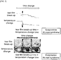
FIG. 3 shows graphs and images of corneal tear film break-up and temperature change for evaporative dry eye syndrome taken through an example of the present invention. -
FIG. 4 shows graphs and images of corneal tear film break-up and temperature change for mucus layer-deficient dry eye syndrome taken through an example of the present invention. -
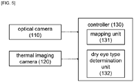
FIG. 5 shows a block diagram of an apparatus for diagnosing dry eye syndrome types according to an example of the present invention. - Hereinafter, preferred embodiments of the present invention will be described in detail with reference to the accompanying drawings. In this case, it should be noted that in the accompanying drawings, the same components are denoted by the same reference numerals as much as possible. In addition, detailed descriptions of well-known functions and configurations that may obscure the gist of the present invention will be omitted. For the same reason, some components are exaggerated, omitted, or schematically illustrated in the accompanying drawings.
- In addition, throughout the specification, when a part "comprises" a certain component, it means that other components may be further included, rather than excluding other components, unless specifically stated to the contrary. In addition, throughout the specification, "on" means to be located above or below the target part, and does not necessarily mean to be located above the direction of gravity.
- Referring to
FIG. 2 to FIG. 4 , the diagnostic method for distinguishing dry eye syndrome types according to an embodiment of the present invention includes a step of checking tear film break-up time point (S100), a step of checking corneal surface temperature (S200), and a step of comparing tear film break-up time point and the time point of the temperature change of the cornea surface (S300) and a step of diagnosing type of dry eye syndrome (S400). - In the step of checking tear film break-up time point (S100), the cornea of the subject's eye is photographed, and at least one times of the tear film break-up time point, at which the image reflected from the photographed cornea appears to be broken is time-sequentially confirmed. At this time, the cornea of the eye is photographed using an optical camera, and the tear film break-up area and location are calculated based on the area and the time at which the image is seen to be broken by reflection from the cornea, and the time can be confirmed.
- The step of checking corneal surface temperature (S200) is performed simultaneously with the photographing of the tear film of the eye, and the surface temperature of the cornea of the subject to be evaluated is checked using a thermal imaging camera to time-sequentially confirm a change in the surface temperature of the cornea with respect to time. In this case, the thermal imaging camera may be disposed adjacent to the optical camera.
- In the step of comparing tear film break-up time point and the time point of the temperature change of the cornea surface (S300), the tear film break-up time point and the time point of the surface temperature change of the cornea are compared by mapping the tear film break-up time point and the change in the surface temperature of the cornea based on time.
- In the step of diagnosing type of dry eye syndrome (S400), the type of dry eye syndrome is diagnosed based on any one of the tear film break-up time point and the corresponding point of the temperature change of the corneal surface.
- At this time, if it is determined that the initial tear film break-up time point (A) of the tear film break-up time point corresponds to time point having greatest decrease slope (B) in the temperature change point of the cornea photographed with the thermal imaging camera, it may be determined as evaporative dry eye syndrome
- In evaporative dry eye syndrome, when moisture evaporates from the cornea, the surrounding heat of evaporation is absorbed and the temperature decreases. When water evaporates, the absorption of heat of evaporation with time is greatest at the beginning of water evaporation, so the temperature change is measured the most. Therefore, it is possible to predict the timing of the start of tear evaporation by measuring the temperature change of the corneal surface.
- If the temperature change of the cornea photographed by the thermal imaging camera linearly decreases beyond the time point at which the tear film break-up is observed (A), and then decreases sharply (C) after a certain period of time (Δt) so that the sharp temperature change of the cornea is observed later than the initial point of the tear film break-up time point, with respect to the initial point of the tear film break-up time point (A), it is determined as mucus layer-deficient dry eye syndrome. In the case of mucus layer-deficient dry eye syndrome, the tear film is destroyed because the tearfilm is not properly attached to the corneal surface rather than the evaporation of tears. Therefore, the decrease in temperature due to the absorption of heat of evaporation is less than the evaporative dry eye syndrome, in which the tear film is destroyed by the evaporation of tears, or the temperature decrease occurs after the tear film is destroyed.
- Referring to
FIG. 5 , the diagnostic apparatus for distinguishing dry eye syndrome types according to an embodiment of the present invention includes anoptical camera 110, a thermal imaging camera 120, and acontroller 130. - The
optical camera 110 serves to take an image of the corneal surface. - The thermal imaging camera 120 serves to obtain a thermal image of the corneal surface and to measure (photograph) the temperature change of the corneal surface.
- The
controller 130 determines the type of dry eye syndrome by mapping and analyzing the image obtained by theoptical camera 110 and the thermal image obtained by the thermal imaging camera 120, and a mapping unit 131 and a dry eyetype determination unit 132 may be provided. - The mapping unit 131 maps the time-series change state of the tear film break-up time point obtained from the
optical camera 110 and the time-series change state of the surface temperature of the cornea obtained from the thermal imaging camera 120 based on time. The dry eyetype determination unit 132 determines the type of dry eye syndrome by comparing the initial time point among the tear film break-up time point and the temperature change time point photographed by the thermal imaging camera 120 from the two mapped images. -
FIG. 6 shows the experimental results related to the heat of evaporation. - As experimental conditions, the ambient temperature of the corneal surface is 10°C, the inner temperature of the eyeball is 34°C, the thickness of the tear film before the break-up is 10 µm, the size of the corneal surface is 10 mm × 10 µm, and the break-up area is 1 mm × 1 mm.
- As shown in
FIG. 6 , it was confirmed that the evaporation heat transfer rate increased as the tear film break-up thickness decreased. - For example, under the surface temperature of the tear layer of the corneal surface of 28°C, it was observed that the corneal surface temperature decreased to 22°C when the tear evaporation occurred in the tear layer with a thickness of 5 µm. In addition, it was observed that the corneal surface temperature decreased to 25.5°C when tears evaporated in the tear layer with a thickness of 7 µm. In general, the evaporative heat transfer coefficient passing through a thin liquid film may be defined by the equation shown in
FIG. 6 . - The diagnostic method and diagnostic apparatus for distinguishing type of dry eye syndrome according to the present invention use an optical camera to photograph the cornea of the eye and check in time-series of the tear film break-up time point therefrom, and at the same time, check the surface temperature of the cornea of the subject to be evaluated using a thermal imaging camera and map the two indices based on time, so that the dry eye syndrome type can be more accurately determined based on the location of the tear film break-up time point and the location of the temperature change time point on the corneal surface.
- While the present invention has been particularly described with reference to specific embodiments thereof, it is apparent that this specific description is only a preferred embodiment and that the scope of the present invention is not limited thereby to those skilled in the art. It will be apparent to those of ordinary skill in the art to which the present invention pertains that other modifications based on the technical spirit of the present invention can be implemented in addition to the embodiments disclosed herein.
Claims (5)
- A diagnostic method for distinguishing type of dry eye syndrome, comprising:(a) checking tear film break-up time point and location by photographing cornea of the subject's eye and checking in time series at least one or more times of the tear film break-up time point, at which the reflected image from the photographed cornea is confirmed to be broken over time;(b) checking corneal surface temperature by measuring the surface temperature of the cornea of the subject to be evaluated using a thermal imaging camera performed simultaneously with the photographing of the tear film of the eye to check in time series a change in the surface temperature of the cornea with respect to time;(c) mapping the tear film break-up time point and the change in the surface temperature of the cornea based on time to compare the tear film break-up time point and time point of the temperature change of the cornea surface; and(d) diagnosing type of dry eye syndrome based on any one of the tear film break-up time point and a location of surface temperature change time point of the corneal corresponding thereto, in mapping result in step (c).
- The diagnostic method for distinguishing type of dry eye syndrome of claim 1, wherein in step (a), the cornea of the eye is photographed using an optical camera, and tear film break-up area and location are calculated based on area and time at which an image is seen to be broken by reflection from the cornea and the time is confirmed.
- The diagnostic method for distinguishing type of dry eye syndrome of claim 1, wherein in step (d),if initial tear film break-up time point of the tear film break-up time point corresponds to time point having greatest decrease slope in the temperature change point of the cornea photographed with the thermal imaging camera, it is determined as evaporative dry eye syndrome, andif the temperature change of the cornea photographed by the thermal imaging camera linearly decreases beyond the time point at which the tear film break-up is observed, and then decreases sharply after a certain period of time so that the temperature change of the cornea is observed later than the initial point of the tear film break-up time point, with respect to the initial point of the tear film break-up time point, it is determined as mucus layer-deficient dry eye syndrome.
- A diagnostic apparatus for distinguishing type of dry eye syndrome, comprising:an optical camera for taking an image of the eye;a thermal imaging camera for obtaining a thermal image of the eye and measuring temperature change of the eye; anda controller configured to determine the type of dry eye syndrome by mapping and analyzing the image photographed by the optical camera and the thermal image obtained by the thermal imaging camera.
- The diagnostic apparatus for distinguishing type of dry eye syndrome of claim 4, wherein the controller comprises:a mapping unit for mapping time-series change state of the tear film break-up time point obtained by the optical camera and time-series change state of the surface temperature of the cornea obtained by the thermal imaging camera on a time basis; anddry eye type determination unit for determining the type of dry eye syndrome by comparing initial time point among the tear film break-up time point and the temperature change time point photographed with the thermal imaging camera from two mapped images.
Applications Claiming Priority (2)
| Application Number | Priority Date | Filing Date | Title |
|---|---|---|---|
| KR1020190175102A KR102293429B1 (en) | 2019-12-26 | 2019-12-26 | Method and diagnostic apparatus to distinguish dry eye sub-type |
| PCT/KR2020/018520 WO2021132978A1 (en) | 2019-12-26 | 2020-12-17 | Diagnosis method and diagnostic device for distinguishing types of dry eye syndrome |
Publications (3)
| Publication Number | Publication Date |
|---|---|
| EP4082423A1 true EP4082423A1 (en) | 2022-11-02 |
| EP4082423A4 EP4082423A4 (en) | 2024-01-10 |
| EP4082423B1 EP4082423B1 (en) | 2026-01-28 |
Family
ID=76573041
Family Applications (1)
| Application Number | Title | Priority Date | Filing Date |
|---|---|---|---|
| EP20905483.2A Active EP4082423B1 (en) | 2019-12-26 | 2020-12-17 | Diagnosis method and diagnostic device for distinguishing types of dry eye syndrome |
Country Status (5)
| Country | Link |
|---|---|
| US (1) | US12690763B2 (en) |
| EP (1) | EP4082423B1 (en) |
| KR (1) | KR102293429B1 (en) |
| CN (1) | CN115003208B (en) |
| WO (1) | WO2021132978A1 (en) |
Families Citing this family (10)
| Publication number | Priority date | Publication date | Assignee | Title |
|---|---|---|---|---|
| CN114240823B (en) * | 2021-10-29 | 2024-10-29 | 深圳莫廷医疗科技有限公司 | Real-time tear film rupture detection method, computer readable storage medium and apparatus |
| US11806078B1 (en) | 2022-05-01 | 2023-11-07 | Globe Biomedical, Inc. | Tear meniscus detection and evaluation system |
| KR102728315B1 (en) | 2022-05-27 | 2024-11-07 | 단국대학교 천안캠퍼스 산학협력단 | Device and method for diagnosing dry eye disease by measuring the thickness of the lipid layer in the tear film |
| KR102843532B1 (en) * | 2023-01-17 | 2025-08-08 | 전남대학교병원 | Dry eye syndrome test equipment and method |
| KR102855797B1 (en) | 2023-03-17 | 2025-09-05 | 서울대학교병원 | Dry-eye severity assessment system and assessment method using the same |
| US12186019B2 (en) | 2023-04-07 | 2025-01-07 | Globe Biomedical, Inc | Mechanical integration of components of wearable devices and ocular health monitoring system |
| KR20250035141A (en) | 2023-09-05 | 2025-03-12 | 삼성중공업 주식회사 | Dry eye prevention unit and dry eye prevention method |
| KR102853382B1 (en) * | 2023-09-20 | 2025-09-01 | 가톨릭대학교 산학협력단 | Tear break up detection method, device and computer program including the same |
| FI131790B1 (en) * | 2024-09-10 | 2025-12-01 | Pixieray Oy | Method and optical apparatus for continual monitoring tear film integrity of eye |
| CN120490118A (en) * | 2025-05-18 | 2025-08-15 | 南京耘瞳科技有限公司 | Water film on-line detection method, system and equipment |
Family Cites Families (15)
| Publication number | Priority date | Publication date | Assignee | Title |
|---|---|---|---|---|
| JPH09182723A (en) * | 1995-12-28 | 1997-07-15 | Taisho Pharmaceut Co Ltd | How to diagnose dry eye |
| JP4863207B2 (en) | 2006-07-04 | 2012-01-25 | 国立大学法人愛媛大学 | Ophthalmic equipment |
| US20080174733A1 (en) * | 2007-01-18 | 2008-07-24 | United Integrated Services Co., Ltd. | Method of using infrared thermal imager to diagnose eye diseases and the device thereof |
| US8215775B2 (en) * | 2010-09-07 | 2012-07-10 | United Integrated Services Co., Ltd. | Method for analyzing tear film thermograph of contactless tear film thermal imager |
| US20130079660A1 (en) * | 2011-09-23 | 2013-03-28 | O Chang | Method utilizing thermograph of a tear film for generating a quantified index |
| CN103799976B (en) * | 2014-03-04 | 2016-01-20 | 厦门大学 | A kind of xerophthalmia comprehensive diagnostic instrument |
| KR101662775B1 (en) | 2014-11-26 | 2016-10-05 | 박범철 | Apparatus for diagnosing dry eye syndrome and method for diagnosing dry eye syndrome using the same |
| KR101742049B1 (en) * | 2015-06-12 | 2017-06-15 | 한림대학교 산학협력단 | Meibomian photographing gland device using infrared ray and meibomian gland photographing method using the same |
| US10076250B2 (en) | 2015-06-14 | 2018-09-18 | Facense Ltd. | Detecting physiological responses based on multispectral data from head-mounted cameras |
| CA3034608A1 (en) * | 2016-08-24 | 2018-03-01 | The University Of Melbourne | Diagnostic methods and device |
| KR20180072911A (en) | 2016-12-21 | 2018-07-02 | 희성전자 주식회사 | Apparatus of treating for dry eye syndrome |
| KR101776226B1 (en) * | 2017-01-31 | 2017-09-11 | 단국대학교 천안캠퍼스 산학협력단 | Dry eye syndrome diagnosis and treatment apparatus and the method thereof |
| US20200383564A1 (en) * | 2017-12-08 | 2020-12-10 | Beyond 700 Pty Ltd | Methods based on tear film behaviour |
| US11564563B2 (en) * | 2018-02-17 | 2023-01-31 | Aizhong Zhang | Apparatus and method of a multifunctional ophthalmic instrument |
| US11253151B2 (en) * | 2019-09-08 | 2022-02-22 | Aizhong Zhang | Multispectral and hyperspectral ocular surface evaluator |
-
2019
- 2019-12-26 KR KR1020190175102A patent/KR102293429B1/en active Active
-
2020
- 2020-12-17 WO PCT/KR2020/018520 patent/WO2021132978A1/en not_active Ceased
- 2020-12-17 CN CN202080094376.7A patent/CN115003208B/en active Active
- 2020-12-17 EP EP20905483.2A patent/EP4082423B1/en active Active
- 2020-12-17 US US17/788,302 patent/US12690763B2/en active Active
Also Published As
| Publication number | Publication date |
|---|---|
| CN115003208B (en) | 2026-02-24 |
| KR102293429B1 (en) | 2021-08-26 |
| EP4082423B1 (en) | 2026-01-28 |
| US12690763B2 (en) | 2026-07-28 |
| EP4082423A4 (en) | 2024-01-10 |
| CN115003208A (en) | 2022-09-02 |
| US20230025623A1 (en) | 2023-01-26 |
| WO2021132978A1 (en) | 2021-07-01 |
| KR20210082801A (en) | 2021-07-06 |
Similar Documents
| Publication | Publication Date | Title |
|---|---|---|
| EP4082423B1 (en) | Diagnosis method and diagnostic device for distinguishing types of dry eye syndrome | |
| Chansangpetch et al. | Anterior segment imaging for angle closure | |
| Wei et al. | Assessment of lower tear meniscus | |
| Kovács et al. | Evaluation of keratometric, pachymetric, and elevation parameters of keratoconic corneas with pentacam | |
| Leung et al. | Analysis of bleb morphology after trabeculectomy with Visante anterior segment optical coherence tomography | |
| Mathers et al. | Ocular water evaporation and the dry eye: a new measuring device | |
| Raj et al. | Anterior segment optical coherence tomography for tear meniscus evaluation and its correlation with other tear variables in healthy individuals | |
| CN103799976B (en) | A kind of xerophthalmia comprehensive diagnostic instrument | |
| JP3008197B2 (en) | Ophthalmic equipment | |
| US8215775B2 (en) | Method for analyzing tear film thermograph of contactless tear film thermal imager | |
| US20140309554A1 (en) | Device for sampling tear fluid | |
| KR101662775B1 (en) | Apparatus for diagnosing dry eye syndrome and method for diagnosing dry eye syndrome using the same | |
| Bhandari et al. | Non-invasive assessment of tear film stability with a novel corneal topographer in Indian subjects | |
| Eter et al. | A new technique for tear film fluorophotometry | |
| Luke et al. | Parameter estimation for mixed-mechanism tear film thinning | |
| Al-Mohtaseb et al. | Repeatability and comparability of corneal thickness measurements obtained from Dual Scheimpflug Analyzer and from ultrasonic pachymetry | |
| TW201622634A (en) | Examining system for eye gland function | |
| Azuara-Blanco et al. | Tono-Pen determination of intraocular pressure in patients with band keratopathy or glued cornea | |
| Bandlitz et al. | Advances in tear film assessment | |
| Kiuchi et al. | Corneal displacement during tonometry with a noncontact tonometer | |
| Maurice | An aplanation tonometer of new principle | |
| US20230049316A1 (en) | Techniques for quantitatively assessing tear-film dynamics | |
| De Sanctis et al. | Graft central thickness measurement by rotating Scheimpflug camera and ultrasound pachymetry after penetrating keratoplasty | |
| US9370587B2 (en) | Coloring substance for diagnostic ophtalmologic use | |
| KR102728315B1 (en) | Device and method for diagnosing dry eye disease by measuring the thickness of the lipid layer in the tear film |
Legal Events
| Date | Code | Title | Description |
|---|---|---|---|
| STAA | Information on the status of an ep patent application or granted ep patent |
Free format text: STATUS: THE INTERNATIONAL PUBLICATION HAS BEEN MADE |
|
| PUAI | Public reference made under article 153(3) epc to a published international application that has entered the european phase |
Free format text: ORIGINAL CODE: 0009012 |
|
| STAA | Information on the status of an ep patent application or granted ep patent |
Free format text: STATUS: REQUEST FOR EXAMINATION WAS MADE |
|
| 17P | Request for examination filed |
Effective date: 20220701 |
|
| AK | Designated contracting states |
Kind code of ref document: A1 Designated state(s): AL AT BE BG CH CY CZ DE DK EE ES FI FR GB GR HR HU IE IS IT LI LT LU LV MC MK MT NL NO PL PT RO RS SE SI SK SM TR |
|
| DAV | Request for validation of the european patent (deleted) | ||
| DAX | Request for extension of the european patent (deleted) | ||
| A4 | Supplementary search report drawn up and despatched |
Effective date: 20231206 |
|
| RIC1 | Information provided on ipc code assigned before grant |
Ipc: G16H 30/40 20180101ALI20231201BHEP Ipc: G06T 7/00 20170101ALI20231201BHEP Ipc: G06T 5/50 20060101ALI20231201BHEP Ipc: A61B 3/00 20060101ALI20231201BHEP Ipc: A61B 5/01 20060101ALI20231201BHEP Ipc: A61B 3/14 20060101ALI20231201BHEP Ipc: A61B 3/10 20060101AFI20231201BHEP |
|
| STAA | Information on the status of an ep patent application or granted ep patent |
Free format text: STATUS: EXAMINATION IS IN PROGRESS |
|
| 17Q | First examination report despatched |
Effective date: 20250407 |
|
| GRAP | Despatch of communication of intention to grant a patent |
Free format text: ORIGINAL CODE: EPIDOSNIGR1 |
|
| STAA | Information on the status of an ep patent application or granted ep patent |
Free format text: STATUS: GRANT OF PATENT IS INTENDED |
|
| INTG | Intention to grant announced |
Effective date: 20251015 |
|
| GRAS | Grant fee paid |
Free format text: ORIGINAL CODE: EPIDOSNIGR3 |
|
| GRAA | (expected) grant |
Free format text: ORIGINAL CODE: 0009210 |
|
| STAA | Information on the status of an ep patent application or granted ep patent |
Free format text: STATUS: THE PATENT HAS BEEN GRANTED |
|
| AK | Designated contracting states |
Kind code of ref document: B1 Designated state(s): AL AT BE BG CH CY CZ DE DK EE ES FI FR GB GR HR HU IE IS IT LI LT LU LV MC MK MT NL NO PL PT RO RS SE SI SK SM TR |
|
| REG | Reference to a national code |
Ref country code: CH Ref legal event code: F10 Free format text: ST27 STATUS EVENT CODE: U-0-0-F10-F00 (AS PROVIDED BY THE NATIONAL OFFICE) Effective date: 20260128 Ref country code: GB Ref legal event code: FG4D |
|
| REG | Reference to a national code |
Ref country code: DE Ref legal event code: R096 Ref document number: 602020066340 Country of ref document: DE |
|
| REG | Reference to a national code |
Ref country code: IE Ref legal event code: FG4D |
|
| REG | Reference to a national code |
Ref country code: NL Ref legal event code: MP Effective date: 20260128 |
|
| PG25 | Lapsed in a contracting state [announced via postgrant information from national office to epo] |
Ref country code: NL Free format text: LAPSE BECAUSE OF FAILURE TO SUBMIT A TRANSLATION OF THE DESCRIPTION OR TO PAY THE FEE WITHIN THE PRESCRIBED TIME-LIMIT Effective date: 20260128 |
|
| REG | Reference to a national code |
Ref country code: AT Ref legal event code: MK05 Ref document number: 1879998 Country of ref document: AT Kind code of ref document: T Effective date: 20260128 |
|
| PG25 | Lapsed in a contracting state [announced via postgrant information from national office to epo] |
Ref country code: RS Free format text: LAPSE BECAUSE OF FAILURE TO SUBMIT A TRANSLATION OF THE DESCRIPTION OR TO PAY THE FEE WITHIN THE PRESCRIBED TIME-LIMIT Effective date: 20260428 |
|
| PG25 | Lapsed in a contracting state [announced via postgrant information from national office to epo] |
Ref country code: ES Free format text: LAPSE BECAUSE OF FAILURE TO SUBMIT A TRANSLATION OF THE DESCRIPTION OR TO PAY THE FEE WITHIN THE PRESCRIBED TIME-LIMIT Effective date: 20260128 |
|
| PG25 | Lapsed in a contracting state [announced via postgrant information from national office to epo] |
Ref country code: PL Free format text: LAPSE BECAUSE OF FAILURE TO SUBMIT A TRANSLATION OF THE DESCRIPTION OR TO PAY THE FEE WITHIN THE PRESCRIBED TIME-LIMIT Effective date: 20260128 |
|
| PG25 | Lapsed in a contracting state [announced via postgrant information from national office to epo] |
Ref country code: GR Free format text: LAPSE BECAUSE OF FAILURE TO SUBMIT A TRANSLATION OF THE DESCRIPTION OR TO PAY THE FEE WITHIN THE PRESCRIBED TIME-LIMIT Effective date: 20260429 |
|
| REG | Reference to a national code |
Ref country code: LT Ref legal event code: MG9D |
|
| PG25 | Lapsed in a contracting state [announced via postgrant information from national office to epo] |
Ref country code: LT Free format text: LAPSE BECAUSE OF FAILURE TO SUBMIT A TRANSLATION OF THE DESCRIPTION OR TO PAY THE FEE WITHIN THE PRESCRIBED TIME-LIMIT Effective date: 20260128 |
|
| PG25 | Lapsed in a contracting state [announced via postgrant information from national office to epo] |
Ref country code: NO Free format text: LAPSE BECAUSE OF FAILURE TO SUBMIT A TRANSLATION OF THE DESCRIPTION OR TO PAY THE FEE WITHIN THE PRESCRIBED TIME-LIMIT Effective date: 20260428 |
|
| PG25 | Lapsed in a contracting state [announced via postgrant information from national office to epo] |
Ref country code: AT Free format text: LAPSE BECAUSE OF FAILURE TO SUBMIT A TRANSLATION OF THE DESCRIPTION OR TO PAY THE FEE WITHIN THE PRESCRIBED TIME-LIMIT Effective date: 20260128 Ref country code: SE Free format text: LAPSE BECAUSE OF FAILURE TO SUBMIT A TRANSLATION OF THE DESCRIPTION OR TO PAY THE FEE WITHIN THE PRESCRIBED TIME-LIMIT Effective date: 20260128 Ref country code: FI Free format text: LAPSE BECAUSE OF FAILURE TO SUBMIT A TRANSLATION OF THE DESCRIPTION OR TO PAY THE FEE WITHIN THE PRESCRIBED TIME-LIMIT Effective date: 20260128 |
|
| PG25 | Lapsed in a contracting state [announced via postgrant information from national office to epo] |
Ref country code: HR Free format text: LAPSE BECAUSE OF FAILURE TO SUBMIT A TRANSLATION OF THE DESCRIPTION OR TO PAY THE FEE WITHIN THE PRESCRIBED TIME-LIMIT Effective date: 20260128 |
|
| PG25 | Lapsed in a contracting state [announced via postgrant information from national office to epo] |
Ref country code: PT Free format text: LAPSE BECAUSE OF FAILURE TO SUBMIT A TRANSLATION OF THE DESCRIPTION OR TO PAY THE FEE WITHIN THE PRESCRIBED TIME-LIMIT Effective date: 20260528 Ref country code: IS Free format text: LAPSE BECAUSE OF FAILURE TO SUBMIT A TRANSLATION OF THE DESCRIPTION OR TO PAY THE FEE WITHIN THE PRESCRIBED TIME-LIMIT Effective date: 20260528 |
|
| PG25 | Lapsed in a contracting state [announced via postgrant information from national office to epo] |
Ref country code: LV Free format text: LAPSE BECAUSE OF FAILURE TO SUBMIT A TRANSLATION OF THE DESCRIPTION OR TO PAY THE FEE WITHIN THE PRESCRIBED TIME-LIMIT Effective date: 20260128 |
|
| PG25 | Lapsed in a contracting state [announced via postgrant information from national office to epo] |
Ref country code: BG Free format text: LAPSE BECAUSE OF FAILURE TO SUBMIT A TRANSLATION OF THE DESCRIPTION OR TO PAY THE FEE WITHIN THE PRESCRIBED TIME-LIMIT Effective date: 20260128 |