EP4079758A1 - Semg2 antibody and use thereof - Google Patents
Semg2 antibody and use thereof Download PDFInfo
- Publication number
- EP4079758A1 EP4079758A1 EP21744026.2A EP21744026A EP4079758A1 EP 4079758 A1 EP4079758 A1 EP 4079758A1 EP 21744026 A EP21744026 A EP 21744026A EP 4079758 A1 EP4079758 A1 EP 4079758A1
- Authority
- EP
- European Patent Office
- Prior art keywords
- seq
- amino acid
- acid sequence
- semg2
- chain variable
- Prior art date
- Legal status (The legal status is an assumption and is not a legal conclusion. Google has not performed a legal analysis and makes no representation as to the accuracy of the status listed.)
- Pending
Links
- 101000739786 Homo sapiens Semenogelin-2 Proteins 0.000 claims abstract description 335
- 102100037547 Semenogelin-2 Human genes 0.000 claims abstract description 334
- 230000027455 binding Effects 0.000 claims abstract description 141
- 102100027207 CD27 antigen Human genes 0.000 claims abstract description 134
- 101000914511 Homo sapiens CD27 antigen Proteins 0.000 claims abstract description 134
- 108090000765 processed proteins & peptides Proteins 0.000 claims abstract description 81
- 210000004881 tumor cell Anatomy 0.000 claims abstract description 63
- 102000004196 processed proteins & peptides Human genes 0.000 claims abstract description 49
- 229920001184 polypeptide Polymers 0.000 claims abstract description 47
- 239000012634 fragment Substances 0.000 claims abstract description 40
- 239000000427 antigen Substances 0.000 claims abstract description 39
- 108091007433 antigens Proteins 0.000 claims abstract description 39
- 102000036639 antigens Human genes 0.000 claims abstract description 39
- 150000001875 compounds Chemical class 0.000 claims abstract description 34
- 238000000034 method Methods 0.000 claims abstract description 32
- 230000002163 immunogen Effects 0.000 claims abstract description 17
- 230000003993 interaction Effects 0.000 claims abstract description 16
- 238000012216 screening Methods 0.000 claims abstract description 15
- 210000002865 immune cell Anatomy 0.000 claims abstract description 9
- 230000003042 antagnostic effect Effects 0.000 claims abstract description 5
- 239000003112 inhibitor Substances 0.000 claims abstract description 5
- 150000003384 small molecules Chemical class 0.000 claims abstract description 3
- 125000003275 alpha amino acid group Chemical group 0.000 claims description 261
- 210000004027 cell Anatomy 0.000 claims description 59
- 206010028980 Neoplasm Diseases 0.000 claims description 52
- 230000014509 gene expression Effects 0.000 claims description 52
- 108090000623 proteins and genes Proteins 0.000 claims description 50
- 102000004169 proteins and genes Human genes 0.000 claims description 45
- 150000001413 amino acids Chemical class 0.000 claims description 29
- 230000000890 antigenic effect Effects 0.000 claims description 28
- 206010009944 Colon cancer Diseases 0.000 claims description 20
- 208000001333 Colorectal Neoplasms Diseases 0.000 claims description 20
- 239000013598 vector Substances 0.000 claims description 20
- 108091033319 polynucleotide Proteins 0.000 claims description 15
- 239000002157 polynucleotide Substances 0.000 claims description 15
- 102000040430 polynucleotide Human genes 0.000 claims description 15
- 238000002360 preparation method Methods 0.000 claims description 15
- 210000001744 T-lymphocyte Anatomy 0.000 claims description 13
- 229940079593 drug Drugs 0.000 claims description 12
- 239000003814 drug Substances 0.000 claims description 12
- 102100035360 Cerebellar degeneration-related antigen 1 Human genes 0.000 claims description 11
- 201000001441 melanoma Diseases 0.000 claims description 10
- 206010058467 Lung neoplasm malignant Diseases 0.000 claims description 8
- 201000005202 lung cancer Diseases 0.000 claims description 8
- 208000020816 lung neoplasm Diseases 0.000 claims description 8
- 239000002671 adjuvant Substances 0.000 claims description 7
- 125000000539 amino acid group Chemical group 0.000 claims description 7
- 238000000338 in vitro Methods 0.000 claims description 7
- YBJHBAHKTGYVGT-ZKWXMUAHSA-N (+)-Biotin Chemical compound N1C(=O)N[C@@H]2[C@H](CCCCC(=O)O)SC[C@@H]21 YBJHBAHKTGYVGT-ZKWXMUAHSA-N 0.000 claims description 6
- 206010060862 Prostate cancer Diseases 0.000 claims description 6
- 208000000236 Prostatic Neoplasms Diseases 0.000 claims description 6
- 230000008878 coupling Effects 0.000 claims description 6
- 238000010168 coupling process Methods 0.000 claims description 6
- 238000005859 coupling reaction Methods 0.000 claims description 6
- 230000002401 inhibitory effect Effects 0.000 claims description 6
- 208000005718 Stomach Neoplasms Diseases 0.000 claims description 5
- 239000003153 chemical reaction reagent Substances 0.000 claims description 5
- 206010017758 gastric cancer Diseases 0.000 claims description 5
- 210000004698 lymphocyte Anatomy 0.000 claims description 5
- 201000011549 stomach cancer Diseases 0.000 claims description 5
- 230000004614 tumor growth Effects 0.000 claims description 5
- 108010003723 Single-Domain Antibodies Proteins 0.000 claims description 4
- 238000011319 anticancer therapy Methods 0.000 claims description 4
- 239000012472 biological sample Substances 0.000 claims description 4
- 239000013604 expression vector Substances 0.000 claims description 4
- 150000002632 lipids Chemical class 0.000 claims description 4
- 239000008194 pharmaceutical composition Substances 0.000 claims description 4
- 230000001105 regulatory effect Effects 0.000 claims description 4
- 238000013518 transcription Methods 0.000 claims description 4
- 230000035897 transcription Effects 0.000 claims description 4
- 102000004190 Enzymes Human genes 0.000 claims description 3
- 108090000790 Enzymes Proteins 0.000 claims description 3
- 206010025323 Lymphomas Diseases 0.000 claims description 3
- 229960002685 biotin Drugs 0.000 claims description 3
- 235000020958 biotin Nutrition 0.000 claims description 3
- 239000011616 biotin Substances 0.000 claims description 3
- 238000009169 immunotherapy Methods 0.000 claims description 3
- 206010005003 Bladder cancer Diseases 0.000 claims description 2
- 206010006187 Breast cancer Diseases 0.000 claims description 2
- 208000026310 Breast neoplasm Diseases 0.000 claims description 2
- 102000014914 Carrier Proteins Human genes 0.000 claims description 2
- 108010078791 Carrier Proteins Proteins 0.000 claims description 2
- 102000004127 Cytokines Human genes 0.000 claims description 2
- 108090000695 Cytokines Proteins 0.000 claims description 2
- BWGNESOTFCXPMA-UHFFFAOYSA-N Dihydrogen disulfide Chemical compound SS BWGNESOTFCXPMA-UHFFFAOYSA-N 0.000 claims description 2
- 206010014733 Endometrial cancer Diseases 0.000 claims description 2
- 206010014759 Endometrial neoplasm Diseases 0.000 claims description 2
- 108010054477 Immunoglobulin Fab Fragments Proteins 0.000 claims description 2
- 102000001706 Immunoglobulin Fab Fragments Human genes 0.000 claims description 2
- 208000008839 Kidney Neoplasms Diseases 0.000 claims description 2
- 241000124008 Mammalia Species 0.000 claims description 2
- 206010033128 Ovarian cancer Diseases 0.000 claims description 2
- 206010061535 Ovarian neoplasm Diseases 0.000 claims description 2
- 108010076504 Protein Sorting Signals Proteins 0.000 claims description 2
- 206010038389 Renal cancer Diseases 0.000 claims description 2
- 208000024313 Testicular Neoplasms Diseases 0.000 claims description 2
- 206010057644 Testis cancer Diseases 0.000 claims description 2
- 208000007097 Urinary Bladder Neoplasms Diseases 0.000 claims description 2
- 230000003302 anti-idiotype Effects 0.000 claims description 2
- -1 antibody Substances 0.000 claims description 2
- 238000002512 chemotherapy Methods 0.000 claims description 2
- 239000013599 cloning vector Substances 0.000 claims description 2
- 239000003937 drug carrier Substances 0.000 claims description 2
- 210000003527 eukaryotic cell Anatomy 0.000 claims description 2
- GNBHRKFJIUUOQI-UHFFFAOYSA-N fluorescein Chemical compound O1C(=O)C2=CC=CC=C2C21C1=CC=C(O)C=C1OC1=CC(O)=CC=C21 GNBHRKFJIUUOQI-UHFFFAOYSA-N 0.000 claims description 2
- 201000010536 head and neck cancer Diseases 0.000 claims description 2
- 208000014829 head and neck neoplasm Diseases 0.000 claims description 2
- 238000001794 hormone therapy Methods 0.000 claims description 2
- 230000028993 immune response Effects 0.000 claims description 2
- 230000003834 intracellular effect Effects 0.000 claims description 2
- 201000010982 kidney cancer Diseases 0.000 claims description 2
- 201000007270 liver cancer Diseases 0.000 claims description 2
- 208000014018 liver neoplasm Diseases 0.000 claims description 2
- 230000008488 polyadenylation Effects 0.000 claims description 2
- 210000001236 prokaryotic cell Anatomy 0.000 claims description 2
- 238000001959 radiotherapy Methods 0.000 claims description 2
- 238000001356 surgical procedure Methods 0.000 claims description 2
- 201000003120 testicular cancer Diseases 0.000 claims description 2
- 239000003053 toxin Substances 0.000 claims description 2
- 231100000765 toxin Toxicity 0.000 claims description 2
- 108700012359 toxins Proteins 0.000 claims description 2
- 201000005112 urinary bladder cancer Diseases 0.000 claims description 2
- 230000000903 blocking effect Effects 0.000 abstract description 49
- 230000005809 anti-tumor immunity Effects 0.000 abstract description 2
- 230000001737 promoting effect Effects 0.000 abstract description 2
- 229940126585 therapeutic drug Drugs 0.000 abstract 1
- 238000002965 ELISA Methods 0.000 description 44
- 235000018102 proteins Nutrition 0.000 description 33
- 241000699666 Mus <mouse, genus> Species 0.000 description 30
- 241000699670 Mus sp. Species 0.000 description 29
- 230000000694 effects Effects 0.000 description 29
- 235000001014 amino acid Nutrition 0.000 description 28
- 230000006870 function Effects 0.000 description 27
- 238000002474 experimental method Methods 0.000 description 26
- 210000001519 tissue Anatomy 0.000 description 25
- 230000000875 corresponding effect Effects 0.000 description 20
- DHMQDGOQFOQNFH-UHFFFAOYSA-N Glycine Chemical compound NCC(O)=O DHMQDGOQFOQNFH-UHFFFAOYSA-N 0.000 description 17
- 238000004458 analytical method Methods 0.000 description 16
- 210000004369 blood Anatomy 0.000 description 16
- 239000008280 blood Substances 0.000 description 16
- 241001529936 Murinae Species 0.000 description 15
- 229940126619 mouse monoclonal antibody Drugs 0.000 description 13
- 230000022534 cell killing Effects 0.000 description 12
- 108091003079 Bovine Serum Albumin Proteins 0.000 description 10
- 229940098773 bovine serum albumin Drugs 0.000 description 10
- 230000003053 immunization Effects 0.000 description 10
- 238000002649 immunization Methods 0.000 description 10
- 230000002147 killing effect Effects 0.000 description 10
- 239000004471 Glycine Substances 0.000 description 9
- 238000002835 absorbance Methods 0.000 description 9
- 238000003556 assay Methods 0.000 description 9
- 238000001514 detection method Methods 0.000 description 9
- 102100025221 CD70 antigen Human genes 0.000 description 8
- 101100112922 Candida albicans CDR3 gene Proteins 0.000 description 8
- 101000934356 Homo sapiens CD70 antigen Proteins 0.000 description 7
- 230000000259 anti-tumor effect Effects 0.000 description 7
- 238000011282 treatment Methods 0.000 description 7
- 238000000749 co-immunoprecipitation Methods 0.000 description 6
- 238000011532 immunohistochemical staining Methods 0.000 description 6
- 210000004072 lung Anatomy 0.000 description 6
- 230000035772 mutation Effects 0.000 description 6
- 239000013612 plasmid Substances 0.000 description 6
- 210000003289 regulatory T cell Anatomy 0.000 description 6
- 239000000243 solution Substances 0.000 description 6
- 238000006467 substitution reaction Methods 0.000 description 6
- 101100367238 Mus musculus Svs3a gene Proteins 0.000 description 5
- 241000283973 Oryctolagus cuniculus Species 0.000 description 5
- 230000006907 apoptotic process Effects 0.000 description 5
- 239000007853 buffer solution Substances 0.000 description 5
- 201000011510 cancer Diseases 0.000 description 5
- UQLDLKMNUJERMK-UHFFFAOYSA-L di(octadecanoyloxy)lead Chemical compound [Pb+2].CCCCCCCCCCCCCCCCCC([O-])=O.CCCCCCCCCCCCCCCCCC([O-])=O UQLDLKMNUJERMK-UHFFFAOYSA-L 0.000 description 5
- 101150042537 dld1 gene Proteins 0.000 description 5
- 238000003364 immunohistochemistry Methods 0.000 description 5
- 238000011813 knockout mouse model Methods 0.000 description 5
- 238000012856 packing Methods 0.000 description 5
- 210000003819 peripheral blood mononuclear cell Anatomy 0.000 description 5
- 210000002966 serum Anatomy 0.000 description 5
- 230000004083 survival effect Effects 0.000 description 5
- 230000004913 activation Effects 0.000 description 4
- 230000001640 apoptogenic effect Effects 0.000 description 4
- 239000000872 buffer Substances 0.000 description 4
- 238000011490 co-immunoprecipitation assay Methods 0.000 description 4
- 238000010494 dissociation reaction Methods 0.000 description 4
- 230000005593 dissociations Effects 0.000 description 4
- 210000002216 heart Anatomy 0.000 description 4
- 210000004408 hybridoma Anatomy 0.000 description 4
- 238000003119 immunoblot Methods 0.000 description 4
- 230000008595 infiltration Effects 0.000 description 4
- 238000001764 infiltration Methods 0.000 description 4
- 238000011081 inoculation Methods 0.000 description 4
- 210000003734 kidney Anatomy 0.000 description 4
- 239000003446 ligand Substances 0.000 description 4
- 239000006166 lysate Substances 0.000 description 4
- 230000010399 physical interaction Effects 0.000 description 4
- 230000002829 reductive effect Effects 0.000 description 4
- 238000010186 staining Methods 0.000 description 4
- 102000017420 CD3 protein, epsilon/gamma/delta subunit Human genes 0.000 description 3
- 108091033409 CRISPR Proteins 0.000 description 3
- 102000047934 Caspase-3/7 Human genes 0.000 description 3
- 108700037887 Caspase-3/7 Proteins 0.000 description 3
- 102000005720 Glutathione transferase Human genes 0.000 description 3
- 108010070675 Glutathione transferase Proteins 0.000 description 3
- 108020005004 Guide RNA Proteins 0.000 description 3
- 108010021625 Immunoglobulin Fragments Proteins 0.000 description 3
- 102000008394 Immunoglobulin Fragments Human genes 0.000 description 3
- 101710175625 Maltose/maltodextrin-binding periplasmic protein Proteins 0.000 description 3
- 241001465754 Metazoa Species 0.000 description 3
- 229940124158 Protease/peptidase inhibitor Drugs 0.000 description 3
- 239000012722 SDS sample buffer Substances 0.000 description 3
- 229920002684 Sepharose Polymers 0.000 description 3
- 230000009824 affinity maturation Effects 0.000 description 3
- 210000004556 brain Anatomy 0.000 description 3
- 210000004899 c-terminal region Anatomy 0.000 description 3
- 230000008045 co-localization Effects 0.000 description 3
- 208000035250 cutaneous malignant susceptibility to 1 melanoma Diseases 0.000 description 3
- 230000001419 dependent effect Effects 0.000 description 3
- 238000011161 development Methods 0.000 description 3
- 230000017188 evasion or tolerance of host immune response Effects 0.000 description 3
- 238000003209 gene knockout Methods 0.000 description 3
- 230000002055 immunohistochemical effect Effects 0.000 description 3
- 230000001506 immunosuppresive effect Effects 0.000 description 3
- 210000004185 liver Anatomy 0.000 description 3
- 210000001165 lymph node Anatomy 0.000 description 3
- 239000013642 negative control Substances 0.000 description 3
- 230000003472 neutralizing effect Effects 0.000 description 3
- 239000000137 peptide hydrolase inhibitor Substances 0.000 description 3
- 239000013641 positive control Substances 0.000 description 3
- 108010045039 seminal vesicle-specific antigen Proteins 0.000 description 3
- 210000000952 spleen Anatomy 0.000 description 3
- 210000002784 stomach Anatomy 0.000 description 3
- 238000012360 testing method Methods 0.000 description 3
- 238000005406 washing Methods 0.000 description 3
- 238000011746 C57BL/6J (JAX™ mouse strain) Methods 0.000 description 2
- CURLTUGMZLYLDI-UHFFFAOYSA-N Carbon dioxide Chemical compound O=C=O CURLTUGMZLYLDI-UHFFFAOYSA-N 0.000 description 2
- 241000282693 Cercopithecidae Species 0.000 description 2
- 206010052360 Colorectal adenocarcinoma Diseases 0.000 description 2
- 101000914514 Homo sapiens T-cell-specific surface glycoprotein CD28 Proteins 0.000 description 2
- 239000002033 PVDF binder Substances 0.000 description 2
- 229920001213 Polysorbate 20 Polymers 0.000 description 2
- 241000288906 Primates Species 0.000 description 2
- PXIPVTKHYLBLMZ-UHFFFAOYSA-N Sodium azide Chemical compound [Na+].[N-]=[N+]=[N-] PXIPVTKHYLBLMZ-UHFFFAOYSA-N 0.000 description 2
- 102100027213 T-cell-specific surface glycoprotein CD28 Human genes 0.000 description 2
- 239000013504 Triton X-100 Substances 0.000 description 2
- 229920004890 Triton X-100 Polymers 0.000 description 2
- 108060008683 Tumor Necrosis Factor Receptor Proteins 0.000 description 2
- 230000002146 bilateral effect Effects 0.000 description 2
- 238000012575 bio-layer interferometry Methods 0.000 description 2
- 239000000090 biomarker Substances 0.000 description 2
- 238000009534 blood test Methods 0.000 description 2
- 230000037396 body weight Effects 0.000 description 2
- 210000005252 bulbus oculi Anatomy 0.000 description 2
- 210000000170 cell membrane Anatomy 0.000 description 2
- 238000005119 centrifugation Methods 0.000 description 2
- 238000006243 chemical reaction Methods 0.000 description 2
- 238000012761 co-transfection Methods 0.000 description 2
- 239000011248 coating agent Substances 0.000 description 2
- 238000000576 coating method Methods 0.000 description 2
- 201000010897 colon adenocarcinoma Diseases 0.000 description 2
- 210000004922 colonic epithelial cell Anatomy 0.000 description 2
- 238000010276 construction Methods 0.000 description 2
- 230000007423 decrease Effects 0.000 description 2
- 230000003247 decreasing effect Effects 0.000 description 2
- 210000001198 duodenum Anatomy 0.000 description 2
- 239000012149 elution buffer Substances 0.000 description 2
- 229940088598 enzyme Drugs 0.000 description 2
- 210000002919 epithelial cell Anatomy 0.000 description 2
- 238000000684 flow cytometry Methods 0.000 description 2
- 230000004927 fusion Effects 0.000 description 2
- 108020001507 fusion proteins Proteins 0.000 description 2
- 102000037865 fusion proteins Human genes 0.000 description 2
- 201000006585 gastric adenocarcinoma Diseases 0.000 description 2
- 230000002496 gastric effect Effects 0.000 description 2
- 101150086731 ges-1 gene Proteins 0.000 description 2
- 210000004013 groin Anatomy 0.000 description 2
- 206010073071 hepatocellular carcinoma Diseases 0.000 description 2
- 231100000844 hepatocellular carcinoma Toxicity 0.000 description 2
- 210000003405 ileum Anatomy 0.000 description 2
- 238000001114 immunoprecipitation Methods 0.000 description 2
- 238000001727 in vivo Methods 0.000 description 2
- 238000011534 incubation Methods 0.000 description 2
- 239000002198 insoluble material Substances 0.000 description 2
- 238000005305 interferometry Methods 0.000 description 2
- 239000007928 intraperitoneal injection Substances 0.000 description 2
- 210000001630 jejunum Anatomy 0.000 description 2
- 210000002429 large intestine Anatomy 0.000 description 2
- 239000003550 marker Substances 0.000 description 2
- 239000000463 material Substances 0.000 description 2
- 239000012528 membrane Substances 0.000 description 2
- 108020004999 messenger RNA Proteins 0.000 description 2
- 239000000203 mixture Substances 0.000 description 2
- 230000004048 modification Effects 0.000 description 2
- 238000012986 modification Methods 0.000 description 2
- 210000000056 organ Anatomy 0.000 description 2
- 201000008968 osteosarcoma Diseases 0.000 description 2
- 238000002823 phage display Methods 0.000 description 2
- 239000000256 polyoxyethylene sorbitan monolaurate Substances 0.000 description 2
- 235000010486 polyoxyethylene sorbitan monolaurate Nutrition 0.000 description 2
- 229920002981 polyvinylidene fluoride Polymers 0.000 description 2
- 230000008569 process Effects 0.000 description 2
- 238000000746 purification Methods 0.000 description 2
- 108020003175 receptors Proteins 0.000 description 2
- 102000005962 receptors Human genes 0.000 description 2
- 238000005215 recombination Methods 0.000 description 2
- 239000000523 sample Substances 0.000 description 2
- 210000000582 semen Anatomy 0.000 description 2
- 238000012163 sequencing technique Methods 0.000 description 2
- 230000019491 signal transduction Effects 0.000 description 2
- 210000000813 small intestine Anatomy 0.000 description 2
- 238000002415 sodium dodecyl sulfate polyacrylamide gel electrophoresis Methods 0.000 description 2
- 210000000130 stem cell Anatomy 0.000 description 2
- 239000000758 substrate Substances 0.000 description 2
- 231100000331 toxic Toxicity 0.000 description 2
- 230000002588 toxic effect Effects 0.000 description 2
- 102000003298 tumor necrosis factor receptor Human genes 0.000 description 2
- 210000003462 vein Anatomy 0.000 description 2
- 238000012795 verification Methods 0.000 description 2
- HWTAKVLMACWHLD-UHFFFAOYSA-N 2-(9h-carbazol-1-yl)ethanamine Chemical compound C12=CC=CC=C2NC2=C1C=CC=C2CCN HWTAKVLMACWHLD-UHFFFAOYSA-N 0.000 description 1
- ZCYVEMRRCGMTRW-UHFFFAOYSA-N 7553-56-2 Chemical compound [I] ZCYVEMRRCGMTRW-UHFFFAOYSA-N 0.000 description 1
- 206010052747 Adenocarcinoma pancreas Diseases 0.000 description 1
- 241000270730 Alligator mississippiensis Species 0.000 description 1
- 108700042778 Antimicrobial Peptides Proteins 0.000 description 1
- 102000044503 Antimicrobial Peptides Human genes 0.000 description 1
- 201000001320 Atherosclerosis Diseases 0.000 description 1
- 241000283690 Bos taurus Species 0.000 description 1
- 208000003174 Brain Neoplasms Diseases 0.000 description 1
- 206010055113 Breast cancer metastatic Diseases 0.000 description 1
- 108010046080 CD27 Ligand Proteins 0.000 description 1
- 238000010354 CRISPR gene editing Methods 0.000 description 1
- 241000282836 Camelus dromedarius Species 0.000 description 1
- 241000282465 Canis Species 0.000 description 1
- BVKZGUZCCUSVTD-UHFFFAOYSA-L Carbonate Chemical compound [O-]C([O-])=O BVKZGUZCCUSVTD-UHFFFAOYSA-L 0.000 description 1
- 241000938605 Crocodylia Species 0.000 description 1
- 108020004414 DNA Proteins 0.000 description 1
- 239000006144 Dulbecco’s modified Eagle's medium Substances 0.000 description 1
- 241000283073 Equus caballus Species 0.000 description 1
- LFQSCWFLJHTTHZ-UHFFFAOYSA-N Ethanol Chemical compound CCO LFQSCWFLJHTTHZ-UHFFFAOYSA-N 0.000 description 1
- 241000282324 Felis Species 0.000 description 1
- 241000287828 Gallus gallus Species 0.000 description 1
- 102100031181 Glyceraldehyde-3-phosphate dehydrogenase Human genes 0.000 description 1
- HVLSXIKZNLPZJJ-TXZCQADKSA-N HA peptide Chemical compound C([C@@H](C(=O)N[C@@H](CC(O)=O)C(=O)N[C@@H](C(C)C)C(=O)N1[C@@H](CCC1)C(=O)N[C@@H](CC(O)=O)C(=O)N[C@@H](CC=1C=CC(O)=CC=1)C(=O)N[C@@H](C)C(O)=O)NC(=O)[C@H]1N(CCC1)C(=O)[C@@H](N)CC=1C=CC(O)=CC=1)C1=CC=C(O)C=C1 HVLSXIKZNLPZJJ-TXZCQADKSA-N 0.000 description 1
- 108091008036 Immune checkpoint proteins Proteins 0.000 description 1
- 102000037982 Immune checkpoint proteins Human genes 0.000 description 1
- 108060003951 Immunoglobulin Proteins 0.000 description 1
- 102000018071 Immunoglobulin Fc Fragments Human genes 0.000 description 1
- 108010091135 Immunoglobulin Fc Fragments Proteins 0.000 description 1
- 206010062016 Immunosuppression Diseases 0.000 description 1
- 208000008771 Lymphadenopathy Diseases 0.000 description 1
- 108010052285 Membrane Proteins Proteins 0.000 description 1
- 102000018697 Membrane Proteins Human genes 0.000 description 1
- 230000004988 N-glycosylation Effects 0.000 description 1
- 208000002454 Nasopharyngeal Carcinoma Diseases 0.000 description 1
- 206010061306 Nasopharyngeal cancer Diseases 0.000 description 1
- 238000012408 PCR amplification Methods 0.000 description 1
- 241001494479 Pecora Species 0.000 description 1
- 102000035195 Peptidases Human genes 0.000 description 1
- 108091005804 Peptidases Proteins 0.000 description 1
- 239000004793 Polystyrene Substances 0.000 description 1
- 241000700159 Rattus Species 0.000 description 1
- 208000006265 Renal cell carcinoma Diseases 0.000 description 1
- 102100035835 Secretogranin-2 Human genes 0.000 description 1
- 108050000810 Secretogranin-2 Proteins 0.000 description 1
- 108010034546 Serratia marcescens nuclease Proteins 0.000 description 1
- 101150108057 Svs3a gene Proteins 0.000 description 1
- 101710120037 Toxin CcdB Proteins 0.000 description 1
- 102100040247 Tumor necrosis factor Human genes 0.000 description 1
- 108091005956 Type II transmembrane proteins Proteins 0.000 description 1
- 241000251539 Vertebrata <Metazoa> Species 0.000 description 1
- PTFCDOFLOPIGGS-UHFFFAOYSA-N Zinc dication Chemical compound [Zn+2] PTFCDOFLOPIGGS-UHFFFAOYSA-N 0.000 description 1
- 230000005856 abnormality Effects 0.000 description 1
- 238000010521 absorption reaction Methods 0.000 description 1
- 238000001042 affinity chromatography Methods 0.000 description 1
- 238000012867 alanine scanning Methods 0.000 description 1
- 238000010171 animal model Methods 0.000 description 1
- 230000000844 anti-bacterial effect Effects 0.000 description 1
- 238000011091 antibody purification Methods 0.000 description 1
- 230000009831 antigen interaction Effects 0.000 description 1
- 238000003782 apoptosis assay Methods 0.000 description 1
- 230000002238 attenuated effect Effects 0.000 description 1
- 210000003719 b-lymphocyte Anatomy 0.000 description 1
- 230000002457 bidirectional effect Effects 0.000 description 1
- 238000001574 biopsy Methods 0.000 description 1
- 230000015572 biosynthetic process Effects 0.000 description 1
- 244000309466 calf Species 0.000 description 1
- 229910002092 carbon dioxide Inorganic materials 0.000 description 1
- 239000001569 carbon dioxide Substances 0.000 description 1
- 101150038500 cas9 gene Proteins 0.000 description 1
- 230000003915 cell function Effects 0.000 description 1
- 230000008859 change Effects 0.000 description 1
- 238000012512 characterization method Methods 0.000 description 1
- 239000003795 chemical substances by application Substances 0.000 description 1
- 235000013330 chicken meat Nutrition 0.000 description 1
- 238000003501 co-culture Methods 0.000 description 1
- 210000001072 colon Anatomy 0.000 description 1
- 239000003086 colorant Substances 0.000 description 1
- 230000006957 competitive inhibition Effects 0.000 description 1
- 230000000295 complement effect Effects 0.000 description 1
- 238000003271 compound fluorescence assay Methods 0.000 description 1
- 238000012790 confirmation Methods 0.000 description 1
- 238000001218 confocal laser scanning microscopy Methods 0.000 description 1
- 238000007796 conventional method Methods 0.000 description 1
- 230000002596 correlated effect Effects 0.000 description 1
- 238000012258 culturing Methods 0.000 description 1
- 230000009089 cytolysis Effects 0.000 description 1
- 230000034994 death Effects 0.000 description 1
- 238000004925 denaturation Methods 0.000 description 1
- 230000036425 denaturation Effects 0.000 description 1
- 210000004443 dendritic cell Anatomy 0.000 description 1
- 230000010339 dilation Effects 0.000 description 1
- 238000010790 dilution Methods 0.000 description 1
- 239000012895 dilution Substances 0.000 description 1
- 239000000539 dimer Substances 0.000 description 1
- 239000003596 drug target Substances 0.000 description 1
- 235000013601 eggs Nutrition 0.000 description 1
- 238000005516 engineering process Methods 0.000 description 1
- 230000035558 fertility Effects 0.000 description 1
- 239000003527 fibrinolytic agent Substances 0.000 description 1
- 230000003480 fibrinolytic effect Effects 0.000 description 1
- 238000011049 filling Methods 0.000 description 1
- 230000008014 freezing Effects 0.000 description 1
- 238000007710 freezing Methods 0.000 description 1
- 210000004907 gland Anatomy 0.000 description 1
- 239000011521 glass Substances 0.000 description 1
- 108020004445 glyceraldehyde-3-phosphate dehydrogenase Proteins 0.000 description 1
- 125000003630 glycyl group Chemical group [H]N([H])C([H])([H])C(*)=O 0.000 description 1
- 230000006801 homologous recombination Effects 0.000 description 1
- 238000002744 homologous recombination Methods 0.000 description 1
- 238000011577 humanized mouse model Methods 0.000 description 1
- 239000005457 ice water Substances 0.000 description 1
- 230000001900 immune effect Effects 0.000 description 1
- 230000003832 immune regulation Effects 0.000 description 1
- 230000036039 immunity Effects 0.000 description 1
- 238000003125 immunofluorescent labeling Methods 0.000 description 1
- 230000005847 immunogenicity Effects 0.000 description 1
- 102000018358 immunoglobulin Human genes 0.000 description 1
- 229940072221 immunoglobulins Drugs 0.000 description 1
- 238000013115 immunohistochemical detection Methods 0.000 description 1
- 239000002955 immunomodulating agent Substances 0.000 description 1
- 229940121354 immunomodulator Drugs 0.000 description 1
- 230000008975 immunomodulatory function Effects 0.000 description 1
- 239000012742 immunoprecipitation (IP) buffer Substances 0.000 description 1
- 230000005764 inhibitory process Effects 0.000 description 1
- 239000007924 injection Substances 0.000 description 1
- 238000002347 injection Methods 0.000 description 1
- 229910052740 iodine Inorganic materials 0.000 description 1
- 239000011630 iodine Substances 0.000 description 1
- 238000002372 labelling Methods 0.000 description 1
- 208000032839 leukemia Diseases 0.000 description 1
- 230000000670 limiting effect Effects 0.000 description 1
- 239000007788 liquid Substances 0.000 description 1
- 230000003908 liver function Effects 0.000 description 1
- 238000011068 loading method Methods 0.000 description 1
- 230000004807 localization Effects 0.000 description 1
- 230000033001 locomotion Effects 0.000 description 1
- 201000005249 lung adenocarcinoma Diseases 0.000 description 1
- 230000013011 mating Effects 0.000 description 1
- 238000005259 measurement Methods 0.000 description 1
- 238000000691 measurement method Methods 0.000 description 1
- 230000001404 mediated effect Effects 0.000 description 1
- 239000002609 medium Substances 0.000 description 1
- 238000002156 mixing Methods 0.000 description 1
- 230000004001 molecular interaction Effects 0.000 description 1
- 230000009456 molecular mechanism Effects 0.000 description 1
- 210000001616 monocyte Anatomy 0.000 description 1
- 239000000178 monomer Substances 0.000 description 1
- 238000010172 mouse model Methods 0.000 description 1
- 201000011216 nasopharynx carcinoma Diseases 0.000 description 1
- 238000006386 neutralization reaction Methods 0.000 description 1
- 239000002773 nucleotide Substances 0.000 description 1
- 125000003729 nucleotide group Chemical group 0.000 description 1
- 230000003287 optical effect Effects 0.000 description 1
- 230000002018 overexpression Effects 0.000 description 1
- 201000002094 pancreatic adenocarcinoma Diseases 0.000 description 1
- 239000012188 paraffin wax Substances 0.000 description 1
- 230000007170 pathology Effects 0.000 description 1
- 230000037361 pathway Effects 0.000 description 1
- 210000005259 peripheral blood Anatomy 0.000 description 1
- 239000011886 peripheral blood Substances 0.000 description 1
- 239000003910 polypeptide antibiotic agent Substances 0.000 description 1
- 229920002223 polystyrene Polymers 0.000 description 1
- 231100000683 possible toxicity Toxicity 0.000 description 1
- 239000002244 precipitate Substances 0.000 description 1
- 238000001556 precipitation Methods 0.000 description 1
- 125000002924 primary amino group Chemical group [H]N([H])* 0.000 description 1
- 239000000047 product Substances 0.000 description 1
- 238000004393 prognosis Methods 0.000 description 1
- 230000000069 prophylactic effect Effects 0.000 description 1
- 210000002307 prostate Anatomy 0.000 description 1
- 230000004845 protein aggregation Effects 0.000 description 1
- 229940024999 proteolytic enzymes for treatment of wounds and ulcers Drugs 0.000 description 1
- 230000006798 recombination Effects 0.000 description 1
- 230000001850 reproductive effect Effects 0.000 description 1
- 230000002441 reversible effect Effects 0.000 description 1
- 238000009666 routine test Methods 0.000 description 1
- 238000007423 screening assay Methods 0.000 description 1
- 230000005659 seminal clot liquefaction Effects 0.000 description 1
- 238000002864 sequence alignment Methods 0.000 description 1
- 239000007787 solid Substances 0.000 description 1
- 238000003756 stirring Methods 0.000 description 1
- 238000007920 subcutaneous administration Methods 0.000 description 1
- 239000007929 subcutaneous injection Substances 0.000 description 1
- 238000010254 subcutaneous injection Methods 0.000 description 1
- 239000006228 supernatant Substances 0.000 description 1
- 230000008961 swelling Effects 0.000 description 1
- 238000003786 synthesis reaction Methods 0.000 description 1
- 238000010257 thawing Methods 0.000 description 1
- 230000001225 therapeutic effect Effects 0.000 description 1
- 238000010361 transduction Methods 0.000 description 1
- 230000026683 transduction Effects 0.000 description 1
- 238000001890 transfection Methods 0.000 description 1
- 238000003146 transient transfection Methods 0.000 description 1
- 230000032258 transport Effects 0.000 description 1
- 238000011269 treatment regimen Methods 0.000 description 1
- 230000004565 tumor cell growth Effects 0.000 description 1
- 230000005909 tumor killing Effects 0.000 description 1
- 238000010200 validation analysis Methods 0.000 description 1
- 238000001262 western blot Methods 0.000 description 1
Images


Classifications
-
- C—CHEMISTRY; METALLURGY
- C07—ORGANIC CHEMISTRY
- C07K—PEPTIDES
- C07K16/00—Immunoglobulins [IGs], e.g. monoclonal or polyclonal antibodies
- C07K16/18—Immunoglobulins [IGs], e.g. monoclonal or polyclonal antibodies against material from animals or humans
- C07K16/28—Immunoglobulins [IGs], e.g. monoclonal or polyclonal antibodies against material from animals or humans against receptors, cell surface antigens or cell surface determinants
- C07K16/2875—Immunoglobulins [IGs], e.g. monoclonal or polyclonal antibodies against material from animals or humans against receptors, cell surface antigens or cell surface determinants against the NGF/TNF superfamily, e.g. CD70, CD95L, CD153, CD154
-
- A—HUMAN NECESSITIES
- A61—MEDICAL OR VETERINARY SCIENCE; HYGIENE
- A61K—PREPARATIONS FOR MEDICAL, DENTAL OR TOILETRY PURPOSES
- A61K38/00—Medicinal preparations containing peptides
- A61K38/16—Peptides having more than 20 amino acids; Gastrins; Somatostatins; Melanotropins; Derivatives thereof
- A61K38/17—Peptides having more than 20 amino acids; Gastrins; Somatostatins; Melanotropins; Derivatives thereof from animals; from humans
- A61K38/1703—Peptides having more than 20 amino acids; Gastrins; Somatostatins; Melanotropins; Derivatives thereof from animals; from humans from vertebrates
- A61K38/1709—Peptides having more than 20 amino acids; Gastrins; Somatostatins; Melanotropins; Derivatives thereof from animals; from humans from vertebrates from mammals
-
- A—HUMAN NECESSITIES
- A61—MEDICAL OR VETERINARY SCIENCE; HYGIENE
- A61K—PREPARATIONS FOR MEDICAL, DENTAL OR TOILETRY PURPOSES
- A61K39/00—Medicinal preparations containing antigens or antibodies
- A61K39/395—Antibodies; Immunoglobulins; Immune serum, e.g. antilymphocytic serum
- A61K39/39533—Antibodies; Immunoglobulins; Immune serum, e.g. antilymphocytic serum against materials from animals
- A61K39/3955—Antibodies; Immunoglobulins; Immune serum, e.g. antilymphocytic serum against materials from animals against proteinaceous materials, e.g. enzymes, hormones, lymphokines
-
- A—HUMAN NECESSITIES
- A61—MEDICAL OR VETERINARY SCIENCE; HYGIENE
- A61K—PREPARATIONS FOR MEDICAL, DENTAL OR TOILETRY PURPOSES
- A61K45/00—Medicinal preparations containing active ingredients not provided for in groups A61K31/00 - A61K41/00
- A61K45/06—Mixtures of active ingredients without chemical characterisation, e.g. antiphlogistics and cardiaca
-
- A—HUMAN NECESSITIES
- A61—MEDICAL OR VETERINARY SCIENCE; HYGIENE
- A61K—PREPARATIONS FOR MEDICAL, DENTAL OR TOILETRY PURPOSES
- A61K47/00—Medicinal preparations characterised by the non-active ingredients used, e.g. carriers or inert additives; Targeting or modifying agents chemically bound to the active ingredient
- A61K47/50—Medicinal preparations characterised by the non-active ingredients used, e.g. carriers or inert additives; Targeting or modifying agents chemically bound to the active ingredient the non-active ingredient being chemically bound to the active ingredient, e.g. polymer-drug conjugates
- A61K47/51—Medicinal preparations characterised by the non-active ingredients used, e.g. carriers or inert additives; Targeting or modifying agents chemically bound to the active ingredient the non-active ingredient being chemically bound to the active ingredient, e.g. polymer-drug conjugates the non-active ingredient being a modifying agent
- A61K47/68—Medicinal preparations characterised by the non-active ingredients used, e.g. carriers or inert additives; Targeting or modifying agents chemically bound to the active ingredient the non-active ingredient being chemically bound to the active ingredient, e.g. polymer-drug conjugates the non-active ingredient being a modifying agent the modifying agent being an antibody, an immunoglobulin or a fragment thereof, e.g. an Fc-fragment
- A61K47/6835—Medicinal preparations characterised by the non-active ingredients used, e.g. carriers or inert additives; Targeting or modifying agents chemically bound to the active ingredient the non-active ingredient being chemically bound to the active ingredient, e.g. polymer-drug conjugates the non-active ingredient being a modifying agent the modifying agent being an antibody, an immunoglobulin or a fragment thereof, e.g. an Fc-fragment the modifying agent being an antibody or an immunoglobulin bearing at least one antigen-binding site
- A61K47/6843—Medicinal preparations characterised by the non-active ingredients used, e.g. carriers or inert additives; Targeting or modifying agents chemically bound to the active ingredient the non-active ingredient being chemically bound to the active ingredient, e.g. polymer-drug conjugates the non-active ingredient being a modifying agent the modifying agent being an antibody, an immunoglobulin or a fragment thereof, e.g. an Fc-fragment the modifying agent being an antibody or an immunoglobulin bearing at least one antigen-binding site the antibody targeting a material from animals or humans
-
- A—HUMAN NECESSITIES
- A61—MEDICAL OR VETERINARY SCIENCE; HYGIENE
- A61P—SPECIFIC THERAPEUTIC ACTIVITY OF CHEMICAL COMPOUNDS OR MEDICINAL PREPARATIONS
- A61P35/00—Antineoplastic agents
-
- C—CHEMISTRY; METALLURGY
- C07—ORGANIC CHEMISTRY
- C07K—PEPTIDES
- C07K14/00—Peptides having more than 20 amino acids; Gastrins; Somatostatins; Melanotropins; Derivatives thereof
- C07K14/435—Peptides having more than 20 amino acids; Gastrins; Somatostatins; Melanotropins; Derivatives thereof from animals; from humans
- C07K14/46—Peptides having more than 20 amino acids; Gastrins; Somatostatins; Melanotropins; Derivatives thereof from animals; from humans from vertebrates
- C07K14/47—Peptides having more than 20 amino acids; Gastrins; Somatostatins; Melanotropins; Derivatives thereof from animals; from humans from vertebrates from mammals
-
- C—CHEMISTRY; METALLURGY
- C07—ORGANIC CHEMISTRY
- C07K—PEPTIDES
- C07K16/00—Immunoglobulins [IGs], e.g. monoclonal or polyclonal antibodies
- C07K16/18—Immunoglobulins [IGs], e.g. monoclonal or polyclonal antibodies against material from animals or humans
-
- G—PHYSICS
- G01—MEASURING; TESTING
- G01N—INVESTIGATING OR ANALYSING MATERIALS BY DETERMINING THEIR CHEMICAL OR PHYSICAL PROPERTIES
- G01N33/00—Investigating or analysing materials by specific methods not covered by groups G01N1/00 - G01N31/00
- G01N33/48—Biological material, e.g. blood, urine; Haemocytometers
- G01N33/50—Chemical analysis of biological material, e.g. blood, urine; Testing involving biospecific ligand binding methods; Immunological testing
- G01N33/53—Immunoassay; Biospecific binding assay; Materials therefor
- G01N33/574—Immunoassay; Biospecific binding assay; Materials therefor for cancer
-
- A—HUMAN NECESSITIES
- A61—MEDICAL OR VETERINARY SCIENCE; HYGIENE
- A61K—PREPARATIONS FOR MEDICAL, DENTAL OR TOILETRY PURPOSES
- A61K39/00—Medicinal preparations containing antigens or antibodies
- A61K2039/505—Medicinal preparations containing antigens or antibodies comprising antibodies
-
- C—CHEMISTRY; METALLURGY
- C07—ORGANIC CHEMISTRY
- C07K—PEPTIDES
- C07K14/00—Peptides having more than 20 amino acids; Gastrins; Somatostatins; Melanotropins; Derivatives thereof
- C07K14/435—Peptides having more than 20 amino acids; Gastrins; Somatostatins; Melanotropins; Derivatives thereof from animals; from humans
- C07K14/705—Receptors; Cell surface antigens; Cell surface determinants
- C07K14/70578—NGF-receptor/TNF-receptor superfamily, e.g. CD27, CD30, CD40, CD95
-
- C—CHEMISTRY; METALLURGY
- C07—ORGANIC CHEMISTRY
- C07K—PEPTIDES
- C07K2317/00—Immunoglobulins specific features
- C07K2317/20—Immunoglobulins specific features characterized by taxonomic origin
- C07K2317/21—Immunoglobulins specific features characterized by taxonomic origin from primates, e.g. man
-
- C—CHEMISTRY; METALLURGY
- C07—ORGANIC CHEMISTRY
- C07K—PEPTIDES
- C07K2317/00—Immunoglobulins specific features
- C07K2317/20—Immunoglobulins specific features characterized by taxonomic origin
- C07K2317/24—Immunoglobulins specific features characterized by taxonomic origin containing regions, domains or residues from different species, e.g. chimeric, humanized or veneered
-
- C—CHEMISTRY; METALLURGY
- C07—ORGANIC CHEMISTRY
- C07K—PEPTIDES
- C07K2317/00—Immunoglobulins specific features
- C07K2317/30—Immunoglobulins specific features characterized by aspects of specificity or valency
- C07K2317/34—Identification of a linear epitope shorter than 20 amino acid residues or of a conformational epitope defined by amino acid residues
-
- C—CHEMISTRY; METALLURGY
- C07—ORGANIC CHEMISTRY
- C07K—PEPTIDES
- C07K2317/00—Immunoglobulins specific features
- C07K2317/50—Immunoglobulins specific features characterized by immunoglobulin fragments
- C07K2317/56—Immunoglobulins specific features characterized by immunoglobulin fragments variable (Fv) region, i.e. VH and/or VL
- C07K2317/565—Complementarity determining region [CDR]
-
- C—CHEMISTRY; METALLURGY
- C07—ORGANIC CHEMISTRY
- C07K—PEPTIDES
- C07K2317/00—Immunoglobulins specific features
- C07K2317/70—Immunoglobulins specific features characterized by effect upon binding to a cell or to an antigen
- C07K2317/73—Inducing cell death, e.g. apoptosis, necrosis or inhibition of cell proliferation
-
- C—CHEMISTRY; METALLURGY
- C07—ORGANIC CHEMISTRY
- C07K—PEPTIDES
- C07K2317/00—Immunoglobulins specific features
- C07K2317/70—Immunoglobulins specific features characterized by effect upon binding to a cell or to an antigen
- C07K2317/75—Agonist effect on antigen
-
- C—CHEMISTRY; METALLURGY
- C07—ORGANIC CHEMISTRY
- C07K—PEPTIDES
- C07K2317/00—Immunoglobulins specific features
- C07K2317/70—Immunoglobulins specific features characterized by effect upon binding to a cell or to an antigen
- C07K2317/76—Antagonist effect on antigen, e.g. neutralization or inhibition of binding
-
- C—CHEMISTRY; METALLURGY
- C07—ORGANIC CHEMISTRY
- C07K—PEPTIDES
- C07K2317/00—Immunoglobulins specific features
- C07K2317/90—Immunoglobulins specific features characterized by (pharmaco)kinetic aspects or by stability of the immunoglobulin
- C07K2317/92—Affinity (KD), association rate (Ka), dissociation rate (Kd) or EC50 value
-
- C—CHEMISTRY; METALLURGY
- C07—ORGANIC CHEMISTRY
- C07K—PEPTIDES
- C07K2319/00—Fusion polypeptide
-
- G—PHYSICS
- G01—MEASURING; TESTING
- G01N—INVESTIGATING OR ANALYSING MATERIALS BY DETERMINING THEIR CHEMICAL OR PHYSICAL PROPERTIES
- G01N2333/00—Assays involving biological materials from specific organisms or of a specific nature
- G01N2333/435—Assays involving biological materials from specific organisms or of a specific nature from animals; from humans
- G01N2333/705—Assays involving receptors, cell surface antigens or cell surface determinants
- G01N2333/70578—NGF-receptor/TNF-receptor superfamily, e.g. CD27, CD30 CD40 or CD95
-
- G—PHYSICS
- G01—MEASURING; TESTING
- G01N—INVESTIGATING OR ANALYSING MATERIALS BY DETERMINING THEIR CHEMICAL OR PHYSICAL PROPERTIES
- G01N2500/00—Screening for compounds of potential therapeutic value
- G01N2500/02—Screening involving studying the effect of compounds C on the interaction between interacting molecules A and B (e.g. A = enzyme and B = substrate for A, or A = receptor and B = ligand for the receptor)
-
- G—PHYSICS
- G01—MEASURING; TESTING
- G01N—INVESTIGATING OR ANALYSING MATERIALS BY DETERMINING THEIR CHEMICAL OR PHYSICAL PROPERTIES
- G01N33/00—Investigating or analysing materials by specific methods not covered by groups G01N1/00 - G01N31/00
- G01N33/48—Biological material, e.g. blood, urine; Haemocytometers
- G01N33/50—Chemical analysis of biological material, e.g. blood, urine; Testing involving biospecific ligand binding methods; Immunological testing
- G01N33/53—Immunoassay; Biospecific binding assay; Materials therefor
- G01N33/574—Immunoassay; Biospecific binding assay; Materials therefor for cancer
- G01N33/57484—Immunoassay; Biospecific binding assay; Materials therefor for cancer involving compounds serving as markers for tumor, cancer, neoplasia, e.g. cellular determinants, receptors, heat shock/stress proteins, A-protein, oligosaccharides, metabolites
- G01N33/57488—Immunoassay; Biospecific binding assay; Materials therefor for cancer involving compounds serving as markers for tumor, cancer, neoplasia, e.g. cellular determinants, receptors, heat shock/stress proteins, A-protein, oligosaccharides, metabolites involving compounds identifable in body fluids
Definitions
- the invention relates to the field of biomedicine, specifically to a SEMG2 antigenic epitope peptide and the use thereof.
- the CD27 molecule belongs to the tumor necrosis factor receptor (TNFR) superfamily and is a type I membrane protein with a molecular weight of about 55 kDa, and exists as a dimer of two monomers linked by a disulfide bond. CD27 is mainly expressed in lymphocytes. Recent studies based on CD27 knockout mice have shown that activation of the CD27 signaling pathway can increase the infiltration of suppressor T cells (Treg) in solid tumors and reduce anti-tumor immunity ( Claus C, Riether C, Schürch C, Matter MS, Hilmenyuk T, Ochsenbein AF. Cancer Res. 2012 Jul 15;72(14):3664-76 ) .
- Treg suppressor T cells
- Treg cells in skin tissue fail to perform normal immune regulation functions after losing CD27 expression ( Remedios KA, Zirak B, Sandoval PM, Lowe MM, Boda D, Henley E et al., Sci Immunol. 2018. Dec 21;3(30)pii:eaau2042 ) .
- activation of CD27 increases Treg numbers and reduces atherosclerosis in hyperlipidemic mice ( Winkels H, Meiler S, Lievens D, Engel D, Spitz C,sum C, et al., Eur Heart J. 2017; 38(48):3590-3599 ) .
- the recent studies consistently demonstrate that CD27 plays an important role in the functional activation of specific Treg cells (including tumor-infiltrating Treg), and therefore avoiding the activation of CD27 expressed by tumor-infiltrating Treg cells is a potential cancer treatment strategy.
- CD70 is a 193 amino acid polypeptide with a hydrophilic N-terminal domain of 20 amino acids and a C-terminal domain containing 2 potential N-linked glycosylation sites, belonging to the TNF family ( Goodwin, R . G . et al. (1993) Cell 73:447-56 ; Bowman et al. (1994) Immunol 152:1756 - 61 ). These properties suggest that CD70 is a type II transmembrane protein with an extracellular C-terminal portion.
- CD70 is transiently present on activated T and B lymphocytes and dendritic cells ( Hintzen et al., (1994) J. Immunol. 152:1762-1773 ; Oshima et al., (1998) Int. Immunol. 10:517-26 ; Tesselaar et al., (2003) J. Immunol. 170:33-40 ) .
- CD70 expression has been reported in different types of cancer, including renal cell carcinoma, metastatic breast cancer, brain tumor, leukemia, lymphoma, and nasopharyngeal carcinoma ( Junker et al., J. Urol. 2005; 173: 2150-3 ; Sloan et al., Am J Pathol.
- CD27 has other ligands than CD70.
- novel ligands of immune checkpoint pathway receptors especially novel ligands expressed by tumor cells with relatively high specificity
- the present invention aims to develop new anti-tumor treatments and drugs.
- the invention discloses a compound agonizing or antagonizing an interaction between SEMG2 and CD27.
- the interaction between SEMG2 and CD27 is located on the amino acid site at positions 497, 498, 499, 500, 501, 502, 503, 504, 505, 506, and 508 of SEMG2, and the amino acid sequence of the SEMG2 protein is shown in SEQ ID NO:1.
- the compound is a small molecule inhibitor, polypeptide, antibody, or antigen-binding fragment.
- the invention discloses a polypeptide
- the polypeptide comprises an amino acid sequence of SEQ ID NO:2 (QIEKLVEGKS), SEQ ID NO:86 (QIEKLVEGKS(x)I(x)), SEQ ID NO:87 (QIEKLVEGKS(x)I), or SEQ ID NO:88 (QIEKLVEGKS(x)); preferably the polypeptide comprises an amino acid sequence of SEQ ID NO:3 (QIEKLVEGKSQIQ), SEQ ID NO:4 (QIEKLVEGKSQ), or SEQ ID NO:5 (QIEKLVEGKSQI), or an amino acid sequence at least 90%identity to an amino acid sequence as provided in SEQ ID NOs: 2-5.
- the polypeptide agonizes the interaction between SEMG2 and CD27.
- the amino acid site of the interaction between SEMG2 and CD27 is located at positions 497, 498, 499, 500, 501, 502, 503, 504, 505, 506 and 508 of SEMG2, and the amino acid sequence of the SEMG2 protein is shown in SEQ ID NO:1.
- the invention discloses an antibody specifically binding to native or mutant SEMG2 protein, the antibody binds to an antigenic epitope peptide derived from SEMG2 protein, and the antigenic epitope peptide comprises an amino acid sequence of SEQ ID NO:2 (QIEKLVEGKS), SEQ ID NO:3 (QIEKLVEGKSQIQ), SEQ ID NO:4 (QIEKLVEGKSQ), or SEQ ID NO:5 (QIEKLVEGKSQI).
- the antibody antagonizes the interaction between SEMG2 and CD27.
- the invention discloses an antibody specifically binding to native or mutant SEMG2 protein, the antibody recognizes at least one amino acid residue at positions 497, 498, 499, 500, 501, 502, 503, 504, 505, 506 and 508 of the native SEMG2 protein or recognizes an amino acid residue in the corresponding position of the mutant SEMG2 protein, the amino acid sequence of the native SEMG2 protein is shown in SEQ ID NO:1.
- the antibody antagonizes the interaction between SEMG2 and CD27.
- the invention discloses an antibody specifically binding native or mutant SEMG2 protein, wherein the antibody comprises a heavy chain variable region and a light chain variable region, the heavy chain variable region comprises HCDR1, HCDR2 and HCDR3 defined by IMGT; and the light chain variable region comprises LCDR1, LCDR2 and LCDR3 defined by IMGT,
- the CDR sequence of the antibody is selected from any one of the combinations in (a)-(k):
- the invention discloses an antibody binding to native or mutant SEMG2 protein specifically, wherein the antibody comprises a heavy chain variable region and a light chain variable region,
- the heavy chain variable region and the light chain variable region are selected from any one of the combinations in (a)-(o):
- the antibody of the invention further comprises a coupling moiety linked to the polypeptide, the coupling moiety is selected from the group consisting one or more of radionuclides, drugs, toxins, cytokines, enzymes, fluorescein, carrier proteins, lipids, and biotin, wherein the polypeptide or antibody is selectively linked to the coupling moiety by a linker, preferably the linker is a peptide or polypeptide.
- the antibody is selected from monoclonal antibodies, polyclonal antibodies, antisera, chimeric antibodies, humanized antibodies, and human antibodies.
- the antibody is selected from multi-specific antibodies, single-chain variable fragments (scFvs), single-chain antibodies, anti-idiotype (anti-Id) antibodies, diabodies, minibodies, nanobodies, single domain antibodies, Fab fragments, F(ab') Fragments, disulfide-linked bispecific Fv (sdFv) and intracellular antibodies.
- the invention discloses an antigenic epitope peptide, wherein the antigenic epitope peptide is derived from SEMG2 protein, and the amino acid of the antigenic epitope peptide comprises an amino acid sequence selected from the group consisting of SEQ ID NO:2 (QIEKLVEGKS), SEQ ID NO:3 (QIEKLVEGKSQIQ), SEQ ID NO:4 (QIEKLVEGKSQ), and SEQ ID NO:5 (QIEKLVEGKSQI).
- the invention discloses a protein, wherein the protein comprises an amino acid sequence as the amino acid sequences shown in SEQ ID NO:2 (QIEKLVEGKS), SEQ ID NO:86 (QIEKLVEGKS(x)I(x)), SEQ ID NO:87 (QIEKLVEGKS(x))I), or SEQ ID NO:88 (QIEKLVEGKS(x)), preferably the polypeptide comprises an amino acid sequence of SEQ ID NO:3 (QIEKLVEGKSQIQ), SEQ ID NO:4 (QIEKLVEGKSQ), or SEQ ID NO:5 (QIEKLVEGKSQI) or an amino acid sequence at least 90% identity to any one of SEQ ID NOs:2-5, more preferably SEQ ID NOs:89-94 and SEQ ID NO:3 (corresponding to P1-P6, and P7 respectively); and a tag sequence which can selectively be linked at the N-terminus or C-terminus.
- SEQ ID NO:2 amino acid sequence as the amino acid sequences shown in S
- the protein tag includes, but is not limited to, C-Myc, His, GST (glutathione S-transferase), HA, MBP (maltose-binding protein), Flag, SUMO, eGFP/eCFP/eYFP/mCherry, etc.
- the polypeptide has amino acid sequence of SEQ ID NO:3 (P7: QIEKLVEGKSQIQ) or SEQ ID NO:93 (P5).
- the invention also discloses a method of preparing an antibody or an antigen-binding fragment thereof, wherein the protein is used as an immunogen to inject a subject such as a mouse or screening natural library to prepare the antibody, and the amino acid sequence of the antibody comprises an amino acid sequence of SEQ ID NO:2 (QIEKLVEGKS), SEQ ID NO: 86 (QIEKLVEGKS(x)I(x)), SEQ ID NO:87 (QIEKLVEGKS(x)I), or SEQ ID NO:88(QIEKLVEGKS(x)), preferably the polypeptide comprises an amino acid sequence of SEQ ID NO:3 (QIEKLVEGKSQIQ), SEQ ID NO:4 (QIEKLVEGKSQ), or SEQ ID NO:5 (QIEKLVEGKSQI) or an amino acid sequence at least 90% identity to any one of SEQ ID NOs:2-5, more preferably SEQ ID NO:93 (P5) or SEQ ID NO:3 (P7).
- the polypeptide comprises an amino acid sequence
- a method of obtaining isolated antibodies which uses a key epitope polypeptide of the binding between SEMG2 and CD27 as immunogen and screens with murine hybridoma and phage display in human and camel natural library.
- the invention discloses an isolated polynucleotide encoding the compound, antigenic peptide, or protein.
- the invention discloses a recombinant vector comprising the polynucleotide and optional regulatory sequences; preferably, the recombinant vector is a cloning vector or an expression vector.
- the regulatory sequence is selected from a leading sequence, a polyadenylation sequence, a polypeptide sequence, a promoter, a signal sequence, a transcription terminator, or any combination thereof.
- the invention discloses a host cell comprising the recombinant vector.
- the host cell is a prokaryotic cell or a eukaryotic cell.
- the invention discloses a pharmaceutical composition
- a pharmaceutical composition comprising the compound, the antigenic peptide, the protein, the polynucleotide, the recombinant vector, and one or more types of the host cells as previously described.
- composition further comprises a pharmaceutically acceptable carrier or adjuvant.
- the invention also discloses the use of the compounds, the antigenic peptide, the protein, the polynucleotide, the recombinant vector, or the host cell in preparation of products for agonizing or antagonizing the interaction between SEMG2 and CD27, preferably SEMG2 is expressed in tumor cells and CD27 is expressed in immune cells.
- the invention also discloses the use of the compound, the antigenic peptide, the protein, the polynucleotide, the recombinant vector, or the host cell in preparation of a drug for preventing or treating tumors or a drug for modulating an immune response elicited against tumors.
- the tumor is selected from one or more of colorectal cancer, lung cancer, melanoma, lymphoma, liver cancer, head and neck cancer, stomach cancer, kidney cancer, bladder cancer, prostate cancer, testicular cancer, endometrial cancer, breast cancer and ovarian cancer.
- the invention also discloses a method of screening drugs or reagents preventing or treating tumors, comprising obtaining candidate drugs or reagents by screening inhibitors or antibodies which inhibit the interaction between SEMG2 and CD27.
- the invention also discloses a method of preventing or treating tumors, comprising: contacting immune cells such as lymphocytes (T lymphocytes) or tumor cells of the subject with an effective dose of any one of the compound; wherein the expression of SEMG2 in tumor cells can be selectively detected before contacting immune cells such as lymphocytes and/or tumor cells of the subject with an effective dose of the compound.
- immune cells such as lymphocytes (T lymphocytes) or tumor cells of the subject with an effective dose of any one of the compound.
- the additional anti-cancer therapy comprises surgery, radiotherapy, chemotherapy, immunotherapy, or hormone therapy.
- the invention also discloses a kit comprising one or more of the compounds, the antigenic peptides, the proteins, the polynucleotides, the recombinant vectors, and one or more types of the host cells, and the above components are accommodated in a suitable container.
- the invention also discloses a method of detecting the presence or absence of SEMG2 in a biological sample in vitro, comprising: contacting the biological sample with the compound.
- a method of inhibiting tumor cell growth of tumor cells comprising the following steps: A) analyzing the expression of SEMG2 in tumor cells; B) contacting the tumor cells with an antibody recognizing SEMG2, the binding of the antibody to SEMG2 is KD ⁇ 2 ⁇ 10 -8 ; C) contacting T lymphocytes, with the antibody and tumor cells.
- the term “subject” includes any human or nonhuman animals.
- nonhuman primate includes all vertebrates, such as mammals or nonmammals, for example nonhuman primates, sheep, canines, felines, equines, bovines, chickens, rats, mice, amphibians, reptiles, and the like.
- patient and “subject” can be used interchangeably.
- a subject is preferably human.
- SEMG2 is human semenogelin 2, one of the major components in human semen, secreted by seminal gland, and forms colloidal material to coat sperm cells and restrict their movement.
- the proteolytic enzymes and fibrinolytic enzymes secreted by the prostate gland in semen can break down the semenogelin and promote semen liquefaction, allowing sperm to move more freely. See Yoshida K, Karzai ZT, Krishna Z, Yoshika M, Kawano N, Yoshida M, et al., Cell Motil Cytoskeleton. 2009;66(2):99-108 .
- the "SgII A” polypeptide isolated from SEMG2 protein has antibacterial activity, and the sequence is H-KQEGRDHDKSKGHFHMIVIHHKGGQAHHG-OH. It should be noted that different from the key amino acid sequence for binding between SEMG2 and CD27 described in the present invention, the antimicrobial peptide sequence is located in a completely different region of SEMG2. See Edström AM, Malm J, Frohm B, Martellini JA, Giwercman A, Mörgelin M, et al., J Immunol. 2008;181(5):3413-21 . In addition, SEMG2 has also been reported to bind to zinc ions and affect the activity of prostatic proteolytic enzyme PSA. See Jonsson M, Linse S, Frohm B, Lundwall A, Malm J. Biochem J. 2005;387(Pt 2):447-53 .
- the term “antibody” includes intact antibody and any antigen-binding fragment (i.e., "antigen-binding part") or the single chain thereof.
- Antibody refers to a protein containing at least two heavy (H) chains and two light (L) chains connected by disulfide bond, or its antigen-binding part.
- Each heavy chain consists of a heavy chain variable region (short for VH herein) and a heavy chain constant region.
- the heavy chain constant region consists of three domains, CH1, CH2 and CH3.
- Each light chain consists of a light chain variable region (short for VL herein) and a light chain constant region.
- the light chain constant region consists of a CL domain.
- VH and VL regions can be further subdivided into high-variable regions, known as complementary decision area (CDR), scattered over more conservative regions known as framework region (FR).
- CDR complementary decision area
- FR framework region
- Each VH and VL consists of three CDRs and four FRs arranged in the following order from the amino terminus to the carboxy terminus: FR1, CDR1, FR2, CDR2, FR3, CDR3, FR4.
- the variable regions of heavy and light chains contain binding domains for antigen interaction.
- antibody refers to immunoglobulins or their fragments or derivatives thereof, and includes any polypeptides that contain antigen binding sites, whether they are produced in vitro or in vivo.
- the term includes, but is not limited to, multi-clone, monoclonal, monospecific, multispecific, nonspecific, humanized, single-chain, chimeric, synthetic, recombinant, hybridized, mutation, and graft antibodies.
- antibody also includes antibody fragments such as Fab, F(ab') 2, Fv, scFv, Fd, dAb, and other antibody fragments that retain antigen binding function, i.e., can specifically bind to PD-1. Generally, such fragments will contain antigen binding fragments.
- antigen-binding fragment refers to an antibody molecule, which contains amino acids responsible for the binding between specific antibodies and antigens. For example, where the antigen is large and the antigen-binding fragment binds only a portion of the antigen. That is, the part of the antigen molecule responsible for the specific interaction with the antigen binding fragment is called “epitope” or "antigenic determinant”.
- An antigen-binding fragment typically comprises an antibody light chain variable region (VL) and an antibody heavy chain variable region (VH), however, it does not necessarily have to comprise both.
- VL antibody light chain variable region
- VH antibody heavy chain variable region
- Fd antibody fragment consists only of a VH domain, but still retains some of the antigen binding functions of the intact antibody.
- epitope is defined as an antigenic determinant which specifically binds/recognizes a binding fragment. Binding fragments can specifically bind/react with a conformation that is unique to the target structure or a contiguous epitope, the conformation or discontinuous epitope is characterized by that the polypeptide antigen being two or more separated discrete amino acid residues in the primary sequence, but the polypeptides are aggregated together on the surface of the molecule when they are folded into native proteins/antigens. Two or more discrete amino acid residues of an epitope exist in separate parts of one or more polypeptide chains.
- treating refers to both therapeutic treatment and prophylactic/preventing measures.
- Those in need of treatment include individuals who already have a particular medical condition, as well as those who may eventually acquire the condition.
- vector refers to a molecular tool for the transport, transduction, and expression in a target cell of a contained exogenous gene of interest (for example, a polynucleotide according to the present invention).
- the tool provides a suitable nucleotide sequence that initiates transcription, i.e., the promoter.
- tag protein and “protein tag” in the present invention are interchangeable, and refer to a polypeptide or protein fused and expressed with the target protein by using DNA in vitro recombination technology, to facilitate protein expression, detection, tracking and purification.
- Tag proteins include, but not limited to, His6, Flag, GST, MBP, HA, GFP and Myc.
- Example 1 detection of binding betweenSEMG2 and CD27.
- Human HEK293 cells were co-transfected in a 10 cm diameter culture dish. 48 hours after co-transfection with complex including pcDNA3-Flag-SEMG2 plasmid and pcDNA3-HA-CD2 plasmid, cells were collected and lysed, CD27 in lysate was enriched by standard immunoprecipitation procedure.
- the antibody used for immunoprecipitation was Flag antibody, and IgG nonspecific antibody was used for the control group. Immunoblotting (western blot) experiment was carried out later using HA antibody to detect the amount of co-immunoprecipitated CD27 and using Flag antibody to detect the amount of immunoprecipitated SEMG2.
- the supernatant was then incubated with primary antibody slowly rotated overnight at 4°C, followed by the addition of protein A or protein G dynabeads and incubated at 4°C for 2 hours, washed for 4 times in PBST (PBS with 0.01% Tween 20), eluted with 50 mM DTT in SDS sample buffer for 10 minutes at 100°C, separated with SDS, and immunoblotted as previously described.
- PBST PBS with 0.01% Tween 20
- the pre-placed cell slides in a 10 cm dish were fixed, permeabilized, and blocked, and further immunolabeled with antibodies containing HA tag (mouse anti) and Flag tag (rabbit anti) simultaneously for CD27 and SEMG2, and then labeled with secondary antibodies to show red and green colors, respectively.
- the co-localization of SEMG2 and CD27 in cells was observed under fluorescence confocal microscopy. The results are shown in FIGURE 2 .
- the co-expressed SEMG2 and CD27 proteins showed obvious co-localization in cells, and the localization patterns were even nearly identical. This is consistent with the finding that the binding between SEMG2 and CD27 proteins.
- SEMG2 fragments of SEMG2 protein were designed.
- the amino acid sequence of full-length SEMG2 protein (SEQ ID NO:1) was divided into 6 segments of sequences, fused with GFP and named as SEMG2-P1, SEMG2-P2, SEMG2-P3, SEMG2-P4, SEMG2-P5 and SEMG2-P6 (See Table 1 for specific sequences, with corresponding abbreviation as P1-P6 respectively). Plasmids expressing these amino acid sequences were co-transfected with CD27 into HEK293 cells, and co-immunoprecipitation experiments were performed to identify the main fragment of SEMG2 that binds to CD27.
- Table 1 Corresponding amino acid sequences for construction of SEMG2 expression fragments SEMG2-P1 SEQ ID NO:89 SEMG2-P2 SEQ ID NO:90 SEMG2-P3 SEQ ID NO:91 SEMG2-P4 SEQ ID NO:92 SEMG2-P5 FQIEKLVEGKSQIQTPNPNQDQWSGQNAKGKSGQSADSKQDLLSH SEQ ID NO:93 SEMG2-P6 EQKGRYKQESSESHNIVITEHEVAQDDHLTQQYNEDRNPIST SEQ ID NO:94 SEMG2-P7 QIEKLVEGKSQIQ SEQ ID NO:3
- SEMG2(497-509) fragment was selected and named as SEMG2-P7 (the specific sequence is QIEKLVEGKSQIQ, abbreviated as P7 or SP7).
- SEMG2-P7(497-509), SEMG2-P5(positive control), SEMG2-P4 (negative control) or SEMG2-P6 (negative control) was co-transfected with CD27 into HEK293 cells respectively, including human CD27 and mouse CD27.
- the result of co-immunoprecipitation experiment performed later showed that both SEMG2-P7 and SEMG2-P5 bind to CD27, and the results of human CD27 and mouse CD27 were the same.
- the experimental results are shown in FIGURE 4 .
- This co-immunoprecipitation experiment confirmed that SEMG2(497-509) is the main structure that binds to human and mouse CD27.
- each amino acid of SEMG2(497-509) was replaced one by one with glycine, and the resulting sequences are mutant amino acid sequences numbered 1-13 (see FIGURE 5 ).
- These mutant plasmids and CD27 expression vector were co-transfected into HEK293 cells, and the degree of GFP-fused 1-13 polypeptide variants binding to CD27 was detected by co-immunoprecipitation assay.
- mutants 5 and 9 completely lost the binding to CD27; mutants 11 and 13 did not affect the binding of SEMG2 (497-509) to CD27; mutants at other sites (1, 2, 3, 4, 6, 7, 8, 10, 12) weakened the binding between SEMG2(497-509) and CD27 to some extent. Therefore, it can be seen that the amino acids at positions 497, 498, 499, 500, 501, 502, 503, 504, 505, 506 and 508 of SEMG2 have obvious effects on the binding to CD27.
- the peptide sequence 497-509 was coupled to BSA, and coated onto a 96-well microplate.
- CD27-hFc at different concentrations was used as the primary antibody to detect the ability of CD27 to bind to the peptide sequence.
- the experimental result is shown in FIGURE 6 .
- the result demonstrates that the effect of CD27 concentration on binding to SEMG2 does exist.
- HCT116 human colorectal cancer cells were stably transfected with SEMG2 expression vector or control empty vector, and the proportion of apoptotic cells after co-culturing with activated PBMCs was determined by caspase3/7 lysis assay (green fluorescence assay). Specifically, HCT116 cells stably expressing SEMG2 were seeded in 96-well plate.
- PBMC Human peripheral blood mononuclear cells
- Different types of human tumor cells including LOVO colorectal cancer, RKO colorectal cancer, PC3 prostate cancer, A375 malignant melanoma, SW1116 colorectal cancer, DLD1 colorectal cancer, HEK293 human renal epithelial cell line, HepG2 hepatocellular carcinoma, NCM460 human normal colonic epithelial cells, NCI-H1975 human non-small cell lung adenocarcinoma, CaCo2 colonic adenocarcinoma, HT29 colorectal adenocarcinoma, SW1990 human pancreatic adenocarcinoma, AGS human gastric adenocarcinoma, SW480 colorectal cancer, SaOS2 osteosarcoma, GES-1 human gastric mucosal cells, and so like, were incubated with DMEM medium containing 10% calf serum in cell incubator with 5% carbon dioxide at 37°C.
- FIGURE 8 The results are shown in FIGURE 8 , indicating that SEMG2 was not expressed in GES-1 human gastric mucosal cells and NCM460 human normal colonic epithelial cells, but observably expressed in multiple types of malignant tumor cells, including LOVO colorectal cancer, RKO colorectal cancer, PC3 prostate cancer, A375 malignant melanoma, SW1116 colorectal cancer, HEK293 human renal epithelial cell line, HepG2 hepatocellular carcinoma, CaCo2 colonic adenocarcinoma, HT29 colorectal adenocarcinoma, AGS human gastric adenocarcinoma, SW480 colorectal cancer and SaOS2 osteosarcoma.
- This result demonstrates that SEMG2 is a protein ubiquitously expressed in tumors.
- tissue chips of various tumors from Shanghai Xinchao Biotechnology Company. Briefly, tissue specimens were incubated with anti-SEMG2 antibody (HPA042767, purchased from Sigma Aldrich, 1:100 dilution) and a biotin-conjugated secondary antibody, followed by incubation with an anti-biotin-biotin-peroxidase complex, and observed with chromophoric reagent aminoethylcarbazole. As the histological score, staining intensities were divided into four groups: high (3), moderate (2), low (1), and negative (0).
- tissue chip staining the positive rate of SEMG2 expression in the different tumor types indicated was calculated. Positive expression was defined as moderate or strong positive expression in immunohistochemical staining. Statistical results based on tissue chips (each chip comprises more than 50 tissue samples) are shown as a percentage in FIGURE 9 .
- Example 8 Confirmation of the correlation between high SEMG2 expression and infiltration of Regulatory T cells (Treg) with immunosuppressive function
- antigen preparation synthesize polypeptide according to SEMG2 (497-509), i.e., the "QIEKLVEGKSQIQ" sequence, and couple to VLP carrier for immunization; use full-length SEMG2 protein (purchased from Cusabio, Cat. No. CSB-YP021002HU) as immunogen for another group.
- SEMG2 497-509
- full-length SEMG2 protein purchased from Cusabio, Cat. No. CSB-YP021002HU
- the first immunization remove part of the rabbit hair on both hind paws of the rabbit using a pair of scissors, and disinfect the skin with alcohol and iodine.
- Aspirate 1 mL of antigen solution emulsify by Freund's complete adjuvant (FCA) using a 2 mL syringe, and inject 0.5 mL of which into each sole of the feet subcutaneously.
- FCA Freund's complete adjuvant
- the specific experimental steps for polyclonal antibody purification comprise: (1) Preparation of protein A sepharose CL-4B affinity column. To prepare 10 mL of protein A sepharose CL-4B packing, mix equal volume of packing and TBS buffer solution in a vacuum flask, stir and vacuum for 15 minutes to remove air bubbles in the packing. Slowly add Protein A sepharose CL-4B packing into the glass column using the pump to control the filling speed at 1 mL/min-2 mL/min, avoid column dryness, and use 10 times the bed volume of pre-cooled TBS buffer solution to equilibrate the column. (2) Preparation of antiserum. Slowly thaw the antiserum in ice water or in a 4°C freezer to avoid protein aggregation.
- Aggregates appeared during protein thawing process can be dissolved by preheating at 37°C.
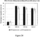
- SEMG2 (497-509) sequence fragment is the key epitope of SEMG2 binding to CD27, and has a relatively short sequence, so SEMG2 (497-509) was used as an immunogen to prepare antibodies, which is theoretically easier to obtain functional antibody molecules with the function of blocking the binding between SEMG2 and CD27 than using full-length SEMG2 to prepare antibodies. For direct comparison, the differences in effective concentration of producing antibody by the two methods were verified using ELISA in the examples.
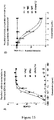
- Antibodies produced with SEMG2 (497-509) as immunogen and antibodies produced with full-length SEMG2 were added into the enzyme-linked immunosorbent assay (ELISA) reaction system at different concentrations (10 ⁇ -2, 10 ⁇ -1, 10 ⁇ 0, 10 ⁇ 1, 10 ⁇ 2, 10 ⁇ 3, 10 ⁇ 4 ng/mL), and the ELISA binding valueswere measured.
- the specific steps of enzyme-linked immunosorbent assay are as follows: (1) Dissolve SEMG2 protein antigen with 50 mM carbonate coating buffer (pH 9.6) to make the antigen concentration 10 ⁇ g/mL, and add into 96-well ELISA plate (purchased from Corning) at 100 ⁇ L/well, place at 4°C overnight.
- the experimental results are shown in FIGURE 10 .
- the antibody produced by SEMG2 (497-509) as an antigen reduced the binding between SEMG2 and CD27 detected by ELISA by 50% at a lower concentration, while the polyclonal antibody produced by full-length protein of SEMG2 as immunogen only exerted such an effect at higher concentration (the required dose is more than 300 times the former). That is, the blocking titer of the antibody produced by SEMG2 (497-509) was more than 300 times higher than that of the antibody produced by the full-length SEMG2 protein. This indicates that recognition of the key epitope SEMG2 (497-509) makes the development of blocking antibodies easier, and enables those skilled in the art to obtain antibodies that can block the binding between SEMG2 and CD27 more easily.
- Example 11 Preparation of mouse monoclonal antibody using SEMG2 (497-509) epitope peptide and SEMG2 full-length protein
- the SEMG2 (497-509) sequence was used to synthesize a polypeptide and coupled to a VLP carrier for immunization; HEK293 cells were used to express the full-length SEMG2 protein, and the purity was tested to reach 92%, and the binding activity of SEMG2 protein to CD27 was verified by ELISA.
- the protein and polypeptide antigens were used to immunize 10 mice respectively, and multiple immunizations were performed to enhance the effect: (1) The first immunization, 50 ⁇ g/mice of antigen, multiple subcutaneous injections together with Freund's complete adjuvant, with an interval of 3 weeks; (2) the second immunization; the same dosage and route as above, with incomplete Freund's adjuvant, and with an interval of 3 weeks; (3) the third immunization, the same dosage as above, without adjuvant, intraperitoneal injection with an interval of 3 weeks; (4) the booster immunization, the dose is 50 ⁇ g, intraperitoneal injection.
- mice with higher titers were selected for hybridoma fusion screening.
- the binding of the monoclonal antibodies to the target antigens was detected by ELISA, and the function of different monoclonal antibodies in blocking the binding between SEMG2 and CD27 was measured by ELISA.
- the monoclonal antibodies produced by hybridomas were screened by ELISA.
- 19 strains had blocking function (inhibiting the binding between SEMG2 and CD27) in the first batch of 27 stains of antibodies, as shown in FIGURE 13 .
- the monoclonal antibodies prepared with the full-length protein of SEMG2 as immunogen only 1 stain of antibody with blocking function was obtained in a total of 108 stains of antibodies after verification in batches, as shown in FIGURE 13 .
- VH amino acid sequences are as follows: MM02 MM05 MM07 MM08 MM13 MM14 MM15
- VL amino acid sequences are as follows: MM02 MM05 MM07 MM08 MM13 MM14/MM 15 Table 4.
- the ELISA plate was coated with SEMG2 protein, and the serially diluted murine monoclonal antibody was used as the primary antibody, and the anti-mouse secondary antibody was used to detect the binding abilities of the murine monoclonal antibodies to SEMG2.
- the results are shown in FIGURE 14A . It is shown that the murine monoclonal antibodies have fine affinities for SEMG2 protein.
- the mouse anti-SEMG2 monoclonal antibody MM05 was humanized to reduce immunogenicity when used in human patients.
- the sequences of the heavy and light chain variable regions (VH and VL) were compared to human antibody sequences in Protein Data Bank (PDB) and homology models were established.
- the CDRs in the heavy and light chains of mouse mAbs were transplanted to human frame regions that most likely maintain the proper structure required for antigen binding.
- Reverse mutations or other mutations from human residues to mouse residues were designed when necessary, for example: the amino acid at position 95 of the humanized light chain VL-V2 was mutated from K to Q, and the corresponding CDR3 sequence of the light chain was converted to QQSYSLPWT (SEQ ID NO:95) according to IMGT analysis. Humanized VH and VL regions were fused to the constant regions of heavy chain and ⁇ light chain of human IgGl, respectively. Transient transfections were performed in 293E cells using the construction vectors corresponding to mAb sequences, and the binding abilities of the purified mAbs to SEMG2 protein were analyzed using ELISA.
- Results are shown in absorbance, where higher absorbance indicates a higher level of interaction between the humanized antibody and SEMG2.
- the amino acid sequences of CDRs, light chain variable regions and heavy chain variable regions, light chains and heavy chains of the 8 humanized antibodies obtained in the present invention are shown in Table 4 and Table 5 below.
- FIGURE 14B shows the fitting curves of the binding of serially diluted humanized monoclonal antibody to SEMG2 protein, and the result show that the humanized antibody maintains the binding ability of murine monoclonal antibody to SEMG2 protein. Table 5.
- VH and VL amino acid sequences of MM05 humanized antibody VH amino acid sequences are as follows: VH_V1 VH_V2 VH_V3 VH_V4 VL amino acid sequences are as follows: VL_V1 VL_V2
- Example 13 Functional comparison between SEMG2 (497-509) epitope-specific antibodies and other epitope-specific antibodies in blocking SEMG2 and CD27 binding
- SEMG2(497-509) epitope-specific antibodies for example, MM02, MM05
- other epitope-specific antibodies for example, HPA042767
- the binding of the above antibodies to SEMG2 (497-509) epitope was confirmed by ELISA: MM02 and MM05 were able to bind to SEMG2(497-509), while HPA042767 could not bind to this epitope in a wide range of concentration, as shown in FIGURE 15A .
- SEMG2 exerts function of inhibiting activated PBMC from killing tumor cells. Since SEMG2 may play the above role by binding to CD27, and SEMG2 (497-509) epitope is a key site for CD27 binding, SEMG2 (497-509) epitope-specific antibody may neutralize the influence of SEMG2 on tumor cell killing by PBMC.
- PBMC Human peripheral blood mononuclear cells
- Example 15 Verifying the correlation between the expression level of SEMG2 and the promotive function of blocking antibodies in tumor cell killing by PBMC
- SEMG2 Since the expression of SEMG2 is a prerequisite for its inhibition of tumor-specific immunity, the expression of SEMG2 is also a potential condition for the suitability of SEMG2-blocking antibody administration.
- tumor cells with high SEMG2 expression will have a relative increase in the tumor cell killing by PBMC after neutralizing of SEMG2 activity; tumor cells that do not express SEMG2 may not rely on SEMG2 to play immune escape function, therefore the tumor cell killing by PBMC may not produce a significant change after neutralizing of SEMG2 activity.
- tumor cells with high SEMG2 expression (A375, LOVO) and SEMG2-negative tumor cells (DLD1, NCM460 and NCI-H1975) were selected.
- Different antibodies (irrelevant mouse IgG antibodies, MM02 or MM05 antibodies) were added during the PBMC killing experiments for the tumor cells. Results are shown in FIGURE 18 .
- MM02 and MM05 antibodies significantly increased the killing of SEMG2-positive tumor cells (A375, LOVO) by activated PBMC, but has no obvious impact to the killing of SEMG2-negative tumor cells (DLD1, NCM460 and NCI-H1975). Therefore, the above experimental results indicate that the positive expression of SEMG2 is a screening condition for administration of SEMG2 and CD27 blocking antibodies, i.e., a corresponding biomarker.
- Example 16 Accurate Definition of Associated Epitopes of Antibody for Blocking the Binding Between SEMG2 and CD27
- blocking antibodies i.e., antibodies that can inhibit the binding between SEMG2 and CD27
- non-blocking antibodies a corresponding ELISA analysis method was established. Specifically, SEMG2 full-length protein (1-582), SEMG2(354-403) fragment, SEMG2(442-453) fragment, SEMG2(497-509) fragment and SEMG2(563-574) fragment were immobilized on the ELISA plate, and the same concentration of antibodies (MM02, MM05, MM07, MM08, MM13, MM14, HPA042767 and HPA042835) were added. Anti-mouse or anti-rabbit secondary antibodies were then used to detect the corresponding bound antibodies.
- MM02, MM05, MM07, MM08, MM13, and MM14 all bound to SEMG2(497-509) epitope, HPA042767 bound to SEMG2 (354-403) epitope, and HPA042835 bound to SEMG2 (497-509) epitope, while none of the antibodies bound to SEMG2 (442-453) control fragment.
- the results are shown in FIGURE 20 .
- the HPA042767 and HPA042835 antibodies did not bind to the sequences, indicating the specificity of the experiment and the different epitope classes of the two types of antibodies. Meanwhile, the different amino acids in the SEMG2(497-509) sequence had different degrees of influence on the binding of similar blocking antibodies (MM02, MM05, MM07, MM08, MM13, MM14) after substitution by glycine.
- the substitution of amino acids at positions 507 and 509 did not significantly affect the binding of MM02 and similar antibodies; the substitution of amino acids at positions 501 and 506 significantly affected the binding of MM02 and similar antibodies (a decrease of more than 70%); amino acids at other sites affected the binding of MM02 and similar antibodies to a certain extent after substitution by glycine.
- the results precisely define the epitope amino acids associated with MM02 and similar antibodies (i.e., antibodies that block the binding between SEMG2 and CD27), and the contribution of each amino acid to the binding.
- the key amino acids of SEMG2 participating in binding to blocking antibodies are highly consistent with those participating in binding to CD27, which indicates that MM02 and its similar antibodies compete with CD27 for binding to SEMG2, which verifies the molecular mechanism of antibody function.
- Example 17 Preparation and screening of fully human antibodies using SEMG2(497-509) epitope to block the binding between SEMG2 and CD27 and to promote the tumor cell killing by PBMC
- results of the examples showed the importance of SEMG2(497-509) epitope in the preparation of blocking antibodies, and this epitope was applied to the screening of fully human antibodies.
- the preparation of polypeptide antigens and the screening of human natural antibody library were firstly performed.
- the SEMG2(497-509) polypeptide was synthesized and coupled to BSA and KLH, respectively, and screened in a fully human phage display antibody library.
- ELISA was used to select clones that bind to antigenic epitopes for preliminary screening. Different unique sequences were obtained after sequencing single colonies, sorted according to affinity sorting, and full-length antibodies were constructed from antigen-binding fragments (Fab) with relatively high affinity.
- the binding ability and blocking function tests were performed after purification, that is, the effect of the antibody on binding between SEMG2 and CD27 was determined by the ELISA experiment.
- H88-93 competes with MM02 and MM05 for binding to SEMG2, as shown in FIGURE 22 . It shows that fully human antibodies and murine monoclonal antibodies are the same type of antibodies binding to SEMG2, and because MM02, MM05 and H88-93 all bind to the short peptide SEMG2 (497-509), this type of antibodies can be defined as a class of SEMG2 (497-509) binding antibodies.
- FIGURE 24 To verify the effect of the human antibodies on the function of activated PBMC on tumor cell killing, A375 and LOVO cells were co-cultured with activated PBMC, and H88-93, H88-96, or H88-67 antibody was added at the same time, and the apoptosis ratio of tumor cells were detected. The result is shown in FIGURE 24 , which indicates that the three fully human antibodies against SEMG2 (497-509) epitope can significantly promote the killing of SEMG2-expressing tumor cells by PBMC cells.
- Example 17 Binding kinetic determination of the monoclonal antibodies of the present invention to antigens by bio-optical interferometry
- the equilibrium dissociation constant (KD) of the antibody of the present invention binding to human SEMG2 was determined by biolayer interferometry (ForteBio Bltz or Gator instrument).
- ForteBio affinity assay was performed according to the existing method, that is, half an hour before start, an appropriate amount of AMQ (Pall, 1506091) (for sample detection) or AHQ (Pall, 1502051) (for positive control detection) sensors were taken and soaked in SD buffer (PBS 1 ⁇ , BSA 0.1%, Tween-20 0.05%). 100 ⁇ l of SD buffer, antibody and SEMG2 were added to a 96-well black polystyrene half area microplate, respectively. Select the sensor location based on the sample location layout.
- KD values were analyzed using molecular interaction analysis software.
- the affinity constants of murine monoclonal antibodies and human antibodies H88-67, H88-93 and H88-96 are shown in Table 8, and the affinity and dissociation curves of SEMG2 and corresponding proteins are shown in FIGURE 25 .
- Table 8 the affinity constants of murine monoclonal antibodies and human antibodies H88-67, H88-93 and H88-96 are shown in Table 8, and the affinity and dissociation curves of SEMG2 and corresponding proteins are shown in FIGURE 25 . Table 8.
- Affinity constants (equilibrium dissociation constants) for the detection of antigen-antibody binding by biolayer optical interferometry Antibody KD (M) MM02 1.33 ⁇ 10 -9 MM05 5.28 ⁇ 10 -9 MM07 1.82 ⁇ 10 -9 MM08 2.34 ⁇ 10 -9 MM13 6.93 ⁇ 10 -10 MM14 1.44 ⁇ 10 -9 H88-67 2.84 ⁇ 10 -8 H88-93 4.60 ⁇ 10 -9 H88-96 1.40 ⁇ 10 -8
- the plasmids were obtained by gene synthesis, and then made single-point and double-point saturation mutation. In vitro ligation method was then performed to recombine antibody genes. Finally, the Fab gene sequence of recombinant antibody was inserted into the vector, and then transformed to obtain 4 phage affinity-matured antibody libraries with titer higher than 10 8 CFU. The antibody mutant library was screened by the immunotube gradient screening assay, and the mutants with finely improved affinity compared to the wild type were obtained.
- the full-length affinity matured human antibody was then constructed according to the detected Fab sequence or the recombination of VH and VL sequences in the Fab sequence.
- the VH and VL sequences derived from H88-67 and the CDR regions of the VH sequence of H88-96 after affinity maturation are shown in Table 9, and CDR regions of the light and heavy chain of the antibody are shown in Table 9. Table 9.
- VH and VL sequences corresponding to affinity matured fully human antibodies Antibody Number Heavy chain variable region VH Light chain variable region VL 67-3-67-3 67-3-67-4 67-3-67-5 67-3-67-6 67-9-67-3 67-6-67-6 96-10R-10 96-10V-10
- anti-human SEMG2 monoclonal antibodies with improved affinity such as 67-3-67-3, 67-3-67-4, 67-3-67-5 and 67-3-67-6 which consist of the combination of the affinity-matured heavy chain numbered 67-3 and the affinity-matured light chain sequence numbered 67-3, 67-4, 67-5 and 67-6
- antibody 67-9-67-3 consists of the combination of the heavy chain numbered 67-9 and the light chain numbered 67-3
- antibody 67-6-67-6 consists of the combination of the light chain and heavy chain numbered 67-6
- antibodies 96-10R-10 and 96-10V- 10 reconstituted by the heavy chains numbered 96-10R and 96-10V and the light chain of H88-96L.
- the recombinant monoclonal antibodies consist of these light and heavy chains have an affinity more than 10-fold higher for SEMG2 and BSA-S2 (497-509) (i.e., BSA-SP7) than that of the parent antibodies (see FIGURE 21B-D).
- mice 30 male NPSG mouse models aged 6-8 week were weighed. A375 cells (with confirmed endogenous expression of SEMG2) were cultured in vitro to obtain 1.8 ⁇ 10 8 cells. After 30 mice were inoculated with PBMC, A375 tumor cells were inoculated on the 3rd day. After that, the proportion of hCD45+ cells in mouse blood and the body weight were measured once a week. After inoculation, tumor volume was measured once a week, and the proportion of hCD45+ cells in mouse blood was measured when the average tumor volume reached about 40-80mm 3 . Mice were grouped randomly based on tumor volume and the proportion of hCD45+ cells in mouse blood, and the administration was started immediately. The date began the administration was considered day 0.
- Dosing regimen SEMG2 antibody (MM05 clone) was injected intraperitoneally at 5 mg/kg three times a week. After the start of administration, the tumor growth status of the mice was observed every week. After the tumor growth, the body weight and tumor volume were measured 3 times a week, and the relative count of hCD45+ cells in mouse blood was monitored by flow cytometry 3 times a week. When the tumor volume reached the end point, blood was collected and the same indexed were detected, and the experiment was ended. The observation of mice includes: daily observation, observation of animal morbidity and death every working day after inoculation. Measurement of tumor volume: after inoculation and before grouping, when tumors were visible, the tumor volume of experimental animals was measured once a week.
- the tumor volume of animals in the experiment was measured twice a week.
- the tumor volume was measured by a bidirectional measurement method.
- the experimental results are shown in Figure 26 .
- SEMG2 antibody significantly inhibited the growth of tumor in mice. This result indicates that SEMG2 is an effective anti-tumor target.
- the obtained gene knockout heterozygous mice (gene+/-) were divided into two parts: a part of heterozygous mice was mated with wild-type mice for expansion of more heterozygous mice; a part of heterozygous mice self-bred to obtain gene knockout homozygous mice (gene-/-) for gene knockout effect verification and subsequent phenotype analysis.
- Phenotype analysis Anticoagulated whole blood was taken from mice for flow cytometry, and the proportion of CD8+, CD4+, CD3+, CD27+ positive cells in blood was analyzed. After the mice rested for 2 days, the anticoagulated whole blood was collected from the inner canthus, and the molecule department would perform the blood routine test. After the mice rested for 3 days, the mice were weighed and anesthetized, and the mouse gross bodies were imaged; the eyeballs of the mice were removed, the blood was collected, and the serum was separated. The molecule department would measure the serum biochemical parameters.
- mice were euthanized for material collection: brain: the whole brain was removed and divided by the sagittal plane, and the left side was fixed, and the right side was quick-frozen; liver: the whole liver was removed and divided in two, the left lobe was fixed, and the rest were quick-frozen; spleen: the whole spleen was removed and divided in two, half fixed, half quick-frozen; kidney: the left kidney was removed for fixation, the right kidney was removed and quick-frozen; stomach: the whole stomach was removed and divided sagittal, the greater curvature was fixed, and the lesser curvature was quick-frozen; large intestine: the intact large intestine was removed for Swiss roll fixation; small intestine: the whole small intestine was removed and divided into three sections (duodenum, ileum, jejunum) for Swiss roll fixation; lung: the left lung was removed for fixation, and the right lung for removed for quick freezing; heart: the entire heart was removed
- FIGURE 27 The phenotype analysis results of wild-type (WT) and homozygous knockout mice (KO) are shown in FIGURE 27 .
- the results show that no offspring was born after mating the Svs3a homozygous knockout mice, while there was no effect on reproductive function for the heterozygous knockout situation. No abnormality was found in other analyses. Therefore, complete loss or blockade of Svs3a function may affect fertility without significant toxic effects on other systems. This suggests that the possible toxicity or side effects of SEMG2 target blockade are limited and have high safety.
Abstract
Description
- The invention relates to the field of biomedicine, specifically to a SEMG2 antigenic epitope peptide and the use thereof.
- The CD27 molecule belongs to the tumor necrosis factor receptor (TNFR) superfamily and is a type I membrane protein with a molecular weight of about 55 kDa, and exists as a dimer of two monomers linked by a disulfide bond. CD27 is mainly expressed in lymphocytes. Recent studies based on CD27 knockout mice have shown that activation of the CD27 signaling pathway can increase the infiltration of suppressor T cells (Treg) in solid tumors and reduce anti-tumor immunity (Claus C, Riether C, Schürch C, Matter MS, Hilmenyuk T, Ochsenbein AF. Cancer Res. 2012 Jul 15;72(14):3664-76). Consistently, the study also found that Treg cells in skin tissue fail to perform normal immune regulation functions after losing CD27 expression ( Remedios KA, Zirak B, Sandoval PM, Lowe MM, Boda D, Henley E et al., Sci Immunol. 2018. Dec 21;3(30)pii:eaau2042). Furthermore, activation of CD27 increases Treg numbers and reduces atherosclerosis in hyperlipidemic mice (Winkels H, Meiler S, Lievens D, Engel D, Spitz C, Bürger C, et al., Eur Heart J. 2017; 38(48):3590-3599). The recent studies consistently demonstrate that CD27 plays an important role in the functional activation of specific Treg cells (including tumor-infiltrating Treg), and therefore avoiding the activation of CD27 expressed by tumor-infiltrating Treg cells is a potential cancer treatment strategy.
- Binding to ligands activates the downstream signal transduction of CD27, and the currently known CD27 ligand molecule is CD70. CD70 is a 193 amino acid polypeptide with a hydrophilic N-terminal domain of 20 amino acids and a C-terminal domain containing 2 potential N-linked glycosylation sites, belonging to the TNF family (Goodwin, R . G . et al. (1993) Cell 73:447-56 ; Bowman et al. (1994) Immunol 152:1756 - 61). These properties suggest that CD70 is a type II transmembrane protein with an extracellular C-terminal portion. CD70 is transiently present on activated T and B lymphocytes and dendritic cells (Hintzen et al., (1994) J. Immunol. 152:1762-1773 ; Oshima et al., (1998) Int. Immunol. 10:517-26 ; Tesselaar et al., (2003) J. Immunol. 170:33-40). In addition to normal cells, CD70 expression has been reported in different types of cancer, including renal cell carcinoma, metastatic breast cancer, brain tumor, leukemia, lymphoma, and nasopharyngeal carcinoma ( Junker et al., J. Urol. 2005; 173: 2150-3 ; Sloan et al., Am J Pathol. 2004; 164:315-23 ; Held-Feindt and Mentlein et al., Int J Cancer 2002; 98:352-6 ). Currently, blocking the binding between CD70 and CD27 is a strategy being investigated for tumor immunotherapy.
- Previous studies have not suggested that CD27 has other ligands than CD70. However, novel ligands of immune checkpoint pathway receptors (especially novel ligands expressed by tumor cells with relatively high specificity) are of great significance for the development of more effective anti-tumor treatments. The present invention aims to develop new anti-tumor treatments and drugs.
- In one aspect, the invention discloses a compound agonizing or antagonizing an interaction between SEMG2 and CD27. Wherein, the interaction between SEMG2 and CD27 is located on the amino acid site at
positions 497, 498, 499, 500, 501, 502, 503, 504, 505, 506, and 508 of SEMG2, and the amino acid sequence of the SEMG2 protein is shown in SEQ ID NO:1. Wherein, the compound is a small molecule inhibitor, polypeptide, antibody, or antigen-binding fragment. - In one embodiment, the invention discloses a polypeptide, the polypeptide comprises an amino acid sequence of SEQ ID NO:2 (QIEKLVEGKS), SEQ ID NO:86 (QIEKLVEGKS(x)I(x)), SEQ ID NO:87 (QIEKLVEGKS(x)I), or SEQ ID NO:88 (QIEKLVEGKS(x)); preferably the polypeptide comprises an amino acid sequence of SEQ ID NO:3 (QIEKLVEGKSQIQ), SEQ ID NO:4 (QIEKLVEGKSQ), or SEQ ID NO:5 (QIEKLVEGKSQI), or an amino acid sequence at least 90%identity to an amino acid sequence as provided in SEQ ID NOs: 2-5. Wherein, the polypeptide agonizes the interaction between SEMG2 and CD27. Wherein, the amino acid site of the interaction between SEMG2 and CD27 is located at
positions 497, 498, 499, 500, 501, 502, 503, 504, 505, 506 and 508 of SEMG2, and the amino acid sequence of the SEMG2 protein is shown in SEQ ID NO:1. - In one embodiment, the invention discloses an antibody specifically binding to native or mutant SEMG2 protein, the antibody binds to an antigenic epitope peptide derived from SEMG2 protein, and the antigenic epitope peptide comprises an amino acid sequence of SEQ ID NO:2 (QIEKLVEGKS), SEQ ID NO:3 (QIEKLVEGKSQIQ), SEQ ID NO:4 (QIEKLVEGKSQ), or SEQ ID NO:5 (QIEKLVEGKSQI). Wherein, the antibody antagonizes the interaction between SEMG2 and CD27.
- In one embodiment, the invention discloses an antibody specifically binding to native or mutant SEMG2 protein, the antibody recognizes at least one amino acid residue at
positions 497, 498, 499, 500, 501, 502, 503, 504, 505, 506 and 508 of the native SEMG2 protein or recognizes an amino acid residue in the corresponding position of the mutant SEMG2 protein, the amino acid sequence of the native SEMG2 protein is shown in SEQ ID NO:1. Wherein, the antibody antagonizes the interaction between SEMG2 and CD27. - In one embodiment, the invention discloses an antibody specifically binding native or mutant SEMG2 protein, wherein the antibody comprises a heavy chain variable region and a light chain variable region, the heavy chain variable region comprises HCDR1, HCDR2 and HCDR3 defined by IMGT; and the light chain variable region comprises LCDR1, LCDR2 and LCDR3 defined by IMGT,
- the HCDR1 consists of or comprises an amino acid sequence selected from the group consisting of SEQ ID NOs:6-11, SEQ ID NOs:60-61 and SEQ ID NO:76;
- the HCDR2 consists of or comprises an amino acid sequence selected from the group consisting of SEQ ID NOs:12-16 and SEQ ID NOs:62-64;
- the HCDR3 consists of or comprises an amino acid sequence selected from the group consisting of SEQ ID NOs:17-20, SEQ ID NOs:65-67 and SEQ ID NOs:77-81;
- the LCDR1 consists of or comprises an amino acid sequence selected from the group consisting of SEQ ID NOs:21-25, SEQ ID NOs:68-70 and SEQ ID NO:82;
- the LCDR2 consists of or comprises an amino acid sequence selected from the group consisting of SEQ ID NOs:26-29, SEQ ID NOs:71-72, SEQ ID NOs:83-84 and SEQ ID NO:28;
- the LCDR3 consists of or comprises an amino acid sequence selected from the group consisting of SEQ ID NOs:30-34, SEQ ID NOs:73-75, SEQ ID NO:85 and SEQ ID NO:99.
- In one specific embodiment, the CDR sequence of the antibody is selected from any one of the combinations in (a)-(k):
- (a) the HCDR1 comprises the amino acid sequence of SEQ ID NO:6; the HCDR2 comprises the amino acid sequence of SEQ ID NO:12; the HCDR3 comprises the amino acid sequence of SEQ ID NO:17; the LCDR1 comprises the amino acid sequence of SEQ ID NO:21; the LCDR2 comprises the amino acid sequence of SEQ ID NO:26; the LCDR3 comprises the amino acid sequence of SEQ ID NO:30;
- (b) the HCDR1 comprises the amino acid sequence of SEQ ID NO:7; the HCDR2 comprises the amino acid sequence of SEQ ID NO:13; the HCDR3 comprises the amino acid sequence of SEQ ID NO:18; the LCDR1 comprises the amino acid sequence of SEQ ID NO:22; the amino acid sequence of LCDR2 comprises the amino acid sequence of SEQ ID NO:27; the LCDR3 comprises the amino acid sequence of SEQ ID NO:31 or SEQ ID NO:99;
- (c) the HCDR1 comprises the amino acid sequence of SEQ ID NO:6; the HCDR2 comprises the amino acid sequence of SEQ ID NO:16; the HCDR3 comprises the amino acid sequence of SEQ ID NO:17; the LCDR1 comprises the amino acid sequence of SEQ ID NO:21; the LCDR2 comprises the amino acid sequence of SEQ ID NO:26; the LCDR3 comprises the amino acid sequence of SEQ ID NO:30;
- (d) the HCDR1 comprises the amino acid sequence of SEQ ID NO:8; the HCDR2 comprises the amino acid sequence of SEQ ID NO:13; the HCDR3 comprises the amino acid sequence of SEQ ID NO:18; the LCDR1 comprises the amino acid sequence of SEQ ID NO:23; the LCDR2 comprises the amino acid sequence of SEQ ID NO:27; the LCDR3 comprises the amino acid sequence of SEQ ID NO:32;
- (e) the HCDR1 comprises the amino acid sequence of SEQ ID NO:9; the HCDR2 comprises the amino acid sequence of SEQ ID NO:14; the HCDR3 comprises the amino acid sequence of SEQ ID NO:19; the LCDR1 comprises the amino acid sequence of SEQ ID NO:24; the LCDR2 comprises the amino acid sequence of SEQ ID NO:28; the LCDR3 comprises the amino acid sequence of SEQ ID NO:33;
- (f) the HCDR1 comprises the amino acid sequence of SEQ ID NO:10; the HCDR2 comprises the amino acid sequence of SEQ ID NO:15; the HCDR3 comprises the amino acid sequence of SEQ ID NO:20; the LCDR1 comprises the amino acid sequence of SEQ ID NO:25; the LCDR2 comprises the amino acid sequence of SEQ ID NO:29; the LCDR3 comprises the amino acid sequence of SEQ ID NO:34;
- (g) the HCDR1 comprises the amino acid sequence of SEQ ID NO:11; the HCDR2 comprises the amino acid sequence of SEQ ID NO:15; the HCDR3 comprises the amino acid sequence of SEQ ID NO:20; the LCDR1 comprises the amino acid sequence of SEQ ID NO:25; the LCDR2 comprises the amino acid sequence of SEQ ID NO:29; the LCDR3 comprises the amino acid sequence of SEQ ID NO:34;
- (h) the HCDR1 comprises the amino acid sequence of SEQ ID NO:60; the HCDR2 comprises the amino acid sequence of SEQ ID NO:62; the HCDR3 comprises the amino acid sequence of SEQ ID NO:65; the LCDR1 comprises the amino acid sequence of SEQ ID NO:68; the LCDR2 comprises the amino acid sequence of SEQ ID NO:71; the LCDR3 comprises the amino acid sequence of SEQ ID NO:73;
- (i) the HCDR1 comprises the amino acid sequence of SEQ ID NO:61; the HCDR2 comprises the amino acid sequence of SEQ ID NO:63; the HCDR3 comprises the amino acid sequence of SEQ ID NO:66; the LCDR1 comprises the amino acid sequence of SEQ ID NO:69; the LCDR2 comprises the amino acid sequence of SEQ ID NO:72; the LCDR3 comprises the amino acid sequence of SEQ ID NO:74;
- (j) the HCDR1 comprises the amino acid sequence of SEQ ID NO:60; the HCDR2 comprises the amino acid sequence of SEQ ID NO:64; the HCDR3 comprises the amino acid sequence of SEQ ID NO:67; the LCDR1 comprises the amino acid sequence of SEQ ID NO:70; the LCDR2 comprises the amino acid sequence of SEQ ID NO:28; the LCDR3 comprises the amino acid sequence of SEQ ID NO:75;
- (k) the HCDR1 comprises the amino acid sequence of SEQ ID NO:60 or 76; the HCDR2 comprises the amino acid sequence of SEQ ID NO:64 or 62; the HCDR3 comprises the amino acid sequence of SEQ ID NO:77, 78 or 79; and/or
- In one embodiment, the invention discloses an antibody binding to native or mutant SEMG2 protein specifically, wherein the antibody comprises a heavy chain variable region and a light chain variable region,
- the heavy chain variable region comprises an amino acid sequence selected from the group consisting of SEQ ID NOs:35-41, 48-51, 54-56 and 96-100, or an amino acid sequence at least 70%, 80%, 90%, 95% or 99% identity to any sequences of SEQ ID NOs:35-41, 48-51, 54-56 and 96-100;
- the light chain variable region comprises the amino acid sequence selected from the group consisting of SEQ ID NOs:42-47, 52-53, 57-69 and 101-103, or an amino acid sequence at least 70%, 80%, 90%, 95% or 99% identity to any sequences of SEQ ID NOs:42-47, 52-53, 57-69 and 101-103.
- In one specific embodiment, the heavy chain variable region and the light chain variable region are selected from any one of the combinations in (a)-(o):
- (a) the heavy chain variable region comprises SEQ ID NO:35 or an amino acid sequence at least 70%, 80%, 90%, 95% or 99% identity to SEQ ID NO:35; the light chain variable region comprises SEQ ID NO:42 or an amino acid sequence at least 70%, 80%, 90%, 95% or 99% identity to SEQ ID NO:42;
- (b) the heavy chain variable region comprises SEQ ID NO:36 or an amino acid sequence at least 70%, 80%, 90%, 95% or 99% identity to SEQ ID NO:36; the light chain variable region comprises SEQ ID NO:43 or an amino acid sequence at least 70%, 80%, 90%, 95% or 99% identity to SEQ ID NO:43;
- (c) the heavy chain variable region comprises SEQ ID NO:37 or an amino acid sequence at least 70%, 80%, 90%, 95% or 99% identity to SEQ ID NO:37; the light chain variable region comprises SEQ ID NO:44 or an amino acid sequence at least 70%, 80%, 90%, 95% or 99% identity to SEQ ID NO:44;
- (d) the heavy chain variable region comprises SEQ ID NO:38 or an amino acid sequence at least 70%, 80%, 90%, 95% or 99% identity to SEQ ID NO:38; the light chain variable region comprises SEQ ID NO:45 or an amino acid sequence at least 70%, 80%, 90%, 95% or 99% identity to SEQ ID NO:45;
- (e) the heavy chain variable region comprises SEQ ID NO:39 or an amino acid sequence at least 70%, 80%, 90%, 95% or 99% identity to SEQ ID NO:39; the light chain variable region comprises SEQ ID NO:46 or an amino acid sequence at least 70%, 80%, 90%, 95% or 99% identity to SEQ ID NO:46;
- (f) the heavy chain variable region comprises SEQ ID NO:40 or an amino acid sequence at least 70%, 80%, 90%, 95% or 99% identity to SEQ ID NO:40; the light chain variable region comprises SEQ ID NO:47 or an amino acid sequence at least 70%, 80%, 90%, 95% or 99% identity to SEQ ID NO:47;
- (g) the heavy chain variable region comprises SEQ ID NO:41 or an amino acid sequence at least 70%, 80%, 90%, 95% or 99% identity to SEQ ID NO:41; the light chain variable region comprises SEQ ID NO:47 or an amino acid sequence at least 70%, 80%, 90%, 95% or 99% identity to SEQ ID NO:47;
- (h) the heavy chain variable region comprises SEQ ID NO:48, 49, 50, 51 or an amino acid sequence at least 70%, 80%, 90%, 95% or 99% identity to SEQ ID NO:48, 49, 50 or 51; the light chain variable region comprises SEQ ID NO:52, 53 or an amino acid sequence at least 70%, 80%, 90%, 95% or 99% identity to SEQ ID NO:52 or 53;
- (i) the heavy chain variable region comprises SEQ ID NO:54 or an amino acid sequence at least 70%, 80%, 90%, 95% or 99%identity to SEQ ID NO:54; the light chain variable region comprises SEQ ID NO:57 or an amino acid sequence at least 70%, 80%, 90%, 95% or 99% identity to SEQ ID NO:57;
- (j) the heavy chain variable region comprises SEQ ID NO:55 or an amino acid sequence at least 70%, 80%, 90%, 95% or 99% identity to SEQ ID NO:55; the light chain variable region comprises SEQ ID NO:58 or an amino acid sequence at least 70%, 80%, 90%, 95% or 99% identity to SEQ ID NO:58;
- (k) the heavy chain variable region comprises SEQ ID NO:56 or an amino acid sequence at least 70%, 80%, 90%, 95% or 99% identity to SEQ ID NO:56; the light chain variable region comprises SEQ ID NO:59 or an amino acid sequence at least 70%, 80%, 90%, 95% or 99% identity to SEQ ID NO:59;
- (l) the heavy chain variable region comprises SEQ ID NO:96 or an amino acid sequence at least 70%, 80%, 90%, 95% or 99% identity to SEQ ID NO:96; the light chain variable region comprises SEQ ID NO:59, 101, 102, 103 or an amino acid sequence at least 70%, 80%, 90%, 95% or 99% identity to SEQ ID NO:59, 101, 102 or 103;
- (m) the heavy chain variable region comprises SEQ ID NO:97 or an amino acid sequence at least 70%, 80%, 90%, 95% or 99% identity to SEQ ID NO:97; the light chain variable region comprises SEQ ID NO:59 or an amino acid sequence at least 70%, 80%, 90%, 95% or 99% identity to SEQ ID NO:59;
- (n) the heavy chain variable region comprises SEQ ID NO:98 or an amino acid sequence at least 70%, 80%, 90%, 95% or 99% identity to SEQ ID NO:98; the light chain variable region comprises SEQ ID NO:103 or an amino acid sequence at least 70%, 80%, 90%, 95% or 99% identity to SEQ ID NO:103;
- (o) the heavy chain variable region comprises SEQ ID NO:99, 100 or an amino acid sequence at least 70%, 80%, 90%, 95% or 99% identity to SEQ ID NO:99 or 100; the light chain variable region comprises SEQ ID NO:57 or an amino acid sequence at least 70%, 80%, 90%, 95% or 99% identity to SEQ ID NO:57.
- The antibody of the invention further comprises a coupling moiety linked to the polypeptide, the coupling moiety is selected from the group consisting one or more of radionuclides, drugs, toxins, cytokines, enzymes, fluorescein, carrier proteins, lipids, and biotin, wherein the polypeptide or antibody is selectively linked to the coupling moiety by a linker, preferably the linker is a peptide or polypeptide.
- Wherein the antibody is selected from monoclonal antibodies, polyclonal antibodies, antisera, chimeric antibodies, humanized antibodies, and human antibodies.
- Wherein the antibody is selected from multi-specific antibodies, single-chain variable fragments (scFvs), single-chain antibodies, anti-idiotype (anti-Id) antibodies, diabodies, minibodies, nanobodies, single domain antibodies, Fab fragments, F(ab') Fragments, disulfide-linked bispecific Fv (sdFv) and intracellular antibodies.
- In another aspect, the invention discloses an antigenic epitope peptide, wherein the antigenic epitope peptide is derived from SEMG2 protein, and the amino acid of the antigenic epitope peptide comprises an amino acid sequence selected from the group consisting of SEQ ID NO:2 (QIEKLVEGKS), SEQ ID NO:3 (QIEKLVEGKSQIQ), SEQ ID NO:4 (QIEKLVEGKSQ), and SEQ ID NO:5 (QIEKLVEGKSQI).
- The invention discloses a protein, wherein the protein comprises an amino acid sequence as the amino acid sequences shown in SEQ ID NO:2 (QIEKLVEGKS), SEQ ID NO:86 (QIEKLVEGKS(x)I(x)), SEQ ID NO:87 (QIEKLVEGKS(x))I), or SEQ ID NO:88 (QIEKLVEGKS(x)), preferably the polypeptide comprises an amino acid sequence of SEQ ID NO:3 (QIEKLVEGKSQIQ), SEQ ID NO:4 (QIEKLVEGKSQ), or SEQ ID NO:5 (QIEKLVEGKSQI) or an amino acid sequence at least 90% identity to any one of SEQ ID NOs:2-5, more preferably SEQ ID NOs:89-94 and SEQ ID NO:3 (corresponding to P1-P6, and P7 respectively); and a tag sequence which can selectively be linked at the N-terminus or C-terminus. Those skilled in the art should understand that the addition of a protein tag will not affect the prepared antibody's participation in binding between SEMG2 and CD27, the protein tag includes, but is not limited to, C-Myc, His, GST (glutathione S-transferase), HA, MBP (maltose-binding protein), Flag, SUMO, eGFP/eCFP/eYFP/mCherry, etc.
- In one specific embodiment, the polypeptide has amino acid sequence of SEQ ID NO:3 (P7: QIEKLVEGKSQIQ) or SEQ ID NO:93 (P5).
- The invention also discloses a method of preparing an antibody or an antigen-binding fragment thereof, wherein the protein is used as an immunogen to inject a subject such as a mouse or screening natural library to prepare the antibody, and the amino acid sequence of the antibody comprises an amino acid sequence of SEQ ID NO:2 (QIEKLVEGKS), SEQ ID NO: 86 (QIEKLVEGKS(x)I(x)), SEQ ID NO:87 (QIEKLVEGKS(x)I), or SEQ ID NO:88(QIEKLVEGKS(x)), preferably the polypeptide comprises an amino acid sequence of SEQ ID NO:3 (QIEKLVEGKSQIQ), SEQ ID NO:4 (QIEKLVEGKSQ), or SEQ ID NO:5 (QIEKLVEGKSQI) or an amino acid sequence at least 90% identity to any one of SEQ ID NOs:2-5, more preferably SEQ ID NO:93 (P5) or SEQ ID NO:3 (P7).
- In a preferred embodiment, a method of obtaining isolated antibodies is disclosed, which uses a key epitope polypeptide of the binding between SEMG2 and CD27 as immunogen and screens with murine hybridoma and phage display in human and camel natural library.
- In another aspect, the invention discloses an isolated polynucleotide encoding the compound, antigenic peptide, or protein.
- The invention discloses a recombinant vector comprising the polynucleotide and optional regulatory sequences; preferably, the recombinant vector is a cloning vector or an expression vector.
- Wherein, the regulatory sequence is selected from a leading sequence, a polyadenylation sequence, a polypeptide sequence, a promoter, a signal sequence, a transcription terminator, or any combination thereof.
- The invention discloses a host cell comprising the recombinant vector.
- Wherein, the host cell is a prokaryotic cell or a eukaryotic cell.
- The invention discloses a pharmaceutical composition comprising the compound, the antigenic peptide, the protein, the polynucleotide, the recombinant vector, and one or more types of the host cells as previously described.
- Wherein, the composition further comprises a pharmaceutically acceptable carrier or adjuvant.
- The invention also discloses the use of the compounds, the antigenic peptide, the protein, the polynucleotide, the recombinant vector, or the host cell in preparation of products for agonizing or antagonizing the interaction between SEMG2 and CD27, preferably SEMG2 is expressed in tumor cells and CD27 is expressed in immune cells.
- The invention also discloses the use of the compound, the antigenic peptide, the protein, the polynucleotide, the recombinant vector, or the host cell in preparation of a drug for preventing or treating tumors or a drug for modulating an immune response elicited against tumors.
- In one specific embodiment, wherein the tumor is selected from one or more of colorectal cancer, lung cancer, melanoma, lymphoma, liver cancer, head and neck cancer, stomach cancer, kidney cancer, bladder cancer, prostate cancer, testicular cancer, endometrial cancer, breast cancer and ovarian cancer.
- The invention also discloses a method of screening drugs or reagents preventing or treating tumors, comprising obtaining candidate drugs or reagents by screening inhibitors or antibodies which inhibit the interaction between SEMG2 and CD27.
- The invention also discloses a method of preventing or treating tumors, comprising:
contacting immune cells such as lymphocytes (T lymphocytes) or tumor cells of the subject with an effective dose of any one of the compound; wherein the expression of SEMG2 in tumor cells can be selectively detected before contacting immune cells such as lymphocytes and/or tumor cells of the subject with an effective dose of the compound. - Wherein the subject has received or is receiving or will receive additional anti-cancer therapy.
- Wherein the additional anti-cancer therapy comprises surgery, radiotherapy, chemotherapy, immunotherapy, or hormone therapy.
- The invention also discloses a kit comprising one or more of the compounds, the antigenic peptides, the proteins, the polynucleotides, the recombinant vectors, and one or more types of the host cells, and the above components are accommodated in a suitable container.
- The invention also discloses a method of detecting the presence or absence of SEMG2 in a biological sample in vitro, comprising: contacting the biological sample with the compound.
- A method of inhibiting tumor cell growth of tumor cells, comprising the following steps: A) analyzing the expression of SEMG2 in tumor cells; B) contacting the tumor cells with an antibody recognizing SEMG2, the binding of the antibody to SEMG2 is KD <2×10⁻⁸; C) contacting T lymphocytes, with the antibody and tumor cells. Wherein the KD<2×10⁻⁸ , <1×10⁻⁸, <9×10⁻⁹ , <8×10⁻⁹ , <7×10⁻⁹, <6×10⁻⁹, <5×10⁻⁹, <4×10⁻⁹, <3×10⁻⁹, <2×10⁻⁹, <1×10⁻⁹, <1×10⁻¹⁰.
-
-
FIGURE 1 depicts the result of co-immunoprecipitation assay, divided into upper panel and lower panel. The upper panel demonstrates the existence of physical interaction between human CD27 and SEMG2 (Flag). The lower panel demonstrates the existence of physical interaction between mouse CD27 and SEMG2 (Flag). -
FIGURE 2 depicts the results of immunofluorescent staining and ELISA.Figure 2(A) demonstrates significant colocalization between CD27 and SEMG2 after overexpression in tumor cells.Figure 2(B) shows the result of ELISA, demonstrating the effect of CD27 concentration-dependent on binding SEMG2 in microplate, while CD27 does not bind to the negative control protein and there is no concentration effect. -
FIGURE 3 depicts the result of co-immunoprecipitation assay for examining whether SEMG2 fragment (i.e., P1 to P6) binds to CD27. Wherein the P5 fragment has been detected with significant binding to CD27. -
FIGURE 4 depicts the result of co-immunoprecipitation assay for examining whether SEMG2 fragment (i.e., P4, P5, P6, P7) binds to CD27. Wherein the P7 sequence is derived from a part of P5, which is "QIEKLVEGKSQIQ". The result includes left panel and right panel. The left panel shows the binding between the SEMG2 fragment and human CD27, and the right panel shows the binding between the SEMG2 fragment and mouse CD27. The results show that both human and mouse CD27 could bind to P5 and P7 fragments. -
FIGURE 5 depicts the contribution of each amino acid of P7 to binding CD27 protein, accurately demonstrated by Alanine Scanning method, including panel A and panel B. Panel A shows the sequence produced by substitution of each amino acid of P7 for glycine one by one, i.e., the mutated amino acid sequences numbered 1-13. Panel B is the result of co-immunoprecipitation experiment, indicating the extent to which the GFP fusion protein of mutant 1-13 polypeptide binds to CD27; whereinmutants mutants positions 497, 498, 499, 500, 501, 502, 503, 504, 505, 506 and 508 of SEMG2 have obvious effects on the binding to CD27. -
FIGURE 6 depicts the binding between SEMG2 epitope polypeptides and CD27, and its competitive inhibition of full-length SEMG2 binding to CD27. (A)FIGURE 6A shows BSA-conjugated human SEMG2 (497-509) polypeptide and monkey SEMG2 polypeptide can bind to CD27 protein on the microplate respectively, which are significantly higher than that of the negative BSA control, therefore indicating that CD27 can bind to both human and monkey SEMG2 (497-509) fragment. (B)FIGURE 6B shows the inhibitory effect of SEMG2-derived polypeptides and derivatives QIEKLVEGKSQIQ, QIEKLVEGKSQI, QIEKLVEGKSQ and QIAKLVEGKSQ on the binding between full-length SEMG2 and CD27. As shown in the figure, different concentrations of peptides are firstly co-incubated with CD27-Fc for binding, and then added into the micro-reaction plate pre-coated with SEMG2 protein. After co-incubation, the unconjugated molecules are washed off, and anti-Fc secondary antibody-HRP are then used for detecting and developing. The results show that the polypeptide molecule can inhibit the binding between full-length SEMG2 and CD27. -
FIGURE 7 depicts apoptosis assay of HCT116 cells stably transfected with SEMG2 or control empty vector and co-cultured with activated human peripheral blood mononuclear cells. (A)FIGURE 7A is a representative image of apoptosis analysis, green field shows apoptotic cells. (B)FIGURE 7B is based on the statistics of three independent biological experiments (error bars represent standard deviation). -
FIGURE 8 depicts immunoblotting assay to show the expression of SEMG2 protein in different tumor cells. The names of the tumor cells are indicated above (the font is tilted 45 degrees). About half of the tested cell lines have detectable SEMG2 protein expression. -
FIGURE 9 depicts the result of immunohistochemistry (IHC) assay revealing the expression of SEMG2 protein in different tumor tissues. (A)FIGURE 9A shows the expression of SEMG2 in different colorectal cancer tumor tissues, with normal colorectal tissue as control; (B)FIGURE 9B shows the expression of SEMG2 in different lung cancer tissues, with normal lung tissue as control; (C)FIGURE 9C shows representative images of SEMG2 positive expression in prostate cancer, melanoma, and gastric cancer. Due to space limitation, the detection results of all tumor types are not listed here in detail; (D)FIGURE 9D is the rate of SEMG2 positive expression among different types of tumors. Positive expression is defined as moderate or strong positive expression by immunohistochemical staining. Statistical results based on tissue chips (each chip included more than 50 tissue samples) are plotted as percentages to show the positive expression ratio of SEMG2. -
FIGURE 10 depicts the statistical result of the Kaplan-Meier factor survival analysis, which suggests that high expression of SEMG2 (defined as moderate, strong positive staining by SEMG2 immunohistochemical staining) is significantly associated with shortened overall survival in colorectal cancer patients. P value below 0.001 indicates a highly significant association. -
FIGURE 11 depicts the result of the immunohistochemical assay. The upper panel shows the statistical result of the correlation between the staining of regulatory T lymphocytes, namely Treg and SEMG2 in lung cancer. The intensity of SEMG2 immunohistochemical staining is divided into different levels, and the number of Treg (labeled with Foxp3 antibody) in each field of view is counted separately and compared. The bottom panels are Treg marked representative images of SEMG2 positive and negative expression, respectively. -
FIGURE 12 depicts the result of ELISA. The ordinate shows the normalized A405 absorbance value as the reading of ELISA, showing the degree of binding between SEMG2 and CD27; the abscissa shows the concentration of antibody added. The solid line represents the blocking effect of the polyclonal antibody generated by SEMG2(497-509) as an antigen; the dotted line represents the blocking effect of the polyclonal antibody generated by the full-length protein of SEMG2 as an immunogen. The polyclonal antibody generated by SEMG2(497-509) requires a lower concentration to exert the blocking effect, that is, the blocking titer of the antibody generated by SEMG2(497-509) is higher than that of the SEMG2 full-length protein. It suggests that the recognition of the key role epitope, SEMG2(497-509), makes the development of blocking antibodies much easier. -
FIGURE 13 depicts the number of blocking monoclonal antibodies and the total number of antibodies obtained after injecting mice with SEMG2 (497-509) epitope peptide and full-length SEMG2 protein as immunogens. Among the mouse monoclonal antibodies obtained by hybridoma fusion, the antibodies confirmed by ELISA that can inhibit the binding between SEMG2 and CD27 are counted and displayed as black bars. Most of the antibodies prepared from SEMG2 (497-509) epitope fragment as immunogen can block the binding between SEMG2 and CD27, and the positive rate is significantly higher than that of antibodies prepared using full-length SEMG2 as immunogen. -
FIGURE 14 depicts the binding ability of the mouse monoclonal antibody and the humanized mouse monoclonal antibody to SEMG2 protein. The reading of the ELISA, the OD450 absorbance, is used as the ordinate, and the abscissa shows the different concentrations of antibody added. As the concentration of the antibody in the ELISA system increases, the OD450 value gradually increases, indicating that the binding of SEMG2 to the murine monoclonal antibody (Figure 14A ) or the humanized monoclonal antibody (Figure 14B ) increases gradually. The fitting curve is a representative result based on statistics from three independent biological experiments. -
FIGURE 15 depicts the ability of mouse monoclonal antibody in binding to BSA-SEMG2 (497-509) and blocking the binding between SEMG2 and receptor protein. (A) Panel A shows the reading of the ELISA, the OD450 absorbance, is used as the ordinate, and the abscissa shows the added antibody with different concentrations, which indicates a gradually increased binding between SEMG2 (497-509) and the mouse monoclonal antibody. (B) Panel B shows that mouse monoclonal antibody blocks the binding between SEMG2 and CD27, and the blocking effect increases with the increasing concentration. The control mouse IgG antibody does not show blocking function. The ordinate is the blocking ratio which is the normalized blocking ratio; the abscissa shows the different concentrations of antibody added. The binding between SEMG2 and CD27 gradually decreased as the antibody concentration increased in the ELISA system. The fitting curve is based on statistics of three independent biological experiments (error bars represent standard deviation). -
FIGURE 16 depicts the result of ELISA. The ordinate shows the normalized OD450 absorbance as reading of the ELISA, which indicates the extent of binding between SEMG2 (fixed on the surface of ELISA plate) and CD27-Fc added; the abscissa shows different experimental conditions, i.e., different antibodies co-incubated (the concentration is 10 µg/mL): HPA042767 and HPA042835 are rabbit polyclonal antibodies against SEMG2(354-403) and SEMG2(563-574) respectively; MM02, MM05, MM07, MM08, MM13, MM14 are mouse monoclonal antibodies against SEMG2(497-509) epitope. The results show that mouse monoclonal antibodies against SEMG2(497-509) epitope, but not antibodies against other epitopes, block the binding between SEMG2 and CD27. This experiment demonstrates that the mouse monoclonal antibodies against SEMG2(497-509) epitope functionally belong to the same type of antibodies. -
FIGURE 17 depicts the effect of different types of antibodies on tumor cell killing effect by T cells. Activated human peripheral blood monocytes (PBMC) are co-cultured with human melanoma cell A375 highly expressing SEMG2 or colorectal cancer cell LOVO respectively, and meanwhile different antibodies are added: irrelevant mouse IgG, HPA042767, HPA042835, MM02, MM05, MM07, MM08, MM13, or MM14. The ordinate shows the percentage of apoptotic tumor cells; the abscissa shows different treatment conditions in experiment, i.e., the different antibodies added. Mouse monoclonal antibodies (MM02, MM05, MM07, MM08, MM13, or MM14) against SEMG2(497-509) epitope significantly promote the tumor killing effect by T cells, while the control irrelevant IgG or HPA042767 and HPA042835 antibodies against SEMG2(354-403) and SEMG2(563-574) antigenic epitopes do not show such a function. It demonstrates that SEMG2(497-509) epitope are key sites of SEMG2 expressed by tumor cells in immune escape function, and the antibodies directed against this epitope belong to the same type in terms of anti-tumor immunomodulatory function. -
FIGURE 18 depicts the result of T cell killing experiment to different tumor cells in presence of SEMG2 blocking antibodies. Wherein A375 and LOVO are tumor cells highly expressing SEMG2 protein, while DLD1, NCM460 and NCI-H1975 are SEMG2-negative cells. During the T cell killing experiment on the tumor cells, different antibodies are added, i.e.: irrelevant murine IgG antibody, MM02 or MM05 mouse monoclonal antibody. The abscissa represents the different tumor cell lines, while the ordinate represents the percentage of apoptotic tumor cells. Tumor cells with higher expression of SEMG2 (A375 and LOVO) can be more effectively killed by T cells after antibody treatment, while there is no obvious increase in apoptosis level of tumor cells without SEMG2 expression (DLD1, NCM460 and NCI-H1975) after administration of SEMG2 blocking antibodies MM02 and MM05. This demonstrates that positive expression of SEMG2 can be used as a selective marker for administration of SEMG2-blocking antibodies. When SEMG2 blocking antibody is used as an anti-tumor immune drug, the expression of SEMG2 has guiding significance for the selection of suitable patients. -
FIGURE 19 depicts the A450 absorbance as a reading in ELISA to detect the extent of SEMG2 binding to different antibodies. Different antigens from SEMG2 (shown on the left) were coated on the ELISA plates and conjugated with HPA04276, HPA042835, MM02, MM05, MM07, MM08, MM13, MM14, followed by bound antibody detection using anti-mouse secondary antibody (against HPA04276, HPA042835, MM02, MM05, MM07 and MM08) or anti-rabbit secondary antibody (against HPA04276, HPA042835). MM02, MM05, MM07, MM08, MM13, and MM14 all bind to the SEMG2 (497-509) epitope and belong to the same type; HPA04276 binds to the SEMG2 (354-403) epitope, and HPA042835 binds to the SEMG2 (563-574) epitope. -
FIGURE 20 depicts the value detected by ELISA, i.e., OD450 absorbance. In this experiment, the SEMG2 (497-509) epitope peptide and its glycine scan mutant (i.e., amino acid substitution to glycine one by one) polypeptides are immobilized on an ELISA plate, and further bind to different antibodies as shown in figure. This experiment is used to determine the precise amino acid epitopes that different monoclonal antibodies bind to, and the relative importance of each amino acid to the binding antibody. The control antibodies HPA04276 and HPA042835 do not bind to the epitopes and mutants; the amino acids at different sites contribute differently to the binding of the antibodies; and important amino acids bound by each antibody (MM02, MM05, MM07, MM08, MM13, MM14) which blocks the binding between SEMG2 and CD27 are similar. This demonstrates that antibodies with blocking function belong to the same type in terms of binding epitopes. -
FIGURE 21 depicts the result of ELISA, which shows effects of fully human antibodies H88-67, H88-93, H88-96 and affinity mature fully human antibodies concentration-dependent on binding to SEMG2 and BSA-SEMG2(497-509) polypeptide. The reading of the ELISA, the OD450 absorbance, is used as the ordinate, and the abscissa shows the different concentrations of antibody added. -
FIGURE 22 depicts the result of ELISA shows effects of the fully human antibody and mouse monoclonal antibodies concentration-dependent on competitively binding to SEMG2. The ordinate shows the ratio of fully human antibody blocking the binding between SEMG2's and mouse antibodies. As the concentration of fully human antibody increases, the detected signal of mouse antibodies binding to SEMG2 gradually decreases. -
FIGURE 23 depicts the result of ELISA, which shows the effect of different human antibodies H88-93, H88-96 and H88-67 blocking the binding between SEMG2 and CD27. All antibody concentrations are 10 µg/mL. Antibody clones H88-93, H88-96 and H88-67 are all fully human antibodies screened in the natural phage library using the SEMG2 (497-509) epitope. -
FIGURE 24 depicts the degree of killing of co-cultured A375 and LOVO tumor cells by T cells, and the influence of human antibodies H88-93, H88-96, and H88-67 on the killing effect. The result demonstrates that the three antibodies against SEMG2 (497-509) epitope significantly promote killing SEMG2-expressing tumor cells by T cells. -
FIGURE 25 depicts the binding between SEMG2 and fully human antibody molecules determined by Bio-Layer Interferometry shows the changes in the binding and dissociation of fully human antibodies in solution to the SEMG2 protein molecules immobilized on the biosensor, based on which the affinity constant between the fully human antibody and SEMG2 is calculated. -
FIGURE 26 depicts the SEMG2 antibody significantly inhibits tumor growth in the A375 melanoma mouse in vivo model. -
FIGURE 27 depicts the phenotypic analysis results of the homozygous knockout of the mouse gene Svs3a corresponding to human SEMG2 compared to wild-type mice, including specific results of gross morphology, biopsy examination of various tissues and organs, ratio analysis of different subtypes of T lymphocytes, blood biochemistry, liver function and routine blood tests. - The following makes further explanations to the invention by detailed description.
- Unless defined otherwise, all technical and scientific terms used herein have the same meaning as commonly understood by one of ordinary skill in the art to which the invention pertains.
- In this application, the singular forms "a", "an" and "the" include plural reference, unless the context clearly dictates otherwise.
- As used herein, the term "subject" includes any human or nonhuman animals. The term "nonhuman primate" includes all vertebrates, such as mammals or nonmammals, for example nonhuman primates, sheep, canines, felines, equines, bovines, chickens, rats, mice, amphibians, reptiles, and the like. Unless otherwise specified, the terms "patient" and "subject" can be used interchangeably. In the present invention, a subject is preferably human.
- As used herein, the term "SEMG2" is
human semenogelin 2, one of the major components in human semen, secreted by seminal gland, and forms colloidal material to coat sperm cells and restrict their movement. The proteolytic enzymes and fibrinolytic enzymes secreted by the prostate gland in semen can break down the semenogelin and promote semen liquefaction, allowing sperm to move more freely. See Yoshida K, Karzai ZT, Krishna Z, Yoshika M, Kawano N, Yoshida M, et al., Cell Motil Cytoskeleton. 2009;66(2):99-108. The "SgII A" polypeptide isolated from SEMG2 protein has antibacterial activity, and the sequence is H-KQEGRDHDKSKGHFHMIVIHHKGGQAHHG-OH. It should be noted that different from the key amino acid sequence for binding between SEMG2 and CD27 described in the present invention, the antimicrobial peptide sequence is located in a completely different region of SEMG2. See Edström AM, Malm J, Frohm B, Martellini JA, Giwercman A, Mörgelin M, et al., J Immunol. 2008;181(5):3413-21. In addition, SEMG2 has also been reported to bind to zinc ions and affect the activity of prostatic proteolytic enzyme PSA. See Jonsson M, Linse S, Frohm B, Lundwall A, Malm J. Biochem J. 2005;387(Pt 2):447-53. - As used herein, the term "antibody" includes intact antibody and any antigen-binding fragment (i.e., "antigen-binding part") or the single chain thereof. "Antibody" refers to a protein containing at least two heavy (H) chains and two light (L) chains connected by disulfide bond, or its antigen-binding part. Each heavy chain consists of a heavy chain variable region (short for VH herein) and a heavy chain constant region. The heavy chain constant region consists of three domains, CH1, CH2 and CH3. Each light chain consists of a light chain variable region (short for VL herein) and a light chain constant region. The light chain constant region consists of a CL domain. The VH and VL regions can be further subdivided into high-variable regions, known as complementary decision area (CDR), scattered over more conservative regions known as framework region (FR). Each VH and VL consists of three CDRs and four FRs arranged in the following order from the amino terminus to the carboxy terminus: FR1, CDR1, FR2, CDR2, FR3, CDR3, FR4. The variable regions of heavy and light chains contain binding domains for antigen interaction.
- The term "antibody" refers to immunoglobulins or their fragments or derivatives thereof, and includes any polypeptides that contain antigen binding sites, whether they are produced in vitro or in vivo. The term includes, but is not limited to, multi-clone, monoclonal, monospecific, multispecific, nonspecific, humanized, single-chain, chimeric, synthetic, recombinant, hybridized, mutation, and graft antibodies. The term "antibody" also includes antibody fragments such as Fab, F(ab') 2, Fv, scFv, Fd, dAb, and other antibody fragments that retain antigen binding function, i.e., can specifically bind to PD-1. Generally, such fragments will contain antigen binding fragments.
- The terms "antigen-binding fragment", "antigen-binding domain" and "binding fragment" refer to an antibody molecule, which contains amino acids responsible for the binding between specific antibodies and antigens. For example, where the antigen is large and the antigen-binding fragment binds only a portion of the antigen. That is, the part of the antigen molecule responsible for the specific interaction with the antigen binding fragment is called "epitope" or "antigenic determinant".
- An antigen-binding fragment typically comprises an antibody light chain variable region (VL) and an antibody heavy chain variable region (VH), however, it does not necessarily have to comprise both. For example, a so-called Fd antibody fragment consists only of a VH domain, but still retains some of the antigen binding functions of the intact antibody.
- The term "epitope" is defined as an antigenic determinant which specifically binds/recognizes a binding fragment. Binding fragments can specifically bind/react with a conformation that is unique to the target structure or a contiguous epitope, the conformation or discontinuous epitope is characterized by that the polypeptide antigen being two or more separated discrete amino acid residues in the primary sequence, but the polypeptides are aggregated together on the surface of the molecule when they are folded into native proteins/antigens. Two or more discrete amino acid residues of an epitope exist in separate parts of one or more polypeptide chains. When the polypeptide chain folds into a three-dimensional structure, these residues gather on the surface of the molecule to form an epitope. In contrast, contiguous or linear epitopes, consisting of two or more discrete amino acid residues, are present in a single linear segment of a polypeptide chain.
- The terms "treating" or "treatment" refer to both therapeutic treatment and prophylactic/preventing measures. Those in need of treatment include individuals who already have a particular medical condition, as well as those who may eventually acquire the condition.
- The term "vector" as used herein refers to a molecular tool for the transport, transduction, and expression in a target cell of a contained exogenous gene of interest (for example, a polynucleotide according to the present invention). The tool provides a suitable nucleotide sequence that initiates transcription, i.e., the promoter.
- The terms "tag protein" and "protein tag" in the present invention are interchangeable, and refer to a polypeptide or protein fused and expressed with the target protein by using DNA in vitro recombination technology, to facilitate protein expression, detection, tracking and purification. Tag proteins include, but not limited to, His6, Flag, GST, MBP, HA, GFP and Myc.
- Unless otherwise specifically explained, the implementation methods in the following examples are all conventional methods. The invention will be further understood with reference to the following non-limiting experimental examples.
- Human HEK293 cells were co-transfected in a 10 cm diameter culture dish. 48 hours after co-transfection with complex including pcDNA3-Flag-SEMG2 plasmid and pcDNA3-HA-CD2 plasmid, cells were collected and lysed, CD27 in lysate was enriched by standard immunoprecipitation procedure. The antibody used for immunoprecipitation was Flag antibody, and IgG nonspecific antibody was used for the control group. Immunoblotting (western blot) experiment was carried out later using HA antibody to detect the amount of co-immunoprecipitated CD27 and using Flag antibody to detect the amount of immunoprecipitated SEMG2. In immunoblotting assay, cells were lysed by Roche Complete protease inhibitor in 1%Triton X-100 (TBS pH7.6) for 30 minutes on ice, and insoluble material was pelleted by centrifugation. Lysate in SDS sample buffer with 50 mM DTT was heated to 100°C for 10 minutes, separated by SDS-PAGE and transferred to PVDF membrane (Millipore). The cell membrane was blocked in TBS with 5% bovine serum albumin (BSA) and probed with the indicated antibodies. The bands were visualized with West Pico (Thermo Fisher Scientific).
- In co-immunoprecipitation experiment, cells were lysed in IP buffer (Thermo Scientific) and Roche complete protease inhibitor for 10 minutes, followed by the addition of benzonase (sigma) for 25 minutes at room temperature. The lysate was then centrifuged at 15,000 rpm at 4°C to remove the precipitate. The supernatant was then incubated with primary antibody slowly rotated overnight at 4°C, followed by the addition of protein A or protein G dynabeads and incubated at 4°C for 2 hours, washed for 4 times in PBST (PBS with 0.01% Tween 20), eluted with 50 mM DTT in SDS sample buffer for 10 minutes at 100°C, separated with SDS, and immunoblotted as previously described.
- The result showed that precipitation with Flag antibody made the precipitated complex contain both SEMG2 and CD27, while the control group did not contain SEMG2 or CD27. See
FIGURE 1 for the experimental result. This experiment indicates a physical interaction between SEMG2 and human CD27. In addition, the interaction between SEMG2 and mouse CD27 was detected using the same method described above. In this experiment, mouse CD27 was used instead of human CD27 for HEK293 cell transfection, and the other experimental conditions remained unchanged. The experimental result is shown inFIGURE 1 . This experiment indicates a physical interaction between SEMG2 and mouse CD27. - Under the above co-transfection experimental conditions, the pre-placed cell slides in a 10 cm dish were fixed, permeabilized, and blocked, and further immunolabeled with antibodies containing HA tag (mouse anti) and Flag tag (rabbit anti) simultaneously for CD27 and SEMG2, and then labeled with secondary antibodies to show red and green colors, respectively. The co-localization of SEMG2 and CD27 in cells was observed under fluorescence confocal microscopy. The results are shown in
FIGURE 2 . The co-expressed SEMG2 and CD27 proteins showed obvious co-localization in cells, and the localization patterns were even nearly identical. This is consistent with the finding that the binding between SEMG2 and CD27 proteins. - To further confirm which part of SEMG2 binds to CD27, fragments of SEMG2 protein were designed. The amino acid sequence of full-length SEMG2 protein (SEQ ID NO:1) was divided into 6 segments of sequences, fused with GFP and named as SEMG2-P1, SEMG2-P2, SEMG2-P3, SEMG2-P4, SEMG2-P5 and SEMG2-P6 (See Table 1 for specific sequences, with corresponding abbreviation as P1-P6 respectively). Plasmids expressing these amino acid sequences were co-transfected with CD27 into HEK293 cells, and co-immunoprecipitation experiments were performed to identify the main fragment of SEMG2 that binds to CD27. The co-immunoprecipitation results are shown in
FIGURE 3 . Only SEMG2-P5 had significant binding to CD27, while SEMG2-P1, SEMG2-P2, SEMG2-P3, SEMG2-P4 and SEMG2-P6 did not bind to CD27. The above results indicated that the SEMG2-P5 fragment is the major part that binds to CD27.
Table 1: Corresponding amino acid sequences for construction of SEMG2 expression fragments SEMG2-P1 
SEQ ID NO:89 SEMG2-P2 
SEQ ID NO:90 SEMG2-P3 
SEQ ID NO:91 SEMG2-P4 
SEQ ID NO:92 SEMG2-P5 FQIEKLVEGKSQIQTPNPNQDQWSGQNAKGKSGQSADSKQDLLSH SEQ ID NO:93 SEMG2-P6 EQKGRYKQESSESHNIVITEHEVAQDDHLTQQYNEDRNPIST SEQ ID NO:94 SEMG2-P7 QIEKLVEGKSQIQ SEQ ID NO:3 - To further confirm the key amino acids in the binding between SEMG2-P5 sequence and CD27, SEMG2(497-509) fragment was selected and named as SEMG2-P7 (the specific sequence is QIEKLVEGKSQIQ, abbreviated as P7 or SP7). SEMG2-P7(497-509), SEMG2-P5(positive control), SEMG2-P4 (negative control) or SEMG2-P6 (negative control) was co-transfected with CD27 into HEK293 cells respectively, including human CD27 and mouse CD27. The result of co-immunoprecipitation experiment performed later showed that both SEMG2-P7 and SEMG2-P5 bind to CD27, and the results of human CD27 and mouse CD27 were the same. The experimental results are shown in
FIGURE 4 . This co-immunoprecipitation experiment confirmed that SEMG2(497-509) is the main structure that binds to human and mouse CD27. - To characterize the epitope of SEMG2 binding to CD27 with higher resolution, and to demonstrate the contribution of each amino acid of SEMG2(497-509) to binding CD27 protein more accurately, each amino acid of SEMG2(497-509) was replaced one by one with glycine, and the resulting sequences are mutant amino acid sequences numbered 1-13 (see
FIGURE 5 ). These mutant plasmids and CD27 expression vector were co-transfected into HEK293 cells, and the degree of GFP-fused 1-13 polypeptide variants binding to CD27 was detected by co-immunoprecipitation assay. The experimental results are shown inFIGURE 5 , whereinmutants mutants positions 497, 498, 499, 500, 501, 502, 503, 504, 505, 506 and 508 of SEMG2 have obvious effects on the binding to CD27. The peptide sequence 497-509 was coupled to BSA, and coated onto a 96-well microplate. CD27-hFc at different concentrations was used as the primary antibody to detect the ability of CD27 to bind to the peptide sequence. The experimental result is shown inFIGURE 6 . The result demonstrates that the effect of CD27 concentration on binding to SEMG2 does exist. - T cell-mediated killing assay of tumor cells. HCT116 human colorectal cancer cells were stably transfected with SEMG2 expression vector or control empty vector, and the proportion of apoptotic cells after co-culturing with activated PBMCs was determined by caspase3/7 lysis assay (green fluorescence assay). Specifically, HCT116 cells stably expressing SEMG2 were seeded in 96-well plate. Human peripheral blood mononuclear cells (PBMC; #70025, Stem Cell) were activated with 100 ng/mL of CD3 antibody, 100 ng/mL of CD28 antibody, and 10 ng/mL of IL2 (#317303; #302913; #589102, BioLegend) respectively, and co-cultured with the colorectal cancer cells (#4440, Essen Bioscience) at a ratio of 10:1 in presence of fluorescent caspase-3/7 substrate. After 10 hours, cells were observed under a fluorescence microscope. The result is shown in
FIGURE 7 . Compared to control cells, tumor cells overexpressing SEMG2 exhibited significantly reduced apoptosis after co-culture with activated PBMCs. The result of this experiment supports that SEMG2 has a role in suppressing immune cell function. - Different types of human tumor cells, including LOVO colorectal cancer, RKO colorectal cancer, PC3 prostate cancer, A375 malignant melanoma, SW1116 colorectal cancer, DLD1 colorectal cancer, HEK293 human renal epithelial cell line, HepG2 hepatocellular carcinoma, NCM460 human normal colonic epithelial cells, NCI-H1975 human non-small cell lung adenocarcinoma, CaCo2 colonic adenocarcinoma, HT29 colorectal adenocarcinoma, SW1990 human pancreatic adenocarcinoma, AGS human gastric adenocarcinoma, SW480 colorectal cancer, SaOS2 osteosarcoma, GES-1 human gastric mucosal cells, and so like, were incubated with DMEM medium containing 10% calf serum in cell incubator with 5% carbon dioxide at 37°C.
- In immunoblotting assay, cells were lysed by Roche complete protease inhibitor in 1%Triton X-100 (TBS pH7.6) for 30 minutes on ice, and insoluble material was pelleted by centrifugation. Lysate in SDS sample buffer with 50 mM DTT was heated to 100°C for 10 minutes, separated by SDS-PAGE and transferred to PVDF membrane (Millipore). The cell membrane was blocked in TBS with 5% bovine serum albumin (BSA) and probed with the primary antibodies specifically against SEMG2 and internal control GAPDH, respectively. The primary antibodies were labeled with HRP-conjugated secondary antibodies. The bands were visualized with West Pico (Thermo Fisher Scientific). The results are shown in
FIGURE 8 , indicating that SEMG2 was not expressed in GES-1 human gastric mucosal cells and NCM460 human normal colonic epithelial cells, but observably expressed in multiple types of malignant tumor cells, including LOVO colorectal cancer, RKO colorectal cancer, PC3 prostate cancer, A375 malignant melanoma, SW1116 colorectal cancer, HEK293 human renal epithelial cell line, HepG2 hepatocellular carcinoma, CaCo2 colonic adenocarcinoma, HT29 colorectal adenocarcinoma, AGS human gastric adenocarcinoma, SW480 colorectal cancer and SaOS2 osteosarcoma. This result demonstrates that SEMG2 is a protein ubiquitously expressed in tumors. - For immunohistochemical staining, we obtained tissue chips of various tumors from Shanghai Xinchao Biotechnology Company. Briefly, tissue specimens were incubated with anti-SEMG2 antibody (HPA042767, purchased from Sigma Aldrich, 1:100 dilution) and a biotin-conjugated secondary antibody, followed by incubation with an anti-biotin-biotin-peroxidase complex, and observed with chromophoric reagent aminoethylcarbazole. As the histological score, staining intensities were divided into four groups: high (3), moderate (2), low (1), and negative (0).
- First, the expression of SEMG2 in tissue chips of colorectal cancer tumor was stained, and normal colorectal tissue was used as control; it was found that there was extensively high expression of SEMG2 in colorectal cancer tissue. The result is shown in
FIGURE 9 . - Next, the expression of SEMG2 in different lung cancer tissues was stained, and normal lung tissue was used as control; it was found that there was extensively high expression of SEMG2 in lung cancer tissues. The results are shown in
FIGURE 9 . - Again, the positive expression of SEMG2 in prostate cancer, melanoma, and gastric cancer was stained, and the results are shown in
FIGURE 9 . - Finally, based on the tissue chip staining, the positive rate of SEMG2 expression in the different tumor types indicated was calculated. Positive expression was defined as moderate or strong positive expression in immunohistochemical staining. Statistical results based on tissue chips (each chip comprises more than 50 tissue samples) are shown as a percentage in
FIGURE 9 . - In immunohistochemical staining, we obtained tissue chips of various tumors from Shanghai Xinchao Biotechnology Co., Ltd., all with follow-up data of survival time information. Immunohistochemical detection was performed by the method described in Example 6. Taking colorectal cancer as an example, the patients were classified according to the expression of SEMG2, and divided into two groups: high SEMG2 (immunohistochemical score of 2, 3) and low SEMG2 (immunohistochemical score of 0, 1). The Kaplan-Meier method was used to compare the overall survival of the two patient groups. The results are shown in
FIGURE 10 . It was found that the survival of patients with high SEMG2 expression was significantly shorter than that of tumor patients with low SEMG2 expression. There was also such significant correlation for other tumors such as lung cancer (P<0.05) and gastric cancer (P<0.05). The above results suggest that SEMG2 is a key molecule for tumor immune evasion and may serve as a new anti-tumor target. - To analyze the correlation between SEMG2 expression in tumor tissues and infiltration of Regulatory T cells (Treg) with immunosuppressive function, we examined a variety of tumor tissue chips purchased from Shanghai Xinchao Biotechnology Co., Ltd using immunohistochemistry assay. Take lung cancer for example, the infiltration of Treg in tumor tissues (labeled by Foxp3 antibody) was compared according to the expression of SEMG2. It was found that the higher the expression of SEMG2 was, the more Treg infiltrated (there was a statistically significant difference among the tissues, P<0.05), see
FIGURE 11 . The result demonstrates that the expression of SEMG2 is significantly correlated with the local immune microenvironment of the tumor, and the expression of SEMG2 can be used as a biomarker of immunosuppression status and a companion diagnostic marker for tumor immunomodulators. - Specifically, the following steps are included: (1) antigen preparation, synthesize polypeptide according to SEMG2 (497-509), i.e., the "QIEKLVEGKSQIQ" sequence, and couple to VLP carrier for immunization; use full-length SEMG2 protein (purchased from Cusabio, Cat. No. CSB-YP021002HU) as immunogen for another group. (2) The first immunization: remove part of the rabbit hair on both hind paws of the rabbit using a pair of scissors, and disinfect the skin with alcohol and iodine.
Aspirate 1 mL of antigen solution emulsify by Freund's complete adjuvant (FCA) using a 2 mL syringe, and inject 0.5 mL of which into each sole of the feet subcutaneously. (3) The second immunization: After an interval of 10-14 days, inject the antigen solution into the swollen lymph nodes on bilateral fossa and groin, 0.1 mL for each lymph node and 1 mL for the rest under the skin near the lymph nodes. If the lymph nodes are not swollen or the swelling is not obvious, directly inject it into bilateral fossa and subcutaneous of groin. (4) After an interval of 7-10 days, collect 0.5-1.0 mL of blood from the ear vein, separate the serum, and determine the serum titer using indirect ELISA which coated with 10 µg/mL of antigen. Collect the blood if the titer is 1:64,000 or more. (5) If the titer does not meet the requirements, inject the antigen liquid without adjuvant into the ear vein for immunization. Which is, inject for 3 times within 1 week, 0.1, 0.3 and 0.5 mL for each time, respectively. Repeat the blood test after an interval of 1 week. If the titer meets the requirement, take the blood immediately, and collect all the antiserum. - The specific experimental steps for polyclonal antibody purification comprise: (1) Preparation of protein A sepharose CL-4B affinity column. To prepare 10 mL of protein A sepharose CL-4B packing, mix equal volume of packing and TBS buffer solution in a vacuum flask, stir and vacuum for 15 minutes to remove air bubbles in the packing. Slowly add Protein A sepharose CL-4B packing into the glass column using the pump to control the filling speed at 1 mL/min-2 mL/min, avoid column dryness, and use 10 times the bed volume of pre-cooled TBS buffer solution to equilibrate the column. (2) Preparation of antiserum. Slowly thaw the antiserum in ice water or in a 4°C freezer to avoid protein aggregation. Aggregates appeared during protein thawing process can be dissolved by preheating at 37°C. Add solid sodium azide to a concentration of 0.05%, centrifuge at 15,000 × g for 5 minutes at 4°C, remove the clarified antiserum and filter through a filter to remove excess lipids. (3) Affinity chromatography. Dilute the antibody with TBS buffer solution at 1:5 and filtered through a filter. Load the antiserum onto the column at a speed of 0.5 mL/min. To ensure the binding of the antiserum to the packing, the column should be loaded continuously for 2 times and the loading effluent should be kept. Wash the column with TBS buffer solution until Aλ280nm<0.008, add pH 2.7 elution buffer solution, and elute at a speed of 0.5mL/min until all proteins flow down. Use a 1.5mL EP tube with 100µL of neutralizing buffer solution added to collect the eluate in separate tubes. After mixing, check the pH of the eluate with pH test paper. If the pH is lower than 7, use the neutralization buffer to adjust to about pH 7.4 to prevent antibodies denaturation. Add 10 mL of elution buffer solution, pH 1.9, into the column, and collect the eluate until Aλ280nm<0.008 according to the method. The protein content in each tube was determined using a spectrophotometer.
- Because SEMG2 (497-509) sequence fragment is the key epitope of SEMG2 binding to CD27, and has a relatively short sequence, so SEMG2 (497-509) was used as an immunogen to prepare antibodies, which is theoretically easier to obtain functional antibody molecules with the function of blocking the binding between SEMG2 and CD27 than using full-length SEMG2 to prepare antibodies. For direct comparison, the differences in effective concentration of producing antibody by the two methods were verified using ELISA in the examples. Antibodies produced with SEMG2 (497-509) as immunogen and antibodies produced with full-length SEMG2 were added into the enzyme-linked immunosorbent assay (ELISA) reaction system at different concentrations (10^-2, 10^-1, 10^0, 10^1, 10^2, 10^3, 10^4 ng/mL), and the ELISA binding valueswere measured. The specific steps of enzyme-linked immunosorbent assay are as follows: (1) Dissolve SEMG2 protein antigen with 50 mM carbonate coating buffer (pH 9.6) to make the
antigen concentration 10 µg/mL, and add into 96-well ELISA plate (purchased from Corning) at 100 µL/well, place at 4°C overnight. (2) After discarding the coating solution on the next day, wash with PBST for three times, add 150 µL of 1% BSA to each well, and block for 2 hours at 37°C. (3) After washing for 3 times with PBST, add the indicated antibodies (polyclonal antibody produced with SEMG2 (497-509) as immunogen, polyclonal antibody produced with full-length SEMG2 as immunogen) to each well to make different final concentrations as shown inFIGURE 12 , add 10 µg/mL of CD27-Fc fusion protein (i.e., the extracellular region of human CD27 protein fused to the Fc fragment of human antibody), and incubate at 37° C for 2 hours. (4) After washing for 5 times with PBST, add 100 µl of diluted HRP-labeled anti-human Fc secondary antibody, and incubate at 37° C for 1 hour. (5) After washing with PBST for 5 times, 20 min after developing with chromogenic agent, the A450 absorption value was read using the microplate reader.. - The experimental results are shown in
FIGURE 10 . The antibody produced by SEMG2 (497-509) as an antigen reduced the binding between SEMG2 and CD27 detected by ELISA by 50% at a lower concentration, while the polyclonal antibody produced by full-length protein of SEMG2 as immunogen only exerted such an effect at higher concentration (the required dose is more than 300 times the former). That is, the blocking titer of the antibody produced by SEMG2 (497-509) was more than 300 times higher than that of the antibody produced by the full-length SEMG2 protein. This indicates that recognition of the key epitope SEMG2 (497-509) makes the development of blocking antibodies easier, and enables those skilled in the art to obtain antibodies that can block the binding between SEMG2 and CD27 more easily. - The SEMG2 (497-509) sequence was used to synthesize a polypeptide and coupled to a VLP carrier for immunization; HEK293 cells were used to express the full-length SEMG2 protein, and the purity was tested to reach 92%, and the binding activity of SEMG2 protein to CD27 was verified by ELISA. The protein and polypeptide antigens were used to immunize 10 mice respectively, and multiple immunizations were performed to enhance the effect: (1) The first immunization, 50 µg/mice of antigen, multiple subcutaneous injections together with Freund's complete adjuvant, with an interval of 3 weeks; (2) the second immunization; the same dosage and route as above, with incomplete Freund's adjuvant, and with an interval of 3 weeks; (3) the third immunization, the same dosage as above, without adjuvant, intraperitoneal injection with an interval of 3 weeks; (4) the booster immunization, the dose is 50 µg, intraperitoneal injection. 3 days after the last injection, blood was collected to measure its titer and the immune effect, and mice with higher titers were selected for hybridoma fusion screening. After subcloning, the binding of the monoclonal antibodies to the target antigens was detected by ELISA, and the function of different monoclonal antibodies in blocking the binding between SEMG2 and CD27 was measured by ELISA.
- The monoclonal antibodies produced by hybridomas were screened by ELISA. Among the monoclonal antibodies prepared with SEMG2(497-509) as immunogen, 19 strains had blocking function (inhibiting the binding between SEMG2 and CD27) in the first batch of 27 stains of antibodies, as shown in
FIGURE 13 . Among the monoclonal antibodies prepared with the full-length protein of SEMG2 as immunogen, only 1 stain of antibody with blocking function was obtained in a total of 108 stains of antibodies after verification in batches, as shown inFIGURE 13 . - Therefore, preparing monoclonal antibodies using SEMG2 (497-509) epitope peptide as immunogen significantly improved the efficiency of finding blocking antibodies. The subtypes (Table 2) and sequences (Table 3) of murine monoclonal antibodies are shown in following tables.
Table 2. Subtypes of murine monoclonal antibodies Antibody clone ID Subtype Light chain MM02 mlgG2b kappa MM05 mlgG1 kappa MM07 mlgG2b kappa MM08 mlgG1 kappa MM13 mlgG1 kappa MM14 mlgG2b kappa MM15 mlgG2a kappa Table 3. Heavy and light chain variable region sequences of murine monoclonal antibodies VH amino acid sequences are as follows: MM02 
MM05 
MM07 
MM08 
MM13 
MM14 
MM15 
VL amino acid sequences are as follows: MM02 
MM05 
MM07 
MM08 
MM13 
MM14/ MM 
15 
Table 4. CDR amino acid sequences of mouse antibodies VH CDR sequence -IMGT analysis Antibody CDR1 CDR2 CDR3 MM02 GYTLTTAG (SEQ ID NO: 6) INTHSGVP (SEQ ID NO: 12) ARLGLLGY (SEQ ID NO: 17) MM05 GYTFTSYW (SEQ ID NO: 7) IDPSDSET (SEQ ID NO: 13) ARYLGGKEGSFDY (SEQ ID NO: 18) MM07 GYTLTTAG (SEQ ID NO: 6) INTHSGVA (SEQ ID NO:16) ARLGLLGY (SEQ ID NO: 17) MM08 DYTFTRYW (SEQ ID NO: 8) IDPSDSET (SEQ ID NO: 13) ARYLGGKEGSFDY (SEQ ID NO: 18) MM13 GFNIKDYY (SEQ ID NO: 9) IDPENGDN (SEQ ID NO: 14) NVGGAHY (SEQ ID NO: 19) MM14 GFSLTSYA (SEQ ID NO: 10) IWGDGST (SEQ ID NO: 15) AKQERFSDGYYDGFAY (SEQ ID NO: 20) MM15 GFSLTRYG (SEQ ID NO: 11) IWGDGST (SEQ ID NO: 15) AKQERFSDGYYDGFAY (SEQ ID NO: 20) VL CDR sequence -IMGT analysis Antibody CDR1 CDR2 CDR3 MM02 QSIGTT (SEQ ID NO: 21) YA (SEQ ID NO: 26) QQSNSWPWT (SEQ ID NO: 30) MM05 QSLLNSRTRKNY (SEQ ID NO: 22) WA (SEQ ID NO: 27) KQSYSLPWT (SEQ ID NO: 31) MM05-2 QSLLNSRTRKNY (SEQ ID NO: 22) WA (SEQ ID NO: 27) QQSYSLPWT (SEQ ID NO: 95) MM07 QSIGTT (SEQ ID NO: 21) YA (SEQ ID NO: 26) QQSNSWPWT (SEQ ID NO: 30) MM08 QSLFNSRTRKNY (SEQ ID NO: 23) WA (SEQ ID NO: 27) KQSYELPWT (SEQ ID NO: 32) MM13 QSLVHSNGNTY (SEQ ID NO: 24) KV (SEQ ID NO: 28) SQSTHVPYT (SEQ ID NO: 33) MM14 SSVSY (SEQ ID NO: 25) ST (SEQ ID NO: 29) QQRSSYPFT (SEQ ID NO: 34) MM15 SSVSY (SEQ ID NO: 25) ST (SEQ ID NO: 29) QQRSSYPFT (SEQ ID NO: 34) - The ELISA plate was coated with SEMG2 protein, and the serially diluted murine monoclonal antibody was used as the primary antibody, and the anti-mouse secondary antibody was used to detect the binding abilities of the murine monoclonal antibodies to SEMG2. The results are shown in
FIGURE 14A . It is shown that the murine monoclonal antibodies have fine affinities for SEMG2 protein. - The mouse anti-SEMG2 monoclonal antibody MM05 was humanized to reduce immunogenicity when used in human patients. The sequences of the heavy and light chain variable regions (VH and VL) were compared to human antibody sequences in Protein Data Bank (PDB) and homology models were established. The CDRs in the heavy and light chains of mouse mAbs were transplanted to human frame regions that most likely maintain the proper structure required for antigen binding. Reverse mutations or other mutations from human residues to mouse residues were designed when necessary, for example: the amino acid at position 95 of the humanized light chain VL-V2 was mutated from K to Q, and the corresponding CDR3 sequence of the light chain was converted to QQSYSLPWT (SEQ ID NO:95) according to IMGT analysis. Humanized VH and VL regions were fused to the constant regions of heavy chain and κ light chain of human IgGl, respectively. Transient transfections were performed in 293E cells using the construction vectors corresponding to mAb sequences, and the binding abilities of the purified mAbs to SEMG2 protein were analyzed using ELISA. Results are shown in absorbance, where higher absorbance indicates a higher level of interaction between the humanized antibody and SEMG2. The amino acid sequences of CDRs, light chain variable regions and heavy chain variable regions, light chains and heavy chains of the 8 humanized antibodies obtained in the present invention are shown in Table 4 and Table 5 below.
FIGURE 14B shows the fitting curves of the binding of serially diluted humanized monoclonal antibody to SEMG2 protein, and the result show that the humanized antibody maintains the binding ability of murine monoclonal antibody to SEMG2 protein.Table 5. VH and VL amino acid sequences of MM05 humanized antibody VH amino acid sequences are as follows: VH_V1 
VH_V2 
VH_V3 
VH_V4 
VL amino acid sequences are as follows: VL_V1 
VL_V2 
- To demonstrate the importance of the SEMG2 (497-509) epitope for the preparation of blocking antibodies, the functions of antibodies against different SEMG2 epitopes in blocking binding between SEMG2 and CD27 were further compared. It is known that the existing commercial antibodies of HPA042767 and HPA042835 (both purchased from Sigma Aldrich Company) are rabbit polyclonal antibodies against epitopes of SEMG2 (354-403) and SEMG2 (563-574), respectively.
- First, SEMG2(497-509) epitope-specific antibodies (for example, MM02, MM05) were compared with other epitope-specific antibodies (for example, HPA042767) for their function in blocking binding between SEMG2 and CD27 at different concentration ranges. The binding of the above antibodies to SEMG2 (497-509) epitope was confirmed by ELISA: MM02 and MM05 were able to bind to SEMG2(497-509), while HPA042767 could not bind to this epitope in a wide range of concentration, as shown in
FIGURE 15A . The blocking function of different antibodies (irrelative mouse IgG, MM02, MM05, HPA042767) on binding between SEMG2 and CD27 was analyzed by the ELISA experiment as described in Example 11. As shown inFIGURE 15B , as the concentrations of MM02 and MM05 increased, the binding of SEMG2 and CD27 gradually decreased, and this phenomenon was observed for both the irrelative mouse IgG and HPA042767 antibodies, indicating that the latter does not possess the function of blocking the binding betweenSEMG2 and CD27 in a wide range of concentration. These results support the importance of the SEMG2(497-509) epitope in preparing blocking antibodies. - Further, the effects of different antibodies on the binding between SEMG2 and CD27 were compared under the condition of the same antibody concentration. In the ELISA, the same concentration (10 µg/mL) of antibody was used, and the strength of the binding between SEMG2 and CD27 was measured. The results are shown in
FIGURE 16 . MM02, MM05, MM07, MM08, MM13 and MM14 antibodies against SEMG2(497-509) epitope significantly reduced the binding between SEMG2 and CD27; while none of the HPA042767 and HPA042835 antibodies against other epitopes of SEMG2 reduced the binding between SEMG2 and CD27. - In the examples, SEMG2 exerts function of inhibiting activated PBMC from killing tumor cells. Since SEMG2 may play the above role by binding to CD27, and SEMG2 (497-509) epitope is a key site for CD27 binding, SEMG2 (497-509) epitope-specific antibody may neutralize the influence of SEMG2 on tumor cell killing by PBMC.
- To test the above hypothesis, the effects of different epitope-specific antibodies on tumor cell killing by activated PBMC were compared. Specifically, A375 human melanoma and LOVO human colorectal cancer cells highly expressing SEMG2 were seeded in 96-well plate. Human peripheral blood mononuclear cells (PBMC; #70025, Stem Cell) were activated with 100 ng/mL of CD3 antibody, 100 ng/mL of CD28 antibody, and 10 ng/mL of IL2 (#317303; #302913; #589102, BioLegend), respectively, and co-cultured with the above tumor cells at a ratio of 10:1 in presence of fluorescent caspase-3/7 substrate (#4440, Essen Bioscience). After 10 hours, cells were observed under fluorescence microscope. The results are shown in
FIGURE 17 . Neither HPA042767 nor HPA042835 antibody could affect activated PBMC to kill tumor cells; while MM02, MM05, MM07, MM08, MM13 and MM14 antibodies against SEMG2 (497-509) epitope significantly increased apoptosis tumor cell ratio. The above results indicate that SEMG2 (497-509) epitope-specific antibody can neutralize the activity of SEMG2 (i.e., eliminating the inhibitory effect of SEMG2 on tumor cell killing by PBMC). - Since the expression of SEMG2 is a prerequisite for its inhibition of tumor-specific immunity, the expression of SEMG2 is also a potential condition for the suitability of SEMG2-blocking antibody administration. In theory, tumor cells with high SEMG2 expression will have a relative increase in the tumor cell killing by PBMC after neutralizing of SEMG2 activity; tumor cells that do not express SEMG2 may not rely on SEMG2 to play immune escape function, therefore the tumor cell killing by PBMC may not produce a significant change after neutralizing of SEMG2 activity.
- To verify the above hypothesis, tumor cells with high SEMG2 expression (A375, LOVO) and SEMG2-negative tumor cells (DLD1, NCM460 and NCI-H1975) were selected. Different antibodies (irrelevant mouse IgG antibodies, MM02 or MM05 antibodies) were added during the PBMC killing experiments for the tumor cells. Results are shown in
FIGURE 18 . MM02 and MM05 antibodies significantly increased the killing of SEMG2-positive tumor cells (A375, LOVO) by activated PBMC, but has no obvious impact to the killing of SEMG2-negative tumor cells (DLD1, NCM460 and NCI-H1975). Therefore, the above experimental results indicate that the positive expression of SEMG2 is a screening condition for administration of SEMG2 and CD27 blocking antibodies, i.e., a corresponding biomarker. - To clearly distinguish the binding epitopes of blocking antibodies (i.e., antibodies that can inhibit the binding between SEMG2 and CD27) and non-blocking antibodies, a corresponding ELISA analysis method was established. Specifically, SEMG2 full-length protein (1-582), SEMG2(354-403) fragment, SEMG2(442-453) fragment, SEMG2(497-509) fragment and SEMG2(563-574) fragment were immobilized on the ELISA plate, and the same concentration of antibodies (MM02, MM05, MM07, MM08, MM13, MM14, HPA042767 and HPA042835) were added. Anti-mouse or anti-rabbit secondary antibodies were then used to detect the corresponding bound antibodies. The results are shown in
FIGURE 19 . MM02, MM05, MM07, MM08, MM13, and MM14 all bound to SEMG2(497-509) epitope, HPA042767 bound to SEMG2 (354-403) epitope, and HPA042835 bound to SEMG2 (497-509) epitope, while none of the antibodies bound to SEMG2 (442-453) control fragment. These results support the labelling specificity of the antibodies. - To further precisely define the exact epitopes (specific to the level of single amino acid) to which blocking antibodies MM02, MM05, MM07, MM08, MM13, MM14 bind, corresponding ELISA analysis method was established. As shown in
FIGURE 20 , SEMG2(497-509) polypeptide and a group of polypeptide sequences substituted by glycine one by one (glycine mutation scanning sequence group) were immobilized on the ELISA plate, and the same concentration of antibodies (MM02, MM05, MM07, MM08, MM13, MM14, HPA042767 and HPA042835) were added respectively. Anti-mouse or anti-rabbit secondary antibodies were then used to detect the corresponding bound antibodies. The results are shown inFIGURE 20 . The HPA042767 and HPA042835 antibodies did not bind to the sequences, indicating the specificity of the experiment and the different epitope classes of the two types of antibodies. Meanwhile, the different amino acids in the SEMG2(497-509) sequence had different degrees of influence on the binding of similar blocking antibodies (MM02, MM05, MM07, MM08, MM13, MM14) after substitution by glycine. For example: the substitution of amino acids atpositions 507 and 509 did not significantly affect the binding of MM02 and similar antibodies; the substitution of amino acids at positions 501 and 506 significantly affected the binding of MM02 and similar antibodies (a decrease of more than 70%); amino acids at other sites affected the binding of MM02 and similar antibodies to a certain extent after substitution by glycine. The results precisely define the epitope amino acids associated with MM02 and similar antibodies (i.e., antibodies that block the binding between SEMG2 and CD27), and the contribution of each amino acid to the binding. In addition, the key amino acids of SEMG2 participating in binding to blocking antibodies are highly consistent with those participating in binding to CD27, which indicates that MM02 and its similar antibodies compete with CD27 for binding to SEMG2, which verifies the molecular mechanism of antibody function. - Results of the examples showed the importance of SEMG2(497-509) epitope in the preparation of blocking antibodies, and this epitope was applied to the screening of fully human antibodies. Specifically, the preparation of polypeptide antigens and the screening of human natural antibody library were firstly performed. The SEMG2(497-509) polypeptide was synthesized and coupled to BSA and KLH, respectively, and screened in a fully human phage display antibody library. ELISA was used to select clones that bind to antigenic epitopes for preliminary screening. Different unique sequences were obtained after sequencing single colonies, sorted according to affinity sorting, and full-length antibodies were constructed from antigen-binding fragments (Fab) with relatively high affinity. The binding ability and blocking function tests were performed after purification, that is, the effect of the antibody on binding between SEMG2 and CD27 was determined by the ELISA experiment.
- In the same batch of screening, a total of 3 unique sequences of antibodies that bind to SEMG2 (497-509) epitope and inhibit binding of SEMG2 and CD27 were obtained. The three clones were named respectively: H88-93, H88-96 and H88-67. The effect of the corresponding full-length antibody concentration on binding SEMG2 is shown in FIGURE 22A. The amino acid sequences of VH and VL corresponding to the three fully human antibodies and the corresponding CDR sequences are shown in Table 6 and Table 7.
Table 6. Variable region sequences of fully human antibodies VH amino acid sequences are as follows: H88-96 
H88-93 
H88-67 
VL amino acid sequences are as follows: H88-96 
H88-93 
H88-67 
Table 7. CDR amino acid sequences of human antibodies VH CDR sequence -IMGT analysis Antibody CDR1 CDR2 CDR3 H88-96 GFTFSSYA (SEQ ID NO: 60) ISSNGGST (SEQ ID NO: 62) VIEGGSTTGTTSGAFD (SEQ ID NO: 65) H88-93 GFSLTTSGVG (SEQ ID NO: 61) IYWDDDQ (SEQ ID NO: 63) AHLSYGPGWGYYMDV (SEQ ID NO: 66) H88-67 GFTFSSYA ISYDGSNK ARMDGSGSPDY (SEQ ID NO: 60) (SEQ ID NO: 64) (SEQ ID NO: 67) VL CDR sequence -IMGT analysis Antibody CDR1 CDR2 CDR3 H88-96 QGIDSW (SEQ ID NO: 68) SA (SEQ ID NO: 71) QQALSLPIT (SEQ ID NO: 73) H88-93 SQSVRNNY (SEQ ID NO: 69) GA (SEQ ID NO: 72) QQYGHSPIT (SEQ ID NO: 74) H88-67 QSLVYSDGNTY (SEQ ID NO: 70) KV (SEQ ID NO: 28) MQGTHWPPA (SEQ ID NO: 75) - The binding abilities of fully human antibodies and murine antibodies to SEMG2 were tested, that is, murine antibodies MM02 and MM05 and concentration gradient diluted fully human antibodies H88-93 were mixed and added as primary antibodies in a 96-well microplate coated with SEMG2. Murine monoclonal antibody bound to SEMG2 was measured using anti-mouse HRP secondary antibody. The blocking percentage is calculated according to the following formula:

- The result shows that H88-93 competes with MM02 and MM05 for binding to SEMG2, as shown in
FIGURE 22 . It shows that fully human antibodies and murine monoclonal antibodies are the same type of antibodies binding to SEMG2, and because MM02, MM05 and H88-93 all bind to the short peptide SEMG2 (497-509), this type of antibodies can be defined as a class of SEMG2 (497-509) binding antibodies. - Furthermore, the effect of human antibodies H88-93, H88-96 and H88-67 on blocking the binding between SEMG2 and CD27 was detected by ELISA. All antibodies at a concentration of 10 µg/mL inhibited the binding between SEMG2 and CD27 in varying degrees, as shown in
FIGURE 23 . - To verify the effect of the human antibodies on the function of activated PBMC on tumor cell killing, A375 and LOVO cells were co-cultured with activated PBMC, and H88-93, H88-96, or H88-67 antibody was added at the same time, and the apoptosis ratio of tumor cells were detected. The result is shown in
FIGURE 24 , which indicates that the three fully human antibodies against SEMG2 (497-509) epitope can significantly promote the killing of SEMG2-expressing tumor cells by PBMC cells. - The equilibrium dissociation constant (KD) of the antibody of the present invention binding to human SEMG2 was determined by biolayer interferometry (ForteBio Bltz or Gator instrument). For example, the ForteBio affinity assay was performed according to the existing method, that is, half an hour before start, an appropriate amount of AMQ (Pall, 1506091) (for sample detection) or AHQ (Pall, 1502051) (for positive control detection) sensors were taken and soaked in SD buffer (
PBS 1×, BSA 0.1%, Tween-20 0.05%). 100 µl of SD buffer, antibody and SEMG2 were added to a 96-well black polystyrene half area microplate, respectively. Select the sensor location based on the sample location layout. KD valueswere analyzed using molecular interaction analysis software. In the experiments of the assays, the affinity constants of murine monoclonal antibodies and human antibodies H88-67, H88-93 and H88-96 are shown in Table 8, and the affinity and dissociation curves of SEMG2 and corresponding proteins are shown inFIGURE 25 .Table 8. Affinity constants (equilibrium dissociation constants) for the detection of antigen-antibody binding by biolayer optical interferometry Antibody KD (M) MM02 1.33×10⁻⁹ MM05 5.28×10⁻⁹ MM07 1.82×10⁻⁹ MM08 2.34×10⁻⁹ MM13 6.93×10⁻¹⁰ MM14 1.44×10⁻⁹ H88-67 2.84×10⁻⁸ H88-93 4.60×10⁻⁹ H88-96 1.40×10⁻⁸ - Using the plasmids constructed from the VH and VL coding sequences of fully human antibodies H88-96 and H88-67 as templates, the plasmids were obtained by gene synthesis, and then made single-point and double-point saturation mutation. In vitro ligation method was then performed to recombine antibody genes. Finally, the Fab gene sequence of recombinant antibody was inserted into the vector, and then transformed to obtain 4 phage affinity-matured antibody libraries with titer higher than 108CFU. The antibody mutant library was screened by the immunotube gradient screening assay, and the mutants with finely improved affinity compared to the wild type were obtained. The full-length affinity matured human antibody was then constructed according to the detected Fab sequence or the recombination of VH and VL sequences in the Fab sequence. The VH and VL sequences derived from H88-67 and the CDR regions of the VH sequence of H88-96 after affinity maturation are shown in Table 9, and CDR regions of the light and heavy chain of the antibody are shown in Table 9.
Table 9. CDR sequences corresponding to affinity matured fully human antibodies VH CDR sequence -IMGT analysis VH ID CDR1 CDR2 CDR3 67-3 GFTFSSYA (SEQ ID NO: 60) ISYDGSNK (SEQ ID NO: 64) ARMDNHGSPDY (SEQ ID NO: 77) 67-6 GFTFSSYA (SEQ ID NO: 60) ISYDGSNK (SEQ ID NO: 64) ARMDGHGSPDY (SEQ ID NO: 78) 67-9 GFTFSSYA (SEQ ID NO: 60) ISYDGSNK (SEQ ID NO: 64) ARMDSGGSPDY (SEQ ID NO: 79) 96-10R GFTFSSRA (SEQ ID NO: 76) ISSNGGST (SEQ ID NO: 62) VIEGGSTGSTTSGAFDI (SEQ ID NO: 80) 96-10V GFTFSSYA (SEQ ID NO: 60) ISSNGGST (SEQ ID NO: 62) VIEGGSTGSTVSGAFDI (SEQ ID NO: 81) VL CDR sequence -IMGT analysis VL ID CDR1 CDR2 CDR3 67-3 QSLVYSDGNTY (SEQ ID NO: 70) KV (SEQ ID NO: 28) MQGTHWPPA (SEQ ID NO: 75) 67-4 QSLVYSDGNTY (SEQ ID NO: 70) EV (SEQ ID NO: 83) MQGTHWPPA (SEQ ID NO: 75) 67-5 QSLVYSDGNTY (SEQ ID NO: 70) GV (SEQ ID NO: 84) MQGTHWPPA (SEQ ID NO: 75) 67-6 QSLVYKDGNTY (SEQ ID NO: 82) KV (SEQ ID NO: 28) MQGTHWPPR (SEQ ID NO: 85) Table 10. VH and VL sequences corresponding to affinity matured fully human antibodies Antibody Number Heavy chain variable region VH Light chain variable region VL 67-3-67-3 

67-3-67-4 

67-3-67-5 

67-3-67-6 

67-9-67-3 


67-6-67-6 

96-10R-10 

96-10V-10 

- Through the above affinity maturation process, we obtained anti-human SEMG2 monoclonal antibodies with improved affinity, such as 67-3-67-3, 67-3-67-4, 67-3-67-5 and 67-3-67-6 which consist of the combination of the affinity-matured heavy chain numbered 67-3 and the affinity-matured light chain sequence numbered 67-3, 67-4, 67-5 and 67-6, antibody 67-9-67-3 consists of the combination of the heavy chain numbered 67-9 and the light chain numbered 67-3, antibody 67-6-67-6 consists of the combination of the light chain and heavy chain numbered 67-6, and antibodies 96-10R-10 and 96-10V- 10 reconstituted by the heavy chains numbered 96-10R and 96-10V and the light chain of H88-96L. The recombinant monoclonal antibodies consist of these light and heavy chains have an affinity more than 10-fold higher for SEMG2 and BSA-S2 (497-509) (i.e., BSA-SP7) than that of the parent antibodies (see
FIGURE 21B-D). - 30 male NPSG mouse models aged 6-8 week were weighed. A375 cells (with confirmed endogenous expression of SEMG2) were cultured in vitro to obtain 1.8×10⁸ cells. After 30 mice were inoculated with PBMC, A375 tumor cells were inoculated on the 3rd day. After that, the proportion of hCD45+ cells in mouse blood and the body weight were measured once a week. After inoculation, tumor volume was measured once a week, and the proportion of hCD45+ cells in mouse blood was measured when the average tumor volume reached about 40-80mm3. Mice were grouped randomly based on tumor volume and the proportion of hCD45+ cells in mouse blood, and the administration was started immediately. The date began the administration was considered
day 0. Dosing regimen: SEMG2 antibody (MM05 clone) was injected intraperitoneally at 5 mg/kg three times a week. After the start of administration, the tumor growth status of the mice was observed every week. After the tumor growth, the body weight and tumor volume were measured 3 times a week, and the relative count of hCD45+ cells in mouse blood was monitored byflow cytometry 3 times a week. When the tumor volume reached the end point, blood was collected and the same indexed were detected, and the experiment was ended. The observation of mice includes: daily observation, observation of animal morbidity and death every working day after inoculation. Measurement of tumor volume: after inoculation and before grouping, when tumors were visible, the tumor volume of experimental animals was measured once a week. After inoculation and grouping, the tumor volume of animals in the experiment was measured twice a week. The tumor volume was measured by a bidirectional measurement method. First, the long and short diameters of the tumor were measured with a vernier caliper, and the tumor volume was then calculated using the formula TV=0.5∗a∗b2, where a is the long diameter of the tumor and b is the short diameter of the tumor. The experimental results are shown inFigure 26 . SEMG2 antibody significantly inhibited the growth of tumor in mice. This result indicates that SEMG2 is an effective anti-tumor target. - To prove the possible toxic and side effects after functional blockade of SEMG2 as a drug target, the corresponding gene Svs3a in mice was knocked out systemically. The specific scheme was as follows: CRISPR/cas9 technology was adopted in the project, and non-homologous recombination was used to introduce mutation, resulting in a shift in the reading frame and loss of function of Svs3a gene. The brief process is as follows: Cas9 mRNA and gRNA were obtained by in vitro transcription; Cas9 mRNA and gRNA were microinjected into the fertilized eggs of C57BL/6J mice to obtain F0 generation mice. The positive F0 mice verified by PCR amplification and sequencing were mated with C57BL/6J mice to obtain positive F1 mice.
gRNAs sequence (5'-3'): - gRNA1, CAGCCGCAGAGAGGCACTCAGGG ;
- gRNA2, ATGCACCACCAAGAAACACTGGG.
- Sequence alignment before and after knockout:
- Wild-type:

- Mutant:

- Subsequent reproduction: the obtained gene knockout heterozygous mice (gene+/-) were divided into two parts: a part of heterozygous mice was mated with wild-type mice for expansion of more heterozygous mice; a part of heterozygous mice self-bred to obtain gene knockout homozygous mice (gene-/-) for gene knockout effect verification and subsequent phenotype analysis.
- Phenotype analysis: Anticoagulated whole blood was taken from mice for flow cytometry, and the proportion of CD8+, CD4+, CD3+, CD27+ positive cells in blood was analyzed. After the mice rested for 2 days, the anticoagulated whole blood was collected from the inner canthus, and the molecule department would perform the blood routine test. After the mice rested for 3 days, the mice were weighed and anesthetized, and the mouse gross bodies were imaged; the eyeballs of the mice were removed, the blood was collected, and the serum was separated. The molecule department would measure the serum biochemical parameters. After the eyeball was removed and the blood was collected, the mice were euthanized for material collection: brain: the whole brain was removed and divided by the sagittal plane, and the left side was fixed, and the right side was quick-frozen; liver: the whole liver was removed and divided in two, the left lobe was fixed, and the rest were quick-frozen; spleen: the whole spleen was removed and divided in two, half fixed, half quick-frozen; kidney: the left kidney was removed for fixation, the right kidney was removed and quick-frozen; stomach: the whole stomach was removed and divided sagittal, the greater curvature was fixed, and the lesser curvature was quick-frozen; large intestine: the intact large intestine was removed for Swiss roll fixation; small intestine: the whole small intestine was removed and divided into three sections (duodenum, ileum, jejunum) for Swiss roll fixation; lung: the left lung was removed for fixation, and the right lung for removed for quick freezing; heart: the entire heart was removed for fixation after dilation. All fixed samples were sent to pathology for paraffin embedding, wherein 11 organs (brain, heart, lung, kidney, spleen, liver, stomach, duodenum, jejunum, ileum, and colon) of one KO mouse (#98) were sectioned, HE stained, and read for analysis.
- The phenotype analysis results of wild-type (WT) and homozygous knockout mice (KO) are shown in
FIGURE 27 . The results show that no offspring was born after mating the Svs3a homozygous knockout mice, while there was no effect on reproductive function for the heterozygous knockout situation. No abnormality was found in other analyses. Therefore, complete loss or blockade of Svs3a function may affect fertility without significant toxic effects on other systems. This suggests that the possible toxicity or side effects of SEMG2 target blockade are limited and have high safety. - The embodiments of the present invention have been described above by the inventors, but the present invention is not limited thereto, and those skilled in the art can understand that modifications and changes can be made within the scope of the purpose of the present invention. The manner of modifications and changes should fall within the scope of protection of the present invention.


Claims (36)
- A compound agonizing or antagonizing an interaction between SEMG2 and CD27.
- The compound of claim 1, wherein the interaction between SEMG2 and CD27 is located on the amino acid site at positions 497, 498, 499, 500, 501, 502, 503, 504, 505, 506, and 508 of SEMG2, and the amino acid sequence of the SEMG2 protein is shown in SEQ ID NO:1.
- The compound of claim 1, wherein the compound is a small molecule inhibitor, polypeptide, antibody, or antigen binding fragment.
- The compound of claim 3, wherein the polypeptide comprises an amino acid sequence of SEQ ID NO:2 (QIEKLVEGKS), SEQ ID NO:86 (QIEKLVEGKS(x)I(x)), SEQ ID NO:87 (QIEKLVEGKS(x)I), or SEQ ID NO:88 (QIEKLVEGKS(x)); preferably the polypeptide comprises an amino acid sequence of SEQ ID NO:2, SEQ ID NO:3 (QIEKLVEGKSQIQ), SEQ ID NO:4 (QIEKLVEGKSQ), or SEQ ID NO:5 (QIEKLVEGKSQI), or an amino acid sequence at least 90% identity to an amino acid sequence as provided in SEQ ID NO: 2-5, wherein the x is selected from any amino acid.
- The compound of claim 3, wherein the antibody specifically binds to native or mutant SEMG2 protein, the antibody binds to an antigenic epitope peptide derived from SEMG2 protein, the antigenic epitope peptide comprises an amino acid sequence of SEQ ID NO:2 (QIEKLVEGKS), SEQ ID NO:3 (QIEKLVEGKSQIQ), SEQ ID NO:4 (QIEKLVEGKSQ), or SEQ ID NO:5 (QIEKLVEGKSQI).
- The compound of claim 3, wherein the antibody specifically binds to native or mutant SEMG2 protein, the antibody recognizes at least one amino acid residue at positions 497, 498, 499, 500, 501, 502, 503, 504, 505, 506 and 508 of the native SEMG2 protein, or recognizes an amino acid residue in the corresponding position of the mutant SEMG2 protein, the amino acid sequence of the native SEMG2 protein is shown in SEQ ID NO:1.
- The compound of claim 3, wherein the antibody comprises a heavy chain variable region and a light chain variable region, the heavy chain variable region comprises HCDR1, HCDR2 and HCDR3 defined by IMGT; and the light chain variable region comprises LCDR1, LCDR2 and LCDR3 defined by IMGT,the HCDR1 consists of or comprises an amino acid sequence selected from the group consisting of SEQ ID NOs:6-11, SEQ ID NOs:60-61 and SEQ ID NO:76;the HCDR2 consists of or comprises an amino acid sequence selected from the group consisting of SEQ ID NOs:12-16 and SEQ ID NOs:62-64;the HCDR3 consists of or comprises an amino acid sequence selected from the group consisting of SEQ ID NOs:17-20, SEQ ID NOs:65-67 and SEQ ID NOs:77-81;the LCDR1 consists of or comprises an amino acid sequence selected from the group consisting of SEQ ID NOs:21-25, SEQ ID NOs:68-70 and SEQ ID NO:82;the LCDR2 consists of or comprises an amino acid sequence selected from the group consisting of SEQ ID NOs:26-29, SEQ ID NOs:71-72, SEQ ID NOs:83-84 and SEQ ID NO:28;the LCDR3 consists of or comprises an amino acid sequence selected from the group consisting of SEQ ID NOs:30-34, SEQ ID NOs:73-75, SEQ ID NO:85 and SEQ ID NO:95.
- The compound of claim 7, wherein the antibody comprises a heavy chain variable region and a light chain variable region, the heavy chain variable region comprises HCDR1, HCDR2 and HCDR3 defined by IMGT; and the light chain variable region comprises LCDR1, LCDR2 and LCDR3 defined by IMGT, the CDR sequence of the antibody is selected from any one of the combinations in (a)-(k):(a) the HCDR1 comprises the amino acid sequence of SEQ ID NO:6; the HCDR2 comprises the amino acid sequence of SEQ ID NO:12; the HCDR3 comprises the amino acid sequence of SEQ ID NO:17; the LCDR1 comprises the amino acid sequence of SEQ ID NO:21; the LCDR2 comprises the amino acid sequence of SEQ ID NO:26; the LCDR3 comprises the amino acid sequence of SEQ ID NO:30;(b) the HCDR1 comprises the amino acid sequence of SEQ ID NO:7; the HCDR2 comprises the amino acid sequence of SEQ ID NO:13; the HCDR3 comprises the amino acid sequence of SEQ ID NO:18; the LCDR1 comprises the amino acid sequence of SEQ ID NO:22; the LCDR2 comprises the amino acid sequence of SEQ ID NO:27; the LCDR3 comprises the amino acid sequence of SEQ ID NO:31 or SEQ ID NO:95;(c) the HCDR1 comprises the amino acid sequence of SEQ ID NO:6; the HCDR2 comprises the amino acid sequence of SEQ ID NO:16; the HCDR3 comprises the amino acid sequence of SEQ ID NO:17; the LCDR1 comprises the amino acid sequence of SEQ ID NO:21; the LCDR2 comprises the amino acid sequence of SEQ ID NO:26; the LCDR3 comprises the amino acid sequence of SEQ ID NO:30;(d) the HCDR1 comprises the amino acid sequence of SEQ ID NO:8; the HCDR2 comprises the amino acid sequence of SEQ ID NO:13; the HCDR3 comprises the amino acid sequence of SEQ ID NO:18; the LCDR1 comprises the amino acid sequence of SEQ ID NO:23; the LCDR2 comprises the amino acid sequence of SEQ ID NO:27; the LCDR3 comprises the amino acid sequence of SEQ ID NO:32;(e) the HCDR1 comprises the amino acid sequence of SEQ ID NO:9; the HCDR2 comprises the amino acid sequence of SEQ ID NO:14; the HCDR3 comprises the amino acid sequence of SEQ ID NO:19; the LCDR1 comprises the amino acid sequence of SEQ ID NO:24; the LCDR2 comprises the amino acid sequence of SEQ ID NO:28; the LCDR3 comprises the amino acid sequence of SEQ ID NO:33;(f) the HCDR1 comprises the amino acid sequence of SEQ ID NO:10; the HCDR2 comprises the amino acid sequence of SEQ ID NO:15; the HCDR3 comprises the amino acid sequence of SEQ ID NO:20; the LCDR1 comprises the amino acid sequence of SEQ ID NO:25; the LCDR2 comprises the amino acid sequence of SEQ ID NO:29; the LCDR3 comprises the amino acid sequence of SEQ ID NO:34;(g) the HCDR1 comprises the amino acid sequence of SEQ ID NO:11; the HCDR2 comprises the amino acid sequence of SEQ ID NO:15; the HCDR3 comprises the amino acid sequence of SEQ ID NO:20; the LCDR1 comprises the amino acid sequence of SEQ ID NO:25; the LCDR2 comprises the amino acid sequence of SEQ ID NO:29; the LCDR3 comprises the amino acid sequence of SEQ ID NO:34;(h) the HCDR1 comprises the amino acid sequence of SEQ ID NO:60; the HCDR2 comprises the amino acid sequence of SEQ ID NO:62; the HCDR3 comprises the amino acid sequence of SEQ ID NO:65; the LCDR1 comprises the amino acid sequence of SEQ ID NO:68; the LCDR2 comprises the amino acid sequence of SEQ ID NO:71; the LCDR3 comprises the amino acid sequence of SEQ ID NO:73;(i) the HCDR1 comprises the amino acid sequence of SEQ ID NO:61; the HCDR2 comprises the amino acid sequence of SEQ ID NO:63; the HCDR3 comprises the amino acid sequence of SEQ ID NO:66; the LCDR1 comprises the amino acid sequence of SEQ ID NO:69; the LCDR2 comprises the amino acid sequence of SEQ ID NO:72; the LCDR3 comprises the amino acid sequence of SEQ ID NO:74;(j) the HCDR1 comprises the amino acid sequence of SEQ ID NO:60; the HCDR2 comprises the amino acid sequence of SEQ ID NO:64; the HCDR3 comprises the amino acid sequence of SEQ ID NO:67; the LCDR1 comprises the amino acid sequence of SEQ ID NO:70; the LCDR2 comprises the amino acid sequence of SEQ ID NO:28; the LCDR3 comprises the amino acid sequence of SEQ ID NO:75;(k) the HCDR1 comprises the amino acid sequence of SEQ ID NO:60 or 76; the HCDR2 comprises the amino acid sequence of SEQ ID NO:64 or 62; the HCDR3 comprises the amino acid sequence of SEQ ID NO:77, 78 or 79; and/orthe LCDR1 comprises the amino acid sequence of SEQ ID NO:70 or 82; the LCDR2 comprises the amino acid sequence of SEQ ID NO:28, 83 or 84; the LCDR3 comprises the amino acid sequence of SEQ ID NO:75 or 85.
- The compound of claim 3, wherein the antibody comprises a heavy chain variable region and a light chain variable region,the heavy chain variable region comprises an amino acid sequence selected from the group consisting of SEQ ID NOs:35-41, 48-51, 54-56 and 96-100, or an amino acid sequence at least 70%, 80%, 90%, 95% or 99% identity to any sequence of SEQ ID NOs:35-41, 48-51, 54-56 and 96-100;the light chain variable region comprises an amino acid sequence selected from the group consisting of SEQ ID NOs:42-47, 52-53, 57-69 and 101-103, or an amino acid sequence at least 70%, 80%, 90%, 95% or 99% identity to any sequences of SEQ ID NOs:42-47, 52-53, 57-69 and 101-103.
- The compound of claim 9, wherein the antibody comprises a heavy chain variable region and a light chain variable region, the heavy chain variable region and the light chain variable region are selected from any one of the combinations in (a)-(o):(a) the heavy chain variable region comprises SEQ ID NO:35 or an amino acid sequence at least 70%, 80%, 90%, 95% or 99% identity to SEQ ID NO:35; the light chain variable region comprises SEQ ID NO:42 or an amino acid sequence at least 70%, 80%, 90%, 95% or 99% identity to SEQ ID NO:42;(b) the heavy chain variable region comprises SEQ ID NO:36 or an amino acid sequence at least 70%, 80%, 90%, 95% or 99% identity to SEQ ID NO:36; the light chain variable region comprises SEQ ID NO:43 or an amino acid sequence at least 70%, 80%, 90%, 95% or 99% identity to SEQ ID NO:43;(c) the heavy chain variable region comprises SEQ ID NO:37 or an amino acid sequence at least 70%, 80%, 90%, 95% or 99% identity to SEQ ID NO:37; the light chain variable region comprises SEQ ID NO:44 or an amino acid sequence at least 70%, 80%, 90%, 95% or 99% identity to SEQ ID NO:44;(d) the heavy chain variable region comprises SEQ ID NO:38 or an amino acid sequence at least 70%, 80%, 90%, 95% or 99% identity to SEQ ID NO:38; the light chain variable region comprises SEQ ID NO:45 or an amino acid sequence at least 70%, 80%, 90%, 95% or 99% identity to SEQ ID NO:45;(e) the heavy chain variable region comprises SEQ ID NO:39 or an amino acid sequence at least 70%, 80%, 90%, 95% or 99% identity to SEQ ID NO:39; the light chain variable region comprises SEQ ID NO:46 or an amino acid sequence at least 70%, 80%, 90%, 95% or 99% identity to SEQ ID NO:46;(f) the heavy chain variable region comprises SEQ ID NO:40 or an amino acid sequence at least 70%, 80%, 90%, 95% or 99% identity to SEQ ID NO:40; the light chain variable region comprises SEQ ID NO:47 or an amino acid sequence at least 70%, 80%, 90%, 95% or 99% identity to SEQ ID NO:47;(g) the heavy chain variable region comprises SEQ ID NO:41 or an amino acid sequence at least 70%, 80%, 90%, 95% or 99% identity to SEQ ID NO:41; the light chain variable region comprises SEQ ID NO:47 or an amino acid sequence at least 70%, 80%, 90%, 95% or 99% identity to SEQ ID NO:47;(h) the heavy chain variable region comprises SEQ ID NO:48, 49, 50, 51 or an amino acid sequence at least 70%, 80%, 90%, 95% or 99% identity to SEQ ID NO:48, 49, 50 or 51; the light chain variable region comprises SEQ ID NO:52 or 53 or an amino acid sequence at least 70%, 80%, 90%, 95% or 99% to SEQ ID NO:52 or 53;(i) the heavy chain variable region comprises SEQ ID NO:54 or an amino acid sequence at least 70%, 80%, 90%, 95% or 99% identity to SEQ ID NO:54; the light chain variable region comprises SEQ ID NO:57 or an amino acid sequence at least 70%, 80%, 90%, 95% or 99% identity to SEQ ID NO:57;(j) the heavy chain variable region comprises SEQ ID NO:55 or an amino acid sequence at least 70%, 80%, 90%, 95% or 99% identity to SEQ ID NO:55; the light chain variable region comprises SEQ ID NO:58 or an amino acid sequence at least 70%, 80%, 90%, 95% or 99% identity to SEQ ID NO:58;(k) the heavy chain variable region comprises SEQ ID NO:56 or an amino acid sequence at least 70%, 80%, 90%, 95% or 99% identity to SEQ ID NO:56; the light chain variable region comprises SEQ ID NO:59 or an amino acid sequence at least 70%, 80%, 90%, 95% or 99% identity to SEQ ID NO:59;(l) the heavy chain variable region comprises SEQ ID NO:96 or an amino acid sequence at least 70%, 80%, 90%, 95% or 99% identity to SEQ ID NO:96; the light chain variable region comprises SEQ ID NO:59, 101, 102, 103 or an amino acid sequence at least 70%, 80%, 90%, 95% or 99% identity to SEQ ID NO:59, 101, 102 or 103;(m) the heavy chain variable region comprises SEQ ID NO:97 or an amino acid sequence at least 70%, 80%, 90%, 95% or 99% identity to SEQ ID NO:97; the light chain variable region comprises SEQ ID NO:59 or an amino acid sequence at least 70%, 80%, 90%, 95% or 99% identity to SEQ ID NO:59;(n) the heavy chain variable region comprises SEQ ID NO:98 or an amino acid sequence at least 70%, 80%, 90%, 95% or 99% identity to SEQ ID NO:98; the light chain variable region comprises SEQ ID NO:103 or an amino acid sequence at least 70%, 80%, 90%, 95% or 99% identity to SEQ ID NO:103;(o) the heavy chain variable region comprises SEQ ID NO:99 or 100 or an amino acid sequence at least 70%, 80%, 90%, 95% or 99% identity to SEQ ID NO:99 or 100; the light chain variable region comprises SEQ ID NO:57 or an amino acid sequence at least 70%, 80%, 90%, 95% or 99% identity to SEQ ID NO:57.
- The compound of any of the claims 5-10, wherein the antibody further comprises a coupling moiety linked to the polypeptide, the coupling moiety is selected from the group consisting one or more of radionuclides, drugs, toxins, cytokines, enzymes, fluorescein, carrier proteins, lipids, and biotin, wherein the polypeptide or antibody is selectively linked to the coupling moiety by a linker, preferably the linker is a peptide or polypeptide.
- The compound of any of the claims 5-10, wherein the antibody is selected from monoclonal antibodies, polyclonal antibodies, antisera, chimeric antibodies, humanized antibodies, and human antibodies.
- The compound of any of the claims 5-10, wherein the antibody is selected from multispecific antibodies, single chain Fv (scFv), single chain antibodies, anti-idiotype (anti-Id) antibodies, diabodies, minibodies, nanobodies, single domain antibodies, Fab fragments, F(ab') Fragments, disulfide-linked bispecific Fvs (sdFv) and intracellular antibodies.
- An antigenic epitope peptide, wherein the antigenic epitope peptide is derived from SEMG2 protein, and the amino acid of the antigenic epitope peptide comprises an amino acid sequence selected from the group consisting of SEQ ID NO:2 (QIEKLVEGKS), SEQ ID NO:3 (QIEKLVEGKSQIQ), SEQ ID NO:4 (QIEKLVEGKSQ), and SEQ ID NO:5 (QIEKLVEGKSQI).
- A protein, wherein the protein comprises the antigenic epitope peptide of claim 14 and a tag sequence which can selectively be linked at the N-terminus or C-terminus.
- The protein of claim 15, wherein the protein comprises an amino acid sequence of SEQ ID NO:2 (QIEKLVEGKS), SEQ ID NO:86 (QIEKLVEGKS(x)I(x)), SEQ ID NO:87 (QIEKLVEGKS(x))I), or SEQ ID NO:88 (QIEKLVEGKS(x)), preferably the polypeptide comprises an amino acid sequence of SEQ ID NO:3 (QIEKLVEGKSQIQ), SEQ ID NO:4 (QIEKLVEGKSQ), or SEQ ID NO:5 (QIEKLVEGKSQI) or an amino acid sequence at least 90% identity to any one of SEQ ID NOs:2-5, more preferably SEQ ID NOs:89-94 and SEQ ID NO:3.
- A method for preparing an antibody, including using an antigenic epitope peptide of claim 14 or a protein of claim 15 or 16 as immunogen to immunize mammals or obtained by screening in natural antibody library.
- An isolated polynucleotide encoding the compound of any one of the claims 3-13, the antigenic peptide of claim 14, or the protein of claim 15 or 16.
- A recombinant vector comprising the polynucleotide of claim 18 and optional regulatory sequences; preferably, the recombinant vector is a cloning vector or an expression vector.
- The polynucleotide of claim 19, wherein the regulatory sequence is selected from a leading sequence, a polyadenylation sequence, a leaderpetide sequence, a promoter, a signal sequence, a transcription terminator, or any combination thereof.
- A host cell comprising the recombinant vector of claim 19 or 20.
- The host cell of claim 21, wherein, the host cell is a prokaryotic cell or a eukaryotic cell.
- A pharmaceutical composition comprising the compound of any one of the claims 1 to 13, the antigenic peptide of claim 14, the protein of claim 15 or 16, the polynucleotide of claim 18, the recombinant vector of claim 19 or 20, and one or more types of the host cells of claim 21 or 22.
- The pharmaceutical composition of claim 23, wherein, the pharmaceutical composition further comprises a pharmaceutically acceptable carrier or adjuvant.
- The use of the compound of any of the claims 1 to 13, the antigenic peptide of claim 14, the protein of claim 15 or 16, the polynucleotide of claim 18, the recombinant vector of claim 19 or 20, or the host cells of claim 21 or 22 in preparation of products for agonizing or antagonizing the interaction between SEMG2 and CD27, preferably SEMG2 is expressed in tumor cells and CD27 is expressed in immune cells.
- The use of the compound of any of the claims 1 to 13, the antigenic peptide of claim 14, the protein of claim 15 or 16, the polynucleotide of claim 18, the recombinant vector of claim 19 or 20, or the host cells of claim 21 or 22 in preparation of a drug for preventing or treating tumors.
- The use of the compound of any of the claims 1 to 13, the antigenic peptide of claim 14, the protein of claim 15 or 16, the polynucleotide of claim 18, the recombinant vector of claim 19 or 20, or the host cells of claim 21 or 22 in preparation of a drug for modulating an immune response elicited against tumors.
- The use of claims 26 and 27, wherein the tumor is selected from one or more of colorectal cancer, lung cancer, melanoma, lymphoma, liver cancer, head and neck cancer, stomach cancer, kidney cancer, bladder cancer, prostate cancer, testicular cancer, endometrial cancer, breast cancer and ovarian cancer.
- A method of screening drugs or reagents for preventing or treating tumors, comprising obtaining candidate drugs or reagents by screening inhibitors or antibodies which inhibit the interaction between SEMG2 and CD27.
- A method of preventing or treating tumors, comprising:
contacting immune cells such as lymphocytes and/or tumor cells of the subject with an effective dose of the compound of any one of the claims 1 to 13. - The method of claim 30, wherein the expression of SEMG2 in tumor cells is detected before contacting immune cells such as lymphocytes and/or tumor cells of the subject with an effective dose of the compound.
- The method of claim 30, wherein the subject has received or is receiving or will receive additional anti-cancer therapy.
- The method of claim 30, wherein the additional anti-cancer therapy comprises surgery, radiotherapy, chemotherapy, immunotherapy, or hormone therapy.
- A kit comprising one or more of the compounds of any of the claims 1 to 13, the antigenic peptide of claim 14, the protein of claim 15 or 16, the polynucleotide of claim 18, and the recombinant vector of claim 19 or 20, and one or more types of the host cells of claim 21 or 22, and the above components are contained in a suitable container.
- A method of detecting the presence or absence of SEMG2 in a biological sample in vitro, comprising: contacting the biological sample with the compound of any one of the claims 1-13.
- A method of inhibiting the growth of tumor cells, comprising the following steps: A) analyzing the expression of SEMG2 in tumor cells; B) contacting the tumor cells with an antibody recognizing SEMG2, the binding of the antibody to SEMG2 is KD <2×10⁻⁸; C) contacting T lymphocytes with the antibody and tumor cells.
Applications Claiming Priority (2)
| Application Number | Priority Date | Filing Date | Title |
|---|---|---|---|
| CN202010072952 | 2020-01-21 | ||
| PCT/CN2021/073100 WO2021147954A1 (en) | 2020-01-21 | 2021-01-21 | Semg2 antibody and use thereof |
Publications (2)
| Publication Number | Publication Date |
|---|---|
| EP4079758A1 true EP4079758A1 (en) | 2022-10-26 |
| EP4079758A4 EP4079758A4 (en) | 2024-02-14 |
Family
ID=76993162
Family Applications (1)
| Application Number | Title | Priority Date | Filing Date |
|---|---|---|---|
| EP21744026.2A Pending EP4079758A4 (en) | 2020-01-21 | 2021-01-21 | Semg2 antibody and use thereof |
Country Status (8)
| Country | Link |
|---|---|
| US (1) | US20230080534A1 (en) |
| EP (1) | EP4079758A4 (en) |
| JP (1) | JP2023511189A (en) |
| KR (1) | KR20220131233A (en) |
| CN (1) | CN113939530A (en) |
| AU (1) | AU2021209740A1 (en) |
| CA (1) | CA3161701A1 (en) |
| WO (1) | WO2021147954A1 (en) |
Families Citing this family (1)
| Publication number | Priority date | Publication date | Assignee | Title |
|---|---|---|---|---|
| AU2022240549A1 (en) * | 2021-03-16 | 2023-10-05 | Shanghai Biotroy Biotechnique Co., Ltd | Semenogelin neutralizing antibody and epitope and application thereof |
Family Cites Families (5)
| Publication number | Priority date | Publication date | Assignee | Title |
|---|---|---|---|---|
| US7618789B2 (en) * | 2001-04-06 | 2009-11-17 | The United States Of America As Represented By The Secretary Of Department Of Health And Human Services | Use of semenogelin in the diagnosis, prognosis and treatment of cancer |
| US7618793B2 (en) * | 2004-10-20 | 2009-11-17 | The Regents Of The University Of Washington | Identifying agents for decreasing cellular toxicity associated with huntingin polypeptide |
| CN106414415A (en) * | 2014-03-14 | 2017-02-15 | 北卡罗来纳大学教堂山分校 | Small molecules for inhibiting male fertility |
| MY190974A (en) * | 2015-05-20 | 2022-05-25 | Massachusetts Gen Hospital | Shared neoantigens |
| JOP20170154B1 (en) * | 2016-08-01 | 2023-03-28 | Omeros Corp | Compositions and methods of inhibiting masp-3 for the treatment of various diseases and disorders |
-
2021
- 2021-01-21 US US17/794,598 patent/US20230080534A1/en active Pending
- 2021-01-21 CA CA3161701A patent/CA3161701A1/en active Pending
- 2021-01-21 EP EP21744026.2A patent/EP4079758A4/en active Pending
- 2021-01-21 WO PCT/CN2021/073100 patent/WO2021147954A1/en unknown
- 2021-01-21 CN CN202180003362.4A patent/CN113939530A/en active Pending
- 2021-01-21 AU AU2021209740A patent/AU2021209740A1/en active Pending
- 2021-01-21 JP JP2022544425A patent/JP2023511189A/en active Pending
- 2021-01-21 KR KR1020227023643A patent/KR20220131233A/en unknown
Also Published As
| Publication number | Publication date |
|---|---|
| US20230080534A1 (en) | 2023-03-16 |
| CN113939530A (en) | 2022-01-14 |
| KR20220131233A (en) | 2022-09-27 |
| EP4079758A4 (en) | 2024-02-14 |
| AU2021209740A1 (en) | 2022-07-14 |
| JP2023511189A (en) | 2023-03-16 |
| CA3161701A1 (en) | 2021-07-29 |
| WO2021147954A1 (en) | 2021-07-29 |
Similar Documents
| Publication | Publication Date | Title |
|---|---|---|
| US11332542B2 (en) | Anti-CEACAM5 antibodies and uses thereof | |
| CA2669731C (en) | Anti-human dlk-1 antibody showing anti-tumor activity in vivo | |
| CA2855699C (en) | Anti-human trop-2 antibody having an antitumor activity in vivo | |
| TWI523865B (en) | Antibodies directed to her-3 and uses thereof | |
| CN112154157A (en) | anti-ROR 1 antibodies and uses thereof | |
| RU2488593C2 (en) | Human tumour-associated monoclonal antibody | |
| US8017118B2 (en) | Anti-hDlk-1 antibody having an antitumor activity in vivo | |
| KR102029248B1 (en) | Cancer-cell-specific antibody, anticancer agent, and cancer testing method | |
| US20210269549A1 (en) | Anti-sas1b antibodies, associated methods of use, and compositions and methods for detecting and treating cancer | |
| US20190276557A1 (en) | Antibody binding to carbonic anhydrase and use thereof | |
| US10975160B2 (en) | Antibody binding to carbonic anhydrase and use thereof | |
| EP4079758A1 (en) | Semg2 antibody and use thereof | |
| WO2023174029A1 (en) | Prlr antigen-binding protein, preparation method therefor and use thereof | |
| EP2990418B1 (en) | Functional monoclonal antibody against heparin-binding epidermal growth factor-like growth factor | |
| TWI782000B (en) | Anti gpr20 antibodies, preparation method and application thereof | |
| CN114729013A (en) | anti-CD 22 antibodies and uses thereof | |
| KR101856904B1 (en) | Antibody specifically binding to PAUF and use thereof | |
| CA3027417C (en) | Anti-human trop-2 antibody having an antitumor activity in vivo | |
| JP2023517754A (en) | Anti-MUC1-SEA antibody | |
| NZ716839A (en) | Anti-human trop-2 antibody having an antitumor activity in vivo |
Legal Events
| Date | Code | Title | Description |
|---|---|---|---|
| STAA | Information on the status of an ep patent application or granted ep patent |
Free format text: STATUS: THE INTERNATIONAL PUBLICATION HAS BEEN MADE |
|
| PUAI | Public reference made under article 153(3) epc to a published international application that has entered the european phase |
Free format text: ORIGINAL CODE: 0009012 |
|
| STAA | Information on the status of an ep patent application or granted ep patent |
Free format text: STATUS: REQUEST FOR EXAMINATION WAS MADE |
|
| 17P | Request for examination filed |
Effective date: 20220720 |
|
| AK | Designated contracting states |
Kind code of ref document: A1 Designated state(s): AL AT BE BG CH CY CZ DE DK EE ES FI FR GB GR HR HU IE IS IT LI LT LU LV MC MK MT NL NO PL PT RO RS SE SI SK SM TR |
|
| DAV | Request for validation of the european patent (deleted) | ||
| DAX | Request for extension of the european patent (deleted) | ||
| REG | Reference to a national code |
Ref country code: HK Ref legal event code: DE Ref document number: 40080158 Country of ref document: HK |
|
| A4 | Supplementary search report drawn up and despatched |
Effective date: 20240117 |
|
| RIC1 | Information provided on ipc code assigned before grant |
Ipc: A61P 35/00 20060101ALI20240111BHEP Ipc: A61K 39/395 20060101ALI20240111BHEP Ipc: C12N 15/13 20060101ALI20240111BHEP Ipc: C07K 16/00 20060101AFI20240111BHEP |