EP4073755B1 - Kompensation von organverformung zur registrierung medizinischer bilder - Google Patents
Kompensation von organverformung zur registrierung medizinischer bilder Download PDFInfo
- Publication number
- EP4073755B1 EP4073755B1 EP19842912.8A EP19842912A EP4073755B1 EP 4073755 B1 EP4073755 B1 EP 4073755B1 EP 19842912 A EP19842912 A EP 19842912A EP 4073755 B1 EP4073755 B1 EP 4073755B1
- Authority
- EP
- European Patent Office
- Prior art keywords
- medical image
- anatomical object
- input medical
- region
- motion
- Prior art date
- Legal status (The legal status is an assumption and is not a legal conclusion. Google has not performed a legal analysis and makes no representation as to the accuracy of the status listed.)
- Active
Links
Images

Classifications
-
- G—PHYSICS
- G06—COMPUTING OR CALCULATING; COUNTING
- G06T—IMAGE DATA PROCESSING OR GENERATION, IN GENERAL
- G06T7/00—Image analysis
- G06T7/0002—Inspection of images, e.g. flaw detection
- G06T7/0012—Biomedical image inspection
-
- G—PHYSICS
- G06—COMPUTING OR CALCULATING; COUNTING
- G06T—IMAGE DATA PROCESSING OR GENERATION, IN GENERAL
- G06T7/00—Image analysis
- G06T7/30—Determination of transform parameters for the alignment of images, i.e. image registration
-
- G—PHYSICS
- G06—COMPUTING OR CALCULATING; COUNTING
- G06T—IMAGE DATA PROCESSING OR GENERATION, IN GENERAL
- G06T7/00—Image analysis
- G06T7/10—Segmentation; Edge detection
- G06T7/11—Region-based segmentation
-
- G—PHYSICS
- G06—COMPUTING OR CALCULATING; COUNTING
- G06V—IMAGE OR VIDEO RECOGNITION OR UNDERSTANDING
- G06V10/00—Arrangements for image or video recognition or understanding
- G06V10/20—Image preprocessing
- G06V10/25—Determination of region of interest [ROI] or a volume of interest [VOI]
-
- G—PHYSICS
- G06—COMPUTING OR CALCULATING; COUNTING
- G06V—IMAGE OR VIDEO RECOGNITION OR UNDERSTANDING
- G06V10/00—Arrangements for image or video recognition or understanding
- G06V10/70—Arrangements for image or video recognition or understanding using pattern recognition or machine learning
- G06V10/74—Image or video pattern matching; Proximity measures in feature spaces
- G06V10/761—Proximity, similarity or dissimilarity measures
-
- G—PHYSICS
- G06—COMPUTING OR CALCULATING; COUNTING
- G06V—IMAGE OR VIDEO RECOGNITION OR UNDERSTANDING
- G06V10/00—Arrangements for image or video recognition or understanding
- G06V10/70—Arrangements for image or video recognition or understanding using pattern recognition or machine learning
- G06V10/77—Processing image or video features in feature spaces; using data integration or data reduction, e.g. principal component analysis [PCA] or independent component analysis [ICA] or self-organising maps [SOM]; Blind source separation
- G06V10/7715—Feature extraction, e.g. by transforming the feature space, e.g. multi-dimensional scaling [MDS]; Mappings, e.g. subspace methods
-
- G—PHYSICS
- G06—COMPUTING OR CALCULATING; COUNTING
- G06V—IMAGE OR VIDEO RECOGNITION OR UNDERSTANDING
- G06V10/00—Arrangements for image or video recognition or understanding
- G06V10/70—Arrangements for image or video recognition or understanding using pattern recognition or machine learning
- G06V10/82—Arrangements for image or video recognition or understanding using pattern recognition or machine learning using neural networks
-
- G—PHYSICS
- G06—COMPUTING OR CALCULATING; COUNTING
- G06T—IMAGE DATA PROCESSING OR GENERATION, IN GENERAL
- G06T2207/00—Indexing scheme for image analysis or image enhancement
- G06T2207/10—Image acquisition modality
- G06T2207/10072—Tomographic images
-
- G—PHYSICS
- G06—COMPUTING OR CALCULATING; COUNTING
- G06T—IMAGE DATA PROCESSING OR GENERATION, IN GENERAL
- G06T2207/00—Indexing scheme for image analysis or image enhancement
- G06T2207/10—Image acquisition modality
- G06T2207/10116—X-ray image
-
- G—PHYSICS
- G06—COMPUTING OR CALCULATING; COUNTING
- G06T—IMAGE DATA PROCESSING OR GENERATION, IN GENERAL
- G06T2207/00—Indexing scheme for image analysis or image enhancement
- G06T2207/10—Image acquisition modality
- G06T2207/10132—Ultrasound image
-
- G—PHYSICS
- G06—COMPUTING OR CALCULATING; COUNTING
- G06T—IMAGE DATA PROCESSING OR GENERATION, IN GENERAL
- G06T2207/00—Indexing scheme for image analysis or image enhancement
- G06T2207/20—Special algorithmic details
- G06T2207/20081—Training; Learning
-
- G—PHYSICS
- G06—COMPUTING OR CALCULATING; COUNTING
- G06T—IMAGE DATA PROCESSING OR GENERATION, IN GENERAL
- G06T2207/00—Indexing scheme for image analysis or image enhancement
- G06T2207/20—Special algorithmic details
- G06T2207/20084—Artificial neural networks [ANN]
-
- G—PHYSICS
- G06—COMPUTING OR CALCULATING; COUNTING
- G06T—IMAGE DATA PROCESSING OR GENERATION, IN GENERAL
- G06T2207/00—Indexing scheme for image analysis or image enhancement
- G06T2207/30—Subject of image; Context of image processing
- G06T2207/30004—Biomedical image processing
-
- G—PHYSICS
- G06—COMPUTING OR CALCULATING; COUNTING
- G06V—IMAGE OR VIDEO RECOGNITION OR UNDERSTANDING
- G06V2201/00—Indexing scheme relating to image or video recognition or understanding
- G06V2201/03—Recognition of patterns in medical or anatomical images
-
- G—PHYSICS
- G06—COMPUTING OR CALCULATING; COUNTING
- G06V—IMAGE OR VIDEO RECOGNITION OR UNDERSTANDING
- G06V2201/00—Indexing scheme relating to image or video recognition or understanding
- G06V2201/07—Target detection
Definitions
- the present invention relates generally to medical image registration, and more particularly to the compensation of organ deformation for medical image registration.
- Registration of medical images is the basis for many medical imaging analysis tasks.
- registration of medical images may be heavily influenced by motion patterns of a patient, such as, e.g., respiratory or cardiac motion.
- Such motion patterns of the patient cause various states of deformation of organs and other anatomical objects in the medical images, resulting in misalignment between the medical images, and typically occur in regions of the patient where there is a high need for accurate registration.
- Conventional registration techniques are often unable to accurately compensate for such deformation.
- many convention registration techniques focus on aligning structures with high intensity differences while missing misalignment of structures of low intensity differences.
- a registration algorithm for 2D CT/MRI medical images with an unsupervised end-to-end strategy using convolutional neural networks is known from " Unsupervised End-to-end Learning for Deformable Medical Image Registration", Siyuan Shan, August 8, 2015, Journal of Latex Class Files .
- a method and system for image registration using an intelligent artificial agent is known from EP 3 246 875 A2 , wherein a current state observation of an artificial agent is determined based on the medical images to be registered and current transformation parameters.
- Action-values are calculated for a plurality of actions available to the artificial agent based on the current state observation using a machine learning based model, such as a trained deep neural network (DNN).
- the actions correspond to predetermined adjustments of the transformation parameters.
- An action having a highest action-value is selected from the plurality of actions and the transformation parameters are adjusted by the predetermined adjustment corresponding to the selected action.
- the determining, calculating, and selecting steps are repeated for a plurality of iterations, and the medical images are registered using final transformation parameters resulting from the plurality of iterations.
- a method for medical image registration according to claim 1 is provided, and an apparatus according to claim 9 is provided.
- the regularization term may be determined by summing the distances between the motion distribution of each respective anatomical object and the prior distribution.
- the motion model specific to the respective anatomical object may include a variational autoencoder including an encoder and the motion distribution of the respective anatomical object may be determined using the encoder.
- the machine learning network may be trained by training an encoder of a variational autoencoder to generate a code representing an encoding of deformation between the region of interest extracted from the first training image and the region of interest extracted from the second training image, and training a decoder of the variational autoencoder to generate a deformation field from the code and the region of interest extracted from the first training image, the deformation field representing the deformation between the region of interest extracted from the first training image and the region of interest extracted from the second training image.
- the motion model specific to the respective anatomical object may be learned by receiving a first training image and a second training image of the respective anatomical object, detecting a region of interest comprising the respective anatomical object in the first training image and a region of interest comprising the respective anatomical object in the second training image, extracting the region of interest from the first training image and the second training image, and training a machine learning network to model motion of the respective anatomical object from the region of interest extracted from the first training image and the region of interest extracted from the second training image as the motion model specific to the respective anatomical object.
- the region of interest comprising the respective anatomical object may be detected by segmenting the respective anatomical object in one of the first input medical image or the second input medical image and centering the region of interest around the segmented respective anatomical object.
- the present invention generally relates to methods and systems for compensation of organ deformation for medical image registration. Embodiments of the present invention are described herein to give a visual understanding of such methods and systems for compensation of organ deformation for medical image registration .
- a digital image is often composed of digital representations of one or more objects (or shapes).
- the digital representation of an object is often described herein in terms of identifying and manipulating the objects. Such manipulations are virtual manipulations accomplished in the memory or other circuitry/hardware of a computer system. Accordingly, is to be understood that embodiments of the present invention may be performed within a computer system using data stored within the computer system.
- the goal is to find a spatial transformation to transform a first image to align with a second image.
- the first image is typically referred to as the moving image and the second image is typically referred to as the fixed image.
- motion such as, e.g., respiratory motion or cardiac motion causes deformations of anatomical objects depicted in the medical images, resulting in inaccuracies in such medical image registration.
- Certain embodiments of the present invention learn a motion model specific to an anatomical object and use the motion model to determine a regularization term for the anatomical object for medical image registration, thereby compensating for deformation or other motion of the anatomical object.
- the regularization term provides for medical image registration with a high degree of accuracy.
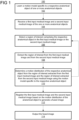
- Figure 1 shows a method 100 for medical image registration, in accordance with one or more embodiment.
- Method 100 may be performed using any suitable computing device, such as, e.g., computer 402 of Figure 4 .
- Step 102 a motion model specific to a respective anatomical object of one or more anatomical objects is learned. Step 102 may be performed during an offline or preprocessing stage. In one embodiment, the motion model specific to the respective anatomical object is learned by performing the steps of method 200 of Figure 2 .
- Method 200 may be performed by any suitable computing device, such as, e.g., computer 402 of Figure 4 .
- a first training image and a second training image of one or more particular anatomical objects are received.
- the first training image may correspond to a moving image denoted I 0 and the second training image may correspond to a fixed image denoted I 1 .
- the first training image I 0 and second training image I 1 depict the same one or more particular anatomical objects in various states of deformation due to, e.g., respiratory motion or cardiac motion.
- the first training image I 0 and second training image I 1 are of a sequence of images acquired over a period of time.
- the one or more anatomical objects may include any anatomical structure of a patient, such as, e.g., an organ (e.g., lung, heart, liver, kidney, bladder, etc.), a vessel, a bone, etc.
- the first training image I 0 and second training image I 1 are of the same modality.
- the first training image I 0 and second training image I 1 may be of any suitable modality, such as, e.g., x-ray, magnetic resonance imaging (MRI), computed tomography (CT), ultrasound (US), single-photon emission computed tomography (SPECT), positron emission tomography (PET), or any other suitable modality or combination of modalities.
- the first training image I 0 and second training image I 1 may be received directly from an image acquisition device (e.g., image acquisition device 414 of Figure 4 ) used to acquire the images.
- first training image I 0 and second training image I 1 may be received by loading previously acquired images from a storage or memory of a computer system (e.g., a picture archiving and communication system, PACS) or receiving images that have been transmitted from a remote computer system.
- a computer system e.g., a picture archiving and communication system, PACS
- PACS picture archiving and communication system
- a region of interest comprising the respective particular anatomical object is detected in one of the first training image I 0 or the second training image I 1 .
- the region of interest may be detected manually via input from a user (e.g., a clinician) or automatically using any suitable known technique.
- the respective particular anatomical object is segmented from the training image according to a selective and iterative method for performance level estimation (SIMPLE) method and the region of interest is centered around the segmented respective particular anatomical object.
- SIMPLE performance level estimation
- the region of interest comprising the respective particular anatomical object is extracted from the first training image and the second training image.
- a machine learning network is trained to model motion of the respective particular anatomical object based on the region of interest extracted from the first training image and the region of interest extracted from the second training image to learn a motion model specific to the respective particular anatomical object.
- the machine learning network may be any suitable machine learning for modeling motion of the anatomical object.
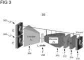
- the machine learning network is a variational autoencoder (VAE), such as, e.g., VAE 300 shown in Figure 3 , to provide for a probabilistic motion model specific to the respective particular anatomical object, in accordance with one or more embodiments.
- VAE variational autoencoder
- VAE 300 includes an encoder p ⁇ 306 and a decoder p ⁇ 310.
- Encoder p ⁇ 306 is a neural network that receives as input sub-images I 0 o 302 and I 1 o 304 and outputs code z 0 308.
- Sub-images I 0 o 302 and I 1 o 304 are the regions of interest o extracted from the first training image I 0 and the second training image I 1 respectively.
- Code z 0 308 is a low-dimensional vector representing the mean and variance of the multivariate Gaussian sampled from the first training image I 0 and the second training image I 1 by encoder p ⁇ 306.
- Decoder p ⁇ 310 is a neural network that receives as input sub-image I 0 o 302 and code z 0 308 and outputs velocities v 312 and deformation field ⁇ 314.
- Velocities v 312 are raw outputs of VAE 300 and are non-diffeomorphic. Velocities v 312 comprise the velocity value for each pixel.
- Deformation field ⁇ 314 is computed by exponentiation and represents the deformation between sub-images I 0 o 302 and I 1 o 304. By making appearance information of the moving sub-image I 0 o 302 available to decoder p ⁇ 310, deformation field ⁇ 314 is more likely to encode deformation information rather than appearance information.
- deformation field ⁇ 314 is applied to sub-image I 0 o 302 to reconstruct sub-image I 1 o 304.
- VAE 300 is trained according to Equation 1 to optimize code z 0 308 to best transform sub-image I 0 o 302 to align with sub-image I 1 o 304.
- p ⁇ I 1 o I 0 o ⁇ Z 0 p ⁇ I 1 o Z 0 , I 0 o p Z 0 dz
- p ( z 0 ) is a prior distribution of z 0 and is assumed to follow a multivariate unit Gaussian distribution.
- Prior distribution p ( z 0 ) refers to the distribution learned by VAE 300 representing the distribution of all probable motions of the respective particular anatomical object (as observed in a training set).
- method 200 may return to step 204 and steps 204-208 may be iteratively repeated to learn a motion model specific to each particular anatomical object of the one or more particular anatomical objects.
- a first input medical image and a second input medical image of the one or more anatomical objects are received.
- the first input medical image may correspond to a moving image M and the second input medical image may correspond to a fixed image F of a dataset to be registered.
- the first input medical image M and the second input medical image F depict the same one or more anatomical objects in various states of deformation due to, e.g., respiratory motion or cardiac motion.
- the first input medical image M and the second input medical image F are of a sequence of images acquired over a period of time.
- the first input medical image M and the second input medical image F may be of any suitable modality.
- the first input medical image M and the second input medical image F may be received directly from an image acquisition device (e.g., image acquisition device 414 of Figure 4 ) used to acquire the images.
- the first input medical image M and the second input medical image F may be received by loading previously acquired images from a storage or memory of a computer system (e.g., a picture archiving and communication system, PACS) or receiving images that have been transmitted from a remote computer system.
- a computer system e.g., a picture archiving and communication system, PACS
- a region of interest o comprising the respective anatomical object is detected in one of the first input medical image M or the second input medical image F .
- the region of interest o may be detected manually via input from a user (e.g., a clinician) or automatically using any suitable known technique.
- the region of interest may be automatically by segmenting the respective anatomical object from the first input medical image M or the second input medical image F and centering the region of interest around the segmented respective anatomical object, as described above with respect to step 204 of method 200 in Figure 2 .
- the region of interest is extracted from the first input medical image and from the second input medical image.
- a motion distribution of the respective anatomical object is determined from the region of interest extracted from the first input medical image M and the region of interest extracted from the second input medical image F using the motion model specific to the respective anatomical object (learned at step 102).
- the motion model is a VAE
- a trained encoder p ⁇ e.g., encoder p ⁇ 306 of Figure 3
- the motion model is applied on the region of interest extracted from the first input medical image M and the region of interest extracted from the second input medical image F according to the function p ⁇ ( M 0 , M 0 ⁇ ⁇ ⁇ ) to determine the motion distribution z 0 of the respective anatomical object.
- the encoder p ⁇ receives as input the region of interest extracted from the first input medical image M and the region of interest extracted from the first input medical image M as modified by the deformation field ⁇ ⁇ and outputs the motion distribution z 0 .
- Deformation field ⁇ ⁇ represents the global motion between the first input medical image M and the second input medical image F.
- computing motion distribution z 0 on the same image e.g., the first input medical image M
- motion distribution z 0 may instead be determined according to p ⁇ ( F 0 , F 0 ⁇ ⁇ ⁇ ) by inverting the resulting motion distribution z 0 where p ⁇ is trained using the fixed image.
- the distance is a Kullback-Leibler Divergence.
- other distance metrics may be applied, such as, e.g., optimal transport loss, generative adversarial networks, adversarial autoencoders, or any other suitable distance metric.
- Method 100 may return back to step 106 and steps 106-110 may be iteratively repeated for each respective anatomical object of the one or more anatomical objects (e.g., for whole body registration) to thereby determine a motion distribution of each of the one or more anatomical objects.
- the first input medical image and the second input medical image are registered based on the motion distribution of each respective anatomical object of the one or more anatomical objects to generate a fused image.
- o the region of interest for a respective anatomical object
- M 0 0 is the region of interest o extracted from the first input medical image M
- ⁇ ⁇ is the global motion between the first input medical image M and the second input medical image F
- the first input medical image and the second input medical image are then registered according to the loss function of Equation 3: where is a distance metric (e.g., Kullback-Leibler Divergence), F is the second input medical image, M is the first input medical image, ⁇ ⁇ is the divergence field of the motion model, ( ⁇ ⁇ ) is a spatial regularization term, ( ⁇ ⁇ ) is a regularization term for the one or more anatomical objects defined in Equation 2, and ⁇ 1 and ⁇ 2 are parameters that weight ( ⁇ ⁇ ) and ( ⁇ ⁇ ) respectively.
- the loss function of Equation 3 includes the regularization term for the one or more anatomical objects and therefore may be used to compensate for organ deformation to train a machine learning network to register the first input medical image and the second input medical image to generate the fused image.
- the fused image is output.
- the fused image can be output by displaying the fused image on a display device of a computer system, storing the fused image on a memory or storage of a computer system, or by transmitting the fused image to a remote computer system.
- embodiments of the present invention provide for an anatomical object-specific regularization term ( ⁇ ⁇ ) for medical image registration to focus the registration process on regions of interest and compensate for motion of anatomical objects in the regions of interest.
- Embodiments of the present invention are not as computationally expensive to perform as convention techniques that require segmentation of anatomical objects in both images, as certain embodiments of the present invention involve detecting a bounding box in one of the input medical images.
- Embodiments of the present invention allow for medical image registration with a high degree of accuracy.
- a mean posterior distribution p ⁇ ( ⁇ , ⁇ ) may be used instead of the prior distribution p ( z 0 ).
- the mean posterior distribution p ⁇ ( ⁇ , ⁇ ) may be extracted after learning the motion models for each anatomical object.
- Systems, apparatuses, and methods described herein may be implemented using digital circuitry, or using one or more computers using well-known computer processors, memory units, storage devices, computer software, and other components.
- a computer includes a processor for executing instructions and one or more memories for storing instructions and data.
- a computer may also include, or be coupled to, one or more mass storage devices, such as one or more magnetic disks, internal hard disks and removable disks, magneto-optical disks, optical disks, etc.
- Systems, apparatus, and methods described herein may be implemented using computers operating in a client-server relationship.
- the client computers are located remotely from the server computer and interact via a network.
- the client-server relationship may be defined and controlled by computer programs running on the respective client and server computers.
- Systems, apparatus, and methods described herein may be implemented within a network-based cloud computing system.
- a server or another processor that is connected to a network communicates with one or more client computers via a network.
- a client computer may communicate with the server via a network browser application residing and operating on the client computer, for example.
- a client computer may store data on the server and access the data via the network.
- a client computer may transmit requests for data, or requests for online services, to the server via the network.
- the server may perform requested services and provide data to the client computer(s).
- the server may also transmit data adapted to cause a client computer to perform a specified function, e.g., to perform a calculation, to display specified data on a screen, etc.
- the server may transmit a request adapted to cause a client computer to perform one or more of the steps or functions of the methods and workflows described herein, including one or more of the steps or functions of Figures 1-2 .
- Certain steps or functions of the methods and workflows described herein, including one or more of the steps or functions of Figures 1-2 may be performed by a server or by another processor in a network-based cloud-computing system.
- Certain steps or functions of the methods and workflows described herein, including one or more of the steps of Figures 1-2 may be performed by a client computer in a network-based cloud computing system.
- the steps or functions of the methods and workflows described herein, including one or more of the steps of Figures 1-2 may be performed by a server and/or by a client computer in a network-based cloud computing system, in any combination.
- Systems, apparatus, and methods described herein may be implemented using a computer program product tangibly embodied in an information carrier, e.g., in a non-transitory machine-readable storage device, for execution by a programmable processor; and the method and workflow steps described herein, including one or more of the steps or functions of Figures 1-2 , may be implemented using one or more computer programs that are executable by such a processor.
- a computer program is a set of computer program instructions that can be used, directly or indirectly, in a computer to perform a certain activity or bring about a certain result.
- a computer program can be written in any form of programming language, including compiled or interpreted languages, and it can be deployed in any form, including as a stand-alone program or as a module, component, subroutine, or other unit suitable for use in a computing environment.
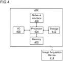
- Computer 402 includes a processor 404 operatively coupled to a data storage device 412 and a memory 410.
- Processor 404 controls the overall operation of computer 402 by executing computer program instructions that define such operations.
- the computer program instructions may be stored in data storage device 412, or other computer readable medium, and loaded into memory 410 when execution of the computer program instructions is desired.
- the method and workflow steps or functions of Figures 1-2 can be defined by the computer program instructions stored in memory 410 and/or data storage device 412 and controlled by processor 404 executing the computer program instructions.
- Computer 402 may also include one or more network interfaces 406 for communicating with other devices via a network.
- Computer 402 may also include one or more input/output devices 408 that enable user interaction with computer 402 (e.g., display, keyboard, mouse, speakers, buttons, etc.).
- Processor 404 may include both general and special purpose microprocessors, and may be the sole processor or one of multiple processors of computer 402.
- Processor 404 may include one or more central processing units (CPUs), for example.
- Processor 404, data storage device 412, and/or memory 410 may include, be supplemented by, or incorporated in, one or more application-specific integrated circuits (ASICs) and/or one or more field programmable gate arrays (FPGAs).
- ASICs application-specific integrated circuits
- FPGAs field programmable gate arrays
- Data storage device 412 and memory 410 each include a tangible non-transitory computer readable storage medium.
- Data storage device 412, and memory 410 may each include high-speed random access memory, such as dynamic random access memory (DRAM), static random access memory (SRAM), double data rate synchronous dynamic random access memory (DDR RAM), or other random access solid state memory devices, and may include non-volatile memory, such as one or more magnetic disk storage devices such as internal hard disks and removable disks, magneto-optical disk storage devices, optical disk storage devices, flash memory devices, semiconductor memory devices, such as erasable programmable read-only memory (EPROM), electrically erasable programmable read-only memory (EEPROM), compact disc read-only memory (CD-ROM), digital versatile disc read-only memory (DVD-ROM) disks, or other non-volatile solid state storage devices.
- DRAM dynamic random access memory
- SRAM static random access memory
- DDR RAM double data rate synchronous dynamic random access memory
- non-volatile memory such as one
- Input/output devices 408 may include peripherals, such as a printer, scanner, display screen, etc.
- input/output devices 408 may include a display device such as a cathode ray tube (CRT) or liquid crystal display (LCD) monitor for displaying information to the user, a keyboard, and a pointing device such as a mouse or a trackball by which the user can provide input to computer 402.
- display device such as a cathode ray tube (CRT) or liquid crystal display (LCD) monitor for displaying information to the user
- keyboard a keyboard
- pointing device such as a mouse or a trackball by which the user can provide input to computer 402.
- An image acquisition device 414 can be connected to the computer 402 to input image data (e.g., medical images) to the computer 402. It is possible to implement the image acquisition device 414 and the computer 402 as one device. It is also possible that the image acquisition device 414 and the computer 402 communicate wirelessly through a network. In a possible embodiment, the computer 402 can be located remotely with respect to the image acquisition device 414.
- image data e.g., medical images
Landscapes
- Engineering & Computer Science (AREA)
- Theoretical Computer Science (AREA)
- Computer Vision & Pattern Recognition (AREA)
- Physics & Mathematics (AREA)
- General Physics & Mathematics (AREA)
- General Health & Medical Sciences (AREA)
- Medical Informatics (AREA)
- Multimedia (AREA)
- Evolutionary Computation (AREA)
- Health & Medical Sciences (AREA)
- Computing Systems (AREA)
- Artificial Intelligence (AREA)
- Databases & Information Systems (AREA)
- Software Systems (AREA)
- Quality & Reliability (AREA)
- Radiology & Medical Imaging (AREA)
- Nuclear Medicine, Radiotherapy & Molecular Imaging (AREA)
- Image Analysis (AREA)
Claims (13)
- Computerimplementiertes Verfahren, das Folgendes umfasst:Empfangen (104) eines ersten medizinischen Eingabebildes und eines zweiten medizinischen Eingabebildes, das dieselben ein oder mehreren anatomischen Objekte in verschiedenen Zuständen der Verformung darstellt;für jedes entsprechende anatomische Objekt der ein oder mehreren anatomischen Objekte:Erkennen (106) einer Region von Interesse, die das jeweilige anatomische Objekt umfasst, in dem ersten medizinischen Eingabebild oder dem zweiten medizinischen Eingabebild,Extrahieren (108) der Region von Interesse aus dem ersten medizinischen Eingabebild bzw. aus dem zweiten medizinischen Eingabebild, wobei die extrahierten Regionen von Interesse Teilbilder des ersten medizinischen Eingabebildes bzw. des zweiten medizinischen Eingabebildes sind, undBestimmen (110) einer Bewegungsverteilung des jeweiligen anatomischen Objekts, wobei die Bewegungsverteilung eine wahrscheinliche Bewegung für das jeweilige anatomische Objekt ist, durch Anwenden eines Bewegungsmodells auf die aus dem ersten medizinischen Eingabebild extrahierte Region von Interesse und die aus dem zweiten medizinischen Eingabebild extrahierte Region von Interesse unter Verwendung des Bewegungsmodells, wobei das Bewegungsmodell ein erlerntes Bewegungsmodell ist, das für das jeweilige anatomische Objekt spezifisch ist;
und
Registrieren (112) des ersten medizinischen Eingabebildes und des zweiten medizinischen Eingabebildes basierend auf der Bewegungsverteilung jedes einzelnen anatomischen Objektes der ein oder mehreren anatomischen Objekte,dadurch gekennzeichnet, dassdas Registrieren des ersten medizinischen Eingabebildes und des zweiten medizinischen Eingabebildes basierend auf der Bewegungsverteilung jedes einzelnen anatomischen Objektes der ein oder mehreren anatomischen Objekte umfasst:
Bestimmen eines für das anatomische Objekt spezifischen Regularisierungsterms für die ein oder mehreren anatomischen Objekte basierend auf Abständen zwischen der Bewegungsverteilung jedes einzelnen anatomischen Objekts und einer vorherigen Verteilung des jeweiligen anatomischen Objekts. - Verfahren nach Anspruch 1, wobei das Bestimmen eines für das anatomische Objekt spezifischen Regularisierungsterms für die ein oder mehreren anatomischen Objekte basierend auf Abständen zwischen der Bewegungsverteilung jedes einzelnen anatomischen Objekts und einer vorherigen Verteilung des jeweiligen anatomischen Objekts umfasst:
Summieren der Abstände, über die Region von Interesse für das jeweilige anatomische Objekt, zwischen der Bewegungsverteilung jedes einzelnen anatomischen Objekts und der jeweiligen vorherigen Verteilung. - Verfahren nach Anspruch 1, wobei das Registrieren des ersten medizinischen Eingabebildes und des zweiten medizinischen Eingabebildes basierend auf der Bewegungsverteilung jedes einzelnen anatomischen Objektes der ein oder mehreren anatomischen Objekte umfasst:
Minimieren einer Verlustfunktion, die den Regularisierungsterm für die ein oder mehreren anatomischen Objekte enthält. - Verfahren nach Anspruch 1, wobei das für das jeweilige anatomische Objekt spezifische Bewegungsmodell einen Variational-Autoencoder umfasst, der einen Codierer umfasst, und wobei das Bestimmen einer Bewegungsverteilung des jeweiligen anatomischen Objekts aus der aus dem ersten medizinischen Eingabebild extrahierten Region von Interesse oder der aus dem zweiten medizinischen Eingabebild extrahierten Region von Interesse unter Verwendung eines für das jeweilige anatomische Objekt spezifischen Bewegungsmodells umfasst:
Bestimmen der Bewegungsverteilung des jeweiligen anatomischen Objekts mit Hilfe des Codierers. - Verfahren nach Anspruch 1, ferner umfassend, für jedes jeweilige anatomische Objekt, das Lernen (102) des für das jeweilige anatomische Objekt spezifischen Bewegungsmodells durch:Empfangen (202) eines ersten Trainingsbildes und eines zweiten Trainingsbildes des jeweiligen anatomischen Objekts;Erkennen (204) einer Region von Interesse, die das jeweilige anatomische Objekt umfasst, in dem ersten Trainingsbild oder dem zweiten Trainingsbild;Extrahieren (206) der Region von Interesse aus dem ersten Trainingsbild und dem zweiten Trainingsbild; undTrainieren (208) eines Maschinenlernnetzes, um die Bewegung des jeweiligen anatomischen Objekts aus der aus dem ersten Trainingsbild extrahierten Region von Interesse und der aus dem zweiten Trainingsbild extrahierten Region von Interesse als das für das jeweilige anatomische Objekt spezifische Bewegungsmodell zu modellieren.
- Verfahren nach Anspruch 5, wobei das Trainieren eines Maschinenlernnetzes, um die Bewegung des jeweiligen anatomischen Objekts aus der aus dem ersten Trainingsbild extrahierten Region von Interesse und der aus dem zweiten Trainingsbild extrahierten Region von Interesse als das für das jeweilige anatomische Objekt spezifische Bewegungsmodell zu modellieren, umfasst:Trainieren eines Codierers eines Variational-Autoencoders, um einen Code zu erzeugen, der eine Codierung der Verformung zwischen der aus dem ersten Trainingsbild extrahierten Region von Interesse und der aus dem zweiten Trainingsbild extrahierten Region von Interesse darstellt; undTrainieren eines Decodierers des Variational-Autoencoders, um ein Verformungsfeld aus dem Code und der aus dem ersten Trainingsbild extrahierten Region von Interesse zu erzeugen, wobei das Verformungsfeld die Verformung zwischen der aus dem ersten Trainingsbild extrahierten Region von Interesse und der aus dem zweiten Trainingsbild extrahierten Region von Interesse darstellt.
- Verfahren nach Anspruch 1, wobei das Erkennen einer Region von Interesse, die das jeweilige anatomische Objekt umfasst, in dem ersten medizinischen Eingabebild oder dem zweiten medizinischen Eingabebild umfasst:Segmentieren der des jeweiligen anatomischen Objekts in dem ersten medizinischen Eingabebild oder dem zweiten medizinischen Eingabebild; undZentrieren der Region von Interesse um das jeweilige segmentierte anatomische Objekt.
- Verfahren nach Anspruch 1, wobei die ein oder mehreren anatomischen Objekte ein oder mehrere Organe umfassen.
- Einrichtung, umfassend:Mittel zum Empfangen (104) eines ersten medizinischen Eingabebildes und eines zweiten medizinischen Eingabebildes, das dieselben ein oder mehreren anatomischen Objekte in verschiedenen Zuständen der Verformung darstellt;Mittel zum, für jedes einzelne anatomische Objekt der ein oder mehreren anatomischen Objekte, Erkennen (106) einer Region von Interesse, die das jeweilige anatomische Objekt umfasst, in dem ersten medizinischen Eingabebild oder dem zweiten medizinischen Eingabebild;Mittel zum, für jedes einzelne anatomische Objekt, Extrahieren (108) der Region von Interesse aus dem ersten medizinischen Eingabebild bzw. aus dem zweiten medizinischen Eingabebild, wobei die extrahierten Regionen von Interesse Teilbilder des ersten medizinischen Eingabebildes bzw. des zweiten medizinischen Eingabebildes sind;Mittel zum, für jedes einzelne anatomische Objekt, Bestimmen (110) einer Bewegungsverteilung des jeweiligen anatomischen Objekts, wobei die Bewegungsverteilung eine wahrscheinliche Bewegung für das jeweilige anatomische Objekt ist, durch Anwenden eines Bewegungsmodells auf die aus dem ersten medizinischen Eingabebild extrahierte Region von Interesse und die aus dem zweiten medizinischen Eingabebild extrahierte Region von Interesse unter Verwendung des Bewegungsmodells, wobei das Bewegungsmodell ein erlerntes Bewegungsmodell ist, das für das jeweilige anatomische Objekt spezifisch ist; undMittel zum Registrieren (112) des ersten medizinischen Eingabebildes und des zweiten medizinischen Eingabebildes basierend auf der Bewegungsverteilung jedes einzelnen anatomischen Objektes der ein oder mehreren anatomischen Objekte,dadurch gekennzeichnet, dassdas Mittel zum Registrieren des ersten medizinischen Eingabebildes und des zweiten medizinischen Eingabebildes basierend auf der Bewegungsverteilung jedes einzelnen anatomischen Objektes der ein oder mehreren anatomischen Objekte umfasst:
Mittel zum Bestimmen eines für das anatomische Objekt spezifischen Regularisierungsterms für die ein oder mehreren anatomischen Objekte basierend auf Abständen zwischen der Bewegungsverteilung jedes einzelnen anatomischen Objekts und einer vorherigen Verteilung des jeweiligen anatomischen Objekts. - Einrichtung nach Anspruch 9, wobei das Mittel zum Bestimmen eines für das anatomische Objekt spezifischen Regularisierungsterms für die ein oder mehreren anatomischen Objekte basierend auf Abständen zwischen der Bewegungsverteilung jedes einzelnen anatomischen Objekts und einer vorherigen Verteilung des jeweiligen anatomischen Objekts umfasst:
Mittel zum Summieren der Abstände, über die Region von Interesse für das jeweilige anatomische Objekt, zwischen der Bewegungsverteilung jedes einzelnen anatomischen Objekts und der jeweiligen vorherigen Verteilung. - Einrichtung nach Anspruch 9, wobei das Mittel zum Registrieren des ersten medizinischen Eingabebildes und des zweiten medizinischen Eingabebildes basierend auf der Bewegungsverteilung jedes einzelnen anatomischen Objektes der ein oder mehreren anatomischen Objekte umfasst:
Mittel zum Minimieren einer Verlustfunktion, die den Regularisierungsterm für die ein oder mehreren anatomischen Objekte enthält. - Einrichtung nach Anspruch 9, wobei das für das jeweilige anatomische Objekt spezifische Bewegungsmodell einen Variational-Autoencoder umfasst, der einen Codierer umfasst, und wobei das Mittel zum Bestimmen einer Bewegungsverteilung des jeweiligen anatomischen Objekts aus der aus dem ersten medizinischen Eingabebild extrahierten Region von Interesse oder der aus dem zweiten medizinischen Eingabebild extrahierten Region von Interesse unter Verwendung eines für das jeweilige anatomische Objekt spezifischen Bewegungsmodells umfasst:
Mittel zum Bestimmen der Bewegungsverteilung des jeweiligen anatomischen Objekts mit Hilfe des Codierers. - Nichtflüchtiges computerlesbares Medium, das Computerprogrammanweisungen speichert, wobei die Computerprogrammanweisungen, wenn sie von einem Prozessor ausgeführt werden, den Prozessor veranlassen, Operationen gemäß dem Verfahren nach einem der Ansprüche 1, 3 und 5-7 durchzuführen.
Applications Claiming Priority (1)
| Application Number | Priority Date | Filing Date | Title |
|---|---|---|---|
| PCT/US2019/066141 WO2021118581A1 (en) | 2019-12-13 | 2019-12-13 | Compensation of organ deformation for medical image registration |
Publications (3)
| Publication Number | Publication Date |
|---|---|
| EP4073755A1 EP4073755A1 (de) | 2022-10-19 |
| EP4073755B1 true EP4073755B1 (de) | 2025-05-07 |
| EP4073755C0 EP4073755C0 (de) | 2025-05-07 |
Family
ID=69326622
Family Applications (1)
| Application Number | Title | Priority Date | Filing Date |
|---|---|---|---|
| EP19842912.8A Active EP4073755B1 (de) | 2019-12-13 | 2019-12-13 | Kompensation von organverformung zur registrierung medizinischer bilder |
Country Status (4)
| Country | Link |
|---|---|
| US (1) | US12260551B2 (de) |
| EP (1) | EP4073755B1 (de) |
| CN (1) | CN114787867B (de) |
| WO (1) | WO2021118581A1 (de) |
Families Citing this family (3)
| Publication number | Priority date | Publication date | Assignee | Title |
|---|---|---|---|---|
| CN113469180A (zh) * | 2020-03-31 | 2021-10-01 | 阿里巴巴集团控股有限公司 | 医学图像的处理方法和系统、数据处理方法 |
| DE102020212515A1 (de) * | 2020-10-02 | 2022-04-07 | Robert Bosch Gesellschaft mit beschränkter Haftung | Verfahren und Vorrichtung zum Trainieren eines maschinellen Lernsystems |
| CN114612527B (zh) * | 2022-03-01 | 2025-01-17 | 京东科技信息技术有限公司 | 图像配准方法、装置、电子设备及存储介质 |
Family Cites Families (7)
| Publication number | Priority date | Publication date | Assignee | Title |
|---|---|---|---|---|
| US8498459B2 (en) | 2009-10-08 | 2013-07-30 | Siemens Aktiengesellschaft | System and method for verifying registration accuracy in digital medical images |
| US8878102B2 (en) | 2011-06-15 | 2014-11-04 | Scentsy, Inc. | Base structures, scent warmers including such base structures, and related methods |
| US9280819B2 (en) * | 2013-08-26 | 2016-03-08 | International Business Machines Corporation | Image segmentation techniques |
| KR102205898B1 (ko) * | 2013-09-04 | 2021-01-21 | 삼성전자주식회사 | 의료영상들을 정합하는 방법 및 장치 |
| US20170337682A1 (en) * | 2016-05-18 | 2017-11-23 | Siemens Healthcare Gmbh | Method and System for Image Registration Using an Intelligent Artificial Agent |
| US11154196B2 (en) | 2017-06-20 | 2021-10-26 | Siemens Healthcare Gmbh | Deep-learnt tissue deformation for medical imaging |
| US11449759B2 (en) | 2018-01-03 | 2022-09-20 | Siemens Heathcare Gmbh | Medical imaging diffeomorphic registration based on machine learning |
-
2019
- 2019-12-13 EP EP19842912.8A patent/EP4073755B1/de active Active
- 2019-12-13 CN CN201980102896.5A patent/CN114787867B/zh active Active
- 2019-12-13 US US17/755,558 patent/US12260551B2/en active Active
- 2019-12-13 WO PCT/US2019/066141 patent/WO2021118581A1/en not_active Ceased
Non-Patent Citations (2)
| Title |
|---|
| FRANK PREISWERK ET AL: "Model-guided respiratory organ motion prediction of the liver from 2D ultrasound", MEDICAL IMAGE ANALYSIS, vol. 18, no. 5, 1 July 2014 (2014-07-01), pages 740 - 751, XP055212423, ISSN: 1361-8415, DOI: 10.1016/j.media.2014.03.006 * |
| SIYUAN SHAN ET AL: "Unsupervised End-to-end Learning for Deformable Medical Image Registration", ARXIV.ORG, CORNELL UNIVERSITY LIBRARY, 201 OLIN LIBRARY CORNELL UNIVERSITY ITHACA, NY 14853, 23 November 2017 (2017-11-23), XP081308245 * |
Also Published As
| Publication number | Publication date |
|---|---|
| WO2021118581A1 (en) | 2021-06-17 |
| EP4073755A1 (de) | 2022-10-19 |
| CN114787867B (zh) | 2025-11-18 |
| US12260551B2 (en) | 2025-03-25 |
| EP4073755C0 (de) | 2025-05-07 |
| CN114787867A (zh) | 2022-07-22 |
| US20220270256A1 (en) | 2022-08-25 |
Similar Documents
| Publication | Publication Date | Title |
|---|---|---|
| US11055847B2 (en) | Adversarial and dual inverse deep learning networks for medical image analysis | |
| EP3511942B1 (de) | Domänenübergreifende bildanalyse unter verwendung von tiefgehenden bild-zu-bild-netzwerken und gegnerischen netzwerken | |
| CN113362272B (zh) | 具有不确定性估计的医学图像分割 | |
| US11514571B2 (en) | Hierarchical analysis of medical images for identifying and assessing lymph nodes | |
| US10970829B2 (en) | Synthesizing and segmenting cross-domain medical images | |
| US10600184B2 (en) | Automated segmentation utilizing fully convolutional networks | |
| CN107403446B (zh) | 用于使用智能人工代理的图像配准的方法和系统 | |
| Girum et al. | Learning with context feedback loop for robust medical image segmentation | |
| EP3579189B1 (de) | Adaptive nichtlineare optimierung von formparametern zur objektortung in medizinischen 3d-bildern | |
| US9704256B2 (en) | Systems and method for computation and visualization of segmentation uncertainty in medical images | |
| JP2021521993A (ja) | 敵対的生成ネットワークを使用した画像強調 | |
| US11403761B2 (en) | Probabilistic motion model for generating medical images or medical image sequences | |
| EP3696724A1 (de) | Kontinuierliches lernen zur automatischen ansichtsplanung zur bilderfassung | |
| US11908047B2 (en) | Generating synthetic x-ray images and object annotations from CT scans for augmenting x-ray abnormality assessment systems | |
| EP4073755B1 (de) | Kompensation von organverformung zur registrierung medizinischer bilder | |
| EP3648057B1 (de) | Bestimmung der malignität von lungenknötchen unter verwendung von tiefenlernen | |
| JP2023131287A (ja) | 情報処理装置および学習方法 | |
| Le Folgoc | Statistical learning for image-based personalization of cardiac models | |
| LE FOLGOC | THÈSE DOCTORALE |
Legal Events
| Date | Code | Title | Description |
|---|---|---|---|
| STAA | Information on the status of an ep patent application or granted ep patent |
Free format text: STATUS: UNKNOWN |
|
| STAA | Information on the status of an ep patent application or granted ep patent |
Free format text: STATUS: THE INTERNATIONAL PUBLICATION HAS BEEN MADE |
|
| PUAI | Public reference made under article 153(3) epc to a published international application that has entered the european phase |
Free format text: ORIGINAL CODE: 0009012 |
|
| STAA | Information on the status of an ep patent application or granted ep patent |
Free format text: STATUS: REQUEST FOR EXAMINATION WAS MADE |
|
| 17P | Request for examination filed |
Effective date: 20220713 |
|
| AK | Designated contracting states |
Kind code of ref document: A1 Designated state(s): AL AT BE BG CH CY CZ DE DK EE ES FI FR GB GR HR HU IE IS IT LI LT LU LV MC MK MT NL NO PL PT RO RS SE SI SK SM TR |
|
| DAV | Request for validation of the european patent (deleted) | ||
| DAX | Request for extension of the european patent (deleted) | ||
| RAP1 | Party data changed (applicant data changed or rights of an application transferred) |
Owner name: SIEMENS HEALTHINEERS AG |
|
| GRAP | Despatch of communication of intention to grant a patent |
Free format text: ORIGINAL CODE: EPIDOSNIGR1 |
|
| STAA | Information on the status of an ep patent application or granted ep patent |
Free format text: STATUS: GRANT OF PATENT IS INTENDED |
|
| INTG | Intention to grant announced |
Effective date: 20241209 |
|
| GRAS | Grant fee paid |
Free format text: ORIGINAL CODE: EPIDOSNIGR3 |
|
| GRAA | (expected) grant |
Free format text: ORIGINAL CODE: 0009210 |
|
| STAA | Information on the status of an ep patent application or granted ep patent |
Free format text: STATUS: THE PATENT HAS BEEN GRANTED |
|
| AK | Designated contracting states |
Kind code of ref document: B1 Designated state(s): AL AT BE BG CH CY CZ DE DK EE ES FI FR GB GR HR HU IE IS IT LI LT LU LV MC MK MT NL NO PL PT RO RS SE SI SK SM TR |
|
| REG | Reference to a national code |
Ref country code: GB Ref legal event code: FG4D |
|
| REG | Reference to a national code |
Ref country code: CH Ref legal event code: EP |
|
| REG | Reference to a national code |
Ref country code: DE Ref legal event code: R096 Ref document number: 602019069745 Country of ref document: DE |
|
| REG | Reference to a national code |
Ref country code: IE Ref legal event code: FG4D |
|
| U01 | Request for unitary effect filed |
Effective date: 20250515 |
|
| U07 | Unitary effect registered |
Designated state(s): AT BE BG DE DK EE FI FR IT LT LU LV MT NL PT RO SE SI Effective date: 20250521 |
|
| PG25 | Lapsed in a contracting state [announced via postgrant information from national office to epo] |
Ref country code: ES Free format text: LAPSE BECAUSE OF FAILURE TO SUBMIT A TRANSLATION OF THE DESCRIPTION OR TO PAY THE FEE WITHIN THE PRESCRIBED TIME-LIMIT Effective date: 20250507 |
|
| PG25 | Lapsed in a contracting state [announced via postgrant information from national office to epo] |
Ref country code: NO Free format text: LAPSE BECAUSE OF FAILURE TO SUBMIT A TRANSLATION OF THE DESCRIPTION OR TO PAY THE FEE WITHIN THE PRESCRIBED TIME-LIMIT Effective date: 20250807 Ref country code: GR Free format text: LAPSE BECAUSE OF FAILURE TO SUBMIT A TRANSLATION OF THE DESCRIPTION OR TO PAY THE FEE WITHIN THE PRESCRIBED TIME-LIMIT Effective date: 20250808 |
|
| PG25 | Lapsed in a contracting state [announced via postgrant information from national office to epo] |
Ref country code: PL Free format text: LAPSE BECAUSE OF FAILURE TO SUBMIT A TRANSLATION OF THE DESCRIPTION OR TO PAY THE FEE WITHIN THE PRESCRIBED TIME-LIMIT Effective date: 20250507 |
|
| PG25 | Lapsed in a contracting state [announced via postgrant information from national office to epo] |
Ref country code: HR Free format text: LAPSE BECAUSE OF FAILURE TO SUBMIT A TRANSLATION OF THE DESCRIPTION OR TO PAY THE FEE WITHIN THE PRESCRIBED TIME-LIMIT Effective date: 20250507 |
|
| PG25 | Lapsed in a contracting state [announced via postgrant information from national office to epo] |
Ref country code: RS Free format text: LAPSE BECAUSE OF FAILURE TO SUBMIT A TRANSLATION OF THE DESCRIPTION OR TO PAY THE FEE WITHIN THE PRESCRIBED TIME-LIMIT Effective date: 20250807 |
|
| PG25 | Lapsed in a contracting state [announced via postgrant information from national office to epo] |
Ref country code: IS Free format text: LAPSE BECAUSE OF FAILURE TO SUBMIT A TRANSLATION OF THE DESCRIPTION OR TO PAY THE FEE WITHIN THE PRESCRIBED TIME-LIMIT Effective date: 20250907 |
|
| PG25 | Lapsed in a contracting state [announced via postgrant information from national office to epo] |
Ref country code: SM Free format text: LAPSE BECAUSE OF FAILURE TO SUBMIT A TRANSLATION OF THE DESCRIPTION OR TO PAY THE FEE WITHIN THE PRESCRIBED TIME-LIMIT Effective date: 20250507 |
|
| PG25 | Lapsed in a contracting state [announced via postgrant information from national office to epo] |
Ref country code: CZ Free format text: LAPSE BECAUSE OF FAILURE TO SUBMIT A TRANSLATION OF THE DESCRIPTION OR TO PAY THE FEE WITHIN THE PRESCRIBED TIME-LIMIT Effective date: 20250507 |
|
| PG25 | Lapsed in a contracting state [announced via postgrant information from national office to epo] |
Ref country code: SK Free format text: LAPSE BECAUSE OF FAILURE TO SUBMIT A TRANSLATION OF THE DESCRIPTION OR TO PAY THE FEE WITHIN THE PRESCRIBED TIME-LIMIT Effective date: 20250507 |
|
| U20 | Renewal fee for the european patent with unitary effect paid |
Year of fee payment: 7 Effective date: 20251219 |