EP4069303B1 - Injizierbare hydrogele zur zellabgabe an den glaskörper - Google Patents
Injizierbare hydrogele zur zellabgabe an den glaskörper Download PDFInfo
- Publication number
- EP4069303B1 EP4069303B1 EP20896215.9A EP20896215A EP4069303B1 EP 4069303 B1 EP4069303 B1 EP 4069303B1 EP 20896215 A EP20896215 A EP 20896215A EP 4069303 B1 EP4069303 B1 EP 4069303B1
- Authority
- EP
- European Patent Office
- Prior art keywords
- composition
- cells
- hpa
- eye
- gtn
- Prior art date
- Legal status (The legal status is an assumption and is not a legal conclusion. Google has not performed a legal analysis and makes no representation as to the accuracy of the status listed.)
- Active
Links
Images


Classifications
-
- A—HUMAN NECESSITIES
- A61—MEDICAL OR VETERINARY SCIENCE; HYGIENE
- A61K—PREPARATIONS FOR MEDICAL, DENTAL OR TOILETRY PURPOSES
- A61K9/00—Medicinal preparations characterised by special physical form
- A61K9/0012—Galenical forms characterised by the site of application
- A61K9/0048—Eye, e.g. artificial tears
- A61K9/0051—Ocular inserts or implants
-
- C—CHEMISTRY; METALLURGY
- C08—ORGANIC MACROMOLECULAR COMPOUNDS; THEIR PREPARATION OR CHEMICAL WORKING-UP; COMPOSITIONS BASED THEREON
- C08B—POLYSACCHARIDES; DERIVATIVES THEREOF
- C08B37/00—Preparation of polysaccharides not provided for in groups C08B1/00 - C08B35/00; Derivatives thereof
- C08B37/006—Heteroglycans, i.e. polysaccharides having more than one sugar residue in the main chain in either alternating or less regular sequence; Gellans; Succinoglycans; Arabinogalactans; Tragacanth or gum tragacanth or traganth from Astragalus; Gum Karaya from Sterculia urens; Gum Ghatti from Anogeissus latifolia; Derivatives thereof
- C08B37/0063—Glycosaminoglycans or mucopolysaccharides, e.g. keratan sulfate; Derivatives thereof, e.g. fucoidan
- C08B37/0072—Hyaluronic acid, i.e. HA or hyaluronan; Derivatives thereof, e.g. crosslinked hyaluronic acid (hylan) or hyaluronates
-
- A—HUMAN NECESSITIES
- A61—MEDICAL OR VETERINARY SCIENCE; HYGIENE
- A61K—PREPARATIONS FOR MEDICAL, DENTAL OR TOILETRY PURPOSES
- A61K31/00—Medicinal preparations containing organic active ingredients
- A61K31/045—Hydroxy compounds, e.g. alcohols; Salts thereof, e.g. alcoholates
-
- A—HUMAN NECESSITIES
- A61—MEDICAL OR VETERINARY SCIENCE; HYGIENE
- A61K—PREPARATIONS FOR MEDICAL, DENTAL OR TOILETRY PURPOSES
- A61K31/00—Medicinal preparations containing organic active ingredients
- A61K31/70—Carbohydrates; Sugars; Derivatives thereof
- A61K31/715—Polysaccharides, i.e. having more than five saccharide radicals attached to each other by glycosidic linkages; Derivatives thereof, e.g. ethers, esters
- A61K31/726—Glycosaminoglycans, i.e. mucopolysaccharides
- A61K31/728—Hyaluronic acid
-
- A—HUMAN NECESSITIES
- A61—MEDICAL OR VETERINARY SCIENCE; HYGIENE
- A61K—PREPARATIONS FOR MEDICAL, DENTAL OR TOILETRY PURPOSES
- A61K35/00—Medicinal preparations containing materials or reaction products thereof with undetermined constitution
- A61K35/12—Materials from mammals; Compositions comprising non-specified tissues or cells; Compositions comprising non-embryonic stem cells; Genetically modified cells
- A61K35/30—Nerves; Brain; Eyes; Corneal cells; Cerebrospinal fluid; Neuronal stem cells; Neuronal precursor cells; Glial cells; Oligodendrocytes; Schwann cells; Astroglia; Astrocytes; Choroid plexus; Spinal cord tissue
-
- A—HUMAN NECESSITIES
- A61—MEDICAL OR VETERINARY SCIENCE; HYGIENE
- A61K—PREPARATIONS FOR MEDICAL, DENTAL OR TOILETRY PURPOSES
- A61K47/00—Medicinal preparations characterised by the non-active ingredients used, e.g. carriers or inert additives; Targeting or modifying agents chemically bound to the active ingredient
- A61K47/30—Macromolecular organic or inorganic compounds, e.g. inorganic polyphosphates
- A61K47/36—Polysaccharides; Derivatives thereof, e.g. gums, starch, alginate, dextrin, hyaluronic acid, chitosan, inulin, agar or pectin
-
- A—HUMAN NECESSITIES
- A61—MEDICAL OR VETERINARY SCIENCE; HYGIENE
- A61K—PREPARATIONS FOR MEDICAL, DENTAL OR TOILETRY PURPOSES
- A61K47/00—Medicinal preparations characterised by the non-active ingredients used, e.g. carriers or inert additives; Targeting or modifying agents chemically bound to the active ingredient
- A61K47/30—Macromolecular organic or inorganic compounds, e.g. inorganic polyphosphates
- A61K47/42—Proteins; Polypeptides; Degradation products thereof; Derivatives thereof, e.g. albumin, gelatin or zein
-
- A—HUMAN NECESSITIES
- A61—MEDICAL OR VETERINARY SCIENCE; HYGIENE
- A61K—PREPARATIONS FOR MEDICAL, DENTAL OR TOILETRY PURPOSES
- A61K9/00—Medicinal preparations characterised by special physical form
- A61K9/0012—Galenical forms characterised by the site of application
- A61K9/0019—Injectable compositions; Intramuscular, intravenous, arterial, subcutaneous administration; Compositions to be administered through the skin in an invasive manner
- A61K9/0024—Solid, semi-solid or solidifying implants, which are implanted or injected in body tissue
-
- A—HUMAN NECESSITIES
- A61—MEDICAL OR VETERINARY SCIENCE; HYGIENE
- A61K—PREPARATIONS FOR MEDICAL, DENTAL OR TOILETRY PURPOSES
- A61K9/00—Medicinal preparations characterised by special physical form
- A61K9/0012—Galenical forms characterised by the site of application
- A61K9/0048—Eye, e.g. artificial tears
-
- A—HUMAN NECESSITIES
- A61—MEDICAL OR VETERINARY SCIENCE; HYGIENE
- A61P—SPECIFIC THERAPEUTIC ACTIVITY OF CHEMICAL COMPOUNDS OR MEDICINAL PREPARATIONS
- A61P27/00—Drugs for disorders of the senses
- A61P27/02—Ophthalmic agents
-
- C—CHEMISTRY; METALLURGY
- C08—ORGANIC MACROMOLECULAR COMPOUNDS; THEIR PREPARATION OR CHEMICAL WORKING-UP; COMPOSITIONS BASED THEREON
- C08G—MACROMOLECULAR COMPOUNDS OBTAINED OTHERWISE THAN BY REACTIONS ONLY INVOLVING UNSATURATED CARBON-TO-CARBON BONDS
- C08G73/00—Macromolecular compounds obtained by reactions forming a linkage containing nitrogen with or without oxygen or carbon in the main chain of the macromolecule, not provided for in groups C08G12/00 - C08G71/00
- C08G73/02—Polyamines
-
- C—CHEMISTRY; METALLURGY
- C08—ORGANIC MACROMOLECULAR COMPOUNDS; THEIR PREPARATION OR CHEMICAL WORKING-UP; COMPOSITIONS BASED THEREON
- C08L—COMPOSITIONS OF MACROMOLECULAR COMPOUNDS
- C08L5/00—Compositions of polysaccharides or of their derivatives not provided for in groups C08L1/00 or C08L3/00
- C08L5/08—Chitin; Chondroitin sulfate; Hyaluronic acid; Derivatives thereof
-
- C—CHEMISTRY; METALLURGY
- C08—ORGANIC MACROMOLECULAR COMPOUNDS; THEIR PREPARATION OR CHEMICAL WORKING-UP; COMPOSITIONS BASED THEREON
- C08L—COMPOSITIONS OF MACROMOLECULAR COMPOUNDS
- C08L79/00—Compositions of macromolecular compounds obtained by reactions forming in the main chain of the macromolecule a linkage containing nitrogen with or without oxygen or carbon only, not provided for in groups C08L61/00 - C08L77/00
- C08L79/02—Polyamines
Definitions
- the present disclosure presents hydrogel compositions for use in the treatment of an ocular disorder (e.g., a retinal disease) and methods of treating an ocular disorder in a subject in need thereof with the hydrogel compositions.
- the hydrogel compositions can include hydroxyphenylpropionic acid (gelatin-HPA), hyaluronic acid-tyramine (HA-Tyr), a catalyzer (e.g., horseradish peroxidase (HRP)), a cell, a crosslinker (e.g., hydrogen peroxide), or any combination thereof.
- the methods include administering a therapeutically effective amount of a composition into an eye of the subject, wherein the composition includes any of the hydrogel compositions of the disclosure.
- Stem and differentiated cell therapies critically depend on viability, phenotype, and engraftment of injected cells in the targeted tissue.
- many of these therapies have failed to achieve desired outcomes, partly due to low cell viability and failure to attach to a site of injury.
- transplanted cells lack matrix protection and may undergo apoptosis. Of the cells that survive, most lack a matrix to be spatially retained and organized and typically leave the lesion for the surrounding viable host tissue.
- transplanting cells also indicate that a protecting gel matrix might be beneficial to match mechanical properties, maintain integrity and viability of cells during transplantation, and biodegrade within an acceptable time frame without triggering an inflammatory response from the host.
- biomaterials have been shown to play a major role in maintaining cell phenotype, proliferation and viability exvivo, protecting cells from the most adverse stresses present in-vivo (e.g. shear, oxidative, chemical or mechanical stress).
- Certain aspects of the present disclosure are directed to methods of treating or preventing an ocular disorder in a subject in need thereof, the method including: administering a therapeutically effective amount of a composition into an eye of the subject; wherein the composition includes gelatin hydroxyphenylpropionic acid (gelatin-HPA), hyaluronic acid-tyramine (HA-Tyr), and an ocular cell, and wherein the composition attaches to an inner portion eye after administration into the eye.
- gelatin-HPA gelatin hydroxyphenylpropionic acid
- H-Tyr hyaluronic acid-tyramine
- the ocular disorder includes congenital retinal disease, diabetic retinopathy, glaucoma, optic neuropathy, retinal neuron damage, or any combination thereof.
- the composition includes water. In some embodiments, the composition includes water at a concentration of about 88% to 98%. In some embodiments, the composition is administered as a liquid. In some embodiments, the composition gels in situ. In some embodiments, the composition is administered via an intravitreal injection. In some embodiments, the composition is administered after a pars plana vitrectomy. In some embodiments, the composition has a polymer concentration of about at least 2 weight %. In some embodiments, the composition includes gelatin-HPA at a concentration of about 30% to 100%.
- the composition includes HA-Tyr at a concentration of about 70% to about 0%. In some embodiments, the composition includes a concentration ratio of gelatin-HPA to HA-Tyr of about 50%:50%. In some embodiments, the composition includes a concentration ratio of gelatin-HPA to HA-Tyr of about 75%:25%.
- the gelatin-HPA, HA-Tyr, and ocular cell are mixed prior to administration into the eye of the subject. In some embodiments, the gelatin-HPA, HA-Tyr, and ocular cell are mixed during administration into the eye of the subject.
- the ocular cell includes a human retinal progenitor cells (hRPGs), a human retinal ganglion cell, or any combination thereof. In some embodiments, the composition includes an ocular cell concentration of about 1,000 cells per milliliter to 1,000,000 cells per milliliter.
- the methods further include enzymatically crosslinking the composition by contacting the composition with hydrogen peroxide and horseradish peroxidase and waiting for about 30 seconds to 240 seconds for the composition to gel.
- the composition reaches the gel point after administration into the eye of the subject.
- the composition is contacted with hydrogen peroxide at a concentration of about 0.5 mM to 2.5 mM.
- the composition is contacted with horseradish peroxidase at a concentration of about 0.1 units per milliliter (U/ml) to 0.2 U/ml.
- the composition is enzymatically crosslinked in situ.
- the enzymatically crosslinked composition has a stiffness ranging from about 500 Pa to 1500 Pa.
- the enzymatically crosslinked composition has a Young's modulus ranging from about 2300 Pa to 7000 Pa.
- the composition is administered into a vitreous of the eye of the subject.
- the inner portion of the eye is the retina.
- the inner portion of the eye is the inner limiting membrane of the retina.
- the composition has a cell viability of about 85% to 95%.
- optical disorder can include retinal diseases and optic nerve disorders (e.g., macular degeneration, retinal detachment, congenital retinal disease, diabetic retinopathy, glaucoma, optic neuropathy, retinal neuron damage, or any combination thereof).
- optic nerve disorders e.g., macular degeneration, retinal detachment, congenital retinal disease, diabetic retinopathy, glaucoma, optic neuropathy, retinal neuron damage, or any combination thereof.
- subject refers to any mammal (e.g., a human or a veterinary subject, e.g., a dog, cat, horse, cow, goat, sheep, mouse, rat, or rabbit) to which a composition or method of the present disclosure may be administered, e.g., for experimental, diagnostic, prophylactic, and/or therapeutic purposes.
- the subject may seek or need treatment, require treatment, is receiving treatment, will receive treatment, or is under care by a trained professional for a particular disease or condition.
- IPN interpenetrating polymer network
- IPNs interpenetrating network
- IPN hydrogel can be used interchangeably and refer to a hydrogel including two or more polymer networks, which are at least partially interlaced on a molecular scale but not covalently bonded to each other and cannot be separated unless chemical bonds are broken (e.g., a hydrogel including Gtn-HPA and HA-Tyr), as provided by the corresponding context of the disclosure.
- Certain embodiments of the present disclosure include methods of using hydrogel compositions or formulations for the treatment of ocular disorders. There is currently a need for improved compositions that can meet the necessary requirements to successfully deliver cells into the eye and promote cell growth and engraftment of the delivered cells.
- the hydrogel compositions and methods of using the hydrogel compositions of the present disclosure address the above-mentioned necessary requirements.
- the methods of using the hydrogel compositions described herein can prevent, treat, reduce and/or eliminate symptoms associated with ocular diseases (e.g., retinal and optic nerve diseases).
- the hydrogel compositions of the present disclosure are quick and easy to deliver into the eye.
- the hydrogel compositions of the disclosure can be delivered using a syringe (e.g., a single barrel or a dual barrel syringe).
- a syringe e.g., a single barrel or a dual barrel syringe.
- An issue often encountered with delivery of cells is poor cell viability after delivery, which negatively affects the therapeutic outcome.
- the hydrogel compositions have a high cell viability allowing for long-term survival (e.g., one month) of a majority of the delivered cells.
- the hydrogel compositions have the ability to permit controlled and sustained release of cells over a defined period of time.
- the hydrogel compositions have biocompatibility with ocular tissue. In some embodiments, the hydrogel compositions promote engraftment and growth of encapsulated cells without eliciting an immune response from the patient. In some embodiments, the biomechanical properties of the hydrogel compositions are similar to the biomechanical properties of the native tissue (e.g., the vitreous). In some embodiments, the hydrogel compositions adhere to an inner portion of the eye (e.g., the retina), which is key for the long-term survival of the delivered cells. In some embodiments, key physical characteristics (e.g., stiffness and elasticity) of the hydrogel compositions can be fine-tuned by modulating the concentration of certain components (e.g., crosslinker and/or polymer concentrations)
- certain components e.g., crosslinker and/or polymer concentrations
- biomaterials to provide a required mechanical, chemical and environmental niche for cell proliferation, differentiation, and apoptosis is critical in eye diseases because the environment at degenerative sites in the eye is highly unfavorable for the introduction of most stem cells.
- the vitreous, being a large heterogeneous hydrogel, remains the typical injection site for many eye and retinal diseases, and targeting the retina requires control of cell release, migration, and viability, which could be regulated by an injectable polymeric network.
- the compositions of the present disclosure are suitable for vitreal injections.
- the compositions are able to be injected, and once injected, the hydrogel compositions can support the growth and engraftment of encapsulated cells (e.g., human retinal ganglion cells) that can eventually start regenerating an axon along the optical nerve.
- encapsulated cells e.g., human retinal ganglion cells
- the hydrogel compositions of the disclosure by precisely matching the chemical and mechanical properties of the hydrogel compositions of the disclosure with the chemical and mechanical properties of the vitreous, attachment of delivered cells to the retina and enhancement of cell migration, engraftment and retinal regeneration is provided.
- hydrogel compositions or formulations which include biomaterials (e.g., one or more of Gtn-HPA and HA-Tyr), and a delivery payload (e.g., a cell).
- the hydrogel composition includes Gtn-HPA, HA-Tyr, a catalyzer (e.g., horseradish peroxidase (HRP)), and a crosslinker (e.g., hydrogen peroxide).
- HRP horseradish peroxidase
- the hydrogel composition includes Gtn-HPA, HA-Tyr, a catalyzer (e.g., horseradish peroxidase (HRP)), a crosslinker (e.g., hydrogen peroxide), and an ocular cell.
- the hydrogel composition is an interpenetrating polymer network (IPN).
- the hydrogel composition is not a random heteropolymer network.
- the hydrogel composition is biocompatible.
- the hydrogel composition is non-toxic to an ocular environment.
- the hydrogel composition is a crosslinkable, viscoelastic, composite hydrogel.
- the hydrogel composition includes a polymer network filled with an interstitial solvent (e.g., a fluid) which includes water.
- hyaluronic acid-tyramine H-Tyr
- Gtn-HPA gelatinhydroxyphenyl propionic acid
- CP copolymeric network
- HRP horseradish peroxidase
- the copolymeric network can undergo a crosslinking process with hydrogen peroxide (H 2 O 2 ) to form an injectable and biodegradable hydrogel.
- the hydrogel attaches to the retina and releases its payload (e.g., cells) within the interior of the eye (e.g., the macula) for a therapeutic effect.
- the hydrogel composition includes water. In some embodiments, the hydrogel composition includes water at a concentration of about 88% to 98%. In some embodiments, the hydrogel composition includes water and one or more of polymers, catalyzers, and/or crosslinkers. In some embodiments, the hydrogel composition includes a polymer concentration ranging from about 2% weight (% wt) to 12% wt. In some embodiments, the hydrogel composition includes a polymer concentration of about 2% wt. In some embodiments, the hydrogel composition includes a polymer concentration of about 3% wt. In some embodiments, the hydrogel composition includes a polymer concentration of about 4% wt.
- the hydrogel composition includes a polymer concentration of about 5% wt. In some embodiments, the hydrogel composition includes a polymer concentration of about 6% wt. In some embodiments, the hydrogel composition includes a polymer concentration of about 7% wt. In some embodiments, the hydrogel composition includes a polymer concentration of about 8% wt. In some embodiments, the hydrogel composition includes a polymer concentration of about 9% wt. In some embodiments, the hydrogel composition includes a polymer concentration of about 10% wt. In some embodiments, the hydrogel composition includes a polymer concentration of about 11% wt. In some embodiments, the hydrogel composition includes a polymer concentration of about 12% wt.
- the hydrogel composition includes a polymer concentration ranging from about 2% wt to 3% wt, about 3% wt to 4% wt, about 4% wt to 5% wt, about 5% wt to 6% wt, about 6% wt to 7% wt, about 7% wt to 8% wt, about 8% wt to 9% wt, about 9% wt to 10% wt, about 10% wt to 11% wt, about 11% wt to 12% wt, or more.
- Hyaluronic acid is a viscoelastic and highly biocompatible glycosaminoglycan, that is naturally present in the vitreous or vitreous body.
- the vitreous serves as a mechanical damper for the eye, absorbing impacts and protecting the lens and retina.
- HA is further known to play a role in the regeneration and reconstruction of soft tissues.
- a chemically modified HA can be included in the hydrogel compositions of the present disclosure.
- the chemically modified HA can be hyaluronic acid-tyramine (HA-Tyr).
- the HA-Tyr is present at a specific ratio in the polymer concentration (e.g., ranging from about 2% wt to about 12% wt) of the hydrogel composition. In some embodiments, the HA-Tyr is present at a concentration between about 0% and about 70% in the hydrogel composition. In some embodiments, the HA-Tyr is present at a concentration between about 1% and about 70% in the hydrogel composition. In some embodiments, the hydrogel composition includes HA-Tyr at a concentration of about 50%. In some embodiments, the hydrogel composition includes HA-Tyr at a concentration of about 25%. In some embodiments, the hydrogel composition includes HA-Tyr at a concentration of about 70%.
- the hydrogel composition includes HA-Tyr at a concentration of about 60%. In some embodiments, the hydrogel composition includes HA-Tyr at a concentration of about 40%. In some embodiments, the hydrogel composition includes HA-Tyr at a concentration of about 30%. In some embodiments, the hydrogel composition includes HA-Tyr at a concentration of about 20%. In some embodiments, the hydrogel composition includes HA-Tyr at a concentration of about 1%. In some embodiments, the hydrogel composition includes methacrylated hyaluronic acid at a concentration ranging from about 0% to about 20%, about 20% to about 30%, about 30% to about 40%, about 40% to about 50%, about 50% to about 60%, or about 60% to about 70% or more.
- Gelatin is a derivative from collagen, which is naturally present in the vitreous in the form of collagen type II and collagen type IX. Gelatin has strong adhesive properties to cells and tissue due to the presence of RGD motifs that facilitate integrin binding.
- a chemically modified gelatin can be included in the hydrogel compositions of the present disclosure.
- the chemically modified gelatin can be modified with hydroxyphenylpropionic acid (HPA) to form Gtn-HPA, a crosslinkable derivative of gelatin.
- HPA hydroxyphenylpropionic acid
- the Gtn-HPA is present at a specific ratio in the polymer concentration (e.g., ranging from about 2% wt to about 12% wt) of the hydrogel composition.
- the concentration of Gtn-HPA in the hydrogel composition can range from about 30% to about 100%.
- the hydrogel composition includes Gtn-HPA at a concentration of about 50%.
- the hydrogel composition includes Gtn-HPA at a concentration of about 75%.
- the hydrogel composition includes Gtn-HPA at a concentration of about 30%.
- the hydrogel composition includes Gtn-HPA at a concentration of about 40%.
- the hydrogel composition includes Gtn-HPA at a concentration of about 60%. In some embodiments, the hydrogel composition includes Gtn-HPA at a concentration of about 70%. In some embodiments, the hydrogel composition includes Gtn-HPA at a concentration of about 80%. In some embodiments, the hydrogel composition includes Gtn-HPA at a concentration of about 90%. In some embodiments, the hydrogel composition includes Gtn-HPA at a concentration of about 100%.
- the hydrogel composition includes methacrylated hyaluronic acid at a concentration ranging from about 30% to about 40%, about 40% to about 50%, about 50% to about 60%, about 60% to about 70%, about 70% to about 80%, about 80% to about 90%, or about 90% to about 100%.
- the hydrogel composition includes a concentration ratio of Gtn-HPA to HA-Tyr of about 50%:50%. In some embodiments, the hydrogel composition includes a concentration ratio of Gtn-HPA to HA-Tyr of about 75%:25%. In some embodiments, the hydrogel composition can consist essentially of Gtn-HPA and HA-Tyr. In some embodiments, the hydrogel composition includes about 75% Gtn-HPA and about 25% HA-Tyr totaling about 2% wt of the total hydrogel composition. In some embodiments, the hydrogel composition includes about 50% Gtn-HPA and about 50% HA-Tyr totaling about 2% wt of the total hydrogel composition.
- the hydrogel composition can consist essentially of water, Gtn-HPA, and HA-Tyr. In some embodiments, the hydrogel composition includes about 98% wt water and about 2% wt of Gtn-HPA and HA-Tyr. In some embodiments, the hydrogel composition includes about 97% wt water and about 3% wt of Gtn-HPA and HA-Tyr. In some embodiments, the hydrogel composition includes about 96% wt water and about 4% wt of Gtn-HPA and HA-Tyr. In some embodiments, the hydrogel composition includes about 95% wt water and about 5% wt of Gtn-HPA and HA-Tyr.
- the hydrogel composition includes about 94% wt water and about 6% wt of Gtn-HPA and HA-Tyr. In some embodiments, the hydrogel composition includes about 93% wt water and about 7% wt of Gtn-HPA and HA-Tyr. In some embodiments, the hydrogel composition includes about 92% wt water and about 8% wt of Gtn-HPA and HA-Tyr. In some embodiments, the hydrogel composition includes about 91% wt water and about 9% wt of Gtn-HPA and HA-Tyr.
- the hydrogel composition includes about 90% wt water and about 10% wt of Gtn-HPA and HA-Tyr. In some embodiments, the hydrogel composition includes about 89% wt water and about 11% wt of Gtn-HPA and HA-Tyr. In some embodiments, the hydrogel composition includes about 88% wt water and about 12% wt of Gtn-HPA and HA-Tyr.
- the hydrogel compositions can include a crosslinker that can be used to activate polymerization and solidification or gelation of the hydrogel composition when it is in a non-solid (e.g., viscous liquid, gel, or liquid) state.
- the crosslinker is an enzymatic crosslinker.
- the crosslinker is hydrogen peroxide.
- the hydrogel composition is contacted with hydrogen peroxide at a concentration of about 0.5 mM to 5 mM. In some embodiments, the hydrogel composition is contacted with hydrogen peroxide at a concentration of about 1 mM.
- the hydrogel composition is contacted with hydrogen peroxide at a concentration ranging from about at least 0.5 mM to about 1 mM, about 1 mM to about 2 mM, about 2 mM to about 3 mM, about 3 mM to about 4 mM, about 4 mM to about 5 mM, or more.
- the crosslinker is a photo-crosslinker.
- the photo-crosslinker is a visible light photo-crosslinker.
- the hydrogel compositions can include a catalyzer that can be used to activate and/or catalyze the crosslinking reaction of the hydrogel composition.
- the catalyzer is an enzymatic crosslinker.
- the catalyzer is horseradish peroxidase (HRP).
- HRP horseradish peroxidase
- the hydrogel composition is contacted with HRP at a concentration of about 0.1 units per milliliter (U/ml) to 0.2 U/ml.
- the hydrogel composition is contacted with HRP at a concentration of about 0.1 U/ml.
- the hydrogel composition is contacted with HRP at a concentration of about 0.2 U/ml.
- the hydrogel composition is contacted with HRP at a concentration ranging from about at least 0.1 U/ml to 0.2 U/ml, about 0.2 U/ml to 0.3 U/ml, about 0.3 m U/ml M to 0.4 U/ml, about 0.4 U/ml to 0.5 U/ml, 0.5 U/ml, about 0.5 U/ml to 0.6 U/ml, about 0.6 m U/ml M to 0.7 U/ml, about 0.7 U/ml to 0.8 U/ml, about 0.8 U/ml to 0.9 U/ml, about 0.9 m U/ml M to 1 U/ml, about 1 U/ml to 1.5 U/ml, about 1.5 U/ml to 2 U/ml or more.
- the hydrogel composition can consist essentially of Gtn-HPA. In some embodiments, the hydrogel composition can consist essentially of Gtn-HPA, a crosslinker (e.g., hydrogen peroxide), a catalyzer (e.g., horseradish peroxidase), and water. In some embodiments, the hydrogel composition can consist essentially of HA-Tyr, a crosslinker (e.g., hydrogen peroxide), a catalyzer (e.g., horseradish peroxidase), and water.
- a crosslinker e.g., hydrogen peroxide
- a catalyzer e.g., horseradish peroxidase
- the physical properties of the hydrogel compositions of the disclosure can be finely tuned by modulating the concentration of one or more of the polymers (e.g., Gtn-HPA, HA-Tyr, crosslinker, and/or catalyzer).
- the polymers e.g., Gtn-HPA, HA-Tyr, crosslinker, and/or catalyzer.
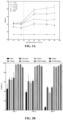
- Table 1 shows how modulation of the concentrations of Gtn-HPA, HA-Tyr, and crosslinker, and overall polymer weight of the hydrogel compositions can be used to fine tune key physical properties (e.g., stiffness, elasticity, and degradation rate).
- IPN100 corresponds to a hydrogel composition including 100% Gtn-HPA and 0% HA-Tyr.
- IPN75 corresponds to a hydrogel composition including 75% Gtn-HPA and 25% HA-Tyr.
- IPN75 corresponds to a hydrogel composition including 5% Gtn-HPA and 50% HA-Tyr.
- IPN30 corresponds to a hydrogel composition including 30% Gtn-HPA and 70% HA-Tyr.
- IPN0 corresponds to a hydrogel composition including 0% Gtn-HPA and 100% HA-Tyr.
- FIG. 2C shows the calculated Young's moduli of hydrogel compositions as a function of the concentration of Gtn-HPA in the compositions.
- the hydrogel composition has a Young's modulus ranging from about 2300 Pa to 7000 Pa.
- the Young's modulus of the hydrogel compositions can be determined by calculating the slope of the stress vs. strain curve, which is generated by performing compression tests on the hydrogel compositions at a constant strain rate (see, e.g., Example 1; section titled "Hydrogel compression test”).
- the hydrogel composition has a Young's modulus ranging from about 2900 Pa to 4500 Pa.
- the hydrogel composition has a Young's modulus of about 3100 Pa.
- the hydrogel composition has a Young's modulus of about 4300 Pa.
- the hydrogel composition has a Young's modulus ranging from at least about 2300 Pa to 2500 Pa, about 2500 Pa to 2700 Pa, about 2700 Pa to 2900 Pa, about 2900 Pa to 3100 Pa, about 3100 Pa to 3300 Pa, about 3300 Pa to 3500 Pa, about 3500 Pa to 3700 Pa, about 3700 Pa to 3900 Pa, about 4100 Pa to 4300 Pa, about 4300 Pa to 4300 Pa, or more.
- FIG. 2C shows the calculated storage or elastic moduli (i.e., G') of hydrogel compositions as a function of the concentration of Gtn-HPA in the compositions.
- the hydrogel composition has a storage or elastic modulus (i.e., G'), ranging from about 500 Pa to 6000 Pa.
- the term "stiffness," as used herein, is defined as the storage or elastic modulus (i.e., G') of a composition.
- the storage or elastic modulus (G') is a mechanical property that measures the elasticity of a material.
- the storage or elastic modulus (G') of the hydrogel compositions can be determined by performing oscillatory rheology measurements at a constant strain.
- storage or elastic modulus (G') of the hydrogel compositions can be determined by performing passive micro-rheology measurements of microbeads immersed in the compositions of the disclosure (see, e.g., Example 1; section titled “Passive micro-rheology measurements and PLGA microbeads tracking”).
- the hydrogel composition has a stiffness ranging from about 500 Pa to 1500 Pa. In some embodiments, the hydrogel composition has a stiffness of about 800 Pa. In some embodiments, the hydrogel composition has a stiffness of about 1000 Pa. In some embodiments, the hydrogel composition has a stiffness ranging from at least about 500 Pa to 600Pa, about 600 Pa to 700 Pa, about 700 Pa to 800 Pa, about 800 Pa to 900 Pa, about 900 Pa to 1000 Pa, about 1000 Pa to 1100 Pa, about 1100 Pa to 1200 Pa, about 1200 Pa to 1300 Pa, about 1300 Pa to 1400 Pa, about 1400 Pa to 1500 Pa, or more.
- the degradation rate of the hydrogel composition can be controlled based on the concentration of one or more polymers (e.g., Gtn-HPA and/or HA-Tyr) and/or concentration of crosslinker added as well as the total polymer weight.
- the degradation rate of they hydrogel composition can be quantified in vitro and in vivo (see, e.g., Example 1; section titled “In vitro degradation assays for IPNs” and Example 3; section titled “Hydrogels-retina interface and OCT analysis algorithms”).
- the hydrogel composition has a degradation rate of about 5 to 7 days. In some embodiments, the hydrogel composition has a degradation rate of about 5 days.
- the hydrogel composition has a degradation rate of about 6 days. In some embodiments, the hydrogel composition has a degradation rate of about 7 days. In some embodiments, the hydrogel composition has a degradation rate ranging from about 5 to 20 days. In some embodiments, the hydrogel composition has a degradation rate ranging from about 5 to about 6 days, about 6 to about 7 days, about 7 to about 8 days, about 8 to about 9 days, about 9 to about 10 days, about 10 to about 15 days, about 15 to about 20 days. In some embodiments, the hydrogel composition has a degradation rate of less than about 20, about 15, about 10, or about 7 days. In some embodiments, the hydrogel composition has a degradation rate of more than about 1, about 5, about 7, about 10, about 14, about 21.
- a hydrogel composition having a volume of about 200 ⁇ l has a degradation rate of about 5 to 7 days. In some embodiments, the hydrogel composition having a volume of about 200 ⁇ l has a degradation rate of about 5 days. In some embodiments, a hydrogel composition having a volume of about 200 ⁇ l has a degradation rate of about 6 days. In some embodiments, a hydrogel composition having a volume of about 200 ⁇ l has a degradation rate of about 7 days. In some embodiments, a hydrogel composition having a volume of about 200 ⁇ l has a degradation rate ranging from about 5 to 20 days.
- a hydrogel composition having a volume of about 200 ⁇ l has a degradation rate ranging from about 5 to about 6 days, about 6 to about 7 days, about 7 to about 8 days, about 8 to about 9 days, about 9 to about 10 days, about 10 to about 15 days, about 15 to about 20 days. In some embodiments, a hydrogel composition having a volume of about 200 ⁇ l has a degradation rate of less than about 20, about 15, about 10, or about 7 days. In some embodiments, a hydrogel composition having a volume of about 200 ⁇ l has a degradation rate of more than about 1, about 5, about 7, about 10, about 14, about 21.
- a hydrogel composition having a volume of about 1.5 cubed centimeters (cm 3 ) has a degradation rate of about 5 to 7 days. In some embodiments, the hydrogel composition having a volume of about 1.5 cm 3 has a degradation rate of about 5 days. In some embodiments, a hydrogel composition having a volume of about 1.5 cm 3 has a degradation rate of about 6 days. In some embodiments, a hydrogel composition having a volume of about 1.5 cm 3 has a degradation rate of about 7 days. In some embodiments, a hydrogel composition having a volume of about 1.5 cm 3 has a degradation rate ranging from about 5 to 20 days.
- a hydrogel composition having a volume of about 1.5 cm 3 has a degradation rate ranging from about 5 to about 6 days, about 6 to about 7 days, about 7 to about 8 days, about 8 to about 9 days, about 9 to about 10 days, about 10 to about 15 days, about 15 to about 20. In some embodiments, a hydrogel composition having a volume of about 1.5 cm 3 has a degradation rate of less about 20, about 15, about 10, or about 7 days. In some embodiments, a hydrogel composition having a volume of about 1.5 cm 3 has a degradation rate of more than about 1, about 5, about 7, about 10, about 14, about 21.
- the hydrogel compositions of the present disclosure can include a plurality of cells as a delivery payload.
- the plurality of cells can be mixed in with the polymers (e.g., Gtn-HPA and/or HA-Tyr) in a liquid state prior to crosslinking. After crosslinking the polymeric composition with the cells mixed in it, the cells can thus become encapsulated within the hydrogel composition.
- the hydrogel composition has a cell viability of about 85% to 95%. In some embodiments, the hydrogel composition has a cell viability of about 85% to 95% after at least about 1 day of encapsulation to about 30 days of encapsulation or more.
- the hydrogel composition has a cell viability of about 85% to 95% after 5 days of encapsulation. In some embodiments, the hydrogel composition has a cell viability of about 85% to 95% after 7 days of encapsulation. In some embodiments, the hydrogel composition has a cell viability of about 85% to 95% after 15 days of encapsulation. In some embodiments, the hydrogel composition has a cell viability of about 85% to 95% after 30 days of encapsulation.
- the hydrogel composition can include a plurality of ocular cells.
- ocular cells or "ocular cell,” as used herein, can include one or more cells that are native to the eye and/or are part of the ocular cell system.
- the plurality of ocular cells is a plurality of human retinal progenitor cells (hRPGs), human retinal ganglion cells, or any combination thereof.
- the plurality of ocular cells is a plurality of autologous stem cells that can differentiate into cells that are part of the ocular cell system.
- the plurality of ocular cells is a plurality of allogeneic stem cells that can differentiate into cells that are part of the ocular cell system.
- the plurality of ocular cells encapsulated in hydrogel compositions of the disclosure does not elicit an immune response when delivered to a subject (e.g., into an eye of the subject).
- the plurality of ocular cells is human-derived ocular cells.
- the plurality of ocular cells is a plurality of ocular cells derived from induced pluripotent stem cells (iPSCs).
- the plurality of ocular cells is a plurality of ocular cells derived from mesenchymal stem cells (MSCs).
- the hydrogel composition can include a plurality of cells at a concentration ranging from about at least about 1,000 cells per milliliter (cells/ml) to 1,000,000 cells/ml or more. In some embodiments, the hydrogel composition can include a plurality of cells at a concentration of about 1,000 cells/ml. In some embodiments, the hydrogel composition can include a plurality of cells at a concentration of about 10,000 cells/ml. In some embodiments, the hydrogel composition can include a plurality of cells at a concentration of about 100,000 cells/ml. In some embodiments, the hydrogel composition can include a plurality of cells at a concentration of about 1,000,000 cells/ml.
- the cells delivered by the hydrogel composition have a cell viability of greater than about 50% after 1 day post-delivery to the eye of the subject. In some embodiments, the cells delivered by the hydrogel composition have a cell viability of greater than about 60% after 1 day post-delivery to the eye of the subject. In some embodiments, the cells delivered by the hydrogel composition have a cell viability of greater than about 70% after 1 day post-delivery to the eye of the subject. In some embodiments, the cells delivered by the hydrogel composition have a cell viability of greater than about 80% after 1 day post-delivery to the eye of the subject. In some embodiments, the cells delivered by the hydrogel composition have a cell viability of greater than about 90% after 1 day post-delivery to the eye of the subject.
- the cells delivered by the hydrogel composition have a cell viability of greater than about 95% after 1 day post-delivery to the eye of the subject. In some embodiments, the cells delivered by the hydrogel composition have a cell viability of greater than about 98% after 1 day post-delivery to the eye of the subject.
- the cells delivered by the hydrogel composition have a cell viability of greater than about 50% after 3 days post-delivery to the eye of the subject. In some embodiments, the cells delivered by the hydrogel composition have a cell viability of greater than about 60% after 3 days post-delivery to the eye of the subject. In some embodiments, the cells delivered by the hydrogel composition have a cell viability of greater than about 70% after 3 days post-delivery to the eye of the subject. In some embodiments, the cells delivered by the hydrogel composition have a cell viability of greater than about 80% after 3 days post-delivery to the eye of the subject. In some embodiments, the cells delivered by the hydrogel composition have a cell viability of greater than about 90% after 3 days post-delivery to the eye of the subject.
- the cells delivered by the hydrogel composition have a cell viability of greater than about 95% after 3 days post-delivery to the eye of the subject. In some embodiments, the cells delivered by the hydrogel composition have a cell viability of greater than about 98% after 3 days post-delivery to the eye of the subject.
- the cells delivered by the hydrogel composition have a cell viability of greater than about 50% after 5 days post-delivery to the eye of the subject. In some embodiments, the cells delivered by the hydrogel composition have a cell viability of greater than about 60% after 5 days post-delivery to the eye of the subject. In some embodiments, the cells delivered by the hydrogel composition have a cell viability of greater than about 70% after 5 days post-delivery to the eye of the subject. In some embodiments, the cells delivered by the hydrogel composition have a cell viability of greater than about 80% after 5 days post-delivery to the eye of the subject. In some embodiments, the cells delivered by the hydrogel composition have a cell viability of greater than about 90% after 5 days post-delivery to the eye of the subject.
- the cells delivered by the hydrogel composition have a cell viability of greater than about 95% after 5 days post-delivery to the eye of the subject. In some embodiments, the cells delivered by the hydrogel composition have a cell viability of greater than about 98% after 5 days post-delivery to the eye of the subject.
- the cells delivered by the hydrogel composition have a cell viability of greater than about 50% after 7 days post-delivery to the eye of the subject. In some embodiments, the cells delivered by the hydrogel composition have a cell viability of greater than about 60% after 7 days post-delivery to the eye of the subject. In some embodiments, the cells delivered by the hydrogel composition have a cell viability of greater than about 70% after 7 days post-delivery to the eye of the subject. In some embodiments, the cells delivered by the hydrogel composition have a cell viability of greater than about 80% after 7 days post-delivery to the eye of the subject. In some embodiments, the cells delivered by the hydrogel composition have a cell viability of greater than about 90% after 7 days post-delivery to the eye of the subject.
- the cells delivered by the hydrogel composition have a cell viability of greater than about 95% after 7 days post-delivery to the eye of the subject. In some embodiments, the cells delivered by the hydrogel composition have a cell viability of greater than about 98% after 7 days post-delivery to the eye of the subject.
- the cells delivered by the hydrogel composition have a cell viability of greater than about 50% after 10 days post-delivery to the eye of the subject. In some embodiments, the cells delivered by the hydrogel composition have a cell viability of greater than about 60% after 10 days post-delivery to the eye of the subject. In some embodiments, the cells delivered by the hydrogel composition have a cell viability of greater than about 70% after 10 days post-delivery to the eye of the subject. In some embodiments, the cells delivered by the hydrogel composition have a cell viability of greater than about 80% after 10 days post-delivery to the eye of the subject. In some embodiments, the cells delivered by the hydrogel composition have a cell viability of greater than about 90% after 10 days post-delivery to the eye of the subject.
- the cells delivered by the hydrogel composition have a cell viability of greater than about 95% after 10 days post-delivery to the eye of the subject. In some embodiments, the cells delivered by the hydrogel composition have a cell viability of greater than about 98% after 10 days post-delivery to the eye of the subject.
- the cells delivered by the hydrogel composition have a cell viability of greater than about 50% after 15 days post-delivery to the eye of the subject. In some embodiments, the cells delivered by the hydrogel composition have a cell viability of greater than about 60% after 15 days post-delivery to the eye of the subject. In some embodiments, the cells delivered by the hydrogel composition have a cell viability of greater than about 70% after 15 days post-delivery to the eye of the subject. In some embodiments, the cells delivered by the hydrogel composition have a cell viability of greater than about 80% after 15 days post-delivery to the eye of the subject. In some embodiments, the cells delivered by the hydrogel composition have a cell viability of greater than about 90% after 15 days post-delivery to the eye of the subject.
- the cells delivered by the hydrogel composition have a cell viability of greater than about 95% after 15 days post-delivery to the eye of the subject. In some embodiments, the cells delivered by the hydrogel composition have a cell viability of greater than about 98% after 15 days post-delivery to the eye of the subject.
- the cells delivered by the hydrogel composition have a cell viability of greater than about 50% after 20 days post-delivery to the eye of the subject. In some embodiments, the cells delivered by the hydrogel composition have a cell viability of greater than about 60% after 20 days post-delivery to the eye of the subject. In some embodiments, the cells delivered by the hydrogel composition have a cell viability of greater than about 70% after 20 days post-delivery to the eye of the subject. In some embodiments, the cells delivered by the hydrogel composition have a cell viability of greater than about 80% after 20 days post-delivery to the eye of the subject. In some embodiments, the cells delivered by the hydrogel composition have a cell viability of greater than about 90% after 20 days post-delivery to the eye of the subject.
- the cells delivered by the hydrogel composition have a cell viability of greater than about 95% after 20 days post-delivery to the eye of the subject. In some embodiments, the cells delivered by the hydrogel composition have a cell viability of greater than about 98% after 20 days post-delivery to the eye of the subject.
- the cells delivered by the hydrogel composition have a cell viability of greater than about 50% after 30 days or more post-delivery to the eye of the subject. In some embodiments, the cells delivered by the hydrogel composition have a cell viability of greater than about 60% after 30 days or more post-delivery to the eye of the subject. In some embodiments, the cells delivered by the hydrogel composition have a cell viability of greater than about 70% after 30 days or more post-delivery to the eye of the subject. In some embodiments, the cells delivered by the hydrogel composition have a cell viability of greater than about 80% after 30 days or more post-delivery to the eye of the subject.
- the cells delivered by the hydrogel composition have a cell viability of greater than about 90% after 30 days or more post-delivery to the eye of the subject. In some embodiments, the cells delivered by the hydrogel composition have a cell viability of greater than about 95% after 30 days or more post-delivery to the eye of the subject. In some embodiments, the cells delivered by the hydrogel composition have a cell viability of greater than about 98% after 30 days or more post-delivery to the eye of the subject.
- about 50% of the cells delivered by the hydrogel composition survive after delivery to the eye of the subject. In some embodiments, about 60% of the cells delivered by the hydrogel composition survive after delivery to the eye of the subject. In some embodiments, about 70% of the cells delivered by the hydrogel composition survive after delivery to the eye of the subject. In some embodiments, about 80% of the cells delivered by the hydrogel composition survive after delivery to the eye of the subject. In some embodiments, about 90% of the cells delivered by the hydrogel composition survive after delivery to the eye of the subject. In some embodiments, about 95% of the cells delivered by the hydrogel composition survive after delivery to the eye of the subject. In some embodiments, about 96% of the cells delivered by the hydrogel composition survive after delivery to the eye of the subject.
- the hydrogel composition In some embodiments, about 97% of the cells delivered by the hydrogel composition survive after delivery to the eye of the subject. In some embodiments, about 98% of the cells delivered by the hydrogel composition survive after delivery to the eye of the subject. In some embodiments, about 99% of the cells delivered by the hydrogel composition survive after delivery to the eye of the subject.
- the present disclosure presents methods of treating or preventing (i.e., reducing risk of) an ocular disorder (e.g., a retinal disease or a disorder affecting the optic nerve) in an eye of a subject.
- an ocular disorder e.g., a retinal disease or a disorder affecting the optic nerve
- the present disclosure presents compositions for use in the treatment or prevention of an ocular disorder (e.g., a retinal disease or a disorder affecting the optic nerve) in an eye of a subject.
- the eye disorder includes congenital retinal disease, diabetic retinopathy, glaucoma, optic neuropathy, retinal neuron damage, neurofibromatosis type 1 optic nerve glioma, or any combination thereof. Treating can include improving one or more clinical parameters of the disorder, e.g., visual acuity, or delaying or reducing risk of progression of the disorder.
- the methods of treatment or prevention can include the steps of administering a therapeutically effective amount of the composition into an eye of the subject.
- the composition attaches to an inner portion of the eye after administration into the eye.
- the inner portion of the eye is the retina.
- the inner portion of the eye is the inner limiting membrane of the retina.
- the composition is administered as a liquid.
- the Gtn-HPA, HA-Tyr, and ocular cells are mixed prior to administration into the eye of the subject.
- the Gtn-HPA, HA-Tyr, ocular cell, crosslinker (e.g., hydrogen peroxide), and catalyst (e.g., HRP) are mixed prior to administration into the eye of the subject.
- these components can be mixed prior to administration and then loaded into an applicator (e.g., a syringe).
- the user e.g., clinician or healthcare practitioner
- the composition gelates in situ (i.e., inside the eye of the subject after injection) to the eye (e.g., via an intravitreal injection).
- the composition is enzymatically crosslinked in situ.
- the gelatin-HPA, HA-Tyr, and ocular cell are mixed during administration into the eye of the subject.
- the Gtn-HPA, HA-Tyr, ocular cells, crosslinker (e.g., hydrogen peroxide), and catalyst (e.g., HRP) are mixed during administration into the eye of the subject.
- these components can be mixed loaded into an applicator (e.g., a syringe) without any previous mixing.
- the components of the composition can be loaded into an applicator having separate compartments (e.g., a dual barrel syringe).
- the composition is injected into an eye of the subject by using a dual barrel syringe where a first compartment of the dual barrel syringe houses the polymers (e.g., Gtn-HPA and/or HA-Tyr) and optionally the cells, and the second compartment of the dual barrel syringe contains the crosslinker (e.g., hydrogen peroxide) and/or the catalyzer (e.g., HRP).
- the user e.g., clinician or healthcare practitioner
- the composition can then inject the composition into the eye in a liquid state and prevent gelation or crosslinking from starting until the contents of both compartments mix in situ (e.g., in the vitreous of the eye of the subject).
- the composition gelates in situ (i.e., inside the eye of the subject) after injection into the eye (e.g., via an intravitreal injection).
- the composition is enzymatically crosslinked in situ.
- microliters ( ⁇ L) of the composition e.g., precursor hydrogel composition
- ⁇ L microliters
- between about 50 and 250 microliters ( ⁇ L) of the composition e.g., precursor hydrogel composition
- the methods of treating and/or preventing an ocular disorder further include enzymatically crosslinking the composition by contacting the composition with hydrogen peroxide and horseradish peroxidase and waiting for about 30 seconds to 240 seconds for the composition to reach a gel point.
- the hydrogel composition transitions from a liquid to a gel state within about 240 seconds after adding a crosslinker. In some embodiments, the hydrogel composition transitions from a liquid to a gel state within about 200 seconds after adding a crosslinker. In some embodiments, the hydrogel composition transitions from a liquid to a gel state within about 300 seconds after adding a crosslinker.
- the hydrogel composition transitions from a liquid to a gel state within about 150 seconds after adding a crosslinker. In some embodiments, the hydrogel composition transitions from a liquid to a gel state within about 100 seconds after adding a crosslinker.
- the composition is administered via a pars plana vitrectomy. In some embodiments, the composition is administered into a vitreous of the eye of the subject. In some embodiments, the composition is administered into the inner portion of the eye is the retina.
- a new biomaterial design that can protect and catalyze the in vivo regeneration of retinal ganglion cells in the eye to potentially regenerate vision lost during diseases such as NF1-OPG is disclosed herein.
- the interpenetrating polymer network hydrogels developed can be ideal candidates for encapsulating nutrients and cells.
- the IPN hydrogels of the disclosure enabled a uniform release of stem cells throughout time and enhanced their evolution into an elongated axon-like morphology.
- the mechanical properties of hydrogels can surprisingly trigger the IPN hydrogels to graft to the correct surface (e.g., a retinal surface) by having an increased stiffness while maintaining high biocompatibility and biodegradability.
- This phenomenon might be due to the continuous shear forces present in the eye due to its movement and the dual interaction of the inner limiting membrane (e.g., integrin bonding and collagen fiber entanglement).
- the IPN hydrogels of the disclosure can provide design principles for and stem cell therapies in the eye and other organs, which depend critically on viability and stability of cells during injection and development.
- Gtn-HPA and HA-Tyr conjugate were prepared via a general carbodiimide/active ester-mediated coupling reaction (in PBS) that conjugated hydroxyphenylpropionic acid (HPA) to gelatin (MW 20-80kDA) and Tyramine (Tyr) to Hyaluronidase (HA) (MW 80-150 kDa). 90% of the amine groups were conjugated with HPA or Tyramine.
- Hybrid interpenetrating networks (IPN10, 25, 50, 75 and IPN90) were prepared by mixing the corresponding amounts of Gtn-HPA and HA-Tyr at 2wt% solution (e.g. IPN75 corresponds to 75% of Gtn-HPA and 25% of HA-Tyr both at 2wt% solution).
- IPN75 corresponds to 75% of Gtn-HPA and 25% of HA-Tyr both at 2wt% solution.
- H2O2 0.8, 0.9, 1 and 1.3 mM
- Hyaluronic acid with high molecular weight (HHA) (MW 1200 kDA, Sigma-Aldrich) was dissolved in PBS at 2, 3 and 5% wt solution with hRGCs by thoroughly mixing the samples with a vortex throughout the experiment. No chemical reagent was added, and physical crosslinking was seen in less than a minute.
- Collagen-Genipin (CG) hydrogels were prepared by dissolving collagen type I from calf skin (Sigma-Aldrich) in a 2%wt solution and mixing it with hRGCs. Genipin 98%-HPCL (Sigma-Aldrich) at concentrations of 0.5, 1, 5, and 10 mM were then added to the solution containing polymer and cells. Hydrogels were incubated at 37 °C and reached stability after 20 min.
- Oscillatory rheology was performed with a TA instruments AR-G2 rheometer using cone and plate geometry of 40 mm diameter and 2° angle. For each measurement, 200 ⁇ l of each sample (Gtn-HPA, IPN90, IPN75, IPN50, IPN25, IPN10, and HA-Tyr) at 2% wt/vol, containing 0.1 U/ml of HRP and varying concentrations of H 2 O 2 (ranging from 0.8-1.3 mM) was applied to the bottom plate immediately after mixing. All hydrogels having a gelation time comprised between 30s-3min samples were still liquid when applied onto the bottom plate. The upper cone was lowered to a measurement gap of 51 ⁇ m.
- DSC Differential scanning calorimetry
- GPC Gel permeation chromatography
- HA-Tyr and Gtn-HPA were dissolved in 2 mL of the mobile phase, at a concentration of 10% wt/mL, by thoroughly mixing and incubating samples for 1h at 37 °C.
- Molecular weights were then referenced against polyethylene glycol standards (Waters).
- Unconfined compression tests were performed using a Zwick/Roell Z2.5 static materials tester (Zwick GmbH & Co., Ulm, Germany) with integrated testing software (testXpert, Zwick).
- 1 mL of Gtn-HPA, IPN50, IPN75, and HA-Tyr were disposed into 24-well plates to create samples about 16 mm in diameter and about 3-4 mm in thickness. All hydrogels were left to fully crosslink and stabilize for 2 hours at 37 °C before performing compression testing. All gels were swelled in PBS for 1 hour before compression testing.
- FTIR Fourier transform infrared
- FTIR Fourier transform infrared
- 200 ⁇ l gels (Gtn-HPA, IPN75, IPN50, IPN25 and HA-Tyr) were prepared as previously described in the section titled "Hydrogel Preparation.” Hydrogels were prepared and incubated for 30 min at 37 °C to reach stability. Samples were then combined with 200 ⁇ l of phosphate-buffered saline (PBS) containing 1000 U/ml type IV collagenase (Invitrogen) or containing 500 U/mL hyaluronidase type I-S (Sigma-Aldrich) and incubated at 37 °C on an orbital shaker at 150 revolutions per minute (rpm). Samples were collected every 5 or 10 minutes for 1 or 2 hours, for collagenase or hyaluronidase treatments, respectively, and analyzed for degradation products using the bicinchoninic assay (Thermo Fisher Scientific).
- PBS phosphate-buffered saline
- I-S hy
- a MATLAB program was then used to calculate the mean square displacement and fit the data with an exponential function.
- This fit function was then used to calculate the complex modulus by feeding the fit function data into a MATLAB function that fits this data with a second-order polynomial function from which the first- and second-time derivatives are computed and from that the complex modulus.
- the storage modulus (elastic) G' and loss modulus (viscous) G" were measured in order to calculate the gel point (defined by the crossing of G' and G").
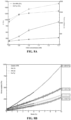
- FIG. 2A It is desirable for a biomaterial to have optimal characteristics ( FIG. 2A ) and tunability as well as biocompatibility and biodegradability.
- a short-term viability study corresponding to the encapsulation of retinal stem cells in different example hydrogels, was performed for 3 days. The results suggested that only networks incorporating Gelatin Hydroxyphenyl propionic acid (Gtn-HPA) and Hyaluronic acid tyramine (HA-Tyr) have a sufficient biocompatibility (hence cell viability) to be considered as candidate for enhancing retinal regeneration (see FIG. 6E ). These might have been anticipated because the vitreous is the only part of the body that contains both of these biopolymers. However, their particular structure is not the same as that of the hybrid gels.
- FIGS. 8A-8C show the time evolution of loss and storage modulus, calculated from microbeads' MSD, which corresponded to the MSD being null.
- Gel point ranged from 42 seconds (s) for Gtn-HPA to 2 minutes 47 seconds for IPN50, which conveyed the fact that gelation time is tunable by controlling the Gtn-HPA content of IPN.
- the PLGA microparticles are bigger than the pore size of the IPN and, while they showed what the gel point is, they did not present the crosslinking kinetics of these hydrogels.
- FIGS. 9A-9C Complete characterization of solid polymers ( FIGS. 9A-9C ), homo-polymeric networks, and IPN was performed and is summarized in Table 2 shown below.
- hRGCs Human retinal ganglion cells
- hRGCs Human retinal ganglion cells
- Hydrogels preparation were pipetted onto fibronectin-coated round coverslips glass (thickness 5mm, diameter 1 cm, VWR). After 1, 3, and 7 days of incubation with media or PBS, they were incubated with 2.5 ⁇ M calcein AM (FITC) and 10 ⁇ M ethidium homodimer-1 (Cy3) in PBS for 15 min at 37 °C and 5% CO 2 .
- FITC ⁇ M calcein AM
- Cy3 10 ⁇ M ethidium homodimer-1
- hRGCs (3 ⁇ 10 5 cells/mL in PBS or in 1 mL of Gtn-HPA, IPN75 and IPN50 hydrogels, in 6-well plates (3.5 cm diameter, polystyrene, flat bottom, sterile, fisher scientific)] were maintained in PBS for 5 days (replicating the in vivo conditions).
- hRGCs from the 4 different conditions-in PBS, in Gtn-HPA, IPN75 and IPN50 were collected and fixed with a Perm/Fix buffer (BD Biosciences) at 4 °C for 15 min. Cells were then washed in a wash buffer (BD Biosciences) and incubated, at room temperature, in a blocking buffer (Pharmingen staining buffer with 2% goat serum) for 30 min.
- Blocked cells were seeded onto a flat bottom 96-well plate (treated, sterile, polystyrene, Thomas Scientific) and stained overnight at 4°C with the following primary antibodies: Brn3a, RBPMS, Thy1.1 (ganglion cell marker), Caspase9 (apoptosis marker), Ki67 (proliferation marker), Cmyc, Oct4 (sternness markers) and NeuN (neuronal marker).
- Primary antibodies were diluted in 200 ⁇ L of antibody buffer (TBS, 0.3% Triton X-100 and 1% goat serum).
- a conjunctival incision and a small sclerotomy were performed using a fine disposal scalpel in all rats.
- H&E Hematoxylin and Eosin
- hRGCs cultured on microscope cover slips, and cryosections from Long Evans rats left eyes were fixed with 4% paraformaldehyde in 0.1 M PBS (Irvine Scientific) at room temperature for 20 min. These fixed cells and sections were blocked and permeabilized with a blocking solution ((Tris-buffered saline (TBS), 0.3% Triton X-100 and 3% goat serum (Jackson Immunoresearch Laboratories, West Grove, PA) for 15 min.
- TBS Tris-buffered saline
- Triton X-100 0.3% Triton X-100
- goat serum Jackson Immunoresearch Laboratories, West Grove, PA
- hydrogels for cell encapsulation implies tuning the materials to be able to conserve many cell characteristics: high viability, differentiation, phenotype, shape or distribution through the gel.
- human retinal ganglion cells see Example 2; section titled “Source and viability of hRGC”
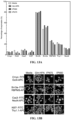
- results obtained from a short-term viability study, seen in FIGS. 10A-10B and FIG. 3A yielded the optimal crosslinker (H 2 O 2 ) concentration which would lead to the highest cell viability both in IPN and homopolymeric hydrogels: 1mM of H 2 O 2 .
- 3A shows the impact of Gtn-HPA content in IPN on encapsulated cell viability.
- a viability threshold corresponding to a content of >30% of Gtn-HPA, was observed for all crosslinker concentrations proving that cells do not thrive in HA-Tyr.
- This finding can be related to the understanding that hRGCs possess integrins to attach to the gelatin backbone but not to the hyaluronic acid backbone.
- This result reduced the number of hydrogel candidates to IPN possessing a Gtn-HPA content higher than 50% (which include Gtn-HPA itself).
- cell viability in all IPN was significantly higher than when cells were left in phosphate buffer saline (PBS) for 3 days.
- PBS phosphate buffer saline
- IPN reduced this drop of viability by protecting the cells from the lack of nutrients for all time points, by being significantly higher for all IPN compared to PBS. After 1 week of media deprivation, 35-45% of hRGCs were still viable. This result was unexpected due to the relative fragility of hRGC, and further suggested that these IPNs are excellent candidates for enhancing the ability of encapsulated cells to regenerate the retina.
- the intensity of live (CalceinAM-FITC) and dead (Ethidium Bromide-APC) cells was averaged, normalized, and measured in function of all directions (x,y,z). Directions were averaged into 150 ⁇ m and compared to a sample of cells in media on a flat slide ( FIG. 2C ).
- the flat microscope slide showed a peak in intensity around its zero positions with a quickly decaying intensity when distancing from the slide, while all hydrogel samples showed a constant intensity in all directions.
- FIG. 3D The size, compactness, and distribution of hRGC (after live and dead staining) was visualized via confocal microscopy throughout 300 ⁇ m sections of gels ( FIG. 3D ).
- Image processing algorithms FIGS. 11 and 12A-12B ) permitted the quantification of live cells size and compactness in all samples. No difference was noted between time points; however, cell size was measured to be significantly larger for cells encapsulated in gel compared to media and PBS while their compactness was smaller ( FIG. 12A-12B ). This finding suggested that hydrogels may enable cell differentiation and processes formation, which could not be seen in media samples.
- hRGC viability images were taken at 20x magnification with a z-stack of 300 ⁇ m and 22 steps.
- a 3D projection was used for qualitative analysis while maximum projection was applied as quantification.
- Cells in 15 randomly selected, maximumprojected fields of view were counted under 20x objective lens magnification with a cell counting and an analyzing image processing algorithm.
- Cell numbers green for live and red for dead
- size area of positive pixels
- compactness difference in cell shape as compared to a disk
- the percentage of viable cells was calculated by dividing the number of live cells (FITC) by the total number of cells in the given area (live and dead cells added).
- LASX Leica software enabled the measurement of the average intensity of a marker along each (x,y,z) direction. These intensities were measured for each z-stack, in each group after normalization, and averaged.
- a MATLAB algorithm was created that consisted in calculating and counting (with a tolerance of 0.01%) the number of colored pixels (Green for FITC channel and Red for phycoerythrin (PE) or allophycocyanin (APC) channels) for each marker. Then, a percentage of the total number of colored pixels as compared to the total number of pixels of the image was calculated; this percentage corresponded to the surface coverage of the specific marker.
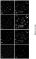
- Entire tiling of the retina was performed on all injected groups (Gtn-HPA, IPN75, IPN50 and PBS) at 63x with oil objective magnification and z-stack of 20 ⁇ m with 50 steps.
- the tiling square size was 25x10 fields of views, which was reduced by only choosing the field of views containing parts of the retina.
- STEM121-FITC with DAPI-Vioblue and TdTomato-PE staining were analyzed in a larger quantity than all other images. Images were taken at 63x magnification with a 15- ⁇ m z-stack and 22 steps. For each group, 60 fields of view, chosen in the center of the retina (where the injection was performed) were analyzed.
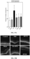
- Islands of gels were visible during SD-OCT data acquisition and a quantification of gel presence was performed by applying an image processing thresholding algorithm.
- This algorithm was based on Otsu's method of thresholding and enables a specific quantification of the gel degradation in vivo.
- After taking the OCT images of the section of the retina 10 fields of views were analyzed for each group.
- Otsu's method the retina and the noise were processed to be considered as the background while the gels were processed to be considered as the foreground.
- Most of the noise was deleted by the plugin in the SD-OCT software, hence the rest of the image was just composed of a bright retina (curved with different layers) and islands of gel on top of it).
- the improvement in engraftment of hRGCs by the IPN hydrogels was quantified by measuring cell migration (e.g., location in retinal layers after 1 month), co-localization (e.g., expressing both Brn3b intrinsically and STEM121 human markers), and, for large cells, orientation (e.g., angle formed by extended processes and retina).
- cell migration e.g., location in retinal layers after 1 month
- co-localization e.g., expressing both Brn3b intrinsically and STEM121 human markers
- orientation e.g., angle formed by extended processes and retina.
- a two-step algorithm was designed. First, pixel intensities of input images were normalized from 0 to 1. Due to the large amount of noise in the image, the segmentation pipeline was started with a small amount of gaussian blur to smooth the image. Part of the cells were very dim; therefore, to segment them, a first low threshold (around 10% of intensity) was used to segment cells. To remediate cells which displayed small holes, a closing morphological operation (consisting of a dilation followed by an erosion) was incorporated. An opening (an erosion followed by a dilation) was run to remove small clusters of pixels that were most likely noise. For large cells, the low threshold followed by morphological operation led to a high recall for cell pixels.
- the brightest part of cells was segmented using a second larger threshold (about 0.3).
- a fusion algorithm was designed. Using the rough segmentation obtained from the first threshold, a connected component algorithm was employed to label each independent group of segmented pixels. A group was defined as independent if it was not in contact with another group of segmented pixels. Then, each labeled group of segmented pixels, which did not contain pixels from the high threshold segmentation, was eliminated. The final result had a high precision (due to the high threshold) and a high recall (due to the low threshold). Labeled groups of segmented pixels were used to compute the area of segmented cells.
- the co-localization program can analyze the content of images taken with confocal microscopy. First, regions of interest in images taken previously (cells in RGC layer) were cropped and transformed into DIP images. Following this, images were filtered, selected based on a threshold, and analyzed with the co-localization, Pearson's, and Mader's algorithms. Co-localization consists in finding the fraction of pixels which possess a high intensity in both colors (green and red, in this study) with linear approximation. The p-value, Pearson coefficient, Mader's coefficient, and a co-localization number were then calculated and reported.
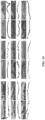
- IPN50, IPN75 and Gtn-HPA hydrogels were injected into the vitreous of Long Evans rats.
- the daily morphology of their vitreous and retina was checked daily with SD-OCT, and, after 3 days, these retinal samples were histologically stained with H&E and imaged ( FIG. 4A ). Histology images show the presence of islands of hydrogels in the vitreous and attachment of the hydrogels to the inner limiting membrane (ILM) of the retina.
- IPM inner limiting membrane
- IPN50 hydrogel possessed the highest attachment with the lowest number of "holes” (shown as dark bands), while the distribution of Gtn-HPA hydrogel was uneven with numerous "holes” throughout the interface.
- the average interface length was 10 ⁇ m smaller for IPN75 and Gtn-HPA hydrogels as compared to IPN50 hydrogels.
- IPN hydrogels including Gtn-HPA and HA-Tyr may enhance engraftment, extension of encapsulated hRGC processes, and retinal regeneration.
- 50,000 hRGC encapsulated in 3 ⁇ l of Gtn-HPA, IPN75, IPN50 hydrogels, and in PBS were injected into the vitreous of immunosuppressed Long Evans rats. Viability of cells was checked through the experiment and ranged from about 91% at the start of the experiment to about 87% after 4h. A long-term study lasting 1 month was performed to measure the impact of IPN hydrogels on cell engraftment and STEM121 (human marker shown in green on the immunohistochemistry images) was used to stain the injected cells.
- FIGS. 16A-16B By tiling multiple images of the entire retina ( FIGS. 16A-16B ), the presence of cells one month after the injection in the center of the retina was shown. The cells were located next to the base of the optic nerve for all groups (IPNs and PBS) while analyzed sections showed their location: mainly in RGC and INL layers of the retina for all groups. These findings suggested the success of injection in most animals. A major difference was observed ( FIG. 5A ) in terms of cell morphology between groups. Cells encapsulated in hydrogels were found to have high processes extended toward the optic nerve (i.e., long axons). IPN50 and IPN75 hydrogels also showed cells already at the base of the Optic Nerve (ON) and extending their axons inside the fibrous layers of the ON.
- ON Optic Nerve
- NF1-OPG Type 1 neurofibromatosis optic pathway glioma
- FIG. 5C shows, in polar coordinates, the size of these cells (as indicated by distance “r” extending from the origin) as a function of their relative orientation to the retina (as indicated by angle theta " ⁇ ,” defined as the angle measured between distance "r” and the x-axis).
- Immune response and Müller cell activation assays were also performed by checking and analyzing the surface coverage of immune cells markers (IBA1 and CD45) and activated Müller cell marker (GFAP) for all groups ( FIGS. 18A-18B ). Immune response was found to be significantly higher for cells injected in PBS while the immune response to IPN-cells injection was similar to the sham condition. A similar trend was observed for GFAP expression suggesting a higher retinal disturbance with PBS injections.
- IBA1 and CD45 immune cells markers
- GFAP activated Müller cell marker
- FIGS. 6A-6D Chemical structures of all materials (polymer and hydrogels), used in the first viability assay to find potential candidates for the enhancement of retinal regeneration, are shown in FIGS. 6A-6D .
- Gtn-HPA FIG. 6A
- HA-Tyr FIG. 6Be
- HRP horseradish peroxidase
- H 2 O 2 hydrogen peroxide
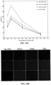
- Collagen-Genipin (CG) hydrogel samples (averaged for all Genipin concentrations) showed the lowest viability of all, due to the fragility of retinal cells and the relatively high cytotoxicity of Genipin needed to produce a stable hydrogel. Due to its high molecular weight and high stiffness, HA alone showed really low viability after 2 days, even lower than PBS. By reducing the percent of HA and using the chemical induced crosslinking with HA-Tyr and H 2 O 2 viability reached 37-39%.
- FIG. 2B shows the in vitro enzymatic degradation kinetics for both homopolymeric networks Gtn-HPA and HA-Tyr and the IPN with different content of each polymer: ranging from 25% Gtn-HPA in IPN25 to 75% in IPN75.
- Enzymatic degradation by collagenase or hyaluronidase shows a percentage of mass loss being equal to the content of respectively Gtn-HPA or HA-Tyr in the IPN. This result suggested a strong crosslink selectivity producing a hybrid IPN hydrogel.
- Fourier transform infrared spectroscopy FTIR
- the amide A band arising from N-H stretching was distributed at 3308 and 3277 cm -1 , C-H stretching at ⁇ 2945 and 2912 cm -1 for the amide B, N-H deformation at 1539 and 1574 cm -1 for the amide II respectively for Gtn-HPA and HA-Tyr.
- C O stretching at 1609 cm -1 for the amide I was observed in HA-Tyr while the amide III was seen at 1237 cm -1 for Gtn-HPA.
- the presence of HPA side group was seen by the peaks at 1452, 1633 and 3085 cm-1 while Tyramine was visible at 1378 and 3085 cm -1 .
- This fit function was then used to calculate the average complex modulus (every 2s) by feeding it into MATLAB.
- the data was fitted a second-order polynomial function from which the first and second time derivative and subsequently the complex modulus were computed.
- the storage modulus (elastic) G' and loss modulus (viscous) G" were then calculated and plotted in function of time post mixing (addition of crosslinker) as seen in FIG. 8C .
- G' and G" measurements confirmed the dependency of gel point on the Gtn-HPA content. Gel point ranged from 42 s for Gtn-HPA, being far too quick for surgical needs, to 162 s for IPN50 hydrogels. This was a crucial result as it confirmed the hypothesis that these biocompatible IPN can be tuned to fulfill surgical needs.
- Cells were encapsulated in hydrogels (deprived from nutrients) and, after 5 days, were analyzed to evaluate the efficiency of culture condition using a live-dead assay.
- the percentage of viable cells was calculated by dividing the number of live cells (green) by the total number of cells in the given area. Data of each group were calculated from 15 randomly chosen fields in each group.
- Oxidative stress was already really high at 2.5 mM with a viability ranging from 20% to 35% while being maximal at 5 mM where most cells died (only 5-8% viable).
- This broad testing of hydrogen peroxide effect on cell viability in both homo-polymeric networks suggested that a concentration around 1 mM should be used in order to make the most biocompatible hydrogel.
- a sharper testing for different IPNs content with crosslinker concentration ranging from 0.8 to 1.3 mM was performed.
- the optimal IPN was found to contain at least 30% of Gtn-HPA with a crosslinker concentration of 1 mM.
- the catalyst (HRP) concentration was optimized at 0.1 U/mL to enable encapsulated cells to thrive.
- FIG. 10B Live and dead staining of cells encapsulated in IPNs and grown in PBS are presented in FIG. 10B . Images represent a maximal projection of 200 um z-stack for hydrogels samples which were seeded at the same concentration. Cells in 2D culture appeared more fibroblastic with a higher number of dead cells. In contrast, cells encapsulated in Gtn-HPA and IPNS looked to retain their phenotypic morphology. These results clearly demonstrated the effect of oxidative stress and protective nature of Gtn-HPA and IPNs for hRGC culture and possible transplantation.
- the final image can be seen in FIG. 11B .
- perimeters extraction of cell or cell groups were performed, as seen in FIG. 11C . Some cells might be grouped and counting their number may be critical for viability results.
- the image was modified by finding the maxima (corresponding to the cell nuclei) and transformed the image to show the perimeter and these maxima ( FIG. 11D ).
- the watershed algorithm was finally applied, which finds all connected components and enables for an easy cell counting ( FIG. 11E ).
- 33 cells were extracted ( FIG. 11F ).
- Cell size was also extracted and measured with the longest line drawn inside each object ( FIG. 11G ).
- Segmenting grouped cells worked well and enabled a coherent cell size and number measurement. Detected cells appeared to have a similar size to real cells (broadly measured with scale on confocal microscope). By counting some fields of views manually and comparing to the algorithm result, it is estimated that this algorithm has a rate of success of about 95%.
- This image processing algorithm was applied to the long viability assay performed on hRGC for 1, 3, and 7 days in different conditions (see FIG. 3B in the main text). All extracted values from the image processing algorithm can be seen in FIGS. 12A-12B for all samples. Live (green) and dead (red) cell size distribution was analyzed for all groups and shows no significant difference in time. The results suggested that dead cells are, on average, 10 ⁇ m smaller than live cells. A significantly higher live cell size was found in hydrogel samples (around 15um for Gtn-HPA, 20 ⁇ m for IPN75 and IPN50) compared to cells cultured in 2D environment (8-10um for media and PBS).
- Phenotype was checked by flow cytometry and immunohistochemistry on cells cultured for 5 days in 2D (media) or 3D (Gtn-HPA, IPN75 and IPN50 with media) conditions.
- the hRGC phenotypic expression was measured in previous studies and was shown to have a high positive cell population expressing early RGC markers (as Brn3a and Brn3b) with a moderate population of late retinal ganglion cells.
- Percentage of expression of retinal ganglion cells (Brn3b, Brn3a, RBPMS and Thy1.1), neuronal (NeuN), apoptosis (Cas3), proliferation (Ki67) and sternness (Cmyc and Oct4) was measured and is reported in FIG.
- FIG. 13A While actual cell expression is shown with confocal microscopy fluorescent images ( FIG. 13B ). Images were taken at field of views presenting a high expression of each marker and are not representative of overall marker percentage of expression. Of note is that a critical factor of using a scaffold to encapsulate cells was to ensure the maintenance of phenotype throughout the entire experiment.
- hRGCs were Brn3b-Tdtomato positive; therefore, Brn3b expression was primordial to confirm maintenance of cell phenotype by hydrogels and protocols.
- Brn3b expression was high for all samples (about 80%), which also indicated the possibility of locating injected cells in tissue without the need to stain for multiple cell markers.
- Brn3a is a marker which overlaps with Brn3b and was shown here to be relatively high for all samples (about 50%) indicating a high percent of early retinal ganglion cells in the population.
- RBPMS and Thy 1.1 are markers for late retinal ganglion cells, already fully differentiated. Part of the hRGC population (about 20%) was expressing those two markers, suggesting cells differentiated already present.
- retinal ganglion cells main objective is to extend processes (e.g., axons) that merge into fibers to form the optic nerve.
- processes e.g., axons
- These fibers express some neuronal markers such as NeuN.
- a small part of the hRGC population expressed NeuN after 5 days of encapsulation in hydrogels (about 10%), which suggested that injected cells could potentially be able to extend processes that could attach the retina and regenerate a dying RGC layer.
- This assay enabled the confirmation of the maintenance of hRGC phenotype when encapsulated in IPN with nutrients for 5 days in vitro.
- IPN hydrogels could be mechanically tuned in order to make contact or attach with the back of the eye (inner limiting membrane of the retina: ILM)
- ILM inner limiting membrane of the retina
- H&E staining was performed 1- and 10-days post-injections.
- the hydrogel was not fully attached to the eye but was present in the vitreous cavity, while in the latter case, most of the hydrogel was already degraded without any detachment of the retina or its layers.
- STEM121 positive cells were observed next to the optic nerve for IPN50 and IPN75 while being in the center of the retina for Gtn-HPA and sparsely distributed everywhere for the PBS sample. Due to its low intensity and the size of tiling, intrinsic TdTomato-Brn3b was observed to be really dimmed in all tiling. These tiling suggested not only the success of the xenotransplantation, but also of engraftment due to the long-term experiment. Indeed, due to the fast degradation of hydrogels injected cells had to attach and integrate to living tissue in order to thrive for 1 month in vivo. These tiling also enabled to measure the size of the retina and by using an image processing algorithm (explained in the next section) to calculate the exact fraction of injected cells (50k per eye) engrafted after 1 month for each whole eye.
- image processing algorithm explained in the next section
- FIG. 16B To further analyze location and engraftment of injected human retinal ganglion cells, sample sections were imaged at 20x magnification ( FIG. 16B ). Cells were found to be mainly in the retinal ganglion cell (RGC), which is the targeted layer, and the inner nuclear layer (INL) some migrated towards the choroid and stopping in the outer nuclear layer (ONL). On first sight, it was observed that cells in stiffer hydrogels (IPN5) and IPN75) were larger and more abundant than the one in Gtn-HPA. Overall cells injected in PBS were found to have the smallest size and were really low in number throughout all sections analyzed. Compared to tiling, intrinsic TdTomato-Brn3b was observable in these images and shows coherent location with injected cells (co-localization of both markers being analyzed in the next section).
- RRC retinal ganglion cell
- IPN75 inner nuclear layer
- the image processing algorithm was capable of localizing cells in their respective layer of the retina, calculating their relative orientation compared to the tissue, and measuring the amount of both markers (STEM121 and Brn3b) co-localization in the cell body.
- Example 3 Section titled “Image processing algorithms for detection of cell migration, co-localization and orientation compare to retinal layers”
- the algorithm consisted in a 2-step thresholding process which separated the cell center (possessing a high intensity) from their surrounding (dimmer intensity).
- To use a simple non-realistic analogy whole cells were first segmented, then nuclei were segmented, and finally cells not containing any nucleus were removed. This extraction worked with most cells however, precision suffered: some pixels - especially around the border of the image - were above the threshold, leading to segmentation towards unwanted parts of the image.
- the 5-pixel border of each image (which corresponded to ⁇ 1% of the image) was deleted. The 5-pixel border usually only removed about 1-2 cells per picture.
- each image was annotated with the retinal layers as seen with DAPI staining ( FIGS. 17A-17B ).
- This annotation was done by hand using a simple online tool (Makesense.ai), which enabled each image to be divided into retinal regions (Choroid, ONL, INL, RGC and Vitreous). By merging the coordinates of cell extraction with the retinal layer, each cell engraftment in the retina was finally able to be localized (as seen in FIG. 5B ).
- the algorithm consisted in calculating the surface coverage of each marker: calculating the number of positive pixels for each marker (green for IBA1 and red for CD45) and normalizing by the total number of pixel in the image.
- expression of both immune markers was significantly higher in PBS than all other samples.
- the sham condition i.e., consisting in stabbing the eye with the needle without injecting hydrogels, cells, or buffers. All hydrogel samples possessed a similar expression of immune cell markers compared to the sham condition but had a higher expression than the control condition.
- Müller cells form the entrance path inside the retina. They extend their process through all the layers partly forming the barrier between the retinal ganglion cell layer and the vitreous: the inner limiting membrane. When activated, due to entrance of pathogens, migrating immune cells or exogenous ed human cells, Müller cells express largely Glial fibrillary acidic protein (GFAP). To measure Müller cells activation, ocular tissue sections were stained with GFAP and imaged via fluorescent confocal microscopy, as seen in FIG. 19B . By using the same image processing algorithm used for the immune response, GFAP expression was quantified for all samples ( FIG. 19A ).
- GFAP Glial fibrillary acidic protein
Landscapes
- Health & Medical Sciences (AREA)
- Chemical & Material Sciences (AREA)
- Life Sciences & Earth Sciences (AREA)
- Medicinal Chemistry (AREA)
- General Health & Medical Sciences (AREA)
- Pharmacology & Pharmacy (AREA)
- Veterinary Medicine (AREA)
- Public Health (AREA)
- Animal Behavior & Ethology (AREA)
- Epidemiology (AREA)
- Engineering & Computer Science (AREA)
- Organic Chemistry (AREA)
- Chemical Kinetics & Catalysis (AREA)
- Ophthalmology & Optometry (AREA)
- Polymers & Plastics (AREA)
- Biomedical Technology (AREA)
- Dermatology (AREA)
- Neurosurgery (AREA)
- Molecular Biology (AREA)
- Inorganic Chemistry (AREA)
- Cell Biology (AREA)
- Developmental Biology & Embryology (AREA)
- Bioinformatics & Cheminformatics (AREA)
- General Chemical & Material Sciences (AREA)
- Nuclear Medicine, Radiotherapy & Molecular Imaging (AREA)
- Neurology (AREA)
- Immunology (AREA)
- Virology (AREA)
- Zoology (AREA)
- Proteomics, Peptides & Aminoacids (AREA)
- Biotechnology (AREA)
- Biochemistry (AREA)
- Materials Engineering (AREA)
- Materials For Medical Uses (AREA)
- Medicinal Preparation (AREA)
- Pharmaceuticals Containing Other Organic And Inorganic Compounds (AREA)
Claims (15)
- Zusammensetzung bestehend aus Gelatine-Hydroxyphenylpropionsäure (Gelatine-HPA), Hyaluronsäure-Tyramin (HA-Tyr) und einer Augenzelle zur Verwendung in einem Verfahren zur Behandlung oder zur Vorbeugung einer Augenerkrankung bei einem Patienten, der diese benötigt, wobei das Verfahren umfasst:Verabreichung einer therapeutisch wirksamen Menge der Zusammensetzung in ein Auge des Patienten;wobei die Zusammensetzung nach der Verabreichung in das Auge an einem inneren Teil des Auges haftet.
- Zusammensetzung zur Verwendung nach Anspruch 1, wobei die Augenerkrankung angeborene Netzhauterkrankung, diabetische Retinopathie, Glaukom, optische Neuropathie, Schädigung der Netzhautneuronen oder irgendeine Kombination davon umfasst.
- Zusammensetzung zur Verwendung nach Anspruch 1 oder 2, wobei die Zusammensetzung Wasser und/oder, wobei die Zusammensetzung Wasser in einer Konzentration von etwa 88 % bis 98 % umfasst.
- Zusammensetzung zur Verwendung nach irgendeinem der Ansprüche 1-3, wobei die Zusammensetzung als eine Flüssigkeit verabreicht wird.
- Zusammensetzung zur Verwendung nach irgendeinem der Ansprüche 1-4, wobei die Zusammensetzung in situ geliert.
- Zusammensetzung zur Verwendung nach irgendeinem der Ansprüche 1-5, wobei die Zusammensetzung über eine intravitreale Injektion verabreicht wird; und/oder, wobei die Zusammensetzung nach einer Pars-plana-Vitrektomie verabreicht wird.
- Zusammensetzung zur Verwendung nach irgendeinem der Ansprüche 1-6, wobei die Zusammensetzung eine polymere Konzentration von etwa mindestens 2 Gew.-% aufweist; und/oder, wobei die Zusammensetzung Gelatine-HPA in einer Konzentration von etwa 30 % bis 100 % umfasst, und/oder wobei die Zusammensetzung HA-Tyr in einer Konzentration von etwa 70 % bis etwa 0 % umfasst.
- Zusammensetzung zur Verwendung nach irgendeinem der Ansprüche 1-7, wobei die Zusammensetzung ein Konzentrationsverhältnis von Gelatine-HPA zu HA-Tyr von etwa 50 %: 50 % umfasst; oder,
wobei Zusammensetzung ein Konzentrationsverhältnis von Gelatine-HPA zu HA-Tyr von etwa 75 %: 25 % umfasst. - Zusammensetzung zur Verwendung nach irgendeinem der Ansprüche 1-8, wobei die Gelatine-HPA, HA-Tyr und Augenzelle vor der Verabreichung in das Auge des Patienten vermischt werden.
- Zusammensetzung zur Verwendung nach irgendeinem der Ansprüche 1-9, wobei die Gelatine-HPA, HA-Tyr und die Augenzelle während der Verabreichung in das Auge des Patienten vermischt werden.
- Zusammensetzung zur Verwendung nach irgendeinem der Ansprüche 1-10, wobei die Augenzelle menschliche Netzhaut-Vorläuferzellen (hRPGs), eine menschliche Ganglienzelle der Netzhaut oder irgendeine Kombination davon umfasst.
- Zusammensetzung zur Verwendung nach irgendeinem der Ansprüche 1-11, wobei die Zusammensetzung eine Konzentration der Augenzellen von etwa 1.000 Zellen pro Milliliter bis 1.000.000 Zellen pro Milliliter umfasst.
- Zusammensetzung zur Verwendung nach irgendeinem der Ansprüche 1-12, die weiter enzymatisches Vernetzen der Zusammensetzung durch Kontakt der Zusammensetzung mit Wasserstoffperoxid und Meerrettich-Peroxidase und Warten für etwa 30 Sekunden bis 240 Sekunden bis die Zusammensetzung geliert.
- Zusammensetzung zur Verwendung nach Anspruch 13, wobei die Zusammensetzung den Gelpunkt nach der Verabreichung ins Auge des Patienten erreicht; und/oder, wobei die Zusammensetzung mit Wasserstoffperoxid in einer Konzentration von etwa 0,5 mM bis 2,5 mM in Kontakt gebracht wird und/oder, wobei die Zusammensetzung mit Meerrettich-Peroxidase in einer Konzentration von etwa 0,1 Einheiten pro Milliliter (U/ml) bis 0,2 U/ml in Kontakt gebracht wird; und/oder, wobei die Zusammensetzung enzymatisch in situ vernetzt wird; und/oder, wobei die enzymatisch vernetzte Zusammensetzung eine Steifheit aufweist, die von etwa 500 Pa bis 1500 Pa reicht; und/oder, wobei die enzymatisch vernetzte Zusammensetzung einen Elastizitätsmodul aufweist, das von etwa 2300 Pa bis 7000 Pa reicht; und/oder, wobei die Zusammensetzung in einen Glaskörper des Auges des Patienten verabreicht wird; und/oder, wobei der innere Teil des Auges die Netzhaut ist; optional, wobei der innere Teil des Auges die innere Grenzmembran der Netzhaut ist.
- Zusammensetzung zur Verwendung nach irgendeinem der Ansprüche 1-14, wobei die Zusammensetzung eine Zelllebensfähigkeit von etwa 85 % bis 95 % aufweist.
Priority Applications (1)
| Application Number | Priority Date | Filing Date | Title |
|---|---|---|---|
| EP24213735.4A EP4523693A3 (de) | 2019-12-03 | 2020-12-03 | Injizierbare hydrogele zur zellabgabe an den glaskörper |
Applications Claiming Priority (2)
| Application Number | Priority Date | Filing Date | Title |
|---|---|---|---|
| US201962942928P | 2019-12-03 | 2019-12-03 | |
| PCT/US2020/063115 WO2021113515A1 (en) | 2019-12-03 | 2020-12-03 | Injectable hydrogels for cell delivery to the vitreous |
Related Child Applications (2)
| Application Number | Title | Priority Date | Filing Date |
|---|---|---|---|
| EP24213735.4A Division-Into EP4523693A3 (de) | 2019-12-03 | 2020-12-03 | Injizierbare hydrogele zur zellabgabe an den glaskörper |
| EP24213735.4A Division EP4523693A3 (de) | 2019-12-03 | 2020-12-03 | Injizierbare hydrogele zur zellabgabe an den glaskörper |
Publications (4)
| Publication Number | Publication Date |
|---|---|
| EP4069303A1 EP4069303A1 (de) | 2022-10-12 |
| EP4069303A4 EP4069303A4 (de) | 2023-11-29 |
| EP4069303B1 true EP4069303B1 (de) | 2024-11-20 |
| EP4069303B8 EP4069303B8 (de) | 2024-12-25 |
Family
ID=76222267
Family Applications (2)
| Application Number | Title | Priority Date | Filing Date |
|---|---|---|---|
| EP24213735.4A Pending EP4523693A3 (de) | 2019-12-03 | 2020-12-03 | Injizierbare hydrogele zur zellabgabe an den glaskörper |
| EP20896215.9A Active EP4069303B8 (de) | 2019-12-03 | 2020-12-03 | Injizierbare hydrogele zur zellabgabe an den glaskörper |
Family Applications Before (1)
| Application Number | Title | Priority Date | Filing Date |
|---|---|---|---|
| EP24213735.4A Pending EP4523693A3 (de) | 2019-12-03 | 2020-12-03 | Injizierbare hydrogele zur zellabgabe an den glaskörper |
Country Status (4)
| Country | Link |
|---|---|
| US (1) | US12478578B2 (de) |
| EP (2) | EP4523693A3 (de) |
| ES (1) | ES3005332T3 (de) |
| WO (1) | WO2021113515A1 (de) |
Families Citing this family (1)
| Publication number | Priority date | Publication date | Assignee | Title |
|---|---|---|---|---|
| WO2025212772A1 (en) * | 2024-04-02 | 2025-10-09 | Ohio State Innovation Foundation | Methods and compositions for retinal gene therapy |
Family Cites Families (4)
| Publication number | Priority date | Publication date | Assignee | Title |
|---|---|---|---|---|
| US8790637B2 (en) * | 2003-06-27 | 2014-07-29 | DePuy Synthes Products, LLC | Repair and regeneration of ocular tissue using postpartum-derived cells |
| US8287906B2 (en) * | 2008-05-06 | 2012-10-16 | Agency For Science, Technology And Research | Formation of hydrogel in the presence of peroxidase and low concentration of hydrogen peroxide |
| US8541016B2 (en) * | 2009-05-29 | 2013-09-24 | Agency For Science, Technology And Research | Cell-adhesive, enzymatically crosslinked flavonoid hydrogels and methods for making same |
| UY38427A (es) | 2018-10-26 | 2020-05-29 | Novartis Ag | Métodos y composiciones para terapia con células oculares |
-
2020
- 2020-12-03 WO PCT/US2020/063115 patent/WO2021113515A1/en not_active Ceased
- 2020-12-03 EP EP24213735.4A patent/EP4523693A3/de active Pending
- 2020-12-03 ES ES20896215T patent/ES3005332T3/es active Active
- 2020-12-03 EP EP20896215.9A patent/EP4069303B8/de active Active
- 2020-12-03 US US17/781,076 patent/US12478578B2/en active Active
Also Published As
| Publication number | Publication date |
|---|---|
| EP4069303A4 (de) | 2023-11-29 |
| WO2021113515A1 (en) | 2021-06-10 |
| US20230000762A1 (en) | 2023-01-05 |
| ES3005332T3 (en) | 2025-03-14 |
| EP4069303B8 (de) | 2024-12-25 |
| US12478578B2 (en) | 2025-11-25 |
| EP4523693A3 (de) | 2025-05-14 |
| EP4523693A2 (de) | 2025-03-19 |
| EP4069303A1 (de) | 2022-10-12 |
Similar Documents
| Publication | Publication Date | Title |
|---|---|---|
| Dromel et al. | A bioinspired gelatin-hyaluronic acid-based hybrid interpenetrating network for the enhancement of retinal ganglion cells replacement therapy | |
| TWI389713B (zh) | 交聯氧化玻尿酸作為眼球玻璃體之替代物 | |
| Lai | Biocompatibility of chemically cross-linked gelatin hydrogels for ophthalmic use | |
| Brown et al. | Plasma polymer-coated contact lenses for the culture and transfer of corneal epithelial cells in the treatment of limbal stem cell deficiency | |
| US20240091273A1 (en) | Immunoisolation device | |
| KR20180091125A (ko) | 인간 rpe 세포의 생산 방법 및 인간 rpe 세포의 제약 제제 | |
| Dromel et al. | Injectable gelatin hydroxyphenyl propionic acid hydrogel protects human retinal progenitor cells (hRPCs) from shear stress applied during small-bore needle injection | |
| Lai et al. | Low Bloom strength gelatin as a carrier for potential use in retinal sheet encapsulation and transplantation | |
| Balaji et al. | Injectable antioxidant and oxygen-releasing lignin composites to promote wound healing | |
| US9289500B2 (en) | Saccharide-peptide hydrogels | |
| US20250339581A1 (en) | Synthetic hydrogel carriers for muscle repair | |
| Li et al. | Fish-scale collagen membrane seeded with corneal endothelial cells as alternative graft for endothelial keratoplasty transplantation | |
| US20240424163A1 (en) | Functionalized, decellularized corneal extracellular matrix hydrogels for ocular tissue treatment | |
| EP4069303B1 (de) | Injizierbare hydrogele zur zellabgabe an den glaskörper | |
| Lee et al. | Fast in situ enzymatic gelation of PPO-PEO block copolymer for injectable intraocular lens in vivo | |
| Calafiore et al. | Microencapsulation of islets for the treatment of type 1 diabetes mellitus (T1D) | |
| JP2013048643A (ja) | 透明化羊膜の作製方法及び透明化羊膜 | |
| Basoz et al. | Preclinical testing of 3D printed, cell loaded hydrogel based corneal substitutes on rabbit model | |
| Angunawela et al. | Live or let die: epithelial flap vitality and keratocyte proliferation following LASEK and epi-LASIK in human donor and porcine eyes | |
| Wu et al. | Efficacy and safety of biodegradable collagen-glycosaminoglycan polymer as a material for scleral buckling | |
| WO2022011125A1 (en) | Crosslinked hydrogel for immune checkpoint blockade delivery | |
| Hu et al. | Bioactive Glial-Derived Neurotrophic Factor from a Safe Injectable Collagen–Alginate Composite Gel Rescues Retinal Photoreceptors from Retinal Degeneration in Rabbits | |
| Dong et al. | Enhanced Corneal Wound Healing and Neovascularization Suppression via AME-SupraGel: A Biocompatible Hydrogel with Sustained Release and Stability | |
| Hui et al. | Fibroblast-loaded carboxymethyl chitosan—aldehyde hyaluronic acid injectable hydrogel for scleral remodelling to prevent development of myopia | |
| US20250360172A1 (en) | Microgel-encapsulated ipsc-derived notochordal cells for treatment of intervertebral disc degeneration and discogenic pain |
Legal Events
| Date | Code | Title | Description |
|---|---|---|---|
| STAA | Information on the status of an ep patent application or granted ep patent |
Free format text: STATUS: THE INTERNATIONAL PUBLICATION HAS BEEN MADE |
|
| PUAI | Public reference made under article 153(3) epc to a published international application that has entered the european phase |
Free format text: ORIGINAL CODE: 0009012 |
|
| STAA | Information on the status of an ep patent application or granted ep patent |
Free format text: STATUS: REQUEST FOR EXAMINATION WAS MADE |
|
| 17P | Request for examination filed |
Effective date: 20220623 |
|
| AK | Designated contracting states |
Kind code of ref document: A1 Designated state(s): AL AT BE BG CH CY CZ DE DK EE ES FI FR GB GR HR HU IE IS IT LI LT LU LV MC MK MT NL NO PL PT RO RS SE SI SK SM TR |
|
| DAV | Request for validation of the european patent (deleted) | ||
| DAX | Request for extension of the european patent (deleted) | ||
| A4 | Supplementary search report drawn up and despatched |
Effective date: 20231030 |
|
| RIC1 | Information provided on ipc code assigned before grant |
Ipc: A61K 35/30 20150101ALI20231024BHEP Ipc: A61K 47/42 20170101ALI20231024BHEP Ipc: A61K 47/36 20060101ALI20231024BHEP Ipc: C08L 79/02 20060101ALI20231024BHEP Ipc: C08G 73/02 20060101ALI20231024BHEP Ipc: A61K 47/00 20060101AFI20231024BHEP |
|
| GRAP | Despatch of communication of intention to grant a patent |
Free format text: ORIGINAL CODE: EPIDOSNIGR1 |
|
| STAA | Information on the status of an ep patent application or granted ep patent |
Free format text: STATUS: GRANT OF PATENT IS INTENDED |
|
| INTG | Intention to grant announced |
Effective date: 20240619 |
|
| GRAS | Grant fee paid |
Free format text: ORIGINAL CODE: EPIDOSNIGR3 |
|
| GRAA | (expected) grant |
Free format text: ORIGINAL CODE: 0009210 |
|
| STAA | Information on the status of an ep patent application or granted ep patent |
Free format text: STATUS: THE PATENT HAS BEEN GRANTED |
|
| GRAT | Correction requested after decision to grant or after decision to maintain patent in amended form |
Free format text: ORIGINAL CODE: EPIDOSNCDEC |
|
| AK | Designated contracting states |
Kind code of ref document: B1 Designated state(s): AL AT BE BG CH CY CZ DE DK EE ES FI FR GB GR HR HU IE IS IT LI LT LU LV MC MK MT NL NO PL PT RO RS SE SI SK SM TR |
|
| REG | Reference to a national code |
Ref country code: GB Ref legal event code: FG4D |
|
| P01 | Opt-out of the competence of the unified patent court (upc) registered |
Free format text: CASE NUMBER: APP_57941/2024 Effective date: 20241023 |
|
| REG | Reference to a national code |
Ref country code: CH Ref legal event code: PK Free format text: BERICHTIGUNG B8 Ref country code: CH Ref legal event code: EP |
|
| REG | Reference to a national code |
Ref country code: DE Ref legal event code: R096 Ref document number: 602020041855 Country of ref document: DE |
|
| REG | Reference to a national code |
Ref country code: CH Ref legal event code: PK Free format text: BERICHTIGUNGEN |
|
| REG | Reference to a national code |
Ref country code: IE Ref legal event code: FG4D |
|
| P02 | Opt-out of the competence of the unified patent court (upc) changed |
Free format text: CASE NUMBER: APP_61362/2024 Effective date: 20241115 |
|
| RAP4 | Party data changed (patent owner data changed or rights of a patent transferred) |
Owner name: THE BRIGHAM AND WOMEN'S HOSPITAL, INC. Owner name: MASSACHUSETTS INSTITUTE OF TECHNOLOGY Owner name: THE SCHEPENS EYE RESEARCH INSTITUTE, INC. |
|
| P02 | Opt-out of the competence of the unified patent court (upc) changed |
Free format text: CASE NUMBER: APP_65367/2024 Effective date: 20241211 |
|
| P02 | Opt-out of the competence of the unified patent court (upc) changed |
Free format text: CASE NUMBER: APP_65728/2024 Effective date: 20241212 |
|
| REG | Reference to a national code |
Ref country code: ES Ref legal event code: FG2A Ref document number: 3005332 Country of ref document: ES Kind code of ref document: T3 Effective date: 20250314 |
|
| REG | Reference to a national code |
Ref country code: LT Ref legal event code: MG9D |
|
| REG | Reference to a national code |
Ref country code: NL Ref legal event code: MP Effective date: 20241120 |
|
| PG25 | Lapsed in a contracting state [announced via postgrant information from national office to epo] |
Ref country code: PT Free format text: LAPSE BECAUSE OF FAILURE TO SUBMIT A TRANSLATION OF THE DESCRIPTION OR TO PAY THE FEE WITHIN THE PRESCRIBED TIME-LIMIT Effective date: 20250320 Ref country code: IS Free format text: LAPSE BECAUSE OF FAILURE TO SUBMIT A TRANSLATION OF THE DESCRIPTION OR TO PAY THE FEE WITHIN THE PRESCRIBED TIME-LIMIT Effective date: 20250320 Ref country code: HR Free format text: LAPSE BECAUSE OF FAILURE TO SUBMIT A TRANSLATION OF THE DESCRIPTION OR TO PAY THE FEE WITHIN THE PRESCRIBED TIME-LIMIT Effective date: 20241120 |
|
| PG25 | Lapsed in a contracting state [announced via postgrant information from national office to epo] |
Ref country code: NL Free format text: LAPSE BECAUSE OF FAILURE TO SUBMIT A TRANSLATION OF THE DESCRIPTION OR TO PAY THE FEE WITHIN THE PRESCRIBED TIME-LIMIT Effective date: 20241120 Ref country code: FI Free format text: LAPSE BECAUSE OF FAILURE TO SUBMIT A TRANSLATION OF THE DESCRIPTION OR TO PAY THE FEE WITHIN THE PRESCRIBED TIME-LIMIT Effective date: 20241120 |
|
| REG | Reference to a national code |
Ref country code: AT Ref legal event code: MK05 Ref document number: 1742991 Country of ref document: AT Kind code of ref document: T Effective date: 20241120 |
|
| PG25 | Lapsed in a contracting state [announced via postgrant information from national office to epo] |
Ref country code: BG Free format text: LAPSE BECAUSE OF FAILURE TO SUBMIT A TRANSLATION OF THE DESCRIPTION OR TO PAY THE FEE WITHIN THE PRESCRIBED TIME-LIMIT Effective date: 20241120 |
|
| PG25 | Lapsed in a contracting state [announced via postgrant information from national office to epo] |
Ref country code: NO Free format text: LAPSE BECAUSE OF FAILURE TO SUBMIT A TRANSLATION OF THE DESCRIPTION OR TO PAY THE FEE WITHIN THE PRESCRIBED TIME-LIMIT Effective date: 20250220 |
|
| PG25 | Lapsed in a contracting state [announced via postgrant information from national office to epo] |
Ref country code: GR Free format text: LAPSE BECAUSE OF FAILURE TO SUBMIT A TRANSLATION OF THE DESCRIPTION OR TO PAY THE FEE WITHIN THE PRESCRIBED TIME-LIMIT Effective date: 20250221 Ref country code: AT Free format text: LAPSE BECAUSE OF FAILURE TO SUBMIT A TRANSLATION OF THE DESCRIPTION OR TO PAY THE FEE WITHIN THE PRESCRIBED TIME-LIMIT Effective date: 20241120 Ref country code: LV Free format text: LAPSE BECAUSE OF FAILURE TO SUBMIT A TRANSLATION OF THE DESCRIPTION OR TO PAY THE FEE WITHIN THE PRESCRIBED TIME-LIMIT Effective date: 20241120 |
|
| PGFP | Annual fee paid to national office [announced via postgrant information from national office to epo] |
Ref country code: CH Payment date: 20250108 Year of fee payment: 5 |
|
| PG25 | Lapsed in a contracting state [announced via postgrant information from national office to epo] |
Ref country code: PL Free format text: LAPSE BECAUSE OF FAILURE TO SUBMIT A TRANSLATION OF THE DESCRIPTION OR TO PAY THE FEE WITHIN THE PRESCRIBED TIME-LIMIT Effective date: 20241120 |
|
| PG25 | Lapsed in a contracting state [announced via postgrant information from national office to epo] |
Ref country code: RS Free format text: LAPSE BECAUSE OF FAILURE TO SUBMIT A TRANSLATION OF THE DESCRIPTION OR TO PAY THE FEE WITHIN THE PRESCRIBED TIME-LIMIT Effective date: 20250220 |
|
| PG25 | Lapsed in a contracting state [announced via postgrant information from national office to epo] |
Ref country code: SM Free format text: LAPSE BECAUSE OF FAILURE TO SUBMIT A TRANSLATION OF THE DESCRIPTION OR TO PAY THE FEE WITHIN THE PRESCRIBED TIME-LIMIT Effective date: 20241120 |
|
| REG | Reference to a national code |
Ref country code: DE Ref legal event code: R081 Ref document number: 602020041855 Country of ref document: DE Owner name: THE BRIGHAM & WOMEN'S HOSPITAL, INC., BOSTON, US Free format text: FORMER OWNERS: MASSACHUSETTS INSTITUTE OF TECHNOLOGY, CAMBRIDGE, MA, US; THE BRIGHAM & WOMEN'S HOSPITAL, INC., BOSTON, MA, US; THE SCHEPENS EYE RESEARCH INSTITUTE, INC., BOSTON, MA, US Ref country code: DE Ref legal event code: R081 Ref document number: 602020041855 Country of ref document: DE Owner name: MASSACHUSETTS INSTITUTE OF TECHNOLOGY, CAMBRID, US Free format text: FORMER OWNERS: MASSACHUSETTS INSTITUTE OF TECHNOLOGY, CAMBRIDGE, MA, US; THE BRIGHAM & WOMEN'S HOSPITAL, INC., BOSTON, MA, US; THE SCHEPENS EYE RESEARCH INSTITUTE, INC., BOSTON, MA, US Ref country code: DE Ref legal event code: R081 Ref document number: 602020041855 Country of ref document: DE Owner name: MASSACHUSETTS EYE AND EAR INFIRMARY, BOSTON, US Free format text: FORMER OWNERS: MASSACHUSETTS INSTITUTE OF TECHNOLOGY, CAMBRIDGE, MA, US; THE BRIGHAM & WOMEN'S HOSPITAL, INC., BOSTON, MA, US; THE SCHEPENS EYE RESEARCH INSTITUTE, INC., BOSTON, MA, US |
|
| REG | Reference to a national code |
Ref country code: GB Ref legal event code: 732E Free format text: REGISTERED BETWEEN 20250612 AND 20250618 |
|
| PG25 | Lapsed in a contracting state [announced via postgrant information from national office to epo] |
Ref country code: DK Free format text: LAPSE BECAUSE OF FAILURE TO SUBMIT A TRANSLATION OF THE DESCRIPTION OR TO PAY THE FEE WITHIN THE PRESCRIBED TIME-LIMIT Effective date: 20241120 |
|
| PG25 | Lapsed in a contracting state [announced via postgrant information from national office to epo] |
Ref country code: RO Free format text: LAPSE BECAUSE OF FAILURE TO SUBMIT A TRANSLATION OF THE DESCRIPTION OR TO PAY THE FEE WITHIN THE PRESCRIBED TIME-LIMIT Effective date: 20241120 |
|
| PG25 | Lapsed in a contracting state [announced via postgrant information from national office to epo] |
Ref country code: SK Free format text: LAPSE BECAUSE OF FAILURE TO SUBMIT A TRANSLATION OF THE DESCRIPTION OR TO PAY THE FEE WITHIN THE PRESCRIBED TIME-LIMIT Effective date: 20241120 |
|
| PG25 | Lapsed in a contracting state [announced via postgrant information from national office to epo] |
Ref country code: CZ Free format text: LAPSE BECAUSE OF FAILURE TO SUBMIT A TRANSLATION OF THE DESCRIPTION OR TO PAY THE FEE WITHIN THE PRESCRIBED TIME-LIMIT Effective date: 20241120 |
|
| RAP2 | Party data changed (patent owner data changed or rights of a patent transferred) |
Owner name: MASSACHUSETTS EYE AND EAR INFIRMARY Owner name: MASSACHUSETTS INSTITUTE OF TECHNOLOGY Owner name: THE BRIGHAM AND WOMEN'S HOSPITAL, INC. |
|
| PG25 | Lapsed in a contracting state [announced via postgrant information from national office to epo] |
Ref country code: LU Free format text: LAPSE BECAUSE OF NON-PAYMENT OF DUE FEES Effective date: 20241203 |
|
| REG | Reference to a national code |
Ref country code: DE Ref legal event code: R097 Ref document number: 602020041855 Country of ref document: DE |
|
| REG | Reference to a national code |
Ref country code: BE Ref legal event code: PD Owner name: MASSACHUSETTS EYE AND EAR INFIRMARY; US Free format text: DETAILS ASSIGNMENT: CHANGE OF OWNER(S), ASSIGNMENT; FORMER OWNER NAME: THE SCHEPENS EYE RESEARCH INSTITUTE, INC. Effective date: 20250703 |
|
| PG25 | Lapsed in a contracting state [announced via postgrant information from national office to epo] |
Ref country code: SE Free format text: LAPSE BECAUSE OF FAILURE TO SUBMIT A TRANSLATION OF THE DESCRIPTION OR TO PAY THE FEE WITHIN THE PRESCRIBED TIME-LIMIT Effective date: 20241120 |
|
| PG25 | Lapsed in a contracting state [announced via postgrant information from national office to epo] |
Ref country code: MC Free format text: LAPSE BECAUSE OF FAILURE TO SUBMIT A TRANSLATION OF THE DESCRIPTION OR TO PAY THE FEE WITHIN THE PRESCRIBED TIME-LIMIT Effective date: 20241120 |
|
| PLBE | No opposition filed within time limit |
Free format text: ORIGINAL CODE: 0009261 |
|
| STAA | Information on the status of an ep patent application or granted ep patent |
Free format text: STATUS: NO OPPOSITION FILED WITHIN TIME LIMIT |
|
| 26N | No opposition filed |
Effective date: 20250821 |
|
| PG25 | Lapsed in a contracting state [announced via postgrant information from national office to epo] |
Ref country code: IE Free format text: LAPSE BECAUSE OF NON-PAYMENT OF DUE FEES Effective date: 20241203 |
|
| REG | Reference to a national code |
Ref country code: CH Ref legal event code: U11 Free format text: ST27 STATUS EVENT CODE: U-0-0-U10-U11 (AS PROVIDED BY THE NATIONAL OFFICE) Effective date: 20260101 |
|
| PGFP | Annual fee paid to national office [announced via postgrant information from national office to epo] |
Ref country code: GB Payment date: 20251229 Year of fee payment: 6 |
|
| PGFP | Annual fee paid to national office [announced via postgrant information from national office to epo] |
Ref country code: IT Payment date: 20251219 Year of fee payment: 6 |
|
| PGFP | Annual fee paid to national office [announced via postgrant information from national office to epo] |
Ref country code: FR Payment date: 20251226 Year of fee payment: 6 |
|
| PGFP | Annual fee paid to national office [announced via postgrant information from national office to epo] |
Ref country code: BE Payment date: 20251229 Year of fee payment: 6 |
|
| PG25 | Lapsed in a contracting state [announced via postgrant information from national office to epo] |
Ref country code: CY Free format text: LAPSE BECAUSE OF FAILURE TO SUBMIT A TRANSLATION OF THE DESCRIPTION OR TO PAY THE FEE WITHIN THE PRESCRIBED TIME-LIMIT; INVALID AB INITIO Effective date: 20201203 |
|
| PGFP | Annual fee paid to national office [announced via postgrant information from national office to epo] |
Ref country code: ES Payment date: 20260102 Year of fee payment: 6 |
|
| PGFP | Annual fee paid to national office [announced via postgrant information from national office to epo] |
Ref country code: DE Payment date: 20251229 Year of fee payment: 6 |