EP4049653A1 - Injectable intraocular microgel as drug delivery system, and hydrogel comprising same - Google Patents
Injectable intraocular microgel as drug delivery system, and hydrogel comprising same Download PDFInfo
- Publication number
- EP4049653A1 EP4049653A1 EP20878289.6A EP20878289A EP4049653A1 EP 4049653 A1 EP4049653 A1 EP 4049653A1 EP 20878289 A EP20878289 A EP 20878289A EP 4049653 A1 EP4049653 A1 EP 4049653A1
- Authority
- EP
- European Patent Office
- Prior art keywords
- hyaluronic acid
- microgel
- hydrogel
- cross
- injectable intraocular
- Prior art date
- Legal status (The legal status is an assumption and is not a legal conclusion. Google has not performed a legal analysis and makes no representation as to the accuracy of the status listed.)
- Granted
Links
Images










Classifications
-
- A—HUMAN NECESSITIES
- A61—MEDICAL OR VETERINARY SCIENCE; HYGIENE
- A61K—PREPARATIONS FOR MEDICAL, DENTAL OR TOILETRY PURPOSES
- A61K47/00—Medicinal preparations characterised by the non-active ingredients used, e.g. carriers or inert additives; Targeting or modifying agents chemically bound to the active ingredient
- A61K47/30—Macromolecular organic or inorganic compounds, e.g. inorganic polyphosphates
- A61K47/36—Polysaccharides; Derivatives thereof, e.g. gums, starch, alginate, dextrin, hyaluronic acid, chitosan, inulin, agar or pectin
-
- A—HUMAN NECESSITIES
- A61—MEDICAL OR VETERINARY SCIENCE; HYGIENE
- A61F—FILTERS IMPLANTABLE INTO BLOOD VESSELS; PROSTHESES; DEVICES PROVIDING PATENCY TO, OR PREVENTING COLLAPSING OF, TUBULAR STRUCTURES OF THE BODY, e.g. STENTS; ORTHOPAEDIC, NURSING OR CONTRACEPTIVE DEVICES; FOMENTATION; TREATMENT OR PROTECTION OF EYES OR EARS; BANDAGES, DRESSINGS OR ABSORBENT PADS; FIRST-AID KITS
- A61F9/00—Methods or devices for treatment of the eyes; Devices for putting in contact-lenses; Devices to correct squinting; Apparatus to guide the blind; Protective devices for the eyes, carried on the body or in the hand
- A61F9/0008—Introducing ophthalmic products into the ocular cavity or retaining products therein
- A61F9/0017—Introducing ophthalmic products into the ocular cavity or retaining products therein implantable in, or in contact with, the eye, e.g. ocular inserts
-
- A—HUMAN NECESSITIES
- A61—MEDICAL OR VETERINARY SCIENCE; HYGIENE
- A61K—PREPARATIONS FOR MEDICAL, DENTAL OR TOILETRY PURPOSES
- A61K31/00—Medicinal preparations containing organic active ingredients
-
- A—HUMAN NECESSITIES
- A61—MEDICAL OR VETERINARY SCIENCE; HYGIENE
- A61K—PREPARATIONS FOR MEDICAL, DENTAL OR TOILETRY PURPOSES
- A61K47/00—Medicinal preparations characterised by the non-active ingredients used, e.g. carriers or inert additives; Targeting or modifying agents chemically bound to the active ingredient
- A61K47/30—Macromolecular organic or inorganic compounds, e.g. inorganic polyphosphates
- A61K47/34—Macromolecular compounds obtained otherwise than by reactions only involving carbon-to-carbon unsaturated bonds, e.g. polyesters, polyamino acids, polysiloxanes, polyphosphazines, copolymers of polyalkylene glycol or poloxamers
-
- A—HUMAN NECESSITIES
- A61—MEDICAL OR VETERINARY SCIENCE; HYGIENE
- A61K—PREPARATIONS FOR MEDICAL, DENTAL OR TOILETRY PURPOSES
- A61K47/00—Medicinal preparations characterised by the non-active ingredients used, e.g. carriers or inert additives; Targeting or modifying agents chemically bound to the active ingredient
- A61K47/30—Macromolecular organic or inorganic compounds, e.g. inorganic polyphosphates
- A61K47/42—Proteins; Polypeptides; Degradation products thereof; Derivatives thereof, e.g. albumin, gelatin or zein
-
- A—HUMAN NECESSITIES
- A61—MEDICAL OR VETERINARY SCIENCE; HYGIENE
- A61K—PREPARATIONS FOR MEDICAL, DENTAL OR TOILETRY PURPOSES
- A61K9/00—Medicinal preparations characterised by special physical form
- A61K9/0012—Galenical forms characterised by the site of application
- A61K9/0019—Injectable compositions; Intramuscular, intravenous, arterial, subcutaneous administration; Compositions to be administered through the skin in an invasive manner
- A61K9/0024—Solid, semi-solid or solidifying implants, which are implanted or injected in body tissue
-
- A—HUMAN NECESSITIES
- A61—MEDICAL OR VETERINARY SCIENCE; HYGIENE
- A61K—PREPARATIONS FOR MEDICAL, DENTAL OR TOILETRY PURPOSES
- A61K9/00—Medicinal preparations characterised by special physical form
- A61K9/0012—Galenical forms characterised by the site of application
- A61K9/0048—Eye, e.g. artificial tears
-
- A—HUMAN NECESSITIES
- A61—MEDICAL OR VETERINARY SCIENCE; HYGIENE
- A61K—PREPARATIONS FOR MEDICAL, DENTAL OR TOILETRY PURPOSES
- A61K9/00—Medicinal preparations characterised by special physical form
- A61K9/0012—Galenical forms characterised by the site of application
- A61K9/0048—Eye, e.g. artificial tears
- A61K9/0051—Ocular inserts or implants
-
- A—HUMAN NECESSITIES
- A61—MEDICAL OR VETERINARY SCIENCE; HYGIENE
- A61K—PREPARATIONS FOR MEDICAL, DENTAL OR TOILETRY PURPOSES
- A61K9/00—Medicinal preparations characterised by special physical form
- A61K9/06—Ointments; Bases therefor; Other semi-solid forms, e.g. creams, sticks, gels
-
- A—HUMAN NECESSITIES
- A61—MEDICAL OR VETERINARY SCIENCE; HYGIENE
- A61K—PREPARATIONS FOR MEDICAL, DENTAL OR TOILETRY PURPOSES
- A61K9/00—Medicinal preparations characterised by special physical form
- A61K9/14—Particulate form, e.g. powders, Processes for size reducing of pure drugs or the resulting products, Pure drug nanoparticles
- A61K9/16—Agglomerates; Granulates; Microbeadlets ; Microspheres; Pellets; Solid products obtained by spray drying, spray freeze drying, spray congealing,(multiple) emulsion solvent evaporation or extraction
- A61K9/1605—Excipients; Inactive ingredients
- A61K9/1629—Organic macromolecular compounds
- A61K9/1652—Polysaccharides, e.g. alginate, cellulose derivatives; Cyclodextrin
-
- A—HUMAN NECESSITIES
- A61—MEDICAL OR VETERINARY SCIENCE; HYGIENE
- A61P—SPECIFIC THERAPEUTIC ACTIVITY OF CHEMICAL COMPOUNDS OR MEDICINAL PREPARATIONS
- A61P27/00—Drugs for disorders of the senses
- A61P27/02—Ophthalmic agents
-
- C—CHEMISTRY; METALLURGY
- C07—ORGANIC CHEMISTRY
- C07K—PEPTIDES
- C07K16/00—Immunoglobulins [IG], e.g. monoclonal or polyclonal antibodies
- C07K16/18—Immunoglobulins [IG], e.g. monoclonal or polyclonal antibodies against material from animals or humans
- C07K16/22—Immunoglobulins [IG], e.g. monoclonal or polyclonal antibodies against material from animals or humans against growth factors ; against growth regulators
-
- C—CHEMISTRY; METALLURGY
- C08—ORGANIC MACROMOLECULAR COMPOUNDS; THEIR PREPARATION OR CHEMICAL WORKING-UP; COMPOSITIONS BASED THEREON
- C08B—POLYSACCHARIDES; DERIVATIVES THEREOF
- C08B37/00—Preparation of polysaccharides not provided for in groups C08B1/00 - C08B35/00; Derivatives thereof
- C08B37/006—Heteroglycans, i.e. polysaccharides having more than one sugar residue in the main chain in either alternating or less regular sequence; Gellans; Succinoglycans; Arabinogalactans; Tragacanth or gum tragacanth or traganth from Astragalus; Gum Karaya from Sterculia urens; Gum Ghatti from Anogeissus latifolia; Derivatives thereof
- C08B37/0063—Glycosaminoglycans or mucopolysaccharides, e.g. keratan sulfate; Derivatives thereof, e.g. fucoidan
- C08B37/0072—Hyaluronic acid, i.e. HA or hyaluronan; Derivatives thereof, e.g. crosslinked hyaluronic acid (hylan) or hyaluronates
-
- C—CHEMISTRY; METALLURGY
- C08—ORGANIC MACROMOLECULAR COMPOUNDS; THEIR PREPARATION OR CHEMICAL WORKING-UP; COMPOSITIONS BASED THEREON
- C08H—DERIVATIVES OF NATURAL MACROMOLECULAR COMPOUNDS
- C08H1/00—Macromolecular products derived from proteins
- C08H1/06—Macromolecular products derived from proteins derived from horn, hoofs, hair, skin or leather
-
- C—CHEMISTRY; METALLURGY
- C08—ORGANIC MACROMOLECULAR COMPOUNDS; THEIR PREPARATION OR CHEMICAL WORKING-UP; COMPOSITIONS BASED THEREON
- C08J—WORKING-UP; GENERAL PROCESSES OF COMPOUNDING; AFTER-TREATMENT NOT COVERED BY SUBCLASSES C08B, C08C, C08F, C08G or C08H
- C08J3/00—Processes of treating or compounding macromolecular substances
- C08J3/02—Making solutions, dispersions, lattices or gels by other methods than by solution, emulsion or suspension polymerisation techniques
- C08J3/03—Making solutions, dispersions, lattices or gels by other methods than by solution, emulsion or suspension polymerisation techniques in aqueous media
- C08J3/075—Macromolecular gels
-
- C—CHEMISTRY; METALLURGY
- C08—ORGANIC MACROMOLECULAR COMPOUNDS; THEIR PREPARATION OR CHEMICAL WORKING-UP; COMPOSITIONS BASED THEREON
- C08J—WORKING-UP; GENERAL PROCESSES OF COMPOUNDING; AFTER-TREATMENT NOT COVERED BY SUBCLASSES C08B, C08C, C08F, C08G or C08H
- C08J3/00—Processes of treating or compounding macromolecular substances
- C08J3/24—Crosslinking, e.g. vulcanising, of macromolecules
-
- C—CHEMISTRY; METALLURGY
- C08—ORGANIC MACROMOLECULAR COMPOUNDS; THEIR PREPARATION OR CHEMICAL WORKING-UP; COMPOSITIONS BASED THEREON
- C08L—COMPOSITIONS OF MACROMOLECULAR COMPOUNDS
- C08L5/00—Compositions of polysaccharides or of their derivatives not provided for in groups C08L1/00 or C08L3/00
- C08L5/08—Chitin; Chondroitin sulfate; Hyaluronic acid; Derivatives thereof
-
- C—CHEMISTRY; METALLURGY
- C08—ORGANIC MACROMOLECULAR COMPOUNDS; THEIR PREPARATION OR CHEMICAL WORKING-UP; COMPOSITIONS BASED THEREON
- C08L—COMPOSITIONS OF MACROMOLECULAR COMPOUNDS
- C08L89/00—Compositions of proteins; Compositions of derivatives thereof
- C08L89/04—Products derived from waste materials, e.g. horn, hoof or hair
- C08L89/06—Products derived from waste materials, e.g. horn, hoof or hair derived from leather or skin, e.g. gelatin
-
- A—HUMAN NECESSITIES
- A61—MEDICAL OR VETERINARY SCIENCE; HYGIENE
- A61K—PREPARATIONS FOR MEDICAL, DENTAL OR TOILETRY PURPOSES
- A61K39/00—Medicinal preparations containing antigens or antibodies
- A61K2039/505—Medicinal preparations containing antigens or antibodies comprising antibodies
-
- A—HUMAN NECESSITIES
- A61—MEDICAL OR VETERINARY SCIENCE; HYGIENE
- A61K—PREPARATIONS FOR MEDICAL, DENTAL OR TOILETRY PURPOSES
- A61K39/00—Medicinal preparations containing antigens or antibodies
- A61K2039/54—Medicinal preparations containing antigens or antibodies characterised by the route of administration
-
- A—HUMAN NECESSITIES
- A61—MEDICAL OR VETERINARY SCIENCE; HYGIENE
- A61K—PREPARATIONS FOR MEDICAL, DENTAL OR TOILETRY PURPOSES
- A61K38/00—Medicinal preparations containing peptides
-
- C—CHEMISTRY; METALLURGY
- C07—ORGANIC CHEMISTRY
- C07K—PEPTIDES
- C07K2317/00—Immunoglobulins specific features
- C07K2317/20—Immunoglobulins specific features characterized by taxonomic origin
- C07K2317/24—Immunoglobulins specific features characterized by taxonomic origin containing regions, domains or residues from different species, e.g. chimeric, humanized or veneered
-
- C—CHEMISTRY; METALLURGY
- C08—ORGANIC MACROMOLECULAR COMPOUNDS; THEIR PREPARATION OR CHEMICAL WORKING-UP; COMPOSITIONS BASED THEREON
- C08J—WORKING-UP; GENERAL PROCESSES OF COMPOUNDING; AFTER-TREATMENT NOT COVERED BY SUBCLASSES C08B, C08C, C08F, C08G or C08H
- C08J2305/00—Characterised by the use of polysaccharides or of their derivatives not provided for in groups C08J2301/00 or C08J2303/00
- C08J2305/08—Chitin; Chondroitin sulfate; Hyaluronic acid; Derivatives thereof
Definitions
- the present invention relates to a drug delivery system and, more particularly, to an injectable intraocular microgel, an injectable intraocular hydrogel including the same, and a method of preparing the microgel and the hydrogel.
- Hydrogels are jelly-type materials having water as a dispersion medium or water as a basic component. Since hydrogels can easily absorb water due to their high hydrophilicity and can easily change their strength and shape, the hydrogels are used as a support for tissue engineering or used for drug delivery, etc. Hydrogels swell by absorbing a large amount of water in an aqueous solution or in an aqueous environment due to the hydrophilicity of the constituent materials thereof but do not dissolve due to the cross-linked structure thereof. Therefore, hydrogels may have various shapes and properties depending on the constituent components and preparing methods thereof and typically contain a large amount of water, so they are positioned as intermediate materials between liquid and solid in terms of characteristics.
- the inventors have completed the present invention as an injectable intraocular drug delivery system for a protein therapeutic agent for the treatment of ophthalmic diseases.
- An objective of the present invention is to provide an injectable intraocular microgel as a drug delivery system that is safe for a human body and that can release a drug in the eye for a long period of time and to provide a preparing method of the microgel.
- Another objective of the present invention is to provide an injectable intraocular hydrogel including such microgel and to provide a preparing method of the hydrogel.
- a method of preparing an injectable intraocular microgel includes: generating a hyaluronic acid microgel by causing a hyaluronic acid copolymer to undergo a cross-linking reaction through a w/o emulsion method; and loading a drug on the hyaluronic acid microgel.
- the generating of the hyaluronic acid microgel may include: synthesizing a hyaluronic acid vinylsulfone (HA-VS) copolymer by causing divinylsulfone (DVS) and hyaluronic acid (HA) to undergo a bonding reaction; and causing the synthesized hyaluronic acid vinylsulfone (HA-VS) copolymer and dithiothreitol (DTT) to undergo a thiol-ene cross-linking reaction.
- HA-VS hyaluronic acid vinylsulfone
- DTT dithiothreitol
- the generating of the hyaluronic acid microgel may be NHS-NH 2 cross-linking of hyaluronic acid-(N-hydroxy succinimide) (HA-NHS) and hyaluronic acid-amine (HA-NH 2 ).
- the generating of the hyaluronic acid microgel may include: adding horseradish peroxidase (HRP) and hydrogen peroxide (H 2 O 2 ) to hyaluronic acid-tyramine (HA-TA) and causing the mixture to undergo an enzymatic cross-linking reaction.
- HRP horseradish peroxidase
- H 2 O 2 hydrogen peroxide
- HA-TA hyaluronic acid-tyramine
- the generating of the hyaluronic acid microgel may be thiol-ene cross-linking of hyaluronic acid vinylsulfone (HA-VS) and hyaluronic acid thiol (HA-SH).
- HA-VS hyaluronic acid vinylsulfone
- HA-SH hyaluronic acid thiol
- an injectable intraocular microgel includes: a hyaluronic acid microgel composed of a hyaluronic acid copolymer cross-linked by a cross-linking functional group represented by at least one selected from the group consisting of Chemical Formulas 1-1 to 1-6 below; and a drug loaded on the hyaluronic acid microgel.
- the hyaluronic acid copolymer may be at least one selected from the group consisting of hyaluronic acid vinylsulfone (HA-VS), hyaluronic acid-(N-hydroxy succinimide) (HA-NHS), hyaluronic acid-tyramine (HA-TA), and hyaluronic acid-maleimide (HA-Mal).
- HA-VS hyaluronic acid vinylsulfone
- HA-NHS hyaluronic acid-(N-hydroxy succinimide)
- HA-TA hyaluronic acid-tyramine
- H-Mal hyaluronic acid-maleimide
- the drug may be one or more selected from an injectable intraocular protein therapeutic agent, a chemical drug, a small molecule therapeutic, a gene, and a cell.
- the injectable intraocular microgels have an average size of 30 to 160 ⁇ m.
- an injectable intraocular hydrogel includes the injectable intraocular microgels dispersed in the hydrogel.
- the hydrogel may be cross-linked by a cross-linking functional group represented by at least one selected from the group consisting of Chemical Formulas 2-1 to 2-3 below.
- a method of preparing an injectable intraocular hydrogel includes: preparing an injectable intraocular microgel through the method described above; and dispersing the prepared injectable intraocular microgels in the hydrogel.
- the injectable intraocular hydrogel preparation method may include: preparing a GPT hydrogel by adding horseradish peroxidase (HRP) and hydrogen peroxide to a gelatin-polyethylene glycol-tyramine (GPT) copolymer so that phenol derivatives in the GPT copolymer cross-link with each other; and mixing the injectable intraocular microgel in the GPT hydrogel.
- HRP horseradish peroxidase
- GPT gelatin-polyethylene glycol-tyramine
- the injectable intraocular hydrogel preparation method may include: preparing a GH hydrogel by adding horseradish peroxidase (HRP) and hydrogen peroxide to a gelatin-hydroxyphenylacetic acid (GH) copolymer so that phenol derivatives in the GH copolymer cross-link with each other; and mixing the eye-injecting microgel in the GH hydrogel.
- HRP horseradish peroxidase
- GH gelatin-hydroxyphenylacetic acid
- the injectable intraocular hydrogel preparation method may include: mixing hyaluronic acid thiol (HA-SH) and hyaluronic acid vinylsulfone (HA-VS) so that a vinyl group of HA-VS and a thiol group of HA-SH cross-link with each other through a thiol-ene reaction, thereby obtaining a HA-VS/HA-SH hydrogel; and mixing the injectable intraocular microgel in the HA-VS/HA-SH hydrogel.
- HA-SH hyaluronic acid thiol
- HA-VS hyaluronic acid vinylsulfone
- the eye-injecting microgel of the present invention has a structure in which a drug is loaded on a hyaluronic acid microgel. Therefore, the injectable intraocular microgel has high in vivo stability and the loaded drug is released in a sustained-release form for more than 30 days. This minimizes the inconvenience of a patient being injected.
- the injectable intraocular hydrogel of the present invention has a structure in which drug-loaded hyaluronic acid microgels are distributed inside the GPT hydrogel. It is also confirmed that injectable intraocular hydrogel has high in vivo stability and the loaded drug is slowly released over a long period of time. Furthermore, the injectable intraocular hydrogel can control the gelation time or mechanical strength by controlling the concentration of horseradish peroxidase or hydrogen peroxide.
- the present invention relates to a drug delivery system for injectable intraocular of a protein therapeutic agent or a drug for treating eye diseases, and may be implemented as a microgel and a hydrogel comprising the same.
- a drug applicable to the present invention may be one or more selected from an injectable intraocular protein therapeutic agent, a chemical drug, a small molecule therapeutic, a gene, and a cell.
- the protein therapeutic agent may consist of anti-vascular endothelial growth factors used for macular degeneration and diabetic retinopathy, etc., that is anti-VEGF (ranibizumab, aflibercept, bevacizumab, brolucizumab, etc.), rituximab or infliximab, etc.
- the chemical drug may consist of dexamethasone, triamcinolone, ganciclovir, methotrexate or vancomycin, etc.
- the small molecule therapeutic may consist of sugars, lipids, amino acids, fatty acids, phenolic compounds or alkaloids, etc.
- the gene may consist of siRNA(anti-VEGF), etc.
- the cell may consist of retinal and intraocular cells such as RPE, photoreceptor, etc. and stem cell, etc.
- ranibizumab as a drug is described, but the present invention is not limited to such drug and may comprise any ophthalmic protein therapeutic agents, chemical drugs, small molecule therapeutic
- the injectable intraocular microgel of the present invention may be formed in a form in which a drug is loaded on a hyaluronic acid microgel generated by cross-linking a hyaluronic acid copolymer through a w/o emulsion method.
- the hyaluronic acid copolymer may be at least one selected from the group consisting of hyaluronic acid vinylsulfone (HA-VS), hyaluronic acid-(N-hydroxy succinimide) (HA-NHS), hyaluronic acid-tyramine (HA-TA), and hyaluronic acid-maleimide (HA-Mal).
- the injectable intraocular hydrogel of the present invention may consist of any hydrogel in which drug-loaded hyaluronic acid microgels are distributed.
- a GPT hydrogel, a GH hydrogel, and a HA-VA/HA-SH hydrogel are described as examples of the injectable intraocular hydrogel, but the present invention is not limited such kinds of hydrogels.
- FIG. 1 is a diagram a process of reacting hyaluronic acid (HA) and divninylsulfone (DVS).
- the hyaluronic acid vinylsulfone (HA-VS) copolymer, the dithiothreitol (DTT), and a drug (10-fold concentrated ranibizumab) were added and mixed with 50 ml of mineral oil through a water in oil (w/o) emulsion method.
- the mixture was then dispersed in a hyaluronic acid microgel (HA microgel), using 2 ml of emulsifier (Span 80) and reacted 24 hours.
- HA microgel hyaluronic acid microgel
- Span 80 2 ml of emulsifier
- FIG. 2 is a schematic diagram illustrating the formation of a hyaluronic acid microgel from HA-VS and DTT.
- the hyaluronic acid microgel shown in FIG. 2 has a structure in which a hyaluronic acid copolymer is cross-linked by a cross-linking functional group represented by Chemical Formula 1-1 below.
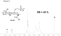
- FIG. 3 is a 1 H NMR spectrum of a hyaluronic acid vinylsulfone (HA-VS) copolymer. It was confirmed that the synthesis was well performed by the peak points a and b of the vinylsulfone (VS) substituent. The degree of substitution (DS) of vinylsulfone (VS) was confirmed to be 48%.
- HA-VS hyaluronic acid vinylsulfone
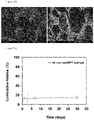
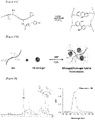
- FIG. 4 is a CLSM image of a hyaluronic acid microgel.
- FIG. 5 is a graph illustrating the measurements of the drug release behavior of a drug-loaded hyaluronic acid microgel. It was confirmed that the drug release behavior of the hyaluronic acid microgels was released in a sustained release form for more than 30 days.
- Pharmacokinetics was evaluated for the experimental group in which the hyaluronic acid microgel loaded with a drug (10-fold concentrated ranibizumab) was injected into a rabbit eye.
- the drug was injected into the rabbit eye without using a hyaluronic acid microgel carrier and evaluated as a control group.
- the control group consisted of a control group 1 injected with ranibizumab as a drug and control 2 injected with a 10-fold concentrated ranibizumab as a drug.
- the evaluation of in vivo pharmacokinetic was performed in the following manner.
- the hyaluronic acid microgel and the control drug were injected into the rabbit eye in the range of 30-50 ⁇ l.
- blood was collected, and after sacrificing the rabbit, the eyeballs were extracted and stored frozen. The collected blood was centrifuged to obtain a plasma sample and stored frozen.
- the extracted eyeballs were separated into anterior chamber, vitreous humor, and retina tissue, and then lysate was prepared through lysis buffer treatment.
- the dilution rate was set and the ranibizumab concentration in the tissue was measured through ranibizumab quantitative enzyme immunoassay (ELISA).
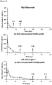
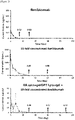
- FIG. 6 illustrates measurement results of the change in the concentration of ranibizumab in vitreous humor of the experimental group and the control group.
- FIG. 7 illustrates the measurements of the change in the concentration of ranibizumab in vitreous humor to retina of the experimental group and the control group.
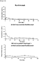
- FIG. 8 illustrates the measurements of the change in the concentration of ranibizumab in anterior chamber of the experimental group and the control group.
- FIG. 9 illustrates the measurements of the change in the concentration of ranibizumab in plasma of the experimental group and the control group.
- FIGs. 6 to 9 as a result of ELISA analysis, it was confirmed that the experimental group in which a hyaluronic acid microgel was injected in the vitreous humor, retina, anterior chamber, and plasma had a longer release behavior than the control group.
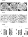
- FIG. 10A illustrates the first day of culturing LB medium bacteria for the experimental group and the control group
- FIG. 10B illustrates the third day of culturing LB medium bacteria for the experimental group and the control group (From the left, control group 1, control group 2, and experimental group are shown).
- a hyaluronic acid microgel which was cross-linked by the NHS-NH 2 cross-linking reaction of 8 wt% of hyaluronic acid-(N-hydroxysuccinimide) (HA-NHS)/hyaluronic acid-amine (NHS-NH 2 ), enzymatic cross-linking reaction (HRP/H 2 O 2 ) of hyaluronic acid-tyramine (HA-TA), thiol-maleimide cross-linking reaction of hyaluronic acid-maleimide (HA-Mal)/hyaluronic acid-thiol (HA-SH), and thiol-ene reaction of hyaluronic acid vinylsulfone (HA-VS)/hyaluronic acid thiol (HA-SH), and on which a drug is carried through a water in oil (w/o) emulsion method, was prepared.
- HRP/H 2 O 2 enzymatic cross-linking reaction
- FIG. 12A is a diagram illustrating the formation of a hyaluronic acid microgel using HA-NHS and HA-NH 2 .
- the hyaluronic acid microgel shown in FIG. 12A has a structure in which the hyaluronic acid copolymer is cross-linked by a cross-linking functional group represented by Chemical Formula 1-2 below.
- FIG. 12B is a diagram illustrating the formation of a hyaluronic acid microgel using HA-TA.
- the hyaluronic acid microgel shown in FIG. 12B has a structure in which the hyaluronic acid copolymer is cross-linked by a cross-linking functional group represented by Chemical Formula 1-3 and/or Chemical Formula 1-4 below.
- FIG. 12C is a diagram illustrating the formation of a hyaluronic acid microgel using HA-Mal and HA-SH.
- the hyaluronic acid microgel shown in FIG. 12C has a structure in which the hyaluronic acid copolymer is cross-linked by a cross-linking functional group represented by Chemical Formula 1-5 below.
- FIG. 12D is a diagram illustrating the formation of a hyaluronic acid microgel using HA-VS and HA-SH.
- the hyaluronic acid microgel shown in FIG. 12D has a structure in which the hyaluronic acid copolymer is cross-linked by a cross-linking functional group represented by Chemical Formula 1-6 below.
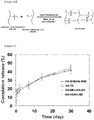
- FIG. 13 is a graph illustrating the measurements of the drug release behavior of a drug-loaded hyaluronic acid microgel. It was confirmed that the drug release behavior of the hyaluronic acid microgels prepared according to each cross-linking method was released in a sustained release form for more than 30 days.
- hyaluronic acid microgel of the present invention various cross-linking methods may be applied to prepare the hyaluronic acid microgel of the present invention.
- the present invention is not limited thereto, and hyaluronic acid microgels can be formed through other cross-linking reactions.
- the reaction was carried out by adding a solution obtained by dissolving 0.202 g (1.471 mmol) of tyramine (TA) in 50 ml of DMSO to a solution obtained by dissolving 5 g (1.471 mmol) of PEG-PNC in 100 ml of dimethylsulfoxide (DMSO).
- TA tyramine
- DMSO dimethylsulfoxide
- the molar ratio of PEG-PNC:TA was 1:1.
- the reaction temperature was 30°C, and the reaction was carried out in a nitrogen atmosphere for 6 hours. After the reaction, a gelatin solution (1 g/200 ml in DMSO) was mixed and the reaction was performed at 40°C in a nitrogen atmosphere for 24 hours.
- FIG. 14 is a diagram illustrating the synthesis of a GPT copolymer.
- a GPT hydrogel was formed by adding horseradish peroxidase (HRP) and hydrogen peroxide (H 2 O 2 ) to the synthesized gelatin derivative, i.e., the GPT copolymer, and cross-linking the phenol derivatives in the GPT copolymer with each other.
- the GPT hydrogel can control the gelation time or mechanical strength by controlling the concentration of horseradish peroxidase (HRP) or hydrogen peroxide.
- FIG. 15A is a diagram illustrating the preparation of a GPT hydrogel.
- the hydrogel shown in FIG. 15A has a structure that is cross-linked by a cross-linking functional group represented by Chemical Formula 2-1 and/or Chemical Formula 2-2 below.
- a composite formation i.e., an injectable intraocular hydrogel, in which drug-loaded hyaluronic acid microgels are distributed in the GPT hydrogel, is prepared by mixing hyaluronic acid microgel loaded with a drug (10-fold concentrated ranibizumab) with 150 ⁇ l of a GPT copolymer dissolved in a horseradish peroxidase (HRP) solution and 150 ⁇ l of a GPT copolymer dissolved in a hydrogen peroxide solution.
- FIG. 15B is a diagram illustrating the formation of a GPT hydrogel in which drug-loaded hyaluronic acid microgels are distributed.
- FIG. 16 is the 1 H NMR spectrum and UV analysis results of the GPT copolymer. It was confirmed that the synthesis was well performed by the peak points a, b, and c of the GPT copolymer and it was confirmed that 280.17 ⁇ mol per g of phenol gelatin derivative, which is a cross-linking molecule, was introduced.
- GPT hydrogel can control the gelation time according to the concentration of horseradish peroxidase (HRP) and hydrogen peroxide.
- the gelation time can be adjusted from a minimum of 2 seconds to a maximum of 2 minutes according to each element. That is, as the concentration of HRP increases, the gelation time becomes shorter, and as the concentration of HRP increases, the decomposition of hydrogen peroxide is accelerated, and eventually the generation rate of radical becomes faster. Therefore, the gelation time is accelerated because the gel is formed by the generated radical.
- FIG. 17 illustrates the change in the gelation time of the GPT hydrogel according to the change in the HRP concentration.
- the mechanical strength can be adjusted according to the concentration of hydrogen peroxide. If the concentration of hydrogen peroxide is adjusted from 0.15 wt% to 0.2 wt%, the mechanical strength is adjusted from 800 Pa to 7000 Pa.
- the adjustment of mechanical strength is a phenomenon that occurs when the cross-linking density of hydrogel is adjusted, and the drug inside hydrogel is slowly released as the cross-linking density increases, which is confirmed using a rheometer.
- FIG. 18 is a graph measuring the mechanical strength of the GPT hydrogel over time.
- FIG. 19 is an SEM image illustrating an internal structure of the GPT hydrogel. It was confirmed that the hyaluronic acid microgels were well distributed in a GPT hydrogel.
- FIG. 20 is a graph illustrating the measurements the drug release behavior of the GPT hydrogel carrying drug-loaded hyaluronic acid microgels. It was confirmed that the drug release behavior of the GPT hydrogel was slowly released for more than 30 days.
- Pharmacokinetics was evaluated for the experimental group in which a composite formulation consisting of the hyaluronic acid microgel loaded with a drug (10-fold concentrated ranibizumab) and a GPT hydrogel was injected into a rabbit eye.
- the drug was injected into the rabbit eye without using the composite formulation of the hyaluronic acid microgel/GPT hydrogel and evaluated as a control group.
- the control group consisted of a control group 1 injected with ranibizumab as a drug and control 2 injected with a 10-fold concentrated ranibizumab as a drug.
- the evaluation of in vivo pharmacokinetic was performed in the following manner.
- the composite formulation and the control drug were injected into the rabbit eye in the range of 30-50 ⁇ l.
- blood was collected, and after sacrificing the rabbit, the eyeballs were extracted and stored frozen. The collected blood was centrifuged to obtain a plasma sample and stored frozen.
- the extracted eyeballs were separated into anterior chamber, vitreous humor, and retina tissue, and then lysate was prepared through lysis buffer treatment.
- the dilution rate was set and the ranibizumab concentration in the tissue was measured through ranibizumab quantitative enzyme immunoassay (ELISA).
- FIG. 21 illustrates the measurement results of the change in the concentration of ranibizumab in vitreous humor of the experimental group and the control group.
- FIG. 22 illustrates the measurements of the change in the concentration of ranibizumab in vitreous humor to retina of the experimental group and the control group.
- FIG. 23 illustrates the measurements of the change in the concentration of ranibizumab in anterior chamber of the experimental group and the control group.
- FIG. 24 illustrates the measurements of the change in the concentration of ranibizumab in plasma of the experimental group and the control group. As shown in FIGs.
- FIG. 25A shows the first day of culturing LB medium bacteria for the experimental group and the control group.
- FIG. 25b shows the third day of culturing LB medium bacteria for the experimental group and the control group (From the left, control group 1, control group 2, and experimental group are shown).
- reaction solution was filtered using a syringe filter (450 nm). Then, membrane dialysis (3500 da molecular weight blocking) was performed in distilled water. The dialysis-completed solution was freeze-dried to obtain a GH (gelatin-hydroxyphenylacetic acid) copolymer.
- GH gelatin-hydroxyphenylacetic acid
- FIG. 27A is a diagram illustrating the preparation of a GH hydrogel.
- the hydrogel shown in FIG. 27A has a structure cross-linked by a cross-linking functional group represented by Chemical Formula 2-1 and/or Chemical Formula (2-2) below.
- a composite formation i.e., an injectable intraocular hydrogel, in which drug-loaded hyaluronic acid microgels are distributed in the GH hydrogel, is prepared by mixing hyaluronic acid microgel loaded with a drug (10-fold concentrated ranibizumab) with 150 ⁇ l of a GH copolymer dissolved in HRP solution and 150 ⁇ l of a GH copolymer dissolved in a hydrogen peroxide solution.
- FIG. 27B is a diagram illustrating the formation of the GH hydrogel in which the drug-loaded hyaluronic acid microgels are distributed.
- FIG. 28 is the 1 H NMR spectrum and UV analysis results of the GH copolymer. It was confirmed that the synthesis was well performed by the peak points a and b of the GH copolymer and it was confirmed that 153.8 ⁇ mol per GH g of phenol, which is a cross-linking molecule, was introduced.
- FIG. 29 is a graph illustrating the measurements the drug release behavior of the GH hydrogel carrying the drug-loaded hyaluronic acid microgels. It was confirmed that the drug release behavior of the GH hydrogel was slowly released for more than 30 days.
- hyaluronic acid 1 g was dissolved in 80 ml of MES buffer (pH 5). 1.63 g (10.5 mmol) of EDC and 1.812 g (15.75 mmol) of NHS were dissolved in 20 ml of MES buffer, respectively. EDC solution and NHS solution were sequentially added to the hyaluronic acid solution at 15-minute intervals. 1.193 g (10.5 mmol) of Cysteamine HCL was added to the hyaluronic acid solution activated by EDC/NHS to start the reaction. At this time, the reaction temperature was a room temperature and the reaction time was 24 hours.
- HA-SH thiolated hyaluronic acid
- FIG. 30A is a diagram of the preparation of a HA-VS/HA-SH hydrogel.
- the hydrogel shown in FIG. 30A has a cross-linked structure by a cross-linking functional group represented by Chemical Formula 2-3 below.
- a composite formation i.e., an injectable intraocular hydrogel, in which drug-loaded hyaluronic acid microgels are distributed in the HA-VS/HA-SH hydrogel, was prepared by mixing hyaluronic acid microgel loaded with a drug (10-fold concentrated ranibizumab) with 150 ⁇ l of a HA-SH solution and 150 ⁇ l of HA-VS solution.
- FIG. 30B is a diagram illustrating the formation of the HA-VS/HA-SH hydrogel in which the drug-loaded hyaluronic acid microgels are distributed.
- FIG. 31 is the 1 H NMR spectrum and UV analysis results of the HA-VS/HA-SH copolymer. It was confirmed that the synthesis was well performed by the peak points a and b of the HA-SH copolymer and it was confirmed that 230.5 ⁇ mol per HA-SH g of thiol, which is a cross-linking molecule, was introduced.
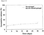
- FIG. 32 is a graph illustrating the measurements the drug release behavior of the HA-VS/HA-SH hydrogel carrying the drug-loaded hyaluronic acid microgels. It was confirmed that the drug release behavior of the HA-VS/HA-SH hydrogel was slowly released for more than 30 days.
Landscapes
- Health & Medical Sciences (AREA)
- Chemical & Material Sciences (AREA)
- Medicinal Chemistry (AREA)
- Life Sciences & Earth Sciences (AREA)
- General Health & Medical Sciences (AREA)
- Animal Behavior & Ethology (AREA)
- Veterinary Medicine (AREA)
- Public Health (AREA)
- Pharmacology & Pharmacy (AREA)
- Epidemiology (AREA)
- Organic Chemistry (AREA)
- Chemical Kinetics & Catalysis (AREA)
- Polymers & Plastics (AREA)
- Engineering & Computer Science (AREA)
- Ophthalmology & Optometry (AREA)
- Inorganic Chemistry (AREA)
- Dispersion Chemistry (AREA)
- Biochemistry (AREA)
- Bioinformatics & Cheminformatics (AREA)
- Molecular Biology (AREA)
- Materials Engineering (AREA)
- Proteomics, Peptides & Aminoacids (AREA)
- Biomedical Technology (AREA)
- Dermatology (AREA)
- Vascular Medicine (AREA)
- Heart & Thoracic Surgery (AREA)
- Immunology (AREA)
- General Chemical & Material Sciences (AREA)
- Nuclear Medicine, Radiotherapy & Molecular Imaging (AREA)
- Biophysics (AREA)
- Genetics & Genomics (AREA)
- Neurosurgery (AREA)
- Medicinal Preparation (AREA)
Abstract
Description
- The present invention relates to a drug delivery system and, more particularly, to an injectable intraocular microgel, an injectable intraocular hydrogel including the same, and a method of preparing the microgel and the hydrogel.
- Hydrogels are jelly-type materials having water as a dispersion medium or water as a basic component. Since hydrogels can easily absorb water due to their high hydrophilicity and can easily change their strength and shape, the hydrogels are used as a support for tissue engineering or used for drug delivery, etc. Hydrogels swell by absorbing a large amount of water in an aqueous solution or in an aqueous environment due to the hydrophilicity of the constituent materials thereof but do not dissolve due to the cross-linked structure thereof. Therefore, hydrogels may have various shapes and properties depending on the constituent components and preparing methods thereof and typically contain a large amount of water, so they are positioned as intermediate materials between liquid and solid in terms of characteristics.
- On the other hand, in the case of an anti-VEGF agent used for macular degeneration and diabetic macular degeneration among ophthalmic diseases, it is known that the intraocular half-life is short and thus the retention is short. Therefore, in order for the anti-VEGF agent to exhibit the maximum effect, the anti-VEGF agent is required to be frequently injected into the eyes. This not only causes a lot of inconvenience to patients but also imposes a heavy economic burden on patients.
- Therefore, after a long period of research and efforts, the inventors have completed the present invention as an injectable intraocular drug delivery system for a protein therapeutic agent for the treatment of ophthalmic diseases.
- An objective of the present invention is to provide an injectable intraocular microgel as a drug delivery system that is safe for a human body and that can release a drug in the eye for a long period of time and to provide a preparing method of the microgel.
- Another objective of the present invention is to provide an injectable intraocular hydrogel including such microgel and to provide a preparing method of the hydrogel.
- The objectives of the present invention are not limited to the above-mentioned ones, and another objectives not mentioned will also be clearly understood by those skilled in the art from the following description.
- According to an embodiment of the present invention to accomplish the above objectives, a method of preparing an injectable intraocular microgel includes: generating a hyaluronic acid microgel by causing a hyaluronic acid copolymer to undergo a cross-linking reaction through a w/o emulsion method; and loading a drug on the hyaluronic acid microgel.
- The generating of the hyaluronic acid microgel may include: synthesizing a hyaluronic acid vinylsulfone (HA-VS) copolymer by causing divinylsulfone (DVS) and hyaluronic acid (HA) to undergo a bonding reaction; and causing the synthesized hyaluronic acid vinylsulfone (HA-VS) copolymer and dithiothreitol (DTT) to undergo a thiol-ene cross-linking reaction.
- The generating of the hyaluronic acid microgel may be NHS-NH2 cross-linking of hyaluronic acid-(N-hydroxy succinimide) (HA-NHS) and hyaluronic acid-amine (HA-NH2).
- The generating of the hyaluronic acid microgel may include: adding horseradish peroxidase (HRP) and hydrogen peroxide (H2O2) to hyaluronic acid-tyramine (HA-TA) and causing the mixture to undergo an enzymatic cross-linking reaction.
- The generating of the hyaluronic acid microgel may be thiol-ene cross-linking of hyaluronic acid vinylsulfone (HA-VS) and hyaluronic acid thiol (HA-SH).
- According to another embodiment of the present invention to accomplish the objectives, an injectable intraocular microgel includes: a hyaluronic acid microgel composed of a hyaluronic acid copolymer cross-linked by a cross-linking functional group represented by at least one selected from the group consisting of Chemical Formulas 1-1 to 1-6 below; and a drug loaded on the hyaluronic acid microgel.


- The hyaluronic acid copolymer may be at least one selected from the group consisting of hyaluronic acid vinylsulfone (HA-VS), hyaluronic acid-(N-hydroxy succinimide) (HA-NHS), hyaluronic acid-tyramine (HA-TA), and hyaluronic acid-maleimide (HA-Mal).
- The drug may be one or more selected from an injectable intraocular protein therapeutic agent, a chemical drug, a small molecule therapeutic, a gene, and a cell.
- The injectable intraocular microgels have an average size of 30 to 160 µm.
- According to a further embodiment of the present invention to accomplish the objectives, an injectable intraocular hydrogel includes the injectable intraocular microgels dispersed in the hydrogel.
- The hydrogel may be cross-linked by a cross-linking functional group represented by at least one selected from the group consisting of Chemical Formulas 2-1 to 2-3 below.


- According to a yet further embodiment of the present invention to accomplish the above objectives, a method of preparing an injectable intraocular hydrogel includes: preparing an injectable intraocular microgel through the method described above; and dispersing the prepared injectable intraocular microgels in the hydrogel.
- The injectable intraocular hydrogel preparation method may include: preparing a GPT hydrogel by adding horseradish peroxidase (HRP) and hydrogen peroxide to a gelatin-polyethylene glycol-tyramine (GPT) copolymer so that phenol derivatives in the GPT copolymer cross-link with each other; and mixing the injectable intraocular microgel in the GPT hydrogel.
- The injectable intraocular hydrogel preparation method may include: preparing a GH hydrogel by adding horseradish peroxidase (HRP) and hydrogen peroxide to a gelatin-hydroxyphenylacetic acid (GH) copolymer so that phenol derivatives in the GH copolymer cross-link with each other; and mixing the eye-injecting microgel in the GH hydrogel.
- The injectable intraocular hydrogel preparation method may include: mixing hyaluronic acid thiol (HA-SH) and hyaluronic acid vinylsulfone (HA-VS) so that a vinyl group of HA-VS and a thiol group of HA-SH cross-link with each other through a thiol-ene reaction, thereby obtaining a HA-VS/HA-SH hydrogel; and mixing the injectable intraocular microgel in the HA-VS/HA-SH hydrogel.
- Details of other embodiments are disclosed in the following detailed description and the accompanying drawings.
- The eye-injecting microgel of the present invention has a structure in which a drug is loaded on a hyaluronic acid microgel. Therefore, the injectable intraocular microgel has high in vivo stability and the loaded drug is released in a sustained-release form for more than 30 days. This minimizes the inconvenience of a patient being injected.
- In addition, the injectable intraocular hydrogel of the present invention has a structure in which drug-loaded hyaluronic acid microgels are distributed inside the GPT hydrogel. It is also confirmed that injectable intraocular hydrogel has high in vivo stability and the loaded drug is slowly released over a long period of time. Furthermore, the injectable intraocular hydrogel can control the gelation time or mechanical strength by controlling the concentration of horseradish peroxidase or hydrogen peroxide.
-
-
FIG. 1 is a schematic diagram illustrating a process in which hyaluronic acid (HA) and divninylsulfone (DVS) are synthesized; -
FIG. 2 is a diagram illustrating the formation of a hyaluronic acid microgel using HA-VS and DTT; -
FIG. 3 is a 1H NMR spectrum of a hyaluronic acid vinylsulfone (HA-VS) copolymer; -
FIG. 4 is a CLSM image of a hyaluronic acid microgel; -
FIG. 5 is a graph illustrating the measurements of the drug release behavior of a drug-loaded hyaluronic acid microgel; -
FIG. 6 illustrates the measurement results of the change in the concentration of ranibizumab in vitreous humor of the experimental group and the control group; -
FIG. 7 illustrates the measurements of the change in the concentration of ranibizumab in vitreous humor to retina of the experimental group and the control group; -
FIG. 8 illustrates the measurements of the change in the concentration of ranibizumab in anterior chamber of the experimental group and the control group; -
FIG. 9 illustrates the measurements of the change in the concentration of ranibizumab in plasma of the experimental group and the control group; -
FIG. 10A illustrates the first day of culturing LB medium bacteria for the experimental group and the control group; -
FIG. 10B illustrates the third day of culturing LB medium bacteria for the experimental group and the control group; -
FIG. 11 illustrates the quantitative results of ELISA for inflammatory factors in the vitreous humor tissue; -
FIG. 12A is a diagram illustrating the formation of a hyaluronic acid microgel using HA-NHS and HA-NH2; -
FIG. 12B is a diagram illustrating the formation of a hyaluronic acid microgel using HA-TA; -
FIG. 12C is a diagram illustrating the formation of a hyaluronic acid microgel using HA-Mal and HA-SH; -
FIG. 12D is a diagram illustrating the formation of a hyaluronic acid microgel using HA-VS and HA-SH; -
FIG. 13 is a graph illustrating the measurements of the drug release behavior of a drug-loaded hyaluronic acid microgel; -
FIG. 14 is a diagram illustrating the synthesis of a GPT copolymer; -
FIG. 15A is a diagram illustrating the preparation of a GPT hydrogel; -
FIG. 15B is a diagram illustrating the formation of a GPT hydrogel in which drug-loaded hyaluronic acid microgels are distributed; -
FIG. 16 is the 1H NMR spectrum and UV analysis results of the GPT copolymer; -
FIG. 17 illustrates the change in the gelation time of the GPT hydrogel according to the change in the HRP concentration; -
FIG. 18 is a graph measuring the mechanical strength of the GPT hydrogel over time; -
FIG. 19 is an SEM image illustrating an internal structure of the GPT hydrogel; -
FIG. 20 is a graph illustrating the measurements the drug release behavior of the GPT hydrogel carrying drug-loaded hyaluronic acid microgels; -
FIG. 21 illustrates the measurements results of the change in the concentration of ranibizumab in vitreous humor of the experimental group and the control group; -
FIG. 22 illustrates the measurements of the change in the concentration of ranibizumab in vitreous humor to retina of the experimental group and the control group; -
FIG. 23 illustrates the measurements of the change in the concentration of ranibizumab in anterior chamber of the experimental group and the control group; -
FIG. 24 illustrates the measurements of the change in the concentration of ranibizumab in plasma of the experimental group and the control group; -
FIG. 25A illustrates the first day of culturing LB medium bacteria for the experimental group and the control group; -
FIG. 25B illustrates the third day of culturing LB medium bacteria for the experimental group and the control group; -
FIG. 26 illustrates the quantitative results of ELISA for inflammatory factors in the vitreous humor tissue; -
FIG. 27A is a diagram illustrating the preparation of a GH hydrogel; -
FIG. 27B is a diagram illustrating the formation of the GH hydrogel in which the drug-loaded hyaluronic acid microgels are distributed; -
FIG. 28 is the 1H NMR spectrum and UV analysis results of the GH copolymer; -
FIG. 29 is a graph illustrating the measurements the drug release behavior of the GH hydrogel carrying the drug-loaded hyaluronic acid microgels; -
FIG. 30A is a diagram of the preparation of a HA-VS/HA-SH hydrogel; -
FIG. 30B is a diagram illustrating the formation of the HA-VS/HA-SH hydrogel in which the drug-loaded hyaluronic acid microgels are distributed; -
FIG. 31 is the 1H NMR spectrum and UV analysis results of the HA-VS/HA-SH copolymer; and -
FIG. 32 is a graph illustrating the measurements the drug release behavior of the HA-VS/HA-SH hydrogel carrying the drug-loaded hyaluronic acid microgels. - The advantages and features of the present invention and methods of accomplishing the same may be understood more readily with reference to the following detailed description of examples and the accompanying drawings. However, the present invention may be embodied in many different forms, and should not be construed as being limited to the examples set forth herein. Rather, these examples are provided so that this disclosure will be thorough and complete and will fully convey the concept of the invention to those skilled in the art, and the present invention will only be defined by the appended claims. Throughout the specification, the same reference numerals refer to the same components.
- The present invention relates to a drug delivery system for injectable intraocular of a protein therapeutic agent or a drug for treating eye diseases, and may be implemented as a microgel and a hydrogel comprising the same. For example, a drug applicable to the present invention may be one or more selected from an injectable intraocular protein therapeutic agent, a chemical drug, a small molecule therapeutic, a gene, and a cell. Here, the protein therapeutic agent may consist of anti-vascular endothelial growth factors used for macular degeneration and diabetic retinopathy, etc., that is anti-VEGF (ranibizumab, aflibercept, bevacizumab, brolucizumab, etc.), rituximab or infliximab, etc., the chemical drug may consist of dexamethasone, triamcinolone, ganciclovir, methotrexate or vancomycin, etc., the small molecule therapeutic may consist of sugars, lipids, amino acids, fatty acids, phenolic compounds or alkaloids, etc., the gene may consist of siRNA(anti-VEGF), etc., and the cell may consist of retinal and intraocular cells such as RPE, photoreceptor, etc. and stem cell, etc. Hereinafter, examples using ranibizumab as a drug is described, but the present invention is not limited to such drug and may comprise any ophthalmic protein therapeutic agents, chemical drugs, small molecule therapeutics, genes and cells, etc.
- The injectable intraocular microgel of the present invention may be formed in a form in which a drug is loaded on a hyaluronic acid microgel generated by cross-linking a hyaluronic acid copolymer through a w/o emulsion method. Here, the hyaluronic acid copolymer may be at least one selected from the group consisting of hyaluronic acid vinylsulfone (HA-VS), hyaluronic acid-(N-hydroxy succinimide) (HA-NHS), hyaluronic acid-tyramine (HA-TA), and hyaluronic acid-maleimide (HA-Mal).
- In addition, the injectable intraocular hydrogel of the present invention may consist of any hydrogel in which drug-loaded hyaluronic acid microgels are distributed. In the present invention, a GPT hydrogel, a GH hydrogel, and a HA-VA/HA-SH hydrogel are described as examples of the injectable intraocular hydrogel, but the present invention is not limited such kinds of hydrogels.
- 70 mmol of divinylsulfone (DVS) was added to a solution with 0.1 mmol/ml of -OH obtained by mixing 1 g of 4 kDa hyaluronic acid (HA) in 100 ml of 0.1 M sodium hydroxide solution, followed by reaction for 10 minutes. Using the bonding reaction of the vinyl group of divinylsulfone (DVS) and the hydroxy group of hyaluronic acid (HA), a hyaluronic acidvinylsulfone (HA-VS) copolymer in which vinylsulfone (VS) is bonded to hyaluronic acid (HA) was obtained. The OH molar ratio of divinylsulfone:hyaluronic acid was 7:1. After synthesis, 5M HC1 was used for titration to
pH 5 so that the reaction can end. After membrane dialysis (6000-8000 Da molecular weight blocking) was performed for 3 days, a hyaluronic acid vinylsulfone (HA-VA) copolymer was obtained through freeze drying.FIG. 1 is a diagram a process of reacting hyaluronic acid (HA) and divninylsulfone (DVS). - 8 wt% of the synthesized hyaluronic acid vinylsulfone (HA-VS) copolymer and dithiothreitol (DTT) were cross-linked through a thiol-ene reaction. The hyaluronic acid vinylsulfone (HA-VS) copolymer, the dithiothreitol (DTT), and a drug (10-fold concentrated ranibizumab) were added and mixed with 50 ml of mineral oil through a water in oil (w/o) emulsion method. The mixture was then dispersed in a hyaluronic acid microgel (HA microgel), using 2 ml of emulsifier (Span 80) and reacted 24 hours. After the reaction was complete, the resultant was put into 100 ml of isopropyl alcohol, and the mineral oil layer was removed. Next, the microgels were separated through centrifugation. After the separation, the microgels were redispersed in du-ionized water (DIW) to obtain drug-loaded hyaluronic acid microgels (HA microgels) through freeze-drying.
FIG. 2 is a schematic diagram illustrating the formation of a hyaluronic acid microgel from HA-VS and DTT. The hyaluronic acid microgel shown inFIG. 2 has a structure in which a hyaluronic acid copolymer is cross-linked by a cross-linking functional group represented by Chemical Formula 1-1 below.
-
FIG. 3 is a 1H NMR spectrum of a hyaluronic acid vinylsulfone (HA-VS) copolymer. It was confirmed that the synthesis was well performed by the peak points a and b of the vinylsulfone (VS) substituent. The degree of substitution (DS) of vinylsulfone (VS) was confirmed to be 48%. - As a result of analyzing the hyaluronic acid microgel using a confocal laser scanning microscopy (CLSM), it was confirmed that the injectable intraocular microgels have an average size of 30 to 160 µm.
FIG. 4 is a CLSM image of a hyaluronic acid microgel. - Drug release of 8 mg of drug-loaded hyaluronic acid microgels was induced in DPBS. The release time interval was 1 hour, 3 hours, 6 hours, 12 hours, 1 day, 3 days, 7 days, 14 days, and 30 days, and the released drugs were quantified through enzyme immunoassay (ELISA).
FIG. 5 is a graph illustrating the measurements of the drug release behavior of a drug-loaded hyaluronic acid microgel. It was confirmed that the drug release behavior of the hyaluronic acid microgels was released in a sustained release form for more than 30 days. - Pharmacokinetics was evaluated for the experimental group in which the hyaluronic acid microgel loaded with a drug (10-fold concentrated ranibizumab) was injected into a rabbit eye. In addition, the drug was injected into the rabbit eye without using a hyaluronic acid microgel carrier and evaluated as a control group. The control group consisted of a
control group 1 injected with ranibizumab as a drug andcontrol 2 injected with a 10-fold concentrated ranibizumab as a drug. The evaluation of in vivo pharmacokinetic was performed in the following manner. - After mixing zoletil (0.3 cc/kg) and rompun (0.25 cc/kg) as an anesthetic and anesthetizing the rabbit by intramuscular injection, the hyaluronic acid microgel and the control drug were injected into the rabbit eye in the range of 30-50 µl. After anesthesia in the same manner for each planned period (1 hour, 1 day, 4 days, 8 days, 14 days, 30 days, 60 days, and 90 days), blood was collected, and after sacrificing the rabbit, the eyeballs were extracted and stored frozen. The collected blood was centrifuged to obtain a plasma sample and stored frozen.
- The extracted eyeballs were separated into anterior chamber, vitreous humor, and retina tissue, and then lysate was prepared through lysis buffer treatment. For plasma and intraocular tissues, the dilution rate was set and the ranibizumab concentration in the tissue was measured through ranibizumab quantitative enzyme immunoassay (ELISA).
-
FIG. 6 illustrates measurement results of the change in the concentration of ranibizumab in vitreous humor of the experimental group and the control group.FIG. 7 illustrates the measurements of the change in the concentration of ranibizumab in vitreous humor to retina of the experimental group and the control group.FIG. 8 illustrates the measurements of the change in the concentration of ranibizumab in anterior chamber of the experimental group and the control group.FIG. 9 illustrates the measurements of the change in the concentration of ranibizumab in plasma of the experimental group and the control group. As shown inFIGs. 6 to 9 , as a result of ELISA analysis, it was confirmed that the experimental group in which a hyaluronic acid microgel was injected in the vitreous humor, retina, anterior chamber, and plasma had a longer release behavior than the control group. - In order to check for infection and inflammation of the hyaluronic acid microgel formulations injected into the eye, bacterium were cultured in LB medium for control group 1 (ranibizumab injected group), control group 2 (10-fold concentrated ranibizumab injected group), and the experimental group (hyaluronic acid microgel injected group) and inflammatory factor (MIP-3α and IL-2β) quantitative ELISA was performed on the 1st and 3rd days for the vitreous humor tissue.
FIG. 10A illustrates the first day of culturing LB medium bacteria for the experimental group and the control group, andFIG. 10B illustrates the third day of culturing LB medium bacteria for the experimental group and the control group (From the left,control group 1,control group 2, and experimental group are shown).FIG. 11 illustrates the quantitative results of ELISA for inflammatory factors in the vitreous humor tissue (left = MIP-3α, right = IL-2β, A =control group 1, B =control group 2, and C = experimental group). - As shown in
FIGs. 10A to 11 , infection and inflammation were not observed in the two experiments. - Besides the method explained in Example 1, a hyaluronic acid microgel which was cross-linked by the NHS-NH2 cross-linking reaction of 8 wt% of hyaluronic acid-(N-hydroxysuccinimide) (HA-NHS)/hyaluronic acid-amine (NHS-NH2), enzymatic cross-linking reaction (HRP/H2O2) of hyaluronic acid-tyramine (HA-TA), thiol-maleimide cross-linking reaction of hyaluronic acid-maleimide (HA-Mal)/hyaluronic acid-thiol (HA-SH), and thiol-ene reaction of hyaluronic acid vinylsulfone (HA-VS)/hyaluronic acid thiol (HA-SH), and on which a drug is carried through a water in oil (w/o) emulsion method, was prepared.
-
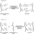
FIG. 12A is a diagram illustrating the formation of a hyaluronic acid microgel using HA-NHS and HA-NH2. The hyaluronic acid microgel shown inFIG. 12A has a structure in which the hyaluronic acid copolymer is cross-linked by a cross-linking functional group represented by Chemical Formula 1-2 below.

-
FIG. 12B is a diagram illustrating the formation of a hyaluronic acid microgel using HA-TA. The hyaluronic acid microgel shown inFIG. 12B has a structure in which the hyaluronic acid copolymer is cross-linked by a cross-linking functional group represented by Chemical Formula 1-3 and/or Chemical Formula 1-4 below.


-
FIG. 12C is a diagram illustrating the formation of a hyaluronic acid microgel using HA-Mal and HA-SH. The hyaluronic acid microgel shown inFIG. 12C has a structure in which the hyaluronic acid copolymer is cross-linked by a cross-linking functional group represented by Chemical Formula 1-5 below.

-
FIG. 12D is a diagram illustrating the formation of a hyaluronic acid microgel using HA-VS and HA-SH. The hyaluronic acid microgel shown inFIG. 12D has a structure in which the hyaluronic acid copolymer is cross-linked by a cross-linking functional group represented by Chemical Formula 1-6 below.

- The drug release behavior of hyaluronic acid microgels prepared through these different cross-linking methods was reviewed. Drug release of 8 mg of drug-loaded hyaluronic acid microgels was induced in DPBS. The release time interval was 1 hour, 3 hours, 6 hours, 12 hours, 1 day, 3 days, 7 days, 14 days, and 30 days, and the released drugs were quantified through enzyme immunoassay (ELISA).
FIG. 13 is a graph illustrating the measurements of the drug release behavior of a drug-loaded hyaluronic acid microgel. It was confirmed that the drug release behavior of the hyaluronic acid microgels prepared according to each cross-linking method was released in a sustained release form for more than 30 days. - As reviewed above, various cross-linking methods may be applied to prepare the hyaluronic acid microgel of the present invention. However, the present invention is not limited thereto, and hyaluronic acid microgels can be formed through other cross-linking reactions.
- After dissolving 10 g (2.9 mmol) of polyethylene glycol (PEG) in 100 ml of methylene chloride (MC), in this solution, a solution of 0.779 g (6.38 mmol) of 4-dimethylaminopyridine (DMAP) and 0.645 g (6.38 mmol) of triethylamine (TEA) dissolved in 10 ml of methylene chloride (MC) and a solution of 1.286 g (6.38 mmol) of 4-nitrophenylchloroformate (PNC) dissolved in 50 mL of methylene chloride (MC) were sequentially mixed. At this time, the molar ratio of PEG:DMAP:TEA:PNC was 1:2.2:2.2:2.2, the reaction temperature was 30°C, and the reaction was carried out in a nitrogen atmosphere for 24 hours.
- After completion of the reaction, the reagents remaining in the solution were removed using a filter, and then the reaction solution was concentrated using a rotary evaporation concentrator. The concentrated solution was added dropwise to 1800 ml of cold ether to generate a precipitate, and the precipitate was filtered using a filter to obtain a product. The obtained product was left in a vacuum oven for 24 hours to remove the residual organic solvent, and as a result, polyethylene glycol-4-nitrophenylchloroformate (PEG-PNC), which is a white powdery product, was obtained.
- The reaction was carried out by adding a solution obtained by dissolving 0.202 g (1.471 mmol) of tyramine (TA) in 50 ml of DMSO to a solution obtained by dissolving 5 g (1.471 mmol) of PEG-PNC in 100 ml of dimethylsulfoxide (DMSO). The molar ratio of PEG-PNC:TA was 1:1. The reaction temperature was 30°C, and the reaction was carried out in a nitrogen atmosphere for 6 hours. After the reaction, a gelatin solution (1 g/200 ml in DMSO) was mixed and the reaction was performed at 40°C in a nitrogen atmosphere for 24 hours.
- After completion of the reaction, unreacted PEG-TA was removed from the reaction solution through membrane dialysis (6000-8000 Da molecular weight blocking) using distilled water. After the dialysis was completed, the solution was freeze-dried to obtain a gelatin-PEG-TA (GPT) copolymer.
-
FIG. 14 is a diagram illustrating the synthesis of a GPT copolymer. - A GPT hydrogel was formed by adding horseradish peroxidase (HRP) and hydrogen peroxide (H2O2) to the synthesized gelatin derivative, i.e., the GPT copolymer, and cross-linking the phenol derivatives in the GPT copolymer with each other. Here, the GPT hydrogel can control the gelation time or mechanical strength by controlling the concentration of horseradish peroxidase (HRP) or hydrogen peroxide.
FIG. 15A is a diagram illustrating the preparation of a GPT hydrogel. The hydrogel shown inFIG. 15A has a structure that is cross-linked by a cross-linking functional group represented by Chemical Formula 2-1 and/or Chemical Formula 2-2 below.


- A composite formation, i.e., an injectable intraocular hydrogel, in which drug-loaded hyaluronic acid microgels are distributed in the GPT hydrogel, is prepared by mixing hyaluronic acid microgel loaded with a drug (10-fold concentrated ranibizumab) with 150 µl of a GPT copolymer dissolved in a horseradish peroxidase (HRP) solution and 150 µl of a GPT copolymer dissolved in a hydrogen peroxide solution.
FIG. 15B is a diagram illustrating the formation of a GPT hydrogel in which drug-loaded hyaluronic acid microgels are distributed. -
FIG. 16 is the 1H NMR spectrum and UV analysis results of the GPT copolymer. It was confirmed that the synthesis was well performed by the peak points a, b, and c of the GPT copolymer and it was confirmed that 280.17 µmol per g of phenol gelatin derivative, which is a cross-linking molecule, was introduced. - GPT hydrogel can control the gelation time according to the concentration of horseradish peroxidase (HRP) and hydrogen peroxide. In addition, the gelation time can be adjusted from a minimum of 2 seconds to a maximum of 2 minutes according to each element. That is, as the concentration of HRP increases, the gelation time becomes shorter, and as the concentration of HRP increases, the decomposition of hydrogen peroxide is accelerated, and eventually the generation rate of radical becomes faster. Therefore, the gelation time is accelerated because the gel is formed by the generated radical.
FIG. 17 illustrates the change in the gelation time of the GPT hydrogel according to the change in the HRP concentration. - The mechanical strength can be adjusted according to the concentration of hydrogen peroxide. If the concentration of hydrogen peroxide is adjusted from 0.15 wt% to 0.2 wt%, the mechanical strength is adjusted from 800 Pa to 7000 Pa. The adjustment of mechanical strength is a phenomenon that occurs when the cross-linking density of hydrogel is adjusted, and the drug inside hydrogel is slowly released as the cross-linking density increases, which is confirmed using a rheometer.
FIG. 18 is a graph measuring the mechanical strength of the GPT hydrogel over time. -
FIG. 19 is an SEM image illustrating an internal structure of the GPT hydrogel. It was confirmed that the hyaluronic acid microgels were well distributed in a GPT hydrogel. - After preparing a composite formulation by carrying drug-loaded hyaluronic acid microgels in a GPT hydrogel, drug release was induced in DPBS. The release time interval was 1 hour, 3 hours, 6 hours, 12 hours, 1 day, 3 days, 7 days, 14 days, and 30 days, and the released drugs were quantified through ELISA.
FIG. 20 is a graph illustrating the measurements the drug release behavior of the GPT hydrogel carrying drug-loaded hyaluronic acid microgels. It was confirmed that the drug release behavior of the GPT hydrogel was slowly released for more than 30 days. - Pharmacokinetics was evaluated for the experimental group in which a composite formulation consisting of the hyaluronic acid microgel loaded with a drug (10-fold concentrated ranibizumab) and a GPT hydrogel was injected into a rabbit eye. In addition, the drug was injected into the rabbit eye without using the composite formulation of the hyaluronic acid microgel/GPT hydrogel and evaluated as a control group. The control group consisted of a
control group 1 injected with ranibizumab as a drug andcontrol 2 injected with a 10-fold concentrated ranibizumab as a drug. The evaluation of in vivo pharmacokinetic was performed in the following manner. - After mixing zoletil (0.3 cc/kg) and rompun (0.25 cc/kg) as an anesthetic and anesthetizing the rabbit by intramuscular injection, the composite formulation and the control drug were injected into the rabbit eye in the range of 30-50 µl. After anesthesia in the same manner for each planned period (1 hour, 1 day, 4 days, 8 days, 14 days, 30 days, 60 days, and 90 days), blood was collected, and after sacrificing the rabbit, the eyeballs were extracted and stored frozen. The collected blood was centrifuged to obtain a plasma sample and stored frozen.
- The extracted eyeballs were separated into anterior chamber, vitreous humor, and retina tissue, and then lysate was prepared through lysis buffer treatment. For plasma and intraocular tissues, the dilution rate was set and the ranibizumab concentration in the tissue was measured through ranibizumab quantitative enzyme immunoassay (ELISA).
-
FIG. 21 illustrates the measurement results of the change in the concentration of ranibizumab in vitreous humor of the experimental group and the control group.FIG. 22 illustrates the measurements of the change in the concentration of ranibizumab in vitreous humor to retina of the experimental group and the control group.FIG. 23 illustrates the measurements of the change in the concentration of ranibizumab in anterior chamber of the experimental group and the control group.FIG. 24 illustrates the measurements of the change in the concentration of ranibizumab in plasma of the experimental group and the control group. As shown inFIGs. 21 to 24 , as a result of ELISA analysis, it was confirmed that the experimental group in which the composite formulation of hyaluronic acid microgel/GPT hydrogel was injected in the vitreous humor, retina, anterior chamber, and plasma had a longer release behavior than the control group. - In order to check for infection and inflammation of the composite formulation of the hyaluronic acid microgel/GPT hydrogel injected into the eye, bacteria were cultured in LB medium for control group 1 (ranibizumab injected group), control group 2 (10-fold concentrated ranibizumab injected group), and the experimental group (composite formulation injected group) and inflammatory factor (MIP-3α and IL-2β) quantitative ELISA was performed on the 1st and 3rd days for the vitreous tissue.
FIG. 25A shows the first day of culturing LB medium bacteria for the experimental group and the control group.FIG. 25b shows the third day of culturing LB medium bacteria for the experimental group and the control group (From the left,control group 1,control group 2, and experimental group are shown).FIG. 26 shows the quantitative results of ELISA for inflammatory factors in the vitreous humor tissue (left = MIP-3α, right = IL-2β, A =control group 1, B =control group 2, and C = experimental group). - As shown in
FIGs. 25A to 26 , infection and inflammation were not observed in the two experiments. - 10 g of gelatin was dissolved in 200 ml of 0.1M 2-(N-morpholino)ethanesulfonic acid (MES) (solution A). 0.609 g (4 mmol) of 4-hydroxyphenylacetic acid (HPA) was dissolved in 50 ml of 0.1M MES (solution B). 0.92 g (4.8 mmol) of 1-ethyl-3-(3-dimethylaminopropyl)-carbodiimide (EDC) and 0.276 g (2.4 mmol) of N-hydroxysuccinimide (NHS) were dissolved in 5 ml of 0.1M MES, respectively. Then, the EDC solution and NHS solution were sequentially added to the solution B at 15-minute intervals. Solution B activated by EDC/NHS was mixed with solution A to start the reaction. At this time, the reaction temperature was 40°C, and the reaction time was 24 hours.
- After completion of the reaction, the reaction solution was filtered using a syringe filter (450 nm). Then, membrane dialysis (3500 da molecular weight blocking) was performed in distilled water. The dialysis-completed solution was freeze-dried to obtain a GH (gelatin-hydroxyphenylacetic acid) copolymer.
- A GH hydrogel was formed by adding HRP and H2O2 to the synthesized GH copolymer and cross-linking the phenol derivatives in the GH copolymer with each other.
FIG. 27A is a diagram illustrating the preparation of a GH hydrogel. The hydrogel shown inFIG. 27A has a structure cross-linked by a cross-linking functional group represented by Chemical Formula 2-1 and/or Chemical Formula (2-2) below.


- A composite formation, i.e., an injectable intraocular hydrogel, in which drug-loaded hyaluronic acid microgels are distributed in the GH hydrogel, is prepared by mixing hyaluronic acid microgel loaded with a drug (10-fold concentrated ranibizumab) with 150 µl of a GH copolymer dissolved in HRP solution and 150 µl of a GH copolymer dissolved in a hydrogen peroxide solution.
FIG. 27B is a diagram illustrating the formation of the GH hydrogel in which the drug-loaded hyaluronic acid microgels are distributed. -
FIG. 28 is the 1H NMR spectrum and UV analysis results of the GH copolymer. It was confirmed that the synthesis was well performed by the peak points a and b of the GH copolymer and it was confirmed that 153.8 µmol per GH g of phenol, which is a cross-linking molecule, was introduced. - After preparing a composite formulation by carrying drug-loaded hyaluronic acid microgels in a GH hydrogel, drug release was induced in DPBS. The release time interval was 1 hour, 3 hours, 6 hours, 12 hours, 1 day, 3 days, 7 days, 14 days, and 30 days, and the released drugs were quantified through ELISA.
FIG. 29 is a graph illustrating the measurements the drug release behavior of the GH hydrogel carrying the drug-loaded hyaluronic acid microgels. It was confirmed that the drug release behavior of the GH hydrogel was slowly released for more than 30 days. - 1 g of hyaluronic acid (HA) was dissolved in 80 ml of MES buffer (pH 5). 1.63 g (10.5 mmol) of EDC and 1.812 g (15.75 mmol) of NHS were dissolved in 20 ml of MES buffer, respectively. EDC solution and NHS solution were sequentially added to the hyaluronic acid solution at 15-minute intervals. 1.193 g (10.5 mmol) of Cysteamine HCL was added to the hyaluronic acid solution activated by EDC/NHS to start the reaction. At this time, the reaction temperature was a room temperature and the reaction time was 24 hours.
- After completion of the reaction, membrane dialysis (6000-8000 Da molecular weight blocking) of the reaction solution was performed. The dialysate-completed solution was freeze-dried to obtain a thiolated hyaluronic acid (HA-SH) derivative.
- A HA-VS/HA-SH hydrogel was formed by mixing the synthesized HA-VS and HA-SH and cross-linking a vinyl group of HA-VS and a thiol group of HA-SH through thiol-ene reaction with each other.
FIG. 30A is a diagram of the preparation of a HA-VS/HA-SH hydrogel. The hydrogel shown inFIG. 30A has a cross-linked structure by a cross-linking functional group represented by Chemical Formula 2-3 below.

- A composite formation, i.e., an injectable intraocular hydrogel, in which drug-loaded hyaluronic acid microgels are distributed in the HA-VS/HA-SH hydrogel, was prepared by mixing hyaluronic acid microgel loaded with a drug (10-fold concentrated ranibizumab) with 150 µl of a HA-SH solution and 150 µl of HA-VS solution.
FIG. 30B is a diagram illustrating the formation of the HA-VS/HA-SH hydrogel in which the drug-loaded hyaluronic acid microgels are distributed. -
FIG. 31 is the 1H NMR spectrum and UV analysis results of the HA-VS/HA-SH copolymer. It was confirmed that the synthesis was well performed by the peak points a and b of the HA-SH copolymer and it was confirmed that 230.5 µmol per HA-SH g of thiol, which is a cross-linking molecule, was introduced. - After preparing a composite formulation by carrying drug-loaded hyaluronic acid microgels in a HA-VS/HA-SH hydrogel, drug release was induced in DPBS. The release time interval was 1 hour, 3 hours, 6 hours, 12 hours, 1 day, 3 days, 7 days, 14 days, and 30 days, and the released drugs were quantified through ELISA.
FIG. 32 is a graph illustrating the measurements the drug release behavior of the HA-VS/HA-SH hydrogel carrying the drug-loaded hyaluronic acid microgels. It was confirmed that the drug release behavior of the HA-VS/HA-SH hydrogel was slowly released for more than 30 days. - Although the preferred examples of the present invention have been disclosed for illustrative purposes, those skilled in the art will appreciate that various modifications, additions and substitutions are possible, without departing from the scope and spirit of the invention as disclosed in the accompanying claims. Therefore, it should be understood that the examples described above are exemplary and not limited in all respects.
Claims (15)
- A method of preparing an injectable intraocular microgel, the method comprising:generating a hyaluronic acid microgel by causing a hyaluronic acid copolymer to undergo a cross-linking reaction of a w/o emulsion method; andloading a drug on the hyaluronic acid microgel.
- The method of claim 1, wherein the generating of the hyaluronic acid microgel comprises:synthesizing a hyaluronic acid vinylsulfone (HA-VS) copolymer by causing divinylsulfone (DVS) and hyaluronic acid (HA) to undergo a bonding reaction; andcausing the synthesized hyaluronic acid vinylsulfone (HA-VS) copolymer and dithiothreitol (DTT) to undergo a thiol-ene cross-linking reaction.
- The method of claim 1, wherein the generating of the hyaluronic acid microgel is performed by NHS-NH2 cross-linking of hyaluronic acid-(N-hydroxy succinimide) (HA-NHS) and hyaluronic acid-amine (HA-NH2).
- The method of claim 1, wherein the generating of the hyaluronic acid microgel comprises:adding horseradish peroxidase (HRP) and hydrogen peroxide (H2O2) to hyaluronic acid-tyramine (HA-TA); andcausing the resulting mixture to undergo an enzymatic cross-linking.
- The method of claim 1, wherein the generating of the hyaluronic acid microgel is performed by thiol-ene cross-linking of hyaluronic acid vinylsulfone (HA-VS) and hyaluronic acid thiol (HA-SH).
- An injectable intraocular microgel comprising:a hyaluronic acid microgel composed of a hyaluronic acid copolymer cross-linked by a cross-linking functional represented by at least one Chemical Formula selected from the group consisting of Chemical Formulas 1-1 to 1-6 below; anda drug loaded on the hyaluronic acid microgel.


- The injectable intraocular microgel of claim 6, wherein the hyaluronic acid copolymer is at least one selected from the group consisting of hyaluronic acid vinylsulfone (HA-VS), hyaluronic acid-(N-hydroxy succinimide) (HA-NHS), hyaluronic acid-tyramine (HA-TA), and hyaluronic acid-maleimide (HA-Mal).
- The injectable intraocular microgel of claim 6, wherein the drug is one or more selected from an injectable intraocular protein therapeutic agent, a chemical drug, a small molecule therapeutic, a gene, and a cell.
- The injectable intraocular microgel of claim 6, wherein the injectable intraocular microgels have an average size of 30 to 160 µm.
- An injectable intraocular hydrogel in which the injectable intraocular microgels of any one of claims 6 to 9 are distributed.
- The injectable intraocular hydrogel of claim 10, wherein the hydrogel is cross-linked by a cross-linking functional group represented by at least one selected from the group consisting of Chemical Formulas 2-1 to 2-3 below:


- A method of preparing an injectable intraocular hydrogel, the method comprising:preparing an injectable intraocular microgel through the method of any of claims 1 to 5; anddistributing the prepared injectable intraocular microgels in the hydrogel.
- The method of claim 12, comprising:preparing a GPT hydrogel by adding horseradish peroxidase (HRP) and hydrogen peroxide to a gelatin-polyethylene glycol-tyramine (GPT) copolymer so that phenol derivatives in the GPT copolymer cross-link with each other; andmixing the injectable intraocular microgel in the GPT hydrogel.
- The method of claim 12, comprising:preparing a GH hydrogel by adding horseradish peroxidase (HRP) and hydrogen peroxide to a gelatin-hydroxyphenylacetic acid (GH) copolymer so that phenol derivatives in the GH copolymer cross-link with each other; andmixing the injectable intraocular microgel in the GH hydrogel.
- The method of claim 12, comprising:mixing hyaluronic acid thiol (HA-SH) and hyaluronic acid vinylsulfone (HA-VS) so that a vinyl group of HA-VS and a thiol group of HA-SH cross-link with each other through a thiol-ene reaction, thereby obtaining a HA-VS/HA-SH hydrogel; andmixing the injectable intraocular microgel in the HA-VS/HA-SH hydrogel.
Applications Claiming Priority (2)
| Application Number | Priority Date | Filing Date | Title |
|---|---|---|---|
| KR1020190132626A KR102435274B1 (en) | 2019-10-24 | 2019-10-24 | Eye-injecting microgel as drug delivery system and eye-injecting hydrogel having the same |
| PCT/KR2020/014488 WO2021080345A1 (en) | 2019-10-24 | 2020-10-22 | Injectable intraocular microgel as drug delivery system, and hydrogel comprising same |
Publications (3)
| Publication Number | Publication Date |
|---|---|
| EP4049653A1 true EP4049653A1 (en) | 2022-08-31 |
| EP4049653A4 EP4049653A4 (en) | 2023-11-01 |
| EP4049653B1 EP4049653B1 (en) | 2025-11-26 |
Family
ID=75619910
Family Applications (1)
| Application Number | Title | Priority Date | Filing Date |
|---|---|---|---|
| EP20878289.6A Active EP4049653B1 (en) | 2019-10-24 | 2020-10-22 | Injectable intraocular microgel as drug delivery system, and hydrogel comprising same |
Country Status (4)
| Country | Link |
|---|---|
| US (1) | US20220387597A1 (en) |
| EP (1) | EP4049653B1 (en) |
| KR (2) | KR102435274B1 (en) |
| WO (1) | WO2021080345A1 (en) |
Families Citing this family (5)
| Publication number | Priority date | Publication date | Assignee | Title |
|---|---|---|---|---|
| KR102935445B1 (en) * | 2022-02-07 | 2026-03-06 | 아주대학교산학협력단 | Injectable hydrogel rod using pre-crosslinked hydrogel, preparation method thereof, and biomedical use thereof |
| CN115304788B (en) * | 2022-07-08 | 2023-09-29 | 浙江工业大学 | Polylactic acid loaded hyaluronic acid hydrogel and preparation method and application thereof |
| KR102761671B1 (en) * | 2022-08-02 | 2025-01-31 | 인천대학교 산학협력단 | Method for preparing gelatin based functional and tissue adhesive hydrogel |
| KR102937803B1 (en) | 2023-03-28 | 2026-03-13 | 국립공주대학교 산학협력단 | Light-controllable hydrogel using photo-switchable green fluorescent protein and hyaluronic acid |
| CN117643573B (en) * | 2024-01-29 | 2024-05-03 | 天津医科大学眼科医院 | Nano eye drops capable of delivering drugs to the posterior segment of the eye, preparation method thereof, and application thereof |
Family Cites Families (5)
| Publication number | Priority date | Publication date | Assignee | Title |
|---|---|---|---|---|
| US8287906B2 (en) * | 2008-05-06 | 2012-10-16 | Agency For Science, Technology And Research | Formation of hydrogel in the presence of peroxidase and low concentration of hydrogen peroxide |
| KR101091028B1 (en) * | 2009-07-02 | 2011-12-09 | 아주대학교산학협력단 | In situ forming hydrogel and biomedical use thereof |
| CN103687622A (en) * | 2011-06-16 | 2014-03-26 | 香港科技大学 | Multiple vinyl sulfone containing molecules |
| KR101522462B1 (en) * | 2012-09-19 | 2015-05-21 | 아주대학교산학협력단 | Preparation Method of In situ forming Hydrogel Using Enzyme-Immobilized Supports and Biomedical Use Thereof |
| CN118126415A (en) * | 2017-09-19 | 2024-06-04 | 香港科技大学 | Biocompatible materials and methods for preparing and using the same |
-
2019
- 2019-10-24 KR KR1020190132626A patent/KR102435274B1/en active Active
-
2020
- 2020-10-22 EP EP20878289.6A patent/EP4049653B1/en active Active
- 2020-10-22 US US17/771,160 patent/US20220387597A1/en active Pending
- 2020-10-22 WO PCT/KR2020/014488 patent/WO2021080345A1/en not_active Ceased
-
2022
- 2022-08-17 KR KR1020220102857A patent/KR102752199B1/en active Active
Also Published As
| Publication number | Publication date |
|---|---|
| KR102435274B1 (en) | 2022-08-24 |
| KR20210049218A (en) | 2021-05-06 |
| US20220387597A1 (en) | 2022-12-08 |
| KR102435274B9 (en) | 2023-03-23 |
| EP4049653B1 (en) | 2025-11-26 |
| WO2021080345A1 (en) | 2021-04-29 |
| EP4049653A4 (en) | 2023-11-01 |
| KR102752199B1 (en) | 2025-01-10 |
| KR20220126671A (en) | 2022-09-16 |
Similar Documents
| Publication | Publication Date | Title |
|---|---|---|
| EP4049653B1 (en) | Injectable intraocular microgel as drug delivery system, and hydrogel comprising same | |
| JP4390845B2 (en) | Polymer micelles for drug delivery | |
| US10137199B2 (en) | Thiolated hyaluronan-based hydrogels cross-linked using oxidized glutathione | |
| Lai et al. | Chitosan-g-poly (N-isopropylacrylamide) copolymers as delivery carriers for intracameral pilocarpine administration | |
| Bongiovì et al. | Hyaluronic acid‐based micelles as ocular platform to modulate the loading, release, and corneal permeation of corticosteroids | |
| EP2143446A1 (en) | Hybrid gel comprising chemically crosslinked hyaluronic acid derivative and pharmaceutical composition using the same | |
| EA032552B1 (en) | Controlled release formulations for the delivery of hif-1 inhibitors | |
| JP2008537943A5 (en) | ||
| US9592251B2 (en) | Multi-vinylsulfone containing molecule | |
| US20190314506A1 (en) | Polymer hydrogels for in vivo applications and methods for using and preparing same | |
| US10966955B2 (en) | Ocular drug delivery system and uses thereof | |
| CN103381273B (en) | Amycin prodrug and preparation method thereof and injectable compositions | |
| CN114466869B (en) | Hydrogel compositions and uses thereof | |
| Wu et al. | Self-assemblied nanocomplexes based on biomimetic amphiphilic chitosan derivatives for protein delivery | |
| US20220241251A1 (en) | Stabilisation and controlled-release systems for medicaments unstable on sterilisation | |
| Wang et al. | Chondrocyte‐Targeted Nanoparticles Loaded with N‐Acetylcysteine Protect Articular Cartilage and Attenuate Osteoarthritis by Inhibiting Ferroptosis via Glutathione Maintenance | |
| US12564598B2 (en) | Nitric oxide-sensitive hydrogel | |
| Mei et al. | Covalent cross-linking approaches for all-trans retinoic acid-loaded thermo-responsive hydrogels | |
| US20240299535A1 (en) | Targeted glycosaminoglycan-particles and methods of use | |
| CN110227059B (en) | Nasal cavity absorption enhancer and application thereof | |
| KR20240112280A (en) | Solid form of resiquimod and dosage forms thereof | |
| EA044423B1 (en) | STABILIZATION AND CONTROLLED RELEASE SYSTEMS FOR DRUGS UNSTABLE BY STERILIZATION | |
| Holden | MODIFIED PAMAM DENDRIMERS IN TUNABLE DRUG-DELIVERY SYSTEMS: A SUSTAINED-RELEASE DENDRIMER HYDROGEL FOR ANTI-GLAUCOMA DRUGS AND SURFACE-ENGINEERED MACROPHAGES AS NANOPARTICLE CARRIERS FOR TARGETED ANTI-CANCER THERAPY | |
| Zhang | Diblock Polypeptide Hydrogel Synthesis and Biomedical Applications in Central Nervous System | |
| Kgesa | Novel Variant for Application as a Prolonged Release Drug Delivery System |
Legal Events
| Date | Code | Title | Description |
|---|---|---|---|
| STAA | Information on the status of an ep patent application or granted ep patent |
Free format text: STATUS: THE INTERNATIONAL PUBLICATION HAS BEEN MADE |
|
| PUAI | Public reference made under article 153(3) epc to a published international application that has entered the european phase |
Free format text: ORIGINAL CODE: 0009012 |
|
| STAA | Information on the status of an ep patent application or granted ep patent |
Free format text: STATUS: REQUEST FOR EXAMINATION WAS MADE |
|
| 17P | Request for examination filed |
Effective date: 20220421 |
|
| AK | Designated contracting states |
Kind code of ref document: A1 Designated state(s): AL AT BE BG CH CY CZ DE DK EE ES FI FR GB GR HR HU IE IS IT LI LT LU LV MC MK MT NL NO PL PT RO RS SE SI SK SM TR |
|
| DAV | Request for validation of the european patent (deleted) | ||
| DAX | Request for extension of the european patent (deleted) | ||
| A4 | Supplementary search report drawn up and despatched |
Effective date: 20230929 |
|
| RIC1 | Information provided on ipc code assigned before grant |
Ipc: C08K 5/00 20060101ALI20230925BHEP Ipc: C08L 5/08 20060101ALI20230925BHEP Ipc: C08B 37/08 20060101ALI20230925BHEP Ipc: C08J 3/24 20060101ALI20230925BHEP Ipc: C08J 3/075 20060101ALI20230925BHEP Ipc: A61F 9/00 20060101ALI20230925BHEP Ipc: A61K 9/00 20060101ALI20230925BHEP Ipc: A61K 47/36 20060101ALI20230925BHEP Ipc: A61K 9/06 20060101AFI20230925BHEP |
|
| GRAP | Despatch of communication of intention to grant a patent |
Free format text: ORIGINAL CODE: EPIDOSNIGR1 |
|
| STAA | Information on the status of an ep patent application or granted ep patent |
Free format text: STATUS: GRANT OF PATENT IS INTENDED |
|
| INTG | Intention to grant announced |
Effective date: 20250618 |
|
| GRAS | Grant fee paid |
Free format text: ORIGINAL CODE: EPIDOSNIGR3 |
|
| GRAA | (expected) grant |
Free format text: ORIGINAL CODE: 0009210 |
|
| STAA | Information on the status of an ep patent application or granted ep patent |
Free format text: STATUS: THE PATENT HAS BEEN GRANTED |
|
| AK | Designated contracting states |
Kind code of ref document: B1 Designated state(s): AL AT BE BG CH CY CZ DE DK EE ES FI FR GB GR HR HU IE IS IT LI LT LU LV MC MK MT NL NO PL PT RO RS SE SI SK SM TR |
|
| P01 | Opt-out of the competence of the unified patent court (upc) registered |
Free format text: CASE NUMBER: UPC_APP_0010058_4049653/2025 Effective date: 20251016 |
|
| REG | Reference to a national code |
Ref country code: CH Ref legal event code: F10 Free format text: ST27 STATUS EVENT CODE: U-0-0-F10-F00 (AS PROVIDED BY THE NATIONAL OFFICE) Effective date: 20251126 Ref country code: GB Ref legal event code: FG4D |
|
| REG | Reference to a national code |
Ref country code: DE Ref legal event code: R096 Ref document number: 602020062975 Country of ref document: DE |
|
| REG | Reference to a national code |
Ref country code: IE Ref legal event code: FG4D |
|
| REG | Reference to a national code |
Ref country code: NL Ref legal event code: MP Effective date: 20251126 |
|
| PG25 | Lapsed in a contracting state [announced via postgrant information from national office to epo] |
Ref country code: ES Free format text: LAPSE BECAUSE OF FAILURE TO SUBMIT A TRANSLATION OF THE DESCRIPTION OR TO PAY THE FEE WITHIN THE PRESCRIBED TIME-LIMIT Effective date: 20251126 |
|
| REG | Reference to a national code |
Ref country code: LT Ref legal event code: MG9D |
|
| PG25 | Lapsed in a contracting state [announced via postgrant information from national office to epo] |
Ref country code: NO Free format text: LAPSE BECAUSE OF FAILURE TO SUBMIT A TRANSLATION OF THE DESCRIPTION OR TO PAY THE FEE WITHIN THE PRESCRIBED TIME-LIMIT Effective date: 20260226 |
|
| PG25 | Lapsed in a contracting state [announced via postgrant information from national office to epo] |
Ref country code: HR Free format text: LAPSE BECAUSE OF FAILURE TO SUBMIT A TRANSLATION OF THE DESCRIPTION OR TO PAY THE FEE WITHIN THE PRESCRIBED TIME-LIMIT Effective date: 20251126 Ref country code: FI Free format text: LAPSE BECAUSE OF FAILURE TO SUBMIT A TRANSLATION OF THE DESCRIPTION OR TO PAY THE FEE WITHIN THE PRESCRIBED TIME-LIMIT Effective date: 20251126 Ref country code: AT Free format text: LAPSE BECAUSE OF FAILURE TO SUBMIT A TRANSLATION OF THE DESCRIPTION OR TO PAY THE FEE WITHIN THE PRESCRIBED TIME-LIMIT Effective date: 20251126 |
|
| REG | Reference to a national code |
Ref country code: AT Ref legal event code: MK05 Ref document number: 1860395 Country of ref document: AT Kind code of ref document: T Effective date: 20251126 |
|
| PG25 | Lapsed in a contracting state [announced via postgrant information from national office to epo] |
Ref country code: NL Free format text: LAPSE BECAUSE OF FAILURE TO SUBMIT A TRANSLATION OF THE DESCRIPTION OR TO PAY THE FEE WITHIN THE PRESCRIBED TIME-LIMIT Effective date: 20251126 |
|
| PG25 | Lapsed in a contracting state [announced via postgrant information from national office to epo] |
Ref country code: RS Free format text: LAPSE BECAUSE OF FAILURE TO SUBMIT A TRANSLATION OF THE DESCRIPTION OR TO PAY THE FEE WITHIN THE PRESCRIBED TIME-LIMIT Effective date: 20260226 |
|
| PG25 | Lapsed in a contracting state [announced via postgrant information from national office to epo] |
Ref country code: IS Free format text: LAPSE BECAUSE OF FAILURE TO SUBMIT A TRANSLATION OF THE DESCRIPTION OR TO PAY THE FEE WITHIN THE PRESCRIBED TIME-LIMIT Effective date: 20260326 |
|
| PG25 | Lapsed in a contracting state [announced via postgrant information from national office to epo] |
Ref country code: PT Free format text: LAPSE BECAUSE OF FAILURE TO SUBMIT A TRANSLATION OF THE DESCRIPTION OR TO PAY THE FEE WITHIN THE PRESCRIBED TIME-LIMIT Effective date: 20260326 |
|
| PG25 | Lapsed in a contracting state [announced via postgrant information from national office to epo] |
Ref country code: PL Free format text: LAPSE BECAUSE OF FAILURE TO SUBMIT A TRANSLATION OF THE DESCRIPTION OR TO PAY THE FEE WITHIN THE PRESCRIBED TIME-LIMIT Effective date: 20251126 |
|
| PG25 | Lapsed in a contracting state [announced via postgrant information from national office to epo] |
Ref country code: LV Free format text: LAPSE BECAUSE OF FAILURE TO SUBMIT A TRANSLATION OF THE DESCRIPTION OR TO PAY THE FEE WITHIN THE PRESCRIBED TIME-LIMIT Effective date: 20251126 |
|
| PG25 | Lapsed in a contracting state [announced via postgrant information from national office to epo] |
Ref country code: BG Free format text: LAPSE BECAUSE OF FAILURE TO SUBMIT A TRANSLATION OF THE DESCRIPTION OR TO PAY THE FEE WITHIN THE PRESCRIBED TIME-LIMIT Effective date: 20251126 |