EP4048331B1 - Procédés et systèmes permettant de générer des structures biologiques tridimensionnelles - Google Patents
Procédés et systèmes permettant de générer des structures biologiques tridimensionnelles Download PDFInfo
- Publication number
- EP4048331B1 EP4048331B1 EP20829629.3A EP20829629A EP4048331B1 EP 4048331 B1 EP4048331 B1 EP 4048331B1 EP 20829629 A EP20829629 A EP 20829629A EP 4048331 B1 EP4048331 B1 EP 4048331B1
- Authority
- EP
- European Patent Office
- Prior art keywords
- cell
- cells
- substrate
- biological
- layer
- Prior art date
- Legal status (The legal status is an assumption and is not a legal conclusion. Google has not performed a legal analysis and makes no representation as to the accuracy of the status listed.)
- Active
Links
Images



Classifications
-
- C—CHEMISTRY; METALLURGY
- C12—BIOCHEMISTRY; BEER; SPIRITS; WINE; VINEGAR; MICROBIOLOGY; ENZYMOLOGY; MUTATION OR GENETIC ENGINEERING
- C12N—MICROORGANISMS OR ENZYMES; COMPOSITIONS THEREOF; PROPAGATING, PRESERVING, OR MAINTAINING MICROORGANISMS; MUTATION OR GENETIC ENGINEERING; CULTURE MEDIA
- C12N5/00—Undifferentiated human, animal or plant cells, e.g. cell lines; Tissues; Cultivation or maintenance thereof; Culture media therefor
- C12N5/0062—General methods for three-dimensional culture
-
- A—HUMAN NECESSITIES
- A61—MEDICAL OR VETERINARY SCIENCE; HYGIENE
- A61L—METHODS OR APPARATUS FOR STERILISING MATERIALS OR OBJECTS IN GENERAL; DISINFECTION, STERILISATION OR DEODORISATION OF AIR; CHEMICAL ASPECTS OF BANDAGES, DRESSINGS, ABSORBENT PADS OR SURGICAL ARTICLES; MATERIALS FOR BANDAGES, DRESSINGS, ABSORBENT PADS OR SURGICAL ARTICLES
- A61L27/00—Materials for grafts or prostheses or for coating grafts or prostheses
- A61L27/36—Materials for grafts or prostheses or for coating grafts or prostheses containing ingredients of undetermined constitution or reaction products thereof, e.g. transplant tissue, natural bone, extracellular matrix
- A61L27/38—Materials for grafts or prostheses or for coating grafts or prostheses containing ingredients of undetermined constitution or reaction products thereof, e.g. transplant tissue, natural bone, extracellular matrix containing added animal cells
-
- B—PERFORMING OPERATIONS; TRANSPORTING
- B29—WORKING OF PLASTICS; WORKING OF SUBSTANCES IN A PLASTIC STATE IN GENERAL
- B29C—SHAPING OR JOINING OF PLASTICS; SHAPING OF MATERIAL IN A PLASTIC STATE, NOT OTHERWISE PROVIDED FOR; AFTER-TREATMENT OF THE SHAPED PRODUCTS, e.g. REPAIRING
- B29C64/00—Additive manufacturing, i.e. manufacturing of three-dimensional [3D] objects by additive deposition, additive agglomeration or additive layering, e.g. by 3D printing, stereolithography or selective laser sintering
- B29C64/10—Processes of additive manufacturing
- B29C64/106—Processes of additive manufacturing using only liquids or viscous materials, e.g. depositing a continuous bead of viscous material
-
- B—PERFORMING OPERATIONS; TRANSPORTING
- B33—ADDITIVE MANUFACTURING TECHNOLOGY
- B33Y—ADDITIVE MANUFACTURING, i.e. MANUFACTURING OF THREE-DIMENSIONAL [3D] OBJECTS BY ADDITIVE DEPOSITION, ADDITIVE AGGLOMERATION OR ADDITIVE LAYERING, e.g. BY 3D PRINTING, STEREOLITHOGRAPHY OR SELECTIVE LASER SINTERING
- B33Y10/00—Processes of additive manufacturing
-
- B—PERFORMING OPERATIONS; TRANSPORTING
- B33—ADDITIVE MANUFACTURING TECHNOLOGY
- B33Y—ADDITIVE MANUFACTURING, i.e. MANUFACTURING OF THREE-DIMENSIONAL [3D] OBJECTS BY ADDITIVE DEPOSITION, ADDITIVE AGGLOMERATION OR ADDITIVE LAYERING, e.g. BY 3D PRINTING, STEREOLITHOGRAPHY OR SELECTIVE LASER SINTERING
- B33Y70/00—Materials specially adapted for additive manufacturing
-
- B—PERFORMING OPERATIONS; TRANSPORTING
- B33—ADDITIVE MANUFACTURING TECHNOLOGY
- B33Y—ADDITIVE MANUFACTURING, i.e. MANUFACTURING OF THREE-DIMENSIONAL [3D] OBJECTS BY ADDITIVE DEPOSITION, ADDITIVE AGGLOMERATION OR ADDITIVE LAYERING, e.g. BY 3D PRINTING, STEREOLITHOGRAPHY OR SELECTIVE LASER SINTERING
- B33Y80/00—Products made by additive manufacturing
-
- C—CHEMISTRY; METALLURGY
- C12—BIOCHEMISTRY; BEER; SPIRITS; WINE; VINEGAR; MICROBIOLOGY; ENZYMOLOGY; MUTATION OR GENETIC ENGINEERING
- C12M—APPARATUS FOR ENZYMOLOGY OR MICROBIOLOGY; APPARATUS FOR CULTURING MICROORGANISMS FOR PRODUCING BIOMASS, FOR GROWING CELLS OR FOR OBTAINING FERMENTATION OR METABOLIC PRODUCTS, i.e. BIOREACTORS OR FERMENTERS
- C12M33/00—Means for introduction, transport, positioning, extraction, harvesting, peeling or sampling of biological material in or from the apparatus
Definitions
- Bioprinting technology possesses the capability of precise positioning of biomaterials and living cells to reconstruct complex structures, with the goal of generating repeatable models of both diseased and healthy tissue. Bioprinting offers opportunities to integrate patient-specific cell sources into tissue models, further improving the biological relevance of in vitro screening.
- WO 2019/021061 A1 discloses tissue-like structures being assembled using a microfluidic print head via cell deposition onto patterned substrates.
- MICHIYA MATSUSAKI ET AL "Tree-Dimensional Human Tissue Chips Fabricated by Rapid and Automatic Inkjet Cell Printing", ADVANCED HEALTHCARE MATERIALS, vol. 2, no. 4, 2 November 2012, pages 534-539 discloses layer-by-layer assembly using inkjet printing of single cells and proteins.
- MICHIYA MATSUSAKI ET AL "Fabrication of Cellular Multilayers with Nanometer-Sized Extracellular Matrix Films", ANGEWANDTE CHEMIE INTERNATIONAL EDITION, vol. 46, no. 25, 18 June 2007, pages 4689-4692 discloses multilayer cell films on a surface.
- One aspect of the invention provides a method of generating three-dimensional biological structures.
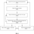
- the method includes: (a) depositing a first layer of a suspension over a substrate, the suspension including a liquid and a plurality of cells; (b) allowing the plurality of cells to attach to the substrate and form a first layer of attached cells; (c) depositing a cell-attachment agent over the first layer of attached cells; and (d) depositing a second layer of the suspension over the cell-attachment agent.
- the method can further include (b') culturing the plurality of cells in the first layer for a period of time in order to establish or increase the amount of, one or more selected from the group consisting of: cell-cell interactions, cell-cell attachments, cell-substrate interactions, cell-substrate attachments, cell mass, and cell quantity.
- the period of time can include one or more ranges of time selected from the group consisting of: from about 10 minutes to about 30 minutes, from about 30 minutes to about 60 minutes, from about 1 hour to about 12 hours, form about 12 hours to about 1 day, from about 1 day to about 3 days, from about 3 days to about 1 week, from about 1 week to about 2 weeks, from about 2 weeks to about 4 weeks, and from about 4 weeks to about 8 weeks.
- the method can further include: (e) repeating steps (b)-(d) until a biological structure having a predefined thickness is generated.
- the predefined thickness can be selected from the group consisting of: about 1 ⁇ m, between about 1 ⁇ m and about 10 ⁇ m, between about 10 ⁇ m and about 100 ⁇ m, between about 100 ⁇ m and about 1 mm, between about 1 mm and about 1.5 mm, between about 1.5 mm and about 5 mm, between about 5 mm and about 2 cm, between about 2 cm and 10 cm, and between about 10 cm and 50 cm .
- the suspension can be deposited using a flow-confinement device.
- the cell-attachment agent can be applied globally or in a pattern.
- the suspension can be applied globally or in a pattern.
- the suspension can include a single cell type or a plurality of cell types.
- the method can further include: (f) depositing a cell blocking agent over the second layer of the suspension.
- the cell-blocking agent can be selected from the group consisting of: polyethylene glycol (PEG), block copolymers, bovine serum albumin (BSA), surfactant, casein, polytetrafluoroethylene (PTFE), silanes, particulate solutions, nanoparticles, metal nanoparticles, gold nanoparticles, silver nanoparticles, aluminum nanoparticles, titanium nanoparticles, and polymeric microspheres.
- the cell-attachment agent can be selected from the group consisting of: adhesion proteins, cell-attachment proteins, cell-adhesion biomolecules, cell-adhesion molecules, polylysine, laminin, fibronectin, vitronectin, extracellular matrix (ECM), actin, integrin, perlecan, desmin, poly glycans, fibulin, elastin, tropoelastin, cadherin, gelatin, concanavalin, collagen, lipid structures, lipid nanotubes, lipid vesicles, lipid droplets, growth serum, fetal bovine serum, calf serum, horse serum, nucleic acids, DNA strands, RNA strands, cationic solutions, calcium solutions, magnesium solutions, and zinc solutions.
- ECM extracellular matrix
- the substrate can be selected from the group consisting of: cell-attachment agent, plastic, glass, silicon, paper, cotton, biomaterial, lipid films, gel, aerogel, hydrogel, polymer matrix, metals, metal oxides, semipermeable membranes, permeable membranes, polysaccharides, starch, collagen, cellulose, gelatin, biological scaffolds, biological tissue, decellularized tissue, non-biological tissue, and biological cells.
- the substrate can have a planar, uneven, structured, or patterned topology.
- attachment can refer to both physical, chemical, biological ionic, and electrostatic interactions, such as steric, covalent, ionic and hydrogen bonds, between cells and a substrate, surface or other cells. Attachment can be formed on the order of a minute for some cells. Other cells (e.g., neurons) may attach over weeks.
- Exemplary time ranges for allowing for attachment may include: from about 1 second to about 10 seconds, from about 10 seconds to about 30 seconds, from about 30 seconds to about 60 seconds, from about 1 minute to about 10 minutes, from about 10 minutes to about 30 minutes, from about 30 minutes to about 60 minutes, from about 1 hour to about 12 hours, form about 12 hours to about 1 day, from about 1 day to about 3 days, from about 3 days to about 1 week, from about 1 week to about 2 weeks, from about 2 weeks to about 4 weeks, and from about 4 weeks to about 8 weeks, from about 8 weeks to about 12 weeks, and so on.
- recirculation can have the meaning of a fluid flow path exiting the device and returning to the same device.
- Ranges provided herein are understood to be shorthand for all of the values within the range.
- a range of 1 to 50 is understood to include any number, combination of numbers, or sub-range from the group consisting 1, 2, 3, 4, 5, 6, 7, 8, 9, 10, 11, 12, 13, 14, 15, 16, 17, 18, 19, 20, 21, 22, 23, 24, 25, 26, 27, 28, 29, 30, 31, 32, 33, 34, 35, 36, 37, 38, 39, 40, 41, 42, 43, 44, 45, 46, 47, 48, 49, or 50 (as well as fractions thereof unless the context clearly dictates otherwise).
- aspects of the invention provide systems and methods for the controlled deposition of biological cells or cell constituents onto surfaces, including but not limited to the additive manufacturing of small-to-large cellular networks, tissues, organs, and other biological structures.
- embodiments of the invention can be used to create histologically complex cell structures in 2D and 3D.
- the fluid dispenser includes a nozzle or other device that ejects a fluid.
- a nozzle or other device that ejects a fluid.
- embodiments of the invention can be applied to existing additive manufacturing (also known as "3-D printing") devices, e.g., by incorporating the systems and methods described herein alongside existing additive manufacturing printheads that apply materials such as polymers, e.g., biocompatible polymers, for the production of organs-on-a-chip.
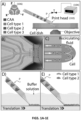
- the printing systems and methods utilize a flow-confinement device 102 and a controller 104 configured to generate a confined liquid volume outside of the flow-confinement device 102 where the confined liquid volume can be modulated by said controller 104 to include modulated and non-confined flow modes, as well as zero-flow modes, and where the controller 104 can be used to switch between any flow modes in arbitrary sequence, for arbitrary periods of time.
- Some embodiments of the invention utilize fluid flows or recirculating fluid flows, generated at the tip of a flow-confinement device 102, to achieve high resolution printing (e.g., with respect to both the lateral and axial position of the cells, deposition spot size for cell groupings, layer thicknesses, quantities of cells deposited, types of cells deposited, and the like).
- high resolution printing e.g., with respect to both the lateral and axial position of the cells, deposition spot size for cell groupings, layer thicknesses, quantities of cells deposited, types of cells deposited, and the like.
- embodiments of the invention provide single-cell-deposition capabilities, embodiments of the invention can also deposit multiple cells at a time.
- embodiments of the invention can reliably deposit layers having a thickness of less than two cells (e.g., less than about 500 ⁇ m, less than about 300 ⁇ m, less than about 200 ⁇ m, less than about 150 ⁇ m, less than about 100 ⁇ m, less than about 75 ⁇ m, less than about 50 ⁇ m, less than about 20 ⁇ m, less than about 10 ⁇ m, less than about 5 ⁇ m, and the like).
- a thickness of less than two cells e.g., less than about 500 ⁇ m, less than about 300 ⁇ m, less than about 200 ⁇ m, less than about 150 ⁇ m, less than about 100 ⁇ m, less than about 75 ⁇ m, less than about 50 ⁇ m, less than about 20 ⁇ m, less than about 10 ⁇ m, less than about 5 ⁇ m, and the like).
- flow-confinement device 102 can include one or more channels of which some channels are configured as outlet channels, through which liquid leaves the device 102 into an open volume and some channels are inlet channels, through which liquid is withdrawn from the open volume into the device.
- outlet channels and inlet channels are different channels.
- same channel can be intermittently switched to be outlet channel and an inlet channel, e.g., as further described in U.S. Patent No. 9,126,197 .
- Controller 104 can be configured to control the liquid flow such that all liquid leaving from the device is returning eventually back into the device when in recirculatory mode.
- Outflow can reach a limited maximal distance from the outlet channel before returning into the inlet channel. This distance defines the size of the confined liquid volume. Therefore, the liquid outflow can reach a substrate ( e.g., cell dish in FIG. 1A ) or a surface, if it is positioned closer than the critical dimension of the confined liquid volume. In some embodiments, the outflow can reach the substrate, such that the flow-confinement device 102 does not have a direct contact with the substrate. The tip of the flow-confinement device 102 can, in some non-limiting applications, be in contact with the substrate.
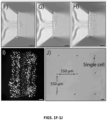
- the device can reliably deposit single cells, and or groups of cells, with a lateral resolution on the order of a single cell (e.g., less than about 150 ⁇ m, less than about 100 ⁇ m, less than about 75 ⁇ m, less than about 50 ⁇ m, less than about 30 ⁇ m, less than about 20 ⁇ m, and the like).
- a lateral resolution on the order of a single cell e.g., less than about 150 ⁇ m, less than about 100 ⁇ m, less than about 75 ⁇ m, less than about 50 ⁇ m, less than about 30 ⁇ m, less than about 20 ⁇ m, and the like).
- the recirculating fluid flow-confinement device 102 can be readily positioned relative to an object of interest using either a manual positioning system, or through the implementation of an electronically controlled positioning device 106.
- the positioning system may include hydraulic, mechanical, or electrical (e.g., stepper or servo motor) micromanipulators. These actuators can be arranged in one, two, three, four or greater directions.
- the electronically controlled position of both the printhead and the substrate can be achieved through on-demand control, e.g., through the use of a joystick, gamepad controller, scroll wheel, a touch screen and combinations thereof, and/or through the use of a computationally determined path.
- These paths can for example, be derived from a simple coordinate system, a design file, such as .stl (STereoLithography) CAD files, and/or through a relative feedback response, e.g., adapting to the changing position of cell propagation.
- methods of cell deposition may be carried out under the control of a preprogrammed flow controller.
- the system may further comprise a cell concentration measuring device for measuring cell deposition.
- the preprogrammed flow controller may be programmed to assess whether a predefined number of cells are correctly located on the substrate. The feature of various embodiments by which parameters of the method are adjusted based on information collected over the course of the method is termed "feedback".
- the system may further comprise a device or method for measuring a cell property, such as a method for measuring protein expression, gene expression, metabolic markers, cell-cell interactions, ions, or other biological, physical or chemical properties of one or several cells in the various flow modes, including recirculatory flow.
- a device or method for measuring a cell property such as a method for measuring protein expression, gene expression, metabolic markers, cell-cell interactions, ions, or other biological, physical or chemical properties of one or several cells in the various flow modes, including recirculatory flow.
- suspended particles are tracked, e.g., after exiting from the microfluidic printhead.
- sperm tracking technology such as described in U.S. Patent Application Publication No. 2017/0109879 can be applied to track a plurality of moving suspended particles.
- the fluid flow can be modified or paused in order to facilitate desired placement of the suspended particle(s).
- Cell suspensions may be prepared using standard techniques, as understood in the art. For example, mammalian cells may be suspended in a solution of about 1 ⁇ 10 6 cells/mL to about 10 ⁇ 0 6 cells/mL. The cell suspension solution may be combined with a PEG ( e.g., mol wt 6000). In some embodiments, the cell suspension solutions may have a final concentration of about 15 mg/ml.
- a PEG e.g., mol wt 6000.
- the cell attachment agent can be delivered globally (over the whole sample surface), locally (within a defined region of the sample) and/or directly patterned (over the whole sample or locally, where the region is defined by the path of the fluidic printhead).
- CAA types include, but are not limited to adhesion proteins, cell-attachment proteins, cell-adhesion biomolecules, cell-adhesion molecules and further include, but are not limited to: polylysine (e.g., poly-L-lysine (PLL)), laminin, fibronectin, vitronectin, extracellur matrix (ECM) ( e.g., available under the GELTREX ® mark from Life Technologies Corporation of Carlsbad, California), actin, integrin, perlecan, desmin, poly glycans, fibulin, elastin, tropoelastin, cadherin, , gelatin, concanavalin, collagen, organic and inorganic gels, lipid structures (e.g., nanotubes, vesicles, droplets, and the like), growth serum (e.g., fetal bovine serum, calf serum, horse serum, and the like), nucleic acids (e.g., DNA and/or RNA
- the CAA may be prepared as a working solution for pretreating one or more substrates.
- the CAA working solution for pretreating may include one or more of PLL, poly-D-lysine (PDL), ECM, and a buffer including for example sodium borate buffer.
- the working solution for pretreating includes: 0.5 mg/mL poly-L-lysine (PLL) and 75 ⁇ g/mL extracellular matrix (protein concentration 120-180 ⁇ g/mL, GELTREX ® , Gibco), prepared using 0.1 M sodium borate buffer.
- the working solution for pretreating may be prepared a time interval before use, including immediately prior to use.
- the CAA may be prepared as a working solution for direct printing, further known as CAA-dp.
- the CAA-dp may be of the same composition as CAA or be of a composition including one or more of PLL, PDL, serum (e.g., fetal bovine serum), and buffer (e.g., phosphate-buffered saline buffer).
- the working solution for direct printing includes: 0.1 mg/mL poly-L-lysine (PLL) and 10% FBS, prepared in a 1 ⁇ phosphate-buffered saline (PBS) buffer.
- PLL poly-L-lysine
- FBS 1 ⁇ phosphate-buffered saline
- the working solution for direct printing may be prepared a time interval before use, including immediately prior to use.
- CAA may additionally include envrinmentally responsive compounds, such as Poly(N-isopropylacrylamide (PNIPAAm) for thermal responsivity or Rose Bengal or riboflavin for photoactive linkers.
- PNIPAAm Poly(N-isopropylacrylamide
- Rose Bengal Rose Bengal
- riboflavin for photoactive linkers.
- cell-blocking agent can be applied globally over a substrate, locally, or patterned using the fluidic printhead.
- the CBA can prevent attachment between adjacent cells and/or act as a placeholder.
- the CBA is a dissolvable substance.
- a CBA such as PEG can be deposited as a scaffold to establish a lumen while cells are deposited around the lumen to form a vessel.
- CBA types include, but are not limited to, polyethylene glycol (PEG), block copolymers, bovine serum albumin (BSA), surfactants (e.g., PLUORONIC TM surfactants available from ThermoFisher Scientific of Waltham, Massachusetts), casein, polytetrafluoroethylene (PTFE) (e.g., TELLON ® available from The Chemours Company of Wilmington, Delaware), silanes, particulate solutions (e.g., including nanoparticles (e.g., metal nanoparticles such as gold, silver, aluminium, titanium, and the like), polymeric microspheres, and the like), or a composite solution comprised of any of the preceding list.
- PEG polyethylene glycol
- BSA bovine serum albumin
- surfactants e.g., PLUORONIC TM surfactants available from ThermoFisher Scientific of Waltham, Massachusetts
- casein polytetrafluoroethylene (PTFE) (e.g., T
- Embodiments of the invention can be applied to add material to living organisms.
- the organism can be considered the substrate.
- Such embodiments can be used for a variety of therapeutic and/or cosmetic applications including regenerative medicine, tissue grafting, plastic surgery, orthopedics, and the like.
- Embodiments of the invention utilizing a soft printhead are particularly advantageous in such embodiments, especially when applied to delicate tissues such as the eye and the ear.
- Such an in - situ -generated graft can be bound to the organism through various techniques including the use of cross-linking as discussed herein.
- In-situ -generated grafts can also be used for research purposes.
- cancer cells can be printed within an organism ( e.g., in a predefined and consistent pattern across multiple organisms) for experimental animal research.
- references to methods of treatment above under heading "Additions to Living Organisms”, are to be interpreted as references to the compounds, pharmaceutical compositions and medicaments of the present invention for use in a method for treatment of the human (or animal) body by therapy (or for diagnosis).
- HaCaT cells were maintained and grown in complete Dulbecco's Modified eagle's Medium (DMEM, Gibco) supplemented with 10% fetal bovine serum (FBS, Gibco), at 37°C and 5% CO 2 .
- DMEM Dulbecco's Modified eagle's Medium
- FBS fetal bovine serum
- Cells used as experimental controls were sub-cultured when they attained a confluency of approximately 80%, preventing the cells from differentiating.
- a separate culture of HaCaT cells were prepared and allowed to differentiate, by growing the culture for 3-4 additional days, once a 100% confluency had been achieved.
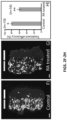
- Undifferentiated and differentiated psoriasis-like (cytokine-treated) HaCaT cells were used for bioprinting a skin model with the BIOPIXLAR ® platform.
- the skin model was formed by printing stripes of psoriasis-like ("diseased") and undifferentiated ("healthy") cells side-by-side. The printing process was performed on CAA-precoated IBIDI ® dishes. After printing, the cells were allowed to grow and proliferate in full growth media (DMEM +10% FBS) at 37°C, 5% CO 2 for 24 hrs.
- DMEM full growth media
Landscapes
- Engineering & Computer Science (AREA)
- Health & Medical Sciences (AREA)
- Chemical & Material Sciences (AREA)
- Life Sciences & Earth Sciences (AREA)
- Biomedical Technology (AREA)
- Zoology (AREA)
- Materials Engineering (AREA)
- Bioinformatics & Cheminformatics (AREA)
- Wood Science & Technology (AREA)
- Organic Chemistry (AREA)
- Manufacturing & Machinery (AREA)
- Biotechnology (AREA)
- General Health & Medical Sciences (AREA)
- Genetics & Genomics (AREA)
- Microbiology (AREA)
- Biochemistry (AREA)
- General Engineering & Computer Science (AREA)
- Cell Biology (AREA)
- Botany (AREA)
- Veterinary Medicine (AREA)
- Dermatology (AREA)
- Medicinal Chemistry (AREA)
- Oral & Maxillofacial Surgery (AREA)
- Transplantation (AREA)
- Epidemiology (AREA)
- Animal Behavior & Ethology (AREA)
- Public Health (AREA)
- Chemical Kinetics & Catalysis (AREA)
- Sustainable Development (AREA)
- Physics & Mathematics (AREA)
- Molecular Biology (AREA)
- Mechanical Engineering (AREA)
- Optics & Photonics (AREA)
- Apparatus Associated With Microorganisms And Enzymes (AREA)
- Micro-Organisms Or Cultivation Processes Thereof (AREA)
- Measuring Or Testing Involving Enzymes Or Micro-Organisms (AREA)
- Immobilizing And Processing Of Enzymes And Microorganisms (AREA)
Claims (12)
- Procédé de génération de structures biologiques tridimensionnelles, le procédé comprenant :(a) le dépôt, utilisant un dispositif de confinement de flux, d'une première couche d'une suspension selon un certain motif sur un substrat, la suspension comprenant un liquide et une pluralité de cellules ;(b) le fait de laisser la pluralité de cellules s'attacher au substrat et former une première couche de cellules attachées ;(b') la culture de la pluralité de cellules dans la première couche pendant une période de temps comprise entre environ 10 minutes et environ 8 semaines, afin que soit établie ou augmentée la quantité d'un ou plusieurs choisis dans le groupe constitué par : les interactions cellule-cellule, les attachements cellule-cellule, la masse cellulaire, et la quantité de cellules, la culture ayant pour résultat une structure analogue à un tissu ;(c) le dépôt d'un agent d'attachement cellulaire sur la première couche de cellules attachées ;(d) le dépôt, utilisant un dispositif de confinement de flux, d'une deuxième couche de la suspension selon un certain motif sur l'agent d'attachement cellulaire ; et(e) la répétition des étapes (b) à (d) jusqu'à ce qu'une structure biologique ayant une épaisseur prédéfinie soit générée,dans lequel les étapes a) à d) ne sont pas effectuées sur un corps humain ou animal.
- Procédé selon la revendication 1, comprenant en outre la mesure de l'expression de protéines, de l'expression de gènes, de marqueurs métaboliques, d'interactions cellule-cellule, d'ions, ou d'autres propriétés biologiques, physiques ou chimiques de la pluralité de cellules, y compris le flux recirculatoire.
- Procédé selon la revendication 1, dans lequel la période de temps comprend une ou plusieurs plages de temps choisies dans le groupe constitué par : d'environ 10 minutes à environ 30 minutes, d'environ 30 minutes à environ 60 minutes, d'environ 1 heure à environ 12 heures, d'environ 12 heures à environ 1 jour, d'environ 1 jour à environ 3 jours, d'environ 3 jours à environ 1 semaine, d'environ 1 semaine à environ 2 semaines, d'environ 2 semaines à environ 4 semaines, et d'environ 4 semaines à environ 8 semaines.
- Procédé selon la revendication 1, dans lequel l'épaisseur prédéfinie est choisie dans le groupe constitué par : environ 1 µm, entre environ 1 µm et environ 10 µm, entre environ 10 µm et environ 100 µm, entre environ 100 µm et environ 1 mm, entre environ 1 mm et environ 1,5 mm, entre environ 1,5 mm et environ 5 mm, entre environ 5 mm et environ 2 cm, entre environ 2 cm et 10 cm, entre environ 10 cm et 50 cm.
- Procédé selon la revendication 1, comprenant en outre :
(e) la répétition des étapes (a) à (c) en un nouvel emplacement sur le substrat. - Procédé selon la revendication 1, dans lequel l'agent d'attachement cellulaire est appliqué selon un certain motif.
- Procédé selon la revendication 1, dans lequel la suspension comprend une pluralité de types cellulaires.
- Procédé selon la revendication 1, comprenant en outre :
(f) le dépôt d'un agent bloqueur de cellules sur la deuxième couche de la suspension. - Procédé selon la revendication 8, dans lequel l'agent bloqueur de cellules est choisi dans le groupe constitué par : le polyéthylèneglycol (PEG), les copolymères séquencés, la sérumalbumine bovine (SAS), un tensioactif, la caséine, le polytétrafluoroéthylène (PTFE), les silanes, les solutions particulaires, les nanoparticules, les nanoparticules métalliques, les nanoparticules d'or, les nanoparticules d'argent, les nanoparticules d'aluminium, les nanoparticules de titane, et les microsphères polymères.
- Procédé selon la revendication 1, dans lequel l'agent d'attachement cellulaire est choisi dans le groupe constitué par : les protéines d'adhésion, les protéines d'attachement cellulaire, les biomolécules d'adhésion cellulaire, les molécules d'adhésion cellulaire, la polylysine, la laminine, la fibronectine, la vitronectine, une matrice extracellulaire (ECM), l'actine, l'intégrine, le perlécan, la desmine, les polyglycanes, la fibuline, l'élastine, la tropoélastine, la cadhérine, la gélatine, la concanavaline, le collagène, les structures lipidiques, les nanotubes lipidiques, les vésicules lipidiques, les gouttelettes lipidiques, un sérum de croissance, le sérum de veau foetal, le sérum de veau, le sérum de cheval, les acides nucléiques, les brins d'ADN, les brins d'ARN, les solutions cationiques, les solutions de calcium, les solutions de magnésium, et les solutions de zinc.
- Procédé selon la revendication 1, dans lequel le substrat est choisi dans le groupe constitué par : un agent d'attachement cellulaire, un plastique, un verre, le silicium, le papier, le coton, un biomatériau, les films lipidiques, un gel, un aérogel, un hydrogel, une matrice polymère, les métaux, les oxydes métalliques, les membranes semi-perméables, les membranes perméables, les polysaccharides, l'amidon, le collagène, la cellulose, la gélatine, les charpentes biologiques, un tissu biologique, un tissu décellularisé, un tissu non biologique, et les cellules biologiques.
- Procédé selon la revendication 1, dans lequel le substrat comprend des sections adjacentes de différents matériaux de substrat.
Applications Claiming Priority (2)
| Application Number | Priority Date | Filing Date | Title |
|---|---|---|---|
| US201962924111P | 2019-10-21 | 2019-10-21 | |
| PCT/IB2020/000900 WO2021079192A1 (fr) | 2019-10-21 | 2020-10-19 | Procédés et systèmes permettant de générer des structures biologiques tridimensionnelles |
Publications (2)
| Publication Number | Publication Date |
|---|---|
| EP4048331A1 EP4048331A1 (fr) | 2022-08-31 |
| EP4048331B1 true EP4048331B1 (fr) | 2025-01-22 |
Family
ID=74068287
Family Applications (1)
| Application Number | Title | Priority Date | Filing Date |
|---|---|---|---|
| EP20829629.3A Active EP4048331B1 (fr) | 2019-10-21 | 2020-10-19 | Procédés et systèmes permettant de générer des structures biologiques tridimensionnelles |
Country Status (9)
| Country | Link |
|---|---|
| US (1) | US20220389373A1 (fr) |
| EP (1) | EP4048331B1 (fr) |
| CN (1) | CN114867499A (fr) |
| CA (1) | CA3154441A1 (fr) |
| DK (1) | DK4048331T3 (fr) |
| ES (1) | ES3024481T3 (fr) |
| FI (1) | FI4048331T3 (fr) |
| LT (1) | LT4048331T (fr) |
| WO (1) | WO2021079192A1 (fr) |
Family Cites Families (8)
| Publication number | Priority date | Publication date | Assignee | Title |
|---|---|---|---|---|
| US20040126405A1 (en) * | 2002-12-30 | 2004-07-01 | Scimed Life Systems, Inc. | Engineered scaffolds for promoting growth of cells |
| JP4919464B2 (ja) * | 2006-03-02 | 2012-04-18 | 国立大学法人大阪大学 | 三次元組織の製造方法およびそれに用いる細胞外マトリックスの製造方法。 |
| EP2506976A2 (fr) | 2009-12-03 | 2012-10-10 | Owe Owar | Pipettes, procédés d'utilisation, et procédés de stimulation d'un objet d'intérêt |
| FI3885045T3 (fi) | 2011-05-06 | 2026-03-19 | Fluicell Ab | Mikrofluidilaite pitoliitännällä ja käyttömenetelmä |
| US20170109879A1 (en) | 2014-04-03 | 2017-04-20 | Drexel University | Computer-implemented methods, computer-readable media, and systems for tracking a plurality of spermatozoa |
| WO2017147183A1 (fr) * | 2016-02-23 | 2017-08-31 | University of Central Oklahoma | Procédé de création d'échafaudage de tissu 3d au moyen d'une matrice de nanofibres électrofilées et d'un hydrogel photosensible |
| US10597639B2 (en) * | 2016-10-20 | 2020-03-24 | Washington University | 3D-printed scaffold device for cell transplantation |
| DK3658199T3 (da) * | 2017-07-28 | 2025-02-03 | Fluicell Ab | Fremgangsmåder og systemer, der udnytter recirkulerende væskestrømme |
-
2020
- 2020-10-19 WO PCT/IB2020/000900 patent/WO2021079192A1/fr not_active Ceased
- 2020-10-19 CA CA3154441A patent/CA3154441A1/fr active Pending
- 2020-10-19 FI FIEP20829629.3T patent/FI4048331T3/fi active
- 2020-10-19 CN CN202080088952.7A patent/CN114867499A/zh active Pending
- 2020-10-19 US US17/755,065 patent/US20220389373A1/en active Pending
- 2020-10-19 EP EP20829629.3A patent/EP4048331B1/fr active Active
- 2020-10-19 ES ES20829629T patent/ES3024481T3/es active Active
- 2020-10-19 DK DK20829629.3T patent/DK4048331T3/da active
- 2020-10-19 LT LTEPPCT/IB2020/000900T patent/LT4048331T/lt unknown
Also Published As
| Publication number | Publication date |
|---|---|
| DK4048331T3 (da) | 2025-03-31 |
| WO2021079192A1 (fr) | 2021-04-29 |
| EP4048331A1 (fr) | 2022-08-31 |
| FI4048331T3 (fi) | 2025-04-09 |
| CN114867499A (zh) | 2022-08-05 |
| ES3024481T3 (en) | 2025-06-04 |
| LT4048331T (lt) | 2025-04-25 |
| CA3154441A1 (fr) | 2021-04-29 |
| US20220389373A1 (en) | 2022-12-08 |
Similar Documents
| Publication | Publication Date | Title |
|---|---|---|
| JP7227211B2 (ja) | 人工三次元皮膚組織、そのアレイ、およびその製造方法 | |
| Miri et al. | Bioprinters for organs-on-chips | |
| Lee et al. | Multi-layered culture of human skin fibroblasts and keratinocytes through three-dimensional freeform fabrication | |
| Lee et al. | Three-dimensional bioprinting of rat embryonic neural cells | |
| EP2335370B1 (fr) | Dispositif d'organe sur puce | |
| US20110076734A1 (en) | Electrowetting Microarray Printing System and Methods for Bioactive Tissue Construct Manufacturing | |
| Hasirci et al. | Novel surface patterning approaches for tissue engineering and their effect on cell behavior | |
| Tortorella et al. | Laser Assisted Bioprinting of laminin on biodegradable PLGA substrates: Effect on neural stem cell adhesion and differentiation | |
| Zhou et al. | Electrowetting based multi-microfluidics array printing of high resolution tissue construct with embedded cells and growth factors | |
| Cheng et al. | “Chip-on-a-Transwell” Devices for User-Friendly Control of the Microenvironment of Cultured Cells | |
| US20220389373A1 (en) | Methods and systems for generating three-dimensional biological structures | |
| US12012578B2 (en) | Cell printing utilizing recirculating fluid flows | |
| Knowlton et al. | Building blocks for bottom-up neural tissue engineering: tools for in vitro assembly and interrogation of neural circuits | |
| Frega | Neuronal network dynamics in 2D and 3D in vitro neuroengineered systems | |
| Tröndle | Fabrication of hollow 3D tissue models with drop-on-demand bioprinting and controlled cellular self-assembly | |
| Bosmans et al. | A Quantitative Printability Framework for Programmable Assembly of Pre‐Vascular Patterns via Laser‐Induced Forward Transfer | |
| Yoo et al. | 3D on-demand bioprinting for the creation of engineered tissues | |
| Tse | Utilising inkjet printing for tissue engineering | |
| Perhed | Using open-volume microfluidics to develop gap closure assay in a confluent monolayer of cells | |
| Woodfield | New Frontiers for Biofabrication and Bioreactor Design in Microphysiological System Development | |
| HK1159157B (en) | Organ-on-a-chip-device |
Legal Events
| Date | Code | Title | Description |
|---|---|---|---|
| STAA | Information on the status of an ep patent application or granted ep patent |
Free format text: STATUS: UNKNOWN |
|
| STAA | Information on the status of an ep patent application or granted ep patent |
Free format text: STATUS: THE INTERNATIONAL PUBLICATION HAS BEEN MADE |
|
| PUAI | Public reference made under article 153(3) epc to a published international application that has entered the european phase |
Free format text: ORIGINAL CODE: 0009012 |
|
| STAA | Information on the status of an ep patent application or granted ep patent |
Free format text: STATUS: REQUEST FOR EXAMINATION WAS MADE |
|
| 17P | Request for examination filed |
Effective date: 20220518 |
|
| AK | Designated contracting states |
Kind code of ref document: A1 Designated state(s): AL AT BE BG CH CY CZ DE DK EE ES FI FR GB GR HR HU IE IS IT LI LT LU LV MC MK MT NL NO PL PT RO RS SE SI SK SM TR |
|
| DAV | Request for validation of the european patent (deleted) | ||
| DAX | Request for extension of the european patent (deleted) | ||
| STAA | Information on the status of an ep patent application or granted ep patent |
Free format text: STATUS: EXAMINATION IS IN PROGRESS |
|
| 17Q | First examination report despatched |
Effective date: 20230220 |
|
| GRAP | Despatch of communication of intention to grant a patent |
Free format text: ORIGINAL CODE: EPIDOSNIGR1 |
|
| STAA | Information on the status of an ep patent application or granted ep patent |
Free format text: STATUS: GRANT OF PATENT IS INTENDED |
|
| RIN1 | Information on inventor provided before grant (corrected) |
Inventor name: KIREJEV, VLADIMIR Inventor name: XU, SHIJUN Inventor name: JEFFRIES, GAVIN, D.M. Inventor name: ORWAR, OWE |
|
| INTG | Intention to grant announced |
Effective date: 20240913 |
|
| GRAS | Grant fee paid |
Free format text: ORIGINAL CODE: EPIDOSNIGR3 |
|
| GRAA | (expected) grant |
Free format text: ORIGINAL CODE: 0009210 |
|
| STAA | Information on the status of an ep patent application or granted ep patent |
Free format text: STATUS: THE PATENT HAS BEEN GRANTED |
|
| AK | Designated contracting states |
Kind code of ref document: B1 Designated state(s): AL AT BE BG CH CY CZ DE DK EE ES FI FR GB GR HR HU IE IS IT LI LT LU LV MC MK MT NL NO PL PT RO RS SE SI SK SM TR |
|
| REG | Reference to a national code |
Ref country code: GB Ref legal event code: FG4D |
|
| P01 | Opt-out of the competence of the unified patent court (upc) registered |
Free format text: CASE NUMBER: APP_68039/2024 Effective date: 20241225 |
|
| REG | Reference to a national code |
Ref country code: CH Ref legal event code: EP |
|
| REG | Reference to a national code |
Ref country code: IE Ref legal event code: FG4D |
|
| REG | Reference to a national code |
Ref country code: DE Ref legal event code: R096 Ref document number: 602020045265 Country of ref document: DE |
|
| REG | Reference to a national code |
Ref country code: DK Ref legal event code: T3 Effective date: 20250325 |
|
| REG | Reference to a national code |
Ref country code: FI Ref legal event code: FGE |
|
| REG | Reference to a national code |
Ref country code: SE Ref legal event code: TRGR |
|
| REG | Reference to a national code |
Ref country code: NL Ref legal event code: FP |
|
| REG | Reference to a national code |
Ref country code: ES Ref legal event code: FG2A Ref document number: 3024481 Country of ref document: ES Kind code of ref document: T3 Effective date: 20250604 |
|
| PG25 | Lapsed in a contracting state [announced via postgrant information from national office to epo] |
Ref country code: RS Free format text: LAPSE BECAUSE OF FAILURE TO SUBMIT A TRANSLATION OF THE DESCRIPTION OR TO PAY THE FEE WITHIN THE PRESCRIBED TIME-LIMIT Effective date: 20250422 |
|
| PG25 | Lapsed in a contracting state [announced via postgrant information from national office to epo] |
Ref country code: PL Free format text: LAPSE BECAUSE OF FAILURE TO SUBMIT A TRANSLATION OF THE DESCRIPTION OR TO PAY THE FEE WITHIN THE PRESCRIBED TIME-LIMIT Effective date: 20250122 |
|
| PG25 | Lapsed in a contracting state [announced via postgrant information from national office to epo] |
Ref country code: IS Free format text: LAPSE BECAUSE OF FAILURE TO SUBMIT A TRANSLATION OF THE DESCRIPTION OR TO PAY THE FEE WITHIN THE PRESCRIBED TIME-LIMIT Effective date: 20250522 |
|
| REG | Reference to a national code |
Ref country code: AT Ref legal event code: MK05 Ref document number: 1761001 Country of ref document: AT Kind code of ref document: T Effective date: 20250122 |
|
| PG25 | Lapsed in a contracting state [announced via postgrant information from national office to epo] |
Ref country code: HR Free format text: LAPSE BECAUSE OF FAILURE TO SUBMIT A TRANSLATION OF THE DESCRIPTION OR TO PAY THE FEE WITHIN THE PRESCRIBED TIME-LIMIT Effective date: 20250122 |
|
| PG25 | Lapsed in a contracting state [announced via postgrant information from national office to epo] |
Ref country code: LV Free format text: LAPSE BECAUSE OF FAILURE TO SUBMIT A TRANSLATION OF THE DESCRIPTION OR TO PAY THE FEE WITHIN THE PRESCRIBED TIME-LIMIT Effective date: 20250122 Ref country code: PT Free format text: LAPSE BECAUSE OF FAILURE TO SUBMIT A TRANSLATION OF THE DESCRIPTION OR TO PAY THE FEE WITHIN THE PRESCRIBED TIME-LIMIT Effective date: 20250522 |
|
| PG25 | Lapsed in a contracting state [announced via postgrant information from national office to epo] |
Ref country code: GR Free format text: LAPSE BECAUSE OF FAILURE TO SUBMIT A TRANSLATION OF THE DESCRIPTION OR TO PAY THE FEE WITHIN THE PRESCRIBED TIME-LIMIT Effective date: 20250423 Ref country code: BG Free format text: LAPSE BECAUSE OF FAILURE TO SUBMIT A TRANSLATION OF THE DESCRIPTION OR TO PAY THE FEE WITHIN THE PRESCRIBED TIME-LIMIT Effective date: 20250122 |
|
| PG25 | Lapsed in a contracting state [announced via postgrant information from national office to epo] |
Ref country code: AT Free format text: LAPSE BECAUSE OF FAILURE TO SUBMIT A TRANSLATION OF THE DESCRIPTION OR TO PAY THE FEE WITHIN THE PRESCRIBED TIME-LIMIT Effective date: 20250122 |
|
| PG25 | Lapsed in a contracting state [announced via postgrant information from national office to epo] |
Ref country code: SM Free format text: LAPSE BECAUSE OF FAILURE TO SUBMIT A TRANSLATION OF THE DESCRIPTION OR TO PAY THE FEE WITHIN THE PRESCRIBED TIME-LIMIT Effective date: 20250122 |
|
| PGFP | Annual fee paid to national office [announced via postgrant information from national office to epo] |
Ref country code: LT Payment date: 20250918 Year of fee payment: 6 |
|
| PG25 | Lapsed in a contracting state [announced via postgrant information from national office to epo] |
Ref country code: EE Free format text: LAPSE BECAUSE OF FAILURE TO SUBMIT A TRANSLATION OF THE DESCRIPTION OR TO PAY THE FEE WITHIN THE PRESCRIBED TIME-LIMIT Effective date: 20250122 Ref country code: CZ Free format text: LAPSE BECAUSE OF FAILURE TO SUBMIT A TRANSLATION OF THE DESCRIPTION OR TO PAY THE FEE WITHIN THE PRESCRIBED TIME-LIMIT Effective date: 20250122 |
|
| REG | Reference to a national code |
Ref country code: DE Ref legal event code: R097 Ref document number: 602020045265 Country of ref document: DE |
|
| PG25 | Lapsed in a contracting state [announced via postgrant information from national office to epo] |
Ref country code: RO Free format text: LAPSE BECAUSE OF FAILURE TO SUBMIT A TRANSLATION OF THE DESCRIPTION OR TO PAY THE FEE WITHIN THE PRESCRIBED TIME-LIMIT Effective date: 20250122 |
|
| PG25 | Lapsed in a contracting state [announced via postgrant information from national office to epo] |
Ref country code: SK Free format text: LAPSE BECAUSE OF FAILURE TO SUBMIT A TRANSLATION OF THE DESCRIPTION OR TO PAY THE FEE WITHIN THE PRESCRIBED TIME-LIMIT Effective date: 20250122 |
|
| REG | Reference to a national code |
Ref country code: CH Ref legal event code: U11 Free format text: ST27 STATUS EVENT CODE: U-0-0-U10-U11 (AS PROVIDED BY THE NATIONAL OFFICE) Effective date: 20251101 |
|
| PGFP | Annual fee paid to national office [announced via postgrant information from national office to epo] |
Ref country code: NL Payment date: 20251016 Year of fee payment: 6 |
|
| PLBE | No opposition filed within time limit |
Free format text: ORIGINAL CODE: 0009261 |
|
| STAA | Information on the status of an ep patent application or granted ep patent |
Free format text: STATUS: NO OPPOSITION FILED WITHIN TIME LIMIT |
|
| 26N | No opposition filed |
Effective date: 20251023 |
|
| PGFP | Annual fee paid to national office [announced via postgrant information from national office to epo] |
Ref country code: DE Payment date: 20251020 Year of fee payment: 6 |
|
| PGFP | Annual fee paid to national office [announced via postgrant information from national office to epo] |
Ref country code: GB Payment date: 20251020 Year of fee payment: 6 |
|
| PGFP | Annual fee paid to national office [announced via postgrant information from national office to epo] |
Ref country code: NO Payment date: 20251023 Year of fee payment: 6 |
|
| PGFP | Annual fee paid to national office [announced via postgrant information from national office to epo] |
Ref country code: FI Payment date: 20251021 Year of fee payment: 6 Ref country code: DK Payment date: 20251022 Year of fee payment: 6 Ref country code: IT Payment date: 20251020 Year of fee payment: 6 |
|
| PGFP | Annual fee paid to national office [announced via postgrant information from national office to epo] |
Ref country code: FR Payment date: 20251017 Year of fee payment: 6 |
|
| PGFP | Annual fee paid to national office [announced via postgrant information from national office to epo] |
Ref country code: BE Payment date: 20251016 Year of fee payment: 6 |
|
| PGFP | Annual fee paid to national office [announced via postgrant information from national office to epo] |
Ref country code: SE Payment date: 20251022 Year of fee payment: 6 Ref country code: CH Payment date: 20251101 Year of fee payment: 6 |
|
| PGFP | Annual fee paid to national office [announced via postgrant information from national office to epo] |
Ref country code: ES Payment date: 20251114 Year of fee payment: 6 |