EP4047356A1 - Detection method and detection system for mastitis in domestic animal - Google Patents
Detection method and detection system for mastitis in domestic animal Download PDFInfo
- Publication number
- EP4047356A1 EP4047356A1 EP20877479.4A EP20877479A EP4047356A1 EP 4047356 A1 EP4047356 A1 EP 4047356A1 EP 20877479 A EP20877479 A EP 20877479A EP 4047356 A1 EP4047356 A1 EP 4047356A1
- Authority
- EP
- European Patent Office
- Prior art keywords
- milk
- quarter
- mastitis
- somatic cell
- collected
- Prior art date
- Legal status (The legal status is an assumption and is not a legal conclusion. Google has not performed a legal analysis and makes no representation as to the accuracy of the status listed.)
- Withdrawn
Links
Images



Classifications
-
- G—PHYSICS
- G01—MEASURING; TESTING
- G01N—INVESTIGATING OR ANALYSING MATERIALS BY DETERMINING THEIR CHEMICAL OR PHYSICAL PROPERTIES
- G01N24/00—Investigating or analyzing materials by the use of nuclear magnetic resonance, electron paramagnetic resonance or other spin effects
- G01N24/08—Investigating or analyzing materials by the use of nuclear magnetic resonance, electron paramagnetic resonance or other spin effects by using nuclear magnetic resonance
- G01N24/087—Structure determination of a chemical compound, e.g. of a biomolecule such as a protein
-
- G—PHYSICS
- G01—MEASURING; TESTING
- G01R—MEASURING ELECTRIC VARIABLES; MEASURING MAGNETIC VARIABLES
- G01R33/00—Arrangements or instruments for measuring magnetic variables
- G01R33/20—Arrangements or instruments for measuring magnetic variables involving magnetic resonance
- G01R33/44—Arrangements or instruments for measuring magnetic variables involving magnetic resonance using nuclear magnetic resonance [NMR]
- G01R33/448—Relaxometry, i.e. quantification of relaxation times or spin density
-
- G—PHYSICS
- G01—MEASURING; TESTING
- G01N—INVESTIGATING OR ANALYSING MATERIALS BY DETERMINING THEIR CHEMICAL OR PHYSICAL PROPERTIES
- G01N24/00—Investigating or analyzing materials by the use of nuclear magnetic resonance, electron paramagnetic resonance or other spin effects
- G01N24/08—Investigating or analyzing materials by the use of nuclear magnetic resonance, electron paramagnetic resonance or other spin effects by using nuclear magnetic resonance
-
- G—PHYSICS
- G01—MEASURING; TESTING
- G01N—INVESTIGATING OR ANALYSING MATERIALS BY DETERMINING THEIR CHEMICAL OR PHYSICAL PROPERTIES
- G01N33/00—Investigating or analysing materials by specific methods not covered by groups G01N1/00 - G01N31/00
- G01N33/02—Food
- G01N33/04—Dairy products
-
- G—PHYSICS
- G01—MEASURING; TESTING
- G01N—INVESTIGATING OR ANALYSING MATERIALS BY DETERMINING THEIR CHEMICAL OR PHYSICAL PROPERTIES
- G01N33/00—Investigating or analysing materials by specific methods not covered by groups G01N1/00 - G01N31/00
- G01N33/48—Biological material, e.g. blood, urine; Haemocytometers
- G01N33/483—Physical analysis of biological material
- G01N33/487—Physical analysis of biological material of liquid biological material
-
- G—PHYSICS
- G01—MEASURING; TESTING
- G01R—MEASURING ELECTRIC VARIABLES; MEASURING MAGNETIC VARIABLES
- G01R33/00—Arrangements or instruments for measuring magnetic variables
- G01R33/20—Arrangements or instruments for measuring magnetic variables involving magnetic resonance
- G01R33/28—Details of apparatus provided for in groups G01R33/44 - G01R33/64
- G01R33/30—Sample handling arrangements, e.g. sample cells, spinning mechanisms
- G01R33/307—Sample handling arrangements, e.g. sample cells, spinning mechanisms specially adapted for moving the sample relative to the MR system, e.g. spinning mechanisms, flow cells or means for positioning the sample inside a spectrometer
Definitions
- the present invention relates to a method and a system for testing for mastitis in a domestic animal.
- Patent Literature 1 discloses a method for diagnosing mastitis by measuring a somatic cell count in milk.
- Patent Literature 2 discloses a method for diagnosing mastitis by measuring an electrical conductivity of milk.
- Patent Literature 3 discloses a method for diagnosing mastitis by decomposing somatic cells in milk to produce cytokine and the like and thus activate somatic cells, and then measuring a somatic cell count.
- Patent Literature 4 discloses a method for diagnosing mastitis by measuring an electrical conductivity of centrifuged milk.
- Methods for diagnosing mastitis by somatic cell count measurement have a problem that devices for measuring a somatic cell count are expensive. Measurement of a somatic cell count by microscopic observation has a problem that the operation is complicated. Methods for diagnosing mastitis by measuring an electrical conductivity of milk are not suitable for early diagnosis of mastitis because the methods observe a change caused by an inflammatory reaction after invasion by a pathogen. Further, since a composition ratio of electrolytes contained in milk varies among quarters of udder of the same individual or among individuals, reproducibility in the measurement of an electrical conductivity of milk is poor. Thus, there has not been developed a technique that can easily detect mastitis in a domestic animal, such as a dairy cow, at an early stage.
- the present invention includes the following aspects.
- a testing method for testing for mastitis in a domestic animal said method including the step of: measuring, by nuclear magnetic resonance (NMR), a spin-spin relaxation time of milk collected from a quarter of the domestic animal.
- a testing system for testing for mastitis in a domestic animal including: a milking machine including a milker unit and a milk claw; and an NMR measurement device, a flow cell for NMR measurement being provided at a predetermined position between the milker unit and a position short of the milk claw.
- An aspect of the present invention makes it possible to provide a test that can easily detect mastitis in a domestic animal, such as a dairy cow, at an early stage.
- a method for testing for mastitis in a domestic animal includes the step of measuring, by nuclear magnetic resonance (NMR), a spin-spin relaxation time of milk (hereinafter, also referred to as “sample milk”) collected from a quarter of the domestic animal.
- NMR nuclear magnetic resonance
- sample milk a spin-spin relaxation time of milk
- Examples of the domestic animal include cattle such as a dairy cow, a goat, a buffalo, a sheep, and a horse.
- cattle such as a dairy cow have four independent quarters: a right front quarter; a right rear quarter; a left front quarter; and a left rear quarter.
- the nuclear magnetic resonance (NMR) used in the testing method of the present invention only needs to be capable of measuring a spin-spin relaxation time (T 2 ) of sample milk, and examples of the NMR include pulsed NMR. From perspectives such as reduction of a size of a measuring device, it is preferable to use pulsed NMR.
- the milk can be collected from a quarter of the domestic animal by means ordinarily used by a person skilled in the art, such as a milking machine.
- the spin-spin relaxation time (T 2 ) of the milk can be measured easily and non-invasively.
- NMR in particular, pulsed NMR
- one measuring time is as short as several tens of seconds.
- the spin-spin relaxation time (T 2 ) can be measured continuously at regular intervals.
- an NMR measurement device to an automated milking machine, there is no need to sample milk for testing for mastitis, and the burden on a manager of domestic animals can be reduced.
- the testing method of the present invention therefore allows real-time measurement (testing). Note that it is preferable to have the above configuration in which an NMR measurement device is attached to a milking machine and a portion of the milking machine is used as a flow cell for NMR measurement. However, the present invention is not particularly limited to such a configuration.
- the spin-spin relaxation time or a specific surface area of particles contained in the milk, which specific surface area is obtained by converting the spin-spin relaxation time, may be compared with a predetermined standard S1.
- the comparison allows determining whether or not the quarter from which the milk was collected has mastitis or is likely to have mastitis.
- measured data (the spin-spin relaxation time or the specific surface area) of the sample milk is compared with the predetermined standard S1, and in a case where the measured data of the sample milk is significantly increased or decreased in comparison to the predetermined standard S1, it is possible to determine whether or not the quarter from which the sample milk was collected has mastitis or is likely to have mastitis.
- Rav is a reciprocal of an average relaxation time.
- ⁇ p is a volume fraction (volume concentration) of the particles contained in the milk.
- L is a thickness (wet thickness) of a liquid layer adsorbed on surfaces of the particles contained in the milk.
- ⁇ p is a density of the particles contained in the milk.
- Rs is a reciprocal of a relaxation time of molecules of a liquid adsorbed on the surfaces of the particles contained in the milk.
- Rb is a reciprocal of a relaxation time of solvent water.
- Examples of the predetermined standard S1 include: (1) data of milk collected, from the quarter from which the sample milk was collected, at a time point (e.g., in advance) different from a time point at which the sample milk was collected; (2) data of milk collected from another quarter of the individual from which the sample milk was collected; and (3) data of milk collected from a quarter of an individual different from the individual from which the sample milk was collected.
- the data (1) through (3) are each a spin-spin relaxation time of the milk or a specific surface area of particles contained in the milk, which specific surface area is obtained by converting the spin-spin relaxation time.
- the data (2) and (3) may each be data of milk collected simultaneously with the collection of the sample milk, or data of milk collected in advance of the collection of the sample milk.
- the predetermined standard S1 can be determined such that: a spin-spin relaxation time (specific surface area) of milk collected in advance from a control quarter having no mastitis is measured; and a normal range (e.g., a standard S1 that serves as a threshold) is determined on the basis of the spin-spin relaxation time (specific surface area).
- the predetermined standard S1 may be, for example, a collection of time-series data obtained at a plurality of time points with respect to the quarter from which the sample milk was collected. In this case, an evaluation is made of a change over time in the spin-spin relaxation time of milk from the quarter from which the sample milk was collected, or a change over time in the specific surface area of the particles contained in the milk, which specific surface area is obtained by converting the spin-spin relaxation time.
- the testing method of the present invention may further include a step of comparing a somatic cell count in the sample milk with a predetermined standard S2.
- a somatic cell count contained in milk refers to a total number of blood-derived cells (leukocytes and the like) and mammary gland epithelial cells contained in the milk. The somatic cell count can be measured by a well-known method.
- Examples of the predetermined standard S2 include: (4) a somatic cell count in milk collected, from the quarter from which the sample milk was collected, at a timing (e.g., in advance) different from a timing at which the sample milk was collected; (5) a somatic cell count in milk collected from another quarter of the individual from which the sample milk was collected; and (6) a somatic cell count in milk collected from a quarter of an individual different from the individual from which the sample milk was collected.
- the above data (5) and (6) may each be a somatic cell count in milk collected simultaneously with the collection of the sample milk, or a somatic cell count in milk collected in advance of the collection of the sample milk.
- the predetermined standard S2 can be determined such that: a somatic cell count in milk collected in advance from a control quarter having no mastitis is measured; and a normal range (e.g., a standard S2 that serves as a threshold) is determined on the basis of the somatic cell count.
- the predetermined standard S2 may be, for example, a collection of time-series data obtained at a plurality of time points with respect to the quarter from which the sample milk was collected. In this case, an evaluation is made of a change over time in the somatic cell count in milk from the quarter from which the sample milk was collected, or a change over time in the specific surface area of the particles contained in the milk, which specific surface area is obtained by converting the spin-spin relaxation time.
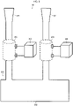
- the testing system 100 includes a milking machine 200 and an NMR measurement device 300.
- the milking machine 200 includes: a milker unit 201 which is attached to a quarter and takes in collected milk; a milk claw (milk mixing tank) 202 which mixes milk collected from respective quarters; and a flow cell 203 for NMR measurement.
- the milking machine 200 can be a well-known milking machine. For example, in a case where four quarters of a dairy cow are tested simultaneously, four milker units may be provided.
- the flow cell 203 is installed in a predetermined position between the milker unit 201 and a position short of the milk claw 202. This allows a spin-spin relaxation time of milk collected from each quarter to be measured.
- the NMR measurement device 300 can be attached to the flow cell 203 of the milking machine 200.
- the NMR measurement device 300 can be, for example, a well-known NMR measurement device. From perspectives such as easy attachment to the milking machine 200 and reduction of a size of a measuring device, it is preferable that the NMR measurement device 300 be a pulsed NMR measurement device.
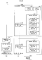
- Fig. 9 is a block diagram illustrating a configuration of a main part of a testing system.
- the testing system 101 includes a milking machine 200, an NMR measurement device 300, a management terminal (e.g., a small mobile terminal such as a smartphone) 400, and a somatic cell measurement device 500.
- a milking machine 200 includes a milking machine 200, an NMR measurement device 300, a management terminal (e.g., a small mobile terminal such as a smartphone) 400, and a somatic cell measurement device 500.
- the NMR measurement device 300 includes a communication section 301, a measurement section 303, a calculation section 304, and a control section 302 which controls the measurement section 303 and the calculation section 304.
- the communication section 301 communicates with the management terminal 400.
- the measurement section 303 measures a spin-spin relaxation time of milk collected from each quarter.
- the calculation section 304 converts a specific surface area of particles contained in the milk.
- the management terminal 400 includes a communication section 401, an NMR measured value comparison section 405, a somatic cell count comparison section 406, a determination section 407, an abnormality notification section 408, a storage section 403, a display section 404, and a control section 402.
- the communication section 401 communicates with the NMR measurement device 300 and the somatic cell measurement device 500.
- the NMR measured value comparison section 405 compares the spin-spin relaxation time or the specific surface area of the sample milk with the predetermined standard S1.
- the somatic cell count comparison section 406 compares the somatic cell count in the sample milk with the predetermined standard S2.
- the determination section 407 determines, on the basis of a result of comparison by the NMR measured value comparison section 405, whether or not the quarter from which the sample milk was collected has mastitis or is likely to have mastitis. In a case where the determination section 407 has determined that the quarter has mastitis or is likely to have mastitis, the abnormality notification section 408 provides notification of such to a user of the management terminal 400.
- the display section 404 displays the results of measurement by the NMR measurement device 300 and the somatic cell measurement device 500, as well as the notification by the abnormality notification section 408.
- the determination section 407 may presume a cause of the mastitis (whether or not the mastitis is due to Gram-positive bacterial infection) on the basis of the result of the somatic cell count comparison section 406 in addition to the result of comparison by the NMR measured value comparison section 405.
- the somatic cell measurement device 500 includes: a communication section 501 which communicates with the management terminal 400; a measurement section 503 which measures a somatic cell count in milk collected from each quarter; and a control section 502 which controls the measurement section 503.
- the present invention is not limited to the embodiments, but can be altered by a skilled person in the art within the scope of the claims.
- the present invention also encompasses, in its technical scope, any embodiment derived by combining technical means disclosed in differing embodiments.
- the present invention is not limited to the embodiments, but can be altered by a skilled person in the art within the scope of the claims.
- the present invention also encompasses, in its technical scope, any embodiment derived by combining technical means disclosed in differing embodiments.
- the embodiments of the present invention include, for example, the following aspects.
- a dairy cow has four quarters. Each quarter is an independent organ. Accordingly, milk produced by respective quarters differs in concentration of a component and the like, depending on a health condition of each quarter. Further, mastitis develops individually in each quarter. As such, in the following Examples, analysis was conducted with respect to milk collected from each quarter of a dairy cow. In Examples 1 and 2, in a case where a somatic cell count in milk was not less than 20 ⁇ 10 4 /mL, it was determined that a quarter from which the milk was collected had mastitis. In Examples 3 and 4, in a case where a somatic cell count in milk was not less than 50 ⁇ 10 4 /mL, it was determined that a quarter from which the milk was collected had mastitis.
- Milk was collected from each of the four udder quarters (right front, right rear, left front, and left rear beasts) of a research dairy cow A kept and managed at the National Institute of Animal Health of the National Agriculture and Food Research Organization.
- a somatic cell count in the collected milk and a spin-spin relaxation time (T 2 ) of the collected milk were measured.
- the somatic cell count was measured by a cell counter DCC (manufactured by DeLaval).
- the collected milk was inverted and mixed, and then sucked into a dedicated cassette and inserted into a main body for measurement.
- the relaxation time (T 2 ) was measured at 25°C with a pulsed NMR device (manufactured by Xigo Nanotools, device name: ACORN AREA) with use of the milk which was put into an NMR tube as it was without diluting the milk or adding anything to the milk.
- a mode in which measurement of T 2 was conducted was a Carr-Purcell Meiboom-Gill sequence (CPMG) method.
- CPMG Carr-Purcell Meiboom-Gill sequence
- Rav is a reciprocal of an average relaxation time.
- ⁇ p is a volume fraction (volume concentration) of the particles contained in the milk.
- L is a thickness (wet thickness) of a liquid layer adsorbed on surfaces of the particles contained in the milk.
- ⁇ p is a density of the particles contained in the milk.
- Rs is a reciprocal of a relaxation time of molecules of a liquid adsorbed on the surfaces of the particles contained in the milk.
- Rb is a reciprocal of a relaxation time of a solvent water.
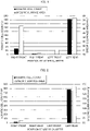
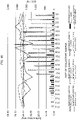
- Fig. 1 illustrates results of the measured somatic cell count and the specific surface area calculated on the basis of the measured relaxation time (T 2 ).
- T 2 measured relaxation time
- an axis on the left represents a somatic cell count (10 4 ) per milliliter.
- An axis on the right represents a specific surface area (m 2 /g).
- somatic cell counts in milk respectively collected from the right rear quarter and the left front quarter were each not more than 20 ⁇ 10 4 /mL, and the right rear quarter and the left front quarter were thus found to be healthy.
- somatic cell counts in milk collected from the right front quarter and the left rear quarter were 30 ⁇ 10 4 /mL and 182 ⁇ 10 4 /mL, respectively, and it was thus indicated that both the right front quarter and the left rear quarter had mastitis.
- Neither milk was found by a bacteria test to have Staphylococcus aureus therein, and it was thus determined that the mastitis was due to E . coli or a trauma.
- a value of a specific area of particles was higher in the milk from the right front quarter and in the milk from the left rear quarter, as compared with the milk from the right rear quarter and the milk from the left front quarter. It was thus confirmed that a specific surface area corresponds to a somatic cell count.
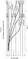
- Example 2 After the collection of milk in Example 1, the research dairy cow A was kept for 2 days, and then milk was collected again from each quarter. Then, a somatic cell count and a relaxation time (T 2 ) were measured and a specific surface area was calculated by the same procedure as in Example 1. Fig. 2 illustrates results of the measured somatic cell count and the specific surface area calculated on the basis of the measured relaxation time (T 2 ).
- somatic cell counts in milk respectively collected from the right rear quarter and the left front quarter were each not more than 20 ⁇ 10 4 /mL, and the right rear quarter and the left front quarter were thus found to be healthy similarly as in the results of Example 1.
- a somatic cell count for the right front quarter was found to be on a healthier level in comparison to Example 1.
- the milk collected from the left rear quarter had a somatic cell count of 780 ⁇ 10 4 /mL, and it was thus indicated that the left rear quarter had not recovered from mastitis.
- a value of a specific surface area of particles contained in milk was low in each of the milk from the right front quarter, the milk from the right rear quarter, and the milk from the left front quarter, whereas the value was high in the milk from the left rear quarter, corresponding to the somatic cell count.
- FIG. 3 illustrates results of the measured somatic cell count and the specific surface area calculated on the basis of the measured relaxation time (T 2 ).
- somatic cell counts in milk respectively collected from a right front quarter, a right rear quarter, and a left front quarter were each not more than 50 ⁇ 10 4 /mL, and the right front quarter, the right rear quarter, and the left front quarter were thus found to be healthy.
- a somatic cell count in milk collected from the left rear quarter was 180 ⁇ 10 4 /mL, and it was thus indicated that the left rear quarter had mastitis.
- a bacteria test of the milk from the left rear quarter found Staphylococcus aureus, and it was thus determined that the mastitis developed in the left rear quarter was due to Staphylococcus aureus.
- a specific surface area of particles contained in the milk from the left rear quarter having mastitis was significantly lower than those in the milk from the right front quarter, the milk from the right rear quarter, and the milk from the left front quarter. This is because tycolic acid produced by Gram-positive bacteria such as Staphylococcus aureus caused micelles (such as casein) dispersed in milk to aggregate, so that the specific surface area was reduced.
- a somatic cell count and a relaxation time (T 2 ) were measured and a specific surface area was calculated by the same procedure as in Example 1, except that the dairy cow was changed to a research dairy cow C kept and managed at the National Institute of Animal Health of the National Agriculture and Food Research Organization.
- Fig. 4 illustrates results of the measured somatic cell count and the specific surface area calculated on the basis of the measured relaxation time (T 2 ).
- somatic cell counts in milk respectively collected from a right front quarter, a right rear quarter, and a left front quarter were each not more than 50 ⁇ 10 4 /mL, and the right front quarter, the right rear quarter, and the left front quarter were thus found to be healthy.
- a somatic cell count in milk collected from the left rear quarter was 250 ⁇ 10 4 /mL, and it was thus indicated that the left rear quarter had mastitis.
- a bacteria test of milk from the left rear quarter found Staphylococcus aureus, and it was thus determined that the mastitis developed in the left rear quarter was due to Staphylococcus aureus.
- a specific surface area for the milk from the left rear quarter having mastitis was significantly lower than those for the right front quarter, the right rear quarter, and the left front quarter, and obtained results were thus similar to those in Example 3.
- an axis on the left represents a specific surface area (m 2 /g).
- An axis on the right represents a pH.
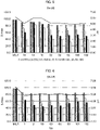
- a horizontal axis represents a number of a Staphylococcus aureus strain used in Evaluation Example 1. "Milk” represents a control to which Staphylococcus aureus was not added. Four bars for each strain represent, from left, results of measurement of milk at 0, 3, 6, and 9 hours, respectively, after the addition of Staphylococcus aureus.
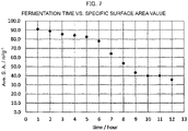
- Fig. 7 illustrates results of calculation of the specific surface area on the basis of the measured relaxation time (T 2 ).
- an axis on the left represents a specific surface area (m 2 /g).
- a horizontal axis represents an elapsed time after the addition of the lactic acid bacteria.
- a decrease in specific surface area was observed after the addition of the lactic acid bacteria, similarly as in the results of the addition of Staphylococcus aureus in Evaluation Example 1. This is because lactic acid produced by lactic acid fermentation caused casein micelles, which had been dispersed in milk, to lose a zeta potential and aggregate. It is considered that the decreases in specific surface area through the addition of Staphylococcus aureus in Evaluation Example 1 was also due to aggregation of casein micelles by lactic acid fermentation.
- FIG. 10 illustrates results of the measured somatic cell count and the specific surface area calculated on the basis of the measured relaxation time (T 2 ).
- an axis on the left represents a specific surface area (m 2 /g), and an axis on the right represents a somatic cell count (10 4 /mL).
- a line graph shows results of measurement of a specific surface area
- a bar graph shows results of measurement of a somatic cell count.
- a result of measurement on July 21 indicated that a somatic cell count for a right front quarter (RF) was approximately 1,000 ⁇ 10 4 /mL, which was considerably higher than somatic cell counts for the other quarters. From this result, it was found that the right front quarter developed mastitis on July 21. Further, several days before July 21, a decrease in specific surface area of particles contained in milk from the right front quarter had been observed. It was thus determined that the mastitis that developed in the right front quarter was due to infection with Staphylococcus aureus. Further, several days before July 28, on which date the somatic cell count in the right front quarter considerably increased, a decrease had been observed in specific surface area of particles contained in milk from the right front quarter. The decrease in specific surface area of the particles contained in the milk is considered to be due to a decrease in pH and casein micelle aggregation in the milk which were caused by multiplication of Staphylococcus aureus.
- FIG. 11 illustrates results of the measured somatic cell count and the specific surface area calculated on the basis of the measured relaxation time (T 2 ).
- an axis on the left represents a specific surface area (m 2 /g), and an axis on the right represents a somatic cell count (10 4 /mL).
- Two line graphs show results of measurement of a specific surface area (broken lines) and results of measurement of an amount of milk (solid lines), respectively, and a bar graph shows results of measurement of a somatic cell count.
- a somatic cell count in milk collected from a quarter of a dairy cow No. 1256 increased to not less than 100 ⁇ 10 4 /mL on August 31.
- a change in specific surface area of particles was observed in milk from the dairy cow No. 1256.
- a somatic cell count in milk collected from a quarter of a dairy cow No. 1405 increased to not less than 100 ⁇ 10 4 /mL on September 4.
- a change in specific surface area of particles contained in milk was observed before the increase in cell count.
- a somatic cell count and a relaxation time (T 2 ) were continuously measured and a specific surface area was calculated by the same procedure as in Example 1, with respect to a milk sample collected from each of three dairy cows kept at Hokkaido Agricultural Technical College.
- Fig. 12 illustrates results of the measured somatic cell count and the specific surface area calculated on the basis of the measured relaxation time (T 2 ). Milk was collected twice a day, in the morning (AM) and evening (PM).
- an axis on the left represents a specific surface area (m 2 /g), and an axis on the right represents a somatic cell count (10 4 /mL).
- m 2 /g specific surface area
- somatic cell count 10 4 /mL
- Staphylococcus aureus was added to 3% aqueous solutions of skim milk.
- a bacterial count was adjusted to 1.0 ⁇ 10 10 CFU/mL, 5.0 ⁇ 10 9 CFU/mL, 2.5 ⁇ 10 9 CFU/mL, and 1.25 ⁇ 10 9 CFU/mL, respectively.
- the samples were each cultured at 37°C. Every hour after a start of the culturing, a relaxation time (T 2 ) of milk was measured and a specific surface area was calculated by the same procedure as in Example 1. Results of measurement are shown in Fig. 13 .
- a vertical axis represents a specific surface area (m 2 /g), and a horizontal axis represents a culture time.
- m 2 /g specific surface area
- a horizontal axis represents a culture time.
- the present invention can be used in testing for mastitis in a domestic animal.
- the present invention can be widely used in fields such as food and medical diagnosis.
Landscapes
- Physics & Mathematics (AREA)
- Health & Medical Sciences (AREA)
- Life Sciences & Earth Sciences (AREA)
- Chemical & Material Sciences (AREA)
- Engineering & Computer Science (AREA)
- General Physics & Mathematics (AREA)
- High Energy & Nuclear Physics (AREA)
- Immunology (AREA)
- General Health & Medical Sciences (AREA)
- Analytical Chemistry (AREA)
- Pathology (AREA)
- Biochemistry (AREA)
- Food Science & Technology (AREA)
- Biomedical Technology (AREA)
- Medicinal Chemistry (AREA)
- Condensed Matter Physics & Semiconductors (AREA)
- Molecular Biology (AREA)
- Biophysics (AREA)
- Urology & Nephrology (AREA)
- Hematology (AREA)
- Crystallography & Structural Chemistry (AREA)
- Spectroscopy & Molecular Physics (AREA)
- Investigating Or Analysing Biological Materials (AREA)
Abstract
Description
- The present invention relates to a method and a system for testing for mastitis in a domestic animal.
- Economic losses in Japan due to mastitis, which is one of infectious diseases suffered by dairy cows, amount to 80 billion yen per year. Since early detection of mastitis can reduce the cost of treatment and inhibit decreased milk production, research and development for early detection of mastitis is underway.
- For example,
Patent Literature 1 discloses a method for diagnosing mastitis by measuring a somatic cell count in milk.Patent Literature 2 discloses a method for diagnosing mastitis by measuring an electrical conductivity of milk.Patent Literature 3 discloses a method for diagnosing mastitis by decomposing somatic cells in milk to produce cytokine and the like and thus activate somatic cells, and then measuring a somatic cell count.Patent Literature 4 discloses a method for diagnosing mastitis by measuring an electrical conductivity of centrifuged milk. -
- [Patent Literature 1]
PublishedJapanese Translation of PCT International Application, Tokuhyo, No. 2002-503097 - [Patent Literature 2]
Japanese Patent Application Publication, Tokukai, No. 2010-230363 - [Patent Literature 3]
Japanese Patent Application Publication, Tokukai, No. 2010-256231 - [Patent Literature 4]
Japanese Patent Application Publication, Tokukai, No. 2011-33599 - Methods for diagnosing mastitis by somatic cell count measurement have a problem that devices for measuring a somatic cell count are expensive. Measurement of a somatic cell count by microscopic observation has a problem that the operation is complicated. Methods for diagnosing mastitis by measuring an electrical conductivity of milk are not suitable for early diagnosis of mastitis because the methods observe a change caused by an inflammatory reaction after invasion by a pathogen. Further, since a composition ratio of electrolytes contained in milk varies among quarters of udder of the same individual or among individuals, reproducibility in the measurement of an electrical conductivity of milk is poor. Thus, there has not been developed a technique that can easily detect mastitis in a domestic animal, such as a dairy cow, at an early stage.
- It is an object of an aspect of the present invention to provide a test that can easily detect mastitis in a domestic animal, such as a dairy cow, at an early stage.
- In order to attain the object, the present invention includes the following aspects. - A testing method for testing for mastitis in a domestic animal, said method including the step of: measuring, by nuclear magnetic resonance (NMR), a spin-spin relaxation time of milk collected from a quarter of the domestic animal. - A testing system for testing for mastitis in a domestic animal, including: a milking machine including a milker unit and a milk claw; and an NMR measurement device, a flow cell for NMR measurement being provided at a predetermined position between the milker unit and a position short of the milk claw.
- An aspect of the present invention makes it possible to provide a test that can easily detect mastitis in a domestic animal, such as a dairy cow, at an early stage.
-
-
Fig. 1 is a graph illustrating a result of analysis of milk collected from each quarter in Example 1. -
Fig. 2 is a graph illustrating a result of analysis of milk collected from each quarter in Example 2. -
Fig. 3 is a graph illustrating a result of analysis of milk collected from each quarter in Example 3. -
Fig. 4 is a graph illustrating a result of analysis of milk collected from each quarter in Example 4. -
Fig. 5 is a graph illustrating a result of analysis of milk by addition of Staphylococcus aureus (SA-VB) in Evaluation Example 1. -
Fig. 6 is a graph illustrating a result of analysis of milk by addition of Staphylococcus aureus (SA-VR) in Evaluation Example 1. -
Fig. 7 is a graph illustrating a result of analysis of milk by addition of lactic acid bacteria in Evaluation Example 2. -
Fig. 8 is a schematic view illustrating an example of a testing system in accordance with an embodiment of the present invention. -
Fig. 9 is a block diagram illustrating a configuration of a main part of a testing system in accordance with an embodiment of the present invention. -
Fig. 10 is a graph illustrating a result of analysis of milk collected from each quarter in Example 5. -
Fig. 11 is a graph illustrating a result of analysis of milk collected from each dairy cow in Example 6. -
Fig. 12 is a graph illustrating a result of analysis of milk collected from each dairy cow in Example 7. -
Fig. 13 is a graph illustrating a result of analysis of milk by addition of Staphylococcus aureus in Evaluation Example 3. - A method (hereinafter, also abbreviated as a "testing method of the present invention"), in accordance with an embodiment of the present invention, for testing for mastitis in a domestic animal includes the step of measuring, by nuclear magnetic resonance (NMR), a spin-spin relaxation time of milk (hereinafter, also referred to as "sample milk") collected from a quarter of the domestic animal.
- Examples of the domestic animal include cattle such as a dairy cow, a goat, a buffalo, a sheep, and a horse. For example, cattle such as a dairy cow have four independent quarters: a right front quarter; a right rear quarter; a left front quarter; and a left rear quarter.
- The nuclear magnetic resonance (NMR) used in the testing method of the present invention only needs to be capable of measuring a spin-spin relaxation time (T2) of sample milk, and examples of the NMR include pulsed NMR. From perspectives such as reduction of a size of a measuring device, it is preferable to use pulsed NMR.
- The milk can be collected from a quarter of the domestic animal by means ordinarily used by a person skilled in the art, such as a milking machine. By attaching an NMR measurement device to the milking machine, the spin-spin relaxation time (T2) of the milk can be measured easily and non-invasively. In NMR (in particular, pulsed NMR), one measuring time is as short as several tens of seconds. Further, by attaching an NMR measurement device to an automatic milking machine, the spin-spin relaxation time (T2) can be measured continuously at regular intervals. Further, by attaching an NMR measurement device to an automated milking machine, there is no need to sample milk for testing for mastitis, and the burden on a manager of domestic animals can be reduced. The testing method of the present invention therefore allows real-time measurement (testing). Note that it is preferable to have the above configuration in which an NMR measurement device is attached to a milking machine and a portion of the milking machine is used as a flow cell for NMR measurement. However, the present invention is not particularly limited to such a configuration.
- The spin-spin relaxation time or a specific surface area of particles contained in the milk, which specific surface area is obtained by converting the spin-spin relaxation time, may be compared with a predetermined standard S1. The comparison allows determining whether or not the quarter from which the milk was collected has mastitis or is likely to have mastitis. Specifically, measured data (the spin-spin relaxation time or the specific surface area) of the sample milk is compared with the predetermined standard S1, and in a case where the measured data of the sample milk is significantly increased or decreased in comparison to the predetermined standard S1, it is possible to determine whether or not the quarter from which the sample milk was collected has mastitis or is likely to have mastitis. When comparing the data of the sample milk with the predetermined standard S1, whether or not there is a significant difference can be determined by a statistical method such as, for example, a t-test, an F-test, a chi-square test, or a Mann-Whitney's U test.
- The specific surface area of the particles contained in the milk can be obtained from the following equation (1).

- In equation (1), Rav is a reciprocal of an average relaxation time. Ψp is a volume fraction (volume concentration) of the particles contained in the milk. L is a thickness (wet thickness) of a liquid layer adsorbed on surfaces of the particles contained in the milk. ρp is a density of the particles contained in the milk. Rs is a reciprocal of a relaxation time of molecules of a liquid adsorbed on the surfaces of the particles contained in the milk. Rb is a reciprocal of a relaxation time of solvent water.
- Examples of the predetermined standard S1 include: (1) data of milk collected, from the quarter from which the sample milk was collected, at a time point (e.g., in advance) different from a time point at which the sample milk was collected; (2) data of milk collected from another quarter of the individual from which the sample milk was collected; and (3) data of milk collected from a quarter of an individual different from the individual from which the sample milk was collected. The data (1) through (3) are each a spin-spin relaxation time of the milk or a specific surface area of particles contained in the milk, which specific surface area is obtained by converting the spin-spin relaxation time. The data (2) and (3) may each be data of milk collected simultaneously with the collection of the sample milk, or data of milk collected in advance of the collection of the sample milk. The predetermined standard S1 can be determined such that: a spin-spin relaxation time (specific surface area) of milk collected in advance from a control quarter having no mastitis is measured; and a normal range (e.g., a standard S1 that serves as a threshold) is determined on the basis of the spin-spin relaxation time (specific surface area).
- The predetermined standard S1 may be, for example, a collection of time-series data obtained at a plurality of time points with respect to the quarter from which the sample milk was collected. In this case, an evaluation is made of a change over time in the spin-spin relaxation time of milk from the quarter from which the sample milk was collected, or a change over time in the specific surface area of the particles contained in the milk, which specific surface area is obtained by converting the spin-spin relaxation time.
- The testing method of the present invention may further include a step of comparing a somatic cell count in the sample milk with a predetermined standard S2. When comparing the somatic cell count of the sample milk with the predetermined standard S2, whether or not there is a significant difference can be determined by a statistical method such as, for example, a t-test, an F-test, a chi-square test, or a Mann-Whitney's U test. In the present specification, "a somatic cell count contained in milk" refers to a total number of blood-derived cells (leukocytes and the like) and mammary gland epithelial cells contained in the milk. The somatic cell count can be measured by a well-known method.
- Examples of the predetermined standard S2 include: (4) a somatic cell count in milk collected, from the quarter from which the sample milk was collected, at a timing (e.g., in advance) different from a timing at which the sample milk was collected; (5) a somatic cell count in milk collected from another quarter of the individual from which the sample milk was collected; and (6) a somatic cell count in milk collected from a quarter of an individual different from the individual from which the sample milk was collected. The above data (5) and (6) may each be a somatic cell count in milk collected simultaneously with the collection of the sample milk, or a somatic cell count in milk collected in advance of the collection of the sample milk. The predetermined standard S2 can be determined such that: a somatic cell count in milk collected in advance from a control quarter having no mastitis is measured; and a normal range (e.g., a standard S2 that serves as a threshold) is determined on the basis of the somatic cell count.
- The predetermined standard S2 may be, for example, a collection of time-series data obtained at a plurality of time points with respect to the quarter from which the sample milk was collected. In this case, an evaluation is made of a change over time in the somatic cell count in milk from the quarter from which the sample milk was collected, or a change over time in the specific surface area of the particles contained in the milk, which specific surface area is obtained by converting the spin-spin relaxation time.
- As indicated in Examples, in a case where mastitis has developed due to infection with Gram-positive bacteria such as Staphylococcus aureus, a specific surface area of particles contained in milk decreases as a somatic cell count increases. This is considered to be because an acidic substance (e.g., lactic acid) produced by the Gram-positive bacteria causes casein micelles, which are dispersed in milk, to aggregate. In contrast, in a case where mastitis (e.g., mastitis due to infection with Gram-negative bacteria such as E. coli, mastitis due to trauma, or the like), which is other than mastitis due to infection with Gram-positive bacteria, has developed, a specific surface area of particles contained in milk increases as a somatic cell count increases. As such, by carrying out somatic cell measurement in addition to NMR measurement, it is possible to presume a cause of mastitis (whether or not the mastitis is caused by Gram-positive bacterial infection).
- With reference to
Fig. 8 , the following description will discuss a system (hereinafter, also abbreviated as a "testing system of the present invention") 100, in accordance with an embodiment of the present invention, for testing for mastitis in a domestic animal. Thetesting system 100 includes amilking machine 200 and anNMR measurement device 300. Themilking machine 200 includes: amilker unit 201 which is attached to a quarter and takes in collected milk; a milk claw (milk mixing tank) 202 which mixes milk collected from respective quarters; and aflow cell 203 for NMR measurement. - The
milking machine 200 can be a well-known milking machine. For example, in a case where four quarters of a dairy cow are tested simultaneously, four milker units may be provided. Theflow cell 203 is installed in a predetermined position between themilker unit 201 and a position short of themilk claw 202. This allows a spin-spin relaxation time of milk collected from each quarter to be measured. - It is only necessary that the
NMR measurement device 300 can be attached to theflow cell 203 of themilking machine 200. TheNMR measurement device 300 can be, for example, a well-known NMR measurement device. From perspectives such as easy attachment to themilking machine 200 and reduction of a size of a measuring device, it is preferable that theNMR measurement device 300 be a pulsed NMR measurement device. - With reference to
Fig. 9 , the following description will discuss atesting system 101, which is another embodiment, of the present invention.Fig. 9 is a block diagram illustrating a configuration of a main part of a testing system. Thetesting system 101 includes amilking machine 200, anNMR measurement device 300, a management terminal (e.g., a small mobile terminal such as a smartphone) 400, and a somaticcell measurement device 500. - The
NMR measurement device 300 includes acommunication section 301, ameasurement section 303, acalculation section 304, and acontrol section 302 which controls themeasurement section 303 and thecalculation section 304. Thecommunication section 301 communicates with themanagement terminal 400. Themeasurement section 303 measures a spin-spin relaxation time of milk collected from each quarter. On the basis of the spin-spin relaxation time, thecalculation section 304 converts a specific surface area of particles contained in the milk. - The
management terminal 400 includes acommunication section 401, an NMR measuredvalue comparison section 405, a somatic cellcount comparison section 406, adetermination section 407, anabnormality notification section 408, astorage section 403, adisplay section 404, and acontrol section 402. Thecommunication section 401 communicates with theNMR measurement device 300 and the somaticcell measurement device 500. The NMR measuredvalue comparison section 405 compares the spin-spin relaxation time or the specific surface area of the sample milk with the predetermined standard S1. The somatic cellcount comparison section 406 compares the somatic cell count in the sample milk with the predetermined standard S2. Thedetermination section 407 determines, on the basis of a result of comparison by the NMR measuredvalue comparison section 405, whether or not the quarter from which the sample milk was collected has mastitis or is likely to have mastitis. In a case where thedetermination section 407 has determined that the quarter has mastitis or is likely to have mastitis, theabnormality notification section 408 provides notification of such to a user of themanagement terminal 400. Thedisplay section 404 displays the results of measurement by theNMR measurement device 300 and the somaticcell measurement device 500, as well as the notification by theabnormality notification section 408. - The
determination section 407 may presume a cause of the mastitis (whether or not the mastitis is due to Gram-positive bacterial infection) on the basis of the result of the somatic cellcount comparison section 406 in addition to the result of comparison by the NMR measuredvalue comparison section 405. - The somatic
cell measurement device 500 includes: acommunication section 501 which communicates with themanagement terminal 400; ameasurement section 503 which measures a somatic cell count in milk collected from each quarter; and acontrol section 502 which controls themeasurement section 503. - The present invention is not limited to the embodiments, but can be altered by a skilled person in the art within the scope of the claims. The present invention also encompasses, in its technical scope, any embodiment derived by combining technical means disclosed in differing embodiments.
- The present invention is not limited to the embodiments, but can be altered by a skilled person in the art within the scope of the claims. The present invention also encompasses, in its technical scope, any embodiment derived by combining technical means disclosed in differing embodiments.
- The embodiments of the present invention include, for example, the following aspects.
- <1> A testing method for testing for mastitis in a domestic animal, said method including the step of: measuring, by nuclear magnetic resonance, NMR, a spin-spin relaxation time of milk (sample milk) collected from a quarter of the domestic animal.
- <2> The testing method as set forth in <1>, further including comparing, with a predetermined standard S1, the spin-spin relaxation time or a specific surface area of particles contained in the milk, the specific surface area being obtained by conversion of the spin-spin relaxation time.
- <3> The testing method as set forth in <1> or <2>, further including: the step of comparing a somatic cell count in the milk with a predetermined standard S2.
- <4> The testing method as set forth in any one of <1> through <3>, wherein the domestic animal is a dairy cow.
- <5> A testing system for testing for mastitis in a domestic animal, including: a milking machine including a milker unit and a milk claw; and an NMR measurement device, a flow cell for NMR measurement being provided at a predetermined position between the milker unit and a position short of the milk claw.
- <6> The testing system as set forth in <5>, further including a somatic cell measurement device.
- <7> The testing system as set forth in <5> or <6>, wherein the domestic animal is a dairy cow.
- <8>The testing method as set forth in <2>, wherein whether or not mastitis has developed is presumed by comparison with the predetermined standard S1.
- <9>The testing method as set forth in <3>, wherein a cause of mastitis is presumed by comparison with the predetermined standard S2.
- <10> Note that the testing methods of <1> through <4> can be interpreted as a method for obtaining data for carrying out testing for mastitis in a domestic animal and a method for analyzing the data.
- A dairy cow has four quarters. Each quarter is an independent organ. Accordingly, milk produced by respective quarters differs in concentration of a component and the like, depending on a health condition of each quarter. Further, mastitis develops individually in each quarter. As such, in the following Examples, analysis was conducted with respect to milk collected from each quarter of a dairy cow. In Examples 1 and 2, in a case where a somatic cell count in milk was not less than 20×104/mL, it was determined that a quarter from which the milk was collected had mastitis. In Examples 3 and 4, in a case where a somatic cell count in milk was not less than 50×104/mL, it was determined that a quarter from which the milk was collected had mastitis.
- Milk was collected from each of the four udder quarters (right front, right rear, left front, and left rear beasts) of a research dairy cow A kept and managed at the National Institute of Animal Health of the National Agriculture and Food Research Organization. A somatic cell count in the collected milk and a spin-spin relaxation time (T2) of the collected milk were measured. The somatic cell count was measured by a cell counter DCC (manufactured by DeLaval). The collected milk was inverted and mixed, and then sucked into a dedicated cassette and inserted into a main body for measurement. The relaxation time (T2) was measured at 25°C with a pulsed NMR device (manufactured by Xigo Nanotools, device name: ACORN AREA) with use of the milk which was put into an NMR tube as it was without diluting the milk or adding anything to the milk. A mode in which measurement of T2 was conducted was a Carr-Purcell Meiboom-Gill sequence (CPMG) method.
- On the basis of the relaxation time (T2) thus measured, a specific surface area (S) of particles contained in the milk was calculated using the following equation (1):

- In equation (1), Rav is a reciprocal of an average relaxation time. Ψp is a volume fraction (volume concentration) of the particles contained in the milk. L is a thickness (wet thickness) of a liquid layer adsorbed on surfaces of the particles contained in the milk. ρp is a density of the particles contained in the milk. Rs is a reciprocal of a relaxation time of molecules of a liquid adsorbed on the surfaces of the particles contained in the milk. Rb is a reciprocal of a relaxation time of a solvent water.
-
Fig. 1 illustrates results of the measured somatic cell count and the specific surface area calculated on the basis of the measured relaxation time (T2). InFig. 1 , an axis on the left represents a somatic cell count (104) per milliliter. An axis on the right represents a specific surface area (m2/g). - As illustrated in
Fig. 1 , somatic cell counts in milk respectively collected from the right rear quarter and the left front quarter were each not more than 20×104/mL, and the right rear quarter and the left front quarter were thus found to be healthy. In contrast, somatic cell counts in milk collected from the right front quarter and the left rear quarter were 30×104/mL and 182×104/mL, respectively, and it was thus indicated that both the right front quarter and the left rear quarter had mastitis. Neither milk was found by a bacteria test to have Staphylococcus aureus therein, and it was thus determined that the mastitis was due to E. coli or a trauma. Further, a value of a specific area of particles was higher in the milk from the right front quarter and in the milk from the left rear quarter, as compared with the milk from the right rear quarter and the milk from the left front quarter. It was thus confirmed that a specific surface area corresponds to a somatic cell count. - After the collection of milk in Example 1, the research dairy cow A was kept for 2 days, and then milk was collected again from each quarter. Then, a somatic cell count and a relaxation time (T2) were measured and a specific surface area was calculated by the same procedure as in Example 1.
Fig. 2 illustrates results of the measured somatic cell count and the specific surface area calculated on the basis of the measured relaxation time (T2). - As illustrated in
Fig. 2 , somatic cell counts in milk respectively collected from the right rear quarter and the left front quarter were each not more than 20×104/mL, and the right rear quarter and the left front quarter were thus found to be healthy similarly as in the results of Example 1. A somatic cell count for the right front quarter was found to be on a healthier level in comparison to Example 1. The milk collected from the left rear quarter had a somatic cell count of 780×104/mL, and it was thus indicated that the left rear quarter had not recovered from mastitis. A value of a specific surface area of particles contained in milk was low in each of the milk from the right front quarter, the milk from the right rear quarter, and the milk from the left front quarter, whereas the value was high in the milk from the left rear quarter, corresponding to the somatic cell count. - A somatic cell count and a relaxation time (T2) were measured and a specific surface area was calculated by the same procedure as in Example 1, except that the dairy cow was changed to a research dairy cow B kept and managed at the National Institute of Animal Health of the National Agriculture and Food Research Organization.
Fig. 3 illustrates results of the measured somatic cell count and the specific surface area calculated on the basis of the measured relaxation time (T2). - As illustrated in
Fig. 3 , somatic cell counts in milk respectively collected from a right front quarter, a right rear quarter, and a left front quarter were each not more than 50×104/mL, and the right front quarter, the right rear quarter, and the left front quarter were thus found to be healthy. In contrast, a somatic cell count in milk collected from the left rear quarter was 180×104/mL, and it was thus indicated that the left rear quarter had mastitis. A bacteria test of the milk from the left rear quarter found Staphylococcus aureus, and it was thus determined that the mastitis developed in the left rear quarter was due to Staphylococcus aureus. A specific surface area of particles contained in the milk from the left rear quarter having mastitis was significantly lower than those in the milk from the right front quarter, the milk from the right rear quarter, and the milk from the left front quarter. This is because tycolic acid produced by Gram-positive bacteria such as Staphylococcus aureus caused micelles (such as casein) dispersed in milk to aggregate, so that the specific surface area was reduced. - A somatic cell count and a relaxation time (T2) were measured and a specific surface area was calculated by the same procedure as in Example 1, except that the dairy cow was changed to a research dairy cow C kept and managed at the National Institute of Animal Health of the National Agriculture and Food Research Organization.
Fig. 4 illustrates results of the measured somatic cell count and the specific surface area calculated on the basis of the measured relaxation time (T2). - As illustrated in
Fig. 4 , somatic cell counts in milk respectively collected from a right front quarter, a right rear quarter, and a left front quarter were each not more than 50×104/mL, and the right front quarter, the right rear quarter, and the left front quarter were thus found to be healthy. In contrast, a somatic cell count in milk collected from the left rear quarter was 250×104/mL, and it was thus indicated that the left rear quarter had mastitis. A bacteria test of milk from the left rear quarter found Staphylococcus aureus, and it was thus determined that the mastitis developed in the left rear quarter was due to Staphylococcus aureus. A specific surface area for the milk from the left rear quarter having mastitis was significantly lower than those for the right front quarter, the right rear quarter, and the left front quarter, and obtained results were thus similar to those in Example 3. - To 3 mL of milk collected from a healthy quarter, two different strains of Staphylococcus aureus (SA-VB and SA-VR) were each added in an amount of approximately 30 mg. The Staphylococcus aureus strains used were those preserved at the National Institute of Animal Health of the National Agriculture and Food Research Organization. SA-VR is more toxic than SA-VB. After Staphylococcus aureus was added, the milk was kept at 37°C. At 0, 3, 6, and 9 hours after the addition of Staphylococcus aureus, a relaxation time (T2) of milk was measured and a specific surface area was calculated by the same procedure as in Example 1. A pH of the milk was also measured. Results of the measured pH and the specific surface area calculated on the basis of the measured relaxation time (T2) are illustrated in
Fig. 5 (SA-VB) andFig. 6 (SA-VR). - In
Figs. 5 and 6 , an axis on the left represents a specific surface area (m2/g). An axis on the right represents a pH. A horizontal axis represents a number of a Staphylococcus aureus strain used in Evaluation Example 1. "Milk" represents a control to which Staphylococcus aureus was not added. Four bars for each strain represent, from left, results of measurement of milk at 0, 3, 6, and 9 hours, respectively, after the addition of Staphylococcus aureus. - As illustrated in
Figs. 5 and 6 , a decrease in specific surface area was observed in milk with any type of Staphylococcus aureus added therein. It was also found that a degree of decrease in specific surface area differed depending on the pH of milk. - To 100 mL of milk collected from a healthy quarter, 1 g of fermented milk containing lactic acid bacteria was added. After the lactic acid bacteria was added, the milk was kept at 37°C. Every hour after the addition of the lactic acid bacteria, a relaxation time (T2) of the milk was measured and a specific surface area was calculated by the same procedure as in Example 1.
Fig. 7 illustrates results of calculation of the specific surface area on the basis of the measured relaxation time (T2). - In
Fig. 7 , an axis on the left represents a specific surface area (m2/g). A horizontal axis represents an elapsed time after the addition of the lactic acid bacteria. As illustrated inFig. 7 , a decrease in specific surface area was observed after the addition of the lactic acid bacteria, similarly as in the results of the addition of Staphylococcus aureus in Evaluation Example 1. This is because lactic acid produced by lactic acid fermentation caused casein micelles, which had been dispersed in milk, to lose a zeta potential and aggregate. It is considered that the decreases in specific surface area through the addition of Staphylococcus aureus in Evaluation Example 1 was also due to aggregation of casein micelles by lactic acid fermentation. - A somatic cell count and a relaxation time (T2) were measured and a specific surface area was calculated by the same procedure as in Example 1, except that the dairy cow was changed to a research dairy cow D kept and managed at the National Institute of Animal Health of the National Agriculture and Food Research Organization.
Fig. 10 illustrates results of the measured somatic cell count and the specific surface area calculated on the basis of the measured relaxation time (T2). - In
Fig. 10 , an axis on the left represents a specific surface area (m2/g), and an axis on the right represents a somatic cell count (104/mL). A line graph shows results of measurement of a specific surface area, and a bar graph shows results of measurement of a somatic cell count. - As illustrated in
Fig. 10 , a result of measurement on July 21 indicated that a somatic cell count for a right front quarter (RF) was approximately 1,000×104/mL, which was considerably higher than somatic cell counts for the other quarters. From this result, it was found that the right front quarter developed mastitis on July 21. Further, several days before July 21, a decrease in specific surface area of particles contained in milk from the right front quarter had been observed. It was thus determined that the mastitis that developed in the right front quarter was due to infection with Staphylococcus aureus. Further, several days before July 28, on which date the somatic cell count in the right front quarter considerably increased, a decrease had been observed in specific surface area of particles contained in milk from the right front quarter. The decrease in specific surface area of the particles contained in the milk is considered to be due to a decrease in pH and casein micelle aggregation in the milk which were caused by multiplication of Staphylococcus aureus. - A somatic cell count and a relaxation time (T2) were continuously measured and a specific surface area was calculated by the same procedure as in Example 1, with respect to a milk sample collected, directly with use of a catheter, from inside a quarter of each of two dairy cows kept at Rakuno Gakuen University.
Fig. 11 illustrates results of the measured somatic cell count and the specific surface area calculated on the basis of the measured relaxation time (T2). - In
Fig. 11 , an axis on the left represents a specific surface area (m2/g), and an axis on the right represents a somatic cell count (104/mL). Two line graphs show results of measurement of a specific surface area (broken lines) and results of measurement of an amount of milk (solid lines), respectively, and a bar graph shows results of measurement of a somatic cell count. - As illustrated in
Fig. 11 , a somatic cell count in milk collected from a quarter of a dairy cow No. 1256 increased to not less than 100×104/mL on August 31. Before the increase in somatic cell count, a change in specific surface area of particles was observed in milk from the dairy cow No. 1256. Further, a somatic cell count in milk collected from a quarter of a dairy cow No. 1405 increased to not less than 100×104/mL on September 4. Also for the dairy cow No. 1405, a change in specific surface area of particles contained in milk was observed before the increase in cell count. - A somatic cell count and a relaxation time (T2) were continuously measured and a specific surface area was calculated by the same procedure as in Example 1, with respect to a milk sample collected from each of three dairy cows kept at Hokkaido Agricultural Technical College.
Fig. 12 illustrates results of the measured somatic cell count and the specific surface area calculated on the basis of the measured relaxation time (T2). Milk was collected twice a day, in the morning (AM) and evening (PM). - In
Fig. 12 , an axis on the left represents a specific surface area (m2/g), and an axis on the right represents a somatic cell count (104/mL). Among the three dairy cows, Rose and Justice had their milk collected from two quarters each. - As illustrated in
Fig. 12 , with respect to all of the three dairy cows, it was found that a specific surface area of particles contained in milk changed periodically in the morning and evening. It is generally known that a calcium content is higher in milk collected in the evening than in milk collected in the morning. As such, it is considered that the specific surface area changed periodically because a casein micelle itself, which is a calcium medium, was contained in a greater amount in milk collected in the evening. - Similarly as in
Fig. 11 , there were some milk samples having a somatic cell count of more than 100×104/mL. However, no particular change in specific surface area was observed among the dairy cows. Particles contained in a milk sample (Justice and Michelle) in which the somatic cell count increased tended to have a lower specific surface area than particles contained in a milk samples in which the somatic cell count did not increase. - For quantitative in vitro observation of casein micelle aggregation caused by multiplication of Staphylococcus aureus, Staphylococcus aureus was added to 3% aqueous solutions of skim milk. In respective samples, a bacterial count was adjusted to 1.0×10¹⁰ CFU/mL, 5.0×10⁹ CFU/mL, 2.5×10⁹ CFU/mL, and 1.25×10⁹ CFU/mL, respectively. Then, the samples were each cultured at 37°C. Every hour after a start of the culturing, a relaxation time (T2) of milk was measured and a specific surface area was calculated by the same procedure as in Example 1. Results of measurement are shown in
Fig. 13 . - In
Fig. 13 , a vertical axis represents a specific surface area (m2/g), and a horizontal axis represents a culture time. As illustrated inFig. 13 , the greater an amount of Staphylococcus aureus added to a sample was, the earlier a decrease in specific surface area was observed in the sample. It was also observed that a pH of milk decreased in accordance with a decrease in specific surface area. - From the above results, it was found that, by using NMR to measure a spin-spin relaxation time (T2) of milk collected from each quarter of a domestic animal such as a dairy cow, it is possible to determine whether or not the quarter had mastitis. It was also found that, by also carrying out somatic cell measurement simultaneously, it is possible to presume a type of mastitis (whether or not the mastitis is due to infection with Gram-positive bacteria such as Staphylococcus aureus).
- The present invention can be used in testing for mastitis in a domestic animal. Thus, the present invention can be widely used in fields such as food and medical diagnosis.
-
- 100, 101: testing system
- 200: milking machine
- 201: milker unit
- 202: milk craw
- 203: flow cell
- 300: NMR device
- 301, 401, 501: communication section
- 302, 402, 502: control section
- 303, 503: measurement section
- 304: calculation section
- 400: management terminal
- 403: storage section
- 404: display section
- 405: NMR measured value comparison section
- 406: somatic cell count comparison section
- 407: determination section
- 408: abnormality notification section
- 500: somatic cell measurement device
Claims (7)
- A testing method for testing for mastitis in a domestic animal,
said method comprising the step of:
measuring, by nuclear magnetic resonance, NMR, a spin-spin relaxation time of milk collected from a quarter of the domestic animal. - The testing method as set forth in claim 1, further comprising
comparing, with a predetermined standard S1, the spin-spin relaxation time or a specific surface area of particles contained in the milk, the specific surface area being obtained by conversion of the spin-spin relaxation time. - The testing method as set forth in claim 1 or 2, further comprising:
the step of comparing a somatic cell count in the milk with a predetermined standard S2. - The testing method as set forth in any one of claims 1 through 3, wherein the domestic animal is a dairy cow.
- A testing system for testing for mastitis in a domestic animal, comprising:a milking machine including a milker unit and a milk claw; andan NMR measurement device,a flow cell for NMR carried out by the NMR measurement device being provided at a predetermined position between the milker unit and a position short of the milk claw.
- The testing system as set forth in claim 5, further comprising a somatic cell measurement device.
- The testing system as set forth in claim 5 or 6, wherein the domestic animal is a dairy cow.
Applications Claiming Priority (2)
| Application Number | Priority Date | Filing Date | Title |
|---|---|---|---|
| JP2019190477 | 2019-10-17 | ||
| PCT/JP2020/039008 WO2021075519A1 (en) | 2019-10-17 | 2020-10-16 | Detection method and detection system for mastitis in domestic animal |
Publications (2)
| Publication Number | Publication Date |
|---|---|
| EP4047356A1 true EP4047356A1 (en) | 2022-08-24 |
| EP4047356A4 EP4047356A4 (en) | 2023-11-22 |
Family
ID=75538105
Family Applications (1)
| Application Number | Title | Priority Date | Filing Date |
|---|---|---|---|
| EP20877479.4A Withdrawn EP4047356A4 (en) | 2019-10-17 | 2020-10-16 | Detection method and detection system for mastitis in domestic animal |
Country Status (5)
| Country | Link |
|---|---|
| US (1) | US20230251217A1 (en) |
| EP (1) | EP4047356A4 (en) |
| JP (1) | JP7550407B2 (en) |
| CN (1) | CN114599967A (en) |
| WO (1) | WO2021075519A1 (en) |
Families Citing this family (1)
| Publication number | Priority date | Publication date | Assignee | Title |
|---|---|---|---|---|
| EP4423520A4 (en) * | 2021-10-28 | 2025-09-03 | Massachusetts Inst Technology | Micromagnetic resonance relaxation (MMRR) for the rapid and non-invasive detection of senescence in mesenchymal stem cells |
Family Cites Families (15)
| Publication number | Priority date | Publication date | Assignee | Title |
|---|---|---|---|---|
| DE19603033A1 (en) * | 1996-01-19 | 1997-07-24 | Schering Ag | Perfluoroalkyl-containing metal complexes, processes for their preparation and their use in NMR diagnostics |
| US6288539B1 (en) * | 1998-09-10 | 2001-09-11 | Pheno Imaging, Inc. | System for measuring an embryo, reproductive organs, and tissue in an animal |
| JP4477219B2 (en) | 2000-11-14 | 2010-06-09 | 富士ゼロックス株式会社 | Intermediate transfer body manufacturing method, intermediate transfer body, and image forming apparatus |
| KR100618382B1 (en) * | 2005-05-03 | 2006-08-31 | 삼성전자주식회사 | Method and apparatus for measuring nuclear magnetic resonance longitudinal relaxation time of blood |
| JP2010230363A (en) | 2009-03-26 | 2010-10-14 | Kuraray Co Ltd | Raw milk inspection device, method for measuring the number of somatic cells using the same, and method for diagnosing livestock symptoms |
| JP2010256231A (en) | 2009-04-27 | 2010-11-11 | Kuraray Co Ltd | Milk inspection method, milk inspection apparatus and mastitis diagnosis method |
| JP2011033599A (en) | 2009-08-06 | 2011-02-17 | Kuraray Co Ltd | Raw milk inspection apparatus |
| EP2407796B1 (en) | 2010-07-12 | 2013-03-06 | Bruker BioSpin Corporation | NMR flow cell |
| SG10201608139VA (en) | 2011-06-30 | 2016-11-29 | Liposcience Inc | Quantitative nmr clinical analyzers with automatic nmr temperature sensitivity compensation that accommodate large ambient operational temperature ranges |
| US10156567B2 (en) * | 2012-12-17 | 2018-12-18 | General Electric Company | In-vitro magnetic resonance detection of a target substance without separating bound magnetic nanoparticles from unbound magnetic nanoparticles |
| GB2524587A (en) * | 2014-03-28 | 2015-09-30 | Perspectum Diagnostics Ltd | Method and apparatus for non-invasive detection of inflammation of a visceral organ |
| CN104122286B (en) * | 2014-07-09 | 2017-02-08 | 张祥林 | Method for detecting number of rare cells in real time based on low field NMR (nuclear magnetic resonance) |
| US20180020947A1 (en) * | 2015-02-06 | 2018-01-25 | University Of North Texas Health Science Center At Fort Worth | Methods and tools for diagnosing insulin resistance and assessing health status using nmr relaxation times for water |
| CN106033066B (en) * | 2015-03-11 | 2018-08-28 | 中国科学院宁波材料技术与工程研究所 | A kind of method that nuclear magnetic resonance quickly detects metal ion and micromolecular compound |
| DE102015206030B3 (en) | 2015-04-02 | 2016-04-28 | Bruker Biospin Gmbh | Monitoring cell designed as an NMR flow cell |
-
2020
- 2020-10-16 EP EP20877479.4A patent/EP4047356A4/en not_active Withdrawn
- 2020-10-16 US US17/768,693 patent/US20230251217A1/en not_active Abandoned
- 2020-10-16 WO PCT/JP2020/039008 patent/WO2021075519A1/en not_active Ceased
- 2020-10-16 JP JP2021552448A patent/JP7550407B2/en active Active
- 2020-10-16 CN CN202080072744.8A patent/CN114599967A/en active Pending
Also Published As
| Publication number | Publication date |
|---|---|
| WO2021075519A1 (en) | 2021-04-22 |
| CN114599967A (en) | 2022-06-07 |
| JPWO2021075519A1 (en) | 2021-04-22 |
| JP7550407B2 (en) | 2024-09-13 |
| EP4047356A4 (en) | 2023-11-22 |
| US20230251217A1 (en) | 2023-08-10 |
Similar Documents
| Publication | Publication Date | Title |
|---|---|---|
| Pegolo et al. | Associations between differential somatic cell count and milk yield, quality, and technological characteristics in Holstein cows | |
| Khatun et al. | Suitability of somatic cell count, electrical conductivity, and lactate dehydrogenase activity in foremilk before versus after alveolar milk ejection for mastitis detection | |
| Guliye et al. | Detection of subclinical mastitis in dromedary camels (Camelus dromedarius) using somatic cell counts and the N-acetyl-β-d-glucosaminidase test | |
| Guha et al. | Comparison of somatic cell count, California mastitis test, chloride test and rennet coagulation time with bacterial culture examination to detect subclinical mastitis in riverine buffalo (Bubalus bubalis) | |
| Singha et al. | Occurrence and aetiology of subclinical mastitis in water buffalo in Bangladesh | |
| Gáspárdy et al. | Evaluation of the on-line electrical conductivity of milk in mastitic dairy cows | |
| US20120115184A1 (en) | Method for the detection of mastitis and milk quality and mastitis sensor | |
| US20230251217A1 (en) | Detection method and detection system for mastitis in domestic animal | |
| Jagadeesh et al. | Prevalence of sub-clinical mastitis in cattle and effect on milk quality | |
| Toft et al. | Composite or aseptic quarter milk samples: Sensitivity and specificity of PCR and bacterial culture of Staphylococcus aureus based on Bayesian latent class evaluation | |
| Dimitrov et al. | Diagnosis of subclinical mastitis in dairy goats | |
| Alemu et al. | Prevalence of bacteria associated with subclinical mastitis in haramaya university dairy cattle, goat and sheep farms | |
| Hadef et al. | Effect of subclinical mastitis on milk yield and milk composition parameters in dairy camels | |
| Åkerstedt et al. | Haptoglobin and serum amyloid A in bulk tank milk in relation to raw milk quality | |
| Koop et al. | Protease activity measurement in milk as a diagnostic test for clinical mastitis in dairy cows | |
| Nirmali et al. | Assessment of milk quality of upcountry diary farm in Sri Lanka | |
| Denis-Robichaud et al. | Validity of luminometry and bacteriological tests for diagnosing intramammary infection at dry-off in dairy cows | |
| Saber et al. | Evaluation of field techniques to diagnose early subclinical mastitis in relation to hygiene score in a buffalo farm | |
| Poudel et al. | Study of prevalence and causes of sub-clinical mastitis in goats of gandaki province, nepal | |
| Müller-Langhans et al. | Cows with diverging haplotypes show differences in differential milk cell count, milk parameters and vaginal temperature after S. aureus challenge but not after E. coli challenge | |
| Mazón-Vélez et al. | Isolation and identification of bacteria causing bovine mastitis as a function of lactation time in dairy cows from northwestern Pichincha–Ecuador | |
| RU2506891C2 (en) | Method of early diagnostics of mammary gland disease in cows | |
| RU2798226C1 (en) | Method of diagnosing latent mastitis in cows | |
| Ariton et al. | Correlations among the number of somatic cells and lactose content in bovine milk | |
| Huțu et al. | Notes on somatic cells count in dairy mastitis detection |
Legal Events
| Date | Code | Title | Description |
|---|---|---|---|
| STAA | Information on the status of an ep patent application or granted ep patent |
Free format text: STATUS: THE INTERNATIONAL PUBLICATION HAS BEEN MADE |
|
| PUAI | Public reference made under article 153(3) epc to a published international application that has entered the european phase |
Free format text: ORIGINAL CODE: 0009012 |
|
| STAA | Information on the status of an ep patent application or granted ep patent |
Free format text: STATUS: REQUEST FOR EXAMINATION WAS MADE |
|
| 17P | Request for examination filed |
Effective date: 20220504 |
|
| AK | Designated contracting states |
Kind code of ref document: A1 Designated state(s): AL AT BE BG CH CY CZ DE DK EE ES FI FR GB GR HR HU IE IS IT LI LT LU LV MC MK MT NL NO PL PT RO RS SE SI SK SM TR |
|
| DAV | Request for validation of the european patent (deleted) | ||
| DAX | Request for extension of the european patent (deleted) | ||
| A4 | Supplementary search report drawn up and despatched |
Effective date: 20231024 |
|
| RIC1 | Information provided on ipc code assigned before grant |
Ipc: G01R 33/30 20060101ALN20231018BHEP Ipc: G01N 33/487 20060101ALI20231018BHEP Ipc: G01N 33/04 20060101ALI20231018BHEP Ipc: G01R 33/44 20060101ALI20231018BHEP Ipc: G01N 24/08 20060101AFI20231018BHEP |
|
| STAA | Information on the status of an ep patent application or granted ep patent |
Free format text: STATUS: THE APPLICATION HAS BEEN WITHDRAWN |
|
| 18W | Application withdrawn |
Effective date: 20240917 |