EP4043072B1 - Durch strahlung induzierte toxizität und maschinenlernen - Google Patents
Durch strahlung induzierte toxizität und maschinenlernen Download PDFInfo
- Publication number
- EP4043072B1 EP4043072B1 EP21157289.6A EP21157289A EP4043072B1 EP 4043072 B1 EP4043072 B1 EP 4043072B1 EP 21157289 A EP21157289 A EP 21157289A EP 4043072 B1 EP4043072 B1 EP 4043072B1
- Authority
- EP
- European Patent Office
- Prior art keywords
- toxicity
- data
- region
- learning algorithm
- interest
- Prior art date
- Legal status (The legal status is an assumption and is not a legal conclusion. Google has not performed a legal analysis and makes no representation as to the accuracy of the status listed.)
- Active
Links
Images

Classifications
-
- A—HUMAN NECESSITIES
- A61—MEDICAL OR VETERINARY SCIENCE; HYGIENE
- A61N—ELECTROTHERAPY; MAGNETOTHERAPY; RADIATION THERAPY; ULTRASOUND THERAPY
- A61N5/00—Radiation therapy
- A61N5/10—X-ray therapy; Gamma-ray therapy; Particle-irradiation therapy
- A61N5/103—Treatment planning systems
-
- A—HUMAN NECESSITIES
- A61—MEDICAL OR VETERINARY SCIENCE; HYGIENE
- A61N—ELECTROTHERAPY; MAGNETOTHERAPY; RADIATION THERAPY; ULTRASOUND THERAPY
- A61N5/00—Radiation therapy
- A61N5/10—X-ray therapy; Gamma-ray therapy; Particle-irradiation therapy
- A61N5/103—Treatment planning systems
- A61N5/1039—Treatment planning systems using functional images, e.g. PET or MRI
-
- G—PHYSICS
- G06—COMPUTING OR CALCULATING; COUNTING
- G06N—COMPUTING ARRANGEMENTS BASED ON SPECIFIC COMPUTATIONAL MODELS
- G06N20/00—Machine learning
-
- G—PHYSICS
- G06—COMPUTING OR CALCULATING; COUNTING
- G06N—COMPUTING ARRANGEMENTS BASED ON SPECIFIC COMPUTATIONAL MODELS
- G06N3/00—Computing arrangements based on biological models
- G06N3/02—Neural networks
- G06N3/04—Architecture, e.g. interconnection topology
- G06N3/045—Combinations of networks
-
- G—PHYSICS
- G06—COMPUTING OR CALCULATING; COUNTING
- G06N—COMPUTING ARRANGEMENTS BASED ON SPECIFIC COMPUTATIONAL MODELS
- G06N3/00—Computing arrangements based on biological models
- G06N3/02—Neural networks
- G06N3/08—Learning methods
- G06N3/084—Backpropagation, e.g. using gradient descent
-
- G—PHYSICS
- G06—COMPUTING OR CALCULATING; COUNTING
- G06T—IMAGE DATA PROCESSING OR GENERATION, IN GENERAL
- G06T7/00—Image analysis
- G06T7/0002—Inspection of images, e.g. flaw detection
- G06T7/0012—Biomedical image inspection
-
- G—PHYSICS
- G16—INFORMATION AND COMMUNICATION TECHNOLOGY [ICT] SPECIALLY ADAPTED FOR SPECIFIC APPLICATION FIELDS
- G16H—HEALTHCARE INFORMATICS, i.e. INFORMATION AND COMMUNICATION TECHNOLOGY [ICT] SPECIALLY ADAPTED FOR THE HANDLING OR PROCESSING OF MEDICAL OR HEALTHCARE DATA
- G16H20/00—ICT specially adapted for therapies or health-improving plans, e.g. for handling prescriptions, for steering therapy or for monitoring patient compliance
- G16H20/40—ICT specially adapted for therapies or health-improving plans, e.g. for handling prescriptions, for steering therapy or for monitoring patient compliance relating to mechanical, radiation or invasive therapies, e.g. surgery, laser therapy, dialysis or acupuncture
-
- G—PHYSICS
- G16—INFORMATION AND COMMUNICATION TECHNOLOGY [ICT] SPECIALLY ADAPTED FOR SPECIFIC APPLICATION FIELDS
- G16H—HEALTHCARE INFORMATICS, i.e. INFORMATION AND COMMUNICATION TECHNOLOGY [ICT] SPECIALLY ADAPTED FOR THE HANDLING OR PROCESSING OF MEDICAL OR HEALTHCARE DATA
- G16H50/00—ICT specially adapted for medical diagnosis, medical simulation or medical data mining; ICT specially adapted for detecting, monitoring or modelling epidemics or pandemics
- G16H50/30—ICT specially adapted for medical diagnosis, medical simulation or medical data mining; ICT specially adapted for detecting, monitoring or modelling epidemics or pandemics for calculating health indices; for individual health risk assessment
-
- G—PHYSICS
- G16—INFORMATION AND COMMUNICATION TECHNOLOGY [ICT] SPECIALLY ADAPTED FOR SPECIFIC APPLICATION FIELDS
- G16H—HEALTHCARE INFORMATICS, i.e. INFORMATION AND COMMUNICATION TECHNOLOGY [ICT] SPECIALLY ADAPTED FOR THE HANDLING OR PROCESSING OF MEDICAL OR HEALTHCARE DATA
- G16H70/00—ICT specially adapted for the handling or processing of medical references
- G16H70/20—ICT specially adapted for the handling or processing of medical references relating to practices or guidelines
-
- G—PHYSICS
- G06—COMPUTING OR CALCULATING; COUNTING
- G06T—IMAGE DATA PROCESSING OR GENERATION, IN GENERAL
- G06T2207/00—Indexing scheme for image analysis or image enhancement
- G06T2207/20—Special algorithmic details
- G06T2207/20081—Training; Learning
-
- G—PHYSICS
- G06—COMPUTING OR CALCULATING; COUNTING
- G06T—IMAGE DATA PROCESSING OR GENERATION, IN GENERAL
- G06T2207/00—Indexing scheme for image analysis or image enhancement
- G06T2207/30—Subject of image; Context of image processing
- G06T2207/30004—Biomedical image processing
- G06T2207/30096—Tumor; Lesion
Definitions
- Various embodiments of the disclosure relate to methods and devices for predicting radiation-induced toxicity associated with a radiotherapy treatment by utilizing a machine-learning algorithm.
- Radiotherapy uses charged particles, e.g., electrons or ions, to treat cancer. It is also possible to use photons. The particles deposit energy at a tumor to physically destroy the tumor.
- charged particles e.g., electrons or ions
- Radiotherapy has been a useful treatment for many types of cancer. Nonetheless, it has been observed that some patients subsequently experience radiation-induced toxicity. This is explained in further detail hereinafter.
- US patent application 2017/083682 A1 discloses a method, a system and a computer-readable media for treatment plan risk analysis.
- Non-patent literature - Men, Kuo, et al. "A deep learning model for predicting xerostomia due to radiation therapy for head and neck squamous cell carcinoma in the RTOG 0522 clinical trial.”
- - discloses a xerostomia prediction model with radiation treatment data using a 3-dimensional (3D) residual convolutional neural network.
- Chinese patent application 109 671 499 A discloses a method for constructing a rectal toxicity prediction system.
- Non-patent literature - Zhen, Xin, et al. "Deep convolutional neural network with transfer learning for rectum toxicity prediction in cervical cancer radio-therapy: a feasibility study.” Physics in Medicine & Biology 62.21 (2017): 8246 . - discloses a convolutional neural network model to analyze rectum dose distribution and predict rectum toxicity.
- NSCLC non-small cell lung cancer
- SBRT Stereotactic body radiation therapy
- the lung is a radiosensitive organ and radiation pneumonitis - as an example of radiation-induced toxicity - can occur after exposure to radiation of larger than 5 Gray in only a few months.
- Lung pneumonitis is manifested by loss of epithelial cells, edema, inflammation, fibrosis, and occlusion of airways, blood vessels, and sacs.
- the vulnerability of patients to be subject to radiation pneumonitis is directly correlated with any underlying preexisting disease of the lungs.
- standard approaches, such as SBRT for radiotherapy that demonstrate efficacy for a population may not achieve optimal results for individual patients.
- a method for predicting risks of radiation-induced toxicity associated with a radiotherapy treatment of a target region of a patient comprises receiving data associated with a region of interest comprising the target region.
- the received data comprises a predefined dose map of the radiotherapy treatment and pre-radiotherapy-treatment imaging data of the region of interest.
- the method further comprises applying a trained machine-learning algorithm to the received data, and generating, by the trained machine-learning algorithm, at least one toxicity indicator based on the received data.
- the at least one toxicity indicator is indicative of the risks of the radiation-induced toxicity.
- the trained machine-learning algorithm is trained using the method of performing a training of a machine-learning algorithm described below.
- a method of performing a training of a machine-learning algorithm for predicting risks of radiation-induced toxicity associated with a radiotherapy treatment of a target region of a patient comprises receiving multiple instances of training data associated with a region of interest comprising the target region and multiple instances of reference data. Each one of the multiple instances of the reference data corresponds to a respective instance of the training data. Each one of the multiple instances of the training data comprises a dose map of the radiotherapy treatment and pre-radiotherapy-treatment imaging data of the region of interest. Each instance of the multiple instances of the reference data comprises at least one diagnosed toxicity indicator indicative of diagnosed risks of radiation-induced toxicity.
- the method further comprises processing the multiple instances of the training data by the machine-learning algorithm, and generating, by the machine-learning algorithm and for each one of the multiple instances of the training data, at least one respective estimated toxicity indicator indicative of estimated risks of radiation-induced toxicity.
- the method further comprises performing the training of the machine-learning algorithm by updating parameter values of the machine-learning algorithm based on a comparison between the diagnosed toxicity indicators and corresponding estimated toxicity indicators.
- a device comprises a processing unit, a memory unit and an input/output interface.
- the processing unit is configured to execute a program stored in the memory unit to perform a method for predicting risks of radiation-induced toxicity associated with a radiotherapy treatment of a target region of a patient.
- the method comprises receiving data associated with a region of interest comprising the target region.
- the received data comprises a predefined dose map of the radiotherapy treatment and pre-radiotherapy-treatment imaging data of the region of interest.
- the method further comprises applying a trained machine-learning algorithm to the received data, and generating, by the trained machine-learning algorithm, at least one toxicity indicator based on the received data.
- the at least one toxicity indicator is indicative of the risks of the radiation-induced toxicity.
- a device comprises a processing unit, a memory unit and an input/output interface.
- the processing unit is configured to execute a program stored in the memory unit to perform a method of performing a training of a machine-learning algorithm for predicting risks of radiation-induced toxicity associated with a radiotherapy treatment of a target region of a patient.
- the method comprises receiving multiple instances of training data associated with a region of interest comprising the target region and multiple instances of reference data. Each one of the multiple instances of the reference data corresponds to a respective instance of the training data.
- Each one of the multiple instances of the training data comprises a dose map of the radiotherapy treatment and pre-radiotherapy-treatment imaging data of the region of interest.
- Each instance of the multiple instances of the reference data comprises at least one diagnosed toxicity indicator indicative of diagnosed risks of radiation-induced toxicity.
- the method further comprises processing the multiple instances of the training data by the machine-learning algorithm, and generating, by the machine-learning algorithm and for each one of the multiple instances of the training data, at least one respective estimated toxicity indicator indicative of estimated risks of radiation-induced toxicity.
- the method additionally comprises performing the training of the machine-learning algorithm by updating parameter values of the machine-learning algorithm based on a comparison between the diagnosed toxicity indicators and corresponding estimated toxicity indicators.
- a computer program product or a computer program or a computer-readable storage medium includes program code.
- the program code can be executed by at least one processor. Executing the program code causes the at least one processor to perform a method for predicting risks of radiation-induced toxicity associated with a radiotherapy treatment of a target region of a patient.
- the method comprises receiving data associated with a region of interest comprising the target region.
- the received data comprises a predefined dose map of the radiotherapy treatment and pre-radiotherapy-treatment imaging data of the region of interest.
- the method further comprises applying a trained machine-learning algorithm to the received data, and generating, by the trained machine-learning algorithm, at least one toxicity indicator based on the received data.
- the at least one toxicity indicator is indicative of the risks of the radiation-induced toxicity.
- Each instance of the multiple instances of the reference data comprises at least one diagnosed toxicity indicator indicative of diagnosed risks of radiation-induced toxicity.
- the method further comprises processing the multiple instances of the training data by the machine-learning algorithm, and generating, by the machine-learning algorithm and for each one of the multiple instances of the training data, at least one respective estimated toxicity indicator indicative of estimated risks of radiation-induced toxicity.
- the method additionally comprises performing the training of the machine-learning algorithm by updating parameter values of the machine-learning algorithm based on a comparison between the diagnosed toxicity indicators and corresponding estimated toxicity indicators.
- the radiotherapy treatment can rely on irradiating a tumor to destroy cancer cells. It is possible to use charged particles such as ions or electrons, or even high-energy photons.
- radiotherapy also called radiation therapy
- radiation therapy is a cancer treatment that uses high doses of radiation to kill cancer cells and shrink tumors.
- radiation therapy kills cancer cells or slows their growth by damaging their deoxyribonucleic acid (DNA).
- DNA deoxyribonucleic acid
- Cancer cells whose DNA is damaged beyond repair stop dividing or die. When the damaged cells die, they are broken down and removed by the body. Radiation therapy does not necessarily kill cancer cells right away. It takes days or weeks of treatment before DNA is damaged enough for cancer cells to die. Then, cancer cells keep dying for weeks or months after radiation therapy ends.
- External beam radiation therapy is generated by a machine that aims the radiation at a target region.
- the incident path of the radiation is adjustable by relatively positioning the machine with respect to the patient.
- internal radiation therapy is a treatment in which a source of radiation is put inside the patient's body.
- the radiation source can be solid or liquid, which are called brachytherapy and systemic therapy, respectively.
- Various techniques described herein generally relate to predicting risks of radiation-induced toxicity associated with the radiotherapy treatment. More specifically, at least one toxicity indicator indicative of the risks of the radiation-induced toxicity can be determined. Thereby, a prediction of a risk of adverse side-effects of the radiotherapy treatment can be made.
- the aggregated risk score can provide an overall risk of the patient suffering from radiation-induced toxicity after the radiotherapy treatment.
- the aggregated risk score can be of low dimensionality, e.g., not including a spatial resolution.
- the aggregated risk score can be defined with respect to a predefined metric.
- Example metrics may include thresholds or non-linear activations resulting in a significant aggregated risk score once a certain threshold - e.g., with respect to a total accumulated dose, etc. - have been crossed.
- the aggregated risk score may be obtained based on a manifestation of the toxicity and its severity, e.g., a score on scales 1 to 5, 1 noting mild, and 5 noting severe.
- Dose sensitivity can characterize effects of doses defined by the predefined dose map on the radiation -induced toxicity. In other words, a dose sensitivity can specify a change of the radiation-induced toxicity triggered by a change of the doses defined by the dose map. There are various options available for specifying such input-output dependencies, e.g., it would be possible to use a nomogram.
- III Toxicity area map A toxicity area map can include a spatial resolution within a region of interest.
- TAB. 1 Various options for a least one toxicity indicator that is generated by the techniques described herein, to predict the risks of radiation-induced toxicity. According to various examples described herein, it is possible to obtain multiple such toxicity indicators or only one toxicity indicator.
- a trained machine-learning algorithm such as a deep neural network
- TAB. 2 Various options for input data to be considered when determining at least one toxicity indicator, cf. TAB. 1.
- such another input data can be combined with each other and provided as aggregated inputs to a machine-learning algorithm, to then generate at least one toxicity indicator.
- different channels of an input vector can be associated with different types of input data according to the examples described herein.
- Various techniques are based on the finding that such input data is listed in TAB. 2 can show strong correlations with the risk of radiation-induced toxicity and, therefore, are suitable to be used as input data for making such a prediction.
- Dose map can specify the spatially-resolved dose deposited by radiation during the radiotherapy treatment.
- different types of radiation are characterized by a different spatial activation profiles, e.g., specifying the dose deposited per area depending on a propagation path in the tissue and the energy of the particles. Based on such considerations, it is possible to calculate the dose map.
- the dose map may be based on at least one of the following dosimetric parameters: a percentage depth dose (PDD), a tissue-air ration (TAR), a tissue-phantom ratio (TPR), or a tissue-maximum ratio (TMR).
- PDD percentage depth dose
- TAR tissue-air ration
- TPR tissue-phantom ratio
- TMR tissue-maximum ratio
- Pre-radiotherapy-treatment imaging data can be used to capture the anatomy of the patient in the target region and around the target region. For instance, organs next to the target region could be identified.
- Various imaging modalities may be used such as Magnetic resonance Imaging (MRI), computed tomography (CT), ultrasound imaging, positron emission tomography (PET) imaging, to name just a few.
- the pre-radiotherapy-treatment imaging data may be 1D, 2D, or 3D.
- the clinical stage is often a key part of deciding the best treatment options. It can also be used when trying to get an idea of what a person's outlook (prognosis) might be. For example, the survival rates for most types of cancer are based mainly on the stage at the time of diagnosis.
- V Clinical information re patient
- Various clinical data such as comorbidities of the patient and/or demographics of the patient - e.g., age, drug prescriptions, use of narcotics, et cetera - can be considered as input data.
- Comorbidities of a patient having cancer indicate other diseases suffered by the patient, such as hypertension, diabetes, heart diseases and etc.
- Demographics of the patient may comprise at least one of an age, a gender, an address, an ethnicity, a blood type, contact information, allergies, major diagnoses, major medical history, and so on.
- the microwave radiation of the microwave pulse 1001 is propagated through an accelerator waveguide 1002 and electrons which are generated by an electron gun 1003 and used to create an X-ray - i.e., photon - (or electron) beam for treatment 1004 are injected into the accelerator waveguide 1002 and accelerated by the generated electric field.
- the structure of the accelerator waveguide 1002 comprises a stack of cylindrical metal cavities having an axial hole 1005 through which the accelerated electrons pass.
- the linac 1000 further comprises a modulator 1008 which converts a line AC (analog current) power 1007 to an RF (radio frequency) power source 1009 needed for the production of the microwave pulse 1001, and a high voltage pulse 1006 supplied to the electron gun 1003.
- the accelerated high-energy electrons emerge at the axial hole 1005 and are directed to strike a metal target (not shown in FIG. 1 ).
- This abrupt stopping of the electrons results in the conversion of their kinetic energy partially to heat in the metal target and partially to the production of bremsstrahlung X-rays.
- the accelerator waveguide 1002 is typically placed horizontally or at some angle with respect to the horizontal, the electrons are bent by a bending system 1012 and through a suitable angle, usually 90 or 270 degrees between the accelerator waveguide 1002 and the X-ray target.
- the linac 1000 also comprises a dosimetry system 1010 and/or collimation system 1011 for adjusting radiation doses.
- the dosimetry system 1010 and/or the collimation system 1011 can be controlled by a computing device, such as a computer or a server (not shown in FIG. 1 ) directly connected to the linac 1000, a server or a computer remotely connected to the linac 1000 via a network.
- a computing device such as a computer or a server (not shown in FIG. 1 ) directly connected to the linac 1000, a server or a computer remotely connected to the linac 1000 via a network.
- the linac 1000 can operate based on a treatment plan that specifies its relative positioning to the patient and the energy of the emitted particles in each position.
- the duration of the emission can be specified.
- the treatment plan thus defines a dose map (cf. TAB. 2, example I).
- FIG. 2 schematically illustrates a target region 2030 comprising a tumor 2050 and a region of interest (ROI) 2020 comprising the target region 2030.
- the ROI 2020 can include or be part of a specific organ, such as a lung, a brain, a liver, a pancreas, a kidney and so on.
- Images of ROI 2020 may be obtained from imaging data 2010 of a patient acquired by using at least one medicine imaging scanning techniques, such as radiographic imaging comprising CT (computed tomography) and X-ray, nuclear medicine imaging comprising positron emission tomography (PET) and single-photon emission computerized tomography (SPECT), MRI (magnetic resonance imaging) scans, ultrasound imaging and etc. (cf. TAB. 2, example II).
- CT computed tomography
- SPECT single-photon emission computerized tomography
- MRI magnetic resonance imaging
- the ROI 2020 there are healthy/normal tissues/cells 2040 surrounding the target region 2030.
- both tumor cells and healthy cells within the target region 2030 as well as healthy cells closely surrounding the target region 2030 are irradiated by radiation - emitted by, e.g., the linac 1000 according to FIG. 1 .
- the healthy cells within the ROI 2020 may suffer from radiation-induced damages or diseases, e.g., radiation-induced pneumonitis.
- a radiation oncologist when planning a radiotherapy treatment for a patient with cancer, determines a treatment plan a radiation dose that is large enough to potentially cure or control the disease within the target region 2030, but does not cause serious healthy/normal tissue complications, such as radiation-induced toxicity.
- Various examples are based on the finding that this task can be challenging, because tumor control and healthy/normal tissue effect responses are typically steep functions of radiation dose; that is, a small change in the dose delivered ( ⁇ 5%) can result in a change in the local response of the tissue ( ⁇ 20%).
- the prescribed curative doses are often, by necessity, very close to the doses tolerated by the healthy/normal tissues. Because of this small "therapeutic window" for optimum treatment, the radiation dose must be planned and delivered with a high degree of accuracy to avoid serious normal tissue complications, such as radiation-induced toxicity.
- an individual patient can be assigned a score (cf. TAB. 1) - i.e., the at least one toxicity indicator - indicating his/her level of vulnerability to radiation-induced toxicity, then this information can be used to modify the treatment plan, e.g., to thereby obtain a modified dose map.
- a score cf. TAB. 1 - i.e., the at least one toxicity indicator - indicating his/her level of vulnerability to radiation-induced toxicity
- a predictive method is provided which, based on various pre-treatment diagnostic or planning tests such as radiographic imaging scans, nuclear medicine imaging scans, MRI scans, ultrasound imaging scans and other clinical data such as EEG (electroencephalogram), ECG (electrocardiogram), can determine the at least one radiation-induced toxicity indicator as a risk score quantifying the toxicity level the patient could potentially experience after the therapy.
- EEG electroencephalogram
- ECG electrocardiogram
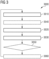
- FIG. 3 is a flowchart of a method 3000 according to various examples. Optional blocks are labeled with dashed lines.
- the method 3000 according to FIG. 3 may be executed by a computing device directly or remotely connected to the linac 1000 according to the example of FIG. 1 or another radiation-therapy hardware, e.g., upon loading program code from a memory. It would also be possible that the method 3000 is at least partially executed by a separate compute unit, e.g. at a server backend or using cloud computing.
- the computing device or the separate compute unit may comprise at least one circuit - e.g., a CPU and/or a GPU and/or a TPU - for executing the method 3000.
- the method 3000 can predict risks of radiation-induced toxicity based on pre-treatment data capturing underlying and subtle signatures of healthy tissues surrounding a tumor, e.g., lung parenchyma, vulnerability to radiation.
- a tumor e.g., lung parenchyma
- Such information may be combined with planning medicine imaging scans as mentioned-above and a planned dose map capturing both the tumor characteristics and potential dosimetric level and its distribution over not only the tumor but the surrounding presumably healthy tissues, e.g., lung parenchyma.
- input data according to TAB. 2 may be used.
- the received data may comprise a predefined dose map of the radiotherapy treatment and pre-radiotherapy-treatment imaging data of the ROI 2020.
- pre-radiotherapy-treatment imaging data of the ROI 2020 may be used (cf. TAB. 2, option II).
- This imaging data may comprise at least one of X-ray imaging data, CT imaging data, MRI imaging data, PET imaging data, SPECT imaging data, ultrasound imaging data.
- registration may be applied to the pre-radiotherapy-treatment imaging data, e.g., registering CT imaging data with MRI imaging data, and/or PET imaging data. Further, the pre-radiotherapy-treatment imaging data may be registered with the predefined dose map.
- the received data may further comprise at least one of a dose level (e.g., 8 Gy) of the radiotherapy treatment, a clinical stage of a tumor located in the target region, comorbidities of the patient, and/or demographics of the patient, as previously explained in connection with TAB. 2.
- a dose level e.g. 8 Gy
- the computing device or the separate compute unit applies a trained machine-learning algorithm to the received data.
- the trained machine-learning algorithm may be a (deep) neural network, e.g., a convolutional neural network, a recurrent neural network, a generative adversarial network, a residual network and etc.
- the trained machine-learning algorithm comprises an encoder for extracting pertinent features (sometimes also referred to as latent features) of the ROI based on the received data and a classifier for generating the at least one toxicity indicator based on the extracted pertinent features of the ROI.
- the trained machine-learning algorithm may be obtained by utilizing supervised learning, semi-supervised learning, or reinforcement learning.
- the trained machine-learning algorithm is trained by using supervised learning.
- a computer-implemented method of performing a training to obtain the trained machine-learning algorithm will be explained below later in connection with FIG. 5 .
- At block 3030 at least one toxicity indicator is generated based on the received data via the trained machine-learning algorithm.
- the at least one toxicity indicator is indicative of the risks of the radiation-induced toxicity and thereby shows a prediction of potential level of radiation-induced toxicity. Details have been explained in connection with TAB. 1.
- the method 3000 may further comprise, at block 3040, applying imputation and/or normalization to the received data before applying the trained machine-learning algorithm to the received data.
- Imputation is the process of replacing missing data with substituted values.
- imputation may be used to determine the missed pixel value based on pixel values of pixels surrounding the pixel.
- Imputation may comprise hotdeck imputation, Cold-deck imputation, mean imputation, Regression imputation, and multiple imputation.
- the method 3000 may further comprise, at block 3050, assessing whether the at least one generated toxicity indicator indicates acceptable radiation-induced toxicity, i.e., the predefined dose map is acceptable (or optimal), based on the at least one generated toxicity indicator.
- Such an assessment may be performed by comparing each of the at least one generated toxicity indicator with a corresponding threshold of respective toxicity indicator.
- Other predefined rules may be used. Patient-specific rules could be applied.
- the method 3000 may additionally comprise, at block 3060, adjusting the predefined dose map based on the at least one generated toxicity indicator to generate a further predefined dose map. Then, the method 3000 can be iteratively executed by replacing the predefined dose map with the further predefined dose map until acceptable (or optimal) toxicity indicators, i.e. predefined dose map, are generated.
- a predefined dose map M1 is received and processed by the trained machine-learning algorithm together with the other data, such as the pre-radiotherapy-treatment imaging data, and thereby a toxicity indicator T1 is generated by the trained machine-learning algorithm.
- the toxicity indicator T1 may be assessed by experienced radiation oncologists or by comparing with a threshold of toxicity indicator value, to determine whether the predefined dose map M1 is optimal or not. An automated analysis may be used instead.
- a further predefined dose map M2 is generated by adjusting the previous predefined dose map M1 based on the previous toxicity indicator T1. Then, this adjusted dose map M2 is used to predict the risks of the radiation-induced toxicity with the respectively defined radiotherapy treatment, and thereby a further toxicity indicator T2 is generated by the trained machine-learning algorithm.
- the toxicity indicator T2 may be assessed in the same manner as T1.
- the above-mentioned actions - generating a further predefined dose map Mn, receiving the further dose map Mn, processing the further dose map Mn together with the same other data by the same trained machine-learning algorithm, generating a further toxicity indicator Tn, and assessing the further toxicity indicator Tn - may be iteratively executed until the optimal (or maybe suboptimal) predefined dose map is determined.
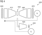
- FIG. 4 schematically illustrates an exemplary implementation of a machine-learning algorithm that can be used in the various examples described herein.
- the machine-learning algorithm is implemented as a neural network 4000.
- the neural network 4000 comprises an encoder 4010 for extracting pertinent features 4030 of input data 4040 (such as data associated with the ROI 2020) and a classifier 4020 for generating at least one toxicity indicator 4050 based on the extracted pertinent features 4030 of the input data 4040.
- the trained neural network 4000 can be utilized to process the data 4040 (cf. TAB. 2) associated with the ROI 2020 to determine at least one toxicity indicator indicative of the risks of the radiation-induced toxicity. Details with respect to the training are described in connection with FIG. 5 .
- FIG. 5 is a flowchart of a method 5000 for performing a training of a machine-learning algorithm (such as the neural network 4000 as shown in FIG. 4 ).
- the trained machine-learning algorithm can then be used for the method 3000 of FIG. 3 .
- the method 5000 may be executed by the same computing device that executes the method 3000, or by a different computing device.
- the method 5000 is a computer-implemented method of performing a training of a machine-learning algorithm for predicting risks of radiation-induced toxicity associated with a radiotherapy treatment of a target region (e.g., 2030 according to FIG. 2 ) of a patient.
- a target region e.g., 2030 according to FIG. 2
- multiple instances of training data associated with a ROI e.g., ROI 2020 according to FIG. 2
- the target region e.g., 2030 according to FIG. 2
- multiple instances of reference data are received, each one of the multiple instances of the reference data corresponding to a respective instance of the training data.
- Each one of the multiple instances of the training data comprises a dose map of the radiotherapy treatment and pre-radiotherapy-treatment imaging data of the ROI 2020.
- Each instance of the multiple instances of the reference data comprises at least one diagnosed toxicity indicator indicative of diagnosed risks of radiation-induced toxicity - thus serving as a ground truth.
- the multiple instances define a training dataset.
- a larger training dataset can be helpful to achieve more accurate training.
- the multiple instances of training data may comprise pre-radiotherapy-treatment data associated with various patients suffering from the same type of cancers/tumors, which ensure that the ROI 2020 and the target region 2030 have the same or at least similar anatomical and physiological characteristics and thereby the multiple instances of training data may be regarded as instances based on the same probability distribution.
- the multiple instances of reference data may comprise post-radiotherapy-treatment data associated with the same various patients suffering from the same type of cancers/tumors and obtained after undergoing the radiotherapy treatment.
- the multiple instances of training data and the multiple instances of reference data may have a one-to-one correspondence (or bijection).
- each one of the multiple instances of the training data may further comprise at least one of a dose level (e.g., expressed in units of Gray) of the radiotherapy treatment, a clinical-stage of a tumor located in the target region, comorbidities of a further patient, and/or demographics of the further patient.
- a dose level e.g., expressed in units of Gray
- the at least one diagnosed toxicity indicator - details have been explained in connection with TAB. 1 - may obtained by manually or automatically analyzing the post-radiotherapy-treatment data.
- the at least one diagnosed toxicity indicator corresponding to each instance of the multiple instances of the reference data is generated by a further trained machine-learning algorithm based on diagnosed data obtained at two or more time points after the radiotherapy treatment, and each one of the diagnosed data comprises imaging data of the ROI.
- a further trained machine-learning algorithm based on diagnosed data obtained at two or more time points after the radiotherapy treatment
- each one of the diagnosed data comprises imaging data of the ROI.
- changes in the diagnosed data for the different time points can be indicative of toxicity, e.g., due to changes in the tissue.
- the method 5000 may further comprise performing a registration of the imaging data of the ROI obtained at the two or more time points with each other or with the pre-radiotherapy-treatment imaging data.
- the multiple instances of the training data are processed using the machine-learning algorithm.
- the method 5000 may further comprise applying imputation and/or normalization to each one of the multiple instances of the training data before processing the multiple instances of the training data by the machine-learning algorithm.
- At block 5030 at least one respective estimated toxicity indicator indicative of estimated risks of radiation-induced toxicity is generated by the machine-learning algorithm, for each one of the multiple instances of the training data.
- the at least one estimated toxicity indicator may be regarded as an estimate of the at least one diagnosed toxicity indicator - according to the current training state of the machine-learning algorithm.
- the computing device performs the training of the machine-learning algorithm by updating parameter values of the machine-learning algorithm based on a comparison between the diagnosed toxicity indicators and corresponding estimated toxicity indicators.
- a loss function can be defined. Optimization techniques can be employed to adjust the parameters, e.g., backpropagation, etc..
- the neural network 4000 can comprise a decoder 4060.
- the training can be facilitated.
- Based on an output 4070 of the decoder 4060 it is possible to adjust parameters of both the encoder 4010 and the classifier 4020 when performing the training. Thereby, a better performance of the classifier 4020 can be achieved, i.e., the at least one toxicity indicator 4050 can be accurately determined.
- the decoder 4060 may generate reconstructed input data as the output 4070.
- the reconstructed input data of the output 4070 corresponds to the reference data.
- the decoder 4060 may generate multiple instances of estimated radiomic features as the output 4070 of the ROI based on the pertinent features 4030 of the training data.
- the estimated radiomic features of the output 4070 may comprise intensity, geometry, texture, and wavelet features of the ROI.
- the encoder 4010 and the classifier 4020 may be trained jointly based on a classification loss Cl, i.e. by updating parameter values of both the encoder 4010 and the classifier 4020 based on a comparison between the diagnosed toxicity indicators and corresponding estimated toxicity indicators.
- the encoder 4010, the classifier 4020 and the decoder 4060 may be trained jointly based on a sum or a weighted sum of the classification loss Cl and a reconstruction loss Rl, i.e. Cl+Rl or w1*Cl+w2*Rl, wherein w1 and w2 are manually selected or hyperparameters adjusted during the training.
- Rl reconstruction loss
- the encoder 4010 and the classifier 4020 will be used to predict risks of radiation-induced toxicity associated with a radiotherapy treatment of a target region of a patient, for example by executing the method 3000.
- the method 5000 may optionally further comprises generating reconstructed data based on the training data using the decoder 4060 of the machine-learning algorithm. Then, the updating of the parameter values of the machine-learning algorithm is further based on a comparison between each one of the multiple instances of the training data and corresponding reconstructed data.
- the method 5000 may optionally comprise receiving multiple instances of diagnosed radiomic features of the ROI, for example together with various data received at block 5010, and generates multiple instances of estimated radiomic features of the ROI via the decoder 4060.
- the diagnosed radiomic features may be extracted from gross tumour volumes encompassing regions of interest.
- the diagnosed radiomic features may comprise intensity, geometry, texture, and wavelet features of the ROI.
- the intensity features quantified the first-order statistical distribution of the voxel intensities within the gross tumor volumes.
- the geometry features quantified shape characteristics of the tumor.
- the texture features described spatial distribution of the voxel intensities, thereby quantifying the intratumoral heterogeneity.
- the intensity and texture features may be also computed after applying wavelet transformations to the original image.
- the diagnosed radiomic features may be handcrafted by experts or computed based on predefined mathematical formulas.
- the machine-learning algorithm further comprises the decoder 4060 for generating the estimated radiomic features of the ROI based on the extracted pertinent features of the ROI.
- the updating of the parameter values of the machine-learning algorithm is further based on a comparison between each one of the multiple instances of the diagnosed radiomic features of the ROI and corresponding estimated radiomic features of the ROI.
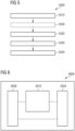
- FIG. 6 schematically illustrates a device 6000 according to various examples.
- the device 6000 may be the computing device mentioned above.
- the device 6000 comprises a processing unit 6010, a memory unit 6020 and an input/output interface 6030.
- the processing unit 6010 is configured to execute a program stored in the memory unit 6020 to perform the method 3000 and/or the method 5000.
- the input/output interface 6030 may communicate with actuators associated with the dosimetry system 1010 and/or the collimation system 1011 to adjust radiation doses of a radiotherapy treatment.
- patients to be treated with radiotherapy can be stratified based on a likelihood of toxicity as indicated by at least one toxicity indicator and high-risk patients could be flagged to be managed differently.
- a dose level and/or a dose map as input could be adjusted to compute a new toxicity score to a certain threshold for example. I.e. offering a dose prescription solution that uses data from a large cohort of outcome matched patients with known planning dose and toxicity profile to help an oncologist find an optimal dose with respect both outcome and toxicity profile for a specific patient.
- radiotherapy treatments can be precisely and reliably individualized based on anatomical and physiological characteristics of individual patients and thereby can give clinicians a chance to select a radiotherapy treatment that is most likely to be adapted to an individual patient to maximize the likelihood of treatment response while minimizing the risk of adverse effects, such as radiation-induced toxicity.
Landscapes
- Engineering & Computer Science (AREA)
- Health & Medical Sciences (AREA)
- General Health & Medical Sciences (AREA)
- Medical Informatics (AREA)
- Public Health (AREA)
- Biomedical Technology (AREA)
- Theoretical Computer Science (AREA)
- Physics & Mathematics (AREA)
- Nuclear Medicine, Radiotherapy & Molecular Imaging (AREA)
- Epidemiology (AREA)
- Primary Health Care (AREA)
- Life Sciences & Earth Sciences (AREA)
- Data Mining & Analysis (AREA)
- General Physics & Mathematics (AREA)
- Software Systems (AREA)
- Radiology & Medical Imaging (AREA)
- Pathology (AREA)
- Evolutionary Computation (AREA)
- Computing Systems (AREA)
- Mathematical Physics (AREA)
- General Engineering & Computer Science (AREA)
- Artificial Intelligence (AREA)
- Surgery (AREA)
- Computer Vision & Pattern Recognition (AREA)
- Urology & Nephrology (AREA)
- Animal Behavior & Ethology (AREA)
- Veterinary Medicine (AREA)
- Biophysics (AREA)
- Computational Linguistics (AREA)
- Molecular Biology (AREA)
- Databases & Information Systems (AREA)
- Quality & Reliability (AREA)
- Bioethics (AREA)
- Radiation-Therapy Devices (AREA)
Claims (15)
- Computerimplementiertes Verfahren zum Vorhersagen von Risiken einer durch Strahlung induzierten Toxizität, die mit einer Strahlentherapie-Behandlung einer Zielregion (2030) eines Patienten verknüpft ist, wobei das Verfahren umfasst:- Empfangen (3010) von Daten (4040), die mit einer Region von Interesse (2020) verknüpft sind, welche die Zielregion (2030) umfasst, wobei die empfangenen Daten (4040) eine vordefinierte Dosiskarte der Strahlentherapie-Behandlung und Bildgebungsdaten (2010) der Region von Interesse (2020) vor der Strahlentherapie-Behandlung umfassen;- Anwenden (3020) eines trainierten Maschinenlernalgorithmus auf die empfangenen Daten; und- Generieren (3030), durch den trainierten Maschinenlernalgorithmus, wenigstens eines Toxizitätsindikators (4050) basierend auf den empfangenen Daten, wobei der wenigstens eine Toxizitätsindikator (4050) indikativ für die Risiken der durch Strahlung induzierten Toxizität sind,wobei der trainierte Maschinenlernalgorithmus unter Verwendung des Verfahrens nach einem der Ansprüche 7-15 trainiert wird.
- Verfahren nach Anspruch 1, wobei die empfangenen Daten (4040) ferner wenigstens eines hiervon umfassen:ein Dosisniveau der Strahlentherapie-Behandlung,eine klinisches Stadium eines Tumors in der Zielregion,Begleiterkrankungen des Patienten und/oderdemografische Daten des Patienten.
- Verfahren nach einem der vorhergehenden Ansprüche, ferner umfassend,- Anwenden einer Imputation auf die empfangenen Daten; und/oder- Anwenden einer Normalisierung auf die empfangenen Daten.
- Verfahren nach einem der vorhergehenden Ansprüche,
wobei der wenigstens eine Toxizitätsindikator (4050) wenigstens eines hiervon umfasst: eine aggregierte Risikobewertung, eine Dosisempfindlichkeit zum Charakterisieren der Auswirkungen von Dosen, angegeben durch die vordefinierte Dosiskarte zu der durch Strahlung induzierten Toxizität, oder eine Toxizitätsbereichskarte der Region von Interesse. - Verfahren nach einem der vorhergehenden Ansprüche, ferner umfassend:- Anpassen (3060) der vordefinierten Dosiskarte basierend auf dem wenigstens einen Toxizitätsindikator (4050).
- Verfahren nach einem der vorhergehenden Ansprüche,
wobei der trainierte Maschinenlernalgorithmus einen Codierer (4010) zum Extrahieren von relevanten Merkmalen (4030) der Region von Interesse basierend auf den empfangenen Daten und einen Klassifikator (4020) zum Generieren des wenigstens einen Toxizitätsindikators (4050) basierend auf den extrahierten relevanten Merkmalen der Region von Interesse umfasst. - Computerimplementiertes Verfahren zum Durchführen eines Trainings eines Maschinenlernalgorithmus zum Vorhersagen von Risiken einer durch Strahlung induzierten Toxizität, die mit einer Strahlentherapie-Behandlung einer Zielregion eines Patienten verknüpft ist, wobei das Verfahren umfasst:- Empfangen mehrerer Instanzen von Trainingsdaten, die mit einer Region von Interesse verknüpft sind, welche die Zielregion umfasst, und mehrerer Instanzen von Referenzdaten, wobei jede der mehreren Instanzen der Referenzdaten einer jeweiligen Instanz der Trainingsdaten entspricht, wobei jede der mehreren Instanzen der Trainingsdaten eine Dosiskarte der Strahlentherapie-Behandlung und Bildgebungsdaten der Region von Interesse vor der Strahlentherapie-Behandlung umfasst, wobei jede Instanz der mehreren Instanzen der Referenzdaten wenigstens einen diagnostizierten Toxizitätsindikator (4050) umfasst, der indikativ für diagnostizierte Risiken einer durch Strahlung induzierten Toxizität ist;- Verarbeiten der mehreren Instanzen der Trainingsdaten durch den Maschinenlernalgorithmus;- Generieren, durch den Maschinenlernalgorithmus und für jede der mehreren Instanzen der Trainingsdaten, wenigstens eines jeweiligen geschätzten Toxizitätsindikators (4050), der indikativ für geschätzte Risiken einer durch Strahlung induzierten Toxizität ist; und dadurch gekennzeichnet, dass das Verfahren ferner umfasst:- Durchführen des Trainings des Maschinenlernalgorithmus durch Aktualisieren von Parameterwerten des Maschinenlernalgorithmus basierend auf einem Vergleich zwischen den diagnostizierten Toxizitätsindikatoren und entsprechenden geschätzten Toxizitätsindikatoren.
- Verfahren nach Anspruch 7, wobei die Trainingsdaten ferner wenigstens eines hiervon umfassen:ein Dosisniveau der Strahlentherapie-Behandlung,eine klinisches Stadium eines Tumors in der Zielregion,Begleiterkrankungen eines weiteren Patienten und/oderdemografische Daten des weiteren Patienten.
- Vorrichtung nach Anspruch 7 oder 8, ferner umfassend:- Anwenden einer Imputation auf jede der mehreren Instanzen der Trainingsdaten; und/oder- Anwenden einer Normalisierung auf jede der mehreren Instanzen der Trainingsdaten.
- Verfahren nach einem der Ansprüche 7 bis 9,
wobei der wenigstens eine diagnostizierte Toxizitätsindikator und der wenigstens eine geschätzte Toxizitätsindikator wenigstens eines hiervon umfassen: eine diagnostizierte aggregierte Risikobewertung, eine Dosisempfindlichkeit zum Charakterisieren der Auswirkungen von Dosen, angegeben durch die Dosiskarte zu der durch Strahlung induzierten Toxizität, oder eine diagnostizierte Toxizitätsbereichskarte der Region von Interesse. - Verfahren nach einem der Ansprüche 7 bis 10,
wobei der trainierte Maschinenlernalgorithmus einen Codierer (4010) zum Extrahieren von relevanten Merkmalen (4030) der Region von Interesse basierend auf den empfangenen Daten und einen Klassifikator (4020) zum Generieren des wenigstens einen geschätzten Toxizitätsindikators (4050) basierend auf den extrahierten relevanten Merkmalen der Region von Interesse umfasst. - Verfahren nach Anspruch 11, ferner umfassend:- Generieren von rekonstruierten Daten basierend auf den Trainingsdaten unter Verwendung eines Decodierers des Maschinenlernalgorithmus;wobei das Aktualisieren der Parameterwerte des Maschinenlernalgorithmus ferner auf einem Vergleich zwischen jeder der mehreren Instanzen der Trainingsdaten und entsprechenden rekonstruierten Daten basiert.
- Verfahren nach Anspruch 11, ferner umfassend:- Empfangen mehrerer Instanzen von diagnostizierten radiomischen Merkmalen der Region von Interesse;- Generieren mehrerer Instanzen von geschätzten radiomischen Merkmalen der Region von Interesse;wobei der Maschinenlernalgorithmus ferner einen Decodierer zum Generieren der geschätzten radiomischen Merkmale der Region von Interesse basierend auf den extrahierten relevanten Merkmalen der Region von Interesse umfasst;wobei das Aktualisieren der Parameterwerte des Maschinenlernalgorithmus ferner auf einem Vergleich zwischen jeder der mehreren Instanzen der diagnostizierten radiomischen Merkmale der Region von Interesse und entsprechenden geschätzten radiomischen Merkmalen der Region von Interesse basiert.
- Verfahren nach einem der Ansprüche 7-13,
wobei der wenigstens eine diagnostizierte Toxizitätsindikator, welcher jeder Instanz der mehreren Instanzen der Referenzdaten entspricht, durch einen weiteren trainierten Maschinenlernalgorithmus, basierend auf diagnostizierten Daten, die zu zwei oder mehr Zeitpunkten nach der Strahlentherapie-Behandlung erhalten werden, generiert wird und die jeweiligen diagnostizierten Daten Bildgebungsdaten der Region von Interesse umfassen. - Verfahren nach Anspruch 14, ferner umfassend:- Durchführen einer Registrierung der Bildgebungsdaten der Region von Interesse, die zu den zwei oder mehr Zeitpunkten erhalten werden.
Priority Applications (3)
| Application Number | Priority Date | Filing Date | Title |
|---|---|---|---|
| EP21157289.6A EP4043072B1 (de) | 2021-02-16 | 2021-02-16 | Durch strahlung induzierte toxizität und maschinenlernen |
| US17/649,824 US11980776B2 (en) | 2021-02-16 | 2022-02-03 | Radiation-induced toxicity and machine learning |
| CN202210133113.3A CN114974609A (zh) | 2021-02-16 | 2022-02-14 | 放射诱发毒性与机器学习 |
Applications Claiming Priority (1)
| Application Number | Priority Date | Filing Date | Title |
|---|---|---|---|
| EP21157289.6A EP4043072B1 (de) | 2021-02-16 | 2021-02-16 | Durch strahlung induzierte toxizität und maschinenlernen |
Publications (2)
| Publication Number | Publication Date |
|---|---|
| EP4043072A1 EP4043072A1 (de) | 2022-08-17 |
| EP4043072B1 true EP4043072B1 (de) | 2025-04-30 |
Family
ID=74661269
Family Applications (1)
| Application Number | Title | Priority Date | Filing Date |
|---|---|---|---|
| EP21157289.6A Active EP4043072B1 (de) | 2021-02-16 | 2021-02-16 | Durch strahlung induzierte toxizität und maschinenlernen |
Country Status (3)
| Country | Link |
|---|---|
| US (1) | US11980776B2 (de) |
| EP (1) | EP4043072B1 (de) |
| CN (1) | CN114974609A (de) |
Families Citing this family (4)
| Publication number | Priority date | Publication date | Assignee | Title |
|---|---|---|---|---|
| US12087007B2 (en) | 2021-03-31 | 2024-09-10 | Auris Health, Inc. | Vision-based 6DOF camera pose estimation in bronchoscopy |
| US20230128966A1 (en) * | 2021-10-21 | 2023-04-27 | Imam Abdulrahman Bin Faisal University | System, method, and computer readable storage medium for accurate and rapid early diagnosis of covid-19 from chest x ray |
| WO2025175093A1 (en) * | 2024-02-14 | 2025-08-21 | The Cleveland Clinic Foundation | System and method for dose accumulation determination and patient profile generation |
| CN120473183B (zh) * | 2025-07-09 | 2025-10-24 | 北京药云数据科技有限公司 | 基于多模态时序特征的癌症复发概率深度学习预测系统 |
Family Cites Families (14)
| Publication number | Priority date | Publication date | Assignee | Title |
|---|---|---|---|---|
| US20040254448A1 (en) * | 2003-03-24 | 2004-12-16 | Amies Christopher Jude | Active therapy redefinition |
| US8238516B2 (en) * | 2008-01-09 | 2012-08-07 | Kabushiki Kaisha Toshiba | Radiotherapy support apparatus |
| WO2015176011A1 (en) * | 2014-05-15 | 2015-11-19 | The Johns Hopkins University | Method, system and computer-readable media for treatment plan risk analysis |
| US9857415B1 (en) * | 2016-06-21 | 2018-01-02 | Lucid Circuit, Inc. | System and methods for analyzing and estimating susceptibility of circuits to radiation-induced single-event-effects |
| WO2018048575A1 (en) * | 2016-09-07 | 2018-03-15 | Elekta, Inc. | System and method for learning models of radiotherapy treatment plans to predict radiotherapy dose distributions |
| TWI637187B (zh) * | 2017-09-19 | 2018-10-01 | 臺北榮民總醫院 | 磁共振影像的分析方法及評估放射治療風險的方法 |
| US10997716B2 (en) * | 2017-10-09 | 2021-05-04 | The Board Of Trustees Of The Leland Stanford Junior University | Contrast dose reduction for medical imaging using deep learning |
| US11557390B2 (en) * | 2018-04-30 | 2023-01-17 | Elekta, Inc. | Radiotherapy treatment plan modeling using generative adversarial networks |
| US11756667B2 (en) | 2018-05-30 | 2023-09-12 | Siemens Healthcare Gmbh | Decision support system for medical therapy planning |
| CN108898642B (zh) * | 2018-06-01 | 2022-11-11 | 安徽工程大学 | 一种基于卷积神经网络的稀疏角度ct成像方法 |
| CN109671499B (zh) * | 2018-10-22 | 2023-06-13 | 南方医科大学 | 一种直肠毒性预测系统构建方法 |
| EP3714937A1 (de) * | 2019-03-28 | 2020-09-30 | Koninklijke Philips N.V. | Bestimmung des risikos einer hämatologischen toxizität nach einer strahlentherapie |
| CN110085298B (zh) * | 2019-04-26 | 2022-02-01 | 南方医科大学 | 基于深度网络学习的调强放疗计划三维剂量分布预测方法 |
| CN111951962B (zh) * | 2020-07-27 | 2022-03-11 | 中山大学孙逸仙纪念医院 | 一种贝伐单抗治疗放射性脑损伤疗效预测模型及其构建方法 |
-
2021
- 2021-02-16 EP EP21157289.6A patent/EP4043072B1/de active Active
-
2022
- 2022-02-03 US US17/649,824 patent/US11980776B2/en active Active
- 2022-02-14 CN CN202210133113.3A patent/CN114974609A/zh active Pending
Also Published As
| Publication number | Publication date |
|---|---|
| CN114974609A (zh) | 2022-08-30 |
| US20220257978A1 (en) | 2022-08-18 |
| EP4043072A1 (de) | 2022-08-17 |
| US11980776B2 (en) | 2024-05-14 |
Similar Documents
| Publication | Publication Date | Title |
|---|---|---|
| US11980776B2 (en) | Radiation-induced toxicity and machine learning | |
| US20240212111A1 (en) | Machine learning training corpus apparatus and method | |
| Wang et al. | Development of methods for beam angle optimization for IMRT using an accelerated exhaustive search strategy | |
| US11358003B2 (en) | Generation of realizable radiotherapy plans | |
| US10342994B2 (en) | Methods and systems for generating dose estimation models for radiotherapy treatment planning | |
| US11013936B2 (en) | Methods and systems for generating dose estimation models for radiotherapy treatment planning | |
| CN114401768A (zh) | 使用机器学习的放射疗法治疗计划优化 | |
| CN107072624A (zh) | 用于自动治疗计划的系统和方法 | |
| US11529531B2 (en) | Using reinforcement learning in radiation treatment planning optimization to locate dose-volume objectives | |
| Sheng et al. | Artificial intelligence applications in intensity modulated radiation treatment planning: an overview | |
| Cortes et al. | Knowledge-based three-dimensional dose prediction for tandem-and-ovoid brachytherapy | |
| US10556125B2 (en) | Knowledge based treatment planning corrected for biological effects | |
| CN119499565A (zh) | 一种用于患者放射治疗的自适应辅助决策方法、系统、设备及介质 | |
| CN118366614B (zh) | 硼中子俘获治疗计划疗效预测模型构建方法和装置 | |
| Hammers et al. | Evaluation of the clinical impact of the differences between planned and delivered dose in prostate cancer radiotherapy based on CT‐on‐rails IGRT and patient‐reported outcome scores | |
| Wildman et al. | Recent advances in applying machine learning to proton radiotherapy | |
| Xu et al. | The benefits evaluation of abdominal deep inspiration breath hold based on knowledge‐based radiotherapy treatment planning for left‐sided breast cancer | |
| Monti et al. | External validation of dose patterns and dosimetric predictors for radiation-induced esophagitis in non-small cell lung cancer | |
| Achlatis et al. | Physics-Guided Radiotherapy Treatment Planning with Deep Learning | |
| US20250242176A1 (en) | UPDATING RADIOTHERAPY TREATMENT PLANS USING INFORMATION DERIVED FROM WHOLE SLIDE IMAGES (WSIs) | |
| Sharma et al. | Does time to retreatment matter? An NTCP model to predict radionecrosis after repeat SRS for recurrent brain metastases incorporating time-dependent discounted dose | |
| Kim et al. | Integrating Deep Learning–Based Dose Distribution Prediction with Bayesian Networks for Decision Support in Radiotherapy for Upper Gastrointestinal Cancer | |
| Porter et al. | Deep learning for contour quality assurance for RTOG 0933: In-silico evaluation | |
| US20250160936A1 (en) | Motion compensation during cardiac radioablation | |
| Ma | Efficient and intelligent radiotherapy planning and adaptation |
Legal Events
| Date | Code | Title | Description |
|---|---|---|---|
| PUAI | Public reference made under article 153(3) epc to a published international application that has entered the european phase |
Free format text: ORIGINAL CODE: 0009012 |
|
| STAA | Information on the status of an ep patent application or granted ep patent |
Free format text: STATUS: REQUEST FOR EXAMINATION WAS MADE |
|
| 17P | Request for examination filed |
Effective date: 20210216 |
|
| AK | Designated contracting states |
Kind code of ref document: A1 Designated state(s): AL AT BE BG CH CY CZ DE DK EE ES FI FR GB GR HR HU IE IS IT LI LT LU LV MC MK MT NL NO PL PT RO RS SE SI SK SM TR |
|
| RBV | Designated contracting states (corrected) |
Designated state(s): AL AT BE BG CH CY CZ DE DK EE ES FI FR GB GR HR HU IE IS IT LI LT LU LV MC MK MT NL NO PL PT RO RS SE SI SK SM TR |
|
| RAP1 | Party data changed (applicant data changed or rights of an application transferred) |
Owner name: SIEMENS HEALTHINEERS AG |
|
| GRAP | Despatch of communication of intention to grant a patent |
Free format text: ORIGINAL CODE: EPIDOSNIGR1 |
|
| STAA | Information on the status of an ep patent application or granted ep patent |
Free format text: STATUS: GRANT OF PATENT IS INTENDED |
|
| INTG | Intention to grant announced |
Effective date: 20241204 |
|
| GRAS | Grant fee paid |
Free format text: ORIGINAL CODE: EPIDOSNIGR3 |
|
| GRAA | (expected) grant |
Free format text: ORIGINAL CODE: 0009210 |
|
| STAA | Information on the status of an ep patent application or granted ep patent |
Free format text: STATUS: THE PATENT HAS BEEN GRANTED |
|
| AK | Designated contracting states |
Kind code of ref document: B1 Designated state(s): AL AT BE BG CH CY CZ DE DK EE ES FI FR GB GR HR HU IE IS IT LI LT LU LV MC MK MT NL NO PL PT RO RS SE SI SK SM TR |
|
| REG | Reference to a national code |
Ref country code: CH Ref legal event code: EP Ref country code: GB Ref legal event code: FG4D |
|
| REG | Reference to a national code |
Ref country code: IE Ref legal event code: FG4D |
|
| REG | Reference to a national code |
Ref country code: DE Ref legal event code: R096 Ref document number: 602021029850 Country of ref document: DE |
|
| REG | Reference to a national code |
Ref country code: NL Ref legal event code: MP Effective date: 20250430 |
|
| REG | Reference to a national code |
Ref country code: AT Ref legal event code: MK05 Ref document number: 1789505 Country of ref document: AT Kind code of ref document: T Effective date: 20250430 |
|
| PG25 | Lapsed in a contracting state [announced via postgrant information from national office to epo] |
Ref country code: FI Free format text: LAPSE BECAUSE OF FAILURE TO SUBMIT A TRANSLATION OF THE DESCRIPTION OR TO PAY THE FEE WITHIN THE PRESCRIBED TIME-LIMIT Effective date: 20250430 Ref country code: PT Free format text: LAPSE BECAUSE OF FAILURE TO SUBMIT A TRANSLATION OF THE DESCRIPTION OR TO PAY THE FEE WITHIN THE PRESCRIBED TIME-LIMIT Effective date: 20250901 Ref country code: ES Free format text: LAPSE BECAUSE OF FAILURE TO SUBMIT A TRANSLATION OF THE DESCRIPTION OR TO PAY THE FEE WITHIN THE PRESCRIBED TIME-LIMIT Effective date: 20250430 |
|
| REG | Reference to a national code |
Ref country code: LT Ref legal event code: MG9D |
|
| PG25 | Lapsed in a contracting state [announced via postgrant information from national office to epo] |
Ref country code: NO Free format text: LAPSE BECAUSE OF FAILURE TO SUBMIT A TRANSLATION OF THE DESCRIPTION OR TO PAY THE FEE WITHIN THE PRESCRIBED TIME-LIMIT Effective date: 20250730 Ref country code: GR Free format text: LAPSE BECAUSE OF FAILURE TO SUBMIT A TRANSLATION OF THE DESCRIPTION OR TO PAY THE FEE WITHIN THE PRESCRIBED TIME-LIMIT Effective date: 20250731 |
|
| PG25 | Lapsed in a contracting state [announced via postgrant information from national office to epo] |
Ref country code: NL Free format text: LAPSE BECAUSE OF FAILURE TO SUBMIT A TRANSLATION OF THE DESCRIPTION OR TO PAY THE FEE WITHIN THE PRESCRIBED TIME-LIMIT Effective date: 20250430 Ref country code: PL Free format text: LAPSE BECAUSE OF FAILURE TO SUBMIT A TRANSLATION OF THE DESCRIPTION OR TO PAY THE FEE WITHIN THE PRESCRIBED TIME-LIMIT Effective date: 20250430 |
|
| PG25 | Lapsed in a contracting state [announced via postgrant information from national office to epo] |
Ref country code: BG Free format text: LAPSE BECAUSE OF FAILURE TO SUBMIT A TRANSLATION OF THE DESCRIPTION OR TO PAY THE FEE WITHIN THE PRESCRIBED TIME-LIMIT Effective date: 20250430 |
|
| PG25 | Lapsed in a contracting state [announced via postgrant information from national office to epo] |
Ref country code: HR Free format text: LAPSE BECAUSE OF FAILURE TO SUBMIT A TRANSLATION OF THE DESCRIPTION OR TO PAY THE FEE WITHIN THE PRESCRIBED TIME-LIMIT Effective date: 20250430 |
|
| PG25 | Lapsed in a contracting state [announced via postgrant information from national office to epo] |
Ref country code: AT Free format text: LAPSE BECAUSE OF FAILURE TO SUBMIT A TRANSLATION OF THE DESCRIPTION OR TO PAY THE FEE WITHIN THE PRESCRIBED TIME-LIMIT Effective date: 20250430 |
|
| PG25 | Lapsed in a contracting state [announced via postgrant information from national office to epo] |
Ref country code: RS Free format text: LAPSE BECAUSE OF FAILURE TO SUBMIT A TRANSLATION OF THE DESCRIPTION OR TO PAY THE FEE WITHIN THE PRESCRIBED TIME-LIMIT Effective date: 20250731 |
|
| PG25 | Lapsed in a contracting state [announced via postgrant information from national office to epo] |
Ref country code: IS Free format text: LAPSE BECAUSE OF FAILURE TO SUBMIT A TRANSLATION OF THE DESCRIPTION OR TO PAY THE FEE WITHIN THE PRESCRIBED TIME-LIMIT Effective date: 20250830 |
|
| PG25 | Lapsed in a contracting state [announced via postgrant information from national office to epo] |
Ref country code: LV Free format text: LAPSE BECAUSE OF FAILURE TO SUBMIT A TRANSLATION OF THE DESCRIPTION OR TO PAY THE FEE WITHIN THE PRESCRIBED TIME-LIMIT Effective date: 20250430 |
|
| PG25 | Lapsed in a contracting state [announced via postgrant information from national office to epo] |
Ref country code: SM Free format text: LAPSE BECAUSE OF FAILURE TO SUBMIT A TRANSLATION OF THE DESCRIPTION OR TO PAY THE FEE WITHIN THE PRESCRIBED TIME-LIMIT Effective date: 20250430 Ref country code: DK Free format text: LAPSE BECAUSE OF FAILURE TO SUBMIT A TRANSLATION OF THE DESCRIPTION OR TO PAY THE FEE WITHIN THE PRESCRIBED TIME-LIMIT Effective date: 20250430 |
|
| PG25 | Lapsed in a contracting state [announced via postgrant information from national office to epo] |
Ref country code: CZ Free format text: LAPSE BECAUSE OF FAILURE TO SUBMIT A TRANSLATION OF THE DESCRIPTION OR TO PAY THE FEE WITHIN THE PRESCRIBED TIME-LIMIT Effective date: 20250430 |
|
| PG25 | Lapsed in a contracting state [announced via postgrant information from national office to epo] |
Ref country code: EE Free format text: LAPSE BECAUSE OF FAILURE TO SUBMIT A TRANSLATION OF THE DESCRIPTION OR TO PAY THE FEE WITHIN THE PRESCRIBED TIME-LIMIT Effective date: 20250430 |
|
| PG25 | Lapsed in a contracting state [announced via postgrant information from national office to epo] |
Ref country code: SK Free format text: LAPSE BECAUSE OF FAILURE TO SUBMIT A TRANSLATION OF THE DESCRIPTION OR TO PAY THE FEE WITHIN THE PRESCRIBED TIME-LIMIT Effective date: 20250430 |
|
| PG25 | Lapsed in a contracting state [announced via postgrant information from national office to epo] |
Ref country code: IT Free format text: LAPSE BECAUSE OF FAILURE TO SUBMIT A TRANSLATION OF THE DESCRIPTION OR TO PAY THE FEE WITHIN THE PRESCRIBED TIME-LIMIT Effective date: 20250430 |