EP4023265A1 - Tissue scaffold for use in tendon and/or ligament - Google Patents
Tissue scaffold for use in tendon and/or ligament Download PDFInfo
- Publication number
- EP4023265A1 EP4023265A1 EP21193592.9A EP21193592A EP4023265A1 EP 4023265 A1 EP4023265 A1 EP 4023265A1 EP 21193592 A EP21193592 A EP 21193592A EP 4023265 A1 EP4023265 A1 EP 4023265A1
- Authority
- EP
- European Patent Office
- Prior art keywords
- tissue scaffold
- weave
- tissue
- bioceramic material
- main body
- Prior art date
- Legal status (The legal status is an assumption and is not a legal conclusion. Google has not performed a legal analysis and makes no representation as to the accuracy of the status listed.)
- Granted
Links
Images





Classifications
-
- A—HUMAN NECESSITIES
- A61—MEDICAL OR VETERINARY SCIENCE; HYGIENE
- A61L—METHODS OR APPARATUS FOR STERILISING MATERIALS OR OBJECTS IN GENERAL; DISINFECTION, STERILISATION OR DEODORISATION OF AIR; CHEMICAL ASPECTS OF BANDAGES, DRESSINGS, ABSORBENT PADS OR SURGICAL ARTICLES; MATERIALS FOR BANDAGES, DRESSINGS, ABSORBENT PADS OR SURGICAL ARTICLES
- A61L27/00—Materials for grafts or prostheses or for coating grafts or prostheses
- A61L27/28—Materials for coating prostheses
- A61L27/30—Inorganic materials
- A61L27/306—Other specific inorganic materials not covered by A61L27/303 - A61L27/32
-
- A—HUMAN NECESSITIES
- A61—MEDICAL OR VETERINARY SCIENCE; HYGIENE
- A61L—METHODS OR APPARATUS FOR STERILISING MATERIALS OR OBJECTS IN GENERAL; DISINFECTION, STERILISATION OR DEODORISATION OF AIR; CHEMICAL ASPECTS OF BANDAGES, DRESSINGS, ABSORBENT PADS OR SURGICAL ARTICLES; MATERIALS FOR BANDAGES, DRESSINGS, ABSORBENT PADS OR SURGICAL ARTICLES
- A61L27/00—Materials for grafts or prostheses or for coating grafts or prostheses
- A61L27/14—Macromolecular materials
- A61L27/22—Polypeptides or derivatives thereof, e.g. degradation products
- A61L27/24—Collagen
-
- A—HUMAN NECESSITIES
- A61—MEDICAL OR VETERINARY SCIENCE; HYGIENE
- A61F—FILTERS IMPLANTABLE INTO BLOOD VESSELS; PROSTHESES; DEVICES PROVIDING PATENCY TO, OR PREVENTING COLLAPSING OF, TUBULAR STRUCTURES OF THE BODY, e.g. STENTS; ORTHOPAEDIC, NURSING OR CONTRACEPTIVE DEVICES; FOMENTATION; TREATMENT OR PROTECTION OF EYES OR EARS; BANDAGES, DRESSINGS OR ABSORBENT PADS; FIRST-AID KITS
- A61F2/00—Filters implantable into blood vessels; Prostheses, i.e. artificial substitutes or replacements for parts of the body; Appliances for connecting them with the body; Devices providing patency to, or preventing collapsing of, tubular structures of the body, e.g. stents
- A61F2/02—Prostheses implantable into the body
- A61F2/08—Muscles; Tendons; Ligaments
-
- A—HUMAN NECESSITIES
- A61—MEDICAL OR VETERINARY SCIENCE; HYGIENE
- A61F—FILTERS IMPLANTABLE INTO BLOOD VESSELS; PROSTHESES; DEVICES PROVIDING PATENCY TO, OR PREVENTING COLLAPSING OF, TUBULAR STRUCTURES OF THE BODY, e.g. STENTS; ORTHOPAEDIC, NURSING OR CONTRACEPTIVE DEVICES; FOMENTATION; TREATMENT OR PROTECTION OF EYES OR EARS; BANDAGES, DRESSINGS OR ABSORBENT PADS; FIRST-AID KITS
- A61F2/00—Filters implantable into blood vessels; Prostheses, i.e. artificial substitutes or replacements for parts of the body; Appliances for connecting them with the body; Devices providing patency to, or preventing collapsing of, tubular structures of the body, e.g. stents
- A61F2/02—Prostheses implantable into the body
- A61F2/08—Muscles; Tendons; Ligaments
- A61F2/0811—Fixation devices for tendons or ligaments
-
- A—HUMAN NECESSITIES
- A61—MEDICAL OR VETERINARY SCIENCE; HYGIENE
- A61L—METHODS OR APPARATUS FOR STERILISING MATERIALS OR OBJECTS IN GENERAL; DISINFECTION, STERILISATION OR DEODORISATION OF AIR; CHEMICAL ASPECTS OF BANDAGES, DRESSINGS, ABSORBENT PADS OR SURGICAL ARTICLES; MATERIALS FOR BANDAGES, DRESSINGS, ABSORBENT PADS OR SURGICAL ARTICLES
- A61L27/00—Materials for grafts or prostheses or for coating grafts or prostheses
- A61L27/02—Inorganic materials
- A61L27/10—Ceramics or glasses
-
- A—HUMAN NECESSITIES
- A61—MEDICAL OR VETERINARY SCIENCE; HYGIENE
- A61L—METHODS OR APPARATUS FOR STERILISING MATERIALS OR OBJECTS IN GENERAL; DISINFECTION, STERILISATION OR DEODORISATION OF AIR; CHEMICAL ASPECTS OF BANDAGES, DRESSINGS, ABSORBENT PADS OR SURGICAL ARTICLES; MATERIALS FOR BANDAGES, DRESSINGS, ABSORBENT PADS OR SURGICAL ARTICLES
- A61L27/00—Materials for grafts or prostheses or for coating grafts or prostheses
- A61L27/14—Macromolecular materials
- A61L27/16—Macromolecular materials obtained by reactions only involving carbon-to-carbon unsaturated bonds
-
- A—HUMAN NECESSITIES
- A61—MEDICAL OR VETERINARY SCIENCE; HYGIENE
- A61L—METHODS OR APPARATUS FOR STERILISING MATERIALS OR OBJECTS IN GENERAL; DISINFECTION, STERILISATION OR DEODORISATION OF AIR; CHEMICAL ASPECTS OF BANDAGES, DRESSINGS, ABSORBENT PADS OR SURGICAL ARTICLES; MATERIALS FOR BANDAGES, DRESSINGS, ABSORBENT PADS OR SURGICAL ARTICLES
- A61L27/00—Materials for grafts or prostheses or for coating grafts or prostheses
- A61L27/14—Macromolecular materials
- A61L27/18—Macromolecular materials obtained otherwise than by reactions only involving carbon-to-carbon unsaturated bonds
-
- A—HUMAN NECESSITIES
- A61—MEDICAL OR VETERINARY SCIENCE; HYGIENE
- A61L—METHODS OR APPARATUS FOR STERILISING MATERIALS OR OBJECTS IN GENERAL; DISINFECTION, STERILISATION OR DEODORISATION OF AIR; CHEMICAL ASPECTS OF BANDAGES, DRESSINGS, ABSORBENT PADS OR SURGICAL ARTICLES; MATERIALS FOR BANDAGES, DRESSINGS, ABSORBENT PADS OR SURGICAL ARTICLES
- A61L27/00—Materials for grafts or prostheses or for coating grafts or prostheses
- A61L27/14—Macromolecular materials
- A61L27/26—Mixtures of macromolecular compounds
-
- A—HUMAN NECESSITIES
- A61—MEDICAL OR VETERINARY SCIENCE; HYGIENE
- A61L—METHODS OR APPARATUS FOR STERILISING MATERIALS OR OBJECTS IN GENERAL; DISINFECTION, STERILISATION OR DEODORISATION OF AIR; CHEMICAL ASPECTS OF BANDAGES, DRESSINGS, ABSORBENT PADS OR SURGICAL ARTICLES; MATERIALS FOR BANDAGES, DRESSINGS, ABSORBENT PADS OR SURGICAL ARTICLES
- A61L27/00—Materials for grafts or prostheses or for coating grafts or prostheses
- A61L27/28—Materials for coating prostheses
- A61L27/30—Inorganic materials
- A61L27/32—Phosphorus-containing materials, e.g. apatite
-
- A—HUMAN NECESSITIES
- A61—MEDICAL OR VETERINARY SCIENCE; HYGIENE
- A61L—METHODS OR APPARATUS FOR STERILISING MATERIALS OR OBJECTS IN GENERAL; DISINFECTION, STERILISATION OR DEODORISATION OF AIR; CHEMICAL ASPECTS OF BANDAGES, DRESSINGS, ABSORBENT PADS OR SURGICAL ARTICLES; MATERIALS FOR BANDAGES, DRESSINGS, ABSORBENT PADS OR SURGICAL ARTICLES
- A61L27/00—Materials for grafts or prostheses or for coating grafts or prostheses
- A61L27/28—Materials for coating prostheses
- A61L27/34—Macromolecular materials
-
- A—HUMAN NECESSITIES
- A61—MEDICAL OR VETERINARY SCIENCE; HYGIENE
- A61F—FILTERS IMPLANTABLE INTO BLOOD VESSELS; PROSTHESES; DEVICES PROVIDING PATENCY TO, OR PREVENTING COLLAPSING OF, TUBULAR STRUCTURES OF THE BODY, e.g. STENTS; ORTHOPAEDIC, NURSING OR CONTRACEPTIVE DEVICES; FOMENTATION; TREATMENT OR PROTECTION OF EYES OR EARS; BANDAGES, DRESSINGS OR ABSORBENT PADS; FIRST-AID KITS
- A61F2/00—Filters implantable into blood vessels; Prostheses, i.e. artificial substitutes or replacements for parts of the body; Appliances for connecting them with the body; Devices providing patency to, or preventing collapsing of, tubular structures of the body, e.g. stents
- A61F2/02—Prostheses implantable into the body
- A61F2/30—Joints
- A61F2002/30001—Additional features of subject-matter classified in A61F2/28, A61F2/30 and subgroups thereof
- A61F2002/30003—Material related properties of the prosthesis or of a coating on the prosthesis
- A61F2002/30004—Material related properties of the prosthesis or of a coating on the prosthesis the prosthesis being made from materials having different values of a given property at different locations within the same prosthesis
- A61F2002/30009—Material related properties of the prosthesis or of a coating on the prosthesis the prosthesis being made from materials having different values of a given property at different locations within the same prosthesis differing in fibre orientations
-
- A—HUMAN NECESSITIES
- A61—MEDICAL OR VETERINARY SCIENCE; HYGIENE
- A61L—METHODS OR APPARATUS FOR STERILISING MATERIALS OR OBJECTS IN GENERAL; DISINFECTION, STERILISATION OR DEODORISATION OF AIR; CHEMICAL ASPECTS OF BANDAGES, DRESSINGS, ABSORBENT PADS OR SURGICAL ARTICLES; MATERIALS FOR BANDAGES, DRESSINGS, ABSORBENT PADS OR SURGICAL ARTICLES
- A61L2430/00—Materials or treatment for tissue regeneration
- A61L2430/10—Materials or treatment for tissue regeneration for reconstruction of tendons or ligaments
-
- C—CHEMISTRY; METALLURGY
- C08—ORGANIC MACROMOLECULAR COMPOUNDS; THEIR PREPARATION OR CHEMICAL WORKING-UP; COMPOSITIONS BASED THEREON
- C08L—COMPOSITIONS OF MACROMOLECULAR COMPOUNDS
- C08L75/00—Compositions of polyureas or polyurethanes; Compositions of derivatives of such polymers
- C08L75/04—Polyurethanes
Definitions
- This disclosure relates to scaffolds for use in tissue engineering, in particular to a tissue scaffold for use in a tendon and/or ligament injury.
- Tear or rupture of a tendon or ligament is a common clinical sports injury, especially when the injury occurs on the cruciate ligament in the knee joint. Due to biological environment and limited blood supply of the ligament tissue, it does not have the ability to heal naturally after it is ruptured, and needs to be reconstructed through surgery.
- autograft As tendons or ligaments, it can be divided into autograft, allograft and artificial graft according to the type of repair material.
- autografts have good reconstruction effects and no immune rejections, there are problems such as complications at donor site. Allografts are not only costly, but also have the risk of immune rejection and disease transmission. By contrast, because of its convenient materials, no risk of disease transmission, and high mechanical strength, artificial grafts have gradually attracted the attention of the medical community recently. However, artificial grafts only have mechanical load-bearing effects, and do not have biological activities and tissue inductions, which are not good for the healing of tendons/ligaments and bone interface.
- the cells and tissues on the surface of the artificial ligament are difficult to grow to form a normal tendon or ligament tissue.
- the artificial graft is prone to fatigue, abrasion, and fracture, which can lead to instability of the knee joint, and the wear fragments can easily cause iatrogenic injury such as water accumulation in the joint cavity.
- tissue scaffold with a biological activity and tissue induction to improve fatigue, abrasion, fracture and poor stability of existing artificial grafts after long-term use.
- the disclosure provides a tissue scaffold for use in a tendon and/or ligament, which comprises a weave formed by interlacing warp yarns and weft yarns, wherein the warp yarns include a plurality of fibers with an alternative shaped cross section structure.
- the weave comprises a main body area, wherein a bioactive component is on the fiber surface of the main body area, and the bioactive component can be further impregnated into the pores of the main body area; and a fixed area, wherein the fixed area is formed on the two sides of the main body area, and the weft yarns comprises a bioceramic material.
- the corresponding bioactive materials are respectively combined according to the tissue scaffold sections, to provide more cell attachment area and a good proliferation environment, effectively enhancing the activity and proliferation capacity of soft and bone tissues. It can be provided for the reconstruction of tendon and/or ligament tissue, and it has application prospects.
- a tissue scaffold for use in a tendon and/or ligament is provided.
- a tissue scaffold 1 is constituted by a weave formed by interlacing warp yarns 101 and weft yarns 110 and 120.
- the warp yarn 101 comprises a plurality of fibers with an alternative shaped cross section structure, and the plurality of fibers constitute the warp yarn.
- the weave comprises a main body area 11 containing a bioactive component on a surface thereof, and the bioactive component may be further impregnated into the pores of the main body area 11; and fixed areas 121 and 122 containing the bioceramic material weft yarn 120 being formed on two sides of the main body area.
- warp yarn used herein refers to the yarn that extends along the length of a loom during weaving and is used as the main support structure in the weave, and the direction is the same as that of tension when performing the stretching action of tendons and/or ligaments. Further, as shown in FIG. 1 , the warp yarn 101 of the main body area 11 and the fixed area 121 and 122 of the tissue scaffold of the present disclosure are integrally formed. On the other hand, the term “weft yarn” refers to yarns interlaced or perpendicular with the warps.
- the above-mentioned warp yarns and the weft yarns are formed by twisting and drawing multiple fibers.
- the warp yarns in the tissue scaffold of the present disclosure are twisted into 200 to 800 denier, the weft yarns are twisted into 50 to 100 denier.
- weave used herein is woven by warp yarns and weft yarns in an interlaced and or mutually perpendicular manner, and the interweaving points formed thereby may be arranged continuously or discontinuously, and the positions of the interweaving points may be selected periodically or irregularly.
- the pore size of the weave of the tissue scaffold of the present disclosure is 0.1 to 1 mm, and the pore of the weave provides sufficient growth space for cells and provides the transportation space for gas exchange, nutrient and metabolism required during cells growth.
- the weave of the disclosure is not limited to a single-layer weaving structure, but also includes a multi-layer upper and lower weaving structure.
- the thickness or diameter of the weave is 1.0 to 10 mm.
- the material of the weave can be a polymer material or a polymer composite material, wherein the polymer composite material comprises other fillers, such as carbon fiber, in addition to the polymer material.
- the polymer material is selected in consideration of the mechanical properties, stability, abrasion resistance and biocompatibility thereof, but is not limited thereto.
- the polymer material may be one of polyethylene terephthalate, polyethylene, polytetrafluoroethylene, polyurethane, polycaprolactone, polylactic acid, polyglycolic acid, polyether ether ketone, polyether ketone, or a mixture or copolymer thereof, wherein polyethylene may include ultra-high molecular weight polyethylene.
- the material of the above-mentioned weave is polyethylene terephthalate.
- alternative shaped cross section structure refers to a fiber structure with a non-circular cross-section, which has a relatively high fiber surface area and brings the effect of enhancing the adhesion of cells.
- the alternative shaped cross section structure of the warp yarn fiber of the present disclosure may comprise an H-shaped cross-section, an S-shaped cross-section, a W-shaped cross-section, a Y-shaped cross-section, or a cross-shaped cross-section.
- the fiber's thickness of the weave corresponds to the overall surface area, which affects the surface properties and mechanical properties of the weave.
- the diameter of the long axis of the fibers with the alternative shaped cross section structure comprised in the warp yarn is 15 to 50 micrometers, and the fibers' diameter of the weft yarn constituting the weave is 20 to 50 micrometers.
- the fineness of the fibers with the alternative shaped cross section structure comprised in the warp yarn is 1.5 to 50 denier, and the fineness of the fibers of the weft yarn constituting the weave is 40 to 100 denier.
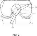
- FIG. 2 illustrates the actual implementation of the tissue scaffold of the present disclosure.
- the "main body area” 11 refers to the part of the weave exposed outside the bone
- the "fixed area” 121 and 122 refers to the part of the weave implanted in bone, for example, used to the bone connected with the original tendon and/or ligament tissue.
- bioactive component on the fiber surface of the main body area, and the bioactive component can be further impregnated into the pores of the main body area to increase cell proliferation ability, wherein the bioactive component is collagen.
- the modification of tissue scaffold comprising the bioactive component of the present disclosure is prepared by the following steps of:
- reaction solution includes a solvent and a pH adjusting agent in addition to a bioactive component.
- the above bioactive component includes collagen, gelatin, silk protein, and the like.
- the bioactive component comprises collagen, and the collagen accounts for 0.5 to 5% by weight of the total weight of the main body area.
- the weight ratio of the collagen to the total weight of the main body area may be about 0.6, 0.7, 0.8, 0.9, 1.0, 1.5, 2.0, 2.5, 3.0, 3.5, 4.0 or 4.5% by weight, and but it is also not limited thereto.
- the bioactive component is collagen.
- the solvent is an aqueous acetic acid solution with pH ⁇ 3.0.
- the pH adjusting agent is phosphate buffered saline and sodium hydroxide.
- the specific preparation method comprises: dissolving 0.1 g of the collagen powder in a 60 ml of an aqueous acetic acid solution, adding 1 g of the main body area of the weave, then adding 40 ml of phosphate buffered saline and adjust the pH to 7.5 with sodium hydroxide. So that the collagen is formed on the fiber surface of the main body area.
- the solid content of the collagen dissolved in the acetic acid is 0.01 to 0.1% by weight, and the reaction temperature of the main body area and the reaction solution is 20 to 40 °C, and the reaction time is 24 to 48 hours.
- main body area of the present disclosure can be modified to comprise other cell proliferation stimulating components, such as gelatin, silk protein, keratin, and the like.
- the bioceramic material is, for example, calcium phosphate, calcium sulfate, bioglass or a combination of the foregoing.
- the calcium phosphate may be hydroxyapatite or tricalcium phosphate.
- the bioceramic material is hydroxyapatite, and an average particle size thereof is about 10 to 200 nanometers.
- the bioceramic material is present in the weft yarn of the fixed area, and the bioceramic material accounts for 1 to 4% by weight of the total weight of the weft yarn of the fixed area. In another embodiment, the bioceramic material accounts for more than 1% to 4% by weight of the total weight of the weft yarn in the fixed area.
- the weight ratio of bioceramic materials is too low, the bone growth cannot be enhanced. If the weight ratio of the bioceramic material is too high, it is easy to break when spinning the fiber.
- the weight ratio of the bioceramic material to the total weight of the weft yarns of the fixed area may be about 1.5, 2.0, 2.5, 3.0, or 3.5% by weight, and is not limited thereto.
- the weft yarns comprising the bioceramic material and the weft yarns free of bioceramic material may be selected in the fixed area of the present disclosure, or the bioceramic material may be included in each weft yarn.
- the ratio of weft yarns comprising the bioceramic material to the weft yarns free of bioceramic material is about 1:9 to 10:0.
- the weft yarns comprising the bioceramic material can be used alone in the fixed area of the present disclosure.
- weft fiber comprising the bioceramic material in the fixed area of the tissue scaffold of the present disclosure
- it is prepared by the following steps of:
- the masterbatch comprises the above weave material and bioceramic material, and the content of the bioceramic material accounts for 1 to 4% by weight of the masterbatch.
- the masterbatch system also comprises a dispersant.
- the dispersant may be a polyester polymer material, and the content of the dispersant accounts for 0.1 to 2% by weight of the masterbatch.
- the specific preparation method of the masterbatch comprises the steps of: subjecting the polyethylene terephthalate, hydroxyapatite and polyester dispersant to mixing and granulation to form the masterbatch.
- Example 1 Preparation of a tissue scaffold
- Preparation of a warp yarn polyethylene terephthalate was subjected to hot melt spinning to obtain a semi-stretched yarn through a modified cross-section spinning nozzle at a temperature of 270°C or higher, and then stretched at a temperature of 100 to 200°C to obtain a fully stretched yarn with Y-shaped cross-section structure.
- Preparation of a weft yarn the preparation method of the weft yarn was the same as the above preparation method of the warp yarn, except that the modified cross-section spinning nozzle was adjusted to a circular spinning nozzle.
- bioceramics-incorporated masterbatch When preparing the weft yarns comprising bioceramics, bioceramics-incorporated masterbatch was used, wherein the method for preparing bioceramics-incorporated masterbatch was prepared by the following steps of: mixing polyethylene terephthalate, hydroxyapatite with an average particle size of 60 nm, and polyester dispersant, and granulating the mixture to form a masterbatch with 2% by weight of the bioceramic material.
- Preparation of the weave according to the main body area and fixed area of the weave shown in FIG. 1 , the warp yarns and the weft yarns were selected for interlaced weaving, and the main body area was brought into contact with the reaction solution comprising the bioactive components.
- the reaction solution also included a solvent and pH adjusting agents, wherein the solvent was an aqueous acetic acid solution with pH ⁇ 3.0, and the pH adjusting agents were phosphate buffered saline and sodium hydroxide.
- the specific modification method thereof includes the following steps for preparation: dissolving 0.1 g of the collagen powder in a 60 ml of an aqueous acetic acid solution, adding 1 g of the main body area of the weave, then adding 40 ml of phosphate buffered saline and adjust the pH to 7.5 with sodium hydroxide, and after 48 hours of reaction at a reaction temperature of 20 to 40°C, forming collagen on the fiber surface of the main body area.
- tissue scaffold prepared above was subjected to subsequent animal experiments.
- the preparation method of the tissue scaffold is the same as in Example 1, except that the cross-sectional structure of the warp yarn fiber was changed to H-shaped, S-shaped, W-shaped and cross-shaped respectively. Then, the prepared tissue scaffolds were used to determine the number of cell proliferation and analyze the cell attachment ratio, and are recorded in FIG. 3 and FIG. 4 .
- the preparation method of the tissue scaffold was the same as in Example 1, except that the content of the bioceramic material was 4%. Then, the prepared tissue scaffold was analyzed for the activity of osteogenic enzymes, and recorded in FIG. 5 .
- the preparation method of the tissue scaffold was the same as in Example 1, except that the cross-sectional structure of the warp yarn fibers was a circular cross-section. Then, the prepared tissue scaffold was used to determine the number of cell proliferation and analyze the cell attachment ratio, and is recorded in FIG. 3 and FIG. 4 .
- the preparation method of the tissue scaffold was the same as in Example 1, except that the surface of the main body area of the weave was not modified. Then, the prepared tissue scaffold was used to determine the number of cell proliferation and analyze the cell attachment ratio, and are recorded in FIG. 3 and FIG. 4 .
- the preparation method of the tissue scaffold was the same as in Comparative Example 2, except that the cross-sectional structure of the warp yarn fiber was changed to a circular cross-section, an H-shaped cross-section, an S-shaped cross-section, a W-shaped cross-section, and a cross-shaped cross-section, respectively. Then, the prepared tissue scaffolds were used to determine the number of cell proliferation and analyze the cell attachment ratio, and are recorded in FIG. 3 and FIG. 4 .
- the preparation method of the tissue scaffold was the same as in Example 1, except that the bioceramic material was not added. Then, the prepared tissue scaffold was analyzed for the activity of osteogenic enzymes and bone differentiation ability, which is recorded in FIG. 5 and FIG. 6 .
- the preparation method of the tissue scaffold was the same as in Example 1, except that the content of the bioceramic material was adjusted to 1%. Then, the prepared tissue scaffold was analyzed for the activity of osteogenic enzymes, which is recorded in FIG. 5 .
- a commercial tissue scaffold (Orthomed, LCA60NEF) was used for the animal test of the ligament reconstruction surgery of Example 1. Then, the tissue scaffold was analyzed by the surface observation, tissue section, micro-CT and biomechanical testing, etc. and the results are recorded in FIGs. 7D to 7E , 8C to 8D , 9C to 9D , and FIG. 10 .
- the present disclosure combines the corresponding bioactive materials according to different sections of the tissue scaffold to induce the regeneration of tendons or ligaments and at the same time improve the osseointegration ability. Accordingly, a tendon or a ligament tissue similar to autogenous ones is gradually formed, and the problems of fatigue, abrasion, rupture and poor stability of existing artificial grafts after long-term use are solved.
Landscapes
- Health & Medical Sciences (AREA)
- Chemical & Material Sciences (AREA)
- Life Sciences & Earth Sciences (AREA)
- Veterinary Medicine (AREA)
- Oral & Maxillofacial Surgery (AREA)
- Transplantation (AREA)
- Animal Behavior & Ethology (AREA)
- General Health & Medical Sciences (AREA)
- Public Health (AREA)
- Epidemiology (AREA)
- Dermatology (AREA)
- Medicinal Chemistry (AREA)
- Engineering & Computer Science (AREA)
- Inorganic Chemistry (AREA)
- Rheumatology (AREA)
- Orthopedic Medicine & Surgery (AREA)
- Rehabilitation Therapy (AREA)
- Cardiology (AREA)
- Biomedical Technology (AREA)
- Heart & Thoracic Surgery (AREA)
- Vascular Medicine (AREA)
- Chemical Kinetics & Catalysis (AREA)
- Ceramic Engineering (AREA)
- Biophysics (AREA)
- Materials For Medical Uses (AREA)
- Prostheses (AREA)
Abstract
Description
- This disclosure relates to scaffolds for use in tissue engineering, in particular to a tissue scaffold for use in a tendon and/or ligament injury.
- Tear or rupture of a tendon or ligament is a common clinical sports injury, especially when the injury occurs on the cruciate ligament in the knee joint. Due to biological environment and limited blood supply of the ligament tissue, it does not have the ability to heal naturally after it is ruptured, and needs to be reconstructed through surgery.
- Regarding the reconstruction of tendons or ligaments, it can be divided into autograft, allograft and artificial graft according to the type of repair material. Although autografts have good reconstruction effects and no immune rejections, there are problems such as complications at donor site. Allografts are not only costly, but also have the risk of immune rejection and disease transmission. By contrast, because of its convenient materials, no risk of disease transmission, and high mechanical strength, artificial grafts have gradually attracted the attention of the medical community recently. However, artificial grafts only have mechanical load-bearing effects, and do not have biological activities and tissue inductions, which are not good for the healing of tendons/ligaments and bone interface. Further, the cells and tissues on the surface of the artificial ligament are difficult to grow to form a normal tendon or ligament tissue. After a long-term use, the artificial graft is prone to fatigue, abrasion, and fracture, which can lead to instability of the knee joint, and the wear fragments can easily cause iatrogenic injury such as water accumulation in the joint cavity.
- Accordingly, it is necessary to propose a tissue scaffold with a biological activity and tissue induction to improve fatigue, abrasion, fracture and poor stability of existing artificial grafts after long-term use.
- The disclosure provides a tissue scaffold for use in a tendon and/or ligament, which comprises a weave formed by interlacing warp yarns and weft yarns, wherein the warp yarns include a plurality of fibers with an alternative shaped cross section structure. The weave comprises a main body area, wherein a bioactive component is on the fiber surface of the main body area, and the bioactive component can be further impregnated into the pores of the main body area; and a fixed area, wherein the fixed area is formed on the two sides of the main body area, and the weft yarns comprises a bioceramic material.
- According to the present disclosure, through the design of a unique weave structure, the corresponding bioactive materials are respectively combined according to the tissue scaffold sections, to provide more cell attachment area and a good proliferation environment, effectively enhancing the activity and proliferation capacity of soft and bone tissues. It can be provided for the reconstruction of tendon and/or ligament tissue, and it has application prospects.
- The embodiments of the present disclosure are explained by referring to the drawings as examples:
-
FIG. 1 is a schematic diagram of a tissue scaffold according to an exemplary embodiment; -
FIG. 2 is a schematic diagram of an actual use state of the tissue scaffold according to an exemplary embodiment; -
FIG. 3 is a comparison diagram of the number of proliferated cells of the tissue scaffolds between the embodiments and comparative examples, wherein each group of the embodiments and comparative examples are D1, D3, and D7 from left to right, respectively, and D1, D3, and D7 show the number of cells cultured on the fiber surface of the weaves for 1, 3 and 7 days, respectively; -
FIG. 4 is a comparison diagram of the cell attachment ratios of tissue scaffolds of the embodiments and comparative examples according to the present disclosure; -
FIG. 5 is a comparison diagram of the osteogenic enzyme ALP activity on the 7th and 14th days of cell culture of the tissue scaffolds of the embodiments and comparative examples, wherein each group on the 7th and 14th days are 0%, 1%, 2%, 4% and dish from left to right, 0% indicates the surface culture of the woven fiber without adding the bioceramic material in comparative example 8; 1% indicates the surface culture of the woven fiber containing the bioceramic material of 1% by weight in comparative example 9; 2% indicates the surface culture of the woven fiber containing the bioceramic material of 2% by weight in example 1; 4% indicates the surface culture of the woven fiber containing the bioceramic material of 4% by weight in example 6; dish means the osteogenic enzyme ALP activity results which are only cultured on the cell culture plate but not on the surface of the woven fiber as a control; -
FIG. 6 is a comparison diagram of calcium deposition staining of the tissue scaffolds on the 7th, 14th and 21st day of cell culture of the embodiments and comparative examples according to the present disclosure; -
FIGs. 7A to 7C are scanning electron microscope images of the regeneration of tendon and ligament tissue promoted by the tissue scaffold of the embodiments according to the present disclosure; -
FIGs. 7D and7E are scanning electron microscope images of commercial tissue scaffolds; -
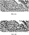
FIGs. 8A and 8B are the tissue sections stained with hematoxylin-eosin and Masson's trichrome of the tissue scaffold, respectively, of the embodiments according to the present disclosure; -
FIGs. 8C and 8D are the tissue sections stained with hematoxylin-eosin and Masson's trichrome of the commercial scaffold, respectively; -
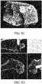
FIGs. 9A and 9B are micro-computed tomography images of the osseointegration ability promoted by the tissue scaffold of the embodiments according to the present disclosure; -
FIGs. 9C and 9D are micro-computed tomography images of the commercial scaffold; and -
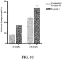
FIG. 10 is a comparison diagram of the mechanical strength of the tissue scaffolds in the bone tunnel in the first month and the third month of the embodiments and the comparative examples according to the present disclosure. - The following is a description of the implementation of the present disclosure through specific embodiments. A person having ordinary skill in the art can easily understand the advantages and effects of the present disclosure from the content disclosed in this specification. The present disclosure can also be implemented or applied by other different implementations, and various details in this specification can also be based on different viewpoints and applications, without departing from the scope disclosed in the present disclosure, with different modifications and changes. In addition, all ranges and values herein are inclusive and combinable. Any value or point falling within the range described herein, for example, any integer can be used as the minimum or maximum value to derive the lower range, etc.
- According to the present disclosure, a tissue scaffold for use in a tendon and/or ligament is provided. As shown in
FIG. 1 , atissue scaffold 1 is constituted by a weave formed by interlacingwarp yarns 101 andweft yarns warp yarn 101 comprises a plurality of fibers with an alternative shaped cross section structure, and the plurality of fibers constitute the warp yarn. The weave comprises amain body area 11 containing a bioactive component on a surface thereof, and the bioactive component may be further impregnated into the pores of themain body area 11; and fixedareas material weft yarn 120 being formed on two sides of the main body area. - The term "warp yarn" used herein refers to the yarn that extends along the length of a loom during weaving and is used as the main support structure in the weave, and the direction is the same as that of tension when performing the stretching action of tendons and/or ligaments. Further, as shown in
FIG. 1 , thewarp yarn 101 of themain body area 11 and thefixed area - The above-mentioned warp yarns and the weft yarns are formed by twisting and drawing multiple fibers. In a specific embodiment, the warp yarns in the tissue scaffold of the present disclosure are twisted into 200 to 800 denier, the weft yarns are twisted into 50 to 100 denier.
- The term "weave" used herein is woven by warp yarns and weft yarns in an interlaced and or mutually perpendicular manner, and the interweaving points formed thereby may be arranged continuously or discontinuously, and the positions of the interweaving points may be selected periodically or irregularly.
- In one embodiment, the pore size of the weave of the tissue scaffold of the present disclosure is 0.1 to 1 mm, and the pore of the weave provides sufficient growth space for cells and provides the transportation space for gas exchange, nutrient and metabolism required during cells growth.
- On the other hand, the weave of the disclosure is not limited to a single-layer weaving structure, but also includes a multi-layer upper and lower weaving structure. In another embodiment, the thickness or diameter of the weave is 1.0 to 10 mm.
- In the tissue scaffold of the present disclosure, the material of the weave can be a polymer material or a polymer composite material, wherein the polymer composite material comprises other fillers, such as carbon fiber, in addition to the polymer material. The polymer material is selected in consideration of the mechanical properties, stability, abrasion resistance and biocompatibility thereof, but is not limited thereto.
- In an exemplary embodiment, the polymer material may be one of polyethylene terephthalate, polyethylene, polytetrafluoroethylene, polyurethane, polycaprolactone, polylactic acid, polyglycolic acid, polyether ether ketone, polyether ketone, or a mixture or copolymer thereof, wherein polyethylene may include ultra-high molecular weight polyethylene.
- In another specific embodiment, the material of the above-mentioned weave is polyethylene terephthalate.
- The term "alternative shaped cross section structure" used herein refers to a fiber structure with a non-circular cross-section, which has a relatively high fiber surface area and brings the effect of enhancing the adhesion of cells. In one embodiment, the alternative shaped cross section structure of the warp yarn fiber of the present disclosure may comprise an H-shaped cross-section, an S-shaped cross-section, a W-shaped cross-section, a Y-shaped cross-section, or a cross-shaped cross-section.
- In the tissue scaffold of the present disclosure, the fiber's thickness of the weave corresponds to the overall surface area, which affects the surface properties and mechanical properties of the weave. In another embodiment, the diameter of the long axis of the fibers with the alternative shaped cross section structure comprised in the warp yarn is 15 to 50 micrometers, and the fibers' diameter of the weft yarn constituting the weave is 20 to 50 micrometers. In another embodiment, the fineness of the fibers with the alternative shaped cross section structure comprised in the warp yarn is 1.5 to 50 denier, and the fineness of the fibers of the weft yarn constituting the weave is 40 to 100 denier. As shown in
FIG. 2, FIG. 2 illustrates the actual implementation of the tissue scaffold of the present disclosure. The "main body area" 11 refers to the part of the weave exposed outside the bone, and the "fixed area" 121 and 122 refers to the part of the weave implanted in bone, for example, used to the bone connected with the original tendon and/or ligament tissue. - In order to further induce tendon and/or ligament tissue repair, there is a bioactive component on the fiber surface of the main body area, and the bioactive component can be further impregnated into the pores of the main body area to increase cell proliferation ability, wherein the bioactive component is collagen.
- In one embodiment, the modification of tissue scaffold comprising the bioactive component of the present disclosure is prepared by the following steps of:
- providing a reaction solution comprising a bioactive component; and
- bringing the main body area into contact with the reaction solution.
- The term "reaction solution" includes a solvent and a pH adjusting agent in addition to a bioactive component.
- The above bioactive component includes collagen, gelatin, silk protein, and the like.
- In another embodiment, the bioactive component comprises collagen, and the collagen accounts for 0.5 to 5% by weight of the total weight of the main body area.
- In embodiments, the weight ratio of the collagen to the total weight of the main body area may be about 0.6, 0.7, 0.8, 0.9, 1.0, 1.5, 2.0, 2.5, 3.0, 3.5, 4.0 or 4.5% by weight, and but it is also not limited thereto.
- In exemplary embodiment, in the mentioned above reaction solution, the bioactive component is collagen. The solvent is an aqueous acetic acid solution with pH ≦ 3.0. The pH adjusting agent is phosphate buffered saline and sodium hydroxide. The specific preparation method comprises: dissolving 0.1 g of the collagen powder in a 60 ml of an aqueous acetic acid solution, adding 1 g of the main body area of the weave, then adding 40 ml of phosphate buffered saline and adjust the pH to 7.5 with sodium hydroxide. So that the collagen is formed on the fiber surface of the main body area.
- In another embodiment, the solid content of the collagen dissolved in the acetic acid is 0.01 to 0.1% by weight, and the reaction temperature of the main body area and the reaction solution is 20 to 40 °C, and the reaction time is 24 to 48 hours.
- In addition, the main body area of the present disclosure can be modified to comprise other cell proliferation stimulating components, such as gelatin, silk protein, keratin, and the like.
- Regarding the mentioned above weft yarn of the fixed area, in order to effectively enhance the healing ability of the bone interface, a bioceramic material is added into it. In one embodiment, the bioceramic material is, for example, calcium phosphate, calcium sulfate, bioglass or a combination of the foregoing. In one embodiment, the calcium phosphate may be hydroxyapatite or tricalcium phosphate.
- In another embodiment, the bioceramic material is hydroxyapatite, and an average particle size thereof is about 10 to 200 nanometers.
- In one embodiment, the bioceramic material is present in the weft yarn of the fixed area, and the bioceramic material accounts for 1 to 4% by weight of the total weight of the weft yarn of the fixed area. In another embodiment, the bioceramic material accounts for more than 1% to 4% by weight of the total weight of the weft yarn in the fixed area. When the weight ratio of bioceramic materials is too low, the bone growth cannot be enhanced. If the weight ratio of the bioceramic material is too high, it is easy to break when spinning the fiber.
- In other embodiments, the weight ratio of the bioceramic material to the total weight of the weft yarns of the fixed area may be about 1.5, 2.0, 2.5, 3.0, or 3.5% by weight, and is not limited thereto.
- On the other hand, the weft yarns comprising the bioceramic material and the weft yarns free of bioceramic material may be selected in the fixed area of the present disclosure, or the bioceramic material may be included in each weft yarn. In exemplary embodiment, the ratio of weft yarns comprising the bioceramic material to the weft yarns free of bioceramic material is about 1:9 to 10:0. In another embodiment, the weft yarns comprising the bioceramic material can be used alone in the fixed area of the present disclosure.
- For the weft fiber comprising the bioceramic material in the fixed area of the tissue scaffold of the present disclosure, it is prepared by the following steps of:
- providing a masterbatch mixed with bioceramic materials; and
- spinning with the masterbatch.
- In a specific embodiment, the masterbatch comprises the above weave material and bioceramic material, and the content of the bioceramic material accounts for 1 to 4% by weight of the masterbatch.
- In addition to the above weave material and bioceramic material, the masterbatch system also comprises a dispersant. The dispersant may be a polyester polymer material, and the content of the dispersant accounts for 0.1 to 2% by weight of the masterbatch.
- In one embodiment, the specific preparation method of the masterbatch comprises the steps of: subjecting the polyethylene terephthalate, hydroxyapatite and polyester dispersant to mixing and granulation to form the masterbatch.
- The disclosure will be further described in detail below through specific embodiments, but the scope of the disclosure is not limited by the description of the embodiments.
- Preparation of a warp yarn: polyethylene terephthalate was subjected to hot melt spinning to obtain a semi-stretched yarn through a modified cross-section spinning nozzle at a temperature of 270°C or higher, and then stretched at a temperature of 100 to 200°C to obtain a fully stretched yarn with Y-shaped cross-section structure.
- Preparation of a weft yarn: the preparation method of the weft yarn was the same as the above preparation method of the warp yarn, except that the modified cross-section spinning nozzle was adjusted to a circular spinning nozzle.
- When preparing the weft yarns comprising bioceramics, bioceramics-incorporated masterbatch was used, wherein the method for preparing bioceramics-incorporated masterbatch was prepared by the following steps of: mixing polyethylene terephthalate, hydroxyapatite with an average particle size of 60 nm, and polyester dispersant, and granulating the mixture to form a masterbatch with 2% by weight of the bioceramic material.
- Preparation of the weave: according to the main body area and fixed area of the weave shown in
FIG. 1 , the warp yarns and the weft yarns were selected for interlaced weaving, and the main body area was brought into contact with the reaction solution comprising the bioactive components. - Regarding the above reaction solution, collagen was taken as the bioactive component. In addition to the bioactive component, the reaction solution also included a solvent and pH adjusting agents, wherein the solvent was an aqueous acetic acid solution with pH≦3.0, and the pH adjusting agents were phosphate buffered saline and sodium hydroxide. The specific modification method thereof includes the following steps for preparation: dissolving 0.1 g of the collagen powder in a 60 ml of an aqueous acetic acid solution, adding 1 g of the main body area of the weave, then adding 40 ml of phosphate buffered saline and adjust the pH to 7.5 with sodium hydroxide, and after 48 hours of reaction at a reaction temperature of 20 to 40°C, forming collagen on the fiber surface of the main body area.
- Finally, the weave of the tissue scaffold prepared above was cut into an appropriate size, and the following cell tests and analysis were performed:
- (1) Determination of the number of cell proliferation and cell attachment ratio: A weave (1cm2) of the main body area of the present embodiment was taken and placed in a 48-well plate, a small amount of cell suspension (20µl) was dropped on the surface, and placed in an incubator at 37°C for 2 hours. 0.8 ml of bone marrow mesenchymal stem cell culture medium was added, and the unattached cells was taken away from the fiber surface of the weave by the culture medium. After overnight incubation, the weave attached with cells was transferred to a new 48-well plate, 0.3 ml of PrestoBlue was and performed in an incubator at 37°C. PrestoBlue was reduced to pink by nicotine adenine dinucleotide (NADH) dehydrogenase in mitochondria, the number of reacted cells could be detected by fluorescence (Ex/Em: 560 nm/590 nm). After 1 hour of reaction, 100 ul of the reactant was removed and transferred to a 96-well plate for analysis to detect a fluorescence read (Ex/Em: 560 nm/590 nm) thereof. The number of cells attached to the fiber surface was quantified by interpolation according to the standard curve, and the results of the number of cell proliferation and the cell attachment ratio are recorded in
FIG. 3 andFIG. 4 respectively. From the results inFIGs. 3 and4 , it is known that the tissue scaffold of the present embodiment brings the effect of significantly improving cell proliferation and cell adhesion due to the surface modification of the warp yarn fiber with an alternative shaped cross section structure and the main body area of the weave. - (2) Determination of ALP activity of osteogenic enzyme: mesenchymal stem cells (MSC) were used to determine the activity of osteogenic enzyme ALP. The cell seeding density was 5x10⁴/cm2, the bone differentiation medium contained α-MEM containing 10% fetal bovine serum (FBS), ascorbic acid (50 µg/ml), dexamethasone (0.1 µM), and β-sugar phosphate (10 mM), and the control medium contained α-MEM containing 10% FBS. Samples were collected on the 7th and 14th days for analysis. A pNPP alkaline phosphatase detection kit (SensoLyte®) and a Microplate reader (Biotek, Synergy™ HI) were used to measure the activity of osteogenic enzyme ALP, and the results are recorded in
FIG. 5 . The results of the ALP activity of the cell culture plate (dish) were used as a control. It is obvious that the tissue scaffold of the present embodiment enhancing the effect of the activity of the osteogenic enzyme ALP due to comprising the bioceramic materials, and thereby showing that the tissue scaffold of the present disclosure can effectively promote bone regeneration. - (3) Determination of bone differentiation ability: mesenchymal stem cells (MSC) were used for a calcium deposition analysis. The cell seeding density was 5x10⁴/cm2, and the bone differentiation medium contained α-MEM containing 10% fetal bovine serum (FBS), ascorbic acid (50 µg/ml), dexamethasone (0.1 µM), and β-sugar phosphate (10 mM), while the control medium contained α-MEM containing 10% FBS. Samples were collected for analysis on the 7th, 14th and 21st days. After the cell samples were treated with the culture medium, they were stained with alizarin red to evaluate the calcium deposition on the 7th, 14th and 21st days, and recorded in
FIG. 6 . From the results inFIG. 6 , it is shown that the calcium deposition is significantly increased due to the tissue scaffold comprising bioceramic materials of the present embodiment. It shows that the tissue scaffold of the present disclosure has the ability to promote bone differentiation of mesenchymal stem cells. - Next, the tissue scaffold prepared above was subjected to subsequent animal experiments.
- Animal test of ligament reconstruction surgery: New Zealand white rabbits were used as the animal model of Medial Collateral Ligament (MCL) reconstruction surgery. Anesthetics (Zoletil50 : Rompun20 = 1 : 1, 0.5 ml/kg) was used for anesthesia before surgery. The knee joint of the hind limb was opened with a scalpel, and drilled a bone tunnel in the femur and tibia, respectively. Then, the tissue scaffold was penetrated through the bone tunnel from the tibial end, and then penetrated into the femoral end of the bone tunnel. The tibial end was fixed with a metal button, and the femoral end was fixed with a metal bone nail. Finally, the separated layers of tissue and skin were sutured to complete the operation.
- Finally, the following analysis on the implanted tissue scaffold of the above-mentioned
animal experiment post 1 month or 3 months was performed: - (1) Scanning electron microscope: a scanning electron microscope (QUANTA 400F/Thermo Scientific™) was used to observe the tissue growth of the cross-section and surface of the implanted tissue scaffold in the animal experiment after 3 months. It is known from
FIG. 7A that fibroblasts are observed on the fiber surface of the tissue scaffold of the present embodiment. InFIG. 7B andFIG. 7C , it is known that the collagen fiber soft tissue is distributed inside and on the surface of the tissue scaffold, forming a rope-like structure of tissue network and tightly covering the tissue scaffold of the present embodiment. It shows that the tissue scaffold of the present embodiment has excellent biocompatibility and biological activity, and can induce the regeneration of tendon and ligament tissue (fibroblasts and collagen fibers). - (2) Tissue section: The tissue section was embedded in paraffin, then stained with hematoxylin-eosin and Masson's trichrome stain. Tissue growth of the implanted tissue scaffold in the animal experiment after 3 months. It is known that the peripheral and internal spaces of the tissue scaffold of the present embodiment are full of new cells and collagen fibrous tissue via both hematoxylin-eosin staining (
FIG. 8A ) and Mason's trichrome staining (FIG. 8B ). It is obvious that the tissue scaffold of the present embodiment brings the effect of promoting the regeneration of tendon and ligament tissue (cells and collagen fibers), which means that the tissue scaffold is ligamented. - (3) Micro-computer tomography analysis (micro-CT): A micro-computer tomography (Bruker micro-CT, Kontich, Belgium) was used to analyze the tissue scaffold fixed in the bone tunnel of implantation in an animal experiment after 3 months. In
FIG. 9A and 9B , it is known that the tissue scaffold fixed in the bone tunnel of the present embodiment has formed a new bone. It is obvious that the tissue scaffold of the present embodiment has the ability to heal the interface between the implant and the bone (meaning it has the ability of osseointegration). - (4) Biomechanics test: a tensile testing machine (INSTRON 3400) was used to measure the mechanical strength of the tissue scaffold fixed to the bone tunnel. First, the metal button that fixes the specimen to the tibia was cut off, and then the specimen was moved to the test stage and fixed. The tissue scaffold was tested for the force for pulling out from the tibia or finally rupturing it, when it is implanted in animal experiments after 1 month and 3 months. As that in
FIG. 10 , due to the unique weave structure design and excellent osseointegration ability of the tissue scaffold of the present embodiment, it can withstand greater tensile tension, thus improve the mechanical properties of the tissue scaffold. - The preparation method of the tissue scaffold is the same as in Example 1, except that the cross-sectional structure of the warp yarn fiber was changed to H-shaped, S-shaped, W-shaped and cross-shaped respectively. Then, the prepared tissue scaffolds were used to determine the number of cell proliferation and analyze the cell attachment ratio, and are recorded in
FIG. 3 andFIG. 4 . - The preparation method of the tissue scaffold was the same as in Example 1, except that the content of the bioceramic material was 4%. Then, the prepared tissue scaffold was analyzed for the activity of osteogenic enzymes, and recorded in
FIG. 5 . - The preparation method of the tissue scaffold was the same as in Example 1, except that the cross-sectional structure of the warp yarn fibers was a circular cross-section. Then, the prepared tissue scaffold was used to determine the number of cell proliferation and analyze the cell attachment ratio, and is recorded in
FIG. 3 andFIG. 4 . - The preparation method of the tissue scaffold was the same as in Example 1, except that the surface of the main body area of the weave was not modified. Then, the prepared tissue scaffold was used to determine the number of cell proliferation and analyze the cell attachment ratio, and are recorded in
FIG. 3 andFIG. 4 . - The preparation method of the tissue scaffold was the same as in Comparative Example 2, except that the cross-sectional structure of the warp yarn fiber was changed to a circular cross-section, an H-shaped cross-section, an S-shaped cross-section, a W-shaped cross-section, and a cross-shaped cross-section, respectively. Then, the prepared tissue scaffolds were used to determine the number of cell proliferation and analyze the cell attachment ratio, and are recorded in
FIG. 3 andFIG. 4 . - The preparation method of the tissue scaffold was the same as in Example 1, except that the bioceramic material was not added. Then, the prepared tissue scaffold was analyzed for the activity of osteogenic enzymes and bone differentiation ability, which is recorded in
FIG. 5 andFIG. 6 . - The preparation method of the tissue scaffold was the same as in Example 1, except that the content of the bioceramic material was adjusted to 1%. Then, the prepared tissue scaffold was analyzed for the activity of osteogenic enzymes, which is recorded in
FIG. 5 . - A commercial tissue scaffold (Orthomed, LCA60NEF) was used for the animal test of the ligament reconstruction surgery of Example 1. Then, the tissue scaffold was analyzed by the surface observation, tissue section, micro-CT and biomechanical testing, etc. and the results are recorded in
FIGs. 7D to 7E ,8C to 8D ,9C to 9D , andFIG. 10 . - In summary, the present disclosure combines the corresponding bioactive materials according to different sections of the tissue scaffold to induce the regeneration of tendons or ligaments and at the same time improve the osseointegration ability. Accordingly, a tendon or a ligament tissue similar to autogenous ones is gradually formed, and the problems of fatigue, abrasion, rupture and poor stability of existing artificial grafts after long-term use are solved.
- The foregoing embodiments are merely illustrative, and not used to limit the present disclosure. Anyone who is familiar with this technique can modify and change the above embodiments without departing from the scope of this disclosure. Therefore, the scope of protection of the claims of the present disclosure is defined by the scope of the appended claims attached to the present disclosure, and as long as it does not affect the effect and implementable purpose of the present disclosure, it should be included in the technical content of the present disclosure.
Claims (10)
- A tissue scaffold (1) for use in a tendon and/or a ligament, comprising a weave formed by interlacing a warp yarn (101) and a weft yarn (110, 120), wherein the warp yarn (101) comprises a plurality of fibers each with an alternative shaped cross section structure, and the weave comprises:a main body area (11) comprising a bioactive component on a fiber surface thereof; anda fixed area (121, 122), wherein the fixed area (121, 122) is formed on both sides of the main body area (11), and the weft yarn (120) comprises a bioceramic material.
- The tissue scaffold (1) of claim 1, wherein the warp yarn (101) comprises a fiber with the alternative shaped cross section structure, and a diameter of a long axis of the fibers is 15 to 50 micrometers, and a diameter of each of the fibers of the weft yarn (110, 120) constituting the weave is 20 to 50 micrometers.
- The tissue scaffold (1) of claim 1, wherein the alternative shaped cross section structure comprises an H-shaped section, an S-shaped section, a W-shaped section, a Y-shaped section or a cross-shaped section.
- The tissue scaffold (1) of claim 1, wherein a fineness of the warp yarn comprising the fiber with the alternative shaped cross section structure is 1.5 to 50 denier, and a fineness of the fibers of the weft yarn (110, 120) constituting the weave is 40 to 100 denier.
- The tissue scaffold (1) of claim 1, wherein a pore size of the weave is 0.1 to 1 mm.
- The tissue scaffold (1) of claim 1, wherein the material of the weave comprises polyethylene terephthalate, polyethylene, polytetrafluoroethylene, polyurethane, or a combination thereof.
- The tissue scaffold (1) of claim 1, wherein the bioactive component comprises collagen, and collagen accounts for 0.5 to 5% by weight of the total weight of the main body area (11).
- The tissue scaffold (1) of claim 1, wherein the bioceramic material comprises calcium phosphate, calcium sulfate, bioglass, or a combination thereof.
- The tissue scaffold (1) of claim 1, wherein the weft yarn (120) of the fixed area (121, 122) comprises the bioceramic material, and the bioceramic material accounts for 1 to 4% by weight of the total weight of the weft yarn (120) of the fixed area (121, 122), and the warp yarn (101) of the fixed area (121, 122) is free from containing the bioceramic material.
- The tissue scaffold (1) of claim 1, wherein the bioceramic material is hydroxyapatite, and an average particle size thereof is 10 to 200 nanometers.
Applications Claiming Priority (1)
| Application Number | Priority Date | Filing Date | Title |
|---|---|---|---|
| US17/136,077 US12194192B2 (en) | 2020-12-29 | 2020-12-29 | Tissue scaffold for use in tendon and/or ligament |
Publications (2)
| Publication Number | Publication Date |
|---|---|
| EP4023265A1 true EP4023265A1 (en) | 2022-07-06 |
| EP4023265B1 EP4023265B1 (en) | 2024-02-07 |
Family
ID=77520636
Family Applications (1)
| Application Number | Title | Priority Date | Filing Date |
|---|---|---|---|
| EP21193592.9A Active EP4023265B1 (en) | 2020-12-29 | 2021-08-27 | Tissue scaffold for use in tendon and/or ligament |
Country Status (2)
| Country | Link |
|---|---|
| US (1) | US12194192B2 (en) |
| EP (1) | EP4023265B1 (en) |
Families Citing this family (1)
| Publication number | Priority date | Publication date | Assignee | Title |
|---|---|---|---|---|
| TWI852580B (en) * | 2023-05-24 | 2024-08-11 | 捷立康生物科技股份有限公司 | Surgical wires, their uses and methods of manufacture |
Citations (2)
| Publication number | Priority date | Publication date | Assignee | Title |
|---|---|---|---|---|
| CN111450316A (en) * | 2020-03-04 | 2020-07-28 | 东华大学 | An integrated scaffold that simulates a bone-tendon-bone mineralized to non-mineralized gradient structure |
| CN112030556A (en) * | 2019-06-03 | 2020-12-04 | 财团法人纺织产业综合研究所 | Fiber with cell affinity and fiber product prepared by using same |
Family Cites Families (22)
| Publication number | Priority date | Publication date | Assignee | Title |
|---|---|---|---|---|
| US5004474A (en) * | 1989-11-28 | 1991-04-02 | Baxter International Inc. | Prosthetic anterior cruciate ligament design |
| US6599319B2 (en) | 2001-12-14 | 2003-07-29 | Celanese Advanced Materials, Inc. | Prosthetic ligament |
| FR2850026B1 (en) | 2003-01-17 | 2007-08-31 | L A R S Laboratoire D Applic E | BIOMIMETIC PROTHETIC LIGAMENT AND PROCESS FOR OBTAINING |
| JP4627978B2 (en) | 2003-10-27 | 2011-02-09 | 泰晴 野一色 | Low blood permeability medical material |
| FR2865380B1 (en) | 2004-01-23 | 2006-03-03 | L A R S Laboratoire D Applic E | METHOD FOR ATTACHING TRACTION WIRES TO THE END OF A PROTHETIC LIGAMENT |
| FI20055304L (en) * | 2005-06-13 | 2007-02-20 | Bioretec Oy | A bioabsorbable implant with variable properties |
| EP1964583A1 (en) * | 2007-02-09 | 2008-09-03 | Royal College of Surgeons in Ireland | Process for producing a collagen/hydroxyapatite composite scaffold |
| JP5363076B2 (en) | 2008-11-14 | 2013-12-11 | 国立大学法人神戸大学 | Materials for tissue regeneration |
| CN102908210B (en) | 2012-10-15 | 2015-06-17 | 复旦大学附属华山医院 | Single-hole suspended artificial ligament |
| GB201317636D0 (en) * | 2013-10-04 | 2013-11-20 | Isis Innovation | Scaffold |
| US10632230B2 (en) * | 2015-07-10 | 2020-04-28 | Warsaw Orthopedic, Inc. | Implants having a high drug load of an oxysterol and methods of use |
| US11008676B2 (en) * | 2015-12-16 | 2021-05-18 | Edwards Lifesciences Corporation | Textured woven fabric for use in implantable bioprostheses |
| TWI766853B (en) | 2016-01-14 | 2022-06-11 | 學校法人大阪醫科藥科大學 | Warp knitted fabrics and medical materials |
| US10813763B2 (en) * | 2016-07-26 | 2020-10-27 | Warsaw Orthopedic, Inc. | Implantable mesh |
| WO2019055656A1 (en) * | 2017-09-13 | 2019-03-21 | Northwestern University | Photo-reactive inks and thermal-curable materials made therefrom |
| CN107510520B (en) | 2017-09-21 | 2020-08-11 | 上海松力生物技术有限公司 | Cruciate ligament regenerative implant and preparation method and application thereof |
| AU2019226562C1 (en) * | 2018-03-01 | 2022-10-06 | Tepha, Inc. | Medical devices containing poly(butylene succinate) and copolymers thereof |
| US11589973B2 (en) * | 2018-05-16 | 2023-02-28 | Mimedx Group, Inc. | Methods of making collagen fiber medical constructs and related medical constructs, including patches |
| CN110680558A (en) | 2019-10-28 | 2020-01-14 | 北京天星博迈迪医疗器械有限公司 | Ligament reconstruction fixation device and method of use |
| CN111345920B (en) | 2020-03-11 | 2022-11-11 | 上海市第六人民医院 | Transitional structured textile-based scaffold for promoting healing of tendon and bone and preparation method thereof |
| CN111359012A (en) | 2020-04-24 | 2020-07-03 | 李军 | Artificial rotator cuff patch capable of inducing tendon-bone gradient structure formation and preparation method thereof |
| CN111544654B (en) | 2020-05-15 | 2022-06-28 | 李军 | Composite artificial ligament and manufacturing method thereof |
-
2020
- 2020-12-29 US US17/136,077 patent/US12194192B2/en active Active
-
2021
- 2021-08-27 EP EP21193592.9A patent/EP4023265B1/en active Active
Patent Citations (2)
| Publication number | Priority date | Publication date | Assignee | Title |
|---|---|---|---|---|
| CN112030556A (en) * | 2019-06-03 | 2020-12-04 | 财团法人纺织产业综合研究所 | Fiber with cell affinity and fiber product prepared by using same |
| CN111450316A (en) * | 2020-03-04 | 2020-07-28 | 东华大学 | An integrated scaffold that simulates a bone-tendon-bone mineralized to non-mineralized gradient structure |
Also Published As
| Publication number | Publication date |
|---|---|
| EP4023265B1 (en) | 2024-02-07 |
| US12194192B2 (en) | 2025-01-14 |
| US20220202990A1 (en) | 2022-06-30 |
Similar Documents
| Publication | Publication Date | Title |
|---|---|---|
| Ma et al. | PEEK (Polyether-ether-ketone) and its composite materials in orthopedic implantation | |
| Vaquette et al. | The effect of polystyrene sodium sulfonate grafting on polyethylene terephthalate artificial ligaments on in vitro mineralisation and in vivo bone tissue integration | |
| Cooper et al. | Fiber-based tissue-engineered scaffold for ligament replacement: design considerations and in vitro evaluation | |
| Lu et al. | Interface tissue Engineeringand the formulation of multiple-tissue systems | |
| US5522895A (en) | Biodegradable bone templates | |
| US7108721B2 (en) | Tissue regrafting | |
| Zheng et al. | Promotion of tendon growth into implant through pore-size design of a Ti-6Al-4 V porous scaffold prepared by 3D printing | |
| US20070041952A1 (en) | Three-dimensional fiber scaffolds for tissue engineering | |
| JP2003500162A (en) | Connective tissue reconstruction implant | |
| Milthorpe | Xenografts for tendon and ligament repair | |
| JP2003512896A (en) | Tissue repair | |
| US20100136086A1 (en) | Dynamic bioactive nanofiber scaffolding | |
| Zhou et al. | Repair of segmental defects with nano-hydroxyapatite/collagen/PLA composite combined with mesenchymal stem cells | |
| Wang et al. | Phytic acid/magnesium ion complex coating on PEEK fiber woven fabric as an artificial ligament with anti-fibrogenesis and osteogenesis for ligament-bone healing | |
| Hasegawa et al. | Marrow cell culture on poly-L-lactic acid fabrics. | |
| Savić et al. | Early development of a polycaprolactone electrospun augment for anterior cruciate ligament reconstruction | |
| EP4023265B1 (en) | Tissue scaffold for use in tendon and/or ligament | |
| Leitao et al. | In vitro testing of surface-modified biomaterials | |
| Mäkisalo et al. | Reconstruction of anterior cruciate ligament with carbon fiber: An experimental study on pigs | |
| TWI765500B (en) | Tissue scaffold for use in tendon and/or ligament | |
| CN114681123A (en) | Tissue scaffolds for tendons and/or ligaments | |
| Bao et al. | A degradable scaffold enabling shape matching and bone-mimetic biomechanics via in-situ forming for promoting bone defect repair | |
| Mutsuzaki et al. | Firm anchoring between a calcium phosphate-hybridized tendon and bone for anterior cruciate ligament reconstruction in a goat model | |
| US20250205038A1 (en) | Tissue scaffold and method of manufacturing the same | |
| Dunn et al. | The tissue engineering approach to ligament reconstruction |
Legal Events
| Date | Code | Title | Description |
|---|---|---|---|
| PUAI | Public reference made under article 153(3) epc to a published international application that has entered the european phase |
Free format text: ORIGINAL CODE: 0009012 |
|
| STAA | Information on the status of an ep patent application or granted ep patent |
Free format text: STATUS: REQUEST FOR EXAMINATION WAS MADE |
|
| 17P | Request for examination filed |
Effective date: 20210827 |
|
| AK | Designated contracting states |
Kind code of ref document: A1 Designated state(s): AL AT BE BG CH CY CZ DE DK EE ES FI FR GB GR HR HU IE IS IT LI LT LU LV MC MK MT NL NO PL PT RO RS SE SI SK SM TR |
|
| STAA | Information on the status of an ep patent application or granted ep patent |
Free format text: STATUS: EXAMINATION IS IN PROGRESS |
|
| 17Q | First examination report despatched |
Effective date: 20230630 |
|
| GRAP | Despatch of communication of intention to grant a patent |
Free format text: ORIGINAL CODE: EPIDOSNIGR1 |
|
| STAA | Information on the status of an ep patent application or granted ep patent |
Free format text: STATUS: GRANT OF PATENT IS INTENDED |
|
| INTG | Intention to grant announced |
Effective date: 20231020 |
|
| GRAS | Grant fee paid |
Free format text: ORIGINAL CODE: EPIDOSNIGR3 |
|
| GRAA | (expected) grant |
Free format text: ORIGINAL CODE: 0009210 |
|
| STAA | Information on the status of an ep patent application or granted ep patent |
Free format text: STATUS: THE PATENT HAS BEEN GRANTED |
|
| AK | Designated contracting states |
Kind code of ref document: B1 Designated state(s): AL AT BE BG CH CY CZ DE DK EE ES FI FR GB GR HR HU IE IS IT LI LT LU LV MC MK MT NL NO PL PT RO RS SE SI SK SM TR |
|
| REG | Reference to a national code |
Ref country code: GB Ref legal event code: FG4D |
|
| REG | Reference to a national code |
Ref country code: CH Ref legal event code: EP |
|
| REG | Reference to a national code |
Ref country code: IE Ref legal event code: FG4D |
|
| REG | Reference to a national code |
Ref country code: DE Ref legal event code: R096 Ref document number: 602021009170 Country of ref document: DE |
|
| REG | Reference to a national code |
Ref country code: LT Ref legal event code: MG9D |
|
| REG | Reference to a national code |
Ref country code: NL Ref legal event code: MP Effective date: 20240207 |
|
| PG25 | Lapsed in a contracting state [announced via postgrant information from national office to epo] |
Ref country code: IS Free format text: LAPSE BECAUSE OF FAILURE TO SUBMIT A TRANSLATION OF THE DESCRIPTION OR TO PAY THE FEE WITHIN THE PRESCRIBED TIME-LIMIT Effective date: 20240607 |
|
| PG25 | Lapsed in a contracting state [announced via postgrant information from national office to epo] |
Ref country code: LT Free format text: LAPSE BECAUSE OF FAILURE TO SUBMIT A TRANSLATION OF THE DESCRIPTION OR TO PAY THE FEE WITHIN THE PRESCRIBED TIME-LIMIT Effective date: 20240207 |
|
| PG25 | Lapsed in a contracting state [announced via postgrant information from national office to epo] |
Ref country code: GR Free format text: LAPSE BECAUSE OF FAILURE TO SUBMIT A TRANSLATION OF THE DESCRIPTION OR TO PAY THE FEE WITHIN THE PRESCRIBED TIME-LIMIT Effective date: 20240508 |
|
| REG | Reference to a national code |
Ref country code: AT Ref legal event code: MK05 Ref document number: 1654968 Country of ref document: AT Kind code of ref document: T Effective date: 20240207 |
|
| PG25 | Lapsed in a contracting state [announced via postgrant information from national office to epo] |
Ref country code: RS Free format text: LAPSE BECAUSE OF FAILURE TO SUBMIT A TRANSLATION OF THE DESCRIPTION OR TO PAY THE FEE WITHIN THE PRESCRIBED TIME-LIMIT Effective date: 20240507 Ref country code: HR Free format text: LAPSE BECAUSE OF FAILURE TO SUBMIT A TRANSLATION OF THE DESCRIPTION OR TO PAY THE FEE WITHIN THE PRESCRIBED TIME-LIMIT Effective date: 20240207 Ref country code: NL Free format text: LAPSE BECAUSE OF FAILURE TO SUBMIT A TRANSLATION OF THE DESCRIPTION OR TO PAY THE FEE WITHIN THE PRESCRIBED TIME-LIMIT Effective date: 20240207 |
|
| PG25 | Lapsed in a contracting state [announced via postgrant information from national office to epo] |
Ref country code: ES Free format text: LAPSE BECAUSE OF FAILURE TO SUBMIT A TRANSLATION OF THE DESCRIPTION OR TO PAY THE FEE WITHIN THE PRESCRIBED TIME-LIMIT Effective date: 20240207 |
|
| PG25 | Lapsed in a contracting state [announced via postgrant information from national office to epo] |
Ref country code: AT Free format text: LAPSE BECAUSE OF FAILURE TO SUBMIT A TRANSLATION OF THE DESCRIPTION OR TO PAY THE FEE WITHIN THE PRESCRIBED TIME-LIMIT Effective date: 20240207 |
|
| PG25 | Lapsed in a contracting state [announced via postgrant information from national office to epo] |
Ref country code: RS Free format text: LAPSE BECAUSE OF FAILURE TO SUBMIT A TRANSLATION OF THE DESCRIPTION OR TO PAY THE FEE WITHIN THE PRESCRIBED TIME-LIMIT Effective date: 20240507 Ref country code: NO Free format text: LAPSE BECAUSE OF FAILURE TO SUBMIT A TRANSLATION OF THE DESCRIPTION OR TO PAY THE FEE WITHIN THE PRESCRIBED TIME-LIMIT Effective date: 20240507 Ref country code: NL Free format text: LAPSE BECAUSE OF FAILURE TO SUBMIT A TRANSLATION OF THE DESCRIPTION OR TO PAY THE FEE WITHIN THE PRESCRIBED TIME-LIMIT Effective date: 20240207 Ref country code: LT Free format text: LAPSE BECAUSE OF FAILURE TO SUBMIT A TRANSLATION OF THE DESCRIPTION OR TO PAY THE FEE WITHIN THE PRESCRIBED TIME-LIMIT Effective date: 20240207 Ref country code: IS Free format text: LAPSE BECAUSE OF FAILURE TO SUBMIT A TRANSLATION OF THE DESCRIPTION OR TO PAY THE FEE WITHIN THE PRESCRIBED TIME-LIMIT Effective date: 20240607 Ref country code: HR Free format text: LAPSE BECAUSE OF FAILURE TO SUBMIT A TRANSLATION OF THE DESCRIPTION OR TO PAY THE FEE WITHIN THE PRESCRIBED TIME-LIMIT Effective date: 20240207 Ref country code: GR Free format text: LAPSE BECAUSE OF FAILURE TO SUBMIT A TRANSLATION OF THE DESCRIPTION OR TO PAY THE FEE WITHIN THE PRESCRIBED TIME-LIMIT Effective date: 20240508 Ref country code: FI Free format text: LAPSE BECAUSE OF FAILURE TO SUBMIT A TRANSLATION OF THE DESCRIPTION OR TO PAY THE FEE WITHIN THE PRESCRIBED TIME-LIMIT Effective date: 20240207 Ref country code: ES Free format text: LAPSE BECAUSE OF FAILURE TO SUBMIT A TRANSLATION OF THE DESCRIPTION OR TO PAY THE FEE WITHIN THE PRESCRIBED TIME-LIMIT Effective date: 20240207 Ref country code: BG Free format text: LAPSE BECAUSE OF FAILURE TO SUBMIT A TRANSLATION OF THE DESCRIPTION OR TO PAY THE FEE WITHIN THE PRESCRIBED TIME-LIMIT Effective date: 20240207 Ref country code: AT Free format text: LAPSE BECAUSE OF FAILURE TO SUBMIT A TRANSLATION OF THE DESCRIPTION OR TO PAY THE FEE WITHIN THE PRESCRIBED TIME-LIMIT Effective date: 20240207 |
|
| PG25 | Lapsed in a contracting state [announced via postgrant information from national office to epo] |
Ref country code: PL Free format text: LAPSE BECAUSE OF FAILURE TO SUBMIT A TRANSLATION OF THE DESCRIPTION OR TO PAY THE FEE WITHIN THE PRESCRIBED TIME-LIMIT Effective date: 20240207 Ref country code: PT Free format text: LAPSE BECAUSE OF FAILURE TO SUBMIT A TRANSLATION OF THE DESCRIPTION OR TO PAY THE FEE WITHIN THE PRESCRIBED TIME-LIMIT Effective date: 20240607 |
|
| PG25 | Lapsed in a contracting state [announced via postgrant information from national office to epo] |
Ref country code: SE Free format text: LAPSE BECAUSE OF FAILURE TO SUBMIT A TRANSLATION OF THE DESCRIPTION OR TO PAY THE FEE WITHIN THE PRESCRIBED TIME-LIMIT Effective date: 20240207 Ref country code: PT Free format text: LAPSE BECAUSE OF FAILURE TO SUBMIT A TRANSLATION OF THE DESCRIPTION OR TO PAY THE FEE WITHIN THE PRESCRIBED TIME-LIMIT Effective date: 20240607 Ref country code: PL Free format text: LAPSE BECAUSE OF FAILURE TO SUBMIT A TRANSLATION OF THE DESCRIPTION OR TO PAY THE FEE WITHIN THE PRESCRIBED TIME-LIMIT Effective date: 20240207 Ref country code: LV Free format text: LAPSE BECAUSE OF FAILURE TO SUBMIT A TRANSLATION OF THE DESCRIPTION OR TO PAY THE FEE WITHIN THE PRESCRIBED TIME-LIMIT Effective date: 20240207 |
|
| PG25 | Lapsed in a contracting state [announced via postgrant information from national office to epo] |
Ref country code: DK Free format text: LAPSE BECAUSE OF FAILURE TO SUBMIT A TRANSLATION OF THE DESCRIPTION OR TO PAY THE FEE WITHIN THE PRESCRIBED TIME-LIMIT Effective date: 20240207 |
|
| PG25 | Lapsed in a contracting state [announced via postgrant information from national office to epo] |
Ref country code: SM Free format text: LAPSE BECAUSE OF FAILURE TO SUBMIT A TRANSLATION OF THE DESCRIPTION OR TO PAY THE FEE WITHIN THE PRESCRIBED TIME-LIMIT Effective date: 20240207 |
|
| PG25 | Lapsed in a contracting state [announced via postgrant information from national office to epo] |
Ref country code: EE Free format text: LAPSE BECAUSE OF FAILURE TO SUBMIT A TRANSLATION OF THE DESCRIPTION OR TO PAY THE FEE WITHIN THE PRESCRIBED TIME-LIMIT Effective date: 20240207 Ref country code: CZ Free format text: LAPSE BECAUSE OF FAILURE TO SUBMIT A TRANSLATION OF THE DESCRIPTION OR TO PAY THE FEE WITHIN THE PRESCRIBED TIME-LIMIT Effective date: 20240207 |
|
| PG25 | Lapsed in a contracting state [announced via postgrant information from national office to epo] |
Ref country code: SK Free format text: LAPSE BECAUSE OF FAILURE TO SUBMIT A TRANSLATION OF THE DESCRIPTION OR TO PAY THE FEE WITHIN THE PRESCRIBED TIME-LIMIT Effective date: 20240207 |
|
| PG25 | Lapsed in a contracting state [announced via postgrant information from national office to epo] |
Ref country code: SM Free format text: LAPSE BECAUSE OF FAILURE TO SUBMIT A TRANSLATION OF THE DESCRIPTION OR TO PAY THE FEE WITHIN THE PRESCRIBED TIME-LIMIT Effective date: 20240207 Ref country code: SK Free format text: LAPSE BECAUSE OF FAILURE TO SUBMIT A TRANSLATION OF THE DESCRIPTION OR TO PAY THE FEE WITHIN THE PRESCRIBED TIME-LIMIT Effective date: 20240207 Ref country code: RO Free format text: LAPSE BECAUSE OF FAILURE TO SUBMIT A TRANSLATION OF THE DESCRIPTION OR TO PAY THE FEE WITHIN THE PRESCRIBED TIME-LIMIT Effective date: 20240207 Ref country code: EE Free format text: LAPSE BECAUSE OF FAILURE TO SUBMIT A TRANSLATION OF THE DESCRIPTION OR TO PAY THE FEE WITHIN THE PRESCRIBED TIME-LIMIT Effective date: 20240207 Ref country code: DK Free format text: LAPSE BECAUSE OF FAILURE TO SUBMIT A TRANSLATION OF THE DESCRIPTION OR TO PAY THE FEE WITHIN THE PRESCRIBED TIME-LIMIT Effective date: 20240207 Ref country code: CZ Free format text: LAPSE BECAUSE OF FAILURE TO SUBMIT A TRANSLATION OF THE DESCRIPTION OR TO PAY THE FEE WITHIN THE PRESCRIBED TIME-LIMIT Effective date: 20240207 |
|
| REG | Reference to a national code |
Ref country code: DE Ref legal event code: R097 Ref document number: 602021009170 Country of ref document: DE |
|
| PG25 | Lapsed in a contracting state [announced via postgrant information from national office to epo] |
Ref country code: IT Free format text: LAPSE BECAUSE OF FAILURE TO SUBMIT A TRANSLATION OF THE DESCRIPTION OR TO PAY THE FEE WITHIN THE PRESCRIBED TIME-LIMIT Effective date: 20240207 |
|
| PLBE | No opposition filed within time limit |
Free format text: ORIGINAL CODE: 0009261 |
|
| STAA | Information on the status of an ep patent application or granted ep patent |
Free format text: STATUS: NO OPPOSITION FILED WITHIN TIME LIMIT |
|
| PG25 | Lapsed in a contracting state [announced via postgrant information from national office to epo] |
Ref country code: IT Free format text: LAPSE BECAUSE OF FAILURE TO SUBMIT A TRANSLATION OF THE DESCRIPTION OR TO PAY THE FEE WITHIN THE PRESCRIBED TIME-LIMIT Effective date: 20240207 |
|
| 26N | No opposition filed |
Effective date: 20241108 |
|
| REG | Reference to a national code |
Ref country code: CH Ref legal event code: PL |
|
| PG25 | Lapsed in a contracting state [announced via postgrant information from national office to epo] |
Ref country code: LU Free format text: LAPSE BECAUSE OF NON-PAYMENT OF DUE FEES Effective date: 20240827 |
|
| PG25 | Lapsed in a contracting state [announced via postgrant information from national office to epo] |
Ref country code: MC Free format text: LAPSE BECAUSE OF FAILURE TO SUBMIT A TRANSLATION OF THE DESCRIPTION OR TO PAY THE FEE WITHIN THE PRESCRIBED TIME-LIMIT Effective date: 20240207 Ref country code: SI Free format text: LAPSE BECAUSE OF FAILURE TO SUBMIT A TRANSLATION OF THE DESCRIPTION OR TO PAY THE FEE WITHIN THE PRESCRIBED TIME-LIMIT Effective date: 20240207 Ref country code: CH Free format text: LAPSE BECAUSE OF NON-PAYMENT OF DUE FEES Effective date: 20240831 |
|
| REG | Reference to a national code |
Ref country code: BE Ref legal event code: MM Effective date: 20240831 |
|
| PG25 | Lapsed in a contracting state [announced via postgrant information from national office to epo] |
Ref country code: BE Free format text: LAPSE BECAUSE OF NON-PAYMENT OF DUE FEES Effective date: 20240831 |
|
| PG25 | Lapsed in a contracting state [announced via postgrant information from national office to epo] |
Ref country code: IE Free format text: LAPSE BECAUSE OF NON-PAYMENT OF DUE FEES Effective date: 20240827 |
|
| PGFP | Annual fee paid to national office [announced via postgrant information from national office to epo] |
Ref country code: DE Payment date: 20250827 Year of fee payment: 5 |
|
| PGFP | Annual fee paid to national office [announced via postgrant information from national office to epo] |
Ref country code: FR Payment date: 20250825 Year of fee payment: 5 |
|
| PG25 | Lapsed in a contracting state [announced via postgrant information from national office to epo] |
Ref country code: CY Free format text: LAPSE BECAUSE OF FAILURE TO SUBMIT A TRANSLATION OF THE DESCRIPTION OR TO PAY THE FEE WITHIN THE PRESCRIBED TIME-LIMIT; INVALID AB INITIO Effective date: 20210827 |
|
| PG25 | Lapsed in a contracting state [announced via postgrant information from national office to epo] |
Ref country code: HU Free format text: LAPSE BECAUSE OF FAILURE TO SUBMIT A TRANSLATION OF THE DESCRIPTION OR TO PAY THE FEE WITHIN THE PRESCRIBED TIME-LIMIT; INVALID AB INITIO Effective date: 20210827 |
|
| GBPC | Gb: european patent ceased through non-payment of renewal fee |
Effective date: 20250827 |