EP4016533B1 - Verfahren und gerät für die auf maschinellem lernen basierende identifizierung von strukturvarianten in krebsgenomen - Google Patents
Verfahren und gerät für die auf maschinellem lernen basierende identifizierung von strukturvarianten in krebsgenomen Download PDFInfo
- Publication number
- EP4016533B1 EP4016533B1 EP21197318.5A EP21197318A EP4016533B1 EP 4016533 B1 EP4016533 B1 EP 4016533B1 EP 21197318 A EP21197318 A EP 21197318A EP 4016533 B1 EP4016533 B1 EP 4016533B1
- Authority
- EP
- European Patent Office
- Prior art keywords
- structural variant
- candidate
- structural
- variant candidate
- data
- Prior art date
- Legal status (The legal status is an assumption and is not a legal conclusion. Google has not performed a legal analysis and makes no representation as to the accuracy of the status listed.)
- Active
Links
Images


Classifications
-
- G—PHYSICS
- G16—INFORMATION AND COMMUNICATION TECHNOLOGY [ICT] SPECIALLY ADAPTED FOR SPECIFIC APPLICATION FIELDS
- G16B—BIOINFORMATICS, i.e. INFORMATION AND COMMUNICATION TECHNOLOGY [ICT] SPECIALLY ADAPTED FOR GENETIC OR PROTEIN-RELATED DATA PROCESSING IN COMPUTATIONAL MOLECULAR BIOLOGY
- G16B40/00—ICT specially adapted for biostatistics; ICT specially adapted for bioinformatics-related machine learning or data mining, e.g. knowledge discovery or pattern finding
-
- G—PHYSICS
- G16—INFORMATION AND COMMUNICATION TECHNOLOGY [ICT] SPECIALLY ADAPTED FOR SPECIFIC APPLICATION FIELDS
- G16B—BIOINFORMATICS, i.e. INFORMATION AND COMMUNICATION TECHNOLOGY [ICT] SPECIALLY ADAPTED FOR GENETIC OR PROTEIN-RELATED DATA PROCESSING IN COMPUTATIONAL MOLECULAR BIOLOGY
- G16B20/00—ICT specially adapted for functional genomics or proteomics, e.g. genotype-phenotype associations
- G16B20/20—Allele or variant detection, e.g. single nucleotide polymorphism [SNP] detection
-
- C—CHEMISTRY; METALLURGY
- C12—BIOCHEMISTRY; BEER; SPIRITS; WINE; VINEGAR; MICROBIOLOGY; ENZYMOLOGY; MUTATION OR GENETIC ENGINEERING
- C12Q—MEASURING OR TESTING PROCESSES INVOLVING ENZYMES, NUCLEIC ACIDS OR MICROORGANISMS; COMPOSITIONS OR TEST PAPERS THEREFOR; PROCESSES OF PREPARING SUCH COMPOSITIONS; CONDITION-RESPONSIVE CONTROL IN MICROBIOLOGICAL OR ENZYMOLOGICAL PROCESSES
- C12Q1/00—Measuring or testing processes involving enzymes, nucleic acids or microorganisms; Compositions therefor; Processes of preparing such compositions
- C12Q1/68—Measuring or testing processes involving enzymes, nucleic acids or microorganisms; Compositions therefor; Processes of preparing such compositions involving nucleic acids
- C12Q1/6869—Methods for sequencing
-
- C—CHEMISTRY; METALLURGY
- C12—BIOCHEMISTRY; BEER; SPIRITS; WINE; VINEGAR; MICROBIOLOGY; ENZYMOLOGY; MUTATION OR GENETIC ENGINEERING
- C12Q—MEASURING OR TESTING PROCESSES INVOLVING ENZYMES, NUCLEIC ACIDS OR MICROORGANISMS; COMPOSITIONS OR TEST PAPERS THEREFOR; PROCESSES OF PREPARING SUCH COMPOSITIONS; CONDITION-RESPONSIVE CONTROL IN MICROBIOLOGICAL OR ENZYMOLOGICAL PROCESSES
- C12Q1/00—Measuring or testing processes involving enzymes, nucleic acids or microorganisms; Compositions therefor; Processes of preparing such compositions
- C12Q1/68—Measuring or testing processes involving enzymes, nucleic acids or microorganisms; Compositions therefor; Processes of preparing such compositions involving nucleic acids
- C12Q1/6876—Nucleic acid products used in the analysis of nucleic acids, e.g. primers or probes
- C12Q1/6883—Nucleic acid products used in the analysis of nucleic acids, e.g. primers or probes for diseases caused by alterations of genetic material
-
- G—PHYSICS
- G06—COMPUTING OR CALCULATING; COUNTING
- G06N—COMPUTING ARRANGEMENTS BASED ON SPECIFIC COMPUTATIONAL MODELS
- G06N20/00—Machine learning
-
- G—PHYSICS
- G16—INFORMATION AND COMMUNICATION TECHNOLOGY [ICT] SPECIALLY ADAPTED FOR SPECIFIC APPLICATION FIELDS
- G16B—BIOINFORMATICS, i.e. INFORMATION AND COMMUNICATION TECHNOLOGY [ICT] SPECIALLY ADAPTED FOR GENETIC OR PROTEIN-RELATED DATA PROCESSING IN COMPUTATIONAL MOLECULAR BIOLOGY
- G16B30/00—ICT specially adapted for sequence analysis involving nucleotides or amino acids
- G16B30/10—Sequence alignment; Homology search
-
- G—PHYSICS
- G16—INFORMATION AND COMMUNICATION TECHNOLOGY [ICT] SPECIALLY ADAPTED FOR SPECIFIC APPLICATION FIELDS
- G16B—BIOINFORMATICS, i.e. INFORMATION AND COMMUNICATION TECHNOLOGY [ICT] SPECIALLY ADAPTED FOR GENETIC OR PROTEIN-RELATED DATA PROCESSING IN COMPUTATIONAL MOLECULAR BIOLOGY
- G16B40/00—ICT specially adapted for biostatistics; ICT specially adapted for bioinformatics-related machine learning or data mining, e.g. knowledge discovery or pattern finding
- G16B40/20—Supervised data analysis
-
- G—PHYSICS
- G16—INFORMATION AND COMMUNICATION TECHNOLOGY [ICT] SPECIALLY ADAPTED FOR SPECIFIC APPLICATION FIELDS
- G16B—BIOINFORMATICS, i.e. INFORMATION AND COMMUNICATION TECHNOLOGY [ICT] SPECIALLY ADAPTED FOR GENETIC OR PROTEIN-RELATED DATA PROCESSING IN COMPUTATIONAL MOLECULAR BIOLOGY
- G16B5/00—ICT specially adapted for modelling or simulations in systems biology, e.g. gene-regulatory networks, protein interaction networks or metabolic networks
-
- G—PHYSICS
- G16—INFORMATION AND COMMUNICATION TECHNOLOGY [ICT] SPECIALLY ADAPTED FOR SPECIFIC APPLICATION FIELDS
- G16B—BIOINFORMATICS, i.e. INFORMATION AND COMMUNICATION TECHNOLOGY [ICT] SPECIALLY ADAPTED FOR GENETIC OR PROTEIN-RELATED DATA PROCESSING IN COMPUTATIONAL MOLECULAR BIOLOGY
- G16B50/00—ICT programming tools or database systems specially adapted for bioinformatics
-
- C—CHEMISTRY; METALLURGY
- C12—BIOCHEMISTRY; BEER; SPIRITS; WINE; VINEGAR; MICROBIOLOGY; ENZYMOLOGY; MUTATION OR GENETIC ENGINEERING
- C12Q—MEASURING OR TESTING PROCESSES INVOLVING ENZYMES, NUCLEIC ACIDS OR MICROORGANISMS; COMPOSITIONS OR TEST PAPERS THEREFOR; PROCESSES OF PREPARING SUCH COMPOSITIONS; CONDITION-RESPONSIVE CONTROL IN MICROBIOLOGICAL OR ENZYMOLOGICAL PROCESSES
- C12Q1/00—Measuring or testing processes involving enzymes, nucleic acids or microorganisms; Compositions therefor; Processes of preparing such compositions
- C12Q1/68—Measuring or testing processes involving enzymes, nucleic acids or microorganisms; Compositions therefor; Processes of preparing such compositions involving nucleic acids
- C12Q1/6876—Nucleic acid products used in the analysis of nucleic acids, e.g. primers or probes
- C12Q1/6883—Nucleic acid products used in the analysis of nucleic acids, e.g. primers or probes for diseases caused by alterations of genetic material
- C12Q1/6886—Nucleic acid products used in the analysis of nucleic acids, e.g. primers or probes for diseases caused by alterations of genetic material for cancer
Definitions
- the present invention relates to the identification of structural variants in genomes.
- Next generation sequencing is a high-throughput genome sequencing technology that reads the genome by fragments and analyzes the sequence of the genome by assembling the fragments.
- WGS whole-genome sequencing
- Structural variants play an important role in tumorigenesis, so many bioinformatics algorithms and tools have been developed to detect the somatic variants in cancer genomes.
- SV search tools inevitably include a significant number of false positives in their output to achieve high sensitivity. Therefore, the operation of generating an accurate list of SVs (i.e., true positives) by removing the false positives from among the structural variant candidates called by the structural variant search tool should be followed.
- the method comprises a screening step which selects genomic loci where the tumor sample shows an enrichment of clipped bases from either side of its mapped reads.
- a classifier is built to distinguish genomic regions that harbour a somatic structural variant from those regions that are essentially unchanged between normal and tumor samples.
- the present disclosure provides a method and apparatus for identifying somatic structural variants for detecting various structural variants in genome data through a machine learning model.
- the present disclosure provides a method and apparatus for extracting features of structural variant candidates searched by a structural variant search tool, and identifying positive structural variants through a machine learning model that learns the features of positive structural variants (true positives) and negative structural variants (false positives).
- the time required for filtering false positives by experts setting the optimal filtering conditions through heuristic trial and error through manual inspection can be significantly reduced.
- scalability can be increased through standardized identification of structural variants using a machine learning model, and manual curation by experts is unnecessary, thereby reducing cost and time.
- it can be applied to various cohorts through simple re-training of the machine learning model, and an optimal cutoff can be found based on classification performance.
- a method performed by a computing device for identifying structural variants in genomes may comprise obtaining structural variant candidates identified from whole-genome sequencing data including a pair of tumor genome data and normal tissue genome data, extracting features of each structural variant candidate based on data located within a predetermined range from each structural variant candidate in the whole-genome sequencing data, labeling each structural variant candidate with classification information based on a list of known structural variants, and training a machine learning model by using a dataset, in which features of each structural variant candidate and the labeled classification information are annotated, wherein the machine learning model receives an identification target structural variant candidate and outputs a classification of the identification target structural variant candidate.
- the method may further comprise, modifying an estimated position of a first structural variant candidate based on supplementary alignment (SA) tag information associated with the first structural variant candidate among the structural variant candidates before extracting features of each structural variant candidate.
- SA supplementary alignment
- modifying the estimated position of the first structural variant candidate may comprise identifying a first region and a second region on a reference sequence, to which the first structural variant candidate is mapped, and the first region and the second region may be regions that are not adjacent to each other.
- modifying the estimated position of the first structural variant candidate may comprise determining a position of a breakpoint associated with the first structural variant candidate.
- extracting features of each structural variant candidate based on data located within a predetermined range from each structural variant candidate may comprise, obtaining data of a first length or more in a direction, in which variant-supporting reads of the first structural variant candidate are located, from a breakpoint associated with the first structural variant candidate among the structural variant candidates in the whole-genome sequencing data.
- the first length distance may be an average insert size of the whole-genome sequencing data.
- the features of the structural variant candidate may include at least one of a number of variant-supporting reads, a number of split reads, a number of split reads with supplementary alignment (SA) tags, and a number of reads having the same clipped sequences as split reads within normal tissue genome data.
- SA supplementary alignment
- extracting features of each structural variant candidate based on data located within a predetermined range from each structural variant candidate may comprise obtaining data of a second length or less in a direction opposite to variant-supporting reads of a first structural variant candidate from a breakpoint associated with the first structural variant candidate among the structural variant candidates in the whole-genome sequencing data.
- the second length may be 200 base pairs.
- labeling with the classification information may comprise, labeling a first structural variant candidate among the structural variant candidates as positive, wherein a position of the first structural variant candidate is marked as positive in the list of known structural variants and labeling a second structural variant candidate among the structural variant candidates as negative, wherein a position of the second structural variant candidate is marked as negative in the list of known structural variants.
- extracting the features of each structural variant candidate may comprise, extracting, for each structural variant candidate, a number of variant-supporting reads, a number of split reads with supplementary alignment tag, a number of split reads, mapping quality, read depth change, a number of background noise reads, and a number of samples, in which the same variant is detected among the panel of normal tissue genome data.
- extracting the features of each structural variant candidate may comprise, extracting, for each structural variant candidate, tumor histology, whole-genome duplication status, tumor purity, and tumor ploidy of a tumor genome from clinical data.
- the machine learning model may receive features of the identification target structural variant candidate and outputs a probability value for classifying the identification target structural variant candidate as positive or negative.
- the method may further comprise, evaluating classification performance of the machine learning model by using the validation samples included in the whole-genome sequencing data, and determining a cutoff value for determining whether a probability value output from the machine learning model indicates positive or negative based on the classification performance validation result of the machine learning model.
- obtaining structural variant candidates identified from the whole-genome sequencing data may comprise, inputting the whole-genome sequencing data into a structural variant search tool; and obtaining structural variant candidates output by the structural variant search tool.
- a method performed by a computing device for identifying structural variants in genomes may comprise obtaining structural variant candidates from identification target whole-genome sequencing data, extracting features of each structural variant candidate based on data located within a predetermined range from each structural variant candidate in the whole-genome sequencing data, and inputting the extracted features of each structural variant candidate into a trained machine learning model to identify each structural variant candidate as a negative structural variant candidate or a positive structural variant candidate, wherein the machine learning model is trained by using features of structural variant candidates for training obtained from whole-genome sequencing data including a pair of tumor genome data and normal tissue genome data, and classification information of the structural variant candidate for training.
- the method may further comprise generating a list of true structural variants by removing structural variant candidates identified as the negative structural variant from among the obtained structural variant candidates.
- a computer readable non-transitory storage medium may include instructions that, when executed by one or more processors of a computing device, cause the computing device to perform operations comprising obtaining structural variant candidates identified from whole-genome sequencing data including a pair of tumor genome data and normal tissue genome data, extracting features of each structural variant candidate based on data located within a predetermined range from each structural variant candidate in the whole-genome sequencing data, labeling each structural variant candidate with classification information based on a list of known structural variants, and training a machine learning model by using a dataset, in which features of each structural variant candidate and the labeled classification information are annotated, wherein the machine learning model receives an identification target structural variant candidate and outputs a classification of the identification target structural variant candidate.
- a computing device may comprise a processor, and a memory for storing instructions, wherein the instructions, when executed by the processor, cause the computing device to perform operations comprising, obtaining structural variant candidates identified from whole-genome sequencing data including a pair of tumor genome data and normal tissue genome data, extracting features of each structural variant candidate based on data located within a predetermined range from each structural variant candidate in the whole-genome sequencing data, labeling each structural variant candidate with classification information based on a list of known structural variants, and training a machine learning model by using a dataset, in which features of each structural variant candidate and the labeled classification information are annotated, wherein the machine learning model receives an identification target structural variant candidate and outputs a classification of the identification target structural variant candidate.
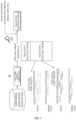
- FIG. 1 is a diagram schematically illustrating a conventional structural variant identification method.
- a tool 10 for searching for structural variants (SVs) in a pair of tumor and normal whole-genome sequences DELLY, BRASS, SvABA, dRanger, and the like exist.
- the output structural variant candidates include significant false positives (FP) as well as true positives (TP).
- a point mutation or a small insertion and deletion is a variant that exists in a read, which is a basic unit of sequencing data, since the exact position can be specified, a read with variants and a reference read can be examined at that position.
- the variant-supporting reads do not exist only at the breakpoint, and thus the exact position is often not specified. Therefore, a position searched by a known structural variant search tool is often inaccurate.
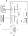
- FIG. 2 is a block diagram of a genomic structural variant identification device according to an embodiment
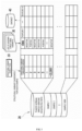
- FIG. 3 is a diagram for describing a data structure according to an embodiment.
- a genomic structural variant identification device 100 (referred to simply as “device") is a computing device operated by at least one processor, and includes a feature extractor 110 for extracting the features of structural variant candidates output by a structural variant search tool 10, an annotator 130 for labeling positive (True)/negative (False) for each structural variant candidate, a trainer 150 for training a machine learning model 200 using a dataset labeled with the features.
- the machine learning model 200 may be implemented as various models such as a logistic regression model, a random forest model, a stochastic gradient boosting model, and the like.
- the device 100 includes at least one processor, and the processor executes instructions included in a computer program, thereby performing the operations of the present disclosure.
- the computer program includes instructions that are described for a processor to execute the operations of the present disclosure, and may be stored in a non-transitory computer readable storage medium.
- the feature extractor 110, the annotator 130, the trainer 150, and the machine learning model 200 may be implemented as a computer program stored in a computer readable storage medium, and the device 100 may execute instructions included in the computer program, thereby operating the feature extractor 110, the annotator 130, the trainer 150, and the machine learning model 200.
- the trained machine learning model 200, the feature extractor 110, the annotator 130, the trainer 150, and the filtering unit 170 described in FIG. 6 are manufactured as a computer program and downloaded through a network, or may be sold in the form of a product, and may be installed in computing devices of various sites such as research institutes and hospitals.
- the structural variant search tool 10 is a tool for outputting structural variant candidates by calling the putative structural variants in whole-genome sequencing, and outputs the called structural variant candidates to the device 100.
- the structural variant search tool 10 uses a known tool.
- the tumor-normal whole-genome sequencing data 20 includes a pair of tumor genome data and normal tissue genome data extracted from the same person, and can be produced by short-read sequencing platforms such as Illumina.
- the tumor-normal whole-genome sequencing data 20 may include various types of cancer samples.
- the tumor-normal whole-genome sequencing data 20 constructed in the present disclosure includes a total of 1,808 samples, and may include training samples and validation samples.
- the training samples used in this disclosure included a total of 1,212 cancer genomes corresponding to 35 tumor types from 20 tumor tissues obtained from the international cancer genome consortium, and the validation samples include 499 cancer genomes from the same cohort and 97 cancer genomes from independent studies, and the composition of the samples can be variously adjusted.
- the feature extractor 110 receives structural variant candidates searched for in the whole-genome sequencing (WGS) data by the structural variant search tool 10, and examines a surrounding region of each structural variant candidate in a pair of tumor genome data and normal tissue genome data to extract the features of each structural variant candidate. Also, the feature extractor 110 may include some features extracted from clinical data. The feature extractor 110 stores the features of each structural variant candidate as a dataset 40 for machine learning.
- WGS whole-genome sequencing
- the feature extractor 110 may modify the positions of the structural variant candidates called by the structural variant search tool 10 to the correct coordinates using a supplementary alignment tag (SA tag) since they may be incorrect, and merge candidates within 500 base pairs with each other in the same strand direction (e.g., 5' to 3').
- SA tag supplementary alignment tag
- the supplementary alignment tag may be one of data output as a result of the alignment tool mapping each read to a reference sequence.
- the supplementary alignment tag of a specific read may be information indicating another position on the reference sequence, to which the corresponding read can be mapped, in addition to the main position, to which the corresponding read is mapped on the reference sequence. For example, such as read 315 in FIG. 4b , if a particular read is a split read located at breakpoints (e.g., points 311 and 312 in FIG. 4b ), the corresponding read may be mapped to two different positions or regions that are not continuous with each other on the reference sequence.
- the alignment tool may determine that the read maps to the first region on a reference sequence, but record information about the second region, to which the remaining portion of the read (e.g., reference numeral 316 in FIG. 4b ) maps, as the SA tag and output it.
- the feature extractor 110 may additionally use the supplementary alignment tag to more accurately determine whether any one read is a split read located at a breakpoint of the structural variant candidate, and through this, determine the position of the breakpoint of the structural variant candidate in a single base-pair resolution, thereby optimizing the region range, from which to extract features.
- each structural variant candidate may be displayed as a junction between two breakpoints (breakpoint1 and breakpoint2) in the genome data.
- breakpoint1 and breakpoint2 are aligned according to chromosome and position.
- the feature extractor 110 extracts the features of the tumor genome data different from the normal tissue genome data by examining the breakpoints (breakpoint1 and breakpoint2) of each structural variant candidate in the tumor genome data and the normal tissue genome data.
- Structural variants have features, in which a sufficient number of variant-supporting reads and many split reads by breakpoints are included.
- short deletions or short inversions which are also found in normal tissue genomes, cause errors and are searched as a false-positive structural variant.
- false-positive structural variants are found in the noise region characterized by high read depth, low mapping quality, and the like.
- the feature extractor 110 extracts the number of variant-supporting reads (Features 13 and 14 in Table 1), the number of split reads (Features 16 and 17 in Table 1), the number of split reads with supplementary alignment tag (Feature 18 in Table 1), mapping quality (Features 22 and 23 in Table 1), read depth change (Features 24 and 25 in Table 1), the number of background noise reads (Features 33, 34, 37, 38 in Table 1), and the number of samples, in which the same variant is detected among the panel of normal tissue genome data (Feature 45 in Table 1), and the like.
- the feature extractor 110 may store 45 features defined as shown in Table 1, and extract a value indicated by each feature using user-defined functions set to find the features. Among the 45 features, tumor histology, whole-genome duplication status, tumor purity, and tumor ploidy of the tumor genome may be extracted from clinical data of the sample. For the remaining 41 features, the feature extractor 110 examines the surrounding regions of each structural variant candidate in the whole-genome sequencing data, and extracts a value corresponding to each feature (binary value, number of counts, p-value, frequency, quality, ratio, distance, etc.). [Table 1] No.
- tumor histology tumor tissue (20 categories): Biliary, Bladder, Bone_SoftTissue, Breast, Cervix, CNS, Colon_Rectum, Esophagus, Head_Neck, Hematologic, Kideny, Liver, Lung, Ovary, Pancreas, Prostate, Skin, Stomach, Thyroid, Uterus 2 whole-genome duplication status feature indicating whether tumor whole-genome was duplciated (binary value): TRUE, FALSE 3 structural variant type structural variant type (4 categories): deletion, duplication, inversion, and translocation 4 positional variation at breakpoint 1 whether or not the mapping position of the variant-supporting reads at the breakpoint1 of the tumor genome data is variable (binary value): TRUE, FALSE 5 positional variation at breakpoint2 whether or not the mapping position of the variant-supporting reads at the breakpoint2 of the tumor genome data is variable (binary value): TRUE, FALSE 6 microhomology whether presence or absence of microhomolog
- tumor histology whole-genome duplication status, structural variant type, positional variant at breakpoint1, positional variant at breakpoint2, and whether presence or absence of microhomologous DNA sequences at two breakpoints (microhomology) are categorical variables, and the number converted using one-hot encoding may be described.
- the remaining 39 features are continuous variables, and the number corresponding to each feature may be described.
- the number of samples, in which the same variant is detected among the panel of normal tissue genome data, which is Feature 45 of Table 1, is extracted from the panel of normal tissue genome data included in the training cohort. As the number of data increases, the number of objects to compare whether the structural variant is true or an artifact increases. Thus, the larger the data, the better the performance.
- the extracted features may not represent the structural variant well due to inaccuracy in the structural variant detection by a structural variant search tool.
- the feature extractor 110 uses the supplementary alignment tag to more accurately determine the split read located at the breakpoint of the structural variant candidate, and through this, to determine the position of the breakpoint of the structural variant candidates in a single base-pair resolution, thereby optimizing the range of candidate regions from which to extract features.
- the feature extractor 110 may detect a noise region, in which noise is greater than or equal to a reference level in the genome data, and omit feature extraction of the detected noise region. Through this, it is possible to increase the feature extraction speed of structural variant candidates.
- the feature extractor 110 may examine the surrounding area of each structural variant candidate through user-defined functions set to extract the features of Table 1.
- the search range for the surrounding region of the breakpoint may be set differently depending on the type of structural variant or the extracted features.
- the mapping position of the variant-supporting reads in the surrounding region of the breakpoint is variable (Features 4 and 5 in Table 1), whether presence or absence of a microhomologous DNA sequences at the two breakpoints (Feature 6 in Table 1), the number of reference reads at the two breakpoints (Features 9-12 in Table 1), the number of variant-supporting reads at the two breakpoints (Features 13 and 14 in Table 1), the number of split reads at the two breakpoints (Features 16 and 17 in Table 1), and the number of split reads with supplementary alignment tags at the two breakpoints (Features 18 in Table 1), the number of reads with the same clipped sequence as the split read in the normal tissue genome data at the two breakpoints (Feature 19 in Table 1), the median value of mapping quality of the variant-supporting reads at the breakpoint in the tumor genome data (Features 22
- the range to examine the surrounding region of the breakpoint may be set as follows.
- the breakpoint e.g., positions 311 and 312 in FIG. 4b
- the direction, in which structural variant-supporting reads are located e.g., the direction indicated as segment in FIG. 4b
- it can be set to examine up to the average insert size of the entire paired-end reads of the processing target whole-genome sequencing data or further. This is because the size of the structural variant is longer than one read, so there may be variant-supporting reads located in the segment direction of the two breakpoints without overlapping any other breakpoints.
- a predetermined range also in the opposite direction of the structural variant-supporting reads e.g., the direction marked as gap in FIG. 4b
- the breakpoint e.g., the positions 311 and 312 in FIG. 4b
- it may be set to examine, for example, 200 base pairs in the gap direction from the breakpoint. It should be noted that if a range wider than 200 base pairs is examined in the gap direction from the breakpoint, there is a possibility that data resulting from structural variants other than the target structural variant candidate near the breakpoint or other discordant reads are examined.
- the annotator 130 labels each structural variant candidate as positive (True)/negative (False) for the structural variant candidates searched for in the whole-genome sequencing data, and stores it as a dataset 40 for machine learning.
- the annotator 130 may refer to a list of known structural variants 30, and label it as positive if each structural variant candidate exists in the structural variant list 30, otherwise label it as negative.
- each structural variant candidate extracted by the feature extractor 110 and the classification classes of each structural variant candidate classified by the annotator 130 are stored in the dataset 40 as shown in FIG. 3 .
- the trainer 150 trains the machine learning model 200 using the dataset 40.
- the trainer 150 trains the machine learning model 200 to classify the corresponding structural variant candidate into a classification class from features of each structural variant candidate.
- the machine learning model 200 may be implemented as various models such as a logistic regression model, a random forest model, a stochastic gradient boosting model, and the like.
- the trainer 150 may use the validation samples included in the tumor-normal whole-genome sequencing data 20 to verify the performance of the machine learning model 200 and then complete the training.
- the trainer 150 may evaluate performance using a precision-recall curve.
- the trained machine learning model 200 may receive features of each structural variant candidate and output a probability value between 0 and 1.
- the probability value of each structural variant candidate is determined as positive or negative according to the set cutoff value.
- a structural variant candidate that is negative may be filtered out as false positive.
- FIGS. 4a and 4b are diagrams exemplarily describing a method of extracting structural variant information in a structural variant candidate region according to an exemplary embodiment.
- the feature extractor 110 extracts features defined for classifying false positives for each structural variant candidate called by the structural variant search tool 10.
- reads of sequencing data are colored with their mate reads, and thus non-gray reads indicate discordant read pairs.
- the feature extractor 110 extracts values of variant-supporting reads in tumor genome data, split reads, mapping quality, variant-supporting reads in normal tissue genome data, read depth change, and background noise reads, as shown in Table 2, and in addition, may extract values of a plurality of features defined in Table 1. If desired, discordant read pairs can be calculated.
- Table 2 Extracted Feature True Positive Structural Variant False Positive Structural Variant-supporting reads 7 4 Split reads 4 0 Mapping quality 56 20 Variant-supporting reads in normal 0 1 Read depth change 1.8 0.9 Background noise reads 0 8
- the feature extractor 110 stores data filled with features of each structural variant candidate as a dataset 40 for machine learning.
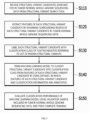
- FIG. 5 is a flowchart of a training method for identifying structural variants in genomes according to an exemplary embodiment.
- the device 100 receives structural variant candidates searched for in tumor-normal whole-genome sequencing data 20 from the structural variant search tool 10 (S 110).
- the device 100 extracts the features of each structural variant candidate by examining a surrounding region of each structural variant candidate in the tumor-normal whole-genome sequencing data 20 (S120).
- the device 100 extracts a value corresponding to each feature by examining the vicinity of two breakpoints (breakpoint1 and breakpoint2) for each structural variant candidate.
- the number of variant-supporting reads, the number of split reads with supplementary alignment tag, the number of split reads, mapping quality, read depth change, the number of background noise reads, and the number of samples, in which the same variant is detected among the panel of normal tissue genome data, etc. are extracted as the features.
- the device 100 may additionally receive tumor histology, whole-genome duplication status, tumor purity, and tumor ploidy of the tumor genome from the clinical data of the sample.
- the device 100 labels each structural variant candidate with a classification class of positive (True)/negative (False) with reference to the list of known structural variants 30 (S130).
- the device 100 may label it as positive if each structural variant candidate has the same direction as the structural variant in the structural variant list 30 and exits within a distance of 500 base pairs, otherwise may label it as negative.
- the device 100 trains the machine learning model 200 to classify the structural variant candidate into a classification class from the features of each structural variant candidate using a dataset, in which classification classes are mapped to the features of each structural variant candidate (S140).
- the machine learning model 200 may receive features of each structural variant candidate and output a classification probability of positive or negative (a probability value between 0 and 1).
- the trainer 150 may verify the classification performance of the machine learning model 200 by using the validation samples included in the tumor-normal whole-genome sequencing data 20, and then complete the training (S 150). In this case, the trainer 150 may determine a cutoff value for classifying into the labeled positive or negative according to a probability value between 0 and 1 output from the machine learning model 200 based on the classification performance evaluation.
- FIG. 6 is a diagram schematically describing structural variant identification using a trained machine learning model according to an embodiment
- FIG. 7 is a flowchart of a structural variant identification method using the trained machine learning model according to an embodiment.
- the device 100 may use the feature extractor 110 and the trained machine learning model 200, and additionally use a filtering unit 170.
- the feature extractor 110, the filtering unit 170, and the trained machine learning model 200 may be implemented as a computer program, and may be driven in any computing device, in which they are installed.
- the annotator 130 and the trainer 150 of FIG. 2 may also be implemented as computer programs, and may be driven in a computing device, in which they are installed. In this case, the users can re-train the trained machine learning model 200 with the genome data of their cohort.
- the feature extractor 110 receives structural variant candidates searched for in the whole-genome sequencing data 50 from the structural variant search tool 10.
- the feature extractor 110 extracts the features of each structural variant candidate used for training the machine learning model 200 by examining the surrounding region of each structural variant candidate.
- the feature extractor 110 inputs the features of each structural variant candidate into the trained machine learning model 200.
- the trained machine learning model 200 outputs a classification probability value of positive or negative from the input features.
- the filtering unit 170 may classify the classification probability value of each structural variant candidate output from the trained machine learning model 200 as negative or positive based on the cutoff value to identify the true structural variant.
- the filtering unit 170 may filter and remove structural variant candidates classified as negative among the structural variant candidates, and generate a list of true structural variants including True Positive Structural Variants (TP SVs) classified as positive.
- TP SVs True Positive Structural Variants
- the feature extractor 110 receives structural variant candidates searched for in the whole-genome sequencing data 50 from the structural variant search tool 10 (S210).
- the feature extractor 110 extracts the features of each structural variant candidate by examining the surrounding region of each structural variant candidate (S220).
- the feature extractor 110 may examine the surrounding region of each structural variant candidate through user-defined functions set to extract the features of Table 1. In this case, the range to be examined in the vicinity of the breakpoint may be set differently according to the type of a structural variant or the extracted feature.
- the feature extractor 110 inputs the features of each structural variant candidate into the trained machine learning model 200 (S230).
- the trained machine learning model 200 outputs a classification probability value of positive or negative from the input features (S240).
- the filtering unit 170 determines the classification probability value of each structural variant candidate output from the trained machine learning model 200 as negative or positive based on the cutoff value, and identifies it as a negative structural variant or a positive structural variant (S250).
- the filtering unit 170 removes candidates identified as negative structural variants from among the structural variant candidates searched for in the whole-genome sequencing data 50, and generates a list of true structural variants (S260). Since the structural variant candidates searched by the structural variant search tool 10 contain false positives, the filtering unit 170 determines that structural variant candidates classified as negative by the machine learning model 200 among the structural variant candidates as false positives and removes them.
- various structural variants in the genome can be accurately detected through the machine learning model, and an accurate structural variant list can be generated by removing false positives, thereby contributing to precision oncology.
- the time required for experts setting the optimal filtering conditions through heuristic trial and error through manual inspection, and filtering false positives can be significantly reduced.
- scalability can be increased through standardized structural variant search using a machine learning model, and manual curation by experts is unnecessary, thereby reducing cost and time.
- it can be applied to various cohorts through simple re-training of the machine learning model, and an optimal cutoff can be found based on classification performance.
- FIG. 8 is a hardware configuration diagram of a computing device according to an embodiment.
- the device 100 may be implemented as a computing device 400 operated by at least one processor.
- each of the feature extractor 110, the annotator 130, the trainer 150, the filtering unit 170, and the machine learning model 200 may be mounted on the computing device 400 operated by at least one processor.
- the computing device 400 includes one or more processors 410, a memory 430 for loading a computer program executed by the processor 410, a storage device 450 for storing computer programs and various data, a communication interface 470, and a bus 490 connecting them.
- the computing device 400 may further include various components.
- the processor 410 is a device for controlling the operation of the computing device 400, and may be various types of processors that process instructions included in a computer program, and may include, for example, a central processing unit (CPU), a microprocessor unit (MPU), a micro controller unit (MCU), a graphic processing unit (GPU), or any type of processor well known in the art of the present disclosure.
- CPU central processing unit
- MPU microprocessor unit
- MCU micro controller unit
- GPU graphic processing unit
- the memory 430 stores various data, instructions and/or information.
- the memory 430 may load a corresponding computer program from the storage device 450 so that the instructions described to execute the operations of the present disclosure are processed by the processor 410.
- the memory 430 may be, for example, read only memory (ROM), random access memory (RAM), or the like.
- the storage device 450 may non-temporarily store computer programs and various data.
- the storage device 450 may include a non-volatile memory, such as a read only memory (ROM), an erasable programmable ROM (EPROM), an electrically erasable programmable ROM (EEPROM), a flash memory, a hard disk, a removable disk, or any well-known computer-readable recording medium in the art, to which the present disclosure pertains.
- ROM read only memory
- EPROM erasable programmable ROM
- EEPROM electrically erasable programmable ROM
- the communication interface 470 may be a wired/wireless communication module supporting wired/wireless communication.
- the bus 490 provides a communication function between the components of the computing device 400.
- the computer program includes instructions executed by the processor 410, and is stored in a non-transitory computer readable storage medium, wherein the instructions have the processor 410 execute the operation of the present disclosure.
- the computer program may be downloaded over a network or sold as a product.
- the computer program for training may comprise instructions described to execute the step of receiving, from the structural variant search tool, structural variant candidates searched for in whole-genome sequencing data including a pair of tumor genome data and normal tissue genome data, the step of extracting features of each structural variant candidate by examining the surrounding regions of each structural variant candidate in the whole-genome sequencing data, the step of labeling each structural variant candidate with a classification class of positive or negative with reference to a list of known structural variant candidates, and the step of training the machine learning model to classify the corresponding structural variant candidate into a classification class from the features of each structural variant candidate using the dataset, in which the classification classes are mapped to the features of each structural variant candidate.
- the computer program for training may further comprise instructions described to execute the step of using the validation samples included in the whole-genome sequencing data to verify the classification performance of the machine learning model, and then completing the training, and the step of determining a cutoff value for classifying the probability value output from the machine learning model as positive or negative based on the evaluation of the classification performance of the machine learning model.
- the computer program for training may store features extracted from each structural variant candidate.
- the features may include the number of variant-supporting reads, the number of split reads with supplementary alignment tag, the number of split reads, mapping quality, read depth change, the number of background noise reads, and the number of samples, in which the same variant is detected among the panel of normal tissue genome data.
- the features may further include tumor histology, whole-genome duplication status, tumor purity, and tumor ploidy of the tumor genome extracted from clinical data.
- the computer program for training may include a machine learning model that receives features of each structural variant candidate and outputs a classification probability value of positive or negative.
- the computer program distributed for structural variant identification may include a first computer program trained to classify a corresponding structural variant candidate into a classification class from features of each structural variant candidate, and a second computer program.
- the second computer program may include instructions described to receive structural variant candidates searched for in the whole-genome sequencing data to be identified, examine the surrounding area of each structural variant candidate in the whole-genome sequencing data to extract the features of each structural variant candidate, then input the features of each structural variant candidate into the machine learning model to obtain a classification probability value of positive or negative for the structural variant candidate, and determine the classification probability value as negative or positive based on the set cutoff value to identify each structural variant candidate as a negative or positive structural variant.
- the first computer program may be a machine learning model implemented as at least one of a logistic regression model, a random forest model, and a stochastic gradient boosting model.
- the second computer program may further include instructions for removing the structural variant candidates identified as negative structural variant candidates from among the structural variant candidates searched by the structural variant search tool to generate a list of true structural variants.
- the second computer program stores features extracted from each structural variant candidate, wherein the features may include the number of variant-supporting reads, the number of split reads with supplementary alignment tag, the number of split reads, mapping quality, read depth change, the number of background noise reads, and the number of samples, in which the same variant is detected among the panel of normal tissue genome data.
- the second computer program may further include instructions for additionally extracting features of each structural variant candidate from clinical data, and the additionally extracted features may include tumor histology, whole-genome duplication status, tumor purity in the sample, and tumor ploidy of the tumor genome.
- the embodiment of the present invention described above is not implemented only through the apparatus and method, but may be implemented through a program for realizing a function corresponding to the configuration of the embodiment of the present invention or a recording medium, in which the program is recorded.
Landscapes
- Engineering & Computer Science (AREA)
- Life Sciences & Earth Sciences (AREA)
- Health & Medical Sciences (AREA)
- Physics & Mathematics (AREA)
- Bioinformatics & Cheminformatics (AREA)
- Medical Informatics (AREA)
- Chemical & Material Sciences (AREA)
- Theoretical Computer Science (AREA)
- General Health & Medical Sciences (AREA)
- Biophysics (AREA)
- Biotechnology (AREA)
- Proteomics, Peptides & Aminoacids (AREA)
- Bioinformatics & Computational Biology (AREA)
- Evolutionary Biology (AREA)
- Spectroscopy & Molecular Physics (AREA)
- Analytical Chemistry (AREA)
- Data Mining & Analysis (AREA)
- Organic Chemistry (AREA)
- Molecular Biology (AREA)
- Genetics & Genomics (AREA)
- Software Systems (AREA)
- Computer Vision & Pattern Recognition (AREA)
- Evolutionary Computation (AREA)
- Bioethics (AREA)
- Databases & Information Systems (AREA)
- Artificial Intelligence (AREA)
- Zoology (AREA)
- Wood Science & Technology (AREA)
- Public Health (AREA)
- Epidemiology (AREA)
- General Engineering & Computer Science (AREA)
- Immunology (AREA)
- Microbiology (AREA)
- Biochemistry (AREA)
- Pathology (AREA)
- Computing Systems (AREA)
- General Physics & Mathematics (AREA)
- Mathematical Physics (AREA)
- Physiology (AREA)
- Measuring Or Testing Involving Enzymes Or Micro-Organisms (AREA)
Claims (11)
- Verfahren, das von einer Rechenvorrichtung zur Identifizierung von Strukturvarianten in Genomen durchgeführt wird, Folgendes umfassend:Erhalten von strukturellen Variantenkandidaten, die anhand von Ganzgenom-Sequenzierungsdaten identifiziert wurden, einschließlich eines Paars von Tumor-Genomdaten und Normalgewebe-Genomdaten;Modifizieren einer geschätzten Position eines ersten Strukturvariantenkandidaten auf der Grundlage von ergänzenden Ausrichtungs-(SA)-Tag-Informationen, die mit dem ersten Strukturvariantenkandidaten unter den Strukturvariantenkandidaten verbunden sind,Extrahieren von Merkmalen jedes Strukturvariantenkandidaten, einschließlich des ersten Strukturvariantenkandidaten, auf der Grundlage von Daten, die innerhalb eines vorbestimmten Bereichs von jedem Strukturvariantenkandidaten, einschließlich des ersten Strukturvariantenkandidaten, in den Ganzgenom-Sequenzierungsdaten liegen;Kennzeichnen jedes Strukturvariantenkandidaten einschließlich des ersten Strukturvariantenkandidaten mit Klassifizierungsinformationen auf der Grundlage einer Liste bekannter Strukturvarianten,wobei jeder Strukturvariantenkandidat, einschließlich des ersten Strukturvariantenkandidaten, als positiv gekennzeichnet ist, wenn der Strukturvariantenkandidat in der Liste der bekannten Strukturvarianten existiert, oder anderenfalls der Strukturvariantenkandidat als negativ gekennzeichnet ist; undTrainieren eines Machine-Learning-Modells unter Verwendung eines Datensatzes, in dem die extrahierten Merkmale jedes Strukturvariantenkandidaten, einschließlich des ersten Strukturvariantenkandidaten, und die markierten Klassifizierungsinformationen gespeichert sind,wobei das Machine-Learning-Modell einen Identifikationsziel-Strukturvariantenkandidaten empfängt und eine Klassifizierung des Identifikationsziel-Strukturvariantenkandidaten ausgibt,wobei das Machine-Learning-Modell Merkmale des Identifikationsziel-Strukturvariantenkandidaten empfängt und einen Wahrscheinlichkeitswert zur Klassifizierung des Identifikationsziel-Strukturvariantenkandidaten als positiv oder negativ ausgibt;wobei das Extrahieren von Merkmalen jedes Strukturvariantenkandidaten einschließlich des ersten Strukturvariantenkandidaten Folgendes umfasst:Erhalten von Daten einer ersten Länge oder mehr in einer Richtung, in der sich variantenunterstützende Reads des Strukturvariantenkandidaten befinden, von einem Bruchpunkt, der mit dem Strukturvariantenkandidaten unter den Strukturvariantenkandidaten in den Ganzgenom-Sequenzierungsdaten verbunden ist, undwobei die erste Länge eine durchschnittliche Insertgröße der Ganzgenom-Sequenzierungsdaten ist.
- Verfahren nach Anspruch 1, wobei das Modifizieren der geschätzten Position des ersten Strukturvariantenkandidaten das Identifizieren einer ersten Region und einer zweiten Region auf einer Referenzsequenz umfasst, auf die der erste Strukturvariantenkandidat abgebildet wird,
wobei der erste Bereich und der zweite Bereich Bereiche sind, die nicht aneinander angrenzen. - Verfahren nach Anspruch 1, wobei das Modifizieren der geschätzten Position des ersten Strukturvariantenkandidaten das Bestimmen einer Position eines Bruchpunktes umfasst, der mit dem ersten Strukturvariantenkandidaten verbunden ist.
- Verfahren nach Anspruch 1, wobei das Extrahieren von Merkmalen jedes Strukturvariantenkandidaten auf der Grundlage von Daten, die innerhalb eines vorbestimmten Bereichs von jedem Strukturvariantenkandidaten liegen, Folgendes umfasst:
Erhalten von Daten einer zweiten Länge oder weniger in einer Richtung entgegengesetzt zu variantenunterstützenden Reads eines ersten Strukturvariantenkandidaten von einem Bruchpunkt, der mit dem ersten Strukturvariantenkandidaten unter den Strukturvariantenkandidaten in den Ganzgenom-Sequenzierungsdaten assoziiert ist. - Verfahren nach Anspruch 4, wobei die zweite Länge 200 Basispaare beträgt.
- Verfahren nach Anspruch 1, wobei das Extrahieren der Merkmale jedes Strukturvariantenkandidaten Folgendes umfasst:
Extrahieren, für jeden strukturellen Variantenkandidaten, einer Anzahl von variantenunterstützenden Reads, einer Anzahl von Split-Reads mit zusätzlichem Alignment-Tag, einer Anzahl von Split-Reads, Mapping-Qualität, Änderung der Lesetiefe, einer Anzahl von Hintergrundrauschen-Reads und einer Anzahl von Proben, in denen dieselbe Variante unter normalen Gewebegenomdaten entdeckt wird. - Verfahren nach Anspruch 1, wobei das Extrahieren der Merkmale jedes Strukturvariantenkandidaten das Extrahieren der Tumorhistologie, des Ganzgenom-Duplikationsstatus, der Tumorreinheit in einer Probe und der Tumorploidie eines Tumorgenoms aus klinischen Daten für jeden Strukturvariantenkandidaten umfasst.
- Verfahren nach Anspruch 1, das ferner Folgendes umfasst:Bewerten der Klassifizierungsleistung des Machine-Learning-Modells unter Verwendung der in den Ganzgenom-Sequenzierungsdaten enthaltenen Validierungsproben; undBestimmen eines Grenzwertes zum Bestimmen, ob ein von dem Machine-Learning-Modell ausgegebener Wahrscheinlichkeitswert positiv oder negativ ist, basierend auf dem Ergebnis der Klassifikationsleistungsbewertung des Machine-Learning-Modells.
- Verfahren nach Anspruch 1, wobei das Erhalten von Strukturvariantenkandidaten, die aus den Ganzgenom-Sequenzierungsdaten identifiziert wurden, Folgendes umfasst:Eingeben der Ganzgenom-Sequenzierungsdaten in ein Strukturvarianten-Suchwerkzeug; undErhalten von Strukturvariantenkandidaten, die von dem Strukturvarianten-Suchwerkzeug ausgegeben werden.
- Computerlesbares nichtflüchtiges Speichermedium, das Anweisungen enthält,
wobei die Anweisungen, wenn sie von einem oder mehreren Prozessoren einer Rechenvorrichtung ausgeführt werden, die Rechenvorrichtung veranlassen, Operationen durchzuführen, die Folgendes umfassen:Erhalten von strukturellen Variantenkandidaten, die anhand von Ganzgenom-Sequenzierungsdaten identifiziert wurden, einschließlich eines Paars von Tumor-Genomdaten und Normalgewebe-Genomdaten;Modifizieren einer geschätzten Position eines ersten Strukturvariantenkandidaten auf der Grundlage von ergänzenden Ausrichtungs-(SA)-Tag-Informationen, die mit dem ersten Strukturvariantenkandidaten unter den Strukturvariantenkandidaten verbunden sind, bevor Merkmale jedes Strukturvariantenkandidaten extrahiert werden, Extrahieren von Merkmalen jedes Strukturvariantenkandidaten, einschließlich des ersten Strukturvariantenkandidaten, auf der Grundlage von Daten, die innerhalb eines vorbestimmten Bereichs von jedem Strukturvariantenkandidaten, einschließlich des ersten Strukturvariantenkandidaten, in den Ganzgenom-Sequenzierungsdaten liegen;Kennzeichnen jedes Strukturvariantenkandidaten einschließlich des ersten Strukturvariantenkandidaten mit Klassifizierungsinformationen auf der Grundlage einer Liste bekannter Strukturvarianten,wobei jeder Strukturvariantenkandidat als positiv gekennzeichnet ist, wenn der Strukturvariantenkandidat in der Liste der bekannten Strukturvarianten existiert, oder anderenfalls als negativ gekennzeichnet ist; undTrainieren eines Machine-Learning-Modells unter Verwendung eines Datensatzes, in dem die extrahierten Merkmale jedes Strukturvariantenkandidaten, einschließlich des ersten Strukturvariantenkandidaten, und die markierten Klassifizierungsinformationen gespeichert sind,wobei das Machine-Learning-Modell einen Identifikationsziel-Strukturvariantenkandidaten empfängt und eine Klassifizierung des Identifikationsziel-Strukturvariantenkandidaten ausgibt,wobei das Machine-Learning-Modell Merkmale des Identifikationsziel-Strukturvariantenkandidaten empfängt und einen Wahrscheinlichkeitswert zur Klassifizierung des Identifikationsziel-Strukturvariantenkandidaten als positiv oder negativ ausgibt;wobei das Extrahieren von Merkmalen jedes Strukturvariantenkandidaten einschließlich des ersten Strukturvariantenkandidaten Folgendes umfasst:Erhalten von Daten einer ersten Länge oder mehr in einer Richtung, in der sich variantenunterstützende Reads des Strukturvariantenkandidaten befinden, von einem Bruchpunkt, der mit dem Strukturvariantenkandidaten unter den Strukturvariantenkandidaten in den Ganzgenom-Sequenzierungsdaten verbunden ist, undwobei die erste Länge eine durchschnittliche Insertgröße der Ganzgenom-Sequenzierungsdaten ist. - Rechenvorrichtung, die Folgendes umfasst:einen Prozessor; undeinen Speicher zum Speichern von Anweisungen,wobei die Anweisungen, wenn sie von dem Prozessor ausgeführt werden, die Rechenvorrichtung veranlassen, Operationen durchzuführen, die Folgendes umfassen:Erhalten von strukturellen Variantenkandidaten, die anhand von Ganzgenom-Sequenzierungsdaten identifiziert wurden, einschließlich eines Paars von Tumor-Genomdaten und Normalgewebe-Genomdaten;Modifizieren einer geschätzten Position eines ersten Strukturvariantenkandidaten auf der Grundlage von ergänzenden Ausrichtungs-(SA)-Tag-Informationen, die mit dem ersten Strukturvariantenkandidaten unter den Strukturvariantenkandidaten verbunden sind, bevor Merkmale jedes Strukturvariantenkandidaten extrahiert werden,Extrahieren von Merkmalen jedes Strukturvariantenkandidaten, einschließlich des ersten Strukturvariantenkandidaten, auf der Grundlage von Daten, die innerhalb eines vorbestimmten Bereichs von jedem Strukturvariantenkandidaten, einschließlich des ersten Strukturvariantenkandidaten, in den Ganzgenom-Sequenzierungsdaten liegen;Kennzeichnen jedes Strukturvariantenkandidaten einschließlich des ersten Strukturvariantenkandidaten mit Klassifizierungsinformationen auf der Grundlage einer Liste bekannter Strukturvarianten,wobei jeder Strukturvariantenkandidat als positiv gekennzeichnet ist, wenn der Strukturvariantenkandidat in der Liste der bekannten Strukturvarianten existiert, oder anderenfalls als negativ gekennzeichnet ist; undTrainieren eines Machine-Learning-Modells unter Verwendung eines Datensatzes, in dem die extrahierten Merkmale jedes Strukturvariantenkandidaten, einschließlich des ersten Strukturvariantenkandidaten, und die markierten Klassifizierungsinformationen gespeichert sind,wobei das Machine-Learning-Modell einen Identifikationsziel-Strukturvariantenkandidaten empfängt und eine Klassifizierung des Identifikationsziel-Strukturvariantenkandidaten ausgibt,wobei das Machine-Learning-Modell Merkmale des Identifikationsziel-Strukturvariantenkandidaten empfängt und einen Wahrscheinlichkeitswert zur Klassifizierung des Identifikationsziel-Strukturvariantenkandidaten als positiv oder negativ ausgibt;wobei das Extrahieren von Merkmalen jedes Strukturvariantenkandidaten einschließlich des ersten Strukturvariantenkandidaten Folgendes umfasst:Erhalten von Daten einer ersten Länge oder mehr in einer Richtung, in der sich variantenunterstützende Reads des Strukturvariantenkandidaten befinden, von einem Bruchpunkt, der mit dem Strukturvariantenkandidaten unter den Strukturvariantenkandidaten in den Ganzgenom-Sequenzierungsdaten verbunden ist, undwobei die erste Länge eine durchschnittliche Insertgröße der Ganzgenom-Sequenzierungsdaten ist.
Priority Applications (1)
| Application Number | Priority Date | Filing Date | Title |
|---|---|---|---|
| EP23203162.5A EP4287190A3 (de) | 2020-09-17 | 2021-09-17 | Verfahren und gerät für die auf maschinellem lernen basierende identifizierung von strukturvarianten in krebsgenomen |
Applications Claiming Priority (3)
| Application Number | Priority Date | Filing Date | Title |
|---|---|---|---|
| KR20200119798 | 2020-09-17 | ||
| KR20210054190 | 2021-04-27 | ||
| KR1020210123291A KR102404947B1 (ko) | 2020-09-17 | 2021-09-15 | 기계학습 기반의 유전체 구조 변이 식별 방법 및 장치 |
Related Child Applications (2)
| Application Number | Title | Priority Date | Filing Date |
|---|---|---|---|
| EP23203162.5A Division-Into EP4287190A3 (de) | 2020-09-17 | 2021-09-17 | Verfahren und gerät für die auf maschinellem lernen basierende identifizierung von strukturvarianten in krebsgenomen |
| EP23203162.5A Division EP4287190A3 (de) | 2020-09-17 | 2021-09-17 | Verfahren und gerät für die auf maschinellem lernen basierende identifizierung von strukturvarianten in krebsgenomen |
Publications (3)
| Publication Number | Publication Date |
|---|---|
| EP4016533A1 EP4016533A1 (de) | 2022-06-22 |
| EP4016533B1 true EP4016533B1 (de) | 2023-12-20 |
| EP4016533C0 EP4016533C0 (de) | 2023-12-20 |
Family
ID=78293845
Family Applications (2)
| Application Number | Title | Priority Date | Filing Date |
|---|---|---|---|
| EP21197318.5A Active EP4016533B1 (de) | 2020-09-17 | 2021-09-17 | Verfahren und gerät für die auf maschinellem lernen basierende identifizierung von strukturvarianten in krebsgenomen |
| EP23203162.5A Pending EP4287190A3 (de) | 2020-09-17 | 2021-09-17 | Verfahren und gerät für die auf maschinellem lernen basierende identifizierung von strukturvarianten in krebsgenomen |
Family Applications After (1)
| Application Number | Title | Priority Date | Filing Date |
|---|---|---|---|
| EP23203162.5A Pending EP4287190A3 (de) | 2020-09-17 | 2021-09-17 | Verfahren und gerät für die auf maschinellem lernen basierende identifizierung von strukturvarianten in krebsgenomen |
Country Status (3)
| Country | Link |
|---|---|
| US (1) | US20220084631A1 (de) |
| EP (2) | EP4016533B1 (de) |
| KR (1) | KR102812123B1 (de) |
Families Citing this family (5)
| Publication number | Priority date | Publication date | Assignee | Title |
|---|---|---|---|---|
| JP6922793B2 (ja) * | 2018-03-12 | 2021-08-18 | オムロン株式会社 | 制御装置、制御方法、および制御プログラム |
| US20230207054A1 (en) * | 2021-12-29 | 2023-06-29 | Illumina, Inc. | Deep learning network for evolutionary conservation |
| KR20240105981A (ko) * | 2022-12-29 | 2024-07-08 | 씨제이올리브네트웍스 주식회사 | 인공지능에 기반한 미생물의 종을 예측하는 방법 및 장치 |
| CN116364178B (zh) * | 2023-04-18 | 2024-01-30 | 哈尔滨星云生物信息技术开发有限公司 | 一种体细胞序列数据分类方法及相关设备 |
| CN117789820B (zh) * | 2023-11-30 | 2025-12-26 | 北京基石生命科技有限公司 | 三代全外显子测序转录本数据中结构变异检测方法和系统 |
Family Cites Families (3)
| Publication number | Priority date | Publication date | Assignee | Title |
|---|---|---|---|---|
| KR102233740B1 (ko) | 2017-09-27 | 2021-03-30 | 이화여자대학교 산학협력단 | Dna 복제수 변이 기반의 암 종 예측 방법 |
| US11972841B2 (en) * | 2017-12-18 | 2024-04-30 | Personal Genome Diagnostics Inc. | Machine learning system and method for somatic mutation discovery |
| JP7166434B2 (ja) * | 2018-08-13 | 2022-11-07 | エフ.ホフマン-ラ ロシュ アーゲー | 生殖細胞系列および体細胞変異の呼び出しのためにニューラルネットワークを使用するシステムおよび方法 |
-
2021
- 2021-09-16 US US17/476,875 patent/US20220084631A1/en not_active Abandoned
- 2021-09-17 EP EP21197318.5A patent/EP4016533B1/de active Active
- 2021-09-17 EP EP23203162.5A patent/EP4287190A3/de active Pending
-
2022
- 2022-05-30 KR KR1020220065793A patent/KR102812123B1/ko active Active
Non-Patent Citations (1)
| Title |
|---|
| ZEV N. KRONENBERG ET AL: "Wham: Identifying Structural Variants of Biological Consequence", PLOS COMPUTATIONAL BIOLOGY, vol. 7, no. 12, 1 December 2015 (2015-12-01), pages e1002216, XP055334268, DOI: 10.1371/journal.pcbi.1004572 * |
Also Published As
| Publication number | Publication date |
|---|---|
| KR102812123B1 (ko) | 2025-05-26 |
| EP4287190A3 (de) | 2024-03-06 |
| EP4287190A2 (de) | 2023-12-06 |
| KR20220076444A (ko) | 2022-06-08 |
| EP4016533C0 (de) | 2023-12-20 |
| US20220084631A1 (en) | 2022-03-17 |
| EP4016533A1 (de) | 2022-06-22 |
Similar Documents
| Publication | Publication Date | Title |
|---|---|---|
| EP4016533B1 (de) | Verfahren und gerät für die auf maschinellem lernen basierende identifizierung von strukturvarianten in krebsgenomen | |
| KR102404947B1 (ko) | 기계학습 기반의 유전체 구조 변이 식별 방법 및 장치 | |
| JP7607264B2 (ja) | 無細胞dna末端特性 | |
| CN109767810B (zh) | 高通量测序数据分析方法及装置 | |
| US11804285B2 (en) | Hilbert-cnn: ai-driven convolutional neural networks with conversion data of genome for biomarker discovery | |
| CN106909806B (zh) | 定点检测变异的方法和装置 | |
| US20210265011A1 (en) | Systems and methods for analyzing sequence data | |
| JP7684287B2 (ja) | 単一細胞rna-seqデータ処理 | |
| CN109243530B (zh) | 遗传变异判定方法、系统以及存储介质 | |
| KR101542529B1 (ko) | 대립유전자의 바이오마커 발굴방법 | |
| CN112669903A (zh) | 基于Sanger测序的HLA分型方法及设备 | |
| US12272431B2 (en) | Detecting false positive variant calls in next-generation sequencing | |
| CN111180013B (zh) | 检测血液病融合基因的装置 | |
| CN119832980B (zh) | 基因变异检测方法、装置、电子设备及存储介质 | |
| CN120164524B (zh) | 一种遗传病基因检测的数据分析方法、系统及存储介质 | |
| CN112863602B (zh) | 染色体异常的检测方法、装置、计算机设备和存储介质 | |
| CN110164504A (zh) | 二代测序数据的处理方法、装置及电子设备 | |
| CN119359841A (zh) | 一种通过组合图形直观展示生物个体间遗传差异及组合图形生成方法 | |
| CN117877575A (zh) | 区分胚系变异和体细胞变异的方法和装置 | |
| EP4502133A1 (de) | Informationsverarbeitungsvorrichtung, informationsverarbeitungsverfahren und informationsverarbeitungsprogramm | |
| CN116543907A (zh) | 身体质量指数的预测方法、模型训练方法及设备 | |
| CN113990392A (zh) | 短串联重复序列的致病性分析方法、装置及服务器 | |
| CN114171118A (zh) | 用于无创基因检测的数据处理方法和装置 | |
| CN119152934B (zh) | 基于低投入起始量的高通量基因组测序变异检测系统和方法 | |
| Gunturkun et al. | SVJAM: Joint Analysis of Structural Variants Using Linked Read Sequencing Data |
Legal Events
| Date | Code | Title | Description |
|---|---|---|---|
| PUAI | Public reference made under article 153(3) epc to a published international application that has entered the european phase |
Free format text: ORIGINAL CODE: 0009012 |
|
| STAA | Information on the status of an ep patent application or granted ep patent |
Free format text: STATUS: REQUEST FOR EXAMINATION WAS MADE |
|
| 17P | Request for examination filed |
Effective date: 20210917 |
|
| AK | Designated contracting states |
Kind code of ref document: A1 Designated state(s): AL AT BE BG CH CY CZ DE DK EE ES FI FR GB GR HR HU IE IS IT LI LT LU LV MC MK MT NL NO PL PT RO RS SE SI SK SM TR |
|
| RAP1 | Party data changed (applicant data changed or rights of an application transferred) |
Owner name: GENOME INSIGHT INC. |
|
| RAP3 | Party data changed (applicant data changed or rights of an application transferred) |
Owner name: GENOME INSIGHT TECHNOLOGY, INC. |
|
| STAA | Information on the status of an ep patent application or granted ep patent |
Free format text: STATUS: EXAMINATION IS IN PROGRESS |
|
| 17Q | First examination report despatched |
Effective date: 20230213 |
|
| GRAP | Despatch of communication of intention to grant a patent |
Free format text: ORIGINAL CODE: EPIDOSNIGR1 |
|
| STAA | Information on the status of an ep patent application or granted ep patent |
Free format text: STATUS: GRANT OF PATENT IS INTENDED |
|
| RIC1 | Information provided on ipc code assigned before grant |
Ipc: C12Q 1/6886 20180101ALN20230713BHEP Ipc: G16B 30/10 20190101ALI20230713BHEP Ipc: G16B 40/20 20190101ALI20230713BHEP Ipc: G16B 20/20 20190101AFI20230713BHEP |
|
| RIC1 | Information provided on ipc code assigned before grant |
Ipc: C12Q 1/6886 20180101ALN20230714BHEP Ipc: G16B 30/10 20190101ALI20230714BHEP Ipc: G16B 40/20 20190101ALI20230714BHEP Ipc: G16B 20/20 20190101AFI20230714BHEP |
|
| INTG | Intention to grant announced |
Effective date: 20230808 |
|
| GRAS | Grant fee paid |
Free format text: ORIGINAL CODE: EPIDOSNIGR3 |
|
| GRAA | (expected) grant |
Free format text: ORIGINAL CODE: 0009210 |
|
| STAA | Information on the status of an ep patent application or granted ep patent |
Free format text: STATUS: THE PATENT HAS BEEN GRANTED |
|
| AK | Designated contracting states |
Kind code of ref document: B1 Designated state(s): AL AT BE BG CH CY CZ DE DK EE ES FI FR GB GR HR HU IE IS IT LI LT LU LV MC MK MT NL NO PL PT RO RS SE SI SK SM TR |
|
| REG | Reference to a national code |
Ref country code: GB Ref legal event code: FG4D |
|
| REG | Reference to a national code |
Ref country code: CH Ref legal event code: EP |
|
| REG | Reference to a national code |
Ref country code: DE Ref legal event code: R096 Ref document number: 602021007850 Country of ref document: DE |
|
| REG | Reference to a national code |
Ref country code: IE Ref legal event code: FG4D |
|
| U01 | Request for unitary effect filed |
Effective date: 20240112 |
|
| U07 | Unitary effect registered |
Designated state(s): AT BE BG DE DK EE FI FR IT LT LU LV MT NL PT SE SI Effective date: 20240123 |
|
| PG25 | Lapsed in a contracting state [announced via postgrant information from national office to epo] |
Ref country code: GR Free format text: LAPSE BECAUSE OF FAILURE TO SUBMIT A TRANSLATION OF THE DESCRIPTION OR TO PAY THE FEE WITHIN THE PRESCRIBED TIME-LIMIT Effective date: 20240321 |
|
| PG25 | Lapsed in a contracting state [announced via postgrant information from national office to epo] |
Ref country code: ES Free format text: LAPSE BECAUSE OF FAILURE TO SUBMIT A TRANSLATION OF THE DESCRIPTION OR TO PAY THE FEE WITHIN THE PRESCRIBED TIME-LIMIT Effective date: 20231220 |
|
| PG25 | Lapsed in a contracting state [announced via postgrant information from national office to epo] |
Ref country code: GR Free format text: LAPSE BECAUSE OF FAILURE TO SUBMIT A TRANSLATION OF THE DESCRIPTION OR TO PAY THE FEE WITHIN THE PRESCRIBED TIME-LIMIT Effective date: 20240321 Ref country code: ES Free format text: LAPSE BECAUSE OF FAILURE TO SUBMIT A TRANSLATION OF THE DESCRIPTION OR TO PAY THE FEE WITHIN THE PRESCRIBED TIME-LIMIT Effective date: 20231220 |
|
| PG25 | Lapsed in a contracting state [announced via postgrant information from national office to epo] |
Ref country code: RS Free format text: LAPSE BECAUSE OF FAILURE TO SUBMIT A TRANSLATION OF THE DESCRIPTION OR TO PAY THE FEE WITHIN THE PRESCRIBED TIME-LIMIT Effective date: 20231220 Ref country code: NO Free format text: LAPSE BECAUSE OF FAILURE TO SUBMIT A TRANSLATION OF THE DESCRIPTION OR TO PAY THE FEE WITHIN THE PRESCRIBED TIME-LIMIT Effective date: 20240320 Ref country code: HR Free format text: LAPSE BECAUSE OF FAILURE TO SUBMIT A TRANSLATION OF THE DESCRIPTION OR TO PAY THE FEE WITHIN THE PRESCRIBED TIME-LIMIT Effective date: 20231220 |
|
| PG25 | Lapsed in a contracting state [announced via postgrant information from national office to epo] |
Ref country code: IS Free format text: LAPSE BECAUSE OF FAILURE TO SUBMIT A TRANSLATION OF THE DESCRIPTION OR TO PAY THE FEE WITHIN THE PRESCRIBED TIME-LIMIT Effective date: 20240420 |
|
| PG25 | Lapsed in a contracting state [announced via postgrant information from national office to epo] |
Ref country code: CZ Free format text: LAPSE BECAUSE OF FAILURE TO SUBMIT A TRANSLATION OF THE DESCRIPTION OR TO PAY THE FEE WITHIN THE PRESCRIBED TIME-LIMIT Effective date: 20231220 |
|
| PG25 | Lapsed in a contracting state [announced via postgrant information from national office to epo] |
Ref country code: SK Free format text: LAPSE BECAUSE OF FAILURE TO SUBMIT A TRANSLATION OF THE DESCRIPTION OR TO PAY THE FEE WITHIN THE PRESCRIBED TIME-LIMIT Effective date: 20231220 |
|
| PG25 | Lapsed in a contracting state [announced via postgrant information from national office to epo] |
Ref country code: SM Free format text: LAPSE BECAUSE OF FAILURE TO SUBMIT A TRANSLATION OF THE DESCRIPTION OR TO PAY THE FEE WITHIN THE PRESCRIBED TIME-LIMIT Effective date: 20231220 Ref country code: SK Free format text: LAPSE BECAUSE OF FAILURE TO SUBMIT A TRANSLATION OF THE DESCRIPTION OR TO PAY THE FEE WITHIN THE PRESCRIBED TIME-LIMIT Effective date: 20231220 Ref country code: RO Free format text: LAPSE BECAUSE OF FAILURE TO SUBMIT A TRANSLATION OF THE DESCRIPTION OR TO PAY THE FEE WITHIN THE PRESCRIBED TIME-LIMIT Effective date: 20231220 Ref country code: IS Free format text: LAPSE BECAUSE OF FAILURE TO SUBMIT A TRANSLATION OF THE DESCRIPTION OR TO PAY THE FEE WITHIN THE PRESCRIBED TIME-LIMIT Effective date: 20240420 Ref country code: CZ Free format text: LAPSE BECAUSE OF FAILURE TO SUBMIT A TRANSLATION OF THE DESCRIPTION OR TO PAY THE FEE WITHIN THE PRESCRIBED TIME-LIMIT Effective date: 20231220 |
|
| PG25 | Lapsed in a contracting state [announced via postgrant information from national office to epo] |
Ref country code: PL Free format text: LAPSE BECAUSE OF FAILURE TO SUBMIT A TRANSLATION OF THE DESCRIPTION OR TO PAY THE FEE WITHIN THE PRESCRIBED TIME-LIMIT Effective date: 20231220 |
|
| PG25 | Lapsed in a contracting state [announced via postgrant information from national office to epo] |
Ref country code: PL Free format text: LAPSE BECAUSE OF FAILURE TO SUBMIT A TRANSLATION OF THE DESCRIPTION OR TO PAY THE FEE WITHIN THE PRESCRIBED TIME-LIMIT Effective date: 20231220 |
|
| REG | Reference to a national code |
Ref country code: DE Ref legal event code: R097 Ref document number: 602021007850 Country of ref document: DE |
|
| PLBE | No opposition filed within time limit |
Free format text: ORIGINAL CODE: 0009261 |
|
| STAA | Information on the status of an ep patent application or granted ep patent |
Free format text: STATUS: NO OPPOSITION FILED WITHIN TIME LIMIT |
|
| U20 | Renewal fee for the european patent with unitary effect paid |
Year of fee payment: 4 Effective date: 20240924 |
|
| 26N | No opposition filed |
Effective date: 20240923 |
|
| U1H | Name or address of the proprietor changed after the registration of the unitary effect |
Owner name: INOCRAS KOREA INC.; KR |
|
| PG25 | Lapsed in a contracting state [announced via postgrant information from national office to epo] |
Ref country code: MC Free format text: LAPSE BECAUSE OF FAILURE TO SUBMIT A TRANSLATION OF THE DESCRIPTION OR TO PAY THE FEE WITHIN THE PRESCRIBED TIME-LIMIT Effective date: 20231220 |
|
| REG | Reference to a national code |
Ref country code: CH Ref legal event code: PL |
|
| PG25 | Lapsed in a contracting state [announced via postgrant information from national office to epo] |
Ref country code: CH Free format text: LAPSE BECAUSE OF NON-PAYMENT OF DUE FEES Effective date: 20240930 |
|
| PG25 | Lapsed in a contracting state [announced via postgrant information from national office to epo] |
Ref country code: IE Free format text: LAPSE BECAUSE OF NON-PAYMENT OF DUE FEES Effective date: 20240917 |
|
| PGFP | Annual fee paid to national office [announced via postgrant information from national office to epo] |
Ref country code: GB Payment date: 20250923 Year of fee payment: 5 |
|
| U20 | Renewal fee for the european patent with unitary effect paid |
Year of fee payment: 5 Effective date: 20250924 |
|
| PG25 | Lapsed in a contracting state [announced via postgrant information from national office to epo] |
Ref country code: CY Free format text: LAPSE BECAUSE OF FAILURE TO SUBMIT A TRANSLATION OF THE DESCRIPTION OR TO PAY THE FEE WITHIN THE PRESCRIBED TIME-LIMIT; INVALID AB INITIO Effective date: 20210917 |