EP4016451A1 - Endoscope image processing method and apparatus, and electronic device and storage medium - Google Patents
Endoscope image processing method and apparatus, and electronic device and storage medium Download PDFInfo
- Publication number
- EP4016451A1 EP4016451A1 EP20914926.9A EP20914926A EP4016451A1 EP 4016451 A1 EP4016451 A1 EP 4016451A1 EP 20914926 A EP20914926 A EP 20914926A EP 4016451 A1 EP4016451 A1 EP 4016451A1
- Authority
- EP
- European Patent Office
- Prior art keywords
- endoscopic
- endoscopic image
- thread
- target
- image processing
- Prior art date
- Legal status (The legal status is an assumption and is not a legal conclusion. Google has not performed a legal analysis and makes no representation as to the accuracy of the status listed.)
- Pending
Links
Images




Classifications
-
- A—HUMAN NECESSITIES
- A61—MEDICAL OR VETERINARY SCIENCE; HYGIENE
- A61B—DIAGNOSIS; SURGERY; IDENTIFICATION
- A61B1/00—Instruments for performing medical examinations of the interior of cavities or tubes of the body by visual or photographical inspection, e.g. endoscopes; Illuminating arrangements therefor
- A61B1/00002—Operational features of endoscopes
- A61B1/00004—Operational features of endoscopes characterised by electronic signal processing
- A61B1/00006—Operational features of endoscopes characterised by electronic signal processing of control signals
-
- G—PHYSICS
- G06—COMPUTING OR CALCULATING; COUNTING
- G06T—IMAGE DATA PROCESSING OR GENERATION, IN GENERAL
- G06T7/00—Image analysis
- G06T7/0002—Inspection of images, e.g. flaw detection
- G06T7/0012—Biomedical image inspection
-
- A—HUMAN NECESSITIES
- A61—MEDICAL OR VETERINARY SCIENCE; HYGIENE
- A61B—DIAGNOSIS; SURGERY; IDENTIFICATION
- A61B1/00—Instruments for performing medical examinations of the interior of cavities or tubes of the body by visual or photographical inspection, e.g. endoscopes; Illuminating arrangements therefor
- A61B1/00002—Operational features of endoscopes
- A61B1/00004—Operational features of endoscopes characterised by electronic signal processing
- A61B1/00009—Operational features of endoscopes characterised by electronic signal processing of image signals during a use of endoscope
- A61B1/000094—Operational features of endoscopes characterised by electronic signal processing of image signals during a use of endoscope extracting biological structures
-
- A—HUMAN NECESSITIES
- A61—MEDICAL OR VETERINARY SCIENCE; HYGIENE
- A61B—DIAGNOSIS; SURGERY; IDENTIFICATION
- A61B1/00—Instruments for performing medical examinations of the interior of cavities or tubes of the body by visual or photographical inspection, e.g. endoscopes; Illuminating arrangements therefor
- A61B1/00002—Operational features of endoscopes
- A61B1/00004—Operational features of endoscopes characterised by electronic signal processing
- A61B1/00009—Operational features of endoscopes characterised by electronic signal processing of image signals during a use of endoscope
- A61B1/000095—Operational features of endoscopes characterised by electronic signal processing of image signals during a use of endoscope for image enhancement
-
- G—PHYSICS
- G06—COMPUTING OR CALCULATING; COUNTING
- G06T—IMAGE DATA PROCESSING OR GENERATION, IN GENERAL
- G06T3/00—Geometric image transformations in the plane of the image
- G06T3/40—Scaling of whole images or parts thereof, e.g. expanding or contracting
-
- G—PHYSICS
- G06—COMPUTING OR CALCULATING; COUNTING
- G06T—IMAGE DATA PROCESSING OR GENERATION, IN GENERAL
- G06T3/00—Geometric image transformations in the plane of the image
- G06T3/40—Scaling of whole images or parts thereof, e.g. expanding or contracting
- G06T3/4038—Image mosaicing, e.g. composing plane images from plane sub-images
-
- A—HUMAN NECESSITIES
- A61—MEDICAL OR VETERINARY SCIENCE; HYGIENE
- A61B—DIAGNOSIS; SURGERY; IDENTIFICATION
- A61B1/00—Instruments for performing medical examinations of the interior of cavities or tubes of the body by visual or photographical inspection, e.g. endoscopes; Illuminating arrangements therefor
- A61B1/31—Instruments for performing medical examinations of the interior of cavities or tubes of the body by visual or photographical inspection, e.g. endoscopes; Illuminating arrangements therefor for the rectum, e.g. proctoscopes, sigmoidoscopes, colonoscopes
-
- G—PHYSICS
- G06—COMPUTING OR CALCULATING; COUNTING
- G06T—IMAGE DATA PROCESSING OR GENERATION, IN GENERAL
- G06T2207/00—Indexing scheme for image analysis or image enhancement
- G06T2207/10—Image acquisition modality
- G06T2207/10016—Video; Image sequence
-
- G—PHYSICS
- G06—COMPUTING OR CALCULATING; COUNTING
- G06T—IMAGE DATA PROCESSING OR GENERATION, IN GENERAL
- G06T2207/00—Indexing scheme for image analysis or image enhancement
- G06T2207/10—Image acquisition modality
- G06T2207/10068—Endoscopic image
-
- G—PHYSICS
- G06—COMPUTING OR CALCULATING; COUNTING
- G06T—IMAGE DATA PROCESSING OR GENERATION, IN GENERAL
- G06T2207/00—Indexing scheme for image analysis or image enhancement
- G06T2207/20—Special algorithmic details
- G06T2207/20084—Artificial neural networks [ANN]
-
- G—PHYSICS
- G06—COMPUTING OR CALCULATING; COUNTING
- G06T—IMAGE DATA PROCESSING OR GENERATION, IN GENERAL
- G06T2207/00—Indexing scheme for image analysis or image enhancement
- G06T2207/30—Subject of image; Context of image processing
- G06T2207/30004—Biomedical image processing
- G06T2207/30028—Colon; Small intestine
-
- G—PHYSICS
- G06—COMPUTING OR CALCULATING; COUNTING
- G06T—IMAGE DATA PROCESSING OR GENERATION, IN GENERAL
- G06T2207/00—Indexing scheme for image analysis or image enhancement
- G06T2207/30—Subject of image; Context of image processing
- G06T2207/30004—Biomedical image processing
- G06T2207/30096—Tumor; Lesion
-
- H—ELECTRICITY
- H04—ELECTRIC COMMUNICATION TECHNIQUE
- H04N—PICTORIAL COMMUNICATION, e.g. TELEVISION
- H04N21/00—Selective content distribution, e.g. interactive television or video on demand [VOD]
- H04N21/40—Client devices specifically adapted for the reception of or interaction with content, e.g. set-top-box [STB]; Operations thereof
- H04N21/43—Processing of content or additional data, e.g. demultiplexing additional data from a digital video stream; Elementary client operations, e.g. monitoring of home network or synchronising decoder's clock; Client middleware
- H04N21/44—Processing of video elementary streams, e.g. splicing a video clip retrieved from local storage with an incoming video stream or rendering scenes according to encoded video stream scene graphs
- H04N21/44008—Processing of video elementary streams, e.g. splicing a video clip retrieved from local storage with an incoming video stream or rendering scenes according to encoded video stream scene graphs involving operations for analysing video streams, e.g. detecting features or characteristics in the video stream
Definitions
- This disclosure relates to medical image processing technologies, and in particular, to an artificial intelligence technology-based endoscopic image processing method and apparatus, an electronic device, and a storage medium.
- Category identification performed based on deep learning is always an important tool for resolving classification of a large amount of data in various application scenarios. For example, in application scenarios such as image processing and natural language processing, large-scale classification and identification of a large amount of data may be implemented, so as to rapidly and accurately obtain a related classification prediction result and accelerate the implementation of functions in the application scenarios.
- Various medical images are generated, for example, images are generated continuously as an endoscope continuously performs photographing in digestive tract, and the images further become a large amount of data, so that it is urgently required to implement large-scale classification and identification with the aid of execution of classification prediction.
- a target detection model for example various types of target detection algorithms such as Retina Net, FasterRCNN, and YOLO
- a target detection model with relatively high accuracy is generally integrated in a solution for implementing target detection on a real-time video stream (such as a colon endoscopic video stream), and image frames in the video stream are sequentially inputted into the model for target detection.
- a processing speed of the model is limited, and the processing speed of the model cannot match a speed of the real-time video stream. Therefore, in the related art, the detection model cannot match a frame rate of a real-time video, thereby affecting an output effect of the model.
- embodiments of this disclosure provide an endoscopic image processing method and apparatus, an electronic device, and a storage medium, which can process an original endoscopic image through a first thread and a second thread that are executed in parallel, to cause an output result to match a use environment of an endoscopic video stream.
- An embodiment of this disclosure provides an endoscopic image processing method.
- the method includes: obtaining an endoscopic video stream, the endoscopic video stream including multiple original endoscopic images corresponding video frames respectively; detecting each of the multiple original endoscopic images through a first thread, and transmitting a detection result of the respective original endoscopic image to an integration module; generating a control instruction through the integration module according to the detection result of the respective original endoscopic image; and adjusting, in response to the control instruction and through a second thread, an output result in the second thread to match a use environment of the endoscopic video stream, the first thread and the second thread being executed in parallel.
- An embodiment of this disclosure further provides an endoscopic image processing method.
- the method includes: obtaining, by an endoscopic image processing apparatus, an endoscopic video stream transmitted by an endoscope, the endoscopic video stream including multiple original endoscopic images corresponding video frames respectively, the original endoscopic images being used for observing a lesion of a target object; detecting, by the endoscopic image processing apparatus, each of the original endoscopic images through a first thread, and transmitting a detection result of the respective original endoscopic image to an integration module; generating, by the endoscopic image processing apparatus, a control instruction through the integration module according to the detection result of the respective original endoscopic image; adjusting, by the endoscopic image processing apparatus in response to the control instruction and through a second thread, an output result in the second thread to match a use environment of the endoscopic video stream, the first thread and the second thread being executed in parallel; and transmitting, by the endoscopic image processing apparatus according to the adjusted output result in the second thread, a target endoscopic image frame to a medical device in contact with the target object, where the medical
- An embodiment of this disclosure further provides an endoscopic image processing apparatus.
- the apparatus includes: an information transmission module, configured to obtain an endoscopic video stream, the endoscopic video stream including multiple original endoscopic images corresponding video frames respectively; and an information processing module, configured to: detect each of the original endoscopic images through a first thread, and transmit a detection result of the respective original endoscopic image to an integration module; generate a control instruction through the integration module according to the detection result of the respective original endoscopic image; and adjust, in response to the control instruction and through a second thread, an output result in the second thread to match a use environment of the endoscopic video stream, the first thread and the second thread being executed in parallel.
- the information processing module is configured to: extract an endoscopic video frame in pathological information of a target object; perform definition improvement processing on the endoscopic video frame; convert a format of the endoscopic video frame from a current encoding format to a grayscale value encoding format, to obtain multiple endoscopic video frames; and perform encoding compression on the endoscopic video frames to form the endoscopic video stream in a video stream state.
- the information processing module is configured to: perform encoding compression on the endoscopic video frame by using an encoding method matching a target format; write a serial number corresponding to the video frame and generating a timestamp corresponding to an image packet, to generate a packet matching the format of the endoscopic video frame; and splice the image packet to generate multiple endoscopic video frames.
- the information processing module is configured to: adjust the original endoscopic images to a target size to generate standard endoscopic image frames; and filter the standard endoscopic image frames to delete a standard endoscopic image frame having interference, to generate target endoscopic image frames including different target endoscopic image frames.
- the information processing module is configured to: detect an existing foreign body through a detector in the first thread to obtain a foreign body box in the target endoscopic image frame, the foreign body box being used for indicating a region where a foreign body exists in the target endoscopic image frame; filter the target endoscopic image frame based on the foreign body box, to position a lesion region in the target endoscopic image frame and determine a lesion category corresponding to the lesion region; and transmit the lesion region positioned by using the foreign body box in the target endoscopic image frame to the integration module.
- the information processing module is configured to: in a case that a detection time of the detector in the first thread exceeds a detection time threshold, stop detecting a current target endoscopic image frame, and obtain a subsequent target endoscopic image frame for detection.
- the information processing module is configured to: in a case that a tracker in the second thread is in a continue tracking state and a tracking box corresponding to the tracker matches a foreign body box corresponding to a detector, output, by using the tracker in the second thread, a current target endoscopic image frame; or in a case that a tracker in the second thread is in a reinitialization state and a tracking box corresponding to the tracker does not match a foreign body box corresponding to a detector, output, by using the tracker in the second thread, a new target endoscopic image frame.
- the information processing module is configured to: determine a box regression parameter in a case that it is determined through the detector in the first thread that a foreign body box exists in the target endoscopic image frame; and activate the tracker in the second thread by using the box regression parameter.
- the information processing module is configured to: in a case that the tracker in the second thread is activated, assign a target identifier to a foreign body in the current target endoscopic image frame; and in a case that foreign bodies in the target endoscopic image frame when the tracker in the second thread is in an activated state, the continue tracking state, and the reinitialization state are consistent, initialize the target identifier to implement continuous tracking on the foreign body in the current target endoscopic image frame.
- the information processing module is configured to: transmit an adjustment instruction in response to the output result in the second thread, the adjustment instruction being used for adjusting a detection state of a medical device in contact with a target object, to obtain new pathological information.
- An embodiment of this disclosure further provides an endoscopic image processing system.
- the system includes: an endoscope, configured to transmit an endoscopic video stream to an endoscopic image processing apparatus; the endoscopic image processing apparatus, configured to: obtain the endoscopic video stream transmitted by the endoscope, the endoscopic video stream including multiple original endoscopic images corresponding video frames respectively, the original endoscopic images being used for observing a lesion of a target object; detect each of the original endoscopic images through a first thread, and transmit a detection result of the respective original endoscopic image to an integration module; generate a control instruction through the integration module according to the detection result of the respective original endoscopic image; adjust, in response to the control instruction and through a second thread, an output result in the second thread to match a use environment of the endoscopic video stream, the first thread and the second thread being executed in parallel; and transmit, according to the adjusted output result in the second thread, a target endoscopic image frame to a medical device in contact with the target object; and the medical device being configured to output the target endoscopic image frame.
- An embodiment of this disclosure further provides an electronic device.
- the electronic device includes: a memory, configured to store executable instructions; and a processor, configured to implement, when executing the executable instructions stored in the memory, the foregoing endoscopic image processing method.
- An embodiment of this disclosure further provides a computer-readable storage medium.
- the computer-readable storage medium stores executable instructions, the executable instructions, when executed by a processor, cause the processor to implement the foregoing endoscopic image processing method.
- An endoscopic video stream is obtained, where the endoscopic video stream includes original endoscopic images; the original endoscopic image in a video frame is detected through a first thread, and a detection result of the original endoscopic image is transmitted to an integration module; a control instruction is generated through the integration module according to the detection result of the original endoscopic image; and in response to the control instruction and through a second thread, an output result in the second thread is adjusted.
- This disclosure can control the output result to match a use environment of the endoscopic video stream through a first thread and a second thread that are executed in parallel, thereby improving real-time accuracy of processing on the endoscopic image and improving a real-time recall rate.
- FIG. 1 is a schematic diagram of a use scenario of an endoscopic image processing method according to an embodiment of this disclosure.
- clients that can execute different functions are disposed on a terminal (including a terminal 10-1 and a terminal 10-2), the clients are used for the terminal (including the terminal 10-1 and the terminal 10-2) to obtain different pathological information from a server 200 through a network 300 for browsing.
- the terminal is connected to the server 200 through the network 300, the network 300 may be a wide area network or a local area network or may be a combination thereof to implement data transmission by using a wireless link.
- Types of the pathological information obtained by the terminal (including the terminal 10-1 and the terminal 10-2) from the server 200 through the network 300 may be the same or may be different.
- the terminal may obtain a pathological image or a pathological video matching a target object from the server 200 through the network 300, or may only obtain a pathological video (for example, an endoscopic video stream) matching a current target from the server 200 through the network 300 for browsing.
- the server 200 may store pathological information corresponding to different target objects, or may store auxiliary analytical information matching the pathological information of the target objects.
- different types of pathological information stored in the server 200 may be endoscopic video streams acquired by an endoscope. There are at least two original endoscopic images in the endoscopic video stream of this embodiment.
- the endoscopic video stream is a collection of multi-field-of-view pathological pictures obtained by repeatedly observing suspected lesion regions when a doctor uses an endoscope by moving a camera, switching magnifications and other operations, and the collection combines information under a specific field of view of the endoscope. Because the endoscopic video stream records all the information in the field of view of the endoscope when the doctor observes a lesion of a patient. In this way, the information of a lesion of a single patient observed by the doctor in the field of view of the endoscope is used as a continuous video stream to prevent the doctor from ignoring tiny lesion regions in the process of rapidly moving the endoscope. Therefore, the endoscopic video stream provides more information than a single-frame picture does, to help the doctor diagnose and find tiny lesion regions.
- the patient lesions observed under the endoscope may include multiple different application scenarios, for example, screening of different video streams such as diabetic retinopathy screening and cervical cancer early screening.
- the endoscopic image processing method based on this embodiment may be deployed to multiple application scenarios, thereby making it convenient for a doctor to consult and use remotely.
- the server 200 transmits pathological information of the same target object to the terminal (the terminal 10-1 and/or the terminal 10-2) through the network 300, to cause a user of the terminal (the terminal 10-1 and/or the terminal 10-2) to analyze the pathological information of the target object.
- the server 200 is provided with a neural network model, configured to obtain a video stream of an endoscope device 400, the endoscopic video stream including original endoscopic images; detect the original endoscopic image in a video frame through a first thread, and transmit a detection result of the original endoscopic image to an integration module; generate a control instruction through the integration module according to the detection result of the original endoscopic image; and adjust, in response to the control instruction and through a second thread, an output result in the second thread, to cause the output result to match a use environment of the endoscopic video stream, the first thread and the second thread being executed in parallel.
- a neural network model configured to obtain a video stream of an endoscope device 400, the endoscopic video stream including original endoscopic images; detect the original endoscopic image in a video frame through a first thread, and transmit a detection result of the original endoscopic image to an integration module; generate a control instruction through the integration module according to the detection result of the original endoscopic image; and adjust, in response to the control instruction and through a second thread, an output result
- FIG. 2 is a schematic structural diagram of components of an electronic device according to an embodiment of this disclosure. It may be understood that, FIG. 2 shows only an exemplary structure rather than a complete structure of the electronic device. The structure shown in FIG. 2 may be partially or entirely implemented based on requirements.
- the electronic device includes: at least one processor 201, a memory 202, a user interface 203, and at least one network interface 204.
- the components in the electronic device are coupled by using a bus system 205.
- the bus system 205 is configured to implement connection and communication between the components.
- the bus system 205 further includes a power bus, a control bus, and a state signal bus.
- all types of buses are labeled as the bus system 205 in FIG. 2 .
- the user interface 203 may include a display, a keyboard, a mouse, a track ball, a click wheel, a key, a button, a touch panel, a touchscreen, or the like.
- the memory 202 may be a volatile memory or a non-volatile memory, or may include both a volatile memory and a non-volatile memory.
- the memory 202 in this embodiment of this disclosure can store data to support operations of the terminal (for example, 10-1).
- An example of the data includes any computer program to be operated on the terminal (for example, 10-1), for example, an operating system and an application program.
- the operating system includes various system programs, such as a framework layer, a kernel library layer, and a driver layer, which are configured to implement various basic services and process a task based on hardware.
- the application program may include various application programs.
- the endoscopic image processing apparatus provided in the embodiments of this disclosure may be implemented by a combination of software and hardware.
- the endoscopic image processing apparatus provided in the embodiments of this disclosure may be a processor in the form of a hardware decoding processor, and is programmed to perform the endoscopic image processing method provided in the embodiments of this disclosure.
- the processor in the form of a hardware decoding processor may use one or more application specific integrated circuits (ASICs), DSPs, programmable logic devices (PLDs), complex programmable logic devices (CPLDs), field-programmable gate arrays (FPGAs), or other electronic elements.
- ASICs application specific integrated circuits
- DSPs digital signal processor
- PLDs programmable logic devices
- CPLDs complex programmable logic devices
- FPGAs field-programmable gate arrays
- the endoscopic image processing apparatus provided in the embodiments of this disclosure may be directly embodied as a combination of software modules executed by the processor 201.
- the software module may be located in a storage medium, the storage medium is located in the memory 202, and the processor 201 reads executable instructions included in the software module in the memory 202, and implements, in combination with necessary hardware (for example, including the processor 201 and another component connected to the bus 205), the endoscopic image processing method provided in the embodiments of this disclosure.
- the processor 201 may be an integrated circuit chip, and has a signal processing capability, for example, a general-purpose processor, a digital signal processor (DSP), or another programable logic device, a discrete gate or a transistor logical device, or a discrete hardware component.
- the general-purpose processor may be a microprocessor, any conventional processor, or the like.
- the apparatus provided in the embodiments of this disclosure may be directly executed by using the processor 201 in the form of a hardware decoding processor, for example, one or more application specific integrated circuits (ASICs), DSPs, programmable logic devices (PLDs), complex programmable logic devices (CPLDs), field-programmable gate arrays (FPGAs), or other electronic elements, to execute the endoscopic image processing method provided in the embodiments of this disclosure.
- ASICs application specific integrated circuits
- DSPs digital signal processor
- PLDs programmable logic devices
- CPLDs complex programmable logic devices
- FPGAs field-programmable gate arrays
- the memory 202 in this embodiment of this disclosure is configured to store various types of data to support operations of the electronic device.
- An example of the data includes: any executable instruction configured to be operated on the electronic device, such as an executable instruction, and a program that implements the endoscopic image processing method of the embodiments of this disclosure may be included in the executable instruction.
- the endoscopic image processing apparatus provided in the embodiments of this disclosure may be implemented in the form of software.
- FIG. 2 shows an endoscopic image processing apparatus 2020 stored in the memory 202, which may be software in the form of a program, a plug-in, or the like, and include a series of modules.
- An example of the program stored in the memory 202 may include the endoscopic image processing apparatus 2020.
- the endoscopic image processing apparatus 2020 includes the following software modules: an information transmission module 2081, configured to obtain an endoscopic video stream, the endoscopic video stream including original endoscopic images; and an information processing module 2082, configured to: detect the original endoscopic image in a video frame through a first thread, and transmit a detection result of the original endoscopic image to an integration module; generate a control instruction through the integration module according to the detection result of the original endoscopic image; and adjust, in response to the control instruction and through a second thread, an output result in the second thread, to cause the output result to match a use environment of the endoscopic video stream, the first thread and the second thread being executed in parallel.
- an information transmission module 2081 configured to obtain an endoscopic video stream, the endoscopic video stream including original endoscopic images
- an information processing module 2082 configured to: detect the original endoscopic image in a video frame through a first thread, and transmit a detection result of the original endoscopic image to an integration module; generate a control instruction through the integration module according to the detection result of the original endoscopic image; and
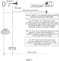
- FIG. 3 is an optional schematic flowchart of an endoscopic image processing method according to an embodiment of this disclosure. It may be understood that, steps shown in FIG. 3 may be performed by various servers running the endoscopic image processing apparatus, such as a dedicated terminal, a server, a cloud server, or a server cluster with an endoscopic image processing function. The following describes the steps shown in FIG. 3 .
- Step 301 Obtain, by an endoscopic image processing apparatus, an endoscopic video stream.
- the endoscopic video stream includes original endoscopic images, and the endoscopic video stream is a video stream photographed by an endoscope in a medical environment such as a real use environment in a hospital. Under the movement and photographing of the endoscope, the endoscopic video stream presents video endoscopy captured by a lens of the endoscope. Therefore, multiple frames of consecutive original endoscopic images are obtained from the endoscopic video stream, and each frame of obtained original endoscopic image describes video endoscopy photographed by the endoscope at a time point, so that identification on the endoscopic image may be implemented based on each frame of original endoscopic image.
- the endoscope photographs an endoscopic video stream in organism, such as an internal part of a human body.
- the endoscope photographs an endoscopic video stream in a cavity communicating with the outside or an enclosed cavity.
- the mentioned cavity communicating with the outside may be a digestive tract, a respiratory tract, or a urinary tract
- the enclosed cavity may be a cavity into which the endoscope needs to be put through an incision such as a chest cavity, an abdominal cavity, or an articular cavity.
- Organ conditions in a cavity is obtained through the endoscopic video stream photographing and identification performed by the endoscope.
- an obtained endoscopic video stream is accessed into performed endoscopic image identification.
- the implemented endoscopic image identification may be further performed on an endoscopic video stream obtained before such as a historical endoscopic video stream, and the implemented endoscopic image identification is not limited to real-time identification, but may be performed oriented to a large amount of endoscopic video streams that are stored.
- obtaining an endoscopic video stream may be implemented in the following manner: extracting an endoscopic video frame in pathological information of a target object; performing definition improvement processing on the endoscopic video frame; converting a format of the endoscopic video frame from a current encoding format to a grayscale value encoding format, to obtain multiple endoscopic video frames; and performing encoding compression on the endoscopic video frames to form the endoscopic video stream in a video stream state.
- Multiple endoscopic video frames are obtained by converting the format of the endoscopic video frame from a current encoding format to a grayscale value encoding format; and encoding compression is performed on all obtained endoscopic video frames to form pathological information in a video state, thereby reducing a data transmission amount and reducing freezing.
- dynamic fuzzy and picture quality may be improved, and user experience is further improved, and it is helpful for a user (doctor) of a remote terminal to make accurate judgment on the pathological information of the target object more accurately.
- the converting a format of the endoscopic video frame from a current encoding format to a grayscale value encoding format may be implemented in the following manner: performing encoding compression on the endoscopic video frame by using an encoding method matching a target format; writing a serial number corresponding to the video frame and generating a timestamp corresponding to an image packet, to generate a packet matching the format of the endoscopic video frame; and splicing the image packet to generate multiple endoscopic video frames.
- preset video encoding software such as ffmpeg codec software may be used.
- the ffmpeg codec software is free software, may run video recording, conversion, and stream functions of multiple formats of audio and video, and includes a decoding/encoding library of audio and video.
- encoding compression may be performed on the image frames by using a video compression algorithm in the video encoding software such as H264 or X264 to form a video stream, where H264 or X264 is a digital video compression format and is also a video codec standard.
- a YUV format refers to a pixel format that represents luminance parameters and chrominance parameters, and separating the luminance parameters and the chrominance parameters can both avoid mutual interference and reduce a sampling rate of chrominance from greatly affecting image quality.
- the YUV does not require three independent video signals to be transmitted at the same time as that of the RGB, so that converting image frames into the YUV format for transmission may occupy less bandwidth, thereby saving a large amount of resources.
- the YUV format is divided into a planar type and a packed type, for the planar YUV format, luminance of all pixels is first stored consecutively, chrominance of all the pixels is then stored, and saturation of all the pixels is finally stored.
- luminance, chrominance, and saturation of each pixel are stored consecutively and alternately.
- the image frames in the YUV format the image frames may be stored by using a storage format such as YUV422P, YUV420, or YUV420sp, to save storage resources of the terminal device and improve transmission efficiency of pathological information.
- the transmission protocol may be a real-time data transmission protocol such as a UDP, where the user datagram protocol (UDP) is a connectionless protocol used for processing packets.
- UDP user datagram protocol
- a type of the data transmission protocol is not specifically limited.
- the video stream may be transmitted to the terminal device by using the UDP.
- the method further includes: adjusting the original endoscopic images to a target size to generate standard endoscopic image frames; and filtering the standard endoscopic image frames to delete a standard endoscopic image frame having interference, to generate target endoscopic image frames including different target endoscopic image frames.
- Step 302 Detect, by the endoscopic image processing apparatus, the original endoscopic image in a video frame through a first thread, and transmit a detection result of the original endoscopic image to an integration module.
- Step 303 Generate, by the endoscopic image processing apparatus, a control instruction through the integration module according to the detection result of the original endoscopic image.
- Step 304 Adjust, by the endoscopic image processing apparatus in response to the control instruction and through a second thread, an output result in the second thread.
- the first thread and the second thread are executed in parallel.
- the output result may match a use environment of the endoscopic video stream.
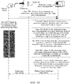
- FIG. 4A is an optional schematic flowchart of an endoscopic image processing method according to an embodiment of this disclosure. It may be understood that, steps shown in FIG. 4A may be performed by various servers running the endoscopic image processing apparatus, such as a dedicated terminal, a server, a cloud server, or a server cluster with an endoscopic image processing function. The following describes steps shown in FIG. 4A .
- Step 401 Detect, by the endoscopic image processing apparatus, an existing foreign body through a detector in the first thread to obtain a foreign body box in the target endoscopic image frame.
- the foreign body box is used for indicating a region where a foreign body exists in the target endoscopic image frame.
- Step 402 Filter, by the endoscopic image processing apparatus, the target endoscopic image frame based on the foreign body box, to position a lesion region in the target endoscopic image frame and determine a lesion category corresponding to the lesion region.
- Step 403 Transmit, by the endoscopic image processing apparatus, the lesion region positioned by using the foreign body box in the target endoscopic image frame to the integration module.
- the endoscopic image processing method further includes: in a case that a detection time of the detector in the first thread exceeds a detection time threshold, stopping detecting a current target endoscopic image frame, and obtaining a subsequent target endoscopic image frame for detection.
- the adjusting, in response to the control instruction and through a second thread, an output result in the second thread may be implemented in the following manner: in a case that a tracker in the second thread is in a continue tracking state and a tracking box corresponding to the tracker matches a foreign body box corresponding to a detector, outputting, by using the tracker in the second thread, a current target endoscopic image frame.
- the adjusting, in response to the control instruction and through a second thread, an output result in the second thread may be implemented in the following manner: in a case that a tracker in the second thread is in a reinitialization state and a tracking box corresponding to the tracker does not match a foreign body box corresponding to a detector, outputting, by using the tracker in the second thread, a new target endoscopic image frame.
- the endoscopic image processing method further includes: determining a box regression parameter in a case that it is determined through the detector in the first thread that a foreign body box exists in the target endoscopic image frame; and activating the tracker in the second thread by using the box regression parameter.
- the endoscopic image processing method further includes: in a case that the tracker in the second thread is activated, assigning a target identifier to a foreign body in the current target endoscopic image frame; and in a case that foreign bodies in the target endoscopic image frame when the tracker in the second thread is in an activated state, the continue tracking state, and the reinitialization state are consistent, initializing the target identifier to implement continuous tracking on the foreign body in the current target endoscopic image frame.
- the endoscopic image processing method further includes: transmitting an adjustment instruction in response to the output result in the second thread, the adjustment instruction being used for adjusting a detection state of a medical device in contact with a target object, to obtain new pathological information.
- FIG. 4B is an optional schematic flowchart of an endoscopic image processing method according to an embodiment of this disclosure. It may be understood that, steps shown in FIG. 4B may be performed by various servers running the endoscopic image processing apparatus, such as a dedicated terminal, a server, a cloud server, or a server cluster with an endoscopic image processing function. The following describes the steps shown in FIG. 4B .
- Step 4001 Transmit, by an endoscope, an endoscopic video stream to the endoscopic image processing apparatus.
- Step 4002 Obtain, by the endoscopic image processing apparatus, the endoscopic video stream transmitted by the endoscope.
- the endoscopic video stream includes original endoscopic images used for observing a lesion of a target object.
- Step 4003 Detect, by the endoscopic image processing apparatus, an original endoscopic image in a video frame through a first thread, and transmit a detection result of the original endoscopic image to an integration module.
- Step 4004 Generate, by the endoscopic image processing apparatus, a control instruction through the integration module according to the detection result of the original endoscopic image.
- Step 4005 Adjust, by the endoscopic image processing apparatus in response to the control instruction and through a second thread, an output result in the second thread.
- the output result may match a use environment of the endoscopic video stream, and the first thread and the second thread are executed in parallel.
- Step 4006 Transmit, by the endoscopic image processing apparatus according to the adjusted output result in the second thread, a target endoscopic image frame to a medical device in contact with the target object.
- Step 4007 Output, by the medical device, the target endoscopic image frame.
- the output result is controlled to match a use environment of the endoscopic video stream through a first thread and a second thread that are executed in parallel, thereby improving real-time accuracy of processing on the endoscopic image and improving a real-time recall rate.
- an observation situation of the target object may be known in time according to the target endoscopic image frame outputted by the medical device.
- the endoscopic image processing method provided in this disclosure is described below by using an example in which a polyp (a foreign body) in a colon image is determined by using an endoscopic video stream.
- Various medical images are generated by an endoscopic device, for example, images are generated continuously as an endoscope continuously performs photographing in digestive tract, and the images further become a large amount of data, so that it is urgently required to implement large-scale classification and identification with the aid of execution of classification prediction.
- the artificial intelligence (AI) technology provides a properly-trained auxiliary analytical information generating network to support the foregoing application solution.
- Artificial intelligence involves a theory, a method, a technology, and an application system that use a digital computer or a machine controlled by the digital computer to simulate, extend, and expand human intelligence, perceive an environment, obtain knowledge, and use knowledge to obtain an optimal result.
- Artificial intelligence is to study design principles and implementation methods of various intelligent machines, to enable the machines to have functions of perception, reasoning, and decision-making.
- identification of low-latency videos and messages is implemented by using a digital computer or a machine controlled by the digital computer, to obtain auxiliary analytical information conveniently according to a pathological analysis result.
- a target detection model using various types of target detection algorithms such as Retinal detection algorithm (Retina Net), Regional convolution neural network algorithm (FasterRCNN), and You Only Look Once (YOLO) with relatively high accuracy is generally integrated in a solution for implementing target detection on an endoscopic video stream (such as a colon endoscopic video stream), and image frames in the video stream are sequentially inputted into the model for target detection.
- a processing speed of the model is limited, and the processing speed of the model cannot match a speed of a real-time video stream (most detection models cannot keep pace with a frame rate of a real-time video), thereby affecting an output effect of the model.
- a process of processing an original endoscopic image in the endoscopic video stream by using a neural network model to determine the polyp (the foreign body) includes following 1) to 3).
- defects mainly include following 1) to 3).
- FIG. 5 is an optional schematic flowchart of an endoscopic image processing method according to an embodiment of this disclosure, where the user may be a user operating a colon endoscope, and the method specifically includes the following steps 501 to 504.
- Step 501 Obtain an endoscopic video stream, the endoscopic video stream includes original endoscopic images.
- Step 502 Detect the original endoscopic image in a video frame through a detector in a first thread, and transmit a detection result of the original endoscopic image to an integration module.
- Step 503 Form a control instruction through the integration module according to the detection result of the original endoscopic image.
- Step 504 Adjust, in response to the control instruction and through a tracker in a second thread, an output result in the tracker in the second thread, to cause the output result to match a delay parameter of the endoscopic video stream, the first thread and the second thread are executed in parallel.
- FIG. 6 is a schematic diagram of different threads in an endoscopic image processing method according to an embodiment of this disclosure. Details are described as follows.
- the tracker is responsible for ensuring the real-time capability and improving the robustness; and the tracker may receive a control instruction of the integration module, to directly return a detection box to the video stream in real time. Further, the tracker used in the second thread needs to meet a low-latency requirement, so that any low-latency real-time tracker such as CSR-DCF or SiamRPN based on manual features may be used to implement direct information exchange with the video stream.
- any low-latency real-time tracker such as CSR-DCF or SiamRPN based on manual features may be used to implement direct information exchange with the video stream.
- the detector is responsible for ensuring the accuracy. Specifically, the detector may transmit a control instruction by using the integration module, to implement initialization, continuous working, reinitialization, and stop of the tracker.
- the detector does not need to meet a real-time requirement, and if a previous frame is not completed, detection of the frame is given up. Any detector algorithm such as YOLOv3, RetinaNet, or FasterRCNN may be used, and the detector does not directly perform information exchange with the video stream.
- Any detector algorithm such as YOLOv3, RetinaNet, or FasterRCNN may be used, and the detector does not directly perform information exchange with the video stream.
- the integration module is responsible for collecting information of two threads executed in parallel, and the detector and the integration module are jointly adjusted.
- states of the tracker include: a wait state (wait); an initialization state (init); a continue tracking state (cont); a reinitialization state (reinit); and a stop state (stop).
- the wait state indicates that the detector does not find any target, and the tracker is in a wait state.
- FIG. 7 is a schematic diagram of an initialization state in an endoscopic image processing method according to an embodiment of this disclosure.
- the initialization state (init) indicates that the detector finds a first frame target and a box regression parameter (bbox bounding-box regression) thereof, and the tracker is activated and initialized by using the box regression parameter.
- box regression parameter bbox bounding-box regression
- FIG. 8 is a schematic diagram of a continue tracking state in an endoscopic image processing method according to an embodiment of this disclosure.
- the continue tracking state (cont) indicates that in a tracking process, if a box provided by the tracker highly overlaps with an instruction box provided by the detector (IoU ⁇ ), it indicates that the target is still well tracked by the tracker (the target does not depart), so that the tracker keeps tracking the target.
- FIG. 9 is a schematic diagram of a reinitialization state a continue tracking state in an endoscopic image processing method according to an embodiment of this disclosure.
- the reinitialization state (reinit) indicates that in a tracking process, if an overlapping degree between a box provided by the tracker and an instruction box provided by the detector is low (IoU ⁇ ), where ⁇ may be dynamically adjusted according to the use environment of the endoscopic video stream, it indicates that the tracker loses the target or tracking of the target is not accurate, so that bbox of the detector is used to reactivate and initialize the tracker.
- FIG. 10 is a schematic diagram of a reinitialization state a continue tracking state in an endoscopic image processing method according to an embodiment of this disclosure.
- the stop state indicates that in a tracking process, if the detector does not find an effective target in N consecutive frames, it indicates that the target may be not in a visual field already, so that the tracker is stopped.
- FIG. 11 is a schematic diagram of a determination process on the same target in an endoscopic image processing method according to an embodiment of this disclosure, where the endoscopic image processing method provided in this disclosure is used to perform detection on an endoscopic video stream of a patient.
- a use effect of the endoscopic image processing method may be monitored.
- Video evaluation of the endoscopic video stream needs to consider a video frame rate.
- a real-time frame rate is 25 fps, namely, each frame is 40 ms, and if a time consumed by the model exceeds 40 ms, a result of a previous frame is directly used as a current result (represented as a stopped picture output).
- Table 1 Indicator dimension Indicator description Speed (indicator) One indicator: an average latency of a single frame, unit: ms (If the frame rate of the video stream is 25 fps, a real-time standard is reached when the latency is less than 40 ms) Accuracy (indicator) Four indicators: real-time accuracy, real-time recall rate, real-time F1 value, and real-time F2 value, unit: percentage
- FIG. 12 is a schematic diagram of a display effect of an endoscopic image processing method according to an embodiment of this disclosure. Due to high latency in the conventional technology (the Detector solution), tracking boxes corresponding to the tracker cannot match foreign body boxes corresponding to the detector, many tracking boxes continue to use a result of a previous frame, resulting in a decrease in the accuracy and the recall rate, and a serious decrease in a visual effect of the endoscopic image. According to the endoscopic image processing method (AIPDT) provided in the embodiments of this disclosure, positions of the tracking boxes are more accurate and match with the foreign body boxes, and the robustness is better, which facilitates continuous working of the endoscope.
- AIPDT endoscopic image processing method
- An endoscopic video stream is obtained, where the endoscopic video stream includes original endoscopic images; the original endoscopic image in a video frame is detected through a first thread, and a detection result of the original endoscopic image is transmitted to an integration module; a control instruction is generated through the integration module according to the detection result of the original endoscopic image; and in response to the control instruction and through a second thread, an output result in the second thread is adjusted.
- the first thread and the second thread are executed in parallel, so that the output result may match a use environment of the endoscopic video stream, thereby improving real-time accuracy of processing on the endoscopic image and improving a real-time recall rate.
- an endoscopic video stream is obtained, the endoscopic video stream includes original endoscopic images; the original endoscopic image in a video frame is detected through a first thread, and a detection result of the original endoscopic image is transmitted to an integration module; a control instruction is generated through the integration module according to the detection result of the original endoscopic image; and in response to the control instruction and through a second thread, an output result in the second thread is adjusted, to make the output result to match a use environment of the endoscopic video stream.
- the first thread and the second thread are executed in parallel, so that the output result may be controlled to match the use environment of the endoscopic video stream by using the first thread and the second thread that are executed in parallel, thereby improving real-time accuracy of processing on the endoscopic image and improving a real-time recall rate.
Landscapes
- Engineering & Computer Science (AREA)
- Health & Medical Sciences (AREA)
- Life Sciences & Earth Sciences (AREA)
- Physics & Mathematics (AREA)
- Medical Informatics (AREA)
- General Health & Medical Sciences (AREA)
- Nuclear Medicine, Radiotherapy & Molecular Imaging (AREA)
- Radiology & Medical Imaging (AREA)
- Surgery (AREA)
- General Physics & Mathematics (AREA)
- Theoretical Computer Science (AREA)
- Signal Processing (AREA)
- Optics & Photonics (AREA)
- Public Health (AREA)
- Computer Vision & Pattern Recognition (AREA)
- Pathology (AREA)
- Quality & Reliability (AREA)
- Biomedical Technology (AREA)
- Heart & Thoracic Surgery (AREA)
- Molecular Biology (AREA)
- Animal Behavior & Ethology (AREA)
- Biophysics (AREA)
- Veterinary Medicine (AREA)
- Multimedia (AREA)
- Endoscopes (AREA)
- Image Analysis (AREA)
- Closed-Circuit Television Systems (AREA)
- Artificial Intelligence (AREA)
- Evolutionary Computation (AREA)
Abstract
Description
- This application claims priority to
Chinese Patent Application No. 202010076394.4 filed on January 23, 2020 - This disclosure relates to medical image processing technologies, and in particular, to an artificial intelligence technology-based endoscopic image processing method and apparatus, an electronic device, and a storage medium.
- Category identification performed based on deep learning is always an important tool for resolving classification of a large amount of data in various application scenarios. For example, in application scenarios such as image processing and natural language processing, large-scale classification and identification of a large amount of data may be implemented, so as to rapidly and accurately obtain a related classification prediction result and accelerate the implementation of functions in the application scenarios.
- Various medical images are generated, for example, images are generated continuously as an endoscope continuously performs photographing in digestive tract, and the images further become a large amount of data, so that it is urgently required to implement large-scale classification and identification with the aid of execution of classification prediction.
- In the related art, a target detection model (for example various types of target detection algorithms such as Retina Net, FasterRCNN, and YOLO) with relatively high accuracy is generally integrated in a solution for implementing target detection on a real-time video stream (such as a colon endoscopic video stream), and image frames in the video stream are sequentially inputted into the model for target detection. However, a processing speed of the model is limited, and the processing speed of the model cannot match a speed of the real-time video stream. Therefore, in the related art, the detection model cannot match a frame rate of a real-time video, thereby affecting an output effect of the model.
- In view of this, embodiments of this disclosure provide an endoscopic image processing method and apparatus, an electronic device, and a storage medium, which can process an original endoscopic image through a first thread and a second thread that are executed in parallel, to cause an output result to match a use environment of an endoscopic video stream.
- The technical solutions in the embodiments of this disclosure are implemented as follows.
- An embodiment of this disclosure provides an endoscopic image processing method. The method includes: obtaining an endoscopic video stream, the endoscopic video stream including multiple original endoscopic images corresponding video frames respectively; detecting each of the multiple original endoscopic images through a first thread, and transmitting a detection result of the respective original endoscopic image to an integration module; generating a control instruction through the integration module according to the detection result of the respective original endoscopic image; and adjusting, in response to the control instruction and through a second thread, an output result in the second thread to match a use environment of the endoscopic video stream, the first thread and the second thread being executed in parallel.
- An embodiment of this disclosure further provides an endoscopic image processing method. The method includes: obtaining, by an endoscopic image processing apparatus, an endoscopic video stream transmitted by an endoscope, the endoscopic video stream including multiple original endoscopic images corresponding video frames respectively, the original endoscopic images being used for observing a lesion of a target object; detecting, by the endoscopic image processing apparatus, each of the original endoscopic images through a first thread, and transmitting a detection result of the respective original endoscopic image to an integration module; generating, by the endoscopic image processing apparatus, a control instruction through the integration module according to the detection result of the respective original endoscopic image; adjusting, by the endoscopic image processing apparatus in response to the control instruction and through a second thread, an output result in the second thread to match a use environment of the endoscopic video stream, the first thread and the second thread being executed in parallel; and transmitting, by the endoscopic image processing apparatus according to the adjusted output result in the second thread, a target endoscopic image frame to a medical device in contact with the target object, where the medical device outputs the target endoscopic image frame.
- An embodiment of this disclosure further provides an endoscopic image processing apparatus. The apparatus includes: an information transmission module, configured to obtain an endoscopic video stream, the endoscopic video stream including multiple original endoscopic images corresponding video frames respectively; and an information processing module, configured to: detect each of the original endoscopic images through a first thread, and transmit a detection result of the respective original endoscopic image to an integration module; generate a control instruction through the integration module according to the detection result of the respective original endoscopic image; and adjust, in response to the control instruction and through a second thread, an output result in the second thread to match a use environment of the endoscopic video stream, the first thread and the second thread being executed in parallel.
- In the foregoing solution, the information processing module is configured to: extract an endoscopic video frame in pathological information of a target object; perform definition improvement processing on the endoscopic video frame; convert a format of the endoscopic video frame from a current encoding format to a grayscale value encoding format, to obtain multiple endoscopic video frames; and perform encoding compression on the endoscopic video frames to form the endoscopic video stream in a video stream state.
- In the foregoing solution, the information processing module is configured to: perform encoding compression on the endoscopic video frame by using an encoding method matching a target format; write a serial number corresponding to the video frame and generating a timestamp corresponding to an image packet, to generate a packet matching the format of the endoscopic video frame; and splice the image packet to generate multiple endoscopic video frames.
- In the foregoing solution, the information processing module is configured to: adjust the original endoscopic images to a target size to generate standard endoscopic image frames; and filter the standard endoscopic image frames to delete a standard endoscopic image frame having interference, to generate target endoscopic image frames including different target endoscopic image frames.
- In the foregoing solution, the information processing module is configured to: detect an existing foreign body through a detector in the first thread to obtain a foreign body box in the target endoscopic image frame, the foreign body box being used for indicating a region where a foreign body exists in the target endoscopic image frame; filter the target endoscopic image frame based on the foreign body box, to position a lesion region in the target endoscopic image frame and determine a lesion category corresponding to the lesion region; and transmit the lesion region positioned by using the foreign body box in the target endoscopic image frame to the integration module.
- In the foregoing solution, the information processing module is configured to: in a case that a detection time of the detector in the first thread exceeds a detection time threshold, stop detecting a current target endoscopic image frame, and obtain a subsequent target endoscopic image frame for detection.
- In the foregoing solution, the information processing module is configured to: in a case that a tracker in the second thread is in a continue tracking state and a tracking box corresponding to the tracker matches a foreign body box corresponding to a detector, output, by using the tracker in the second thread, a current target endoscopic image frame; or in a case that a tracker in the second thread is in a reinitialization state and a tracking box corresponding to the tracker does not match a foreign body box corresponding to a detector, output, by using the tracker in the second thread, a new target endoscopic image frame.
- In the foregoing solution, the information processing module is configured to: determine a box regression parameter in a case that it is determined through the detector in the first thread that a foreign body box exists in the target endoscopic image frame; and activate the tracker in the second thread by using the box regression parameter.
- In the foregoing solution, the information processing module is configured to: in a case that the tracker in the second thread is activated, assign a target identifier to a foreign body in the current target endoscopic image frame; and in a case that foreign bodies in the target endoscopic image frame when the tracker in the second thread is in an activated state, the continue tracking state, and the reinitialization state are consistent, initialize the target identifier to implement continuous tracking on the foreign body in the current target endoscopic image frame.
- In the foregoing solution, the information processing module is configured to: transmit an adjustment instruction in response to the output result in the second thread, the adjustment instruction being used for adjusting a detection state of a medical device in contact with a target object, to obtain new pathological information.
- An embodiment of this disclosure further provides an endoscopic image processing system. The system includes: an endoscope, configured to transmit an endoscopic video stream to an endoscopic image processing apparatus; the endoscopic image processing apparatus, configured to: obtain the endoscopic video stream transmitted by the endoscope, the endoscopic video stream including multiple original endoscopic images corresponding video frames respectively, the original endoscopic images being used for observing a lesion of a target object; detect each of the original endoscopic images through a first thread, and transmit a detection result of the respective original endoscopic image to an integration module; generate a control instruction through the integration module according to the detection result of the respective original endoscopic image; adjust, in response to the control instruction and through a second thread, an output result in the second thread to match a use environment of the endoscopic video stream, the first thread and the second thread being executed in parallel; and transmit, according to the adjusted output result in the second thread, a target endoscopic image frame to a medical device in contact with the target object; and the medical device being configured to output the target endoscopic image frame.
- An embodiment of this disclosure further provides an electronic device. The electronic device includes: a memory, configured to store executable instructions; and a processor, configured to implement, when executing the executable instructions stored in the memory, the foregoing endoscopic image processing method.
- An embodiment of this disclosure further provides a computer-readable storage medium. The computer-readable storage medium stores executable instructions, the executable instructions, when executed by a processor, cause the processor to implement the foregoing endoscopic image processing method.
- The embodiments of this disclosure have the following beneficial effects. An endoscopic video stream is obtained, where the endoscopic video stream includes original endoscopic images; the original endoscopic image in a video frame is detected through a first thread, and a detection result of the original endoscopic image is transmitted to an integration module; a control instruction is generated through the integration module according to the detection result of the original endoscopic image; and in response to the control instruction and through a second thread, an output result in the second thread is adjusted. This disclosure can control the output result to match a use environment of the endoscopic video stream through a first thread and a second thread that are executed in parallel, thereby improving real-time accuracy of processing on the endoscopic image and improving a real-time recall rate.
- To describe the technical solutions of the embodiments of this disclosure or the related art more clearly, the following briefly introduces the accompanying drawings required for describing the embodiments or the related art. The accompanying drawings in the following description show merely some embodiments of this disclosure, and a person of ordinary skill in the art may still derive other accompanying drawings from these accompanying drawings without creative efforts.
-
FIG. 1 is a schematic diagram of a use environment of an endoscopic image processing method according to an embodiment of this disclosure. -
FIG. 2 is a schematic structural diagram of components of an electronic device according to an embodiment of this disclosure. -
FIG. 3 is an optional schematic flowchart of an endoscopic image processing method according to an embodiment of this disclosure. -
FIG. 4A is an optional schematic flowchart of an endoscopic image processing method according to an embodiment of this disclosure. -
FIG. 4B is an optional schematic flowchart of an endoscopic image processing method according to an embodiment of this disclosure. -
FIG. 5 is an optional schematic flowchart of an endoscopic image processing method according to an embodiment of this disclosure. -
FIG. 6 is a schematic diagram of different threads in an endoscopic image processing method according to an embodiment of this disclosure. -
FIG. 7 is a schematic diagram of an initialization state of a tracker in an endoscopic image processing method according to an embodiment of this disclosure. -
FIG. 8 is a schematic diagram of a continue tracking state of a tracker in an endoscopic image processing method according to an embodiment of this disclosure. -
FIG. 9 is a schematic diagram of a reinitialization state a continue tracking state in an endoscopic image processing method according to an embodiment of this disclosure. -
FIG. 10 is a schematic diagram of a reinitialization state a continue tracking state in an endoscopic image processing method according to an embodiment of this disclosure. -
FIG. 11 is a schematic diagram of a determination process on the same target in an endoscopic image processing method according to an embodiment of this disclosure. -
FIG. 12 is a schematic diagram of a display effect of an endoscopic image processing method according to an embodiment of this disclosure. - To make the objectives, technical solutions, and advantages of this disclosure clearer, the following further describes this disclosure in detail with reference to the accompanying drawings. The described embodiments are not to be considered as a limitation to this disclosure. All other embodiments obtained by a person of ordinary skill in the art without creative efforts shall fall within the protection scope of this disclosure.
- In the following descriptions, the term "some embodiments" describes subsets of all possible embodiments, but it may be understood that "some embodiments" may be the same subset or different subsets of all the possible embodiments, and can be combined with each other without conflict.
- Before the embodiments of this disclosure are further described in detail, a description is made on nouns and terms in the embodiments of this disclosure, and the nouns and terms in the embodiments of this disclosure are applicable to the following explanations.
- 1) "In response to" is used for representing a condition or status on which one or more operations to be performed depend. When the condition or status is met, the one or more operations may be performed in real time or after a set delay. Unless explicitly stated, there is no limitation on the order in which the multiple operations are performed.
- 2) "AD Computed-aided diagnosis (CAD)" is used for finding a lesion with the aid of imaging, medical image processing technologies, and other possible physical or biochemical manners in combination with analytical calculation of a computer, to improve the accuracy of diagnosis.
- 3) "Endoscopic video stream" is used for pathological information in a video state formed by performing image acquisition on organism parts (different target organs of a human body or lesions in a human body) through an image acquisition device (such as an endoscope).
- 4) "Lesion" generally refers to a part with a pathological change on an organism. In other words, a limited pathological tissue with pathogenic microbes may be referred to as a lesion.
- 5) "YUV" is a grayscale value encoding method, which is a color encoding method, where "Y" represents luminance or luma, namely, a grayscale value, "U" and "V" represent chrominance or chroma, and functions thereof are to describe colors and saturation of an image and are used to specify colors of pixels.
- 6) "RGB" is three-primary color encoding method and is also referred to as an RGB color mode, which is a color standard in the industry, and obtains various colors through changes of a red (R) channel, a green (G) channel, and a blue (B) channel and superpositions of the three color channels. RGB represents colors of the red channel, the green channel, and the blue channel, and this standard almost covers all colors that human eyes can perceive and is one of the most widely used color systems.
-
FIG. 1 is a schematic diagram of a use scenario of an endoscopic image processing method according to an embodiment of this disclosure. Referring toFIG. 1 , clients that can execute different functions are disposed on a terminal (including a terminal 10-1 and a terminal 10-2), the clients are used for the terminal (including the terminal 10-1 and the terminal 10-2) to obtain different pathological information from a server 200 through anetwork 300 for browsing. The terminal is connected to the server 200 through thenetwork 300, thenetwork 300 may be a wide area network or a local area network or may be a combination thereof to implement data transmission by using a wireless link. Types of the pathological information obtained by the terminal (including the terminal 10-1 and the terminal 10-2) from the server 200 through thenetwork 300 may be the same or may be different. For example, the terminal (including the terminal 10-1 and the terminal 10-2) may obtain a pathological image or a pathological video matching a target object from the server 200 through thenetwork 300, or may only obtain a pathological video (for example, an endoscopic video stream) matching a current target from the server 200 through thenetwork 300 for browsing. The server 200 may store pathological information corresponding to different target objects, or may store auxiliary analytical information matching the pathological information of the target objects. In some embodiments of this disclosure, different types of pathological information stored in the server 200 may be endoscopic video streams acquired by an endoscope. There are at least two original endoscopic images in the endoscopic video stream of this embodiment. The endoscopic video stream is a collection of multi-field-of-view pathological pictures obtained by repeatedly observing suspected lesion regions when a doctor uses an endoscope by moving a camera, switching magnifications and other operations, and the collection combines information under a specific field of view of the endoscope. Because the endoscopic video stream records all the information in the field of view of the endoscope when the doctor observes a lesion of a patient. In this way, the information of a lesion of a single patient observed by the doctor in the field of view of the endoscope is used as a continuous video stream to prevent the doctor from ignoring tiny lesion regions in the process of rapidly moving the endoscope. Therefore, the endoscopic video stream provides more information than a single-frame picture does, to help the doctor diagnose and find tiny lesion regions. - The patient lesions observed under the endoscope (a medical device in contact with the target object) may include multiple different application scenarios, for example, screening of different video streams such as diabetic retinopathy screening and cervical cancer early screening. The endoscopic image processing method based on this embodiment may be deployed to multiple application scenarios, thereby making it convenient for a doctor to consult and use remotely.
- The server 200 transmits pathological information of the same target object to the terminal (the terminal 10-1 and/or the terminal 10-2) through the
network 300, to cause a user of the terminal (the terminal 10-1 and/or the terminal 10-2) to analyze the pathological information of the target object. Therefore, as an example, the server 200 is provided with a neural network model, configured to obtain a video stream of anendoscope device 400, the endoscopic video stream including original endoscopic images; detect the original endoscopic image in a video frame through a first thread, and transmit a detection result of the original endoscopic image to an integration module; generate a control instruction through the integration module according to the detection result of the original endoscopic image; and adjust, in response to the control instruction and through a second thread, an output result in the second thread, to cause the output result to match a use environment of the endoscopic video stream, the first thread and the second thread being executed in parallel. - The following describes a structure of the electronic device according to the embodiments of this disclosure in detail. The electronic device may be implemented in various forms, for example, a dedicated terminal with an endoscopic image processing function, or an electronic device or cloud server with an endoscopic image processing function, for example, the server 200 in
FIG. 1. FIG. 2 is a schematic structural diagram of components of an electronic device according to an embodiment of this disclosure. It may be understood that,FIG. 2 shows only an exemplary structure rather than a complete structure of the electronic device. The structure shown inFIG. 2 may be partially or entirely implemented based on requirements. - The electronic device provided in this embodiment of this disclosure includes: at least one
processor 201, amemory 202, auser interface 203, and at least onenetwork interface 204. The components in the electronic device are coupled by using abus system 205. It may be understood that, thebus system 205 is configured to implement connection and communication between the components. In addition to a data bus, thebus system 205 further includes a power bus, a control bus, and a state signal bus. However, for ease of clear description, all types of buses are labeled as thebus system 205 inFIG. 2 . - The
user interface 203 may include a display, a keyboard, a mouse, a track ball, a click wheel, a key, a button, a touch panel, a touchscreen, or the like. - It may be understood that, the
memory 202 may be a volatile memory or a non-volatile memory, or may include both a volatile memory and a non-volatile memory. Thememory 202 in this embodiment of this disclosure can store data to support operations of the terminal (for example, 10-1). An example of the data includes any computer program to be operated on the terminal (for example, 10-1), for example, an operating system and an application program. The operating system includes various system programs, such as a framework layer, a kernel library layer, and a driver layer, which are configured to implement various basic services and process a task based on hardware. The application program may include various application programs. - In some embodiments, the endoscopic image processing apparatus provided in the embodiments of this disclosure may be implemented by a combination of software and hardware. For example, the endoscopic image processing apparatus provided in the embodiments of this disclosure may be a processor in the form of a hardware decoding processor, and is programmed to perform the endoscopic image processing method provided in the embodiments of this disclosure. For example, the processor in the form of a hardware decoding processor may use one or more application specific integrated circuits (ASICs), DSPs, programmable logic devices (PLDs), complex programmable logic devices (CPLDs), field-programmable gate arrays (FPGAs), or other electronic elements.
- In an example in which the endoscopic image processing apparatus provided in the embodiments of this disclosure is implemented by a combination of software and hardware, the endoscopic image processing apparatus provided in the embodiments of this disclosure may be directly embodied as a combination of software modules executed by the
processor 201. The software module may be located in a storage medium, the storage medium is located in thememory 202, and theprocessor 201 reads executable instructions included in the software module in thememory 202, and implements, in combination with necessary hardware (for example, including theprocessor 201 and another component connected to the bus 205), the endoscopic image processing method provided in the embodiments of this disclosure. - For example, the
processor 201 may be an integrated circuit chip, and has a signal processing capability, for example, a general-purpose processor, a digital signal processor (DSP), or another programable logic device, a discrete gate or a transistor logical device, or a discrete hardware component. The general-purpose processor may be a microprocessor, any conventional processor, or the like. - In an example in which the endoscopic image processing apparatus provided in the embodiments of this disclosure is implemented by hardware, the apparatus provided in the embodiments of this disclosure may be directly executed by using the
processor 201 in the form of a hardware decoding processor, for example, one or more application specific integrated circuits (ASICs), DSPs, programmable logic devices (PLDs), complex programmable logic devices (CPLDs), field-programmable gate arrays (FPGAs), or other electronic elements, to execute the endoscopic image processing method provided in the embodiments of this disclosure. - The
memory 202 in this embodiment of this disclosure is configured to store various types of data to support operations of the electronic device. An example of the data includes: any executable instruction configured to be operated on the electronic device, such as an executable instruction, and a program that implements the endoscopic image processing method of the embodiments of this disclosure may be included in the executable instruction. - In some other embodiments, the endoscopic image processing apparatus provided in the embodiments of this disclosure may be implemented in the form of software.
FIG. 2 shows an endoscopic image processing apparatus 2020 stored in thememory 202, which may be software in the form of a program, a plug-in, or the like, and include a series of modules. An example of the program stored in thememory 202 may include the endoscopic image processing apparatus 2020. The endoscopic image processing apparatus 2020 includes the following software modules: an information transmission module 2081, configured to obtain an endoscopic video stream, the endoscopic video stream including original endoscopic images; and aninformation processing module 2082, configured to: detect the original endoscopic image in a video frame through a first thread, and transmit a detection result of the original endoscopic image to an integration module; generate a control instruction through the integration module according to the detection result of the original endoscopic image; and adjust, in response to the control instruction and through a second thread, an output result in the second thread, to cause the output result to match a use environment of the endoscopic video stream, the first thread and the second thread being executed in parallel. - The endoscopic image processing method provided in the embodiments of this disclosure is described with reference to the electronic device shown in
FIG. 2 .FIG. 3 is an optional schematic flowchart of an endoscopic image processing method according to an embodiment of this disclosure. It may be understood that, steps shown inFIG. 3 may be performed by various servers running the endoscopic image processing apparatus, such as a dedicated terminal, a server, a cloud server, or a server cluster with an endoscopic image processing function. The following describes the steps shown inFIG. 3 . - Step 301: Obtain, by an endoscopic image processing apparatus, an endoscopic video stream.
- The endoscopic video stream includes original endoscopic images, and the endoscopic video stream is a video stream photographed by an endoscope in a medical environment such as a real use environment in a hospital. Under the movement and photographing of the endoscope, the endoscopic video stream presents video endoscopy captured by a lens of the endoscope. Therefore, multiple frames of consecutive original endoscopic images are obtained from the endoscopic video stream, and each frame of obtained original endoscopic image describes video endoscopy photographed by the endoscope at a time point, so that identification on the endoscopic image may be implemented based on each frame of original endoscopic image.
- In implemented endoscopic image identification, the endoscope photographs an endoscopic video stream in organism, such as an internal part of a human body. For example, the endoscope photographs an endoscopic video stream in a cavity communicating with the outside or an enclosed cavity. For example, the mentioned cavity communicating with the outside may be a digestive tract, a respiratory tract, or a urinary tract, and the enclosed cavity may be a cavity into which the endoscope needs to be put through an incision such as a chest cavity, an abdominal cavity, or an articular cavity. Organ conditions in a cavity is obtained through the endoscopic video stream photographing and identification performed by the endoscope. In a process of using the endoscope to check a cavity, an obtained endoscopic video stream is accessed into performed endoscopic image identification. In addition, the implemented endoscopic image identification may be further performed on an endoscopic video stream obtained before such as a historical endoscopic video stream, and the implemented endoscopic image identification is not limited to real-time identification, but may be performed oriented to a large amount of endoscopic video streams that are stored.
- In some embodiments of this disclosure, obtaining an endoscopic video stream may be implemented in the following manner: extracting an endoscopic video frame in pathological information of a target object; performing definition improvement processing on the endoscopic video frame; converting a format of the endoscopic video frame from a current encoding format to a grayscale value encoding format, to obtain multiple endoscopic video frames; and performing encoding compression on the endoscopic video frames to form the endoscopic video stream in a video stream state. Multiple endoscopic video frames are obtained by converting the format of the endoscopic video frame from a current encoding format to a grayscale value encoding format; and encoding compression is performed on all obtained endoscopic video frames to form pathological information in a video state, thereby reducing a data transmission amount and reducing freezing. On the other hand, dynamic fuzzy and picture quality may be improved, and user experience is further improved, and it is helpful for a user (doctor) of a remote terminal to make accurate judgment on the pathological information of the target object more accurately.
- In some embodiments of this disclosure, the converting a format of the endoscopic video frame from a current encoding format to a grayscale value encoding format may be implemented in the following manner: performing encoding compression on the endoscopic video frame by using an encoding method matching a target format; writing a serial number corresponding to the video frame and generating a timestamp corresponding to an image packet, to generate a packet matching the format of the endoscopic video frame; and splicing the image packet to generate multiple endoscopic video frames. When encoding compression is performed on each image frame to form a video stream, preset video encoding software such as ffmpeg codec software may be used. The ffmpeg codec software is free software, may run video recording, conversion, and stream functions of multiple formats of audio and video, and includes a decoding/encoding library of audio and video. Specifically, encoding compression may be performed on the image frames by using a video compression algorithm in the video encoding software such as H264 or X264 to form a video stream, where H264 or X264 is a digital video compression format and is also a video codec standard. In this process, a YUV format refers to a pixel format that represents luminance parameters and chrominance parameters, and separating the luminance parameters and the chrominance parameters can both avoid mutual interference and reduce a sampling rate of chrominance from greatly affecting image quality. The YUV does not require three independent video signals to be transmitted at the same time as that of the RGB, so that converting image frames into the YUV format for transmission may occupy less bandwidth, thereby saving a large amount of resources. The YUV format is divided into a planar type and a packed type, for the planar YUV format, luminance of all pixels is first stored consecutively, chrominance of all the pixels is then stored, and saturation of all the pixels is finally stored. For the packed YUV format, luminance, chrominance, and saturation of each pixel are stored consecutively and alternately. For image frames in the YUV format, the image frames may be stored by using a storage format such as YUV422P, YUV420, or YUV420sp, to save storage resources of the terminal device and improve transmission efficiency of pathological information.
- Further, in a process of transmitting the endoscopic video stream to the terminal device according to a preset transmission protocol, the transmission protocol may be a real-time data transmission protocol such as a UDP, where the user datagram protocol (UDP) is a connectionless protocol used for processing packets. In this disclosure, a type of the data transmission protocol is not specifically limited. As some optional embodiments of this disclosure, the video stream may be transmitted to the terminal device by using the UDP.
- In some embodiments of this disclosure, the method further includes: adjusting the original endoscopic images to a target size to generate standard endoscopic image frames; and filtering the standard endoscopic image frames to delete a standard endoscopic image frame having interference, to generate target endoscopic image frames including different target endoscopic image frames.
- Step 302: Detect, by the endoscopic image processing apparatus, the original endoscopic image in a video frame through a first thread, and transmit a detection result of the original endoscopic image to an integration module.
- Step 303: Generate, by the endoscopic image processing apparatus, a control instruction through the integration module according to the detection result of the original endoscopic image.
- Step 304: Adjust, by the endoscopic image processing apparatus in response to the control instruction and through a second thread, an output result in the second thread. The first thread and the second thread are executed in parallel.
- In this way, the output result may match a use environment of the endoscopic video stream.
- The endoscopic image processing method provided in the embodiments of this disclosure is described with reference to the electronic device shown in
FIG. 2 .FIG. 4A is an optional schematic flowchart of an endoscopic image processing method according to an embodiment of this disclosure. It may be understood that, steps shown inFIG. 4A may be performed by various servers running the endoscopic image processing apparatus, such as a dedicated terminal, a server, a cloud server, or a server cluster with an endoscopic image processing function. The following describes steps shown inFIG. 4A . - Step 401: Detect, by the endoscopic image processing apparatus, an existing foreign body through a detector in the first thread to obtain a foreign body box in the target endoscopic image frame. The foreign body box is used for indicating a region where a foreign body exists in the target endoscopic image frame.
- Step 402: Filter, by the endoscopic image processing apparatus, the target endoscopic image frame based on the foreign body box, to position a lesion region in the target endoscopic image frame and determine a lesion category corresponding to the lesion region.
- Step 403: Transmit, by the endoscopic image processing apparatus, the lesion region positioned by using the foreign body box in the target endoscopic image frame to the integration module.
- In some embodiments of this disclosure, the endoscopic image processing method further includes: in a case that a detection time of the detector in the first thread exceeds a detection time threshold, stopping detecting a current target endoscopic image frame, and obtaining a subsequent target endoscopic image frame for detection.
- In some embodiments of this disclosure, the adjusting, in response to the control instruction and through a second thread, an output result in the second thread may be implemented in the following manner: in a case that a tracker in the second thread is in a continue tracking state and a tracking box corresponding to the tracker matches a foreign body box corresponding to a detector, outputting, by using the tracker in the second thread, a current target endoscopic image frame.
- In some embodiments of this disclosure, the adjusting, in response to the control instruction and through a second thread, an output result in the second thread may be implemented in the following manner: in a case that a tracker in the second thread is in a reinitialization state and a tracking box corresponding to the tracker does not match a foreign body box corresponding to a detector, outputting, by using the tracker in the second thread, a new target endoscopic image frame.
- In some embodiments of this disclosure, the endoscopic image processing method further includes: determining a box regression parameter in a case that it is determined through the detector in the first thread that a foreign body box exists in the target endoscopic image frame; and activating the tracker in the second thread by using the box regression parameter.
- In some embodiments of this disclosure, the endoscopic image processing method further includes: in a case that the tracker in the second thread is activated, assigning a target identifier to a foreign body in the current target endoscopic image frame; and in a case that foreign bodies in the target endoscopic image frame when the tracker in the second thread is in an activated state, the continue tracking state, and the reinitialization state are consistent, initializing the target identifier to implement continuous tracking on the foreign body in the current target endoscopic image frame.
- In some embodiments of this disclosure, the endoscopic image processing method further includes: transmitting an adjustment instruction in response to the output result in the second thread, the adjustment instruction being used for adjusting a detection state of a medical device in contact with a target object, to obtain new pathological information.
- The endoscopic image processing method provided in the embodiments of this disclosure is described with reference to the electronic device shown in
FIG. 2 . In an endoscopic image processing system, -
FIG. 4B is an optional schematic flowchart of an endoscopic image processing method according to an embodiment of this disclosure. It may be understood that, steps shown inFIG. 4B may be performed by various servers running the endoscopic image processing apparatus, such as a dedicated terminal, a server, a cloud server, or a server cluster with an endoscopic image processing function. The following describes the steps shown inFIG. 4B . - Step 4001: Transmit, by an endoscope, an endoscopic video stream to the endoscopic image processing apparatus.
- Step 4002: Obtain, by the endoscopic image processing apparatus, the endoscopic video stream transmitted by the endoscope. The endoscopic video stream includes original endoscopic images used for observing a lesion of a target object.
- Step 4003: Detect, by the endoscopic image processing apparatus, an original endoscopic image in a video frame through a first thread, and transmit a detection result of the original endoscopic image to an integration module.
- Step 4004: Generate, by the endoscopic image processing apparatus, a control instruction through the integration module according to the detection result of the original endoscopic image.
- Step 4005: Adjust, by the endoscopic image processing apparatus in response to the control instruction and through a second thread, an output result in the second thread.
- In this way, the output result may match a use environment of the endoscopic video stream, and the first thread and the second thread are executed in parallel.
- Step 4006: Transmit, by the endoscopic image processing apparatus according to the adjusted output result in the second thread, a target endoscopic image frame to a medical device in contact with the target object.
- Step 4007: Output, by the medical device, the target endoscopic image frame.
- According to the technical solution shown in this embodiment, the output result is controlled to match a use environment of the endoscopic video stream through a first thread and a second thread that are executed in parallel, thereby improving real-time accuracy of processing on the endoscopic image and improving a real-time recall rate. Besides, an observation situation of the target object may be known in time according to the target endoscopic image frame outputted by the medical device.
- The endoscopic image processing method provided in this disclosure is described below by using an example in which a polyp (a foreign body) in a colon image is determined by using an endoscopic video stream. Various medical images are generated by an endoscopic device, for example, images are generated continuously as an endoscope continuously performs photographing in digestive tract, and the images further become a large amount of data, so that it is urgently required to implement large-scale classification and identification with the aid of execution of classification prediction.
- In view of this, the artificial intelligence (AI) technology provides a properly-trained auxiliary analytical information generating network to support the foregoing application solution. Artificial intelligence involves a theory, a method, a technology, and an application system that use a digital computer or a machine controlled by the digital computer to simulate, extend, and expand human intelligence, perceive an environment, obtain knowledge, and use knowledge to obtain an optimal result. Artificial intelligence is to study design principles and implementation methods of various intelligent machines, to enable the machines to have functions of perception, reasoning, and decision-making. In the AI medical field, identification of low-latency videos and messages is implemented by using a digital computer or a machine controlled by the digital computer, to obtain auxiliary analytical information conveniently according to a pathological analysis result.
- However, in the related art, a target detection model using various types of target detection algorithms such as Retinal detection algorithm (Retina Net), Regional convolution neural network algorithm (FasterRCNN), and You Only Look Once (YOLO) with relatively high accuracy is generally integrated in a solution for implementing target detection on an endoscopic video stream (such as a colon endoscopic video stream), and image frames in the video stream are sequentially inputted into the model for target detection. However, a processing speed of the model is limited, and the processing speed of the model cannot match a speed of a real-time video stream (most detection models cannot keep pace with a frame rate of a real-time video), thereby affecting an output effect of the model.
- In the related art, a process of processing an original endoscopic image in the endoscopic video stream by using a neural network model to determine the polyp (the foreign body) includes following 1) to 3).
- 1) Directly processing image frames in a real-time video stream by using the neural network model. However, a frame rate of the real-time video stream is generally not less than 25 fps and a frame interval is less than 40 ms, and the model processing speed cannot match the speed of the real-time video stream.
- 2) Only extracting a part of frames from the video stream for detection (for example, extraction is performed at intervals of N frames), and skipping processing video frames that are not processed in time (frame-skipping processing).
- 3) Adding a tracking algorithm (for example, various types of target tracking algorithms such as KCF, CSRDCF, SiamFC, or SiamRPN) based on conventional detection. For example, a segment processing method is used, where an initial frame is found by the detection model, and target positioning is performed by placing detection with tracking in subsequent stages. Although this solution can ensure the real-time capability to some extent, the accuracy of polyp detection may be reduced. Alternatively, a model integration method is used, where the tracking algorithm is regarded as another detection model, and the detection model and tracking algorithm are performed on all frames. Although this solution can ensure the accuracy of polyp detection, however, the real-time capability is reduced, which is adverse to continuous use for a doctor.
- With reference to summary of the related art, in the conventional process of processing a colon endoscopic video stream by using a neural network model, defects mainly include following 1) to 3).
- 1) The detection model processing consumes long time and cannot keep pace with a video frame rate, the detection is stopped, and an effect is distorted.
When the speed of the detection model cannot keep pace with the frame rate of the real-time video stream, a common phenomenon is that: a picture detection box is stopped, the detection box stayed at an original place, but a target in the video is no longer at the original place. Under a specific evaluation value indicator, the phenomenon may cause a serious decrease in real-time accuracy, a real-time recall rate, and a real-time F value. From the perspective of user's visual experience, the phenomenon may cause a decrease in user experience, and for a doctor, not only diagnosis experience is seriously affected, but also missed detection may be caused easily. - 2) The detection model has low stability and poor robustness.
In a changeable and unpredictable scene of a video stream, performance of the detection model jitters easily and is not stable, and common trigger scenes include: a lens is blur, a lens changes rapidly, a target deforms, a target is partially blocked, and a target comes in and goes out at a visual field boundary. - 3) Lack of ID identification of the same target.
The detection model does not include an ID identification function of the same target. In a target detection scene, people always hope to trigger a warning or prompt when finding a new target, but the robustness of the detection model is relatively low and the model lacks of an ID identification function of the same target, so that repeated warnings/prompts are generally triggered, which may easily generate unnecessary disturbance to a user. - To resolve the foregoing defects,
FIG. 5 is an optional schematic flowchart of an endoscopic image processing method according to an embodiment of this disclosure, where the user may be a user operating a colon endoscope, and the method specifically includes the followingsteps 501 to 504. - Step 501: Obtain an endoscopic video stream, the endoscopic video stream includes original endoscopic images.
- Step 502: Detect the original endoscopic image in a video frame through a detector in a first thread, and transmit a detection result of the original endoscopic image to an integration module.
- Step 503: Form a control instruction through the integration module according to the detection result of the original endoscopic image.
- Step 504: Adjust, in response to the control instruction and through a tracker in a second thread, an output result in the tracker in the second thread, to cause the output result to match a delay parameter of the endoscopic video stream, the first thread and the second thread are executed in parallel.
-
FIG. 6 is a schematic diagram of different threads in an endoscopic image processing method according to an embodiment of this disclosure. Details are described as follows. - 1) In a timeline of an endoscopic video stream, each point represents one frame, if an input frame rate of a real-time video is 25 fps, a point/frame interval represents 40 ms, and the rest may be deduced by analogy.
- 2) Tracker: all outputted detection boxes are provided by the tracker in real time, and the tracker directly controls video outputs.
- 3) Detector: the detector maintains and instructs a next action of the tracker by using an integration module, and indirectly controls video outputs.
- 4) Intersected dotted line: represents information transmission between components, start time points and end time points of information transmission may be noted.
- The tracker is responsible for ensuring the real-time capability and improving the robustness; and the tracker may receive a control instruction of the integration module, to directly return a detection box to the video stream in real time. Further, the tracker used in the second thread needs to meet a low-latency requirement, so that any low-latency real-time tracker such as CSR-DCF or SiamRPN based on manual features may be used to implement direct information exchange with the video stream.
- The detector is responsible for ensuring the accuracy. Specifically, the detector may transmit a control instruction by using the integration module, to implement initialization, continuous working, reinitialization, and stop of the tracker.
- The detector does not need to meet a real-time requirement, and if a previous frame is not completed, detection of the frame is given up. Any detector algorithm such as YOLOv3, RetinaNet, or FasterRCNN may be used, and the detector does not directly perform information exchange with the video stream.
- The integration module is responsible for collecting information of two threads executed in parallel, and the detector and the integration module are jointly adjusted.
- Referring to
FIG. 7 to FIG. 10 , in a working process of the tracker in the second thread, states of the tracker include: a wait state (wait); an initialization state (init); a continue tracking state (cont); a reinitialization state (reinit); and a stop state (stop). - The wait state indicates that the detector does not find any target, and the tracker is in a wait state.
-
FIG. 7 is a schematic diagram of an initialization state in an endoscopic image processing method according to an embodiment of this disclosure. The initialization state (init) indicates that the detector finds a first frame target and a box regression parameter (bbox bounding-box regression) thereof, and the tracker is activated and initialized by using the box regression parameter. -
FIG. 8 is a schematic diagram of a continue tracking state in an endoscopic image processing method according to an embodiment of this disclosure. The continue tracking state (cont) indicates that in a tracking process, if a box provided by the tracker highly overlaps with an instruction box provided by the detector (IoU≥µ), it indicates that the target is still well tracked by the tracker (the target does not depart), so that the tracker keeps tracking the target. -
FIG. 9 is a schematic diagram of a reinitialization state a continue tracking state in an endoscopic image processing method according to an embodiment of this disclosure. The reinitialization state (reinit) indicates that in a tracking process, if an overlapping degree between a box provided by the tracker and an instruction box provided by the detector is low (IoU≤µ), where µ may be dynamically adjusted according to the use environment of the endoscopic video stream, it indicates that the tracker loses the target or tracking of the target is not accurate, so that bbox of the detector is used to reactivate and initialize the tracker. -
FIG. 10 is a schematic diagram of a reinitialization state a continue tracking state in an endoscopic image processing method according to an embodiment of this disclosure. The stop state (stop) indicates that in a tracking process, if the detector does not find an effective target in N consecutive frames, it indicates that the target may be not in a visual field already, so that the tracker is stopped. -
FIG. 11 is a schematic diagram of a determination process on the same target in an endoscopic image processing method according to an embodiment of this disclosure, where the endoscopic image processing method provided in this disclosure is used to perform detection on an endoscopic video stream of a patient. - 1) appearance of a new target: a target in an initialization state is regarded as a new target that is found for the first time, and a new ID is assigned to the target; and
- 2) maintenance of an old target: targets in an initialization state, a continue tracking state, and a reinitialization state are regarded as the same target, and an ID during initialization is assigned to the target. According to the foregoing identification strategies, warnings and prompts of a repeated target may be reduced, thereby reducing unnecessary disturbance to the user.
- In a process of determining a polyp in a colon image by using the endoscopic image processing method (an asynchronous in parallel detection and tracking (AIPDT) solution) provided in this disclosure, a use effect of the endoscopic image processing method may be monitored. Video evaluation of the endoscopic video stream needs to consider a video frame rate. Generally, a real-time frame rate is 25 fps, namely, each frame is 40 ms, and if a time consumed by the model exceeds 40 ms, a result of a previous frame is directly used as a current result (represented as a stopped picture output). Based on the priori evaluation, indicators shown in table 1 are calculated:
Table 1 Indicator dimension Indicator description Speed (indicator) One indicator: an average latency of a single frame, unit: ms (If the frame rate of the video stream is 25 fps, a real-time standard is reached when the latency is less than 40 ms) Accuracy (indicator) Four indicators: real-time accuracy, real-time recall rate, real-time F1 value, and real-time F2 value, unit: percentage - When the detection algorithm uses YOLOv3, and the tracking algorithm uses CSR-DCF,
(1) For improvement of the speed indicator, refer to table 2,Table 2 Indicator Average latency of a single frame Conventional solution (Detector solution) 84.7 ms AIPDT solution 24.4 ms
If the detector uses RetinaNet or a more complex network, the latency of the detector solution generally falls within 100 ms to 200 ms.
(2) For improvement of the real-time accuracy, refer to table 3,Table 3 Indicator Real-time accuracy Real-time recall rate Real- time F 1Real-time F2 Conventional solution (Detector solution) 92.48 85.13 88.65 86.51 AIPDT solution 96.23 90.19 93.11 91.33 -
FIG. 12 is a schematic diagram of a display effect of an endoscopic image processing method according to an embodiment of this disclosure. Due to high latency in the conventional technology (the Detector solution), tracking boxes corresponding to the tracker cannot match foreign body boxes corresponding to the detector, many tracking boxes continue to use a result of a previous frame, resulting in a decrease in the accuracy and the recall rate, and a serious decrease in a visual effect of the endoscopic image. According to the endoscopic image processing method (AIPDT) provided in the embodiments of this disclosure, positions of the tracking boxes are more accurate and match with the foreign body boxes, and the robustness is better, which facilitates continuous working of the endoscope. - An endoscopic video stream is obtained, where the endoscopic video stream includes original endoscopic images; the original endoscopic image in a video frame is detected through a first thread, and a detection result of the original endoscopic image is transmitted to an integration module; a control instruction is generated through the integration module according to the detection result of the original endoscopic image; and in response to the control instruction and through a second thread, an output result in the second thread is adjusted. The first thread and the second thread are executed in parallel, so that the output result may match a use environment of the endoscopic video stream, thereby improving real-time accuracy of processing on the endoscopic image and improving a real-time recall rate.
- The foregoing descriptions are merely embodiments of this disclosure, but are not intended to limit the protection scope of this disclosure. Any modification, equivalent replacement and improvement made within the spirit and principle of this disclosure shall fall within the protection scope of this disclosure.
- In the embodiments of this disclosure, an endoscopic video stream is obtained, the endoscopic video stream includes original endoscopic images; the original endoscopic image in a video frame is detected through a first thread, and a detection result of the original endoscopic image is transmitted to an integration module; a control instruction is generated through the integration module according to the detection result of the original endoscopic image; and in response to the control instruction and through a second thread, an output result in the second thread is adjusted, to make the output result to match a use environment of the endoscopic video stream. The first thread and the second thread are executed in parallel, so that the output result may be controlled to match the use environment of the endoscopic video stream by using the first thread and the second thread that are executed in parallel, thereby improving real-time accuracy of processing on the endoscopic image and improving a real-time recall rate.
Claims (15)
- An endoscopic image processing method, performed by an electronic device, the endoscopic image processing method comprising:obtaining an endoscopic video stream, the endoscopic video stream comprising a plurality of original endoscopic images corresponding video frames respectively;detecting each of the plurality of original endoscopic images through a first thread, and transmitting a detection result of the respective original endoscopic image to an integration module;generating a control instruction through the integration module according to the detection result of the respective original endoscopic image; andadjusting, in response to the control instruction and through a second thread, an output result in the second thread to match a use environment of the endoscopic video stream, the first thread and the second thread being executed in parallel.
- The method according to claim 1, wherein the obtaining an endoscopic video stream comprises:extracting an endoscopic video frame in pathological information of a target object;performing definition improvement processing on the endoscopic video frame;converting a format of the endoscopic video frame from a current encoding format to a grayscale value encoding format, to obtain a plurality of endoscopic video frames; andperforming encoding compression on the endoscopic video frames to form the endoscopic video stream in a video stream state.
- The method according to claim 2, wherein the converting a format of the endoscopic video frame from a current encoding format to a grayscale value encoding format comprises:performing encoding compression on the endoscopic video frame by using an encoding method matching a target format;writing a serial number corresponding to the video frame and generating a timestamp corresponding to an image packet, to generate a packet matching the format of the endoscopic video frame; andsplicing the image packet to generate a plurality of endoscopic video frames.
- The method according to claim 1, further comprising:adjusting the original endoscopic images to a target size to generate standard endoscopic image frames; andfiltering the standard endoscopic image frames to delete a standard endoscopic image frame having interference, to generate target endoscopic image frames comprising different target endoscopic image frames.
- The method according to claim 4, wherein the detecting the original endoscopic image in a video frame through a first thread, and transmitting a detection result of the original endoscopic image to an integration module comprises:detecting an existing foreign body through a detector in the first thread to obtain a foreign body box in the target endoscopic image frame, the foreign body box being used for indicating a region where a foreign body exists in the target endoscopic image frame;filtering the target endoscopic image frame based on the foreign body box, to position a lesion region in the target endoscopic image frame and determine a lesion category corresponding to the lesion region; andtransmitting the lesion region positioned by using the foreign body box in the target endoscopic image frame to the integration module.
- The method according to claim 5, further comprising:
in a case that a detection time of the detector in the first thread exceeds a detection time threshold, stopping detecting a current target endoscopic image frame, and obtaining a subsequent target endoscopic image frame for detection. - The method according to claim 4, wherein the adjusting, in response to the control instruction and through a second thread, an output result in the second thread comprises:in a case that a tracker in the second thread is in a continue tracking state and a tracking box corresponding to the tracker matches a foreign body box corresponding to a detector, outputting, by using the tracker in the second thread, a current target endoscopic image frame; orin a case that a tracker in the second thread is in a reinitialization state and a tracking box corresponding to the tracker does not match a foreign body box corresponding to a detector, outputting, by using the tracker in the second thread, a new target endoscopic image frame.
- The method according to claim 7, further comprising:determining a box regression parameter in a case that it is determined through the detector in the first thread that a foreign body box exists in the target endoscopic image frame; andactivating the tracker in the second thread by using the box regression parameter.
- The method according to claim 8, further comprising:in a case that the tracker in the second thread is activated, assigning a target identifier to a foreign body in the current target endoscopic image frame; andin a case that foreign bodies in the target endoscopic image frame when the tracker in the second thread is in an activated state, the continue tracking state, and the reinitialization state are consistent, initializing the target identifier to implement continuous tracking on the foreign body in the current target endoscopic image frame.
- The method according to claim 1, further comprises:
transmitting an adjustment instruction in response to the output result in the second thread, the adjustment instruction being used for adjusting a detection state of a medical device in contact with a target object, to obtain new pathological information. - An endoscopic image processing method, performed by an endoscopic image processing system, the method comprising:obtaining, by an endoscopic image processing apparatus, an endoscopic video stream transmitted by an endoscope, the endoscopic video stream comprising a plurality of original endoscopic images corresponding video frames respectively, the original endoscopic images being used for observing a lesion of a target object;detecting, by the endoscopic image processing apparatus, each of the plurality of original endoscopic images through a first thread, and transmitting a detection result of the respective original endoscopic image to an integration module;generating, by the endoscopic image processing apparatus, a control instruction through the integration module according to the detection result of the respective original endoscopic image;adjusting, by the endoscopic image processing apparatus in response to the control instruction and through a second thread, an output result in the second thread to match a use environment of the endoscopic video stream, the first thread and the second thread being executed in parallel; andtransmitting, by the endoscopic image processing apparatus according to the adjusted output result in the second thread, a target endoscopic image frame to a medical device in contact with the target object, wherein the medical device outputs the target endoscopic image frame.
- The endoscopic image processing method according to any one of claims 1 to 11, wherein the endoscopic image processing method is performed by an endoscopic image processing system, and the system comprises:an endoscope, configured to transmit an endoscopic video stream to an endoscopic image processing apparatus;the endoscopic image processing apparatus, configured to: obtain the endoscopic video stream transmitted by the endoscope, the endoscopic video stream comprising a plurality of original endoscopic images corresponding video frames respectively, the original endoscopic images being used for observing a lesion of a target object; detect each of the plurality of original endoscopic images through a first thread, and transmit a detection result of the respective original endoscopic image to an integration module; generate a control instruction through the integration module according to the detection result of the respective original endoscopic image; adjust, in response to the control instruction and through a second thread, an output result in the second thread to match a use environment of the endoscopic video stream, the first thread and the second thread being executed in parallel; and transmit, according to the adjusted output result in the second thread, a target endoscopic image frame to a medical device in contact with the target object; andthe medical device being configured to output the target endoscopic image frame.
- An endoscopic image processing apparatus, comprising:an information transmission module, configured to obtain an endoscopic video stream, the endoscopic video stream comprising a plurality of original endoscopic images corresponding video frames respectively; andan information processing module, configured to: detect each of the plurality of original endoscopic images through a first thread, and transmit a detection result of the respective original endoscopic image to an integration module; generate a control instruction through the integration module according to the detection result of the respective original endoscopic image; and adjust, in response to the control instruction and through a second thread, an output result in the second thread to match a use environment of the endoscopic video stream, the first thread and the second thread being executed in parallel.
- An electronic device, comprising:a memory, configured to store executable instructions; anda processor, configured to implement, when executing the executable instructions stored in the memory, the endoscopic image processing method according to any one of claims 1 to 10 or the endoscopic image processing method according to claim 11 or 12.
- A computer-readable storage medium, storing executable instructions, the executable instructions, when executed by a processor, cause the processor to implement the endoscopic image processing method according to any one of claims 1 to 10 or the endoscopic image processing method according to claim 11 or 12.
Applications Claiming Priority (2)
| Application Number | Priority Date | Filing Date | Title |
|---|---|---|---|
| CN202010076394.4A CN111311557A (en) | 2020-01-23 | 2020-01-23 | Endoscope image processing method, device, electronic device and storage medium |
| PCT/CN2020/130769 WO2021147500A1 (en) | 2020-01-23 | 2020-11-23 | Endoscope image processing method and apparatus, and electronic device and storage medium |
Publications (2)
| Publication Number | Publication Date |
|---|---|
| EP4016451A1 true EP4016451A1 (en) | 2022-06-22 |
| EP4016451A4 EP4016451A4 (en) | 2023-06-28 |
Family
ID=71161669
Family Applications (1)
| Application Number | Title | Priority Date | Filing Date |
|---|---|---|---|
| EP20914926.9A Pending EP4016451A4 (en) | 2020-01-23 | 2020-11-23 | Endoscope image processing method and apparatus, and electronic device and storage medium |
Country Status (5)
| Country | Link |
|---|---|
| US (1) | US12315144B2 (en) |
| EP (1) | EP4016451A4 (en) |
| JP (1) | JP7673060B2 (en) |
| CN (1) | CN111311557A (en) |
| WO (1) | WO2021147500A1 (en) |
Families Citing this family (22)
| Publication number | Priority date | Publication date | Assignee | Title |
|---|---|---|---|---|
| CN111311557A (en) | 2020-01-23 | 2020-06-19 | 腾讯科技(深圳)有限公司 | Endoscope image processing method, device, electronic device and storage medium |
| CN112365417B (en) * | 2020-11-10 | 2023-06-23 | 华中科技大学鄂州工业技术研究院 | Confocal endoscope image correction stitching method, device and readable storage medium |
| CN112614152B (en) * | 2020-12-22 | 2022-03-25 | 电子科技大学 | Soft tissue surface motion tracking method based on depth feature learning |
| CN112862752A (en) * | 2020-12-31 | 2021-05-28 | 北京小白世纪网络科技有限公司 | Image processing display method, system electronic equipment and storage medium |
| CN112967276B (en) * | 2021-03-31 | 2023-09-05 | 杭州海康威视数字技术股份有限公司 | Object detection method, device, endoscope system, electronic device and storage medium |
| CN114022820A (en) * | 2021-11-09 | 2022-02-08 | 交通运输部北海航海保障中心烟台航标处 | Intelligent beacon light quality detection method based on machine vision |
| CN114582455A (en) * | 2022-02-08 | 2022-06-03 | 中国人民解放军总医院第一医学中心 | Endoscopic report generation method |
| US12236499B2 (en) * | 2022-04-12 | 2025-02-25 | Utech Products, Inc. | Multithreading video processing with cloud services to detect and analyze objects |
| US12608803B2 (en) | 2022-04-12 | 2026-04-21 | Utech Products, Inc. | Multithreading video processing with cloud services to detect and analyze landmarks and objects during a clinical procedure |
| CN114663424B (en) * | 2022-04-14 | 2025-01-24 | 联通(广东)产业互联网有限公司 | Endoscopic video-assisted diagnosis method, system, device and medium based on edge-cloud collaboration |
| CN114882132B (en) * | 2022-04-25 | 2024-11-29 | 武汉博视曼医疗科技有限公司 | Image conversion method, device, storage medium and apparatus for endoscope system |
| CN115277985A (en) * | 2022-06-24 | 2022-11-01 | 江苏济远医疗科技有限公司 | Multifunctional endoscope image processing device |
| CN115345823A (en) * | 2022-06-30 | 2022-11-15 | 上海微觅医疗器械有限公司 | Medical image processing method, medical image processing system, equipment and medium |
| CN115035152B (en) * | 2022-08-12 | 2022-10-25 | 武汉楚精灵医疗科技有限公司 | Medical image processing method and device and related equipment |
| US20240129515A1 (en) * | 2022-09-27 | 2024-04-18 | Verily Life Sciences Llc | Combined Compression and Feature Extraction Models for Storing and Analyzing Medical Videos |
| CN117793076B (en) * | 2022-09-28 | 2025-10-10 | 荣耀终端股份有限公司 | Audio transmission method and device |
| TR2022020874A2 (en) * | 2022-12-28 | 2023-01-23 | Smart Alfa Teknoloji Sanayi Ve Ticaret Anonim Sirketi | A method and system that can be used to adapt the markings of deep learning systems used for acquiring anatomical images for medical purposes to the movement, changes, and variations in processing speed in the image. |
| CN116687313A (en) * | 2023-05-23 | 2023-09-05 | 江苏无右微创医疗科技有限公司 | Endoscope system and image processing method based on FPGA (field programmable Gate array) multimode image processing |
| CN116527727B (en) * | 2023-06-06 | 2024-03-26 | 深圳杰泰科技有限公司 | Remote control system of cloud endoscope |
| CN117557776B (en) * | 2023-11-10 | 2024-07-12 | 朗信医疗科技(无锡)有限公司 | Multi-mode target detection method and device |
| CN117528131B (en) * | 2024-01-05 | 2024-04-05 | 青岛美迪康数字工程有限公司 | AI integrated display system and method for medical image |
| CN118338077B (en) * | 2024-06-13 | 2024-09-17 | 天津御锦人工智能医疗科技有限公司 | Intelligent medical live broadcast method, system, device and storage medium |
Family Cites Families (36)
| Publication number | Priority date | Publication date | Assignee | Title |
|---|---|---|---|---|
| US9402601B1 (en) * | 1999-06-22 | 2016-08-02 | Teratech Corporation | Methods for controlling an ultrasound imaging procedure and providing ultrasound images to an external non-ultrasound application via a network |
| US20040015079A1 (en) * | 1999-06-22 | 2004-01-22 | Teratech Corporation | Ultrasound probe with integrated electronics |
| US6426367B1 (en) * | 1999-09-09 | 2002-07-30 | Efa Sciences Llc | Methods for selectively occluding blood supplies to neoplasias |
| US8073528B2 (en) * | 2007-09-30 | 2011-12-06 | Intuitive Surgical Operations, Inc. | Tool tracking systems, methods and computer products for image guided surgery |
| US8317098B2 (en) * | 2006-12-01 | 2012-11-27 | Ncr Corporation | Item having a data tag |
| US8385971B2 (en) * | 2008-08-19 | 2013-02-26 | Digimarc Corporation | Methods and systems for content processing |
| JP5754171B2 (en) * | 2011-02-28 | 2015-07-29 | コニカミノルタ株式会社 | Radiographic imaging system and radiographic imaging device |
| EP2833325A1 (en) * | 2013-07-30 | 2015-02-04 | Fraunhofer-Gesellschaft zur Förderung der angewandten Forschung e.V. | Apparatus and method for resource-adaptive object detection and tracking |
| US20150080652A1 (en) * | 2013-09-18 | 2015-03-19 | Cerner Innovation, Inc. | Lesion detection and image stabilization using portion of field of view |
| RU2013146467A (en) * | 2013-10-17 | 2015-04-27 | ЭлЭсАй Корпорейшн | METHOD AND DEVICE FOR RECOGNITION OF GESTURES USING ASYNCHRONOUS MULTI-THREAD PROCESSING |
| WO2017042812A2 (en) * | 2015-09-10 | 2017-03-16 | Magentiq Eye Ltd. | A system and method for detection of suspicious tissue regions in an endoscopic procedure |
| JP2017064091A (en) * | 2015-09-30 | 2017-04-06 | ソニー株式会社 | Image processing device, image processing method, surgical system, and surgical thread |
| JP6847636B2 (en) | 2016-11-18 | 2021-03-24 | ソニー・オリンパスメディカルソリューションズ株式会社 | Medical signal processing equipment and medical observation system |
| JP2018092495A (en) * | 2016-12-07 | 2018-06-14 | ソニーセミコンダクタソリューションズ株式会社 | Image sensor |
| JP7147562B2 (en) * | 2017-02-20 | 2022-10-05 | ソニーグループ株式会社 | Photometric device, photometric method, program and imaging device |
| WO2018198161A1 (en) * | 2017-04-24 | 2018-11-01 | オリンパス株式会社 | Endoscope image processing apparatus and endoscope image processing method |
| WO2019003911A1 (en) * | 2017-06-27 | 2019-01-03 | Sony Corporation | Medical image processing apparatus, medical image processing method, and computing device |
| US11350043B2 (en) * | 2017-07-17 | 2022-05-31 | Huawei Technologies Co., Ltd. | Image processing method and terminal |
| CN111031889B (en) * | 2017-08-24 | 2022-04-05 | 富士胶片株式会社 | Medical image processing apparatus and medical image processing method |
| JP2019058074A (en) * | 2017-09-25 | 2019-04-18 | オリンパス株式会社 | Image processing apparatus, cell cluster recognition apparatus, cell cluster recognition method, and cell cluster recognition program |
| US10628961B2 (en) * | 2017-10-13 | 2020-04-21 | Qualcomm Incorporated | Object tracking for neural network systems |
| WO2019138773A1 (en) * | 2018-01-10 | 2019-07-18 | 富士フイルム株式会社 | Medical image processing apparatus, endoscope system, medical image processing method, and program |
| CN108509248B (en) * | 2018-04-13 | 2021-11-23 | 网易(杭州)网络有限公司 | Data processing method and device, storage medium, processor and terminal |
| JP2019180966A (en) | 2018-04-13 | 2019-10-24 | 学校法人昭和大学 | Endoscope observation support apparatus, endoscope observation support method, and program |
| CN108852268A (en) * | 2018-04-23 | 2018-11-23 | 浙江大学 | A kind of digestive endoscopy image abnormal characteristic real-time mark system and method |
| CN108852286A (en) | 2018-05-03 | 2018-11-23 | 腾讯科技(深圳)有限公司 | Show the method, apparatus and terminal of backbone measurement data |
| CN108805834A (en) * | 2018-05-28 | 2018-11-13 | 电子科技大学 | A kind of image processing algorithm that endoscope is with an automatic light meter |
| CN109993769B (en) * | 2019-03-07 | 2022-09-13 | 安徽创世科技股份有限公司 | Multi-target tracking system combining deep learning SSD algorithm with KCF algorithm |
| CN109919988A (en) * | 2019-03-27 | 2019-06-21 | 武汉万屏电子科技有限公司 | A kind of stereoscopic image processing method suitable for three-dimensional endoscope |
| CN110211152A (en) * | 2019-05-14 | 2019-09-06 | 华中科技大学 | A kind of endoscopic instrument tracking based on machine vision |
| US10671934B1 (en) * | 2019-07-16 | 2020-06-02 | DOCBOT, Inc. | Real-time deployment of machine learning systems |
| CN110634564B (en) * | 2019-09-16 | 2023-01-06 | 腾讯科技(深圳)有限公司 | Pathological information processing method, device and system, electronic equipment and storage medium |
| JP7346285B2 (en) * | 2019-12-24 | 2023-09-19 | 富士フイルム株式会社 | Medical image processing device, endoscope system, operating method and program for medical image processing device |
| CN111311557A (en) * | 2020-01-23 | 2020-06-19 | 腾讯科技(深圳)有限公司 | Endoscope image processing method, device, electronic device and storage medium |
| JP7764903B2 (en) * | 2022-02-10 | 2025-11-06 | 株式会社Ihi | Ultrasound diagnostic equipment |
| US12482193B2 (en) * | 2022-12-07 | 2025-11-25 | Qualcomm Incorporated | Independent scene movement based on mask layers |
-
2020
- 2020-01-23 CN CN202010076394.4A patent/CN111311557A/en active Pending
- 2020-11-23 EP EP20914926.9A patent/EP4016451A4/en active Pending
- 2020-11-23 JP JP2022523504A patent/JP7673060B2/en active Active
- 2020-11-23 WO PCT/CN2020/130769 patent/WO2021147500A1/en not_active Ceased
-
2022
- 2022-02-21 US US17/676,680 patent/US12315144B2/en active Active
Also Published As
| Publication number | Publication date |
|---|---|
| CN111311557A (en) | 2020-06-19 |
| JP2022554119A (en) | 2022-12-28 |
| US20220230311A1 (en) | 2022-07-21 |
| US12315144B2 (en) | 2025-05-27 |
| JP7673060B2 (en) | 2025-05-08 |
| WO2021147500A1 (en) | 2021-07-29 |
| EP4016451A4 (en) | 2023-06-28 |
Similar Documents
| Publication | Publication Date | Title |
|---|---|---|
| US12315144B2 (en) | Endoscopic image processing method and apparatus, electronic device, and storage medium | |
| US20220172828A1 (en) | Endoscopic image display method, apparatus, computer device, and storage medium | |
| EP3968272A1 (en) | Medical endoscope image identification method and system, and endoscope image system | |
| WO2021022623A1 (en) | Camera having transdermal optical imaging function | |
| CN109223178B (en) | Cavity mirror intelligent edge computing system with target positioning function | |
| CN113744266A (en) | Method and device for displaying focus detection frame, electronic equipment and storage medium | |
| CN113823400A (en) | Method and device for monitoring speed of endoscope withdrawal of intestinal tract and computer readable storage medium | |
| CN118870162A (en) | Endoscope device and method for automatically adjusting exposure parameters | |
| CN116523907B (en) | Endoscope imaging quality detection method, device, equipment and storage medium | |
| CN110634564B (en) | Pathological information processing method, device and system, electronic equipment and storage medium | |
| CN110930340B (en) | An image processing method and device | |
| CN113870151B (en) | Method, device and equipment for correcting endoscopic image and storage medium | |
| JP7540087B2 (en) | IMAGE PROCESSING METHOD, ELECTRONIC DEVICE, AND READABLE STORAGE MEDIUM | |
| US20230326072A1 (en) | Image-based position detection method, electronic device, and readable storage medium | |
| CN102509076A (en) | Principal-component-analysis-based video image background detection method | |
| CN114677676A (en) | Target phenomenon monitoring method, device, computer equipment and storage medium | |
| CN113744151A (en) | Method, device and equipment for processing images to be diagnosed and storage medium | |
| CN119446434A (en) | An automatic pre-operative frame retrieval system based on artificial intelligence | |
| HK40024084A (en) | Method and apparatus for processing endoscopic image, electronic device and storage medium | |
| CN116236147A (en) | An endoscope control method, device and storage medium based on surgical behavior understanding | |
| CN113115054B (en) | Video stream encoding method, device, system, electronic device and storage medium | |
| CN114399542A (en) | Endoscope smoke suction method and system | |
| CN116109536A (en) | Image freezing, secondary processing method, processing system, program and storage medium | |
| CN114511796A (en) | Laryngeal mask ventilation monitoring method and device | |
| CN115802160B (en) | Intelligent photographing method and system for fundus images |
Legal Events
| Date | Code | Title | Description |
|---|---|---|---|
| STAA | Information on the status of an ep patent application or granted ep patent |
Free format text: STATUS: THE INTERNATIONAL PUBLICATION HAS BEEN MADE |
|
| PUAI | Public reference made under article 153(3) epc to a published international application that has entered the european phase |
Free format text: ORIGINAL CODE: 0009012 |
|
| STAA | Information on the status of an ep patent application or granted ep patent |
Free format text: STATUS: REQUEST FOR EXAMINATION WAS MADE |
|
| 17P | Request for examination filed |
Effective date: 20220301 |
|
| AK | Designated contracting states |
Kind code of ref document: A1 Designated state(s): AL AT BE BG CH CY CZ DE DK EE ES FI FR GB GR HR HU IE IS IT LI LT LU LV MC MK MT NL NO PL PT RO RS SE SI SK SM TR |
|
| RAP3 | Party data changed (applicant data changed or rights of an application transferred) |
Owner name: TENCENT TECHNOLOGY (SHENZHEN) COMPANY LIMITED |
|
| RIC1 | Information provided on ipc code assigned before grant |
Ipc: H04N 21/44 20110101ALI20221216BHEP Ipc: A61B 1/31 20060101ALI20221216BHEP Ipc: A61B 1/00 20060101ALI20221216BHEP Ipc: G06T 7/00 20170101AFI20221216BHEP |
|
| DAV | Request for validation of the european patent (deleted) | ||
| DAX | Request for extension of the european patent (deleted) | ||
| A4 | Supplementary search report drawn up and despatched |
Effective date: 20230531 |
|
| RIC1 | Information provided on ipc code assigned before grant |
Ipc: H04N 21/44 20110101ALI20230524BHEP Ipc: A61B 1/31 20060101ALI20230524BHEP Ipc: A61B 1/00 20060101ALI20230524BHEP Ipc: G06T 7/00 20170101AFI20230524BHEP |
|
| STAA | Information on the status of an ep patent application or granted ep patent |
Free format text: STATUS: EXAMINATION IS IN PROGRESS |
|
| 17Q | First examination report despatched |
Effective date: 20260123 |