EP4006830A1 - Lernvorrichtung, verfahren und programm, extraktionsvorrichtung für interessenregion, verfahren und programm sowie gelerntes extraktionsmodell - Google Patents
Lernvorrichtung, verfahren und programm, extraktionsvorrichtung für interessenregion, verfahren und programm sowie gelerntes extraktionsmodell Download PDFInfo
- Publication number
- EP4006830A1 EP4006830A1 EP20844578.3A EP20844578A EP4006830A1 EP 4006830 A1 EP4006830 A1 EP 4006830A1 EP 20844578 A EP20844578 A EP 20844578A EP 4006830 A1 EP4006830 A1 EP 4006830A1
- Authority
- EP
- European Patent Office
- Prior art keywords
- image
- decoder
- learning
- discriminator
- encoder
- Prior art date
- Legal status (The legal status is an assumption and is not a legal conclusion. Google has not performed a legal analysis and makes no representation as to the accuracy of the status listed.)
- Granted
Links
Images



Classifications
-
- A—HUMAN NECESSITIES
- A61—MEDICAL OR VETERINARY SCIENCE; HYGIENE
- A61B—DIAGNOSIS; SURGERY; IDENTIFICATION
- A61B6/00—Apparatus or devices for radiation diagnosis; Apparatus or devices for radiation diagnosis combined with radiation therapy equipment
- A61B6/52—Devices using data or image processing specially adapted for radiation diagnosis
- A61B6/5211—Devices using data or image processing specially adapted for radiation diagnosis involving processing of medical diagnostic data
-
- A—HUMAN NECESSITIES
- A61—MEDICAL OR VETERINARY SCIENCE; HYGIENE
- A61B—DIAGNOSIS; SURGERY; IDENTIFICATION
- A61B6/00—Apparatus or devices for radiation diagnosis; Apparatus or devices for radiation diagnosis combined with radiation therapy equipment
- A61B6/52—Devices using data or image processing specially adapted for radiation diagnosis
- A61B6/5211—Devices using data or image processing specially adapted for radiation diagnosis involving processing of medical diagnostic data
- A61B6/5229—Devices using data or image processing specially adapted for radiation diagnosis involving processing of medical diagnostic data combining image data of a patient, e.g. combining a functional image with an anatomical image
- A61B6/5247—Devices using data or image processing specially adapted for radiation diagnosis involving processing of medical diagnostic data combining image data of a patient, e.g. combining a functional image with an anatomical image combining images from an ionising-radiation diagnostic technique and a non-ionising radiation diagnostic technique, e.g. X-ray and ultrasound
-
- G—PHYSICS
- G06—COMPUTING OR CALCULATING; COUNTING
- G06V—IMAGE OR VIDEO RECOGNITION OR UNDERSTANDING
- G06V10/00—Arrangements for image or video recognition or understanding
- G06V10/20—Image preprocessing
- G06V10/25—Determination of region of interest [ROI] or a volume of interest [VOI]
-
- G—PHYSICS
- G06—COMPUTING OR CALCULATING; COUNTING
- G06V—IMAGE OR VIDEO RECOGNITION OR UNDERSTANDING
- G06V10/00—Arrangements for image or video recognition or understanding
- G06V10/40—Extraction of image or video features
-
- G—PHYSICS
- G06—COMPUTING OR CALCULATING; COUNTING
- G06V—IMAGE OR VIDEO RECOGNITION OR UNDERSTANDING
- G06V10/00—Arrangements for image or video recognition or understanding
- G06V10/70—Arrangements for image or video recognition or understanding using pattern recognition or machine learning
- G06V10/77—Processing image or video features in feature spaces; using data integration or data reduction, e.g. principal component analysis [PCA] or independent component analysis [ICA] or self-organising maps [SOM]; Blind source separation
- G06V10/7715—Feature extraction, e.g. by transforming the feature space, e.g. multi-dimensional scaling [MDS]; Mappings, e.g. subspace methods
-
- A—HUMAN NECESSITIES
- A61—MEDICAL OR VETERINARY SCIENCE; HYGIENE
- A61B—DIAGNOSIS; SURGERY; IDENTIFICATION
- A61B6/00—Apparatus or devices for radiation diagnosis; Apparatus or devices for radiation diagnosis combined with radiation therapy equipment
- A61B6/02—Arrangements for diagnosis sequentially in different planes; Stereoscopic radiation diagnosis
- A61B6/03—Computed tomography [CT]
-
- G—PHYSICS
- G06—COMPUTING OR CALCULATING; COUNTING
- G06V—IMAGE OR VIDEO RECOGNITION OR UNDERSTANDING
- G06V2201/00—Indexing scheme relating to image or video recognition or understanding
- G06V2201/03—Recognition of patterns in medical or anatomical images
Definitions
- the present disclosure relates to a learning apparatus, a learning method, and a learning program that perform learning of an extraction model which extracts a region of interest from an image, a region-of-interest extraction apparatus, a region-of-interest extraction method, and a region-of-interest extraction program that extract a region of interest from an image, and a learned extraction model.
- GAN Generative adversarial networks that alternately perform learning of a generator which creates data and a discriminator which identifies data
- GAN Generative adversarial networks
- Ian J. Goodfellow, Jean Pouget-Abadie, Mehdi Mirza, Bing Xu, David Warde-Farley, Sherjil Ozair, Aaron Courville, Yoshua Bengio "Generative Adversarial Nets", arXiv: 1406.2661 describes research on GAN. According to GAN, learning of a generative model that generates highly accurate data based on the characteristics of data for learning can be performed.
- a region of interest such as an organ and a lesion included in such a medical image has also been automatically extracted.
- a technique has been proposed in which learning of a machine learning model such as a neural network is performed by using an image for learning and a ground-truth mask of the region of interest, and the region of interest is extracted from an input image.
- image representation formats are also different from each other.
- the density is different between a CT image and an MRI image even in a case where a tissue of a human body that is included in an image is the same.
- the MRI image has various imaging conditions such as a T1-weighted image, a T2-weighted image, a fat-suppressed image, and a diffusion-weighted image, and the representation format differs depending on the imaging conditions.
- adipose tissue For example, on the T1-weighted image, predominantly adipose tissue appears white, water, humoral components, and cysts appear black, and tumors appear slightly black. Further, on the T2-weighted image, not only adipose tissue but also water, humoral components, and cysts appear white. Therefore, in order to extract a region of interest made to correspond to the medical images of various representation formats, it is necessary to prepare a ground-truth mask for each representation format of the medical image, that is, for each domain, and perform learning of a machine learning model.
- the present disclosure has been made in view of the above circumstances, and an object thereof is to make it possible to extract a region of interest that is included in a target image regardless of a representation format of the target image.
- a first learning apparatus is a learning apparatus of an extraction model that extracts a region of interest from images having a plurality of different representation formats
- the learning unit may make the encoder derive a feature map of the second virtual image in response to an input of the second virtual image
- the learning unit may perform learning of the second decoder on the basis of the extraction result of the region of interest and the ground-truth mask.
- the extraction model may further have a third discriminator that discriminates the representation format of the first image input to the encoder on the basis of the feature map, and that outputs a third discrimination result
- the learning unit may perform learning of the encoder and the third discriminator on the basis of the third discrimination result.
- a second learning apparatus is a learning apparatus of an extraction model that extracts a region of interest from images having a plurality of different representation formats
- the image may be a three-dimensional medical image
- the representation format may include representation formats of a CT image and an MRI image.
- the representation format may include at least one representation format of a T1-weighted image, a T2-weighted image, a diffusion-weighted image, a FLAIR image, a T1-weighted image before contrast enhancement, or a T1-weighted image after contrast enhancement, in an MRI image.
- a region-of-interest extraction apparatus comprises an extraction unit that extracts a region of interest of an image of any representation format from the image, in which an encoder and a second decoder in an extraction model learned by the first or second learning apparatus according to the present disclosure are provided.
- a learned extraction model according to the present disclosure comprises an encoder and a second decoder in an extraction model learned by the first or second learning apparatus according to the present disclosure.
- a first learning method is a learning method of an extraction model that extracts a region of interest from images having a plurality of different representation formats
- a second learning method is a learning method of an extraction model that extracts a region of interest from images having a plurality of different representation formats
- a region-of-interest extraction method comprises extracting a region of interest of an image of any representation format from the image, in which an encoder and a second decoder in an extraction model learned by the first or second learning method according to the present disclosure are provided.
- the first and second learning methods and the region-of-interest extraction method according to the present disclosure may be provided as programs to be executed by a computer.
- a third learning apparatus is a learning apparatus of an extraction model that extracts a region of interest from images having a plurality of different representation formats, the learning apparatus comprising:
- a fourth learning apparatus is a learning apparatus of an extraction model that extracts a region of interest from images having a plurality of different representation formats, the learning apparatus comprising:
- Another region-of-interest extraction apparatus comprises: a memory that stores a command to be executed by a computer; and a processor configured to execute the stored command, in which the processor executes a process including functioning as an encoder and a second decoder in an extraction model learned by the first or second learning method according to the present disclosure, and extracting a region of interest of an image of any representation format from the image.
- a region of interest included in a target image can be extracted regardless of a representation format of the target image.
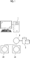
- Fig. 1 is a hardware configuration diagram showing an outline of a diagnosis support system to which a learning apparatus and a region-of-interest extraction apparatus according to a first embodiment of the present disclosure are applied.
- a learning apparatus and region-of-interest extraction apparatus hereinafter, represented by a region-of-interest extraction apparatus
- a plurality of three-dimensional image capturing apparatuses 2A and 2B, and an image storage server 3 according to the first embodiment are connected to communicate with one another via a network 4.
- the three-dimensional image capturing apparatuses 2A and 2B are apparatuses each of which images an area to be diagnosed of a subject and generates a three-dimensional image representing the area, and specific examples thereof include a CT apparatus, an MRI apparatus, and a positron emission tomography (PET) apparatus.
- PET positron emission tomography
- the three-dimensional image capturing apparatus 2A is a CT apparatus
- the three-dimensional image capturing apparatus 2B is an MRI apparatus
- the three-dimensional image capturing apparatuses 2A and 2B generate a CT image and an MRI image including the area to be diagnosed of the subject, as a three-dimensional image, respectively.
- the three-dimensional image capturing apparatus 2B generates a T1-weighted image and a T2-weighted image, as an MRI image.
- the three-dimensional image consists of a plurality of tomographic images.
- the three-dimensional image is acquired in a case where the abdomen of the subject is imaged, and the liver included in the abdomen is a region of interest to be extracted.
- image representation formats are also different from each other.
- the density is different between a CT image and an MRI image even in a case where a tissue of a human body that is included in an image is the same.
- representation formats are different between the T1-weighted image and the T2-weighted image even in a case where the same MRI image is used. Specifically, on the T1-weighted image, predominantly adipose tissue appears white, water, humoral components, and cysts appear black, and tumors appear slightly black.
- the CT image, the T1-weighted image, and the T2-weighted image are images having different representation formats from each other.
- the representation format corresponds to a domain
- different representation formats are synonymous with different domains.
- the image storage server 3 is a computer that stores and manages various data and comprises a large-capacity external storage device and database management software.
- the image storage server 3 communicates with another apparatus via a wired or wireless network 4 to transmit and receive image data and the like.
- the image storage server 3 acquires various data including the image data of the three-dimensional image generated by the three-dimensional image capturing apparatus 2 via the network, and stores and manages the acquired data on a recording medium such as a large-capacity external storage device.
- a storage format of the image data and the communication between the apparatuses via the network 4 are based on a protocol such as digital imaging and communication in medicine (DICOM).
- DICOM digital imaging and communication in medicine
- the image storage server 3 also stores and manages an image for learning that is used to perform learning of an extraction model, which will be described later.
- the region-of-interest extraction apparatus 1 including the learning apparatus of the present embodiment is an apparatus in which a learning program and a region-of-interest extraction program of the first embodiment are installed on one computer.
- the computer may be a workstation or a personal computer directly operated by a doctor who makes diagnosis, or may be a server computer connected to the workstation or the personal computer via a network.
- the learning program and the region-of-interest extraction program are stored on a storage device of a server computer connected to the network or on network storage so as to be accessible from the outside, and are downloaded and installed on a computer that the doctor uses according to a request.
- the learning program and the region-of-interest extraction program are recorded on a recording medium, such as a digital versatile disc (DVD) or a compact disc read only memory (CD-ROM), and are distributed and installed on a computer from the recording medium.
- DVD digital versatile disc
- CD-ROM compact disc read only memory
- Fig. 2 is a diagram showing a schematic configuration of the region-of-interest extraction apparatus which is realized with the learning program and the region-of-interest extraction program installed on a computer.
- the region-of-interest extraction apparatus 1 comprises a central processing unit (CPU) 11, a memory 12, and a storage 13, as a standard workstation configuration.
- a display unit 14 such as a liquid crystal display
- an input unit 15, such as a keyboard and a mouse are connected to the region-of-interest extraction apparatus 1.
- the storage 13 includes a hard disk drive or the like, and stores a target image as a region-of-interest extraction target, which is acquired from the image storage server 3 via the network 4, an image for learning that is used to perform learning of an extraction model 30, which will be described later, and various information including information required for processing.
- the learning program and the region-of-interest extraction program are stored on the memory 12.
- the learning program defines information acquisition processing of acquiring an image for learning that is used to perform learning of an extraction model, a target image as a region-of-interest extraction target, and various information that is used to perform learning and extract a region of interest, and learning processing of performing learning of the extraction model.
- the region-of-interest extraction program defines extraction processing of extracting a region of interest included in the target image as a region-of-interest extraction target, which is acquired by the information acquisition processing, labeling processing of labeling the region of interest included in the target image according to an extraction result of the region of interest, and display control processing of displaying the labeled target image on the display unit 14.
- the CPU 11 executes the processing in accordance with the learning program and the region-of-interest extraction program, so that the computer functions as an information acquisition unit 21, a learning unit 22, an extraction unit 23, a labeling unit 24, and a display control unit 25.
- the information acquisition unit 21 acquires, as the target image, a three-dimensional image of the abdomen of the subject, from the image storage server 3 via an interface (not shown) connected to the network.
- the information acquisition unit 21 also acquires an image for learning and a ground-truth mask, which will be described later. Further, the information acquisition unit 21 acquires various information that is used to perform learning by the learning unit 22.
- the information that is used to perform learning is, for example, information on the representation format after conversion of the image for learning (that is, the target), which will be described later.
- the learning unit 22 performs learning of an extraction model that extracts a region of interest from images having a plurality of different representation formats.
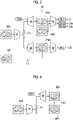
- Figs. 3 and 4 are schematic block diagrams showing the configuration of the extraction model according to the first embodiment.
- the extraction model 30 has an encoder 31, a first decoder 32, a first discriminator 33, a second decoder 34, and a second discriminator 35.
- the encoder 31, the first decoder 32, and the first discriminator 33 included in the extraction model 30 constitute a domain conversion network that is used to convert the representation format of an input image, that is, convert the domain.
- the encoder 31, the second decoder 34, and the second discriminator 35 included in the extraction model 30 constitute a region extraction network that is used to extract a region of interest from the input image.
- the encoder 31 is shared in the domain conversion network and the region extraction network.
- the domain conversion network constitutes generative adversarial networks (GAN).
- GAN comprises a "generator” that creates data and a "discriminator” that identifies data.
- the combination of the encoder 31 and the first decoder 32 corresponds to the generator, and the first discriminator 33 corresponds to the discriminator.
- the region extraction network also constitutes generative adversarial networks, and the combination of the encoder 31 and the second decoder 34 corresponds to the generator, and the second discriminator 35 corresponds to the discriminator.
- the encoder 31 consists of a convolutional neural network (CNN), which is one of multi-layer neural networks in which a plurality of processing layers are hierarchically connected, and derives a feature map representing a feature amount of an input image. Specifically, as shown in Fig. 3 , in a case where a first image GR1 of a first representation format is input, the encoder 31 derives a feature map F1 representing the feature amount of the first image GR1.
- the first image GR1 is an actual image (real image) generated by the three-dimensional image capturing apparatuses 2A and 2B.
- a convolutional neural network consists of a plurality of convolutional layers.
- the convolutional layer performs convolution processing using various kernels on an image that is input, and outputs a feature map consisting of feature amount data obtained by the convolution processing.
- the number of channels of the first convolutional layer (that is, the input layer) is 48 or more, but the number is not limited thereto.
- the convolutional layer applies the kernel to the entire input image or feature map output from the previous processing layer, while shifting the pixel of interest of the kernel.
- the convolutional layer applies an activation function such as a sigmoid function to the convolution value, and outputs a feature map F1.
- an activation function such as a sigmoid function
- the kernel is applied to the input image or feature map while being shifted by two or more strides during the convolution processing.
- a plurality of types of images having different representation formats that is, different domains, such as a CT image, a T1-weighted image, and a T2-weighted image, are used for learning of the extraction model 30.
- a label indicating the representation format is given to the image that is input, as a class label.
- the first image GR1 shown in Fig. 3 is given a class label indicating a representation format class of the first image GR1.
- the images that are used at the time of learning are prepared with and without a ground-truth mask.
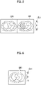
- Fig. 5 is a diagram showing an example of the first image GR1 having a ground-truth mask M1
- Fig. 6 is a diagram showing an example of the first image GR1 having no ground-truth mask.
- the first image GR1 shown in Figs. 5 and 6 is given a class label CL1.
- the class label CL1 is a vector having three elements that are used to identify the classes of the three representation formats, and the element to which 1 is assigned indicates the representation format of the image.
- the three elements indicate a CT image, a T1-weighted image, and a T2-weighted image from the top.
- the representation format is a CT image.
- the representation format is a T2-weighted image.
- the representation format given to the image that is input is denoted by a first representation format.
- the first decoder 32 converts the first representation format of the input image into a second representation format different from the first representation format on the basis of the feature map, to derive a virtual image. That is, as shown in Fig. 3 , in a case where the feature map F1 of the first image GR1 is input, the first decoder 32 converts the first representation format of the first image GR1 into a second representation format different from the first representation format, to derive a second virtual image GV2.
- the first representation format corresponds to the source domain that is the conversion source
- the second representation format corresponds to the target domain that is the conversion destination. For this reason, information TD2 on the target domain, which is the second representation format, is input to the first decoder 32.
- the second representation format is input from, for example, the input unit 15.
- the information TD2 on the second representation format is a vector indicating the class label of the second representation format, and has the same three elements as in the class label CL1.
- the first decoder 32 has a plurality of deconvolutional layers.
- the deconvolutional layer performs the same processing as the convolutional layer of the encoder 31, but applies the kernel for the deconvolution operation to the feature map while upsampling the input feature map.
- the first decoder 32 performs processing of converting the representation format of the first image GR1 into the second representation format while increasing the resolution of the feature map F1 output by the encoder 31 to the resolution of the first image GR1, to derive the second virtual image GV2 of the second representation format.
- the first discriminator 33 consists of, for example, a convolutional neural network, and discriminates the representation format of the input image and whether the input image is a real image or a virtual image generated by the first decoder 32, and outputs a first discrimination result.
- the first discrimination result includes a discrimination result RF1 as to whether the input image is a real image or a virtual image.
- the first discrimination result includes a discrimination result DC1 on the representation format of the input image.
- the real image is not an image generated by the first decoder 32 but an image generated by the three-dimensional image generation apparatuses 2A and 2B, so to speak, a genuine image.
- the discrimination result RF1 is correct, and in a case where the first discriminator 33 discriminates that the input real image is a virtual image, the discrimination result RF1 is incorrect.
- the discrimination result RF1 is incorrect, and in a case where the first discriminator 33 discriminates that the input virtual image is a virtual image, the discrimination result RF1 is correct.
- the learning unit 22 derives a loss on the basis of the discrimination result RF1 output by the first discriminator 33.
- the loss is referred to as an adversarial loss.
- the adversarial loss in the first discriminator 33 is denoted by a first loss L1.
- the discrimination result DC1 is correct, and if the first discriminator 33 discriminates that the representation format is an image other than the CT image, the discrimination result DC1 is incorrect.
- the representation format of the input image is a T2-weighted image
- the discrimination result DC1 is correct, and if the first discriminator 33 discriminates that the representation format is an image other than the T2-weighted image, the discrimination result DC1 is incorrect.
- the second image GR2 which is a real image, is input to the first discriminator 33.
- the virtual image derived by the first decoder 32 is input to the first discriminator 33, and the discrimination result DC1 is output.
- the second virtual image GV2 is generated by using the T2-weighted image as the second representation format.
- the discrimination result DC1 is correct and if the discrimination result DC1 is a CT image or T1-weighted image other than the T2-weighted image, the discrimination result DC1 is incorrect.
- the discrimination result DC1 output from the first discriminator 33 is referred to as a discrimination result DC1d
- the discrimination result DC1g is referred to as a discrimination result DC1g.
- the learning unit 22 derives losses on the basis of the discrimination results DCld and DC1g output by the first discriminator 33.
- the loss is referred to as a classification loss.
- the classification losses derived on the basis of the discrimination results DCld and DC1g are denoted by second losses L2d and L2g, respectively.
- the discrimination results DCld and DC1g may be represented by the discrimination result DC1
- the second losses L2d and L2g may be represented by the second loss L2.
- the second virtual image GV2 is input to the encoder 31, the feature map F1 derived by the encoder 31 is input to the first decoder 32, and information TD1 on the first representation format of the first image GR1 is further input to the first decoder 32.
- the first decoder 32 derives a first virtual image GV1. It is desirable that the first virtual image GV1 derived in this way completely matches the first image GR1, but there is a difference between the first virtual image GV1 that has been subjected to processing by the encoder 31 and the first decoder 32 and the first image GR1.
- the learning unit 22 derives the difference between the first virtual image GV1 and the first image GR1, as a loss.
- the loss is referred to as a cycle loss.
- the cycle loss is denoted by a third loss L3.
- the learning unit 22 performs learning of the first discriminator 33 so that the first discriminator 33 correctly discriminates the discrimination result RF1 as to whether the input image is a real image or a virtual image generated by the first decoder 32 and correctly discriminates the discrimination result DCld on the representation format of the input image. That is, learning of the first discriminator 33 is performed so that the first loss L1 is maximized and the second loss L2d is minimized. Specifically, learning of the first discriminator 33 is performed so that -L1 + L2d is a predetermined threshold value or less.
- the learning unit 22 performs learning of the first decoder 32 so that the first decoder 32 derives a virtual image of the representation format designated from the input image, for which the first discriminator 33 incorrectly discriminates the discrimination result RF1 and correctly discriminates the discrimination result DC1g. Further, the learning unit 22 performs learning of the encoder 31 so that the encoder 31 derives the feature map F1 from which a virtual image for which the first discriminator 33 incorrectly discriminates the discrimination result RF1 and correctly discriminates the discrimination result DC1 can be generated. That is, learning of the encoder 31 and the first decoder 32 is performed so that the first loss L1 and the second loss L2g are minimized. Specifically, learning of the encoder 31 and the first decoder 32 is performed so that L1 + L2g is a predetermined threshold value or less.
- the learning unit 22 performs learning of the first decoder 32 so that the first virtual image GV1 generated from the second virtual image GV2 matches the first image GR1. Further, the learning unit 22 performs learning of the encoder 31 so that the encoder 31 derives the feature map F1 from which the first decoder 32 can derive the first virtual image GV1 that matches the first image GR1. That is, learning of the encoder 31 and the first decoder 32 is performed so that the third loss L3 is minimized. Specifically, learning of the encoder 31 and the first decoder 32 is performed so that L3 is a predetermined threshold value or less.
- the encoder 31 and the first decoder 32, and the first discriminator 33 improve the accuracy thereof with each other, and the first discriminator 33 can more accurately discriminate a real image or a virtual image even in a case where an image of any representation format is input.
- the encoder 31 and the first decoder 32 can generate a virtual image closer to a genuine representation format image, which is not discriminated by the first discriminator 33.
- Fig. 7 is a diagram showing representation formats of real images that are input and representation formats of converted virtual images.
- the leftmost column is the real images that are input
- the representation formats are a CT image, a T1-weighted image, and a T1-weighted image in order from the top.
- the virtual images of the converted representation formats are shown from the second to fourth columns on the left side.
- the representation formats are a CT image, a T1-weighted image, and a T2-weighted image, in order from the second column on the left side.
- a virtual image close to a representation format as instructed is generated regardless of the representation format of the real image that is input.
- the second decoder 34 extracts a region of interest of the image input to the encoder 31 by using the feature map derived by the encoder 31. Specifically, as shown in Fig. 3 , the second decoder 34 extracts the region of interest of the first image GR1 by using the feature map F1 of the first image GR1.
- the second decoder 34 consists of a convolutional neural network having a plurality of deconvolutional layers.
- the second decoder 34 performs processing of deriving the probability of being the region of interest included in the first image GR1 (liver in the present embodiment), and extracting a pixel in which the probability is a threshold value or more, as the region of interest, while increasing the resolution of the feature map F1 output by the encoder 31 to the resolution of the first image GR1. As a result, as shown in Fig. 3 , the second decoder 34 outputs an extraction result PM1 of a liver in the first image GR1.
- the learning unit 22 derives a difference between the extraction result PM1 of the region of interest output by the second decoder 34 and the ground-truth mask M1, as a loss.
- the loss is referred to as a task loss.
- the task loss is denoted by a fourth loss L4.
- the second discriminator 35 consists of, for example, a convolutional neural network, and discriminates whether the extraction result PM1 of the region of interest by the second decoder 34 is an extraction result of the first image GR1 with the ground-truth mask M1 as shown in Fig. 5 or an extraction result of the first image GR1 without the ground-truth mask as shown in Fig. 6 , and outputs a second discrimination result RF2.
- a convolutional neural network discriminates whether the extraction result PM1 of the region of interest by the second decoder 34 is an extraction result of the first image GR1 with the ground-truth mask M1 as shown in Fig. 5 or an extraction result of the first image GR1 without the ground-truth mask as shown in Fig. 6 , and outputs a second discrimination result RF2.
- the second discriminator 35 discriminates the first image GR1 with the ground-truth mask, the second discrimination result RF2 is incorrect, and if the second discriminator 35 discriminates the first image GR1 without the ground-truth mask, the second discrimination result RF2 is correct.
- the learning unit 22 derives a loss on the basis of the output of the second discriminator 35 with regard to the second discrimination result RF2.
- the loss is referred to as an adversarial loss.
- the adversarial loss in the second discriminator 35 is denoted by a fifth loss L5.
- the learning unit 22 performs learning of the second discriminator 35 so that the second discriminator 35 correctly discriminates the second discrimination result RF2 as to whether the extraction result is the extraction result of the first image GR1 with the ground-truth mask or the extraction result of the first image GR1 without the ground-truth mask. That is, learning of the second discriminator 35 is performed so that the fifth loss L5 is maximized. Specifically, learning of the second discriminator 35 is performed so that -L5 is a predetermined threshold value or less.
- learning is performed by alternately using the first image GR1 with the ground-truth mask and the first image GR1 without the ground-truth mask.
- the learning unit 22 performs learning of the second decoder 34 so that the extraction result PM1 matches the ground-truth mask M1 in a case where the feature map F1 of the first image GR1 with the ground-truth mask M1 is input. Further, the learning unit 22 performs learning of the encoder 31 so that the encoder 31 derives the feature map F1 from which the extraction result PM1 that matches the ground-truth mask M1 can be obtained. That is, learning of the encoder 31 and the second decoder 34 is performed so that the fourth loss L4 is minimized. Specifically, learning of the encoder 31 and the second decoder 34 is performed so that L4 is a predetermined threshold value or less.
- the learning unit 22 performs learning of the second decoder 34 so that second decoder 34 outputs the extraction result PM1 for which the second discriminator 35 incorrectly discriminates the second discrimination result RF2. Further, the learning unit 22 performs learning of the encoder 31 so that the encoder 31 derives the feature map F1 from which the extraction result PM1 for which the second discriminator 35 incorrectly discriminates the second discrimination result RF2 is output. That is, learning of the encoder 31 and the first decoder 32 is performed so that the fifth loss L5 is minimized. Specifically, learning of the encoder 31 and the first decoder 32 is performed so that L5 is a predetermined threshold value or less.
- the encoder 31 and the first decoder 32, and the first discriminator 33 improve the accuracy thereof with each other, and the second discriminator 35 can more accurately discriminate whether the input image has the ground-truth mask or no ground-truth mask.
- the encoder 31 and the second decoder 34 can output the more accurate extraction result of the region of interest, in which the presence or absence of the ground-truth mask is not discriminated by the second discriminator 35, regardless of the image with or without the ground-truth mask.
- the extraction model 30 that extracts a region of interest included in the target image is constructed regardless of the representation format of the target image.
- the encoder 31 and the second decoder 34 in the extraction model 30 that has been learned are applied to the extraction unit 23 as a learned extraction model 40.
- the extraction unit 23 extracts the region of interest included in the target image by using the learned extraction model 40 and outputs the extraction result.
- the labeling unit 24 labels the region of interest included in the target image on the basis of the extraction result of the region of interest output by the extraction unit 23.
- Fig. 8 is a diagram showing a display screen of the labeled target image. As shown in Fig. 8 , the target image 51 is displayed on the display screen 60, and labeling 52 is performed on the region of interest in the target image 51. Although hatching is used for the labeling 52 shown in Fig. 8 , the labeling may be performed by changing color, surrounding the region of interest with a line, or the like.
- Fig. 9 is a flowchart showing learning processing performed in the first embodiment. It is assumed that the first image GR1 for learning and the ground-truth mask M1 in a case of the presence of a ground-truth mask are acquired from the image storage server 3 by the information acquisition unit 21 and are stored on the storage 13. First, the learning unit 22 acquires the first image GR1 for learning stored on the storage 13 (Step ST1). The learning unit 22 makes the encoder 31 derive the feature map F1 in response to the input of the first image GR1 (Step ST2).
- the learning unit 22 makes the first decoder 32 derive the second virtual image GV2 in response to the input of the feature map F1 and the information TD2 on the second representation format (Step ST3). Then, the learning unit 22 makes the first discriminator 33 output the discrimination result RF1 and the discrimination result DC1, which are a first discrimination result, in response to the input of the second image GR2 or the second virtual image GV2 (output of the first discrimination result, Step ST4).
- the learning unit 22 derives the first loss L1 and the second loss L2 on the basis of the first discrimination result. Further, the learning unit 22 makes the encoder 31 derive the feature map of the second virtual image GV2 in response to the input of the second virtual image GV2, and makes the first decoder 32 output the first virtual image GV1 in response to the input of the feature map of the second virtual image GV2. Then, the learning unit 22 derives the third loss L3 from the first image GR1 and the first virtual image GV1 (loss derivation; Step ST5).
- the learning unit 22 performs learning of the encoder 31, the first decoder 32, and the first discriminator 33 on the basis of the first loss L1, the second loss L2, and the third loss L3 (first learning, Step ST6).
- the learning unit 22 makes the second decoder 34 output the extraction result PM1 of the region of interest in response to the input of the feature map F1 (Step ST7). Further, the learning unit 22 makes the second discriminator 35 output the second discrimination result RF2 in response to the input of the extraction result PM1 (Step ST8). Then, the learning unit 22 derives the fifth loss L5 on the basis of the second discrimination result RF2. In a case where the first image GR1 has the ground-truth mask M1, the learning unit 22 derives the fourth loss L4 (loss derivation; Step ST9).
- the learning unit 22 performs learning of the encoder 31, the second decoder 34, and the second discriminator 35 on the basis of the fourth loss L4 (if any) and the fifth loss L5 (second learning, Step ST10). Then, the process returns to Step ST1, a next first image GR1 for learning is acquired from the storage 13, and the processing of Steps ST1 to ST10 is repeated. As a result, the extraction model 30 that has been learned is constructed.
- the processing of Steps ST3 to ST6 and the processing of Steps ST7 to ST10 may be performed in parallel, and the processing of Steps ST7 to ST10 may be performed before the processing of Steps ST3 to ST6.
- the learning unit 22 repeats learning until the various losses L1 to L5 become a predetermined threshold value or less, but may repeat learning a predetermined number of times.
- Fig. 10 is a flowchart of region-of-interest extraction processing performed in the present embodiment.
- the information acquisition unit 21 acquires the target image (Step ST11), and the extraction unit 23 outputs the extraction result of the region of interest included in the target image (Step ST12).
- the labeling unit 24 performs labeling 52 of the region of interest included in the target image 51 on the basis of the extraction result of the region of interest (Step ST13).
- the display control unit 25 displays the labeled target image on the display unit 14 (Step ST14), and the processing ends.
- the representation format of the input image and whether the input image is a real image or a virtual image generated by the first decoder 32 are discriminated, and the first discrimination result is output. Further, whether the extraction result PM1 of the region of interest by the second decoder 34 is the extraction result of the first image with the ground-truth mask or the extraction result of a first image without the ground-truth mask is discriminated, and the second discrimination result is output. Then, learning of the encoder 31, the first decoder 32, and the first discriminator 33 is performed on the basis of the first discrimination result, and learning of the encoder 31, the second decoder 34, and the second discriminator 35 is performed on the basis of the second discrimination result.
- the encoder 31 and the first decoder 32, and the first discriminator 33 improve the accuracy thereof with each other, and the first discriminator 33 can more accurately discriminate a real image or a virtual image even in a case where an image of any representation format is input. Further, the encoder 31 and the first decoder 32 can generate a virtual image closer to a genuine representation format image, which is not discriminated by the first discriminator 33. Further, the encoder 31 and the first decoder 32, and the first discriminator 33 improve the accuracy thereof with each other, and the second discriminator 35 can more accurately discriminate whether the input image has the ground-truth mask or no ground-truth mask.
- the encoder 31 and the second decoder 34 can output the more accurate extraction result of the region of interest, in which the presence or absence of the ground-truth mask is not discriminated by the second discriminator 35, regardless of the image with or without the ground-truth mask.
- the learned extraction model 40 having the learned encoder 31 and second decoder 34 is used to make it possible to extract a region of interest included in a target image regardless of the representation format of the target image, without preparation of a model for each representation format as in the method described in Judy Hoffman, Eric Tzeng, Taesung Park, Jun-Yan Zhu, Phillip Isola, Kate Saenko, Alexei A.
- Fig. 11 is a schematic block diagram showing a configuration of an extraction model according to the second embodiment.
- the same reference numerals are assigned to the same configurations as those in Fig. 3 , and detailed description thereof will be omitted.
- the second embodiment is different from the first embodiment in that an extraction model 30A in the second embodiment comprises a third discriminator 36 instead of the second discriminator 35.
- the encoder 31 and the third discriminator 36 constitute GAN. Since learning of the first decoder 32, the first discriminator 33, and the second decoder 34 is the same as that of the first embodiment, detailed description thereof will be omitted here.
- the third discriminator 36 consists of, for example, a convolutional neural network, and discriminates the representation format of the input image on the basis of the feature map output by the encoder 31, and outputs a third discrimination result DC2. That is, as shown in Fig. 11 , the third discriminator 36 discriminates from which representation format image the feature map F1 of the first image GR1 derived by the encoder 31 is derived, and outputs the third discrimination result DC2. For example, in a case where the representation format of the input image is a CT image, if the third discriminator 36 discriminates a CT image, the third discrimination result DC2 is correct, and if the third discriminator 36 discriminates an image other than the CT image, the third discrimination result DC2 is incorrect.
- the third discrimination result DC2 is a T2-weighted image
- the third discrimination result DC2 is correct
- the third discrimination result DC2 is a CT image or T1-weighted image other than the T2-weighted image
- the third discrimination result DC2 is incorrect.
- the learning unit 22 derives a loss on the basis of the output of the third discriminator 36 with regard to the third discrimination result DC2.
- the loss is referred to as a classification loss.
- the classification loss is denoted by a sixth loss L6.
- the learning unit 22 performs learning of the third discriminator 36 so that the third discriminator 36 correctly discriminates the third discrimination result DC2 on the representation format of the input image. Further, the learning unit 22 performs learning of the encoder 31 so that the encoder 31 derives the feature map F1 from which the third discriminator 36 incorrectly discriminates the third discrimination result DC2. That is, learning of the encoder 31 and the third discriminator 36 is performed so that the first loss L1 and the sixth loss L6 are minimized. Specifically, learning of the encoder 31 and the third discriminator 36 is performed so that L1 + L6 is a predetermined threshold value or less.
- the encoder 31 and the third discriminator 36 improve the accuracy thereof with each other, and the third discriminator 36 can more accurately discriminate the representation format no matter what kind of image is input.
- the encoder 31 can generate the same feature map F1 regardless of the representation format, which is not discriminated by the third discriminator 36, no matter what kind of image is input.
- the second decoder 34 can extract the region of interest regardless of the representation format of the image input to the extraction model 30A.
- the representation format of the input image and whether the input image is a real image or a virtual image generated by the first decoder 32 are discriminated and the first discrimination result is output, and the representation format of the input image is discriminated on the basis of the feature map F1 derived by the encoder 31 and the third discrimination result is output. Then, learning of the encoder 31, the first decoder 32, and the first discriminator 33 is performed on the basis of the first discrimination result, and learning of the encoder 31 and the third discriminator 36 is performed on the basis of the third discrimination result. Therefore, as in the first embodiment, the region of interest included in the target image can be extracted regardless of the representation format of the target image.
- the third discriminator 36 may be provided without omitting the second discriminator 35.
- the encoder 31 and the second decoder 34 included in the extraction models 30A and 30B that have been learned are used as the learned extraction model 40.
- the region of interest is extracted from the three-dimensional image of the abdomen of the human body, but the present disclosure is not limited thereto.
- the technique of the present disclosure can be applied to extraction of a region of interest from a simple X-ray image, an ultrasound image, or the like, in addition to a three-dimensional image such as a CT image or an MRI image.
- the T1-weighted image and the T2-weighted image are used as an MRI image, but the present disclosure is not limited thereto.
- the MRI image at least one representation format of a T1-weighted image, a T2-weighted image, a diffusion-weighted image, a FLAIR image, a T1-weighted image before contrast enhancement, or a T1-weighted image after contrast enhancement may be included.
- the liver is extracted as a region of interest, but the present disclosure is not limited thereto.
- the techniques of the present disclosure can also be applied to a case where regions of various structures in the human body, such as lungs, heart, kidneys, and brain, are extracted as a region of interest, in addition to the liver.
- the various processors include, for example, a programmable logic device (PLD), such as a field programmable gate array (FPGA), which is a processor having a changeable circuit configuration after manufacture, and a dedicated electrical circuit, such as an application specific integrated circuit (ASIC), which is a processor having a dedicated circuit configuration designed to perform specific processing, in addition to the CPU, which is a general-purpose processor that executes software (programs) to function as various processing units, as described above.
- PLD programmable logic device
- FPGA field programmable gate array
- ASIC application specific integrated circuit
- One processing unit may be constituted of one of the various processors or may be constituted of a combination of two or more processors of the same type or different types (for example, a combination of a plurality of FPGAs and a combination of a CPU and an FPGA). Further, the plurality of processing units may constitute one processor.
- a first example of the configuration in which the plurality of processing units are constituted of one processor is an aspect in which one or more CPUs and software are combined to constitute one processor and the processor functions as a plurality of processing units.
- a representative example of the aspect is a computer such as a client and server.
- a second example of the configuration is an aspect in which a processor that implements all of the functions of a system including the plurality of processing units with one integrated circuit (IC) chip is used.
- IC integrated circuit
- a representative example of the aspect is a system on chip (SoC).
- SoC system on chip
- circuitry in which circuit elements, such as semiconductor elements, are combined may be used.
Landscapes
- Engineering & Computer Science (AREA)
- Health & Medical Sciences (AREA)
- Life Sciences & Earth Sciences (AREA)
- Physics & Mathematics (AREA)
- Medical Informatics (AREA)
- Theoretical Computer Science (AREA)
- Computer Vision & Pattern Recognition (AREA)
- General Physics & Mathematics (AREA)
- Multimedia (AREA)
- General Health & Medical Sciences (AREA)
- Biomedical Technology (AREA)
- Public Health (AREA)
- Veterinary Medicine (AREA)
- Animal Behavior & Ethology (AREA)
- Surgery (AREA)
- Biophysics (AREA)
- High Energy & Nuclear Physics (AREA)
- Nuclear Medicine, Radiotherapy & Molecular Imaging (AREA)
- Optics & Photonics (AREA)
- Pathology (AREA)
- Radiology & Medical Imaging (AREA)
- Molecular Biology (AREA)
- Heart & Thoracic Surgery (AREA)
- Artificial Intelligence (AREA)
- Computing Systems (AREA)
- Software Systems (AREA)
- Evolutionary Computation (AREA)
- Databases & Information Systems (AREA)
- Image Analysis (AREA)
- Apparatus For Radiation Diagnosis (AREA)
- Magnetic Resonance Imaging Apparatus (AREA)
Applications Claiming Priority (2)
| Application Number | Priority Date | Filing Date | Title |
|---|---|---|---|
| JP2019137033 | 2019-07-25 | ||
| PCT/JP2020/028415 WO2021015231A1 (ja) | 2019-07-25 | 2020-07-22 | 学習装置、方法およびプログラム、関心領域抽出装置、方法およびプログラム、並びに学習済み抽出モデル |
Publications (3)
| Publication Number | Publication Date |
|---|---|
| EP4006830A1 true EP4006830A1 (de) | 2022-06-01 |
| EP4006830A4 EP4006830A4 (de) | 2022-09-28 |
| EP4006830B1 EP4006830B1 (de) | 2026-03-18 |
Family
ID=74194213
Family Applications (1)
| Application Number | Title | Priority Date | Filing Date |
|---|---|---|---|
| EP20844578.3A Active EP4006830B1 (de) | 2019-07-25 | 2020-07-22 | Lernvorrichtung, verfahren und programm, extraktionsvorrichtung für interessenregion, verfahren und programm sowie gelerntes extraktionsmodell |
Country Status (4)
| Country | Link |
|---|---|
| US (1) | US12608900B2 (de) |
| EP (1) | EP4006830B1 (de) |
| JP (1) | JP7203978B2 (de) |
| WO (1) | WO2021015231A1 (de) |
Families Citing this family (5)
| Publication number | Priority date | Publication date | Assignee | Title |
|---|---|---|---|---|
| EP3767536B1 (de) * | 2019-07-17 | 2025-02-19 | Naver Corporation | Latenter code für unüberwachte domänenanpassung |
| US11657292B1 (en) * | 2020-01-15 | 2023-05-23 | Architecture Technology Corporation | Systems and methods for machine learning dataset generation |
| KR102159052B1 (ko) * | 2020-05-12 | 2020-09-23 | 주식회사 폴라리스쓰리디 | 영상 분류 방법 및 장치 |
| CN113077491B (zh) * | 2021-04-02 | 2023-05-02 | 安徽大学 | 基于跨模态共享和特定表示形式的rgbt目标跟踪方法 |
| CN114581558B (zh) * | 2022-02-25 | 2023-07-07 | 北京百度网讯科技有限公司 | 图像处理方法、装置、设备以及存储介质 |
Family Cites Families (4)
| Publication number | Priority date | Publication date | Assignee | Title |
|---|---|---|---|---|
| US10346974B2 (en) | 2017-05-18 | 2019-07-09 | Toshiba Medical Systems Corporation | Apparatus and method for medical image processing |
| US10753997B2 (en) * | 2017-08-10 | 2020-08-25 | Siemens Healthcare Gmbh | Image standardization using generative adversarial networks |
| US10482600B2 (en) * | 2018-01-16 | 2019-11-19 | Siemens Healthcare Gmbh | Cross-domain image analysis and cross-domain image synthesis using deep image-to-image networks and adversarial networks |
| WO2019241659A1 (en) * | 2018-06-15 | 2019-12-19 | Subtle Medical, Inc. | Systems and methods for magnetic resonance imaging standardization using deep learning |
-
2020
- 2020-07-22 WO PCT/JP2020/028415 patent/WO2021015231A1/ja not_active Ceased
- 2020-07-22 JP JP2021534066A patent/JP7203978B2/ja active Active
- 2020-07-22 EP EP20844578.3A patent/EP4006830B1/de active Active
-
2022
- 2022-01-19 US US17/578,465 patent/US12608900B2/en active Active
Also Published As
| Publication number | Publication date |
|---|---|
| EP4006830B1 (de) | 2026-03-18 |
| US20220139062A1 (en) | 2022-05-05 |
| US12608900B2 (en) | 2026-04-21 |
| EP4006830A4 (de) | 2022-09-28 |
| WO2021015231A1 (ja) | 2021-01-28 |
| JP7203978B2 (ja) | 2023-01-13 |
| JPWO2021015231A1 (de) | 2021-01-28 |
Similar Documents
| Publication | Publication Date | Title |
|---|---|---|
| EP4006830B1 (de) | Lernvorrichtung, verfahren und programm, extraktionsvorrichtung für interessenregion, verfahren und programm sowie gelerntes extraktionsmodell | |
| Faragallah et al. | A comprehensive survey analysis for present solutions of medical image fusion and future directions | |
| Tomar et al. | Self-attentive spatial adaptive normalization for cross-modality domain adaptation | |
| Iqbal et al. | Generative adversarial networks and its applications in the biomedical image segmentation: a comprehensive survey | |
| US10803354B2 (en) | Cross-modality image synthesis | |
| US12254553B2 (en) | Learning device, learning method, learning program, image generation device, image generation method, image generation program, and image generation model | |
| US11816833B2 (en) | Method for reconstructing series of slice images and apparatus using same | |
| JP7129869B2 (ja) | 疾患領域抽出装置、方法及びプログラム | |
| CN104346821A (zh) | 用于医学成像的自动规划 | |
| JP2022077991A (ja) | 医用画像処理装置、医用画像処理方法、医用画像処理プログラム、モデルトレーニング装置、およびトレーニング方法 | |
| JP7662654B2 (ja) | 学習装置、方法およびプログラム、画像生成装置、方法およびプログラム、学習済みモデル、仮想画像並びに記録媒体 | |
| JP6945493B2 (ja) | 医用画像処理装置、方法およびプログラム | |
| JP2021140769A (ja) | 医用情報処理装置、医用情報処理方法及び医用情報処理プログラム | |
| KR20200116278A (ko) | 치과 영상으로부터 피검체의 성별 및 연령을 판정하는 방법 및 이를 이용한 장치 | |
| Pavarut et al. | Improving kidney tumor classification with multi-modal medical images recovered partially by conditional CycleGAN | |
| Amara et al. | Augmented reality for medical practice: a comparative study of deep learning models for ct-scan segmentation | |
| Athreya et al. | Ultrasound image enhancement using CycleGAN and perceptual loss | |
| CN115965785A (zh) | 图像分割方法、装置、设备、程序产品及介质 | |
| KR20230094671A (ko) | Icv 세그먼테이션 정보를 출력하는 방법 및 이를 위한 장치 | |
| Afnaan et al. | Hybrid deep learning framework for bidirectional medical image synthesis | |
| Chen et al. | Structure-enhanced unsupervised domain adaptation for ct whole-brain segmentation | |
| Hu et al. | Anatomy-Aware Self-Supervised Learning for Aligned Multi-Modal Medical Data. | |
| WO2020246192A1 (ja) | 修正指示領域表示装置、方法およびプログラム | |
| KR102840437B1 (ko) | 컴퓨팅 장치가 이미지 데이터에 대한 분류 정보를 출력하는 방법 및 이를 위한 장치 | |
| Raza et al. | CycleGAN with mutual information loss constraint generates structurally aligned CT images from functional EIT images |
Legal Events
| Date | Code | Title | Description |
|---|---|---|---|
| STAA | Information on the status of an ep patent application or granted ep patent |
Free format text: STATUS: THE INTERNATIONAL PUBLICATION HAS BEEN MADE |
|
| PUAI | Public reference made under article 153(3) epc to a published international application that has entered the european phase |
Free format text: ORIGINAL CODE: 0009012 |
|
| STAA | Information on the status of an ep patent application or granted ep patent |
Free format text: STATUS: REQUEST FOR EXAMINATION WAS MADE |
|
| 17P | Request for examination filed |
Effective date: 20220118 |
|
| AK | Designated contracting states |
Kind code of ref document: A1 Designated state(s): AL AT BE BG CH CY CZ DE DK EE ES FI FR GB GR HR HU IE IS IT LI LT LU LV MC MK MT NL NO PL PT RO RS SE SI SK SM TR |
|
| A4 | Supplementary search report drawn up and despatched |
Effective date: 20220829 |
|
| RIC1 | Information provided on ipc code assigned before grant |
Ipc: G06V 10/40 20220101ALI20220823BHEP Ipc: A61B 6/03 20060101ALI20220823BHEP Ipc: G06T 7/00 20170101AFI20220823BHEP |
|
| DAV | Request for validation of the european patent (deleted) | ||
| DAX | Request for extension of the european patent (deleted) | ||
| STAA | Information on the status of an ep patent application or granted ep patent |
Free format text: STATUS: EXAMINATION IS IN PROGRESS |
|
| 17Q | First examination report despatched |
Effective date: 20240626 |
|
| REG | Reference to a national code |
Ref country code: DE Ref legal event code: R079 Free format text: PREVIOUS MAIN CLASS: G06T0007000000 Ipc: G06V0010250000 Ref country code: DE Ref legal event code: R079 Ref document number: 602020068939 Country of ref document: DE Free format text: PREVIOUS MAIN CLASS: G06T0007000000 Ipc: G06V0010250000 |
|
| GRAP | Despatch of communication of intention to grant a patent |
Free format text: ORIGINAL CODE: EPIDOSNIGR1 |
|
| STAA | Information on the status of an ep patent application or granted ep patent |
Free format text: STATUS: GRANT OF PATENT IS INTENDED |
|
| RIC1 | Information provided on ipc code assigned before grant |
Ipc: G06V 10/25 20220101AFI20251118BHEP Ipc: G06V 10/77 20220101ALI20251118BHEP Ipc: G06V 10/40 20220101ALI20251118BHEP Ipc: A61B 6/00 20060101ALN20251118BHEP Ipc: A61B 6/03 20060101ALN20251118BHEP |
|
| RIC1 | Information provided on ipc code assigned before grant |
Ipc: G06V 10/25 20220101AFI20251128BHEP Ipc: G06V 10/77 20220101ALI20251128BHEP Ipc: G06V 10/40 20220101ALI20251128BHEP Ipc: A61B 6/00 20060101ALN20251128BHEP Ipc: A61B 6/03 20060101ALN20251128BHEP |
|
| INTG | Intention to grant announced |
Effective date: 20251212 |
|
| GRAS | Grant fee paid |
Free format text: ORIGINAL CODE: EPIDOSNIGR3 |
|
| GRAA | (expected) grant |
Free format text: ORIGINAL CODE: 0009210 |
|
| STAA | Information on the status of an ep patent application or granted ep patent |
Free format text: STATUS: THE PATENT HAS BEEN GRANTED |
|
| AK | Designated contracting states |
Kind code of ref document: B1 Designated state(s): AL AT BE BG CH CY CZ DE DK EE ES FI FR GB GR HR HU IE IS IT LI LT LU LV MC MK MT NL NO PL PT RO RS SE SI SK SM TR |
|
| REG | Reference to a national code |
Ref country code: CH Ref legal event code: F10 Free format text: ST27 STATUS EVENT CODE: U-0-0-F10-F00 (AS PROVIDED BY THE NATIONAL OFFICE) Effective date: 20260318 Ref country code: GB Ref legal event code: FG4D |
|
| REG | Reference to a national code |
Ref country code: DE Ref legal event code: R096 Ref document number: 602020068939 Country of ref document: DE |
|
| P01 | Opt-out of the competence of the unified patent court (upc) registered |
Free format text: CASE NUMBER: UPC_APP_0008072_4006830/2026 Effective date: 20260226 |
|
| REG | Reference to a national code |
Ref country code: IE Ref legal event code: FG4D |