EP4002268A1 - Medical image processing method, image processing method, and device - Google Patents
Medical image processing method, image processing method, and device Download PDFInfo
- Publication number
- EP4002268A1 EP4002268A1 EP20919308.5A EP20919308A EP4002268A1 EP 4002268 A1 EP4002268 A1 EP 4002268A1 EP 20919308 A EP20919308 A EP 20919308A EP 4002268 A1 EP4002268 A1 EP 4002268A1
- Authority
- EP
- European Patent Office
- Prior art keywords
- image
- image data
- pixel
- processed
- difference
- Prior art date
- Legal status (The legal status is an assumption and is not a legal conclusion. Google has not performed a legal analysis and makes no representation as to the accuracy of the status listed.)
- Pending
Links
- 238000003672 processing method Methods 0.000 title claims abstract description 46
- 238000012545 processing Methods 0.000 claims abstract description 210
- 230000001575 pathological effect Effects 0.000 claims abstract description 92
- 238000000034 method Methods 0.000 claims abstract description 61
- 238000000605 extraction Methods 0.000 claims abstract description 4
- 238000001914 filtration Methods 0.000 claims description 28
- 238000012549 training Methods 0.000 claims description 9
- 238000004891 communication Methods 0.000 claims description 3
- 238000010191 image analysis Methods 0.000 abstract description 14
- 238000013473 artificial intelligence Methods 0.000 abstract description 4
- 230000000875 corresponding effect Effects 0.000 description 130
- 210000001519 tissue Anatomy 0.000 description 84
- 238000005516 engineering process Methods 0.000 description 30
- 238000010586 diagram Methods 0.000 description 26
- 238000013461 design Methods 0.000 description 10
- 239000011159 matrix material Substances 0.000 description 10
- 230000008569 process Effects 0.000 description 8
- 238000004458 analytical method Methods 0.000 description 7
- 238000010801 machine learning Methods 0.000 description 7
- 230000000007 visual effect Effects 0.000 description 7
- 241000282412 Homo Species 0.000 description 4
- 230000006399 behavior Effects 0.000 description 4
- 238000004040 coloring Methods 0.000 description 4
- 230000006870 function Effects 0.000 description 4
- 230000000877 morphologic effect Effects 0.000 description 4
- 230000008859 change Effects 0.000 description 3
- 239000003086 colorant Substances 0.000 description 3
- 230000008878 coupling Effects 0.000 description 3
- 238000010168 coupling process Methods 0.000 description 3
- 238000005859 coupling reaction Methods 0.000 description 3
- 230000000694 effects Effects 0.000 description 3
- 239000011521 glass Substances 0.000 description 3
- 238000012544 monitoring process Methods 0.000 description 3
- 238000004445 quantitative analysis Methods 0.000 description 3
- WZUVPPKBWHMQCE-UHFFFAOYSA-N Haematoxylin Chemical compound C12=CC(O)=C(O)C=C2CC2(O)C1C1=CC=C(O)C(O)=C1OC2 WZUVPPKBWHMQCE-UHFFFAOYSA-N 0.000 description 2
- 230000003044 adaptive effect Effects 0.000 description 2
- 238000004364 calculation method Methods 0.000 description 2
- 238000006243 chemical reaction Methods 0.000 description 2
- 238000013135 deep learning Methods 0.000 description 2
- 238000011161 development Methods 0.000 description 2
- 230000018109 developmental process Effects 0.000 description 2
- 230000003993 interaction Effects 0.000 description 2
- 238000012986 modification Methods 0.000 description 2
- 230000004048 modification Effects 0.000 description 2
- 238000003058 natural language processing Methods 0.000 description 2
- 238000012015 optical character recognition Methods 0.000 description 2
- 238000011160 research Methods 0.000 description 2
- 238000012935 Averaging Methods 0.000 description 1
- 235000002566 Capsicum Nutrition 0.000 description 1
- 239000006002 Pepper Substances 0.000 description 1
- 235000016761 Piper aduncum Nutrition 0.000 description 1
- 235000017804 Piper guineense Nutrition 0.000 description 1
- 244000203593 Piper nigrum Species 0.000 description 1
- 235000008184 Piper nigrum Nutrition 0.000 description 1
- 230000004075 alteration Effects 0.000 description 1
- 238000013528 artificial neural network Methods 0.000 description 1
- 230000003190 augmentative effect Effects 0.000 description 1
- 230000008901 benefit Effects 0.000 description 1
- 210000004556 brain Anatomy 0.000 description 1
- 239000000969 carrier Substances 0.000 description 1
- 210000004027 cell Anatomy 0.000 description 1
- 230000006835 compression Effects 0.000 description 1
- 238000007906 compression Methods 0.000 description 1
- 238000010276 construction Methods 0.000 description 1
- 230000002596 correlated effect Effects 0.000 description 1
- 210000000805 cytoplasm Anatomy 0.000 description 1
- 238000001514 detection method Methods 0.000 description 1
- 238000003709 image segmentation Methods 0.000 description 1
- 230000006872 improvement Effects 0.000 description 1
- 230000001939 inductive effect Effects 0.000 description 1
- 230000001788 irregular Effects 0.000 description 1
- 238000005259 measurement Methods 0.000 description 1
- 238000012806 monitoring device Methods 0.000 description 1
- 230000003287 optical effect Effects 0.000 description 1
- 238000010827 pathological analysis Methods 0.000 description 1
- 230000008447 perception Effects 0.000 description 1
- 230000002085 persistent effect Effects 0.000 description 1
- 230000002787 reinforcement Effects 0.000 description 1
- 150000003839 salts Chemical class 0.000 description 1
- 238000001228 spectrum Methods 0.000 description 1
- 238000010186 staining Methods 0.000 description 1
- 238000007447 staining method Methods 0.000 description 1
- 230000001360 synchronised effect Effects 0.000 description 1
- 238000012546 transfer Methods 0.000 description 1
- 238000013526 transfer learning Methods 0.000 description 1
- 230000009466 transformation Effects 0.000 description 1
Images


Classifications
-
- G—PHYSICS
- G06—COMPUTING; CALCULATING OR COUNTING
- G06T—IMAGE DATA PROCESSING OR GENERATION, IN GENERAL
- G06T7/00—Image analysis
- G06T7/10—Segmentation; Edge detection
- G06T7/11—Region-based segmentation
-
- G—PHYSICS
- G06—COMPUTING; CALCULATING OR COUNTING
- G06T—IMAGE DATA PROCESSING OR GENERATION, IN GENERAL
- G06T5/00—Image enhancement or restoration
- G06T5/20—Image enhancement or restoration by the use of local operators
-
- G06T5/73—
-
- G—PHYSICS
- G06—COMPUTING; CALCULATING OR COUNTING
- G06T—IMAGE DATA PROCESSING OR GENERATION, IN GENERAL
- G06T7/00—Image analysis
- G06T7/0002—Inspection of images, e.g. flaw detection
- G06T7/0012—Biomedical image inspection
-
- G—PHYSICS
- G06—COMPUTING; CALCULATING OR COUNTING
- G06T—IMAGE DATA PROCESSING OR GENERATION, IN GENERAL
- G06T7/00—Image analysis
- G06T7/10—Segmentation; Edge detection
- G06T7/136—Segmentation; Edge detection involving thresholding
-
- G—PHYSICS
- G06—COMPUTING; CALCULATING OR COUNTING
- G06T—IMAGE DATA PROCESSING OR GENERATION, IN GENERAL
- G06T7/00—Image analysis
- G06T7/10—Segmentation; Edge detection
- G06T7/194—Segmentation; Edge detection involving foreground-background segmentation
-
- G—PHYSICS
- G06—COMPUTING; CALCULATING OR COUNTING
- G06T—IMAGE DATA PROCESSING OR GENERATION, IN GENERAL
- G06T7/00—Image analysis
- G06T7/90—Determination of colour characteristics
-
- G—PHYSICS
- G06—COMPUTING; CALCULATING OR COUNTING
- G06T—IMAGE DATA PROCESSING OR GENERATION, IN GENERAL
- G06T2207/00—Indexing scheme for image analysis or image enhancement
- G06T2207/10—Image acquisition modality
- G06T2207/10024—Color image
-
- G—PHYSICS
- G06—COMPUTING; CALCULATING OR COUNTING
- G06T—IMAGE DATA PROCESSING OR GENERATION, IN GENERAL
- G06T2207/00—Indexing scheme for image analysis or image enhancement
- G06T2207/20—Special algorithmic details
- G06T2207/20024—Filtering details
- G06T2207/20032—Median filtering
-
- G—PHYSICS
- G06—COMPUTING; CALCULATING OR COUNTING
- G06T—IMAGE DATA PROCESSING OR GENERATION, IN GENERAL
- G06T2207/00—Indexing scheme for image analysis or image enhancement
- G06T2207/20—Special algorithmic details
- G06T2207/20081—Training; Learning
-
- G—PHYSICS
- G06—COMPUTING; CALCULATING OR COUNTING
- G06T—IMAGE DATA PROCESSING OR GENERATION, IN GENERAL
- G06T2207/00—Indexing scheme for image analysis or image enhancement
- G06T2207/30—Subject of image; Context of image processing
- G06T2207/30004—Biomedical image processing
-
- G—PHYSICS
- G06—COMPUTING; CALCULATING OR COUNTING
- G06T—IMAGE DATA PROCESSING OR GENERATION, IN GENERAL
- G06T2207/00—Indexing scheme for image analysis or image enhancement
- G06T2207/30—Subject of image; Context of image processing
- G06T2207/30004—Biomedical image processing
- G06T2207/30024—Cell structures in vitro; Tissue sections in vitro
-
- Y—GENERAL TAGGING OF NEW TECHNOLOGICAL DEVELOPMENTS; GENERAL TAGGING OF CROSS-SECTIONAL TECHNOLOGIES SPANNING OVER SEVERAL SECTIONS OF THE IPC; TECHNICAL SUBJECTS COVERED BY FORMER USPC CROSS-REFERENCE ART COLLECTIONS [XRACs] AND DIGESTS
- Y02—TECHNOLOGIES OR APPLICATIONS FOR MITIGATION OR ADAPTATION AGAINST CLIMATE CHANGE
- Y02A—TECHNOLOGIES FOR ADAPTATION TO CLIMATE CHANGE
- Y02A10/00—TECHNOLOGIES FOR ADAPTATION TO CLIMATE CHANGE at coastal zones; at river basins
- Y02A10/40—Controlling or monitoring, e.g. of flood or hurricane; Forecasting, e.g. risk assessment or mapping
Definitions
- the present disclosure relates to the artificial intelligence field, and specifically, to image processing technologies.
- WSI whole slide images
- the pathological tissue region is extracted from the WSI image mainly in the following manner of first scaling down the WSI image to a particular size, then converting the image into a gray-scale image, then performing further image processing on the gray-scale image, for example, image binarization processing and hole removal processing, and finally extracting a pathological tissue region from the processed image.
- the present disclosure provides a medical image processing method, a medical image processing apparatus, an image processing method and an image processing apparatus, to generate a difference image based on color information of different channels before binarization processing is performed on an image, thereby effectively using the color information in the image.
- the pathological tissue region extracted based on the difference image is more accurate, which facilitates subsequent image analysis.
- a first aspect of the present disclosure provides a medical image processing method, executable by a server, including:
- a second aspect of the present disclosure provides an image processing method, executable by a server, including:
- a third aspect of the present disclosure provides a medical image processing apparatus, including:
- the medical image processing apparatus further includes a determining module
- the image processing apparatus further includes a training module
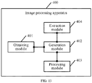
- a fourth aspect of the present disclosure provides an image processing apparatus, including:
- a fifth aspect of the present disclosure provides a computer-readable storage medium, storing instructions, where the instructions, when executed on a computer, causing the computer to execute the methods according to the foregoing aspects.
- the embodiments of the present disclosure provide the medical image processing method.
- the to-be-processed medical color image may be first obtained, where the to-be-processed medical image includes the first image data, the second image data, and the third image data, and the first image data, the second image data, and the third image data respectively represent color information of different attributes.
- the difference image is generated according to the first image data, the second image data, and the third image data.
- binarization processing is performed on the difference image, to obtain the binarized image, where the foreground region of the binarized image corresponds to the pathological tissue region of the to-be-processed medical image.
- Color information of a gray-scale pixel in different channels is slightly different, and color information of a color pixel in different channels is greatly different. Therefore, in the foregoing manner, a difference image is generated based on color information of different channels before binarization processing is performed on an image, thereby effectively using the color information in the image.
- the pathological tissue region extracted based on the difference image is more accurate, which facilitates subsequent image analysis.
- the embodiments of the present disclosure provide a medical image processing method, an image processing method and an apparatus, which generate a difference image based on color information of different channels before binarization processing is performed on an image, thereby effectively using the color information in the image.
- the pathological tissue region extracted based on the difference image is more accurate, which facilitates subsequent image analysis.
- the embodiments of the present disclosure may be applied to a scenario of image processing.
- An image as the visual basis for humans to perceive the world, is an important means for humans to obtain information, express information, and transfer information.
- Image processing is a technology that may be used to analyze an image to obtain a required result.
- Image processing generally refers to processing of a digital image, and the digital image is a large two-dimensional array obtained by an industrial camera, a video camera, or a scanner device.
- An element of the array is referred to as a pixel, and a value of a pixel is referred to as a gray-scale value.
- An image processing technology may help people to understand the world more objectively and accurately.
- the visual system of humans may help humans obtain a large amount of information from the outside world.
- Image processing technologies may include, but are not limited to, image transformation, image encoding compression, image enhancement and restoration, image segmentation, image description, image matting, and image classification.
- the image processing method provided in the present disclosure may be applied to a scenario of the medical field.
- a medical image that may be processed includes, but is not limited to, a brain image, a heart image, a chest image, and a cell image.
- a medical image may be affected by noise, the field offset effect, the partial volume effect, and tissue movement. Because individual creatures are different and have complex organizational structural shapes, a medical image is usually more blurred and uneven than an ordinary image.
- the medical image in the present disclosure is a color image, which may be a color ultrasonic image or a whole slide image (WSI), and may also include a color digital image obtained from a microscope.
- a WSI image is used as an example, and the a side of a WSI image usually includes 10000 pixels to 100000 pixels.
- a WSI image usually needs to be scaled down or cut into a small image for further processing.
- a region with a pathological tissue slide needs to be obtained, and then pathological analysis is performed according to the region, for example, nucleus quantitative analysis, cytomembrane quantitative analysis, cytoplasm quantitative analysis, and tissue microvascular analysis. Therefore, based on the feature of a medical image, in the medical image processing method in the present disclosure, the to-be-processed medical image may be obtained.
- the difference image may be generated based on the first image data, the second image data, and the third image data included in the to-be-processed medical image, where the first image data, the second image data, and the third image data respectively represent to color information of different attributes.
- binarization processing is performed on the difference image, to obtain the binarized image, where the foreground region of the binarized image corresponds to the pathological tissue region of the to-be-processed medical image.
- Color information of a gray-scale pixel in different channels is slightly different, while color information of a color pixel in different channels is greatly different. Therefore, a difference image is generated based on color information of different channels before binarization processing is performed on an image, thereby effectively using the color information in the image.
- the pathological tissue region extracted based on the difference image is more accurate, which facilitates subsequent image analysis.
- image processing may be further applied to a scenario of the remote sensing field.
- high-resolution remote sensing images may be applied to ocean monitoring, land coverage monitoring, marine pollution, and maritime rescue.
- High-resolution remote sensing images are characterized by abundant image detail information, obvious geometrical structures of surface features, and complex target structures. For example, in a high-resolution remote sensing image, shadows of objects on the coastline are complex, the vegetation coverage area is large, or artificial facilities are not clear enough.
- a high-resolution remote sensing image usually has more details and is more complex than an ordinary image, when the vegetation coverage area in the high-resolution remote sensing image needs to be determined, vegetation may be removed from the high-resolution remote sensing image, to determine the corresponding area.
- the difference image may be generated based on the first image data, the second image data, and the third image data included in the first to-be-processed image, where the first to-be-processed image is a color image, and the first image data, the second image data, and the third image data included in the first to-be-processed image respectively represent color information of a corresponding one of a plurality of channels.
- binarization processing is performed on the generated difference image, to obtain the binarized image, where the foreground region of the binarized image corresponds to the pathological tissue region of the to-be-processed medical image.
- a target object for example, a vegetation region
- the difference image is generated based on color information of different channels before binarization processing is performed on the image. Because color information of a gray-scale pixel in different channels is slightly different, while color information of a color pixel in different channels is greatly different, color information of the image are effectively used.
- the target object extracted based on the difference image is more accurate, and details of the high-resolution remote sensing image may be more precisely obtained, thereby improving processing accuracy of the high-resolution remote sensing image.
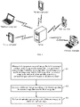
- FIG. 1 is a schematic architectural diagram of a medical image processing system according to an embodiment of the application.
- the image processing system includes a server and a terminal device.
- the medical image processing apparatus may be deployed on a server, or may be deployed on a terminal device with a high computing capability.
- the medical image processing apparatus is deployed on a server.
- the server obtains a to-be-processed medical image, then generates a difference image according to first image data, second image data, and third image data included in the to-be-processed medical image, and further performs binarization processing on the difference image, to obtain a binarized image, where a foreground region of the binarized image corresponds to a pathological tissue region of the to-be-processed medical image.
- the server may analyze the medical image based on the pathological tissue region.
- the medical image processing apparatus is deployed on a terminal device.
- the terminal device obtains a to-be-processed medical image, then generates a difference image according to first image data, second image data, and third image data included in the to-be-processed medical image, and further performs binarization processing on the difference image, to obtain a binarized image, where a foreground region of the binarized image corresponds to a pathological tissue region of the to-be-processed medical image.
- the terminal device may analyze the medical image based on the pathological tissue region.
- the server in FIG. 1 may be one server, or a server cluster including a plurality of servers, or a cloud computing center, which is not specifically limited herein.
- the terminal device may be a tablet computer, a notebook computer, a palmtop computer, a mobile phone, a personal computer (PC), or a voice interaction device shown in FIG. 1 , and may also be a monitoring device, a face recognition device, or the like, which is not limited herein.
- FIG. 1 only shows five terminal devices and one server, it is to be understood that, the example in FIG. 1 is only used for understanding this solution, and a specific quantity of the terminal devices and the servers is to be determined flexibly according to actual situations.
- AI is a theory, method, technology, or application system that uses a digital computer or a machine controlled by the digital computer to simulate, extend, and expand human intelligence, to perceive an environment, acquire knowledge, and use knowledge to obtain an optimal result.
- the AI is a comprehensive technology of computer science, which attempts to understand essence of intelligence and produces a new intelligent machine that can respond in a manner similar to human intelligence.
- the AI is to study the design principles and implementation methods of various intelligent machines, to enable the machines to have the functions of perception, reasoning, and decision-making.
- the AI technology is a comprehensive discipline, covering a wide range of fields including both a hardware-level technology and a software-level technology.
- Basic AI technologies generally include technologies such as sensors, dedicated AI chips, cloud computing, distributed storage, big data processing technologies, operating/interaction systems, and mechatronics.
- AI software technologies mainly include a computer vision technology, a speech processing technology, a natural language processing (NLP) technology, machine learning (ML)/deep learning, and the like.
- ML is an interapprity involving a plurality of disciplines such as the probability theory, statistics, approximation theory, convex analysis, algorithm complexity theory, and the like.
- ML specializes in studying how a computer simulates or implements a human learning behavior to obtain new knowledge or skills, and reorganize an existing knowledge structure, so as to keep improving its performance.
- Machine learning is the core of artificial intelligence, and the fundamental way to make a computer intelligent, and machine learning applications cover all fields of artificial intelligence.
- the ML and deep learning generally include technologies such as an artificial neural network, a belief network, reinforcement learning, transfer learning, inductive learning, and learning from demonstrations.
- the computer vision is a science that studies how to use a machine to "see” in many research directions of the AI technology, and furthermore, is machine vision that a camera and a computer are used for replacing human eyes to perform recognition, tracking, measurement, and the like on a target, and further perform graphic processing, so that the computer processes the target into an image more suitable for human eyes to observe, or an image transmitted to an instrument for detection.
- CV studies related theories and technologies and attempts to establish an AI system that can obtain information from images or multidimensional data.
- the CV technologies generally include technologies such as image processing, image recognition, image semantic understanding (ISU), image retrieval, optical character recognition (OCR), video processing, video semantic understanding, video content/behavior recognition, 3D object reconstruction, a 3D technology, virtual reality, augmented reality, synchronous positioning, and map construction, and further include biological feature recognition technologies such as common face recognition and fingerprint recognition.
- technologies such as image processing, image recognition, image semantic understanding (ISU), image retrieval, optical character recognition (OCR), video processing, video semantic understanding, video content/behavior recognition, 3D object reconstruction, a 3D technology, virtual reality, augmented reality, synchronous positioning, and map construction, and further include biological feature recognition technologies such as common face recognition and fingerprint recognition.
- a schematic diagram of a medical image processing method according to an embodiment of the present disclosure includes the following steps.
- to-be-processed medical image being a color image
- the to-be-processed medical image including first image data, second image data, and third image data, and the first image data, the second image data, and the third image data respectively representing color information of a corresponding one of a plurality of channels.
- the medical image processing apparatus may obtain a to-be-processed medical image that is a color image, where the to-be-processed medical image may include first image data, second image data, and third image data, and the first image data, the second image data, and the third image data respectively represent color information of a corresponding one of a plurality of channels.
- the to-be-processed medical image may be a medical image received by the medical image processing apparatus through a wired network, or may be a medical image stored by the medical image processing apparatus.
- the to-be-processed medical image may be a region clipped from a WSI image.
- the WSI image may be obtained by scanning a finished slide through a microscope. Because a finished slide is a glass slide produced by performing hematoxylin staining or other staining methods, a WSI image obtained after scanning a finished slide through a microscope is a color image.
- Image color modes of a color image include, but are not limited to, a red green blue (RGB) color mode, a luminance bandwidth chrominance (YUV) color mode, and a hue-saturation-value (HSV) color mode.
- Color information may be expressed as pixel values in different channels, for example, a pixel value on an R channel, a pixel value on a G channel, and a pixel value on a B channel.
- Formats of a WSI image include, but are not limited to, SVS and NDPI, while the length and the width of a WSI image usually is in the range of tens of thousands of pixels so that the image is large. Directly processing a WSI image requires a large memory. Therefore, a WSI image needs to be cut.
- a WSI image may be usually read through an openslide tool of python, and the openslide tool may implement file format conversion and may further store, as an image with a resolution of 12 ⁇ 12, a region clipped from a WSI image. In an actual case, resolutions include, but are not limited to, 15 ⁇ 15 and 50 ⁇ 50.
- a plurality of images are stored in the same WSI image file.
- an image with the maximum resolution is read from the WSI image file as a to-be-processed image.
- the to-be-processed medical image may be clipped from the WSI image that is scaled down, and the WSI image may be scaled down by any multiple, for example, 20 times or 10 times.
- the length and the width of the WSI image that is scaled down is in the range of several thousands of pixels. The multiple for scaling down is can be flexibly determined according to actual requirements.
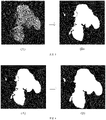
- FIG. 3 is a schematic diagram of a to-be-processed medical image according to an embodiment of the present disclosure.
- the to-be-processed medical image includes a pathological tissue region, without other gray-scale background or pure-white background.
- an image color mode of the to-be-processed medical image is RGB.
- the first image data, the second image data, and the third image data included in the to-be-processed medical image respectively represent to color information in different channels
- the first image data may have a pixel value 200 corresponding to an R channel
- the second image data may have a pixel value 100 corresponding to a G channel
- the third image data may have a pixel value 60 corresponding to a B channel.
- RGB corresponding to the color image is (100, 80, 40)
- the first image data may have a pixel value 100 corresponding to the R channel
- the second image data may have a pixel value 800 corresponding to the G channel
- the third image data may have a pixel value 40 corresponding to the B channel.
- An HSV image or a YUV image may be first converted into an RGB image for subsequent processing.
- the medical image processing apparatus may be deployed on a server, and may also be deployed on a terminal device with a high computing capability. In this embodiment, for example, the medical image processing apparatus is deployed on a server.
- the medical image processing apparatus may generate the difference image according to the first image data, the second image data, and the third image data.
- the difference image is represented as a gray-scale image.
- FIG. 4 is a schematic diagram of a difference image according to an embodiment of the present disclosure.
- a difference image shown in (B) in FIG. 4 may be generated according to first image data, second image data, and third image data included in a to-be-processed medical image in (A) in FIG. 4 .
- the difference image may be an image that includes a pathological tissue region shown in FIG. 4 . Because color information corresponding to different channels is used, the values of a pixel are different from each other.
- the image color mode of the to-be-processed medical image is RGB. If the to-be-processed medical image is gray, the R, G, B values are similar. If the to-be-processed medical image is a color image, the R, G, B values are greatly different, so that a region of a pathological tissue has a large chromatic aberration.
- the medical image processing apparatus may perform binarization processing on the difference image generated in step 102, to obtain the binarized image.
- a foreground region of the binarized image corresponds to a pathological tissue region of the to-be-processed medical image.
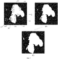
- FIG. 5 is a schematic diagram of a binarized image according to an embodiment of the present disclosure.
- a difference image shown in (A) of FIG. 5 because the difference image is a gray-scale image, a gray-scale image processing method may be performed.
- foreground is obtained by adaptive binarization.
- binarization processing is performed on the difference image, thereby obtaining the binarized image shown in (B) of FIG. 5 .
- white is a foreground region that includes the pathological tissue region
- black is a background region that does not include the pathological tissue region.
- FIG. 6 is a schematic diagram of a foreground region according to an embodiment of the present disclosure.
- the white region is a foreground region that includes the pathological tissue region
- the black region is a background region that does not include the pathological tissue region, so that a region corresponding to a to-be-processed medical image in (B) of FIG. 6 may be generated according to the binarized image.
- a medical image processing method is provided. Color information of a gray-scale pixel in different channels is slightly different, and color information of a color pixel in different channels is greatly different. Therefore, in the foregoing manner, a difference image is generated based on color information of different channels before binarization processing is performed on an image, thereby effectively using the color information in the image.
- the pathological tissue region extracted based on the difference image is more accurate, which facilitates subsequent image analysis.
- the generating the difference image according to the first image data, the second image data, and the third image data included in the to-be-processed medical image may include:
- the medical image processing apparatus may generate the maximum value image and the minimum value image according to the first image data, the second image data, and the third image data included in the to-be-processed medical image; and finally generate the difference image according to the maximum value image and the minimum value image.
- the image color mode of the to-be-processed medical image is RGB. Because the first image data, the second image data, and the third image data included in the to-be-processed medical image respectively represent to color information in different channels, the color information is expressed as pixel values corresponding to an R channel, a G channel, and a B channel. A maximum value in the R channel, the G channel, and the B channel is determined, and the maximum value image is determined based on the maximum value. Similarly, a minimum value in the R channels, the G channel, and the B channel may be determined, and a minimum value image may be determined based on the minimum value. Then, each pixel of the minimum value image is subtracted from a pixel in a corresponding location of the maximum value image, to obtain the difference image.
- This embodiment of the present disclosure provides a method for generating the difference image.
- the maximum value image and the minimum value image are generated according to the first image data, the second image data, and the third image data. Because the different image data corresponds to different color information and the maximum value image and the minimum value image are determined according to different image data, color information of the included to-be-processed medical image is highly accurate, and therefore the difference image is more accurately generated.
- the generating the maximum value image and the minimum value image according to the first image data, the second image data, and the third image data included in the to-be-processed medical image may include:
- a pixel value at a fourth pixel location of the maximum value image is the maximum pixel value
- a pixel value at a fifth pixel location of the minimum value image is the minimum pixel value
- the first pixel location, the second pixel location, the third pixel location, the fourth pixel location, and the fifth pixel location all correspond to the same pixel location of the to-be-processed medical image.
- the generating the difference image according to the maximum value image and the minimum value image may include:
- a minimum pixel value corresponding to a target pixel in the minimum value image is subtracted from a maximum pixel value corresponding to the target pixel in the maximum value image, to obtain a pixel difference, and the pixel difference is used as a difference pixel value corresponding to the target pixel in the difference image.
- the medical image processing apparatus may generate, according to the first image data, the second image data, and the third image data included in the to-be-processed medical image, the maximum pixel value and the minimum pixel value corresponding to the target pixel; and then generate the maximum value image and the minimum value image according to the determined maximum pixel value and minimum pixel value. Finally, the minimum pixel value corresponding to the target pixel in the minimum value image is subtracted from the maximum pixel value corresponding to the target pixel in the maximum value image, to obtain the difference pixel value corresponding to the target pixel in the difference image.
- the image color mode of the to-be-processed medical image is RGB.
- each pixel of the to-be-processed medical image has corresponding image data in all of an R channel, a G channel, and a B channel.
- image data of a pixel in the R channel is a first pixel value
- image data of the pixel in the G channel is a second pixel value
- image data of the pixel in the B channel is a third pixel value.
- a maximum pixel value and a minimum pixel value in the R channel, the G channel, and the B channel may be determined according to the first pixel value, the second pixel value, and the third pixel value.
- color information corresponding to different channels is used to distinguish values of a pixel.
- the image color mode of the to-be-processed medical image is RGB. If the to-be-processed medical image is gray, the R, G, B values are similar to each other. If the to-be-processed medical image is in color, the R, G, B values are greatly different from each other and a region with a pathological tissue is in color.
- a value of a color pixel is the value of a pixel to be obtained in this embodiment.
- the formulas are also applicable to calculation of a maximum pixel value and a minimum pixel value for a multi-dimensional image, for example, a 3-dimensional (3D) image and a 4-dimensional (4D) image.
- a target pixel location is (x1, y1) and the image color mode of the to-be-processed medical image is RGB.
- a first pixel value Ir (x1, y1) of the target pixel location (x1, y1) is 100
- a second pixel value Ig (x1, y1) of the target pixel location (x1, y1) is 200
- a third pixel value Ib (x1, y1) of the target pixel location (x1, y1) is 150.
- a maximum pixel value Imax (x1, y1) of the target pixel location (x1, y1) is the pixel value 200 corresponding to the second pixel value Ig (x1, y1)
- a minimum pixel value Imin (x1, y1) of the target pixel location (x1, y1) is the pixel value 100 corresponding to the first pixel value Ir (x1, y1).
- a target pixel location is (x2, y2) and the image color mode of the to-be-processed medical image is RGB.
- a first pixel value Ir (x2, y2) of the target pixel location (x2, y2) is 30, a second pixel value Ig (x2, y2) of the target pixel location (x2, y2) is 80, and a third pixel value Ib (x2, y2) of the target pixel location (x2, y2) is 120.
- a maximum pixel value Imax (x2, y2) of the target pixel location (x2, y2) is the pixel value 120 corresponding to the third pixel value Ib (x2, y2)
- a minimum pixel value Imin (x2, y2) of the target pixel location (x2, y2) is the pixel value 30 corresponding to the first pixel value Ir (x2, y2).
- the image color mode of the to-be-processed medical image is RGB
- the to-be-processed medical image is a 3D image
- a target pixel location is (x3, y3, z3).
- a first pixel value Ir (x3, y3, z3) of the target pixel location (x3, y3, z3) is 200
- a second pixel value Ig (x3, y3, z3) of the target pixel location (x3, y3, z3) is 10
- a third pixel value Ib (x3, y3, z3) of the target pixel location (x3, y3, z3) is 60.
- a maximum pixel value Imax (x3, y3, z3) of the target pixel location (x3, y3, z3) is the pixel value 200 corresponding to the first pixel value Ir (x3, y3, z3)
- a minimum pixel value Imin (x3, y3, z3) of the target pixel location (x3, y3, z3) is the pixel value 10 corresponding to the second pixel value Ig (x3, y3, z3).
- the minimum pixel value may be subtracted from the maximum pixel value, to obtain a difference pixel value corresponding to the target pixel location in the difference image.
- a target pixel location is (x1, y1) and the image color mode of the to-be-processed medical image is RGB.
- a maximum pixel value Imax (x1, y1) of the target pixel location (x1, y1) is 200
- a minimum pixel value Imin (x1, y1) of the target pixel location (x1, y1) is 100.
- the minimum pixel value Imin (x1, y1) may be subtracted from the maximum pixel value Imax (x1, y1), to obtain a difference pixel value 100 corresponding to the target pixel location (x1, y1).
- a target pixel location is (x2, y2) and the image color mode of the to-be-processed medical image is RGB.
- a maximum pixel value Imax (x2, y2) of the target pixel location (x2, y2) is 120, and a minimum pixel value Imin (x2, y2) of the target pixel location (x2, y2) is 30.
- the minimum pixel value Imin (x2, y2) may be subtracted from the maximum pixel value Imax (x2, y2), to obtain a difference pixel value 90 corresponding to the target pixel location (x2, y2).
- the image color mode of the to-be-processed medical image is RGB
- the to-be-processed medical image is a 3D image
- a target pixel location is (x3, y3, z3).
- Imax x y z Max Ir x y z , Ig x y z , Ib x y z
- Imin x y z Min Ir x y z , Ig x y z , Ib x y z
- Idiff x y z Imax x y z ⁇ Imin x y z .
- a maximum pixel value Imax (x3, y3, z3) of the target pixel location (x3, y3, z3) is 200
- a minimum pixel value Imin (x3, y3, z3) of the target pixel location (x3, y3, z3) is 10.
- the minimum pixel value Imin (x3, y3, z3) may be subtracted from the maximum pixel value Imax (x3, y3, z3), to obtain a difference pixel value 190 corresponding to the target pixel location (x3, y3, z3).
- the difference pixel value in the to-be-processed medical image when the difference pixel value in the to-be-processed medical image is small, it indicates that the first pixel value, the second pixel value, and the third pixel value in the to-be-processed medical image are similar. It may indicate that the to-be-processed medical image is similar to a gray image. When the difference pixel value in the to-be-processed medical image is large, it indicates that the first pixel value, the second pixel value, and the third pixel value in the to-be-processed medical image are greatly different. It may indicate that the to-be-processed medical image is similar to a color image. An image of a pathological tissue region is often a color image. Therefore, it may be preliminarily determined, according to the difference pixel value, whether the to-be-processed medical image includes a pathological tissue region.
- This embodiment of the present disclosure provides a method for generating a maximum value image and a minimum value image.
- a maximum pixel value and a minimum pixel value are determined based on pixel values of a target pixel corresponding to first image data, second image data, and third image data.
- the maximum pixel value and the minimum pixel value reflect color information of the to-be-processed medical image to some extent.
- the minimum pixel value is subtracted from the maximum pixel value to obtain a difference pixel value. Therefore, the difference pixel value can accurately reflect color information of the to-be-processed medical image, and therefore the difference image is more accurately generated.
- the generating the difference image according to the first image data, the second image data, and the third image data included in the to-be-processed medical image may include:
- the medical image processing apparatus may generate the to-be-processed difference image according to the first image data, the second image data, and the third image data included in the to-be-processed medical image, and then perform Gaussian blur processing on the to-be-processed difference image, to obtain the difference image.
- the blur processing may be understood as follows: for each pixel of the to-be-processed difference image, an average of pixels surrounding the pixel is obtained as a pixel value of the pixel. In this way, values of the pixels may tend to be smooth. This is equivalent to producing a blur effect on the to-be-processed difference image, by which details of the pixel are lost. Pixels of the to-be-processed difference image are continuous. Therefore, pixels that are close to each other are closely correlated, and pixels that are far away from each other are alienated from each other. Therefore, in this embodiment, the Gaussian blur (GB) is adopted to perform the blur processing.
- GB Gaussian blur
- Gaussian blur normal distribution (Gaussian distribution) may be used to process the to-be-processed difference image, so that weighted averaging of pixels is more appropriate. A pixel that is closer has a larger weight, and a pixel that is farther has a smaller weight.
- a pixel is (x, y).
- the pixel is specifically (0, 0). Then, 8 pixels surrounding the pixel (0, 0) may be (-1, 1), (0, 1), (1, 1), (-1, 0), (1, 0), (-1, -1), (0, -1), and (1, -1).
- a weight corresponding to the pixel (0, 0) is 0.0707
- a weight corresponding to the pixel (-1, 1) is 0.0453
- a weight corresponding to the pixel (0, 1) is 0.0566
- a weight corresponding to the pixel (1, 1) is 0.0453
- a weight corresponding to the pixel (-1, 0) is 0.0566
- a weight corresponding to the pixel (1, 0) is 0.0566
- a weight corresponding to the pixel (-1, -1) is 0.0453
- a weight corresponding to the pixel (0, -1) is 0.0566
- a weight corresponding to the pixel (1, -1) is 0.0453.
- a sum of the weights of the pixel (0, 0) and the 8 surrounding pixels is approximately 0.479. If only a weighted average of these 9 points is calculated, the sum of weights of these 9 points needs to be 1, that is, the sum of the weights is normalized. For example, the 9 values corresponding to the weight matrix may be divided by the sum 0.479 of the weights, to obtain the normalized weight matrix.
- a normalized weight corresponding to the pixel (0, 0) is 0.147
- a normalized weight corresponding to the pixel (-1, 1) is 0.0947
- a normalized weight corresponding to the pixel (0, 1) is 0.0118
- a weight corresponding to the pixel (1, 1) is 0.0947
- a normalized weight corresponding to the pixel (-1, 0) is 0.0118
- a normalized weight corresponding to the pixel (1, 0) is 0.0118
- a normalized weight corresponding to the pixel (-1, -1) is 0.0947
- a normalized weight corresponding to the pixel (0, -1) is 0.0118
- a normalized weight corresponding to the pixel (1, -1) is 0.0947
- a weight matrix whose weight sum is greater than 1 makes the difference image bright, and a weight matrix whose weight sum is less than 1 makes the difference image dark. Therefore, the normalized weight matrix can make a pathological tissue region presented by the difference image more accurate.
- Gaussian blur calculation may be performed on the pixel.
- a gray-scale value corresponding to the pixel (0, 0) is 25
- a gray-scale value corresponding to the pixel (-1, 1) is 14
- a gray-scale value corresponding to the pixel (0, 1) is 15, a gray-scale value corresponding to the pixel (1, 1) is 16,
- a gray-scale value corresponding to the pixel (-1, 0) is 24,
- a gray-scale value corresponding to the pixel (1, 0) is 26
- a gray-scale value corresponding to the pixel (-1, -1) is 34
- a gray-scale value corresponding to the pixel (0, -1) is 35
- a gray-scale value corresponding to the pixel (1, -1) is 36.
- the gray-scale value is multiplied with a weight corresponding to the pixel, to obtain 9 values.
- 3.69 may be obtained for the pixel (0, 0)
- 1.32 may be obtained for the pixel (-1, 1)
- 1.77 may be obtained for the pixel (0, 1)
- 1.51 may be obtained for the pixel (1, 1)
- 2.83 may be obtained for the pixel (-1, 0)
- 3.07 may be obtained for the pixel (1, 0)
- 3.22 may be obtained for the pixel (-1, -1)
- 4.14 may be obtained for the pixel (0, -1)
- 3.41 may be obtained for the pixel (1, -1).
- the 9 values are added to obtain a Gaussian blur value of the pixel (0, 0).
- Steps performed for the pixel (0, 0) may be performed on all pixels included in the to-be-processed difference image, to obtain the difference image processed by Gaussian blur processing.
- This embodiment of the present disclosure provides another method for generating the difference image.
- Gaussian blur processing is performed on the generated to-be-processed difference image. Because Gaussian blur processing can improve processing robustness, the obtained difference image has better processing robustness, thereby improving stability of the difference image.
- the performing binarization processing on the difference image to obtain the binarized image may include:
- the medical image processing apparatus may determine the binarization threshold according to the difference image, when a pixel value corresponding to a pixel in the difference image is greater than or equal to the binarization threshold, determine the pixel as a foreground pixel of the binarized image, and when a pixel value corresponding to a pixel in the difference image is less than the binarization threshold, determine the pixel as a background pixel of the binarized image.
- the binarization threshold may be set to perform binarization processing on the difference image, to convert a gray-scale image to a binarized image with values 0 or 1.
- the binarization threshold may be set to convert the difference image to the binarized image whose image foreground and image background are respectively represented by only two values (0 or 1).
- a foreground value is 1 and a background value is 0.
- 0 corresponds to that RGB values are all 0, and 1 corresponds to that RGB values are all 255.
- Binarization processing is performed on the difference image to obtain the binarized image.
- Methods for determining the binarization threshold may include a global threshold method and a local threshold method.
- the global threshold method means that the entire difference image is divided according to one threshold.
- gray-scale depths of different difference images are different.
- brightness distribution of different parts of the same difference image may also be different. Therefore, in this embodiment, the binarization threshold is determined by using a dynamic threshold binarization method.
- a pixel value corresponding to a pixel in the difference image is compared with the binarization threshold.
- the pixel is determined as a foreground pixel of the binarized image.
- the pixel is determined as a background pixel of the binarized image. For example, when a pixel value corresponding to a pixel A is greater than the binarization threshold, the pixel A is determined as a foreground pixel of the binarized image, that is, a pixel value is 1.
- the pixel A is in a foreground region and is displayed in white when the image is in an RGB mode.
- the pixel B is determined as a background pixel of the binarized image, that is, a pixel value is 0.
- the pixel B is in a background region and is displayed in black when the image is in an RGB mode.
- This embodiment of the present disclosure provides a method for obtaining the binarized image.
- the binarized image is generated by binarization processing. Because a geometric property of the binarized image is not related to a gray-scale value of a pixel, subsequent processing of the binarized image may become simple, thereby improving the efficiency of generating a result image.
- the determining the binarization threshold according to the difference image may include:
- the medical image processing apparatus may obtain, according to the difference image, the N pixel values corresponding to the N pixels, where the pixel values and the pixels are in a one-to-one correspondence, then determine the reference pixel value from the N pixel values, where the reference pixel value is a maximum value of the N pixel values, and finally may calculate the binarization threshold according to the reference pixel value and the preset ratio, where N is an integer greater than 1.
- the binarization threshold is determined according to the difference image.
- the difference image may be generated by subtracting the minimum value image from the maximum value image in the to-be-processed medical image, and the pixel values and the pixels in the difference image are in a one-to-one correspondence. Therefore, pixel values corresponding to a plurality of pixels in the difference image may be obtained. Then, a maximum value of the plurality of pixel values is determined as the reference pixel value. Then, the binarization threshold is calculated according to the reference pixel value and the preset ratio.
- the preset ratio is 10%.
- the length and the width of the WSI image that is scaled down is in the range of several thousands of pixels.
- the image that is scaled down includes 100 ⁇ 100 pixels, that is, a maximum value needs to be found from pixel values corresponding to the 10000 pixels.
- the maximum value is 150.
- the reference pixel value 150 may be multiplied by the preset ratio 10%, to obtain the binarization threshold 15.
- the preset ratio further may be another percentage, which may be flexibly determined according to actual situations.
- the binarization threshold may be determined based on the reference pixel value determined as the maximum pixel value and the preset ratio. Gray-scale depths of difference images are different. Besides, brightness distribution of different regions may also be different. Therefore, the binarization threshold may be flexibly determined by adjusting the preset ratio, to improve threshold accuracy and flexibility of the threshold, so as to improve the accuracy of the binarized image.
- the method may further include:
- the medical image processing apparatus may detect a background region of the binarized image based on a flood algorithm, where the background region may include a plurality of background pixels, then obtain a background pixel in the foreground region of the binarized image according to the binarized image and the background region of the binarized image, where the foreground region may include a plurality of foreground pixels, then change the background pixel in the foreground region of the binarized image to a foreground pixel to obtain the hole filled image, and finally may perform median filtering processing on the hole filled image, to obtain the result image.
- a foreground region of the result image corresponds to a pathological tissue region of the to-be-processed medical image.
- a foreground region in the obtained binarized image may include a black hole.
- the black hole needs to be detected in the foreground region.
- FIG. 7 is a schematic diagram of a result image according to an embodiment of the present disclosure.
- a white foreground region in the binarized image shown in (A) of FIG. 7 includes a plurality of background pixels. Black dots in the region A1 to region A5 are all formed by background pixels. These black dots in the region A1 to the region A5 are changed from background pixels to foreground pixels.
- White points in the region A6 and the region A7 are formed by foreground pixels. These white points in the region A6 and the region A7 are changed from foreground pixels to background pixels. In this way, a hole filled image shown in (B) of FIG. 7 may be obtained.
- median filtering processing is then performed on the hole filled image shown in (B) of FIG. 7 , and morphological processing may be further performed.Then, a result image shown in (C) in FIG. 7 may be obtained.
- Filtering processing is to suppress noise of the to-be-processed medical image while maintaining detail features of the hole filled image as much as possible, and filtering processing can improve effectiveness and reliability of subsequent processing and analysis of the result image.
- the filtering operation is to eliminate a noise component in the hole filled image. Energy of the hole filled image is mostly concentrated in low and intermediate bands of an amplitude spectrum. In a high band, information of the hole filled image is often affected by noise.
- a filtering operation may be performed on the hole filled image, to meet an image processing requirement and eliminate noise generated when the image is digitalized.
- Median filtering processing is typical non-linear filtering, and can effectively suppress noise based on a sorting statistics theory.
- a median value of neighborhood gray-scale values of a pixel may replace a gray-scale value of the pixel, so that surrounding pixel values are close to real values to eliminate isolated noise points.
- This embodiment of the present disclosure provides a method for generating the result image.
- the background pixels in the foreground region are changed to the foreground pixels, so that the obtained hole filled image is highly reliable.
- the result image corresponding to the to-be-processed medical image is clear and has a desirable visual effect without loss of feature information such as the outline and the edge of the image.
- the performing median filtering processing on the hole filled image may include:
- the medical image processing apparatus performs median filtering processing on the hole filled image, where the filtered image may include a to-be-processed foreground region, obtains a boundary line of the foreground region of the filtered image, where the boundary line includes M pixels, and extends each of the M pixels on the boundary line outwards by K pixels to obtain the result image, where M is an integer greater than 1 and K is an integer greater than or equal to 1.
- a median value of neighborhood gray-scale values of a pixel may replace a gray-scale value of the pixel, so that surrounding pixel values are close to real values to eliminate isolated noise points.
- image edge details of the filtered image are reserved while impulse noise and salt and pepper noise are eliminated.
- a flood algorithm (Flood Fill).
- the basic principle of the flood algorithm is starting from a pixel and expanding and coloring to surrounding pixels until the boundary of a graph.
- the flood algorithm needs to use three parameters: a start node, a target color, and a replacement color.
- all nodes that are connected to the start node through a path of the target color are detected and changed to the replacement color.
- the flood algorithm may be constructed in a plurality of manners which explicitly or implicitly use a queue data structure or a stacking data structure. Examples are a four-neighborhood flood algorithm, an eight-neighborhood flood algorithm, a scanline fill algorithm, and a large-scale behavior algorithm.
- the idea of a conventional four-neighborhood flood algorithm is as follows: after a pixel (x, y) is colored, the upper, lower, left, and right points around the pixel are respectively colored. However, a large part of a memory will be consumed if performing the algorithm in a recursive manner. If an area to be colored is very large, overflow is caused. Therefore, the four-neighborhood flood algorithm may be used in a non-recursive manner.
- the eight-neighborhood flood algorithm colors the pixels at upper, lower, left, right, upper left, lower left, upper right, and lower right locations of a pixel.
- the scanline fill algorithm may accelerate the coloring by using a filling line, in which pixels on a line may be first colored, and then the coloring expands upwards and downwards until the end of coloring. Large-scale behavior is data-centered or procedurecentered.
- a to-be-processed foreground region includes a boundary line of 1000 pixels.
- the 1000 pixels are respectively extended outwards by K pixels through morphological processing. Assuming that K is 2, 2000 pixels are added to the foreground region in addition to the original 1000 pixels, to obtain the result image.
- specific values of M and K should all be flexibly determined according to actual situations.
- This embodiment of the present disclosure provides another method for generating the result image.
- the filtered image is clear and has a desirable visual effect without loss of feature information such as the outline and the edge of the image.
- morphological processing is performed on the filtered image based on the flood algorithm, to improve accuracy and integrity of the result image.
- the obtaining the to-be-processed medical image may include:
- the medical image processing apparatus may first obtain the original medical image, extract the medical sub-image from the original medical image by using the sliding window, when detecting that the medical sub-image includes the pathological tissue region, determine the medical sub-image as a to-be-processed medical image, and when detecting that the medical sub-image does not include the pathological tissue region, determine the medical sub-image as a background image and remove the background image.
- the original medical image may be an image received by the medical image processing apparatus through a wired network, or may be an image stored by the medical image processing apparatus.
- FIG. 8 is a schematic diagram of obtaining a to-be-processed medical image according to an embodiment of the present disclosure.
- (A) of FIG. 8 shows an original medical image.
- a medical sub-image is extracted from the original medical image by using a sliding window.
- Regions of B1 to B3 are medical sub-images extracted from the original medical image.
- a medical sub-image shown in (B) of FIG. 8 may be correspondingly obtained from B1
- a medical sub-image shown in (C) of FIG. 8 may be correspondingly obtained from B2
- a medical sub-image shown in (D) of FIG. 8 may be correspondingly obtained from B3.
- the medical sub-images shown in (B) and (C) of FIG. 8 include a pathological tissue region. Therefore, the medical sub-images shown in (B) and (C) of FIG. 8 may be determined as to-be-processed medical images. However, the medical sub-image shown in (D) of FIG. 8 does not include a pathological tissue region. Therefore, the medical sub-image shown in (D) of FIG. 8 may be determined as a background image and the background image is removed.
- This embodiment of the present disclosure provides a method for obtaining the to-be-processed medical image.
- it is detected whether the medical sub-image includes the pathological tissue region, to determine the to-be-processed medical image, and the result image corresponding to the to-be-processed medical image that includes the pathological tissue region may be obtained through the foregoing steps.
- the result image includes the pathological tissue region. This facilitates subsequent processing and analysis of the pathological tissue region in the result image.
- the medical sub-image that does not include the pathological tissue region is determined as a background image and the background image is removed, to reduce resource occupation.
- the medical image processing method may further include:
- the medical image processing apparatus may further generate the target positive sample image according to the result image, where the target positive sample image is a positive sample image in the positive sample set, and each positive sample image includes a pathological tissue region. Also, the medical image processing apparatus may obtain the negative sample set, where the negative sample set includes at least one negative sample image, and each negative sample image does not include a pathological tissue region. Finally, the medical image processing apparatus may train the image processing model based on the obtained positive sample set and negative sample set. The image processing model may perform processing to obtain a pathological tissue region based on a medical color image.
- This embodiment of the present disclosure provides a method for training the image processing model.
- the image processing model is trained based on the positive sample image set that includes the pathological tissue region and the negative sample set that does not include the pathological tissue region. This improves accuracy and reliability of the image processing model, thereby improving image processing efficiency and accuracy.
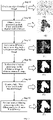
- FIG. 9 is a schematic flowchart of a medical image processing method according to an embodiment of the present disclosure, which includes the following steps.
- step S1 an original medical image as shown in (A) of FIG. 9 may be obtained. Then, in step S2, a medical sub-image is extracted from the original medical image shown in (A) of FIG. 9 by using a sliding window. When it is detected that the medical sub-image includes a pathological tissue region, the medical sub-image is determined as a to-be-processed medical image, to obtain a to-be-processed medical image as shown in (B) of FIG. 9 .
- a maximum pixel value and a minimum pixel value corresponding to a target pixel may be determined among a first pixel value, a second pixel value, and a third pixel value according to first image data, second image data, and third image data included in the to-be-processed medical image, to generate a maximum value image and a minimum value image. Then, a difference image as shown in (C) of FIG. 9 is obtained according to the maximum value image and the minimum value image. Further, in step S4, N pixel values corresponding to N pixels may be obtained according to the difference image as shown in (C) of FIG. 9 , where the pixel values and the pixels are in a one-to-one correspondence.
- a maximum value of the N pixel values is determined as a reference pixel value, and a binarization threshold is calculated according to the reference pixel value and the preset ratio.
- a binarization threshold is calculated according to the reference pixel value and the preset ratio.
- the pixel is determined as a foreground pixel of the binarized image, and when a pixel value corresponding to a pixel in the difference image is less than the binarization threshold, the pixel is determined as a background pixel of the binarized image, to obtain a binarized image as shown in (D) of FIG. 9 .
- step S5 a background region including a plurality of background pixels in the binarized image is detected based on a flood algorithm, and background pixels in a foreground region of the binarized image are obtained according to the binarized image and the background region of the binarized image, and the background pixels in the foreground region of the binarized image are changed to foreground pixels to obtain a hole filled image as shown in (E) of FIG. 9 .
- step S6 median filtering processing is performed on the hole filled image, to obtain a filtered image that includes a to-be-processed foreground region, then a boundary line including M pixels in the to-be-processed foreground region is obtained, and each of the M pixels on the boundary line is extended outwards by K pixels to obtain a result image as shown in (F) of FIG. 9 , where M is an integer greater than 1.
- FIG. 10 is a schematic diagram of a result image according to an embodiment of the present disclosure.
- a result image shown in (B) of FIG. 10 may be obtained through the medical image processing method provided in the embodiments of the present disclosure.
- (C) of FIG. 10 shows a to-be-processed medical image with regular vertical streaks. The regular vertical streaks are produced when a scanner scans a glass slide, which depends on a scanning device.
- FIG. 10 may be obtained through the medical image processing method provided in the embodiments of the present disclosure.
- (E) of FIG. 10 shows a to-be-processed medical image with black and white streaks.
- the black and white streaks may be generated through format conversion, or may be added to an unclear region generated when a scanner scans a glass slide.
- a result image shown in (F) of FIG. 10 may be obtained through the medical image processing method provided in the embodiments of the present disclosure.
- the difference image is generated based on color information of different channels before binarization processing is performed on the image.
- color information of a gray-scale pixel in different channels is slightly different, and color information of a color pixel in different channels is greatly different, color information of various to-be-processed medical images in FIG. 10 may be effectively used, so that the pathological tissue region extracted based on the difference image is more accurate, which facilitates subsequent image analysis.
- FIG. 11 is a schematic diagram of an image processing method according to an embodiment of the present disclosure. As shown in FIG. 11 , an embodiment of the image processing method in the present disclosure includes:
- an image processing apparatus may obtain the first to-be-processed image and the second to-be-processed image, where the first to-be-processed image may include first image data, second image data, and third image data, and the first image data, the second image data, and the third image data respectively represent color information of a corresponding one of a plurality of channels.
- the first to-be-processed image and the second to-be-processed image may be images received by the image processing apparatus through a wired network, or may be images stored by the image processing apparatus.
- the first to-be-processed image is similar to the to-be-processed medical image described in step 101. Details are not described herein again.
- the image processing apparatus may be deployed on a server, and may also be deployed on a terminal device with a high computing capability. In this embodiment, for example, the image processing apparatus is deployed on a server.
- the first to-be-processed image is an image shot on a cloudy day
- the background of the image is cloudy sky and further includes a red car
- the second to-be-processed image is an image of blue sky and sea.
- the image processing apparatus may generate a difference image according to the first image data, the second image data, and the third image data included in the first to-be-processed image obtained in step 201.
- the difference image is a gray-scale image.
- a method for generating the difference image in this embodiment is similar to that in the embodiment corresponding to FIG. 2 . Details are not described herein again.
- the difference image generated in this case may show the outline of the car.
- the image processing apparatus may perform binarization processing on the difference image generated in step 202, to obtain the binarized image.
- foreground processing is performed in an adaptive binarization manner, that is, binarization processing is performed on the difference image, thereby obtaining the binarized image.
- a method for generating the binarized image in this embodiment is similar to that in the embodiment corresponding to FIG. 2 . Details are not described herein again.
- the binarized image generated in this case can accurately show the outline of the car.
- the image processing apparatus may extract the target object from the first to-be-processed image according to the foreground region generated in step 203.
- the target object may be a pathological tissue region.
- the target object may be a vegetation region.
- the target object may be a bicycle or a car.
- the image of the car may be extracted from the first to-be-processed image, that is, the image of the car is the target object.
- the image processing apparatus sets the target object as the first image layer and sets the second to-be-processed image as the second image layer, and use the first image layer to cover the second image layer, to generate a synthesized image.
- the image of the car covers the image of blue sky and white cloud to form a synthesized image. It can be seen on the image that the background of the car is no longer the cloudy sky, but the blue sky and white cloud.
- an image processing method is provided.
- Color information of a gray-scale pixel in different channels is slightly different, and color information of a color pixel in different channels is greatly different. Therefore, in the foregoing manner, a difference image is generated based on color information of different channels before binarization processing is performed on an image, thereby effectively using the color information in the image.
- the target object extracted based on the difference image is more accurate.
- the target object in the generated synthesized image is accurate, thereby improving accuracy of the synthesized image and facilitating subsequent image analysis.
- FIG. 12 is a schematic diagram of a medical image processing apparatus according to an embodiment of the present disclosure.
- the medical image processing apparatus 300 includes:
- a medical image processing method is provided. Color information of a gray-scale pixel in different channels is slightly different, and color information of a color pixel in different channels is greatly different. Therefore, in the foregoing manner, a difference image is generated based on color information of different channels before binarization processing is performed on an image, thereby effectively using the color information in the image.
- the pathological tissue region extracted based on the difference image is more accurate, which facilitates subsequent image analysis.
- the generation module 302 is further configured to:
- This embodiment of the present disclosure provides a method for generating the difference image.
- the maximum value image and the minimum value image are generated according to the first image data, the second image data, and the third image data. Because different image data corresponds to different color information, the maximum value image and the minimum value image determined according to different image data include accurate color information of the to-be-processed medical image, and therefore the difference image is accurately generated.
- the generation module 302 is further configured to
- This embodiment of the present disclosure provides a method for generating a maximum value image.
- a maximum pixel value and a minimum pixel value are determined based on pixel values of a target pixel corresponding to first image data, second image data, and third image data.
- the maximum pixel value and the minimum pixel value reflect color information of the to-be-processed medical image to some extent.
- the minimum pixel value is subtracted from the maximum pixel value to obtain a difference pixel value. Therefore, the difference pixel value can accurately reflect color information of the to-be-processed medical image, and therefore the difference image is more accurately generated.
- the generation module 302 is further configured to:
- This embodiment of the present disclosure provides another method for generating the difference image.
- Gaussian blur processing is performed on the generated to-be-processed difference image. Because Gaussian blur processing can improve robustness, the obtained difference image has better robustness, thereby improving stability of the difference image.
- the medical image processing apparatus 300 further includes a determining module 304.
- the determining module 304 is configured to determine a binarization threshold according to the difference image.
- the determining module 304 is further configured to perform binarization processing on the difference image according to the binarization threshold, to obtain the binarized image.
- This embodiment of the present disclosure provides a method for obtaining the binarized image.
- the binarized image is generated by binarization processing. Because a geometric property of the binarized image is not related to a gray-scale value of a pixel, subsequent processing of the binarized image may become simple, thereby improving the efficiency of generating the foreground region.
- the determining module 304 is further configured to:
- the binarization threshold may be determined based on the reference pixel value determined as the maximum pixel value and the preset ratio. Gray-scale depths of difference images are different. Besides, brightness distribution of different regions may also be different. Therefore, the binarization threshold may be flexibly determined by adjusting the preset ratio, to improve accuracy and flexibility of the threshold, so as to improve the accuracy of the binarized image.
- the generation module 302 is further configured to:
- This embodiment of the present disclosure provides a method for generating the result image.
- the background pixels in the foreground region are changed to the foreground pixels, so that the obtained hole filled image is highly reliable.
- the result image corresponding to the to-be-processed medical image is clear and has a desirable visual effect without loss of feature information such as the outline and the edge of the image.
- the processing module 303 is further configured to:
- This embodiment of the present disclosure provides another method for generating the result image.
- the filtered image is clear and has a desirable visual effect without loss of feature information such as the outline and the edge of the image.
- morphological processing is performed on the filtered image based on the flood algorithm, to improve accuracy and integrity of the result image.
- the obtaining module 301 is further configured to:
- This embodiment of the present disclosure provides a method for obtaining the to-be-processed medical image.
- it is detected whether the medical sub-image includes the pathological tissue region, to determine the to-be-processed medical image, and the result image corresponding to the to-be-processed medical image that includes the pathological tissue region may be obtained through the foregoing steps.
- the result image includes the pathological tissue region. This facilitates subsequent processing and analysis of the pathological tissue region in the result image.
- the medical sub-image that does not include the pathological tissue region is determined as a background image and the background image is removed, to reduce resource occupation.
- the image processing apparatus 300 further includes a training module 305, where
- This embodiment of the present disclosure provides a method for training the image processing model.
- the image processing model is trained based on the positive sample image set that includes the pathological tissue region and the negative sample set that does not include the pathological tissue region. This improves accuracy and reliability of the image processing model, thereby improving image processing efficiency and accuracy.
- FIG. 13 is a schematic diagram of an image processing apparatus according to an embodiment of the present disclosure.
- the image processing apparatus 400 includes:
- an image processing method is provided.
- Color information of a gray-scale pixel in different channels is slightly different, and color information of a color pixel in different channels is greatly different. Therefore, in the foregoing manner, a difference image is generated based on color information of different channels before binarization processing is performed on an image, thereby effectively using the color information in the image.
- the target object extracted based on the difference image is more accurate.
- the target object in the generated synthesized image is accurate, thereby improving accuracy of the synthesized image and facilitating subsequent image analysis.
- FIG. 14 is a schematic structural diagram of a server according to an embodiment of the present disclosure.
- the server 500 may vary greatly due to different configurations or performance, and may include one or more central processing units (CPU) 522 (for example, one or more processors) and a memory 532, and one or more storage media 530 (for example, one or more mass storage devices) that store applications 542 or data 544.
- the memory 532 and the storage medium 530 may be temporary storage or persistent storage.
- a program stored in the storage medium 530 may include one or more modules (which are not marked in the figure), and each module may include a series of instruction operations on the server.
- the CPU 522 may be set to communicate with the storage medium 530, and perform, on the server 500, the series of instruction operations in the storage medium 530.
- the server 500 may further include one or more power supplies 525, one or more wired or wireless network interfaces 550, one or more input/output interfaces 558, and/or one or more operating systems 541, for example, Windows Server TM , Mac OS X TM , Unix TM , Linux TM , or FreeBSD TM .
- the steps performed by the server in the foregoing embodiment may be based on the structure of the server shown in FIG. 14 .
- the CPU 522 is configured to perform steps performed by the medical image processing apparatus in the embodiment corresponding to FIG. 2 , and the CPU 522 is further configured to perform steps performed by the image processing apparatus in the embodiment corresponding to FIG. 1 .
- the disclosed system, apparatus, and method may be implemented in other manners.
- the described apparatus embodiment is merely an example.
- the unit division is merely logical function division and may be other division during actual implementation.
- a plurality of units or components may be combined or integrated into another system, or some features may be omitted or not performed.
- the displayed or discussed mutual couplings or direct couplings or communication connections may be implemented as indirect couplings or communication connections via some interfaces, apparatuses or units, and may be implemented in electronic, mechanical, or other forms.
- the units described as separate parts may or may not be physically separate, and parts displayed as units may or may not be physical units, may be located in one position, or may be distributed on a plurality of network units. Some or all of the units may be selected according to actual requirements to achieve the objectives of the solutions of the embodiments.
- functional units in the embodiments of the present disclosure may be integrated into one processing unit, or each of the units may be physically separated, or two or more units may be integrated into one unit.
- the integrated unit may be implemented in the form of hardware, or may be implemented in the form of a software functional unit.
- the integrated unit When the integrated unit is implemented in the form of a software functional unit and sold or used as an independent product, the integrated unit may be stored in a computer-readable storage medium. Based on such an understanding, the technical solutions of the present disclosure essentially, or the part contributing to the prior art, or all or some of the technical solutions may be implemented in the form of a software product.
- the software product is stored in a storage medium and includes several instructions for instructing a computer device (which may be a personal computer, a server, a network device, or the like) to perform all or some of the steps of the methods described in the embodiments of the present disclosure.
- the foregoing storage medium includes: any medium that can store program code, such as a USB flash drive, a removable hard disk, a read-only memory (read-only memory, ROM), a random access memory (random access memory, RAM), a magnetic disk or an optical disc.
- program code such as a USB flash drive, a removable hard disk, a read-only memory (read-only memory, ROM), a random access memory (random access memory, RAM), a magnetic disk or an optical disc.
Abstract
Description
- This application claims priority to
Chinese Patent Application No. 202010084678.8, entitled "MEDICAL IMAGE PROCESSING METHOD, IMAGE PROCESSING METHOD AND APPARATUS" and filed with the China National Intellectual Property Administration on February 10, 2020 - The present disclosure relates to the artificial intelligence field, and specifically, to image processing technologies.
- With the development of medical technologies, recognition and analysis based on whole slide images (WSI) play an important role in medical treatment. Because a side of a WSI image usually includes tens of thousands of pixels, it is necessary to scale down or cut this type of image into a small image for analysis. In this process, most of a background region of the WSI image needs to be removed to obtain a region with a pathological tissue slide for subsequent image analysis.
- Currently, the pathological tissue region is extracted from the WSI image mainly in the following manner of first scaling down the WSI image to a particular size, then converting the image into a gray-scale image, then performing further image processing on the gray-scale image, for example, image binarization processing and hole removal processing, and finally extracting a pathological tissue region from the processed image.
- However, the foregoing manner of directly converting the color image into the gray-scale image after changing the size causes loss of color information, and the color information is also an important image feature. As a result, the extracted pathological tissue region is not accurate, which easily causes errors in subsequent image analysis.
- The present disclosure provides a medical image processing method, a medical image processing apparatus, an image processing method and an image processing apparatus, to generate a difference image based on color information of different channels before binarization processing is performed on an image, thereby effectively using the color information in the image. The pathological tissue region extracted based on the difference image is more accurate, which facilitates subsequent image analysis.
- In view of this, a first aspect of the present disclosure provides a medical image processing method, executable by a server, including:
- obtaining a to-be-processed medical image, the to-be-processed medical image being a color image, the to-be-processed medical image including first image data, second image data, and third image data, and the first image data, the second image data, and the third image data respectively representing color information of different attributes;
- generating a difference image according to the first image data, the second image data, and the third image data; and
- performing binarization processing on the difference image, to obtain a binarized image, a foreground region of the binarized image corresponding to a pathological tissue region of the to-be-processed medical image.
- A second aspect of the present disclosure provides an image processing method, executable by a server, including:
- obtaining a first to-be-processed image and a second to-be-processed image, the first to-be-processed image being a color image, the first to-be-processed image including first image data, second image data, and third image data, and the first image data, the second image data, and the third image data respectively representing color information of a corresponding one of a plurality of channels;
- generating a difference image according to the first image data, the second image data, and the third image data;
- performing binarization processing on the difference image, to obtain a binarized image, a foreground region of the binarized image corresponding to a pathological tissue region of the to-be-processed medical image; extracting the pathological tissue region from the first to-be-processed image according to the foreground region of the binarized image; and
- generating a synthesized image according to the pathological tissue region and the second to-be-processed image, where the pathological tissue region is on a first image layer, the second to-be-processed image is on a second image layer, and the first image layer covers the second image layer.
- A third aspect of the present disclosure provides a medical image processing apparatus, including:
- an obtaining module, configured to obtain a to-be-processed medical image, the to-be-processed medical image being a color image, the to-be-processed medical image including first image data, second image data, and third image data, and the first image data, the second image data, and the third image data respectively representing color information of a corresponding one of a plurality of channels;
- a generation module, configured to generate a difference image according to the first image data, the second image data, and the third image data; and
- a processing module, configured to perform binarization processing on the difference image, to obtain a binarized image, a foreground region of the binarized image corresponding to a pathological tissue region of the to-be-processed medical image.
- In a possible design, in an implementation of the third aspect of the embodiments of the present disclosure,
- the generation module is configured to generate a maximum value image and a minimum value image according to the first image data, the second image data, and the third image data included in the to-be-processed medical image; and
- generate the difference image according to the maximum value image and the minimum value image.
- In a possible design, in another implementation of the third aspect of the embodiments of the present disclosure,
- the generation module is configured to determine a maximum pixel value and a minimum pixel value according to a first pixel value at a first pixel location of the first image data, a second pixel value at a second pixel location of the second image data, and a third pixel value at a third pixel location of the third image data; and
- obtain a maximum value image according to the maximum pixel value, and obtain a minimum value image according to the minimum pixel value, where a pixel value at a fourth pixel location of the maximum value image is the maximum pixel value, a pixel value at a fifth pixel location of the minimum value image is the minimum pixel value, and the first pixel location, the second pixel location, the third pixel location, the fourth pixel location, and the fifth pixel location all correspond to the same pixel location of the to-be-processed medical image; and
- the generation module is further configured to determine a pixel difference according to the pixel value at the fourth pixel location of the maximum value image and the pixel value at the fifth pixel location of the minimum value image; and
- obtain the difference image according to the pixel difference, where a pixel value at a sixth pixel location of the difference image is the pixel difference, and the fourth pixel location, the fifth pixel location, and the sixth pixel location all correspond to the same pixel location of the to-be-processed medical image.
- In a possible design, in another implementation of the third aspect of the embodiments of the present disclosure,
- the generation module is further configured to generate a to-be-processed difference image according to the first image data, the second image data, and the third image data; and
- perform Gaussian blur processing on the to-be-processed difference image, to obtain the difference image.
- In a possible design, in another implementation of the third aspect of the embodiments of the present disclosure, the medical image processing apparatus further includes a determining module;
- the determining module is configured to determine a binarization threshold according to the difference image; and
- the determining module is further configured to perform binarization processing on the difference image according to the binarization threshold, to obtain the binarized image.
- In a possible design, in another implementation of the third aspect of the embodiments of the present disclosure,
- the determining module is further configured to obtain, according to the difference image, N pixel values corresponding to N pixels, where the pixel values and the pixels are in a one-to-one correspondence, and N is an integer greater than 1;
- determine a reference pixel value from the N pixel values, where the reference pixel value is a maximum value of the N pixel values; and
- determine the binarization threshold according to the reference pixel value and a preset proportion.
- In a possible design, in another implementation of the third aspect of the embodiments of the present disclosure,
- the generation module is further configured to detect a background region in the binarized image based on a flood algorithm, where the background region includes a plurality of background pixels;
- obtain a background pixel in a foreground region in the binarized image according to the binarized image and the background region in the binarized image, where the foreground region includes a plurality of foreground pixels;
- change the background pixel in the foreground region in the binarized image to a foreground pixel, to obtain a hole filled image; and
- perform median filtering processing on the hole filled image, to obtain a result image, where a foreground region of the result image corresponds to the pathological tissue region of the to-be-processed medical image.
- In a possible design, in another implementation of the third aspect of the embodiments of the present disclosure,
- the processing module is further configured to perform median filtering processing on the hole filled image, to obtain a filtered image;
- obtain a boundary line of a foreground region in the filtered image, where the boundary line includes M pixels, and M is an integer greater than 1; and
- extend each of the M pixels of the boundary line outwards by K pixels, to obtain the result image, where K is an integer greater than or equal to 1.
- In a possible design, in another implementation of the third aspect of the embodiments of the present disclosure,
- the obtaining module is further configured to obtain an original medical image;
- extract a medical sub-image from the original medical image by using a sliding window;
- in a case of detecting that the medical sub-image includes the pathological tissue region, determine the medical sub-image as the to-be-processed medical image; and
- in a case of detecting that the medical sub-image does not include the pathological tissue region, determine the medical sub-image as a background image and remove the background image.
- In a possible design, in another implementation of the third aspect of the embodiments of the present disclosure, the image processing apparatus further includes a training module;
- the generation module is further configured to generate a target positive sample image according to the to-be-processed image and a foreground region of the to-be-processed image, where the target positive sample image is a positive sample image in a positive sample set, and each positive sample image includes the pathological tissue region;
- the obtaining module is further configured to obtain a negative sample set, where the negative sample set includes at least one negative sample image, and each negative sample image does not include the pathological tissue region; and
- the training module is configured to train an image processing model based on the positive sample set and the negative sample set.
- A fourth aspect of the present disclosure provides an image processing apparatus, including:
- an obtaining module, configured to obtain a first to-be-processed image and a second to-be-processed image, the first to-be-processed image being a color image, the first to-be-processed image including first image data, second image data, and third image data, and the first image data, the second image data, and the third image data respectively representing color information of a corresponding one of a plurality of channels;
- a generation module, configured to generate a difference image according to the first image data, the second image data, and the third image data; and
- a processing module, configured to perform binarization processing on the difference image, to obtain a binarized image, a foreground region of the binarized image corresponding to a target object of the to-be-processed medical image; and
- an extraction module, configured to extract a target object from the first to-be-processed image according to the foreground region of the binarized image; and
- the generation module is further configured to generate a synthesized image according to the target object and the second to-be-processed image, the target object being on a first image layer, the second to-be-processed image being on a second image layer, and the first image layer covering the second image layer.
- A fifth aspect of the present disclosure provides a computer-readable storage medium, storing instructions, where the instructions, when executed on a computer, causing the computer to execute the methods according to the foregoing aspects.
- It can be seen from the above technical solutions that the embodiments of the present disclosure have the following advantages.
- The embodiments of the present disclosure provide the medical image processing method. The to-be-processed medical color image may be first obtained, where the to-be-processed medical image includes the first image data, the second image data, and the third image data, and the first image data, the second image data, and the third image data respectively represent color information of different attributes. Then, the difference image is generated according to the first image data, the second image data, and the third image data. Further, binarization processing is performed on the difference image, to obtain the binarized image, where the foreground region of the binarized image corresponds to the pathological tissue region of the to-be-processed medical image. Color information of a gray-scale pixel in different channels is slightly different, and color information of a color pixel in different channels is greatly different. Therefore, in the foregoing manner, a difference image is generated based on color information of different channels before binarization processing is performed on an image, thereby effectively using the color information in the image. The pathological tissue region extracted based on the difference image is more accurate, which facilitates subsequent image analysis.
-
-
FIG. 1 is a schematic architectural diagram of a medical image processing system according to an embodiment of the application; -
FIG. 2 is a schematic diagram of a medical image processing method according to an embodiment of the present disclosure; -
FIG. 3 is a schematic diagram of a to-be-processed medical image according to an embodiment of the present disclosure; -
FIG. 4 is a schematic diagram of a difference image according to an embodiment of the present disclosure; -
FIG. 5 is a schematic diagram of a binarized image according to an embodiment of the present disclosure; -
FIG. 6 is a schematic diagram of a result image according to an embodiment of the present disclosure; -
FIG. 7 is a schematic diagram of a result image according to another embodiment of the present disclosure; -
FIG. 8 is a schematic diagram of obtaining a to-be-processed medical image according to an embodiment of the present disclosure; -
FIG. 9 is a schematic flowchart of a medical image processing method according to an embodiment of the present disclosure; -
FIG. 10 is a schematic diagram of a result image according to an embodiment of the present disclosure; -
FIG. 11 is a schematic diagram of an image processing method according to an embodiment of the present disclosure; -
FIG. 12 is a schematic diagram of a medical image processing apparatus according to an embodiment of the present disclosure; -
FIG. 13 is a schematic diagram of an image processing apparatus according to an embodiment of the present disclosure; and -
FIG. 14 is a schematic structural diagram of a server according to an embodiment of the present disclosure. - The embodiments of the present disclosure provide a medical image processing method, an image processing method and an apparatus, which generate a difference image based on color information of different channels before binarization processing is performed on an image, thereby effectively using the color information in the image. The pathological tissue region extracted based on the difference image is more accurate, which facilitates subsequent image analysis.
- In the specification, claims, and accompanying drawings of the present disclosure, the terms "first", "second", "third", "fourth", and so on (if existing) are intended to distinguish between similar objects rather than describe a specific order or sequence. Numbers used in this way may be interchanged in an appropriate case, so that the embodiments of the present disclosure described herein can be implemented in a sequence other than the sequence illustrated or described herein. In addition, the terms "include", "corresponding to" and any other variants are intended to cover the non-exclusive inclusion. For example, a process, method, system, product, or device that includes a series of steps or units is not necessarily limited to those expressly listed steps or units, but may include other steps or units not expressly listed or inherent to such a process, method, product, or device.
- The embodiments of the present disclosure may be applied to a scenario of image processing. An image, as the visual basis for humans to perceive the world, is an important means for humans to obtain information, express information, and transfer information. Image processing is a technology that may be used to analyze an image to obtain a required result. Image processing generally refers to processing of a digital image, and the digital image is a large two-dimensional array obtained by an industrial camera, a video camera, or a scanner device. An element of the array is referred to as a pixel, and a value of a pixel is referred to as a gray-scale value. An image processing technology may help people to understand the world more objectively and accurately. The visual system of humans may help humans obtain a large amount of information from the outside world. Images and graphs are carriers of all visual information. Although human eyes are highly capable of distinguishing thousands of colors, images are blurred or even invisible to human eyes in many cases. Therefore, the image processing technology may make blurred or even invisible images clear. For example, Image processing technologies may include, but are not limited to, image transformation, image encoding compression, image enhancement and restoration, image segmentation, image description, image matting, and image classification.
- For example, the image processing method provided in the present disclosure may be applied to a scenario of the medical field. A medical image that may be processed includes, but is not limited to, a brain image, a heart image, a chest image, and a cell image. A medical image may be affected by noise, the field offset effect, the partial volume effect, and tissue movement. Because individual creatures are different and have complex organizational structural shapes, a medical image is usually more blurred and uneven than an ordinary image. The medical image in the present disclosure is a color image, which may be a color ultrasonic image or a whole slide image (WSI), and may also include a color digital image obtained from a microscope. A WSI image is used as an example, and the a side of a WSI image usually includes 10000 pixels to 100000 pixels. A WSI image usually needs to be scaled down or cut into a small image for further processing. In a process of processing the image, a region with a pathological tissue slide needs to be obtained, and then pathological analysis is performed according to the region, for example, nucleus quantitative analysis, cytomembrane quantitative analysis, cytoplasm quantitative analysis, and tissue microvascular analysis. Therefore, based on the feature of a medical image, in the medical image processing method in the present disclosure, the to-be-processed medical image may be obtained. The difference image may be generated based on the first image data, the second image data, and the third image data included in the to-be-processed medical image, where the first image data, the second image data, and the third image data respectively represent to color information of different attributes. Further, binarization processing is performed on the difference image, to obtain the binarized image, where the foreground region of the binarized image corresponds to the pathological tissue region of the to-be-processed medical image. Color information of a gray-scale pixel in different channels is slightly different, while color information of a color pixel in different channels is greatly different. Therefore, a difference image is generated based on color information of different channels before binarization processing is performed on an image, thereby effectively using the color information in the image. The pathological tissue region extracted based on the difference image is more accurate, which facilitates subsequent image analysis.
- In another example, image processing may be further applied to a scenario of the remote sensing field. Because of rapid development of the information technology and space technology and continuous improvement of satellite space resolutions, high-resolution remote sensing images may be applied to ocean monitoring, land coverage monitoring, marine pollution, and maritime rescue. High-resolution remote sensing images are characterized by abundant image detail information, obvious geometrical structures of surface features, and complex target structures. For example, in a high-resolution remote sensing image, shadows of objects on the coastline are complex, the vegetation coverage area is large, or artificial facilities are not clear enough. Because a high-resolution remote sensing image usually has more details and is more complex than an ordinary image, when the vegetation coverage area in the high-resolution remote sensing image needs to be determined, vegetation may be removed from the high-resolution remote sensing image, to determine the corresponding area. Therefore, based on the feature of a high-resolution remote sensing image, in the image processing method in the present disclosure, the difference image may be generated based on the first image data, the second image data, and the third image data included in the first to-be-processed image, where the first to-be-processed image is a color image, and the first image data, the second image data, and the third image data included in the first to-be-processed image respectively represent color information of a corresponding one of a plurality of channels. Further, binarization processing is performed on the generated difference image, to obtain the binarized image, where the foreground region of the binarized image corresponds to the pathological tissue region of the to-be-processed medical image. Then, a target object (for example, a vegetation region) is extracted from the first to-be-processed image according to a result image. The difference image is generated based on color information of different channels before binarization processing is performed on the image. Because color information of a gray-scale pixel in different channels is slightly different, while color information of a color pixel in different channels is greatly different, color information of the image are effectively used. The target object extracted based on the difference image is more accurate, and details of the high-resolution remote sensing image may be more precisely obtained, thereby improving processing accuracy of the high-resolution remote sensing image.
- For example, the embodiments of the present disclosure are applied to a scenario of the medical field, to improve accuracy of an extracted pathological tissue region and facilitate subsequent image analysis in a scenario of the medical field. The present disclosure proposes a medical image processing method. The method is applied to a medical image processing system shown in
FIG. 1. FIG. 1 is a schematic architectural diagram of a medical image processing system according to an embodiment of the application. As shown inFIG. 1 , the image processing system includes a server and a terminal device. The medical image processing apparatus may be deployed on a server, or may be deployed on a terminal device with a high computing capability. - For example, the medical image processing apparatus is deployed on a server. The server obtains a to-be-processed medical image, then generates a difference image according to first image data, second image data, and third image data included in the to-be-processed medical image, and further performs binarization processing on the difference image, to obtain a binarized image, where a foreground region of the binarized image corresponds to a pathological tissue region of the to-be-processed medical image. The server may analyze the medical image based on the pathological tissue region.
- For example, the medical image processing apparatus is deployed on a terminal device. The terminal device obtains a to-be-processed medical image, then generates a difference image according to first image data, second image data, and third image data included in the to-be-processed medical image, and further performs binarization processing on the difference image, to obtain a binarized image, where a foreground region of the binarized image corresponds to a pathological tissue region of the to-be-processed medical image. The terminal device may analyze the medical image based on the pathological tissue region.
- The server in
FIG. 1 may be one server, or a server cluster including a plurality of servers, or a cloud computing center, which is not specifically limited herein. The terminal device may be a tablet computer, a notebook computer, a palmtop computer, a mobile phone, a personal computer (PC), or a voice interaction device shown inFIG. 1 , and may also be a monitoring device, a face recognition device, or the like, which is not limited herein. - Although
FIG. 1 only shows five terminal devices and one server, it is to be understood that, the example inFIG. 1 is only used for understanding this solution, and a specific quantity of the terminal devices and the servers is to be determined flexibly according to actual situations. - The embodiments of the present disclosure are applicable to the AI field. Therefore, before the model training method provided in the embodiments of the present disclosure is described, some basic concepts of the AI field are introduced first. AI is a theory, method, technology, or application system that uses a digital computer or a machine controlled by the digital computer to simulate, extend, and expand human intelligence, to perceive an environment, acquire knowledge, and use knowledge to obtain an optimal result. In other words, the AI is a comprehensive technology of computer science, which attempts to understand essence of intelligence and produces a new intelligent machine that can respond in a manner similar to human intelligence. The AI is to study the design principles and implementation methods of various intelligent machines, to enable the machines to have the functions of perception, reasoning, and decision-making. The AI technology is a comprehensive discipline, covering a wide range of fields including both a hardware-level technology and a software-level technology. Basic AI technologies generally include technologies such as sensors, dedicated AI chips, cloud computing, distributed storage, big data processing technologies, operating/interaction systems, and mechatronics. AI software technologies mainly include a computer vision technology, a speech processing technology, a natural language processing (NLP) technology, machine learning (ML)/deep learning, and the like. ML is an interdisciplinarity involving a plurality of disciplines such as the probability theory, statistics, approximation theory, convex analysis, algorithm complexity theory, and the like. ML specializes in studying how a computer simulates or implements a human learning behavior to obtain new knowledge or skills, and reorganize an existing knowledge structure, so as to keep improving its performance. Machine learning is the core of artificial intelligence, and the fundamental way to make a computer intelligent, and machine learning applications cover all fields of artificial intelligence. The ML and deep learning generally include technologies such as an artificial neural network, a belief network, reinforcement learning, transfer learning, inductive learning, and learning from demonstrations.
- With the research and progress of the AI technology, the AI technology is studied in many directions. The computer vision (CV) is a science that studies how to use a machine to "see" in many research directions of the AI technology, and furthermore, is machine vision that a camera and a computer are used for replacing human eyes to perform recognition, tracking, measurement, and the like on a target, and further perform graphic processing, so that the computer processes the target into an image more suitable for human eyes to observe, or an image transmitted to an instrument for detection. As a scientific subject, the CV studies related theories and technologies and attempts to establish an AI system that can obtain information from images or multidimensional data. The CV technologies generally include technologies such as image processing, image recognition, image semantic understanding (ISU), image retrieval, optical character recognition (OCR), video processing, video semantic understanding, video content/behavior recognition, 3D object reconstruction, a 3D technology, virtual reality, augmented reality, synchronous positioning, and map construction, and further include biological feature recognition technologies such as common face recognition and fingerprint recognition.
- The solution provided in the embodiments of the present disclosure relates to an image processing technology of AI, and the medical image processing method according to the present disclosure is described below with reference to the foregoing descriptions. Referring to
FIG. 2 , a schematic diagram of a medical image processing method according to an embodiment of the present disclosure includes the following steps. - 101: Obtain a to-be-processed medical image, the to-be-processed medical image being a color image, the to-be-processed medical image including first image data, second image data, and third image data, and the first image data, the second image data, and the third image data respectively representing color information of a corresponding one of a plurality of channels.
- In this embodiment, the medical image processing apparatus may obtain a to-be-processed medical image that is a color image, where the to-be-processed medical image may include first image data, second image data, and third image data, and the first image data, the second image data, and the third image data respectively represent color information of a corresponding one of a plurality of channels. The to-be-processed medical image may be a medical image received by the medical image processing apparatus through a wired network, or may be a medical image stored by the medical image processing apparatus.
- For example, the to-be-processed medical image may be a region clipped from a WSI image. The WSI image may be obtained by scanning a finished slide through a microscope. Because a finished slide is a glass slide produced by performing hematoxylin staining or other staining methods, a WSI image obtained after scanning a finished slide through a microscope is a color image. Image color modes of a color image include, but are not limited to, a red green blue (RGB) color mode, a luminance bandwidth chrominance (YUV) color mode, and a hue-saturation-value (HSV) color mode. Color information may be expressed as pixel values in different channels, for example, a pixel value on an R channel, a pixel value on a G channel, and a pixel value on a B channel.
- Formats of a WSI image include, but are not limited to, SVS and NDPI, while the length and the width of a WSI image usually is in the range of tens of thousands of pixels so that the image is large. Directly processing a WSI image requires a large memory. Therefore, a WSI image needs to be cut. A WSI image may be usually read through an openslide tool of python, and the openslide tool may implement file format conversion and may further store, as an image with a resolution of 12∗12, a region clipped from a WSI image. In an actual case, resolutions include, but are not limited to, 15∗15 and 50∗50. A plurality of images are stored in the same WSI image file. In an actual application, an image with the maximum resolution is read from the WSI image file as a to-be-processed image. Besides, in this embodiment, the to-be-processed medical image may be clipped from the WSI image that is scaled down, and the WSI image may be scaled down by any multiple, for example, 20 times or 10 times. The length and the width of the WSI image that is scaled down is in the range of several thousands of pixels. The multiple for scaling down is can be flexibly determined according to actual requirements.
- For ease of understanding, reference is made to
FIG. 3 , which is a schematic diagram of a to-be-processed medical image according to an embodiment of the present disclosure. As shown inFIG. 3 , the to-be-processed medical image includes a pathological tissue region, without other gray-scale background or pure-white background. To further understand this embodiment, for example, an image color mode of the to-be-processed medical image is RGB. Because the first image data, the second image data, and the third image data included in the to-be-processed medical image respectively represent to color information in different channels, if RGB corresponding to the color image is (200, 100, 60), the first image data may have a pixel value 200 corresponding to an R channel, the second image data may have a pixel value 100 corresponding to a G channel, and the third image data may have a pixel value 60 corresponding to a B channel. If RGB corresponding to the color image is (100, 80, 40), the first image data may have a pixel value 100 corresponding to the R channel, the second image data may have a pixel value 800 corresponding to the G channel, and the third image data may have a pixel value 40 corresponding to the B channel. - An HSV image or a YUV image may be first converted into an RGB image for subsequent processing.
- In actual application, color information specifically corresponding to the first image data, the second image data, and the third image data should be flexibly determined according to actual situations. Besides, the medical image processing apparatus may be deployed on a server, and may also be deployed on a terminal device with a high computing capability. In this embodiment, for example, the medical image processing apparatus is deployed on a server.
- 102: Generate a difference image according to the first image data, the second image data, and the third image data included in the to-be-processed medical image.
- In this embodiment, the medical image processing apparatus may generate the difference image according to the first image data, the second image data, and the third image data. For example, the difference image is represented as a gray-scale image. For ease of understanding, reference is made to
FIG. 4 , which is a schematic diagram of a difference image according to an embodiment of the present disclosure. As shown inFIG. 4 , a difference image shown in (B) inFIG. 4 may be generated according to first image data, second image data, and third image data included in a to-be-processed medical image in (A) inFIG. 4 . The difference image may be an image that includes a pathological tissue region shown inFIG. 4 . Because color information corresponding to different channels is used, the values of a pixel are different from each other. For example, the image color mode of the to-be-processed medical image is RGB. If the to-be-processed medical image is gray, the R, G, B values are similar. If the to-be-processed medical image is a color image, the R, G, B values are greatly different, so that a region of a pathological tissue has a large chromatic aberration. - 103: Perform binarization processing on the difference image, to obtain a binarized image.
- In this embodiment, the medical image processing apparatus may perform binarization processing on the difference image generated in
step 102, to obtain the binarized image. A foreground region of the binarized image corresponds to a pathological tissue region of the to-be-processed medical image. For ease of understanding, reference is made toFIG. 5 , which is a schematic diagram of a binarized image according to an embodiment of the present disclosure. As shown inFIG. 5 , according to a difference image shown in (A) ofFIG. 5 , because the difference image is a gray-scale image, a gray-scale image processing method may be performed. For example, in this embodiment, foreground is obtained by adaptive binarization. That is, binarization processing is performed on the difference image, thereby obtaining the binarized image shown in (B) ofFIG. 5 . In addition, in the binarized image, white is a foreground region that includes the pathological tissue region, and black is a background region that does not include the pathological tissue region. - For ease of understanding, reference is made to
FIG. 6 , which is a schematic diagram of a foreground region according to an embodiment of the present disclosure. As shown inFIG. 6 , according to the binarized image shown in (A) ofFIG. 6 , the white region is a foreground region that includes the pathological tissue region, and the black region is a background region that does not include the pathological tissue region, so that a region corresponding to a to-be-processed medical image in (B) ofFIG. 6 may be generated according to the binarized image. - In this embodiment of the present disclosure, a medical image processing method is provided. Color information of a gray-scale pixel in different channels is slightly different, and color information of a color pixel in different channels is greatly different. Therefore, in the foregoing manner, a difference image is generated based on color information of different channels before binarization processing is performed on an image, thereby effectively using the color information in the image. The pathological tissue region extracted based on the difference image is more accurate, which facilitates subsequent image analysis.
- Optionally, based on the embodiment corresponding to
FIG. 2 , in an optional embodiment of the medical image processing method provided in the present disclosure, the generating the difference image according to the first image data, the second image data, and the third image data included in the to-be-processed medical image may include: - generating a maximum value image and a minimum value image according to the first image data, the second image data, and the third image data included in the to-be-processed medical image; and
- generating the difference image according to the maximum value image and the minimum value image.
- In this embodiment, after obtaining the to-be-processed medical image, the medical image processing apparatus may generate the maximum value image and the minimum value image according to the first image data, the second image data, and the third image data included in the to-be-processed medical image; and finally generate the difference image according to the maximum value image and the minimum value image.
- Specifically, for example, the image color mode of the to-be-processed medical image is RGB. Because the first image data, the second image data, and the third image data included in the to-be-processed medical image respectively represent to color information in different channels, the color information is expressed as pixel values corresponding to an R channel, a G channel, and a B channel. A maximum value in the R channel, the G channel, and the B channel is determined, and the maximum value image is determined based on the maximum value. Similarly, a minimum value in the R channels, the G channel, and the B channel may be determined, and a minimum value image may be determined based on the minimum value. Then, each pixel of the minimum value image is subtracted from a pixel in a corresponding location of the maximum value image, to obtain the difference image.
- This embodiment of the present disclosure provides a method for generating the difference image. In the foregoing manner, the maximum value image and the minimum value image are generated according to the first image data, the second image data, and the third image data. Because the different image data corresponds to different color information and the maximum value image and the minimum value image are determined according to different image data, color information of the included to-be-processed medical image is highly accurate, and therefore the difference image is more accurately generated.
- Optionally, based on the embodiment corresponding to
FIG. 2 , in another optional embodiment of the medical image processing method provided in the present disclosure, the generating the maximum value image and the minimum value image according to the first image data, the second image data, and the third image data included in the to-be-processed medical image may include: - determining the maximum pixel value and the minimum pixel value according to a first pixel value at a first pixel location of the first image data, a second pixel value at a second pixel location of the second image data, and a third pixel value at a third pixel location of the third image data; and
- obtaining the maximum value image according to the maximum pixel value, and obtaining the minimum value image according to the minimum pixel value.
- In the method, a pixel value at a fourth pixel location of the maximum value image is the maximum pixel value, a pixel value at a fifth pixel location of the minimum value image is the minimum pixel value, and the first pixel location, the second pixel location, the third pixel location, the fourth pixel location, and the fifth pixel location all correspond to the same pixel location of the to-be-processed medical image.
- The generating the difference image according to the maximum value image and the minimum value image may include:
- determining a pixel difference according to the pixel value at the fourth pixel location of the maximum value image and the pixel value at the fifth pixel location of the minimum value image; and
- obtaining the difference image according to the pixel difference, where a pixel value at a sixth pixel location of the difference image is the pixel difference, and the fourth pixel location, the fifth pixel location, and the sixth pixel location all correspond to the same pixel location of the to-be-processed medical image.
- A minimum pixel value corresponding to a target pixel in the minimum value image is subtracted from a maximum pixel value corresponding to the target pixel in the maximum value image, to obtain a pixel difference, and the pixel difference is used as a difference pixel value corresponding to the target pixel in the difference image.
- In this embodiment, the medical image processing apparatus may generate, according to the first image data, the second image data, and the third image data included in the to-be-processed medical image, the maximum pixel value and the minimum pixel value corresponding to the target pixel; and then generate the maximum value image and the minimum value image according to the determined maximum pixel value and minimum pixel value. Finally, the minimum pixel value corresponding to the target pixel in the minimum value image is subtracted from the maximum pixel value corresponding to the target pixel in the maximum value image, to obtain the difference pixel value corresponding to the target pixel in the difference image.
- For ease of understanding, for example, the image color mode of the to-be-processed medical image is RGB. For the to-be-processed medical image that includes the first image data, the second image data, and the third image data, each pixel of the to-be-processed medical image has corresponding image data in all of an R channel, a G channel, and a B channel. For example, image data of a pixel in the R channel is a first pixel value, image data of the pixel in the G channel is a second pixel value, and image data of the pixel in the B channel is a third pixel value. A maximum pixel value and a minimum pixel value in the R channel, the G channel, and the B channel may be determined according to the first pixel value, the second pixel value, and the third pixel value.
- Further, A maximum pixel value and a minimum pixel value at a pixel location (x, y) may be calculated based on the following formula:


where Imax (x, y) represents the maximum pixel value, Imin (x, y) represents the minimum pixel value, Ir (x, y) represents the first pixel value, Ig (x, y) represents the second pixel value, and Ib (x, y) represents the third pixel value. - In this embodiment, color information corresponding to different channels is used to distinguish values of a pixel. Still for example, the image color mode of the to-be-processed medical image is RGB. If the to-be-processed medical image is gray, the R, G, B values are similar to each other. If the to-be-processed medical image is in color, the R, G, B values are greatly different from each other and a region with a pathological tissue is in color. A value of a color pixel is the value of a pixel to be obtained in this embodiment. In the foregoing formulas, only a pixel corresponding to a two-dimensional image is used as an example. In actual application, the formulas are also applicable to calculation of a maximum pixel value and a minimum pixel value for a multi-dimensional image, for example, a 3-dimensional (3D) image and a 4-dimensional (4D) image.
- To further understand this embodiment, for example, a target pixel location is (x1, y1) and the image color mode of the to-be-processed medical image is RGB. A first pixel value Ir (x1, y1) of the target pixel location (x1, y1) is 100, a second pixel value Ig (x1, y1) of the target pixel location (x1, y1) is 200, and a third pixel value Ib (x1, y1) of the target pixel location (x1, y1) is 150. As can be seen from the foregoing formulas, a maximum pixel value Imax (x1, y1) of the target pixel location (x1, y1) is the pixel value 200 corresponding to the second pixel value Ig (x1, y1), and a minimum pixel value Imin (x1, y1) of the target pixel location (x1, y1) is the pixel value 100 corresponding to the first pixel value Ir (x1, y1).
- Then, for another example, a target pixel location is (x2, y2) and the image color mode of the to-be-processed medical image is RGB. A first pixel value Ir (x2, y2) of the target pixel location (x2, y2) is 30, a second pixel value Ig (x2, y2) of the target pixel location (x2, y2) is 80, and a third pixel value Ib (x2, y2) of the target pixel location (x2, y2) is 120. As can be seen from the foregoing formulas, a maximum pixel value Imax (x2, y2) of the target pixel location (x2, y2) is the pixel value 120 corresponding to the third pixel value Ib (x2, y2), and a minimum pixel value Imin (x2, y2) of the target pixel location (x2, y2) is the pixel value 30 corresponding to the first pixel value Ir (x2, y2).
- Still, for another example, the image color mode of the to-be-processed medical image is RGB, the to-be-processed medical image is a 3D image, and a target pixel location is (x3, y3, z3). A first pixel value Ir (x3, y3, z3) of the target pixel location (x3, y3, z3) is 200, a second pixel value Ig (x3, y3, z3) of the target pixel location (x3, y3, z3) is 10, and a third pixel value Ib (x3, y3, z3) of the target pixel location (x3, y3, z3) is 60. As can be seen from the foregoing formulas, a maximum pixel value Imax (x3, y3, z3) of the target pixel location (x3, y3, z3) is the pixel value 200 corresponding to the first pixel value Ir (x3, y3, z3), and a minimum pixel value Imin (x3, y3, z3) of the target pixel location (x3, y3, z3) is the pixel value 10 corresponding to the second pixel value Ig (x3, y3, z3).
- Further, after the maximum pixel value and the minimum pixel value corresponding to the target pixel location are determined, the minimum pixel value may be subtracted from the maximum pixel value, to obtain a difference pixel value corresponding to the target pixel location in the difference image. For example, the difference pixel value may be calculated based on the following formula according to the maximum pixel value Imax (x, y) and the minimum pixel value Imin (x, y), where it is assumed that the to-be-processed medical image includes 10,000 pixels:

where Imax (x, y) represents the maximum pixel value, Imin (x, y) represents the minimum pixel value, and Idiff (x, y) represents the difference pixel value in the location (x, y). - For ease of understanding, for example, a target pixel location is (x1, y1) and the image color mode of the to-be-processed medical image is RGB. A maximum pixel value Imax (x1, y1) of the target pixel location (x1, y1) is 200, and a minimum pixel value Imin (x1, y1) of the target pixel location (x1, y1) is 100. The minimum pixel value Imin (x1, y1) may be subtracted from the maximum pixel value Imax (x1, y1), to obtain a difference pixel value 100 corresponding to the target pixel location (x1, y1). Then, for another example, a target pixel location is (x2, y2) and the image color mode of the to-be-processed medical image is RGB. A maximum pixel value Imax (x2, y2) of the target pixel location (x2, y2) is 120, and a minimum pixel value Imin (x2, y2) of the target pixel location (x2, y2) is 30. The minimum pixel value Imin (x2, y2) may be subtracted from the maximum pixel value Imax (x2, y2), to obtain a difference pixel value 90 corresponding to the target pixel location (x2, y2).
- Optionally, for another example, the image color mode of the to-be-processed medical image is RGB, the to-be-processed medical image is a 3D image, and a target pixel location is (x3, y3, z3). Based on the foregoing formulas, the following formulas may be derived:


- It is assumed that a maximum pixel value Imax (x3, y3, z3) of the target pixel location (x3, y3, z3) is 200, and a minimum pixel value Imin (x3, y3, z3) of the target pixel location (x3, y3, z3) is 10. The minimum pixel value Imin (x3, y3, z3) may be subtracted from the maximum pixel value Imax (x3, y3, z3), to obtain a difference pixel value 190 corresponding to the target pixel location (x3, y3, z3).
- For example, when the difference pixel value in the to-be-processed medical image is small, it indicates that the first pixel value, the second pixel value, and the third pixel value in the to-be-processed medical image are similar. It may indicate that the to-be-processed medical image is similar to a gray image. When the difference pixel value in the to-be-processed medical image is large, it indicates that the first pixel value, the second pixel value, and the third pixel value in the to-be-processed medical image are greatly different. It may indicate that the to-be-processed medical image is similar to a color image. An image of a pathological tissue region is often a color image. Therefore, it may be preliminarily determined, according to the difference pixel value, whether the to-be-processed medical image includes a pathological tissue region.
- This embodiment of the present disclosure provides a method for generating a maximum value image and a minimum value image. In the foregoing manner, a maximum pixel value and a minimum pixel value are determined based on pixel values of a target pixel corresponding to first image data, second image data, and third image data. The maximum pixel value and the minimum pixel value reflect color information of the to-be-processed medical image to some extent. The minimum pixel value is subtracted from the maximum pixel value to obtain a difference pixel value. Therefore, the difference pixel value can accurately reflect color information of the to-be-processed medical image, and therefore the difference image is more accurately generated.
- Optionally, based on the embodiment corresponding to
FIG. 2 , in another optional embodiment of the medical image processing method provided in the present disclosure, the generating the difference image according to the first image data, the second image data, and the third image data included in the to-be-processed medical image may include: - generating a to-be-processed difference image according to the first image data, the second image data, and the third image data included in the to-be-processed medical image; and
- performing Gaussian blur processing on the to-be-processed difference image, to obtain the difference image.
- In this embodiment, the medical image processing apparatus may generate the to-be-processed difference image according to the first image data, the second image data, and the third image data included in the to-be-processed medical image, and then perform Gaussian blur processing on the to-be-processed difference image, to obtain the difference image.
- For example, the blur processing may be understood as follows: for each pixel of the to-be-processed difference image, an average of pixels surrounding the pixel is obtained as a pixel value of the pixel. In this way, values of the pixels may tend to be smooth. This is equivalent to producing a blur effect on the to-be-processed difference image, by which details of the pixel are lost. Pixels of the to-be-processed difference image are continuous. Therefore, pixels that are close to each other are closely correlated, and pixels that are far away from each other are alienated from each other. Therefore, in this embodiment, the Gaussian blur (GB) is adopted to perform the blur processing. In Gaussian blur, normal distribution (Gaussian distribution) may be used to process the to-be-processed difference image, so that weighted averaging of pixels is more appropriate. A pixel that is closer has a larger weight, and a pixel that is farther has a smaller weight.
- Further, for example, a pixel is (x, y). The pixel (x, y) is a two-dimensional pixel. Therefore, a two-dimensional Gaussian function may be calculated based on the following formula:

where (x, y) represents a pixel, G(x, y) represents a two-dimensional Gaussian function of a pixel, and σ represents a standard deviation of normal distribution. - For ease of understanding, for example, the pixel is specifically (0, 0). Then, 8 pixels surrounding the pixel (0, 0) may be (-1, 1), (0, 1), (1, 1), (-1, 0), (1, 0), (-1, -1), (0, -1), and (1, -1). To calculate a weight matrix, a value of σ needs to be set. Assuming that σ=1.5, a weight matrix with a blur radius 1 may be obtained. For example, in the weight matrix, a weight corresponding to the pixel (0, 0) is 0.0707, a weight corresponding to the pixel (-1, 1) is 0.0453, a weight corresponding to the pixel (0, 1) is 0.0566, a weight corresponding to the pixel (1, 1) is 0.0453, a weight corresponding to the pixel (-1, 0) is 0.0566, a weight corresponding to the pixel (1, 0) is 0.0566, a weight corresponding to the pixel (-1, -1) is 0.0453, a weight corresponding to the pixel (0, -1) is 0.0566, and a weight corresponding to the pixel (1, -1) is 0.0453. A sum of the weights of the pixel (0, 0) and the 8 surrounding pixels is approximately 0.479. If only a weighted average of these 9 points is calculated, the sum of weights of these 9 points needs to be 1, that is, the sum of the weights is normalized. For example, the 9 values corresponding to the weight matrix may be divided by the sum 0.479 of the weights, to obtain the normalized weight matrix. To be specific, a normalized weight corresponding to the pixel (0, 0) is 0.147, a normalized weight corresponding to the pixel (-1, 1) is 0.0947, a normalized weight corresponding to the pixel (0, 1) is 0.0118, a weight corresponding to the pixel (1, 1) is 0.0947, a normalized weight corresponding to the pixel (-1, 0) is 0.0118, a normalized weight corresponding to the pixel (1, 0) is 0.0118, a normalized weight corresponding to the pixel (-1, -1) is 0.0947, a normalized weight corresponding to the pixel (0, -1) is 0.0118, and a normalized weight corresponding to the pixel (1, -1) is 0.0947. A weight matrix whose weight sum is greater than 1 makes the difference image bright, and a weight matrix whose weight sum is less than 1 makes the difference image dark. Therefore, the normalized weight matrix can make a pathological tissue region presented by the difference image more accurate.
- Further, after the normalized weight matrix is obtained, Gaussian blur calculation may be performed on the pixel. For example, in the case that gray-scale values are in the range of 0 to 255, in the weight matrix, a gray-scale value corresponding to the pixel (0, 0) is 25, a gray-scale value corresponding to the pixel (-1, 1) is 14, a gray-scale value corresponding to the pixel (0, 1) is 15, a gray-scale value corresponding to the pixel (1, 1) is 16, a gray-scale value corresponding to the pixel (-1, 0) is 24, a gray-scale value corresponding to the pixel (1, 0) is 26, a gray-scale value corresponding to the pixel (-1, -1) is 34, a gray-scale value corresponding to the pixel (0, -1) is 35, and a gray-scale value corresponding to the pixel (1, -1) is 36. For each pixel, the gray-scale value is multiplied with a weight corresponding to the pixel, to obtain 9 values. To be specific, 3.69 may be obtained for the pixel (0, 0), 1.32 may be obtained for the pixel (-1, 1), 1.77 may be obtained for the pixel (0, 1), 1.51 may be obtained for the pixel (1, 1), 2.83 may be obtained for the pixel (-1, 0), 3.07 may be obtained for the pixel (1, 0), 3.22 may be obtained for the pixel (-1, -1), 4.14 may be obtained for the pixel (0, -1), and 3.41 may be obtained for the pixel (1, -1). Then, the 9 values are added to obtain a Gaussian blur value of the pixel (0, 0).
- Further, Steps performed for the pixel (0, 0) may be performed on all pixels included in the to-be-processed difference image, to obtain the difference image processed by Gaussian blur processing.
- This embodiment of the present disclosure provides another method for generating the difference image. In the foregoing manner, Gaussian blur processing is performed on the generated to-be-processed difference image. Because Gaussian blur processing can improve processing robustness, the obtained difference image has better processing robustness, thereby improving stability of the difference image.
- Optionally, based on the embodiment corresponding to
FIG. 2 , in another optional embodiment of the medical image processing method provided in the present disclosure, the performing binarization processing on the difference image to obtain the binarized image may include: - determining a binarization threshold according to the difference image; and
- performing binarization processing on the difference image according to the binarization threshold, to obtain the binarized image.
- In this embodiment, the medical image processing apparatus may determine the binarization threshold according to the difference image, when a pixel value corresponding to a pixel in the difference image is greater than or equal to the binarization threshold, determine the pixel as a foreground pixel of the binarized image, and when a pixel value corresponding to a pixel in the difference image is less than the binarization threshold, determine the pixel as a background pixel of the binarized image.
- For example, the binarization threshold may be set to perform binarization processing on the difference image, to convert a gray-scale image to a binarized image with values 0 or 1. To be specific, in binarization of the difference image, the binarization threshold may be set to convert the difference image to the binarized image whose image foreground and image background are respectively represented by only two values (0 or 1). A foreground value is 1 and a background value is 0. In actual application, 0 corresponds to that RGB values are all 0, and 1 corresponds to that RGB values are all 255. Binarization processing is performed on the difference image to obtain the binarized image. Then, when the binarized image is further processed, because a geometric property of the binarized image is only related to locations of 0 and 1 and is no longer related to a gray-scale value of a pixel, processing of the binarized image becomes simple, thereby improving image processing efficiency. Methods for determining the binarization threshold may include a global threshold method and a local threshold method. The global threshold method means that the entire difference image is divided according to one threshold. However, gray-scale depths of different difference images are different. Besides, brightness distribution of different parts of the same difference image may also be different. Therefore, in this embodiment, the binarization threshold is determined by using a dynamic threshold binarization method.
- After the binarization threshold is determined according to the difference image, a pixel value corresponding to a pixel in the difference image is compared with the binarization threshold. When a pixel value corresponding to a pixel in the difference image is greater than or equal to the binarization threshold, the pixel is determined as a foreground pixel of the binarized image. When a pixel value corresponding to a pixel in the difference image is less than the binarization threshold, the pixel is determined as a background pixel of the binarized image. For example, when a pixel value corresponding to a pixel A is greater than the binarization threshold, the pixel A is determined as a foreground pixel of the binarized image, that is, a pixel value is 1. To be specific, the pixel A is in a foreground region and is displayed in white when the image is in an RGB mode. When a pixel value corresponding to a pixel B is less than the binarization threshold, the pixel B is determined as a background pixel of the binarized image, that is, a pixel value is 0. To be specific, the pixel B is in a background region and is displayed in black when the image is in an RGB mode.
- This embodiment of the present disclosure provides a method for obtaining the binarized image. In the foregoing manner, the binarized image is generated by binarization processing. Because a geometric property of the binarized image is not related to a gray-scale value of a pixel, subsequent processing of the binarized image may become simple, thereby improving the efficiency of generating a result image.
- Optionally, based on the embodiment corresponding to
FIG. 2 , in another optional embodiment of the medical image processing method provided in the present disclosure, the determining the binarization threshold according to the difference image may include: - obtaining, according to the difference image, N pixel values corresponding to N pixels, where the pixel values and the pixels are in a one-to-one correspondence, and N is an integer greater than 1;
- determining a reference pixel value from the N pixel values, where the reference pixel value is a maximum value of the N pixel values; and
- calculating the binarization threshold according to the reference pixel value and a preset proportion.
- In this embodiment, the medical image processing apparatus may obtain, according to the difference image, the N pixel values corresponding to the N pixels, where the pixel values and the pixels are in a one-to-one correspondence, then determine the reference pixel value from the N pixel values, where the reference pixel value is a maximum value of the N pixel values, and finally may calculate the binarization threshold according to the reference pixel value and the preset ratio, where N is an integer greater than 1.
- For example, in this embodiment, the binarization threshold is determined according to the difference image. The difference image may be generated by subtracting the minimum value image from the maximum value image in the to-be-processed medical image, and the pixel values and the pixels in the difference image are in a one-to-one correspondence. Therefore, pixel values corresponding to a plurality of pixels in the difference image may be obtained. Then, a maximum value of the plurality of pixel values is determined as the reference pixel value. Then, the binarization threshold is calculated according to the reference pixel value and the preset ratio. For ease of understanding, in this embodiment, for example, the preset ratio is 10%. For example, the length and the width of the WSI image that is scaled down is in the range of several thousands of pixels. It is assumed that the image that is scaled down includes 100∗100 pixels, that is, a maximum value needs to be found from pixel values corresponding to the 10000 pixels. For example, the maximum value is 150. Then, it may be determined that the maximum value 150 is the reference pixel value. Then, the reference pixel value 150 may be multiplied by the preset ratio 10%, to obtain the binarization threshold 15. During actual application, the preset ratio further may be another percentage, which may be flexibly determined according to actual situations.
- This embodiment of the present disclosure provides another method for obtaining the binarization threshold. In the foregoing manner, the binarization threshold may be determined based on the reference pixel value determined as the maximum pixel value and the preset ratio. Gray-scale depths of difference images are different. Besides, brightness distribution of different regions may also be different. Therefore, the binarization threshold may be flexibly determined by adjusting the preset ratio, to improve threshold accuracy and flexibility of the threshold, so as to improve the accuracy of the binarized image.
- Optionally, based on the embodiment corresponding to
FIG. 2 , in another optional embodiment of the medical image processing method provided in the present disclosure, the method may further include: - detecting a background region in the binarized image based on a flood algorithm, where the background region includes a plurality of background pixels;
- obtaining a background pixel in a foreground region in the binarized image according to the binarized image and the background region in the binarized image, where the foreground region includes a plurality of foreground pixels;
- changing the background pixel in the foreground region in the binarized image to a foreground pixel, to obtain a hole filled image; and
- performing median filtering processing on the hole filled image, to obtain a result image, where a foreground region of the result image corresponds to the pathological tissue region of the to-be-processed medical image.
- In this embodiment, the medical image processing apparatus may detect a background region of the binarized image based on a flood algorithm, where the background region may include a plurality of background pixels, then obtain a background pixel in the foreground region of the binarized image according to the binarized image and the background region of the binarized image, where the foreground region may include a plurality of foreground pixels, then change the background pixel in the foreground region of the binarized image to a foreground pixel to obtain the hole filled image, and finally may perform median filtering processing on the hole filled image, to obtain the result image. A foreground region of the result image corresponds to a pathological tissue region of the to-be-processed medical image.
- For example, after binarization processing is performed on the difference image, a foreground region in the obtained binarized image may include a black hole. The black hole needs to be detected in the foreground region. For ease of understanding, reference is made to
FIG. 7 , which is a schematic diagram of a result image according to an embodiment of the present disclosure. As shown inFIG. 7 , a white foreground region in the binarized image shown in (A) ofFIG. 7 includes a plurality of background pixels. Black dots in the region A1 to region A5 are all formed by background pixels. These black dots in the region A1 to the region A5 are changed from background pixels to foreground pixels. White points in the region A6 and the region A7 are formed by foreground pixels. These white points in the region A6 and the region A7 are changed from foreground pixels to background pixels. In this way, a hole filled image shown in (B) ofFIG. 7 may be obtained. - Further, median filtering processing is then performed on the hole filled image shown in (B) of
FIG. 7 , and morphological processing may be further performed.Then, a result image shown in (C) inFIG. 7 may be obtained. Filtering processing is to suppress noise of the to-be-processed medical image while maintaining detail features of the hole filled image as much as possible, and filtering processing can improve effectiveness and reliability of subsequent processing and analysis of the result image. The filtering operation is to eliminate a noise component in the hole filled image. Energy of the hole filled image is mostly concentrated in low and intermediate bands of an amplitude spectrum. In a high band, information of the hole filled image is often affected by noise. Therefore, a filtering operation may be performed on the hole filled image, to meet an image processing requirement and eliminate noise generated when the image is digitalized. Median filtering processing is typical non-linear filtering, and can effectively suppress noise based on a sorting statistics theory. In median filtering processing, a median value of neighborhood gray-scale values of a pixel may replace a gray-scale value of the pixel, so that surrounding pixel values are close to real values to eliminate isolated noise points. - This embodiment of the present disclosure provides a method for generating the result image. In the foregoing manner, the background pixels in the foreground region are changed to the foreground pixels, so that the obtained hole filled image is highly reliable. Besides, through the median filtering processing, the result image corresponding to the to-be-processed medical image is clear and has a desirable visual effect without loss of feature information such as the outline and the edge of the image.
- Optionally, based on the embodiment corresponding to
FIG. 2 , in another optional embodiment of the medical image processing method provided in the present disclosure, the performing median filtering processing on the hole filled image may include: - performing median filtering processing on the hole filled image, to obtain a filtered image, where a foreground region of the result image corresponds to the pathological tissue region of the to-be-processed medical image;
- obtaining a boundary line of a foreground region in the filtered image, where the boundary line includes M pixels, and M is an integer greater than 1; and
- extending each of the M pixels of the boundary line outwards by K pixels, to obtain the result image, where K is an integer greater than or equal to 1.
- In this embodiment, the medical image processing apparatus performs median filtering processing on the hole filled image, where the filtered image may include a to-be-processed foreground region, obtains a boundary line of the foreground region of the filtered image, where the boundary line includes M pixels, and extends each of the M pixels on the boundary line outwards by K pixels to obtain the result image, where M is an integer greater than 1 and K is an integer greater than or equal to 1. For example, in median filtering processing, a median value of neighborhood gray-scale values of a pixel may replace a gray-scale value of the pixel, so that surrounding pixel values are close to real values to eliminate isolated noise points. Through median filtering processing, image edge details of the filtered image are reserved while impulse noise and salt and pepper noise are eliminated.
- Further, connected regions of similar colors are filled based on a flood algorithm (Flood Fill). The basic principle of the flood algorithm is starting from a pixel and expanding and coloring to surrounding pixels until the boundary of a graph. The flood algorithm needs to use three parameters: a start node, a target color, and a replacement color. In the flood algorithm, all nodes that are connected to the start node through a path of the target color are detected and changed to the replacement color. In actual application, the flood algorithm may be constructed in a plurality of manners which explicitly or implicitly use a queue data structure or a stacking data structure. Examples are a four-neighborhood flood algorithm, an eight-neighborhood flood algorithm, a scanline fill algorithm, and a large-scale behavior algorithm. The idea of a conventional four-neighborhood flood algorithm is as follows: after a pixel (x, y) is colored, the upper, lower, left, and right points around the pixel are respectively colored. However, a large part of a memory will be consumed if performing the algorithm in a recursive manner. If an area to be colored is very large, overflow is caused. Therefore, the four-neighborhood flood algorithm may be used in a non-recursive manner. The eight-neighborhood flood algorithm colors the pixels at upper, lower, left, right, upper left, lower left, upper right, and lower right locations of a pixel. The scanline fill algorithm may accelerate the coloring by using a filling line, in which pixels on a line may be first colored, and then the coloring expands upwards and downwards until the end of coloring. Large-scale behavior is data-centered or procedurecentered.
- Because the boundary line of the hole filled image is irregular, in this embodiment, the scanline fill algorithm is used. For example, a to-be-processed foreground region includes a boundary line of 1000 pixels. The 1000 pixels are respectively extended outwards by K pixels through morphological processing. Assuming that K is 2, 2000 pixels are added to the foreground region in addition to the original 1000 pixels, to obtain the result image. In actual application, specific values of M and K should all be flexibly determined according to actual situations.
- This embodiment of the present disclosure provides another method for generating the result image. In the foregoing manner, through the median filtering processing, the filtered image is clear and has a desirable visual effect without loss of feature information such as the outline and the edge of the image. Then, morphological processing is performed on the filtered image based on the flood algorithm, to improve accuracy and integrity of the result image.
- Optionally, based on the embodiment corresponding to
FIG. 2 , in another optional embodiment of the medical image processing method provided in the present disclosure, the obtaining the to-be-processed medical image may include: - obtaining an original medical image;
- extracting a medical sub-image from the original medical image by using a sliding window;
- in a case of detecting that the medical sub-image includes the pathological tissue region, determining the medical sub-image as the to-be-processed medical image; and
- in a case of detecting that the medical sub-image does not include the pathological tissue region, determining the medical sub-image as a background image and removing the background image.
- In this embodiment, the medical image processing apparatus may first obtain the original medical image, extract the medical sub-image from the original medical image by using the sliding window, when detecting that the medical sub-image includes the pathological tissue region, determine the medical sub-image as a to-be-processed medical image, and when detecting that the medical sub-image does not include the pathological tissue region, determine the medical sub-image as a background image and remove the background image. For example, the original medical image may be an image received by the medical image processing apparatus through a wired network, or may be an image stored by the medical image processing apparatus.
- For ease of understanding, reference is made to
FIG. 8 , which is a schematic diagram of obtaining a to-be-processed medical image according to an embodiment of the present disclosure. As shown inFIG. 8, (A) ofFIG. 8 shows an original medical image. A medical sub-image is extracted from the original medical image by using a sliding window. Regions of B1 to B3 are medical sub-images extracted from the original medical image. Thus, a medical sub-image shown in (B) ofFIG. 8 may be correspondingly obtained from B1, a medical sub-image shown in (C) ofFIG. 8 may be correspondingly obtained from B2, and a medical sub-image shown in (D) ofFIG. 8 may be correspondingly obtained from B3. As can be seen, the medical sub-images shown in (B) and (C) ofFIG. 8 include a pathological tissue region. Therefore, the medical sub-images shown in (B) and (C) ofFIG. 8 may be determined as to-be-processed medical images. However, the medical sub-image shown in (D) ofFIG. 8 does not include a pathological tissue region. Therefore, the medical sub-image shown in (D) ofFIG. 8 may be determined as a background image and the background image is removed. - This embodiment of the present disclosure provides a method for obtaining the to-be-processed medical image. In the foregoing manner, it is detected whether the medical sub-image includes the pathological tissue region, to determine the to-be-processed medical image, and the result image corresponding to the to-be-processed medical image that includes the pathological tissue region may be obtained through the foregoing steps. The result image includes the pathological tissue region. This facilitates subsequent processing and analysis of the pathological tissue region in the result image. The medical sub-image that does not include the pathological tissue region is determined as a background image and the background image is removed, to reduce resource occupation.
- Optionally, based on the embodiment corresponding to
FIG. 2 , in another optional embodiment of the medical image processing method provided in the present disclosure, after generating the result image according to the binarized image, the medical image processing method may further include: - generating a target positive sample image according to the result image, where the target positive sample image is a positive sample image in a positive sample set, and each positive sample image includes the pathological tissue region;
- obtaining a negative sample set, where the negative sample set includes at least one negative sample image, and each negative sample image does not include the pathological tissue region; and
- training an image processing model based on the positive sample set and the negative sample set.
- In this embodiment, after the result image is generated according to the binarized image, the medical image processing apparatus may further generate the target positive sample image according to the result image, where the target positive sample image is a positive sample image in the positive sample set, and each positive sample image includes a pathological tissue region. Also, the medical image processing apparatus may obtain the negative sample set, where the negative sample set includes at least one negative sample image, and each negative sample image does not include a pathological tissue region. Finally, the medical image processing apparatus may train the image processing model based on the obtained positive sample set and negative sample set. The image processing model may perform processing to obtain a pathological tissue region based on a medical color image.
- This embodiment of the present disclosure provides a method for training the image processing model. In the foregoing manner, the image processing model is trained based on the positive sample image set that includes the pathological tissue region and the negative sample set that does not include the pathological tissue region. This improves accuracy and reliability of the image processing model, thereby improving image processing efficiency and accuracy.
- For example, this embodiment of the present disclosure can improve accuracy of the extracted pathological tissue region, and facilitate subsequent image analysis. For ease of understanding of this embodiment of the present disclosure, reference is made to
FIG. 9 , which is a schematic flowchart of a medical image processing method according to an embodiment of the present disclosure, which includes the following steps. - Step S1: Obtain an original medical image.
- Step S2: Obtain a to-be-processed medical image based on the original medical image.
- Step S3: Generate a difference image according to the to-be-processed medical image.
- Step S4: Perform binarization processing on the difference image, to obtain a binarized image.
- Step S5: Obtain a hole filled image based on the binarized image.
- Step S6: Perform median filtering processing on the hole filled image, to obtain a result image.
- In step S1, an original medical image as shown in (A) of
FIG. 9 may be obtained. Then, in step S2, a medical sub-image is extracted from the original medical image shown in (A) ofFIG. 9 by using a sliding window. When it is detected that the medical sub-image includes a pathological tissue region, the medical sub-image is determined as a to-be-processed medical image, to obtain a to-be-processed medical image as shown in (B) ofFIG. 9 . Further, in step S3, a maximum pixel value and a minimum pixel value corresponding to a target pixel may be determined among a first pixel value, a second pixel value, and a third pixel value according to first image data, second image data, and third image data included in the to-be-processed medical image, to generate a maximum value image and a minimum value image. Then, a difference image as shown in (C) ofFIG. 9 is obtained according to the maximum value image and the minimum value image. Further, in step S4, N pixel values corresponding to N pixels may be obtained according to the difference image as shown in (C) ofFIG. 9 , where the pixel values and the pixels are in a one-to-one correspondence. A maximum value of the N pixel values is determined as a reference pixel value, and a binarization threshold is calculated according to the reference pixel value and the preset ratio. In addition, when a pixel value corresponding to a pixel in the difference image is greater than or equal to the binarization threshold, the pixel is determined as a foreground pixel of the binarized image, and when a pixel value corresponding to a pixel in the difference image is less than the binarization threshold, the pixel is determined as a background pixel of the binarized image, to obtain a binarized image as shown in (D) ofFIG. 9 . In step S5, a background region including a plurality of background pixels in the binarized image is detected based on a flood algorithm, and background pixels in a foreground region of the binarized image are obtained according to the binarized image and the background region of the binarized image, and the background pixels in the foreground region of the binarized image are changed to foreground pixels to obtain a hole filled image as shown in (E) ofFIG. 9 . In step S6, median filtering processing is performed on the hole filled image, to obtain a filtered image that includes a to-be-processed foreground region, then a boundary line including M pixels in the to-be-processed foreground region is obtained, and each of the M pixels on the boundary line is extended outwards by K pixels to obtain a result image as shown in (F) ofFIG. 9 , where M is an integer greater than 1. - Further, result images may be generated for different to-be-processed medical images.
FIG. 10 is a schematic diagram of a result image according to an embodiment of the present disclosure. As shown inFIG. 10 , for a purely white and gray to-be-processed medical image shown in (A) ofFIG. 10 , a result image shown in (B) ofFIG. 10 may be obtained through the medical image processing method provided in the embodiments of the present disclosure. (C) ofFIG. 10 shows a to-be-processed medical image with regular vertical streaks. The regular vertical streaks are produced when a scanner scans a glass slide, which depends on a scanning device. For (C), a result image shown in (D) ofFIG. 10 may be obtained through the medical image processing method provided in the embodiments of the present disclosure. (E) ofFIG. 10 shows a to-be-processed medical image with black and white streaks. The black and white streaks may be generated through format conversion, or may be added to an unclear region generated when a scanner scans a glass slide. For (E), a result image shown in (F) ofFIG. 10 may be obtained through the medical image processing method provided in the embodiments of the present disclosure. As can be seen, the difference image is generated based on color information of different channels before binarization processing is performed on the image. Because color information of a gray-scale pixel in different channels is slightly different, and color information of a color pixel in different channels is greatly different, color information of various to-be-processed medical images inFIG. 10 may be effectively used, so that the pathological tissue region extracted based on the difference image is more accurate, which facilitates subsequent image analysis. - With reference to the foregoing descriptions, the following describes the image processing method in the present disclosure.
FIG. 11 is a schematic diagram of an image processing method according to an embodiment of the present disclosure. As shown inFIG. 11 , an embodiment of the image processing method in the present disclosure includes: - 201: Obtain a first to-be-processed image and a second to-be-processed image, the first to-be-processed image being a color image, the first to-be-processed image including first image data, second image data, and third image data, and the first image data, the second image data, and the third image data respectively representing color information of a corresponding one of a plurality of channels.
- In this embodiment, an image processing apparatus may obtain the first to-be-processed image and the second to-be-processed image, where the first to-be-processed image may include first image data, second image data, and third image data, and the first image data, the second image data, and the third image data respectively represent color information of a corresponding one of a plurality of channels. The first to-be-processed image and the second to-be-processed image may be images received by the image processing apparatus through a wired network, or may be images stored by the image processing apparatus. For example, the first to-be-processed image is similar to the to-be-processed medical image described in
step 101. Details are not described herein again. - In actual application, color information specifically corresponding to the first image data, the second image data, and the third image data should be flexibly determined according to actual situations. Besides, the image processing apparatus may be deployed on a server, and may also be deployed on a terminal device with a high computing capability. In this embodiment, for example, the image processing apparatus is deployed on a server.
- For example, assuming that the first to-be-processed image is an image shot on a cloudy day, the background of the image is cloudy sky and further includes a red car. The second to-be-processed image is an image of blue sky and sea.
- 202: Generate a difference image according to the first image data, the second image data, and the third image data.
- In this embodiment, the image processing apparatus may generate a difference image according to the first image data, the second image data, and the third image data included in the first to-be-processed image obtained in
step 201. For example, the difference image is a gray-scale image. A method for generating the difference image in this embodiment is similar to that in the embodiment corresponding toFIG. 2 . Details are not described herein again. - For example, the difference image generated in this case may show the outline of the car.
- 203: Perform binarization processing on the difference image, to obtain a binarized image, a foreground region of the binarized image corresponding to a pathological tissue region of the to-be-processed medical image.
- In this embodiment, the image processing apparatus may perform binarization processing on the difference image generated in
step 202, to obtain the binarized image. For example, in this embodiment, foreground processing is performed in an adaptive binarization manner, that is, binarization processing is performed on the difference image, thereby obtaining the binarized image. A method for generating the binarized image in this embodiment is similar to that in the embodiment corresponding toFIG. 2 . Details are not described herein again. - For example, the binarized image generated in this case can accurately show the outline of the car.
- 204: Extract a target object from the first to-be-processed image according to the foreground region of the binarized image.
- In this embodiment, the image processing apparatus may extract the target object from the first to-be-processed image according to the foreground region generated in
step 203. If the first to-be-processed image is a medical image, the target object may be a pathological tissue region. If the first to-be-processed image is a high-resolution remote sensing image, the target object may be a vegetation region. If the first to-be-processed image is a real-time traffic monitoring image, the target object may be a bicycle or a car. - For example, in this case, the image of the car may be extracted from the first to-be-processed image, that is, the image of the car is the target object.
- 205: Generate a synthesized image according to the target object and the second to-be-processed image, the target object being on a first image layer, the second to-be-processed image being on a second image layer, and the first image layer covering the second image layer.
- In this embodiment, the image processing apparatus sets the target object as the first image layer and sets the second to-be-processed image as the second image layer, and use the first image layer to cover the second image layer, to generate a synthesized image.
- For example, the image of the car covers the image of blue sky and white cloud to form a synthesized image. It can be seen on the image that the background of the car is no longer the cloudy sky, but the blue sky and white cloud.
- In this embodiment of the present disclosure, an image processing method is provided.. Color information of a gray-scale pixel in different channels is slightly different, and color information of a color pixel in different channels is greatly different. Therefore, in the foregoing manner, a difference image is generated based on color information of different channels before binarization processing is performed on an image, thereby effectively using the color information in the image. The target object extracted based on the difference image is more accurate. Thus, by using the layer of the target object to cover the layer of the second to-be-processed image, the target object in the generated synthesized image is accurate, thereby improving accuracy of the synthesized image and facilitating subsequent image analysis.
- A medical image processing apparatus in the present disclosure is described below in detail.
FIG. 12 is a schematic diagram of a medical image processing apparatus according to an embodiment of the present disclosure. The medicalimage processing apparatus 300 includes: - an obtaining
module 301, configured to obtain a to-be-processed medical image, the to-be-processed medical image being a color image, the to-be-processed medical image including first image data, second image data, and third image data, and the first image data, the second image data, and the third image data respectively representing color information of a corresponding one of a plurality of channels; - a
generation module 302, configured to generate a difference image according to the first image data, the second image data, and the third image data; and - a
processing module 303, configured to perform binarization processing on the difference image, to obtain a binarized image, a foreground region of the binarized image corresponding to a pathological tissue region of the to-be-processed medical image. - In this embodiment of the present disclosure, a medical image processing method is provided. Color information of a gray-scale pixel in different channels is slightly different, and color information of a color pixel in different channels is greatly different. Therefore, in the foregoing manner, a difference image is generated based on color information of different channels before binarization processing is performed on an image, thereby effectively using the color information in the image. The pathological tissue region extracted based on the difference image is more accurate, which facilitates subsequent image analysis.
- Optionally, based on the embodiment corresponding to
FIG. 12 , in another embodiment of the medicalimage processing apparatus 300 provided in the present disclosure, thegeneration module 302 is further configured to: - generate the maximum value image and the minimum value image according to the first image data, the second image data, and the third image data included in the to-be-processed medical image; and
- generate the difference image according to the maximum value image and the minimum value image.
- This embodiment of the present disclosure provides a method for generating the difference image. In the foregoing manner, the maximum value image and the minimum value image are generated according to the first image data, the second image data, and the third image data. Because different image data corresponds to different color information, the maximum value image and the minimum value image determined according to different image data include accurate color information of the to-be-processed medical image, and therefore the difference image is accurately generated.
- Optionally, based on the embodiment corresponding to
FIG. 12 , in another embodiment of the medicalimage processing apparatus 300 provided in the present disclosure, thegeneration module 302 is further configured to - determine the maximum pixel value and the minimum pixel value according to a first pixel value at a first pixel location of the first image data, a second pixel value at a second pixel location of the second image data, and a third pixel value at a third pixel location of the third image data; and
- obtain a maximum value image according to the maximum pixel value, and obtain a minimum value image according to the minimum pixel value, where a pixel value at a fourth pixel location of the maximum value image is the maximum pixel value, a pixel value at a fifth pixel location of the minimum value image is the minimum pixel value, and the first pixel location, the second pixel location, the third pixel location, the fourth pixel location, and the fifth pixel location all correspond to a same pixel location of the to-be-processed medical image.
- the
generation module 302 is further configured to determine a pixel difference according to the pixel value at the fourth pixel location of the maximum value image and the pixel value at the fifth pixel location of the minimum value image; and - obtain the difference image according to the pixel difference, where a pixel value at a sixth pixel location of the difference image is the pixel difference, and the fourth pixel location, the fifth pixel location, and the sixth pixel location all correspond to the same pixel location of the to-be-processed medical image.
- This embodiment of the present disclosure provides a method for generating a maximum value image. In the foregoing manner, a maximum pixel value and a minimum pixel value are determined based on pixel values of a target pixel corresponding to first image data, second image data, and third image data. The maximum pixel value and the minimum pixel value reflect color information of the to-be-processed medical image to some extent. The minimum pixel value is subtracted from the maximum pixel value to obtain a difference pixel value. Therefore, the difference pixel value can accurately reflect color information of the to-be-processed medical image, and therefore the difference image is more accurately generated.
- Optionally, based on the embodiment corresponding to
FIG. 12 , in another embodiment of the medicalimage processing apparatus 300 provided in the present disclosure, thegeneration module 302 is further configured to: - generate a to-be-processed difference image according to the first image data, the second image data, and the third image data; and
- perform Gaussian blur processing on the to-be-processed difference image, to obtain the difference image.
- This embodiment of the present disclosure provides another method for generating the difference image. In the foregoing manner, Gaussian blur processing is performed on the generated to-be-processed difference image. Because Gaussian blur processing can improve robustness, the obtained difference image has better robustness, thereby improving stability of the difference image.
- Optionally, based on the embodiment corresponding to
FIG. 12 , in another embodiment of the medicalimage processing apparatus 300 provided in the present disclosure, the medicalimage processing apparatus 300 further includes a determiningmodule 304. - The determining
module 304 is configured to determine a binarization threshold according to the difference image. - The determining
module 304 is further configured to perform binarization processing on the difference image according to the binarization threshold, to obtain the binarized image. - This embodiment of the present disclosure provides a method for obtaining the binarized image. In the foregoing manner, the binarized image is generated by binarization processing. Because a geometric property of the binarized image is not related to a gray-scale value of a pixel, subsequent processing of the binarized image may become simple, thereby improving the efficiency of generating the foreground region.
- Optionally, based on the embodiment corresponding to
FIG. 12 , in another embodiment of the medicalimage processing apparatus 300 provided in the present disclosure, the determiningmodule 304 is further configured to: - obtain, according to the difference image, N pixel values corresponding to N pixels, where the pixel values and the pixels are in a one-to-one correspondence, and N is an integer greater than 1;
- determine a reference pixel value from the N pixel values, where the reference pixel value is a maximum value of the N pixel values; and
- determine the binarization threshold according to the reference pixel value and a preset proportion.
- This embodiment of the present disclosure provides another method for obtaining the binarization threshold. In the foregoing manner, the binarization threshold may be determined based on the reference pixel value determined as the maximum pixel value and the preset ratio. Gray-scale depths of difference images are different. Besides, brightness distribution of different regions may also be different. Therefore, the binarization threshold may be flexibly determined by adjusting the preset ratio, to improve accuracy and flexibility of the threshold, so as to improve the accuracy of the binarized image.
- Optionally, based on the embodiment corresponding to
FIG. 12 , in another embodiment of the medicalimage processing apparatus 300 provided in the present disclosure, thegeneration module 302 is further configured to: - detect a background region in the binarized image based on a flood algorithm, where the background region includes a plurality of background pixels;
- obtain a background pixel in a foreground region in the binarized image according to the binarized image and the background region in the binarized image, where the foreground region includes a plurality of foreground pixels;
- change the background pixel in the foreground region in the binarized image to a foreground pixel, to obtain a hole filled image; and
- perform median filtering processing on the hole filled image, to obtain a result image, where a foreground region of the result image corresponds to the pathological tissue region of the to-be-processed medical image.
- This embodiment of the present disclosure provides a method for generating the result image. In the foregoing manner, the background pixels in the foreground region are changed to the foreground pixels, so that the obtained hole filled image is highly reliable. Besides, through the median filtering processing, the result image corresponding to the to-be-processed medical image is clear and has a desirable visual effect without loss of feature information such as the outline and the edge of the image.
- Optionally, based on the embodiment corresponding to
FIG. 12 , in another embodiment of the medicalimage processing apparatus 300 provided in the present disclosure, theprocessing module 303 is further configured to: - perform median filtering processing on the hole filled image, to obtain a filtered image;
- obtain a boundary line of a foreground region in the filtered image, where the boundary line includes M pixels, and M is an integer greater than 1; and
- extend each of the M pixels of the boundary line outwards by K pixels, to obtain the result image, where K is an integer greater than or equal to 1.
- This embodiment of the present disclosure provides another method for generating the result image. In the foregoing manner, through the median filtering processing, the filtered image is clear and has a desirable visual effect without loss of feature information such as the outline and the edge of the image. Then, morphological processing is performed on the filtered image based on the flood algorithm, to improve accuracy and integrity of the result image.
- Optionally, based on the embodiment corresponding to
FIG. 12 , in another embodiment of the medicalimage processing apparatus 300 provided in the present disclosure, the obtainingmodule 301 is further configured to: - obtain an original medical image;
- extract a medical sub-image from the original medical image by using a sliding window;
- in a case of detecting that the medical sub-image includes the pathological tissue region, determine the medical sub-image as the to-be-processed medical image; and
- in a case of detecting that the medical sub-image does not include the pathological tissue region, determine the medical sub-image as a background image and remove the background image.
- This embodiment of the present disclosure provides a method for obtaining the to-be-processed medical image. In the foregoing manner, it is detected whether the medical sub-image includes the pathological tissue region, to determine the to-be-processed medical image, and the result image corresponding to the to-be-processed medical image that includes the pathological tissue region may be obtained through the foregoing steps. The result image includes the pathological tissue region. This facilitates subsequent processing and analysis of the pathological tissue region in the result image. The medical sub-image that does not include the pathological tissue region is determined as a background image and the background image is removed, to reduce resource occupation.
- Optionally, based on the embodiment corresponding to
FIG. 12 , in another embodiment of the medicalimage processing apparatus 300 provided in the present disclosure, theimage processing apparatus 300 further includes atraining module 305, where - the
generation module 302 is further configured to generate a target positive sample image according to the to-be-processed image and a foreground region of the to-be-processed image, where the target positive sample image is a positive sample image in a positive sample set, and each positive sample image includes the pathological tissue region; - the obtaining
module 301 is further configured to obtain a negative sample set, where the negative sample set includes at least one negative sample image, and each negative sample image does not include the pathological tissue region; and - the
training module 305 is configured to train an image processing model based on the positive sample set and the negative sample set. - This embodiment of the present disclosure provides a method for training the image processing model. In the foregoing manner, the image processing model is trained based on the positive sample image set that includes the pathological tissue region and the negative sample set that does not include the pathological tissue region. This improves accuracy and reliability of the image processing model, thereby improving image processing efficiency and accuracy.
- An image processing apparatus in the present disclosure is described below in detail.
FIG. 13 is a schematic diagram of an image processing apparatus according to an embodiment of the present disclosure. Theimage processing apparatus 400 includes: - an obtaining
module 401, configured to obtain a first to-be-processed image and a second to-be-processed image, the first to-be-processed image being a color image, the first to-be-processed image including first image data, second image data, and third image data, and the first image data, the second image data, and the third image data respectively representing color information of a corresponding one of a plurality of channels; - a
generation module 402, configured to generate a difference image according to the first image data, the second image data, and the third image data; - a
processing module 403, configured to perform binarization processing on the difference image, to obtain a binarized image, a foreground region of the binarized image corresponding to a pathological tissue region of the to-be-processed medical image; and - an
extraction module 404, configured to extract a target object from the first to-be-processed image according to the foreground region of the binarized image; and - the
generation module 402 is further configured to generate a synthesized image according to the target object and the second to-be-processed image, the target object being on a first image layer, the second to-be-processed image being on a second image layer, and the first image layer covering the second image layer. - In this embodiment of the present disclosure, an image processing method is provided. Color information of a gray-scale pixel in different channels is slightly different, and color information of a color pixel in different channels is greatly different. Therefore, in the foregoing manner, a difference image is generated based on color information of different channels before binarization processing is performed on an image, thereby effectively using the color information in the image. The target object extracted based on the difference image is more accurate. Thus, by using the layer of the target object to cover the layer of the second to-be-processed image, the target object in the generated synthesized image is accurate, thereby improving accuracy of the synthesized image and facilitating subsequent image analysis.
-
FIG. 14 is a schematic structural diagram of a server according to an embodiment of the present disclosure. Theserver 500 may vary greatly due to different configurations or performance, and may include one or more central processing units (CPU) 522 (for example, one or more processors) and amemory 532, and one or more storage media 530 (for example, one or more mass storage devices) thatstore applications 542 ordata 544. Thememory 532 and thestorage medium 530 may be temporary storage or persistent storage. A program stored in thestorage medium 530 may include one or more modules (which are not marked in the figure), and each module may include a series of instruction operations on the server. Further, theCPU 522 may be set to communicate with thestorage medium 530, and perform, on theserver 500, the series of instruction operations in thestorage medium 530. - The
server 500 may further include one or more power supplies 525, one or more wired or wireless network interfaces 550, one or more input/output interfaces 558, and/or one ormore operating systems 541, for example, Windows Server™, Mac OS X™, Unix™, Linux™, or FreeBSD™. - The steps performed by the server in the foregoing embodiment may be based on the structure of the server shown in
FIG. 14 . - In this embodiment, the
CPU 522 is configured to perform steps performed by the medical image processing apparatus in the embodiment corresponding toFIG. 2 , and theCPU 522 is further configured to perform steps performed by the image processing apparatus in the embodiment corresponding toFIG. 1 . - Persons skilled in the art can clearly understand that for convenience and conciseness of description, for specific working processes of the foregoing described system, apparatus and unit, reference may be made to the corresponding processes in the foregoing method embodiments, and details are not described herein.
- In the several embodiments provided in the present disclosure, the disclosed system, apparatus, and method may be implemented in other manners. For example, the described apparatus embodiment is merely an example. For example, the unit division is merely logical function division and may be other division during actual implementation. For example, a plurality of units or components may be combined or integrated into another system, or some features may be omitted or not performed. In addition, the displayed or discussed mutual couplings or direct couplings or communication connections may be implemented as indirect couplings or communication connections via some interfaces, apparatuses or units, and may be implemented in electronic, mechanical, or other forms.
- The units described as separate parts may or may not be physically separate, and parts displayed as units may or may not be physical units, may be located in one position, or may be distributed on a plurality of network units. Some or all of the units may be selected according to actual requirements to achieve the objectives of the solutions of the embodiments.
- In addition, functional units in the embodiments of the present disclosure may be integrated into one processing unit, or each of the units may be physically separated, or two or more units may be integrated into one unit. The integrated unit may be implemented in the form of hardware, or may be implemented in the form of a software functional unit.
- When the integrated unit is implemented in the form of a software functional unit and sold or used as an independent product, the integrated unit may be stored in a computer-readable storage medium. Based on such an understanding, the technical solutions of the present disclosure essentially, or the part contributing to the prior art, or all or some of the technical solutions may be implemented in the form of a software product. The software product is stored in a storage medium and includes several instructions for instructing a computer device (which may be a personal computer, a server, a network device, or the like) to perform all or some of the steps of the methods described in the embodiments of the present disclosure. The foregoing storage medium includes: any medium that can store program code, such as a USB flash drive, a removable hard disk, a read-only memory (read-only memory, ROM), a random access memory (random access memory, RAM), a magnetic disk or an optical disc.
- The foregoing embodiments are used for describing, instead of limiting the technical solutions of the present disclosure. A person of ordinary skill in the art shall understand that although the present disclosure has been described in detail with reference to the foregoing embodiments, modifications can be made to the technical solutions described in the foregoing embodiments, or equivalent replacements can be made to some technical features in the technical solutions, provided that such modifications or replacements do not cause the essence of corresponding technical solutions to depart from the spirit and scope of the technical solutions of the embodiments of the present disclosure.
Claims (15)
- A medical image processing method, executable by a server, comprising:obtaining a to-be-processed medical image, the to-be-processed medical image being a color image, the to-be-processed medical image comprising first image data, second image data, and third image data, and the first image data, the second image data, and the third image data respectively representing color information of a corresponding one of a plurality of channels;generating a difference image according to the first image data, the second image data, and the third image data; andperforming binarization processing on the difference image, to obtain a binarized image, a foreground region of the binarized image corresponding to a pathological tissue region of the to-be-processed medical image.
- The method according to claim 1, wherein the generating a difference image according to the first image data, the second image data, and the third image data comprises:generating a maximum value image and a minimum value image according to the first image data, the second image data, and the third image data comprised in the to-be-processed medical image; andgenerating the difference image according to the maximum value image and the minimum value image.
- The method according to claim 2, wherein the generating a maximum value image and a minimum value image according to the first image data, the second image data, and the third image data comprises:determining a maximum pixel value and a minimum pixel value according to a first pixel value at a first pixel location of the first image data, a second pixel value at a second pixel location of the second image data, and a third pixel value at a third pixel location of the third image data; andobtaining the maximum value image according to the maximum pixel value, and obtaining the minimum value image according to the minimum pixel value, wherein a pixel value at a fourth pixel location of the maximum value image is the maximum pixel value, a pixel value at a fifth pixel location of the minimum value image is the minimum pixel value, and the first pixel location, the second pixel location, the third pixel location, the fourth pixel location, and the fifth pixel location all correspond to a same pixel location of the to-be-processed medical image; andthe generating the difference image according to the maximum value image and the minimum value image comprises:determining a pixel difference according to the pixel value at the fourth pixel location of the maximum value image and the pixel value at the fifth pixel location of the minimum value image; andobtaining the difference image according to the pixel difference, wherein a pixel value at a sixth pixel location of the difference image is the pixel difference, and the fourth pixel location, the fifth pixel location, and the sixth pixel location all correspond to the same pixel location of the to-be-processed medical image.
- The method according to claim 1, wherein the generating a difference image according to the first image data, the second image data, and the third image data comprises:generating a to-be-processed difference image according to the first image data, the second image data, and the third image data; andperforming Gaussian blur processing on the to-be-processed difference image, to obtain the difference image.
- The method according to claim 1, wherein the performing binarization processing on the difference image, to obtain a binarized image comprises:determining a binarization threshold according to the difference image; andperforming binarization processing on the difference image according to the binarization threshold, to obtain the binarized image.
- The method according to claim 5, wherein the determining a binarization threshold according to the difference image comprises:obtaining, according to the difference image, N pixel values corresponding to N pixels, wherein the pixel values and the pixels are in a one-to-one correspondence, and N is an integer greater than 1;determining a reference pixel value from the N pixel values, wherein the reference pixel value is a maximum value of the N pixel values; anddetermining the binarization threshold according to the reference pixel value and a preset proportion.
- The method according to claim 1, wherein the method further comprises:detecting a background region in the binarized image based on a flood algorithm, wherein the background region comprises a plurality of background pixels;obtaining a background pixel in a foreground region in the binarized image according to the binarized image and the background region in the binarized image, wherein the foreground region comprises a plurality of foreground pixels;changing the background pixel in the foreground region in the binarized image to a foreground pixel, to obtain a hole filled image; andperforming median filtering processing on the hole filled image, to obtain a result image, wherein a foreground region of the result image corresponds to the pathological tissue region of the to-be-processed medical image.
- The method according to claim 7, wherein the performing median filtering processing on the hole filled image, to obtain a result image comprises:performing the median filtering processing on the hole filled image, to obtain a filtered image;obtaining a boundary line of a foreground region in the filtered image, wherein the boundary line comprises M pixels, and M is an integer greater than 1; andextending each of the M pixels of the boundary line outwards by K pixels, to obtain the result image, wherein K is an integer greater than or equal to 1.
- The method according to claim 1, wherein the obtaining a to-be-processed medical image comprises:obtaining an original medical image;extracting a medical sub-image from the original medical image by using a sliding window;in a case of detecting that the medical sub-image comprises the pathological tissue region, determining the medical sub-image as the to-be-processed medical image; andin a case of detecting that the medical sub-image does not comprise the pathological tissue region, determining the medical sub-image as a background image and removing the background image.
- The method according to any one of claims 1 to 9, wherein the method further comprises:generating a target positive sample image according to the to-be-processed image and a foreground region of the to-be-processed image, wherein the target positive sample image is a positive sample image in a positive sample set, and each positive sample image comprises the pathological tissue region;obtaining a negative sample set, wherein the negative sample set comprises at least one negative sample image, and each negative sample image does not comprise the pathological tissue region; andtraining an image processing model based on the positive sample set and the negative sample set.
- An image processing method, executable by a server, comprising:obtaining a first to-be-processed image and a second to-be-processed image, the first to-be-processed image being a color image, the first to-be-processed image comprising first image data, second image data, and third image data, and the first image data, the second image data, and the third image data respectively representing color information of a corresponding one of a plurality of channels;generating a difference image according to the first image data, the second image data, and the third image data; andperforming binarization processing on the difference image, to obtain a binarized image, a foreground region of the binarized image corresponding to a pathological tissue region of the to-be-processed medical image;extracting a target object from the first to-be-processed image according to the foreground region of the binarized image; andgenerating a synthesized image according to the target object and the second to-be-processed image, wherein the target object is on a first image layer, the second to-be-processed image is on a second image layer, and the first image layer covers the second image layer.
- A medical image processing apparatus, comprising:an obtaining module, configured to obtain a to-be-processed medical image, the to-be-processed medical image being a color image, the to-be-processed medical image comprising first image data, second image data, and third image data, and the first image data, the second image data, and the third image data respectively representing color information of a corresponding one of a plurality of channels;a generation module, configured to generate a difference image according to the first image data, the second image data, and the third image data; anda processing module, configured to perform binarization processing on the difference image, to obtain a binarized image, a foreground region of the binarized image corresponding to a pathological tissue region of the to-be-processed medical image.
- An image processing apparatus, comprising:an obtaining module, configured to obtain a first to-be-processed image and a second to-be-processed image, the first to-be-processed image being a color image, the first to-be-processed image comprising first image data, second image data, and third image data, and the first image data, the second image data, and the third image data respectively representing color information of a corresponding one of a plurality of channels;a generation module, configured to generate a difference image according to the first image data, the second image data, and the third image data;a processing module, configured to perform binarization processing on the difference image, to obtain a binarized image, a foreground region of the binarized image corresponding to a target object of the to-be-processed medical image; andan extraction module, configured to extract a target object from the first to-be-processed image according to the foreground region of the binarized image; andthe generation module is further configured to generate a synthesized image according to the target object and the second to-be-processed image, the target object being on a first image layer, the second to-be-processed image being on a second image layer, and the first image layer covering the second image layer.
- A computer device, comprising: a memory, a transceiver, a processor, and a bus system;the memory being configured to store a program;the processor being configured to execute the program in the memory, to perform the method according to any one of claims 1 to 10, or perform the method according to claim 11; andthe bus system being configured to connect the memory to the processor, for communication between the memory and the processor.
- A computer-readable storage medium, comprising instructions, the instructions, when running on a computer, causing the computer to perform the method according to any one of claims 1 to 10 or perform the method according to claim 11.
Applications Claiming Priority (2)
| Application Number | Priority Date | Filing Date | Title |
|---|---|---|---|
| CN202010084678.8A CN111275696B (en) | 2020-02-10 | 2020-02-10 | Medical image processing method, image processing method and device |
| PCT/CN2020/126063 WO2021159767A1 (en) | 2020-02-10 | 2020-11-03 | Medical image processing method, image processing method, and device |
Publications (2)
| Publication Number | Publication Date |
|---|---|
| EP4002268A1 true EP4002268A1 (en) | 2022-05-25 |
| EP4002268A4 EP4002268A4 (en) | 2022-12-21 |
Family
ID=71000325
Family Applications (1)
| Application Number | Title | Priority Date | Filing Date |
|---|---|---|---|
| EP20919308.5A Pending EP4002268A4 (en) | 2020-02-10 | 2020-11-03 | Medical image processing method, image processing method, and device |
Country Status (6)
| Country | Link |
|---|---|
| US (1) | US20220189017A1 (en) |
| EP (1) | EP4002268A4 (en) |
| JP (1) | JP2022553979A (en) |
| KR (1) | KR20220050977A (en) |
| CN (1) | CN111275696B (en) |
| WO (1) | WO2021159767A1 (en) |
Families Citing this family (8)
| Publication number | Priority date | Publication date | Assignee | Title |
|---|---|---|---|---|
| CN111275696B (en) * | 2020-02-10 | 2023-09-15 | 腾讯医疗健康(深圳)有限公司 | Medical image processing method, image processing method and device |
| CN112070708B (en) * | 2020-08-21 | 2024-03-08 | 杭州睿琪软件有限公司 | Image processing method, image processing apparatus, electronic device, and storage medium |
| CN112149509B (en) * | 2020-08-25 | 2023-05-09 | 浙江中控信息产业股份有限公司 | Traffic signal lamp fault detection method integrating deep learning and image processing |
| CN114979589B (en) * | 2021-02-26 | 2024-02-06 | 深圳怡化电脑股份有限公司 | Image processing method, device, electronic equipment and medium |
| CN113160974B (en) * | 2021-04-16 | 2022-07-19 | 山西大学 | Mental disease biological type mining method based on hypergraph clustering |
| CN115205156B (en) * | 2022-07-27 | 2023-06-30 | 上海物骐微电子有限公司 | Distortion-free median filtering boundary filling method and device, electronic equipment and storage medium |
| CN115830459B (en) * | 2023-02-14 | 2023-05-12 | 山东省国土空间生态修复中心(山东省地质灾害防治技术指导中心、山东省土地储备中心) | Mountain forest grass life community damage degree detection method based on neural network |
| CN117252893B (en) * | 2023-11-17 | 2024-02-23 | 科普云医疗软件(深圳)有限公司 | Segmentation processing method for breast cancer pathological image |
Family Cites Families (29)
| Publication number | Priority date | Publication date | Assignee | Title |
|---|---|---|---|---|
| CN1153564A (en) * | 1994-06-03 | 1997-07-02 | 神经医药体系股份有限公司 | Intensity texture based classification system and method |
| JPH08279046A (en) * | 1995-04-06 | 1996-10-22 | Mitsubishi Rayon Co Ltd | Pattern inspection device |
| US6704456B1 (en) * | 1999-09-02 | 2004-03-09 | Xerox Corporation | Automatic image segmentation in the presence of severe background bleeding |
| US20050136509A1 (en) * | 2003-09-10 | 2005-06-23 | Bioimagene, Inc. | Method and system for quantitatively analyzing biological samples |
| US20060036372A1 (en) * | 2004-03-18 | 2006-02-16 | Bulent Yener | Method and apparatus for tissue modeling |
| WO2010011356A2 (en) * | 2008-07-25 | 2010-01-28 | Aureon Laboratories, Inc. | Systems and methods of treating, diagnosing and predicting the occurrence of a medical condition |
| JP5295044B2 (en) * | 2009-08-27 | 2013-09-18 | Kddi株式会社 | Method and program for extracting mask image and method and program for constructing voxel data |
| CN102360500B (en) * | 2011-07-08 | 2013-06-12 | 西安电子科技大学 | Treelet curvelet domain denoising- based method for change detection of remote sensing image |
| JP5995215B2 (en) * | 2012-05-14 | 2016-09-21 | 学校法人東京理科大学 | Cancer cell region extraction apparatus, method, and program |
| CN102982519B (en) * | 2012-11-23 | 2015-04-01 | 南京邮电大学 | Extracting and splicing method of video images |
| CN103325117B (en) * | 2013-06-17 | 2016-08-10 | 中国石油天然气集团公司 | A kind of core image processing method based on MATLAB and system |
| CN104036490B (en) * | 2014-05-13 | 2017-03-29 | 重庆大学 | Foreground segmentation method suitable for mobile communications network transmission |
| CN106469267B (en) * | 2015-08-20 | 2019-12-17 | 深圳市腾讯计算机系统有限公司 | Verification code sample collection method and system |
| CN105740844A (en) * | 2016-03-02 | 2016-07-06 | 成都翼比特自动化设备有限公司 | Insulator cracking fault detection method based on image identification technology |
| CN106295645B (en) * | 2016-08-17 | 2019-11-29 | 东方网力科技股份有限公司 | A kind of license plate character recognition method and device |
| WO2018180386A1 (en) * | 2017-03-30 | 2018-10-04 | 国立研究開発法人産業技術総合研究所 | Ultrasound imaging diagnosis assistance method and system |
| CN107609468B (en) * | 2017-07-28 | 2021-11-16 | 一飞智控(天津)科技有限公司 | Class optimization aggregation analysis method for active safety detection of unmanned aerial vehicle landing area and application |
| CN107563373B (en) * | 2017-07-28 | 2021-06-04 | 一飞智控(天津)科技有限公司 | Unmanned aerial vehicle landing area active safety detection method based on stereoscopic vision and application |
| CN107644429B (en) * | 2017-09-30 | 2020-05-19 | 华中科技大学 | Video segmentation method based on strong target constraint video saliency |
| JP2018152095A (en) * | 2018-04-19 | 2018-09-27 | 株式会社ニコン | Image processing apparatus, imaging apparatus, and image processing program |
| CN108924525B (en) * | 2018-06-06 | 2021-07-06 | 平安科技(深圳)有限公司 | Image brightness adjusting method and device, computer equipment and storage medium |
| CN109708813B (en) * | 2018-06-28 | 2021-07-27 | 浙江森拉特暖通设备有限公司 | Real-time detection platform for water leakage state of heating plate |
| CN109461143B (en) * | 2018-10-12 | 2021-01-12 | 上海联影医疗科技股份有限公司 | Image display method, image display device, computer equipment and storage medium |
| CN109784344B (en) * | 2019-01-24 | 2020-09-29 | 中南大学 | Image non-target filtering method for ground plane identification recognition |
| CN110675420B (en) * | 2019-08-22 | 2023-03-24 | 华为技术有限公司 | Image processing method and electronic equipment |
| CN110472616B (en) * | 2019-08-22 | 2022-03-08 | 腾讯科技(深圳)有限公司 | Image recognition method and device, computer equipment and storage medium |
| CN110575178B (en) * | 2019-09-10 | 2022-05-10 | 北京择天众康科技有限公司 | Diagnosis and monitoring integrated medical system for judging motion state and judging method thereof |
| CN110705425B (en) * | 2019-09-25 | 2022-06-28 | 广州西思数字科技有限公司 | Tongue picture multi-label classification method based on graph convolution network |
| CN111275696B (en) * | 2020-02-10 | 2023-09-15 | 腾讯医疗健康(深圳)有限公司 | Medical image processing method, image processing method and device |
-
2020
- 2020-02-10 CN CN202010084678.8A patent/CN111275696B/en active Active
- 2020-11-03 KR KR1020227009792A patent/KR20220050977A/en unknown
- 2020-11-03 EP EP20919308.5A patent/EP4002268A4/en active Pending
- 2020-11-03 JP JP2022524010A patent/JP2022553979A/en active Pending
- 2020-11-03 WO PCT/CN2020/126063 patent/WO2021159767A1/en unknown
-
2022
- 2022-03-03 US US17/685,847 patent/US20220189017A1/en active Pending
Also Published As
| Publication number | Publication date |
|---|---|
| JP2022553979A (en) | 2022-12-27 |
| WO2021159767A1 (en) | 2021-08-19 |
| CN111275696B (en) | 2023-09-15 |
| EP4002268A4 (en) | 2022-12-21 |
| US20220189017A1 (en) | 2022-06-16 |
| KR20220050977A (en) | 2022-04-25 |
| CN111275696A (en) | 2020-06-12 |
Similar Documents
| Publication | Publication Date | Title |
|---|---|---|
| EP4002268A1 (en) | Medical image processing method, image processing method, and device | |
| CN108171698B (en) | Method for automatically detecting human heart coronary calcified plaque | |
| EP3916627A1 (en) | Living body detection method based on facial recognition, and electronic device and storage medium | |
| CN104050471B (en) | Natural scene character detection method and system | |
| CN109559310B (en) | Power transmission and transformation inspection image quality evaluation method and system based on significance detection | |
| CN103914699A (en) | Automatic lip gloss image enhancement method based on color space | |
| CN110570353A (en) | Dense connection generation countermeasure network single image super-resolution reconstruction method | |
| CN103996198A (en) | Method for detecting region of interest in complicated natural environment | |
| CN112837344B (en) | Target tracking method for generating twin network based on condition countermeasure | |
| CN109829926B (en) | Road scene semantic segmentation method and device | |
| CN109523558A (en) | A kind of portrait dividing method and system | |
| CN112419333A (en) | Remote sensing image self-adaptive feature selection segmentation method and system | |
| CN111160194B (en) | Static gesture image recognition method based on multi-feature fusion | |
| CN114140844A (en) | Face silence living body detection method and device, electronic equipment and storage medium | |
| CN115713469A (en) | Underwater image enhancement method for generating countermeasure network based on channel attention and deformation | |
| CN112633274A (en) | Sonar image target detection method and device and electronic equipment | |
| CN112668675B (en) | Image processing method and device, computer equipment and storage medium | |
| CN110348442A (en) | A kind of shipborne radar image sea oil film recognition methods based on support vector machines | |
| CN112561813B (en) | Face image enhancement method and device, electronic equipment and storage medium | |
| CN113129214A (en) | Super-resolution reconstruction method based on generation countermeasure network | |
| CN113052923A (en) | Tone mapping method, tone mapping apparatus, electronic device, and storage medium | |
| CN114943869B (en) | Airport target detection method with enhanced style migration | |
| CN110473212A (en) | A kind of Electronic Speculum diatom image partition method and device merging conspicuousness and super-pixel | |
| CN116342519A (en) | Image processing method based on machine learning | |
| CN115239607A (en) | Method and system for self-adaptive fusion of infrared and visible light images |
Legal Events
| Date | Code | Title | Description |
|---|---|---|---|
| STAA | Information on the status of an ep patent application or granted ep patent |
Free format text: STATUS: THE INTERNATIONAL PUBLICATION HAS BEEN MADE |
|
| PUAI | Public reference made under article 153(3) epc to a published international application that has entered the european phase |
Free format text: ORIGINAL CODE: 0009012 |
|
| STAA | Information on the status of an ep patent application or granted ep patent |
Free format text: STATUS: REQUEST FOR EXAMINATION WAS MADE |
|
| 17P | Request for examination filed |
Effective date: 20220221 |
|
| AK | Designated contracting states |
Kind code of ref document: A1 Designated state(s): AL AT BE BG CH CY CZ DE DK EE ES FI FR GB GR HR HU IE IS IT LI LT LU LV MC MK MT NL NO PL PT RO RS SE SI SK SM TR |
|
| RAP3 | Party data changed (applicant data changed or rights of an application transferred) |
Owner name: TENCENT TECHNOLOGY (SHENZHEN) COMPANY LIMITED |
|
| A4 | Supplementary search report drawn up and despatched |
Effective date: 20221123 |
|
| RIC1 | Information provided on ipc code assigned before grant |
Ipc: G06T 7/194 20170101ALI20221117BHEP Ipc: G06T 7/136 20170101ALI20221117BHEP Ipc: G06T 7/11 20170101ALI20221117BHEP Ipc: G06T 7/00 20170101AFI20221117BHEP |
|
| DAV | Request for validation of the european patent (deleted) | ||
| DAX | Request for extension of the european patent (deleted) |