EP4001306A1 - Humanized anti-vegf monoclonal antibody - Google Patents
Humanized anti-vegf monoclonal antibody Download PDFInfo
- Publication number
- EP4001306A1 EP4001306A1 EP20844414.1A EP20844414A EP4001306A1 EP 4001306 A1 EP4001306 A1 EP 4001306A1 EP 20844414 A EP20844414 A EP 20844414A EP 4001306 A1 EP4001306 A1 EP 4001306A1
- Authority
- EP
- European Patent Office
- Prior art keywords
- antibody
- seq
- antigen
- vegf
- amino acid
- Prior art date
- Legal status (The legal status is an assumption and is not a legal conclusion. Google has not performed a legal analysis and makes no representation as to the accuracy of the status listed.)
- Granted
Links
Images




Classifications
-
- C—CHEMISTRY; METALLURGY
- C07—ORGANIC CHEMISTRY
- C07K—PEPTIDES
- C07K16/00—Immunoglobulins [IG], e.g. monoclonal or polyclonal antibodies
- C07K16/18—Immunoglobulins [IG], e.g. monoclonal or polyclonal antibodies against material from animals or humans
- C07K16/22—Immunoglobulins [IG], e.g. monoclonal or polyclonal antibodies against material from animals or humans against growth factors ; against growth regulators
-
- A—HUMAN NECESSITIES
- A61—MEDICAL OR VETERINARY SCIENCE; HYGIENE
- A61K—PREPARATIONS FOR MEDICAL, DENTAL OR TOILETRY PURPOSES
- A61K47/00—Medicinal preparations characterised by the non-active ingredients used, e.g. carriers or inert additives; Targeting or modifying agents chemically bound to the active ingredient
- A61K47/50—Medicinal preparations characterised by the non-active ingredients used, e.g. carriers or inert additives; Targeting or modifying agents chemically bound to the active ingredient the non-active ingredient being chemically bound to the active ingredient, e.g. polymer-drug conjugates
- A61K47/51—Medicinal preparations characterised by the non-active ingredients used, e.g. carriers or inert additives; Targeting or modifying agents chemically bound to the active ingredient the non-active ingredient being chemically bound to the active ingredient, e.g. polymer-drug conjugates the non-active ingredient being a modifying agent
- A61K47/68—Medicinal preparations characterised by the non-active ingredients used, e.g. carriers or inert additives; Targeting or modifying agents chemically bound to the active ingredient the non-active ingredient being chemically bound to the active ingredient, e.g. polymer-drug conjugates the non-active ingredient being a modifying agent the modifying agent being an antibody, an immunoglobulin or a fragment thereof, e.g. an Fc-fragment
- A61K47/6835—Medicinal preparations characterised by the non-active ingredients used, e.g. carriers or inert additives; Targeting or modifying agents chemically bound to the active ingredient the non-active ingredient being chemically bound to the active ingredient, e.g. polymer-drug conjugates the non-active ingredient being a modifying agent the modifying agent being an antibody, an immunoglobulin or a fragment thereof, e.g. an Fc-fragment the modifying agent being an antibody or an immunoglobulin bearing at least one antigen-binding site
-
- A—HUMAN NECESSITIES
- A61—MEDICAL OR VETERINARY SCIENCE; HYGIENE
- A61K—PREPARATIONS FOR MEDICAL, DENTAL OR TOILETRY PURPOSES
- A61K47/00—Medicinal preparations characterised by the non-active ingredients used, e.g. carriers or inert additives; Targeting or modifying agents chemically bound to the active ingredient
- A61K47/50—Medicinal preparations characterised by the non-active ingredients used, e.g. carriers or inert additives; Targeting or modifying agents chemically bound to the active ingredient the non-active ingredient being chemically bound to the active ingredient, e.g. polymer-drug conjugates
- A61K47/51—Medicinal preparations characterised by the non-active ingredients used, e.g. carriers or inert additives; Targeting or modifying agents chemically bound to the active ingredient the non-active ingredient being chemically bound to the active ingredient, e.g. polymer-drug conjugates the non-active ingredient being a modifying agent
- A61K47/68—Medicinal preparations characterised by the non-active ingredients used, e.g. carriers or inert additives; Targeting or modifying agents chemically bound to the active ingredient the non-active ingredient being chemically bound to the active ingredient, e.g. polymer-drug conjugates the non-active ingredient being a modifying agent the modifying agent being an antibody, an immunoglobulin or a fragment thereof, e.g. an Fc-fragment
- A61K47/6835—Medicinal preparations characterised by the non-active ingredients used, e.g. carriers or inert additives; Targeting or modifying agents chemically bound to the active ingredient the non-active ingredient being chemically bound to the active ingredient, e.g. polymer-drug conjugates the non-active ingredient being a modifying agent the modifying agent being an antibody, an immunoglobulin or a fragment thereof, e.g. an Fc-fragment the modifying agent being an antibody or an immunoglobulin bearing at least one antigen-binding site
- A61K47/6845—Medicinal preparations characterised by the non-active ingredients used, e.g. carriers or inert additives; Targeting or modifying agents chemically bound to the active ingredient the non-active ingredient being chemically bound to the active ingredient, e.g. polymer-drug conjugates the non-active ingredient being a modifying agent the modifying agent being an antibody, an immunoglobulin or a fragment thereof, e.g. an Fc-fragment the modifying agent being an antibody or an immunoglobulin bearing at least one antigen-binding site the antibody targeting a cytokine, e.g. growth factors, VEGF, TNF, a lymphokine or an interferon
-
- A—HUMAN NECESSITIES
- A61—MEDICAL OR VETERINARY SCIENCE; HYGIENE
- A61P—SPECIFIC THERAPEUTIC ACTIVITY OF CHEMICAL COMPOUNDS OR MEDICINAL PREPARATIONS
- A61P35/00—Antineoplastic agents
-
- A—HUMAN NECESSITIES
- A61—MEDICAL OR VETERINARY SCIENCE; HYGIENE
- A61K—PREPARATIONS FOR MEDICAL, DENTAL OR TOILETRY PURPOSES
- A61K39/00—Medicinal preparations containing antigens or antibodies
- A61K2039/505—Medicinal preparations containing antigens or antibodies comprising antibodies
-
- C—CHEMISTRY; METALLURGY
- C07—ORGANIC CHEMISTRY
- C07K—PEPTIDES
- C07K2317/00—Immunoglobulins specific features
- C07K2317/20—Immunoglobulins specific features characterized by taxonomic origin
- C07K2317/24—Immunoglobulins specific features characterized by taxonomic origin containing regions, domains or residues from different species, e.g. chimeric, humanized or veneered
-
- C—CHEMISTRY; METALLURGY
- C07—ORGANIC CHEMISTRY
- C07K—PEPTIDES
- C07K2317/00—Immunoglobulins specific features
- C07K2317/50—Immunoglobulins specific features characterized by immunoglobulin fragments
- C07K2317/56—Immunoglobulins specific features characterized by immunoglobulin fragments variable (Fv) region, i.e. VH and/or VL
- C07K2317/565—Complementarity determining region [CDR]
-
- C—CHEMISTRY; METALLURGY
- C07—ORGANIC CHEMISTRY
- C07K—PEPTIDES
- C07K2317/00—Immunoglobulins specific features
- C07K2317/50—Immunoglobulins specific features characterized by immunoglobulin fragments
- C07K2317/56—Immunoglobulins specific features characterized by immunoglobulin fragments variable (Fv) region, i.e. VH and/or VL
- C07K2317/567—Framework region [FR]
-
- C—CHEMISTRY; METALLURGY
- C07—ORGANIC CHEMISTRY
- C07K—PEPTIDES
- C07K2317/00—Immunoglobulins specific features
- C07K2317/70—Immunoglobulins specific features characterized by effect upon binding to a cell or to an antigen
- C07K2317/73—Inducing cell death, e.g. apoptosis, necrosis or inhibition of cell proliferation
-
- C—CHEMISTRY; METALLURGY
- C07—ORGANIC CHEMISTRY
- C07K—PEPTIDES
- C07K2317/00—Immunoglobulins specific features
- C07K2317/70—Immunoglobulins specific features characterized by effect upon binding to a cell or to an antigen
- C07K2317/76—Antagonist effect on antigen, e.g. neutralization or inhibition of binding
-
- C—CHEMISTRY; METALLURGY
- C07—ORGANIC CHEMISTRY
- C07K—PEPTIDES
- C07K2317/00—Immunoglobulins specific features
- C07K2317/90—Immunoglobulins specific features characterized by (pharmaco)kinetic aspects or by stability of the immunoglobulin
- C07K2317/92—Affinity (KD), association rate (Ka), dissociation rate (Kd) or EC50 value
Definitions
- the present invention relates to the field of tumor immunotherapy, specifically relates to a humanized monoclonal antibody that binds to VEGF.
- VEGF Vascular endothelial growth factor
- VEGF vascular endothelial growth factor
- the human VEGF gene is localized on chromosome 6p21.3 and belongs to the VEGF/PDGF supergene family, which encodes VEGF linked by disulfide bonds to form a dimer.
- the VEGF family includes multiple members with different functions: VEGFA (VEGF, with several different splicing variants), VEGFB, VEGFC, VEGFD, VEGFE, VEGFF, and placenta growth factor (PIGF).
- VEGFA VEGF, with several different splicing variants
- VEGFB vascular endothelial growth factor
- VEGFD vascular endothelial growth factor
- VEGFF placenta growth factor
- PIGF placenta growth factor
- EG-VEGF endocrine gland-derived vascular endothelial growth factor
- VEGF endocrine gland-derived vascular endothelial growth factor
- VEGF is widely distributed in human tissues and organs, among which the eye retinal pigment epithelial cells, vascular endothelial cells, nerve cells, etc. are expressed ( Goel H L et al., Nat Rev Cancer.
- VEGFR1 and VEGFR2 are mainly expressed in vascular endothelial cells, while VEGFR3 is mainly expressed in lymphatic endothelial cells.
- VEGF has been confirmed to play an important role in the regulation of normal and pathological angiogenesis ( Melincovici C S et al., Rom J Morphol Embryol. 2018; 59(2): 455-467 ).
- VEGF is overexpressed in a variety of tumors that can cause malignant ascites, and the expression of VEGF in tumors is correlated with the migration ability of tumor cells.
- the concentration of VEGF in patients with solid tumors of poorer survival rate such as gastrointestinal, ovarian, breast and lung cancers is positively correlated with disease staging ( Sebastian, K et al., Oncologist. 2009; 14(12): 1242 -1251 ).
- VEGF tumor cell transcription factor
- HIF-la tumor cell transcription factor
- HRE element of VEGFA HRE element of VEGFA
- the high concentration of VEGF acts on VEGFRs on vascular endothelial cells, inducing a large number of neovascularization, enhancing blood supply, and providing sufficient nutrients for the growth of tumor cells.
- More VEGF is secreted from the rapid-grown tumor cells, which further promotes the proliferation and migration of vascular endothelial cells and induces tumor metastasis.
- VEGF also stimulates monocytes in tumor tissues to convert to M2 suppressor macrophages, producing more negative immune factors, and up-regulating Treg cells at the same time, which synergistically reduce the killing ability of T cells.
- VEGF monoclonal antibody drugs block downstream signaling pathways, inhibit endothelial cell proliferation and neovascularization, deprive tumor tissues of blood supply, and control the internal nutrient supply of tumors, thereby limiting tumor growth and ultimately achieving anticancer efficacy.
- Avastin (bevacizumab, approved in 2009) is the first antibody drug approved to inhibit tumor angiogenesis, and is mainly used for the treatment of breast cancer, cervical cancer, colorectal cancer, glioblastoma, glioma, non-small cell lung cancer, ovarian cancer, and renal cell carcinoma.
- the present invention provides a novel human VEGF antibody for the treatment of colorectal cancer.
- the present invention provides an isolated anti-VEGF antibody or antigen-binding fragment thereof, comprising a heavy chain variable region having a heavy chain CDR1 region having the amino acid sequence as set forth in SEQ ID NO: 30, a heavy chain CDR2 region having the amino acid sequence as set forth in SEQ ID NO: 31 and a heavy chain CDR3 region having the amino acid sequence as set forth in SEQ ID NO: 32; and a light chain variable region having a light chain CDR1 region having the amino acid sequence as set forth in SEQ ID NO: 27, a light chain CDR2 region having the amino acid sequence as set forth in SEQ ID NO: 28, and a light chain CDR3 region having the amino acid sequence as set forth in SEQ ID NO: 29.
- said anti-VEGF antibody or antigen-binding fragment thereof has a heavy chain variable region having the amino acid sequence as set forth in SEQ ID NO: 39, or the amino acid sequences having at least 90%, 92%, 95%, 98% or 99% sequence identity to SEQ ID NO: 39; and a light chain variable region having the amino acid sequence as set forth in SEQ ID NO: 40, or the amino acid sequences having at least 90%, 92%, 95%, 98% or 99% sequence identity to SEQ ID NO: 40.
- said antibody further comprises a light chain constant region and a heavy chain constant region, preferably the light chain constant region is the light chain constant region having the amino acid sequence as set forth in SEQ ID NO: 42, or the amino acid sequences having at least 90%, 92%, 95%, 98%, or 99% sequence identity to SEQ ID NO: 42; and/or the heavy chain constant region is the IgG1 heavy chain constant region having the amino acid sequence as set forth in SEQ ID NO: 41, or the amino acid sequences having at least 90%, 92%, 95%, 98%, or 99% sequence identity to SEQ ID NO:41.
- said anti-VEGF antibody or antigen-binding fragment thereof is an IgG antibody, preferably an IgG1 antibody.
- said anti-VEGFR antibody or antigen-binding fragment thereof is a monoclonal antibody.
- the binding affinity K D of said anti-VEGF antibody or antigen-binding fragment thereof to the recombinant human VEGF165 protein is 1-100 pM, preferably 5-50 pM, and more preferably 19.5 pM.
- said antigen-binding fragment is Fv, Fab, Fab', Fab'-SH, F(ab')2, Fd fragment, Fd' fragment, single chain antibody molecule or single domain antibody; wherein the single chain antibody molecule is preferably scFv, di-scFv, tri-scFv, diabody or scFab.
- the present invention provides an antibody-drug conjugate, comprising the anti-VEGF antibody or antigen-binding fragment thereof as described herein and an additional therapeutic agent, preferably said anti-VEGF antibody or antigen-binding fragment thereof is connected with the additional therapeutic agent via a connector.
- the present invention provides an isolated anti-VEGF antibody or antigen-binding fragment thereof, comprising a heavy chain variable region having a heavy chain CDR1 region having the amino acid sequence as set forth in SEQ ID NO: 16 and a heavy chain CDR2 region having the amino acid sequence as set forth in SEQ ID NO: 17 and a heavy chain CDR3 region having the amino acid sequence as set forth in SEQ ID NO: 18; and a light chain variable region having a light chain CDR1 region having the amino acid sequence as set forth in SEQ ID NO: 13, a light chain CDR2 region having the amino acid sequence as set forth in SEQ ID NO: 14, and a light chain CDR3 region having the amino acid sequence as set forth in SEQ ID NO: 15.
- said anti-VEGF antibody or antigen-binding fragment thereof comprises a heavy chain constant region having the amino acid sequence as set forth in SEQ ID NO: 25, or the amino acid sequences having at least 90%, 92%, 95%, 98%, or 99% sequence identity to SEQ ID NO: 25; and a light chain variable region having the amino acid sequence as set forth in SEQ ID NO: 26, or the amino acid sequences having at least 90%, 92%, 95%, 98%, or 99% sequence identity to SEQ ID NO: 26.
- said anti-VEGF antibody or antigen-binding fragment thereof is a humanized antibody or a chimeric antibody.
- the present invention provides a nucleic acid encoding the anti-VEGF antibody or antigen-binding fragment thereof as described herein.
- said nucleic acid comprises the nucleotide sequence as set forth in SEQ ID NO: 7 and/or the nucleotide sequence as set forth in SEQ ID NO: 8; or comprises the nucleotide sequence as set forth in SEQ ID NO: 23 and/or the nucleotide sequence as set forth in SEQ ID NO: 24; or comprises the nucleotide sequence as set forth in SEQ ID NO: 47 and/or the nucleotide sequence as set forth in SEQ ID NO: 48.
- the present invention provides an expression vector, comprising the nucleic acid as described herein.
- the present invention provides a host cell, comprising the nucleic acid as described herein or the expression vector as described herein.
- the present invention provides a method for producing the anti-VEGF antibody or antigen-binding fragment thereof as described herein, comprising culturing the host cell as described herein under conditions suitable for antibody expression, and harvesting the expressed antibody from the culture medium.
- the present invention provides a pharmaceutical composition, comprising the anti-VEGF antibody or antigen-binding fragment thereof as described herein, or the antibody-drug conjugate as described herein, or the nucleic acid as described herein, or the expression vector as described herein, and a pharmaceutically acceptable carrier.
- the present invention provides the anti-VEGF antibody or antigen-binding fragment thereof as described herein, or the antibody-drug conjugate as described herein, or the pharmaceutical composition as described herein, for use in the treatment of colorectal cancer.
- the present invention provides a method for treating colorectal cancer, comprising administering to a subject in need a therapeutically effective amount of the anti-VEGF antibody or antigen-binding fragment thereof as described herein, or the antibody-drug conjugate as described herein, or the pharmaceutical composition as described herein.
- the present invention provides the use of the anti-VEGF antibody or antigen-binding fragment thereof as described herein or the antibody-drug conjugate as described herein or the pharmaceutical composition as described herein in the preparation of a medicament for the treatment of colorectal cancer.
- the present invention provides a pharmaceutical combination, comprising the anti-VEGFR2 antibody or antigen-binding fragment thereof as described herein, or the antibody-drug conjugate as described herein, or the pharmaceutical composition as described herein, and one or more additional therapeutic agents.
- the present invention provides a kit, comprising the anti-VEGF antibody or antigen-binding fragment as described herein, or the antibody-drug conjugate as described herein, or the pharmaceutical composition as described herein, preferably, further comprising a device for administration.
- Various aspects of the present invention relate to an isolated anti-VEGF antibody or antigen-binding fragment thereof, an antibody-drug conjugate comprising said antibody or antigen-binding fragment thereof, a nucleic acid and an expression vector encoding said antibody or antigen-binding fragment thereof, and a host cell containing said nucleic acid or expression vector, a method for producing said anti-VEGF antibody or antigen-binding fragment thereof, a pharmaceutical composition comprising said anti-VEGF antibody or antigen-binding fragment thereof, and a method of using said anti-VEGF antibody or antigen-binding fragment thereof for treating colorectal cancer.
- antibody refers to an immunoglobulin molecule and refers to any form of antibody that exhibits the desired biological activity. These include, but are not limited to, monoclonal antibodies (including full-length monoclonal antibodies), polyclonal antibodies and multispecific antibodies (e.g. bispecific antibodies), and even antibody fragments.
- full-length antibody structures preferably comprise four polypeptide chains, two heavy (H) chains and two light (L) chains, typically interconnected by disulfide bonds. Each heavy chain contains a heavy chain variable region and a heavy chain constant region. Each light chain contains a light chain variable region and a light chain constant region. In addition to this typical full-length antibody structure, the structure also includes other derivative forms.
- Said heavy chain variable region and light chain variable region can be further subdivided into more conservative regions (called framework regions (FR)) and hypervariable regions (called complementarity determining regions (CDR)) interspersed therewith.
- framework regions FR
- CDR complementarity determining regions
- CDR complementary determining region
- CDR1, CDR2 and CDR3 refers to such amino acid residues in the variable region of an antibody whose presence is necessary for antigen binding.
- Each variable region typically has three CDR regions identified as CDR1, CDR2 and CDR3.
- Each complementary determining region may contain amino acid residues from a "complementary determining region” as defined by Kabat ( Kabat et al., Sequences of Proteins of Immunological Interest, 5th Ed. Public Health Service, National Institutes of Health, Bethesda, MD. 1991 ) and/or those residues from the "high-variable loop" ( Chothia and Lesk; J MolBiol 196: 901-917 (1987 )).
- framework or "FR” residues are those residues within the variable region other than CDR residues as defined herein.
- Each heavy chain variable region and light chain variable region typically contains 3 CDRs and up to 4 FRs, said CDRs and FRs being arranged from the amino terminus to the carboxyl terminus in the following order, for example: FR1, CDR1, FR2, CDR2, FR3, CDR3, and FR4.
- the complementary determining region (CDR) and the framework region (FR) of a given antibody can be identified using the Kabat system ( Kabat et al: Sequences of Proteins of Immunological Interest, 5th edition, US Department of Health and Human Services, PHS, NIH, NIH Publication No. 91- 3242, 1991 ).
- constant region refers to such amino acid sequences in the light and heavy chains of an antibody that are not directly involved in the binding of the antibody to the antigen but exhibit a variety of effector functions such as antibody-dependent cytotoxicity.
- the heavy chain of an antibody can be classified into five classes: ⁇ , ⁇ , ⁇ , ⁇ , and ⁇ .
- ⁇ When it forms a complete antibody with the light chain, it can be classified into five classes: IgA , IgD, IgE, IgG and IgM, of which can be further classified into subclasses (isotypes), such as IgG1, IgG2, IgG3, IgG4, IgA and IgA2.
- the light chain of an antibody can be classified into ⁇ and ⁇ .
- an "antigen-binding fragment of an antibody” comprises a portion of an intact antibody molecule that retains at least some of the binding specificity of the parent antibody and typically includes at least a portion of the antigen-binding region or variable region (e.g. one or more CDRs) of the parent antibody.
- antigen-binding fragments include, but are not limited to, Fv, Fab, Fab', Fab'-SH, F(ab') 2 , Fd fragment, Fd' fragment, single chain antibody molecules (e.g. scFv, di-scFv or tri-scFv, diabody or scFab), single domain antibodies.
- antibody fragment refers to a non-intact antibody molecule that retains at least some of the biological properties of the parent antibody, including, but not limited to, an Fc fragment, in addition to those described above as “antigen-binding fragments”.
- an antibody-drug conjugate refers to a binding protein, such as an antibody or antigen-binding fragment thereof, that chemically linked to one or more of chemical drugs (also referred to as agents herein), which may optionally be a therapeutic agent or a cytotoxic agent.
- an ADC includes an antibody, a cytotoxic or therapeutic drug, and a linker that enables the drug to be linked or conjugated to the antibody.
- ADCs usually have any value of 1 to 8 drugs conjugated to the antibody, including 2, 4, 6, or 8 drug-loading substances.
- Non-limiting examples of drugs that can be included in the ADCs are mitotic inhibitors, anti-tumor antibiotics, immunomodulators, vectors for gene therapy, alkylating agents, anti-angiogenic agents, antimetabolites, boron-containing agents, chemotherapeutic protective agents, hormones, antihormonal agents, corticosteroids, photoactive therapeutic agents, oligonucleotides, radionuclide agents, topoisomerase inhibitors, tyrosine kinase inhibitors and radio sensitizers.
- chimeric antibody refers to an antibody in which a part of the heavy chain and/or light chain is derived from a specific source or species, and the remaining part is derived from a different source or species.
- the “chimeric antibody” may also be a functional fragment as defined above.
- Humanized antibodies are a subset of “chimeric antibodies.”
- humanized antibody or “humanized antigen-binding fragment” is defined herein as an antibody or antibody fragment that is: (i) derived from a non-human source (e.g., a transgenic mouse carrying a heterologous immune system) and based on a human germline sequence; or (ii) a chimeric antibody where the variable region is of non-human origin and the constant region is of human origin; or (iii) a CDR transplant where the CDR of the variable region is of non-human origin and one or more frame work regions of the variable region are of human origin and the constant region, if any, is of human origin.
- the aim of "humanization” is to eliminate the immunogenicity of antibodies of non-human origin in the human body, while retaining the greatest possible affinity.
- the term “monoclonal antibody” refers to an antibody derived from a substantially homogeneous population of antibodies, i.e. every single antibody comprised in the population is identical except for possible mutations (e.g. natural mutations) which may be present in very small quantities.
- the term “monoclonal” therefore indicates the nature of the antibody in question, i.e. not a mixture of unrelated antibodies.
- polyclonal antibody preparations which usually comprise different antibodies against different epitopes
- each monoclonal antibody in a monoclonal antibody preparation is directed against a single epitope on the antigen.
- monoclonal antibody preparations have the advantage that they are usually not contaminated by other antibodies.
- the term “monoclonal” should not be understood as requiring the production of said antibodies by any particular method.
- the antibody "specifically binds" to a target antigen such as a tumor-associated peptide antigen target (in this case, PD-1), i.e. binds said antigen with sufficient affinity to enable said antibody to be used as a therapeutic agent, targeting a cell or tissue expressing said antigen, and does not significantly cross-react with other proteins, or does not significantly cross-react with proteins other than the homologues and variants of the target proteins mentioned above (e.g. mutant forms, splice variants, or protein hydrolysis truncated forms).
- a target antigen such as a tumor-associated peptide antigen target (in this case, PD-1)
- binding affinity refers to the strength of the sum of the non-covalent interactions between a molecule's individual binding sites and its binding partners. Unless otherwise stated, “binding affinity”, when used herein, refers to the intrinsic binding affinity, which reflects a 1:1 interaction between members of a binding pair (e.g. antibody and antigen).
- KD refers to the equilibrium dissociation constant of the antibody-antigen interaction.
- k on refers to the rate constant at which an antibody binds to an antigen.
- k off refers to the rate constant at which an antibody dissociates from an antibody/antigen complex.
- Binding affinity i.e. the tight degree at which a ligand binds a particular protein. Binding affinity is influenced by non-covalent intermolecular interactions such as hydrogen bonding, electrostatic interactions, hydrophobic and van der Waals forces between two molecules. In addition, the binding affinity between a ligand and its target molecule may be influenced by the presence of other molecules. Affinity can be analyzed by conventional methods known in the art, including the ELISA described herein.
- epitope includes any protein determinant cluster that specifically binds to an antibody or T-cell receptor.
- Epitope determinant clusters typically consist of a molecule's chemically active surface groups (e.g. amino acid or sugar side chains, or a combination thereof) and often have specific three-dimensional structural features as well as specific charge characteristics.
- isolated antibody is an antibody that has been identified and isolated from the components of the cell where the antibody expressed. Isolated antibodies include in situ antibodies inside of recombinant cells, where at least one component in natural environment of said antibody is absent. However, usually, the isolated antibody is prepared through at least one purification step.
- sequence identity between two polypeptides or nucleic acid sequences indicates the number of residues that are identical between said sequences as a percentage of the total number of residues, and is calculated based on the size of the smaller of the compared molecules. When calculating the percentage identity, the sequences being aligned are matched in such a way as to produce a maximum match between the sequences, with the gaps in the match (if present) being resolved by a specific algorithm.
- Preferred computer program methods for determining identity between two sequences include, but are not limited to, GCG program packages including GAP, BLASTP, BLASTN and FASTA ( Altschul et al., 1990, J. Mol. Biol. 215: 403-410 ).
- GCG program packages including GAP, BLASTP, BLASTN and FASTA ( Altschul et al., 1990, J. Mol. Biol. 215: 403-410 ).
- the above procedures are publicly available from the International Center for Biotechnology Information (NCBI) and other sources.
- NCBI International Center for Biotechnology Information
- the well-known Smith Waterman algorithm can also be used to determine identity.
- Fc receptor refers to a receptor that binds to the Fc region of an antibody.
- Human FcRs of natural sequence are preferred, and preferably receptors that bind to IgG antibodies (gamma receptors), which include the FcyRI, FcyRII and FcyRIII isoforms, as well as variants of these receptors. All other FcRs are included in the term "FcR”.
- the term also includes the neonatal receptor (FcRn), which is responsible for the transport of maternal IgG to the fetus ( Guyer et al, Journal of Immunology 117: 587 (1976 ) and Kim et al, Journal of Immunology 24: 249 (1994 )).
- FcRn neonatal Fc receptor
- the neonatal Fc receptor (FcRn) plays an important role in the metabolic fate of IgG-like antibodies in vivo.
- FcRn functions to rescue IgG from the lysosomal degradation pathway, thereby reducing its clearance in serum and lengthening its half-life. Therefore, the in vitro FcRn binding properties/characteristics of IgG are indicative of its in vivo pharmacokinetic properties in the circulation.
- effector function refers to those biological activities attributable to the Fc region of an antibody, which vary from isotype to isotype.
- antibody effector functions include C1q binding and complement-dependent cytotoxicity (CDC), Fc receptor binding, antibody-dependent cell-mediated cytotoxicity (ADCC), antibody-dependent cellular phagocytosis (ADCP), cytokine secretion, immune complex-mediated uptake of antigen by antigen-presenting cells, cell surface receptors down-regulation (e.g. B-cell receptors) and B-cell activation.
- effector cell refers to a cell that expresses one or more FcRs and performs effector functions.
- said effector cells express at least FcyRIII and perform ADCC effector functions.
- human cells that mediate ADCC include peripheral blood mononuclear cells (PBMCs), natural killer (NK) cells, monocytes, cytotoxic T cells and neutrophils.
- PBMCs peripheral blood mononuclear cells
- NK natural killer cells
- monocytes cytotoxic T cells
- neutrophils effector cells
- Effector cells can be isolated from natural sources, for example, blood. Effector cells are usually lymphocytes associated with effector phase and function to produce cytokines (helper T cells), kill cells infected by pathogens (cytotoxic T cells) or secrete antibodies (differentiated B cells).
- Immune cells include cells that have a haematopoietic origin and play a role in the immune response. Immune cells include: lymphocytes, such as B cells and T cells; natural killer cells; and myeloid cells, such as monocytes, macrophages, eosinophils, mast cells, basophils and granulocytes.
- ADCC antibody-dependent cell-mediated cytotoxicity
- cytotoxic cells e.g. NK cells, neutrophils and macrophages
- cytotoxic effector cells to specifically bind to target cells bearing antigens and subsequently kill said target cells using, for example, a cytotoxin.
- in vitro ADCC assays can be performed, such as the in vitro ADCC assays documented in US Patent No. 5,500,362 or 5,821,337 or US Patent No. 6,737,056 (Presta ).
- Useful effector cells for use in such assays include PBMCs and NK cells.
- “Complement-dependent cytotoxicity” or “CDC” refers to the lysis of target cells in the presence of complement.
- the classic pathway for complement activation is initiated by binding the first component of the complement system (C1q) to an antibody (of the appropriate subclass) that binds to its corresponding antigen.
- a CDC assay can be performed, such as the CDC assay recited in Gazzano-Santoro et al., J. Immunol Methods 202: 163 (1996 ).
- CDC assay such as the CDC assay recited in Gazzano-Santoro et al., J. Immunol Methods 202: 163 (1996 ).
- the present invention used recombinant human VEGF165 protein to immunize rabbit, and then obtained the antibody clones VEGF-R859, VEGF-R988, VEGF-R613 and VEGF-R812 that specifically bind to recombinant human VEGF165 protein by phage display library screening.
- the nucleotide sequence encoding the heavy and light chain variable regions of the VEGFR2-MK19 scFv antibody was then inserted by PCR into pSTEP2 vector harboring nucleotide sequence encoding the rabbit IgG1 constant region or the rabbit kappa constant region, and cultured for expression.
- the high purity antibodies were purified using a protein A purification column.
- ELISA showed that said rabbit antibody was able to block the binding of VEGF165 protein to VEGFR2 protein, and VEGF-R988 and VEGF-R613 could effectively reduce the ability of VEGF165 to promote HUVEC proliferation, and VEGF-R988 showed a higher maximum inhibition rate.
- the human antibody light chain or heavy chain variable region whose sequence is closer to the sequence of rabbit light chain or heavy chain variable region was elected as the template
- the humanized light chain variable region (VL) and heavy chain variable region (VH) sequences were obtained by inserting each of the three CDRs (Table 1) of the rabbit antibody light chain or heavy chain into the variable regions of said human antibody.
- the key sites of the rabbit framework region are essential for maintaining the stability of the CDR activity, the key sites were reverse-mutated to the corresponding sequence of rabbit antibody.
- VEGF-H988-10 light chain/heavy chain expression vector was obtained by whole gene synthesis, transfected into HEK-293 cells and cultured for expression, and the culture supernatant was purified using a protein A purification column to obtain high purity antibodies.
- VEGF-H988-10 SDM libraries of CDR regions of heavy and light chain variable regions (including LCDR1, LCDR3, HCDR2 and HCDR3) were constructed, and the four mutant libraries were constructed in scFv form and were cloned into phage vectors as svFv-gIII fusion protein; for each CDR, the CDR clones having optimal binding ability to soluble antigen VEGF were screened, and finally the antibody VEGF-H988 having optimized CDR affinity and stability was obtained.
- CDR regions of heavy and light chain variable regions including LCDR1, LCDR3, HCDR2 and HCDR3
- the present invention also relates to nucleic acid molecules encoding antibodies or portions thereof of the present invention.
- the sequences of these nucleic acid molecules include, but are not limited to, SEQ ID NOs: 11, 19-20, 23-24, 43-51 and 53-54.
- nucleic acid molecules of the present invention are not limited to the sequences disclosed herein, but also include variants thereof. Variants in the present invention may be described with reference to their physical properties in hybridization. It will be recognized by those of skill in the art that using nucleic acid hybridization techniques, nucleic acids can be used to identify their complements as well as their equivalents or homologues. It will also be recognized that hybridization can occur at less than 100% complementarity. However, given the appropriate choice of conditions, hybridization techniques can be used to distinguish said DNA sequences based on the structural relevance of the DNA sequence to a particular probe. For guidance on such conditions see Sambrook et al., Molecular Cloning: A Laboratory Manual, 2nd Ed. Cold Spring Harbor Press, Cold Spring Harbor, N.
- the present invention also provides recombinant constructs comprising one or more nucleotide sequences of the present invention.
- the recombinant construct of the present invention is constructed by inserting the nucleic acid molecule encoding the antibody of the present invention into a vector such as a plasmid, phagemid, phage or viral vector.
- the antibodies provided herein can be prepared by recombinantly expressing nucleotide sequences encoding light and heavy chains or portions thereof in a host cell.
- the host cell may be transfected with one or more recombinant expression vectors carrying nucleotide sequences encoding the light and/or heavy chains or portions thereof, so that said light and heavy chains are expressed in said host cell.
- Standard recombinant DNA methodologies are used to prepare and/or obtain nucleic acids encoding heavy and light chains, to incorporate these nucleic acids into recombinant expression vectors and to introduce said vectors into host cells, e.g.
- Suitable host cells are prokaryotic and eukaryotic cells.
- prokaryotic host cells are bacteria and examples of eukaryotic host cells are yeast, insect or mammalian cells. It should be understood that the design of an expression vector including the selection of a regulatory sequence is determined by a number of factors, such as the choice of host cell, the level of expression of the desired protein and whether the expression is constitutive or inducible.
- an expression vector for use in bacteria is constructed.
- the vector will contain one or more phenotypic selection markers and an origin of replication to ensure the maintenance of the vector and provide amplification in the host as needed.
- Suitable prokaryotic hosts for transformation include multiple species of E. coli, Bacillus subtilis, Salmonella typhimurium, as well as Pseudomonas, Streptomyces and Staphylococcus.
- the bacterial vector may be, for example, phage-, plasmid- or phagemid-based. These vectors may contain selection markers and bacterial replication origins, which are derived from commercially available plasmids that usually contain elements of the well-known cloning vector pBR322 (ATCC 37017). After transforming an appropriate host strain and growing the host strain to an appropriate cell density, the selected promoter is de-repressed/induced by an appropriate method (for example, temperature change or chemical induction), and the cells are cultured for an additional time. The cells are usually harvested by centrifugation, disrupted by physical or chemical methods, and the resulting crude extract is retained for further purification.
- selection markers and bacterial replication origins which are derived from commercially available plasmids that usually contain elements of the well-known cloning vector pBR322 (ATCC 37017).
- a variety of expression vectors can be advantageously selected according to the intended use of the expressed protein. For example, when a large number of such proteins are to be produced for antibody production or for peptide library screening, for example, a vector that directs high-level expression of a fusion protein product to be easily purified may be required.
- Preferred regulatory sequences for expression in mammalian host cells include viral elements that direct high-level protein expression in mammalian cells, such as promoters and/or enhancers derived from cytomegalovirus (CMV) (e.g., CMV promoter/enhancer), promoters and/or enhancers of simian virus 40 (SV40) (e.g. SV40 promoter/enhancer), promoters and/or enhancers of adenovirus (e.g. adenovirus major late promoter (AdMLP) ) and promoters and/or enhancers of polyoma virus.
- CMV cytomegalovirus
- SV40 simian virus 40
- AdMLP adenovirus major late promoter
- the recombinant expression vector may also include an origin of replication and a selection marker (see, for example, U.S. 4,399,216 , U.S. 4,634,665 and U.S. 5,179,017 by Axel et al ).
- Suitable selection markers include genes that confer resistance to drugs such as G418, hygromycin, or methotrexate to host cells into which the vector has been introduced.
- the dihydrofolate reductase (DHFR) gene confers resistance to methotrexate
- the neo gene confers resistance to G418.
- the transfection of the expression vector into host cells can be performed using standard techniques such as electroporation, calcium phosphate precipitation, and DEAE-dextran transfection.
- Suitable mammalian host cells for expressing the antibodies provided herein include Chinese Hamster Ovary (CHO cells) [including dhfr-CHO cells, as described in Urlaub and Chasin, (1980) Proc. Natl. Acad. Sci. USA 77:4216-4220 , DHFR selection markers are employed, as described in, for example, R.J. Kaufman and P.A. Sharp (1982) Mol. Biol. 159:601-621 ], NSO myeloma cells, COS cells, and SP2 cells.
- Chinese Hamster Ovary CHO cells
- DHFR selection markers are employed, as described in, for example, R.J. Kaufman and P.A. Sharp (1982) Mol. Biol. 159:601-621 .
- the antibodies of the present invention can be recovered and purified from recombinant cell culture by known methods, including but not limited to, ammonium sulfate or ethanol precipitation, acid extraction, protein A affinity chromatography, protein G affinity chromatography, anion or cation exchange chromatography, phosphocellulose chromatography, hydrophobic interaction chromatography, affinity chromatography, hydroxyapatite chromatography, and lectin chromatography.
- High performance liquid chromatography (“HPLC”) can be used for purification as well.
- VEGF-H988 Characteristic analysis and function analysis of the humanized antibody VEGF-H988 of the present invention were performed. The analyses showed that the antibody of the present invention has the following advantages: (1) The ability of VEGF-H988 to bind VEGF165 protein is slightly better than that of Avastin; (2) The binding affinity of VEGF165-H988 to recombinant human VEGF165 protein is slightly higher than that of Avastin, which is about 1.5 times that of Avastin; (3) VEGF165-H988 specifically binds to recombinant human VEGF165 protein and cross-binds to recombinant mouse mVEGF164 protein; (4) antibody VEGF-H988 can effectively inhibit the binding of VEGFR2 protein to VEGF165 protein, and its inhibitory ability is weaker than EYLEA but better than Avastin; (5) antibody VEGF-H988 can effectively reduce the ability of VEGF165 to promote HUVEC proliferation; (6) antibody VEGF-H98
- the antibodies of the present invention can be used to treat colorectal cancer.
- the antibody of the present invention can also be used to prepare medicines for the treatment of said disorders.
- Antibodies of the present invention may be prepared with at least one other agent (e.g. a stable compound) to form pharmaceutical compositions comprising an antibody of the present invention and one or more pharmaceutically acceptable carriers, diluents or excipients.
- the pharmaceutical compositions may contain additional therapeutic agents.
- the present invention also relates to a pharmaceutical package and a kit comprising one or more containers, said containers contains the foregoing pharmaceutical compositions of the present invention.
- containers may be specifications in the form prescribed by the governmental agency governing the manufacture, use or distribution of the drug or biological product, which reflect approval for human administration by the agency in which said product is manufactured, used or distributed.
- compositions of the present invention can be prepared in a manner known in the art, for example by conventional mixing, dissolution, granulation, pastille preparation, grinding, emulsification, encapsulation, embedding or lyophilization methods.
- compositions comprising compounds of the present invention formulated in an acceptable carrier, they may be placed in appropriate containers and labeled for the treatment of the condition indicated.
- labeling would include the amount, frequency and administration routes of the drug.
- compositions comprising the antibodies of the present invention described above are also combined with one or more other therapeutic agents, such as antineoplastic agents, wherein the resulting combination does not cause unacceptable adverse effects.
- Example 1 Screening of rabbit antibodies that block the binding of VEGF165 to VEGFR1/VEGFR2 using antibody phage display library 1.1 Immunization of rabbits
- Recombinant human VEGF165 protein (from Sino Biological, Inc, Cat. 11066-HNAH) was used to immunize rabbits.
- the amino acid sequence of the extracellular region Met1-Arg191 of the human VEGF165 protein (UniProt P15692-4) is SEQ ID NO: 1.
- the detailed method was: The recombinant human VEGF165 protein was mixed with Freund's adjuvant, the rabbits were subcutaneously immunized with the mixture for 4 times at intervals of 3 weeks, 2 weeks and 2 weeks respectively, at a dose of 500 ⁇ g each time. Since the fourth immunization, blood was collected 4 days after immunization via the medial canthal plexus of the eye.
- the serum titer of rabbit anti-VEGF165 was measured by ELISA using coated recombinant human VEGF165 protein. The titer of the serum from the fifth immunization reached 1:250000, and the rabbits were boosted intravenously with 25 ⁇ g recombinant human VEGF165 protein 9 weeks after the fifth immunization. 7 days later, the mice were executed and the spleen tissue was removed and frozen in liquid nitrogen.
- phage vector pComb3x from Sino Biological, Inc.
- restriction endonuclease Sfi I from Fermentas
- the recombinant human VEGF165 protein was coated on an ELISA plate, and a phage library enriched with anti-VEGF165 positive antibodies was screened according to the process of phage antibody panning process ( O'Brien, PM, & Aitken, R. (Eds.), Springer Science & Business Media. 2002; ISBN: 9780896037113 ).
- Single colony phages were selected from the enriched library for expression, and their binding to recombinant human VEGF165 protein was detected by ELISA.
- the scFv antibody clones that specifically bind to recombinant human VEGF165 were selected and were sent to a sequencing service company for sequencing to obtain the nucleotide sequences of the antibodies, wherein several scFv antibody clones were derived into VEGF-R859, VEGF-R988, VEGF-R613, VEGF-R812 by the method described in Example 1.3.
- the nucleotide sequences of their scFV antibody clones are SEQ ID NO: 3.
- SEQ ID NO: 4 SEQ ID NO: 5
- the nucleotide sequence encoding the heavy chain variable region of the scFv antibody of VEGF-R988 was PCR amplified and inserted into the Sca I + Kpn I (Fermentas) digested pSTEP2 vector harboring nucleotide sequences encoding the heavy chain signal peptide (SEQ ID NO: 45) and rabbit IgG1 constant region (SEQ ID NO: 9) by in-fusion method, thus the heavy chain (SEQ ID NO:53) expression vector was obtained.
- the nucleotide sequence encoding the light chain variable region of the scFv antibody of VEGF-R988 was PCR amplified and inserted into the Sca I + Bam H I (Fermentas) digested pSTEP2 vector harboring nucleotide sequences encoding the light chain signal peptide (SEQ ID NO: 46) and rabbit kappa constant region (SEQ ID NO: 10) by in-fusion method, thus the light chain (SEQ ID NO: 54) expression vector was obtained.
- the recombinant plasmids were extracted, and transfected into HEK-293 cells and cultured for expression for 7 days, and the culture supernatant was purified by a protein A purification column to obtain high-purity antibodies.
- VEGF165 protein (from SinoBiological, Inc.) at a concentration of 1 ⁇ g/mL was coated on a 96-well plate in 100 ⁇ L/well overnight at 4°C. The plate was washed the next day and blocked at room temperature for 1 h. 100 ⁇ L of 5 ⁇ g/mL VEGFR2-biotin protein (from SinoBiological, Inc.) and said rabbit antibodies targeting VEGF165 at different concentrations were added and co-incubated. The plate was washed to remove unbound antibodies, incubated with Streptavidin/HRP (from Beijing ZSGB-Bio Co., Ltd.) and then repeatedly washed, and the chromogenic substrate solution was added for color development. OD 450 was measured after the color development was ended.
- Streptavidin/HRP from Beijing ZSGB-Bio Co., Ltd.
- Inhibition rate (%) (OD blank -OD sample )/ OD blank ⁇ 100%, where OD blank indicates the OD value of the wells with only VEGFR2-biotin added but no rabbit antibody, and OD sample indicates the OD value of the wells with both VEGFR2-biotin and rabbit antibodies added.
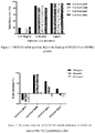
- VEGFR2 protein can effectively bind to the coated VEGF165 protein, and the rabbit antibodies of VEGF-R859, VEGF-R988, VEGF-R613, VEGF-R812 can effectively inhibit the binding of VEGFR165 protein to VEGFR2 protein.
- Antibody inhibits the proliferative effect of VEGF 165 on umbilical vein endothelial cells
- the effect of said rabbit antibodies neutralizing the VEGF 165-induced umbilical vein endothelial cells proliferation was detected by using the WST-8 method.
- Human umbilical vein endothelial cells HUVEC were inoculated into a 96-well plate at 4 ⁇ 10 3 cells/well, cultured in M199 medium containing 10% FBS and 5% L-Gln for 4 h, and then different concentrations of rabbit antibodies were added in 50 ⁇ L/well, then VEGF-165 at a final concentration of 10 ng/mL was added in 10 ⁇ L/well, the 96-well plate was incubated in a 37°C, 5% CO 2 cell incubator for 3 days, and the blank well B (no cells), negative control M (cells inoculated, no antibody sample, VEGF-165 added) and M'(cells inoculated, no antibody sample and no VEGF-165) were used.
- OD 450 and OD 630 were measured with a microplate reader after the color development was stabilized.
- the standard curve was calculated using the automatic analysis function of the statistical software GraphPad Prism, taking the antibody sample concentration as the horizontal coordinate and the neutralization rate as the vertical coordinate, and the four-parameter logistic regression equation was used to fit the standard "S" curve to calculate the half maximal effective concentration (EC 50 ) of the antibody sample.
- VEGF-R988 and VEGF-R613 effectively reduce the ability of VEGF165 to promoting HUVEC proliferation, and VEGF-R988 shows a higher maximum inhibition rate, VEGF-R812 has basically no inhibitory ability, while VEGF-R859 has no inhibitory ability at all.
- VEGF-R988 was selected for humanization and production.
- Example 1.2 Based on the nucleotide sequence of the VEGF-R988 scFv antibody determined in Example 1.2, the amino acid sequences of the heavy chain and light chain variable regions of the VEGF-R988 scFv were deduced, see SEQ ID NO: 11/12.
- Table 1 CDR sequences of VEGF-R988 light chain and heavy chains Name Sequences LCDR1 QSSQTIYANRRLA (SEQ ID NO:13) LCDR2 GASTLAS (SEQ ID NO:14) LCDR3 AGYKSYDGDDVG (SEQ ID NO:15) HCDR1 GIDLSSYAISWV (SEQ ID NO:16) HCDR2 YIWNAGNTYYASWAKG (SEQ ID NO:17) HCDR3 ARGTLGDYNGMDP (SEQ ID NO:18)
- the humanization of the rabbit antibody was performed using the classic humanization method of CDR transplantation.
- the human antibody light or heavy chain variable region whose sequence is closer to the sequence of rabbit light or heavy chain variable region, was elected as the template, and each of three CDRs (Table 1) from the rabbit light or heavy chain were inserted into the variable regions of the human antibody to obtain the humanized light chain variable region (VL) or heavy chain variable region (VH) sequences respectively.
- the human template for the light chain variable region of VEGF-R988 is IGKV1-27 ⁇ 01, which is 65.30% homologous to the light chain of VEGF-R988, and the human template for the heavy chain variable region is IGHV4-4 ⁇ 08, which is 53.20% homologous to the heavy chain of VEGF-R988.
- the key amino acids in the rabbit-derived framework region are essential to maintain the CDR activity
- the key amino acids were reverse-mutated to the corresponding rabbit antibody amino acid sequences, the following sites were reversely mutated: in the light chain, Position 1 was reversely mutated to E, Position 2 was reversely mutated to L, Position 4 was reversely mutated to L, and Position 63 was reversely mutated to K; while in the heavy chain, Position 3 was reversely mutated to V, Position 37 was reversely mutated to V, Position 47 was reversely mutated to Y, Position 78 was reversely mutated to V, Position 79 was reversely mutated to D, and Position 91 was reversely mutated to F; all the above sites were numbered by reference to the Kabat numbering scheme.
- the humanized antibody VEGF-H988-10 was obtained by CDR humanized transplantation and framework region reverse-mutations.
- VEGF-H988-10 heavy chain variable region (SEQ ID NO: 23) was obtained by the whole gene synthesis method, and then inserted, by in-fusion method, into Sca I + Nhe I (Fermentas) digested pSTEP2 vector harboring the nucleotide sequence encoding the heavy chain signal peptide (SEQ ID NO: 45) and the nucleotide sequence encoding the human IgG1 constant region (SEQ ID NO: 49), to obtain the VEGF-H988-10 heavy chain (SEQ ID NO:19) expression vector.
- VEGF-H988-10 light chain variable region (SEQ ID NO: 24) was obtained by the whole gene synthesis method, and then inserted, by in-fusion method, into Sca I + Bsi W I (Fermentas) digested pSTEP2 vector harboring the nucleotide sequence encoding the light chain signal peptide (SEQ ID NO: 46) and the nucleotide sequence encoding the human kappa constant region (SEQ ID NO: 50) to obtain the VEGF-H988-10 light chain (SEQ ID NO: 20) expression vector. Plasmids were extracted and co-transfected into HEK-293 cells, the cells were cultured for 7 days. The culture supernatant was purified with a protein A purification column to obtain high-purity antibodies.
- VEGF-H988-10 SDM libraries of CDR regions of heavy and light chain variable regions (including three saturated mutation libraries of LCDR1, LCDR3, and HCDR2) were constructed; Meanwhile, to improve the chemical stability of the antibody, the amino acid residues capable of undergoing deamidation or isomerization should be modified to another amino acid residue.
- the deamidation of asparagine can occur in, such as NG, NS, NA, NT, etc., leading to the generation of isoaspartic residues, which affects the stability or biological function of the antibody.
- the VEGF-H988 variable region HCDR3 has (a) deamidation-susceptible site(s), thus SDM libraries were constructed to improve the chemical stability and biological function of the antibody.
- the above four mutant libraries were constructed in scFv form and were cloned into phage vector as scFv- gIII fusion protein; for each CDR, the CDR clones having optimal binding ability to soluble antigen VEGF were screened, and finally the antibody VEGF-H988 having optimized CDR affinity and stability was obtained.
- the sequences of VEGF-H988 light and heavy chain CDRs are shown in Table 2.
- Table 2 CDR sequences of VEGF-H988 light chain and heavy chains Name Sequences LCDR1 QSSKFLWQGRRLA (SEQ ID NO:27) LCDR2 GASTLAS (SEQ ID NO:28) LCDR3 AGYKSYDGDVVG (SEQ ID NO:29) HCDR1 GIDLSSYAIS (SEQ ID NO:30) HCDR2 YIWNDLFTYYASWAKG (SEQ ID NO:31) HCDR3 ARGTLGDYGGMDP (SEQ ID NO:32)
- the nucleotide sequence (SEQ ID NO: 44) encoding the aforementioned antibody VEGF-H988 light chain and the signal peptide which contains the following nucleotide sequences encoding light chain signal peptide (SEQ ID NO: 46), the humanized antibody light chain variable region (SEQ ID NO: 48) and the human antibody kappa light chain constant region (SEQ ID NO: 50) connected in order, was PCR amplified and inserted into the self-developed pGS vector ( Kpn I+ Xba I) by in-fusion method, and the correct plasmids were verified by sequencing.
- the nucleotide sequence (SEQ ID NO: 43) encoding the aforementioned antibody VEGF-H988 heavy chain and the signal peptide which contains the following nucleotide sequences encoding heavy chain signal peptide (SEQ ID NO: 45), the humanized antibody heavy chain variable region (SEQ ID NO: 47) and the human antibody kappa heavy chain constant region (SEQ ID NO: 49) connected in order, was PCR amplified and inserted into pGS vector ( Nhe I+ Not I) which had been verified to contain the light chain correctly by in-fusion method, and the correct vectors expressing both light and heavy chains of VEGF-R988 were verified by sequencing.
- pGS vector Nhe I+ Not I
- These expression vectors are eukaryotic expression vectors containing the GS genes as the selection marker and the expression elements of the antibody light and heavy chains. These expression vectors were transfected into CHO-K1-GS-deficient cells and VEGF-H988 high expression cell lines were obtained by MSX screening. The clones with high antibody expression were selected by ELISA assay, and the high expression cell lines were selected by taking into account both the cell growth status and the key quality characteristics for antibody drugs. A serum-free suspension culture was used to culture the VEGF-H988 producing CHO cell line to obtain high purity and quality VEGF-H988 antibodies.
- Recombinant human VEGF165 protein (from SinoBiological, Inc.) in different concentrations (0.15ng/mL, 0.46 ng/mL, 1.37 ng/mL, 4.12ng/mL, 12.35 ng/mL, 37.04 ng/mL, 111.11 ng/mL, 333.33 ng/mL, 1000 ng/mL and 3000 ng/mL) was coated on a 96-well plate overnight at 4°C in 100 ⁇ L/well. The plate was washed the next day and blocked at room temperature for 1 h.
- VEGF165-H988, Avastin (from Roche) or negative control antibody H7N9-R1 respectively the plate was washed to remove unbound antibodies, then incubated with goat F(ab')2 anti-human IgG F(ab')2/HRP (from Jackson ImmunoResearch Laboratories, Inc.) and washed repeatedly, and the chromogenic substrate solution was added for color development. OD 450 was measured after the color development was ended.
- the graphPad Prism 6.0 software was used to fit an "S" curve chart and the binding of the antibody to recombinant human VEGF165 protein was analyzed.
- VEGF165-H988 and Avastin were measured at multiple concentrations using streptavidin-coated Sensor and immobilized biotin-labeled VEGF165 protein.
- the recombinant human VEGF165 protein was first labeled with biotin in a molar ratio of 1:2 as the following process: the recombinant VEGF protein buffer (20 mM Tris, 150 mM NaCl, pH 8.0) was replaced with PBS through ultrafiltration in a 5000 MW ultrafiltration centrifugal tube, and 567.57 ⁇ g of protein was obtained as measured by UV quantification, and the resulting proteins were mixed with 20 mM biotin solution in a 1:2 molar ratio for incubation for 30 min at room temperature in the dark, then filtered again in a 5000 MW ultrafiltration centrifugal tube to remove the unlabeled biotin. After UV quantification, the biotin-labeled proteins were obtained by adding an equal volume of glycerol and a final concentration of 0.1% BSA. The concentration of the VEGF165 protein was 2.08 mg/mL, detected by UV.
- Recombinant human VEGF165 proteins or recombinant mouse mVEGF164 proteins were diluted to 0.1 ⁇ g/mL, 1 ⁇ g/mL and 10 ⁇ g/mL, respectively, and coated on a 96-well plate overnight at 4°C in 100 ⁇ L/well. The plate was washed the next day, blocked at room temperature for 1 h. 100 ⁇ L of VEGF165-H988, Avastin (from Roche) or negative control antibody H7N9-R1 was added respectively in a concentration of 1 ⁇ g/mL and incubated for 1 h. The plate was washed to remove unbound antibodies.
- the plate was incubated with goat F(ab')2 anti-human IgG F(ab')2/HRP (Jackson ImmunoResearch Laboratories, Inc. ) and then repeatedly washed, and the chromogenic substrate solution was added for color development.
- OD 450 was measured after the color development was ended. Taking the protein concentration as the horizontal coordinate and the OD 450 value as the vertical coordinate, the graphPad Prism 6.0 software was used for generating a bar chart.
- VEGF165 protein (from SinoBiological, Inc.) at a concentration of 1 ⁇ g/mL was coated on a 96-well plate in 100 ⁇ L/well overnight at 4°C. The plate was washed the next day and blocked at room temperature for 1 h. 100 ⁇ L of 2 ⁇ g/mL VEGFR2-his protein (from SinoBiological, Inc.) was added in each well and different concentrations of antibody VEGF-H988, EYLEA, Avastin (from Roche) or negative control antibody H7N9-R1 was added respectively and co-incubated. The plate was washed to remove unbound antibodies.
- Inhibition rate (%) (OD blank - OD sample )/ OD blank ⁇ 100%, where OD blank indicates the OD value of the wells with only VEGFR2-his added but no humanized antibody added, and OD sample indicates the OD value of the wells with both VEGFR2-his and the humanized antibodies added.
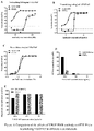
- VEGFR2 protein could effectively bind to the coated VEGF165 protein, and the antibodies VEGF-H988 could effectively inhibit the binding of VEGFR2 protein to VEGFR165 protein, with a relatively weaker inhibitory ability than EYLEA but greater than Avastin, and the negative control antibody has no inhibitory effect.
- HUVEC cells were inoculated into a 96-well plate at 4 ⁇ 103 cells/well, cultured in M199 medium containing 10% FBS and 5% L-Gln for 4 h, and then VEGF-H988, EYLEA or Avastin (from Roche) in different concentrations were added in 50 ⁇ L/well, then VEGF-165 at the final concentrations of 1000 ng/mL, 100 ng/mL or 10 ng/mL were added in 10 ⁇ L/well, the 96-well plate was incubated in a 37°C, 5% CO2 cell incubator for 3 days, and the blank well B (no cells), negative control M (cells inoculated, no antibody sample added, VEGF-165 added) and M'(cells inoculated, no antibody sample added and no VEGF-165 added) were used.
- OD 450 and OD 630 were measured with a microplate reader after the color development was stabilized.
- the standard curve was calculated using the automatic analysis function of the statistical software GraphPad Prism, taking the antibody sample concentration as the horizontal coordinate and the neutralization rate as the vertical coordinate, and the 4-parameter logistic regression equation was used to fit the standard "S" curve to calculate the half maximal effective concentration (EC 50 ) of the antibody sample.
- antibody VEGF-H988 can effectively reduce the ability of VEGF165 to promoting HUVEC proliferation.
- the neutralizing ability of VEGF-H988 was stronger than that of EYLEA and Avastin at different concentrations of recombinant human VEGF165; and the difference of the neutralizing ability was stronger as the concentrations of VEGF165 is higher.
- VEGF-H988 still has a low neutralizing EC 50 and could still maintain the maximum neutralization rate, while the neutralizing EC 50 of EYLEA and Avastin gradually increased and was accompanied by a decrease of the maximum neutralization rate.
- the WST-8 method was used to detect the effect of VEGF-H988 neutralizing different subtypes of VEGF (VEGF165, VEGFC and VEGFD) - induced HUVEC cells proliferation.
- HUVEC cells were inoculated into a 96-well plate at 4 ⁇ 10 3 cells/well, cultured in M199 medium containing 10% FBS and 5% L-Gln for 4 h, and then different concentrations of VEGF-H988 or Avastin (from Roche) were added in 50 ⁇ L/well, then VEGF-165, VEGFC and VEGFD mixture (the final concentrations were 25 ng/mL, 1000 ng/mL, 6000 ng/mL respectively) was added in 10 ⁇ L/well, the 96-well plate was incubated in a 37°C, 5% CO 2 cell incubator for 3 days, and the blank well B (no cells), negative control M (cells inoculated, no antibody sample added, VEGFs added) and M'(cell
- OD 450 and OD 630 were measured with a microplate reader after the color development was stabilized.
- the standard curve was calculated using the automatic analysis function of the statistical software GraphPad Prism, taking the antibody sample concentration as the horizontal coordinate and the neutralization rate as the vertical coordinate, and the 4-parameter logistic regression equation was used to fit the standard "S" curve to calculate the half maximal effective concentration (EC 50 ) of the antibody sample.
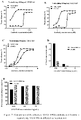
- VEGF-H988 has a stronger neutralizing effect than Avastin.
- concentration range of 0.016-4.000 nM VEGF-H988 has a smaller EC 50 than Avastin, being 0.26 nM and 0.77 nM, respectively; and VEGF-H988 has a higher neutralization rate than Avastin, being 86.1% and 58.5% respectively.
- the half maximal effective concentrations (EC 50 ) and the maximum neutralization rates of VEGF and Avastin to neutralize different subtypes of VEGF are summarized in Table 5.
- CD-1 mice 8 each in the aged group (35 weeks of age) and normal group (9 weeks of age), half male and half female, were used.
- the mice were divided into four groups (G1-G4) according to the complete randomization method, with four mice in each group and half of the males and half of the females.
- the specific grouping was as follows: G1: old mice solvent control group; G2: old mice study group; G3: normal mice solvent control group; G4: normal mice study group.
- the VEGF-H988 antibodies were repeatedly administered to the study groups, and the control groups were given an equal volume of solvent.
- mice were subjected to orbital blood sampling before and 0.5 h, 2 h, 4 h, 6 h, 24 h, 48 h, 72 h after the first dosing as well as before and 1 h after every other dosing, for pharmacokinetic and immunogenicity assays. The body weights of mice were measured and recorded before each dosing. The detailed dosing regimen is shown in Table 6. Table 6.
- Dosing regimen of antibody VEGF-H988 Group Antibody Dose Mode of administration Frequency of administration Mice status G1 None (solvent control group) 0 mg/kg i.p. Twice weekly 2 ⁇ & 2 ⁇ (aged) G2 VEGF-H988 50 mg/kg i.p. Twice weekly 2 ⁇ & 2 ⁇ (aged) G3 None (solvent control group) 0 mg/kg i.p. Twice weekly 2 ⁇ & 2 ⁇ (normal age) G4 VEGF-H988 50 mg/kg i.p. Twice weekly 2 ⁇ & 2 ⁇ (normal age)
- mice in the antibody VEGF-H988 groups were similar to those of the corresponding solvent control groups, indicating that the antibody VEGF-H988 had no effect on the body weight of the mice.
- the clinical abnormalities were observed in the antibody VEGF-H988 groups. All mice were subjected to gross autopsy, and there were no visible abnormalities in the tissues and organs. Histopathological examination showed that all mice had venous congestion in the liver, lungs, and kidneys, but there was no significant difference between the control groups and the study groups. Sparse individual mice showed pink serous or erythrocyte stasis in the spleen and the inflammatory cell infiltration in the kidneys, which were considered to be individual difference and not related to drug administration.
- mice after the first dose are summarized in Table 7.
- Table 7 Toxicokinetic parameters of toxicity study of repeated administration in mice Group Gender / tl/2 (h) Tmax (h) Cmax ( ⁇ g/mL ) AUClast (h ⁇ g/mL) AUC0- ⁇ (h ⁇ g/mL) Vz_F (mL/kg) C1_F (mL/h/kg) MRTlast (h) G2 (Aged) ⁇ Mean 53.246 4.000 408.899 11735.125 14696.594 263.934 3.469 26.128 SD 5.161 0.000 63.837 1381.429 2880.380 26.398 0.680 4.929 ⁇ Mean 55.751 2.000 435.691 12033.935 15499.970 254.922 3.276 26.934 SD 20.717 0.000 25.122 975.896 2717.582 51.721 0.574 0.748 ⁇ & ⁇ Mean 54.499 3.000 422.295 11884.530 15098.282 259.
- mice in each group showed no significant increase or even some decrease in the blood drug concentration at 1 h after administration, which did not correlate well with immunogenicity (see Table 8 for immunogenicity test data).
- the abnormal blood drug concentration at 1 h after administration may also be related to the drug distribution speed in vivo and individual differences.
- Table 8 summarizes the immunogenicity results of the repeated dosing toxicity study of the antibody VEGF-H988 in mice. Table 8. Immunogenicity test results of toxicity study of repeated administration of VEGF-H988 in mice Time point 0 h 432 h Group Mouse No.
- Example 5 Efficacy study in human colorectal cancer cell line HCT-116 xenograft tumor model
- mice Female Balb/c-nu nude mice were from Beijing Vital River Laboratory Animal Technology Co., Ltd. (Animal production license No. SCXK (Beijing) 2016-0006, Quality certificate No. 11400700373019).
- HCT116 tumor blocks were obtained from mice inoculated with self-developed HCT116 cell line.
- Balb/c-nu 6-week-old female mice were inoculated subcutaneously with HCT116 tumor blocks of approximately 2 ⁇ 2 ⁇ 2 (mm 3 ) in size, and when the tumor volume reached approximately 300 mm 3 , the mice were administered in groups with intraperitoneal injection of 1 mg/kg of VEGF-H988 or Avastin (from SinoCelltech Co., Ltd.) or the corresponding solvent, respectively twice weekly. Tumor volumes were measured twice weekly to assess the antitumor efficacy of VEGF-H988 versus Avastin.
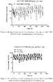
- the tumor growth rate was progressively reduced in the VEGF-H988-treated group compared to the non-dosed control group since Day 7 after administration.
- the inhibitory effect of Avastin on tumor growth began on the 11th day after administration, later than that in the equivalent dose of VEGF-H988 treated group.
- Tumor inhibition assays in the HCT-116 xenograft tumor model showed that the inhibitory effect of VEGF-H988 on tumor growth is better than that of Avastin.
- SEQ ID NO.1 Amino acid sequence of Met 1- Arg191 of the human VEGF protein (UniProtKB P15692)
- SEQ ID NO.2 Nucleotide sequence of the linker used in the construction of the phage antibody library for the linkage of the rabbit antibody scFv
- SEQ ID NO.3 Nucleotide sequence of rabbit antibody scFv which is used in the construction of antibody VEGF165-R859
- Nucleotide sequence of light chain variable region of VEGF165-R859 Nucleotide sequence of light chain variable region of VEGF165-R859
- Linker Nucleotide sequence of the rabbit antibody scFv which is used in the construction of antibody VEGF165-R988 Nucleotide sequence of light chain variable region of VEGF165-R988(SEQ ID NO:8) Linker(SEQ ID NO:2): Nucleotide sequence of heavy chain
Landscapes
- Health & Medical Sciences (AREA)
- Chemical & Material Sciences (AREA)
- Organic Chemistry (AREA)
- Medicinal Chemistry (AREA)
- General Health & Medical Sciences (AREA)
- Life Sciences & Earth Sciences (AREA)
- Immunology (AREA)
- Animal Behavior & Ethology (AREA)
- Veterinary Medicine (AREA)
- Public Health (AREA)
- Pharmacology & Pharmacy (AREA)
- Biochemistry (AREA)
- Biophysics (AREA)
- Genetics & Genomics (AREA)
- Molecular Biology (AREA)
- Proteomics, Peptides & Aminoacids (AREA)
- Epidemiology (AREA)
- Engineering & Computer Science (AREA)
- Bioinformatics & Cheminformatics (AREA)
- Nuclear Medicine, Radiotherapy & Molecular Imaging (AREA)
- General Chemical & Material Sciences (AREA)
- Chemical Kinetics & Catalysis (AREA)
- Peptides Or Proteins (AREA)
- Micro-Organisms Or Cultivation Processes Thereof (AREA)
- Medicines Containing Antibodies Or Antigens For Use As Internal Diagnostic Agents (AREA)
- Preparation Of Compounds By Using Micro-Organisms (AREA)
- Medicinal Preparation (AREA)
- Medicines That Contain Protein Lipid Enzymes And Other Medicines (AREA)
- Pharmaceuticals Containing Other Organic And Inorganic Compounds (AREA)
Abstract
Description
- The present invention relates to the field of tumor immunotherapy, specifically relates to a humanized monoclonal antibody that binds to VEGF.
- The development of the vascular system is the foundation of many physiological and pathological processes. Vascular endothelial growth factor (VEGF) is a group of growth factors possessing important pro-angiogenic activities that promote endothelial cell mitosis and anti-apoptosis, increase vascular permeability, and promote cell migration. The human VEGF gene is localized on chromosome 6p21.3 and belongs to the VEGF/PDGF supergene family, which encodes VEGF linked by disulfide bonds to form a dimer. In humans, the VEGF family includes multiple members with different functions: VEGFA (VEGF, with several different splicing variants), VEGFB, VEGFC, VEGFD, VEGFE, VEGFF, and placenta growth factor (PIGF). Recently, endocrine gland-derived vascular endothelial growth factor (EG-VEGF) has been also included in this family (Samson M et al., J Clin Endocrinol Metab. 2004; 89(8):4078-4088). VEGF is widely distributed in human tissues and organs, among which the eye retinal pigment epithelial cells, vascular endothelial cells, nerve cells, etc. are expressed (Goel H L et al., Nat Rev Cancer. 2013; 13(12): 871). There are three types of VEGF receptors: VEGFR1, VEGFR2 and VEGFR3. The binding of VEGF to the receptor extracellular domain triggers receptor dimerization and promote auto-phosphorylation of tyrosine residues in the intracellular domain, thereby activating downstream signals that promote cell proliferation, migration, anti-apoptosis and increased vascular permeability. VEGFR1 and VEGFR2 are mainly expressed in vascular endothelial cells, while VEGFR3 is mainly expressed in lymphatic endothelial cells.
- VEGF has been confirmed to play an important role in the regulation of normal and pathological angiogenesis (Melincovici C S et al., Rom J Morphol Embryol. 2018; 59(2): 455-467). VEGF is overexpressed in a variety of tumors that can cause malignant ascites, and the expression of VEGF in tumors is correlated with the migration ability of tumor cells. The concentration of VEGF in patients with solid tumors of poorer survival rate such as gastrointestinal, ovarian, breast and lung cancers is positively correlated with disease staging (Sebastian, K et al., Oncologist. 2009; 14(12): 1242 -1251). Hypoxic conditions in the tumor microenvironment induce the entry of tumor cell transcription factor HIF-la into the nucleus, sequentially HIF-la bind to the HRE element of VEGFA, thereby the transcription level of VEGFA is up-regulated and tumor cells is promoted to secrete large amounts of VEGF into the tumor microenvironment . In turn, the high concentration of VEGF acts on VEGFRs on vascular endothelial cells, inducing a large number of neovascularization, enhancing blood supply, and providing sufficient nutrients for the growth of tumor cells. More VEGF is secreted from the rapid-grown tumor cells, which further promotes the proliferation and migration of vascular endothelial cells and induces tumor metastasis. In addition, VEGF also stimulates monocytes in tumor tissues to convert to M2 suppressor macrophages, producing more negative immune factors, and up-regulating Treg cells at the same time, which synergistically reduce the killing ability of T cells.
- By inhibiting the interaction of VEGF with endothelial cell surface receptors VEGFR2 and VEGFR1, VEGF monoclonal antibody drugs block downstream signaling pathways, inhibit endothelial cell proliferation and neovascularization, deprive tumor tissues of blood supply, and control the internal nutrient supply of tumors, thereby limiting tumor growth and ultimately achieving anticancer efficacy. Avastin (bevacizumab, approved in 2009) is the first antibody drug approved to inhibit tumor angiogenesis, and is mainly used for the treatment of breast cancer, cervical cancer, colorectal cancer, glioblastoma, glioma, non-small cell lung cancer, ovarian cancer, and renal cell carcinoma. Although Avastin has been used to treat a variety of cancers, there is still a need in the field for more potent antibodies with greater VEGF inhibition and higher efficacy.
- The present invention provides a novel human VEGF antibody for the treatment of colorectal cancer.
- In one aspect, the present invention provides an isolated anti-VEGF antibody or antigen-binding fragment thereof, comprising a heavy chain variable region having a heavy chain CDR1 region having the amino acid sequence as set forth in SEQ ID NO: 30, a heavy chain CDR2 region having the amino acid sequence as set forth in SEQ ID NO: 31 and a heavy chain CDR3 region having the amino acid sequence as set forth in SEQ ID NO: 32; and a light chain variable region having a light chain CDR1 region having the amino acid sequence as set forth in SEQ ID NO: 27, a light chain CDR2 region having the amino acid sequence as set forth in SEQ ID NO: 28, and a light chain CDR3 region having the amino acid sequence as set forth in SEQ ID NO: 29.
- In one embodiment, said anti-VEGF antibody or antigen-binding fragment thereof has a heavy chain variable region having the amino acid sequence as set forth in SEQ ID NO: 39, or the amino acid sequences having at least 90%, 92%, 95%, 98% or 99% sequence identity to SEQ ID NO: 39; and a light chain variable region having the amino acid sequence as set forth in SEQ ID NO: 40, or the amino acid sequences having at least 90%, 92%, 95%, 98% or 99% sequence identity to SEQ ID NO: 40.
- In one embodiment, said antibody further comprises a light chain constant region and a heavy chain constant region, preferably the light chain constant region is the light chain constant region having the amino acid sequence as set forth in SEQ ID NO: 42, or the amino acid sequences having at least 90%, 92%, 95%, 98%, or 99% sequence identity to SEQ ID NO: 42; and/or the heavy chain constant region is the IgG1 heavy chain constant region having the amino acid sequence as set forth in SEQ ID NO: 41, or the amino acid sequences having at least 90%, 92%, 95%, 98%, or 99% sequence identity to SEQ ID NO:41.
- In one embodiment, said anti-VEGF antibody or antigen-binding fragment thereof is an IgG antibody, preferably an IgG1 antibody.
- In one embodiment, said anti-VEGFR antibody or antigen-binding fragment thereof is a monoclonal antibody.
- In one embodiment, the binding affinity KD of said anti-VEGF antibody or antigen-binding fragment thereof to the recombinant human VEGF165 protein is 1-100 pM, preferably 5-50 pM, and more preferably 19.5 pM.
- In one embodiment, said antigen-binding fragment is Fv, Fab, Fab', Fab'-SH, F(ab')2, Fd fragment, Fd' fragment, single chain antibody molecule or single domain antibody; wherein the single chain antibody molecule is preferably scFv, di-scFv, tri-scFv, diabody or scFab.
- In another aspect, the present invention provides an antibody-drug conjugate, comprising the anti-VEGF antibody or antigen-binding fragment thereof as described herein and an additional therapeutic agent, preferably said anti-VEGF antibody or antigen-binding fragment thereof is connected with the additional therapeutic agent via a connector.
- In one aspect, the present invention provides an isolated anti-VEGF antibody or antigen-binding fragment thereof, comprising a heavy chain variable region having a heavy chain CDR1 region having the amino acid sequence as set forth in SEQ ID NO: 16 and a heavy chain CDR2 region having the amino acid sequence as set forth in SEQ ID NO: 17 and a heavy chain CDR3 region having the amino acid sequence as set forth in SEQ ID NO: 18; and a light chain variable region having a light chain CDR1 region having the amino acid sequence as set forth in SEQ ID NO: 13, a light chain CDR2 region having the amino acid sequence as set forth in SEQ ID NO: 14, and a light chain CDR3 region having the amino acid sequence as set forth in SEQ ID NO: 15.
- In one embodiment, said anti-VEGF antibody or antigen-binding fragment thereof comprises a heavy chain constant region having the amino acid sequence as set forth in SEQ ID NO: 25, or the amino acid sequences having at least 90%, 92%, 95%, 98%, or 99% sequence identity to SEQ ID NO: 25; and a light chain variable region having the amino acid sequence as set forth in SEQ ID NO: 26, or the amino acid sequences having at least 90%, 92%, 95%, 98%, or 99% sequence identity to SEQ ID NO: 26.
- In one embodiment, said anti-VEGF antibody or antigen-binding fragment thereof is a humanized antibody or a chimeric antibody.
- In another aspect, the present invention provides a nucleic acid encoding the anti-VEGF antibody or antigen-binding fragment thereof as described herein.
- In one embodiment, said nucleic acid comprises the nucleotide sequence as set forth in SEQ ID NO: 7 and/or the nucleotide sequence as set forth in SEQ ID NO: 8; or comprises the nucleotide sequence as set forth in SEQ ID NO: 23 and/or the nucleotide sequence as set forth in SEQ ID NO: 24; or comprises the nucleotide sequence as set forth in SEQ ID NO: 47 and/or the nucleotide sequence as set forth in SEQ ID NO: 48.
- In another aspect, the present invention provides an expression vector, comprising the nucleic acid as described herein.
- In another aspect, the present invention provides a host cell, comprising the nucleic acid as described herein or the expression vector as described herein.
- In another aspect, the present invention provides a method for producing the anti-VEGF antibody or antigen-binding fragment thereof as described herein, comprising culturing the host cell as described herein under conditions suitable for antibody expression, and harvesting the expressed antibody from the culture medium.
- In another aspect, the present invention provides a pharmaceutical composition, comprising the anti-VEGF antibody or antigen-binding fragment thereof as described herein, or the antibody-drug conjugate as described herein, or the nucleic acid as described herein, or the expression vector as described herein, and a pharmaceutically acceptable carrier.
- In one embodiment, the present invention provides the anti-VEGF antibody or antigen-binding fragment thereof as described herein, or the antibody-drug conjugate as described herein, or the pharmaceutical composition as described herein, for use in the treatment of colorectal cancer.
- In another aspect, the present invention provides a method for treating colorectal cancer, comprising administering to a subject in need a therapeutically effective amount of the anti-VEGF antibody or antigen-binding fragment thereof as described herein, or the antibody-drug conjugate as described herein, or the pharmaceutical composition as described herein.
- In another aspect, the present invention provides the use of the anti-VEGF antibody or antigen-binding fragment thereof as described herein or the antibody-drug conjugate as described herein or the pharmaceutical composition as described herein in the preparation of a medicament for the treatment of colorectal cancer.
- In another aspect, the present invention provides a pharmaceutical combination, comprising the anti-VEGFR2 antibody or antigen-binding fragment thereof as described herein, or the antibody-drug conjugate as described herein, or the pharmaceutical composition as described herein, and one or more additional therapeutic agents.
- In another aspect, the present invention provides a kit, comprising the anti-VEGF antibody or antigen-binding fragment as described herein, or the antibody-drug conjugate as described herein, or the pharmaceutical composition as described herein, preferably, further comprising a device for administration.
- The present invention is illustrated in combination with the attached drawings, in which:
-
Figure 1 shows that the rabbit antibody VEGF165 blocks the binding of VEGF165 to VEGFR2 protein. -
Figure 2 shows that rabbit antibody VEGF165 neutralizes the VEGF165-induced the proliferation of HUVEC. -
Figure 3 shows the binding of humanized antibody VEGF165-H988 to VEGF165, detected by ELISA. -
Figure 4 shows the binding of humanized antibody VEGF165-H988 to mVEGF165, detected by ELISA. -
Figure 5 shows that the antibody VEGF-H988 blocks the binding of VEGF165 to VEGFR2 protein, detected by ELISA. -
Figure 6 shows the effect of antibody VEGF-H988 in neutralizing VEGF165 in different concentrations. -
Figure 7 shows the effect of VEGF-H988 on the neutralization of VEGF165, VEGFC and VEGFD -induced the proliferation of HUVEC cells. -
Figure 8 shows the mean blood concentration-time curve of G2 group mice injected with VEGF-H988 (50 mg/kg) intraperitoneally. -
Figure 9 shows the mean blood concentration-time curve of G4 group mice injected with VEGF-H988 (50 mg/kg) intraperitoneally. -
Figure 10 shows the mean blood concentration-time curve of G2&G4 groups mice injected with VEGF-H988 (50mg/kg) intraperitoneally . -
Figure 11 shows the comparison of the efficacies of VEGF-H988 and the control drugs in the human colorectal cancer cell line HCT-116 xenograft tumor model. -
Figure 12 shows the comparison of the efficacies of VEGF-H988 and the control drugs in the human colorectal cancer cell line HCT-116 xenograft tumor model. - Various aspects of the present invention relate to an isolated anti-VEGF antibody or antigen-binding fragment thereof, an antibody-drug conjugate comprising said antibody or antigen-binding fragment thereof, a nucleic acid and an expression vector encoding said antibody or antigen-binding fragment thereof, and a host cell containing said nucleic acid or expression vector, a method for producing said anti-VEGF antibody or antigen-binding fragment thereof, a pharmaceutical composition comprising said anti-VEGF antibody or antigen-binding fragment thereof, and a method of using said anti-VEGF antibody or antigen-binding fragment thereof for treating colorectal cancer.
- Unless otherwise stated, all technical and scientific terms used herein have the meaning normally understood by a person skilled in the art to which the present invention belongs. For the purposes of the present invention, the following terms are defined to be consistent with the meanings commonly understood in the art.
- When used herein and in the appended claims, the singular forms "one", "a/an", "another" and "said" include the plural designation of the object unless the context clearly indicates otherwise.
- The term "antibody" refers to an immunoglobulin molecule and refers to any form of antibody that exhibits the desired biological activity. These include, but are not limited to, monoclonal antibodies (including full-length monoclonal antibodies), polyclonal antibodies and multispecific antibodies (e.g. bispecific antibodies), and even antibody fragments. Typically, full-length antibody structures preferably comprise four polypeptide chains, two heavy (H) chains and two light (L) chains, typically interconnected by disulfide bonds. Each heavy chain contains a heavy chain variable region and a heavy chain constant region. Each light chain contains a light chain variable region and a light chain constant region. In addition to this typical full-length antibody structure, the structure also includes other derivative forms.
- Said heavy chain variable region and light chain variable region can be further subdivided into more conservative regions (called framework regions (FR)) and hypervariable regions (called complementarity determining regions (CDR)) interspersed therewith.
- The term "complementary determining region" (CDR, e.g. CDR1, CDR2 and CDR3) refers to such amino acid residues in the variable region of an antibody whose presence is necessary for antigen binding. Each variable region typically has three CDR regions identified as CDR1, CDR2 and CDR3. Each complementary determining region may contain amino acid residues from a "complementary determining region" as defined by Kabat (Kabat et al., Sequences of Proteins of Immunological Interest, 5th Ed. Public Health Service, National Institutes of Health, Bethesda, MD. 1991) and/or those residues from the "high-variable loop" (Chothia and Lesk; J MolBiol 196: 901-917 (1987)).
- The term "framework" or "FR" residues are those residues within the variable region other than CDR residues as defined herein.
- Each heavy chain variable region and light chain variable region typically contains 3 CDRs and up to 4 FRs, said CDRs and FRs being arranged from the amino terminus to the carboxyl terminus in the following order, for example: FR1, CDR1, FR2, CDR2, FR3, CDR3, and FR4.
- The complementary determining region (CDR) and the framework region (FR) of a given antibody can be identified using the Kabat system (Kabat et al: Sequences of Proteins of Immunological Interest, 5th edition, US Department of Health and Human Services, PHS, NIH, NIH Publication No. 91- 3242, 1991).
- The term "constant region" refers to such amino acid sequences in the light and heavy chains of an antibody that are not directly involved in the binding of the antibody to the antigen but exhibit a variety of effector functions such as antibody-dependent cytotoxicity.
- According to the antigenic differences of the amino acid sequence of its constant region, the heavy chain of an antibody can be classified into five classes: α, δ, ε, γ, and µ. When it forms a complete antibody with the light chain, it can be classified into five classes: IgA , IgD, IgE, IgG and IgM, of which can be further classified into subclasses (isotypes), such as IgG1, IgG2, IgG3, IgG4, IgA and IgA2. Based on the amino acid sequence of its constant domain, the light chain of an antibody can be classified into κ and λ.
- An "antigen-binding fragment of an antibody" comprises a portion of an intact antibody molecule that retains at least some of the binding specificity of the parent antibody and typically includes at least a portion of the antigen-binding region or variable region (e.g. one or more CDRs) of the parent antibody. Examples of antigen-binding fragments include, but are not limited to, Fv, Fab, Fab', Fab'-SH, F(ab')2, Fd fragment, Fd' fragment, single chain antibody molecules (e.g. scFv, di-scFv or tri-scFv, diabody or scFab), single domain antibodies.
- The term "antibody fragment" refers to a non-intact antibody molecule that retains at least some of the biological properties of the parent antibody, including, but not limited to, an Fc fragment, in addition to those described above as "antigen-binding fragments".
- The term "antibody-drug conjugate" or "ADC" refers to a binding protein, such as an antibody or antigen-binding fragment thereof, that chemically linked to one or more of chemical drugs (also referred to as agents herein), which may optionally be a therapeutic agent or a cytotoxic agent. In a preferred embodiment, an ADC includes an antibody, a cytotoxic or therapeutic drug, and a linker that enables the drug to be linked or conjugated to the antibody. ADCs usually have any value of 1 to 8 drugs conjugated to the antibody, including 2, 4, 6, or 8 drug-loading substances. Non-limiting examples of drugs that can be included in the ADCs are mitotic inhibitors, anti-tumor antibiotics, immunomodulators, vectors for gene therapy, alkylating agents, anti-angiogenic agents, antimetabolites, boron-containing agents, chemotherapeutic protective agents, hormones, antihormonal agents, corticosteroids, photoactive therapeutic agents, oligonucleotides, radionuclide agents, topoisomerase inhibitors, tyrosine kinase inhibitors and radio sensitizers.
- The term "chimeric antibody" refers to an antibody in which a part of the heavy chain and/or light chain is derived from a specific source or species, and the remaining part is derived from a different source or species. The "chimeric antibody" may also be a functional fragment as defined above. "Humanized antibodies" are a subset of "chimeric antibodies."
- The term "humanized antibody" or "humanized antigen-binding fragment" is defined herein as an antibody or antibody fragment that is: (i) derived from a non-human source (e.g., a transgenic mouse carrying a heterologous immune system) and based on a human germline sequence; or (ii) a chimeric antibody where the variable region is of non-human origin and the constant region is of human origin; or (iii) a CDR transplant where the CDR of the variable region is of non-human origin and one or more frame work regions of the variable region are of human origin and the constant region, if any, is of human origin. The aim of "humanization" is to eliminate the immunogenicity of antibodies of non-human origin in the human body, while retaining the greatest possible affinity. It is advantageous to select the human framework sequence that is most similar to the framework sequence of the non-human source antibody as the template for humanization. In some cases, it may be necessary to replace one or more amino acids in the human framework sequence with corresponding residues in the non-human construct to avoid loss of affinity.
- The term "monoclonal antibody" refers to an antibody derived from a substantially homogeneous population of antibodies, i.e. every single antibody comprised in the population is identical except for possible mutations (e.g. natural mutations) which may be present in very small quantities. The term "monoclonal" therefore indicates the nature of the antibody in question, i.e. not a mixture of unrelated antibodies. In contrast to polyclonal antibody preparations, which usually comprise different antibodies against different epitopes, each monoclonal antibody in a monoclonal antibody preparation is directed against a single epitope on the antigen. In addition to their specificity, monoclonal antibody preparations have the advantage that they are usually not contaminated by other antibodies. The term "monoclonal" should not be understood as requiring the production of said antibodies by any particular method.
- The antibody "specifically binds" to a target antigen such as a tumor-associated peptide antigen target (in this case, PD-1), i.e. binds said antigen with sufficient affinity to enable said antibody to be used as a therapeutic agent, targeting a cell or tissue expressing said antigen, and does not significantly cross-react with other proteins, or does not significantly cross-react with proteins other than the homologues and variants of the target proteins mentioned above (e.g. mutant forms, splice variants, or protein hydrolysis truncated forms).
- The term "binding affinity" refers to the strength of the sum of the non-covalent interactions between a molecule's individual binding sites and its binding partners. Unless otherwise stated, "binding affinity", when used herein, refers to the intrinsic binding affinity, which reflects a 1:1 interaction between members of a binding pair (e.g. antibody and antigen). As used herein, the term "KD" refers to the equilibrium dissociation constant of the antibody-antigen interaction. As used herein, the term "kon" refers to the rate constant at which an antibody binds to an antigen. As used herein, the term "koff" refers to the rate constant at which an antibody dissociates from an antibody/antigen complex. "KD", "binding rate constant kon" and "dissociation rate constant koff" are commonly used to describe the affinity between a molecule (e.g. an antibody) and its binding partner (e.g. an antigen). Affinity, i.e. the tight degree at which a ligand binds a particular protein. Binding affinity is influenced by non-covalent intermolecular interactions such as hydrogen bonding, electrostatic interactions, hydrophobic and van der Waals forces between two molecules. In addition, the binding affinity between a ligand and its target molecule may be influenced by the presence of other molecules. Affinity can be analyzed by conventional methods known in the art, including the ELISA described herein.
- The term "epitope" includes any protein determinant cluster that specifically binds to an antibody or T-cell receptor. Epitope determinant clusters typically consist of a molecule's chemically active surface groups (e.g. amino acid or sugar side chains, or a combination thereof) and often have specific three-dimensional structural features as well as specific charge characteristics.
- The term "isolated" antibody is an antibody that has been identified and isolated from the components of the cell where the antibody expressed. Isolated antibodies include in situ antibodies inside of recombinant cells, where at least one component in natural environment of said antibody is absent. However, usually, the isolated antibody is prepared through at least one purification step.
"sequence identity" between two polypeptides or nucleic acid sequences indicates the number of residues that are identical between said sequences as a percentage of the total number of residues, and is calculated based on the size of the smaller of the compared molecules. When calculating the percentage identity, the sequences being aligned are matched in such a way as to produce a maximum match between the sequences, with the gaps in the match (if present) being resolved by a specific algorithm. Preferred computer program methods for determining identity between two sequences include, but are not limited to, GCG program packages including GAP, BLASTP, BLASTN and FASTA (Altschul et al., 1990, J. Mol. Biol. 215: 403-410). The above procedures are publicly available from the International Center for Biotechnology Information (NCBI) and other sources. The well-known Smith Waterman algorithm can also be used to determine identity. - The term "Fc receptor" or "FcR" refers to a receptor that binds to the Fc region of an antibody. Human FcRs of natural sequence are preferred, and preferably receptors that bind to IgG antibodies (gamma receptors), which include the FcyRI, FcyRII and FcyRIII isoforms, as well as variants of these receptors. All other FcRs are included in the term "FcR". The term also includes the neonatal receptor (FcRn), which is responsible for the transport of maternal IgG to the fetus (Guyer et al, Journal of Immunology 117: 587 (1976) and Kim et al, Journal of Immunology 24: 249 (1994)).
- The term "neonatal Fc receptor", abbreviated as "FcRn", binds to the Fc region of IgG antibodies. The neonatal Fc receptor (FcRn) plays an important role in the metabolic fate of IgG-like antibodies in vivo. FcRn functions to rescue IgG from the lysosomal degradation pathway, thereby reducing its clearance in serum and lengthening its half-life. Therefore, the in vitro FcRn binding properties/characteristics of IgG are indicative of its in vivo pharmacokinetic properties in the circulation.
- The term "effector function" refers to those biological activities attributable to the Fc region of an antibody, which vary from isotype to isotype. Examples of antibody effector functions include C1q binding and complement-dependent cytotoxicity (CDC), Fc receptor binding, antibody-dependent cell-mediated cytotoxicity (ADCC), antibody-dependent cellular phagocytosis (ADCP), cytokine secretion, immune complex-mediated uptake of antigen by antigen-presenting cells, cell surface receptors down-regulation (e.g. B-cell receptors) and B-cell activation.
- The term "effector cell" refers to a cell that expresses one or more FcRs and performs effector functions. In one aspect, said effector cells express at least FcyRIII and perform ADCC effector functions. Examples of human cells that mediate ADCC include peripheral blood mononuclear cells (PBMCs), natural killer (NK) cells, monocytes, cytotoxic T cells and neutrophils. Effector cells can be isolated from natural sources, for example, blood. Effector cells are usually lymphocytes associated with effector phase and function to produce cytokines (helper T cells), kill cells infected by pathogens (cytotoxic T cells) or secrete antibodies (differentiated B cells).
- "Immune cells" include cells that have a haematopoietic origin and play a role in the immune response. Immune cells include: lymphocytes, such as B cells and T cells; natural killer cells; and myeloid cells, such as monocytes, macrophages, eosinophils, mast cells, basophils and granulocytes.
- "Antibody-dependent cell-mediated cytotoxicity" or "ADCC" refers to a form of cytotoxicity in which secreted Ig binds to Fcγ receptors presented on certain cytotoxic cells (e.g. NK cells, neutrophils and macrophages) allows these cytotoxic effector cells to specifically bind to target cells bearing antigens and subsequently kill said target cells using, for example, a cytotoxin. To assess the ADCC activity of the target antibody, in vitro ADCC assays can be performed, such as the in vitro ADCC assays documented in
US Patent No. 5,500,362 or5,821,337 orUS Patent No. 6,737,056 (Presta ). Useful effector cells for use in such assays include PBMCs and NK cells. - "Complement-dependent cytotoxicity" or "CDC" refers to the lysis of target cells in the presence of complement. The classic pathway for complement activation is initiated by binding the first component of the complement system (C1q) to an antibody (of the appropriate subclass) that binds to its corresponding antigen. To assess complement activation, a CDC assay can be performed, such as the CDC assay recited in Gazzano-Santoro et al., J. Immunol Methods 202: 163 (1996). For example in
US Patent No. 6,194,551 B1 andWO1999/51642 - The present invention used recombinant human VEGF165 protein to immunize rabbit, and then obtained the antibody clones VEGF-R859, VEGF-R988, VEGF-R613 and VEGF-R812 that specifically bind to recombinant human VEGF165 protein by phage display library screening. The nucleotide sequence encoding the heavy and light chain variable regions of the VEGFR2-MK19 scFv antibody was then inserted by PCR into pSTEP2 vector harboring nucleotide sequence encoding the rabbit IgG1 constant region or the rabbit kappa constant region, and cultured for expression. The high purity antibodies were purified using a protein A purification column. ELISA showed that said rabbit antibody was able to block the binding of VEGF165 protein to VEGFR2 protein, and VEGF-R988 and VEGF-R613 could effectively reduce the ability of VEGF165 to promote HUVEC proliferation, and VEGF-R988 showed a higher maximum inhibition rate.
- Then, using the classic method for humanized CDR transplantation, the human antibody light chain or heavy chain variable region whose sequence is closer to the sequence of rabbit light chain or heavy chain variable region was elected as the template, the humanized light chain variable region (VL) and heavy chain variable region (VH) sequences were obtained by inserting each of the three CDRs (Table 1) of the rabbit antibody light chain or heavy chain into the variable regions of said human antibody. As the key sites of the rabbit framework region are essential for maintaining the stability of the CDR activity, the key sites were reverse-mutated to the corresponding sequence of rabbit antibody. VEGF-H988-10 light chain/heavy chain expression vector was obtained by whole gene synthesis, transfected into HEK-293 cells and cultured for expression, and the culture supernatant was purified using a protein A purification column to obtain high purity antibodies. To improve the affinity of VEGF-H988-10, SDM libraries of CDR regions of heavy and light chain variable regions (including LCDR1, LCDR3, HCDR2 and HCDR3) were constructed, and the four mutant libraries were constructed in scFv form and were cloned into phage vectors as svFv-gIII fusion protein; for each CDR, the CDR clones having optimal binding ability to soluble antigen VEGF were screened, and finally the antibody VEGF-H988 having optimized CDR affinity and stability was obtained.
- The present invention also relates to nucleic acid molecules encoding antibodies or portions thereof of the present invention. The sequences of these nucleic acid molecules include, but are not limited to, SEQ ID NOs: 11, 19-20, 23-24, 43-51 and 53-54.
- The nucleic acid molecules of the present invention are not limited to the sequences disclosed herein, but also include variants thereof. Variants in the present invention may be described with reference to their physical properties in hybridization. It will be recognized by those of skill in the art that using nucleic acid hybridization techniques, nucleic acids can be used to identify their complements as well as their equivalents or homologues. It will also be recognized that hybridization can occur at less than 100% complementarity. However, given the appropriate choice of conditions, hybridization techniques can be used to distinguish said DNA sequences based on the structural relevance of the DNA sequence to a particular probe. For guidance on such conditions see Sambrook et al., Molecular Cloning: A Laboratory Manual, 2nd Ed. Cold Spring Harbor Press, Cold Spring Harbor, N. Y., 1989 and Ausubel, F. M., Brent, R., Kingston, R. E., Moore, D. D., Sedman, J. G., Smith, J. A., &Struhl, K. eds. (1995). Current Protocols in Molecular Biology. New York: John Wiley and Sons.
- The present invention also provides recombinant constructs comprising one or more nucleotide sequences of the present invention. The recombinant construct of the present invention is constructed by inserting the nucleic acid molecule encoding the antibody of the present invention into a vector such as a plasmid, phagemid, phage or viral vector.
- The antibodies provided herein can be prepared by recombinantly expressing nucleotide sequences encoding light and heavy chains or portions thereof in a host cell. In order to recombinantly express the antibody, the host cell may be transfected with one or more recombinant expression vectors carrying nucleotide sequences encoding the light and/or heavy chains or portions thereof, so that said light and heavy chains are expressed in said host cell. Standard recombinant DNA methodologies are used to prepare and/or obtain nucleic acids encoding heavy and light chains, to incorporate these nucleic acids into recombinant expression vectors and to introduce said vectors into host cells, e.g. Sambrook, Fritsch and Maniatis (eds.), Molecular Cloning; A Laboratory Manual, Second Edition, Cold Spring Harbor, N.Y., (1989), Ausubel, F. M. et al. (eds.) Current Protocols in Molecular Biology, Greene Publishing Associates, (1989) and those documented in
U.S. Patent No. 4,816,397 by Boss et al . - Suitable host cells are prokaryotic and eukaryotic cells. Examples of prokaryotic host cells are bacteria and examples of eukaryotic host cells are yeast, insect or mammalian cells. It should be understood that the design of an expression vector including the selection of a regulatory sequence is determined by a number of factors, such as the choice of host cell, the level of expression of the desired protein and whether the expression is constitutive or inducible.
- By inserting a structural DNA sequence encoding the desired antibody together with appropriate translation initiation and termination signals and a functional promoters into an operable reading frame, an expression vector for use in bacteria is constructed. The vector will contain one or more phenotypic selection markers and an origin of replication to ensure the maintenance of the vector and provide amplification in the host as needed. Suitable prokaryotic hosts for transformation include multiple species of E. coli, Bacillus subtilis, Salmonella typhimurium, as well as Pseudomonas, Streptomyces and Staphylococcus.
- The bacterial vector may be, for example, phage-, plasmid- or phagemid-based. These vectors may contain selection markers and bacterial replication origins, which are derived from commercially available plasmids that usually contain elements of the well-known cloning vector pBR322 (ATCC 37017). After transforming an appropriate host strain and growing the host strain to an appropriate cell density, the selected promoter is de-repressed/induced by an appropriate method (for example, temperature change or chemical induction), and the cells are cultured for an additional time. The cells are usually harvested by centrifugation, disrupted by physical or chemical methods, and the resulting crude extract is retained for further purification.
- In a bacterial system, a variety of expression vectors can be advantageously selected according to the intended use of the expressed protein. For example, when a large number of such proteins are to be produced for antibody production or for peptide library screening, for example, a vector that directs high-level expression of a fusion protein product to be easily purified may be required.
- Preferred regulatory sequences for expression in mammalian host cells include viral elements that direct high-level protein expression in mammalian cells, such as promoters and/or enhancers derived from cytomegalovirus (CMV) (e.g., CMV promoter/enhancer), promoters and/or enhancers of simian virus 40 (SV40) (e.g. SV40 promoter/enhancer), promoters and/or enhancers of adenovirus (e.g. adenovirus major late promoter (AdMLP) ) and promoters and/or enhancers of polyoma virus. For a further description of viral regulatory elements and their sequences, see, for example,
U.S. 5,168,062 by Stinski ,U.S. 4,510,245 by Bell et al. , andU.S. 4,968,615 by Schaffner et al . The recombinant expression vector may also include an origin of replication and a selection marker (see, for example,U.S. 4,399,216 ,U.S. 4,634,665 andU.S. 5,179,017 by Axel et al ). Suitable selection markers include genes that confer resistance to drugs such as G418, hygromycin, or methotrexate to host cells into which the vector has been introduced. For example, the dihydrofolate reductase (DHFR) gene confers resistance to methotrexate, while the neo gene confers resistance to G418. - The transfection of the expression vector into host cells can be performed using standard techniques such as electroporation, calcium phosphate precipitation, and DEAE-dextran transfection.
- Suitable mammalian host cells for expressing the antibodies provided herein include Chinese Hamster Ovary (CHO cells) [including dhfr-CHO cells, as described in Urlaub and Chasin, (1980) Proc. Natl. Acad. Sci. USA 77:4216-4220, DHFR selection markers are employed, as described in, for example, R.J. Kaufman and P.A. Sharp (1982) Mol. Biol. 159:601-621], NSO myeloma cells, COS cells, and SP2 cells.
- The antibodies of the present invention can be recovered and purified from recombinant cell culture by known methods, including but not limited to, ammonium sulfate or ethanol precipitation, acid extraction, protein A affinity chromatography, protein G affinity chromatography, anion or cation exchange chromatography, phosphocellulose chromatography, hydrophobic interaction chromatography, affinity chromatography, hydroxyapatite chromatography, and lectin chromatography. High performance liquid chromatography ("HPLC") can be used for purification as well. See, for example, Colligan, Current Protocols in Immunology, or Current Protocols in Protein Science, John Wiley & Sons, NY, N.Y., (1997-2001), for example,
Chapters - Characteristic analysis and function analysis of the humanized antibody VEGF-H988 of the present invention were performed. The analyses showed that the antibody of the present invention has the following advantages: (1) The ability of VEGF-H988 to bind VEGF165 protein is slightly better than that of Avastin; (2) The binding affinity of VEGF165-H988 to recombinant human VEGF165 protein is slightly higher than that of Avastin, which is about 1.5 times that of Avastin; (3) VEGF165-H988 specifically binds to recombinant human VEGF165 protein and cross-binds to recombinant mouse mVEGF164 protein; (4) antibody VEGF-H988 can effectively inhibit the binding of VEGFR2 protein to VEGF165 protein, and its inhibitory ability is weaker than EYLEA but better than Avastin; (5) antibody VEGF-H988 can effectively reduce the ability of VEGF165 to promote HUVEC proliferation; (6) antibody VEGF-H988 has a stronger neutralizing effect than Avastin in the case of different isoforms of VEGF (VEGF165, VEGFC, VEGFD) acting simultaneously on HUVEC cells; (7) Repeated drug administration toxicity testing in mice showed that no significant drug-related toxic reactions were observed with antibody VEGF-H988 ; and (8) Tumor suppression test of HCT-116 xenograft tumor model showed that VEGF-H988 has a better effect on tumor inhibition than Avastin.
- The antibodies of the present invention can be used to treat colorectal cancer. The antibody of the present invention can also be used to prepare medicines for the treatment of said disorders.
- Antibodies of the present invention may be prepared with at least one other agent (e.g. a stable compound) to form pharmaceutical compositions comprising an antibody of the present invention and one or more pharmaceutically acceptable carriers, diluents or excipients. Optionally, the pharmaceutical compositions may contain additional therapeutic agents.
- The present invention also relates to a pharmaceutical package and a kit comprising one or more containers, said containers contains the foregoing pharmaceutical compositions of the present invention. Accompanied with such containers may be specifications in the form prescribed by the governmental agency governing the manufacture, use or distribution of the drug or biological product, which reflect approval for human administration by the agency in which said product is manufactured, used or distributed.
- The pharmaceutical compositions of the present invention can be prepared in a manner known in the art, for example by conventional mixing, dissolution, granulation, pastille preparation, grinding, emulsification, encapsulation, embedding or lyophilization methods.
- Having already prepared pharmaceutical compositions comprising compounds of the present invention formulated in an acceptable carrier, they may be placed in appropriate containers and labeled for the treatment of the condition indicated. Such labeling would include the amount, frequency and administration routes of the drug.
- The pharmaceutical compositions comprising the antibodies of the present invention described above are also combined with one or more other therapeutic agents, such as antineoplastic agents, wherein the resulting combination does not cause unacceptable adverse effects.
- The following examples facilitate a better understanding of the present invention, but do not intend to limit the present invention. The experimental methods in the following examples, unless otherwise specified, are all conventional methods. The experimental materials used in the following examples, unless otherwise specified, were purchased from conventional biochemical reagent distributors.
- Recombinant human VEGF165 protein (from Sino Biological, Inc, Cat. 11066-HNAH) was used to immunize rabbits. The amino acid sequence of the extracellular region Met1-Arg191 of the human VEGF165 protein (UniProt P15692-4) is SEQ ID NO: 1.
- The detailed method was: The recombinant human VEGF165 protein was mixed with Freund's adjuvant, the rabbits were subcutaneously immunized with the mixture for 4 times at intervals of 3 weeks, 2 weeks and 2 weeks respectively, at a dose of 500 µg each time. Since the fourth immunization, blood was collected 4 days after immunization via the medial canthal plexus of the eye. The serum titer of rabbit anti-VEGF165 was measured by ELISA using coated recombinant human VEGF165 protein. The titer of the serum from the fifth immunization reached 1:250000, and the rabbits were boosted intravenously with 25 µg recombinant human VEGF165 protein 9 weeks after the fifth immunization. 7 days later, the mice were executed and the spleen tissue was removed and frozen in liquid nitrogen.
- RNA was extracted from rabbit spleen tissue using TriPure Isolation Reagent (from Roche, Cat. No.11 667 165 001), and cDNAs were obtained by reverse transcription of RNA using a reverse transcription kit (from Invitrogen Cat.No.18080-051). 10 pairs of primers were designed to amplify the sequence of the light chain variable region of the rabbit antibody and 4 pairs of primers were designed to amplify the sequence of the heavy chain variable region (Barbas C F et al., CSHL Press. 2004). The sequences encoding the light and heavy chain variable regions of the rabbit antibody were assembled into the nucleotide sequence encoding scFv by overlapping extension PCR, the light and heavy chain variable regions were linked (Jones S T et al., Bio/technology. 1991; 9(1): 88) by the following linker:

- Then enzymatically ligated into the phage vector pComb3x (from Sino Biological, Inc.) by restriction endonuclease Sfi I (from Fermentas), and was electrotransformed into the competent X-Blue to construct the rabbit phage-display scFv antibody library. The recombinant human VEGF165 protein was coated on an ELISA plate, and a phage library enriched with anti-VEGF165 positive antibodies was screened according to the process of phage antibody panning process (O'Brien, PM, & Aitken, R. (Eds.), Springer Science & Business Media. 2002; ISBN: 9780896037113). Single colony phages were selected from the enriched library for expression, and their binding to recombinant human VEGF165 protein was detected by ELISA. The scFv antibody clones that specifically bind to recombinant human VEGF165 were selected and were sent to a sequencing service company for sequencing to obtain the nucleotide sequences of the antibodies, wherein several scFv antibody clones were derived into VEGF-R859, VEGF-R988, VEGF-R613, VEGF-R812 by the method described in Example 1.3. The nucleotide sequences of their scFV antibody clones are SEQ ID NO: 3. SEQ ID NO: 4, SEQ ID NO: 5, and SEQ ID NO: 6.
- Taking VEGF-R988 as an example, the nucleotide sequence encoding the heavy chain variable region of the scFv antibody of VEGF-R988 was PCR amplified and inserted into the Sca I + Kpn I (Fermentas) digested pSTEP2 vector harboring nucleotide sequences encoding the heavy chain signal peptide (SEQ ID NO: 45) and rabbit IgG1 constant region (SEQ ID NO: 9) by in-fusion method, thus the heavy chain (SEQ ID NO:53) expression vector was obtained. The nucleotide sequence encoding the light chain variable region of the scFv antibody of VEGF-R988 was PCR amplified and inserted into the Sca I + BamH I (Fermentas) digested pSTEP2 vector harboring nucleotide sequences encoding the light chain signal peptide (SEQ ID NO: 46) and rabbit kappa constant region (SEQ ID NO: 10) by in-fusion method, thus the light chain (SEQ ID NO: 54) expression vector was obtained. The recombinant plasmids were extracted, and transfected into HEK-293 cells and cultured for expression for 7 days, and the culture supernatant was purified by a protein A purification column to obtain high-purity antibodies.
- Primers for amplifying the heavy chain variable region:
F1 ACCAGGGTGCTGAGTCAGTCGGTGGAGGAGTCC R1 TGTGACCAGGGTACCTGGGCCCCA - Primers for amplifying the light chain variable region:
F2 ACAGGAGTGCATAGTGAGCTCGATCTGACCCAGAC R2 GGTGCAACTGGATCCCCTTTGACGACCACCTCGGT - VEGF165 protein (from SinoBiological, Inc.) at a concentration of 1 µg/mL was coated on a 96-well plate in 100 µL/well overnight at 4°C. The plate was washed the next day and blocked at room temperature for 1 h. 100 µL of 5 µg/mL VEGFR2-biotin protein (from SinoBiological, Inc.) and said rabbit antibodies targeting VEGF165 at different concentrations were added and co-incubated. The plate was washed to remove unbound antibodies, incubated with Streptavidin/HRP (from Beijing ZSGB-Bio Co., Ltd.) and then repeatedly washed, and the chromogenic substrate solution was added for color development. OD450 was measured after the color development was ended. Taking the concentration of the rabbit antibody targeting VEGF165 as the horizontal coordinate and the inhibition rate PI% as the vertical coordinate, the graphPad Prism 6.0 software was used for data analysis and generating a curve chart. Inhibition rate (%) = (ODblank-ODsample)/ ODblank× 100%, where ODblank indicates the OD value of the wells with only VEGFR2-biotin added but no rabbit antibody, and ODsample indicates the OD value of the wells with both VEGFR2-biotin and rabbit antibodies added.
- As shown in
Figure 1 , VEGFR2 protein can effectively bind to the coated VEGF165 protein, and the rabbit antibodies of VEGF-R859, VEGF-R988, VEGF-R613, VEGF-R812 can effectively inhibit the binding of VEGFR165 protein to VEGFR2 protein. - Antibody inhibits the proliferative effect of
VEGF 165 on umbilical vein endothelial cells - The effect of said rabbit antibodies neutralizing the VEGF 165-induced umbilical vein endothelial cells proliferation was detected by using the WST-8 method. Human umbilical vein endothelial cells HUVEC were inoculated into a 96-well plate at 4×10³ cells/well, cultured in M199 medium containing 10% FBS and 5% L-Gln for 4 h, and then different concentrations of rabbit antibodies were added in 50 µL/well, then VEGF-165 at a final concentration of 10 ng/mL was added in 10 µL/well, the 96-well plate was incubated in a 37°C, 5% CO2 cell incubator for 3 days, and the blank well B (no cells), negative control M (cells inoculated, no antibody sample, VEGF-165 added) and M'(cells inoculated, no antibody sample and no VEGF-165) were used. After incubation, 10 µL/well of WST-8 chromogenic solution was added, and the 96-well plate was incubated in CO2 incubator for color development, OD450 and OD630 were measured with a microplate reader after the color development was stabilized. For each well, the reading value was (OD450 - OD630), and the neutralization rate of the antibody was calculated as the OD value for each group was defined as the reading value of the group minus the reading value of blank well B, the neutralization rate% = (OD value of negative control M - OD value of sample) / (OD value of negative control M - OD value of M') × 100%. The standard curve was calculated using the automatic analysis function of the statistical software GraphPad Prism, taking the antibody sample concentration as the horizontal coordinate and the neutralization rate as the vertical coordinate, and the four-parameter logistic regression equation was used to fit the standard "S" curve to calculate the half maximal effective concentration (EC50) of the antibody sample.
- The results shown in
Figure 2 demonstrate that VEGF-R988 and VEGF-R613 effectively reduce the ability of VEGF165 to promoting HUVEC proliferation, and VEGF-R988 shows a higher maximum inhibition rate, VEGF-R812 has basically no inhibitory ability, while VEGF-R859 has no inhibitory ability at all. - On the ground of the results of the functional analysis of rabbit antibody in Example 1, VEGF-R988 was selected for humanization and production.
- Based on the nucleotide sequence of the VEGF-R988 scFv antibody determined in Example 1.2, the amino acid sequences of the heavy chain and light chain variable regions of the VEGF-R988 scFv were deduced, see SEQ ID NO: 11/12.
- Refer to the Kabat index and IMGT numbering systems, the amino acid sequence of each of the three CDRs of the light and heavy chains of the rabbit antibody VEGF-R988-scFv were determined, see Table 1. The aforementioned respective three CDRs of the light chain and the heavy chain were transplanted into the humanized antibody VEGF-R988-scFv in the subsequent steps, see
-
Table 1: CDR sequences of VEGF-R988 light chain and heavy chains Name Sequences LCDR1 QSSQTIYANRRLA (SEQ ID NO:13) LCDR2 GASTLAS (SEQ ID NO:14) LCDR3 AGYKSYDGDDVG (SEQ ID NO:15) HCDR1 GIDLSSYAISWV (SEQ ID NO:16) HCDR2 YIWNAGNTYYASWAKG (SEQ ID NO:17) HCDR3 ARGTLGDYNGMDP (SEQ ID NO:18) - The humanization of the rabbit antibody was performed using the classic humanization method of CDR transplantation. The human antibody light or heavy chain variable region, whose sequence is closer to the sequence of rabbit light or heavy chain variable region, was elected as the template, and each of three CDRs (Table 1) from the rabbit light or heavy chain were inserted into the variable regions of the human antibody to obtain the humanized light chain variable region (VL) or heavy chain variable region (VH) sequences respectively. The human template for the light chain variable region of VEGF-R988 is IGKV1-27∗01, which is 65.30% homologous to the light chain of VEGF-R988, and the human template for the heavy chain variable region is IGHV4-4∗08, which is 53.20% homologous to the heavy chain of VEGF-R988.
- As some key amino acids in the rabbit-derived framework region are essential to maintain the CDR activity, the key amino acids were reverse-mutated to the corresponding rabbit antibody amino acid sequences, the following sites were reversely mutated: in the light chain,
Position 1 was reversely mutated to E, Position 2 was reversely mutated to L,Position 4 was reversely mutated to L, and Position 63 was reversely mutated to K; while in the heavy chain,Position 3 was reversely mutated to V, Position 37 was reversely mutated to V, Position 47 was reversely mutated to Y, Position 78 was reversely mutated to V, Position 79 was reversely mutated to D, and Position 91 was reversely mutated to F; all the above sites were numbered by reference to the Kabat numbering scheme. The humanized antibody VEGF-H988-10 was obtained by CDR humanized transplantation and framework region reverse-mutations. - VEGF-H988-10 heavy chain variable region (SEQ ID NO: 23) was obtained by the whole gene synthesis method, and then inserted, by in-fusion method, into Sca I + Nhe I (Fermentas) digested pSTEP2 vector harboring the nucleotide sequence encoding the heavy chain signal peptide (SEQ ID NO: 45) and the nucleotide sequence encoding the human IgG1 constant region (SEQ ID NO: 49), to obtain the VEGF-H988-10 heavy chain (SEQ ID NO:19) expression vector. VEGF-H988-10 light chain variable region (SEQ ID NO: 24) was obtained by the whole gene synthesis method, and then inserted, by in-fusion method, into Sca I + BsiW I (Fermentas) digested pSTEP2 vector harboring the nucleotide sequence encoding the light chain signal peptide (SEQ ID NO: 46) and the nucleotide sequence encoding the human kappa constant region (SEQ ID NO: 50) to obtain the VEGF-H988-10 light chain (SEQ ID NO: 20) expression vector. Plasmids were extracted and co-transfected into HEK-293 cells, the cells were cultured for 7 days. The culture supernatant was purified with a protein A purification column to obtain high-purity antibodies.
-
F3 
R3 CCTGTCTCCCACAGAGGCAGACAGGGAGGATGG F4 TCTGTGGGAGACAGGGTGACCATCACTTGTCAG R4 GGCATAGATGGTCTGGCTGGACTGACAAGTGAT F5 CAGACCATCTATGCCAACAGGAGACTGG R5 TTCTGTTGATACCAAGCCAGTCTCCTGT F6 TTGGTATCAACAGAAGCCTGGCAAGGTG R6 AAATCAGCAGTTTTGGCACCTTGCCAGG F7 CAAAACTGCTGATTTATGGAGCCAGCAC R7 CACTCCAGATGCCAGGGTGCTGGCTCCA F8 CTGGCATCTGGAGTGCCAAGCAGGTTCAAGGGC R8 GAAGTCTGTGCCAGAGCCAGAGCCCTTGAACCT F9 TCTGGCACAGACTTCACCCTGACCATCTCCTCC R9 AGCCACATCCTCAGGTTGGAGGGAGGAGATGGT F10 CCTGAGGATGTGGCTACCTACTACTGTGCTGGC R10 ATCTCCATCATAGGACTTGTAGCCAGCACAGTA F11 TCCTATGATGGAGATGATGTGGGCTTTGGAGGA R11 
-
F12 
R12 GGACAGGGTCTCAGATGGCTTCACCAGTCCAGG F13 TCTGAGACCCTGTCCCTGACTTGTACTGTGTCT R13 ATAGGAGGACAGGTCAATGCCAGACACAGTACA F14 GACCTGTCCTCCTATGCCATCTCCTGGGTGA R14 CCCTTGCCAGGAGGTTGTCTCACCCAGGAGA F15 ACCTCCTGGCAAGGGATTGGAATACATTGGC R15 TGCCAGCATTCCAGATGTAGCCAATGTATTC F16 TCTGGAATGCTGGCAACACCTACTATGCCTC R16 CACCCTGCCCTTAGCCCAGGAGGCATAGTAG F17 GCTAAGGGCAGGGTGACCATCTCTGTGGACACC R17 CAGGTCCACCTGGTTCTTGCTGGTGTCCACAGA F18 AACCAGGTGGACCTGAAACTGTCCTCTGTGACA R18 GTAGACTGCTGTGTCTGCTGCTGTCACAGAGGA F19 GACACAGCAGTCTACTTCTGTGCCAGGGGCACC R19 CATCCCATTGTAGTCTCCCAGGGTGCCCCTGGC F20 GACTACAATGGGATGGACCCATGGGGACCTGGC R20 
- To improve the affinity of VEGF-H988-10, SDM libraries of CDR regions of heavy and light chain variable regions (including three saturated mutation libraries of LCDR1, LCDR3, and HCDR2) were constructed; Meanwhile, to improve the chemical stability of the antibody, the amino acid residues capable of undergoing deamidation or isomerization should be modified to another amino acid residue. The deamidation of asparagine can occur in, such as NG, NS, NA, NT, etc., leading to the generation of isoaspartic residues, which affects the stability or biological function of the antibody. The VEGF-H988 variable region HCDR3 has (a) deamidation-susceptible site(s), thus SDM libraries were constructed to improve the chemical stability and biological function of the antibody. The above four mutant libraries were constructed in scFv form and were cloned into phage vector as scFv- gIII fusion protein; for each CDR, the CDR clones having optimal binding ability to soluble antigen VEGF were screened, and finally the antibody VEGF-H988 having optimized CDR affinity and stability was obtained. The sequences of VEGF-H988 light and heavy chain CDRs are shown in Table 2.
Table 2: CDR sequences of VEGF-H988 light chain and heavy chains Name Sequences LCDR1 QSSKFLWQGRRLA (SEQ ID NO:27) LCDR2 GASTLAS (SEQ ID NO:28) LCDR3 AGYKSYDGDVVG (SEQ ID NO:29) HCDR1 GIDLSSYAIS (SEQ ID NO:30) HCDR2 YIWNDLFTYYASWAKG (SEQ ID NO:31) HCDR3 ARGTLGDYGGMDP (SEQ ID NO:32) - The nucleotide sequence (SEQ ID NO: 44) encoding the aforementioned antibody VEGF-H988 light chain and the signal peptide, which contains the following nucleotide sequences encoding light chain signal peptide (SEQ ID NO: 46), the humanized antibody light chain variable region (SEQ ID NO: 48) and the human antibody kappa light chain constant region (SEQ ID NO: 50) connected in order, was PCR amplified and inserted into the self-developed pGS vector (Kpn I+Xba I) by in-fusion method, and the correct plasmids were verified by sequencing. The nucleotide sequence (SEQ ID NO: 43) encoding the aforementioned antibody VEGF-H988 heavy chain and the signal peptide, which contains the following nucleotide sequences encoding heavy chain signal peptide (SEQ ID NO: 45), the humanized antibody heavy chain variable region (SEQ ID NO: 47) and the human antibody kappa heavy chain constant region (SEQ ID NO: 49) connected in order, was PCR amplified and inserted into pGS vector (Nhe I+Not I) which had been verified to contain the light chain correctly by in-fusion method, and the correct vectors expressing both light and heavy chains of VEGF-R988 were verified by sequencing. These expression vectors are eukaryotic expression vectors containing the GS genes as the selection marker and the expression elements of the antibody light and heavy chains. These expression vectors were transfected into CHO-K1-GS-deficient cells and VEGF-H988 high expression cell lines were obtained by MSX screening. The clones with high antibody expression were selected by ELISA assay, and the high expression cell lines were selected by taking into account both the cell growth status and the key quality characteristics for antibody drugs. A serum-free suspension culture was used to culture the VEGF-H988 producing CHO cell line to obtain high purity and quality VEGF-H988 antibodies.
- Recombinant human VEGF165 protein (from SinoBiological, Inc.) in different concentrations (0.15ng/mL, 0.46 ng/mL, 1.37 ng/mL, 4.12ng/mL, 12.35 ng/mL, 37.04 ng/mL, 111.11 ng/mL, 333.33 ng/mL, 1000 ng/mL and 3000 ng/mL) was coated on a 96-well plate overnight at 4°C in 100µL/well. The plate was washed the next day and blocked at room temperature for 1 h. After incubation with 100 µL of 1 µg/mL of VEGF165-H988, Avastin (from Roche) or negative control antibody H7N9-R1 respectively, the plate was washed to remove unbound antibodies, then incubated with goat F(ab')2 anti-human IgG F(ab')2/HRP (from Jackson ImmunoResearch Laboratories, Inc.) and washed repeatedly, and the chromogenic substrate solution was added for color development. OD450 was measured after the color development was ended. Taking the concentration of recombinant human VEGF165 protein as the horizontal coordinate and the OD450 value as the vertical coordinate, the graphPad Prism 6.0 software was used to fit an "S" curve chart and the binding of the antibody to recombinant human VEGF165 protein was analyzed.
- The results shown in
Figure 3 demonstrate that the EC50 value of humanized molecule VEGF165-H988 specifically binding to recombinant human VEGF165 is 2.42 ng/mL, R2 = 0.999; the EC50 value of Avastin binding to recombinant human VEGF165 is 2.77 ng/mL, R2 = 1.000. This indicates that the ability of VEGF165-H988 binding to recombinant human VEGF165 protein is slightly better than that of Avastin. The negative control antibody H7N9-R1 has no binding ability to recombinant human VEGF165 protein. - The affinities of VEGF165-H988 and Avastin (from Roche) were measured at multiple concentrations using streptavidin-coated Sensor and immobilized biotin-labeled VEGF165 protein.
- The recombinant human VEGF165 protein was first labeled with biotin in a molar ratio of 1:2 as the following process: the recombinant VEGF protein buffer (20 mM Tris, 150 mM NaCl, pH 8.0) was replaced with PBS through ultrafiltration in a 5000 MW ultrafiltration centrifugal tube, and 567.57µg of protein was obtained as measured by UV quantification, and the resulting proteins were mixed with 20 mM biotin solution in a 1:2 molar ratio for incubation for 30 min at room temperature in the dark, then filtered again in a 5000 MW ultrafiltration centrifugal tube to remove the unlabeled biotin. After UV quantification, the biotin-labeled proteins were obtained by adding an equal volume of glycerol and a final concentration of 0.1% BSA. The concentration of the VEGF165 protein was 2.08 mg/mL, detected by UV.
- Then the affinities of VEGF165-H988 and Avastin in different concentrations with biotinylated recombinant human VEGF proteins were measured, and the obtained KD values were the final affinities.
- The results shown in Table 3 demonstrate that, the binding affinity KD value of VEGF165-H988 to recombinant human VEGF165 protein was 19.5 pM, the binding constant kon value was 3.44E+05 M-1s-1, and the dissociation constant kdis value was 6.70E-06 s-1; the binding affinity KD value of Avastin to VEGF protein was 29.2E-11 pM , with a binding constant kon value of 1.87E+05 M-1s-1 and a dissociation constant kdis value of 5.46E-06 s-1, as shown in Table 3. From the results, it can be concluded that the affinity of VEGF165-H988 is slightly higher than that of Avastin, i.e. about 1.5 times higher than the affinity of Avastin.
Table 3: The binding affinities of VEGF-H988 and Avastin with recombinant protein VEGF165 Sample KD(M) kon(M-1s-1) kdis(s-1) VEGF-H988 1.95E-11 3.44E+05 6.70E-06 Avastin 2.92E-11 1.87E+05 5.46E-06 - Recombinant human VEGF165 proteins or recombinant mouse mVEGF164 proteins were diluted to 0.1 µg/mL, 1 µg/mL and 10 µg/mL, respectively, and coated on a 96-well plate overnight at 4°C in 100 µL/well. The plate was washed the next day, blocked at room temperature for 1 h. 100 µL of VEGF165-H988, Avastin (from Roche) or negative control antibody H7N9-R1 was added respectively in a concentration of 1 µg/mL and incubated for 1 h. The plate was washed to remove unbound antibodies. The plate was incubated with goat F(ab')2 anti-human IgG F(ab')2/HRP (Jackson ImmunoResearch Laboratories, Inc. ) and then repeatedly washed, and the chromogenic substrate solution was added for color development. OD450 was measured after the color development was ended. Taking the protein concentration as the horizontal coordinate and the OD450 value as the vertical coordinate, the graphPad Prism 6.0 software was used for generating a bar chart.
- The results shown in
Figure 4 demonstrate that VEGF165-H988 binds to recombinant human VEGF165 protein specifically and show cross-binding with recombinant mouse mVEGF164 protein. - VEGF165 protein (from SinoBiological, Inc.) at a concentration of 1 µg/mL was coated on a 96-well plate in 100 µL/well overnight at 4°C. The plate was washed the next day and blocked at room temperature for 1 h. 100 µL of 2 µg/mL VEGFR2-his protein (from SinoBiological, Inc.) was added in each well and different concentrations of antibody VEGF-H988, EYLEA, Avastin (from Roche) or negative control antibody H7N9-R1 was added respectively and co-incubated. The plate was washed to remove unbound antibodies. The plate was incubated with C-his-R023/HRP and then repeatedly washed, and the chromogenic substrate solution was added for color development. OD450 was measured after the color development was stabilized, with each group tested in duplicate. Taking the concentration of the antibody as the horizontal coordinate and the inhibition rate PI% as the vertical coordinate, the graphPad Prism 6.0 software was used for data analysis and generating a curve chart to calculate the IC50 value. Inhibition rate (%) = (ODblank - ODsample)/ ODblank× 100%, where ODblank indicates the OD value of the wells with only VEGFR2-his added but no humanized antibody added, and ODsample indicates the OD value of the wells with both VEGFR2-his and the humanized antibodies added.
- As shown in
Figure 5 , VEGFR2 protein could effectively bind to the coated VEGF165 protein, and the antibodies VEGF-H988 could effectively inhibit the binding of VEGFR2 protein to VEGFR165 protein, with a relatively weaker inhibitory ability than EYLEA but greater than Avastin, and the negative control antibody has no inhibitory effect. - The effect of VEGF-H988 neutralizing the VEGF 165-induced HUVEC cells proliferation was detected by using the WST-8 method. HUVEC cells were inoculated into a 96-well plate at 4×10³ cells/well, cultured in M199 medium containing 10% FBS and 5% L-Gln for 4 h, and then VEGF-H988, EYLEA or Avastin (from Roche) in different concentrations were added in 50 µL/well, then VEGF-165 at the final concentrations of 1000 ng/mL, 100 ng/mL or 10 ng/mL were added in 10 µL/well, the 96-well plate was incubated in a 37°C, 5% CO2 cell incubator for 3 days, and the blank well B (no cells), negative control M (cells inoculated, no antibody sample added, VEGF-165 added) and M'(cells inoculated, no antibody sample added and no VEGF-165 added) were used. After incubation, 10 µL/well of WST-8 chromogenic solution was added, and the 96-well plate was incubated in CO2 incubator for color development, OD450 and OD630 were measured with a microplate reader after the color development was stabilized. For each well, the reading value was (OD450-OD630), and the neutralization rate of the antibody was calculated as the OD value for each group was defined as the reading value of the group minus the reading value of blank well B, the neutralization rate% = (OD value of negative control M - OD value of sample) / (OD value of negative control M - OD value of M') × 100%. The standard curve was calculated using the automatic analysis function of the statistical software GraphPad Prism, taking the antibody sample concentration as the horizontal coordinate and the neutralization rate as the vertical coordinate, and the 4-parameter logistic regression equation was used to fit the standard "S" curve to calculate the half maximal effective concentration (EC50) of the antibody sample.
- As shown in
Figures 6 and7 , antibody VEGF-H988 can effectively reduce the ability of VEGF165 to promoting HUVEC proliferation. The neutralizing ability of VEGF-H988 was stronger than that of EYLEA and Avastin at different concentrations of recombinant human VEGF165; and the difference of the neutralizing ability was stronger as the concentrations of VEGF165 is higher. Under the condition of high concentrations of VEGF165, VEGF-H988 still has a low neutralizing EC50 and could still maintain the maximum neutralization rate, while the neutralizing EC50 of EYLEA and Avastin gradually increased and was accompanied by a decrease of the maximum neutralization rate. The half maximal effective concentrations (EC50) and maximum neutralization rates of each antibody neutralizing VEGF165 in different concentrations are summarized in Table 4.Table 4: EC50 and maximum neutralization rate of VEGF-H988 neutralizing VEGF-165 of different concentrations VEGF165 concentration Sample EC50(nM) maximum neutralization rate (%) 1000ng/mL VEGF-H988 9.67 103.7 EYLEA 40.18 80.2 100ng/mL VEGF-H988 1.34 102.3 EYLEA 2.90 98.7 10ng/mL VEGF-H988 0.14 100.2 EYLEA 0.16 100.7 1000ng/mL VEGF-H988 9.33 104.5 Avastin 42.49 88.1 100ng/mL VEGF-H988 1.36 102.1 Avastin 4.40 91.3 10ng/mL VEGF-H988 0.13 100.5 Avastin 0.24 92.5 - The WST-8 method was used to detect the effect of VEGF-H988 neutralizing different subtypes of VEGF (VEGF165, VEGFC and VEGFD) - induced HUVEC cells proliferation. HUVEC cells were inoculated into a 96-well plate at 4×10³ cells/well, cultured in M199 medium containing 10% FBS and 5% L-Gln for 4 h, and then different concentrations of VEGF-H988 or Avastin (from Roche) were added in 50 µL/well, then VEGF-165, VEGFC and VEGFD mixture (the final concentrations were 25 ng/mL, 1000 ng/mL, 6000 ng/mL respectively) was added in 10 µL/well, the 96-well plate was incubated in a 37°C, 5% CO2 cell incubator for 3 days, and the blank well B (no cells), negative control M (cells inoculated, no antibody sample added, VEGFs added) and M'(cells inoculated, no antibody sample and no VEGFs) were used. After incubation, 10 µL/well of WST-8 chromogenic solution was added, and the 96-well plate was incubated in CO2 incubator for color development, OD450 and OD630 were measured with a microplate reader after the color development was stabilized. For each well, the reading value was (OD450 - OD630), and the neutralization rate of the antibody was calculated as the OD value for each group was defined as the reading value of the group minus the reading value of blank well B, the neutralization rate% = (OD value of negative control M - OD value of sample) / (OD value of negative control M - OD value of M') × 100%. The standard curve was calculated using the automatic analysis function of the statistical software GraphPad Prism, taking the antibody sample concentration as the horizontal coordinate and the neutralization rate as the vertical coordinate, and the 4-parameter logistic regression equation was used to fit the standard "S" curve to calculate the half maximal effective concentration (EC50) of the antibody sample.
- As shown in
Figure 8 , in the case of different subtypes of VEGF (VEGF165, VEGFC, VEGFD) acting simultaneously on HUVEC cells, antibody VEGF-H988 has a stronger neutralizing effect than Avastin. In the concentration range of 0.016-4.000 nM, VEGF-H988 has a smaller EC50 than Avastin, being 0.26 nM and 0.77 nM, respectively; and VEGF-H988 has a higher neutralization rate than Avastin, being 86.1% and 58.5% respectively. The half maximal effective concentrations (EC50) and the maximum neutralization rates of VEGF and Avastin to neutralize different subtypes of VEGF are summarized in Table 5.Table 5. EC50 and maximum neutralization rate of VEGF-H988 neutralizing different subtypes of VEGF Antibody EC50(nM) Maximum neutralization rate Emax (%) VEGF-H988 0.26 86.1 Avastin 0.77 58.5 - CD-1 mice, 8 each in the aged group (35 weeks of age) and normal group (9 weeks of age), half male and half female, were used. The mice were divided into four groups (G1-G4) according to the complete randomization method, with four mice in each group and half of the males and half of the females. The specific grouping was as follows: G1: old mice solvent control group; G2: old mice study group; G3: normal mice solvent control group; G4: normal mice study group. The VEGF-H988 antibodies were repeatedly administered to the study groups, and the control groups were given an equal volume of solvent. The dose was 50 mg/kg, the volume of each administration was 5 mL/kg (1st to 8th dose) and 10 mL/kg (9th to 16th dose), respectively; the mode of administration was intraperitoneal injection, with a frequency of twice weekly. All mice were subjected to orbital blood sampling before and 0.5 h, 2 h, 4 h, 6 h, 24 h, 48 h, 72 h after the first dosing as well as before and 1 h after every other dosing, for pharmacokinetic and immunogenicity assays. The body weights of mice were measured and recorded before each dosing. The detailed dosing regimen is shown in Table 6.
Table 6. Dosing regimen of antibody VEGF-H988 Group Antibody Dose Mode of administration Frequency of administration Mice status G1 None (solvent control group) 0 mg/kg i.p. Twice weekly 2 ♀ & 2♂ (aged) G2 VEGF- H988 50 mg/kg i.p. Twice weekly 2♀ & 2♂ (aged) G3 None (solvent control group) 0 mg/kg i.p. Twice weekly 2♀ & 2♂ (normal age) G4 VEGF- H988 50 mg/kg i.p. Twice weekly 2♀ & 2♂ (normal age) - The body weights of the mice in the antibody VEGF-H988 groups were similar to those of the corresponding solvent control groups, indicating that the antibody VEGF-H988 had no effect on the body weight of the mice. During the study, the clinical abnormalities were observed in the antibody VEGF-H988 groups. All mice were subjected to gross autopsy, and there were no visible abnormalities in the tissues and organs. Histopathological examination showed that all mice had venous congestion in the liver, lungs, and kidneys, but there was no significant difference between the control groups and the study groups. Sparse individual mice showed pink serous or erythrocyte stasis in the spleen and the inflammatory cell infiltration in the kidneys, which were considered to be individual difference and not related to drug administration. The toxicokinetic parameters of mice after the first dose are summarized in Table 7.
Table 7. Toxicokinetic parameters of toxicity study of repeated administration in mice Group Gender / tl/2 (h) Tmax (h) Cmax (µg/mL ) AUClast (h×µg/mL) AUC0-∞ (h×µg/mL) Vz_F (mL/kg) C1_F (mL/h/kg) MRTlast (h) G2 (Aged) ♂ Mean 53.246 4.000 408.899 11735.125 14696.594 263.934 3.469 26.128 SD 5.161 0.000 63.837 1381.429 2880.380 26.398 0.680 4.929 ♀ Mean 55.751 2.000 435.691 12033.935 15499.970 254.922 3.276 26.934 SD 20.717 0.000 25.122 975.896 2717.582 51.721 0.574 0.748 ♀&♂ Mean 54.499 3.000 422.295 11884.530 15098.282 259.428 3.372 26.531 SD 12.411 1.155 42.521 991.632 2332.899 33.927 0.526 2.916 G4 (Normal age) ♂ Mean 50.699 4.000 299.521 11327.725 14664.044 254.388 3.475 31.698 SD 0.516 0.000 47.342 2175.405 2832.204 51.669 0.671 0.440 ♀ Mean 80.867 4.000 202.054 9150.110 15211.402 469.071 3.873 37.243 SD 11.207 0.000 143.203 5988.595 8367.579 311.174 2.130 1.557 ♀&♂ Mean 65.783 4.000 250.788 10238.918 14937.723 361.730 3.674 34.471 SD 18.583 0.000 103.679 3887.487 5110.035 220.294 1.310 3.335 - As shown in the blood drug concentration-time curve in
Figures 9-11 , there was no significant difference in the trend of blood concentration changes between male and female animals in each group. There was no significant accumulation after multiple dosing in both groups, which may be related to the higher dose administered groups having reached saturation levels. The peak blood concentrations in the G2 group (aged) and the G4 group (normal) were basically the same, and the trough concentrations in the aged group were slightly lower, which may be related to the large individual differences within the group. - Mice in each group showed no significant increase or even some decrease in the blood drug concentration at 1 h after administration, which did not correlate well with immunogenicity (see Table 8 for immunogenicity test data). In addition, the abnormal blood drug concentration at 1 h after administration may also be related to the drug distribution speed in vivo and individual differences.
- Although the immunogenicity test results showed positives in part of subjects, the difference between the SNR and the threshold SCP was small, actually, they are very closely, thus a strong positive is not shown, and it is considered that the aforesaid positive results were probably false positives. Table 8 summarizes the immunogenicity results of the repeated dosing toxicity study of the antibody VEGF-H988 in mice.
Table 8. Immunogenicity test results of toxicity study of repeated administration of VEGF-H988 in mice Time point 0 h 432 h Group Mouse No. SNR Conclusion SNR Conclusion G2 (Aged) 69 0.791 - 0.814 - 78 1.140 + 0.919 - 64 0.791 - 0.791 - 63 1.058 + 0.977 - G4 (Normal) 88 0.814 - 0.860 - 87 0.767 - 0.895 - 89 0.884 - 1.070 + 84 0.791 - 0.907 - - Repeated administration toxicity tests in mice showed that no significant drug-related toxic effects were observed when the antibody VEGF-H988 was repeatedly administered at a dose of 50 mg/kg twice weekly via intraperitoneal injection to aged CD-1 mice and normal CD-1 mice for 16 doses.
- Female Balb/c-nu nude mice were from Beijing Vital River Laboratory Animal Technology Co., Ltd. (Animal production license No. SCXK (Beijing) 2016-0006, Quality certificate No. 11400700373019). HCT116 tumor blocks were obtained from mice inoculated with self-developed HCT116 cell line. Balb/c-nu 6-week-old female mice were inoculated subcutaneously with HCT116 tumor blocks of approximately 2×2×2 (mm3) in size, and when the tumor volume reached approximately 300 mm3, the mice were administered in groups with intraperitoneal injection of 1 mg/kg of VEGF-H988 or Avastin (from SinoCelltech Co., Ltd.) or the corresponding solvent, respectively twice weekly. Tumor volumes were measured twice weekly to assess the antitumor efficacy of VEGF-H988 versus Avastin.
- As shown in
Figure 12 , in the case of a larger initial tumor volume of about 300 mm3 and a lower dose (1mg/kg), the tumor growth rate was progressively reduced in the VEGF-H988-treated group compared to the non-dosed control group sinceDay 7 after administration. The inhibitory effect of Avastin on tumor growth began on the 11th day after administration, later than that in the equivalent dose of VEGF-H988 treated group. Tumor inhibition assays in the HCT-116 xenograft tumor model showed that the inhibitory effect of VEGF-H988 on tumor growth is better than that of Avastin.List of sequences SEQ ID NO Identity Sequence SEQ ID NO.1 Amino acid sequence of Met 1- Arg191 of the human VEGF protein (UniProtKB P15692) 
SEQ ID NO.2 Nucleotide sequence of the linker used in the construction of the phage antibody library for the linkage of the rabbit antibody scFv 
SEQ ID NO.3 Nucleotide sequence of rabbit antibody scFv which is used in the construction of antibody VEGF165-R859 Nucleotide sequence of light chain variable region of VEGF165-R859 

Linker (SEQ ID NO:2): 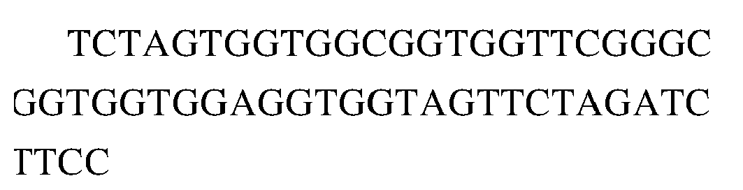
Nucleotide sequence of heavy chain variable region of VEGF165-R859 
SEQ ID NO.4 Nucleotide sequence of the rabbit antibody scFv which is used in the construction of antibody VEGF165-R988 Nucleotide sequence of light chain variable region of VEGF165-R988(SEQ ID NO:8) 

Linker(SEQ ID NO:2): 
Nucleotide sequence of heavy chain variable region of VEGF165-R988(SEQ ID NO:7) 
SEQ ID NO.5 Nucleotide sequence of the rabbit antibody scFv which is used in the construction Nucleotide sequence of light chain variable region of VEGF165-R613 
of antibody VEGF165-R613 
Linker(SEQ ID NO:2): 
Nucleotide sequence of heavy chain variable region of VEGF165-R613 
SEQ ID NO.6 Nucleotide sequence of the rabbit antibody scFv which is used in the construction Nucleotide sequence of light chain variable region of VEGF165-R812 
of antibody VEGF165-R812 
Linker(SEQ ID NO:2): 
Nucleotide sequence of heavy chain variable region of VEGF165-R812 
SEQ ID NO:7 Nucleotide sequence of heavy chain variable region of the 
rabbit antibody VEGF165-R988 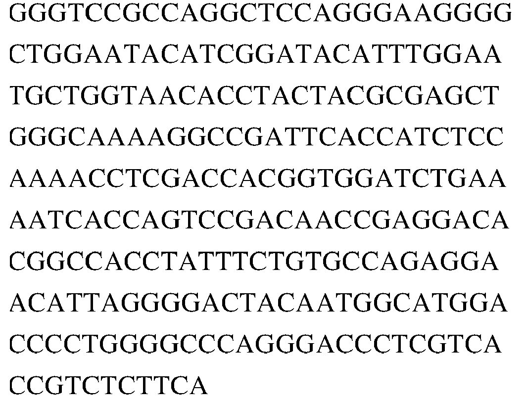
SEQ ID NO:8 Nucleotide sequence of light chain variable region of the rabbit antibody VEGF165-R988 
SEQ ID NO:9 Nucleotide sequence of the heavy chain constant region of rabbit IgG1 

SEQ ID NO:10 Nucleotide sequence of the constant region of the rabbit kappa light chain 

SEQ ID NO:11 Amino acid sequence of the heavy chain variable region of rabbit antibody VEGF165-R988 
SEQ ID NO:12 Amino acid sequence of the light chain variable region of rabbit antibody VEGF165-R988 
SEQ ID NO:13 Amino acid sequence of light chain CDR1 of the rabbit antibody VEGF165-R988 QSSQTIYANRRLA SEQ ID NO:14 Amino acid sequence of light chain CDR2 of the rabbit antibody VEGF165-R988 GASTLAS SEQ ID NO:15 Amino acid sequence of light chain CDR3 of the rabbit antibody VEGF165-R988 AGYKSYDGDDVG SEQ ID NO:16 Amino acid sequence of heavy chain CDR1 of the rabbit antibody VEGF165-R988 GIDLSSYAIS SEQ ID NO:17 Amino acid sequence of heavy chain CDR2 of the rabbit antibody VEGF165-R988 YIWNAGNTYYASWAKG SEQ ID NO:18 Amino acid sequence of heavy chain CDR3 of the rabbit antibody VEGF165-R988 ARGTLGDYNGMDP SEQ ID NO:19 Nucleotide sequence of the humanized antibody VEGF165-H988-10 heavy chain containing the signal peptide Nucleotide sequence of heavy chain signal peptide(SEQ ID NO:45): 
Nucleotide sequence of heavy chain variable region(SEQ ID NO:23): 
Nucleotide sequence of the human IgG1 heavy chain constant region (SEQ ID NO:49): 

SEQ ID NO:20 Nucleotide sequence of humanized antibody VEGF165-H988-10 light chain containing signal peptide Nucleotide sequence of the light chain signal peptide(SEQ ID NO:46): 
Nucleotide sequence of the light chain variable region(SEQ ID NO:24): 
Nucleotide sequence of the human kappa light chain constant region(SEQ ID NO:50): 

SEQ ID NO:21 Amino acid sequence of the heavy chain of the humanized antibody VEGF165-H988-10 containing the signal peptide Amino acid sequence of the heavy chain signal peptide(SEQ ID NO:37): MGWSLILLFLVAVATRVLS Amino acid sequence of the heavy chain variable region(SEQ ID NO:25): 
Amino acid sequence of the human IgG1 heavy chain constant region(SEQ ID NO:41): 
SEQ ID NO:22 Amino acid sequence of the light chain of the humanized antibody VEGF165-H988-10 containing the signal peptide Amino acid sequence of the light chain signal peptide(SEQ ID NO:38): MGWSCIILFLVATATGVHS Amino acid sequence of the light chain variable region(SEQ ID NO:26): 
Amino acid sequence of the human kappa light chain constant region(SEQ ID NO:42) : 
SEQ ID NO:23 Nucleotide sequence of the heavy chain variable region of humanized antibody VEGF165-H988-10 
SEQ ID NO:24 Nucleotide sequence of the light chain variable region of the humanized antibody 
VEGF165-H988-10 
SEQ ID NO:25 Amino acid sequence of the heavy chain variable region of the antibody VEGF165-H988-10 
SEQ ID NO:26 Amino acid sequence of the light chain variable region of the antibody VEGF165-H988-10 
SEQ ID NO:27 Amino acid sequence of light chain CDR1 of humanized antibodyVEGF-H988 QSSKFLWQGRRLA SEQ ID NO:28 Amino acid sequence of light chain CDR2 of humanized antibodyVEGF-H988 GASTLAS SEQ ID NO:29 Amino acid sequence of light AGYKSYDGDVVG chain CDR3 of humanized antibodyVEGF-H988 SEQ ID NO:30 Amino acid sequence of heavy chain CDR1 of humanized antibodyVEGF-H988 GIDLSSYAIS SEQ ID NO:31 Amino acid sequence of heavy chain CDR2 of humanized antibodyVEGF-H988 YIWNDLFTYYASWAKG SEQ ID NO:32 Amino acid sequence of heavy chain CDR3 of humanized antibodyVEGF-H988 ARGTLGDYGGMDP SEQ ID NO:33 Amino acid sequence of the Amino acid sequence of the heavy chain variable region (SEQ ID NO:39): heavy chain of humanized antibody VEGF-H988 
Amino acid sequence of the human IgG1 heavy chain constant region (SEQ ID NO:41): 
SEQ ID NO:34 Amino acid sequence of the light chain of humanized antibody VEGF-H988 Amino acid sequence of the light chain variable region(SEQ ID NO:40): 
Amino acid sequence of the human kappa light chain constant region(SEQ ID NO:42): 

SEQ ID NO:35 Amino acid sequence of the heavy chain of the humanized antibody VEGF-H988 containing the signal peptide Amino acid sequence of the heavy chain signal peptide(SEQ ID NO:37): MGWSLILLFLVAVATRVLS Amino acid sequence of the heavy chain variable region(SEQ ID NO:39): 
Amino acid sequence of the human IgG1 heavychain constant region(SEQ ID NO:41): 
SEQ ID NO:36 Amino acid sequence of the light chain of the humanized antibody VEGF- Amino acid sequence of the light chain signal peptide(SEQ ID NO:38): MGWSCIILFLVATATGVHS Amino acid sequence of the light chain variable region (SEQ ID NO:40): H988 containing the signal peptide 
Amino acid sequence of the human kappa light chain constant region(SEQ ID NO:42): 
SEQ ID NO:37 Amino acid sequence of the heavy chain signal peptide MGWSLILLFLVAVATRVLS SEQ ID NO:38 Amino acid sequence of the light chain signal peptide MGWSCIILFLVATATGVHS SEQ ID NO:39 Amino acid sequence of the heavy chain variable region of the humanized antibody VEGF-H988 
SEQ ID NO:40 Amino acid sequence of the light chain variable region of humanized antibody VEGF-H988 
SEQ ID NO:41 Amino acid sequence of the human IgGlheavy chain constant region 
SEQ ID NO:42 Amino acid sequence of the human kappa light chain constant region 
SEQ ID NO:43 Nucleotide sequence of the humanized antibody VEGF-H988 heavy chain containing the signal peptide Nucleotide sequence of heavy chain signal peptide(SEQ ID NO:45): 
Nucleotide sequence of heavy chain variable region(SEQ ID NO:47): 

Nucleotide sequence of the human IgG1 heavy chain constant region(SEQ ID NO:49): 

SEQ ID NO:44 Nucleotide sequence of humanized antibody VEGF-H988 light chain containing signal peptide Nucleotide sequence of the light chain signal peptide(SEQ ID NO:46): 
Nucleotide sequence of the light chain variable region(SEQ ID NO:48): 
Nucleotide sequence of the human kappa light chain constant region(SEQ ID NO:50): 
SEQ ID NO:45 Nucleotide sequence of heavy chain signal peptide 
SEQ ID NO:46 Nucleotide sequence of the light chain signal peptide 
SEQ ID NO:47 Nucleotide sequence of the heavy chain variable region of the humanized antibody VEGF-H988 

SEQ ID NO:48 Nucleotide sequence of the light chain variable region of the humanized antibody VEGF-H988 
SEQ ID NO:49 Nucleotide sequence of the human IgGlheavy chain constant region 

SEQ ID NO:50 Nucleotide sequence of the human kappa light chain constant region 
SEQ ID NO:51 Amino acid sequence of the Amino acid sequence of VEGF165-R988 light chain variable region(SEQ ID NO:12): rabbit antibody scFv which is used in the construction of antibody VEGF165-R988 
linker(SEQ ID NO:52): SSGGGGSGGGGGGSSRSS Amino acid sequence of VEGF165-R988 heavy chain variable region(SEQ ID NO:11): 
SEQ ID NO.52 Amino acid sequence of the linker used in the construction of the phage antibody library for the linkage of the rabbit antibody scFv SSGGGGSGGGGGGSSRSS SEQ ID NO:53 Nucleotide sequence of the rabbit antibody VEGF165-R988 heavy chain containing the signal peptide Nucleotide sequence of heavy chain signal peptide(SEQ ID NO:45): 
Nucleotide sequence of heavy chain variable region(SEQ ID NO:7): 

Nucleotide sequence of the rabbit IgG heavy chain constant region(SEQ ID NO:9): 
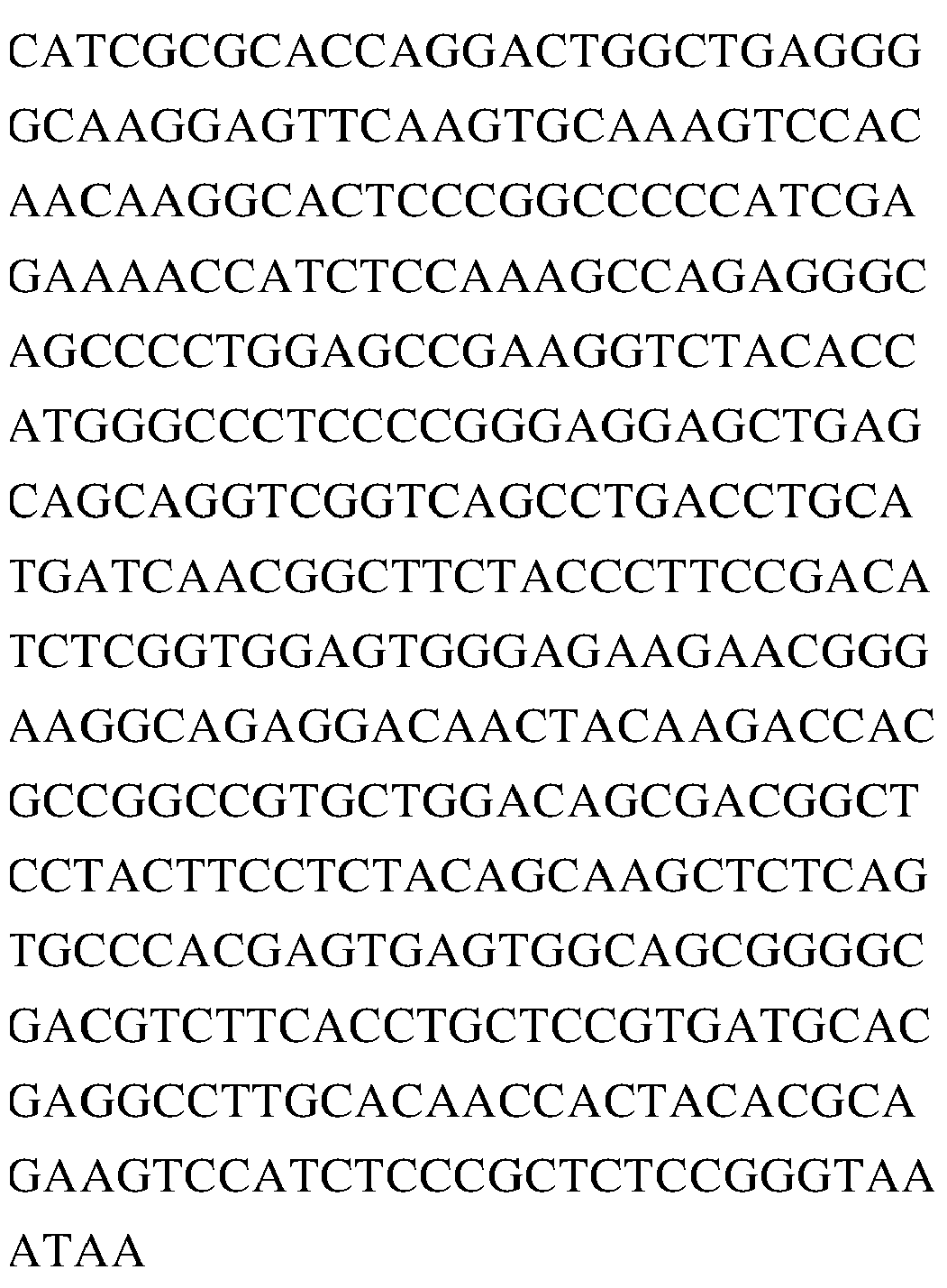
SEQ ID NO:54 Nucleotide sequence of the rabbit antibody VEGF165-R988 light chain containing signal peptide Nucleotide sequence of the light chain signal peptide(SEQ ID NO:46): 
Nucleotide sequence of the light chain variable region(SEQ ID NO:8): 

Nucleotide sequence of the rabbit kappa light chain constant region(SEQ ID NO:10): 
SEQ ID NO:55 Amino acid sequence of the rabbit antibody VEGF165-R988 heavy chain containing the signal peptide Amino acid sequence of heavy chain signal peptide(SEQ ID NO:37): MGWSLILLFLVAVATRVLS Amino acid sequence of heavy chain variable region(SEQ ID NO:11): 
Amino acid sequence of the rabbit heavy chain constant region(SEQ ID NO:57): 
SEQ ID NO:56 Amino acid sequence of the rabbit antibody VEGF165-R988 light chain containing signal peptide Amino acid sequence of the light chain signal peptide(SEQ ID NO:38): MGWSCIILFLVATATGVHS Amino acid sequence of the light chain variable region(SEQ ID NO:12): 
Amino acid sequence of the rabbit light chain constant region(SEQ ID NO:58) SEQ ID NO:57 Amino acid sequence of the rabbit heavy chain constant region 

SEQ ID NO:58 Amino acid sequence of the rabbit light chain constant region 

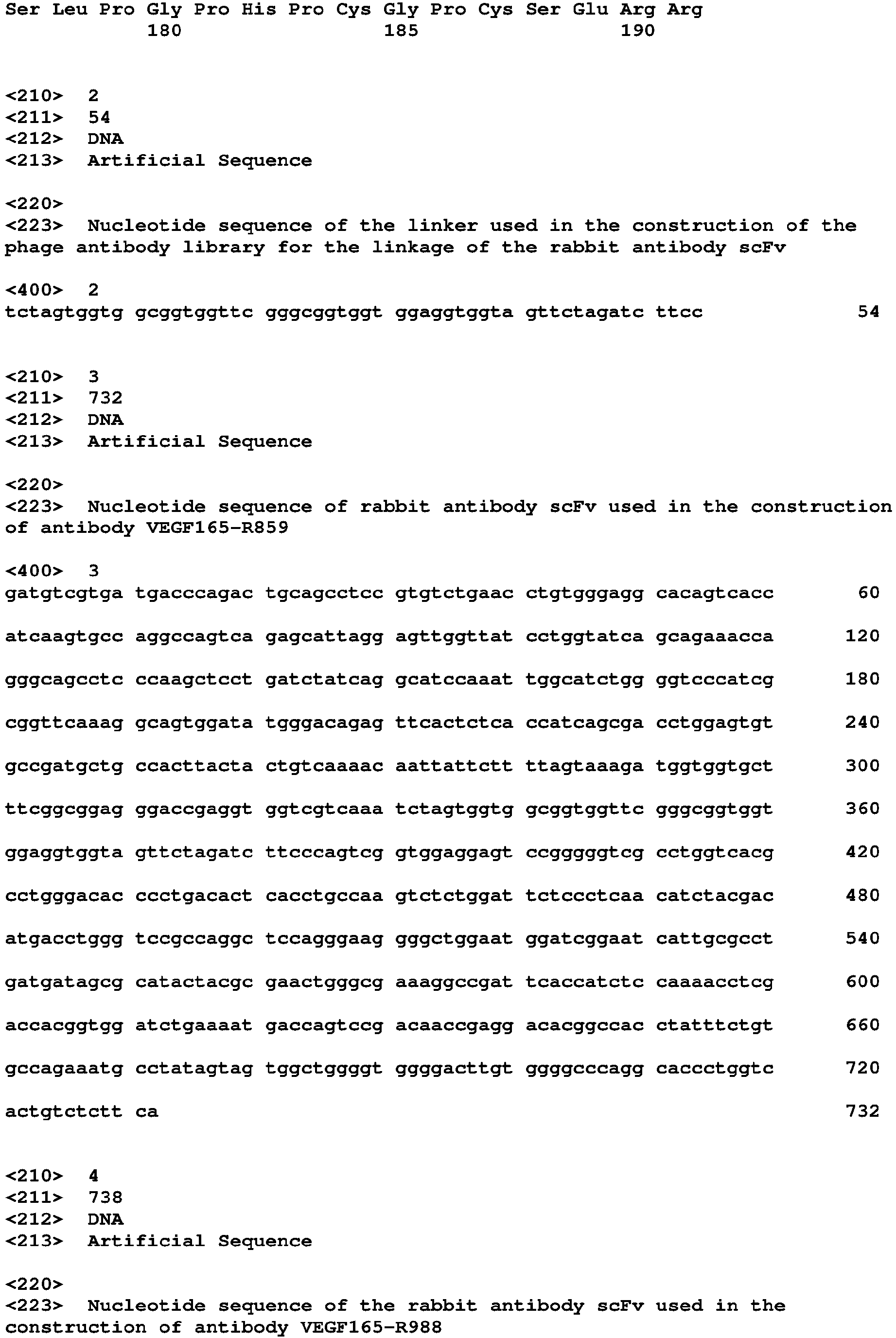


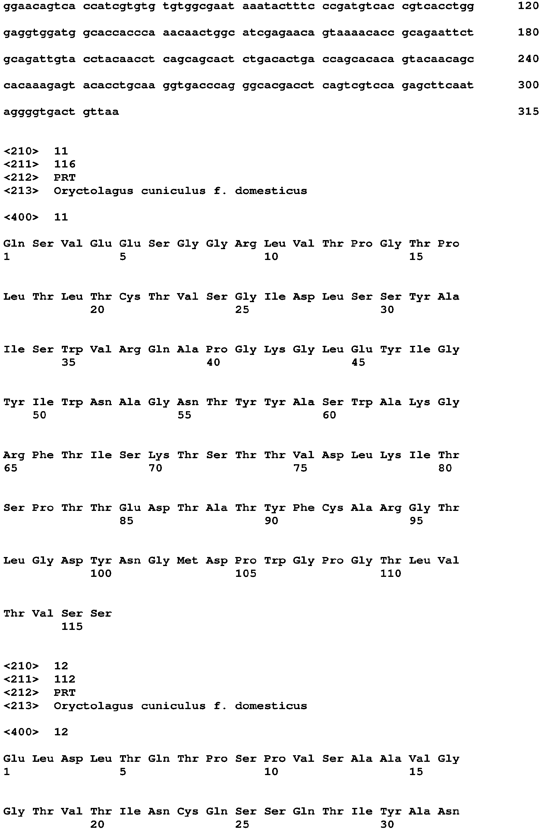


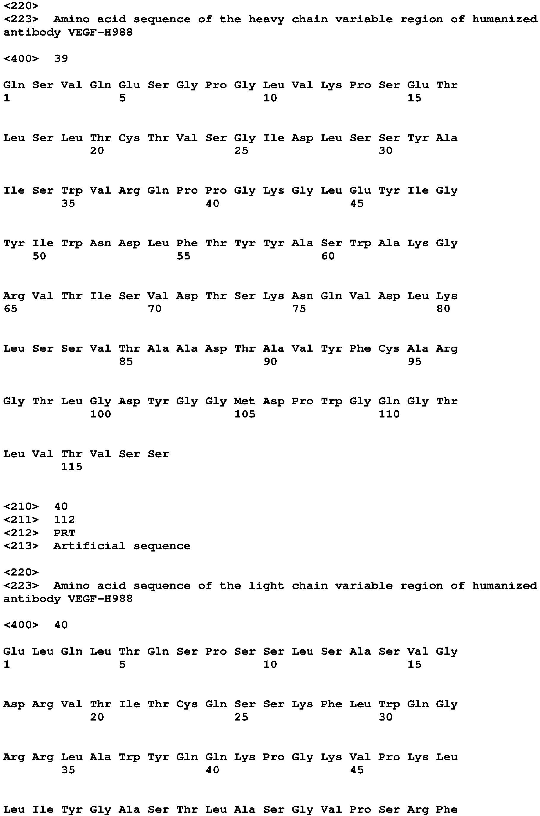


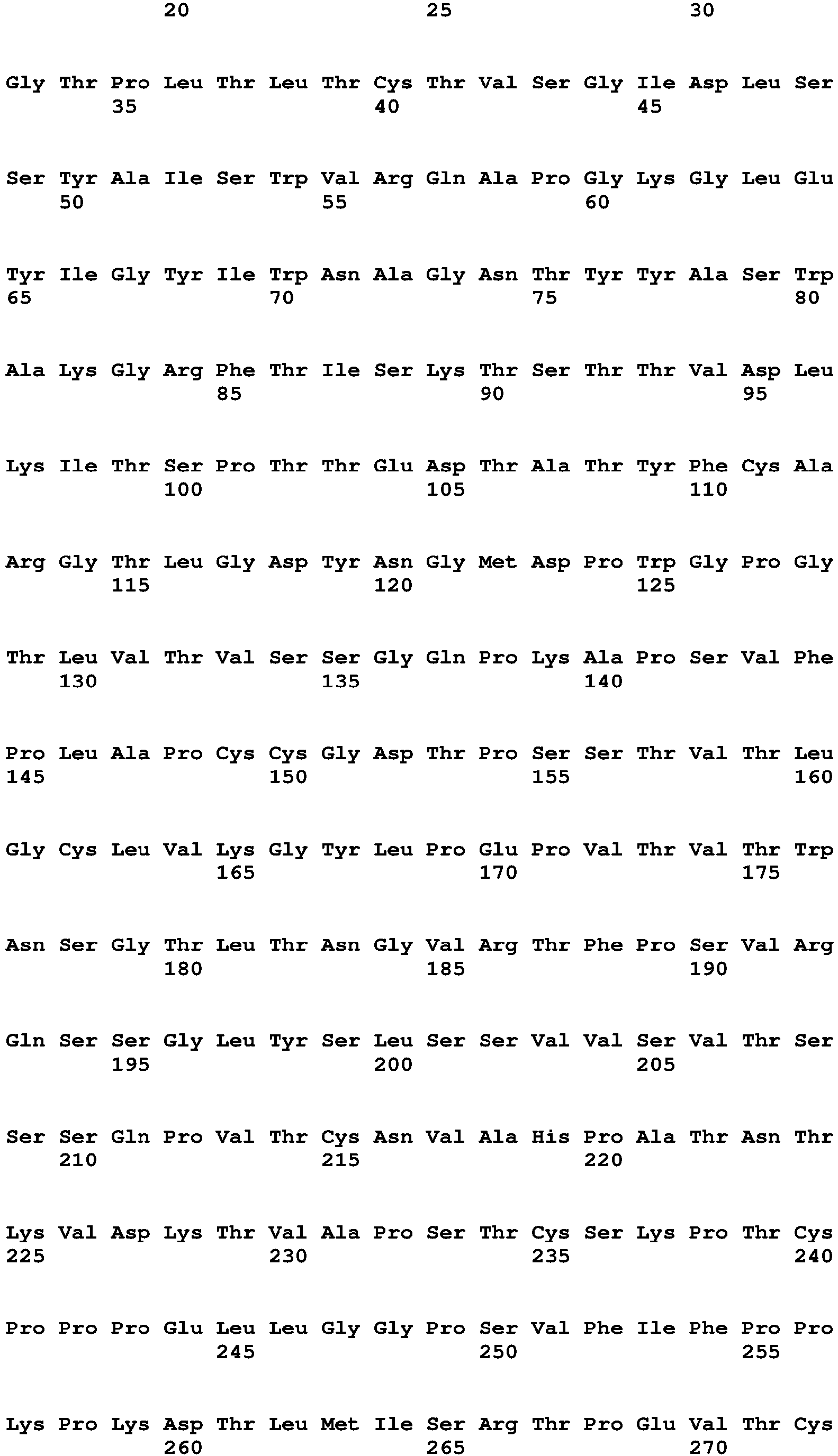


Claims (25)
- An isolated anti-VEGF antibody or antigen-binding fragment thereof, comprisinga heavy chain variable region having a heavy chain CDR1 region having the amino acid sequence as set forth in SEQ ID NO: 30 and a heavy chain CDR2 region having the amino acid sequence as set forth in SEQ ID NO: 31 and a heavy chain CDR3 region having the amino acid sequence as set forth in SEQ ID NO: 32; anda light chain variable region having a light chain CDR1 region having the amino acid sequence as set forth in SEQ ID NO: 27, a light chain CDR2 region having the amino acid sequence as set forth in SEQ ID NO: 28, and a light chain CDR3 region having the amino acid sequence as set forth in SEQ ID NO: 29.
- The anti-VEGF antibody or antigen-binding fragment thereof according to claim 1, comprisinga heavy chain variable region having the amino acid sequence as set forth in SEQ ID NO: 39, or the amino acid sequences having at least 90%, 92%, 95%, 98% or 99% sequence identity to SEQ ID NO: 39; anda light chain variable region having the amino acid sequence as set forth in SEQ ID NO: 40, or the amino acid sequences having at least 90%, 92%, 95%, 98% or 99% sequence identity to SEQ ID NO: 40.
- The anti-VEGF antibody or antigen-binding fragment thereof according to claim 1 or 2, further comprising
a light chain constant region and a heavy chain constant region, preferably,the light chain constant region is the light chain constant region having the amino acid sequence as set forth in SEQ ID NO: 42, or the amino acid sequences having at least 90%, 92%, 95%, 98%, or 99% sequence identity to SEQ ID NO: 42; and/orthe heavy chain constant region is the IgG1 heavy chain constant region having the amino acid sequence as set forth in SEQ ID NO: 41, or the amino acid sequences having at least 90%, 92%, 95%, 98%, or 99% sequence identity to SEQ ID NO:41. - The anti-VEGF antibody or antigen-binding fragment thereof according to any one of claims 1 to 3, wherein said anti-VEGF antibody or antigen-binding fragment thereof is a humanized antibody or a chimeric antibody.
- The anti-VEGF antibody or antigen-binding fragment thereof according to any one of claims 1 to 4, wherein said anti-VEGF antibody or antigen-binding fragment thereof is a monoclonal antibody.
- The anti-VEGF antibody or antigen-binding fragment thereof according to any one of claims 1 to 5, wherein said anti-VEGF antibody or antigen-binding fragment thereof is an IgG antibody, preferably an IgG1 antibody.
- The anti-VEGF antibody or antigen-binding fragment thereof according to any one of claims 1 to 6, wherein the binding affinity KD of said anti-VEGF antibody or antigen-binding fragment thereof to the recombinant human VEG165F protein is 1-100 pM, preferably 5-50 pM, and more preferably 19.5 pM.
- The anti-VEGF antibody or antigen-binding fragment thereof according to any one of claims 1-7, wherein said antigen-binding fragment is Fv, Fab, Fab', Fab'-SH, F(ab')2, Fd fragment, Fd' fragment, single chain antibody molecule or single domain antibody; wherein the single chain antibody molecule is preferably scFv, di-scFv, tri-scFv, diabody or scFab.
- An antibody-drug conjugate, comprising the anti-VEGF antibody or antigen-binding fragment thereof according to any one of claims 18 and an additional therapeutic agent, preferably said anti-VEGF antibody or antigen-binding fragment thereof is connected with the additional therapeutic agent via a linker.
- An isolated anti-VEGF antibody or antigen-binding fragment thereof, comprisinga heavy chain variable region having a heavy chain CDR1 region having the amino acid sequence as set forth in SEQ ID NO: 16 and a heavy chain CDR2 region having the amino acid sequence as set forth in SEQ ID NO: 17 and a heavy chain CDR3 region having the amino acid sequence as set forth in SEQ ID NO: 18; anda light chain variable region having a light chain CDR1 region having the amino acid sequence as set forth in SEQ ID NO: 13, a light chain CDR2 region having the amino acid sequence as set forth in SEQ ID NO: 14, and a light chain CDR3 region having the amino acid sequence as set forth in SEQ ID NO: 15.
- The anti-VEGF antibody or antigen-binding fragment thereof according to claim 10, comprisinga heavy chain constant region having the amino acid sequence as set forth in SEQ ID NO: 25, or the amino acid sequences having at least 90%, 92%, 95%, 98%, or 99% sequence identity to SEQ ID NO: 25; anda light chain variable region having the amino acid sequence as set forth in SEQ ID NO: 26, or the amino acid sequences having at least 90%, 92%, 95%, 98%, or 99% sequence identity to SEQ ID NO: 26.
- The anti-VEGF antibody or antigen-binding fragment thereof according to any one of claims 10-11, wherein said anti-VEGF antibody or antigen-binding fragment thereof is a humanized antibody or a chimeric antibody.
- The anti-VEGF antibody or antigen-binding fragment thereof according to any one of claims 10-12, wherein said anti-VEGF antibody or antigen-binding fragment thereof is a monoclonal antibody.
- The anti-VEGF antibody or antigen-binding fragment thereof according to any one of claims 10-13,wherein said antigen-binding fragment is Fv, Fab, Fab', Fab'-SH, F(ab')2, Fd fragment, Fd' fragment, single chain antibody molecule or single domain antibody;wherein the single chain antibody molecule is preferably scFv, di-scFv, tri-scFv, diabody or scFab.
- A nucleic acid encoding the anti-VEGF antibody or antigen-binding fragment thereof according to any one of claims 1-8, 9-14.
- The nucleic acid according to claim 15, comprisingthe nucleotide sequence as set forth in SEQ ID NO: 7 and/or the nucleotide sequence as set forth in SEQ ID NO: 8; orthe nucleotide sequence as set forth in SEQ ID NO: 23 and/or the nucleotide sequence as set forth in SEQ ID NO: 24; orthe nucleotide sequence as set forth in SEQ ID NO: 47 and/or the nucleotide sequence as set forth in SEQ ID NO: 48.
- An expression vector, comprising the nucleic acid according to claims 15 or 16.
- A host cell, comprising the nucleic acid according to claims 15 or 16 or the expression vector according to claim 17.
- A method for producing the anti-VEGF antibody or antigen-binding fragment thereof according to any one of claims 1-8, 10-14, comprising culturing the host cells according to claim 18 under conditions suitable for antibody expression, and harvesting the expressed antibodies from the culture medium.
- A pharmaceutical composition, comprising the anti-VEGF antibody or antigen-binding fragment thereof according to any one of claims 1-8, 10-14, or the antibody-drug conjugate according to claim 9, or the nucleic acid according to any one of claims 15-16, or the expression vector according to claim 17, and a pharmaceutically acceptable carrier.
- The anti-VEGF antibody or antigen-binding fragment thereof according to any one of claims 1-8, 10-14 or the antibody-drug conjugate according to claim 9 or the pharmaceutical composition according to claim 20, for use in the treatment of colorectal cancer.
- A method for treating colorectal cancer, comprising administering to a subject in need a therapeutically effective amount of the anti-VEGF antibody or antigen-binding fragment thereof according to any one of claims 1-8, 10-14, or the antibody-drug conjugate according to claim 9, or the pharmaceutical composition according to claim 20.
- A use of the anti-VEGF antibody or antigen-binding fragment thereof according to any one of claims 1-8, 10-14 or the antibody-drug conjugate according to claim 9 or the pharmaceutical composition according to claim 20 in the preparation of a medicament for the treatment of colorectal cancer.
- A pharmaceutical combination, comprising the anti-VEGF antibody or antigen-binding fragment thereof according to any one of claims 1-8, 10-14 or the antibody-drug conjugate according to claim 9 or the pharmaceutical composition according to claim 20, and one or more additional therapeutic agents.
- A kit, comprising the anti-VEGF antibody or antigen-binding fragment according to any one of claims 1-8, 10-14, or the antibody-drug conjugate according to claim 9, or the pharmaceutical composition according to claim 20, or the pharmaceutical combination according to claim 24, preferably further comprising a device for administration.
Applications Claiming Priority (2)
| Application Number | Priority Date | Filing Date | Title |
|---|---|---|---|
| CN201910657497 | 2019-07-19 | ||
| PCT/CN2020/102622 WO2021013080A1 (en) | 2019-07-19 | 2020-07-17 | Humanized anti-vegf monoclonal antibody |
Publications (4)
| Publication Number | Publication Date |
|---|---|
| EP4001306A1 true EP4001306A1 (en) | 2022-05-25 |
| EP4001306A4 EP4001306A4 (en) | 2023-11-15 |
| EP4001306B1 EP4001306B1 (en) | 2025-08-27 |
| EP4001306C0 EP4001306C0 (en) | 2025-08-27 |
Family
ID=74193255
Family Applications (1)
| Application Number | Title | Priority Date | Filing Date |
|---|---|---|---|
| EP20844414.1A Active EP4001306B1 (en) | 2019-07-19 | 2020-07-17 | Humanized anti-vegf monoclonal antibody |
Country Status (11)
| Country | Link |
|---|---|
| US (1) | US12060416B2 (en) |
| EP (1) | EP4001306B1 (en) |
| JP (1) | JP7352007B2 (en) |
| KR (1) | KR102920217B1 (en) |
| CN (1) | CN114008075B (en) |
| AU (1) | AU2020316498B2 (en) |
| BR (1) | BR112022001017A2 (en) |
| CA (1) | CA3147921C (en) |
| MX (1) | MX2022000779A (en) |
| MY (1) | MY206255A (en) |
| WO (1) | WO2021013080A1 (en) |
Families Citing this family (4)
| Publication number | Priority date | Publication date | Assignee | Title |
|---|---|---|---|---|
| AU2020316495B2 (en) * | 2019-07-19 | 2024-07-04 | Sinocelltech Ltd | Humanized anti-VEGF Fab antibody fragment and use thereof |
| BR112023016095A2 (en) * | 2021-02-10 | 2023-11-14 | Jiangxi Jemincare Group Co Ltd | ANTI-VEGF ANTIBODY AND USE THEREOF |
| CN115850470B (en) * | 2022-12-12 | 2023-07-07 | 三门峡市眼科医院 | VEGF antibodies and uses thereof |
| CN117820474A (en) * | 2023-04-25 | 2024-04-05 | 南京贝思奥生物科技有限公司 | Drugs for improving ocular angiogenesis-related diseases and their applications |
Family Cites Families (23)
| Publication number | Priority date | Publication date | Assignee | Title |
|---|---|---|---|---|
| US4399216A (en) | 1980-02-25 | 1983-08-16 | The Trustees Of Columbia University | Processes for inserting DNA into eucaryotic cells and for producing proteinaceous materials |
| US5179017A (en) | 1980-02-25 | 1993-01-12 | The Trustees Of Columbia University In The City Of New York | Processes for inserting DNA into eucaryotic cells and for producing proteinaceous materials |
| US4634665A (en) | 1980-02-25 | 1987-01-06 | The Trustees Of Columbia University In The City Of New York | Processes for inserting DNA into eucaryotic cells and for producing proteinaceous materials |
| US4510245A (en) | 1982-11-18 | 1985-04-09 | Chiron Corporation | Adenovirus promoter system |
| GB8308235D0 (en) | 1983-03-25 | 1983-05-05 | Celltech Ltd | Polypeptides |
| US5168062A (en) | 1985-01-30 | 1992-12-01 | University Of Iowa Research Foundation | Transfer vectors and microorganisms containing human cytomegalovirus immediate-early promoter-regulatory DNA sequence |
| US4968615A (en) | 1985-12-18 | 1990-11-06 | Ciba-Geigy Corporation | Deoxyribonucleic acid segment from a virus |
| IL85035A0 (en) | 1987-01-08 | 1988-06-30 | Int Genetic Eng | Polynucleotide molecule,a chimeric antibody with specificity for human b cell surface antigen,a process for the preparation and methods utilizing the same |
| EP0590058B1 (en) | 1991-06-14 | 2003-11-26 | Genentech, Inc. | HUMANIZED Heregulin ANTIBODy |
| CA2323757C (en) | 1998-04-02 | 2011-08-02 | Genentech, Inc. | Antibody variants and fragments thereof |
| US6194551B1 (en) | 1998-04-02 | 2001-02-27 | Genentech, Inc. | Polypeptide variants |
| US6737056B1 (en) | 1999-01-15 | 2004-05-18 | Genentech, Inc. | Polypeptide variants with altered effector function |
| RS20150135A1 (en) | 2003-05-30 | 2015-08-31 | Genentech Inc. | TREATMENT WITH ANTI-VEGF ANTIBODIES |
| US20050106667A1 (en) * | 2003-08-01 | 2005-05-19 | Genentech, Inc | Binding polypeptides with restricted diversity sequences |
| TWI468417B (en) | 2007-11-30 | 2015-01-11 | Genentech Inc | Anti-vegf antibodies |
| CN102002104A (en) * | 2009-08-28 | 2011-04-06 | 江苏先声药物研究有限公司 | Anti-VEGF monoclonal antibody and medicinal composition containing same |
| TWI507524B (en) * | 2009-11-30 | 2015-11-11 | 建南德克公司 | Composition and method for diagnosing and treating tumor |
| AU2014374055B2 (en) * | 2013-12-30 | 2018-11-08 | Epimab Biotherapeutics Inc. | Fabs-in-tandem immunoglobulin and uses thereof |
| CN108148135B (en) * | 2015-01-06 | 2020-06-12 | 珠海亿胜生物制药有限公司 | anti-VEGF antibodies |
| CN108101986B (en) * | 2015-01-06 | 2020-04-17 | 珠海亿胜生物制药有限公司 | anti-VEGF antibodies |
| WO2016120753A1 (en) | 2015-01-28 | 2016-08-04 | Pfizer Inc. | Stable aqueous anti- vascular endothelial growth factor (vegf) antibody formulation |
| EP3401331B1 (en) * | 2016-01-06 | 2022-04-06 | Order-Made Medical Research Inc. | High-affinity anti-vegf antibody |
| AU2020316495B2 (en) * | 2019-07-19 | 2024-07-04 | Sinocelltech Ltd | Humanized anti-VEGF Fab antibody fragment and use thereof |
-
2020
- 2020-07-17 KR KR1020227004900A patent/KR102920217B1/en active Active
- 2020-07-17 CN CN202080046296.4A patent/CN114008075B/en active Active
- 2020-07-17 BR BR112022001017A patent/BR112022001017A2/en unknown
- 2020-07-17 WO PCT/CN2020/102622 patent/WO2021013080A1/en not_active Ceased
- 2020-07-17 AU AU2020316498A patent/AU2020316498B2/en active Active
- 2020-07-17 CA CA3147921A patent/CA3147921C/en active Active
- 2020-07-17 JP JP2022503865A patent/JP7352007B2/en active Active
- 2020-07-17 MX MX2022000779A patent/MX2022000779A/en unknown
- 2020-07-17 MY MYPI2022000334A patent/MY206255A/en unknown
- 2020-07-17 EP EP20844414.1A patent/EP4001306B1/en active Active
-
2022
- 2022-01-19 US US17/579,376 patent/US12060416B2/en active Active
Also Published As
| Publication number | Publication date |
|---|---|
| CA3147921C (en) | 2023-02-21 |
| CN114008075A (en) | 2022-02-01 |
| EP4001306B1 (en) | 2025-08-27 |
| AU2020316498A1 (en) | 2022-02-03 |
| AU2020316498B2 (en) | 2024-02-29 |
| CA3147921A1 (en) | 2021-01-28 |
| KR20220071177A (en) | 2022-05-31 |
| KR102920217B1 (en) | 2026-01-30 |
| WO2021013080A1 (en) | 2021-01-28 |
| JP7352007B2 (en) | 2023-09-27 |
| EP4001306A4 (en) | 2023-11-15 |
| MY206255A (en) | 2024-12-05 |
| MX2022000779A (en) | 2022-02-14 |
| JP2022542037A (en) | 2022-09-29 |
| BR112022001017A2 (en) | 2022-04-12 |
| CN114008075B (en) | 2024-08-16 |
| EP4001306C0 (en) | 2025-08-27 |
| US12060416B2 (en) | 2024-08-13 |
| US20220144930A1 (en) | 2022-05-12 |
Similar Documents
| Publication | Publication Date | Title |
|---|---|---|
| JP6871415B2 (en) | Anti-LAG3 antibody | |
| EP4043490B1 (en) | Humanized anti-vegf fab antibody fragment and use thereof | |
| JP6023706B2 (en) | Agonist antibody against CD27 | |
| EP2552966B1 (en) | Antibodies against csf-1r | |
| US12060416B2 (en) | Humanized anti-VEGF monoclonal antibody | |
| CN114174335B (en) | A humanized VEGFR2 antibody and its application | |
| KR20220071264A (en) | Anti-PD-L1 antigen binding protein and applications thereof | |
| JP2022515258A (en) | Multispecific antigen-binding proteins capable of binding to CD19 and CD3, and their use | |
| CN114341170B (en) | A kind of humanized anti-VEGFR2 antibody and its application | |
| EP3404040A1 (en) | Anti-myl9 antibody | |
| US11753477B2 (en) | Preparation and use of anti-MET-and-RON bispecific antibody and antibody-drug conjugate thereof | |
| RU2809746C2 (en) | Humanized anti-vegf monoclonal antibody | |
| HK40066231A (en) | Humanized anti-vegf monoclonal antibody | |
| HK40066231B (en) | Humanized anti-vegf monoclonal antibody | |
| HK40066221B (en) | Humanized anti-vegf fab antibody fragment and use thereof | |
| HK40066221A (en) | Humanized anti-vegf fab antibody fragment and use thereof | |
| HK40066220B (en) | Humanized vegfr2 antibody and application thereof | |
| HK40066220A (en) | Humanized vegfr2 antibody and application thereof | |
| HK40066232A (en) | Humanized anti-vegfr2 antibody and application thereof | |
| HK40066232B (en) | Humanized anti-vegfr2 antibody and application thereof | |
| BR122023027661A2 (en) | TGF-BETA-RII BINDING PROTEINS | |
| HK1236581A1 (en) | Assay and method for determining cdc eliciting antibodies | |
| NZ616809A (en) | Anti-b7-h3 antibody |
Legal Events
| Date | Code | Title | Description |
|---|---|---|---|
| STAA | Information on the status of an ep patent application or granted ep patent |
Free format text: STATUS: THE INTERNATIONAL PUBLICATION HAS BEEN MADE |
|
| PUAI | Public reference made under article 153(3) epc to a published international application that has entered the european phase |
Free format text: ORIGINAL CODE: 0009012 |
|
| STAA | Information on the status of an ep patent application or granted ep patent |
Free format text: STATUS: REQUEST FOR EXAMINATION WAS MADE |
|
| 17P | Request for examination filed |
Effective date: 20220209 |
|
| AK | Designated contracting states |
Kind code of ref document: A1 Designated state(s): AL AT BE BG CH CY CZ DE DK EE ES FI FR GB GR HR HU IE IS IT LI LT LU LV MC MK MT NL NO PL PT RO RS SE SI SK SM TR |
|
| DAV | Request for validation of the european patent (deleted) | ||
| DAX | Request for extension of the european patent (deleted) | ||
| RIC1 | Information provided on ipc code assigned before grant |
Ipc: A61P 35/00 20060101ALI20230712BHEP Ipc: A61K 39/395 20060101ALI20230712BHEP Ipc: C12P 21/08 20060101ALI20230712BHEP Ipc: C07K 16/22 20060101AFI20230712BHEP |
|
| A4 | Supplementary search report drawn up and despatched |
Effective date: 20231018 |
|
| RIC1 | Information provided on ipc code assigned before grant |
Ipc: A61P 35/00 20060101ALI20231012BHEP Ipc: A61K 39/395 20060101ALI20231012BHEP Ipc: C12P 21/08 20060101ALI20231012BHEP Ipc: C07K 16/22 20060101AFI20231012BHEP |
|
| STAA | Information on the status of an ep patent application or granted ep patent |
Free format text: STATUS: EXAMINATION IS IN PROGRESS |
|
| 17Q | First examination report despatched |
Effective date: 20240404 |
|
| GRAP | Despatch of communication of intention to grant a patent |
Free format text: ORIGINAL CODE: EPIDOSNIGR1 |
|
| STAA | Information on the status of an ep patent application or granted ep patent |
Free format text: STATUS: GRANT OF PATENT IS INTENDED |
|
| GRAJ | Information related to disapproval of communication of intention to grant by the applicant or resumption of examination proceedings by the epo deleted |
Free format text: ORIGINAL CODE: EPIDOSDIGR1 |
|
| STAA | Information on the status of an ep patent application or granted ep patent |
Free format text: STATUS: EXAMINATION IS IN PROGRESS |
|
| INTG | Intention to grant announced |
Effective date: 20250403 |
|
| GRAJ | Information related to disapproval of communication of intention to grant by the applicant or resumption of examination proceedings by the epo deleted |
Free format text: ORIGINAL CODE: EPIDOSDIGR1 |
|
| GRAP | Despatch of communication of intention to grant a patent |
Free format text: ORIGINAL CODE: EPIDOSNIGR1 |
|
| GRAP | Despatch of communication of intention to grant a patent |
Free format text: ORIGINAL CODE: EPIDOSNIGR1 |
|
| STAA | Information on the status of an ep patent application or granted ep patent |
Free format text: STATUS: GRANT OF PATENT IS INTENDED |
|
| INTC | Intention to grant announced (deleted) | ||
| GRAS | Grant fee paid |
Free format text: ORIGINAL CODE: EPIDOSNIGR3 |
|
| INTG | Intention to grant announced |
Effective date: 20250530 |
|
| GRAA | (expected) grant |
Free format text: ORIGINAL CODE: 0009210 |
|
| STAA | Information on the status of an ep patent application or granted ep patent |
Free format text: STATUS: THE PATENT HAS BEEN GRANTED |
|
| AK | Designated contracting states |
Kind code of ref document: B1 Designated state(s): AL AT BE BG CH CY CZ DE DK EE ES FI FR GB GR HR HU IE IS IT LI LT LU LV MC MK MT NL NO PL PT RO RS SE SI SK SM TR |
|
| REG | Reference to a national code |
Ref country code: CH Ref legal event code: EP |
|
| REG | Reference to a national code |
Ref country code: DE Ref legal event code: R096 Ref document number: 602020057633 Country of ref document: DE |
|
| REG | Reference to a national code |
Ref country code: IE Ref legal event code: FG4D |
|
| U01 | Request for unitary effect filed |
Effective date: 20250923 |
|
| U07 | Unitary effect registered |
Designated state(s): AT BE BG DE DK EE FI FR IT LT LU LV MT NL PT RO SE SI Effective date: 20251001 |
|
| PG25 | Lapsed in a contracting state [announced via postgrant information from national office to epo] |
Ref country code: IS Free format text: LAPSE BECAUSE OF FAILURE TO SUBMIT A TRANSLATION OF THE DESCRIPTION OR TO PAY THE FEE WITHIN THE PRESCRIBED TIME-LIMIT Effective date: 20251227 |
|
| PG25 | Lapsed in a contracting state [announced via postgrant information from national office to epo] |
Ref country code: NO Free format text: LAPSE BECAUSE OF FAILURE TO SUBMIT A TRANSLATION OF THE DESCRIPTION OR TO PAY THE FEE WITHIN THE PRESCRIBED TIME-LIMIT Effective date: 20251127 |
|
| PG25 | Lapsed in a contracting state [announced via postgrant information from national office to epo] |
Ref country code: HR Free format text: LAPSE BECAUSE OF FAILURE TO SUBMIT A TRANSLATION OF THE DESCRIPTION OR TO PAY THE FEE WITHIN THE PRESCRIBED TIME-LIMIT Effective date: 20250827 |
|
| PG25 | Lapsed in a contracting state [announced via postgrant information from national office to epo] |
Ref country code: GR Free format text: LAPSE BECAUSE OF FAILURE TO SUBMIT A TRANSLATION OF THE DESCRIPTION OR TO PAY THE FEE WITHIN THE PRESCRIBED TIME-LIMIT Effective date: 20251128 |
|
| PG25 | Lapsed in a contracting state [announced via postgrant information from national office to epo] |
Ref country code: PL Free format text: LAPSE BECAUSE OF FAILURE TO SUBMIT A TRANSLATION OF THE DESCRIPTION OR TO PAY THE FEE WITHIN THE PRESCRIBED TIME-LIMIT Effective date: 20250827 |
|
| PG25 | Lapsed in a contracting state [announced via postgrant information from national office to epo] |
Ref country code: RS Free format text: LAPSE BECAUSE OF FAILURE TO SUBMIT A TRANSLATION OF THE DESCRIPTION OR TO PAY THE FEE WITHIN THE PRESCRIBED TIME-LIMIT Effective date: 20251127 |
|
| PG25 | Lapsed in a contracting state [announced via postgrant information from national office to epo] |
Ref country code: ES Free format text: LAPSE BECAUSE OF FAILURE TO SUBMIT A TRANSLATION OF THE DESCRIPTION OR TO PAY THE FEE WITHIN THE PRESCRIBED TIME-LIMIT Effective date: 20250827 |
|
| PG25 | Lapsed in a contracting state [announced via postgrant information from national office to epo] |
Ref country code: SM Free format text: LAPSE BECAUSE OF FAILURE TO SUBMIT A TRANSLATION OF THE DESCRIPTION OR TO PAY THE FEE WITHIN THE PRESCRIBED TIME-LIMIT Effective date: 20250827 |
|
| PG25 | Lapsed in a contracting state [announced via postgrant information from national office to epo] |
Ref country code: CZ Free format text: LAPSE BECAUSE OF FAILURE TO SUBMIT A TRANSLATION OF THE DESCRIPTION OR TO PAY THE FEE WITHIN THE PRESCRIBED TIME-LIMIT Effective date: 20250827 |
|
| PG25 | Lapsed in a contracting state [announced via postgrant information from national office to epo] |
Ref country code: SK Free format text: LAPSE BECAUSE OF FAILURE TO SUBMIT A TRANSLATION OF THE DESCRIPTION OR TO PAY THE FEE WITHIN THE PRESCRIBED TIME-LIMIT Effective date: 20250827 |
|
| PLBE | No opposition filed within time limit |
Free format text: ORIGINAL CODE: 0009261 |
|
| STAA | Information on the status of an ep patent application or granted ep patent |
Free format text: STATUS: NO OPPOSITION FILED WITHIN TIME LIMIT |
|
| REG | Reference to a national code |
Ref country code: CH Ref legal event code: L10 Free format text: ST27 STATUS EVENT CODE: U-0-0-L10-L00 (AS PROVIDED BY THE NATIONAL OFFICE) Effective date: 20260708 |
|
| 26N | No opposition filed |
Effective date: 20260528 |
|
| U20 | Renewal fee for the european patent with unitary effect paid |
Year of fee payment: 7 Effective date: 20260626 |