EP3974539A1 - Methods to diagnose and treat acute respiratory infections - Google Patents
Methods to diagnose and treat acute respiratory infections Download PDFInfo
- Publication number
- EP3974539A1 EP3974539A1 EP21207860.4A EP21207860A EP3974539A1 EP 3974539 A1 EP3974539 A1 EP 3974539A1 EP 21207860 A EP21207860 A EP 21207860A EP 3974539 A1 EP3974539 A1 EP 3974539A1
- Authority
- EP
- European Patent Office
- Prior art keywords
- classifier
- ari
- bacterial
- viral
- infectious
- Prior art date
- Legal status (The legal status is an assumption and is not a legal conclusion. Google has not performed a legal analysis and makes no representation as to the accuracy of the status listed.)
- Pending
Links
- 238000000034 method Methods 0.000 title claims abstract description 140
- 230000001154 acute effect Effects 0.000 title claims abstract description 103
- 206010057190 Respiratory tract infections Diseases 0.000 title claims abstract description 55
- 239000012472 biological sample Substances 0.000 claims abstract description 72
- 230000001580 bacterial effect Effects 0.000 claims description 266
- 208000015181 infectious disease Diseases 0.000 claims description 255
- 230000003612 virological effect Effects 0.000 claims description 251
- 108090000623 proteins and genes Proteins 0.000 claims description 218
- 230000014509 gene expression Effects 0.000 claims description 195
- 230000002458 infectious effect Effects 0.000 claims description 191
- 239000000523 sample Substances 0.000 claims description 152
- 230000000241 respiratory effect Effects 0.000 claims description 59
- 230000004044 response Effects 0.000 claims description 39
- 238000001514 detection method Methods 0.000 claims description 33
- 238000005259 measurement Methods 0.000 claims description 33
- 208000024891 symptom Diseases 0.000 claims description 32
- 238000003860 storage Methods 0.000 claims description 30
- 208000035143 Bacterial infection Diseases 0.000 claims description 25
- 208000022362 bacterial infectious disease Diseases 0.000 claims description 25
- 238000004458 analytical method Methods 0.000 claims description 23
- 208000036142 Viral infection Diseases 0.000 claims description 21
- 239000003814 drug Substances 0.000 claims description 18
- 238000003753 real-time PCR Methods 0.000 claims description 18
- 230000009385 viral infection Effects 0.000 claims description 17
- 238000009396 hybridization Methods 0.000 claims description 16
- 102000039446 nucleic acids Human genes 0.000 claims description 16
- 108020004707 nucleic acids Proteins 0.000 claims description 16
- 150000007523 nucleic acids Chemical class 0.000 claims description 16
- 238000002560 therapeutic procedure Methods 0.000 claims description 15
- 210000005259 peripheral blood Anatomy 0.000 claims description 11
- 239000011886 peripheral blood Substances 0.000 claims description 11
- 230000000844 anti-bacterial effect Effects 0.000 claims description 9
- 229940079593 drug Drugs 0.000 claims description 9
- 238000004949 mass spectrometry Methods 0.000 claims description 7
- 238000012163 sequencing technique Methods 0.000 claims description 7
- 230000000840 anti-viral effect Effects 0.000 claims description 6
- 206010036790 Productive cough Diseases 0.000 claims description 5
- 239000003443 antiviral agent Substances 0.000 claims description 5
- 238000012544 monitoring process Methods 0.000 claims description 5
- 210000003802 sputum Anatomy 0.000 claims description 5
- 208000024794 sputum Diseases 0.000 claims description 5
- 230000001186 cumulative effect Effects 0.000 claims description 4
- 229960005486 vaccine Drugs 0.000 claims description 4
- 229940124350 antibacterial drug Drugs 0.000 claims description 3
- 238000012360 testing method Methods 0.000 description 44
- 238000012549 training Methods 0.000 description 28
- 241000700605 Viruses Species 0.000 description 25
- 241000894006 Bacteria Species 0.000 description 24
- 238000005516 engineering process Methods 0.000 description 22
- 238000002493 microarray Methods 0.000 description 22
- 238000011282 treatment Methods 0.000 description 22
- 108020004999 messenger RNA Proteins 0.000 description 20
- 210000004369 blood Anatomy 0.000 description 19
- 239000008280 blood Substances 0.000 description 19
- 102000004169 proteins and genes Human genes 0.000 description 19
- 244000052769 pathogen Species 0.000 description 18
- 208000037265 diseases, disorders, signs and symptoms Diseases 0.000 description 17
- 206010022000 influenza Diseases 0.000 description 17
- 238000011002 quantification Methods 0.000 description 16
- 244000052613 viral pathogen Species 0.000 description 16
- 201000010099 disease Diseases 0.000 description 15
- 230000006870 function Effects 0.000 description 13
- 230000001717 pathogenic effect Effects 0.000 description 13
- 206010035664 Pneumonia Diseases 0.000 description 12
- 206010040047 Sepsis Diseases 0.000 description 12
- 238000003556 assay Methods 0.000 description 12
- 238000011529 RT qPCR Methods 0.000 description 11
- 210000004027 cell Anatomy 0.000 description 11
- 230000034994 death Effects 0.000 description 11
- 231100000517 death Toxicity 0.000 description 11
- 238000007477 logistic regression Methods 0.000 description 11
- 238000003752 polymerase chain reaction Methods 0.000 description 11
- 230000008569 process Effects 0.000 description 11
- 238000012545 processing Methods 0.000 description 11
- 102100034237 Endosome/lysosome-associated apoptosis and autophagy regulator 1 Human genes 0.000 description 10
- 101000925880 Homo sapiens Endosome/lysosome-associated apoptosis and autophagy regulator 1 Proteins 0.000 description 10
- 108010048233 Procalcitonin Proteins 0.000 description 10
- 206010051379 Systemic Inflammatory Response Syndrome Diseases 0.000 description 10
- 239000003242 anti bacterial agent Substances 0.000 description 10
- 229940088710 antibiotic agent Drugs 0.000 description 10
- 230000003115 biocidal effect Effects 0.000 description 10
- 239000002299 complementary DNA Substances 0.000 description 10
- CWCXERYKLSEGEZ-KDKHKZEGSA-N procalcitonin Chemical compound C([C@@H](C(=O)N1CCC[C@H]1C(=O)N[C@@H](CCC(N)=O)C(=O)N[C@H](C(=O)N[C@@H](C)C(=O)N[C@@H]([C@@H](C)CC)C(=O)NCC(=O)N[C@@H](C(C)C)C(=O)NCC(=O)N[C@@H](C)C(=O)N1[C@@H](CCC1)C(=O)NCC(O)=O)[C@@H](C)O)NC(=O)[C@@H](NC(=O)[C@H](CC=1NC=NC=1)NC(=O)[C@H](CC=1C=CC=CC=1)NC(=O)[C@H](CCCCN)NC(=O)[C@H](CC(N)=O)NC(=O)[C@H](CC=1C=CC=CC=1)NC(=O)[C@H](CC(O)=O)NC(=O)[C@H](CCC(N)=O)NC(=O)[C@@H](NC(=O)[C@H](CC=1C=CC(O)=CC=1)NC(=O)[C@@H](NC(=O)CNC(=O)[C@H](CC(C)C)NC(=O)[C@H](CCSC)NC(=O)[C@H]1NC(=O)[C@H]([C@@H](C)O)NC(=O)[C@H](CO)NC(=O)[C@H](CC(C)C)NC(=O)[C@H](CC(N)=O)NC(=O)CNC(=O)[C@@H](N)CSSC1)[C@@H](C)O)[C@@H](C)O)[C@@H](C)O)C1=CC=CC=C1 CWCXERYKLSEGEZ-KDKHKZEGSA-N 0.000 description 10
- 230000035945 sensitivity Effects 0.000 description 10
- 208000003322 Coinfection Diseases 0.000 description 9
- 102100027355 Interferon-induced protein with tetratricopeptide repeats 1 Human genes 0.000 description 9
- 238000003491 array Methods 0.000 description 9
- 244000052616 bacterial pathogen Species 0.000 description 9
- 238000004590 computer program Methods 0.000 description 9
- 238000003762 quantitative reverse transcription PCR Methods 0.000 description 9
- 108020004414 DNA Proteins 0.000 description 8
- 101000946040 Homo sapiens Lysosomal-associated transmembrane protein 4B Proteins 0.000 description 8
- 101000611202 Homo sapiens Peptidyl-prolyl cis-trans isomerase B Proteins 0.000 description 8
- 102100034726 Lysosomal-associated transmembrane protein 4B Human genes 0.000 description 8
- 102100040283 Peptidyl-prolyl cis-trans isomerase B Human genes 0.000 description 8
- 239000000090 biomarker Substances 0.000 description 8
- 230000000694 effects Effects 0.000 description 8
- 230000036541 health Effects 0.000 description 8
- 108090000765 processed proteins & peptides Proteins 0.000 description 8
- 238000010200 validation analysis Methods 0.000 description 8
- 102100021403 2,4-dienoyl-CoA reductase [(3E)-enoyl-CoA-producing], mitochondrial Human genes 0.000 description 7
- 102100040038 Amyloid beta precursor like protein 2 Human genes 0.000 description 7
- 102100038509 E3 ubiquitin-protein ligase ARIH1 Human genes 0.000 description 7
- 102100031181 Glyceraldehyde-3-phosphate dehydrogenase Human genes 0.000 description 7
- 101001041661 Homo sapiens 2,4-dienoyl-CoA reductase [(3E)-enoyl-CoA-producing], mitochondrial Proteins 0.000 description 7
- 101000808922 Homo sapiens E3 ubiquitin-protein ligase ARIH1 Proteins 0.000 description 7
- 101001082065 Homo sapiens Interferon-induced protein with tetratricopeptide repeats 1 Proteins 0.000 description 7
- 101000830568 Homo sapiens Tumor necrosis factor alpha-induced protein 2 Proteins 0.000 description 7
- 108020005187 Oligonucleotide Probes Proteins 0.000 description 7
- 102100024868 SH3 domain-binding protein 1 Human genes 0.000 description 7
- 102100024595 Tumor necrosis factor alpha-induced protein 2 Human genes 0.000 description 7
- 238000002790 cross-validation Methods 0.000 description 7
- 238000003745 diagnosis Methods 0.000 description 7
- 230000004547 gene signature Effects 0.000 description 7
- 108020004445 glyceraldehyde-3-phosphate dehydrogenase Proteins 0.000 description 7
- 239000002751 oligonucleotide probe Substances 0.000 description 7
- 102000004196 processed proteins & peptides Human genes 0.000 description 7
- 230000009467 reduction Effects 0.000 description 7
- 210000002345 respiratory system Anatomy 0.000 description 7
- 241000894007 species Species 0.000 description 7
- 102100035473 2'-5'-oligoadenylate synthase-like protein Human genes 0.000 description 6
- 102100034487 28S ribosomal protein S18b, mitochondrial Human genes 0.000 description 6
- 102100037710 40S ribosomal protein S21 Human genes 0.000 description 6
- 102100024263 CD160 antigen Human genes 0.000 description 6
- 102100038196 Chitinase-3-like protein 1 Human genes 0.000 description 6
- 102100032636 Copine-1 Human genes 0.000 description 6
- 102100035087 Ectoderm-neural cortex protein 1 Human genes 0.000 description 6
- 102100039540 Exocyst complex component 7 Human genes 0.000 description 6
- 101710161408 Folylpolyglutamate synthase Proteins 0.000 description 6
- 102100035067 Folylpolyglutamate synthase, mitochondrial Human genes 0.000 description 6
- 101710200122 Folylpolyglutamate synthase, mitochondrial Proteins 0.000 description 6
- 102100039632 Glioma pathogenesis-related protein 1 Human genes 0.000 description 6
- 102100034009 Glutamate dehydrogenase 1, mitochondrial Human genes 0.000 description 6
- 102100026973 Heat shock protein 75 kDa, mitochondrial Human genes 0.000 description 6
- 101000597360 Homo sapiens 2'-5'-oligoadenylate synthase-like protein Proteins 0.000 description 6
- 101000639839 Homo sapiens 28S ribosomal protein S18b, mitochondrial Proteins 0.000 description 6
- 101000890401 Homo sapiens Amyloid beta precursor like protein 2 Proteins 0.000 description 6
- 101000761938 Homo sapiens CD160 antigen Proteins 0.000 description 6
- 101000877456 Homo sapiens Ectoderm-neural cortex protein 1 Proteins 0.000 description 6
- 101000888759 Homo sapiens Glioma pathogenesis-related protein 1 Proteins 0.000 description 6
- 101000998629 Homo sapiens Importin subunit beta-1 Proteins 0.000 description 6
- 101000761651 Homo sapiens SH3 domain-binding protein 1 Proteins 0.000 description 6
- 102100033258 Importin subunit beta-1 Human genes 0.000 description 6
- 108700042464 KRIT1 Proteins 0.000 description 6
- 101150090242 KRIT1 gene Proteins 0.000 description 6
- 102100035878 Krev interaction trapped protein 1 Human genes 0.000 description 6
- 108091034117 Oligonucleotide Proteins 0.000 description 6
- 102100021762 Phosphoserine phosphatase Human genes 0.000 description 6
- 101710155795 Probable folylpolyglutamate synthase Proteins 0.000 description 6
- 101710151871 Putative folylpolyglutamate synthase Proteins 0.000 description 6
- 206010062106 Respiratory tract infection viral Diseases 0.000 description 6
- 241000191967 Staphylococcus aureus Species 0.000 description 6
- 101710204707 Transforming growth factor-beta receptor-associated protein 1 Proteins 0.000 description 6
- 102100025755 Transmembrane protein 165 Human genes 0.000 description 6
- JLCPHMBAVCMARE-UHFFFAOYSA-N [3-[[3-[[3-[[3-[[3-[[3-[[3-[[3-[[3-[[3-[[3-[[5-(2-amino-6-oxo-1H-purin-9-yl)-3-[[3-[[3-[[3-[[3-[[3-[[5-(2-amino-6-oxo-1H-purin-9-yl)-3-[[5-(2-amino-6-oxo-1H-purin-9-yl)-3-hydroxyoxolan-2-yl]methoxy-hydroxyphosphoryl]oxyoxolan-2-yl]methoxy-hydroxyphosphoryl]oxy-5-(5-methyl-2,4-dioxopyrimidin-1-yl)oxolan-2-yl]methoxy-hydroxyphosphoryl]oxy-5-(6-aminopurin-9-yl)oxolan-2-yl]methoxy-hydroxyphosphoryl]oxy-5-(6-aminopurin-9-yl)oxolan-2-yl]methoxy-hydroxyphosphoryl]oxy-5-(6-aminopurin-9-yl)oxolan-2-yl]methoxy-hydroxyphosphoryl]oxy-5-(6-aminopurin-9-yl)oxolan-2-yl]methoxy-hydroxyphosphoryl]oxyoxolan-2-yl]methoxy-hydroxyphosphoryl]oxy-5-(5-methyl-2,4-dioxopyrimidin-1-yl)oxolan-2-yl]methoxy-hydroxyphosphoryl]oxy-5-(4-amino-2-oxopyrimidin-1-yl)oxolan-2-yl]methoxy-hydroxyphosphoryl]oxy-5-(5-methyl-2,4-dioxopyrimidin-1-yl)oxolan-2-yl]methoxy-hydroxyphosphoryl]oxy-5-(5-methyl-2,4-dioxopyrimidin-1-yl)oxolan-2-yl]methoxy-hydroxyphosphoryl]oxy-5-(6-aminopurin-9-yl)oxolan-2-yl]methoxy-hydroxyphosphoryl]oxy-5-(6-aminopurin-9-yl)oxolan-2-yl]methoxy-hydroxyphosphoryl]oxy-5-(4-amino-2-oxopyrimidin-1-yl)oxolan-2-yl]methoxy-hydroxyphosphoryl]oxy-5-(4-amino-2-oxopyrimidin-1-yl)oxolan-2-yl]methoxy-hydroxyphosphoryl]oxy-5-(4-amino-2-oxopyrimidin-1-yl)oxolan-2-yl]methoxy-hydroxyphosphoryl]oxy-5-(6-aminopurin-9-yl)oxolan-2-yl]methoxy-hydroxyphosphoryl]oxy-5-(4-amino-2-oxopyrimidin-1-yl)oxolan-2-yl]methyl [5-(6-aminopurin-9-yl)-2-(hydroxymethyl)oxolan-3-yl] hydrogen phosphate Polymers Cc1cn(C2CC(OP(O)(=O)OCC3OC(CC3OP(O)(=O)OCC3OC(CC3O)n3cnc4c3nc(N)[nH]c4=O)n3cnc4c3nc(N)[nH]c4=O)C(COP(O)(=O)OC3CC(OC3COP(O)(=O)OC3CC(OC3COP(O)(=O)OC3CC(OC3COP(O)(=O)OC3CC(OC3COP(O)(=O)OC3CC(OC3COP(O)(=O)OC3CC(OC3COP(O)(=O)OC3CC(OC3COP(O)(=O)OC3CC(OC3COP(O)(=O)OC3CC(OC3COP(O)(=O)OC3CC(OC3COP(O)(=O)OC3CC(OC3COP(O)(=O)OC3CC(OC3COP(O)(=O)OC3CC(OC3COP(O)(=O)OC3CC(OC3COP(O)(=O)OC3CC(OC3COP(O)(=O)OC3CC(OC3COP(O)(=O)OC3CC(OC3CO)n3cnc4c(N)ncnc34)n3ccc(N)nc3=O)n3cnc4c(N)ncnc34)n3ccc(N)nc3=O)n3ccc(N)nc3=O)n3ccc(N)nc3=O)n3cnc4c(N)ncnc34)n3cnc4c(N)ncnc34)n3cc(C)c(=O)[nH]c3=O)n3cc(C)c(=O)[nH]c3=O)n3ccc(N)nc3=O)n3cc(C)c(=O)[nH]c3=O)n3cnc4c3nc(N)[nH]c4=O)n3cnc4c(N)ncnc34)n3cnc4c(N)ncnc34)n3cnc4c(N)ncnc34)n3cnc4c(N)ncnc34)O2)c(=O)[nH]c1=O JLCPHMBAVCMARE-UHFFFAOYSA-N 0.000 description 6
- 230000003321 amplification Effects 0.000 description 6
- 230000000875 corresponding effect Effects 0.000 description 6
- 238000011161 development Methods 0.000 description 6
- 230000018109 developmental process Effects 0.000 description 6
- 108060003196 globin Proteins 0.000 description 6
- 102000018146 globin Human genes 0.000 description 6
- 238000007403 mPCR Methods 0.000 description 6
- 230000002906 microbiologic effect Effects 0.000 description 6
- 238000010606 normalization Methods 0.000 description 6
- 238000003199 nucleic acid amplification method Methods 0.000 description 6
- 239000013610 patient sample Substances 0.000 description 6
- 238000011269 treatment regimen Methods 0.000 description 6
- GTVAUHXUMYENSK-RWSKJCERSA-N 2-[3-[(1r)-3-(3,4-dimethoxyphenyl)-1-[(2s)-1-[(2s)-2-(3,4,5-trimethoxyphenyl)pent-4-enoyl]piperidine-2-carbonyl]oxypropyl]phenoxy]acetic acid Chemical compound C1=C(OC)C(OC)=CC=C1CC[C@H](C=1C=C(OCC(O)=O)C=CC=1)OC(=O)[C@H]1N(C(=O)[C@@H](CC=C)C=2C=C(OC)C(OC)=C(OC)C=2)CCCC1 GTVAUHXUMYENSK-RWSKJCERSA-N 0.000 description 5
- 102100025230 2-amino-3-ketobutyrate coenzyme A ligase, mitochondrial Human genes 0.000 description 5
- 102100021660 60S ribosomal protein L28 Human genes 0.000 description 5
- 102100033889 Actin-related protein 2/3 complex subunit 3 Human genes 0.000 description 5
- 102100031172 C-C chemokine receptor type 1 Human genes 0.000 description 5
- 102100021390 C-terminal-binding protein 1 Human genes 0.000 description 5
- 102100032912 CD44 antigen Human genes 0.000 description 5
- 102100025222 CD63 antigen Human genes 0.000 description 5
- 102100033086 Calcium/calmodulin-dependent protein kinase type 1 Human genes 0.000 description 5
- 102100025470 Carcinoembryonic antigen-related cell adhesion molecule 8 Human genes 0.000 description 5
- 108010072135 Cell Adhesion Molecule-1 Proteins 0.000 description 5
- 102100024649 Cell adhesion molecule 1 Human genes 0.000 description 5
- 108010066813 Chitinase-3-Like Protein 1 Proteins 0.000 description 5
- 102100026680 Chromobox protein homolog 7 Human genes 0.000 description 5
- 108010043471 Core Binding Factor Alpha 2 Subunit Proteins 0.000 description 5
- 108010016788 Cyclin-Dependent Kinase Inhibitor p21 Proteins 0.000 description 5
- 102100033270 Cyclin-dependent kinase inhibitor 1 Human genes 0.000 description 5
- 102100023348 DNA-directed RNA polymerases I, II, and III subunit RPABC2 Human genes 0.000 description 5
- 102000005721 Death-Associated Protein Kinases Human genes 0.000 description 5
- 108010031042 Death-Associated Protein Kinases Proteins 0.000 description 5
- 102100040481 Desmocollin-2 Human genes 0.000 description 5
- 102100038415 ELKS/Rab6-interacting/CAST family member 1 Human genes 0.000 description 5
- 102100035786 Guanine nucleotide-binding protein G(I)/G(S)/G(O) subunit gamma-7 Human genes 0.000 description 5
- 102100030493 HEAT repeat-containing protein 1 Human genes 0.000 description 5
- 102100035081 Homeobox protein TGIF1 Human genes 0.000 description 5
- 101001097814 Homo sapiens 40S ribosomal protein S21 Proteins 0.000 description 5
- 101000925574 Homo sapiens Actin-related protein 2/3 complex subunit 3 Proteins 0.000 description 5
- 101000868273 Homo sapiens CD44 antigen Proteins 0.000 description 5
- 101000910835 Homo sapiens Chromobox protein homolog 7 Proteins 0.000 description 5
- 101000941754 Homo sapiens Copine-1 Proteins 0.000 description 5
- 101000686009 Homo sapiens DNA-directed RNA polymerases I, II, and III subunit RPABC2 Proteins 0.000 description 5
- 101000813489 Homo sapiens Exocyst complex component 7 Proteins 0.000 description 5
- 101000870042 Homo sapiens Glutamate dehydrogenase 1, mitochondrial Proteins 0.000 description 5
- 101001073247 Homo sapiens Guanine nucleotide-binding protein G(I)/G(S)/G(O) subunit gamma-7 Proteins 0.000 description 5
- 101000990572 Homo sapiens HEAT repeat-containing protein 1 Proteins 0.000 description 5
- 101000596925 Homo sapiens Homeobox protein TGIF1 Proteins 0.000 description 5
- 101000975401 Homo sapiens Inositol 1,4,5-trisphosphate receptor type 3 Proteins 0.000 description 5
- 101000599868 Homo sapiens Intercellular adhesion molecule 4 Proteins 0.000 description 5
- 101000840275 Homo sapiens Interferon alpha-inducible protein 27, mitochondrial Proteins 0.000 description 5
- 101001076407 Homo sapiens Interleukin-1 receptor antagonist protein Proteins 0.000 description 5
- 101000701614 Homo sapiens Nuclear autoantigen Sp-100 Proteins 0.000 description 5
- 101000987581 Homo sapiens Perforin-1 Proteins 0.000 description 5
- 101000615965 Homo sapiens Phosphoserine phosphatase Proteins 0.000 description 5
- 101000872514 Homo sapiens Probable E3 ubiquitin-protein ligase HERC1 Proteins 0.000 description 5
- 101000686656 Homo sapiens Ribonuclease P protein subunit p25 Proteins 0.000 description 5
- 101000693903 Homo sapiens S phase cyclin A-associated protein in the endoplasmic reticulum Proteins 0.000 description 5
- 101000629594 Homo sapiens S1 RNA-binding domain-containing protein 1 Proteins 0.000 description 5
- 101000880124 Homo sapiens SERTA domain-containing protein 3 Proteins 0.000 description 5
- 101000648042 Homo sapiens Signal-transducing adaptor protein 1 Proteins 0.000 description 5
- 101000875401 Homo sapiens Sterol 26-hydroxylase, mitochondrial Proteins 0.000 description 5
- 101000652707 Homo sapiens Transcription initiation factor TFIID subunit 4 Proteins 0.000 description 5
- 101000645421 Homo sapiens Transmembrane protein 165 Proteins 0.000 description 5
- 101000830565 Homo sapiens Tumor necrosis factor ligand superfamily member 10 Proteins 0.000 description 5
- 101000765926 Homo sapiens tRNA:m(4)X modification enzyme TRM13 homolog Proteins 0.000 description 5
- 102100024035 Inositol 1,4,5-trisphosphate receptor type 3 Human genes 0.000 description 5
- 102100037874 Intercellular adhesion molecule 4 Human genes 0.000 description 5
- 102100029604 Interferon alpha-inducible protein 27, mitochondrial Human genes 0.000 description 5
- 102100038251 Interferon regulatory factor 9 Human genes 0.000 description 5
- 102100021416 Myotubularin-related protein 1 Human genes 0.000 description 5
- 102100022691 NACHT, LRR and PYD domains-containing protein 3 Human genes 0.000 description 5
- 102100027894 Ninjurin-1 Human genes 0.000 description 5
- 102100030436 Nuclear autoantigen Sp-100 Human genes 0.000 description 5
- 102100028467 Perforin-1 Human genes 0.000 description 5
- 102100032931 Peroxisome assembly factor 2 Human genes 0.000 description 5
- 102100034747 Probable E3 ubiquitin-protein ligase HERC1 Human genes 0.000 description 5
- 102100027134 Protein orai-2 Human genes 0.000 description 5
- 102100027663 Rho GTPase-activating protein 12 Human genes 0.000 description 5
- 102100024752 Ribonuclease P protein subunit p25 Human genes 0.000 description 5
- 102100025373 Runt-related transcription factor 1 Human genes 0.000 description 5
- 102100027219 S phase cyclin A-associated protein in the endoplasmic reticulum Human genes 0.000 description 5
- 102100026836 S1 RNA-binding domain-containing protein 1 Human genes 0.000 description 5
- 102100037366 SERTA domain-containing protein 3 Human genes 0.000 description 5
- 102100032855 Sialoadhesin Human genes 0.000 description 5
- 102100025263 Signal-transducing adaptor protein 1 Human genes 0.000 description 5
- 102100036325 Sterol 26-hydroxylase, mitochondrial Human genes 0.000 description 5
- 241000193998 Streptococcus pneumoniae Species 0.000 description 5
- 102100035115 Testin Human genes 0.000 description 5
- 102100034707 Thiosulfate sulfurtransferase Human genes 0.000 description 5
- 102100026043 Transcription factor BTF3 Human genes 0.000 description 5
- 102100030833 Transcription initiation factor TFIID subunit 4 Human genes 0.000 description 5
- 108060008682 Tumor Necrosis Factor Proteins 0.000 description 5
- 102100040247 Tumor necrosis factor Human genes 0.000 description 5
- 102100024598 Tumor necrosis factor ligand superfamily member 10 Human genes 0.000 description 5
- 102100040245 Tumor necrosis factor receptor superfamily member 5 Human genes 0.000 description 5
- 230000000845 anti-microbial effect Effects 0.000 description 5
- -1 antibodies Chemical class 0.000 description 5
- 238000013459 approach Methods 0.000 description 5
- 239000011324 bead Substances 0.000 description 5
- 102100037094 cGMP-inhibited 3',5'-cyclic phosphodiesterase B Human genes 0.000 description 5
- 230000000295 complement effect Effects 0.000 description 5
- 239000012678 infectious agent Substances 0.000 description 5
- 238000012417 linear regression Methods 0.000 description 5
- 239000000463 material Substances 0.000 description 5
- 229920001184 polypeptide Polymers 0.000 description 5
- 102100026593 tRNA:m(4)X modification enzyme TRM13 homolog Human genes 0.000 description 5
- 102100021408 14-3-3 protein beta/alpha Human genes 0.000 description 4
- 102100027769 2'-5'-oligoadenylate synthase 1 Human genes 0.000 description 4
- 102100027621 2'-5'-oligoadenylate synthase 2 Human genes 0.000 description 4
- 102100035389 2'-5'-oligoadenylate synthase 3 Human genes 0.000 description 4
- 102100024419 28S ribosomal protein S31, mitochondrial Human genes 0.000 description 4
- 102100022908 ADP-ribosylation factor-like protein 1 Human genes 0.000 description 4
- 102100039602 ARF GTPase-activating protein GIT2 Human genes 0.000 description 4
- 102100026425 Adhesion G protein-coupled receptor E3 Human genes 0.000 description 4
- 108010087522 Aeromonas hydrophilia lipase-acyltransferase Proteins 0.000 description 4
- 102100034614 Ankyrin repeat domain-containing protein 11 Human genes 0.000 description 4
- 102100037435 Antiviral innate immune response receptor RIG-I Human genes 0.000 description 4
- 102100023579 Autophagy-related protein 2 homolog A Human genes 0.000 description 4
- 201000001178 Bacterial Pneumonia Diseases 0.000 description 4
- 101710149814 C-C chemokine receptor type 1 Proteins 0.000 description 4
- 101150013553 CD40 gene Proteins 0.000 description 4
- 102100038451 CDK5 regulatory subunit-associated protein 2 Human genes 0.000 description 4
- 102100032220 Calcium and integrin-binding family member 2 Human genes 0.000 description 4
- 206010007559 Cardiac failure congestive Diseases 0.000 description 4
- 102100025647 Cilia- and flagella-associated protein 45 Human genes 0.000 description 4
- 108700040183 Complement C1 Inhibitor Proteins 0.000 description 4
- 102000055157 Complement C1 Inhibitor Human genes 0.000 description 4
- 241000711573 Coronaviridae Species 0.000 description 4
- 108010015742 Cytochrome P-450 Enzyme System Proteins 0.000 description 4
- 102000003849 Cytochrome P450 Human genes 0.000 description 4
- 102100034274 Diamine acetyltransferase 1 Human genes 0.000 description 4
- 102100021158 Double homeobox protein 4 Human genes 0.000 description 4
- 102100039922 E3 ISG15-protein ligase HERC5 Human genes 0.000 description 4
- 102100039247 ETS-related transcription factor Elf-4 Human genes 0.000 description 4
- 102100038985 Exosome complex component RRP41 Human genes 0.000 description 4
- 108010013996 Fibromodulin Proteins 0.000 description 4
- 102000017177 Fibromodulin Human genes 0.000 description 4
- 102100040988 Formin-binding protein 4 Human genes 0.000 description 4
- 102100033366 Glutathione hydrolase 1 proenzyme Human genes 0.000 description 4
- 206010019280 Heart failures Diseases 0.000 description 4
- 241000282412 Homo Species 0.000 description 4
- 101000818893 Homo sapiens 14-3-3 protein beta/alpha Proteins 0.000 description 4
- 101001008907 Homo sapiens 2'-5'-oligoadenylate synthase 1 Proteins 0.000 description 4
- 101001008910 Homo sapiens 2'-5'-oligoadenylate synthase 2 Proteins 0.000 description 4
- 101000597332 Homo sapiens 2'-5'-oligoadenylate synthase 3 Proteins 0.000 description 4
- 101000689847 Homo sapiens 28S ribosomal protein S31, mitochondrial Proteins 0.000 description 4
- 101000676271 Homo sapiens 60S ribosomal protein L28 Proteins 0.000 description 4
- 101000974500 Homo sapiens ADP-ribosylation factor-like protein 1 Proteins 0.000 description 4
- 101000888642 Homo sapiens ARF GTPase-activating protein GIT2 Proteins 0.000 description 4
- 101000718235 Homo sapiens Adhesion G protein-coupled receptor E3 Proteins 0.000 description 4
- 101000905707 Homo sapiens Autophagy-related protein 2 homolog A Proteins 0.000 description 4
- 101000984916 Homo sapiens Butyrophilin subfamily 3 member A3 Proteins 0.000 description 4
- 101000934368 Homo sapiens CD63 antigen Proteins 0.000 description 4
- 101000944250 Homo sapiens Calcium/calmodulin-dependent protein kinase type 1 Proteins 0.000 description 4
- 101000914320 Homo sapiens Carcinoembryonic antigen-related cell adhesion molecule 8 Proteins 0.000 description 4
- 101000914176 Homo sapiens Cilia- and flagella-associated protein 45 Proteins 0.000 description 4
- 101000968042 Homo sapiens Desmocollin-2 Proteins 0.000 description 4
- 101000880960 Homo sapiens Desmocollin-3 Proteins 0.000 description 4
- 101000641077 Homo sapiens Diamine acetyltransferase 1 Proteins 0.000 description 4
- 101000968549 Homo sapiens Double homeobox protein 4 Proteins 0.000 description 4
- 101001035145 Homo sapiens E3 ISG15-protein ligase HERC5 Proteins 0.000 description 4
- 101001100208 Homo sapiens ELKS/Rab6-interacting/CAST family member 1 Proteins 0.000 description 4
- 101000813135 Homo sapiens ETS-related transcription factor Elf-4 Proteins 0.000 description 4
- 101000997558 Homo sapiens Glutathione hydrolase 1 proenzyme Proteins 0.000 description 4
- 101001011393 Homo sapiens Interferon regulatory factor 2 Proteins 0.000 description 4
- 101001128393 Homo sapiens Interferon-induced GTP-binding protein Mx1 Proteins 0.000 description 4
- 101000975939 Homo sapiens Kinase D-interacting substrate of 220 kDa Proteins 0.000 description 4
- 101001065568 Homo sapiens Lymphocyte antigen 6E Proteins 0.000 description 4
- 101001027796 Homo sapiens Male-specific lethal 1 homolog Proteins 0.000 description 4
- 101000578920 Homo sapiens Microtubule-actin cross-linking factor 1, isoforms 1/2/3/5 Proteins 0.000 description 4
- 101001039757 Homo sapiens Multiple C2 and transmembrane domain-containing protein 1 Proteins 0.000 description 4
- 101000969334 Homo sapiens Myotubularin-related protein 1 Proteins 0.000 description 4
- 101000589301 Homo sapiens Natural cytotoxicity triggering receptor 1 Proteins 0.000 description 4
- 101000632154 Homo sapiens Ninjurin-1 Proteins 0.000 description 4
- 101000730779 Homo sapiens Peroxisome assembly factor 2 Proteins 0.000 description 4
- 101000657350 Homo sapiens RNA-splicing ligase RtcB homolog Proteins 0.000 description 4
- 101000657037 Homo sapiens Radical S-adenosyl methionine domain-containing protein 2 Proteins 0.000 description 4
- 101000581155 Homo sapiens Rho GTPase-activating protein 12 Proteins 0.000 description 4
- 101000933542 Homo sapiens Transcription factor BTF3 Proteins 0.000 description 4
- 101000651211 Homo sapiens Transcription factor PU.1 Proteins 0.000 description 4
- 101000798696 Homo sapiens Transmembrane protease serine 6 Proteins 0.000 description 4
- 101000595682 Homo sapiens Tubulin beta-1 chain Proteins 0.000 description 4
- 101001057508 Homo sapiens Ubiquitin-like protein ISG15 Proteins 0.000 description 4
- 101000644847 Homo sapiens Ubl carboxyl-terminal hydrolase 18 Proteins 0.000 description 4
- 101000784545 Homo sapiens Zinc finger and SCAN domain-containing protein 18 Proteins 0.000 description 4
- 101001098812 Homo sapiens cGMP-inhibited 3',5'-cyclic phosphodiesterase B Proteins 0.000 description 4
- 102100037872 Intercellular adhesion molecule 2 Human genes 0.000 description 4
- 102100036157 Interferon gamma receptor 2 Human genes 0.000 description 4
- 102100029838 Interferon regulatory factor 2 Human genes 0.000 description 4
- 102100031802 Interferon-induced GTP-binding protein Mx1 Human genes 0.000 description 4
- 102100029607 Interferon-induced protein 44 Human genes 0.000 description 4
- 102100039953 Interferon-induced protein 44-like Human genes 0.000 description 4
- 102100027303 Interferon-induced protein with tetratricopeptide repeats 2 Human genes 0.000 description 4
- 102100027302 Interferon-induced protein with tetratricopeptide repeats 3 Human genes 0.000 description 4
- 102100040021 Interferon-induced transmembrane protein 1 Human genes 0.000 description 4
- 102100026018 Interleukin-1 receptor antagonist protein Human genes 0.000 description 4
- 102100023924 Kinase D-interacting substrate of 220 kDa Human genes 0.000 description 4
- 206010024971 Lower respiratory tract infections Diseases 0.000 description 4
- 102100032131 Lymphocyte antigen 6E Human genes 0.000 description 4
- 102100025533 MTOR-associated protein MEAK7 Human genes 0.000 description 4
- 102100037714 Male-specific lethal 1 homolog Human genes 0.000 description 4
- 102100028322 Microtubule-actin cross-linking factor 1, isoforms 1/2/3/5 Human genes 0.000 description 4
- 102100040889 Multiple C2 and transmembrane domain-containing protein 1 Human genes 0.000 description 4
- 102100032870 Natural cytotoxicity triggering receptor 1 Human genes 0.000 description 4
- 108091028043 Nucleic acid sequence Proteins 0.000 description 4
- 108700027852 ORAI2 Proteins 0.000 description 4
- 108010047871 Phosphopantothenoyl-cysteine decarboxylase Proteins 0.000 description 4
- 102100033809 Phosphopantothenoylcysteine decarboxylase Human genes 0.000 description 4
- 241000589517 Pseudomonas aeruginosa Species 0.000 description 4
- 102100034776 RNA-splicing ligase RtcB homolog Human genes 0.000 description 4
- 102100025165 Rab GTPase-activating protein 1-like, isoform 10 Human genes 0.000 description 4
- 102100033749 Radical S-adenosyl methionine domain-containing protein 2 Human genes 0.000 description 4
- 241000725643 Respiratory syncytial virus Species 0.000 description 4
- 108091006658 SLC9A8 Proteins 0.000 description 4
- 102100032743 Septin-4 Human genes 0.000 description 4
- 102100032770 Signal-regulatory protein beta-1 isoform 3 Human genes 0.000 description 4
- 102100029970 Sodium/hydrogen exchanger 8 Human genes 0.000 description 4
- 102100026263 Sphingomyelin phosphodiesterase Human genes 0.000 description 4
- 108010022173 Thiosulfate sulfurtransferase Proteins 0.000 description 4
- 102100027654 Transcription factor PU.1 Human genes 0.000 description 4
- 102100032452 Transmembrane protease serine 6 Human genes 0.000 description 4
- 102100036084 Tubulin beta-1 chain Human genes 0.000 description 4
- 102100027266 Ubiquitin-like protein ISG15 Human genes 0.000 description 4
- 102100020726 Ubl carboxyl-terminal hydrolase 18 Human genes 0.000 description 4
- 102100039488 XIAP-associated factor 1 Human genes 0.000 description 4
- 102100020915 Zinc finger and SCAN domain-containing protein 18 Human genes 0.000 description 4
- 102100024773 Zinc finger protein 335 Human genes 0.000 description 4
- 208000006673 asthma Diseases 0.000 description 4
- 230000001276 controlling effect Effects 0.000 description 4
- 230000002596 correlated effect Effects 0.000 description 4
- 101150118453 ctbp-1 gene Proteins 0.000 description 4
- 230000003247 decreasing effect Effects 0.000 description 4
- 238000009795 derivation Methods 0.000 description 4
- 238000002405 diagnostic procedure Methods 0.000 description 4
- 238000010586 diagram Methods 0.000 description 4
- 208000037797 influenza A Diseases 0.000 description 4
- 208000020816 lung neoplasm Diseases 0.000 description 4
- 230000037361 pathway Effects 0.000 description 4
- 238000012552 review Methods 0.000 description 4
- 210000002966 serum Anatomy 0.000 description 4
- 230000011664 signaling Effects 0.000 description 4
- 239000007787 solid Substances 0.000 description 4
- 229940031000 streptococcus pneumoniae Drugs 0.000 description 4
- 239000000758 substrate Substances 0.000 description 4
- 229940124597 therapeutic agent Drugs 0.000 description 4
- 230000002103 transcriptional effect Effects 0.000 description 4
- 229940126836 transmembrane protease serine 6 synthesis reducer Drugs 0.000 description 4
- 201000008827 tuberculosis Diseases 0.000 description 4
- 102100033392 ATP-dependent RNA helicase DDX3Y Human genes 0.000 description 3
- 241000193830 Bacillus <bacterium> Species 0.000 description 3
- 102100022970 Basic leucine zipper transcriptional factor ATF-like Human genes 0.000 description 3
- 102100025238 CD302 antigen Human genes 0.000 description 3
- CURLTUGMZLYLDI-UHFFFAOYSA-N Carbon dioxide Chemical compound O=C=O CURLTUGMZLYLDI-UHFFFAOYSA-N 0.000 description 3
- 241000709687 Coxsackievirus Species 0.000 description 3
- 102100036194 Cytochrome P450 2A6 Human genes 0.000 description 3
- 102100036212 Cytochrome P450 2A7 Human genes 0.000 description 3
- 102100039851 DNA-directed RNA polymerases I and III subunit RPAC1 Human genes 0.000 description 3
- 108010002069 Defensins Proteins 0.000 description 3
- 102000000541 Defensins Human genes 0.000 description 3
- 241000709661 Enterovirus Species 0.000 description 3
- 108010055211 EphA1 Receptor Proteins 0.000 description 3
- 102100030322 Ephrin type-A receptor 1 Human genes 0.000 description 3
- 102100021469 Equilibrative nucleoside transporter 1 Human genes 0.000 description 3
- 101000823089 Equus caballus Alpha-1-antiproteinase 1 Proteins 0.000 description 3
- 241000588724 Escherichia coli Species 0.000 description 3
- 244000187656 Eucalyptus cornuta Species 0.000 description 3
- 102100029956 F-actin-capping protein subunit beta Human genes 0.000 description 3
- 102100024421 GTPase IMAP family member 6 Human genes 0.000 description 3
- 102100040482 HLA class II histocompatibility antigen, DR beta 3 chain Human genes 0.000 description 3
- 108010061311 HLA-DRB3 Chains Proteins 0.000 description 3
- 241000606768 Haemophilus influenzae Species 0.000 description 3
- 101000870664 Homo sapiens ATP-dependent RNA helicase DDX3Y Proteins 0.000 description 3
- 101000718041 Homo sapiens Aldo-keto reductase family 1 member B10 Proteins 0.000 description 3
- 101000924476 Homo sapiens Ankyrin repeat domain-containing protein 11 Proteins 0.000 description 3
- 101000952099 Homo sapiens Antiviral innate immune response receptor RIG-I Proteins 0.000 description 3
- 101000903742 Homo sapiens Basic leucine zipper transcriptional factor ATF-like Proteins 0.000 description 3
- 101000934351 Homo sapiens CD302 antigen Proteins 0.000 description 3
- 101000882873 Homo sapiens CDK5 regulatory subunit-associated protein 2 Proteins 0.000 description 3
- 101000943456 Homo sapiens Calcium and integrin-binding family member 2 Proteins 0.000 description 3
- 101000875170 Homo sapiens Cytochrome P450 2A6 Proteins 0.000 description 3
- 101000875173 Homo sapiens Cytochrome P450 2A7 Proteins 0.000 description 3
- 101000669166 Homo sapiens DNA-directed RNA polymerases I and III subunit RPAC1 Proteins 0.000 description 3
- 101000882162 Homo sapiens Exosome complex component RRP41 Proteins 0.000 description 3
- 101000793778 Homo sapiens F-actin-capping protein subunit beta Proteins 0.000 description 3
- 101000892778 Homo sapiens Formin-binding protein 4 Proteins 0.000 description 3
- 101000833389 Homo sapiens GTPase IMAP family member 6 Proteins 0.000 description 3
- 101000599858 Homo sapiens Intercellular adhesion molecule 2 Proteins 0.000 description 3
- 101001082070 Homo sapiens Interferon alpha-inducible protein 6 Proteins 0.000 description 3
- 101001032341 Homo sapiens Interferon regulatory factor 9 Proteins 0.000 description 3
- 101000840293 Homo sapiens Interferon-induced protein 44 Proteins 0.000 description 3
- 101000959664 Homo sapiens Interferon-induced protein 44-like Proteins 0.000 description 3
- 101001082058 Homo sapiens Interferon-induced protein with tetratricopeptide repeats 2 Proteins 0.000 description 3
- 101001082060 Homo sapiens Interferon-induced protein with tetratricopeptide repeats 3 Proteins 0.000 description 3
- 101001034844 Homo sapiens Interferon-induced transmembrane protein 1 Proteins 0.000 description 3
- 101000604998 Homo sapiens Lysosome-associated membrane glycoprotein 3 Proteins 0.000 description 3
- 101000575048 Homo sapiens MTOR-associated protein MEAK7 Proteins 0.000 description 3
- 101000830386 Homo sapiens Neutrophil defensin 3 Proteins 0.000 description 3
- 101000813497 Homo sapiens Nuclease EXOG, mitochondrial Proteins 0.000 description 3
- 101000988395 Homo sapiens PDZ and LIM domain protein 4 Proteins 0.000 description 3
- 101001062760 Homo sapiens Protein FAM13A Proteins 0.000 description 3
- 101000639063 Homo sapiens Protein UXT Proteins 0.000 description 3
- 101000685923 Homo sapiens Protein transport protein Sec24A Proteins 0.000 description 3
- 101000964532 Homo sapiens Protein zer-1 homolog Proteins 0.000 description 3
- 101001077015 Homo sapiens Rab GTPase-activating protein 1-like Proteins 0.000 description 3
- 101001077011 Homo sapiens Rab GTPase-activating protein 1-like, isoform 10 Proteins 0.000 description 3
- 101000712725 Homo sapiens Ras-related protein Rab-7L1 Proteins 0.000 description 3
- 101000635777 Homo sapiens Receptor-transporting protein 4 Proteins 0.000 description 3
- 101000686860 Homo sapiens Reticulophagy regulator 3 Proteins 0.000 description 3
- 101000828739 Homo sapiens SPATS2-like protein Proteins 0.000 description 3
- 101000868472 Homo sapiens Sialoadhesin Proteins 0.000 description 3
- 101000709256 Homo sapiens Signal-regulatory protein beta-1 Proteins 0.000 description 3
- 101000709188 Homo sapiens Signal-regulatory protein beta-1 isoform 3 Proteins 0.000 description 3
- 101000713305 Homo sapiens Sodium-coupled neutral amino acid transporter 1 Proteins 0.000 description 3
- 101000785978 Homo sapiens Sphingomyelin phosphodiesterase Proteins 0.000 description 3
- 101000661807 Homo sapiens Suppressor of tumorigenicity 14 protein Proteins 0.000 description 3
- 101000800616 Homo sapiens Teneurin-3 Proteins 0.000 description 3
- 101001050297 Homo sapiens Transcription factor JunD Proteins 0.000 description 3
- 101000847156 Homo sapiens Tumor necrosis factor-inducible gene 6 protein Proteins 0.000 description 3
- 101000639802 Homo sapiens U2 small nuclear ribonucleoprotein B'' Proteins 0.000 description 3
- 101000814514 Homo sapiens XIAP-associated factor 1 Proteins 0.000 description 3
- 101000760227 Homo sapiens Zinc finger protein 335 Proteins 0.000 description 3
- 206010020751 Hypersensitivity Diseases 0.000 description 3
- 206010021137 Hypovolaemia Diseases 0.000 description 3
- 102100027354 Interferon alpha-inducible protein 6 Human genes 0.000 description 3
- 206010058467 Lung neoplasm malignant Diseases 0.000 description 3
- 102100038213 Lysosome-associated membrane glycoprotein 3 Human genes 0.000 description 3
- 241001465754 Metazoa Species 0.000 description 3
- 102100024761 Neutrophil defensin 3 Human genes 0.000 description 3
- 102100039557 Nuclease EXOG, mitochondrial Human genes 0.000 description 3
- 101150002636 ORAI2 gene Proteins 0.000 description 3
- 102100029178 PDZ and LIM domain protein 4 Human genes 0.000 description 3
- 102100037596 Platelet-derived growth factor subunit A Human genes 0.000 description 3
- 102100030557 Protein FAM13A Human genes 0.000 description 3
- 102100023368 Protein transport protein Sec24A Human genes 0.000 description 3
- 102100040747 Protein zer-1 homolog Human genes 0.000 description 3
- 208000010378 Pulmonary Embolism Diseases 0.000 description 3
- 206010037423 Pulmonary oedema Diseases 0.000 description 3
- 108010001946 Pyrin Domain-Containing 3 Protein NLR Family Proteins 0.000 description 3
- 102100033100 Ras-related protein Rab-7L1 Human genes 0.000 description 3
- 102100030854 Receptor-transporting protein 4 Human genes 0.000 description 3
- 206010061603 Respiratory syncytial virus infection Diseases 0.000 description 3
- 102100024732 Reticulophagy regulator 3 Human genes 0.000 description 3
- 101150097162 SERPING1 gene Proteins 0.000 description 3
- 108091006551 SLC29A1 Proteins 0.000 description 3
- 102100023521 SPATS2-like protein Human genes 0.000 description 3
- 241000194017 Streptococcus Species 0.000 description 3
- 102100033191 Teneurin-3 Human genes 0.000 description 3
- 102100023118 Transcription factor JunD Human genes 0.000 description 3
- 108010047933 Tumor Necrosis Factor alpha-Induced Protein 3 Proteins 0.000 description 3
- 102000007150 Tumor Necrosis Factor alpha-Induced Protein 3 Human genes 0.000 description 3
- 102100032807 Tumor necrosis factor-inducible gene 6 protein Human genes 0.000 description 3
- 208000026935 allergic disease Diseases 0.000 description 3
- 230000007815 allergy Effects 0.000 description 3
- 230000008901 benefit Effects 0.000 description 3
- 230000008859 change Effects 0.000 description 3
- 239000003153 chemical reaction reagent Substances 0.000 description 3
- 239000003795 chemical substances by application Substances 0.000 description 3
- 238000013145 classification model Methods 0.000 description 3
- 238000004891 communication Methods 0.000 description 3
- 238000012937 correction Methods 0.000 description 3
- 238000002474 experimental method Methods 0.000 description 3
- 238000010195 expression analysis Methods 0.000 description 3
- 108010085650 interferon gamma receptor Proteins 0.000 description 3
- 210000000265 leukocyte Anatomy 0.000 description 3
- 201000005202 lung cancer Diseases 0.000 description 3
- 238000004519 manufacturing process Methods 0.000 description 3
- 239000002207 metabolite Substances 0.000 description 3
- 239000000203 mixture Substances 0.000 description 3
- 208000010125 myocardial infarction Diseases 0.000 description 3
- 210000000440 neutrophil Anatomy 0.000 description 3
- 230000003287 optical effect Effects 0.000 description 3
- 230000008520 organization Effects 0.000 description 3
- 108010017843 platelet-derived growth factor A Proteins 0.000 description 3
- 238000007781 pre-processing Methods 0.000 description 3
- 239000000047 product Substances 0.000 description 3
- 208000005333 pulmonary edema Diseases 0.000 description 3
- 102000005962 receptors Human genes 0.000 description 3
- 108020003175 receptors Proteins 0.000 description 3
- 230000002441 reversible effect Effects 0.000 description 3
- 238000012216 screening Methods 0.000 description 3
- YMZPQKXPKZZSFV-CPWYAANMSA-N 2-[3-[(1r)-1-[(2s)-1-[(2s)-2-[(1r)-cyclohex-2-en-1-yl]-2-(3,4,5-trimethoxyphenyl)acetyl]piperidine-2-carbonyl]oxy-3-(3,4-dimethoxyphenyl)propyl]phenoxy]acetic acid Chemical compound C1=C(OC)C(OC)=CC=C1CC[C@H](C=1C=C(OCC(O)=O)C=CC=1)OC(=O)[C@H]1N(C(=O)[C@@H]([C@H]2C=CCCC2)C=2C=C(OC)C(OC)=C(OC)C=2)CCCC1 YMZPQKXPKZZSFV-CPWYAANMSA-N 0.000 description 2
- GXAFMKJFWWBYNW-OWHBQTKESA-N 2-[3-[(1r)-1-[(2s)-1-[(2s)-3-cyclopropyl-2-(3,4,5-trimethoxyphenyl)propanoyl]piperidine-2-carbonyl]oxy-3-(3,4-dimethoxyphenyl)propyl]phenoxy]acetic acid Chemical compound C1=C(OC)C(OC)=CC=C1CC[C@H](C=1C=C(OCC(O)=O)C=CC=1)OC(=O)[C@H]1N(C(=O)[C@@H](CC2CC2)C=2C=C(OC)C(OC)=C(OC)C=2)CCCC1 GXAFMKJFWWBYNW-OWHBQTKESA-N 0.000 description 2
- RZVAJINKPMORJF-UHFFFAOYSA-N Acetaminophen Chemical compound CC(=O)NC1=CC=C(O)C=C1 RZVAJINKPMORJF-UHFFFAOYSA-N 0.000 description 2
- 102100026438 Adhesion G-protein coupled receptor D2 Human genes 0.000 description 2
- BSYNRYMUTXBXSQ-UHFFFAOYSA-N Aspirin Chemical compound CC(=O)OC1=CC=CC=C1C(O)=O BSYNRYMUTXBXSQ-UHFFFAOYSA-N 0.000 description 2
- 102100025351 C-type mannose receptor 2 Human genes 0.000 description 2
- 108091006146 Channels Proteins 0.000 description 2
- 208000035473 Communicable disease Diseases 0.000 description 2
- 208000001528 Coronaviridae Infections Diseases 0.000 description 2
- 206010011224 Cough Diseases 0.000 description 2
- 102100038742 Cytochrome P450 2A13 Human genes 0.000 description 2
- 102000053602 DNA Human genes 0.000 description 2
- 102100024680 Double homeobox protein 4-like protein 2 Human genes 0.000 description 2
- 102100024679 Double homeobox protein 4-like protein 3 Human genes 0.000 description 2
- 102100024681 Double homeobox protein 4-like protein 4 Human genes 0.000 description 2
- 102100024675 Double homeobox protein 4-like protein 5 Human genes 0.000 description 2
- 102100024678 Double homeobox protein 4-like protein 6 Human genes 0.000 description 2
- 102100024676 Double homeobox protein 4-like protein 7 Human genes 0.000 description 2
- 241001466953 Echovirus Species 0.000 description 2
- 102100031780 Endonuclease Human genes 0.000 description 2
- 108010042407 Endonucleases Proteins 0.000 description 2
- 102100031982 Ephrin type-B receptor 3 Human genes 0.000 description 2
- 238000000729 Fisher's exact test Methods 0.000 description 2
- 102100035143 Folate receptor gamma Human genes 0.000 description 2
- 108091006027 G proteins Proteins 0.000 description 2
- 102100040485 HLA class II histocompatibility antigen, DRB1 beta chain Human genes 0.000 description 2
- 108010039343 HLA-DRB1 Chains Proteins 0.000 description 2
- 102100023434 Heterogeneous nuclear ribonucleoprotein A0 Human genes 0.000 description 2
- 102100037487 Histone H1.0 Human genes 0.000 description 2
- 108700005087 Homeobox Genes Proteins 0.000 description 2
- 101000718223 Homo sapiens Adhesion G-protein coupled receptor D2 Proteins 0.000 description 2
- 101000576898 Homo sapiens C-type mannose receptor 2 Proteins 0.000 description 2
- 101000957389 Homo sapiens Cytochrome P450 2A13 Proteins 0.000 description 2
- 101000908465 Homo sapiens Double homeobox protein 4-like protein 2 Proteins 0.000 description 2
- 101000908467 Homo sapiens Double homeobox protein 4-like protein 3 Proteins 0.000 description 2
- 101000908466 Homo sapiens Double homeobox protein 4-like protein 4 Proteins 0.000 description 2
- 101000908450 Homo sapiens Double homeobox protein 4-like protein 5 Proteins 0.000 description 2
- 101000908449 Homo sapiens Double homeobox protein 4-like protein 6 Proteins 0.000 description 2
- 101000908452 Homo sapiens Double homeobox protein 4-like protein 7 Proteins 0.000 description 2
- 101001064458 Homo sapiens Ephrin type-B receptor 3 Proteins 0.000 description 2
- 101001023202 Homo sapiens Folate receptor gamma Proteins 0.000 description 2
- 101001026554 Homo sapiens Histone H1.0 Proteins 0.000 description 2
- 101000635840 Homo sapiens Myosin regulatory light chain 10 Proteins 0.000 description 2
- 101001109465 Homo sapiens NACHT, LRR and PYD domains-containing protein 3 Proteins 0.000 description 2
- 101000918983 Homo sapiens Neutrophil defensin 1 Proteins 0.000 description 2
- 101000617727 Homo sapiens Pregnancy-specific beta-1-glycoprotein 4 Proteins 0.000 description 2
- 101000630197 Homo sapiens Rho GTPase-activating protein SYDE1 Proteins 0.000 description 2
- 101000654734 Homo sapiens Septin-4 Proteins 0.000 description 2
- 101000626125 Homo sapiens Tetranectin Proteins 0.000 description 2
- 101000642514 Homo sapiens Transcription factor SOX-4 Proteins 0.000 description 2
- 241000342334 Human metapneumovirus Species 0.000 description 2
- 206010020772 Hypertension Diseases 0.000 description 2
- 206010058179 Hypertensive emergency Diseases 0.000 description 2
- 208000002979 Influenza in Birds Diseases 0.000 description 2
- 101710166699 Interferon-induced protein with tetratricopeptide repeats 1 Proteins 0.000 description 2
- 108010050904 Interferons Proteins 0.000 description 2
- 102000014150 Interferons Human genes 0.000 description 2
- 206010059176 Juvenile idiopathic arthritis Diseases 0.000 description 2
- 208000007764 Legionnaires' Disease Diseases 0.000 description 2
- 108700018351 Major Histocompatibility Complex Proteins 0.000 description 2
- 238000001347 McNemar's test Methods 0.000 description 2
- 208000025370 Middle East respiratory syndrome Diseases 0.000 description 2
- 241000699670 Mus sp. Species 0.000 description 2
- 241000202934 Mycoplasma pneumoniae Species 0.000 description 2
- 102100030857 Myosin regulatory light chain 10 Human genes 0.000 description 2
- 102100029494 Neutrophil defensin 1 Human genes 0.000 description 2
- 208000031662 Noncommunicable disease Diseases 0.000 description 2
- 206010031252 Osteomyelitis Diseases 0.000 description 2
- KDLHZDBZIXYQEI-UHFFFAOYSA-N Palladium Chemical compound [Pd] KDLHZDBZIXYQEI-UHFFFAOYSA-N 0.000 description 2
- 208000002606 Paramyxoviridae Infections Diseases 0.000 description 2
- 201000007100 Pharyngitis Diseases 0.000 description 2
- 102100022021 Pregnancy-specific beta-1-glycoprotein 4 Human genes 0.000 description 2
- 241000588770 Proteus mirabilis Species 0.000 description 2
- 206010037660 Pyrexia Diseases 0.000 description 2
- 238000002123 RNA extraction Methods 0.000 description 2
- 102100026203 Rho GTPase-activating protein SYDE1 Human genes 0.000 description 2
- MTCFGRXMJLQNBG-UHFFFAOYSA-N Serine Natural products OCC(N)C(O)=O MTCFGRXMJLQNBG-UHFFFAOYSA-N 0.000 description 2
- 108010029176 Sialic Acid Binding Ig-like Lectin 1 Proteins 0.000 description 2
- VYPSYNLAJGMNEJ-UHFFFAOYSA-N Silicium dioxide Chemical compound O=[Si]=O VYPSYNLAJGMNEJ-UHFFFAOYSA-N 0.000 description 2
- 206010041925 Staphylococcal infections Diseases 0.000 description 2
- 210000001744 T-lymphocyte Anatomy 0.000 description 2
- 102000006467 TATA-Box Binding Protein Human genes 0.000 description 2
- 108010044281 TATA-Box Binding Protein Proteins 0.000 description 2
- 102100024554 Tetranectin Human genes 0.000 description 2
- 102100036693 Transcription factor SOX-4 Human genes 0.000 description 2
- 229960001138 acetylsalicylic acid Drugs 0.000 description 2
- 230000004075 alteration Effects 0.000 description 2
- 230000037354 amino acid metabolism Effects 0.000 description 2
- 229940125715 antihistaminic agent Drugs 0.000 description 2
- 239000000739 antihistaminic agent Substances 0.000 description 2
- 238000011203 antimicrobial therapy Methods 0.000 description 2
- 229940121357 antivirals Drugs 0.000 description 2
- 230000006793 arrhythmia Effects 0.000 description 2
- 206010003119 arrhythmia Diseases 0.000 description 2
- 206010064097 avian influenza Diseases 0.000 description 2
- 238000004820 blood count Methods 0.000 description 2
- 238000004422 calculation algorithm Methods 0.000 description 2
- 229910002092 carbon dioxide Inorganic materials 0.000 description 2
- 230000001413 cellular effect Effects 0.000 description 2
- 238000005119 centrifugation Methods 0.000 description 2
- HVYWMOMLDIMFJA-DPAQBDIFSA-N cholesterol Chemical compound C1C=C2C[C@@H](O)CC[C@]2(C)[C@@H]2[C@@H]1[C@@H]1CC[C@H]([C@H](C)CCCC(C)C)[C@@]1(C)CC2 HVYWMOMLDIMFJA-DPAQBDIFSA-N 0.000 description 2
- 238000012790 confirmation Methods 0.000 description 2
- 238000013500 data storage Methods 0.000 description 2
- 208000035475 disorder Diseases 0.000 description 2
- 230000009977 dual effect Effects 0.000 description 2
- 238000011156 evaluation Methods 0.000 description 2
- 230000004129 fatty acid metabolism Effects 0.000 description 2
- 229960002848 formoterol Drugs 0.000 description 2
- 238000009472 formulation Methods 0.000 description 2
- 108010067667 gamma Subunit Interferon-Stimulated Gene Factor 3 Proteins 0.000 description 2
- 230000002068 genetic effect Effects 0.000 description 2
- 230000005182 global health Effects 0.000 description 2
- PCHJSUWPFVWCPO-UHFFFAOYSA-N gold Chemical compound [Au] PCHJSUWPFVWCPO-UHFFFAOYSA-N 0.000 description 2
- 229910052737 gold Inorganic materials 0.000 description 2
- 239000010931 gold Substances 0.000 description 2
- 229940047650 haemophilus influenzae Drugs 0.000 description 2
- 230000007062 hydrolysis Effects 0.000 description 2
- 238000006460 hydrolysis reaction Methods 0.000 description 2
- 201000001421 hyperglycemia Diseases 0.000 description 2
- 230000036039 immunity Effects 0.000 description 2
- 238000003018 immunoassay Methods 0.000 description 2
- 208000037798 influenza B Diseases 0.000 description 2
- 229940079322 interferon Drugs 0.000 description 2
- 230000010468 interferon response Effects 0.000 description 2
- 201000002215 juvenile rheumatoid arthritis Diseases 0.000 description 2
- 201000005857 malignant hypertension Diseases 0.000 description 2
- 239000003550 marker Substances 0.000 description 2
- 238000007837 multiplex assay Methods 0.000 description 2
- 239000002105 nanoparticle Substances 0.000 description 2
- 239000007922 nasal spray Substances 0.000 description 2
- 210000000822 natural killer cell Anatomy 0.000 description 2
- 230000006855 networking Effects 0.000 description 2
- 230000008518 non respiratory effect Effects 0.000 description 2
- 230000012223 nuclear import Effects 0.000 description 2
- 230000025308 nuclear transport Effects 0.000 description 2
- 230000036961 partial effect Effects 0.000 description 2
- 238000003909 pattern recognition Methods 0.000 description 2
- BASFCYQUMIYNBI-UHFFFAOYSA-N platinum Chemical compound [Pt] BASFCYQUMIYNBI-UHFFFAOYSA-N 0.000 description 2
- 210000001147 pulmonary artery Anatomy 0.000 description 2
- 230000002829 reductive effect Effects 0.000 description 2
- 230000022983 regulation of cell cycle Effects 0.000 description 2
- 230000001105 regulatory effect Effects 0.000 description 2
- 238000011160 research Methods 0.000 description 2
- 208000020029 respiratory tract infectious disease Diseases 0.000 description 2
- 238000010839 reverse transcription Methods 0.000 description 2
- 230000000630 rising effect Effects 0.000 description 2
- 238000003196 serial analysis of gene expression Methods 0.000 description 2
- 230000000405 serological effect Effects 0.000 description 2
- 108010068698 spleen exonuclease Proteins 0.000 description 2
- 238000007619 statistical method Methods 0.000 description 2
- 239000013589 supplement Substances 0.000 description 2
- 230000020382 suppression by virus of host antigen processing and presentation of peptide antigen via MHC class I Effects 0.000 description 2
- ZFXYFBGIUFBOJW-UHFFFAOYSA-N theophylline Chemical compound O=C1N(C)C(=O)N(C)C2=C1NC=N2 ZFXYFBGIUFBOJW-UHFFFAOYSA-N 0.000 description 2
- 210000001519 tissue Anatomy 0.000 description 2
- 230000014616 translation Effects 0.000 description 2
- 230000032258 transport Effects 0.000 description 2
- 108091032973 (ribonucleotides)n+m Proteins 0.000 description 1
- 102000040650 (ribonucleotides)n+m Human genes 0.000 description 1
- ZOOGRGPOEVQQDX-UUOKFMHZSA-N 3',5'-cyclic GMP Chemical compound C([C@H]1O2)OP(O)(=O)O[C@H]1[C@@H](O)[C@@H]2N1C(N=C(NC2=O)N)=C2N=C1 ZOOGRGPOEVQQDX-UUOKFMHZSA-N 0.000 description 1
- 101150006923 ARI gene Proteins 0.000 description 1
- 208000004998 Abdominal Pain Diseases 0.000 description 1
- 241001673062 Achromobacter xylosoxidans Species 0.000 description 1
- 208000010444 Acidosis Diseases 0.000 description 1
- 102100023989 Actin-related protein 2 Human genes 0.000 description 1
- 108090000963 Actin-related protein 2 Proteins 0.000 description 1
- 102000003741 Actin-related protein 3 Human genes 0.000 description 1
- 108090000104 Actin-related protein 3 Proteins 0.000 description 1
- 102000007476 Activating Transcription Factor 3 Human genes 0.000 description 1
- 108010085371 Activating Transcription Factor 3 Proteins 0.000 description 1
- 208000009304 Acute Kidney Injury Diseases 0.000 description 1
- 206010001052 Acute respiratory distress syndrome Diseases 0.000 description 1
- 206010001605 Alcohol poisoning Diseases 0.000 description 1
- 101710168921 Amyloid beta precursor like protein 2 Proteins 0.000 description 1
- 101710188804 Ankyrin repeat domain-containing protein 11 Proteins 0.000 description 1
- 108010049777 Ankyrins Proteins 0.000 description 1
- 102000008102 Ankyrins Human genes 0.000 description 1
- 108090000084 Antiporters Proteins 0.000 description 1
- 102000003669 Antiporters Human genes 0.000 description 1
- 101100378610 Arabidopsis thaliana AGL3 gene Proteins 0.000 description 1
- 206010053555 Arthritis bacterial Diseases 0.000 description 1
- 206010060968 Arthritis infective Diseases 0.000 description 1
- 206010003267 Arthritis reactive Diseases 0.000 description 1
- 208000023275 Autoimmune disease Diseases 0.000 description 1
- 238000012935 Averaging Methods 0.000 description 1
- 208000031729 Bacteremia Diseases 0.000 description 1
- 101710146894 Basic transcription factor 3 Proteins 0.000 description 1
- 108010001572 Basic-Leucine Zipper Transcription Factors Proteins 0.000 description 1
- 102000000806 Basic-Leucine Zipper Transcription Factors Human genes 0.000 description 1
- 241000212384 Bifora Species 0.000 description 1
- 241000124740 Bocaparvovirus Species 0.000 description 1
- 241000588779 Bordetella bronchiseptica Species 0.000 description 1
- 241000283690 Bos taurus Species 0.000 description 1
- 208000003174 Brain Neoplasms Diseases 0.000 description 1
- 101710178052 C-terminal-binding protein 1 Proteins 0.000 description 1
- 102000002110 C2 domains Human genes 0.000 description 1
- 108050009459 C2 domains Proteins 0.000 description 1
- 101710150565 CDK5 regulatory subunit-associated protein 2 Proteins 0.000 description 1
- 101710160032 Calcium and integrin-binding family member 2 Proteins 0.000 description 1
- 101710200183 Calcium/calmodulin-dependent protein kinase I Proteins 0.000 description 1
- 241000282472 Canis lupus familiaris Species 0.000 description 1
- 241000283707 Capra Species 0.000 description 1
- 241000906729 Capsodes Species 0.000 description 1
- 101710190844 Carcinoembryonic antigen-related cell adhesion molecule 8 Proteins 0.000 description 1
- 108010078791 Carrier Proteins Proteins 0.000 description 1
- 229930186147 Cephalosporin Natural products 0.000 description 1
- 241000282693 Cercopithecidae Species 0.000 description 1
- 206010008190 Cerebrovascular accident Diseases 0.000 description 1
- 206010008479 Chest Pain Diseases 0.000 description 1
- 241001647372 Chlamydia pneumoniae Species 0.000 description 1
- 208000006545 Chronic Obstructive Pulmonary Disease Diseases 0.000 description 1
- 241000193163 Clostridioides difficile Species 0.000 description 1
- 108010065152 Coagulase Proteins 0.000 description 1
- 208000017781 Cocaine intoxication Diseases 0.000 description 1
- 206010009900 Colitis ulcerative Diseases 0.000 description 1
- 108020004635 Complementary DNA Proteins 0.000 description 1
- 208000034657 Convalescence Diseases 0.000 description 1
- 101710125095 Copine-1 Proteins 0.000 description 1
- 241000938605 Crocodylia Species 0.000 description 1
- 241000701022 Cytomegalovirus Species 0.000 description 1
- 108010060682 DEAD Box Protein 58 Proteins 0.000 description 1
- 239000003298 DNA probe Substances 0.000 description 1
- 101710157873 Desmocollin-2 Proteins 0.000 description 1
- 206010012735 Diarrhoea Diseases 0.000 description 1
- 206010061818 Disease progression Diseases 0.000 description 1
- 101710180090 ELKS/Rab6-interacting/CAST family member 1 Proteins 0.000 description 1
- 201000011001 Ebola Hemorrhagic Fever Diseases 0.000 description 1
- 241000217440 Echovirus E3 Species 0.000 description 1
- 206010014561 Emphysema Diseases 0.000 description 1
- 241000283086 Equidae Species 0.000 description 1
- 102000018428 Equilibrative nucleoside transporters Human genes 0.000 description 1
- 108050007554 Equilibrative nucleoside transporters Proteins 0.000 description 1
- 101710170829 Exocyst complex component 7 Proteins 0.000 description 1
- 108060002716 Exonuclease Proteins 0.000 description 1
- 101710120490 Exosome complex component Rrp41 Proteins 0.000 description 1
- 208000009087 False Aneurysm Diseases 0.000 description 1
- 241000282326 Felis catus Species 0.000 description 1
- 101710130475 Formin-binding protein 4 Proteins 0.000 description 1
- 102000034286 G proteins Human genes 0.000 description 1
- 102000013446 GTP Phosphohydrolases Human genes 0.000 description 1
- 102000030782 GTP binding Human genes 0.000 description 1
- 108091000058 GTP-Binding Proteins 0.000 description 1
- 108091006109 GTPases Proteins 0.000 description 1
- 102100024637 Galectin-10 Human genes 0.000 description 1
- 241000287828 Gallus gallus Species 0.000 description 1
- 108020004206 Gamma-glutamyltransferase Proteins 0.000 description 1
- 208000012671 Gastrointestinal haemorrhages Diseases 0.000 description 1
- 101710122170 Glutamate dehydrogenase 1 Proteins 0.000 description 1
- 108030000900 Glycine C-acetyltransferases Proteins 0.000 description 1
- 108060003393 Granulin Proteins 0.000 description 1
- 102100031547 HLA class II histocompatibility antigen, DO alpha chain Human genes 0.000 description 1
- 206010018852 Haematoma Diseases 0.000 description 1
- 241000606841 Haemophilus sp. Species 0.000 description 1
- 206010019027 Haemothorax Diseases 0.000 description 1
- 208000018565 Hemochromatosis Diseases 0.000 description 1
- 208000000616 Hemoptysis Diseases 0.000 description 1
- GVGLGOZIDCSQPN-PVHGPHFFSA-N Heroin Chemical compound O([C@H]1[C@H](C=C[C@H]23)OC(C)=O)C4=C5[C@@]12CCN(C)[C@@H]3CC5=CC=C4OC(C)=O GVGLGOZIDCSQPN-PVHGPHFFSA-N 0.000 description 1
- 101710203706 Heterogeneous nuclear ribonucleoprotein A0 Proteins 0.000 description 1
- SQUHHTBVTRBESD-UHFFFAOYSA-N Hexa-Ac-myo-Inositol Natural products CC(=O)OC1C(OC(C)=O)C(OC(C)=O)C(OC(C)=O)C(OC(C)=O)C1OC(C)=O SQUHHTBVTRBESD-UHFFFAOYSA-N 0.000 description 1
- 108010033040 Histones Proteins 0.000 description 1
- 108010048671 Homeodomain Proteins Proteins 0.000 description 1
- 102000009331 Homeodomain Proteins Human genes 0.000 description 1
- 101000777564 Homo sapiens C-C chemokine receptor type 1 Proteins 0.000 description 1
- 101000883515 Homo sapiens Chitinase-3-like protein 1 Proteins 0.000 description 1
- 101001051087 Homo sapiens Galectin-10 Proteins 0.000 description 1
- 101000866278 Homo sapiens HLA class II histocompatibility antigen, DO alpha chain Proteins 0.000 description 1
- 101000685879 Homo sapiens Heterogeneous nuclear ribonucleoprotein A0 Proteins 0.000 description 1
- 101001033233 Homo sapiens Interleukin-10 Proteins 0.000 description 1
- 101001122470 Homo sapiens Protein orai-2 Proteins 0.000 description 1
- 101000584785 Homo sapiens Ras-related protein Rab-7a Proteins 0.000 description 1
- 101000800639 Homo sapiens Teneurin-1 Proteins 0.000 description 1
- 101000596825 Homo sapiens Testin Proteins 0.000 description 1
- 101000802084 Homo sapiens Thiosulfate sulfurtransferase Proteins 0.000 description 1
- 238000009015 Human TaqMan MicroRNA Assay kit Methods 0.000 description 1
- 241000713772 Human immunodeficiency virus 1 Species 0.000 description 1
- 241000430519 Human rhinovirus sp. Species 0.000 description 1
- 208000005420 Hyperemesis Gravidarum Diseases 0.000 description 1
- 206010020802 Hypertensive crisis Diseases 0.000 description 1
- 206010021138 Hypovolaemic shock Diseases 0.000 description 1
- 208000004575 Infectious Arthritis Diseases 0.000 description 1
- 101710148794 Intercellular adhesion molecule 2 Proteins 0.000 description 1
- 101710174027 Interferon gamma receptor 2 Proteins 0.000 description 1
- 102000004344 Interferon regulatory factor 2 Human genes 0.000 description 1
- 108090000908 Interferon regulatory factor 2 Proteins 0.000 description 1
- 101710197212 Interferon-induced protein 44 Proteins 0.000 description 1
- 101710142533 Interferon-induced protein 44-like Proteins 0.000 description 1
- 101710166698 Interferon-induced protein with tetratricopeptide repeats 2 Proteins 0.000 description 1
- 101710166376 Interferon-induced protein with tetratricopeptide repeats 3 Proteins 0.000 description 1
- 101710087399 Interferon-induced transmembrane protein 1 Proteins 0.000 description 1
- 229940119178 Interleukin 1 receptor antagonist Drugs 0.000 description 1
- 102000051628 Interleukin-1 receptor antagonist Human genes 0.000 description 1
- 108010062228 Karyopherins Proteins 0.000 description 1
- 102000011781 Karyopherins Human genes 0.000 description 1
- 241000588915 Klebsiella aerogenes Species 0.000 description 1
- 241000588747 Klebsiella pneumoniae Species 0.000 description 1
- 241000589248 Legionella Species 0.000 description 1
- 241000589268 Legionella sp. Species 0.000 description 1
- 102000003960 Ligases Human genes 0.000 description 1
- 108090000364 Ligases Proteins 0.000 description 1
- 108010009489 Lysosomal-Associated Membrane Protein 3 Proteins 0.000 description 1
- 101710088724 MTOR-associated protein MEAK7 Proteins 0.000 description 1
- 108091027974 Mature messenger RNA Proteins 0.000 description 1
- 206010059282 Metastases to central nervous system Diseases 0.000 description 1
- 241000699666 Mus <mouse, genus> Species 0.000 description 1
- 241000282339 Mustela Species 0.000 description 1
- 241000204031 Mycoplasma Species 0.000 description 1
- 108050002119 Myotubularin-related protein 1 Proteins 0.000 description 1
- 102000012064 NLR Proteins Human genes 0.000 description 1
- 238000005481 NMR spectroscopy Methods 0.000 description 1
- 108010004217 Natural Cytotoxicity Triggering Receptor 1 Proteins 0.000 description 1
- 102000002755 Natural Cytotoxicity Triggering Receptor 1 Human genes 0.000 description 1
- 206010029148 Nephrolithiasis Diseases 0.000 description 1
- 102000005348 Neuraminidase Human genes 0.000 description 1
- 108010006232 Neuraminidase Proteins 0.000 description 1
- 108050006720 Ninjurin1 Proteins 0.000 description 1
- 239000000020 Nitrocellulose Substances 0.000 description 1
- 238000000636 Northern blotting Methods 0.000 description 1
- 108020004711 Nucleic Acid Probes Proteins 0.000 description 1
- 206010030113 Oedema Diseases 0.000 description 1
- 206010053159 Organ failure Diseases 0.000 description 1
- 206010067472 Organising pneumonia Diseases 0.000 description 1
- 101000913038 Ornithorhynchus anatinus Defensin-A1 Proteins 0.000 description 1
- 206010068319 Oropharyngeal pain Diseases 0.000 description 1
- 108700005081 Overlapping Genes Proteins 0.000 description 1
- 101150084044 P gene Proteins 0.000 description 1
- 101150053185 P450 gene Proteins 0.000 description 1
- 229910019142 PO4 Inorganic materials 0.000 description 1
- 206010033645 Pancreatitis Diseases 0.000 description 1
- 241000983368 Pantoea sp. Species 0.000 description 1
- 241000606856 Pasteurella multocida Species 0.000 description 1
- 208000031481 Pathologic Constriction Diseases 0.000 description 1
- 241001494479 Pecora Species 0.000 description 1
- 229930182555 Penicillin Natural products 0.000 description 1
- 229940122344 Peptidase inhibitor Drugs 0.000 description 1
- 108091005804 Peptidases Proteins 0.000 description 1
- 101710141834 Peroxisomal biogenesis factor 6 Proteins 0.000 description 1
- 101710205272 Peroxisome assembly factor 2 Proteins 0.000 description 1
- 241000286209 Phasianidae Species 0.000 description 1
- 108091000080 Phosphotransferase Proteins 0.000 description 1
- 206010035734 Pneumonia staphylococcal Diseases 0.000 description 1
- 239000004793 Polystyrene Substances 0.000 description 1
- 239000004365 Protease Substances 0.000 description 1
- 206010056342 Pulmonary mass Diseases 0.000 description 1
- 102100039233 Pyrin Human genes 0.000 description 1
- 108010059278 Pyrin Proteins 0.000 description 1
- 108010009460 RNA Polymerase II Proteins 0.000 description 1
- 102000009572 RNA Polymerase II Human genes 0.000 description 1
- 238000003559 RNA-seq method Methods 0.000 description 1
- 108091030071 RNAI Proteins 0.000 description 1
- 101710183847 Rab GTPase-activating protein 1-like Proteins 0.000 description 1
- 102100030019 Ras-related protein Rab-7a Human genes 0.000 description 1
- 208000033626 Renal failure acute Diseases 0.000 description 1
- 208000013616 Respiratory Distress Syndrome Diseases 0.000 description 1
- 208000037656 Respiratory Sounds Diseases 0.000 description 1
- 102100037486 Reverse transcriptase/ribonuclease H Human genes 0.000 description 1
- 206010039020 Rhabdomyolysis Diseases 0.000 description 1
- 206010039085 Rhinitis allergic Diseases 0.000 description 1
- 101710110386 Rho GTPase-activating protein 12 Proteins 0.000 description 1
- IWUCXVSUMQZMFG-AFCXAGJDSA-N Ribavirin Chemical compound N1=C(C(=O)N)N=CN1[C@H]1[C@H](O)[C@H](O)[C@@H](CO)O1 IWUCXVSUMQZMFG-AFCXAGJDSA-N 0.000 description 1
- 108050009586 Ribosomal protein L28 Proteins 0.000 description 1
- 208000004717 Ruptured Aneurysm Diseases 0.000 description 1
- 241000315672 SARS coronavirus Species 0.000 description 1
- 102000041863 SEC24 family Human genes 0.000 description 1
- 108091079690 SEC24 family Proteins 0.000 description 1
- 101710161467 SH3 domain-binding protein 1 Proteins 0.000 description 1
- 108091006207 SLC-Transporter Proteins 0.000 description 1
- 102000037054 SLC-Transporter Human genes 0.000 description 1
- 108091007406 SLC64A1 Proteins 0.000 description 1
- 108091006647 SLC9A1 Proteins 0.000 description 1
- 101710127283 Septin-4 Proteins 0.000 description 1
- 101150117471 Septin4 gene Proteins 0.000 description 1
- 102000008847 Serpin Human genes 0.000 description 1
- 108050000761 Serpin Proteins 0.000 description 1
- 241000607715 Serratia marcescens Species 0.000 description 1
- 101710168421 Signal-regulatory protein beta-1 Proteins 0.000 description 1
- BQCADISMDOOEFD-UHFFFAOYSA-N Silver Chemical compound [Ag] BQCADISMDOOEFD-UHFFFAOYSA-N 0.000 description 1
- 108020004682 Single-Stranded DNA Proteins 0.000 description 1
- 206010041101 Small intestinal obstruction Diseases 0.000 description 1
- 101710201924 Sphingomyelin phosphodiesterase 1 Proteins 0.000 description 1
- 101710095280 Sphingomyelinase C 1 Proteins 0.000 description 1
- 241000191940 Staphylococcus Species 0.000 description 1
- 241000193996 Streptococcus pyogenes Species 0.000 description 1
- 208000006011 Stroke Diseases 0.000 description 1
- 241000282887 Suidae Species 0.000 description 1
- 102100037942 Suppressor of tumorigenicity 14 protein Human genes 0.000 description 1
- 239000004098 Tetracycline Substances 0.000 description 1
- 208000007536 Thrombosis Diseases 0.000 description 1
- 208000004607 Tracheobronchomalacia Diseases 0.000 description 1
- 208000032109 Transient ischaemic attack Diseases 0.000 description 1
- 102000004271 Tryptophan 5-monooxygenases Human genes 0.000 description 1
- 108090000885 Tryptophan 5-monooxygenases Proteins 0.000 description 1
- 102000004243 Tubulin Human genes 0.000 description 1
- 108090000704 Tubulin Proteins 0.000 description 1
- 108060008683 Tumor Necrosis Factor Receptor Proteins 0.000 description 1
- 208000035896 Twin-reversed arterial perfusion sequence Diseases 0.000 description 1
- 108010037543 Type 3 Cyclic Nucleotide Phosphodiesterases Proteins 0.000 description 1
- 108091000117 Tyrosine 3-Monooxygenase Proteins 0.000 description 1
- 102000048218 Tyrosine 3-monooxygenases Human genes 0.000 description 1
- 201000006704 Ulcerative Colitis Diseases 0.000 description 1
- 206010046306 Upper respiratory tract infection Diseases 0.000 description 1
- 206010046798 Uterine leiomyoma Diseases 0.000 description 1
- 206010048975 Vascular pseudoaneurysm Diseases 0.000 description 1
- 108010067390 Viral Proteins Proteins 0.000 description 1
- 208000005248 Vocal Cord Paralysis Diseases 0.000 description 1
- 206010047924 Wheezing Diseases 0.000 description 1
- 101710199425 XIAP-associated factor 1 Proteins 0.000 description 1
- 101710146918 Zinc finger protein 335 Proteins 0.000 description 1
- 230000002159 abnormal effect Effects 0.000 description 1
- 239000002253 acid Substances 0.000 description 1
- 230000007950 acidosis Effects 0.000 description 1
- 208000026545 acidosis disease Diseases 0.000 description 1
- 230000004913 activation Effects 0.000 description 1
- 208000036981 active tuberculosis Diseases 0.000 description 1
- 201000011040 acute kidney failure Diseases 0.000 description 1
- 208000012998 acute renal failure Diseases 0.000 description 1
- 206010001053 acute respiratory failure Diseases 0.000 description 1
- 239000000654 additive Substances 0.000 description 1
- 230000000996 additive effect Effects 0.000 description 1
- 201000000028 adult respiratory distress syndrome Diseases 0.000 description 1
- 230000002411 adverse Effects 0.000 description 1
- 201000010105 allergic rhinitis Diseases 0.000 description 1
- 102000009899 alpha Karyopherins Human genes 0.000 description 1
- 108010077099 alpha Karyopherins Proteins 0.000 description 1
- 229940126575 aminoglycoside Drugs 0.000 description 1
- IYIKLHRQXLHMJQ-UHFFFAOYSA-N amiodarone Chemical compound CCCCC=1OC2=CC=CC=C2C=1C(=O)C1=CC(I)=C(OCCN(CC)CC)C(I)=C1 IYIKLHRQXLHMJQ-UHFFFAOYSA-N 0.000 description 1
- 229960005260 amiodarone Drugs 0.000 description 1
- 230000000954 anitussive effect Effects 0.000 description 1
- 230000001078 anti-cholinergic effect Effects 0.000 description 1
- 230000001387 anti-histamine Effects 0.000 description 1
- 239000000427 antigen Substances 0.000 description 1
- 108091007433 antigens Proteins 0.000 description 1
- 102000036639 antigens Human genes 0.000 description 1
- 239000004599 antimicrobial Substances 0.000 description 1
- 239000003434 antitussive agent Substances 0.000 description 1
- 229940124584 antitussives Drugs 0.000 description 1
- 206010002906 aortic stenosis Diseases 0.000 description 1
- 230000006907 apoptotic process Effects 0.000 description 1
- 101150010487 are gene Proteins 0.000 description 1
- 230000007487 asymptomatic viral shedding Effects 0.000 description 1
- 230000003190 augmentative effect Effects 0.000 description 1
- 230000001363 autoimmune Effects 0.000 description 1
- 230000004900 autophagic degradation Effects 0.000 description 1
- 210000003719 b-lymphocyte Anatomy 0.000 description 1
- 230000007940 bacterial gene expression Effects 0.000 description 1
- 201000005008 bacterial sepsis Diseases 0.000 description 1
- 229940125388 beta agonist Drugs 0.000 description 1
- 230000008236 biological pathway Effects 0.000 description 1
- 239000002981 blocking agent Substances 0.000 description 1
- 208000037815 bloodstream infection Diseases 0.000 description 1
- 238000010804 cDNA synthesis Methods 0.000 description 1
- 238000010805 cDNA synthesis kit Methods 0.000 description 1
- 230000028956 calcium-mediated signaling Effects 0.000 description 1
- 150000001720 carbohydrates Chemical class 0.000 description 1
- 235000014633 carbohydrates Nutrition 0.000 description 1
- 239000001569 carbon dioxide Substances 0.000 description 1
- 150000001768 cations Chemical group 0.000 description 1
- 230000022131 cell cycle Effects 0.000 description 1
- 230000024245 cell differentiation Effects 0.000 description 1
- 230000010261 cell growth Effects 0.000 description 1
- 230000006037 cell lysis Effects 0.000 description 1
- 230000033077 cellular process Effects 0.000 description 1
- 230000007960 cellular response to stress Effects 0.000 description 1
- 229940124587 cephalosporin Drugs 0.000 description 1
- 150000001780 cephalosporins Chemical class 0.000 description 1
- 208000026106 cerebrovascular disease Diseases 0.000 description 1
- 238000006243 chemical reaction Methods 0.000 description 1
- 230000000723 chemosensory effect Effects 0.000 description 1
- 238000000546 chi-square test Methods 0.000 description 1
- 239000002131 composite material Substances 0.000 description 1
- 150000001875 compounds Chemical class 0.000 description 1
- 208000027744 congestion Diseases 0.000 description 1
- 238000010276 construction Methods 0.000 description 1
- 238000011109 contamination Methods 0.000 description 1
- 208000029078 coronary artery disease Diseases 0.000 description 1
- 229940124446 critical care medicine Drugs 0.000 description 1
- 201000009805 cryptogenic organizing pneumonia Diseases 0.000 description 1
- 230000009089 cytolysis Effects 0.000 description 1
- 230000007402 cytotoxic response Effects 0.000 description 1
- 230000006378 damage Effects 0.000 description 1
- 238000007405 data analysis Methods 0.000 description 1
- 238000013480 data collection Methods 0.000 description 1
- 239000000850 decongestant Substances 0.000 description 1
- 229940124581 decongestants Drugs 0.000 description 1
- 230000007123 defense Effects 0.000 description 1
- 230000005860 defense response to virus Effects 0.000 description 1
- 230000001419 dependent effect Effects 0.000 description 1
- 238000000502 dialysis Methods 0.000 description 1
- 229960002069 diamorphine Drugs 0.000 description 1
- 235000014113 dietary fatty acids Nutrition 0.000 description 1
- 230000004069 differentiation Effects 0.000 description 1
- 238000007847 digital PCR Methods 0.000 description 1
- 230000005750 disease progression Effects 0.000 description 1
- 238000009826 distribution Methods 0.000 description 1
- 230000003828 downregulation Effects 0.000 description 1
- 229940124645 emergency medicine Drugs 0.000 description 1
- 206010014665 endocarditis Diseases 0.000 description 1
- 229940092559 enterobacter aerogenes Drugs 0.000 description 1
- 102000013165 exonuclease Human genes 0.000 description 1
- 239000000284 extract Substances 0.000 description 1
- 238000000605 extraction Methods 0.000 description 1
- 229930195729 fatty acid Natural products 0.000 description 1
- 239000000194 fatty acid Substances 0.000 description 1
- 150000004665 fatty acids Chemical class 0.000 description 1
- 239000000835 fiber Substances 0.000 description 1
- 239000007850 fluorescent dye Substances 0.000 description 1
- 229940014144 folate Drugs 0.000 description 1
- OVBPIULPVIDEAO-LBPRGKRZSA-N folic acid Chemical compound C=1N=C2NC(N)=NC(=O)C2=NC=1CNC1=CC=C(C(=O)N[C@@H](CCC(O)=O)C(O)=O)C=C1 OVBPIULPVIDEAO-LBPRGKRZSA-N 0.000 description 1
- 235000019152 folic acid Nutrition 0.000 description 1
- 239000011724 folic acid Substances 0.000 description 1
- 230000002538 fungal effect Effects 0.000 description 1
- 102000006640 gamma-Glutamyltransferase Human genes 0.000 description 1
- 230000009368 gene silencing by RNA Effects 0.000 description 1
- 239000011521 glass Substances 0.000 description 1
- 230000012010 growth Effects 0.000 description 1
- 210000003709 heart valve Anatomy 0.000 description 1
- 210000003630 histaminocyte Anatomy 0.000 description 1
- 239000005556 hormone Substances 0.000 description 1
- 229940088597 hormone Drugs 0.000 description 1
- 230000028993 immune response Effects 0.000 description 1
- 238000010166 immunofluorescence Methods 0.000 description 1
- 238000000126 in silico method Methods 0.000 description 1
- 208000027866 inflammatory disease Diseases 0.000 description 1
- 230000002757 inflammatory effect Effects 0.000 description 1
- 230000028709 inflammatory response Effects 0.000 description 1
- 229940125369 inhaled corticosteroids Drugs 0.000 description 1
- 239000003112 inhibitor Substances 0.000 description 1
- CDAISMWEOUEBRE-GPIVLXJGSA-N inositol Chemical compound O[C@H]1[C@H](O)[C@@H](O)[C@H](O)[C@H](O)[C@@H]1O CDAISMWEOUEBRE-GPIVLXJGSA-N 0.000 description 1
- 229960000367 inositol Drugs 0.000 description 1
- 230000003993 interaction Effects 0.000 description 1
- 239000003407 interleukin 1 receptor blocking agent Substances 0.000 description 1
- 229940124625 intravenous corticosteroids Drugs 0.000 description 1
- 229960001888 ipratropium Drugs 0.000 description 1
- OEXHQOGQTVQTAT-JRNQLAHRSA-N ipratropium Chemical compound O([C@H]1C[C@H]2CC[C@@H](C1)[N@@+]2(C)C(C)C)C(=O)C(CO)C1=CC=CC=C1 OEXHQOGQTVQTAT-JRNQLAHRSA-N 0.000 description 1
- 238000002955 isolation Methods 0.000 description 1
- 239000010977 jade Substances 0.000 description 1
- 201000010260 leiomyoma Diseases 0.000 description 1
- 239000003199 leukotriene receptor blocking agent Substances 0.000 description 1
- 150000002617 leukotrienes Chemical class 0.000 description 1
- 239000003446 ligand Substances 0.000 description 1
- 230000000670 limiting effect Effects 0.000 description 1
- 244000144972 livestock Species 0.000 description 1
- 238000011068 loading method Methods 0.000 description 1
- 229940125389 long-acting beta agonist Drugs 0.000 description 1
- 230000002132 lysosomal effect Effects 0.000 description 1
- 238000010801 machine learning Methods 0.000 description 1
- 239000003120 macrolide antibiotic agent Substances 0.000 description 1
- 229940041033 macrolides Drugs 0.000 description 1
- 239000002122 magnetic nanoparticle Substances 0.000 description 1
- 238000007620 mathematical function Methods 0.000 description 1
- 238000000691 measurement method Methods 0.000 description 1
- 230000007246 mechanism Effects 0.000 description 1
- 230000001404 mediated effect Effects 0.000 description 1
- 239000012528 membrane Substances 0.000 description 1
- 230000008172 membrane trafficking Effects 0.000 description 1
- 230000002503 metabolic effect Effects 0.000 description 1
- 230000004060 metabolic process Effects 0.000 description 1
- 238000002705 metabolomic analysis Methods 0.000 description 1
- 229910052751 metal Inorganic materials 0.000 description 1
- 239000002184 metal Substances 0.000 description 1
- 239000011325 microbead Substances 0.000 description 1
- 244000005700 microbiome Species 0.000 description 1
- 210000003632 microfilament Anatomy 0.000 description 1
- 239000004005 microsphere Substances 0.000 description 1
- 210000003470 mitochondria Anatomy 0.000 description 1
- 208000012268 mitochondrial disease Diseases 0.000 description 1
- 230000004048 modification Effects 0.000 description 1
- 238000012986 modification Methods 0.000 description 1
- 239000003607 modifier Substances 0.000 description 1
- 210000003205 muscle Anatomy 0.000 description 1
- 239000002077 nanosphere Substances 0.000 description 1
- 229920001220 nitrocellulos Polymers 0.000 description 1
- 229940021182 non-steroidal anti-inflammatory drug Drugs 0.000 description 1
- 239000002853 nucleic acid probe Substances 0.000 description 1
- 125000003729 nucleotide group Chemical group 0.000 description 1
- 229960000470 omalizumab Drugs 0.000 description 1
- 229940124624 oral corticosteroid Drugs 0.000 description 1
- 210000000056 organ Anatomy 0.000 description 1
- VSZGPKBBMSAYNT-RRFJBIMHSA-N oseltamivir Chemical compound CCOC(=O)C1=C[C@@H](OC(CC)CC)[C@H](NC(C)=O)[C@@H](N)C1 VSZGPKBBMSAYNT-RRFJBIMHSA-N 0.000 description 1
- 229960003752 oseltamivir Drugs 0.000 description 1
- 230000036542 oxidative stress Effects 0.000 description 1
- 229910052763 palladium Inorganic materials 0.000 description 1
- 229960005489 paracetamol Drugs 0.000 description 1
- 229940051027 pasteurella multocida Drugs 0.000 description 1
- 230000008506 pathogenesis Effects 0.000 description 1
- 238000003068 pathway analysis Methods 0.000 description 1
- 150000002960 penicillins Chemical class 0.000 description 1
- 210000003819 peripheral blood mononuclear cell Anatomy 0.000 description 1
- 230000002093 peripheral effect Effects 0.000 description 1
- NBIIXXVUZAFLBC-UHFFFAOYSA-K phosphate Chemical compound [O-]P([O-])([O-])=O NBIIXXVUZAFLBC-UHFFFAOYSA-K 0.000 description 1
- 239000010452 phosphate Substances 0.000 description 1
- 150000003904 phospholipids Chemical class 0.000 description 1
- BZQFBWGGLXLEPQ-REOHCLBHSA-N phosphoserine Chemical compound OC(=O)[C@@H](N)COP(O)(O)=O BZQFBWGGLXLEPQ-REOHCLBHSA-N 0.000 description 1
- 108010076573 phosphoserine phosphatase Proteins 0.000 description 1
- 102000020233 phosphotransferase Human genes 0.000 description 1
- 230000001766 physiological effect Effects 0.000 description 1
- 229910052697 platinum Inorganic materials 0.000 description 1
- 108010005636 polypeptide C Proteins 0.000 description 1
- 229920002223 polystyrene Polymers 0.000 description 1
- 230000002980 postoperative effect Effects 0.000 description 1
- 238000004321 preservation Methods 0.000 description 1
- 238000000513 principal component analysis Methods 0.000 description 1
- 230000001737 promoting effect Effects 0.000 description 1
- 235000019419 proteases Nutrition 0.000 description 1
- 238000001243 protein synthesis Methods 0.000 description 1
- 208000005069 pulmonary fibrosis Diseases 0.000 description 1
- 201000003651 pulmonary sarcoidosis Diseases 0.000 description 1
- 208000008128 pulmonary tuberculosis Diseases 0.000 description 1
- 238000000746 purification Methods 0.000 description 1
- 238000007637 random forest analysis Methods 0.000 description 1
- 108700042226 ras Genes Proteins 0.000 description 1
- 208000002574 reactive arthritis Diseases 0.000 description 1
- 230000021014 regulation of cell growth Effects 0.000 description 1
- 230000010076 replication Effects 0.000 description 1
- 201000004193 respiratory failure Diseases 0.000 description 1
- 230000036387 respiratory rate Effects 0.000 description 1
- 208000030925 respiratory syncytial virus infectious disease Diseases 0.000 description 1
- 229960000329 ribavirin Drugs 0.000 description 1
- HZCAHMRRMINHDJ-DBRKOABJSA-N ribavirin Natural products O[C@@H]1[C@H](O)[C@@H](CO)O[C@H]1N1N=CN=C1 HZCAHMRRMINHDJ-DBRKOABJSA-N 0.000 description 1
- 108020004418 ribosomal RNA Proteins 0.000 description 1
- 108010092936 ribosomal protein S21 Proteins 0.000 description 1
- 229960004017 salmeterol Drugs 0.000 description 1
- 238000005070 sampling Methods 0.000 description 1
- 201000000306 sarcoidosis Diseases 0.000 description 1
- CDAISMWEOUEBRE-UHFFFAOYSA-N scyllo-inosotol Natural products OC1C(O)C(O)C(O)C(O)C1O CDAISMWEOUEBRE-UHFFFAOYSA-N 0.000 description 1
- 239000004065 semiconductor Substances 0.000 description 1
- 201000001223 septic arthritis Diseases 0.000 description 1
- 239000003001 serine protease inhibitor Substances 0.000 description 1
- 206010040560 shock Diseases 0.000 description 1
- 229940125390 short-acting beta agonist Drugs 0.000 description 1
- 239000000377 silicon dioxide Substances 0.000 description 1
- 229910052709 silver Inorganic materials 0.000 description 1
- 239000004332 silver Substances 0.000 description 1
- 239000007790 solid phase Substances 0.000 description 1
- 238000004611 spectroscopical analysis Methods 0.000 description 1
- 230000021595 spermatogenesis Effects 0.000 description 1
- 210000000278 spinal cord Anatomy 0.000 description 1
- 230000007480 spreading Effects 0.000 description 1
- 238000003892 spreading Methods 0.000 description 1
- 208000004048 staphylococcal pneumonia Diseases 0.000 description 1
- 208000015339 staphylococcus aureus infection Diseases 0.000 description 1
- 208000011437 staphylococcus aureus pneumonia Diseases 0.000 description 1
- 230000036262 stenosis Effects 0.000 description 1
- 208000037804 stenosis Diseases 0.000 description 1
- 150000003431 steroids Chemical class 0.000 description 1
- 239000000126 substance Substances 0.000 description 1
- 238000012706 support-vector machine Methods 0.000 description 1
- 230000003319 supportive effect Effects 0.000 description 1
- 238000004416 surface enhanced Raman spectroscopy Methods 0.000 description 1
- 201000010740 swine influenza Diseases 0.000 description 1
- 208000011580 syndromic disease Diseases 0.000 description 1
- 230000009897 systematic effect Effects 0.000 description 1
- 230000009885 systemic effect Effects 0.000 description 1
- 201000000596 systemic lupus erythematosus Diseases 0.000 description 1
- 235000019364 tetracycline Nutrition 0.000 description 1
- 150000003522 tetracyclines Chemical class 0.000 description 1
- 229940040944 tetracyclines Drugs 0.000 description 1
- 229960000278 theophylline Drugs 0.000 description 1
- 231100000419 toxicity Toxicity 0.000 description 1
- 230000001988 toxicity Effects 0.000 description 1
- 239000003053 toxin Substances 0.000 description 1
- 231100000765 toxin Toxicity 0.000 description 1
- 238000013518 transcription Methods 0.000 description 1
- 230000035897 transcription Effects 0.000 description 1
- 201000010875 transient cerebral ischemia Diseases 0.000 description 1
- 230000007704 transition Effects 0.000 description 1
- 238000013519 translation Methods 0.000 description 1
- 102000003298 tumor necrosis factor receptor Human genes 0.000 description 1
- 241000701161 unidentified adenovirus Species 0.000 description 1
- 241000712461 unidentified influenza virus Species 0.000 description 1
- 206010046459 urethral obstruction Diseases 0.000 description 1
- 230000002485 urinary effect Effects 0.000 description 1
- 208000010579 uterine corpus leiomyoma Diseases 0.000 description 1
- 201000007954 uterine fibroid Diseases 0.000 description 1
- 230000000982 vasogenic effect Effects 0.000 description 1
- 230000006648 viral gene expression Effects 0.000 description 1
- 208000020017 viral respiratory tract infection Diseases 0.000 description 1
- 230000000007 visual effect Effects 0.000 description 1
- 229960001028 zanamivir Drugs 0.000 description 1
- ARAIBEBZBOPLMB-UFGQHTETSA-N zanamivir Chemical compound CC(=O)N[C@@H]1[C@@H](N=C(N)N)C=C(C(O)=O)O[C@H]1[C@H](O)[C@H](O)CO ARAIBEBZBOPLMB-UFGQHTETSA-N 0.000 description 1
- 101150034188 zyg11 gene Proteins 0.000 description 1
Images
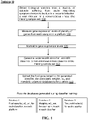

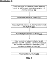
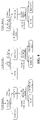

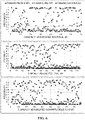


Classifications
-
- C—CHEMISTRY; METALLURGY
- C12—BIOCHEMISTRY; BEER; SPIRITS; WINE; VINEGAR; MICROBIOLOGY; ENZYMOLOGY; MUTATION OR GENETIC ENGINEERING
- C12Q—MEASURING OR TESTING PROCESSES INVOLVING ENZYMES, NUCLEIC ACIDS OR MICROORGANISMS; COMPOSITIONS OR TEST PAPERS THEREFOR; PROCESSES OF PREPARING SUCH COMPOSITIONS; CONDITION-RESPONSIVE CONTROL IN MICROBIOLOGICAL OR ENZYMOLOGICAL PROCESSES
- C12Q1/00—Measuring or testing processes involving enzymes, nucleic acids or microorganisms; Compositions therefor; Processes of preparing such compositions
- C12Q1/68—Measuring or testing processes involving enzymes, nucleic acids or microorganisms; Compositions therefor; Processes of preparing such compositions involving nucleic acids
- C12Q1/6876—Nucleic acid products used in the analysis of nucleic acids, e.g. primers or probes
- C12Q1/6883—Nucleic acid products used in the analysis of nucleic acids, e.g. primers or probes for diseases caused by alterations of genetic material
-
- A—HUMAN NECESSITIES
- A61—MEDICAL OR VETERINARY SCIENCE; HYGIENE
- A61K—PREPARATIONS FOR MEDICAL, DENTAL OR TOILETRY PURPOSES
- A61K31/00—Medicinal preparations containing organic active ingredients
-
- A—HUMAN NECESSITIES
- A61—MEDICAL OR VETERINARY SCIENCE; HYGIENE
- A61K—PREPARATIONS FOR MEDICAL, DENTAL OR TOILETRY PURPOSES
- A61K39/00—Medicinal preparations containing antigens or antibodies
-
- A—HUMAN NECESSITIES
- A61—MEDICAL OR VETERINARY SCIENCE; HYGIENE
- A61P—SPECIFIC THERAPEUTIC ACTIVITY OF CHEMICAL COMPOUNDS OR MEDICINAL PREPARATIONS
- A61P11/00—Drugs for disorders of the respiratory system
-
- A—HUMAN NECESSITIES
- A61—MEDICAL OR VETERINARY SCIENCE; HYGIENE
- A61P—SPECIFIC THERAPEUTIC ACTIVITY OF CHEMICAL COMPOUNDS OR MEDICINAL PREPARATIONS
- A61P31/00—Antiinfectives, i.e. antibiotics, antiseptics, chemotherapeutics
- A61P31/04—Antibacterial agents
-
- A—HUMAN NECESSITIES
- A61—MEDICAL OR VETERINARY SCIENCE; HYGIENE
- A61P—SPECIFIC THERAPEUTIC ACTIVITY OF CHEMICAL COMPOUNDS OR MEDICINAL PREPARATIONS
- A61P31/00—Antiinfectives, i.e. antibiotics, antiseptics, chemotherapeutics
- A61P31/12—Antivirals
-
- A—HUMAN NECESSITIES
- A61—MEDICAL OR VETERINARY SCIENCE; HYGIENE
- A61P—SPECIFIC THERAPEUTIC ACTIVITY OF CHEMICAL COMPOUNDS OR MEDICINAL PREPARATIONS
- A61P37/00—Drugs for immunological or allergic disorders
- A61P37/02—Immunomodulators
- A61P37/04—Immunostimulants
-
- C—CHEMISTRY; METALLURGY
- C12—BIOCHEMISTRY; BEER; SPIRITS; WINE; VINEGAR; MICROBIOLOGY; ENZYMOLOGY; MUTATION OR GENETIC ENGINEERING
- C12Q—MEASURING OR TESTING PROCESSES INVOLVING ENZYMES, NUCLEIC ACIDS OR MICROORGANISMS; COMPOSITIONS OR TEST PAPERS THEREFOR; PROCESSES OF PREPARING SUCH COMPOSITIONS; CONDITION-RESPONSIVE CONTROL IN MICROBIOLOGICAL OR ENZYMOLOGICAL PROCESSES
- C12Q2600/00—Oligonucleotides characterized by their use
- C12Q2600/106—Pharmacogenomics, i.e. genetic variability in individual responses to drugs and drug metabolism
-
- C—CHEMISTRY; METALLURGY
- C12—BIOCHEMISTRY; BEER; SPIRITS; WINE; VINEGAR; MICROBIOLOGY; ENZYMOLOGY; MUTATION OR GENETIC ENGINEERING
- C12Q—MEASURING OR TESTING PROCESSES INVOLVING ENZYMES, NUCLEIC ACIDS OR MICROORGANISMS; COMPOSITIONS OR TEST PAPERS THEREFOR; PROCESSES OF PREPARING SUCH COMPOSITIONS; CONDITION-RESPONSIVE CONTROL IN MICROBIOLOGICAL OR ENZYMOLOGICAL PROCESSES
- C12Q2600/00—Oligonucleotides characterized by their use
- C12Q2600/118—Prognosis of disease development
-
- C—CHEMISTRY; METALLURGY
- C12—BIOCHEMISTRY; BEER; SPIRITS; WINE; VINEGAR; MICROBIOLOGY; ENZYMOLOGY; MUTATION OR GENETIC ENGINEERING
- C12Q—MEASURING OR TESTING PROCESSES INVOLVING ENZYMES, NUCLEIC ACIDS OR MICROORGANISMS; COMPOSITIONS OR TEST PAPERS THEREFOR; PROCESSES OF PREPARING SUCH COMPOSITIONS; CONDITION-RESPONSIVE CONTROL IN MICROBIOLOGICAL OR ENZYMOLOGICAL PROCESSES
- C12Q2600/00—Oligonucleotides characterized by their use
- C12Q2600/158—Expression markers
-
- Y—GENERAL TAGGING OF NEW TECHNOLOGICAL DEVELOPMENTS; GENERAL TAGGING OF CROSS-SECTIONAL TECHNOLOGIES SPANNING OVER SEVERAL SECTIONS OF THE IPC; TECHNICAL SUBJECTS COVERED BY FORMER USPC CROSS-REFERENCE ART COLLECTIONS [XRACs] AND DIGESTS
- Y02—TECHNOLOGIES OR APPLICATIONS FOR MITIGATION OR ADAPTATION AGAINST CLIMATE CHANGE
- Y02A—TECHNOLOGIES FOR ADAPTATION TO CLIMATE CHANGE
- Y02A90/00—Technologies having an indirect contribution to adaptation to climate change
- Y02A90/10—Information and communication technologies [ICT] supporting adaptation to climate change, e.g. for weather forecasting or climate simulation
Definitions
- Acute respiratory infection is common in acute care environments and results in significant mortality, morbidity, and economic losses worldwide.
- Respiratory tract infections, or acute respiratory infections (ARI) caused 3.2 million deaths around the world and 164 million disability-adjusted life years lost in 2011, more than any other cause (World Health Organization., 2013a, 2013b).
- ARI acute respiratory infections
- the fourth leading cause of death worldwide was lower respiratory tract infections, and in low and middle income countries, where less supportive care is available, lower respiratory tract infections are the leading cause of death (WHO factsheet, accessed August 22, 2014).
- WHO factsheet accessed August 22, 2014.
- These illnesses are also problematic in developed countries.
- the Centers for Disease Control (CDC) determined that pneumonia and influenza alone caused 15.1 deaths for every 100,000 people in the US population.
- the aged and children under the age of 5 years are particularly vulnerable to poor outcomes due to ARIs.
- pneumonia accounted for 18.3% of all deaths, or almost 1.4 million deaths, worldwide in children aged 5 years or younger.
- Pneumonia and other lower respiratory tract infections can be due to many different pathogens that are primarily viral, bacterial, or less frequently fungal.
- influenza is among the most notorious based on numbers of affected individuals, variable severity from season to season, and the ever-present worry about new strains causing much higher morbidity and mortality (e.g ., Avian flu).
- Avian flu morbidity and mortality
- influenza is only one of many that cause significant human disease.
- Respiratory Syncytial Virus (RSV) is the leading cause of hospitalization of children in developed countries during the winter months. Worldwide, about 33 million new cases of RSV infections were reported in 2005 in children under 5, with 3.4 million severe enough for hospitalization. It is estimated that this viral infection alone kills between 66,000 and 199,000 children each year.
- bacterial etiologies are also prominent especially in the context of lower respiratory tract infections.
- Specific causes of bacterial ARI vary geographically and by clinical context but include Streptococcus pneumoniae, Staphylococcus aureus, Haemophilus influenzae, Chlamydia pneumoniae, Mycoplasma pneumoniae, Klebsiella pneumoniae, Escherichia coli, and Pseudomonas aeruginosa.
- Streptococcus pneumoniae Staphylococcus aureus
- Haemophilus influenzae Haemophilus influenzae
- Chlamydia pneumoniae Chlamydia pneumoniae
- Mycoplasma pneumoniae Mycoplasma pneumoniae
- Klebsiella pneumoniae Klebsiella pneumoniae
- Escherichia coli Escherichia coli
- Pseudomonas aeruginosa The identification of these pathogens relies on their growth in culture, which typically requires days and has limited sensitivity for
- Acute respiratory infections are frequently characterized by non-specific symptoms (such as fever or cough) that are common to many different illnesses, including illnesses that are not caused by an infection.
- Existing diagnostics for ARI fall short in a number of ways.
- Conventional microbiological testing is limited by poor sensitivity and specificity, slow turnaround times, or by the complexity of the test ( Zaas et al. 2014, Trends Mol Med 20(10):579-88 ).
- One limitation of current tests that detect specific viral pathogens for example the multiplex PCR-based assays, is the inability to detect emergent or pandemic viral strains. Influenza pandemics arise when new viruses circulate against which populations have no natural resistance. Influenza pandemics are frequently devastating.
- the Spanish flu affected about 20% to 40% of the world's population and killed about 50 million people; in 1957-1958, Asian flu killed about 2 million people; in 1968-1969 the Hong Kong flu killed about 1 million people; and in 2009-2010, the Centers for Disease Control estimates that approximately 43 million to 89 million people contracted swine flu resulting in 8,870 to 18,300 related deaths.
- the emergence of these new strains challenges existing diagnostics which are not designed to detect them. This was particularly evident during the 2009 influenza pandemic where confirmation of infection required days and only occurred at specialized testing centers such as state health departments or the CDC ( Kumar & Henrickson 2012, Clin Microbiol Rev 25(2):344-61 ).
- the Ebola virus disease outbreak in West Africa poses similar challenges at the present time. Moreover, there is every expectation we will continue to face this issue as future outbreaks of infectious diseases are inevitable.
- a further limitation of diagnostics that use the paradigm of testing for specific viruses or bacteria is that even though a pathogenic microbe may be detected, this is not proof that the patient's symptoms are due to the detected pathogen.
- a microorganism may be present as part of the individual's normal flora, known as colonization, or it may be detected due to contamination of the tested sample ( e.g. , a nasal swab or wash).
- recently-approved multiplex PCR assays including those that detect viruses and bacteria, offer high sensitivity, these tests do not differentiate between asymptomatic carriage of a virus and true infection. For example, there is a high rate of asymptomatic viral shedding in ARI, particularly in children ( Jansen et al. 2011, J Clin Microbiol 49(7):2631-2636 ). Similarly, even though one pathogen is detected, illness may be due to a second pathogen for which there was no test available or performed.
- the present disclosure provides, in part, a molecular diagnostic test that overcomes many of the limitations of current methods for the determination of the etiology of respiratory symptoms.
- the test detects the host's response to an infectious agent or agents by measuring and analyzing the patterns of co-expressed genes, or signatures. These gene expression signatures may be measured in a blood sample in a human or animal presenting with symptoms that are consistent with an acute respiratory infection or in a human or animal that is at risk of developing ( e.g. , presymptomatic) an acute respiratory infection ( e.g ., during an epidemic or local disease outbreak).
- Measurement of the host response as taught herein differentiates between bacterial ARI, viral ARI, and a non-infectious cause of illness, and may also detect ARI resulting from co-infection with bacteria and virus.
- This multi-component test performs with unprecedented accuracy and clinical applicability, allowing health care providers to use the response of the host (the subject or patient) to reliably determine the nature of the infectious agent, to the level of pathogen class, or to exclude an infectious cause of symptoms in an individual patient presenting with symptoms that, by themselves, are not specific.
- the results are agnostic to the species of respiratory virus or bacteria (i.e. , while differentiating between virus or bacteria, it does not differentiate between particular genus or species of virus or bacteria). This offers an advantage over current tests that include probes or reagents directed to specific pathogens and thus are limited to detecting only those specific pathogens.
- One aspect of the present disclosure provides a method for determining whether acute respiratory symptoms in a subject are bacterial in origin, viral in origin, or non-infectious in origin comprising, consisting of, or consisting essentially of: (a) obtaining a biological sample from the subject; (b) determining the gene expression profile of the subject from the biological sample by evaluating the expression levels of pre-defined sets of genes, termed signatures; (c) normalizing gene expression levels for the technology ( i.e ., platform) used to make said measurement to generate a normalized value; (d) entering the normalized values into a bacterial classifier, a viral classifier and/or a non-infectious illness classifier that have pre-defined weighting values (coefficients) for each of the genes in each signature; (e) comparing the output of the classifiers to pre-defined thresholds, cut-off values, or ranges of values that indicate likelihood of infection; and (f) using the output to determine whether the patient providing the sample has an infection of bacterial origin, viral origin, or has
- Another aspect of the present disclosure provides a method for determining whether an acute respiratory infection (ARI) in a subject is bacterial in origin, viral in origin, or non-infectious in origin comprising, consisting of, or consisting essentially of: (a) obtaining a biological sample from the subject; (b) determining the gene expression profile of the subject from the biological sample by evaluating the expression levels of pre-defined sets of genes; (c) normalizing gene expression levels for the technology (i.e.
- Another aspect of the present disclosure provides a method for determining whether an acute respiratory infection (ARI) in a subject is bacterial in origin, viral in origin, or non-infectious in origin comprising, consisting of, or consisting essentially of: (a) obtaining a biological sample from the subject; (b) determining the gene expression profile of the subject from the biological sample by evaluating the expression levels of pre-defined sets of genes; (c) normalizing gene expression levels for the technology (i.e.
- Another aspect of the present disclosure provides a method for determining whether an acute respiratory infection (ARI) in a subject is bacterial in origin, viral in origin, or non-infectious in origin comprising, consisting of, or consisting essentially of: (a) obtaining a biological sample from the subject; (b) determining the gene expression profile of the subject from the biological sample by evaluating the expression levels of pre-defined sets of genes; (c) normalizing gene expression levels for the technology ( i.e ., platform) used to make said measurement to generate a normalized value; (d) entering the normalized values into classifiers that have pre-defined weighting values for each of the genes in each signature; (e) comparing the output of the classifiers to pre-defined thresholds, cut-off values, or ranges of values that indicate likelihood of infection; (f) if the sample is negative for non-infectious illness, repeating step (d) using only the viral classifier and bacterial classifier; and (g) classifying the sample as being of viral etiology or bacterial
- Yet another aspect of the present disclosure provides a method of treating an acute respiratory infection (ARI) whose etiology is unknown in a subject, said method comprising, consisting of, or consisting essentially of: (a) obtaining a biological sample from the subject; (b) determining the gene expression profile of the subject from the biological sample by evaluating the expression levels of pre-defined sets of genes ( e.g. , one, two or three or more signatures); (c) normalizing gene expression levels for the technology (i.e.
- ARI acute respiratory infection
- step (g) comprises administering an antibacterial therapy when the etiology of the ARI is determined to be bacterial. In other embodiments, step (g) comprises administering an antiviral therapy when the etiology of the ARI is determined to be viral.
- Another aspect is a method of monitoring response to a vaccine or a drug in a subject suffering from or at risk of an acute respiratory illness selected from bacterial, viral and/or non-infectious, comprising determining a host response of said subject, said determining carried out by a method as taught herein.
- the drug is an antibacterial drug or an antiviral drug.
- the methods further comprise generating a report assigning the subject a score indicating the probability of the etiology of the ARI.
- a system for determining an etiology of an acute respiratory illness in a subject selected from bacterial, viral and/or non-infectious comprising one or more of (inclusive of combinations thereof): at least one processor; a sample input circuit configured to receive a biological sample from the subject; a sample analysis circuit coupled to the at least one processor and configured to determine gene expression levels of the biological sample; an input/output circuit coupled to the at least one processor; a storage circuit coupled to the at least one processor and configured to store data, parameters, and/or classifiers; and a memory coupled to the processor and comprising computer readable program code embodied in the memory that when executed by the at least one processor causes the at least one processor to perform operations comprising: controlling/performing measurement via the sample analysis circuit of gene expression levels of a pre-defined set of genes (i.e.
- a bacterial acute respiratory infection (ARI) classifier a viral ARI classifier and a non-infectious illness classifier, said classifier(s) comprising pre-defined weighting values (i.e., coefficients) for each of the genes of the pre-defined set of genes; entering the normalized gene expression values into one or more acute respiratory illness classifiers selected from the bacterial acute respiratory infection (ARI) classifier, the viral ARI classifier and the non-infectious illness classifier; calculating an etiology probability for one or more of a bacterial ARI, viral ARI and non-infectious illness based upon said classifier(s); and controlling output via the input/output circuit of a determination whether the acute respiratory illness in the subject is bacterial in origin, viral in origin, non-infectious in origin, or some combination thereof.
- ARI bacterial acute respiratory infection
- viral ARI classifier a viral ARI classifier and a non-infectious illness classifier
- the system comprises computer readable code to transform quantitative, or semi-quantitative, detection of gene expression to a cumulative score or probability of the etiology of the ARI
- the system comprises an array platform, a thermal cycler platform (e.g. , multiplexed and/or real-time PCR platform), a hybridization and multi-signal coded (e.g. , fluorescence) detector platform, a nucleic acid mass spectrometry platform, a nucleic acid sequencing platform, or a combination thereof.
- a thermal cycler platform e.g. , multiplexed and/or real-time PCR platform
- a hybridization and multi-signal coded e.g. , fluorescence
- the pre-defined sets of genes comprise at least three genetic signatures.
- the biological sample comprises a sample selected from the group consisting of peripheral blood, sputum, nasopharyngeal swab, nasopharyngeal wash, bronchoalveolar lavage, endotracheal aspirate, and combinations thereof.
- the bacterial classifier comprises expression levels of 5, 10, 20, 30 or 50, to 80, 100, 150 or 200 of the genes (measurable, e.g., with oligonucleotide probes homologous to said genes or gene transcripts) listed as part of a bacterial classifier in Table 1, Table 2, Table 9, Table 10 and/or Table 12.
- the viral classifier comprises expression levels of 5, 10, 20, 30 or 50, to 80, 100, 150 or 200 of the genes (measurable, e.g. , with oligonucleotide probes homologous to said genes or gene transcripts) listed as part of a viral classifier in Table 1, Table 2, Table 9, Table 10 and/or Table 12.
- the non-infectious illness classifier comprises expression levels of 5, 10, 20, 30 or 50, to 80, 100, 150 or 200 of the genes (measurable, e.g ., with oligonucleotide probes homologous to said genes or gene transcripts) listed as part of a non-infectious illness classifier in Table 1, Table 2, Table 9, Table 10 and/or Table 12.
- a kit for determining the etiology of an acute respiratory infection (ARI) in a subject comprising, consisting of, or consisting essentially of (a) a means for extracting mRNA from a biological sample; (b) a means for generating one or more arrays consisting of a plurality of synthetic oligonucleotides with regions homologous to transcripts from of 5, 10, 20, 30 or 50, to 80, 100, 150 or 200 of the genes from Table 1, Table 2, Table 9, Table 10 and/or Table 12; and (c) instructions for use.
- a means for extracting mRNA from a biological sample comprising, consisting of, or consisting essentially of (a) a means for extracting mRNA from a biological sample; (b) a means for generating one or more arrays consisting of a plurality of synthetic oligonucleotides with regions homologous to transcripts from of 5, 10, 20, 30 or 50, to 80, 100, 150 or 200 of the genes from Table 1, Table 2, Table 9, Table 10 and/or
- kits for assessing the acute respiratory infection (ARI) classifier comprising, consisting of, or consisting essentially of: (a) generating one or more arrays consisting of a plurality of synthetic oligonucleotides with regions homologous to of 5, 10, 20, 30 or 50, to 80, 100, 150 or 200 of the genes from Table 1, Table 2, Table 9, Table 10 and/or Table 12; (b) adding to said array oligonucleotides with regions homologous to normalizing genes; (c) obtaining a biological sample from a subject suffering from an acute respiratory infection (ARI); (d) isolating RNA from said sample to create a transcriptome; (e) measuring said transcriptome on said array ( e.g ., by measuring fluorescence or electric current proportional to the level of gene expression, etc.); (f) normalizing the measurements of said transcriptome to the normalizing genes, electronically transferring normalized measurements to a computer to implement the classifier(s), (g) generating a report
- the method further comprises externally validating an ARI classifier against a known dataset comprising at least two relevant clinical attributes.
- the dataset is selected from the group consisting of GSE6269, GSE42026, GSE40396, GSE20346, GSE42834 and combinations thereof.
- Yet another aspect of the present disclosure provides all that is disclosed and illustrated herein.
- ARI classifier as taught herein in a method of treatment for acute respiratory infection (ARI) in a subject of unknown etiology.
- Articles "a” and “an” are used herein to refer to one or to more than one (i.e. , at least one) of the grammatical object of the article.
- an element means at least one element and can include more than one element.
- the present disclosure provides that alterations in gene, protein and metabolite expression in blood in response to pathogen exposure that causes acute respiratory infections can be used to identify and characterize the etiology of the ARI in a subject with a high degree of accuracy.
- ARI refers to an infection, or an illness showing symptoms and/or physical findings consistent with an infection (e.g ., symptoms such as coughing, wheezing, fever, sore throat, congestion; physical findings such as elevated heart rate, elevated breath rate, abnormal white blood cell count, low arterial carbon dioxide tension (PaCO 2 ), etc.), of the upper or lower respiratory tract, often due to a bacterial or viral pathogen, and characterized by rapid progression of symptoms over hours to days.
- ARIs may primarily be of the upper respiratory tract (URIs), the lower respiratory tract (LRIs), or a combination of the two.
- ARIs may have systemic effects due to spread of the infection beyond the respiratory tract or due to collateral damage induced by the immune response.
- An example of the former includes Staphylococcus aureus pneumonia that has spread to the blood stream and can result in secondary sites of infection, including endocarditis (infection of the heart valves), septic arthritis (joint infection), or osteomyelitis (bone infection).
- An example of the latter includes influenza pneumonia leading to acute respiratory distress syndrome and respiratory failure.
- signature refers to a set of biological analytes and the measurable quantities of said analytes whose particular combination signifies the presence or absence of the specified biological state. These signatures are discovered in a plurality of subjects with known status (e.g ., with a confirmed respiratory bacterial infection, respiratory viral infection, or suffering from non-infectious illness), and are discriminative (individually or jointly) of one or more categories or outcomes of interest. These measurable analytes, also known as biological markers, can be (but are not limited to) gene expression levels, protein or peptide levels, or metabolite levels. See also US 2015/0227681 to Courchesne et al. ; US 2016/0153993 to Eden et al .
- the "signature” is a particular combination of genes whose expression levels, when incorporated into a classifier as taught herein, discriminate a condition such as a bacterial ARI, viral ARI or non-infectious illness. See, for example, Table 1, Table 2, Table 9, Table 10 and Table 12 hereinbelow.
- the signature is agnostic to the species of respiratory virus or bacteria (i.e. , while differentiating between virus or bacteria, it does not differentiate between particular genus or species of virus or bacteria) and/or agnostic to the particular cause of the non-infectious illness.
- classifier and “predictor” are used interchangeably and refer to a mathematical function that uses the values of the signature (e.g ., gene expression levels for a defined set of genes) and a pre-determined coefficient (or weight) for each signature component to generate scores for a given observation or individual patient for the purpose of assignment to a category.
- the classifier may be linear and/or probabilistic.
- a classifier is linear if scores are a function of summed signature values weighted by a set of coefficients.
- a classifier is probabilistic if the function of signature values generates a probability, a value between 0 and 1.0 (or 0 and 100%) quantifying the likelihood that a subject or observation belongs to a particular category or will have a particular outcome, respectively.
- Probit regression and logistic regression are examples of probabilistic linear classifiers that use probit and logistic link functions, respectively, to generate a probability.
- a classifier as taught herein may be obtained by a procedure known as "training,” which makes use of a set of data containing observations with known category membership (e.g. , bacterial ARI, viral ARI, and/or non-infection illness). See FIG. 1 .
- training seeks to find the optimal coefficient (i.e ., weight) for each component of a given signature (e.g ., gene expression level components), as well as an optimal signature, where the optimal result is determined by the highest achievable classification accuracy.
- Classification refers to a method of assigning a subject suffering from or at risk for acute respiratory symptoms to one or more categories or outcomes (e.g ., a patient is infected with a pathogen or is not infected, another categorization may be that a patient is infected with a virus and/or infected with a bacterium). See FIG. 3 .
- a subject may be classified to more than one category, e.g. , in case of bacterial and viral co-infection.
- the outcome, or category is determined by the value of the scores provided by the classifier, which may be compared to a cut-off or threshold value, confidence level, or limit. In other scenarios, the probability of belonging to a particular category may be given (e.g., if the classifier reports probabilities).
- the term “indicative” when used with gene expression levels means that the gene expression levels are up-regulated or down-regulated, altered, or changed compared to the expression levels in alternative biological states (e.g., bacterial ARI or viral ARI) or control.
- alternative biological states e.g., bacterial ARI or viral ARI
- indicator when used with protein levels means that the protein levels are higher or lower, increased or decreased, altered, or changed compared to the standard protein levels or levels in alternative biological states.
- subject and “patient” are used interchangeably and refer to any animal being examined, studied or treated. It is not intended that the present disclosure be limited to any particular type of subject.
- humans are the preferred subject, while in other embodiments non-human animals are the preferred subject, including, but not limited to, mice, monkeys, ferrets, cattle, sheep, goats, pigs, chicken, turkeys, dogs, cats, horses and reptiles.
- the subject is suffering from an ARI or is displaying ARI-like symptoms.
- Platinum or “technology” as used herein refers to an apparatus (e.g., instrument and associated parts, computer, computer-readable media comprising one or more databases as taught herein, reagents, etc.) that may be used to measure a signature, e.g., gene expression levels, in accordance with the present disclosure.
- platforms include, but are not limited to, an array platform, a thermal cycler platform (e.g., multiplexed and/or real-time PCR platform), a nucleic acid sequencing platform, a hybridization and multi-signal coded (e.g. , fluorescence) detector platform, etc., a nucleic acid mass spectrometry platform, a magnetic resonance platform, and combinations thereof.
- the platform is configured to measure gene expression levels semi-quantitatively, that is, rather than measuring in discrete or absolute expression, the expression levels are measured as an estimate and/or relative to each other or a specified marker or markers (e.g., expression of another, "standard” or “reference,” gene).
- semi-quantitative measuring includes "real-time PCR” by performing PCR cycles until a signal indicating the specified mRNA is detected, and using the number of PCR cycles needed until detection to provide the estimated or relative expression levels of the genes within the signature.
- a real-time PCR platform includes, for example, a TaqMan ® Low Density Array (TLDA), in which samples undergo multiplexed reverse transcription, followed by real-time PCR on an array card with a collection of wells in which real-time PCR is performed. See Kodani et al. 2011, J. Clin. Microbiol. 49(6):2175-2182 .
- a real-time PCR platform also includes, for example, a Biocartis Idylla TM sample-to-result technology, in which cells are lysed, DNA/RNA extracted and real-time PCR is performed and results detected.
- a magnetic resonance platform includes, for example, T2 Biosystems ® T2 Magnetic Resonance (T2MR ® ) technology, in which molecular targets may be identified in biological samples without the need for purification.
- T2MR ® T2 Magnetic Resonance
- arrays can be on a solid substrate (a solid phase array), such as a glass slide, or on a semi-solid substrate, such as nitrocellulose membrane.
- arrays can also be presented on beads, i.e., a bead array. These beads are typically microscopic and may be made of, e.g ., polystyrene.
- the array can also be presented on nanoparticles, which may be made of, e.g. , particularly gold, but also silver, palladium, or platinum.
- the nucleotide sequences can be DNA, RNA, or any permutations thereof (e.g., nucleotide analogues, such as locked nucleic acids (LNAs), and the like). In some embodiments, the nucleotide sequences span exon/intron boundaries to detect gene expression of spliced or mature RNA species rather than genomic DNA.
- the nucleotide sequences can also be partial sequences from a gene, primers, whole gene sequences, non-coding sequences, coding sequences, published sequences, known sequences, or novel sequences.
- the arrays may additionally comprise other compounds, such as antibodies, peptides, proteins, tissues, cells, chemicals, carbohydrates, and the like that specifically bind proteins or metabolites.
- An array platform includes, for example, the TaqMan ® Low Density Array (TLDA) mentioned above, and an Affymetrix ® microarray platform.
- TLDA TaqMan ® Low Density Array
- a hybridization and multi-signal coded detector platform includes, for example, NanoString nCounter ® technology, in which hybridization of a color-coded barcode attached to a target-specific probe (e.g., corresponding to a gene expression transcript of interest) is detected; and Luminex ® xMAP ® technology, in which microsphere beads are color coded and coated with a target-specific (e.g ., gene expression transcript) probe for detection; and Illumina ® BeadArray, in which microbeads are assembled onto fiber optic bundles or planar silica slides and coated with a target-specific (e.g ., gene expression transcript) probe for detection.
- NanoString nCounter ® technology in which hybridization of a color-coded barcode attached to a target-specific probe (e.g., corresponding to a gene expression transcript of interest) is detected
- Luminex ® xMAP ® technology in which microsphere beads are color coded and coated with a target-specific (e.g
- a nucleic acid mass spectrometry platform includes, for example, the Ibis Biosciences Plex-ID ® Detector, in which DNA mass spectrometry is used to detect amplified DNA using mass profiles.
- a thermal cycler platform includes, for example, the FilmArray ® multiplex PCR system, which extract and purifies nucleic acids from an unprocessed sample and performs nested multiplex PCR; and the RainDrop Digital PCR System, which is a droplet-based PCR platform using microfluidic chips.
- computer readable medium refers to any device or system for storing and providing information (e.g ., data and instructions) to a computer processor.
- Examples of computer readable media include, but are not limited to, DVDs, CDs hard disk drives, magnetic tape and servers for streaming media over networks, and applications, such as those found on smart phones and tablets.
- aspects of the present invention including data structures and methods may be stored on a computer readable medium. Processing and data may also be performed on numerous device types, including but not limited to, desk top and lap top computers, tablets, smart phones, and the like.
- a biological sample comprises any sample that may be taken from a subject that contains genetic material that can be used in the methods provided herein.
- a biological sample may comprise a peripheral blood sample.
- peripheral blood sample refers to a sample of blood circulating in the circulatory system or body taken from the system of body.
- Other samples may comprise those taken from the upper respiratory tract, including but not limited to, sputum, nasopharyngeal swab and nasopharyngeal wash.
- a biological sample may also comprise those samples taken from the lower respiratory tract, including but not limited to, bronchoalveolar lavage and endotracheal aspirate.
- a biological sample may also comprise any combinations thereof.
- genetic material refers to a material used to store genetic information in the nuclei or mitochondria of an organism's cells.
- examples of genetic material include, but are not limited to, double-stranded and single-stranded DNA, cDNA, RNA, and mRNA.
- plurality of nucleic acid oligomers refers to two or more nucleic acid oligomers, which can be DNA or RNA.
- treat refers to the reduction or amelioration of the severity, duration and/or progression of a disease or disorder or one or more symptoms thereof resulting from the administration of one or more therapies. Such terms refer to a reduction in the replication of a virus or bacteria, or a reduction in the spread of a virus or bacteria to other organs or tissues in a subject or to other subjects. Treatment may also include therapies for ARIs resulting from non-infectious illness, such as allergy treatment, asthma treatments, and the like.
- the term "effective amount” refers to an amount of a therapeutic agent that is sufficient to exert a physiological effect in the subject.
- response refers to a change in gene expression levels of genes in a subject in response to the subject being infected with a virus or bacteria or suffering from a non-infectious illness compared to the gene expression levels of the genes in a subject that is not infected with a virus, bacteria or suffering from a non-infectious illness or a control subject.
- a therapeutic agent for treating a subject having bacteremia is an antibiotic which include, but are not limited to, penicillins, cephalosporins, fluroquinolones, tetracyclines, macrolides, and aminoglycosides.
- a therapeutic agent for treating a subject having a viral respiratory infection includes, but is not limited to, oseltamivir, RNAi antivirals, inhaled ribavirin, monoclonal antibody respigam, zanamivir, and neuraminidase blocking agents.
- the invention contemplates the use of the methods of the invention to determine treatments with antivirals or antibiotics that are not yet available.
- Appropriate treatment regimes also include treatments for ARIs resulting from non-infectious illness, such as allergy treatments, including but not limited to, administration of antihistamines, decongestants, anticholinergic nasal sprays, leukotriene inhibitors, mast cell inhibitors, steroid nasal sprays, etc.; and asthma treatments, including, but not limited to, inhaled corticosteroids, leukotriene modifiers, long-acting beta agonists, combinations inhalers ( e.g ., fluticasone-salmeterol; budesonide-formoterol; mometasone-formoterol, etc.), theophylline, short-acting beta agonists, ipratropium, oral and intravenous corticosteroids, omalizumab, and the like.
- allergy treatments including but not limited to, administration of antihistamines, decongestants, anticholinergic nasal sprays, leukotriene inhibitors, mast cell
- Such regimens require the act of administering to a subject a therapeutic agent(s) capable of producing reduction of symptoms associated with a disease state.
- therapeutic agents include, but are not limited to, NSAIDS, acetaminophen, anti-histamines, beta-agonists, anti-tussives or other medicaments that reduce the symptoms associated with the disease process.
- the present disclosure provides methods of generating classifiers (also referred to as training 10 ) for use in the methods of determining the etiology of an acute respiratory illness in a subject.
- Gene expression-based classifiers are developed that can be used to identify and characterize the etiology of an ARI in a subject with a high degree of accuracy.
- an acute respiratory infection (ARI) classifier comprising, consisting of, or consisting essentially of: (i) obtaining a biological sample (e.g ., a peripheral blood sample) from a plurality of subjects suffering from bacterial, viral or non-infectious acute respiratory infection 100 ; (ii) optionally, isolating RNA from said sample (e.g., total RNA to create a transcriptome) ( 105 , not shown in FIG.
- a biological sample e.g ., a peripheral blood sample
- RNA from said sample e.g., total RNA to create a transcriptome
- the sample is not purified after collection.
- the sample may be purified to remove extraneous material, before or after lysis of cells.
- the sample is purified with cell lysis and removal of cellular materials, isolation of nucleic acids, and/or reduction of abundant transcripts such as globin or ribosomal RNAs.
- measuring gene expression levels may include generating one or more microarrays using said transcriptomes; measuring said transcriptomes using a plurality of primers; analyzing and correcting batch differences.
- the method further includes uploading 140 the final gene target list for the generated classifier, the associated weights (w n ), and threshold values to one or more databases.
- FIG. 2 An example of generating said classifiers is detailed in FIG. 2 .
- biological samples from a cohort of patients encompassing bacterial ARI, viral ARI, or non-infectious illness are used to develop gene expression-based classifiers for each condition (i.e ., bacterial acute respiratory infection, viral acute respiratory infection, or non-infectious cause of illness).
- the bacterial ARI classifier is obtained to positively identifying those with bacterial ARI vs. either viral ARI or non-infectious illnesses.
- the viral ARI classifier is obtained to positively identifying those with viral ARI vs. bacterial ARI or non-infectious illness (NI).
- the non-infectious illness classifier is generated to improve bacterial and viral ARI classifier specificity.
- signatures for bacterial ARI classifiers, viral ARI classifiers, and non-infectious illness classifiers are generated (e.g. , by applying a sparse logistic regression model).
- the ARI classifier may then be combined, if desired, into a single classifier termed "the ARI classifier" by following a one-versus-all scheme whereby largest membership probability assigns class label. See also FIG. 5 .
- the combined ARI classifier may be validated in some embodiments using leave-one-out cross-validation in the same population from which it was derived and/or may be validated in some embodiments using publically available human gene expression datasets of samples from subjects suffering from illness of known etiology.
- validation may be performed using publically available human gene expression datasets (e.g ., GSE6269, GSE42026, GSE40396, GSE20346, and/or GSE42834), the datasets chosen if they included at least two clinical groups (bacterial ARI, viral ARI, or non-infectious illness).
- publically available human gene expression datasets e.g ., GSE6269, GSE42026, GSE40396, GSE20346, and/or GSE42834
- the datasets chosen if they included at least two clinical groups bacterial ARI, viral ARI, or non-infectious illness.
- the classifier may be validated in a standard set of samples from subjects suffering from illness of known etiology, i.e. , bacterial ARI, viral ARI, or non-infectious illness.
- the methodology for training described herein may be readily translated by one of ordinary skill in the art to different gene expression detection (e.g. , mRNA detection and quantification) platforms.
- the methods and assays of the present disclosure may be based upon gene expression, for example, through direct measurement of RNA, measurement of derived materials (e.g ., cDNA), and measurement of RNA products (e.g ., encoded proteins or peptides). Any method of extracting and screening gene expression may be used and is within the scope of the present disclosure.
- the measuring comprises the detection and quantification (e.g. , semi-quantification) of mRNA in the sample.
- the gene expression levels are adjusted relative to one or more standard gene level(s) ("normalized"). As known in the art, normalizing is done to remove technical variability inherent to a platform to give a quantity or relative quantity ( e . g. , of expressed genes).
- detection and quantification of mRNA may first involve a reverse transcription and/or amplification step, e.g. , RT-PCR such as quantitative RT-PCR.
- detection and quantification may be based upon the unamplified mRNA molecules present in or purified from the biological sample.
- Direct detection and measurement of RNA molecules typically involves hybridization to complementary primers and/or labeled probes.
- Such methods include traditional northern blotting and surface-enhanced Raman spectroscopy (SERS), which involves shooting a laser at a sample exposed to surfaces of plasmonic-active metal structures with gene-specific probes, and measuring changes in light frequency as it scatters.
- SERS surface-enhanced Raman spectroscopy
- RNA derivatives typically involves hybridization to complementary primers and/or labeled probes.
- This may include high-density oligonucleotide probe arrays (e.g. , solid state microarrays and bead arrays) or related probe-hybridization methods, and polymerase chain reaction (PCR)-based amplification and detection, including real-time, digital, and end-point PCR methods for relative and absolute quantitation of specific RNA molecules.
- PCR polymerase chain reaction
- sequencing-based methods can be used to detect and quantify RNA or RNA-derived material levels.
- sequencing methods are referred to as RNAseq, and provide both qualitative (sequence, or presence/absence of an RNA, or its cognate cDNA, in a sample) and quantitative (copy number) information on RNA molecules from a sample. See, e.g. , Wang et al. 2009 Nat. Rev. Genet. 10(1):57-63 .
- Another sequence-based method serial analysis of gene expression (SAGE), uses cDNA "tags" as a proxy to measure expression levels of RNA molecules.
- mRNA detection and quantification may also be used to complete the methods of the present disclosure.
- Pixel TM System incorporating Molecular Indexing TM , developed by CELLULAR RESEARCH, INC., NanoString ® Technologies nCounter gene expression system
- mRNA-Seq Molecular Indexing TM
- Tag-Profiling Tag-Profiling
- VeraCode VeraCode from Illumina
- ICEPlex System from PrimeraDx
- QuantiGene 2.0 Multiplex Assay from Affymetrix.
- RNA from whole blood from a subject can be collected using RNA preservation reagents such as PAXgene TM RNA tubes (PreAnalytiX, Valencia, Calif.).
- the RNA can be extracted using a standard PAXgene TM or Versagene TM (Gentra Systems, Inc, Minneapolis, Minn.) RNA extraction protocol.
- the Versagene TM kit produces greater yields of higher quality RNA from the PAXgene TM RNA tubes.
- GLOBINCIear TM Ambion, Austin, Tex.
- RNA quality can be assessed using an Agilent 2100 Bioanalyzer immediately following extraction. This analysis provides an RNA Integrity Number (RIN) as a quantitative measure of RNA quality. Also, following globin reduction the samples can be compared to the globin-reduced standards. In addition, the scaling factors and background can be assessed following hybridization to microarrays.
- RIN RNA Integrity Number
- Real-time PCR may be used to quickly identify gene expression from a whole blood sample.
- the isolated RNA can be reverse transcribed and then amplified and detected in real time using non-specific fluorescent dyes that intercalate with the resulting ds-DNA, or sequence-specific DNA probes labeled with a fluorescent reporter which permits detection only after hybridization of the probe with its complementary DNA target.
- the expression levels are typically normalized following detection and quantification as appropriate for the particular platform using methods routinely practiced by those of ordinary skill in the art.
- the individual categories of classifiers are formed from a cohort inclusive of a variety of such causes thereof.
- the bacterial ARI classifier is obtained from a cohort having bacterial infections from multiple bacterial genera and/or species
- the viral ARI classifier is obtained from a cohort having viral infections from multiple viral genera and/or species
- the non-infectious illness classifier is obtained from a cohort having a non-infectious illness due to multiple non-infectious causes. See , e.g. , Table 8.
- the respective classifiers obtained are agnostic to the underlying bacteria, virus, and non-infectious cause.
- some or all of the subjects with non-infectious causes of illness in the cohort have symptoms consistent with a respiratory infection.
- the signatures may be obtained using a supervised statistical approach known as sparse linear classification in which sets of genes are identified by the model according to their ability to separate phenotypes during a training process that uses the selected set of patient samples.
- the outcomes of training are gene signatures and classification coefficients for the three comparisons. Together the signatures and coefficients provide a classifier or predictor.
- Training may also be used to establish threshold or cut-off values. Threshold or cut-off values can be adjusted to change test performance, e.g ., test sensitivity and specificity. For example, the threshold for bacterial ARI may be intentionally lowered to increase the sensitivity of the test for bacterial infection, if desired.
- the classifier generating comprises iteratively: (i) assigning a weight for each normalized gene expression value, entering the weight and expression value for each gene into a classifier (e.g. , a linear regression classifier) equation and determining a score for outcome for each of the plurality of subjects, then (ii) determining the accuracy of classification for each outcome across the plurality of subjects, and then (iii) adjusting the weight until accuracy of classification is optimized.
- a classifier e.g. , a linear regression classifier
- the classifier is a linear regression classifier and said generating comprises converting a score of said classifier to a probability using a link function.
- the link function specifies the link between the target/output of the model (e.g., probability of bacterial infection) and systematic components (in this instance, the combination of explanatory variables that comprise the predictor) of the linear model. It says how the expected value of the response relates to the linear predictor of explanatory variable.
- the present disclosure further provides methods for determining whether a patient has a respiratory illness due to a bacterial infection, a viral infection, or a non-infectious cause.
- the method for making this determination relies upon the use of classifiers obtained as taught herein.
- the methods may include: a) measuring the expression levels of pre-defined sets of genes ( i.e. , for one or more of the three signatures); b) normalizing gene expression levels for the technology used to make said measurement; c) taking those values and entering them into a bacterial classifier, a viral classifier and/or non-infectious illness classifier ( i.e.
- predictors that have pre-defined weighting values (coefficients) for each of the genes in each signature; d) comparing the output of the classifiers to pre-defined thresholds, cut-off values, confidence intervals or ranges of values that indicate likelihood of infection; and optionally e) jointly reporting the results of the classifiers.
- each of the three gene signatures is informative of the patient's host response to a different ARI etiology (bacterial or viral) or to an ill, but not infected, state (NI).
- ARI etiology bacterial or viral
- NI ill, but not infected, state
- These signatures are groups of gene transcripts which have consistent and coordinated increased or decreased levels of expression in response to one of three clinical states: bacterial ARI, viral ARI, or a non-infected but ill state.
- These signatures are derived using carefully adjudicated groups of patient samples with the condition(s) of interest (training 10 ).
- the mRNA is extracted.
- the mRNA (or a defined region of each mRNA), is quantified for all, or a subset, of the genes in the signatures.
- the mRNA may have to be first purified from the sample.
- the signature is reflective of a clinical state and is defined relative to at least one of the other two possibilities.
- the bacterial ARI signature is identified as a group of biomarkers (here, represented by gene mRNA transcripts) that distinguish patients with bacterial ARI and those without bacterial ARI (including patients with viral ARI or non-infectious illness as it pertains to this application).
- the viral ARI signature is defined by a group of biomarkers that distinguish patients with viral ARI from those without viral ARI (including patients with either bacterial ARI or non-infectious illness).
- the non-infectious illness signature is defined by a group of biomarkers that distinguish patients with non-infectious causes of illness relative to those with either bacterial or viral ARI.
- the normalized expression levels of each gene of the signature are the explanatory or independent variables or features used in the classifier.
- the value of the coefficients for each explanatory variable will change for each technology platform used to measure the expression of the genes or a subset of genes used in the probit regression model.
- the coefficients for each of the features in the classifier algorithm are shown in Table 9.
- the sensitivity, specificity, and overall accuracy of each classifier may be optimized by changing the threshold for classification using receiving operating characteristic (ROC) curves.
- ROC operating characteristic
- Another aspect of the present disclosure provides a method for determining whether an acute respiratory infection (ARI) in a subject is bacterial in origin, viral in origin, or non-infectious in origin comprising, consisting of, or consisting essentially of a) obtaining a biological sample from the subject; b) determining the gene expression profile of the subject from the biological sample by evaluating the expression levels of pre-defined sets of genes ( i.e ., three signatures); c) normalizing gene expression levels for the technology used to make said measurement to generate a normalized value; d) entering the normalized value into a bacterial classifier, a viral classifier and non-infectious illness classifier (i.e., predictors) that have pre-defined weighting values (coefficients) for each of the genes in each signature; e) comparing the output of the classifiers to pre-defined thresholds, cut-off values, or ranges of values that indicate likelihood of infection; and e) classifying the sample as being of bacterial etiology, viral et
- the classifiers that are developed during training and using a training set of samples are applied for prediction purposes to diagnose new individuals ("classification"). For each subject or patient, a biological sample is taken and the normalized levels of expression (i.e. , the relative amount of mRNA expression) in the sample of each of the genes specified by the signatures found during training are the input for the classifiers. The classifiers also use the weighting coefficients discovered during training for each gene. As outputs, the classifiers are used to compute three probability values. Each probability value may be used to determine the likelihood of the three considered clinical states: bacterial ARI, viral ARI, and non-infectious illness.
- the results of each of the classifiers - the probability a new subject or patient has a bacterial ARI, viral ARI, or non-infectious illness - are reported.
- the three signatures with their corresponding coefficients are applied to an individual patient to obtain three probability values, namely probability of having a bacterial ARI, viral ARI, and a non-infectious illness. In some embodiments, these values may be reported relative to a reference range that indicates the confidence with which the classification is made.
- the output of the classifier may be compared to a threshold value, for example, to report a "positive” in the case that the classifier score or probability exceeds the threshold indicating the presence of one or more of a bacterial ARI, viral ARI, or non-infectious illness. If the classifier score or probability fails to reach the threshold, the result would be reported as "negative” for the respective condition.
- the values for bacterial and viral ARI alone are reported and the report is silent on the likelihood of ill but not infected.
- a classifier obtained with one platform may not show optimal performance on another platform. This could be due to the promiscuity of probes or other technical issues particular to the platform. Accordingly, also described herein are methods to adapt a signature as taught herein from one platform for another.
- a signature obtained from an Affymetrix platform may be adapted to a TLDA platform by the use of corresponding TLDA probes for the genes in the signature and/or substitute genes correlated with those in the signature, for the Affymetrix platform.
- Table 1 shows a list of Affymetrix probes and the genes they measure, plus "replacement genes" that are introduced as resplacements for gene probes that either may not perform well on the TLDA platform for technical reasons or to replace those Affymetrix probes for which there is no cognate TLDA probe. These replacements may indicate highly correlated genes or may be probes that bind to a different location in the same gene transcript. Additional genes may be included, such as pan-viral gene probes.
- the weights shown in Table 1 are weights calculated for a classifier implemented on the microarray platform. Weights that have not been estimated are indicated by "NA” in the table. (Example 4 below provides the completed translation of these classifiers to the TLDA platform.)
- Reference probes for TLDA i.e ., normalization genes, e.g. , TRAP1, PPIB, GAPDH and 18S
- TLDA normalization genes, e.g. , TRAP1, PPIB, GAPDH and 18S
- Additional gene probes that do not necessarily correspond to the Affymetrix probeset also have "NA” in the Affymetrix probeset ID column.
- Table 1 Preliminary Gene List for TLDA platform Columns are as follows: Column 1 : Affymetrix probeset ID - this was the probeset identified in the Affy discovery analyses (primary probeset) Columns 2,3,4 : estimated coefficients (weights) for contribution of each probates to the 3 classifiers from Affymetrix weights Column 5 : Gene name AFFXProbeSet Bacterial Viral NI Gene 216867_s_at 0.0534745 0 0 PDGFA 203313_s_at 1.09463 0 0 TGIF1 NA NA NA NA NA TRAP1 NA NA NA NA PPIB 202720_at 0 0.0787402 0 TES 210657_s_at NA NA NA NA SEPT4 NA NA NA NA EPHB3 NA NA NA NA NA SYDE1 202864_s_at 0 0.100019 0 SP100 213633_at 1.01336 0 0 SH3BP1 NA NA NA NA NA 18S NA NA NA NA NA NA NA GIT2
- This method of determining the etiology of an ARI may be combined with other tests. For example, if the patient is determined to have a viral ARI, a follow-up test may be to determine if influenza A or B can be directly detected or if a host response indicative of such an infection can be detected. Similarly, a follow-up test to a result of bacterial ARI may be to determine if a Gram positive or a Gram negative bacterium can be directly detected or if a host response indicative of such an infection can be detected. In some embodiments, simultaneous testing may be performed to determine the class of infection using the classifiers, and also to test for specific pathogens using pathogen-specific probes or detection methods. See , e.g., US 2015/0284780 to Eley et al. (method for detecting active tuberculosis); US 2014/0323391 to Tsalik et al. (method for classification of bacterial infection).
- an acute respiratory infection (ARI) in a subject is bacterial in origin, viral in origin, or non-infectious in origin
- a method for determining whether an acute respiratory infection (ARI) in a subject is bacterial in origin, viral in origin, or non-infectious in origin comprising, consisting of, or consisting essentially of (a) obtaining a biological sample from the subject; (b) determining the gene expression profile of the subject from the biological sample by evaluating the expression levels of pre-defined sets of genes ( i.e., three signatures); (c) normalizing gene expression levels as required for the technology used to make said measurement to generate a normalized value; (d) entering the normalized value into classifiers ( i.e., predictors) that have pre-defined weighting values (coefficients) for each of the genes in each signature; (e) comparing the output of the classifiers to pre-defined thresholds, cut-off values, or ranges of values that indicate likelihood of infection; (f) if the sample is negative for bacteria, repeating step
- Another aspect of the present provides a method for determining whether an acute respiratory infection (ARI) in a subject is bacterial in origin, viral in origin, or non-infectious in origin comprising, consisting of, or consisting essentially of (a) obtaining a biological sample from the subject; (b) determining the gene expression profile of the subject from the biological sample by evaluating the expression levels of pre-defined sets of genes ( i.e.
- Yet another aspect of the present provides a method for determining whether an acute respiratory infection (ARI) in a subject is bacterial in origin, viral in origin, or non-infectious in origin comprising, consisting of, or consisting essentially of (a) obtaining a biological sample from the subject; (b) determining the gene expression profile of the subject from the biological sample by evaluating the expression levels of pre-defined sets of genes ( i.e.
- the method further comprises generating a report assigning the patient a score indicating the probability of the etiology of the ARI
- Classifying the status of a patient using a secondary classification scheme is shown in FIG. 4 .
- the bacterial ARI classifier will distinguish between patients with a bacterial ARI from those without a bacterial ARI, which could, instead, be a viral ARI or a non-infectious cause of illness.
- a secondary classification can then be imposed on those patients with non-bacterial ARI to further discriminate between viral ARI and non-infectious illness.
- This same process of primary and secondary classification can also be applied to the viral ARI classifier where patients determined not to have a viral infection would then be secondarily classified as having a bacterial ARI or non-infectious cause of illness.
- applying the non-infectious illness classifier as a primary test will determine whether patients have such a non-infectious illness or instead have an infectious cause of symptoms.
- the secondary classification step would determine if that infectious is due to bacterial or viral pathogens.
- Results from the three primary and three secondary classifications can be summed through various techniques by those skilled in the art (such as summation, counts, or average) to produce an actionable report for the provider.
- the genes used for this secondary level of classification can be some or all of those presented in Table 2.
- the three classifiers described above are used to perform the 1 st level classification. Then for those patients with non-bacterial infection, a secondary classifier is defined to distinguish viral ARI from those with non-infectious illness ( FIG. 4 , left panel). Similarly, for those patients with non-viral infection, a new classifier is used to distinguish viral from non-infectious illness ( FIG. 4 , middle panel), and for those patients who are not classified as having a non-infectious illness in the first step, a new classifier is used to distinguish between viral and bacterial ARI ( FIG. 4 , right panel).
- nine probabilities may be generated, and those probabilities may be combined in a number of ways.
- Two strategies are described here as a way to reconcile the three sets of predictions, where each has a probability of bacterial ARI, viral ARI, and non-infectious illness.
- Highest predicted average probability All predicted probabilities for bacterial ARI are averaged, as are all the predicted probabilities of viral ARI and, similarly, all predicted probabilities of non-infectious illness. The greatest averaged probability denotes the diagnosis.
- the Result of Tier 1 classification could be, for example (clinical classification presented in rows; diagnostic test prediction presented in columns) similar to that presented in Table 3.
- Classification can be achieved, for example, as described above, and/or as summarized in Table 2.
- Table 2 summarizes the gene membership in three distinct classification strategies that solve different diagnostic questions. There are a total of 270 probes that collectively comprise three complex classifiers. The first is referred to as BVS (Bacterial ARI, Viral ARI, SIRS), which is the same as that presented below in Example 1. These probes are the same as those presented in Table 9, which offers probe/gene weights used in classification. They also correspond to the genes presented in Table 10.
- the second is referred to as 2L for 2-layer or 2-tier. This is the hierarchical scheme presented in FIG. 4 .
- the third is a one-tier classification scheme, BVSH, which is similar to BVS but also includes a population of healthy controls (similarly described in Example 1).
- This group has been shown to be a poor control for non-infection, but there are use cases in which discrimination from healthy may be clinically important. For example, this can include the serial measurement of signatures to correlate with convalescence. It may also be used to discriminate patients who have been exposed to an infectious agent and are presymptomatic vs. asymptomatic.
- the BVSH scheme four groups are represented in the training cohort - those with bacterial ARI, viral ARI, SIRS (non-infectious illness), and Healthy. These four groups are used to generate four distinct signatures that distinguish each class from all other possibilities.
- Table 2 provides a summary of use of members of the gene sets for viral, bacterial, and non-infectious illness classifiers that are constructed according to the required task.
- a '1' indicates membership of the gene in the classifier.
- Another aspect of the present disclosure provides a method of treating an acute respiratory infection (ARI) whose etiology is unknown in a subject, said method comprising, consisting of, or consisting essentially of (a) obtaining a biological sample from the subject; (b) determining the gene expression profile of the subject from the biological sample by evaluating the expression levels of pre-defined sets of genes ( e.g. , one, two or three or more signatures); (c) normalizing gene expression levels as required for the technology used to make said measurement to generate a normalized value; (d) entering the normalized value into a bacterial classifier, a viral classifier and non-infectious illness classifier (i.e.
- ARI acute respiratory infection
- predictors that have pre-defined weighting values (coefficients) for each of the genes in each signature; (e) comparing the output of the classifiers to pre-defined thresholds, cut-off values, or ranges of values that indicate likelihood of infection; (f) classifying the sample as being of bacterial etiology, viral etiology, or noninfectious illness; and (g) administering to the subject an appropriate treatment regimen as identified by step (f).
- step (g) comprises administering an antibacterial therapy when the etiology of the ARI is determined to be bacterial. In other embodiments, step (g) comprises administering an antiviral therapy when the etiology of the ARI is determined to be viral.
- ARI etiology of the ARI of the subject
- she may undergo treatment, for example anti-viral therapy if the ARI is determined to be viral, and/or she may be quarantined to her home for the course of the infection.
- bacterial therapy regimens may be administered (e.g., administration of antibiotics) if the ARI is determined to be bacterial.
- Those subjects classified as non-infectious illness may be sent home or seen for further diagnosis and treatment (e.g ., allergy, asthma, etc.).
- the person performing the peripheral blood sample need not perform the comparison, however, as it is contemplated that a laboratory may communicate the gene expression levels of the classifiers to a medical practitioner for the purpose of identifying the etiology of the ARI and for the administration of appropriate treatment. Additionally, it is contemplated that a medical professional, after examining a patient, would order an agent to obtain a peripheral blood sample, have the sample assayed for the classifiers, and have the agent report patient's etiological status to the medical professional. Once the medical professional has obtained the etiology of the ARI, the medical professional could order suitable treatment and/or quarantine.
- the methods provided herein can be effectively used to diagnose the etiology of illness in order to correctly treat the patient and reduce inappropriate use of antibiotics. Further, the methods provided herein have a variety of other uses, including but not limited to, (1) a host-based test to detect individuals who have been exposed to a pathogen and have impending, but not symptomatic, illness (e.g. , in scenarios of natural spread of diseases through a population but also in the case of bioterrorism); (2) a host-based test for monitoring response to a vaccine or a drug, either in a clinical trial setting or for population monitoring of immunity; (3) a host-based test for screening for impending illness prior to deployment ( e.g. , a military deployment or on a civilian scenario such as embarkation on a cruise ship); and (4) a host-based test for the screening of livestock for ARIs (e.g ., avian flu and other potentially pandemic viruses).
- a host-based test to detect individuals who have been exposed to a pathogen and have impending, but not
- kits for determining the etiology of an acute respiratory infection (ARI) in a subject comprising, consisting of, or consisting essentially of (a) a means for extracting a biological sample; (b) a means for generating one or more arrays consisting of a plurality of synthetic oligonucleotides with regions homologous to a group of gene transcripts as taught herein; and (c) instructions for use.
- Yet another aspect of the present disclosure provides a method of using a kit for assessing the acute respiratory infection (ARI) classifier comprising, consisting of, or consisting essentially of: (a) generating one or more arrays consisting of a plurality of synthetic oligonucleotides with regions homologous to a a group of gene transcripts as taught herein; (b) adding to said array oligonucleotides with regions homologous to normalizing genes; (c) obtaining a biological sample from a subject suffering from an acute respiratory infection (ARI); (d) isolating RNA from said sample to create a transcriptome; (e) measuring said transcriptome on said array; (f) normalizing the measurements of said transcriptome to the normalizing genes, electronically transferring normalized measurements to a computer to implement the classifier algorithm(s), (g) generating a report; and optionally (h) administering an appropriate treatment based on the results.
- ARI acute respiratory infection
- a classification system and/or computer program product 1100 may be used in or by a platform, according to various embodiments described herein.
- a classification system and/or computer program product 1100 may be embodied as one or more enterprise, application, personal, pervasive and/or embedded computer systems that are operable to receive, transmit, process and store data using any suitable combination of software, firmware and/or hardware and that may be standalone and/or interconnected by any conventional, public and/or private, real and/or virtual, wired and/or wireless network including all or a portion of the global communication network known as the Internet, and may include various types of tangible, non-transitory computer readable medium.
- the classification system 1100 may include a processor subsystem 1140, including one or more Central Processing Units (CPU) on which one or more operating systems and/or one or more applications run. While one processor 1140 is shown, it will be understood that multiple processors 1140 may be present, which may be either electrically interconnected or separate. Processor(s) 1140 are configured to execute computer program code from memory devices, such as memory 1150, to perform at least some of the operations and methods described herein, and may be any conventional or special purpose processor, including, but not limited to, digital signal processor (DSP), field programmable gate array (FPGA), application specific integrated circuit (ASIC), and multi-core processors.
- DSP digital signal processor
- FPGA field programmable gate array
- ASIC application specific integrated circuit
- the memory subsystem 1150 may include a hierarchy of memory devices such as Random Access Memory (RAM), Read-Only Memory (ROM), Erasable Programmable Read-Only Memory (EPROM) or flash memory, and/or any other solid state memory devices.
- RAM Random Access Memory
- ROM Read-Only Memory
- EPROM Erasable Programmable Read-Only Memory
- flash memory any other solid state memory devices.
- a storage circuit 1170 may also be provided, which may include, for example, a portable computer diskette, a hard disk, a portable Compact Disk Read-Only Memory (CDROM), an optical storage device, a magnetic storage device and/or any other kind of disk- or tape-based storage subsystem.
- the storage circuit 1170 may provide non-volatile storage of data/parameters/classifiers for the classification system 1100.
- the storage circuit 1170 may include disk drive and/or network store components.
- the storage circuit 1170 may be used to store code to be executed and/or data to be accessed by the processor 1140.
- the storage circuit 1170 may store databases which provide access to the data/parameters/classifiers used for the classification system 1110 such as the signatures, weights, thresholds, etc. Any combination of one or more computer readable media may be utilized by the storage circuit 1170.
- the computer readable media may be a computer readable signal medium or a computer readable storage medium.
- a computer readable storage medium may be, for example, but not limited to, an electronic, magnetic, optical, electromagnetic, infrared, or semiconductor system, apparatus, or device, or any suitable combination of the foregoing.
- a computer readable storage medium may be any tangible medium that can contain, or store a program for use by or in connection with an instruction execution system, apparatus, or device.
- An input/output circuit 1160 may include displays and/or user input devices, such as keyboards, touch screens and/or pointing devices. Devices attached to the input/output circuit 1160 may be used to provide information to the processor 1140 by a user of the classification system 1100. Devices attached to the input/output circuit 1160 may include networking or communication controllers, input devices (keyboard, a mouse, touch screen, etc.) and output devices (printer or display). The input/output circuit 1160 may also provide an interface to devices, such as a display and/or printer, to which results of the operations of the classification system 1100 can be communicated so as to be provided to the user of the classification system 1100.
- An optional update circuit 1180 may be included as an interface for providing updates to the classification system 1100. Updates may include updates to the code executed by the processor 1140 that are stored in the memory 1150 and/or the storage circuit 1170. Updates provided via the update circuit 1180 may also include updates to portions of the storage circuit 1170 related to a database and/or other data storage format which maintains information for the classification system 1100, such as the signatures, weights, thresholds, etc.
- the sample input circuit 1110 of the classification system 1100 may provide an interface for the platform as described hereinabove to receive biological samples to be analyzed.
- the sample input circuit 1110 may include mechanical elements, as well as electrical elements, which receive a biological sample provided by a user to the classification system 1100 and transport the biological sample within the classification system 1100 and/or platform to be processed.
- the sample input circuit 1110 may include a bar code reader that identifies a bar-coded container for identification of the sample and/or test order form.
- the sample processing circuit 1120 may further process the biological sample within the classification system 1100 and/or platform so as to prepare the biological sample for automated analysis.
- the sample analysis circuit 1130 may automatically analyze the processed biological sample.
- the sample analysis circuit 1130 may be used in measuring, e.g., gene expression levels of a pre-defined set of genes with the biological sample provided to the classification system 1100.
- the sample analysis circuit 1130 may also generate normalized gene expression values by normalizing the gene expression levels.
- the sample analysis circuit 1130 may retrieve from the storage circuit 1170 a bacterial acute respiratory infection (ARI) classifier, a viral ARI classifier and a non-infectious illness classifier, these classifier(s) comprising pre-defined weighting values (i.e ., coefficients) for each of the genes of the pre-defined set of genes.
- ARI bacterial acute respiratory infection
- the sample analysis circuit 1130 may enter the normalized gene expression values into one or more acute respiratory illness classifiers selected from the bacterial acute respiratory infection (ARI) classifier, the viral ARI classifier and the non-infectious illness classifier.
- the sample analysis circuit 1130 may calculate an etiology probability for one or more of a bacterial ARI, viral ARI and non-infectious illness based upon said classifier(s) and control output, via the input/output circuit 1160, of a determination whether the acute respiratory illness in the subject is bacterial in origin, viral in origin, non-infectious in origin, or some combination thereof.
- the sample input circuit 1110, the sample processing circuit 1120, the sample analysis circuit 1130, the input/output circuit 1160, the storage circuit 1170, and/or the update circuit 1180 may execute at least partially under the control of the one or more processors 1140 of the classification system 1100.
- executing "under the control" of the processor 1140 means that the operations performed by the sample input circuit 1110, the sample processing circuit 1120, the sample analysis circuit 1130, the input/output circuit 1160, the storage circuit 1170, and/or the update circuit 1180 may be at least partially executed and/or directed by the processor 1140, but does not preclude at least a portion of the operations of those components being separately electrically or mechanically automated.
- the processor 1140 may control the operations of the classification system 1100, as described herein, via the execution of computer program code.
- Computer program code for carrying out operations for aspects of the present disclosure may be written in any combination of one or more programming languages, including an object oriented programming language such as Java, Scala, Smalltalk, Eiffel, JADE, Emerald, C++, C#, VB.NET, Python or the like, conventional procedural programming languages, such as the "C" programming language, Visual Basic, Fortran 2003, Perl, COBOL 2002, PHP, ABAP, dynamic programming languages such as Python, Ruby and Groovy, or other programming languages.
- the program code may execute entirely on the classification system 1100, partly on the classification system 1100, as a stand-alone software package, partly on the classification system 1100 and partly on a remote computer or entirely on the remote computer or server.
- the remote computer may be connected to the classification system 1100 through any type of network, including a local area network (LAN) or a wide area network (WAN), or the connection may be made to an external computer (for example, through the Internet using an Internet Service Provider) or in a cloud computer environment or offered as a service such as a Software as a Service (SaaS).
- LAN local area network
- WAN wide area network
- SaaS Software as a Service
- the system includes computer readable code that can transform quantitative, or semi-quantitative, detection of gene expression to a cumulative score or probability of the etiology of the ARI
- the system is a sample-to-result system, with the components integrated such that a user can simply insert a biological sample to be tested, and some time later (preferably a short amount of time, e.g. , 30 or 45 minutes, or 1, 2, or 3 hours, up to 8, 12, 24 or 48 hours) receive a result output from the system.
- some time later preferably a short amount of time, e.g. , 30 or 45 minutes, or 1, 2, or 3 hours, up to 8, 12, 24 or 48 hours
- any numerical range recited herein includes all values from the lower value to the upper value. For example, if a concentration range is stated as 1% to 50%, it is intended that values such as 2% to 40%, 10% to 30%, or 1% to 3%, etc., are expressly enumerated in this specification. These are only examples of what is specifically intended, and all possible combinations of numerical values between and including the lowest value and the highest value enumerated are to be considered to be expressly stated in this application.
- Acute respiratory infections due to bacterial or viral pathogens are among the most common reasons for seeking medical care.
- Current pathogen-based diagnostic approaches are not reliable or timely, thus most patients receive inappropriate antibiotics.
- Host response biomarkers offer an alternative diagnostic approach to direct antimicrobial use.
- the samples that formed the basis for discovery were drawn from an observational, cohort study conducted at four tertiary care hospital emergency departments and a student health facility. 44 healthy controls and 273 patients with community-onset acute respiratory infection or non-infectious illness were selected from a larger cohort of patients with suspected sepsis (CAPSOD study). Mean age was 45 years and 45% of participants were male. Further demographic information may be found in Table 1 of Tsalik et al. (2016) Sci Transl Med 9(322): 1-9 , which is incorporated by reference herein.
- Clinical phenotypes were adjudicated through manual chart review. Routine microbiological testing and multiplex PCR for respiratory viral pathogens were performed. Peripheral whole blood gene expression was measured using microarrays. Sparse logistic regression was used to develop classifiers of bacterial vs. viral vs. non-infectious illness. Five independently derived datasets including 328 individuals were used for validation.
- Gene expression-based classifiers were developed for bacterial acute respiratory infection (71 probes), viral acute respiratory infection (33 probes), or a non-infectious cause of illness (26 probes). The three classifiers were applied to 273 patients where class assignment was determined by the highest predicted probability. Overall accuracy was 87% (238/273 concordant with clinical adjudication), which was more accurate than procalcitonin (78%, p ⁇ 0.03) and three published classifiers of bacterial vs. viral infection (78-83%). The classifiers developed here externally validated in five publicly available datasets (AUC 0.90-0.99). We compared the classification accuracy of the host gene expression-based tests to procalcitonin and clinically adjudicated diagnoses, which included bacterial or viral acute respiratory infection or non-infectious illness.
- the host's peripheral blood gene expression response to infection offers a diagnostic strategy complementary to those already in use.
- 8 _ ENREF_8 This strategy has successfully characterized the host response to viral 8-13 and bacterial ARI 11,14 .
- An important consideration in the development of host-based molecular signatures is that they be developed in the intended use population. 15
- nearly all published gene expression-based ARI classifiers used healthy individuals as controls and focused on small or homogeneous populations and are thus not optimized for use in acute care settings where patients present with undifferentiated symptoms.
- the statistical methods used to identify gene-expression classifiers often include redundant genes based on clustering, univariate testing, or pathway association. These strategies identify relevant biology but do not maximize diagnostic performance.
- An alternative, as exemplified here, is to combine genes from unrelated pathways to generate a more informative classifier.
- ARI cases included patients with upper or lower respiratory tract symptoms, as adjudicated by emergency medicine (SWG, EBQ) or infectious diseases (ELT) physicians.
- Adjudications were based on retrospective, manual chart reviews performed at least 28 days after enrollment and prior to any gene expression-based categorization, using previously published criteria. 17 The totality of information used to support these adjudications would not have been available to clinicians at the time of their evaluation. Seventy patients with microbiologically confirmed bacterial ARI were identified including four with pharyngitis and 66 with pneumonia. Microbiological etiologies were determined using conventional culture of blood or respiratory samples, urinary antigen testing (Streptococcus or Legionella), or with serological testing (Mycoplasma).
- This panel detects influenza A and B, adenovirus (B, E), parainfluenza 1-4, respiratory syncytial virus A and B, human metapneumovirus, human rhinovirus, coronavirus (229E, OC43, NL63, HKU1), coxsackie/echo virus, and bocavirus.
- the transcriptomes of 317 subjects (273 ill patients and 44 healthy volunteers) were measured in two microarray batches with seven overlapping samples (GSE63990). Exploratory principal component analysis and hierarchical clustering revealed substantial batch differences. These were corrected by first estimating and removing probe-wise mean batch effects using the Bayesian fixed effects model. Next, we fitted a robust linear regression model with Huber loss function using seven overlapping samples, which was used to adjust the remaining expression values.
- Sparse classification methods such as sparse logistic regression perform classification and variable selection simultaneously while reducing over-fitting risk. 21 Therefore, separate gene selection strategies such as univariate testing or sparse factor models are unnecessary.
- a sparse logistic regression model was fitted independently to each of the binary tasks using the 40% of probes with the largest variance after batch correction. 22 Specifically, we used a Lasso regularized generalized linear model with binomial likelihood with nested cross-validation to select for the regularization parameters. Code was written in Matlab using the Glmnet toolbox. This generated Bacterial ARI, Viral ARI, and Non-Infectious Illness classifiers. Provided that each binary classifier estimates class membership probabilities (e.g., probability of bacterial vs.
- Classification performance metrics included area-under-the-receiving-operating-characteristic-curve (AUC) for binary outcomes and confusion matrices for ternary outcomes.
- the ARI classifier was validated using leave-one-out cross-validation in the same population from which it was derived. Independent, external validation occurred using publically available human gene expression datasets from 328 individuals (GSE6269, GSE42026, GSE40396, GSE20346, and GSE42834). Datasets were chosen if they included at least two clinical groups (bacterial ARI, viral ARI, or non-infectious illness). To match probes across different microarray platforms, each ARI classifier probe was converted to gene symbols, which were used to identify corresponding target microarray probes.
- Clinical decision making is infrequently binary, requiring the simultaneous distinction of multiple diagnostic possibilities.
- the assigned probability represents the extent to which the patient's gene expression response matches that condition's canonical signature. Since each signature intentionally functions independently of the others, the probabilities are not expected to sum to one. To simplify classification, the highest predicted probability determined class assignment. Overall classification accuracy was 87% (238/273 were concordant with adjudicated phenotype).
- Bacterial ARI was identified in 58/70 (83%) patients and excluded 179/191 (94%) without bacterial infection. Viral ARI was identified in 90% (104/115) and excluded in 92% (145/158) of cases. Using the non-infectious illness classifier, infection was excluded in 86% of cases (76/88). Sensitivity analyses was performed for positive and negative predictive values for all three classifiers given that prevalence can vary for numerous reasons including infection type, patient characteristics, or location ( FIG. 8 ). For both bacterial and viral classification, predictive values remained high across a range of prevalence estimates, including those typically found for ARI
- Procalcitonin a widely used biomarker specific for bacterial infection.
- Procalcitonin concentrations were determined for the 238 subjects where samples were available and compared to ARI classifier performance for this subgroup.
- Procalcitonin correctly classified 186 of 238 patients (78%) compared to 204/238 (86%) using the ARI classifier (p 0.03).
- accuracy for the two strategies varied depending on the classification task. For example, performance was similar in discriminating viral from bacterial ARI.
- Generating classifiers from high dimensional, gene expression data can result in over-fitting.
- the Viral classifier included known anti-viral response categories such as interferon response, T-cell signaling, and RNA processing.
- the Viral classifier had the greatest representation of RNA processing pathways such as KPNB1, which is involved in nuclear transport and is co-opted by viruses for transport of viral proteins and genomes. 26,27 Its downregulation suggests it may play an antiviral role in the host response.
- the Bacterial classifier encompassed the greatest breadth of cellular processes, notably cell cycle regulation, cell growth, and differentiation.
- the Bacterial classifier included genes important in T-, B-, and NK-cell signaling.
- Unique to the Bacterial classifier were genes involved in oxidative stress, and fatty acid and amino acid metabolism, consistent with sepsis-related metabolic perturbations. 28
- Including patients with bacterial and viral infections allows for the distinction between these two states but does not address how to classify non-infectious illness.
- This phenotype is important to include because patients present with infectious and non-infectious etiologies that may share symptoms. That is, symptoms may not provide a clinician with a high degree of diagnostic certainty.
- the current approach which uniquely appreciates the necessity of including the three most likely states for ARI symptoms, can be applied to an undifferentiated clinical population where such a test is in greatest need.
- the small number of discordant classifications occurred may have arisen either from errors in classification or clinical phenotyping. Errors in clinical phenotyping can arise from a failure to identify causative pathogens due to limitations in current microbiological diagnostics. Alternatively, some non-infectious disease processes may in fact be infection-related through mechanisms that have yet to be discovered. Discordant cases were not clearly explained by a unifying variable such as pathogen type, syndrome, or patient characteristic. As such, the gene expression classifiers presented herein may be impacted by other factors including patient-specific variables (e.g. , treatment, comorbidity, duration of illness); test-specific variables ( e.g ., sample processing, assay conditions, RNA quality and yield); or as-of-yet unidentified variables.
- patient-specific variables e.g. , treatment, comorbidity, duration of illness
- test-specific variables e.g ., sample processing, assay conditions, RNA quality and yield
- Example 2 Classification Performance in Patients with Co-Infection Defined by the Identification of Bacterial and Viral Pathogens
- the co-identification group was defined by the presence of both bacterial and viral pathogens without further subcategorization as to the likelihood of bacterial or viral disease.
- FIG. 10 is an informative representation of infection status, which could be used by a clinician to diagnose the etiology of ARI
- Example 3 The Bacterial / Viral / SIRS assay contemplated on a TLDA platform
- RT-PCR quantitative real-time PCR
- TLDA TaqMan Low Density Array
- TLDA cards will be manufactured with one or more TaqMan primer/probe sets specific for a gene mRNA transcript in the classifier(s) in each well, along with multiple endogenous control RNA targets (primer/probe sets) for data normalization.
- purified total RNA is reverse transcribed into cDNA, loaded into a master well and distributed into each assay well via centrifugation through microfluidic channels.
- TaqMan hydrolysis probes rely on 5' to 3' exonuclease activity to cleave the dual-labeled probe during hybridization to complementary target sequence with each amplification round, resulting in fluorescent signal production. In this manner, quantitative detection of the accumulated PCR products in "real-time” is possible. During exponential amplification and detection, the number of PCR cycles at which the fluorescent signal exceeds a detection threshold is the threshold cycle (C t ) or quantification cycle (C q ) - as determined by commercial software for the RT-PCR instrument.
- C t threshold cycle
- C q quantification cycle
- the Ct for a target RNA is subtracted from the Ct of endogenous normalization RNA (or the geometric mean of multiple normalization RNAs), providing a deltaCt value for each RNA target within a sample which indicates relative expression of a target RNA normalized for variability in amount or quality of input sample RNA or cDNA.
- the data for the quantified gene signatures are then processed using a computer and according to the probit classifier described above (equation 1) and reproduce here.
- the value of the coefficients for each explanatory variable are specific to the technology platform used to measure the expression of the genes or a subset of genes used in the probit regression model.
- the computer program computes a score, or probability, and compares the score to a threshold value.
- the sensitivity, specificity, and overall accuracy of each classifier is optimized by changing the threshold for classification using receiving operating characteristic (ROC) curves.
- Example 4 Bacterial / Viral / SIRS classification using gene expression measured by RT-qPCR implemented on the TLDA platform
- the genes of the three signatures that compose the Host Response-ARI (HR-ARI) test were transitioned to a Custom TaqMan ® Low Density Array Cards from ThermoFisher Scientific (Waltham, MA). Expression of these gene signatures were measured using custom multianalyte quantitative real time PCR (RT-qPCR) assays on the 384-well TaqMan Low Density Array (TLDA; Thermo-Fisher) platform.
- HR-ARI Host Response-ARI
- TLDA cards were designed and manufactured with one or more TaqMan primer/probe sets per well, each representing a specific RNA transcript in the ARI signatures, along with multiple endogenous control RNA targets (TRAP 1, PPIB, GAPDH, FPGS, DECR1 and 18S) that are used to normalize for RNA loading and to control for plate-to-plate variability.
- endogenous control RNA targets (TRAP 1, PPIB, GAPDH, FPGS, DECR1 and 18S) that are used to normalize for RNA loading and to control for plate-to-plate variability.
- two reference genes out of five available
- primer/probe sets with more than 33% missing values (below limits of quantification) were discarded.
- the remaining missing values are set to 1 + max(Cq), where Cq is the quantification cycle for RT-qPCR. Normalized expression values were then calculated as the average of the selected references minus the observed Cq values for any given primer/probe set. See Hellemans et al. (2007) Geno
- 144 primer/probe sets measure gene targets representative of the 132 previously described Affymetrix (microarray) probes of the three ARI gene signatures (i.e., the genes in the bacterial gene expression signature, the viral gene expression signature and the non-infectious gene expression signature); 6 probe sets are for reference genes, and we additionally assayed 24 probe sets from a previously-discovered pan-viral gene signature. See U.S. Patent No. 8,821,876 ; Zaas et al. Cell Host Microbe (2009) 6(3):207-217 .
- RNA was purified from PAXgene Blood RNA tubes (PreAnalytix) and reverse transcribed into cDNA using the Superscript VILO cDNA synthesis kit (Thermo-Fisher) according to the manufacturer's recommended protocol.
- a standard amount of cDNA for each sample was loaded per master well, and distributed into each TaqMan assay well via centrifugation through microfluidic channels.
- the TaqMan hydrolysis probes rely on 5' to 3' exonuclease activity to cleave the dual-labeled probe during hybridization to complementary target sequence with each amplification round, resulting in fluorescent signal production.
- Quantitative detection of the fluorescence indicates accumulated PCR products in "real-time.”
- the number of PCR cycles at which the fluorescent signal exceeds a detection threshold is the threshold cycle (C t ) or quantification cycle (C q ) - as determined by commercial software for the RT-qPCR instrument.
- Table 11 Demographic information for the enrolled cohort Cohort Number of subjects a Gender (M/F) Mean age, years (Range)b Ethnicity (B/W/O) Admitted # Samples (Viral/ Bacterial/ Non-Infectious Illness) Enrolled Derivation Cohort 317 122/151 45 (6-88) 135/116/22 61% 115/70/88 Viral 115 44/71 45 (6-88) 40/59/16 21% Bacterial 70 35/35 49 (14-88) 46/22/2 94% Non-infectious Illness c 88 43/45 49 (14-88) 49/35/4 88% Healthy 44 23/21 30 (20-59) 8/27/6 d 0% a Only subjects with viral, bacterial, or non-infectious illness were included (when available) from each validation cohort.
- Non-infectious illness was defined by the presence of SIRS criteria, which includes at least two of the following four features: Temperature ⁇ 36° or >38°C; Heart rate >90 beats per minute; Respiratory rate >20 breaths per minute or arterial partial pressure of CO 2 ⁇ 32mmHg; and white blood cell count ⁇ 4000 or >12,000 cells/mm 3 or >10% band form neutrophils.
- SIRS criteria includes at least two of the following four features: Temperature ⁇ 36° or >38°C; Heart rate >90 beats per minute; Respiratory rate >20 breaths per minute or arterial partial pressure of CO 2 ⁇ 32mmHg; and white blood cell count ⁇ 4000 or >12,000 cells/mm 3 or >10% band form neutrophils.
- Three subjects did not report ethnicity. M, Male. F, Female. B, Black. W, White. O, Other/Unknown.
- GSE numbers refer to NCBI Gene Expression Omnibus datasets. N/A, Not available based on published data.
- bacterial ARI vs. viral ARI and non-infectious illness
- viral ARI vs. bacterial ARI and non-infectious illness
- non-infectious illness vs. bacterial and viral ARI.
- the thresholds for each of the classifiers are selected from Receiving Operating Characteristic (ROC) curves using a symmetric cost function (expected sensitivity and specificity are approximately equal) ( Fawcett (2006) Pattern Recogn Lett 27:861-874 ).
- a subject is predicted as bacterial ARI if p(bacterial ARI) > tb, where tb is the threshold for the bacterial ARI classifier.
- tb is the threshold for the viral ARI and non-infectious illness classifiers.
- a combined prediction can be made by selecting the most likely condition, i.e. , the one with largest probability, specifically we write, argmax ⁇ p(bacterial ARI),p(viral ARI),p(non-infectious illness) ⁇ .
- the model yielded bacterial ARI, viral ARI, and non-infectious illness accuracies of 80% (24 correct of 30), 77.4% (24 correct of 31) and 85.3% (29 correct of 34), respectively.
- area under the ROC curves 0.92, 0.86 and 0.91, for the bacterial ARI, viral ARI and non-infectious illness classifier, respectively.
- the weights and thresholds for each of the classifiers are shown in the Table 12, shown below. Note that this Table lists 151 gene targets instead of 174 gene targets because the reference genes were removed in the preprocessing stage, as described above, as were 17 targets for which there were missing values. These 17 targets were also removed during the preprocessing stage.
- panviral signature genes are removed, we see a slight decreased performance, no larger than 5% across AUC, accuracies and percent of agreement values.
- the composite host-response ARI classifier is composed of gene expression signatures that are diagnostic of bacterial ARI versus viral ARI, versus non-infectious illness and a mathematical classification framework.
- the mathematical classifiers provide three discrete probabilities: that a subject has a bacterial ARI, viral ARI, or non-infectious illness. In each case, a cutoff or threshold may be specified above which threshold one would determine that a patient has the condition. In addition, one may modify the threshold to alter the sensitive and specificity of the test.
- the measurement of these gene expression levels can occur on a variety of technical platforms.
- the mathematical framework that determines ARI etiology probabilities is adapted to the platform by platform-specific training to accommodate transcript measurement methods (i.e., establishing platform-specific weights, w 1 , ..., w p ). Similar, straightforward, methodology could be conducted to translate the gene signatures to other gene expression detection platforms, and then train the associated classifiers.
- This Example also demonstrates good concordance between TLDA-based and microarray-based classification of etiology of ARI.
- Table 12 Genes, TLDA probe/primers, and classifier weights for the bacterial, viral and non-infectious illness classifiers.
- TLDA Assay ID Bacterial Viral Non-infectious Group Gene Symbol RefSeq ID Gene Name Hs00153304_ml 0.44206 -0.19499 0 CD44 NM_000610.3; NM_001202555.1; NM_001001392.1; NM_001202556.1; NM_001001391.1; NM_001001390.1; NM_001001389.1 hCG1811182 Celera Annotation; CD44 molecule (Indian blood group) Hs00155778_ml 0 0 0 APLP2 NM_001142278.1; NM_001142277.1; NM_001142276.1; NR_024515.1; NR_024516.1; NM_001642.2; NM_001243299.1 hCG2032871 Celera Annotation; amyloid beta (A4) precursor-like protein 2 Hs00156390_ml 0.07707 -0.15022 0 CD63 NM_001780.5;
Abstract
Description
- This application claims the benefit of
United States Provisional Patent Application Serial No. 62/187,683, filed July 1, 2015 United States Provisional Patent Application Serial No. 62/257,406, filed November 19, 2015 - This invention was made with Government Support under Federal Grant Nos. U01AI066569, P20RR016480 and HHSN266200400064C awarded by the National Institutes of Health (NIH) and Federal Grant Nos. N66001-07-C-2024 and N66001-09-C-2082 awarded by the Defense Advanced Research Projects Agency (DARPA). The U.S. Government has certain rights to this invention.
- Acute respiratory infection is common in acute care environments and results in significant mortality, morbidity, and economic losses worldwide. Respiratory tract infections, or acute respiratory infections (ARI) caused 3.2 million deaths around the world and 164 million disability-adjusted life years lost in 2011, more than any other cause (World Health Organization., 2013a, 2013b). In 2012, the fourth leading cause of death worldwide was lower respiratory tract infections, and in low and middle income countries, where less supportive care is available, lower respiratory tract infections are the leading cause of death (WHO factsheet, accessed August 22, 2014). These illnesses are also problematic in developed countries. In the United States in 2010, the Centers for Disease Control (CDC) determined that pneumonia and influenza alone caused 15.1 deaths for every 100,000 people in the US population. The aged and children under the age of 5 years are particularly vulnerable to poor outcomes due to ARIs. For example, in 2010, pneumonia accounted for 18.3% of all deaths, or almost 1.4 million deaths, worldwide in children aged 5 years or younger.
- Pneumonia and other lower respiratory tract infections can be due to many different pathogens that are primarily viral, bacterial, or less frequently fungal. Among viral pathogens, influenza is among the most notorious based on numbers of affected individuals, variable severity from season to season, and the ever-present worry about new strains causing much higher morbidity and mortality (e.g., Avian flu). However, among viral pathogens, influenza is only one of many that cause significant human disease. Respiratory Syncytial Virus (RSV) is the leading cause of hospitalization of children in developed countries during the winter months. Worldwide, about 33 million new cases of RSV infections were reported in 2005 in children under 5, with 3.4 million severe enough for hospitalization. It is estimated that this viral infection alone kills between 66,000 and 199,000 children each year. And, in the United States alone, about 10,000 deaths annually are associated with RSV infections in the over-65 population. In addition to known viral pathogens, history has shown that new and emerging infections can manifest at any time, spreading globally within days or weeks. Recent examples include SARS-coronavirus, which had a 10% mortality rate when it appeared in 2003-2004. More recently, Middle East respiratory syndrome (MERS) coronavirus continues to simmer in the Middle East and has been associated with a 30% mortality rate. Both of these infections present with respiratory symptoms and may at first be indistinguishable from any other ARI.
- Although viral infections cause the majority of ARI, bacterial etiologies are also prominent especially in the context of lower respiratory tract infections. Specific causes of bacterial ARI vary geographically and by clinical context but include Streptococcus pneumoniae, Staphylococcus aureus, Haemophilus influenzae, Chlamydia pneumoniae, Mycoplasma pneumoniae, Klebsiella pneumoniae, Escherichia coli, and Pseudomonas aeruginosa. The identification of these pathogens relies on their growth in culture, which typically requires days and has limited sensitivity for detection of the infectious agent. Obtaining an adequate sample to test is problematic: In a study of 1669 patients with community-acquired pneumonia, only 14% of patients could provide a "good-quality" sputum sample that resulted in a positive culture (Garcia-Vazquez et al., 2004). Clinicians are aware of the limitations of these tests, which drives uncertainty and, consequently, antibacterial therapies are frequently prescribed without any confirmation of a bacterial infection.
- The ability to rapidly diagnose the etiology of ARIs is an urgent global problem with far-reaching consequences at multiple levels: optimizing treatment for individual patients; epidemiological surveillance to identify and track outbreaks; and guiding appropriate use of antimicrobials to stem the rising tide of antimicrobial resistance. It has been well established that early and appropriate antimicrobial therapy improves outcomes in patients with severe infection. This in part drives the over-utilization of antimicrobial therapies. Up to 73% of ambulatory care patients with acute respiratory illness are prescribed an antibiotic, accounting for approximately 40% of all antibiotics prescribed to adults in this setting. It has, however, been estimated that only a small fraction of these patients require anti-bacterial treatment (Cantrell et al. 2003, Clin. Ther. Jan;24(1):170-82). A similar trend is observed in emergency departments. Even if the presence of a viral pathogen has been microbiologically confirmed, it does not preclude the possibility of a concurrent bacterial infection. As a result, antibacterials are often prescribed "just in case." This spiraling empiricism contributes to the rising tide of antimicrobial resistance (Gould, 2009; Kim & Gallis, 1989), which is itself associated with higher mortality, length of hospitalization, and costs of health care (Cosgrove 2006, Clin. Infect. Dis., Jan 15;42 Suppl 2:S82-9). In addition, the inappropriate use of antibiotics may lead to drug-related adverse effects and other complications, e.g., Clostridium difficile-associated diarrhea (Zaas et al., 2014).
- Acute respiratory infections are frequently characterized by non-specific symptoms (such as fever or cough) that are common to many different illnesses, including illnesses that are not caused by an infection. Existing diagnostics for ARI fall short in a number of ways. Conventional microbiological testing is limited by poor sensitivity and specificity, slow turnaround times, or by the complexity of the test (Zaas et al. 2014, Trends Mol Med 20(10):579-88). One limitation of current tests that detect specific viral pathogens, for example the multiplex PCR-based assays, is the inability to detect emergent or pandemic viral strains. Influenza pandemics arise when new viruses circulate against which populations have no natural resistance. Influenza pandemics are frequently devastating. For example, in 1918-1919 the Spanish flu affected about 20% to 40% of the world's population and killed about 50 million people; in 1957-1958, Asian flu killed about 2 million people; in 1968-1969 the Hong Kong flu killed about 1 million people; and in 2009-2010, the Centers for Disease Control estimates that approximately 43 million to 89 million people contracted swine flu resulting in 8,870 to 18,300 related deaths. The emergence of these new strains challenges existing diagnostics which are not designed to detect them. This was particularly evident during the 2009 influenza pandemic where confirmation of infection required days and only occurred at specialized testing centers such as state health departments or the CDC (Kumar & Henrickson 2012, Clin Microbiol Rev 25(2):344-61). The Ebola virus disease outbreak in West Africa poses similar challenges at the present time. Moreover, there is every expectation we will continue to face this issue as future outbreaks of infectious diseases are inevitable.
- A further limitation of diagnostics that use the paradigm of testing for specific viruses or bacteria is that even though a pathogenic microbe may be detected, this is not proof that the patient's symptoms are due to the detected pathogen. A microorganism may be present as part of the individual's normal flora, known as colonization, or it may be detected due to contamination of the tested sample (e.g., a nasal swab or wash). Although recently-approved multiplex PCR assays, including those that detect viruses and bacteria, offer high sensitivity, these tests do not differentiate between asymptomatic carriage of a virus and true infection. For example, there is a high rate of asymptomatic viral shedding in ARI, particularly in children (Jansen et al. 2011, J Clin Microbiol 49(7):2631-2636). Similarly, even though one pathogen is detected, illness may be due to a second pathogen for which there was no test available or performed.
- Reports have described host gene expression profiles differentiating viral ARI from healthy controls (Huang et al. 2011 PLoS Genetics 7(8): e1002234; Mejias et al., 2013; Thach et al. 2005 Genes and Immunity 6:588-595; Woods et al., 2013; A. K. Zaas et al., 2013; A. K. Zaas et al., 2009). However, few among these differentiate viral from bacterial ARI, which is a more clinically meaningful distinction than is detection of viral infection versus healthy or bacterial infection versus healthy (Hu, Yu, Crosby, & Storch, 2013; Parnell et al., 2012; Ramilo et al., 2007).
- Current diagnostics methods are thus limited in their ability to differentiate between a bacterial and viral infection, and symptoms arising from non-infectious causes, or to identify co-infections with bacteria and virus.
- The present disclosure provides, in part, a molecular diagnostic test that overcomes many of the limitations of current methods for the determination of the etiology of respiratory symptoms. The test detects the host's response to an infectious agent or agents by measuring and analyzing the patterns of co-expressed genes, or signatures. These gene expression signatures may be measured in a blood sample in a human or animal presenting with symptoms that are consistent with an acute respiratory infection or in a human or animal that is at risk of developing (e.g., presymptomatic) an acute respiratory infection (e.g., during an epidemic or local disease outbreak). Measurement of the host response as taught herein differentiates between bacterial ARI, viral ARI, and a non-infectious cause of illness, and may also detect ARI resulting from co-infection with bacteria and virus.
- This multi-component test performs with unprecedented accuracy and clinical applicability, allowing health care providers to use the response of the host (the subject or patient) to reliably determine the nature of the infectious agent, to the level of pathogen class, or to exclude an infectious cause of symptoms in an individual patient presenting with symptoms that, by themselves, are not specific. In some embodiments, the results are agnostic to the species of respiratory virus or bacteria (i.e., while differentiating between virus or bacteria, it does not differentiate between particular genus or species of virus or bacteria). This offers an advantage over current tests that include probes or reagents directed to specific pathogens and thus are limited to detecting only those specific pathogens.
- One aspect of the present disclosure provides a method for determining whether acute respiratory symptoms in a subject are bacterial in origin, viral in origin, or non-infectious in origin comprising, consisting of, or consisting essentially of: (a) obtaining a biological sample from the subject; (b) determining the gene expression profile of the subject from the biological sample by evaluating the expression levels of pre-defined sets of genes, termed signatures; (c) normalizing gene expression levels for the technology (i.e., platform) used to make said measurement to generate a normalized value; (d) entering the normalized values into a bacterial classifier, a viral classifier and/or a non-infectious illness classifier that have pre-defined weighting values (coefficients) for each of the genes in each signature; (e) comparing the output of the classifiers to pre-defined thresholds, cut-off values, or ranges of values that indicate likelihood of infection; and (f) using the output to determine whether the patient providing the sample has an infection of bacterial origin, viral origin, or has a non-infectious illness, or some combination of these conditions.
- Another aspect of the present disclosure provides a method for determining whether an acute respiratory infection (ARI) in a subject is bacterial in origin, viral in origin, or non-infectious in origin comprising, consisting of, or consisting essentially of: (a) obtaining a biological sample from the subject; (b) determining the gene expression profile of the subject from the biological sample by evaluating the expression levels of pre-defined sets of genes; (c) normalizing gene expression levels for the technology (i.e., platform) used to make said measurement to generate a normalized value; (d) entering the normalized value into classifiers that have pre-defined weighting values for each of the genes in each signature; e) comparing the output of the classifiers to pre-defined thresholds, cut-off values, or ranges of values that indicate likelihood of infection; (f) if the sample is negative for bacteria, repeating step (d) using only the viral classifier and non-infectious classifier; and (g) classifying the sample as being of viral etiology or noninfectious illness.
- Another aspect of the present disclosure provides a method for determining whether an acute respiratory infection (ARI) in a subject is bacterial in origin, viral in origin, or non-infectious in origin comprising, consisting of, or consisting essentially of: (a) obtaining a biological sample from the subject; (b) determining the gene expression profile of the subject from the biological sample by evaluating the expression levels of pre-defined sets of genes; (c) normalizing gene expression levels for the technology (i.e., platform) used to make said measurement to generate a normalized value; (d) entering the normalized values into classifiers that have pre-defined weighting values for each of the genes in each signature; (e) comparing the output of the classifiers to pre-defined thresholds, cut-off values, or ranges of values that indicate likelihood of infection; (f) if the sample is negative for virus, repeating step (d) using only the bacteria classifier and non-infectious classifier; and (g) classifying the sample as being of bacterial etiology or noninfectious illness.
- Another aspect of the present disclosure provides a method for determining whether an acute respiratory infection (ARI) in a subject is bacterial in origin, viral in origin, or non-infectious in origin comprising, consisting of, or consisting essentially of: (a) obtaining a biological sample from the subject; (b) determining the gene expression profile of the subject from the biological sample by evaluating the expression levels of pre-defined sets of genes; (c) normalizing gene expression levels for the technology (i.e., platform) used to make said measurement to generate a normalized value; (d) entering the normalized values into classifiers that have pre-defined weighting values for each of the genes in each signature; (e) comparing the output of the classifiers to pre-defined thresholds, cut-off values, or ranges of values that indicate likelihood of infection; (f) if the sample is negative for non-infectious illness, repeating step (d) using only the viral classifier and bacterial classifier; and (g) classifying the sample as being of viral etiology or bacterial etiology.
- Yet another aspect of the present disclosure provides a method of treating an acute respiratory infection (ARI) whose etiology is unknown in a subject, said method comprising, consisting of, or consisting essentially of: (a) obtaining a biological sample from the subject; (b) determining the gene expression profile of the subject from the biological sample by evaluating the expression levels of pre-defined sets of genes (e.g., one, two or three or more signatures); (c) normalizing gene expression levels for the technology (i.e., platform) used to make said measurement to generate a normalized value; (d) entering the normalized values into a bacterial classifier, a viral classifier and non-infectious illness classifier that have pre-defined weighting values for each of the genes in each signature; (e) comparing the output of the classifiers to pre-defined thresholds, cut-off values, or ranges of values that indicate likelihood of infection; (f) classifying the sample as being of bacterial etiology, viral etiology, or noninfectious illness; and (g) administering to the subject an appropriate treatment regimen as identified by step (e). In some embodiments, step (g) comprises administering an antibacterial therapy when the etiology of the ARI is determined to be bacterial. In other embodiments, step (g) comprises administering an antiviral therapy when the etiology of the ARI is determined to be viral.
- Another aspect is a method of monitoring response to a vaccine or a drug in a subject suffering from or at risk of an acute respiratory illness selected from bacterial, viral and/or non-infectious, comprising determining a host response of said subject, said determining carried out by a method as taught herein. In some embodiments, the drug is an antibacterial drug or an antiviral drug.
- In some embodiments of the aspects, the methods further comprise generating a report assigning the subject a score indicating the probability of the etiology of the ARI.
- Further provided is a system for determining an etiology of an acute respiratory illness in a subject selected from bacterial, viral and/or non-infectious, comprising one or more of (inclusive of combinations thereof): at least one processor; a sample input circuit configured to receive a biological sample from the subject; a sample analysis circuit coupled to the at least one processor and configured to determine gene expression levels of the biological sample; an input/output circuit coupled to the at least one processor; a storage circuit coupled to the at least one processor and configured to store data, parameters, and/or classifiers; and a memory coupled to the processor and comprising computer readable program code embodied in the memory that when executed by the at least one processor causes the at least one processor to perform operations comprising: controlling/performing measurement via the sample analysis circuit of gene expression levels of a pre-defined set of genes (i.e., signature) in said biological sample; normalizing the gene expression levels to generate normalized gene expression values; retrieving from the storage circuit a bacterial acute respiratory infection (ARI) classifier, a viral ARI classifier and a non-infectious illness classifier, said classifier(s) comprising pre-defined weighting values (i.e., coefficients) for each of the genes of the pre-defined set of genes; entering the normalized gene expression values into one or more acute respiratory illness classifiers selected from the bacterial acute respiratory infection (ARI) classifier, the viral ARI classifier and the non-infectious illness classifier; calculating an etiology probability for one or more of a bacterial ARI, viral ARI and non-infectious illness based upon said classifier(s); and controlling output via the input/output circuit of a determination whether the acute respiratory illness in the subject is bacterial in origin, viral in origin, non-infectious in origin, or some combination thereof.
- In some embodiments, the system comprises computer readable code to transform quantitative, or semi-quantitative, detection of gene expression to a cumulative score or probability of the etiology of the ARI
- In some embodiments, the system comprises an array platform, a thermal cycler platform (e.g., multiplexed and/or real-time PCR platform), a hybridization and multi-signal coded (e.g., fluorescence) detector platform, a nucleic acid mass spectrometry platform, a nucleic acid sequencing platform, or a combination thereof.
- In some embodiments of the aspects, the pre-defined sets of genes comprise at least three genetic signatures.
- In some embodiments of the aspects, the biological sample comprises a sample selected from the group consisting of peripheral blood, sputum, nasopharyngeal swab, nasopharyngeal wash, bronchoalveolar lavage, endotracheal aspirate, and combinations thereof.
- In some embodiments of the aspects, the bacterial classifier comprises expression levels of 5, 10, 20, 30 or 50, to 80, 100, 150 or 200 of the genes (measurable, e.g., with oligonucleotide probes homologous to said genes or gene transcripts) listed as part of a bacterial classifier in Table 1, Table 2, Table 9, Table 10 and/or Table 12. In some embodiments, the viral classifier comprises expression levels of 5, 10, 20, 30 or 50, to 80, 100, 150 or 200 of the genes (measurable, e.g., with oligonucleotide probes homologous to said genes or gene transcripts) listed as part of a viral classifier in Table 1, Table 2, Table 9, Table 10 and/or Table 12. In some embodiments, the non-infectious illness classifier comprises expression levels of 5, 10, 20, 30 or 50, to 80, 100, 150 or 200 of the genes (measurable, e.g., with oligonucleotide probes homologous to said genes or gene transcripts) listed as part of a non-infectious illness classifier in Table 1, Table 2, Table 9, Table 10 and/or Table 12.
- A kit for determining the etiology of an acute respiratory infection (ARI) in a subject is also provided, comprising, consisting of, or consisting essentially of (a) a means for extracting mRNA from a biological sample; (b) a means for generating one or more arrays consisting of a plurality of synthetic oligonucleotides with regions homologous to transcripts from of 5, 10, 20, 30 or 50, to 80, 100, 150 or 200 of the genes from Table 1, Table 2, Table 9, Table 10 and/or Table 12; and (c) instructions for use.
- Another aspect of the present disclosure provides a method of using a kit for assessing the acute respiratory infection (ARI) classifier comprising, consisting of, or consisting essentially of: (a) generating one or more arrays consisting of a plurality of synthetic oligonucleotides with regions homologous to of 5, 10, 20, 30 or 50, to 80, 100, 150 or 200 of the genes from Table 1, Table 2, Table 9, Table 10 and/or Table 12; (b) adding to said array oligonucleotides with regions homologous to normalizing genes; (c) obtaining a biological sample from a subject suffering from an acute respiratory infection (ARI); (d) isolating RNA from said sample to create a transcriptome; (e) measuring said transcriptome on said array (e.g., by measuring fluorescence or electric current proportional to the level of gene expression, etc.); (f) normalizing the measurements of said transcriptome to the normalizing genes, electronically transferring normalized measurements to a computer to implement the classifier(s), (g) generating a report; and optionally (h) administering an appropriate treatment based on the results.
- In some embodiments, the method further comprises externally validating an ARI classifier against a known dataset comprising at least two relevant clinical attributes. In some embodiments, the dataset is selected from the group consisting of GSE6269, GSE42026, GSE40396, GSE20346, GSE42834 and combinations thereof.
- Yet another aspect of the present disclosure provides all that is disclosed and illustrated herein.
- Also provided is the use of an ARI classifier as taught herein in a method of treatment for acute respiratory infection (ARI) in a subject of unknown etiology.
- The foregoing aspects and other features of the disclosure are explained in the following description, taken in connection with the accompanying drawings, herein:
-
FIG. 1 is a schematic showing a method of obtaining classifiers (training 10) according to some embodiments of the present disclosure, where each classifier is composed of a weighted sum of all or a subset of normalized gene expression levels. This weighted sum defines a probability that allows for a decision (classification), particularly when compared to a threshold value or a confidence interval. The exact combination of genes, their weights and the threshold for each classifier obtained by the training are particular to a specific platform. The classifier (or more precisely its components, namely weights and threshold or confidence interval (values)) go to a database. Weights with a nonzero value determine the subset of genes used by the classifier. Repeat to obtain all three classifiers (bacterial ARI, viral ARI and non-infectious ARI) within a specified platform matching the gene expression values. -
FIG. 2 is a diagram showing an example of generating and/or using classifiers in accordance with some embodiments of the present disclosure. -
FIG. 3 is a schematic showing a method ofclassification 20 of an etiology of acute respiratory symptoms suffered by a subject making use of classifiers according to some embodiments of the present disclosure. -
FIG. 4 presents schematics showing the decision pattern for using secondary classification to determine the etiology of an ARI in a subject in accordance with some embodiments of the present disclosure. -
FIG. 5 is a diagram of an example training method presented in Example 1. A cohort of patients encompassing bacterial ARI, viral ARI, or non-infectious illness was used to develop classifiers of each condition. This combined ARI classifier was validated using leave one out cross-validation and compared to three published classifiers of bacterial vs. viral infection. The combined ARI classifier was also externally validated in six publically available datasets. In one experiment, healthy volunteers were included in the training set to determine their suitability as "no-infection" controls. All subsequent experiments were performed without the use of this healthy subject cohort. -
FIG. 6 presents graphs showing the results of leave-one-out cross-validation of three classifiers (bacterial ARI, viral ARI and noninfectious illness) according an example training method presented in Example 1. Each patient is assigned probabilities of having bacterial ARI (triangle), viral ARI (circle), and non-infectious illness (square). Patients clinically adjudicated as having bacterial ARI, viral ARI, or non-infectious illness, are presented in the top, center, and bottom panels, respectively. Overall classification accuracy was 87%. -
FIG. 7 is a graph showing the evaluation of healthy adults as a no-infection control, rather than an ill-but-uninfected control. This figure demonstrates the unexpected superiority of the use of ill-but-not infected subjects as the control. -
FIG. 8 shows the positive and negative predictive values for A) Bacterial and B) Viral ARI classification as a function of prevalence. -
FIG. 9 is a Venn diagram representing overlap in the Bacterial ARI, Viral ARI, and Non-infectious Illness Classifiers. There are 71 genes in the Bacterial ARI Classifier, 33 in the Viral ARI Classifier, and 26 in the Non-infectious Illness Classifier. One gene overlaps between the Bacterial and Viral ARI Classifiers. Five genes overlap between the Bacterial ARI and Non-infectious Illness Classifiers. Four genes overlap between the Viral ARI and Non-infectious Illness Classifiers. -
FIG. 10 is a graph showing Classifier performance in patients with co-infection by the identification of bacterial and viral pathogens. Bacterial and Viral ARI classifiers were trained on subjects with bacterial (n=22) or viral (n=71) infection (GSE60244). This same dataset also included 25 subjects with bacterial/viral co-infection. Bacterial and viral classifier predictions were normalized to the same scale, as shown in the figure. Each subject receives two probabilities: that of a bacterial ARI host response and a viral ARI host response. A probability score of 0.5 or greater was considered positive. Subjects 1-6 have a bacterial host response. Subjects 7-9 have both bacterial and viral host responses which may indicate true co-infection. Subjects 10-23 have a viral host response. Subjects 24-25 do not have bacterial or viral host responses. -
FIG. 11 is a block diagram of a classification system and/or computer program product that may be used in a platform. A classification system and/orcomputer program product 1100 may include aprocessor subsystem 1140, including one or more Central Processing Units (CPU) on which one or more operating systems and/or one or more applications run. While oneprocessor 1140 is shown, it will be understood thatmultiple processors 1140 may be present, which may be either electrically interconnected or separate. Processor(s) 1140 are configured to execute computer program code from memory devices, such asmemory 1150, to perform at least some of the operations and methods described herein. Thestorage circuit 1170 may store databases which provide access to the data/parameters/classifiers used by theclassification system 1110 such as the signatures, weights, thresholds, etc. An input/output circuit 1160 may include displays and/or user input devices, such as keyboards, touch screens and/or pointing devices. Devices attached to the input/output circuit 1160 may be used to provide information to theprocessor 1140 by a user of theclassification system 1100. Devices attached to the input/output circuit 1160 may include networking or communication controllers, input devices (keyboard, a mouse, touch screen, etc.) and output devices (printer or display). Anoptional update circuit 1180 may be included as an interface for providing updates to theclassification system 1100 such as updates to the code executed by theprocessor 1140 that are stored in thememory 1150 and/or thestorage circuit 1170. Updates provided via theupdate circuit 1180 may also include updates to portions of thestorage circuit 1170 related to a database and/or other data storage format which maintains information for theclassification system 1100, such as the signatures, weights, thresholds, etc. Thesample input circuit 1110 provides an interface for theclassification system 1100 to receive biological samples to be analyzed. Thesample processing circuit 1120 may further process the biological sample within theclassification system 1100 so as to prepare the biological sample for automated analysis. - For the purposes of promoting an understanding of the principles of the present disclosure, reference will now be made to preferred embodiments and specific language will be used to describe the same. It will nevertheless be understood that no limitation of the scope of the disclosure is thereby intended, such alteration and further modifications of the disclosure as illustrated herein, being contemplated as would normally occur to one skilled in the art to which the disclosure relates.
- Articles "a" and "an" are used herein to refer to one or to more than one (i.e., at least one) of the grammatical object of the article. By way of example, "an element" means at least one element and can include more than one element.
- Unless otherwise defined, all technical terms used herein have the same meaning as commonly understood by one of ordinary skill in the art to which this disclosure belongs.
- The present disclosure provides that alterations in gene, protein and metabolite expression in blood in response to pathogen exposure that causes acute respiratory infections can be used to identify and characterize the etiology of the ARI in a subject with a high degree of accuracy.
- As used herein, the term "acute respiratory infection" or "ARI" refers to an infection, or an illness showing symptoms and/or physical findings consistent with an infection (e.g., symptoms such as coughing, wheezing, fever, sore throat, congestion; physical findings such as elevated heart rate, elevated breath rate, abnormal white blood cell count, low arterial carbon dioxide tension (PaCO2), etc.), of the upper or lower respiratory tract, often due to a bacterial or viral pathogen, and characterized by rapid progression of symptoms over hours to days. ARIs may primarily be of the upper respiratory tract (URIs), the lower respiratory tract (LRIs), or a combination of the two. ARIs may have systemic effects due to spread of the infection beyond the respiratory tract or due to collateral damage induced by the immune response. An example of the former includes Staphylococcus aureus pneumonia that has spread to the blood stream and can result in secondary sites of infection, including endocarditis (infection of the heart valves), septic arthritis (joint infection), or osteomyelitis (bone infection). An example of the latter includes influenza pneumonia leading to acute respiratory distress syndrome and respiratory failure.
- The term "signature" as used herein refers to a set of biological analytes and the measurable quantities of said analytes whose particular combination signifies the presence or absence of the specified biological state. These signatures are discovered in a plurality of subjects with known status (e.g., with a confirmed respiratory bacterial infection, respiratory viral infection, or suffering from non-infectious illness), and are discriminative (individually or jointly) of one or more categories or outcomes of interest. These measurable analytes, also known as biological markers, can be (but are not limited to) gene expression levels, protein or peptide levels, or metabolite levels. See also
US 2015/0227681 to Courchesne et al. ;US 2016/0153993 to Eden et al . - In some embodiments as disclosed herein, the "signature" is a particular combination of genes whose expression levels, when incorporated into a classifier as taught herein, discriminate a condition such as a bacterial ARI, viral ARI or non-infectious illness. See, for example, Table 1, Table 2, Table 9, Table 10 and Table 12 hereinbelow. In some embodiments, the signature is agnostic to the species of respiratory virus or bacteria (i.e., while differentiating between virus or bacteria, it does not differentiate between particular genus or species of virus or bacteria) and/or agnostic to the particular cause of the non-infectious illness.
- As used herein, the terms "classifier" and "predictor" are used interchangeably and refer to a mathematical function that uses the values of the signature (e.g., gene expression levels for a defined set of genes) and a pre-determined coefficient (or weight) for each signature component to generate scores for a given observation or individual patient for the purpose of assignment to a category. The classifier may be linear and/or probabilistic. A classifier is linear if scores are a function of summed signature values weighted by a set of coefficients. Furthermore, a classifier is probabilistic if the function of signature values generates a probability, a value between 0 and 1.0 (or 0 and 100%) quantifying the likelihood that a subject or observation belongs to a particular category or will have a particular outcome, respectively. Probit regression and logistic regression are examples of probabilistic linear classifiers that use probit and logistic link functions, respectively, to generate a probability.
- A classifier as taught herein may be obtained by a procedure known as "training," which makes use of a set of data containing observations with known category membership (e.g., bacterial ARI, viral ARI, and/or non-infection illness). See
FIG. 1 . Specifically, training seeks to find the optimal coefficient (i.e., weight) for each component of a given signature (e.g., gene expression level components), as well as an optimal signature, where the optimal result is determined by the highest achievable classification accuracy. - "Classification" refers to a method of assigning a subject suffering from or at risk for acute respiratory symptoms to one or more categories or outcomes (e.g., a patient is infected with a pathogen or is not infected, another categorization may be that a patient is infected with a virus and/or infected with a bacterium). See
FIG. 3 . In some cases, a subject may be classified to more than one category, e.g., in case of bacterial and viral co-infection. The outcome, or category, is determined by the value of the scores provided by the classifier, which may be compared to a cut-off or threshold value, confidence level, or limit. In other scenarios, the probability of belonging to a particular category may be given (e.g., if the classifier reports probabilities). - As used herein, the term "indicative" when used with gene expression levels, means that the gene expression levels are up-regulated or down-regulated, altered, or changed compared to the expression levels in alternative biological states (e.g., bacterial ARI or viral ARI) or control. The term "indicative" when used with protein levels means that the protein levels are higher or lower, increased or decreased, altered, or changed compared to the standard protein levels or levels in alternative biological states.
- The term "subject" and "patient" are used interchangeably and refer to any animal being examined, studied or treated. It is not intended that the present disclosure be limited to any particular type of subject. In some embodiments of the present invention, humans are the preferred subject, while in other embodiments non-human animals are the preferred subject, including, but not limited to, mice, monkeys, ferrets, cattle, sheep, goats, pigs, chicken, turkeys, dogs, cats, horses and reptiles. In certain embodiments, the subject is suffering from an ARI or is displaying ARI-like symptoms.
- "Platform" or "technology" as used herein refers to an apparatus (e.g., instrument and associated parts, computer, computer-readable media comprising one or more databases as taught herein, reagents, etc.) that may be used to measure a signature, e.g., gene expression levels, in accordance with the present disclosure. Examples of platforms include, but are not limited to, an array platform, a thermal cycler platform (e.g., multiplexed and/or real-time PCR platform), a nucleic acid sequencing platform, a hybridization and multi-signal coded (e.g., fluorescence) detector platform, etc., a nucleic acid mass spectrometry platform, a magnetic resonance platform, and combinations thereof.
- In some embodiments, the platform is configured to measure gene expression levels semi-quantitatively, that is, rather than measuring in discrete or absolute expression, the expression levels are measured as an estimate and/or relative to each other or a specified marker or markers (e.g., expression of another, "standard" or "reference," gene).
- In some embodiments, semi-quantitative measuring includes "real-time PCR" by performing PCR cycles until a signal indicating the specified mRNA is detected, and using the number of PCR cycles needed until detection to provide the estimated or relative expression levels of the genes within the signature.
- A real-time PCR platform includes, for example, a TaqMan® Low Density Array (TLDA), in which samples undergo multiplexed reverse transcription, followed by real-time PCR on an array card with a collection of wells in which real-time PCR is performed. See Kodani et al. 2011, J. Clin. Microbiol. 49(6):2175-2182. A real-time PCR platform also includes, for example, a Biocartis Idylla™ sample-to-result technology, in which cells are lysed, DNA/RNA extracted and real-time PCR is performed and results detected.
- A magnetic resonance platform includes, for example, T2 Biosystems® T2 Magnetic Resonance (T2MR®) technology, in which molecular targets may be identified in biological samples without the need for purification.
- The terms "array," "microarray" and "micro array" are interchangeable and refer to an arrangement of a collection of nucleotide sequences presented on a substrate. Any type of array can be utilized in the methods provided herein. For example, arrays can be on a solid substrate (a solid phase array), such as a glass slide, or on a semi-solid substrate, such as nitrocellulose membrane. Arrays can also be presented on beads, i.e., a bead array. These beads are typically microscopic and may be made of, e.g., polystyrene. The array can also be presented on nanoparticles, which may be made of, e.g., particularly gold, but also silver, palladium, or platinum. See, e.g., Nanosphere Verigene® System, which uses gold nanoparticle probe technology. Magnetic nanoparticles may also be used. Other examples include nuclear magnetic resonance microcoils. The nucleotide sequences can be DNA, RNA, or any permutations thereof (e.g., nucleotide analogues, such as locked nucleic acids (LNAs), and the like). In some embodiments, the nucleotide sequences span exon/intron boundaries to detect gene expression of spliced or mature RNA species rather than genomic DNA. The nucleotide sequences can also be partial sequences from a gene, primers, whole gene sequences, non-coding sequences, coding sequences, published sequences, known sequences, or novel sequences. The arrays may additionally comprise other compounds, such as antibodies, peptides, proteins, tissues, cells, chemicals, carbohydrates, and the like that specifically bind proteins or metabolites.
- An array platform includes, for example, the TaqMan® Low Density Array (TLDA) mentioned above, and an Affymetrix® microarray platform.
- A hybridization and multi-signal coded detector platform includes, for example, NanoString nCounter® technology, in which hybridization of a color-coded barcode attached to a target-specific probe (e.g., corresponding to a gene expression transcript of interest) is detected; and Luminex® xMAP® technology, in which microsphere beads are color coded and coated with a target-specific (e.g., gene expression transcript) probe for detection; and Illumina® BeadArray, in which microbeads are assembled onto fiber optic bundles or planar silica slides and coated with a target-specific (e.g., gene expression transcript) probe for detection.
- A nucleic acid mass spectrometry platform includes, for example, the Ibis Biosciences Plex-ID® Detector, in which DNA mass spectrometry is used to detect amplified DNA using mass profiles.
- A thermal cycler platform includes, for example, the FilmArray® multiplex PCR system, which extract and purifies nucleic acids from an unprocessed sample and performs nested multiplex PCR; and the RainDrop Digital PCR System, which is a droplet-based PCR platform using microfluidic chips.
- The term "computer readable medium" refers to any device or system for storing and providing information (e.g., data and instructions) to a computer processor. Examples of computer readable media include, but are not limited to, DVDs, CDs hard disk drives, magnetic tape and servers for streaming media over networks, and applications, such as those found on smart phones and tablets. In various embodiments, aspects of the present invention including data structures and methods may be stored on a computer readable medium. Processing and data may also be performed on numerous device types, including but not limited to, desk top and lap top computers, tablets, smart phones, and the like.
- As used herein, the term "biological sample" comprises any sample that may be taken from a subject that contains genetic material that can be used in the methods provided herein. For example, a biological sample may comprise a peripheral blood sample. The term "peripheral blood sample" refers to a sample of blood circulating in the circulatory system or body taken from the system of body. Other samples may comprise those taken from the upper respiratory tract, including but not limited to, sputum, nasopharyngeal swab and nasopharyngeal wash. A biological sample may also comprise those samples taken from the lower respiratory tract, including but not limited to, bronchoalveolar lavage and endotracheal aspirate. A biological sample may also comprise any combinations thereof.
- The term "genetic material" refers to a material used to store genetic information in the nuclei or mitochondria of an organism's cells. Examples of genetic material include, but are not limited to, double-stranded and single-stranded DNA, cDNA, RNA, and mRNA.
- The term "plurality of nucleic acid oligomers" refers to two or more nucleic acid oligomers, which can be DNA or RNA.
- As used herein, the terms "treat", "treatment" and "treating" refer to the reduction or amelioration of the severity, duration and/or progression of a disease or disorder or one or more symptoms thereof resulting from the administration of one or more therapies. Such terms refer to a reduction in the replication of a virus or bacteria, or a reduction in the spread of a virus or bacteria to other organs or tissues in a subject or to other subjects. Treatment may also include therapies for ARIs resulting from non-infectious illness, such as allergy treatment, asthma treatments, and the like.
- The term "effective amount" refers to an amount of a therapeutic agent that is sufficient to exert a physiological effect in the subject. The term "responsivity" refers to a change in gene expression levels of genes in a subject in response to the subject being infected with a virus or bacteria or suffering from a non-infectious illness compared to the gene expression levels of the genes in a subject that is not infected with a virus, bacteria or suffering from a non-infectious illness or a control subject.
- The term "appropriate treatment regimen" refers to the standard of care needed to treat a specific disease or disorder. Often such regimens require the act of administering to a subject a therapeutic agent(s) capable of producing a curative effect in a disease state. For example, a therapeutic agent for treating a subject having bacteremia is an antibiotic which include, but are not limited to, penicillins, cephalosporins, fluroquinolones, tetracyclines, macrolides, and aminoglycosides. A therapeutic agent for treating a subject having a viral respiratory infection includes, but is not limited to, oseltamivir, RNAi antivirals, inhaled ribavirin, monoclonal antibody respigam, zanamivir, and neuraminidase blocking agents. The invention contemplates the use of the methods of the invention to determine treatments with antivirals or antibiotics that are not yet available. Appropriate treatment regimes also include treatments for ARIs resulting from non-infectious illness, such as allergy treatments, including but not limited to, administration of antihistamines, decongestants, anticholinergic nasal sprays, leukotriene inhibitors, mast cell inhibitors, steroid nasal sprays, etc.; and asthma treatments, including, but not limited to, inhaled corticosteroids, leukotriene modifiers, long-acting beta agonists, combinations inhalers (e.g., fluticasone-salmeterol; budesonide-formoterol; mometasone-formoterol, etc.), theophylline, short-acting beta agonists, ipratropium, oral and intravenous corticosteroids, omalizumab, and the like.
- Often such regimens require the act of administering to a subject a therapeutic agent(s) capable of producing reduction of symptoms associated with a disease state. Examples such therapeutic agents include, but are not limited to, NSAIDS, acetaminophen, anti-histamines, beta-agonists, anti-tussives or other medicaments that reduce the symptoms associated with the disease process.
- The present disclosure provides methods of generating classifiers (also referred to as training 10) for use in the methods of determining the etiology of an acute respiratory illness in a subject. Gene expression-based classifiers are developed that can be used to identify and characterize the etiology of an ARI in a subject with a high degree of accuracy.
- Hence, and as shown in
FIG. 1 , one aspect of the present disclosure provides a method of making an acute respiratory infection (ARI) classifier comprising, consisting of, or consisting essentially of: (i) obtaining a biological sample (e.g., a peripheral blood sample) from a plurality of subjects suffering from bacterial, viral or non-infectious acuterespiratory infection 100; (ii) optionally, isolating RNA from said sample (e.g., total RNA to create a transcriptome) (105, not shown inFIG. 1 ); (iii) measuring gene expression levels of a plurality of genes 110 (i.e., some or all of the genes expressed in the RNA); (iv) normalizing thegene expression levels 120; and (v) generating a bacterial ARI classifier, a viral ARI classifier or a non-infectious illness classifier 130 based on the results. - In some embodiments, the sample is not purified after collection. In some embodiments, the sample may be purified to remove extraneous material, before or after lysis of cells. In some embodiments, the sample is purified with cell lysis and removal of cellular materials, isolation of nucleic acids, and/or reduction of abundant transcripts such as globin or ribosomal RNAs.
- In some embodiments, measuring gene expression levels may include generating one or more microarrays using said transcriptomes; measuring said transcriptomes using a plurality of primers; analyzing and correcting batch differences.
- In some embodiments, the method further includes uploading 140 the final gene target list for the generated classifier, the associated weights (wn), and threshold values to one or more databases.
- An example of generating said classifiers is detailed in
FIG. 2 . As shown inFIG. 2 , biological samples from a cohort of patients encompassing bacterial ARI, viral ARI, or non-infectious illness are used to develop gene expression-based classifiers for each condition (i.e., bacterial acute respiratory infection, viral acute respiratory infection, or non-infectious cause of illness). Specifically, the bacterial ARI classifier is obtained to positively identifying those with bacterial ARI vs. either viral ARI or non-infectious illnesses. The viral ARI classifier is obtained to positively identifying those with viral ARI vs. bacterial ARI or non-infectious illness (NI). The non-infectious illness classifier is generated to improve bacterial and viral ARI classifier specificity. Next, signatures for bacterial ARI classifiers, viral ARI classifiers, and non-infectious illness classifiers are generated (e.g., by applying a sparse logistic regression model). - These three classifiers may then be combined, if desired, into a single classifier termed "the ARI classifier" by following a one-versus-all scheme whereby largest membership probability assigns class label. See also
FIG. 5 . The combined ARI classifier may be validated in some embodiments using leave-one-out cross-validation in the same population from which it was derived and/or may be validated in some embodiments using publically available human gene expression datasets of samples from subjects suffering from illness of known etiology. For example, validation may be performed using publically available human gene expression datasets (e.g., GSE6269, GSE42026, GSE40396, GSE20346, and/or GSE42834), the datasets chosen if they included at least two clinical groups (bacterial ARI, viral ARI, or non-infectious illness). - The classifier may be validated in a standard set of samples from subjects suffering from illness of known etiology, i.e., bacterial ARI, viral ARI, or non-infectious illness.
- The methodology for training described herein may be readily translated by one of ordinary skill in the art to different gene expression detection (e.g., mRNA detection and quantification) platforms.
- The methods and assays of the present disclosure may be based upon gene expression, for example, through direct measurement of RNA, measurement of derived materials (e.g., cDNA), and measurement of RNA products (e.g., encoded proteins or peptides). Any method of extracting and screening gene expression may be used and is within the scope of the present disclosure.
- In some embodiments, the measuring comprises the detection and quantification (e.g., semi-quantification) of mRNA in the sample. In some embodiments, the gene expression levels are adjusted relative to one or more standard gene level(s) ("normalized"). As known in the art, normalizing is done to remove technical variability inherent to a platform to give a quantity or relative quantity (e.g., of expressed genes).
- In some embodiments, detection and quantification of mRNA may first involve a reverse transcription and/or amplification step, e.g., RT-PCR such as quantitative RT-PCR. In some embodiments, detection and quantification may be based upon the unamplified mRNA molecules present in or purified from the biological sample. Direct detection and measurement of RNA molecules typically involves hybridization to complementary primers and/or labeled probes. Such methods include traditional northern blotting and surface-enhanced Raman spectroscopy (SERS), which involves shooting a laser at a sample exposed to surfaces of plasmonic-active metal structures with gene-specific probes, and measuring changes in light frequency as it scatters.
- Similarly, detection of RNA derivatives, such as cDNA, typically involves hybridization to complementary primers and/or labeled probes. This may include high-density oligonucleotide probe arrays (e.g., solid state microarrays and bead arrays) or related probe-hybridization methods, and polymerase chain reaction (PCR)-based amplification and detection, including real-time, digital, and end-point PCR methods for relative and absolute quantitation of specific RNA molecules.
- Additionally, sequencing-based methods can be used to detect and quantify RNA or RNA-derived material levels. When applied to RNA, sequencing methods are referred to as RNAseq, and provide both qualitative (sequence, or presence/absence of an RNA, or its cognate cDNA, in a sample) and quantitative (copy number) information on RNA molecules from a sample. See, e.g., Wang et al. 2009 Nat. Rev. Genet. 10(1):57-63. Another sequence-based method, serial analysis of gene expression (SAGE), uses cDNA "tags" as a proxy to measure expression levels of RNA molecules.
- Moreover, use of proprietary platforms for mRNA detection and quantification may also be used to complete the methods of the present disclosure. Examples of these are Pixel ™ System, incorporating Molecular Indexing™, developed by CELLULAR RESEARCH, INC., NanoString® Technologies nCounter gene expression system; mRNA-Seq, Tag-Profiling, BeadArrayTM technology and VeraCode from Illumina, the ICEPlex System from PrimeraDx, and the QuantiGene 2.0 Multiplex Assay from Affymetrix.
- As an example, RNA from whole blood from a subject can be collected using RNA preservation reagents such as PAXgene™ RNA tubes (PreAnalytiX, Valencia, Calif.). The RNA can be extracted using a standard PAXgene™ or Versagene™ (Gentra Systems, Inc, Minneapolis, Minn.) RNA extraction protocol. The Versagene™ kit produces greater yields of higher quality RNA from the PAXgene™ RNA tubes. Following RNA extraction, one can use GLOBINCIear™ (Ambion, Austin, Tex.) for whole blood globin reduction. (This method uses a bead-oligonucleotide construct to bind globin mRNA and, in our experience, we are able to remove over 90% of the globin mRNA.) Depending on the technology, removal of abundant and non-interesting transcripts may increase the sensitivity of the assay, such as with a microarray platform.
- Quality of the RNA can be assessed by several means. For example, RNA quality can be assessed using an Agilent 2100 Bioanalyzer immediately following extraction. This analysis provides an RNA Integrity Number (RIN) as a quantitative measure of RNA quality. Also, following globin reduction the samples can be compared to the globin-reduced standards. In addition, the scaling factors and background can be assessed following hybridization to microarrays.
- Real-time PCR may be used to quickly identify gene expression from a whole blood sample. For example, the isolated RNA can be reverse transcribed and then amplified and detected in real time using non-specific fluorescent dyes that intercalate with the resulting ds-DNA, or sequence-specific DNA probes labeled with a fluorescent reporter which permits detection only after hybridization of the probe with its complementary DNA target.
- Hence, it should be understood that there are many methods of mRNA quantification and detection that may be used by a platform in accordance with the methods disclosed herein.
- The expression levels are typically normalized following detection and quantification as appropriate for the particular platform using methods routinely practiced by those of ordinary skill in the art.
- With mRNA detection and quantification and a matched normalization methodology in place for platform, it is simply a matter of using carefully selected and adjudicated patient samples for the training methods. For example, the cohort described hereinbelow was used to generate the appropriate weighting values (coefficients) to be used in conjunction with the genes in the three signatures in the classifier for a platform. These subject-samples could also be used to generate coefficients and cut-offs for a test implemented using a different mRNA detection and quantification platform.
- In some embodiments, the individual categories of classifiers (i.e., bacterial ARI, viral ARI, non-infectious illness) are formed from a cohort inclusive of a variety of such causes thereof. For instance, the bacterial ARI classifier is obtained from a cohort having bacterial infections from multiple bacterial genera and/or species, the viral ARI classifier is obtained from a cohort having viral infections from multiple viral genera and/or species, and the non-infectious illness classifier is obtained from a cohort having a non-infectious illness due to multiple non-infectious causes. See, e.g., Table 8. In this way, the respective classifiers obtained are agnostic to the underlying bacteria, virus, and non-infectious cause. In some embodiments, some or all of the subjects with non-infectious causes of illness in the cohort have symptoms consistent with a respiratory infection.
- In some embodiments, the signatures may be obtained using a supervised statistical approach known as sparse linear classification in which sets of genes are identified by the model according to their ability to separate phenotypes during a training process that uses the selected set of patient samples. The outcomes of training are gene signatures and classification coefficients for the three comparisons. Together the signatures and coefficients provide a classifier or predictor. Training may also be used to establish threshold or cut-off values. Threshold or cut-off values can be adjusted to change test performance, e.g., test sensitivity and specificity. For example, the threshold for bacterial ARI may be intentionally lowered to increase the sensitivity of the test for bacterial infection, if desired.
- In some embodiments, the classifier generating comprises iteratively: (i) assigning a weight for each normalized gene expression value, entering the weight and expression value for each gene into a classifier (e.g., a linear regression classifier) equation and determining a score for outcome for each of the plurality of subjects, then (ii) determining the accuracy of classification for each outcome across the plurality of subjects, and then (iii) adjusting the weight until accuracy of classification is optimized. Genes having a non-zero weight are included in the respective classifier.
- In some embodiments, the classifier is a linear regression classifier and said generating comprises converting a score of said classifier to a probability using a link function. As known in the art, the link function specifies the link between the target/output of the model (e.g., probability of bacterial infection) and systematic components (in this instance, the combination of explanatory variables that comprise the predictor) of the linear model. It says how the expected value of the response relates to the linear predictor of explanatory variable.
- The present disclosure further provides methods for determining whether a patient has a respiratory illness due to a bacterial infection, a viral infection, or a non-infectious cause. The method for making this determination relies upon the use of classifiers obtained as taught herein. The methods may include: a) measuring the expression levels of pre-defined sets of genes (i.e., for one or more of the three signatures); b) normalizing gene expression levels for the technology used to make said measurement; c) taking those values and entering them into a bacterial classifier, a viral classifier and/or non-infectious illness classifier (i.e., predictors) that have pre-defined weighting values (coefficients) for each of the genes in each signature; d) comparing the output of the classifiers to pre-defined thresholds, cut-off values, confidence intervals or ranges of values that indicate likelihood of infection; and optionally e) jointly reporting the results of the classifiers.
- A simple overview of such methods is provided in
FIG. 3 . In this representation, each of the three gene signatures is informative of the patient's host response to a different ARI etiology (bacterial or viral) or to an ill, but not infected, state (NI). These signatures are groups of gene transcripts which have consistent and coordinated increased or decreased levels of expression in response to one of three clinical states: bacterial ARI, viral ARI, or a non-infected but ill state. These signatures are derived using carefully adjudicated groups of patient samples with the condition(s) of interest (training 10). - With reference to
FIG. 3 , after obtaining a biological sample from the patient (e.g., a blood sample), in some embodiments the mRNA is extracted. The mRNA (or a defined region of each mRNA), is quantified for all, or a subset, of the genes in the signatures. Depending upon the apparatus that is used for quantification, the mRNA may have to be first purified from the sample. - The signature is reflective of a clinical state and is defined relative to at least one of the other two possibilities. For example, the bacterial ARI signature is identified as a group of biomarkers (here, represented by gene mRNA transcripts) that distinguish patients with bacterial ARI and those without bacterial ARI (including patients with viral ARI or non-infectious illness as it pertains to this application). The viral ARI signature is defined by a group of biomarkers that distinguish patients with viral ARI from those without viral ARI (including patients with either bacterial ARI or non-infectious illness). The non-infectious illness signature is defined by a group of biomarkers that distinguish patients with non-infectious causes of illness relative to those with either bacterial or viral ARI.
- The normalized expression levels of each gene of the signature (e.g., first column Table 9) are the explanatory or independent variables or features used in the classifier. As an example, the classifier may have a general form as a probit regression formulation:

- As would be understood by one skilled in the art, the value of the coefficients for each explanatory variable will change for each technology platform used to measure the expression of the genes or a subset of genes used in the probit regression model. For example, for gene expression measured by Affymetrix U133A 2.0 microarray, the coefficients for each of the features in the classifier algorithm are shown in Table 9.
- The sensitivity, specificity, and overall accuracy of each classifier may be optimized by changing the threshold for classification using receiving operating characteristic (ROC) curves.
- Another aspect of the present disclosure provides a method for determining whether an acute respiratory infection (ARI) in a subject is bacterial in origin, viral in origin, or non-infectious in origin comprising, consisting of, or consisting essentially of a) obtaining a biological sample from the subject; b) determining the gene expression profile of the subject from the biological sample by evaluating the expression levels of pre-defined sets of genes (i.e., three signatures); c) normalizing gene expression levels for the technology used to make said measurement to generate a normalized value; d) entering the normalized value into a bacterial classifier, a viral classifier and non-infectious illness classifier (i.e., predictors) that have pre-defined weighting values (coefficients) for each of the genes in each signature; e) comparing the output of the classifiers to pre-defined thresholds, cut-off values, or ranges of values that indicate likelihood of infection; and e) classifying the sample as being of bacterial etiology, viral etiology, or noninfectious illness. In some embodiments, the method further comprises generating a report assigning the patient a score indicating the probability of the etiology of the ARI
- The classifiers that are developed during training and using a training set of samples are applied for prediction purposes to diagnose new individuals ("classification"). For each subject or patient, a biological sample is taken and the normalized levels of expression (i.e., the relative amount of mRNA expression) in the sample of each of the genes specified by the signatures found during training are the input for the classifiers. The classifiers also use the weighting coefficients discovered during training for each gene. As outputs, the classifiers are used to compute three probability values. Each probability value may be used to determine the likelihood of the three considered clinical states: bacterial ARI, viral ARI, and non-infectious illness.
- In some embodiments, the results of each of the classifiers - the probability a new subject or patient has a bacterial ARI, viral ARI, or non-infectious illness - are reported. In final form, the three signatures with their corresponding coefficients are applied to an individual patient to obtain three probability values, namely probability of having a bacterial ARI, viral ARI, and a non-infectious illness. In some embodiments, these values may be reported relative to a reference range that indicates the confidence with which the classification is made. In some embodiments, the output of the classifier may be compared to a threshold value, for example, to report a "positive" in the case that the classifier score or probability exceeds the threshold indicating the presence of one or more of a bacterial ARI, viral ARI, or non-infectious illness. If the classifier score or probability fails to reach the threshold, the result would be reported as "negative" for the respective condition. Optionally, the values for bacterial and viral ARI alone are reported and the report is silent on the likelihood of ill but not infected.
- It should be noted that a classifier obtained with one platform may not show optimal performance on another platform. This could be due to the promiscuity of probes or other technical issues particular to the platform. Accordingly, also described herein are methods to adapt a signature as taught herein from one platform for another.
- For example, a signature obtained from an Affymetrix platform may be adapted to a TLDA platform by the use of corresponding TLDA probes for the genes in the signature and/or substitute genes correlated with those in the signature, for the Affymetrix platform. Table 1 shows a list of Affymetrix probes and the genes they measure, plus "replacement genes" that are introduced as resplacements for gene probes that either may not perform well on the TLDA platform for technical reasons or to replace those Affymetrix probes for which there is no cognate TLDA probe. These replacements may indicate highly correlated genes or may be probes that bind to a different location in the same gene transcript. Additional genes may be included, such as pan-viral gene probes. The weights shown in Table 1 are weights calculated for a classifier implemented on the microarray platform. Weights that have not been estimated are indicated by "NA" in the table. (Example 4 below provides the completed translation of these classifiers to the TLDA platform.) Reference probes for TLDA (i.e., normalization genes, e.g., TRAP1, PPIB, GAPDH and 18S) also have "NA" in the columns for weights and Affymetrix probeset ID (these are not part of the classifier). Additional gene probes that do not necessarily correspond to the Affymetrix probeset also have "NA" in the Affymetrix probeset ID column.
Table 1: Preliminary Gene List for TLDA platform Columns are as follows: Column 1: Affymetrix probeset ID - this was the probeset identified in the Affy discovery analyses (primary probeset) Columns 2,3,4: estimated coefficients (weights) for contribution of each probates to the 3 classifiers from Affymetrix weights Column 5: Gene name AFFXProbeSet Bacterial Viral NI Gene 216867_s_at 0.0534745 0 0 PDGFA 203313_s_at 1.09463 0 0 TGIF1 NA NA NA NA TRAP1 NA NA NA NA PPIB 202720_at 0 0.0787402 0 TES 210657_s_at NA NA NA SEPT4 NA NA NA NA EPHB3 NA NA NA NA SYDE1 202864_s_at 0 0.100019 0 SP100 213633_at 1.01336 0 0 SH3BP1 NA NA NA NA 18S NA NA NA NA 18S NA NA NA NA GIT2 205153_s_at 0.132886 0 0 CD40 202709_at 0.427849 0 0 FMOD 202973_x_at 0.112081 0 0 FAM13A AFFXProbeSet Bacterial Viral NI Gene 204415_at NA NA NA IFI6 202509_s_at 0 0 0.416714 TNFAIP2 200042_at 0 0.0389975 0 RTCB 206371_at 0.0439022 0 0 FOLR3 212914_at 0 0 0.0099678 CBX7 215804_at 1.94364 0 0 EPHA1 215268_at 0.0381782 0 0 KIAA0754 203153_at NA NA NA IFIT1 217502_at NA NA NA IFIT2 205569_at NA NA NA LAMP3 218943_s_at NA NA NA DDX58 NA NA NA NA GAPDH 213300_at 0.578303 0 0 ATG2A 200663_at 0.176027 0 0 CD63 216303_s_at 0.31126 0 0 MTMR1 NA NA NA NA ICAM2 NA NA NA NA EXOSC4 208702_x_at 0 0 0.0426262 APLP2 NA NA NA NA 18S NA NA NA NA 18S NA NA NA NA FPGS 217408_at 0 1.089 0.0690681 MRPS18B 206918_s_at 1.00926 0 0 CPNE1 208029_s_at 0.020511 0 0.394049 LAPTM4B 203153_at 0.133743 0 0 IFIT1 NA NA NA NA DECR1 200986_at NA NA NA SERPING1 214097_at 0.211804 0.576801 0 RPS21 204392_at 0 0.129465 0 CAMK1 219382_at 0.866643 0 0 SERTAD3 205048_s_at 0.0114514 0 0 PSPH 205552_s_at NA NA NA OAS1 219684_at NA NA NA RTP4 221491_x_at 0.651431 0 0 HLA-DRB3 NA NA NA NA TRAP1 NA NA NA NA PPIB 216571_at 0.878426 0 0 SMPD1 215606_s_at 0.479765 0 0 ERC1 44673_at 0.0307987 0 0 SIGLEC1 222059_at 0 0.112261 0 ZNF335 NA NA NA NA MRC2 209031_at 0 0 0.237916 CADM1 209919_x_at 0.613197 0 0 GGT1 214085_x_at 0.367611 0 0 GLIPR1 NA NA NA NA ELF4 200947_s_at 1.78944 0 0 GLUD1 206676_at 0 0 0.0774651 CEACAM8 NA NA NA NA IFNGR2 207718_x_at 0.0392962 0 0 CYP2A7 220308_at 0 0.0345586 0 CCDC19 205200_at 0.87833 0 0 CLEC3B 202284_s_at 0.356457 0 0 CDKN1A 213223_at 0.686657 0 0 RPL28 205312_at 0 0 0.394304 SPI1 212035_s_at 2.0241 0 1.3618 EXOC7 218306_s_at 0 0 0.784894 HERC1 205008_s_at 0 0.223868 0 CIB2 219777_at 0 0.25509 0 GIMAP6 218812_s_at 0.967987 0 0 ORAI2 NA NA NA NA GAPDH 208736_at 0 0.582264 0.0862941 ARPC3 203455_s_at 0 0 0.0805395 SAT1 208545_x_at 0.265408 0 0 TAF4 NA NA NA NA TLDC1 202509_s_at NA NA NA TNFAIP2 205098_at 0.116414 0 0 CCR1 222154_s_at NA NA NA SPATS2L 201188_s_at 0.606326 0 0 ITPR3 NA NA NA NA FPGS 205483_s_at NA NA NA ISG15 205965_at 0.02668 0 0 BATF 220059_at 0.86817 0 0 STAP1 214955_at 0.100645 0 0 TMPRSS6 NA NA NA NA DECR1 218595_s_at 0 0 0.422722 HEATR1 221874_at 0.40581 0 0.017015 KIAA1324 205001_s_at 0 0.067117 0 DDX3Y 219211_at NA NA NA USP18 209605_at 0.499338 0 0 TST 212708_at 0.0325637 0 0 MSL1 203392_s_at 0 0.0139199 0 CTBP1 202688_at 0 0.0050837 0 TNFSF10 NA NA NA NA TRAP1 NA NA NA NA PPIB 203979_at 0.00999102 0 0.301178 CYP27A1 204490_s_at 0.00732794 0 0 CD44 206207_at 0.0852924 0 0 CLC 216289_at 0 0.00074607 0 GPR144 201949_x_at 0 0 0.034093 CAPZB NA NA NA NA EXOG 216473_x_at 0 0.0769736 0 DUX4 212900_at 0.0573273 0 0 SEC24A 204439_at NA NA NA IFI44L 212162_at 0 0.0102331 0 KIDINS220 209511_at 0 0.031194 0 POLR2F 214175_x_at 0 0 0.266628 PDLIM4 219863_at NA NA NA HERC5 206896_s_at 0.482822 0 0 GNG7 208886_at 0.149103 0 0 H1FO 212697_at 0 0 1.02451 FAM134C NA NA NA NA FNBP4 202672_s_at NA NA NA ATF3 201341_at 0.109677 0 0 ENC1 210797_s_at 0 0.188667 0 OASL 206647_at 0.0650386 0 0 HBZ 215848_at 0 0.326241 0 SCAPER 213573_at 0 0 0.50859 KPNB1 NA NA NA NA GAPDH NA NA NA NA POLR1C 214582_at 0 0 0.0377349 PDE3B 218700_s_at 0 0.00086067 0 RAB7L1 203045_at 0.850903 0 0 NINJ1 NA NA NA NA ZER1 206133_at NA NA NA XAF1 213797_at NA NA NA RSAD2 219437_s_at 0 0.405445 0.217428 ANKRD11 NA NA NA NA FPGS 212947_at 0.286979 0 0 SLC9A8 NA NA NA NA SOX4 202145_at 0 0.166043 0 LY6E 213633_at 1.01336 0 0 SH3BP1 NA NA NA NA DECR1 210724_at 0 0 0.482166 EMR3 220122_at 0.399475 0 0 MCTP1 218400_at NA NA NA OAS3 201659_s_at 0.110991 0 0 ARL1 214326_x_at 0.698109 0 0.261075 JUND NA NA NA NA MRPS31 217717_s_at 0.638943 0 0 YWHAB 218095_s_at 0.00541128 0.613773 0 TMEM165 NA NA NA NA TRAP1 NA NA NA NA PPIB 219066_at 0 0.221446 0 PPCDC 214022_s_at 0 0 0.0380438 IFITM1 214453_s_at NA NA NA IFI44 215342_s_at 0.0497241 0 0 RABGAP1L 204545_at 0.342478 0 0 PEX6 220935_s_at 0.170358 0 0 CDK5RAP2 201802_at 0.00859629 0 0 SLC29A1 202086_at NA NA NA MX1 209360_s_at 0.319632 0 0 RUNX1 NA NA NA NA LY75-CD302 203275_at 0 0.118256 0 IRF2 NA NA NA NA MYL10 203882_at 0 0.0776936 0 IRF9 206934_at 0.151959 0 0 SIRPB1 207860_at 0.376517 0 0 NCR1 207194_s_at 0.3162 0 0 ICAM4 209396_s_at 0 0 0.0355749 CHI3L1 204750_s_at 0.537475 0 0 DSC2 207840_at 0 0.118889 0 CD160 202411_at 0.0522361 0 0 IFI27 215184_at 0 0.0650331 0 DAPK2 202005_at 0.680527 0 0 ST14 214800_x_at 0 0.103261 0 BTF3 NA NA NA NA GAPDH 207075_at 0.0627344 0 0 NLRP3 206026_s_at NA NA NA TNFAIP6 219523_s_at 0 0 0.07715 TENM3 217593_at 0.0747507 0 0 ZSCAN18 204747_at NA NA NA IFIT3 212657_s_at 0 0 0.254507 IL1RN 204972_at NA NA NA OAS2 207606_s_at 0.299775 0 0 ARHGAP12 NA NA NA NA FPGS 205033_s_at 0 0.0878603 0 DEFA3 219143_s_at 0.415444 0 0 RPP25 208601_s_at 0.270581 0 0 TUBB1 216713_at 0.510039 0 0 KRIT1 NA NA NA NA DECR1 214617_at 0.261957 0 0 PRF1 201055_s_at 0 0 1.25363 HNRNPAO 219055_at 0.0852367 0 0 SRBD1 219130_at 0 0.150771 0 TRMT13 202644_s_at 0.340624 0 0 TNFAIP3 205164_at 0.46638 0 0 GCAT - Further discussion of this example signature for a TLDA platform is provided below in Examples 3 and 4.
- This method of determining the etiology of an ARI may be combined with other tests. For example, if the patient is determined to have a viral ARI, a follow-up test may be to determine if influenza A or B can be directly detected or if a host response indicative of such an infection can be detected. Similarly, a follow-up test to a result of bacterial ARI may be to determine if a Gram positive or a Gram negative bacterium can be directly detected or if a host response indicative of such an infection can be detected. In some embodiments, simultaneous testing may be performed to determine the class of infection using the classifiers, and also to test for specific pathogens using pathogen-specific probes or detection methods. See, e.g.,
US 2015/0284780 to Eley et al. (method for detecting active tuberculosis);US 2014/0323391 to Tsalik et al. (method for classification of bacterial infection). - The present disclosure also provides methods of classifying a subject using a secondary classification scheme. Accordingly, another aspect of the present invention provides a method for determining whether an acute respiratory infection (ARI) in a subject is bacterial in origin, viral in origin, or non-infectious in origin comprising, consisting of, or consisting essentially of (a) obtaining a biological sample from the subject; (b) determining the gene expression profile of the subject from the biological sample by evaluating the expression levels of pre-defined sets of genes (i.e., three signatures); (c) normalizing gene expression levels as required for the technology used to make said measurement to generate a normalized value; (d) entering the normalized value into classifiers (i.e., predictors) that have pre-defined weighting values (coefficients) for each of the genes in each signature; (e) comparing the output of the classifiers to pre-defined thresholds, cut-off values, or ranges of values that indicate likelihood of infection; (f) if the sample is negative for bacteria, repeating step (d) using only the viral classifier and non-infectious classifier; and (g) classifying the sample as being of viral etiology or non-infectious illness.
- Another aspect of the present provides a method for determining whether an acute respiratory infection (ARI) in a subject is bacterial in origin, viral in origin, or non-infectious in origin comprising, consisting of, or consisting essentially of (a) obtaining a biological sample from the subject; (b) determining the gene expression profile of the subject from the biological sample by evaluating the expression levels of pre-defined sets of genes (i.e., three signatures); (c) normalizing gene expression levels for the technology used to make said measurement to generate a normalized value; (d) entering the normalized value into classifiers (i.e., predictors) that have pre-defined weighting values (coefficients) for each of the genes in each signature; (e) comparing the output of the classifiers to pre-defined thresholds, cut-off values, or ranges of values that indicate likelihood of infection; (f) if the sample is negative for virus, repeating step (d) using only the bacteria classifier and non-infectious classifier; and (g) classifying the sample as being of bacterial etiology or noninfectious illness.
- Yet another aspect of the present provides a method for determining whether an acute respiratory infection (ARI) in a subject is bacterial in origin, viral in origin, or non-infectious in origin comprising, consisting of, or consisting essentially of (a) obtaining a biological sample from the subject; (b) determining the gene expression profile of the subject from the biological sample by evaluating the expression levels of pre-defined sets of genes (i.e., three signatures); (c) normalizing gene expression levels for the technology used to make said measurement to generate a normalized value; (d) entering the normalized value into classifiers (i.e., predictors) that have pre-defined weighting values (coefficients) for each of the genes in each signature; (e) comparing the output of the classifiers to pre-defined thresholds, cut-off values, or ranges of values that indicate likelihood of infection; (f) if the sample is negative for non-infectious illness, repeating step (d) using only the viral classifier and bacterial classifier; and (g) classifying the sample as being of viral etiology or bacterial etiology.
- In some embodiments, the method further comprises generating a report assigning the patient a score indicating the probability of the etiology of the ARI
- Classifying the status of a patient using a secondary classification scheme is shown in
FIG. 4 . In this example, the bacterial ARI classifier will distinguish between patients with a bacterial ARI from those without a bacterial ARI, which could, instead, be a viral ARI or a non-infectious cause of illness. A secondary classification can then be imposed on those patients with non-bacterial ARI to further discriminate between viral ARI and non-infectious illness. This same process of primary and secondary classification can also be applied to the viral ARI classifier where patients determined not to have a viral infection would then be secondarily classified as having a bacterial ARI or non-infectious cause of illness. Likewise, applying the non-infectious illness classifier as a primary test will determine whether patients have such a non-infectious illness or instead have an infectious cause of symptoms. The secondary classification step would determine if that infectious is due to bacterial or viral pathogens. - Results from the three primary and three secondary classifications can be summed through various techniques by those skilled in the art (such as summation, counts, or average) to produce an actionable report for the provider. In some embodiments, the genes used for this secondary level of classification can be some or all of those presented in Table 2.
- In such examples, the three classifiers described above (bacteria classifier, virus classifier and non-infectious illness classifier) are used to perform the 1st level classification. Then for those patients with non-bacterial infection, a secondary classifier is defined to distinguish viral ARI from those with non-infectious illness (
FIG. 4 , left panel). Similarly, for those patients with non-viral infection, a new classifier is used to distinguish viral from non-infectious illness (FIG. 4 , middle panel), and for those patients who are not classified as having a non-infectious illness in the first step, a new classifier is used to distinguish between viral and bacterial ARI (FIG. 4 , right panel). - In this two-tier method, nine probabilities may be generated, and those probabilities may be combined in a number of ways. Two strategies are described here as a way to reconcile the three sets of predictions, where each has a probability of bacterial ARI, viral ARI, and non-infectious illness. For example: Highest predicted average probability: All predicted probabilities for bacterial ARI are averaged, as are all the predicted probabilities of viral ARI and, similarly, all predicted probabilities of non-infectious illness. The greatest averaged probability denotes the diagnosis.
- Greatest number of predictions: Instead of averaging the predicted probabilities of each condition, the number of times a particular diagnosis is predicted for that patient sample (i.e., bacterial ARI, viral ARI or non-infectious illness) is counted. The best-case scenario is when the three classification schemes give the same answer (e.g., bacterial ARI for
scheme 1, bacterial ARI forscheme 2, and bacterial ARI for scheme 3). The worst case is that each scheme nominates a different diagnosis, resulting in a 3-way tie. - Using the training set of patient samples previously described, the Result of
Tier 1 classification could be, for example (clinical classification presented in rows; diagnostic test prediction presented in columns) similar to that presented in Table 3.Table 3 bacterial viral ni counts bacterial 82.8 12.8 4.2 58 9 3 viral 3.4 90.4 6.0 4 104 7 ni 9.0 4.5 86.3 8 4 76 - Following
Tier 2 classification using the highest predicted average probability strategy (clinical classification presented in rows; diagnostic test prediction presented in columns), results may be similar to Table 4.Table 4 - Mean (average predictions than max): bacterial viral ni counts bacterial 82.8 11.4 5.7 58 8 4 viral 1.7 91.3 6.9 2 105 8 ni 7.9 7.9 84.0 7 7 74 - Following
Tier 2 classification using the greatest number of predictions strategy (clinical classification presented in rows; diagnostic test prediction presented in columns), results may be similar to Table 5.Table 5 - Max (max predictions then count votes, 7 ties): bacterial viral ni counts bacterial 84.2 11.4 4.2 59 8 3 viral 4.3 89.5 6.0 5 103 7 ni 11.3 7.9 80.6 10 7 71 - Classification can be achieved, for example, as described above, and/or as summarized in Table 2. Table 2 summarizes the gene membership in three distinct classification strategies that solve different diagnostic questions. There are a total of 270 probes that collectively comprise three complex classifiers. The first is referred to as BVS (Bacterial ARI, Viral ARI, SIRS), which is the same as that presented below in Example 1. These probes are the same as those presented in Table 9, which offers probe/gene weights used in classification. They also correspond to the genes presented in Table 10.
- The second is referred to as 2L for 2-layer or 2-tier. This is the hierarchical scheme presented in
FIG. 4 . - The third is a one-tier classification scheme, BVSH, which is similar to BVS but also includes a population of healthy controls (similarly described in Example 1). This group has been shown to be a poor control for non-infection, but there are use cases in which discrimination from healthy may be clinically important. For example, this can include the serial measurement of signatures to correlate with convalescence. It may also be used to discriminate patients who have been exposed to an infectious agent and are presymptomatic vs. asymptomatic. In the BVSH scheme, four groups are represented in the training cohort - those with bacterial ARI, viral ARI, SIRS (non-infectious illness), and Healthy. These four groups are used to generate four distinct signatures that distinguish each class from all other possibilities.
- Table 2 legend:
- Probe = Affymetrix probe ID
- BVS = Three-classifier model trained on patients with Bacterial ARI, Viral ARI, and Non-Infectious Illness (with respiratory symptoms). 1 denotes this probe is included in this three-classifier model. 0 denotes the probe is not present in this classification scheme.
- BVS-BO = Genes or probes included in the Bacterial ARI classifier as part of the BVS classification scheme. This classifier specifically discriminates patients with bacterial ARI from other etiologies (viral ARI or or 10)
- BVS-VO = As for BVS-BO except this column identifies genes included in the Viral ARI classifier. This classifier specifically discriminates patients with viral ARI from other etiologies (bacterial ARI or non-infectious illness)
- BVS-SO = As for BVS-BO or BVS-VO, except this column identifies genes included in the non-infectious illness classifier. This classifier specifically discriminates patients with non-infectious illness from other etiologies (bacterial or viral ARI)
- 2L refers to the two-tier hierarchical classification scheme. A 1 in this column indicates the specified probe or gene was included in the classification task. This 2-tier classification scheme is itself comprised of three separate tiered tasks. The first applies a one vs. others, where one can be Bacterial ARI, Viral ARI, or non-infectious illness. If a given subject falls into the "other" category, a 2nd tier classification occurs that distinguishes between the remaining possibilities. 2L-SO is the 1st tier for a model that determines with a given subject has a non-infectious illness or not, followed by SL-BV which discriminates between bacterial and viral ARI as possibilities. A 1 in these columns indicates that gene or probe are included in that specified classification model. 2L-BO and 2L-VS make another 2-tier classification scheme. 2L-VO and 2L-SB comprise the 3rd model in the 2-tier classification scheme.
- Finally, BVSH refers to a one-level classification scheme that includes healthy individuals in the training cohort and therefore includes a classifier for the healthy state as compared to bacterial ARI, viral ARI, or non-infectious illness. The dark grey BVSH column identifies any gene or probe included in this classification scheme. This scheme is itself comprised by BVSH-BO, BVSH-VO, BVSH-SO, and BVSH-HO with their respective probe/gene compositions denoted by '1' in these columns.
- Table 2 provides a summary of use of members of the gene sets for viral, bacterial, and non-infectious illness classifiers that are constructed according to the required task. A '1' indicates membership of the gene in the classifier.


- Another aspect of the present disclosure provides a method of treating an acute respiratory infection (ARI) whose etiology is unknown in a subject, said method comprising, consisting of, or consisting essentially of (a) obtaining a biological sample from the subject; (b) determining the gene expression profile of the subject from the biological sample by evaluating the expression levels of pre-defined sets of genes (e.g., one, two or three or more signatures); (c) normalizing gene expression levels as required for the technology used to make said measurement to generate a normalized value; (d) entering the normalized value into a bacterial classifier, a viral classifier and non-infectious illness classifier (i.e., predictors) that have pre-defined weighting values (coefficients) for each of the genes in each signature; (e) comparing the output of the classifiers to pre-defined thresholds, cut-off values, or ranges of values that indicate likelihood of infection; (f) classifying the sample as being of bacterial etiology, viral etiology, or noninfectious illness; and (g) administering to the subject an appropriate treatment regimen as identified by step (f).
- In some embodiments, step (g) comprises administering an antibacterial therapy when the etiology of the ARI is determined to be bacterial. In other embodiments, step (g) comprises administering an antiviral therapy when the etiology of the ARI is determined to be viral.
- After the etiology of the ARI of the subject has been determined, she may undergo treatment, for example anti-viral therapy if the ARI is determined to be viral, and/or she may be quarantined to her home for the course of the infection. Alternatively, bacterial therapy regimens may be administered (e.g., administration of antibiotics) if the ARI is determined to be bacterial. Those subjects classified as non-infectious illness may be sent home or seen for further diagnosis and treatment (e.g., allergy, asthma, etc.).
- The person performing the peripheral blood sample need not perform the comparison, however, as it is contemplated that a laboratory may communicate the gene expression levels of the classifiers to a medical practitioner for the purpose of identifying the etiology of the ARI and for the administration of appropriate treatment. Additionally, it is contemplated that a medical professional, after examining a patient, would order an agent to obtain a peripheral blood sample, have the sample assayed for the classifiers, and have the agent report patient's etiological status to the medical professional. Once the medical professional has obtained the etiology of the ARI, the medical professional could order suitable treatment and/or quarantine.
- The methods provided herein can be effectively used to diagnose the etiology of illness in order to correctly treat the patient and reduce inappropriate use of antibiotics. Further, the methods provided herein have a variety of other uses, including but not limited to, (1) a host-based test to detect individuals who have been exposed to a pathogen and have impending, but not symptomatic, illness (e.g., in scenarios of natural spread of diseases through a population but also in the case of bioterrorism); (2) a host-based test for monitoring response to a vaccine or a drug, either in a clinical trial setting or for population monitoring of immunity; (3) a host-based test for screening for impending illness prior to deployment (e.g., a military deployment or on a civilian scenario such as embarkation on a cruise ship); and (4) a host-based test for the screening of livestock for ARIs (e.g., avian flu and other potentially pandemic viruses).
- Another aspect of the present disclosure provides a kit for determining the etiology of an acute respiratory infection (ARI) in a subject comprising, consisting of, or consisting essentially of (a) a means for extracting a biological sample; (b) a means for generating one or more arrays consisting of a plurality of synthetic oligonucleotides with regions homologous to a group of gene transcripts as taught herein; and (c) instructions for use.
- Yet another aspect of the present disclosure provides a method of using a kit for assessing the acute respiratory infection (ARI) classifier comprising, consisting of, or consisting essentially of: (a) generating one or more arrays consisting of a plurality of synthetic oligonucleotides with regions homologous to a a group of gene transcripts as taught herein; (b) adding to said array oligonucleotides with regions homologous to normalizing genes; (c) obtaining a biological sample from a subject suffering from an acute respiratory infection (ARI); (d) isolating RNA from said sample to create a transcriptome; (e) measuring said transcriptome on said array; (f) normalizing the measurements of said transcriptome to the normalizing genes, electronically transferring normalized measurements to a computer to implement the classifier algorithm(s), (g) generating a report; and optionally (h) administering an appropriate treatment based on the results.
- With reference to
FIG. 11 , a classification system and/orcomputer program product 1100 may be used in or by a platform, according to various embodiments described herein. A classification system and/orcomputer program product 1100 may be embodied as one or more enterprise, application, personal, pervasive and/or embedded computer systems that are operable to receive, transmit, process and store data using any suitable combination of software, firmware and/or hardware and that may be standalone and/or interconnected by any conventional, public and/or private, real and/or virtual, wired and/or wireless network including all or a portion of the global communication network known as the Internet, and may include various types of tangible, non-transitory computer readable medium. - As shown in
FIG. 11 , theclassification system 1100 may include aprocessor subsystem 1140, including one or more Central Processing Units (CPU) on which one or more operating systems and/or one or more applications run. While oneprocessor 1140 is shown, it will be understood thatmultiple processors 1140 may be present, which may be either electrically interconnected or separate. Processor(s) 1140 are configured to execute computer program code from memory devices, such asmemory 1150, to perform at least some of the operations and methods described herein, and may be any conventional or special purpose processor, including, but not limited to, digital signal processor (DSP), field programmable gate array (FPGA), application specific integrated circuit (ASIC), and multi-core processors. - The
memory subsystem 1150 may include a hierarchy of memory devices such as Random Access Memory (RAM), Read-Only Memory (ROM), Erasable Programmable Read-Only Memory (EPROM) or flash memory, and/or any other solid state memory devices. - A
storage circuit 1170 may also be provided, which may include, for example, a portable computer diskette, a hard disk, a portable Compact Disk Read-Only Memory (CDROM), an optical storage device, a magnetic storage device and/or any other kind of disk- or tape-based storage subsystem. Thestorage circuit 1170 may provide non-volatile storage of data/parameters/classifiers for theclassification system 1100. Thestorage circuit 1170 may include disk drive and/or network store components. Thestorage circuit 1170 may be used to store code to be executed and/or data to be accessed by theprocessor 1140. In some embodiments, thestorage circuit 1170 may store databases which provide access to the data/parameters/classifiers used for theclassification system 1110 such as the signatures, weights, thresholds, etc. Any combination of one or more computer readable media may be utilized by thestorage circuit 1170. The computer readable media may be a computer readable signal medium or a computer readable storage medium. A computer readable storage medium may be, for example, but not limited to, an electronic, magnetic, optical, electromagnetic, infrared, or semiconductor system, apparatus, or device, or any suitable combination of the foregoing. More specific examples (a non-exhaustive list) of the computer readable storage medium would include the following: a portable computer diskette, a hard disk, a random access memory (RAM), a read-only memory (ROM), an erasable programmable read-only memory (EPROM or Flash memory), a portable compact disc read-only memory (CD-ROM), an optical storage device, a magnetic storage device, or any suitable combination of the foregoing. As used herein, a computer readable storage medium may be any tangible medium that can contain, or store a program for use by or in connection with an instruction execution system, apparatus, or device. - An input/
output circuit 1160 may include displays and/or user input devices, such as keyboards, touch screens and/or pointing devices. Devices attached to the input/output circuit 1160 may be used to provide information to theprocessor 1140 by a user of theclassification system 1100. Devices attached to the input/output circuit 1160 may include networking or communication controllers, input devices (keyboard, a mouse, touch screen, etc.) and output devices (printer or display). The input/output circuit 1160 may also provide an interface to devices, such as a display and/or printer, to which results of the operations of theclassification system 1100 can be communicated so as to be provided to the user of theclassification system 1100. - An
optional update circuit 1180 may be included as an interface for providing updates to theclassification system 1100. Updates may include updates to the code executed by theprocessor 1140 that are stored in thememory 1150 and/or thestorage circuit 1170. Updates provided via theupdate circuit 1180 may also include updates to portions of thestorage circuit 1170 related to a database and/or other data storage format which maintains information for theclassification system 1100, such as the signatures, weights, thresholds, etc. - The
sample input circuit 1110 of theclassification system 1100 may provide an interface for the platform as described hereinabove to receive biological samples to be analyzed. Thesample input circuit 1110 may include mechanical elements, as well as electrical elements, which receive a biological sample provided by a user to theclassification system 1100 and transport the biological sample within theclassification system 1100 and/or platform to be processed. Thesample input circuit 1110 may include a bar code reader that identifies a bar-coded container for identification of the sample and/or test order form. Thesample processing circuit 1120 may further process the biological sample within theclassification system 1100 and/or platform so as to prepare the biological sample for automated analysis. Thesample analysis circuit 1130 may automatically analyze the processed biological sample. Thesample analysis circuit 1130 may be used in measuring, e.g., gene expression levels of a pre-defined set of genes with the biological sample provided to theclassification system 1100. Thesample analysis circuit 1130 may also generate normalized gene expression values by normalizing the gene expression levels. Thesample analysis circuit 1130 may retrieve from the storage circuit 1170 a bacterial acute respiratory infection (ARI) classifier, a viral ARI classifier and a non-infectious illness classifier, these classifier(s) comprising pre-defined weighting values (i.e., coefficients) for each of the genes of the pre-defined set of genes. Thesample analysis circuit 1130 may enter the normalized gene expression values into one or more acute respiratory illness classifiers selected from the bacterial acute respiratory infection (ARI) classifier, the viral ARI classifier and the non-infectious illness classifier. Thesample analysis circuit 1130 may calculate an etiology probability for one or more of a bacterial ARI, viral ARI and non-infectious illness based upon said classifier(s) and control output, via the input/output circuit 1160, of a determination whether the acute respiratory illness in the subject is bacterial in origin, viral in origin, non-infectious in origin, or some combination thereof. - The
sample input circuit 1110, thesample processing circuit 1120, thesample analysis circuit 1130, the input/output circuit 1160, thestorage circuit 1170, and/or theupdate circuit 1180 may execute at least partially under the control of the one ormore processors 1140 of theclassification system 1100. As used herein, executing "under the control" of theprocessor 1140 means that the operations performed by thesample input circuit 1110, thesample processing circuit 1120, thesample analysis circuit 1130, the input/output circuit 1160, thestorage circuit 1170, and/or theupdate circuit 1180 may be at least partially executed and/or directed by theprocessor 1140, but does not preclude at least a portion of the operations of those components being separately electrically or mechanically automated. Theprocessor 1140 may control the operations of theclassification system 1100, as described herein, via the execution of computer program code. - Computer program code for carrying out operations for aspects of the present disclosure may be written in any combination of one or more programming languages, including an object oriented programming language such as Java, Scala, Smalltalk, Eiffel, JADE, Emerald, C++, C#, VB.NET, Python or the like, conventional procedural programming languages, such as the "C" programming language, Visual Basic, Fortran 2003, Perl, COBOL 2002, PHP, ABAP, dynamic programming languages such as Python, Ruby and Groovy, or other programming languages. The program code may execute entirely on the
classification system 1100, partly on theclassification system 1100, as a stand-alone software package, partly on theclassification system 1100 and partly on a remote computer or entirely on the remote computer or server. In the latter scenario, the remote computer may be connected to theclassification system 1100 through any type of network, including a local area network (LAN) or a wide area network (WAN), or the connection may be made to an external computer (for example, through the Internet using an Internet Service Provider) or in a cloud computer environment or offered as a service such as a Software as a Service (SaaS). - In some embodiments, the system includes computer readable code that can transform quantitative, or semi-quantitative, detection of gene expression to a cumulative score or probability of the etiology of the ARI
- In some embodiments, the system is a sample-to-result system, with the components integrated such that a user can simply insert a biological sample to be tested, and some time later (preferably a short amount of time, e.g., 30 or 45 minutes, or 1, 2, or 3 hours, up to 8, 12, 24 or 48 hours) receive a result output from the system.
- It is to be understood that the invention is not limited in its application to the details of construction and the arrangement of components set forth in the following description or illustrated in the following drawings. The invention is capable of other embodiments and of being practiced or of being carried out in various ways.
- Recitation of ranges of values herein are merely intended to serve as a shorthand method of referring individually to each separate value falling within the range, unless otherwise indicated herein, and each separate value is incorporated into the specification as if it were individually recited herein. All methods described herein can be performed in any suitable order unless otherwise indicated herein or otherwise clearly contradicted by context. The use of any and all examples, or exemplary language (e.g., "such as") provided herein, is intended merely to better illuminate the invention and does not pose a limitation on the scope of the invention unless otherwise claimed. No language in the specification should be construed as indicating any nonclaimed element as essential to the practice of the invention.
- It also is understood that any numerical range recited herein includes all values from the lower value to the upper value. For example, if a concentration range is stated as 1% to 50%, it is intended that values such as 2% to 40%, 10% to 30%, or 1% to 3%, etc., are expressly enumerated in this specification. These are only examples of what is specifically intended, and all possible combinations of numerical values between and including the lowest value and the highest value enumerated are to be considered to be expressly stated in this application.
- The following examples are illustrative only and are not intended to be limiting in scope.
- Acute respiratory infections due to bacterial or viral pathogens are among the most common reasons for seeking medical care. Current pathogen-based diagnostic approaches are not reliable or timely, thus most patients receive inappropriate antibiotics. Host response biomarkers offer an alternative diagnostic approach to direct antimicrobial use.
- We asked whether host gene expression patterns discriminate infectious from non-infectious causes of illness in the acute care setting. Among those with acute respiratory infection, we determined whether infectious illness is due to viral or bacterial pathogens.
- The samples that formed the basis for discovery were drawn from an observational, cohort study conducted at four tertiary care hospital emergency departments and a student health facility. 44 healthy controls and 273 patients with community-onset acute respiratory infection or non-infectious illness were selected from a larger cohort of patients with suspected sepsis (CAPSOD study). Mean age was 45 years and 45% of participants were male. Further demographic information may be found in Table 1 of Tsalik et al. (2016) Sci Transl Med 9(322): 1-9, which is incorporated by reference herein.
- Clinical phenotypes were adjudicated through manual chart review. Routine microbiological testing and multiplex PCR for respiratory viral pathogens were performed. Peripheral whole blood gene expression was measured using microarrays. Sparse logistic regression was used to develop classifiers of bacterial vs. viral vs. non-infectious illness. Five independently derived datasets including 328 individuals were used for validation.
- Gene expression-based classifiers were developed for bacterial acute respiratory infection (71 probes), viral acute respiratory infection (33 probes), or a non-infectious cause of illness (26 probes). The three classifiers were applied to 273 patients where class assignment was determined by the highest predicted probability. Overall accuracy was 87% (238/273 concordant with clinical adjudication), which was more accurate than procalcitonin (78%, p<0.03) and three published classifiers of bacterial vs. viral infection (78-83%). The classifiers developed here externally validated in five publicly available datasets (AUC 0.90-0.99). We compared the classification accuracy of the host gene expression-based tests to procalcitonin and clinically adjudicated diagnoses, which included bacterial or viral acute respiratory infection or non-infectious illness.
- The host's peripheral blood gene expression response to infection offers a diagnostic strategy complementary to those already in use. 8_ENREF_8 This strategy has successfully characterized the host response to viral 8-13 and bacterial ARI11,14. Despite these advances, several issues preclude their use as diagnostics in patient care settings. An important consideration in the development of host-based molecular signatures is that they be developed in the intended use population.15 However, nearly all published gene expression-based ARI classifiers used healthy individuals as controls and focused on small or homogeneous populations and are thus not optimized for use in acute care settings where patients present with undifferentiated symptoms. Furthermore, the statistical methods used to identify gene-expression classifiers often include redundant genes based on clustering, univariate testing, or pathway association. These strategies identify relevant biology but do not maximize diagnostic performance. An alternative, as exemplified here, is to combine genes from unrelated pathways to generate a more informative classifier.
- Studies were approved by relevant Institutional Review Boards, and in accord with the Declaration of Helsinki. All subjects or their legally authorized representatives provided written informed consent.
- Patients with community-onset, suspected infection were enrolled in the Emergency Departments of Duke University Medical Center (DUMC; Durham, NC), the Durham VA Medical Center (DVAMC; Durham, NC), or Henry Ford Hospital (Detroit, MI) as part of the Community Acquired Pneumonia & Sepsis Outcome Diagnostics study (Clinical Trials Identifier No. NCT00258869).16-19 Additional patients were enrolled through UNC Health Care Emergency Department (UNC; Chapel Hill, NC) as part of the Community Acquired Pneumonia and Sepsis Study. Patients were eligible if they had a known or suspected infection and if they exhibited two or more Systemic Inflammatory Response Syndrome (SIRS) criteria.20 ARI cases included patients with upper or lower respiratory tract symptoms, as adjudicated by emergency medicine (SWG, EBQ) or infectious diseases (ELT) physicians. Adjudications were based on retrospective, manual chart reviews performed at least 28 days after enrollment and prior to any gene expression-based categorization, using previously published criteria.17 The totality of information used to support these adjudications would not have been available to clinicians at the time of their evaluation. Seventy patients with microbiologically confirmed bacterial ARI were identified including four with pharyngitis and 66 with pneumonia. Microbiological etiologies were determined using conventional culture of blood or respiratory samples, urinary antigen testing (Streptococcus or Legionella), or with serological testing (Mycoplasma). Patients with viral ARI (n=115) were ascertained based on identification of a viral etiology and compatible symptoms. In addition, 48 students at Duke University as part of the DARPA Predicting Health and Disease study with definitive viral ARI using the same adjudication methods were included. The ResPlex II v2.0 viral PCR multiplex assay (Qiagen; Hilden, Germany) augmented clinical testing for viral etiology identification. This panel detects influenza A and B, adenovirus (B, E), parainfluenza 1-4, respiratory syncytial virus A and B, human metapneumovirus, human rhinovirus, coronavirus (229E, OC43, NL63, HKU1), coxsackie/echo virus, and bocavirus. Upon adjudication, a subset of enrolled patients were determined to have non-infectious illness (n=88) (Table 8). The determination of "non-infectious illness" was made only when an alternative diagnosis was established and results of any routinely ordered microbiological testing failed to support an infectious etiology. Lastly, healthy controls (n=44;
median age 30 years; range 23-59) were enrolled as part of a study on the effect of aspirin on platelet function among healthy volunteers without symptoms, where gene expression analyses was performed on pre-aspirin challenge time points.21 - Concentrations were measured at different stages during the study and as a result, different platforms were utilized based on availability. Some serum measurements were made on a Roche Elecsys 2010 analyzer (Roche Diagnostics, Laval, Canada) by electrochemiluminescent immunoassay. Additional serum measurements were made using the miniVIDAS immunoassay (bioMerieux, Durham NC, USA). When serum was unavailable, measurements were made by the Phadia Immunology Reference Laboratory in plasma-EDTA by immunofluorescence using the B·R·A·H·M·S PCT sensitive KRYPTOR (Thermo Fisher Scientific, Portage MI, USA). Replicates were performed for some paired serum and plasma samples, revealing equivalence in concentrations. Therefore, all procalcitonin measurements were treated equivalently, regardless of testing platform.
- At initial clinical presentation, patients were enrolled and samples collected for analysis. After adjudications were performed as described above, 317 subjects with clear clinical phenotypes were selected for gene expression analysis. Total RNA was extracted from human blood using the PAXgene Blood RNA Kit (Qiagen, Valencia, CA) according to the manufacturer's protocol. RNA quantity and quality were assessed using the Nanodrop spectrophotometer (Thermo Scientific, Waltham, MA) and Agilent 2100 Bioanalyzer (Agilent, Santa Clara, CA), respectively. Microarrays were RMA-normalized. Hybridization and data collection were performed at Expression Analysis (Durham, NC) using the GeneChip Human Genome U133A 2.0 Array (Affymetrix, Santa Clara, CA) according to the Affymetrix Technical Manual.
- The transcriptomes of 317 subjects (273 ill patients and 44 healthy volunteers) were measured in two microarray batches with seven overlapping samples (GSE63990). Exploratory principal component analysis and hierarchical clustering revealed substantial batch differences. These were corrected by first estimating and removing probe-wise mean batch effects using the Bayesian fixed effects model. Next, we fitted a robust linear regression model with Huber loss function using seven overlapping samples, which was used to adjust the remaining expression values.
- Sparse classification methods such as sparse logistic regression perform classification and variable selection simultaneously while reducing over-fitting risk.21 Therefore, separate gene selection strategies such as univariate testing or sparse factor models are unnecessary. Here, a sparse logistic regression model was fitted independently to each of the binary tasks using the 40% of probes with the largest variance after batch correction.22 Specifically, we used a Lasso regularized generalized linear model with binomial likelihood with nested cross-validation to select for the regularization parameters. Code was written in Matlab using the Glmnet toolbox. This generated Bacterial ARI, Viral ARI, and Non-Infectious Illness classifiers. Provided that each binary classifier estimates class membership probabilities (e.g., probability of bacterial vs. either viral or non-infectious in the case of the Bacterial ARI classifier), we can combine the three classifiers into a single decision model (termed the ARI classifier) by following a one-versus-all scheme whereby largest membership probability assigns class label.21 Classification performance metrics included area-under-the-receiving-operating-characteristic-curve (AUC) for binary outcomes and confusion matrices for ternary outcomes.23
- The ARI classifier was validated using leave-one-out cross-validation in the same population from which it was derived. Independent, external validation occurred using publically available human gene expression datasets from 328 individuals (GSE6269, GSE42026, GSE40396, GSE20346, and GSE42834). Datasets were chosen if they included at least two clinical groups (bacterial ARI, viral ARI, or non-infectious illness). To match probes across different microarray platforms, each ARI classifier probe was converted to gene symbols, which were used to identify corresponding target microarray probes.
- In generating host gene expression-based classifiers that distinguish between clinical states, all relevant clinical phenotypes should be represented during the model training process. This imparts specificity, allowing the model to be applied to these included clinical groups but not to clinical phenotypes that were absent from model training.15 The target population for an ARI diagnostic not only includes patients with viral and bacterial etiologies, but must also distinguish from the alternative - those without bacterial or viral ARI. Historically, healthy individuals have served as the uninfected control group. However, this fails to consider how patients with non-infectious illness, which can present with similar clinical symptoms, would be classified, serving as a potential source of diagnostic error. To our knowledge, no ARI gene-expression based classifier has included ill, uninfected controls in its derivation. We therefore enrolled a large, heterogeneous population of patients at initial clinical presentation with community-onset viral ARI (n=115), bacterial ARI (n=70), or non-infectious illness (n=88) (Table 8). We also included a healthy adult control cohort (n=44) to define the most appropriate control population for ARI classifier development.
- We first determined whether a gene expression classifier derived with healthy individuals as controls could accurately classify patients with non-infectious illness. Array data from patients with bacterial ARI, viral ARI, and healthy controls were used to generate gene expression classifiers for these conditions. Leave-one-out cross validation revealed highly accurate discrimination between bacterial ARI (AUC 0.96), viral ARI (AUC 0.95), and healthy (AUC 1.0) subjects for a combined accuracy of 90% (
FIG. 7 ). However, when the classifier was applied to ill-uninfected patients, 48/88 were identified as bacterial, 35/88 as viral, and 5/88 as healthy. This highlighted that healthy individuals are a poor substitute for patients with non-infectious illness in the biomarker discovery process. - Consequently, we re-derived an ARI classifier using a non-infectious illness control rather than healthy. Specifically, array data from these three groups was used to generate three gene-expression classifiers of host response to bacterial ARI, viral ARI, and non-infectious illness (
FIG. 5 ). Specifically, the Bacterial ARI classifier was tasked with positively identifying those with bacterial ARI vs. either viral ARI or non-infectious illnesses. The Viral ARI classifier was tasked with positively identifying those with viral ARI vs. bacterial ARI or non-infectious illnesses. The Non-Infectious Illness classifier was not generated with the intention of positively identifying all non-infectious illnesses, which would require an adequate representation of all such cases. - Rather, it was generated as an alternative category, so that patients without bacterial or viral ARI could be assigned accordingly. Moreover, we hypothesized that such ill but non-infected patients were more clinically relevant controls because healthy people are unlikely to be the target for such a classification task.
- Six statistical strategies were employed to generate these gene-expression classifiers: linear support vector machines, supervised factor models, sparse multinomial logistic regression, elastic nets, K-nearest neighbor, and random forests. All performed similarly although sparse logistic regression required the fewest number of classifier genes and outperformed other strategies by a small margin (data not shown). We also compared a strategy that generated three separate binary classifiers to a single multinomial classifier that would simultaneously assign a given subject to one of the three clinical categories. This latter approach required more genes and achieved an inferior accuracy. Consequently, we applied a sparse logistic regression model to define Bacterial ARI, Viral ARI, and Non-Infectious Illness classifiers containing 71, 33 and 26 probe signatures, respectively. Probe and classifier weights are shown in Table 9.
- Clinical decision making is infrequently binary, requiring the simultaneous distinction of multiple diagnostic possibilities. We applied all three classifiers, collectively defined as the ARI classifier, using leave-one-out cross-validation to assign probabilities of bacterial ARI, viral ARI, and non-infectious illness (
FIG. 6 ). These conditions are not mutually exclusive. For example, the presence of a bacterial ARI does not preclude a concurrent viral ARI or non-infectious disease. Moreover, the assigned probability represents the extent to which the patient's gene expression response matches that condition's canonical signature. Since each signature intentionally functions independently of the others, the probabilities are not expected to sum to one. To simplify classification, the highest predicted probability determined class assignment. Overall classification accuracy was 87% (238/273 were concordant with adjudicated phenotype). - Bacterial ARI was identified in 58/70 (83%) patients and excluded 179/191 (94%) without bacterial infection. Viral ARI was identified in 90% (104/115) and excluded in 92% (145/158) of cases. Using the non-infectious illness classifier, infection was excluded in 86% of cases (76/88). Sensitivity analyses was performed for positive and negative predictive values for all three classifiers given that prevalence can vary for numerous reasons including infection type, patient characteristics, or location (
FIG. 8 ). For both bacterial and viral classification, predictive values remained high across a range of prevalence estimates, including those typically found for ARI - To determine if there was any effect of age, we included it as a variable in the classification scheme. This resulted in two additional correct classifications, likely due to the over-representation of young people in the viral ARI cohort. However, we observed no statistically significant differences between correctly and incorrectly classified subjects due to age (Wilcoxon rank sum p=0.17).
- We compared this performance to procalcitonin, a widely used biomarker specific for bacterial infection. Procalcitonin concentrations were determined for the 238 subjects where samples were available and compared to ARI classifier performance for this subgroup. Procalcitonin concentrations >0.25µg/L assigned patients as having bacterial ARI, whereas values ≤0.25µg/L assigned patients as non-bacterial, which could be either viral ARI or non-infectious illness. Procalcitonin correctly classified 186 of 238 patients (78%) compared to 204/238 (86%) using the ARI classifier (p=0.03). However, accuracy for the two strategies varied depending on the classification task. For example, performance was similar in discriminating viral from bacterial ARI. Procalcitonin correctly classified 136/155 (AUC 0.89) compared to 140/155 for the ARI classifier (p-value=0.65 using McNemar's test with Yates correction). However, the ARI classifier was significantly better than procalcitonin in discriminating bacterial ARI from non-infectious illness [105/124 vs. 79/124 (AUC 0.72); p-value<0.001], and discriminating bacterial ARI from all other etiologies including viral and non-infectious etiologies [215/238 vs. 186/238 (AUC 0.82); p-value=0.02].
- We next compared the ARI classifier to three published gene expression classifiers of bacterial vs. viral infection, each of which was derived without uninfected ill controls. These included a 35-probe classifier (Ramilo) derived from children with influenza or bacterial sepsis11; a 33-probe classifier (Hu) derived from children with febrile viral illness or bacterial infection14; and a 29-probe classifier (Parnell) derived from adult ICU patients with community-acquired pneumonia or influenza12. We hypothesized that classifiers generated using only patients with viral or bacterial infection would perform poorly when applied to a clinically relevant population that included ill but uninfected patients. Specifically, when presented with an individual with neither a bacterial nor a viral infection, the previously published classifiers would be unable to accurately assign that individual to a third, alternative category. We therefore applied the derived as well as published classifiers to our 273-patient cohort. Discrimination between bacterial ARI, viral ARI, and non-infectious illness was better with the derived ARI classifier (McNemar's test with Yates correction, p=0.002 vs. Ramilo; p=0.0001 vs. Parnell; and p=0.08 vs. Hu) (Table 6).24,25 This underscores the importance of deriving gene-expression classifiers in a cohort representative of the intended use population, which in the case of ARI should include non-infectious illness.15
- To better understand ARI classifier performance, we individually reviewed the 35 discordant cases. Nine adjudicated bacterial infections were classified as viral and three as non-infectious illness. Four viral infections were classified as bacterial and seven as non-infectious. Eight non-infectious cases were classified as bacterial and four as viral. We did not observe a consistent pattern among discordant cases, however, notable examples included atypical bacterial infections. One patient with M. pneumoniae based on serological conversion and one of three patients with Legionella pneumonia were classified as viral ARI. Of six patients with non-infectious illness due to autoimmune or inflammatory diseases, only one adjudicated to have Still's disease was classified as having bacterial infection. See also
eTable 3 of Tsalik et al. (2016) Sci Transl Med 9(322): 1-9, which is incorporated by reference herein. - Generating classifiers from high dimensional, gene expression data can result in over-fitting. We therefore validated the ARI classifier in silico using gene expression data from 328 individuals, represented in five available datasets (GSE6269, GSE42026, GSE40396, GSE20346, and GSE42834). These were chosen because they included at least two relevant clinical groups, varying in age, geographic distribution, and illness severity (Table 7). Applying the ARI classifier to four datasets with bacterial and viral ARI, AUC ranged from 0.90-0.99. Lastly, GSE42834 included patients with bacterial pneumonia (n=19), lung cancer (n=16), and sarcoidosis (n=68). Overall classification accuracy was 96% (99/103) corresponding to an AUC of 0.99. GSE42834 included five subjects with bacterial pneumonia pre- and post-treatment. All five demonstrated a treatment-dependent resolution of the bacterial infection. See also eFigures 3-8 of Tsalik et al. (2016) Sci Transl Med 9(322):1-9, which is incorporated by reference herein.
- The sparse logistic regression model that generated the classifiers penalizes selection of genes from a given pathway if there is no additive diagnostic value. Consequently, conventional gene enrichment pathway analysis is not appropriate to perform. Moreover, such conventional gene enrichment analyses have been described.9,12,14,28,29 Instead a literature review was performed for all classifier genes (Table 10). Overlap between Bacterial, Viral, and Non-infectious Illness Classifiers is shown in
FIG. 9 . - The Viral classifier included known anti-viral response categories such as interferon response, T-cell signaling, and RNA processing. The Viral classifier had the greatest representation of RNA processing pathways such as KPNB1, which is involved in nuclear transport and is co-opted by viruses for transport of viral proteins and genomes.26,27 Its downregulation suggests it may play an antiviral role in the host response.
- The Bacterial classifier encompassed the greatest breadth of cellular processes, notably cell cycle regulation, cell growth, and differentiation. The Bacterial classifier included genes important in T-, B-, and NK-cell signaling. Unique to the Bacterial classifier were genes involved in oxidative stress, and fatty acid and amino acid metabolism, consistent with sepsis-related metabolic perturbations.28
- We determined that host gene expression changes are exquisitely specific to the offending pathogen class and can be used to discriminate common etiologies of respiratory illness. This creates an opportunity to develop and utilize gene expression classifiers as novel diagnostic platforms to combat inappropriate antibiotic use and emerging antibiotic resistance. Using sparse logistic regression, we developed host gene expression profiles that accurately distinguished between bacterial and viral etiologies in patients with acute respiratory symptoms (external validation AUC 0.90-0.99). Deriving the ARI classifier with a non-infectious illness control group imparted a high negative predictive value across a wide range of prevalence estimates.
- Respiratory tract infections caused 3.2 million deaths worldwide and 164 million disability-adjusted life years lost in 2011, more than any other cause.1,2 Despite a viral etiology in the majority of cases, 73% of ambulatory care patients in the U.S. with acute respiratory infection (ARI) are prescribed an antibiotic, accounting for 41% of all antibiotics prescribed in this setting.3,4 Even when a viral pathogen is microbiologically confirmed, this does not exclude a possible concurrent bacterial infection leading to antimicrobial prescribing "just in case". This empiricism drives antimicrobial resistance5,6, recognized as a national security priority.7 The encouraging metrics provided in this example provide an opportunity to provide clinically actionable results which will optimize treatment and mitigate emerging antibiotic resistance.
- Several studies made notable inroads in developing host-response diagnostics for ARI This includes response to respiratory viruses8,10-12,14, bacterial etiologies in an ICU population12,3o, and tuberculosis31-33. Typically, these define host response profiles compared to the healthy state, offering valuable insights into host biology.16,34,35 However, these gene lists are suboptimal with respect to a diagnostic application because the gene expression profiles that are a component of the diagnostic is not representative of the population for which the test will be applied.15 Healthy individuals do not present with acute respiratory complaints, thus they are excluded from the host-response diagnostic development reported herein.
- Including patients with bacterial and viral infections allows for the distinction between these two states but does not address how to classify non-infectious illness. This phenotype is important to include because patients present with infectious and non-infectious etiologies that may share symptoms. That is, symptoms may not provide a clinician with a high degree of diagnostic certainty. The current approach, which uniquely appreciates the necessity of including the three most likely states for ARI symptoms, can be applied to an undifferentiated clinical population where such a test is in greatest need.
- The small number of discordant classifications occurred may have arisen either from errors in classification or clinical phenotyping. Errors in clinical phenotyping can arise from a failure to identify causative pathogens due to limitations in current microbiological diagnostics. Alternatively, some non-infectious disease processes may in fact be infection-related through mechanisms that have yet to be discovered. Discordant cases were not clearly explained by a unifying variable such as pathogen type, syndrome, or patient characteristic. As such, the gene expression classifiers presented herein may be impacted by other factors including patient-specific variables (e.g., treatment, comorbidity, duration of illness); test-specific variables (e.g., sample processing, assay conditions, RNA quality and yield); or as-of-yet unidentified variables.
- In addition to determining that age did not significantly impact classification accuracy, we assessed whether severity of illness or etiology of SIRS affected classification. Patients with viral ARI tended to be less ill, as evidenced by lower rate of hospitalization. In the various cohorts, hospitalization was used as a marker of disease severity and its impact on classification performance was assessed. This test revealed no difference (Fisher's exact test p-value of 1). In addition, the SIRS control cohort included subjects with both respiratory and non-respiratory etiologies. We assessed whether classification was different in subjects with respiratory vs. non-respiratory SIRS and determined it was not (Fisher's exact test p-value of 0.1305).
- Some patients with ARI will have both bacterial and viral pathogens identified, often termed co-infection. However, it is unclear how the host responds in such situations. Illness may be driven by the bacteria, the virus, both, or neither at different times in the patient's clinical course. We therefore determined how the bacterial and viral ARI classifiers performed in a population with bacterial and viral co-identification. GSE60244 included bacterial pneumonia (n=22), viral respiratory tract infection (n=71), and bacterial/viral co-identification (n=25). The co-identification group was defined by the presence of both bacterial and viral pathogens without further subcategorization as to the likelihood of bacterial or viral disease. We trained classifiers on subjects in GSE60244 with bacterial or viral infection and then validated in those with co-identification (
FIG. 10 ). A host response was considered positive above a probability threshold of 0.5. We observed all four possible categories. Six of 25 subjects had a positive bacterial signature; 14/25 had a viral response; 3/25 had positive bacterial and viral signatures; and 2/25 had neither. - The major clinical decision faced by clinicians is whether or not to prescribe antibacterials. A simpler diagnostic strategy might focus only on the probability of bacterial ARI according to the result from the Bacterial ARI classifier. However, there is value in providing information about viral or non-infectious alternatives. For example, the confidence to withhold antibacterials in a patient with a low probability of bacterial ARI can be enhanced by a high probability of an alternative diagnosis. Further, a full diagnostic report could identify concurrent illness that a single classifier would miss. We observed this when validating in a population with bacterial and viral co-identification. These patients are more commonly referred to as "co-infected." To have infection, there must be a pathogen, a host, and a maladaptive interaction between the two. Simply identifying bacterial and viral pathogens should not imply co-infection. Although we cannot know the true infection status in the 25 subjects tested, who had evidence of bacterial/viral co-identification, the host response classifiers suggest the existence of multiple host-response states.
FIG. 10 is an informative representation of infection status, which could be used by a clinician to diagnose the etiology of ARI -
- 1. Organization. WH. Global health estimates summary tables: Deaths by cause, age and sex. 2013. Accessed May 14, 2014.
- 2. Organization. WH. Global Health Estimates Summary Tables: DALYs by cause, age and sex. 2013. Accessed May 14, 2014.
- 3. Shapiro et al. Antibiotic prescribing for adults in ambulatory care in the USA, 2007-09. J Antimicrob Chemother. 2014;69(1):234-240.
- 4. Lee et al. Outpatient antibiotic prescribing in the United States: 2000 to 2010. BMC Medicine. 2014; 12:96.
- 5. Gould. Antibiotic resistance: the perfect storm. Int J Antimicrob Ag. 2009;34, Supplement 3(0):S2-S5.
- 6. Kim and Gallis. Observations on spiraling empiricism: its causes, allure, and perils, with particular reference to antibiotic therapy. Am J Med. 1989;87(2):201-206.
- 7. Obama B. Executive Order - Combating Antibiotic-Resistant Bacteria. In: House TW, ed: Office of the Press Secretary; 2014.
- 8. Zaas et al. Gene expression signatures diagnose influenza and other symptomatic respiratory viral infections in humans. Cell host & microbe. 2009;6(3):207-217.
- 9. Woods et al. A Host Transcriptional Signature for Presymptomatic Detection of Infection in Humans Exposed to Influenza H1N1 or H3N2. PLoS ONE. 2013;8(1):e52198.
- 10. Mejias et al. Whole Blood Gene Expression Profiles to Assess Pathogenesis and Disease Severity in Infants with Respiratory Syncytial Virus Infection. PLoS medicine. 2013;10(11):e1001549.
- 11. Ramilo et al. Gene expression patterns in blood leukocytes discriminate patients with acute infections. Blood. 2007;109(5):2066-2077.
- 12. Parnell et al. A distinct influenza infection signature in the blood transcriptome of patients who presented with severe community acquired pneumonia. Crit Care. 2012;16(4):R157.
- 13. Zaas et al. A host-based RT-PCR gene expression signature to identify acute respiratory viral infection. Sci trans med. 2013;5(203):203ra126.
- 14. Hu et al. Gene expression profiles in febrile children with defined viral and bacterial infection. Proc Natl Acad Sci U S A. 2013.
- 15. Lytkin et al. Expanding the understanding of biases in development of clinical-grade molecular signatures: a case study in acute respiratory viral infections. PLoS One. 2011;6(6):e20662.
- 16. Ahn et al. Gene expression-based classifiers identify Staphylococcus aureus infection in mice and humans. PLoS One. 2013;8(1):e48979.
- 17. Glickman et al. Disease progression in hemodynamically stable patients presenting to the emergency department with sepsis. Acad Emerg Med. 2010;17(4):383-390.
- 18. Tsalik et al. Discriminative value of inflammatory biomarkers for suspected sepsis. J EmergMed. 2012;43(1):97-106.
- 19. Tsalik et al. Multiplex PCR to diagnose bloodstream infections in patients admitted from the emergency department with sepsis. J Clin Microbiol. 2010;48(1):26-33.
- 20. Bone et al. Definitions for sepsis and organ failure and guidelines for the use of innovative therapies in sepsis. The ACCP/SCCM Consensus Conference Committee. American College of Chest Physicians/Society of Critical Care Medicine. Chest. 1992;101(6):1644-1655.
- 21. Bishop. Pattern Recognition and Machine Learning (Information Science and Statistics). Springer-Verlag New York, Inc.; 2006.
- 22. Friedman et al. Regularization Paths for Generalized Linear Models via Coordinate Descent. J. Stat. Softw. 2010;33(1):1-22.
- 23. Fawcett. An introduction to ROC analysis. Pattern Recognition Letters. 2006;27(8):861-874.
- 24. McNemar. Note on the sampling error of the difference between correlated proportions or percentages. Psychometrika. 1947;12(2):153-157.
- 25. Yates. Contingency Tables Involving Small Numbers and the χ2 Test. Supplement to the Journal of the Royal Statistical Society. 1934;1(2):217-235.
- 26. Bukrinsky et al. Active nuclear import of human .
- 27. Ghildyalet al. Nuclear import of the respiratory syncytial virus matrix protein is mediated by importin beta1 independent of importin alpha. Biochemistry. 2005;44(38):12887-12895.
- 28. Langley et al. An Integrated Clinico-Metabolomic Model Improves Prediction of Death in Sepsis. Sci Trans Med. 2013;5(195):195ra195.
- 29. Schappert and Rechisteiner. Ambulatory medical care utilization estimates for 2007. In: Stat VH, ed. Vol 132011.
- 30. Severino et al. Patterns of Gene Expression in Peripheral Blood Mononuclear Cells and Outcomes from Patients with Sepsis Secondary to Community Acquired Pneumonia. PLoS ONE. 2014;9(3):e91886.
- 31. Anderson et al. Diagnosis of Childhood Tuberculosis and Host RNA Expression in Africa. N Engl J Med. 2014;370(18):1712-1723.
- 32. Berry et al. An interferon-inducible neutrophil-driven blood transcriptional signature in human tuberculosis. Nature. 2010;466(7309):973-977.
- 33. Kaforou et al. Detection of tuberculosis in HIV-infected and -uninfected African adults using whole blood RNA expression signatures: a case-control study. PLoS medicine. 2013;10(10):e1001538.
- 34. Banchereau et al. Host immune transcriptional profiles reflect the variability in clinical disease manifestations in patients with Staphylococcus aureus infections. PLoS One. 2012;7(4):e34390.
- 35. Herberg et al. Transcriptomic Profiling in Childhood H1N1/09 Influenza Reveals Reduced Expression of Protein Synthesis Genes. J Infect Dis. 2013;208(10): 1664-1668.
- 36. Bloom et al. Transcriptional blood signatures distinguish pulmonary tuberculosis, pulmonary sarcoidosis, pneumonias and lung cancers. PLoS One. 2013;8(8):e70630.
- We will develop a custom multianalyte, quantitative real-time PCR (RT-PCR) assay on the 384-well TaqMan Low Density Array (TLDA, Applied Biosystems) platform. TLDA cards will be manufactured with one or more TaqMan primer/probe sets specific for a gene mRNA transcript in the classifier(s) in each well, along with multiple endogenous control RNA targets (primer/probe sets) for data normalization. For each patient sample, purified total RNA is reverse transcribed into cDNA, loaded into a master well and distributed into each assay well via centrifugation through microfluidic channels. TaqMan hydrolysis probes rely on 5' to 3' exonuclease activity to cleave the dual-labeled probe during hybridization to complementary target sequence with each amplification round, resulting in fluorescent signal production. In this manner, quantitative detection of the accumulated PCR products in "real-time" is possible. During exponential amplification and detection, the number of PCR cycles at which the fluorescent signal exceeds a detection threshold is the threshold cycle (Ct) or quantification cycle (Cq) - as determined by commercial software for the RT-PCR instrument. To quantify gene expression, the Ct for a target RNA is subtracted from the Ct of endogenous normalization RNA (or the geometric mean of multiple normalization RNAs), providing a deltaCt value for each RNA target within a sample which indicates relative expression of a target RNA normalized for variability in amount or quality of input sample RNA or cDNA.
- The data for the quantified gene signatures are then processed using a computer and according to the probit classifier described above (equation 1) and reproduce here. Normalized gene expression levels of each gene of the signature are the explanatory or independent variables or features used in the classifier, in this example the general form of the classifier is a probit regression formulation:

- A preliminary list of genes for the TLDA platform based on the signature from the Affymetrix platform (Affy signature) as well as from other sources is provided below in Table 1A. Weights appropriate for the TLDA platform for the respective classifiers were thereafter determined as described below in Example 4.
Table 1A: Preliminary list of genes for development of classifiers for TLDA platform. Original Affy ID Alternate Affy ID GROUP Bacterial Viral Non-infectious GENE TLDA assay identifier 219437_s_at 212332_at Affy signature - - - ANKRD11 Hs00331872_s1 208702_x_at 201642_at Affy signature - - - APLP2 Hs00155778_m1 207606_s_at 212633_at Affy signature - - - ARHGAP12 Hs0036795_m1 201659_s_at 209444_at Affy signature - - - ARL1 Hs01029870_m1 208736_at 201132_at Affy signature - - - ARPC3 Hs00855185_g1 205965_at 218695_at Affy signature - - - BATF Hs00232390_m1 214800_x_at 209876_at Affy signature - - - BTF3 Hs00852566_g1 209031_at 209340_at Affy signature - - - CADM1 Hs00296064_s1 204392_at 214054_at Affy signature - - - CAMK1 Hs00269334_m1 201949_x_at 37012_at Affy signature - - - CAPZB Hs00191827_m1 207840_at 213830_at Affy signature - - - CD160 Hs00199894_m1 200663_at 203234_at Affy signature - - - CD63 Hs00156390_m1 220935_s_at 219271_at Affy signature - - - CDK5RAP2 Hs01001427_m1 206676_at 207269_at Affy signature - - - CEACAM8 Hs00266198_m1 209396_s_at 209395_at Affy signature - - - CHI3L1 Hs01072230_g1 205008_s_at 58900_at Affy signature - - - CIB2 Hs00197280_m1 205200_at 206034_at Affy signature - - - CLEC3B Hs00162844_m1 203979_at 49111_at Affy signature - - - CYP27A1 Hs01017992_g1 207244_x_at 209280_at Affy signature - - - CYP2A13 Hs00711162_s1 215184_at 217521_at Affy signature - - - DAPK2 Hs00204888_m1 205001_s_at 214131_at Affy signature - - - DDX3Y Hs00965254_gH 205033_s_at 207269_at Affy signature - - - DEFA3 Hs00414018_m1 204750_s_at 205418_at Affy signature - - - DSC2 Hs00951428_m1 216473_x_at 221660_at Affy signature - - - DUX4 Hs03037970_g1 210724_at 220246_at Affy signature - - - EMR3 Hs01128745_m1 215804_at 206903_at Affy signature - - - EPHA1 Hs00975876_g1 212035_s_at 200935_at Affy signature - - - EXOC7 Hs01117053_m1 212697_at 46665_at Affy signature - - - FAM134C Hs00738661_m1 209919_x_at 218695_at Affy signature - - - GGT1 Hs00980756_m1 219777_at 202963_at Affy signature - - - GIMAP6 Hs00226776_m1 200947_s_at 202126_at Affy signature - - - GLUD1 Hs03989560_s1 218595_s_at 217103_at Affy signature - - - HEATR1 Hs00985319_m1 218306_s_at 212232_at Affy signature - - - HERC1 Hs01032528_m1 221491_x_at 203290_at Affy signature - - - HLA-DRB3 Hs00734212_m1 201055_s_at 37012_at Affy signature - - - HNRNPAO Hs00246543_s1 203153_at 219863_at Affy signature - - - IFIT1 Hs01911452_s1 214022_s_at 35254_at Affy signature - - - IFITM1 Hs00705137_s1 212657_s_at 202837_at Affy signature - - - IL1RN Hs00893626_m1 203275_at 213038_at Affy signature - - - IRF2 Hs01082884_m1 203882_at 201649_at Affy signature - - - IRF9 Hs00196051_m1 215268_at 200837_at Affy signature - - - KIAA0754 Hs03055204_s1 221874_at 203063_at Affy signature - - - KIAA1324 Hs00381767_m1 213573_at 31845_at Affy signature - - - KPNB1 Hs00158514_m1 208029_s_at 212573_at Affy signature - - - LAPTM4B Hs00363282_m1 202145_at 204972_at Affy signature - - - LY6E Hs03045111_g1 220122_at 218323_at Affy signature - - - MCTP1 Hs01115711_m1 217408_at 212846_at Affy signature - - - MRPS18B Hs00204096_m1 207860_at 212318_at Affy signature - - - NCR1 Hs00950814_g1 203045_at 213038_at Affy signature - - - NINJ1 Hs00982607_m1 210797_s_at 205660_at Affy signature - - - OASL Hs00984390_m1 214175_x_at 204600_at Affy signature - - - PDGFA Hs00184792_m1 219066_at 217497_at Affy signature - - - PPCDC Hs00222418_m1 214617_at 212070_at Affy signature - - - PRF1 Hs00169473_m1 218700_s_at 203816_at Affy signature - - - RAB7L1 Hs00187510_m1 215342_s_at 218695_at Affy signature - - - RABGAP1L Hs02567906_s1 219143_s_at 204683_at Affy signature - - - RPP25 Hs00706565_s1 214097_at 201094_at Affy signature - - - RPS21 Hs00963477_g1 210365_at 222307_at Affy signature - - - SAT1 Hs00971739_g1 215848_at 81811_at Affy signature - - - SCAPER Hs02569575_s1 212900_at 204496_at Affy signature - - - SEC24A Hs00378456_m1 44673_at 219211_at Affy signature - - - SIGLEC1 Hs00988063_m1 201802_at 206361_at Affy signature - - - SLC29A1 Hs01085704_g1 202864_s_at 202863_at Affy signature - - - SP100 Hs00162109_m1 205312_at 205707_at Affy signature - - - SPI1 Hs00231368_m1 202005_at 205418_at Affy signature - - - ST14 Hs04330394_g1 220059_at 202478_at Affy signature - - - STAP1 Hs01038134_m1 219523_s_at 206903_at Affy signature - - - TENM3 Hs01111787_m1 202720_at 201344_at Affy signature - - - TES Hs00210319_m1 203313_s_at 212232_at Affy signature - - - TGIF1 Hs00820148_g1 218095_s_at 219157_at Affy signature - - - TMEM165 Hs00218461_m1 202509_s_at 212603_at Affy signature - - - TNFAIP2 Hs00196800_m1 219130_at 200685_at Affy signature - - - TRMT13 Hs00219487_m1 208601_s_at 205127_at Affy signature - - - TUBB1 Hs00258236_m1 217717_s_at 205037_at Affy signature - - - YWHAB Hs00793604_m1 217593_at 222141_at Affy signature - - - ZSCAN18 Hs00225073_m1 213300_at 219014_at Affy signature - - - ATG2A Hs00390076_m1 212914_at 211938_at Affy signature - - - CBX7 Hs00545603_m1 220308_at 202452_at Affy signature - - - CCDC19 Hs01099244_m1 205098_at 213361_at Affy signature - - - CCR1 Hs00928897_s1 205153_s_at 215346_at Affy signature - - - CD40 Hs01002913_g1 204490_s_at 205026_at Affy signature - - - CD44 Hs00153304_m1 202284_s_at 213324_at Affy signature - - - CDKN1A Hs00355782_m1 206207_at 206361_at Affy signature - - - CLC Hs01055743_m1 206918_s_at 200964_at Affy signature - - - CPNE1 Hs00537765_m1 203392_s_at 222265_at Affy signature - - - CTBP1 Hs00972289_g1 207718_x_at 44702_at Affy signature - - - CYP2A6 Hs00711162_s1 207718_x_at 44702_at Affy signature - - - CYP2A7 Hs00711162_s1 201341_at 209717_at Affy signature - - - ENC1 Hs00171580_m1 215606_s_at 211999_at Affy signature - - - ERC1 Hs00327390_s1 202973_x_at 201417_at Affy signature - - - FAM13A Hs01040170_m1 202709_at 222265_at Affy signature - - - FMOD Hs00157619_m1 206371_at 205844_at Affy signature - - - FOLR3 Hs01549264_m1 205164_at 209391_at Affy signature - - - GCAT Hs00606568_gH 214085_x_at 203799_at Affy signature - - - GLIPR1 Hs00199268_m1 206896_s_at 206126_at Affy signature - - - GNG7 Hs00192999_m1 216289_at 206338_at Affy signature - - - GPR144 Hs01369282_m1 208886_at 213096_at Affy signature - - - H1F0 Hs00961932_s1 206647_at 40850_at Affy signature - - - HBZ Hs00744391_s1 207194_s_at 218225_at Affy signature - - - ICAM4 Hs00169941_m1 202411_at 213797_at Affy signature - - - IFI27 Hs01086373_g1 201188_s_at 213958_at Affy signature - - - ITPR3 Hs00609948_m1 212162_at 210148_at Affy signature - - - KIDINS220 Hs01057000_m1 216713_at 213049_at Affy signature - - - KRIT1 Hs01090981_m1 212708_at 202897_at Affy signature - - - MSL1 Hs00290567_s1 216303_s_at 222265_at Affy signature - - - MTMR1 Hs01021250_m1 207075_at 203906_at Affy signature - - - NLRP3 Hs00366465_m1 214582_at 222317_at Affy signature - - - ORAI2 Hs01057217_m1 216867_s_at 202909_at Affy signature - - - PDE3B Hs00236997_m1 204545_at 320_at Affy signature - - - PDLIM4 Hs00165457_m1 209511_at 218333_at Affy signature - - - POLR1C Hs00191646_m1 209511_at 218333_at Affy signature - - - POLR2F Hs00222679_m1 213633_at 204632_at Affy signature - - - PSG4 Hs00978711_m1 213633_at 204632_at Affy signature - - - PSG4 Hs01652476_m1 205048_s_at 203303_at Affy signature - - - PSPH Hs00190154_m1 213223_at 210607_at Affy signature - - - RPL28 Hs00357189_g1 200042_at 212247_at Affy signature - - - RTCB Hs00204783_m1 209360_s_at 203916_at Affy signature - - - RUNX1 Hs00231079_m1 219382_at 209575_at Affy signature - - - SERTAD3 Hs00705989_s1 213633_at 204632_at Affy signature - - - SH3BP1 Hs00978711_m1 213633_at 204632_at Affy signature - - - SH3BP1 Hs01652476_m1 206934_at 202545_at Affy signature - - - SIRPB1 Hs01092173_m1 212947_at 220404_at Affy signature - - - SLC9A8 Hs00905708_m1 216571_at 202396_at Affy signature - - - SMPD1 Hs01086851_m1 219055_at 219439_at Affy signature - - - SRBD1 Hs01005222_m1 208545_x_at 204600_at Affy signature - - - TAF4 Hs01122669_m1 214955_at 217162_at Affy signature - - - TMPRSS6 Hs00541789_s1 202644_s_at 55692_at Affy signature - - - TNFAIP3 Hs01568119_m1 202688_at 219684_at Affy signature - - - TNFSF10 Hs00234356_m1 209605_at 212897_at Affy signature - - - TST Hs04187383_m1 222059_at 216076_at Affy signature - - - ZNF335 Hs00223060_m1 202509_s_at NA InTxAlternate - - - TNFAIP2 Hs00969305_m1 202672_s_at NA PanViralArray - - - ATF3 Hs00910173_m1 218943_s_at NA PanViralArray - - - DDX58 Hs01061436_m1 219863_at NA PanViralArray - - - HERC5 Hs01061821_m1 214059_at NA PanViralArray - - - IFI44 Hs00951349_m1 204439_at NA PanViralArray - - - IFI44L Hs00915294_g1 204415_at NA PanViralArray - - - IFI6 Hs00242571_m1 203153_at NA PanViralArray - - - IFIT1 Hs03027069_s1 217502_at NA PanViralArray - - - IFIT2 Hs01922738_s1 204747_at NA PanViralArray - - - IFIT3 Hs01922752_s1 205483_s_at NA PanViralArray - - - ISG15 Hs01921425_s1 205569_at NA PanViralArray - - - LAMP3 Hs00180880_m1 202145_at NA PanViralArray - - - LY6E Hs03045111_g1 202086_at NA PanViralArray - - - MX1 Hs00182073_m1 205552_s_at NA PanViralArray - - - OAS1 Hs00973637_m1 202869_at NA PanViralArray - - - OAS2 Hs00973637_m1 218400_at NA PanViralArray - - - OAS3 Hs00934282_g1 205660_at NA PanViralArray - - - OASL Hs00984390_m1 213797_at NA PanViralArray - - - RSAD2 Hs00369813_m1 219684_at NA PanViralArray - - - RTP4 Hs00223342_m1 210657_s_at NA PanViralArray - - - SEPT4 Hs00910209_g1 200986_at NA PanViralArray - - - SERPING1 Hs00934330_m1 222154_s_at NA PanViralArray - - - SPATS2L Hs01016364_m1 206026_s_at NA PanViralArray - - - TNFAIP6 Hs01113602_m1 219211_at NA PanViralArray - - - USP18 Hs00276441_m1 206133_at NA PanViralArray - - - XAF1 Hs01550142_m1 NA NA Reference - - - FPGS Hs00191956_m1 NA NA Reference - - - PPIB Hs00168719_m1 NA NA Reference - - - TRAP1 Hs00972326_m1 NA NA Reference - - - DECR1 Hs00154728_m1 NA NA Reference - - - GAPDH Hs99999905_m1 NA NA Reference - - - 18S Hs99999901_s1 NA 203799_at Replacement - - - CD302 Hs00208436_m1 NA 31845_at Replacement - - - ELF4 Hs01086126_m1 NA 204600_at Replacement - - - EPHB3 Hs01082563_g1 NA 206903_at Replacement - - - EXOG Hs01035290_m1 NA 218695_at Replacement - - - EXOSC4 Hs00363401_g1 NA 212232_at Replacement - - - FNBP4 Hs01553131_m1 NA 209876_at Replacement - - - GIT2 Hs00331902_s1 NA 204683_at Replacement - - - ICAM2 Hs01015796_m1 NA 201642_at Replacement - - - IFNGR2 Hs00985251_m1 NA 203799_at Replacement - - - LY75-CD302 Hs00208436_m1 NA 209280_at Replacement - - - MRC2 Hs00195862_m1 NA 212603_at Replacement - - - MRPS31 Hs00960912_m1 NA 221660_at Replacement - - - MYL10 Hs00540809_m1 NA 203290_at Replacement - - - PEX6 Hs00165457_m1 NA 201417_at Replacement - - - SOX4 Hs00268388_s1 NA 44702_at Replacement - - - SYDE1 Hs00973080_m1 NA 222261_at Replacement - - - TLDC1 Hs00297285_m1 NA 202452_at Replacement - - - ZER1 Hs01115240_m1 - The genes of the three signatures that compose the Host Response-ARI (HR-ARI) test were transitioned to a Custom TaqMan® Low Density Array Cards from ThermoFisher Scientific (Waltham, MA). Expression of these gene signatures were measured using custom multianalyte quantitative real time PCR (RT-qPCR) assays on the 384-well TaqMan Low Density Array (TLDA; Thermo-Fisher) platform. TLDA cards were designed and manufactured with one or more TaqMan primer/probe sets per well, each representing a specific RNA transcript in the ARI signatures, along with multiple endogenous control RNA targets (
TRAP 1, PPIB, GAPDH, FPGS, DECR1 and 18S) that are used to normalize for RNA loading and to control for plate-to-plate variability. In practice, two reference genes (out of five available), which have the smallest coefficient of variation across samples for the normalization procedure, were selected and primer/probe sets with more than 33% missing values (below limits of quantification) were discarded. The remaining missing values (if any), are set to 1 + max(Cq), where Cq is the quantification cycle for RT-qPCR. Normalized expression values were then calculated as the average of the selected references minus the observed Cq values for any given primer/probe set. See Hellemans et al. (2007) Genome Biol 2007;8(2):R19. - A total of 174 unique primer/probe sets were assayed per sample. Of these primer/probes, 144 primer/probe sets measure gene targets representative of the 132 previously described Affymetrix (microarray) probes of the three ARI gene signatures (i.e., the genes in the bacterial gene expression signature, the viral gene expression signature and the non-infectious gene expression signature); 6 probe sets are for reference genes, and we additionally assayed 24 probe sets from a previously-discovered pan-viral gene signature. See
U.S. Patent No. 8,821,876 ; Zaas et al. Cell Host Microbe (2009) 6(3):207-217. In addition, a number of primer/probe sets for "replacement" genes were added for training, the expression of these genes being correlated with the expression of some genes from the Affymetrix signature. Some genes are replaced because the RT-qPCR assays for these genes, when performed using TLDA probes, did not perform well. - For each sample, total RNA was purified from PAXgene Blood RNA tubes (PreAnalytix) and reverse transcribed into cDNA using the Superscript VILO cDNA synthesis kit (Thermo-Fisher) according to the manufacturer's recommended protocol. A standard amount of cDNA for each sample was loaded per master well, and distributed into each TaqMan assay well via centrifugation through microfluidic channels. The TaqMan hydrolysis probes rely on 5' to 3' exonuclease activity to cleave the dual-labeled probe during hybridization to complementary target sequence with each amplification round, resulting in fluorescent signal production. Quantitative detection of the fluorescence indicates accumulated PCR products in "real-time." During exponential amplification and detection, the number of PCR cycles at which the fluorescent signal exceeds a detection threshold is the threshold cycle (Ct) or quantification cycle (Cq) - as determined by commercial software for the RT-qPCR instrument.
- Under an IRB-approved protocol, we enrolled patients presenting to the emergency department with acute respiratory illness (See Table 11, below). The patients in this cohort are a subset of those reported in Table 1 of Tsalik et al. (2016) Sci Transl Med 9(322):1-9, which is incorporated by reference herein. Retrospective clinical adjudication of the clinical and other test data for these patients leads to one of three assignments: bacterial ARI, viral ARI, or non-infectious illness.
Table 11: Demographic information for the enrolled cohort Cohort Number of subjectsa Gender (M/F) Mean age, years (Range)b Ethnicity (B/W/O) Admitted # Samples (Viral/ Bacterial/ Non-Infectious Illness) Enrolled Derivation Cohort 317 122/151 45 (6-88) 135/116/22 61% 115/70/88 Viral 115 44/71 45 (6-88) 40/59/16 21% Bacterial 70 35/35 49 (14-88) 46/22/2 94% Non-infectious Illness c88 43/45 49 (14-88) 49/35/4 88% Healthy 44 23/21 30 (20-59) 8/27/6d 0% a Only subjects with viral, bacterial, or non-infectious illness were included (when available) from each validation cohort.
b When mean age was unavailable or could not be calculated, data is presented as either Adult or Pediatric.
c Non-infectious illness was defined by the presence of SIRS criteria, which includes at least two of the following four features: Temperature <36° or >38°C; Heart rate >90 beats per minute; Respiratory rate >20 breaths per minute or arterial partial pressure of CO2 <32mmHg; and white blood cell count <4000 or >12,000 cells/mm3 or >10% band form neutrophils.
d Three subjects did not report ethnicity.
M, Male. F, Female. B, Black. W, White. O, Other/Unknown. GSE numbers refer to NCBI Gene Expression Omnibus datasets. N/A, Not available based on published data. - During the data preprocessing stage, we select a subset of at least two reference gene targets (out of five available) with the smallest coefficient of variation across samples and plates. We discard targets with more than 33% missing values (17 targets below the limit of quantification), only if these values are not over represented in any particular class, e.g., bacterial ARI. Next we impute the remaining missing values to 1 + max(Cq), then normalize the expression values for all targets using the reference combination previously selected. In particular, we compute normalized expression values as the mean of the selected references (DECR1 and PPIB) minus the Cq values of any given target.
- Once the data has been normalized, we proceed to build the classification model by fitting a sparse logistic regression model to the data (Friedman et al. (2010) J. Stat. Soƒtw. 33, 1-22). This model estimates the probability that a subject belongs to a particular class as a weighted sum of normalized gene targets. Specifically, we write, p(subject is of class) = σ (w1x1 + ... + wpxp), where σ is the logistic function, w1, ..., wp are classification weights estimated during the fitting procedure, x1, ..., xp represent the p gene targets containing normalized expression values.
- Similar to the array-based classifier, we build three binary classifiers: (1) bacterial ARI vs. viral ARI and non-infectious illness; (2) viral ARI vs. bacterial ARI and non-infectious illness; and (3) non-infectious illness vs. bacterial and viral ARI. After having fitted the three classifiers, we have estimates for p(bacterial ARI), p(viral ARI) and p(non-infectious illness). The thresholds for each of the classifiers are selected from Receiving Operating Characteristic (ROC) curves using a symmetric cost function (expected sensitivity and specificity are approximately equal) (Fawcett (2006) Pattern Recogn Lett 27:861-874). As a result, a subject is predicted as bacterial ARI if p(bacterial ARI) > tb, where tb is the threshold for the bacterial ARI classifier. We similarly select thresholds for the viral ARI and non-infectious illness classifiers, tv and tn, respectively. If desired, a combined prediction can be made by selecting the most likely condition, i.e., the one with largest probability, specifically we write, argmax{p(bacterial ARI),p(viral ARI),p(non-infectious illness)}.
- During the initial transition of the microarray-discovered genomic classifiers onto the TLDA platform, we assayed 32 samples that also had been assayed by microarray. This group served to confirm that TLDA-based RT-qPCR measurement of the gene transcripts that compose the ARI classifier recapitulates the results obtained for microarray-based measurement of gene transcripts, and is therefore a valid methodology for classifying patients as having bacterial or viral ARI, or having non-infectious illness. We found that from the 32 samples tested both on TLDA and microarray platforms, when assessed using their corresponding classifiers, there is agreement of 84.4%, which means that 27 of 32 subjects had the same combined prediction in both microarray and TLDA-based classification models.
- After demonstrating concordance between microarray and TLDA-based classification, we tested an additional 63 samples, using the TLDA-based classification, from patients with clinical adjudication of ARI status but without previously-characterized gene expression patterns. In total, therefore, 95 samples were assessed using the TLDA-based classification test. This dataset from 95 samples allowed us to evaluate how the TLDA-based RT-qPCR platform classifies new patients, using only the clinical adjudication as the reference standard. In this experiment, we observed an overall accuracy of 81.1%, which corresponds to 77/95 correctly classified samples. More specifically, the model yielded bacterial ARI, viral ARI, and non-infectious illness accuracies of 80% (24 correct of 30), 77.4% (24 correct of 31) and 85.3% (29 correct of 34), respectively. In terms of the performance of the individual classifiers, we observed area under the ROC curves of 0.92, 0.86 and 0.91, for the bacterial ARI, viral ARI and non-infectious illness classifier, respectively. Provided that we do not count with a validation dataset for any of the classifiers, yet we want unbiased estimates of classification performance (accuracies and areas under the ROC curve), we are reporting leave-one-out cross-validated performance metrics.
- The weights and thresholds for each of the classifiers (bacterial ARI, viral ARI and non-infectious illness) are shown in the Table 12, shown below. Note that this Table lists 151 gene targets instead of 174 gene targets because the reference genes were removed in the preprocessing stage, as described above, as were 17 targets for which there were missing values. These 17 targets were also removed during the preprocessing stage.
- If the panviral signature genes are removed, we see a slight decreased performance, no larger than 5% across AUC, accuracies and percent of agreement values.
- The composite host-response ARI classifier is composed of gene expression signatures that are diagnostic of bacterial ARI versus viral ARI, versus non-infectious illness and a mathematical classification framework. The mathematical classifiers provide three discrete probabilities: that a subject has a bacterial ARI, viral ARI, or non-infectious illness. In each case, a cutoff or threshold may be specified above which threshold one would determine that a patient has the condition. In addition, one may modify the threshold to alter the sensitive and specificity of the test.
- The measurement of these gene expression levels can occur on a variety of technical platforms. Here, we describe the measurement of these signatures using a TLDA-based RT-qPCR platform. Moreover, the mathematical framework that determines ARI etiology probabilities is adapted to the platform by platform-specific training to accommodate transcript measurement methods (i.e., establishing platform-specific weights, w1, ..., wp). Similar, straightforward, methodology could be conducted to translate the gene signatures to other gene expression detection platforms, and then train the associated classifiers. This Example also demonstrates good concordance between TLDA-based and microarray-based classification of etiology of ARI. Finally, we show the use of the TLDA-based RT-qPCR platform and associated mathematical classifier to diagnose new patients with acute respiratory illness.
Table 12: Genes, TLDA probe/primers, and classifier weights for the bacterial, viral and non-infectious illness classifiers. TLDA Assay ID Bacterial Viral Non-infectious Group Gene Symbol RefSeq ID Gene Name Hs00153304_ml 0.44206 -0.19499 0 CD44 NM_000610.3; NM_001202555.1; NM_001001392.1; NM_001202556.1; NM_001001391.1; NM_001001390.1; NM_001001389.1 hCG1811182 Celera Annotation; CD44 molecule (Indian blood group) Hs00155778_ml 0 0 0 APLP2 NM_001142278.1; NM_001142277.1; NM_001142276.1; NR_024515.1; NR_024516.1; NM_001642.2; NM_001243299.1 hCG2032871 Celera Annotation; amyloid beta (A4) precursor-like protein 2 Hs00156390_ml 0.07707 -0.15022 0 CD63 NM_001780.5; NM_001267698.1; NM_001257389.1; NM_001257390.1; NM_001257391.1 CD63 molecule; hCG20743 Celera Annotation Hs00158514_ml 0 0 0 KPNB1 NM_002265.5 hCG1773668 Celera Annotation; karyopherin (importin) beta 1 Hs00162109_ml 0 0.012558 0 SP100 NM_003113.3; NM_001080391.1; NM_001206702.1; NM_001206703.1; NM_001206701.1; NM_001206704.1 SP100 nuclear antigen; hCG34336 Celera Annotation Hs00165457_ml 0.14396 -0.00784 0 PEX6 NM_000287.3 peroxisomal biogenesis factor 6; hCG17647 Celera Annotation Hs00169473_ml 0 -0.04883 0.135154 PRF1 NM_005041.4; NM_001083116.1 hCG22817 Celera Annotation; perforin 1 (pore forming protein) Hs00169941_ml 0 -0.33225 0 ICAM4 NM_001544.4; NM_022377.3 intercellular adhesion molecule 4 (Landsteiner-Wiener blood group); hCG28480 Celera Annotation Hs00171580_ml 0 -0.04133 0 ENC1 NM_001256575.1; NM_001256576.1; NM_003633.3; NM_001256574.1 hCG37104 Celera Annotation; ectodermal-neural cortex 1 (with BTB domain) Hs00187510_ml 0.38204 -0.19399 -0.242396 RAB7L1 NM_001135662.1; NM_003929.2 hCG19156 Celera Annotation; RAB7; member RAS oncogene family-like 1 Hs00190154_ml 0.0726 0 -0.128456 PSPH NM_004577.3 phosphoserine phosphatase; hCG1811513 Celera Annotation Hs00191827_ml 0 0 0 CAPZB NM_001282162.1; NM_004930.4 capping protein (actin filament) muscle Z-line; beta; hCG41078 Celera Annotation Hs00192999_ml 0.08266 0 -0.127277 GNG7 NM_052847.2 guanine nucleotide binding protein (G protein); gamma 7; hCG20107 Celera Annotation Hs00196051_ml 0.05 -0.4723 0 IRF9 NM_006084.4 interferon regulatory factor 9; hCG40171 Celera Annotation Hs00196800_m1 0 0 0 TNFAIP2 NM_006291.2 tumor necrosis factor; alpha-induced protein 2; hCG22889 Celera Annotation Hs00197280_m1 -0.14204 0.089619 0.147283 CIB2 NM_006383.3; NM_001271888.1 calcium and integrin binding family member 2; hCG38933 Celera Annotation Hs00199268_ml 0 -0.10536 0.38895 GLIPR1 NM_006851.2 hCG26513 Celera Annotation; GLI pathogenesis-related 1 Hs00199894_ml 0 -0.10571 0.02064 CD160 NR_103845.1; NM_007053.3 hCG1762288 Celera Annotation; CD160 molecule Hs00204096_ml 0 0 0 MRPS18B NM_014046.3 hCG2039591 Celera Annotation; mitochondrial ribosomal protein S18B Hs00204783_ml -0.12369 0.330219 0 RTCB NM_014306.4 RNA 2'; 3'-cyclic phosphate and 5'-OH ligase; hCG41412 Celera Annotation Hs00204888_ml 0 0 0 DAPK2 NM_014326.3 death-associated protein kinase 2; hCG32392 Celera Annotation Hs00210319_ml 0 0.061489 0 TES NM_015641.3; NM_152829.2 testis derived transcript (3 LIM domains); hCG39086 Celera Annotation Hs00218461_ml 0.18667 0 -0.125865 TMEM165 NR_073070.1; NM_018475.4 hCG20603 Celera Annotation; transmembrane protein 165 Hs00219487_ml 0.32643 0 -0.350154 TRMT13 NM_019083.2 hCG31836 Celera Annotation; tRNA methyltransferase 13 homolog (S. cerevisiae) Hs00222418_ml -0.08795 0.254466 0 PPCDC NM_021823.3 phosphopantothenoylcysteine decarboxylase; hCG21917 Celera Annotation Hs00222679_ml 0 0.072372 0 POLR2F; LOC100131530 NM_021974.3 polymerase (RNA) II (DNA directed) polypeptide F; hCG41858 Celera Annotation; uncharacterized LOC100131530 Hs00223060_m1 0 -0.12877 0.034889 ZNF335 NM_022095.3 zinc finger protein 335; hCG40026 Celera Annotation Hs00225073_ml 0 0.661155 -0.183337 ZSCAN18 NM_001145544.1; NM_001145543.1; NM_023926.4; NM_001145542.1 hCG201365 Celera Annotation; zinc finger and SCAN domain containing 18 Hs00226776_ml 0 0.198622 -0.254653 GIMAP6 NM_001244072.1; NM_001244071.1; NM_024711.5 hCG1655100 Celera Annotation; GTPase; IMAP family member 6 Hs00231079_ml 0.0787 0 -0.089259 RUNX1 NM_001001890.2; NM_001754.4 runt-related transcription factor 1; hCG2007747 Celera Annotation Hs00231368_ml 0.30434 0 -0.130472 SPI1 NM_001080547.1; NM_003120.2 spleen focus forming virus (SFFV) proviral integration oncogene; hCG25181 Celera Annotation Hs00232390_m1 0.22771 -0.39445 0 BATF NM_006399.3 hCG22346 Celera Annotation; basic leucine zipper transcription factor; ATF-like Hs00234356_ml 0 0 -0.005804 TNFSF10 NR_033994.1; NM_003810.3 tumor necrosis factor (ligand) superfamily; member 10; hCG20249 Celera Annotation Hs00246543_s1 0 0.096747 0 HNRNPAO NM_006805.3 hCG1639951 Celera Annotation; heterogeneous nuclear ribonucleoprotein A0 Hs00258236_ml 0 0.067758 -0.014686 TUBB1 NM_030773.3 tubulin; beta 1 class VI; hCG28550 Celera Annotation Hs00259863_ml -0.03861 0.156335 0 ORAI2 NM_001126340.2; NM_001271818.1; NM_032831.3 hCG1736771 Celera Annotation; ORAI calcium release-activated calcium modulator 2 Hs00266198_ml -0.03709 0.174789 0 CEACAM8 NM_001816.3 carcinoembryonic antigen-related cell adhesion molecule 8; hCG21882 Celera Annotation Hs00269334_ml 0 0.11804 -0.054795 CAMK1 NM_003656.4 calcium/calmodulin-dependent protein kinase I; hCG21548 Celera Annotation Hs00290567_s1 0.10454 -0.57285 0 MSL1 NM_001012241.1 hCG31740 Celera Annotation; male-specific lethal 1 homolog (Drosophila) Hs00296064_s1 -0.11096 0.162636 0 CADM1 NM_014333.3; NM_001098517.1 cell adhesion molecule 1 Hs00327390_s1 -0.27728 0.219012 0.023246 ERC1 NM_178040.2; NR_027949.1; NR_027946.1; NR_027948.1; NM_178039.2 ELKS/RAB6-interacting/CAST family member 1 Hs00331872_s1 0 -0.04877 0 ANKRD11 NM_013275.5; NM_001256182.1; NM_001256183.1 hCG1980824 Celera Annotation; ankyrin repeat domain 11 Hs00355782_m1 0 0 0 CDKN1A NM_001220778.1; NM_001220777.1; NM_000389.4; NM_078467.2 cyclin-dependent kinase inhibitor 1A (p21; Cipl); hCG15367 Celera Annotation Hs00357189_g1 0 0 0 RPL28 NM_001136137.1; NM_000991.4; NM_001136134.1; NM_001136135.1; NM_001136136.1 ribosomal protein L28; hCG38234 Celera Annotation Hs00363282_m1 0 -0.39826 0.298323 LAPTM4B NM_018407.4 lysosomal protein transmembrane 4 beta; hCG2008559 Celera Annotation Hs00366465_ml 0 0 0 NLRP3 NM_001127461.2; NM_001079821.2; NM_001243133.1; NM_004895.4; NM_001127462.2; NM_183395.2 NLR family; pyrin domain containing 3; hCG1982559 Celera Annotation Hs00367895_ml 0 0 0 ARHGAP12 NM_001270698.1; NM_001270697.1; NM_018287.6; NM_001270699.1; NM_001270696.1; NM_001270695.1 Rho GTPase activating protein 12; hCG2017264 Celera Annotation Hs00378456_ml 0 0 0 SEC24A NM_021982.2; NM_001252231.1 SEC24 family; member A (S. cerevisiae); hCG1981418 Celera Annotation Hs00381767_ml -0.08167 -0.02155 0.251085 KIAA1324 NR_049774.1; NM_020775.4; NM_001267049.1; NM_001267048.1 hCG1997600 Celera Annotation; KIAA1324 Hs00390076_ml -0.4019 0 0.306895 ATG2A NM_015104.2 hCG2039982 Celera Annotation; autophagy related 2A Hs00414018_ml 0 0 0 DEFA3; DEFA1; DEFA1B NM_004084.3; NM_005217.3; NM_001042500.1 defensin; alpha 3; neutrophil-specific; defensin; alpha 1; defensin; alpha 1B Hs00537765_ml 0.12016 0 -0.311567 CPNE1 NM_001198863.1; NM_152926.2; NR_037188.1; NM_152927.2; NM_152925.2; NM_152928.2; NM_003915.5 copine I; hCG38213 Celera Annotation Hs00541789_s1 0 0 0 TMPRSS6 NM_153609.2 hCG2011224 Celera Annotation; transmembrane protease; serine 6 Hs00545603_ml -0.15652 0 0.157219 CBX7 NM_175709.3 chromobox homolog 7; hCG41710 Celera Annotation Hs00606568_gH 0 0.024977 0 GCAT NM_014291.3; NM_001171690.1 hCG41842 Celera Annotation; glycine C-acetyltransferase Hs00609948_ml -0.1261 0 0.132035 ITPR3 NM_002224.3 hCG40301 Celera Annotation; inositol 1; 4; 5-trisphosphate receptor; type 3 Hs00705137_s1 0 0.190805 -0.207955 IFITM1 NM_003641.3 interferon induced transmembrane protein 1; hCG1741134 Celera Annotation Hs00705989_s1 0 0.264586 -0.237834 SERTAD3 NM_203344.2; NM_013368.3 SERTA domain containing 3; hCG201413 Celera Annotation Hs00706565_s1 0 0.247956 -0.127891 RPP25 NM_017793.2 ribonuclease P/MRP 25kDa subunit; hCG1643228 Celera Annotation Hs00711162_s1 -0.01602 0.105815 0 CYP2A13; CYP2A7; CYP2A6 NM_000764.2; NM_030589.2; NM_000766.4; NM_000762.5 cytochrome P450; family 2; subfamily A; polypeptide 13; cytochrome P450; family 2; subfamily A; polypeptide 7; cytochrome P450; family 2; subfamily A; polypeptide 6; hCG2039740 Celera Annotation; hCG1780445 Celera Annotation Hs00734212_m1 0.03633 -0.10881 0 HLA-DRB3; HLA-DRB1 NM_022555.3 hCG2001518 Celera Annotation; major histocompatibility complex; class II; DR beta 3; major histocompatibility complex; class II; DR beta 1 Hs00738661_ml -0.2813 0 0.255274 FAM134C NR_026697.1; NM_178126.3 family with sequence similarity 134; member C; hCG2043027 Celera Annotation Hs00793604_ml 0 0 -0.392469 YWHAB NM_003404.4; NM_139323.3 hCG38378 Celera Annotation; tyrosine 3-monooxygenase/tryptophan 5-monooxygenase activation protein; beta polypeptide Hs00820148_g1 0 0 0.082524 TGIF1 NM_173207.2; NM_003244.3; NM_001278682.1; NM_170695.3; NM_001278686.1; NM_001278684.1; NM_173210.2; NM_173209.2; NM_173208.2; NM_174886.2; NM_173211.1 TGFB-induced factor homeobox 1; hCG1994498 Celera Annotation Hs00852566_g1 0 0 0.090784 BTF3 NM_001207.4; NM_001037637.1 hCG37844 Celera Annotation; basic transcription factor 3 Hs00855185_g1 0.22884 -0.16129 0 ARPC3 NM_001278556.1; NM_005719.2 hCG1787850 Celera Annotation; hCG1730237 Celera Annotation; actin related protein 2/3 complex; subunit 3; 21kDa Hs00893626_ml 0 0 -0.131321 IL1RN NM_000577.4; NM_173841.2; NM_173842.2; NM_173843.2 hCG1733963 Celera Annotation; interleukin 1 receptor antagonist Hs00905708_ml 0 0 0 SLC9A8 NM_001260491.1; NR_048537.1; NR_048538.1; NR_048539.1; NR_048540.1; NM_015266.2 solute carrier family 9; subfamily A (NHE8; cation proton antiporter 8); member 8; hCG37890 Celera Annotation Hs00928897_s1 0 0 0 CCR1 NM_001295.2 hCG15324 Celera Annotation; chemokine (C-C motif) receptor 1 Hs00950814_g1 0 0 0.035502 NCR1 NM_001145457.2; NM_001242356.2; NM_004829.6 hCG19670 Celera Annotation; natural cytotoxicity triggering receptor 1 Hs00951428_ml 0 0.113402 0 DSC2 NM_024422.3; NM_004949.3 hCG24896 Celera Annotation; desmocollin 2 Hs00961932_s1 0 0 0 H1F0 NM_005318.3 hCG1641126 Celera Annotation; H1 histone family; member 0 Hs00963477_g1 0 -0.00884 0 RPS21 NM_001024.3 hCG41768 Celera Annotation; ribosomal protein S21 Hs00971739_g1 0 0.128754 0 SAT1 NR_027783.1; NM_002970.2 hCG17885 Celera Annotation; spermidine/spermine N1-acetyltransferase 1 Hs00972289_g1 -0.36317 0.301793 0.148178 CTBP1 NM_001012614.1; NM_001328.2 hCG1981976 Celera Annotation; C-terminal binding protein 1 Hs00978711_ml 0 -0.19534 0.079881 SH3BP1 NM_018957.3 hCG41861 Celera Annotation; SH3-domain binding protein 1 Hs00980756_ml 0 -0.27613 0.042497 GGT1 NM_001032364.2; NM_001032365.2; NM_005265.2; NM_013430.2 gamma-glutamyltransferase 1; hCG2010666 Celera Annotation Hs00982607_ml 0 0 0 NINJ1 NM_004148.3 ninjurin 1; hCG18015 Celera Annotation Hs00984390_m1 0 0.074028 -0.022201 OASL NM_198213.2; NM_003733.3 hCG27362 Celera Annotation; 2'-5'-oligoadenylate synthetase-like Hs00985319_m1 -0.01147 0.079048 0 HEATR1 NM_018072.5 HEAT repeat containing 1; hCG25461 Celera Annotation Hs00988063_ml -0.08452 0.168519 0 SIGLEC1 NM_023068.3 hCG39260 Celera Annotation; sialic acid binding Ig-like lectin 1; sialoadhesin Hs01001427_ml 0.04332 -0.60556 0 CDK5RAP2 NR_073558.1; NR_073554.1; NR_073555.1; NR_073556.1; NM_001272039.1; NR_073557.1; NM_001011649.2; NM_018249.5 hCG27455 Celera Annotation; CDK5 regulatory subunit associated protein 2 Hs01002913_g1 0 0 0 CD40 NM_152854.2; NM_001250.4 hCG40016 Celera Annotation; CD40 molecule; TNF receptor superfamily member 5 Hs01005222_m1 0 0.326033 0 SRBD1 NM_018079.4 S1 RNA binding domain 1; hCG1987258 Celera Annotation Hs01017992_g1 0 0 0.179899 CYP27A1 NM_000784.3 hCG15569 Celera Annotation; cytochrome P450; family 27; subfamily A; polypeptide 1 Hs01021250_m1 0.01799 0.196899 -0.140181 MTMR1 NM_003828.2 hCG1640369 Celera Annotation; myotubularin related protein 1 Hs01029870_m1 0 -0.58215 0.22929 ARL1 NM_001177.4 hCG1782029 Celera Annotation; ADP-ribosylation factor-like 1 Hs01032528_m1 0 -0.36595 0.410577 HERC1 NM_003922.3 hCG1818283 Celera Annotation; HECT and RLD domain containing E3 ubiquitin protein ligase family member 1 Hs01038134_ml -0.13717 0.004773 0.049685 STAP1 NM_012108.2 signal transducing adaptor family member 1; hCG40344 Celera Annotation Hs01040170_m1 0.04344 -0.17845 -0.052769 FAM13A NM_014883.3; NM_001265578.1; NM_001015045.2; NM_001265580.1; NM_001265579.1 hCG39059 Celera Annotation; family with sequence similarity 13; member A Hs01055743_m1 -0.30697 0 0.257693 CLC NM_001828.5 hCG43348 Celera Annotation; Charcot-Leyden crystal galectin Hs01057000_m1 0 -0.68353 0.082116 KIDINS220 NM_020738.2 hCG23067 Celera Annotation; kinase D-interacting substrate; 220kDa Hs01057217_ml -0.45125 0.327746 0.070281 PDE3B NM_000922.3 phosphodiesterase 3B; cGMP-inhibited; hCG23682 Celera Annotation Hs01072230_g1 0 -0.00364 0.169878 CHI3L1 NM_001276.2 chitinase 3-like 1 (cartilage glycoprotein-39); hCG24326 Celera Annotation Hs01082884_m1 0.29147 -0.1223 0 IRF2 NM_002199.3 hCG16244 Celera Annotation; interferon regulatory factor 2 Hs01085704_g1 0 0 0 SLC29A1 NM_001078174.1; NM_004955.2; NM_001078177.1; NM_001078176.2; NM_001078175.2 hCG19000 Celera Annotation; solute carrier family 29 (equilibrative nucleoside transporter); member 1 Hs01086373_g1 -0.11199 0.274551 -0.063877 IFI27 NM_005532.3; NM_001130080.1 interferon; alpha-inducible protein 27; hCG22330 Celera Annotation Hs01086851_ml 0.37999 -0.28298 0 SMPD1 NM_001007593.2; NM_000543.4 sphingomyelin phosphodiesterase 1; acid lysosomal; hCG24080 Celera Annotation Hs01090981_m1 0 0 0 KRIT1 NM_194456.1; NM_194454.1; NM_004912.3; NM_001013406.1; NM 194455.1 hCG1812017 Celera Annotation; KRIT1; ankyrin repeat containing Hs01092173_m1 0.09825 0 0 SIRPB1 NM_001083910.2; NM_006065.3 signal-regulatory protein beta 1; hCG39419 Celera Annotation Hs01099244_m1 0.01588 -0.22063 0.055484 CCDC19 NM_012337.2 hCG39740 Celera Annotation; coiled-coil domain containing 19 Hs01115711_ml 0.2568 0 -0.127859 MCTP1 NM_001002796.2; NM_024717.4 multiple C2 domains; transmembrane 1; hCG1811111 Celera Annotation Hs01117053_m1 0 0 0 EXOC7 NR_028133.1 exocyst complex component 7; hCG40887 Celera Annotation Hs01122669_ml 0 -0.03893 0.066177 TAF4 NM_003185.3 hCG41771 Celera Annotation; TAF4 RNA polymerase II; TATA box binding protein (TBP)-associated factor; 135kDa Hs01128745_ml 0 0.031228 0 EMR3 NM_032571.3 hCG95683 Celera Annotation; egf-like module containing; mucinlike; hormone receptor-like 3 folate 3 (gamma) Hs01549264_m1 0.02825 -0.12496 0 NM_000804.2 hCG1640300 Celera Annotation; receptor Hs01568119_m1 0 0.181259 -0.076525 TNFAIP3 NM_001270508.1; NM_006290.3; NM_001270507.1 hCG16787 Celera Annotation; tumor necrosis factor; alpha-induced protein 3 Hs01911452_s1 0 0 0 IFIT1 NM_001548.4; NM_001270928.1; NM_001270927.1; NM_001270930.1; NM_001270929.1 hCG24571 Celera Annotation; interferon-induced protein with tetratricopeptide repeats 1 Hs02567906_s1 -0.22881 0.019641 0 RABGAP1L NM_001243763.1; NM_014857.4; NM_001035230.2 hCG2024869 Celera Annotation; RAB GTPase activating protein 1-like Hs02569575_s1 0 0 -0.12916 SCAPER NM_001145923.1; NM_020843.2 hCG40799 Celera Annotation; S-phase cyclin A-associated protein in the ER Hs03037970_g1 0 0 0 DUX4L7; DUX4L5; DUX4L6; DUX4L2; DUX2; DUX4; LOC100653046 ; DUX4L; DUX4L4; DUX4L3 NM_001278056.1; NM_001164467.2; NR_038191.1; NM_001177376.2; NM_012147.4; NM_001127389.2; NM_001127388.2; NM_001127387.2; NM_033178.4; NM_001127386.2 double homeobox 4 like 7; double homeobox 4 like 5; double homeobox 2; double homeobox 4 like 2; double homeobox 4 like 6; double homeobox 4; double homeobox protein 4-like; double homeobox 4-like; double homeobox 4 like 4; double homeobox 4 like 3 Hs03045111_g1 -0.02913 0.054676 0 LY6E NM_002346.2; NM_001127213.1 hCG1765592 Celera Annotation; lymphocyte antigen 6 complex; locus E Hs03055204_s1 0 0 0 KIAA0754 NM_015038.1 KIAA0754 Hs03989560_s1 -0.28689 0.169135 0.040358 GLUD1 NM_005271.3 glutamate dehydrogenase 1 Hs04187383_m1 0 0 0 TST NM_003312.5; NM_001270483.1 thiosulfate sulfurtransferase (rhodanese); hCG41451 Celera Annotation Hs00969305_ml 0 -0.50526 0 InTxAlternate TNFAIP2 NM_006291.2 tumor necrosis factor; alpha-induced protein 2; hCG22889 Celera Annotation Hs00180880_m1 0 0 0 PanViral LAMP3 NM_014398.3 lysosomal-associated membrane protein 3; hCG16067 Celera Annotation Hs00182073_m1 0 0.043305 0 PanViral MX1 NM_002462.3; NM_001144925.1; NM_001178046.1 myxovirus (influenza virus) resistance 1; interferon-inducible protein p78 (mouse); hCG401239 Celera Annotation Hs00213443_m1 0 0.009468 -0.051318 PanViral OAS2 NM_016817.2 2'-5'-oligoadenylate synthetase 2; 69/71kDa; hCG38536 Celera Annotation Hs00223342_m1 0 0 0 PanViral RTP4 NM_022147.2 hCG1653633 Celera Annotation; receptor (chemosensory) transporter protein 4 Hs00242571_m1 0 0 -0.078103 PanViral IFI6 NM_022873.2; NM_002038.3; NM_022872.2 interferon; alpha-inducible protein 6; hCG1727099 Celera Annotation Hs00276441_m1 0 0.033981 -0.048548 PanViral USP18 NM_017414.3 ubiquitin specific peptidase 18; hCG21533 Celera Annotation Hs00369813_m1 -0.02854 0 0 PanViral RSAD2 NM_080657.4 hCG23898 Celera Annotation; radical S-adenosyl methionine domain containing 2 Hs00910173_m1 0 0.065635 -0.003951 PanViral ATF3 NM_001030287.3; NM_001206484.2; NM_001206488.2; NM_001674.3 hCG37734 Celera Annotation; activating transcription factor 3 Hs00910209_g1 -0.00172 0.07212 0 PanViral SEP4 NM_080416.2; NM_004574.3; NM_001256822.1; NM_080415.2; NM_001256782.1; NR_037155.1; NM_001198713.1 septin 4; hCG30696 Celera Annotation Hs00915294_g1 0 0 0 PanViral IFI44L NM_006820.2 hCG24062 Celera Annotation; interferon-induced protein 44-like Hs00934282_g1 0 0 0 PanViral OAS3 NM_006187.2 2'-5'-oligoadenylate synthetase 3; 100kDa; hCG40370 Celera Annotation Hs00934330_m1 0 0.065027 0 PanViral SERPING1 NM_000062.2; NM_001032295.1 serpin peptidase inhibitor; clade G (C1 inhibitor); member 1; hCG39766 Celera Annotation Hs00951349_m1 0 0 0 PanViral IFI44 NM_006417.4 interferon-induced protein 44; hCG24065 Celera Annotation Hs00973637_m1 0 0 -0.060351 PanViral OAS1 NM_001032409.1; NM_016816.2; NM_002534.2 2'-5'-oligoadenylate synthetase 1; 40/46kDa; hCG40366 Celera Annotation Hs01016364_m1 0 0 0 PanViral SPATS2L NM_001100422.1; NM_015535.2; NM_001100424.1; NM_001100423.1 spermatogenesis associated; serine-rich 2-like; hCG1811464 Celera Annotation Hs01061436_m1 0 0.01828 -0.042268 PanViral DDX58 NM_014314.3 DEAD (Asp-Glu-Ala-Asp) box polypeptide 58; hCG1811781 Celera Annotation Hs01061821_m1 0 0 0 PanViral HERC5 NM_016323.3 HECT and RLD domain containing E3 ubiquitin protein ligase 5; hCG1813153 Celera Annotation Hs01113602_m1 0.05847 0 -0.206842 PanViral TNFAIP6 NM_007115.3 hCG41965 Celera Annotation; tumor necrosis factor; alpha-induced protein 6 Hs01550142_m1 0 -0.06086 0 PanViral XAF1 NR_046398.1; NM_199139.2; NM_017523.3; NR_046396.1; NR_046397.1 hCG1777063 Celera Annotation; XIAP associated factor 1 Hs01921425_s1 0 0.018167 -0.032153 PanViral ISG15 NM_005101.3 ISG15 ubiquitin-like modifier; hCG1771418 Celera Annotation Hs01922738_s1 -0.0409 0.185197 -0.007029 PanViral IFIT2 NM_001547.4 interferon-induced protein with tetratricopeptide repeats 2; hCG1643352 Celera Annotation Hs01922752_s1 0 0 0 PanViral IFIT3 NM_001549.4; NM_001031683.2 hCG24570 Celera Annotation; interferon-induced protein with tetratricopeptide repeats 3 Hs03027069_s1 -0.00733 0 0 PanViral IFIT1 NM_001548.4; NM_001270928.1; NM_001270927.1; NM_001270930.1; NM_001270929.1 interferon-induced protein with tetratricopeptide repeats 1; hCG24571 Celera Annotation Hs00191646_m1 0 0 0 Replacement POLR1C NM_203290.2 polymerase (RNA) I polypeptide C; 30kDa; hCG18995 Celera Annotation Hs00208436_m1 0 0.013116 0 Replacement CD302; LY75-CD302 NM_014880.4; NM_001198763.1; NM_001198760.1; NM_001198759.1 CD302 molecule; hCG40834 Celera Annotation; LY75-CD302 readthrough Hs00297285_m1 0 -0.46905 0 Replacement TLDC1 NM_020947.3 TBC/LysM-associated domain containing 1; hCG39793 Celera Annotation Hs00331902_s1 0 -0.45598 0.236611 Replacement GIT2 NM_057170.3; NM_014776.3; NM_001135213.1; NM_001135214.1; NM_057169.3 hCG38510 Celera Annotation; G protein-coupled receptor kinase interacting ArfGAP 2 Hs00363401_g1 0 0 -0.077823 Replacement EXOSC4 NM_019037.2 hCG1747868 Celera Annotation; exosome component 4 Hs00960912_m1 0 0.26766 0 Replacement MRPS31 NM_005830.3 mitochondrial ribosomal protein S31; hCG32763 Celera Annotation Hs00985251_m1 0.10711 -0.17404 0 Replacement IFNGR2 NM_005534.3 interferon gamma receptor 2 (interferon gamma transducer 1); hCG401179 Celera Annotation Hs01015796_m1 0 0.189857 0 Replacement ICAM2 NM_001099786.1; NM_001099787.1; NM_001099788.1; NM_001099789.1; NM_000873.3 intercellular adhesion molecule 2; hCG41817 Celera Annotation Hs01035290_m1 -0.05606 0.248968 0 Replacement EXOG NM_005107.3; NM_001145464.1 endo/exonuclease (5'-3'); endonuclease G-like; hCG40337 Celera Annotation Hs01086126_m1 0 0 0 Replacement ELF4 NM_001421.3; NM_001127197.1 E74-like factor 4 (ets domain transcription factor); hCG21000 Celera Annotation Hs01115240_m1 0 -0.79464 0.589673 Replacement ZER1 NM_006336.3 zyg-11 related; cell cycle regulator; hCG1788209 Celera Annotation Hs01553131_m1 0 -0.26139 0.697495 Replacement FNBP4 NM_015308.2 formin binding protein 4; hCG25190 Celera Annotation - Any patents or publications mentioned in this specification are indicative of the levels of those skilled in the art to which the invention pertains. These patents and publications are herein incorporated by reference to the same extent as if each individual publication was specifically and individually indicated to be incorporated by reference. In case of conflict, the present specification, including definitions, will control.
- One skilled in the art will readily appreciate that the present invention is well adapted to carry out the objects and obtain the ends and advantages mentioned, as well as those inherent therein. The present disclosures described herein are presently representative of preferred embodiments, are exemplary, and are not intended as limitations on the scope of the invention. Changes therein and other uses will occur to those skilled in the art which are encompassed within the spirit of the invention as defined by the scope of the claims.
- The disclosure will now be further described, by way of example only, with reference to the following clauses:
-
Clause 1. A method for making acute respiratory illness classifiers for a platform, wherein the classifiers comprise a bacterial ARI classifier, a viral ARI classifier and a non-infectious illness classifier for the platform, said method comprising:- (a) obtaining biological samples from a plurality of subjects known to be suffering from a bacterial acute respiratory infection;
- (b) obtaining biological samples from a plurality of subjects known to be suffering from a viral acute respiratory infection;
- (c) obtaining biological samples from a plurality of subjects known to be suffering from a non-infectious illness;
- (d) measuring on said platform the gene expression levels of a plurality of genes (e.g., all expressed genes or transcriptome, or a subset thereof) in each of said biological samples from steps (a), (b) and (c);
- (e) normalizing the gene expression levels obtained in step (d) to generate normalized gene expression values; and
- (f) generating a bacterial ARI classifier, a viral ARI classifier and a non-infectious illness classifier for the platform based upon said normalized gene expression values,
-
Clause 2. The method ofclause 1, wherein said measuring comprises or is preceded by one or more steps of: purifying cells from said sample, breaking the cells of said sample, and isolating RNA from said sample. -
Clause 3. The method ofclause 1 orclause 2, wherein said measuring comprises semi-quantitative PCR and/or nucleic acid probe hybridization. - Clause 4. The method of
clause 1 orclause 2, wherein said platform comprises an array platform, a thermal cycler platform (e.g., multiplexed and/or real-time PCR platform), a hybridization and multi-signal coded (e.g., fluorescence) detector platform, a nucleic acid mass spectrometry platform, a nucleic acid sequencing platform, or a combination thereof. -
Clause 5. The method ofclause 1, wherein said generating comprises iteratively:- (i) assigning a weight for each normalized gene expression value, entering the weight and expression value for each gene into a classifier (e.g., a linear regression classifier) equation and determining a score for outcome for each of the plurality of subjects, then
- (ii) determining the accuracy of classification for each outcome across the plurality of subjects, and then
- (iii) adjusting the weight until accuracy of classification is optimized,
- to provide said bacterial ARI classifier, viral ARI classifier and non-infectious illness classifier for the platform,
- wherein genes having a non-zero weight are included in the respective classifier,
- and optionally uploading components of each classifier (genes, weights and/or etiology threshold value) onto one or more databases.
- Clause 6. The method of
clause 5, wherein the classifier is a linear regression classifier and said generating comprises converting a score of said classifier to a probability. - Clause 7. The method according to any one of clauses 1-6 further comprising validating said ARI classifier against a known dataset comprising at least two relevant clinical attributes.
- Clause 8. A bacterial ARI classifier made according to the method of any one of clauses 1-7, wherein the bacterial ARI classifier comprises expression levels of 5, 10, 20, 30 or 50, to 80, 100, 150 or 200 of the genes (measurable, e.g., with oligonucleotide probes homologous to said genes) listed as part of a viral ARI classifier in Table 1, Table 2, Table 9, Table 10 and/or Table 12.
- Clause 9. A viral ARI classifier made according to the method of any one of clauses 1-7, wherein the viral classifier comprises expression levels of 5, 10, 20, 30 or 50, to 80, 100, 150 or 200 of the genes (measurable, e.g., with oligonucleotide probes homologous to said genes) listed as part of a viral ARI classifier in Table 1, Table 2, Table 9, Table 10 and/or Table 12.
-
Clause 10. A non-infectious illness classifier made according to the method of any one of clauses 1-7, said non-infectious classifier comprises expression levels of 5, 10, 20, 30 or 50, to 80, 100, 150 or 200 of the genes (measurable, e.g., with oligonucleotide probes homologous to said genes) listed as part of a non-infectious illness classifier in Table 1, Table 2, Table 9, Table 10 and/or Table 12. - Clause 11. A method for determining an etiology of an acute respiratory illness in a subject suffering therefrom, or at risk thereof, selected from bacterial, viral and/or non-infectious, comprising:
- (a) obtaining a biological sample from the subject;
- (b) measuring on a platform gene expression levels of a pre-defined set of genes (i.e., signature) in said biological sample;
- (c) normalizing the gene expression levels to generate normalized gene expression values;
- (d) entering the normalized gene expression values into one or more acute respiratory illness classifiers selected from a bacterial acute respiratory infection (ARI) classifier, a viral ARI classifier and a non-infectious illness classifier, said classifier(s) comprising pre-defined weighting values (i.e., coefficients) for each of the genes of the pre-defined set of genes for the platform, optionally wherein said classifier(s) are retrieved from one or more databases; and
- (e) calculating an etiology probability for one or more of a bacterial ARI, viral ARI and non-infectious illness based upon said normalized gene expression values and said classifier(s),
- Clause 12. The method of clause 11, further comprising:
(f) comparing the probability to pre-defined thresholds, cut-off values, or ranges of values (e.g., a confidence interval) that indicate likelihood of infection. - Clause 13. The method of clause 11 or clause 12, wherein the subject is suffering from acute respiratory illness symptoms.
- Clause 14. The method of any one of clauses 11-13, wherein said subject is suspected of having a bacterial infection or a viral infection.
-
Clause 15. The method of any one of clauses 11-14, wherein, if the sample does not indicate a likelihood of bacterial ARI, further comprises repeating steps (d) and (e) using only the viral classifier and/or non-infectious classifier, to determine whether the acute respiratory illness in the subject is viral in origin, non-infectious in origin, or a combination thereof. - Clause 16. The method of any one of clauses 11-14, wherein, if the sample does not indicate a likelihood of viral ARI, further comprises repeating steps (d) and (e) using only the bacterial classifier and/or non-infectious classifier, to determine whether the acute respiratory illness in the subject is bacterial in origin, non-infectious in origin, or a combination thereof.
- Clause 17. The method of any one of clauses 11-14, wherein, if the sample does not indicate a likelihood of non-infectious illness, further comprises repeating steps (d) and (e) using only the bacterial classifier and/or viral classifier, to determine whether the acute respiratory illness in the subject is bacterial in origin, viral in origin, or a combination thereof.
- Clause 18. The method of any one of clauses 11-17 in which the method further comprises generating a report assigning the subject a score indicating the probability of the etiology of the acute respiratory illness.
- Clause 19. The method as in any one of clauses 11-18 in which the pre-defined set of genes comprises from 30 to 200 genes.
-
Clause 20. The method according to any one of clauses 11-19 in which the pre-defined set of genes comprises from 30 to 200 genes listed in Table 1, Table 2, Table 9, Table 10 and/or Table 12. - Clause 21. The method as in any one clauses 11-20 in which the biological sample comprises is selected from the group consisting of peripheral blood, sputum, nasopharyngeal swab, nasopharyngeal wash, bronchoalveolar lavage, endotracheal aspirate, and combinations thereof.
- Clause 22. The method as in any one clauses 11-20 in which the biological sample is a peripheral blood sample.
- Clause 23. The method of any one of clauses 11-22, wherein the bacterial acute respiratory infection (ARI) classifier, viral ARI classifier and non-infectious illness classifier are obtained by a method of any one of clauses 1-7
- Clause 24. A method of treating an acute respiratory illness in a subject comprising administering to said subject an appropriate treatment regimen based on an etiology determined by a method of any one of clauses 11-23.
-
Clause 25. The method according to clause 24, wherein the appropriate treatment regimen comprises an antibacterial therapy when the etiology is determined to comprise a bacterial ARI. - Clause 26. The method according to clause 24, wherein the appropriate treatment regimen comprises an antiviral therapy when the etiology is determined comprise a viral ARI.
- Clause 27. A method of monitoring response to a vaccine or a drug in a subject suffering from or at risk of an acute respiratory illness selected from bacterial, viral and/or non-infectious, comprising determining a host response of said subject, said determining carried out by a method of any one of clauses 11-23.
- Clause 28. The method of clause 27, wherein the drug is an antibacterial drug or an antiviral drug.
- Clause 29. A system for determining an etiology of an acute respiratory illness in a subject selected from bacterial, viral and/or non-infectious, comprising:
- at least one processor;
- a sample input circuit configured to receive a biological sample from the subject;
- a sample analysis circuit coupled to the at least one processor and configured to determine gene expression levels of the biological sample;
- an input/output circuit coupled to the at least one processor;
- a storage circuit coupled to the at least one processor and configured to store data, parameters, and/or classifiers; and
- a memory coupled to the processor and comprising computer readable program code embodied in the memory that when executed by the at least one processor causes the at least one processor to perform operations comprising:
- controlling/performing measurement via the sample analysis circuit of gene expression levels of a pre-defined set of genes (i.e., signature) in said biological sample;
- normalizing the gene expression levels to generate normalized gene expression values;
- retrieving from the storage circuit a bacterial acute respiratory infection (ARI) classifier, a viral ARI classifier and a non-infectious illness classifier, said classifier(s) comprising pre-defined weighting values (i.e., coefficients) for each of the genes of the pre-defined set of genes;
- entering the normalized gene expression values into one or more acute respiratory illness classifiers selected from the bacterial acute respiratory infection (ARI) classifier, the viral ARI classifier and the non-infectious illness classifier;
- calculating an etiology probability for one or more of a bacterial ARI, viral ARI and non-infectious illness based upon said classifier(s); and
- controlling output via the input/output circuit of a determination whether the acute respiratory illness in the subject is bacterial in origin, viral in origin, non-infectious in origin, or some combination thereof.
-
Clause 30. The system of clause 29, where said system comprises computer readable code to transform quantitative, or semi-quantitative, detection of gene expression to a cumulative score or probability of the etiology of the ARI - Clause 31. The system of clause 29 or
clause 30, wherein said system comprises an array platform, a thermal cycler platform (e.g., multiplexed and/or real-time PCR platform), a hybridization and multi-signal coded (e.g., fluorescence) detector platform, a nucleic acid mass spectrometry platform, a nucleic acid sequencing platform, or a combination thereof. - Clause 32. The system of any one of clauses 29-31, wherein the pre-defined set of genes comprises from 30 to 200 genes.
-
Clause 33. The system of any one of clauses 29-31, wherein the pre-defined set of genes comprises from 30 to 200 genes listed in Table 1, Table 2, Table 9, Table 10 and/or Table 12.


| Number of subjects | ||||
| Total Cohort | 273 | |||
| All Viral ARI | 115 | |||
| Coronavirus | 7 | |||
| Coxsackievirus/Echovirus | 3 | |||
| Cytomegalovirus | 1 | |||
| Enterovirus | 20 | |||
| Human Metapneumovirus | 9 | |||
| Influenza, non-typed | 7 | |||
| Influenza A, non-subtyped | 6 | |||
| Influenza A, 2009 H1N1 | 37 | |||
| Parainfluenza | 1 | |||
| Polymicrobial (Coronavirus, Rhinovirus, Coxsackievirus/Echovirus) | 1 | |||
| Rhinovirus | 19 | |||
| Respiratory Syncitial Virus | 6 | |||
| All Bacterial ARI | 70 | |||
| Bacillus species a | 1 | |||
| Bordetella bronchiseptica | 1 | |||
| Enterobacter aerogenes | 1 | |||
| Escherichia coli | 1 | |||
| Haemophilus influenza | 3 | |||
| Legionella sp. | 3 | |||
| Mycoplasma pneumoniae | 1 | |||
| Pasteurella multocida | 1 | |||
| Polymicrobial | 11 | |||
| Pantoea sp.; Coagulase negative Staphylococcus | 1 | |||
| Pseudomonas aeruginosa; Alcaligenes xylosoxidans | 1 | |||
| Pseudomonas aeruginosa; Serratia marcescens | 1 | |||
| Staphylococcus aureus; Haemophilus influenzae | 2 | |||
| Staphylococcus aureus; Proteus mirabilis | 1 | |||
| Staphylococcus aureus; Viridans Group Streptococcus; Escherichia coli | 1 | |||
| Streptococcus pneumoniae; Haemophilus sp. | 1 | |||
| Streptococcus pneumoniae; Staphylococcus aureus | 3 | |||
| Proteus mirabilis | 1 | |||
| Pseudomonas aeruginosa | 4 | |||
| Staphylococcus aureus | 7 | |||
| Streptococcus pneumoniae | 30 | |||
| Streptococcus pyogenes | 4 | |||
| Viridans Group Streptococcus | 1 | |||
| All Non-Infectious Illness | 88 | |||
| Acute Renal Failure; Hypovolemia | 1 | |||
| Alcohol intoxication; Spinal cord stenosis; Hyperglycemia | 1 | |||
| Arrhythmia | 2 | |||
| Asthma | 1 | |||
| AV Graft Pseudoaneurysm and Thrombus | 1 | |||
| Brain Metastases with Vasogenic Edema | 1 | |||
| Cerebrovascular Accident | 1 | |||
| Chest Pain | 2 | |||
| Cocaine Intoxication | 1 | |||
| Congestive Heart Failure | 13 | |||
| Congestive Heart Failure; Amiodarone Toxicity | 1 | |||
| Congestive Heart Failure; Arrhythmia | 1 | |||
| Chronic Obstructive Pulmonary Disease | 5 | |||
| Cryptogenic Organizing Pneumonia | 1 | |||
| Emphysema | 1 | |||
| Gastrointestinal Hemorrhage | 3 | |||
| Hematoma in Leg | 1 | |||
| Hemochromatosis; Abdominal Pain and Peritoneal Dialysis | 1 | |||
| Hemothorax | 1 | |||
| Heroin Overdose | 1 | |||
| Hyperglycemia | 2 | |||
| Hypertensive Emergency | 3 | |||
| Hypertensive Emergency with Pulmonary Edema | 1 | |||
| Hypovolemia | 2 | |||
| Infarcted Uterine Fibroid | 1 | |||
| Lung Cancer; Coronary Artery Disease | 1 | |||
| Lung Cancer; Hemoptysis | 1 | |||
| Mitochondrial Disorder; Acidosis | 1 | |||
| Myocardial Infarction | 2 | |||
| Myocardial Infarction; Hypovolemia | 1 | |||
| Nephrolithiasis | 2 | |||
| Pancreatitis | 4 | |||
| Post-operative Vocal Cord Paralysis | 1 | |||
| Hyperemesis Gravidarum; Allergic Rhinitis | 1 | |||
| Pulmonary Edema | 2 | |||
| Pulmonary Edema; Hypertensive Crisis | 1 | |||
| Pulmonary Embolism | 5 | |||
| Pulmonary Embolism; Myocardial Infarction | 1 | |||
| Pulmonary Embolism; Pulmonary Artery Hypertension | 1 | |||
| Pulmonary Fibrosis | 2 | |||
| Pulmonary Mass | 1 | |||
| Reactive Arthritis | 1 | |||
| Rhabdomyolysis | 1 | |||
| Ruptured Aneurysm; Hypovolemic Shock | 1 | |||
| Severe Aortic Stenosis | 1 | |||
| Small Bowel Obstruction | 1 | |||
| Stills Disease | 1 | |||
| Pulmonary Artery Hypertension; Congestive Heart Failure | 1 | |||
| Systemic Lupus Erythematosis | 1 | |||
| Tracheobronchomalacia | 1 | |||
| Transient Ischemic Attack | 1 | |||
| Ulcerative Colitis | 1 | |||
| Urethral Obstruction | 1 | |||
| a This patient was adjudicated as having a bacterial ARI with Bacillus species identified as the etiologic agent. We later recognized Bacillus species was not the correct microbiological etiology although the clinical history was otherwise consistent with bacterial pneumonia. As this error was identified after model derivation, we included the subject in all subsequent analyses. |


| Biologic process | Bacterial | Viral | NI |
| Cell cycle regulation | JUND∗ (-), NINJ1, IFI27, CDKN1A, C7orf19, SERTAD3 | ZNF291 | JUND∗ (+) |
| Regulation of cell growth | YWHAB, PDGFA | APLP2 | |
| Development/ Differentiation | GLIPR1, RUNX1, ST14, TGIF, EPHA1 | CTBP1 | SP1, CEACAM8, ODZ3 |
| RNA transcription, processing | FLJ10379, RPS21∗ (+), RPL28, TAF4, RPP25 | DDX3Y, POLR2F, RPS21∗ (-), BTF3, MRPS18B∗ (+), HSPC117, FLJ10287 | HEATR1, MRPS18B∗ (-) |
| Role in nuclear transport | KPNB1 | KPNB1 | |
| Role in cell and membrane trafficking | RAB6IP2, SH3BP1, EXOC7∗ (+), LAPTM4B, CPNE1, GNG7, TPARL, KIAA1324 | TPARL | EXOC7∗ (-), HERC1, LAPTM4B, KIAA1324, APLP2 |
| Cell structure/ adhesion | TMPRSS6, TUBB1, ARHGAP12, ICAM4, DSC2, FMOD | TES, ARPC3∗ (+), KIDINS220 | PDLIM4, IGSF4, PDE3B, ARPC3∗ (-), CHI3L1 |
| Role in cell stress response | KIAA1324, KRIT1, ENC1 | CBX7, APLP2, KIAA1324 | |
| Role in autophagy | LAPTM4B∗ (-), KIAA1324∗ (-) | KIAA1324∗ (+), LAPTM4B∗ (+) | |
| Role in apoptosis | KRIT1, GLIPR1, CIAS1 | DAPK2, TNFSF10 | |
| General Inflammatory response | TNFA1P3, FMOD, ITPR3, CIAS1, GNG7, CLC, IFI27, CCR1 | TNFSF10 | HNRPA0, EMR3, IL1RN, TNFAIP2, CHI3L1 |
| Interferon response | IFIT1 | SP100, IRF2, OASL, ISGF3G | |
| Cytotoxic response | PRF1 | DefA1/3 | |
| Toxin response | P450 gene cluster, CYP2A6, ENC1, GGT1, TST | ||
| T-cell signaling | TRA/D@, CD44 | Ly6E, CAMK1, CD160 | |
| B-cell signaling | BRDG1, HLA-DRB1/3/4, CD40 | ||
| NK-cell response | NCR1 | CD160 | |
| Phospholipid and calcium signaling | MTMR1, CPNE1, PSPH, ITPR3, CLC, MCTP1 | ||
| Fatty acid metabolism | PEX6, GLUD1 | ||
| Cholesterol metabolism | CYP27A1∗ (-) | CYP27A1∗ (+) | |
| Amino acid metabolism | GLUD1, PSPH, GCAT |
| ∗ Genes listed in more than one classifier. In cases where such overlapping genes have different directions of expression, increased expression is denoted by (+) and decreased expression is denoted by (-). |
Claims (15)
- A method for determining an etiology of an acute respiratory illness in a subject suffering therefrom, or at risk thereof, selected from bacterial, viral and/or non-infectious, comprising:(a) obtaining a biological sample from the subject;(b) measuring on a platform gene expression levels of a pre-defined set of genes (i.e., signature) in said biological sample;(c) normalizing the gene expression levels to generate normalized gene expression values;(d) entering the normalized gene expression values into one or more acute respiratory illness classifiers selected from a bacterial acute respiratory infection (ARI) classifier, a viral ARI classifier and a non-infectious illness classifier, said classifier(s) comprising pre-defined weighting values (i.e., coefficients) for each of the genes of the pre-defined set of genes for the platform, optionally wherein said classifier(s) are retrieved from one or more databases; and(e) calculating an etiology probability for one or more of a bacterial ARI, viral ARI and non-infectious illness based upon said normalized gene expression values and said classifier(s),to thereby determine whether the acute respiratory illness in the subject is bacterial in origin, viral in origin, non-infectious in origin, or some combination thereof.
- The method of claim 1, further comprising:
(f) comparing the probability to pre-defined thresholds, cut-off values, or ranges of values (e.g., a confidence interval) that indicate likelihood of infection. - The method of claim 1 or claim 2, wherein the subject is suffering from acute respiratory illness symptoms, and optionally wherein said subject is suspected of having a bacterial infection or a viral infection.
- The method of any one of claims 1 to 3, wherein:(i) if the sample does not indicate a likelihood of bacterial ARI, further comprises repeating steps (d) and (e) using only the viral classifier and/or non-infectious classifier, to determine whether the acute respiratory illness in the subject is viral in origin, non-infectious in origin, or a combination thereof; or(ii) if the sample does not indicate a likelihood of viral ARI, further comprises repeating steps (d) and (e) using only the bacterial classifier and/or non-infectious classifier, to determine whether the acute respiratory illness in the subject is bacterial in origin, non-infectious in origin, or a combination thereof; or(iii) if the sample does not indicate a likelihood of non-infectious illness, further comprises repeating steps (d) and (e) using only the bacterial classifier and/or viral classifier, to determine whether the acute respiratory illness in the subject is bacterial in origin, viral in origin, or a combination thereof.
- The method of any one of claims 1 to 4 in which the method further comprises generating a report assigning the subject a score indicating the probability of the etiology of the acute respiratory illness.
- The method as in any one of claims 1 to 5 in which the pre-defined set of genes comprises:(i) from 30 to 200 genes; and/or(ii) from 10, 20 or 30 to 200 genes listed in Table 1, Table 2, Table 9, Table 10 and/or Table 12.
- The method as in any one of claims 1 to 6 in which the biological sample is selected from the group consisting of peripheral blood, sputum, nasopharyngeal swab, nasopharyngeal wash, bronchoalveolar lavage, endotracheal aspirate, and combinations thereof, optionally in which the biological sample is a peripheral blood sample.
- The method of any one of claims 1 to 7, wherein the bacterial acute respiratory infection (ARI) classifier, viral ARI classifier and non-infectious illness classifier are obtained by a method comprising:(a) providing biological samples from a plurality of subjects known to be suffering from a bacterial acute respiratory infection;(b) providing biological samples from a plurality of subjects known to be suffering from a viral acute respiratory infection;(c) providing biological samples from a plurality of subjects known to be suffering from a non-infectious illness;(d) measuring on said platform the gene expression levels of a plurality of genes (e.g., all expressed genes or transcriptome, or a subset thereof) in each of said biological samples from steps (a), (b) and (c);(e) normalizing the gene expression levels obtained in step (d) to generate normalized gene expression values; and(f) generating a bacterial ARI classifier, a viral ARI classifier and a non-infectious illness classifier for the platform based upon said normalized gene expression values,to thereby make the acute respiratory illness classifiers for the platform.
- The method of any one of claims 1 to 8, wherein:(i) a subject determined as suffering from an acute respiratory illness when the etiology is determined to comprise a bacterial ARI is intended to be administered an antibacterial therapy; or(ii) a subject determined as suffering from an acute respiratory illness when the etiology is determined to comprise a viral ARI is intended to be administered an antiviral therapy .
- The method of any one of claims 1 to 8, further comprising monitoring a response to a vaccine or a drug in the subject, optionally , wherein the drug is an antibacterial drug or an antiviral drug.
- A system for determining an etiology of an acute respiratory illness in a subject selected from bacterial, viral and/or non-infectious, comprising:at least one processor;a sample input circuit configured to receive a biological sample from the subject;a sample analysis circuit coupled to the at least one processor and configured to determine gene expression levels of the biological sample;an input/output circuit coupled to the at least one processor;a storage circuit coupled to the at least one processor and configured to store data, parameters, and/or classifiers; anda memory coupled to the processor and comprising computer readable program code embodied in the memory that when executed by the at least one processor causes the at least one processor to perform operations comprising:controlling/performing measurement via the sample analysis circuit of gene expression levels of a pre-defined set of genes (i.e., signature) in said biological sample;normalizing the gene expression levels to generate normalized gene expression values;retrieving from the storage circuit a bacterial acute respiratory infection (ARI) classifier, a viral ARI classifier and a non-infectious illness classifier, said classifier(s) comprising pre-defined weighting values (i.e., coefficients) for each of the genes of the pre-defined set of genes;entering the normalized gene expression values into one or more acute respiratory illness classifiers selected from the bacterial acute respiratory infection (ARI) classifier, the viral ARI classifier and the non-infectious illness classifier;calculating an etiology probability for one or more of a bacterial ARI, viral ARI and non-infectious illness based upon said classifier(s); andcontrolling output via the input/output circuit of a determination whether the acute respiratory illness in the subject is bacterial in origin, viral in origin, non-infectious in origin, or some combination thereof.
- The system of claim 11, where said system comprises computer readable code to transform quantitative, or semi-quantitative, detection of gene expression to a cumulative score or probability of the etiology of the ARI
- The system of claim 11 or claim 12, wherein said system comprises an array platform, a thermal cycler platform (e.g., multiplexed and/or real-time PCR platform), a hybridization and multi-signal coded (e.g., fluorescence) detector platform, a nucleic acid mass spectrometry platform, a nucleic acid sequencing platform, or a combination thereof.
- The system of any one of claims 11-13, wherein the pre-defined set of genes comprises:(i) from 30 to 200 genes; and/or(ii) from 10, 20 or 30 to 200 genes listed in Table 1, Table 2, Table 9, Table 10 and/or Table 12.
- The method of any one of claims 1 to 10 or the system of any one of claims 11 to 14, wherein:(i) the bacterial classifier comprises expression levels of 5, 10, 20, 30 or 50, to 80, 100, 150 or 200 of the genes listed as part of a bacterial classifier in Table 1 , Table 2, Table 9, Table 10 and/or Table 12;(ii) the viral classifier comprises expression levels of 5, 10, 20, 30 or 50, to 80, 100, 150 or 200 of the genes listed as part of a viral classifier in Table 1, Table 2, Table 9, Table 10 and/or Table 12; and/or(iii) the non-infectious illness classifier comprises expression levels of 5, 10, 20, 30 or 50, to 80, 100, 150 or 200 of the genes listed as part of a non-infectious illness classifier in Table 1, Table 2, Table 9, Table 10 and/or Table 12.
Applications Claiming Priority (4)
| Application Number | Priority Date | Filing Date | Title |
|---|---|---|---|
| US201562187683P | 2015-07-01 | 2015-07-01 | |
| US201562257406P | 2015-11-19 | 2015-11-19 | |
| EP16818801.9A EP3316875B1 (en) | 2015-07-01 | 2016-06-30 | Methods to diagnose acute respiratory infections |
| PCT/US2016/040437 WO2017004390A1 (en) | 2015-07-01 | 2016-06-30 | Methods to diagnose and treat acute respiratory infections |
Related Parent Applications (1)
| Application Number | Title | Priority Date | Filing Date |
|---|---|---|---|
| EP16818801.9A Division EP3316875B1 (en) | 2015-07-01 | 2016-06-30 | Methods to diagnose acute respiratory infections |
Publications (1)
| Publication Number | Publication Date |
|---|---|
| EP3974539A1 true EP3974539A1 (en) | 2022-03-30 |
Family
ID=57609334
Family Applications (2)
| Application Number | Title | Priority Date | Filing Date |
|---|---|---|---|
| EP16818801.9A Active EP3316875B1 (en) | 2015-07-01 | 2016-06-30 | Methods to diagnose acute respiratory infections |
| EP21207860.4A Pending EP3974539A1 (en) | 2015-07-01 | 2016-06-30 | Methods to diagnose and treat acute respiratory infections |
Family Applications Before (1)
| Application Number | Title | Priority Date | Filing Date |
|---|---|---|---|
| EP16818801.9A Active EP3316875B1 (en) | 2015-07-01 | 2016-06-30 | Methods to diagnose acute respiratory infections |
Country Status (7)
| Country | Link |
|---|---|
| US (1) | US20180245154A1 (en) |
| EP (2) | EP3316875B1 (en) |
| JP (3) | JP7002037B2 (en) |
| CN (1) | CN107847464A (en) |
| AU (2) | AU2016288208A1 (en) |
| CA (1) | CA2989199A1 (en) |
| WO (1) | WO2017004390A1 (en) |
Families Citing this family (15)
| Publication number | Priority date | Publication date | Assignee | Title |
|---|---|---|---|---|
| WO2018140256A1 (en) * | 2017-01-17 | 2018-08-02 | Duke University | Gene expression signatures useful to predict or diagnose sepsis and methods of using the same |
| WO2018144834A1 (en) * | 2017-02-03 | 2018-08-09 | Duke University | Nasopharyngeal protein biomarkers of acute respiratory virus infection and methods of using same |
| GB201703036D0 (en) * | 2017-02-24 | 2017-04-12 | Hvivo Services Ltd | Methods |
| WO2019018099A1 (en) * | 2017-07-21 | 2019-01-24 | Biofire Diagnostics, Llc | Detection using concurrent melting curves |
| WO2019217296A1 (en) * | 2018-05-07 | 2019-11-14 | Yale University | Test to distinguish viral-only from bacterial infection or viral/bacterial coinfection using a respiratory swab |
| EP3608912A1 (en) * | 2018-08-06 | 2020-02-12 | Siemens Healthcare GmbH | Determining a substance class of a nosocomial germ |
| JP2022523564A (en) | 2019-03-04 | 2022-04-25 | アイオーカレンツ, インコーポレイテッド | Data compression and communication using machine learning |
| CN111048200A (en) * | 2019-11-25 | 2020-04-21 | 上海交通大学 | System, method and terminal for assessing stereotypy behavior of autistic patient |
| US20210302425A1 (en) * | 2020-03-26 | 2021-09-30 | Deborah Ann Payne | Methods for detecting the presence of bacteria, fungi, parasites, and viruses in a test sample and treating a patient |
| WO2021252864A1 (en) * | 2020-06-11 | 2021-12-16 | Duke University | Methods to detect and treat sars-cov-2 (covid19) infection |
| US20220399116A1 (en) * | 2021-05-04 | 2022-12-15 | Inflammatix, Inc. | Systems and methods for assessing a bacterial or viral status of a sample |
| GB202107883D0 (en) * | 2021-06-02 | 2021-07-14 | Orph Pharma Ip Company Ltd | Biomarkers and method for classifying subjects following viral exposure |
| WO2023086827A1 (en) * | 2021-11-09 | 2023-05-19 | Rhode Island Hospital | Predicting covid-19 antibodies among survivors with deep rna sequencing |
| WO2023091970A1 (en) * | 2021-11-16 | 2023-05-25 | The General Hospital Corporation | Live-cell label-free prediction of single-cell omics profiles by microscopy |
| JP7235909B1 (en) | 2022-03-16 | 2023-03-08 | 川崎重工業株式会社 | Inspection support device |
Citations (5)
| Publication number | Priority date | Publication date | Assignee | Title |
|---|---|---|---|---|
| US8821876B2 (en) | 2009-05-26 | 2014-09-02 | Duke University | Methods of identifying infectious disease and assays for identifying infectious disease |
| US20140323391A1 (en) | 2013-03-15 | 2014-10-30 | Duke University | Biomarkers for the molecular classification of bacterial infection |
| US20150227681A1 (en) | 2012-07-26 | 2015-08-13 | The Regents Of The University Of California | Screening, Diagnosis and Prognosis of Autism and Other Developmental Disorders |
| US20150284780A1 (en) | 2012-10-30 | 2015-10-08 | Imperial Innovations Limited | Method of detecting active tuberculosis in children in the presence of a co-morbidity |
| US20160153993A1 (en) | 2010-04-21 | 2016-06-02 | Memed Diagnostics Ltd. | Signatures and determinants for distinguishing between a bacterial and viral infection and methods of use thereof |
Family Cites Families (4)
| Publication number | Priority date | Publication date | Assignee | Title |
|---|---|---|---|---|
| US20050209785A1 (en) * | 2004-02-27 | 2005-09-22 | Wells Martin D | Systems and methods for disease diagnosis |
| SG177956A1 (en) * | 2006-08-11 | 2012-02-28 | Baylor Res Inst | Gene expression signatures in blood leukocytes permit differential diagnosis of acute infections |
| US20110318726A1 (en) * | 2009-03-10 | 2011-12-29 | Shah Svati H | Predicting coronary artery disease and risk of cardiovascular events |
| WO2015048098A1 (en) * | 2013-09-24 | 2015-04-02 | Washington University | Diagnostic methods for infectious disease using endogenous gene expression |
-
2016
- 2016-06-30 EP EP16818801.9A patent/EP3316875B1/en active Active
- 2016-06-30 CA CA2989199A patent/CA2989199A1/en active Pending
- 2016-06-30 WO PCT/US2016/040437 patent/WO2017004390A1/en active Application Filing
- 2016-06-30 EP EP21207860.4A patent/EP3974539A1/en active Pending
- 2016-06-30 CN CN201680038489.9A patent/CN107847464A/en active Pending
- 2016-06-30 US US15/738,339 patent/US20180245154A1/en not_active Abandoned
- 2016-06-30 JP JP2017568212A patent/JP7002037B2/en active Active
- 2016-06-30 AU AU2016288208A patent/AU2016288208A1/en not_active Abandoned
-
2021
- 2021-08-23 JP JP2021135662A patent/JP2021192620A/en active Pending
- 2021-10-19 AU AU2021254546A patent/AU2021254546B2/en active Active
-
2023
- 2023-08-21 JP JP2023134150A patent/JP2023179410A/en active Pending
Patent Citations (5)
| Publication number | Priority date | Publication date | Assignee | Title |
|---|---|---|---|---|
| US8821876B2 (en) | 2009-05-26 | 2014-09-02 | Duke University | Methods of identifying infectious disease and assays for identifying infectious disease |
| US20160153993A1 (en) | 2010-04-21 | 2016-06-02 | Memed Diagnostics Ltd. | Signatures and determinants for distinguishing between a bacterial and viral infection and methods of use thereof |
| US20150227681A1 (en) | 2012-07-26 | 2015-08-13 | The Regents Of The University Of California | Screening, Diagnosis and Prognosis of Autism and Other Developmental Disorders |
| US20150284780A1 (en) | 2012-10-30 | 2015-10-08 | Imperial Innovations Limited | Method of detecting active tuberculosis in children in the presence of a co-morbidity |
| US20140323391A1 (en) | 2013-03-15 | 2014-10-30 | Duke University | Biomarkers for the molecular classification of bacterial infection |
Non-Patent Citations (54)
| Title |
|---|
| "American College of Chest Physicians/Society of Critical Care Medicine", CHEST, vol. 101, no. 6, 1992, pages 1644 - 1655 |
| AHN ET AL.: "Gene expression-based classifiers identify Staphylococcus aureus infection in mice and humans", PLOS ONE, vol. 8, no. 1, 2013, pages e48979 |
| AIMEE K ZAAS ET AL: "Gene Expression Signatures Diagnose Influenza and Other Symptomatic Respiratory Viral Infections in Humans", CELL HOST & MICROBE, 1 January 2009 (2009-01-01), United States, pages 207 - 217, XP055522860, Retrieved from the Internet <URL:https://ars.els-cdn.com/content/image/1-s2.0-S1931312809002510-mmc1.pdf> [retrieved on 20181112], DOI: 10.1016/j.chom.2009.07.006 * |
| AIMEE K. ZAAS ET AL: "The current epidemiology and clinical decisions surrounding acute respiratory infections", TRENDS IN MOLECULAR MEDICINE, vol. 20, no. 10, 1 October 2014 (2014-10-01), GB, pages 579 - 588, XP055522333, ISSN: 1471-4914, DOI: 10.1016/j.molmed.2014.08.001 * |
| ANDERSON ET AL.: "Diagnosis of Childhood Tuberculosis and Host RNA Expression in Africa", N ENGL JMED, vol. 370, no. 18, 2014, pages 1712 - 1723, XP055316396, DOI: 10.1056/NEJMoa1303657 |
| BANCHEREAU ET AL.: "Host immune transcriptional profiles reflect the variability in clinical disease manifestations in patients with Staphylococcus aureus infections", PLOS ONE, vol. 7, no. 4, 2012, pages e34390 |
| BERRY ET AL.: "An interferon-inducible neutrophil-driven blood transcriptional signature in human tuberculosis", NATURE, vol. 466, no. 7309, 2010, pages 973 - 977, XP055001768, DOI: 10.1038/nature09247 |
| BLOOM ET AL.: "Transcriptional blood signatures distinguish pulmonary tuberculosis, pulmonary sarcoidosis, pneumonias and lung cancers", PLOS ONE, vol. 8, no. 8, 2013, pages e70630, XP055330577, DOI: 10.1371/journal.pone.0070630 |
| BONE ET AL.: "Definitions for sepsis and organ failure and guidelines for the use of innovative therapies in sepsis", THE ACCP/SCCM CONSENSUS CONFERENCE COMMITTEE |
| BUKRINSKY ET AL.: "Active nuclear import of human immunodeficiency virus type 1 preintegration complexes", PROC NATL ACAD SCI USA., vol. 89, no. 14, 1992, pages 6580 - 6584 |
| CANTRELL ET AL., CLIN. THER., vol. 24, no. 1, 2003, pages 170 - 82 |
| COSGROVE, CLIN. INFECT. DIS., vol. 15, no. 42, 2006, pages 82 - 9 |
| EPHRAIM L. TSALIK ET AL: "Host gene expression classifiers diagnose acute respiratory illness etiology", SCIENCE TRANSLATIONAL MEDICINE, vol. 8, no. 322, 20 January 2016 (2016-01-20), US, pages 322ra11 - 322ra11, XP055478176, ISSN: 1946-6234, DOI: 10.1126/scitranslmed.aad6873 * |
| FAWCETT, PATTERN RECOGN LETT, vol. 27, 2006, pages 861 - 874 |
| FAWCETT: "An introduction to ROC analysis", PATTERN RECOGNITION LETTERS, vol. 27, no. 8, 2006, pages 861 - 874, XP025053099, DOI: 10.1016/j.patrec.2005.10.010 |
| FRIEDMAN ET AL., J. STAT. SOFTW., vol. 33, 2010, pages 1 - 22 |
| FRIEDMAN ET AL.: "Regularization Paths for Generalized Linear Models via Coordinate Descent", J. STAT. SOFTW., vol. 33, no. 1, 2010, pages 1 - 22, XP055480579, DOI: 10.18637/jss.v033.i01 |
| GHILDYALET: "Nuclear import of the respiratory syncytial virus matrix protein is mediated by importin beta1 independent of importin alpha", BIOCHEMISTRY, vol. 44, no. 38, 2005, pages 12887 - 12895, XP002612876, DOI: 10.1021/bi050701e |
| GLICKMAN ET AL.: "Disease progression in hemodynamically stable patients presenting to the emergency department with sepsis", ACADEMERGMED, vol. 17, no. 4, 2010, pages 383 - 390 |
| GOULD: "Antibiotic resistance: the perfect storm", INT JANTIMICROB AG, vol. 3, 2009, pages 34 |
| GRANT P PARNELL ET AL: "A distinct influenza infection signature in the blood transcriptome of patients with severe community-acquired pneumonia", CRITICAL CARE, BIOMED CENTRAL LTD., LONDON, GB, vol. 16, no. 4, 16 August 2012 (2012-08-16), pages R157, XP021129459, ISSN: 1364-8535, DOI: 10.1186/CC11477 * |
| HELLEMANS ET AL., GENOME BIOL, vol. 132011, no. 2, 2007, pages R19 |
| HERBERG: "Transcriptomic Profiling in Childhood H1N1/09 Influenza Reveals Reduced Expression of Protein Synthesis Genes", J INFECT DIS, vol. 208, no. 10, 2013, pages 1664 - 1668, XP055234869, DOI: 10.1093/infdis/jit348 |
| HU ET AL.: "Gene expression profiles in febrile children with defined viral and bacterial infection", PROC NATL ACAD SCI USA., 2013 |
| HUANG ET AL., PLOS GENETICS, vol. 7, no. 8, 2011, pages e1002234 |
| JANSEN ET AL., J CLIN MICROBIOL, vol. 49, no. 7, 2011, pages 2631 - 2636 |
| KAFOROU ET AL.: "Detection of tuberculosis in HIV-infected and -uninfected African adults using whole blood RNA expression signatures: a case-control study", PLOS MEDICINE, vol. 10, no. 10, 2013, pages e1001538, XP055091754, DOI: 10.1371/journal.pmed.1001538 |
| KIMGALLIS: "Observations on spiraling empiricism: its causes, allure, and perils, with particular reference to antibiotic therapy", AM JMED, vol. 87, no. 2, 1989, pages 201 - 206 |
| KODANI ET AL., J. CLIN. MICROBIOL., vol. 49, no. 6, 2011, pages 2175 - 2182 |
| KUMARHENRICKSON, CLIN MICROBIOL REV, vol. 25, no. 2, 2012, pages 344 - 61 |
| LANGLEY ET AL.: "An Integrated Clinico-Metabolomic Model Improves Prediction of Death in Sepsis", SCI TRANSMED., vol. 5, no. 195, 2013, pages 195 |
| LEE ET AL.: "Outpatient antibiotic prescribing in the United States: 2000 to 2010", BMC MEDICINE, vol. 12, 2014, pages 96, XP021189507, DOI: 10.1186/1741-7015-12-96 |
| LYTKIN ET AL.: "Expanding the understanding of biases in development of clinical-grade molecular signatures: a case study in acute respiratory viral infections", PLOS ONE, vol. 6, no. 6, 2011, pages e20662 |
| MCNEMAR: "Note on the sampling error of the difference between correlated proportions or percentages", PSYCHOMETRIKA, vol. 12, no. 2, 1947, pages 153 - 157 |
| MEJIAS ET AL.: "Whole Blood Gene Expression Profiles to Assess Pathogenesis and Disease Severity in Infants with Respiratory Syncytial Virus Infection", PLOS MEDICINE, vol. 10, no. 11, 2013, pages e1001549 |
| MEJIASTHACH ET AL., GENES AND IMMUNITY, vol. 6, 2005, pages 588 - 595 |
| OCTAVIO RAMILO ET AL: "Gene expression patterns in blood leukocytes discriminate patients with acute infections", BLOOD, 14 November 2006 (2006-11-14), pages 2066 - 2077, XP055522875, Retrieved from the Internet <URL:www.bloodjournal.org/highwire/filestream/313625/field_highwire_adjunct_files/5/TableS02.xls> [retrieved on 20181112], DOI: 10.1182/BLOOD-2006-02-002477 * |
| PARNELL ET AL.: "A distinct influenza infection signature in the blood transcriptome of patients who presented with severe community acquired pneumonia", CRIT CARE, vol. 16, no. 4, 2012, pages R157 |
| RAMILO ET AL.: "Gene expression patterns in blood leukocytes discriminate patients with acute infections", BLOOD, vol. 109, no. 5, 2007, pages 2066 - 2077, XP002580520, DOI: 10.1182/BLOOD-2006-02-002477 |
| RAMILO O ET AL: "Gene expression patterns in blood leukocytes discriminate patients with acute infections", BLOOD, AMERICAN SOCIETY OF HEMATOLOGY, US, vol. 109, no. 5, 1 March 2007 (2007-03-01), pages 2066 - 2077, XP002580520, ISSN: 0006-4971, [retrieved on 20061114], DOI: 10.1182/BLOOD-2006-02-002477 * |
| SEVERINO ET AL., PATTERNS OF GENE EXPRESSION IN PERIPHERAL BLOOD MONONUCLEAR CELLS AND OUTCOMES FROM PATIENTS WITH SEPSIS SECONDARY TO COMMUNITY ACQUIRED PNEUMONIA, vol. 9, no. 3, 2014, pages e91886 |
| SHAPIRO ET AL., ANTIBIOTIC PRESCRIBING FOR ADULTS IN AMBULATORY CARE IN THE USA, vol. 69, no. 1, September 2007 (2007-09-01), pages 234 - 240 |
| TSALIK ET AL., DISCRIMINATIVE VALUE OF INFLAMMATORY BIOMARKERS FOR SUSPECTED SEPSIS, vol. 43, no. 1, 2012, pages 97 - 106 |
| TSALIK ET AL., SCI TRANSL MED, vol. 9, no. 322, 2016, pages 1 - 9 |
| TSALIK ET AL., SCI TRANSLMED, vol. 9, no. 322, 2016, pages 1 - 9 |
| TSALIK ET AL.: "Multiplex PCR to diagnose bloodstream infections in patients admitted from the emergency department with sepsis", J CLIN MICROBIOL., vol. 48, no. 1, 2010, pages 26 - 33, XP055112598, DOI: 10.1128/JCM.01447-09 |
| WANG ET AL., NAT. REV. GENET., vol. 10, no. 1, 2009, pages 57 - 63 |
| WOODS ET AL.: "A Host Transcriptional Signature for Presymptomatic Detection of Infection in Humans Exposed to Influenza H1N1 or H3N2", PLOS ONE, vol. 8, no. 1, 2013, pages e52198, XP055472707, DOI: 10.1371/journal.pone.0052198 |
| YATES: "Contingency Tables Involving Small Numbers and the X2 Test", JOURNAL OF THE ROYAL STATISTICAL SOCIETY, vol. 1, no. 2, 1934, pages 217 - 235, XP055652891, DOI: 10.2307/2983604 |
| ZAAS AIMEE K ET AL: "Gene Expression Signatures Diagnose Influenza and Other Symptomatic Respiratory Viral Infections in Humans", CELL HOST & MICROBE, ELSEVIER, NL, vol. 6, no. 3, 17 September 2009 (2009-09-17), pages 207 - 217, XP002670360, ISSN: 1931-3128, [retrieved on 20090806], DOI: 10.1016/J.CHOM.2009.07.006 * |
| ZAAS ET AL., CELL HOST MICROBE, vol. 6, no. 3, 2009, pages 207 - 217 |
| ZAAS ET AL., TRENDS MOL MED, vol. 20, no. 10, 2014, pages 579 - 88 |
| ZAAS ET AL.: "A host-based RT-PCR gene expression signature to identify acute respiratory viral infection", SCI TRANS MED, vol. 5, no. 203, 2013, pages 203 - 126, XP055342257, DOI: 10.1126/scitranslmed.3006280 |
| ZAAS ET AL.: "Gene expression signatures diagnose influenza and other symptomatic respiratory viral infections in humans", CELL HOST & MICROBE, vol. 6, no. 3, 2009, pages 207 - 217 |
Also Published As
| Publication number | Publication date |
|---|---|
| CN107847464A (en) | 2018-03-27 |
| JP2018523998A (en) | 2018-08-30 |
| AU2021254546B2 (en) | 2023-11-16 |
| EP3316875A1 (en) | 2018-05-09 |
| CA2989199A1 (en) | 2017-01-05 |
| JP2021192620A (en) | 2021-12-23 |
| JP7002037B2 (en) | 2022-02-04 |
| EP3316875B1 (en) | 2021-11-17 |
| AU2021254546A1 (en) | 2021-11-18 |
| JP2023179410A (en) | 2023-12-19 |
| WO2017004390A1 (en) | 2017-01-05 |
| EP3316875A4 (en) | 2018-12-26 |
| US20180245154A1 (en) | 2018-08-30 |
| AU2016288208A1 (en) | 2018-02-22 |
Similar Documents
| Publication | Publication Date | Title |
|---|---|---|
| EP3316875B1 (en) | Methods to diagnose acute respiratory infections | |
| EP3362579B1 (en) | Methods for diagnosis of tuberculosis | |
| US8821876B2 (en) | Methods of identifying infectious disease and assays for identifying infectious disease | |
| JP2023098945A (en) | Methods for diagnosis of bacterial and viral infections | |
| EP3356558B1 (en) | Sirs pathogen biomarkers and uses therefor | |
| JP2008518626A (en) | Diagnosis and prognosis of infectious disease clinical phenotypes and other physiological conditions using host gene expression biomarkers in blood | |
| US20220251647A1 (en) | Gene expression signatures useful to predict or diagnose sepsis and methods of using the same | |
| JP2022153436A (en) | Biomarkers for use in prognosis of mortality in critically ill patients | |
| JP2023511658A (en) | Prediction and management of critical illness in individuals with sepsis | |
| US20150011415A1 (en) | Method for calculating a disease risk score | |
| EP3394291B1 (en) | Triage biomarkers and uses therefor | |
| WO2022235518A9 (en) | Method for diagnosing active tuberculosis and progression to active tuberculosis | |
| CN113195738A (en) | Method of identifying a subject with Kawasaki disease | |
| US20230212699A1 (en) | Methods to detect and treat sars-cov-2 (covid19) infection | |
| EP3825418A2 (en) | Molecular signatures for distinguishing liver transplant rejections or injuries | |
| US11739369B2 (en) | Immune profiling using small volume blood samples | |
| EP4352250A1 (en) | Diagnostic for sepsis endotypes and/or severity | |
| CA3204787A1 (en) | Methods to detect and treat a fungal infection | |
| WO2023014598A2 (en) | Isothermal amplification-based diagnosis and treatment of acute infection |
Legal Events
| Date | Code | Title | Description |
|---|---|---|---|
| PUAI | Public reference made under article 153(3) epc to a published international application that has entered the european phase |
Free format text: ORIGINAL CODE: 0009012 |
|
| STAA | Information on the status of an ep patent application or granted ep patent |
Free format text: STATUS: THE APPLICATION HAS BEEN PUBLISHED |
|
| AC | Divisional application: reference to earlier application |
Ref document number: 3316875 Country of ref document: EP Kind code of ref document: P |
|
| AK | Designated contracting states |
Kind code of ref document: A1 Designated state(s): AL AT BE BG CH CY CZ DE DK EE ES FI FR GB GR HR HU IE IS IT LI LT LU LV MC MK MT NL NO PL PT RO RS SE SI SK SM TR |
|
| STAA | Information on the status of an ep patent application or granted ep patent |
Free format text: STATUS: REQUEST FOR EXAMINATION WAS MADE |
|
| 17P | Request for examination filed |
Effective date: 20220905 |
|
| RBV | Designated contracting states (corrected) |
Designated state(s): AL AT BE BG CH CY CZ DE DK EE ES FI FR GB GR HR HU IE IS IT LI LT LU LV MC MK MT NL NO PL PT RO RS SE SI SK SM TR |
|
| STAA | Information on the status of an ep patent application or granted ep patent |
Free format text: STATUS: EXAMINATION IS IN PROGRESS |
|
| 17Q | First examination report despatched |
Effective date: 20230315 |