EP3973854A1 - Système de spectroscopie optique - Google Patents
Système de spectroscopie optique Download PDFInfo
- Publication number
- EP3973854A1 EP3973854A1 EP20290068.4A EP20290068A EP3973854A1 EP 3973854 A1 EP3973854 A1 EP 3973854A1 EP 20290068 A EP20290068 A EP 20290068A EP 3973854 A1 EP3973854 A1 EP 3973854A1
- Authority
- EP
- European Patent Office
- Prior art keywords
- tissue
- light
- wavelength range
- cannula
- fluorescent agent
- Prior art date
- Legal status (The legal status is an assumption and is not a legal conclusion. Google has not performed a legal analysis and makes no representation as to the accuracy of the status listed.)
- Withdrawn
Links
- 230000003287 optical effect Effects 0.000 title claims description 22
- 238000004611 spectroscopical analysis Methods 0.000 title description 7
- 239000003795 chemical substances by application Substances 0.000 claims abstract description 71
- 239000000835 fiber Substances 0.000 claims abstract description 58
- 238000012545 processing Methods 0.000 claims abstract description 24
- 238000001055 reflectance spectroscopy Methods 0.000 claims abstract description 9
- 239000013307 optical fiber Substances 0.000 claims description 37
- 238000002835 absorbance Methods 0.000 claims description 7
- 210000001519 tissue Anatomy 0.000 description 117
- 238000000034 method Methods 0.000 description 16
- 238000002271 resection Methods 0.000 description 15
- 206010028980 Neoplasm Diseases 0.000 description 13
- ZGXJTSGNIOSYLO-UHFFFAOYSA-N 88755TAZ87 Chemical compound NCC(=O)CCC(O)=O ZGXJTSGNIOSYLO-UHFFFAOYSA-N 0.000 description 10
- 229960002749 aminolevulinic acid Drugs 0.000 description 10
- 238000013479 data entry Methods 0.000 description 10
- 238000010801 machine learning Methods 0.000 description 9
- 238000012549 training Methods 0.000 description 9
- 238000013528 artificial neural network Methods 0.000 description 7
- 230000008901 benefit Effects 0.000 description 6
- 239000008280 blood Substances 0.000 description 6
- 210000004369 blood Anatomy 0.000 description 6
- 210000005013 brain tissue Anatomy 0.000 description 5
- 210000002569 neuron Anatomy 0.000 description 5
- 206010018338 Glioma Diseases 0.000 description 4
- 230000006870 function Effects 0.000 description 4
- KSFOVUSSGSKXFI-GAQDCDSVSA-N CC1=C/2NC(\C=C3/N=C(/C=C4\N\C(=C/C5=N/C(=C\2)/C(C=C)=C5C)C(C=C)=C4C)C(C)=C3CCC(O)=O)=C1CCC(O)=O Chemical compound CC1=C/2NC(\C=C3/N=C(/C=C4\N\C(=C/C5=N/C(=C\2)/C(C=C)=C5C)C(C=C)=C4C)C(C)=C3CCC(O)=O)=C1CCC(O)=O KSFOVUSSGSKXFI-GAQDCDSVSA-N 0.000 description 3
- 230000009102 absorption Effects 0.000 description 3
- 238000010521 absorption reaction Methods 0.000 description 3
- 210000004556 brain Anatomy 0.000 description 3
- 201000011510 cancer Diseases 0.000 description 3
- 238000001514 detection method Methods 0.000 description 3
- 230000008569 process Effects 0.000 description 3
- 229950003776 protoporphyrin Drugs 0.000 description 3
- 238000000985 reflectance spectrum Methods 0.000 description 3
- 238000001356 surgical procedure Methods 0.000 description 3
- 208000003174 Brain Neoplasms Diseases 0.000 description 2
- 238000009792 diffusion process Methods 0.000 description 2
- 238000003384 imaging method Methods 0.000 description 2
- 239000002207 metabolite Substances 0.000 description 2
- 239000000523 sample Substances 0.000 description 2
- 230000009466 transformation Effects 0.000 description 2
- 238000012800 visualization Methods 0.000 description 2
- 241001631457 Cannula Species 0.000 description 1
- 102000001554 Hemoglobins Human genes 0.000 description 1
- 108010054147 Hemoglobins Proteins 0.000 description 1
- 230000008499 blood brain barrier function Effects 0.000 description 1
- 210000001218 blood-brain barrier Anatomy 0.000 description 1
- 210000004027 cell Anatomy 0.000 description 1
- 230000008859 change Effects 0.000 description 1
- 238000004590 computer program Methods 0.000 description 1
- 239000002872 contrast media Substances 0.000 description 1
- 238000003066 decision tree Methods 0.000 description 1
- 230000003247 decreasing effect Effects 0.000 description 1
- 230000001419 dependent effect Effects 0.000 description 1
- 230000000694 effects Effects 0.000 description 1
- 230000005284 excitation Effects 0.000 description 1
- 229910052736 halogen Inorganic materials 0.000 description 1
- 150000002367 halogens Chemical class 0.000 description 1
- 208000029824 high grade glioma Diseases 0.000 description 1
- 238000005286 illumination Methods 0.000 description 1
- 238000001727 in vivo Methods 0.000 description 1
- 238000011835 investigation Methods 0.000 description 1
- 230000031700 light absorption Effects 0.000 description 1
- 150000002632 lipids Chemical class 0.000 description 1
- 238000007477 logistic regression Methods 0.000 description 1
- 210000004072 lung Anatomy 0.000 description 1
- 230000003211 malignant effect Effects 0.000 description 1
- 201000011614 malignant glioma Diseases 0.000 description 1
- 238000004519 manufacturing process Methods 0.000 description 1
- 238000005259 measurement Methods 0.000 description 1
- 230000037353 metabolic pathway Effects 0.000 description 1
- 238000000386 microscopy Methods 0.000 description 1
- 210000003205 muscle Anatomy 0.000 description 1
- 210000000056 organ Anatomy 0.000 description 1
- 230000035515 penetration Effects 0.000 description 1
- 238000011176 pooling Methods 0.000 description 1
- 230000009103 reabsorption Effects 0.000 description 1
- 230000009467 reduction Effects 0.000 description 1
- 230000035945 sensitivity Effects 0.000 description 1
- 238000012706 support-vector machine Methods 0.000 description 1
- 230000007704 transition Effects 0.000 description 1
- 210000004881 tumor cell Anatomy 0.000 description 1
- 230000000007 visual effect Effects 0.000 description 1
- XLYOFNOQVPJJNP-UHFFFAOYSA-N water Substances O XLYOFNOQVPJJNP-UHFFFAOYSA-N 0.000 description 1
Images


Classifications
-
- A—HUMAN NECESSITIES
- A61—MEDICAL OR VETERINARY SCIENCE; HYGIENE
- A61B—DIAGNOSIS; SURGERY; IDENTIFICATION
- A61B5/00—Measuring for diagnostic purposes; Identification of persons
- A61B5/0059—Measuring for diagnostic purposes; Identification of persons using light, e.g. diagnosis by transillumination, diascopy, fluorescence
- A61B5/0071—Measuring for diagnostic purposes; Identification of persons using light, e.g. diagnosis by transillumination, diascopy, fluorescence by measuring fluorescence emission
-
- A—HUMAN NECESSITIES
- A61—MEDICAL OR VETERINARY SCIENCE; HYGIENE
- A61B—DIAGNOSIS; SURGERY; IDENTIFICATION
- A61B5/00—Measuring for diagnostic purposes; Identification of persons
- A61B5/0059—Measuring for diagnostic purposes; Identification of persons using light, e.g. diagnosis by transillumination, diascopy, fluorescence
- A61B5/0075—Measuring for diagnostic purposes; Identification of persons using light, e.g. diagnosis by transillumination, diascopy, fluorescence by spectroscopy, i.e. measuring spectra, e.g. Raman spectroscopy, infrared absorption spectroscopy
-
- A—HUMAN NECESSITIES
- A61—MEDICAL OR VETERINARY SCIENCE; HYGIENE
- A61B—DIAGNOSIS; SURGERY; IDENTIFICATION
- A61B5/00—Measuring for diagnostic purposes; Identification of persons
- A61B5/0059—Measuring for diagnostic purposes; Identification of persons using light, e.g. diagnosis by transillumination, diascopy, fluorescence
- A61B5/0082—Measuring for diagnostic purposes; Identification of persons using light, e.g. diagnosis by transillumination, diascopy, fluorescence adapted for particular medical purposes
- A61B5/0084—Measuring for diagnostic purposes; Identification of persons using light, e.g. diagnosis by transillumination, diascopy, fluorescence adapted for particular medical purposes for introduction into the body, e.g. by catheters
-
- A—HUMAN NECESSITIES
- A61—MEDICAL OR VETERINARY SCIENCE; HYGIENE
- A61B—DIAGNOSIS; SURGERY; IDENTIFICATION
- A61B5/00—Measuring for diagnostic purposes; Identification of persons
- A61B5/74—Details of notification to user or communication with user or patient ; user input means
- A61B5/746—Alarms related to a physiological condition, e.g. details of setting alarm thresholds or avoiding false alarms
-
- A—HUMAN NECESSITIES
- A61—MEDICAL OR VETERINARY SCIENCE; HYGIENE
- A61B—DIAGNOSIS; SURGERY; IDENTIFICATION
- A61B17/00—Surgical instruments, devices or methods, e.g. tourniquets
- A61B17/30—Surgical pincettes without pivotal connections
- A61B2017/306—Surgical pincettes without pivotal connections holding by means of suction
-
- A—HUMAN NECESSITIES
- A61—MEDICAL OR VETERINARY SCIENCE; HYGIENE
- A61B—DIAGNOSIS; SURGERY; IDENTIFICATION
- A61B90/00—Instruments, implements or accessories specially adapted for surgery or diagnosis and not covered by any of the groups A61B1/00 - A61B50/00, e.g. for luxation treatment or for protecting wound edges
- A61B90/30—Devices for illuminating a surgical field, the devices having an interrelation with other surgical devices or with a surgical procedure
- A61B2090/306—Devices for illuminating a surgical field, the devices having an interrelation with other surgical devices or with a surgical procedure using optical fibres
-
- A—HUMAN NECESSITIES
- A61—MEDICAL OR VETERINARY SCIENCE; HYGIENE
- A61B—DIAGNOSIS; SURGERY; IDENTIFICATION
- A61B90/00—Instruments, implements or accessories specially adapted for surgery or diagnosis and not covered by any of the groups A61B1/00 - A61B50/00, e.g. for luxation treatment or for protecting wound edges
- A61B90/30—Devices for illuminating a surgical field, the devices having an interrelation with other surgical devices or with a surgical procedure
- A61B2090/309—Devices for illuminating a surgical field, the devices having an interrelation with other surgical devices or with a surgical procedure using white LEDs
-
- A—HUMAN NECESSITIES
- A61—MEDICAL OR VETERINARY SCIENCE; HYGIENE
- A61B—DIAGNOSIS; SURGERY; IDENTIFICATION
- A61B90/00—Instruments, implements or accessories specially adapted for surgery or diagnosis and not covered by any of the groups A61B1/00 - A61B50/00, e.g. for luxation treatment or for protecting wound edges
- A61B90/39—Markers, e.g. radio-opaque or breast lesions markers
- A61B2090/3937—Visible markers
- A61B2090/3941—Photoluminescent markers
-
- A—HUMAN NECESSITIES
- A61—MEDICAL OR VETERINARY SCIENCE; HYGIENE
- A61B—DIAGNOSIS; SURGERY; IDENTIFICATION
- A61B2217/00—General characteristics of surgical instruments
- A61B2217/002—Auxiliary appliance
- A61B2217/005—Auxiliary appliance with suction drainage system
-
- A—HUMAN NECESSITIES
- A61—MEDICAL OR VETERINARY SCIENCE; HYGIENE
- A61B—DIAGNOSIS; SURGERY; IDENTIFICATION
- A61B2218/00—Details of surgical instruments, devices or methods for transferring non-mechanical forms of energy to or from the body
- A61B2218/001—Details of surgical instruments, devices or methods for transferring non-mechanical forms of energy to or from the body having means for irrigation and/or aspiration of substances to and/or from the surgical site
- A61B2218/007—Aspiration
-
- G—PHYSICS
- G06—COMPUTING; CALCULATING OR COUNTING
- G06N—COMPUTING ARRANGEMENTS BASED ON SPECIFIC COMPUTATIONAL MODELS
- G06N3/00—Computing arrangements based on biological models
- G06N3/02—Neural networks
- G06N3/08—Learning methods
- G06N3/084—Backpropagation, e.g. using gradient descent
Definitions
- the invention relates to the field of optical spectroscopy, and more specifically to the field of medical optical spectroscopy.
- the resection, or removal, of a given tissue may be aided using a contrast agent to help the clinician differentiate between the tissue to be removed and the surrounding tissue.
- a contrast agent to help the clinician differentiate between the tissue to be removed and the surrounding tissue.
- the goal of surgical tissue resection for patients with malignant gliomas is the maximal safe resection of the tumor.
- a complete resection of the tumor is achieved in only a minority of patients.
- One reason for this limitation is the difficulty in distinguishing viable tumor tissue from healthy adjacent brain tissue during surgery at the tumor margin using conventional white-light microscopy.
- Fluorescence-guided surgery using 5-aminolevulinic acid (5-ALA) has been introduced in the treatment of malignant tumors to help the clinician distinguish between the tumor and the surrounding tissue, as discussed in C.G. Hadjipanayis, G. Widhalm and W. Stummer, "What is the surgical benefit of utilizing 5-ALA for fluorescence-guided surgery of malignant gliomas," Neurosurgery vol. 77 (215) pp. 663-673 . Tissue fluorescence after oral administration of 5-ALA is associated with high sensitivity, specificity and positive predictive values for identifying malignant tumor tissue. 5-ALA is a natural metabolite in the human body that is produced with the hemoglobin metabolic pathway.
- Exogenous 5-ALA acts as a pro-agent that is orally administered and has unprecedented penetration of the blood brain barrier and tumor interface in brain tumors.
- 5-ALA Once 5-ALA is taken up by malignant glioma cells, it is metabolized into the fluorescent metabolite, protoporphyrin IX (PpIX). Elevated PpIX production within malignant brain tumor cells permits violet-red fluorescence visualization of malignant tumor tissue after excitation with 405nm wavelength blue light.
- the diagnostic accuracy of 5-ALA-induced tissue fluorescence in malignant gliomas is a key benefit for 5-ALA FGS.
- FGS permits the intraoperative visualization of malignant tissue and provides the clinician with real-time guidance for differentiating tumor tissue from healthy brain tissue that is independent of neuronavigation and brain shift.
- the problem with FGS methods as described above is that there is no way to measure the absolute fluorescent intensity of the tumor region in a robust way. Typically, when the intensity drops below a certain value the resection is stopped. Measuring the absolute fluorescence of the tumor region in a robust and controlled way is challenging due to: difficulties in providing constant illumination within a varying cavity in which the surface angle varies significantly; the specular reflection from the brain tissue wet-air interface; the angle of the camera that detects the fluorescent light; the reabsorption of the fluorescence light by the tissue itself; and the blood that may be present at the surface. These factors all make the absolute quantitative measurement of the 5-ALA concentration difficult.
- a problem with determining the concentration of the fluorescent agent based on absorption is that for some surgical applications, such as neurosurgery, constant force between the tissue and an imaging probe is difficult to achieve due to the weak and easy compressible nature off certain tissues, such as the brain. Furthermore, having a separate imaging probe for measuring the concentration of the agent in addition to a resection tool for removing the tissue of interest is not optimal as finding back the detected tumor tissue with the resection device is cumbersome for certain tissues due to the fact that some tissues are easily deformable.
- a system for investigating a tissue of a subject by way of diffuse reflectance spectroscopy the tissue including a fluorescent agent adapted to absorb light in a first wavelength range and emit light in a second wavelength range, the second wavelength range being different to the first wavelength range, wherein the system comprises:
- the system provides a means of accurately determining the concentration of a fluorescent agent within a tissue.
- suction By way of the reduced pressure generated in the cannula, blood may be removed from the vicinity of the fiber optic element, thereby improving the accuracy of the determined concentration. Further, the suction may help to maintain good optical contact between the tissue and the fiber optic element by holding the tissue in place.
- determining the concentration of the fluorescent agent comprises measuring an absorbance of the fluorescent agent based on a comparison between the proportion of light generated by the light source in the first wavelength range and the proportion of light detected in the first wavelength range in the light signal.
- the fiber optic element is positioned relative to the cannula such that a distal end of the fiber optic element aligns with a distal end of the cannula.
- the fiber optic element is provided within the internal cavity of the cannula.
- the positioning of the fiber optic element may be controlled in the same motion as the cannula, thereby simplifying the use of the fiber optic element. Further, by positioning the fiber optic element within the internal cavity, the suction force applied to the tissue by the cannula ensures that any blood in the vicinity of the fiber optic element is removed. In addition, the suction force generated by way of the cannula generates and maintains good optical contact between the tissue and the fiber optic element.
- system further comprises a sleeve adapted to receive the cannula, wherein the fiber optic element is integrated into the sleeve.
- the positioning of the fiber optic element may be controlled based on the motion of the cannula, thereby simplifying the use of the fiber optic element.
- the system may be implemented using existing suction cannulas, which are already in use in tissue investigation and resection procedures, by providing the sleeve containing the fiber optic element to the existing cannula system.
- the fiber optic element comprises a plurality of optical fibers.
- the light source is coupled to a first optical fiber of the plurality of optical fibers, and wherein the input for receiving the light signal is coupled to a second optical fiber of the plurality of optical fibers.
- the light source comprises one or more LEDs, and wherein the input for receiving the light signal comprises a photodiode.
- the light source comprises a white light source
- the system further comprises an optical filter adapted to filter out light outside of the first wavelength range.
- the processing system is further adapted to identify a boundary between the tissue and an adjacent tissue based on the determined concentration of the fluorescent agent in the tissue.
- the extent of the tissue of interest may be identified automatically by the system.
- the processing system is adapted to receive an initial reading of the adjacent tissue by way of the fiber optic element, and wherein determining the concentration of the fluorescent agent is further based on the initial reading.
- the system may be calibrated based on the adjacent tissue, thereby increasing the accuracy of the identification of the boundary.
- the processing system is further adapted to generate an alert to be provided to a user based on the identified boundary.
- the user may be informed of the presence of a tissue boundary.
- system further comprises a display for displaying the alert to the user.
- system further comprises a display for displaying the determined concentration of the fluorescent agent to a user.
- the system is adapted for use when the tissue is being resected.
- the present disclosure provides an arrangement comprising a fiber optic element (170) adapted to be connected to a system (100) for investigating a tissue (110) of a subject by way of diffuse reflectance spectroscopy, the tissue including a fluorescent agent adapted to absorb light in a first wavelength range and emit light in a second wavelength range, the second wavelength range being different to the first wavelength range, the fiber optic element comprising a connector for removably connecting the optical fiber element to a cannula such that it is positioned adjacent the tissue upon use of a cannula (120) having a distal end to be positioned adjacent tissue of a subject, wherein the cannula comprises an internal cavity (130) configured to be connected to a pressure generator in order to generate a reduced pressure within the internal cavity, thereby providing a suction force to the tissue at the distal end of the cannula and, optionally, wherein the connector comprises a sleeve for receiving the cannula and having attached thereto or integrated therein the optical fiber element.
- the invention provides a system for investigating a tissue of a subject by way of diffuse reflectance spectroscopy, the tissue including a fluorescent agent adapted to absorb light in a first wavelength range and emit light in a second wavelength range, different to the first.
- the system includes a cannula having a distal end to be positioned adjacent the tissue and an internal cavity for providing a suction force to the tissue.
- the system further includes a light source adapted to generate light at the first wavelength range and a fiber optic element to be positioned adjacent the tissue.
- the fiber optic element is adapted to receive a light signal from the fluorescent agent, the light signal comprising light in the first and second wavelength ranges.
- the system further includes a processing system adapted to determine a concentration of the fluorescent agent in the tissue based on the light signal.
- FIG. 1 shows a system 100 for investigating a tissue 110 of a subject by way of diffuse reflectance spectroscopy.
- the tissue may be any tissue of a subject that includes a fluorescent agent adapted to absorb light in a first wavelength range and emit light in a second wavelength range, the second wavelength range being different to the first wavelength range.
- the tissue may comprise: tumor tissue; brain tissue; lung tissue; organ tissue; muscle tissue; and the like.
- the system 100 comprises a cannula 120 having a distal end to be positioned adjacent the tissue 110, wherein the cannula comprises an internal cavity 130 configured to be coupled to a pressure generator 140.
- the pressure generator is adapted to generate a reduced pressure within the internal cavity, thereby providing a suction force to the tissue at the distal end of the cannula.
- the cannula and pressure generator form a suction cannula system for providing a suction force to the tissue of interest.
- the system 100 further comprises a light source 150 adapted to generate light 160 in the first wavelength range to be provided to the tissue 110.
- the fluorescent agent present in the tissue will absorb light in the first wavelength range and emit light in a second, different, wavelength range.
- the light source may comprise any suitable source of light, such as: one or more LEDs, wherein the LEDS are adapted to generate light in the first wavelength range only; or a white light source, such as one or more white LEDs or a halogen lamp, which will generate light including at least the first wavelength range.
- the system may further comprise an optical filter adapted to filter out light outside of the first wavelength range.
- the optical filter may permit only light within the first wavelength range to pass through it.
- the optical filter may be positioned at any suitable location within the system along the optical path defined between the light source and the input for receiving the light signal.
- the input for receiving the light signal at the processing system may comprise a detector adapted to resolve the intensity of the received light signal as a function of wavelength. The proportion of the signal within the first wavelength range may then be assessed to determine the concentration of the fluorescent agent in the tissue.
- the system 100 also includes a fiber optic element 170 to be positioned adjacent the tissue.
- the fiber optic element is adapted to receive a light signal from the fluorescent agent within the tissue, the light signal comprising light at the first wavelength range and light at the second wavelength range. Put another way, the fiber optic element will receive incident light in both the first and second wavelength ranges, the light in the first wavelength range being diffuse reflected from the tissue being contacted by the fiber optic element and the light in the second wavelength range being emitted from the fluorescent agent within the tissue.
- the light source may be coupled to the fiber optical element for providing the light in the first wavelength range to the tissue.
- the fiber optic element may be any type of waveguide, such as an optical fiber, for guiding the light signal from the tissue to the processing system.
- the fiber optic element 170 is coupled to a processing system 180, the processing system comprising an input for receiving the light signal, such as a photodiode or a spectrometer.
- the received light signal in the first wavelength range may be analyzed as a function of the wavelength.
- the processing system is adapted to determine a concentration of the fluorescent agent in the tissue based on a proportion of light at the first wavelength range in the light signal. The determined concentration may be indicative of whether the fluorescent agent is present or not and, apart from the presence of the fluorescent agent, the absolute concentration of the fluorescent agent in the tissue. Measuring the presence of the fluorescent agent means that the tissue is still present and further resection of the tissue is required.
- the proportion of light in the first wavelength range is indicative of the presence and concentration of the fluorescent agent in the tissue being investigated.
- the fluorescent agent concentration may be determined, for example, as described in Rami et al., "Estimation of biological chromophores using diffuse optical spectroscopy: benefit of extending the UV-VIS wavelength range to include 1000 to 1600 nm", 22 November 2010 / Vol. 18, No. 24 / OPTICS EXPRESS 1442 .
- Determining the concentration of the fluorescent agent may include measuring an absorbance of the fluorescent agent based on a comparison between the proportion of light generated by the light source in the first wavelength range and the proportion of light detected in the first wavelength range in the light signal.
- the output of the light source may be known, meaning that the proportion of light detected in the first wavelength range in the light signal obtained by way of the optical fiber may be compared to the known output in order to determine the absorbance of the fluorescent agent.
- the concentration of the fluorescent agent within the tissue may be determined algorithmically using the proportion of light in the first wavelength range in the light signal. Further, the concentration of the fluorescent agent within the tissue may be determined using a machine learning algorithm.
- a machine-learning algorithm is any self-training algorithm that processes input data in order to produce or predict output data.
- the input data comprises a proportion of light in the first wavelength range in the light signal and the output data comprises a concentration of a fluorescent agent within a tissue.
- Suitable machine-learning algorithms for being employed in the present invention will be apparent to the skilled person.
- suitable machine-learning algorithms include decision tree algorithms and artificial neural networks.
- Other machine-learning algorithms such as logistic regression, support vector machines or Naive Bayesian models are suitable alternatives.
- Neural networks are comprised of layers, each layer comprising a plurality of neurons.
- Each neuron comprises a mathematical operation.
- each neuron may comprise a different weighted combination of a single type of transformation (e.g. the same type of transformation, sigmoid etc. but with different weightings).
- the mathematical operation of each neuron is performed on the input data to produce a numerical output, and the outputs of each layer in the neural network are fed into the next layer sequentially. The final layer provides the output.
- Methods of training a machine-learning algorithm are well known.
- such methods comprise obtaining a training dataset, comprising training input data entries and corresponding training output data entries.
- An initialized machine-learning algorithm is applied to each input data entry to generate predicted output data entries.
- An error between the predicted output data entries and corresponding training output data entries is used to modify the machine-learning algorithm. This process can be repeated until the error converges, and the predicted output data entries are sufficiently similar (e.g. ⁇ 1%) to the training output data entries. This is commonly known as a supervised learning technique.
- the machine-learning algorithm is formed from a neural network
- (weightings of) the mathematical operation of each neuron may be modified until the error converges.
- Known methods of modifying a neural network include gradient descent, backpropagation algorithms and so on.
- the training input data entries correspond to example proportions of light in the first wavelength range.
- the training output data entries correspond to concentrations of the fluorescent agent in a tissue.
- the system 100 may further comprise a display for displaying the determined concentration of the fluorescent agent to a user.
- the system described above provides a suction cannula system equipped with an optical fiber connected to a processing system, such as an optical spectroscopy console (OSC), capable of determining the concentration of a fluorescent agent based, for example, on the absorbance of a fluorescent agent within the tissue being investigated measured by diffuse reflectance spectroscopy (DRS).
- OSC optical spectroscopy console
- DRS diffuse reflectance spectroscopy
- the processing system 180 may be further adapted to identify a boundary between the tissue 110 and an adjacent tissue 190 based on the determined concentration of the fluorescent agent in the tissue.
- the boundary of the tumor may be identified.
- the concentration of the fluorescent agent will drop to zero outside the tissue 110; however, this decrease in concentration will occur gradually over a transition zone. A significant drop in the concentration of the fluorescent agent may be indicative of the tissue boundary.
- the processing system 180 may be adapted to generate an alert, such as a visual alert by way of a display or an audible alert by way of a speaker, to be provided to the user based on the identified boundary. For example, when a significant drop in the concentration of the fluorescent agent is identified, the user may be warned that the tissue boundary is close and additional care should be taken with the resection of the tissue, thereby sparing as much healthy brain tissue as possible.
- an alert such as a visual alert by way of a display or an audible alert by way of a speaker
- the processing system 180 may be further adapted to receive an initial reading of the adjacent tissue 190 by way of the fiber optic element 170.
- determining the concentration of the fluorescent agent may be further based on the initial reading. Accordingly, the system may undergo a calibration using the initial reading obtained from the adjacent tissue as a baseline value against which the absorbance of the fluorescent agent may be evaluated, thereby improving the accuracy of the determination of the fluorescent agent concentration.
- the fiber optic element may be positioned relative to the cannula such that a distal end of the fiber optic element aligns with a distal end of the cannula.
- the fiber optic element may be coupled to the cannula in order to maintain the alignment of the distal ends as the user moves the cannula.
- the fiber optic element may be integrated into a conventional suction cannula system without requiring the user to operate and position a separate piece of equipment.
- Figure 2A shows an example of a cannula 200 for use in a system such as the system described above with reference to Figure 1 , wherein the cannula comprises an internal cavity 210 in which a fiber optic element 220 has been provided.
- the fiber optic element 220 has been integrated along the internal cavity of cannula, the distal end of the fiber coincides with the distal end of the cannula.
- the fiber optic element may be integrated into the internal cavity of the cannula such that no additional tools, such as a separate fiber optic element requiring manipulation by the user, are introduced to the resection procedure.
- no additional tools such as a separate fiber optic element requiring manipulation by the user
- Figure 2B shows an example of a cannula 230 for use in a system such as the system described above with reference to Figure 1 , wherein the cannula comprises an internal cavity 240 in which a plurality of optical fibers 250, which form part of the fiber optic element of the system, have been provided.
- two optical fibers are integrated into the internal cavity of the cannula.
- the distal ends of the optical fibers are aligned with the distal end of the cannula.
- the cannula will provide a suction force to the tissue with a constant pressure, thereby holding the tissue against the optical fiber ends with a constant force, thereby creating a strong optical connection between the tissue and optical fibers, which is necessary for a robust readout of the diffuse reflectance spectrum of the tissue.
- Figure 2C shows an example of a cannula 260 for use in a system such as the system described above with reference to Figure 1 , wherein the cannula comprises an internal cavity 270.
- a sleeve 280 adapted to receive the cannula, wherein the fiber optic element is integrated into the sleeve in the form of a plurality of optical fibers 290, which form part of the fiber optic element of the system.
- two optical fibers are integrated into the sleeve 280 surrounding the cannula.
- the distal ends of the optical fibers are aligned with the distal end of the cannula.
- the cannula will provide a suction force to the tissue with a constant pressure, thereby holding the tissue against the optical fiber ends with a constant force, thereby creating a strong optical connection between the tissue and optical fibers, which is necessary for a robust readout of the diffuse reflectance spectrum of the tissue.
- the sleeve 280 may be retrofitted onto any existing cannula used to provide a suction force to a tissue.
- the measured absorbance of the fluorescent agent is amplified such that the signal-to-noise ratio of the light signal improves, thereby improving the accuracy of the determined concentration of the fluorescent agent.
- the system determine a concentration of the fluorescent agent at a greater distance in front of the distal end of the cannula, i.e. deeper into the tissue, thereby providing for an earlier detection of when the boundary of the tissue is approaching.
- the light source is coupled to a first optical fiber of the plurality of optical fibers and the input for receiving the light signal is coupled to a second optical fiber of the plurality of optical fibers.
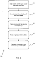
- Figure 3 shows a method 300 for using the systems described above.
- step 310 the cannula and the fiber optic element are brought into contact with a tissue containing a fluorescent agent adapted to absorb light in a first wavelength range and emit light in a second, different wavelength range.
- step 320 the pressure within an internal cavity of the cannula is decreased, thereby providing a suction force to the tissue by way of the cannula, which brings and holds the tissue in contact with the fiber optic element.
- step 330 light in a first wavelength range is provided to the tissue, for example by way of a direct light source or by way of the fiber optical element coupled to the light source.
- Light in the first wavelength range will be absorbed by the fluorescent agent within the tissue of interest.
- a light signal is obtained from the tissue by way of the fiber optic element, the light signal comprising light in the first wavelength range and light in the second wavelength range.
- step 350 the concentration of the fluorescent agent within the tissue is determined based on the proportion of light in the first wavelength range in the light signal.
- a single processor or other unit may fulfill the functions of several items recited in the claims.
- a computer program may be stored/distributed on a suitable medium, such as an optical storage medium or a solid-state medium supplied together with or as part of other hardware, but may also be distributed in other forms, such as via the Internet or other wired or wireless telecommunication systems.
- a suitable medium such as an optical storage medium or a solid-state medium supplied together with or as part of other hardware, but may also be distributed in other forms, such as via the Internet or other wired or wireless telecommunication systems.
Priority Applications (6)
| Application Number | Priority Date | Filing Date | Title |
|---|---|---|---|
| EP20290068.4A EP3973854A1 (fr) | 2020-09-29 | 2020-09-29 | Système de spectroscopie optique |
| US18/029,159 US20230363645A1 (en) | 2020-09-29 | 2021-09-22 | An optical spectroscopy system |
| CN202180066709.XA CN116234517A (zh) | 2020-09-29 | 2021-09-22 | 光谱学系统 |
| JP2023518744A JP2023543756A (ja) | 2020-09-29 | 2021-09-22 | 光学分光システム |
| PCT/EP2021/075996 WO2022069304A1 (fr) | 2020-09-29 | 2021-09-22 | Système de spectroscopie optique |
| EP21777559.2A EP4221567A1 (fr) | 2020-09-29 | 2021-09-22 | Système de spectroscopie optique |
Applications Claiming Priority (1)
| Application Number | Priority Date | Filing Date | Title |
|---|---|---|---|
| EP20290068.4A EP3973854A1 (fr) | 2020-09-29 | 2020-09-29 | Système de spectroscopie optique |
Publications (1)
| Publication Number | Publication Date |
|---|---|
| EP3973854A1 true EP3973854A1 (fr) | 2022-03-30 |
Family
ID=73014452
Family Applications (2)
| Application Number | Title | Priority Date | Filing Date |
|---|---|---|---|
| EP20290068.4A Withdrawn EP3973854A1 (fr) | 2020-09-29 | 2020-09-29 | Système de spectroscopie optique |
| EP21777559.2A Pending EP4221567A1 (fr) | 2020-09-29 | 2021-09-22 | Système de spectroscopie optique |
Family Applications After (1)
| Application Number | Title | Priority Date | Filing Date |
|---|---|---|---|
| EP21777559.2A Pending EP4221567A1 (fr) | 2020-09-29 | 2021-09-22 | Système de spectroscopie optique |
Country Status (5)
| Country | Link |
|---|---|
| US (1) | US20230363645A1 (fr) |
| EP (2) | EP3973854A1 (fr) |
| JP (1) | JP2023543756A (fr) |
| CN (1) | CN116234517A (fr) |
| WO (1) | WO2022069304A1 (fr) |
Citations (8)
| Publication number | Priority date | Publication date | Assignee | Title |
|---|---|---|---|---|
| US5651783A (en) * | 1995-12-20 | 1997-07-29 | Reynard; Michael | Fiber optic sleeve for surgical instruments |
| US6377841B1 (en) * | 2000-03-31 | 2002-04-23 | Vanderbilt University | Tumor demarcation using optical spectroscopy |
| WO2009144653A2 (fr) * | 2008-05-30 | 2009-12-03 | Koninklijke Philips Electronics N.V. | Aiguille avec détecteur de photons intégré |
| WO2011088571A1 (fr) * | 2010-01-25 | 2011-07-28 | University Health Network | Dispositif, système et procédé de quantification de fluorescence et de propriétés optiques |
| US20150148629A1 (en) * | 2011-12-22 | 2015-05-28 | University Health Network | Biopsy Device with Integrated Optical Spectroscopy Guidance |
| US20150182177A1 (en) * | 2005-09-13 | 2015-07-02 | CENTRE NATIONAL DE LA RECHERCHé SCIENTIFIQUE (CNRS) | Peroperative sensing head adapted to be coupled to an ablation tool |
| US20170128150A1 (en) * | 2015-11-11 | 2017-05-11 | Nico Corporation | Illumination sleeve |
| US20170273671A1 (en) * | 2014-08-28 | 2017-09-28 | Koninklijke Philips N.V. | Side-looking lung biopsy device |
Family Cites Families (2)
| Publication number | Priority date | Publication date | Assignee | Title |
|---|---|---|---|---|
| MX2016011230A (es) | 2014-02-28 | 2017-05-30 | Wal Mart Stores Inc | Herramienta para planear medidas de control de multitudes. |
| JP7092668B2 (ja) * | 2015-12-24 | 2022-06-28 | コーニンクレッカ フィリップス エヌ ヴェ | 3d生検組織を染色する装置 |
-
2020
- 2020-09-29 EP EP20290068.4A patent/EP3973854A1/fr not_active Withdrawn
-
2021
- 2021-09-22 WO PCT/EP2021/075996 patent/WO2022069304A1/fr unknown
- 2021-09-22 CN CN202180066709.XA patent/CN116234517A/zh active Pending
- 2021-09-22 JP JP2023518744A patent/JP2023543756A/ja active Pending
- 2021-09-22 US US18/029,159 patent/US20230363645A1/en active Pending
- 2021-09-22 EP EP21777559.2A patent/EP4221567A1/fr active Pending
Patent Citations (8)
| Publication number | Priority date | Publication date | Assignee | Title |
|---|---|---|---|---|
| US5651783A (en) * | 1995-12-20 | 1997-07-29 | Reynard; Michael | Fiber optic sleeve for surgical instruments |
| US6377841B1 (en) * | 2000-03-31 | 2002-04-23 | Vanderbilt University | Tumor demarcation using optical spectroscopy |
| US20150182177A1 (en) * | 2005-09-13 | 2015-07-02 | CENTRE NATIONAL DE LA RECHERCHé SCIENTIFIQUE (CNRS) | Peroperative sensing head adapted to be coupled to an ablation tool |
| WO2009144653A2 (fr) * | 2008-05-30 | 2009-12-03 | Koninklijke Philips Electronics N.V. | Aiguille avec détecteur de photons intégré |
| WO2011088571A1 (fr) * | 2010-01-25 | 2011-07-28 | University Health Network | Dispositif, système et procédé de quantification de fluorescence et de propriétés optiques |
| US20150148629A1 (en) * | 2011-12-22 | 2015-05-28 | University Health Network | Biopsy Device with Integrated Optical Spectroscopy Guidance |
| US20170273671A1 (en) * | 2014-08-28 | 2017-09-28 | Koninklijke Philips N.V. | Side-looking lung biopsy device |
| US20170128150A1 (en) * | 2015-11-11 | 2017-05-11 | Nico Corporation | Illumination sleeve |
Non-Patent Citations (5)
Also Published As
| Publication number | Publication date |
|---|---|
| WO2022069304A1 (fr) | 2022-04-07 |
| CN116234517A (zh) | 2023-06-06 |
| JP2023543756A (ja) | 2023-10-18 |
| EP4221567A1 (fr) | 2023-08-09 |
| US20230363645A1 (en) | 2023-11-16 |
Similar Documents
| Publication | Publication Date | Title |
|---|---|---|
| US11717166B2 (en) | Method and apparatus for the non-invasive measurement of tissue function and metabolism by determination of steady-state fluorescence anisotropy | |
| Mourant et al. | Elastic scattering spectroscopy as a diagnostic tool for differentiating pathologies in the gastrointestinal tract: preliminary testing | |
| Sun et al. | Fluorescence lifetime imaging microscopy for brain tumor image-guided surgery | |
| Kim et al. | Quantification of in vivo fluorescence decoupled from the effects of tissue optical properties using fiber-optic spectroscopy measurements | |
| US10362983B2 (en) | Near infrared photonic prostatoscopy analyzer | |
| Pfefer et al. | Light propagation in tissue during fluorescence spectroscopy with single-fiber probes | |
| CN101553162A (zh) | 获取光学组织特性 | |
| JP2007505645A (ja) | 自動化内視鏡装置、診断方法及び用法 | |
| CN104219998A (zh) | 医用针 | |
| US10105057B2 (en) | Apparatus for optical analysis of an associated tissue | |
| Du Le et al. | Dual-modality optical biopsy of glioblastomas multiforme with diffuse reflectance and fluorescence: ex vivo retrieval of optical properties | |
| US20070265513A1 (en) | Optical measurement of mitochondrial function in blood perfused tissue | |
| Valdes et al. | Quantitative wide-field imaging techniques for fluorescence guided neurosurgery | |
| US20140363063A1 (en) | Imaging apparatus | |
| Shu et al. | Monte Carlo investigation on quantifying the retinal pigment epithelium melanin concentration by photoacoustic ophthalmoscopy | |
| Lu et al. | Combined autofluorescence and diffuse reflectance for brain tumour surgical guidance: initial ex vivo study results | |
| Lee et al. | Compact dual-mode diffuse optical system for blood perfusion monitoring in a porcine model of microvascular tissue flaps | |
| WO2013111053A1 (fr) | Appareil pour l'analyse optique d'un tissu associé | |
| EP3973854A1 (fr) | Système de spectroscopie optique | |
| RU2497558C1 (ru) | Способ проведения интраоперационной комбинированной спектроскопической диагностики опухолей головного и спинного мозга | |
| Rolfe et al. | Advances in fibre-optic sensing in medicine and biology | |
| Owida | Developments and Clinical Applications of Noninvasive Optical Technologies for Skin Cancer Diagnosis | |
| US20230165468A1 (en) | Methods of using optical fiber-based fluorescence spectroscopy for surgical guidance and/or tissue diagnostics and applications of same | |
| Sheth et al. | Quantitative endovascular fluorescence-based molecular imaging through blood of arterial wall inflammation | |
| WO2012127378A1 (fr) | Appareil d'analyse optique d'un échantillon de tissu associé |
Legal Events
| Date | Code | Title | Description |
|---|---|---|---|
| STAA | Information on the status of an ep patent application or granted ep patent |
Free format text: STATUS: UNKNOWN |
|
| PUAI | Public reference made under article 153(3) epc to a published international application that has entered the european phase |
Free format text: ORIGINAL CODE: 0009012 |
|
| STAA | Information on the status of an ep patent application or granted ep patent |
Free format text: STATUS: THE APPLICATION HAS BEEN PUBLISHED |
|
| AK | Designated contracting states |
Kind code of ref document: A1 Designated state(s): AL AT BE BG CH CY CZ DE DK EE ES FI FR GB GR HR HU IE IS IT LI LT LU LV MC MK MT NL NO PL PT RO RS SE SI SK SM TR |
|
| STAA | Information on the status of an ep patent application or granted ep patent |
Free format text: STATUS: THE APPLICATION IS DEEMED TO BE WITHDRAWN |
|
| 18D | Application deemed to be withdrawn |
Effective date: 20221001 |