EP3964136B1 - System und verfahren zum führen eines benutzers bei der ultraschallbeurteilung eines fötalen organs - Google Patents
System und verfahren zum führen eines benutzers bei der ultraschallbeurteilung eines fötalen organs Download PDFInfo
- Publication number
- EP3964136B1 EP3964136B1 EP20305972.0A EP20305972A EP3964136B1 EP 3964136 B1 EP3964136 B1 EP 3964136B1 EP 20305972 A EP20305972 A EP 20305972A EP 3964136 B1 EP3964136 B1 EP 3964136B1
- Authority
- EP
- European Patent Office
- Prior art keywords
- image
- fetal
- classifier
- view
- predefined
- Prior art date
- Legal status (The legal status is an assumption and is not a legal conclusion. Google has not performed a legal analysis and makes no representation as to the accuracy of the status listed.)
- Active
Links
Images






Classifications
-
- A—HUMAN NECESSITIES
- A61—MEDICAL OR VETERINARY SCIENCE; HYGIENE
- A61B—DIAGNOSIS; SURGERY; IDENTIFICATION
- A61B8/00—Diagnosis using ultrasonic, sonic or infrasonic waves
- A61B8/08—Clinical applications
- A61B8/0866—Clinical applications involving foetal diagnosis; pre-natal or peri-natal diagnosis of the baby
-
- A—HUMAN NECESSITIES
- A61—MEDICAL OR VETERINARY SCIENCE; HYGIENE
- A61B—DIAGNOSIS; SURGERY; IDENTIFICATION
- A61B8/00—Diagnosis using ultrasonic, sonic or infrasonic waves
- A61B8/08—Clinical applications
- A61B8/0883—Clinical applications for diagnosis of the heart
-
- A—HUMAN NECESSITIES
- A61—MEDICAL OR VETERINARY SCIENCE; HYGIENE
- A61B—DIAGNOSIS; SURGERY; IDENTIFICATION
- A61B8/00—Diagnosis using ultrasonic, sonic or infrasonic waves
- A61B8/54—Control of the diagnostic device
-
- A—HUMAN NECESSITIES
- A61—MEDICAL OR VETERINARY SCIENCE; HYGIENE
- A61B—DIAGNOSIS; SURGERY; IDENTIFICATION
- A61B8/00—Diagnosis using ultrasonic, sonic or infrasonic waves
- A61B8/52—Devices using data or image processing specially adapted for diagnosis using ultrasonic, sonic or infrasonic waves
- A61B8/5215—Devices using data or image processing specially adapted for diagnosis using ultrasonic, sonic or infrasonic waves involving processing of medical diagnostic data
- A61B8/5223—Devices using data or image processing specially adapted for diagnosis using ultrasonic, sonic or infrasonic waves involving processing of medical diagnostic data for extracting a diagnostic or physiological parameter from medical diagnostic data
Definitions
- the present invention pertains to the field of medical imaging.
- the invention relates to method and system for guiding a user in ultrasound assessment of a fetal organ so as to assess the quality of an examination and to detect pathological conditions.
- a fetal abnormality is a defect in the fetus of a genetic and/or structural nature that can cause significant fetal health and developmental complications, maternal complicated delivery and lead to complex postnatal medical interventions and adverse outcomes [Mai2019].
- Detection of fetal anomalies allows to anticipate a tailored inborn birth, prompt medical care or surgical treatment in specialized centers at birth or in the neonatal period, as well as to provide an adequate information to the parents. For all of these reasons, the ability to detect fetal complications prenatally is critical.
- Cardiac abnormalities are the most common and one of the most impacting group of malformations affecting both fetuses and newborn infants. Accounting for approximately 1% of live births [Sun2018], of which about 25% to 40% are severe forms [Jouannic2010, Sun2018], they are responsible for a half of the infant mortality due to malformations [Jouannic2010].
- fetal heart disease is not always screened antenatally for 3 main reasons: (1) in 49% of cases, lack of skill in adapting ultrasound images (the ultrasound images obtained by the operator are not sufficient to make the correct diagnosis); (2) in 31% of the cases, lack of experience in diagnostics (the images obtained are good and the prenatal pathology is visible but not recognized by the operator); and finally (3), in 20% of cases, pathologies cannot be detected because they are not visible on the ultrasound images.
- US2005251013A1 discloses a device for and method of automatic ultrasound view identification.
- the present invention aims to overcome the above presented problems by proposing a system and a method configured to help the users to assess fetal ultrasound images and analyses these images in order to detect a large spectrum of abnormalities in fetal development.
- the present invention relates to a computer-implemented method for guiding a user in ultrasound assessment of a fetal organ so as to assess the quality of an examination, based on an ultrasound images sequence comprising multiples predefined required views of the fetal organ, said method comprising:
- the assessment method of the present invention allows to automatically select or validate a collection of good quality images.
- the images comprise the necessary prerequisites (i.e. presence of fetal anatomical landmarks), according to medical guidelines.
- the fetal organ is the fetal heart.
- the fetal organ is the fetus heart, brain, head (i.e. comprising the face) and or body.
- the image analysis structure DL1 comprises a first stage employing a convolutional neural network.
- the first classifier of the image analysis structure DL1a comprises a second stage employing a fully connected neural network receiving as input at least a portion of the output of the first stage of the image analysis structure DL1.
- the method is further configured to providing a message so as to inform the user that the valid images list comprises all the predefined required views of the fetal organ.
- a doctor may use the valid images in the evaluation of fetuses examined as part of routine prenatal care [AIUM2003, ACR2003] in order to maximize the possibility of detecting a large spectrum of fetal abnormalities [Lee1998].
- the valid ultrasound images allow as well to identify fetuses at risk for genetic syndromes and to provide useful information for patient counseling, obstetric management and multidisciplinary care.
- the first classifier of the image analysis structure DL1a and the second classifier of the image analysis structure DL1b identifies that the image corresponds to one view's category of the predefined list of view's categories and the second classifier DL1b identifies that a predefined number of fetal anatomical landmarks associated to the identified view's category are present in the image, adding said image to a valid images list.
- the second classifier DL1b proving the image as input to an object detector DL1c of the image analysis structure DL1 comprising a fourth stage configured to receive as input at least a portion of the output of the first stage of the image analysis structure DL1 and comprising region-based fully convolutional neural network architecture being configured to perform segmentation of the image, so as to classify and localize fetal anatomical landmarks in the image.
- Fetal anatomical landmarks localization is computationally costly. In order to be realized in real time it would be needed to either use a high computational power or to simplify the deep learning algorithm and thus to accept to lose in accuracy of the numerical analysis. In order to overcome these two disadvantages, i.e.
- the image analysis structure is divided into two mains architectures: (1) a first architecture comprising the first classifier DL1a and the second classifier DL1b configured to analyze the images of the fetal examination in real-time (neural networks treat each image that the user obtains with the ultrasound device) and (2) a second architecture comprising the object detector DL1c configured to analyze in non-real-time the images provided from the first architecture (DL1a and DL1b).
- This division strategy advantageously allows to obtain a list of valid images in real-time with an excellent precision and, if needed, segment them in non-real-time by keeping an excellent precision.
- the predefined list of view's categories comprises ultrasound view categories from medical ultrasound guidelines [Carvalho2013, Lee2013, CNEOF2016].
- the predefined fetal anatomical landmarks comprise physiological fetal landmarks from medical guidelines.
- the user provides as input to the first classifier of the image analysis structure DL1a at least one image manually selected by the user from the ultrasound video sequence.
- the method further comprises automatically generating and providing a report comprising an examination analysis based on the outputs of the image analysis structure DL1.
- Said report comprises information on:
- the method of the present invention further comprises:
- This embodiment advantageously allows to evaluate the content of the ultrasound images in the valid image list so as to provide a diagnostic suggestion to the user.
- the fully convolutional neural network of the fourth stage of the object detector DL2c is based on the region-based fully convolutional neural network architecture.
- the method further comprises automatically generating and providing a report comprising an examination analysis based on the outputs of the image analysis structure DL1 and a diagnosis based on the outputs of the diagnostic structure DL2.
- Said report comprises information on:
- the first and second classifier, and the object detector of the diagnostic structure DL2 are configured to receive as input a stack of images comprising at least one image.
- the first stage convolutional neural networks of the image analysis structure and of the diagnostic structure have at least one common layer, defined during training.
- the image analysis structure and of the diagnostic structure results from a simultaneous training, notably semi-supervised.
- the present invention further relates to a computer program product for guiding a user in ultrasound assessment of a fetal organ, the computer program product comprising instructions which, when the program is executed by a computer, cause the computer to automatically carry out the steps of the method according to any one of the embodiments described above.
- the present invention further relates to a computer readable storage medium for guiding a user in ultrasound assessment of a fetal organ comprising instructions which, when the program is executed by a computer, cause the computer to carry out the steps of the method according to any one of the embodiments described above.
- the present invention also relates to a computer-implemented method for guiding a user in ultrasound assessment of a fetal organ so as to perform a diagnostic evaluation of the fetal organ development during a medical examination, said method comprising:
- the fully convolutional neural network of the fourth stage of the object detector DL2c is based on the region-based fully convolutional neural network architecture.
- the method further comprises automatically generating and providing a report comprising a diagnosis based on the outputs of the diagnostic structure DL2.
- Said report comprises information on presence or absence of abnormal conditions in the fetus (i.e. output of the first classifier DL2a), and if fetal at least one abnormal condition is detected, a classification of this condition (i.e. output of the second classifier DL2b), as well as its localization on ultrasound images (i.e. output of object detector DL2c).
- the first and the second classifier and the object detector of a diagnostic structure DL2 are configured to receive as input a stack of images comprising at least one image and at the maximum limit the number of views required by the medical guidelines.
- the present invention further relates to a computer program product for guiding a user in ultrasound assessment of a fetal organ so as to perform a diagnostic evaluation of the fetal organ development during a medical examination, the computer program product comprising instructions which, when the program is executed by a computer, cause the computer to automatically carry out the steps of the method according to any one of the embodiments described above.
- the present invention further relates to a computer readable storage medium for guiding a user in ultrasound assessment of a fetal organ so as to perform a diagnostic evaluation of the fetal organ development during a medical examination, said computer readable storage medium comprising instructions which, when the program is executed by a computer, cause the computer to carry out the steps of the method according to any one of the embodiments described above.
- the present invention also relates to a computer-implemented method for guiding a user in ultrasound assessment of a fetal organ so as to perform a diagnostic evaluation of the fetal organ development during a medical examination, said method comprising:
- the modules are to be understood as functional entities rather than material, physically distinct, components. They can consequently be embodied either as grouped together in a same tangible and concrete component, or distributed into several such components. Also, each of those modules is possibly itself shared between at least two physical components.
- the modules are implemented in hardware, software, firmware, or any mixed form thereof as well.
- the present invention also relates to a system for guiding a user in ultrasound assessment of a fetal organ so as to perform a diagnostic evaluation of the fetal organ development during a medical examination, said method comprising:
- the fully convolutional neural network of the fourth stage of the object detector DL2c is based on the region-based fully convolutional neural network architecture.
- the output module is further configured to automatically generating a report comprising a diagnosis based on the outputs of the diagnostic structure DL2.
- Said report comprises information on presence or absence of abnormal conditions in the fetus (i.e. output of the first classifier DL2a), and if fetal at least one abnormal condition is detected, a classification of this condition (i.e. output of the second classifier DL2b), as well as its localization on ultrasound images (i.e. output of the object detector DL2c).
- the first and second classifier and the object detector of a diagnostic structure DL2 are configured to receive as input a stack of images comprising at least one image.
- the present invention relates as well to a system for guiding a user in ultrasound assessment of a fetal organ so as to perform a diagnostic evaluation of the fetal organ development, based on an ultrasound images sequence comprising multiples predefined required views of the fetal organ, said system comprising:
- the image analysis structure DL1 comprises a first stage employing a convolutional neural network and wherein the first classifier of the image analysis structure DL1a comprises a second stage employing a fully connected neural network receiving as input at least a portion of the output of the first stage of the image analysis structure DL1.
- the image analysis module is further configured to provide a message to inform the user that the valid images list comprises all the predefined required views of the fetal organ, when the valid images list comprises all the predefined required views of the fetal organ.
- the system further comprises a manual input module configured to receive images provided manually as input by the user which were selected from the ultrasound video sequence.
- the image analysis module is further configured to provide the image as input to the first classifier of the image analysis structure DL1a and the second classifier of the image analysis structure DL1b and whenever the first classifier DL1a identifies that the image corresponds to one view's category of the predefined list of view's categories and the second classifier DL1b identifies that a predefined number of fetal anatomical landmarks associated to the identified view's category are present in the image, adding said image to a valid images list.
- the image analysis module is further configured to provide the image as input to an object detector DL1c of the image analysis structure DL1 comprising a fourth stage configured to receive as input at least a portion of the output of the first stage of the image analysis structure DL1 and comprising region-based fully convolutional neural network architecture being configured to perform segmentation of the image, so as to classify and localize fetal anatomical landmarks in the image.
- system of the present invention further comprises:
- the fully convolutional neural network of the fourth stage of the object detector DL2c is based on the region-based fully convolutional neural network architecture.
- the output module is further configured to automatically generating a report comprising an examination analysis based on the outputs of the image analysis structure DL1 and a diagnosis based on the outputs of the diagnostic structure DL2.
- Said report comprises information on:
- the first and second classifier and the object detector of the diagnostic structure DL2 are configured to receive as input a stack of images comprising at least one image.
- the first stage convolutional neural networks of the image analysis structure and of the diagnostic structure have at least one common layer, defined during training.
- the image analysis structure and of the diagnostic structure results from a simultaneous training, notably semi-supervised.
- the present invention relates to a computer-implemented method for guiding a user in ultrasound assessment of a fetal organ so as to assess the quality of a medical examination.
- the present computer-implemented method receives as input and perform calculation on an ultrasound images sequence comprising multiples predefined required views of the fetal organ.
- the system is configured to provide to the user a list of some of the ultrasound views categories required, for example by the ISUOG OB/GYN guidelines, for the fetal heart examination of the 2 nd and 3 rd trimesters of pregnancy. Some of these ultrasound views categories required are represent in Figure 1 .
- An ultrasound view of a specific fetal organ is an ultrasound image comprising a standardized list of visible fetal anatomical landmarks of such fetal organ.
- a valid four-chamber view it should be visible the whole heart inside the chest, the size of the picture should take at least half of the screen, and the proportion of the heart, in normal condition, compared to the chest is usually 1/3.
- a four-chamber view it should be visible the following fetal anatomical landmarks: the apex of the heart, both ventricles if present, the atrio-ventricular valves and their offset, both atria, at least one pulmonary vein, the foramen ovale, and the vieussens valve.
- a non-valid view of fetal examination is a view that misses at least one of the important fetal anatomical landmarks required for a correct diagnosis.
- the four-chamber view demonstrated in Figure 3 doesn't allow to assess thoroughly the atrio-ventricular valves and their offset. This failure exposes the operator to misrecognition of inlet abnormalities such as atrio-ventricular septal defect (AVSD) or tricuspid valve abnormalities such as Epstein disease and tricuspid dysplasia.
- AVSD atrio-ventricular septal defect
- tricuspid valve abnormalities such as Epstein disease and tricuspid dysplasia.
- Another example may concern the fetal profile.
- it is mandatory to be able to display the forehead, the nose, the upper and the lower lips, and the chin. If at least one of these landmarks is missing, it will not be possible to properly assess the fetal profile.
- a real challenge for AI is, at first, to be able to distinguish high-quality from low-quality views, at second, to differentiate abnormal from normal low-quality views, and finally, to identify abnormal from normal views. Indeed, in all of these cases some of these landmarks can be missing.
- an ultrasound image comprises all the fetal anatomical landmarks of the standardized list associated to a specific view then the image belongs to a category of images representing said specific view of the fetus.
- An image belonging to a view category of a fetal organ comprises at least a part of information necessary for the correct evaluation of the fetal organ, and this whatever the normal or abnormal condition of the organ.
- multiple images belonging to different views' categories of a fetal organ are necessary to evaluate the fetal organ and provide a correct diagnosis. Said views' categories are defined in medical ultrasound guidelines.
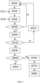
- Figure 4 shows the main steps of this computer-implement method for guiding a user in ultrasound assessment of a fetal organ so as to assess the quality of an examination. No diagnosis is determined in the routine represented by the steps of Figure 4 .
- the method comprises a step M100 of receiving a sequence of 2D ultrasound images comprising multiples predefined required views of the fetal organ, each image comprising at least a portion of the fetal organ. These images may have been previously acquired by the user. Alternatively, the user performs a fetal ultrasound examination in real time resulting in a sequence of 2D ultrasound images which are therefore received in real time.
- the method comprises a step M200 providing each image as input to an image analysis structure DL1.
- DL1 is a deep learning structure configured to validate the correspondence of the fetal ultrasound images to medical guidelines.
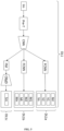
- a schematic representation of the image analysis structure DL1 according to one embodiment of the invention is shown in Figure 5 .
- the box “Im” represents the image inputted in the DL1 structure.
- the image analysis structure DL1 comprising at least one first classifier DL1a which is configured to identify if the image belongs to any view's category comprised in a predefined list of view's categories and, if so, to identify the view's category to which belongs the image among said predefined list of view's categories.
- each view's category is associated to at least one predefined fetal anatomical landmark.
- the quality of each image is evaluated by the presence and/or the absence of specific predefined fetal anatomical landmarks and display criteria. In this case, even with poor signal-to-noise ratio (i.e. a lot of noise for example), but all necessary fetal anatomical landmarks present, the image analysis and further diagnosis will be possible.
- the predefined list of view's categories comprises ultrasound view categories from medical ultrasound guidelines.
- the predefined fetal anatomical landmarks comprise physiological fetal landmarks from medical guidelines.
- the image analysis structure DL1 comprises a first stage employing a convolutional neural network (CNN).
- CNN architecture shown its efficiency for the tasks related to computer vision and image treatment as a very powerful and efficient model performing automatic feature extraction to achieve superhuman accuracy. More in details, CNN architecture presents many advantages compared with its predecessors.
- the filters of CNNs are designed to automatically extract and learn important features from the images.
- CNN architectures treat directly 2D and 3D data which could be gray-scale and color images and are much more computationally efficient (in terms of number of trainable parameters, weights, memory etc.). As a result, the CNN architectures provide higher prediction accuracy on tasks related to computer vision, image/videos treatments, etc.
- the image analysis structure DL1 may further comprise at said first stage bias layer, max pooling layers, batch normalization and/or activation layers.
- the first stage of DL1 may comprise, before the convolutional neural network, a pre-processing the image, for example for denoising the image.
- the first classifier of the image analysis structure DL1a comprises a second stage employing a fully connected neural network (FCNN) receiving as input at least a portion of the output of the first stage of the image analysis structure DL1.
- FCNN fully connected neural network
- the first classifier of the image analysis structure DL1a is configured to provide as output, for each provided image, a softmax vector of size N+1 where N corresponds to the number of view categories (V1, V2, ..., Vn) and the additional one corresponds to the case when the category cannot be defined (NV).
- the category of each image is then determined using the argmax mathematical operation.
- the first classifier of the image analysis structure DL1b comprises a second stage employing a fully connected neural network (FCNN) receiving as input at least a portion of the output of the first stage of the image analysis structure DL1.
- FCNN fully connected neural network
- the method further comprises a step M300 providing each image as input to a second classifier DL1b comprised in the image analysis structure DL1.
- Said second classifier DL1b is configured to detect the presence in the image of predefined fetal anatomical landmarks.
- the second classifier DL1b may be configured to identify but not to localize the fetal anatomical landmarks present in the image.
- the second classifier DL1b provides as output a vector of size M, where M corresponds to the total number of fetal anatomical landmarks associated to the fetal organ.
- M corresponds to the total number of fetal anatomical landmarks associated to the fetal organ.
- the fetal anatomical landmarks present on the provided image are defined using the following mathematical rule: where(vector > 0).
- the method comprises a question step M400 consisting in evaluating whether or not the first classifier DL1a had identified that the image corresponds to one view's category of the predefined list of view's categories and, at the same time, the second classifier DL1b had identified that a predefined number of fetal anatomical landmarks associated to the identified view's category are present in the image.
- the method comprises an updating step M500 consisting in adding the image to a valid images list if the first classifier DL1a had identified that the image corresponds to one view's category of the predefined list of view's categories and the second classifier DL1b had identified that a predefined number of fetal anatomical landmarks associated to the identified view's category are present in the image. All other images are discarded as non-valid.
- the steps M400 and M500 are configured to verify if a view of the predefined list of view's categories was acquired and if all the required fetal anatomical landmarks corresponding to this view were identified, if so the image is considered as valid to be used in future for the evaluation of a diagnosis.
- the method comprises as well as step of providing the valid images list to the user.
- each time the valid images list is updated with a new valid image the method is configured to verify whether the valid images list comprises all the predefined required views of the fetal organ. This step is represented as M510 in Figure 6 .
- the method further comprises a step of providing a message to inform the user that the valid images list comprises all the predefined required views of the fetal organ, when the valid images list comprises all the predefined required views of the fetal organ.
- the method may as well comprise a step M520 of providing a message to inform the user that the valid images list still does not comprise all the predefined required views of the fetal organ and suggest to continue the examination by acquiring new image sequences. In this case the use may continue the examination and therefore acquire more sequences of 2D ultrasound images. These new acquired sequences of 2D ultrasound images are inputted in a processor and analyzed according to step of the method of the present invention as presented here above.
- the user may have the option to provide the required images manually.
- the use may select manually an image from the sequence of 2D ultrasound images which is believed to be conform to the guidelines (i.e. classic way of fetal examination today).
- the method comprises proving the image as input to the first classifier of the image analysis structure DL1a and the second classifier of the image analysis structure DL1b.
- the first classifier DL1a identifies that this manually provided image corresponds to one view's category of the predefined list of view's categories and the second classifier DL1b identifies that a predefined number of fetal anatomical landmarks associated to the identified view's category are present in the image, adding said image to a valid images list.
- the method further comprises, whenever at least one image has been provided by the user manually but not validated by the second classifier DL1b, the step M310 of providing the image as input to an object detector DL1c of the image analysis structure DL1 to classify and localize fetal anatomical landmarks that are present in the image.
- the object detector DL1c is configured to perform a fast segmentation of the image, classify and localize fetal anatomical landmarks that are present in the image.
- the fetal anatomical landmarks detected by the second classifier DL1b at step M300 are compared to the ones identified by the object detector DL1c at step M310. If both structures have identified the same fetal anatomical landmarks (step M320), then these fetal anatomical landmarks are stored as a final set of the image landmarks (step M330). Inversely, if the fetal anatomical landmarks detected by the second classifier DL1b and the object detector DL1c are not de same, the method comprises using a different deep learning algorithm for segmentation which is applied as a judge-method (step M340) and the output provides the final set of the image landmarks (step M350).
- the final set of the image landmarks is provided to the use comprising information on the present and absent fetal anatomical landmarks.
- the method provides user-oriented information allowing to improve the examination quality.
- the object detector DL1c may comprise a fourth stage configured to receive as input at least a portion of the output of the first stage of the image analysis structure DL1 and comprising region-based fully convolutional neural network architecture being configured to perform segmentation of the image.
- the object detector DL1c yields two different answers. For each region of interest found on the image, DL1c returns (M+1)-dimensional softmax responses across M fetal anatomical landmark (+1 for the background). The fetal anatomical landmark is then defined for each region of interest using the argmax mathematical operation. Similarly, for each region of interest found on the image, DL1c returns 4-dimentional average voting responses related to the bounding box and corresponding to the center position of the box along the x axis, center position of the box along the y axis, box width and box height, a 5th position corresponding to the box rotation angle may be added as well.
- the structure of the three neural networks (DL1a, DL1b, DL1c) are fast enough to be applied in real time image processing.
- DL1a and DL1b classifier need to be applied in real time, whereas the object detector DL1c is not required for real-time analysis.
- the architecture of object detector DL1c is presented in Fig. 8 .
- the realization of network DL1c is based on region-based fully convolutional neural network (R-FCN) architecture implemented and adapted to the ultrasound fetal examination.
- R-FCN region-based fully convolutional neural network
- others deep learning architectures may be applicable such as the R-CNN, Fast R-CNN, Faster R-CNN, YOLO or SSD (Single Shot Detection).
- the backbone of object detector DL1c architecture is unique to this invention and pre-trained on the problems of the classification of the view categories and fetal anatomical landmarks (DL1a and DL1b architectures respectively).
- Feature maps (referred to as FM in Fig. 8 ) are generated by some internal convolutional layers of the R-FCN, whereas the last convolutional layers generate the feature maps for DL1a, DL1b, DL2a and DL2b structures which afterword go to the FCNN architecture.
- Regions of Interest (RoI) are independent from the region-based feature maps. Construction of ROIs propositions is done using Region Proposal Network algorithm (referred to as ROIp in Fig. 8 ).
- Position-sensitive score maps (referred to as P-S SM in Fig.
- Position-sensitive score maps are obtained through convolution operation of dimension k 2 ( C + 1), where k 2 corresponds to the total size of the feature score maps, C + 1 is the number of possible fetal anatomical landmarks plus one class of background (non-landmark).
- ROI pooling is produced in a loop for each of the ROI found.
- Each layer is of size k 2 .
- SM vote softmax activation function
- the bounding box regression is shared among the classes and defines for each of the ROI four values: center position along the x axis, center position along the y axis, width and height. As it was previously mentioned, the 5 th value may be added in order to take into account the rotation angle of the box.
- the final number of fetal anatomical landmarks, that is being searched for the given view category, is known in advance. These fetal anatomical landmarks are unique and therefore provide an a-priori information about their location, size and orientation that are implemented into the Region Proposal Network algorithm.
- the valid images list comprises all the predefined required views of the fetal organ (step M530) the examination is validated and the method of the present invention further comprises a step M600 of evaluating whether the fetal development is physiological or not.
- this step M600 comprises providing a stack of at least one image of the valid images list as input to a diagnostic structure DL2.
- the deep learning structure DL2 is schematically represented in Figure 9 .
- the maximum number of images for one input of the DL2 structure is equal to the number of predefined views required, for example, according to the guidelines in France.
- the diagnostic structure DL2 comprises a first classifier DL2a, a second classifier DL2b and an object detector DL2c.
- the stack of images comprises one, two, three, four, five or more fetal ultrasound images simultaneously, wherein the number of images in the stack corresponds to Ni, the predefined number of required views of the fetal organ.
- This input is common for DL2a, DL2b and DL2c deep learning algorithms.
- a stack comprising one image may be used when the user wants to analyze only one image manually provided to check whether a given image is pathological or physiological.
- the stack of images comprises at least two images.
- Advantageously taking into consideration multiple images (i.e. multiple views) at the same time provides much more useful information to the Neural Network structures and increases the detection rate. Indeed, for the detection of some heart (or brain for example) abnormalities, the user has to consider multiple organ views at the same time (example: physicians usually look at the abdominal situs view and 4 chambers view at the same time).
- the diagnostic structure DL2 comprises a first stage employing a convolutional neural network (CNN) receiving as input the stack of images and providing an output.
- CNN convolutional neural network
- the CNN structure guarantees a high precision.
- the object-oriented architectures are not well adapted (i.e. YOLO, SSD etc.).
- the stack of images (“Im") may be pre-treated before being provided as input to the convolutional neural network.
- the method may comprise a step M610 of outputting to the user a message to end examination whenever the output of the first classifier of the diagnostic structure DL2a categorizes the image as comprising a physiological development.
- the DL2a structure independently on the number of the provided ultrasound images in the stack, yields a vector of size two (i.e. two neurons).
- the activation of each of these neurons corresponds respectively to the physiological and pathological development.
- Output values of these neurons are normalized using the softmax activation function.
- an argmax mathematical operation is applied to the normalized vector in order to obtain the neuron containing the maximal value.
- the selected neuron will correspond to the physiological or pathological development.
- the method comprises the step M700 of proving the image as input to the second classifier DL2b of the diagnostic structure DL2.
- the second classifier DL2b comprises a third stage employing a fully connected neural network receiving as input at least a portion of the output of the first stage of the diagnostic structure DL2.
- the second classifier DL2b is configured to classify the pathological development into at least one pathology category Pi or an unknown pathology nP.
- the DL2b algorithm yields a vector of size (N+1) where N corresponds to the total number of the known pathologies (P1, P2, ..., PN) and the plus 1 is added to take into account the case of unknown pathology (nP).
- N corresponds to the total number of the known pathologies (P1, P2, ..., PN) and the plus 1 is added to take into account the case of unknown pathology (nP).
- the pathologies represented on the provided stack of images are defined using the following mathematical rule: where(vector > 0).
- the method comprises as well proving the image as input to the object detector DL2c of the diagnostic structure DL2 (step M710).
- Said object detector DL2c comprises a fourth stage configured to perform segmentation of the image and localization of at least one pathological development region in the fetal organ.
- the object detector DL2c is configured to receive as input at least a portion of the output of the first stage of the diagnostic structure DL2.
- the fourth stage of the object detector DL2c may comprise a fully convolutional neural network configured to perform segmentation of the image and localization of at least one pathological development region in the fetal organ.
- the fully convolutional neural network of the fourth stage of the object detector DL2c may notably be a region-based fully convolutional neural network architecture.
- the object detector DL2c yields two different information.
- the first information concerns the pathology category. More precisely the detector DL2c, for each region of interest found on each image, returns a (M+3)-dimensional softmax, wherein M corresponds to the number of known pathologies, one additional dimension (i.e. +1) is used to take into account an unknown pathology recognized by the object detector DL2c, one other dimension is used to account for physiological anatomy region of interests, and one dimension takes into account the background of the image (i.e. uterus).
- M corresponds to the number of known pathologies
- one additional dimension i.e. +1
- one other dimension is used to account for physiological anatomy region of interests
- one dimension takes into account the background of the image (i.e. uterus).
- the classification is then defined for each region of interest of each of the images of the stack using the argmax mathematical operation.
- the object detector DL2c returns only a 3-dimensional softmax response across: one physiological case, one pathological case and a background.
- this embodiment allows a simpler implementation and the obtention of more precise results.
- it does not allow to classify abnormalities present in the region of interests.
- the classifier is able to identify whether the anatomical parts of the fetus in the ROI are physiological, pathological or belongs to a background (i.e. not physiological or pathological). But in the case if a pathology found, the classifier is not able to classify the anatomical parts of the fetus and precisely determine which pathology is present.
- the second information concerns the result of the image segmentation of the images of the stack and localization of the pathological development region in the fetal organ.
- the object detector DL2c For each region of interest found on each image in the provided stack, the object detector DL2c returns 4-dimentional average voting responses related to the bounding box and corresponding to the center position of the box along the x axis, center position of the box along the y axis, box width and box height (output referred to as "LSP" in Fig. 9 ).
- a fifth position corresponding to the box rotation angle may be added as well.
- the method also comprises providing an output to the user (step M800).
- This output may be the at least one pathology category obtained from the second classifier DL2b and the result of the image segmentation of the image and localization of the pathological development region in the fetal organ obtained from the object detector DL2c of the diagnostic structure DL2.
- the method is configured to prove to the user a message to interrupt the examination.
- the valid images list comprising all the predefined required views of the fetal organ.
- the image analysis structure and the diagnostic structure have some common convolutional layer in the Encoder (i.e. the first stage convolutional neural networks of the image analysis structure and of the diagnostic structure).
- the common layers may be defined during the training part through an optimization technique.
- An example of such application is a particle swarm optimization method applied to a level set approach. This approach results in a simultaneous training of architectures DL1 and DL2 through the transfer learning technique.
- training the two structures at the same time allows to yield better results that two separate structures trained independently.
- the image analysis structure and the diagnostic structure results are obtained by a simultaneous training, notably semi-supervised.
- the fully connected neural networks of DL1a and DL2a comprise at least one hidden layer.
- the fully connected neural networks of DL1b and DL2b comprise at least two hidden layers.
- DL1c and DL2c object detector are fully convolutional and do not comprise any fully connected layers.
- a binary cross entropy loss function is applied to a softmax activation layer for the both DL1 and DL2 neural architectures.
- a parameter ⁇ responsible for the box rotation angle is added to t in order to improve the precision of fetal landmarks search.
- a binary cross entropy loss function is used for the implementation of a Generative Adversarial Network (GAN) as a semi-supervised approach. This loss function takes the output of the supervised model prior to the softmax activation function and applies a special activation function calculating a normalized sum of the exponential outputs.
- a stochastic gradient descent algorithm Adam is used for training of this hybrid architecture with momentum values of 0.5-0.9 and learning rate value of 2 * 10 -4 .
- Another approach may be implemented which is based on the idea of creating a neuron independent from the supervised model for the GAN architecture. The designing an optimal neural architecture can be very challenging in terms of computational requirements.
- a numerical optimization algorithm is thus implemented in order to choose neural network parameters leading to an optimal architecture.
- This algorithm is based on a level-set approach for optimizing discrete parameters and the metamodeling technique allowing to build surrogate models to expensive black-box functions and to reduce the computational burden for optimization purpose.
- the image analysis structure DL1 (i.e DL1a, DL1b and DL1c) is trained using a database containing both physiological and pathological images. It is done with a purpose to learn the image analysis structure DL1 to distinguish fetal abnormalities from the missing fetal landmarks on the image.
- the method is a software on cloud solution to which a user may access through an internet connection.
- the present invention also relates to the system comprising a computer storage medium and a processor configured to carry out the method of the present invention.
- the user has access to a list of some of the ultrasound view categories required, for example by the ISUOG OB/GYN, guidelines for the fetal heart examination of the 2 nd and 3 rd trimesters of pregnancy ( Fig. 1 ).
- the user starts the examination with the ultrasound device knowing that a sequence of ultrasound images should be obtained wherein at least a part of the images is comprised in this list of ultrasound view categories.
- the first classifier DL1a and the second classifier DL1b of the image analysis structure run in order to select the good images of the fetal organ (i.e. images corresponding to one view's category of the predefined list of view's categories and comprising a predefined number of fetal anatomical landmarks associated to the identified view's category) and add them into the list of valid images.
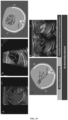
- the user may be informed that some views are still missing from a graphic representation like in Figure 10 . Indeed, in Figure 10 the left ventricular outflow tract (LVOT) view (drawing in Fig. 10C ) and right ventricular outflow tract (RVOT) view (drawing in Fig.
- the invention provides an output to the user to inform him/her that the examination is "valid" (i.e. the valid images list comprises all the predefined required views of the fetal heart and these images therefore correspond to the guidelines).
- the images of the valid images list are treated with the first classifier of the diagnostic structure DL2a in order to distinguish physiological development from the pathological one.
- the system/method of the invention provides an output to the user such as the one illustrated in Figure 11 .
- a written message informs the user that no abnormality is detected.
- the user receives an output with a type of the suspected pathology and a localization of the fetal abnormality as shown in Figure 12 .
- the dashed rectangle enclosed the region comprising the detected pathology.
- Said dashed rectangle is defined using the bounding box and corresponding to the center position of the box along the x axis, center position of the box along the y axis, box width, box height and possibly box rotation obtained from the object detector DL2c.
- the output represented in Figure 12 further comprises a message informing the user of a possible "ventricular septal defect".
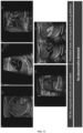
- the first classifier DL1a and the second classifier DL1b treat them in order to identify to which view's category they belong and identify whether the predefined fetal anatomical landmarks associated to the identified view's category are present. If the images provided manually have some fetal anatomical landmarks missing, the user receives a corresponding output such as the one reported in Figure 13 . Indeed, in Figure 13 , the right ventricular outflow tract (RVOT) view has some missing landmarks (bifurcation of pulmonary artery is not visible). Using this image for fetal examination is therefore associated with a risk of misrecognition of some abnormalities.
- RVOT right ventricular outflow tract
- the present invention further relates to a computer program product for guiding a user in ultrasound assessment of a fetal organ, the computer program product comprising instructions which, when the program is executed by a computer, cause the computer to automatically carry out the steps of the method according to any one of the embodiments described hereabove.
- the computer program product to perform the method as described above may be written as computer programs, code segments, instructions or any combination thereof, for individually or collectively instructing or configuring the processor or computer to operate as a machine or special-purpose computer to perform the operations performed by hardware components.
- the computer program product includes machine code that is directly executed by a processor or a computer, such as machine code produced by a compiler.
- the computer program product includes higher-level code that is executed by a processor or a computer using an interpreter. Programmers of ordinary skill in the art can readily write the instructions or software based on the block diagrams and the flow charts illustrated in the drawings and the corresponding descriptions in the specification, which disclose algorithms for performing the operations of the method as described above.
- the present invention further relates to a computer readable storage medium for guiding a user in ultrasound assessment of a fetal organ comprising instructions which, when the program is executed by a computer, cause the computer to carry out the method according to any one of the embodiments described here above.
- the computer-readable storage medium is a non-transitory computer-readable storage medium.
- Computer programs implementing the method of the present embodiments can commonly be distributed to users on a distribution computer-readable storage medium such as, but not limited to, an SD card, an external storage device, a microchip, a flash memory device, a portable hard drive and software websites. From the distribution medium, the computer programs can be copied to a hard disk or a similar intermediate storage medium. The computer programs can be run by loading the computer instructions either from their distribution medium or their intermediate storage medium into the execution memory of the computer, configuring the computer to act in accordance with the method of this invention. All these operations are well- known to those skilled in the art of computer systems.
- the instructions or software to control a processor or computer to implement the hardware components and perform the methods as described above, and any associated data, data files, and data structures, are recorded, stored, or fixed in or on one or more non-transitory computer-readable storage media.
- Examples of a non-transitory computer-readable storage medium include read-only memory (ROM), random-access memory (RAM), flash memory, CD- ROMs, CD- Rs, CD+ Rs, CD- RWs, CD+ RWs, DVD- ROMs, DVD- Rs, DVD+ Rs, DVD- RWs, DVD+ RWs, DVD- RAMs, BD- ROMs, BD- Rs, BD- R LTHs, BD- Res, magnetic tapes, floppy disks, magneto-optical data storage devices, optical data storage devices, hard disks, solid-state disks, and any device known to one of ordinary skill in the art that is capable of storing the instructions or software and any associated data, data files, and data structures in a non-transitory manner and providing
- the instructions or software and any associated data, data files, and data structures are distributed over network-coupled computer systems so that the instructions and software and any associated data, data files, and data structures are stored, accessed, and executed in a distributed fashion by the processor or computer.
- the image analysis architecture DL1 was dynamically trained on a semi-labeled database with the following views distribution:
- a weighted cost function was implemented in order to take into account this imbalanced database.
- This database was divided into train and validation parts of 80% and 20% respectively.
- the first classifier DL1a algorithm shows a precision of 98.41% and the second classifier DL1b algorithm a precision of 92.91%.
- the first classifier DL2a algorithm shows a precision of 96.72% while the second classifier DL2b algorithm presented a precision of 93.11%.
Landscapes
- Health & Medical Sciences (AREA)
- Life Sciences & Earth Sciences (AREA)
- Heart & Thoracic Surgery (AREA)
- Medical Informatics (AREA)
- Biophysics (AREA)
- Nuclear Medicine, Radiotherapy & Molecular Imaging (AREA)
- Pathology (AREA)
- Radiology & Medical Imaging (AREA)
- Engineering & Computer Science (AREA)
- Biomedical Technology (AREA)
- Veterinary Medicine (AREA)
- Physics & Mathematics (AREA)
- Molecular Biology (AREA)
- Surgery (AREA)
- Animal Behavior & Ethology (AREA)
- General Health & Medical Sciences (AREA)
- Public Health (AREA)
- Cardiology (AREA)
- Gynecology & Obstetrics (AREA)
- Pregnancy & Childbirth (AREA)
- Ultra Sonic Daignosis Equipment (AREA)
Claims (15)
- System zur Anleitung eines Benutzers bei der Ultraschallbewertung eines fetalen Organs, um eine diagnostische Bewertung der fetalen Organentwicklung während einer medizinischen Untersuchung durchzuführen, wobei die Ultraschallbewertung auf einer Abfolge von Ultraschallbildern basiert, die mehrere vordefinierte erforderliche Ansichten des fetalen Organs umfasst, wobei das System Folgendes umfasst:- Eingabemodul, eingerichtet zum Empfangen von:o einer Abfolge von 2D-Ultraschallbildern in Echtzeit, wobei die Bilder mehrere vordefinierte erforderliche Ansichten des fetalen Organs umfassen, wobei jedes Bild mindestens einen Teil des fetalen Organs umfasst;o einer vordefinierte Liste von Ansichtskategorien, wobei jede Ansichtskategorie mindestens einem vordefinierten fetalen anatomischen Orientierungspunkt zugeordnet ist;- einem Bildanalysemodul, das für jedes Bild für Folgendes eingerichtet ist:∘ Bereitstellung des Bildes als Eingabe für eine Bildanalysestruktur (DL1), die mindestens einen ersten Klassifikator (DL1a) umfasst, wobei der erste Klassifikator (DL1a) so eingerichtet ist, dass er identifiziert, ob das Bild zu einer Ansichtskategorie gehört, die in der vordefinierten Liste der Ansichtskategorien enthalten ist, und wenn ja, um die Ansichtskategorie zu identifizieren, zu der das Bild unter den vordefinierten Ansichtskategorien gehört;∘ Bereitstellung des Bildes als Eingabe für einen zweiten Klassifikator (DL1b) der Bildanalysestruktur (DL1), wobei der zweite Klassifikator (DL1b) so eingerichtet ist, dass er das Vorhandensein vordefinierter fetaler anatomischer Orientierungspunkte im Bild erkennt;∘ Hinzufügen des Bildes zu einer gültigen Bildliste, wenn:▪ der erste Klassifikator (DL1a) identifiziert, dass das Bild einer Ansichtskategorie der vordefinierten Liste der Ansichtskategorien entspricht; und▪ der zweite Klassifikator (DL1b) identifiziert, dass eine vordefinierte Anzahl fetaler anatomischer Orientierungspunkte im Bild vorhanden ist, wobei die fetalen anatomischen Orientierungspunkte der Ansichtskategorie zugeordnet sind, die durch den ersten Klassifikator identifiziert wird;∘ die gültige Bildliste bereitgestellt wird.
- System nach Anspruch 1, wobei das fetale Organ das fetale Herz ist.
- System nach einem der Ansprüche 1 oder 2, wobei im Bildanalysemodul die Bildanalysestruktur (DL1) eine erste Stufe umfasst, die ein konvolutionales neurales Netzwerk verwendet, und wobei der erste Klassifikator der Bildanalysestruktur (DL1a) eine zweite Stufe umfasst, die ein vollständig verbundenes neurales Netzwerk verwendet, das mindestens einen Teil der Ausgabe der ersten Stufe der Bildanalysestruktur (DL1) als Eingabe empfängt.
- System nach einem der Ansprüche 1 bis 3, wann immer mindestens ein Bild vom Benutzer manuell bereitgestellt wurde, ist das Bildanalysemodul ferner so eingerichtet, dass es das Bild als Eingabe für den ersten Klassifikator der Bildanalysestruktur (DL1a) und den zweiten Klassifikator der Bildanalysestruktur (DL1a) bereitstellt und wann immer der erste Klassifikator (DL1a) identifiziert, dass das Bild der Ansichtskategorie der vordefinierten Liste der Ansichtskategorien entspricht, und der zweite Klassifikator (DL1b) identifiziert, dass eine vordefinierte Anzahl fetaler anatomischer Orientierungspunkte, die der identifizierten Ansichtskategorie zugeordnet sind, im Bild vorhanden sind, wodurch das Bild zu einer gültigen Bildliste hinzugefügt wird.
- System nach Anspruch 4, wann immer mindestens ein Bild vom Benutzer manuell bereitgestellt wurde, aber nicht durch den zweiten Klassifikator (DL1b) validiert wird, ist das Bildanalysemodul ferner so eingerichtet, dass es das Bild als Eingabe für einen Objektdetektor (DL1c) der Bildanalysestruktur (DL1) bereitstellt, die eine vierte Stufe umfasst, die so eingerichtet ist, dass sie als Eingabe mindestens einen Teil der Ausgabe der ersten Stufe der Bildanalysestruktur (DL1) empfängt und umfassend eine regionsbasierte, vollständig konvolutionale neuronale Netzwerkarchitektur, die so eingerichtet ist, dass sie eine Segmentierung des Bildes durchführt, um fetale anatomische Orientierungspunkte im Bild zu klassifizieren und zu lokalisieren.
- System nach einem der Ansprüche 1 bis 5, ferner umfassend:- ein Diagnosemodul, dass, wenn die Liste der gültigen Bilder alle vordefinierten erforderlichen Ansichten des fetalen Organs umfasst, für Folgendes eingerichtet ist:o Bereitstellung eines Stapels eines Bildes der gültigen Bildliste als Eingabe für eine Diagnosestruktur (DL2), wobei die Diagnosestruktur (DL2) eine erste Stufe umfasst, die ein konvolutionales neurales Netzwerk verwendet, das den Stapel von Bildern als Eingabe empfängt und eine Ausgabe bereitstellt, und wobei die Diagnosestruktur (DL2) einen ersten Klassifikator (DL2a) umfasst, der in einer zweiten Stufe verwendet, ein vollständig verbundenes neuronales Netzwerk, das mindestens einen Teil der Ausgabe der ersten Stufe der Diagnosestruktur (DL2) als Eingabe empfängt; wobei der erste Klassifikator der Diagnosestruktur (DL2a) so eingerichtet ist, dass er zwischen pathologischer Entwicklung und physiologischer Entwicklung des fetalen Organs unterscheidet;∘ wenn die Ausgabe des ersten Klassifikators der Diagnosestruktur (DL2a) das Bild als eine pathologische Entwicklung kategorisiert und das Bild als Eingabe für Folgendes bereitstellt:• einen zweiten Klassifikator (DL2b) der Diagnosestruktur (DL2), der eine dritte Stufe umfasst, die ein vollständig verbundenes neuronales Netzwerk verwendet, das mindestens einen Teil der Ausgabe der ersten Stufe der Diagnosetruktur (DL2) als Eingabe empfängt und so eingerichtet ist, dass es die pathologische Entwicklung in mindestens eine Pathologiekategorie einstuft;• einen Objektdetektor (DL2c) der Diagnosestruktur (DL2), der eine vierte Stufe umfasst, die so eingerichtet ist, dass sie mindestens einen Teil der Ausgabe der ersten Stufe der Diagnosestruktur (DL2) als Eingabe empfängt, und der ein vollständig konvolutionales neurales Netzwerk umfasst, das so eingerichtet ist, dass es die Segmentierung des Bildes und die Lokalisierung mindestens eines pathologischen Entwicklungsbereichs im fetalen Organ durchführt;- Ausgabemodul, eingerichtet für:∘ Ausgabe an den Benutzer der mindestens einen Pathologiekategorie, die aus dem zweiten Klassifikator (DL2b) gewonnen wurde, und des Ergebnisses der Bildsegmentierung des Bildes und der Lokalisierung des pathologischen Entwicklungsbereichs im fetalen Organ, die aus dem Objektdetektor (DL2c) der Diagnosestruktur (DL2) gewonnen wurde, an den Benutzer;∘ Ausgabe einer Meldung an den Benutzer, um die Untersuchung zu beenden, wenn die Ausgabe des ersten Klassifikators der Diagnosestruktur (DL2a) das Bild als eine physiologische Entwicklung kategorisiert.
- System nach Anspruch 6, wobei das vollkonvolutionale neuronale Netzwerk der vierten Stufe des Obj ektdetektors (DL2c) auf der regionsbasierten vollkonvolutionalen neuronalen Netzwerkarchitektur basiert.
- System nach einem der Ansprüche 6 oder 7, wobei die ersten, zweiten Klassifikatoren und der Objektdetektor einer Diagnosestruktur (DL2) so eingerichtet sind, dass sie als Eingabe einen Stapel von Bildern empfangen, der mindestens ein Bild umfasst.
- System nach einem der Ansprüche 6 bis 8, wobei die konvolutionalen neuronalen Netze der ersten Stufe der Bildanalysestruktur und der Diagnosestruktur mindestens eine gemeinsame Schicht aufweisen, die während des Trainings definiert ist.
- System nach Anspruch 9, wobei sich die Bildanalysestruktur und die Diagnosestruktur aus einem gleichzeitigen Training, insbesondere semi-beaufsichtigt, ergibt.
- Computerimplementiertes Verfahren zur Anleitung eines Benutzers bei der Ultraschallbewertung eines fetalen Organs, um die Qualität einer Untersuchung zu beurteilen, wobei das Verfahren Folgendes umfasst:- Empfangen einer Abfolge von 2D-Ultraschallbildern in Echtzeit, wobei die Bilder mehrere vordefinierte erforderliche Ansichten des fetalen Organs umfassen, wobei jedes Bild mindestens einen Teil des fetalen Organs (M100) umfasst;- Empfangen einer vordefinierten Liste von Ansichtskategorien, wobei jede Ansichtskategorie mindestens einem vordefinierten fetalen anatomischen Orientierungspunkt zugeordnet ist;- Bereitstellen jedes Bildes als Eingabe für eine Bildanalysestruktur (DL1), die mindestens einen ersten Klassifikator (DL1a) umfasst, wobei der erste Klassifikator (DL1a) so eingerichtet ist, dass er identifiziert, ob das Bild zu einer Ansichtskategorie gehört, die in der vordefinierten Liste der Ansichtskategorien enthalten ist, und wenn ja, um die Ansichtskategorie zu identifizieren, zu der das Bild unter der vordefinierten Liste der Ansichtskategorien (M200) gehört;- Bereitstellen jedes Bildes als Eingabe für einen zweiten Klassifikator (DL1b) der Bildanalysestruktur (DL1), wobei der zweite Klassifikator (DL1b) so eingerichtet ist, dass er das Vorhandensein von vordefinierten fetalen anatomischen Orientierungspunkten (M300) im Bild erkennt;- wenn, für jedes Bild:∘ der erste Klassifikator (DL1a) identifiziert, dass das Bild der Ansichtskategorie der vordefinierten Liste der Ansichtskategorien entspricht; und∘ der zweite Klassifikator (DL1b) identifiziert, dass eine vordefinierte Anzahl fetaler anatomischer Orientierungspunkte im Bild vorhanden ist, wobei die fetalen anatomischen Orientierungspunkte der Ansichtskategorie zugeordnet sind, die durch den ersten Klassifikator (M400) identifiziert wird,Hinzufügen des Bildes zu einer gültigen Bildliste (M500);- Bereitstellen der gültigen Bildliste an den Benutzer.
- Das Verfahren nach Anspruch 11, wenn die Liste der gültigen Bilder alle vordefinierten erforderlichen Ansichten des fetalen Organs umfasst, umfasst ferner das Bereitstellen einer Nachricht, um den Benutzer darüber zu informieren, dass die Liste der gültigen Bilder alle vordefinierten erforderlichen Ansichten des fetalen Organs umfasst.
- Verfahren nach einem der Ansprüche 11 oder 12, wobei die vordefinierte Liste der Ansichtskategorien Ultraschallansichtskategorien aus medizinischen Ultraschallrichtlinien umfasst und die vordefinierten fetalen anatomischen Orientierungspunkte physiologische fetale Orientierungspunkte aus medizinischen Richtlinien umfassen.
- Computerprogrammprodukt zur Anleitung eines Benutzers bei der Ultraschallbewertung eines fetalen Organs, wobei das Computerprogrammprodukt Anweisungen umfasst, die, wenn das Programm von einem Computer ausgeführt wird, den Computer dazu veranlassen, das Verfahren nach einem der Ansprüche 11 bis 13 automatisch auszuführen.
- Computerlesbares Speichermedium zur Anleitung eines Benutzers bei der Ultraschallbewertung eines fetalen Organs, umfassend Anweisungen, die, wenn das Programm von einem Computer ausgeführt wird, den Computer veranlassen, das Verfahren nach einem der Ansprüche 11 bis 13 auszuführen.
Priority Applications (1)
| Application Number | Priority Date | Filing Date | Title |
|---|---|---|---|
| EP20305972.0A EP3964136B1 (de) | 2020-09-02 | 2020-09-02 | System und verfahren zum führen eines benutzers bei der ultraschallbeurteilung eines fötalen organs |
Applications Claiming Priority (1)
| Application Number | Priority Date | Filing Date | Title |
|---|---|---|---|
| EP20305972.0A EP3964136B1 (de) | 2020-09-02 | 2020-09-02 | System und verfahren zum führen eines benutzers bei der ultraschallbeurteilung eines fötalen organs |
Publications (3)
| Publication Number | Publication Date |
|---|---|
| EP3964136A1 EP3964136A1 (de) | 2022-03-09 |
| EP3964136C0 EP3964136C0 (de) | 2025-05-14 |
| EP3964136B1 true EP3964136B1 (de) | 2025-05-14 |
Family
ID=72561726
Family Applications (1)
| Application Number | Title | Priority Date | Filing Date |
|---|---|---|---|
| EP20305972.0A Active EP3964136B1 (de) | 2020-09-02 | 2020-09-02 | System und verfahren zum führen eines benutzers bei der ultraschallbeurteilung eines fötalen organs |
Country Status (1)
| Country | Link |
|---|---|
| EP (1) | EP3964136B1 (de) |
Families Citing this family (15)
| Publication number | Priority date | Publication date | Assignee | Title |
|---|---|---|---|---|
| KR102446343B1 (ko) | 2015-06-15 | 2022-09-22 | 삼성메디슨 주식회사 | 초음파 장치 및 그 제어방법 |
| CN114848019A (zh) * | 2022-04-19 | 2022-08-05 | 山东大学 | 基于柔性协作机器人的自动心脏超声检测系统及方法 |
| KR20240032563A (ko) * | 2022-09-02 | 2024-03-12 | 삼성메디슨 주식회사 | 초음파 진단 장치 및 진단 방법 |
| US12082969B1 (en) | 2023-02-22 | 2024-09-10 | BrightHeart SAS | Systems and methods for improving detection of fetal congenital heart defects |
| US12148162B2 (en) | 2023-02-22 | 2024-11-19 | BrightHeart SAS | Systems and methods for detecting cardiovascular anomalies using spatiotemporal neural networks |
| US12343197B2 (en) | 2023-02-22 | 2025-07-01 | BrightHeart SAS | Systems and methods for improving detection of fetal congenital heart defects |
| US11875507B1 (en) | 2023-02-22 | 2024-01-16 | BrightHeart SAS | Systems and methods for detecting cardiovascular anomalies using spatiotemporal neural networks |
| US12493962B2 (en) | 2023-02-22 | 2025-12-09 | BrightHeart SAS | Systems and methods for detecting cardiovascular anomalies using spatiotemporal neural networks |
| WO2024176114A1 (en) | 2023-02-22 | 2024-08-29 | BrightHeart SAS | Systems and methods for improving detection of fetal congenital heart defects |
| US11869188B1 (en) | 2023-02-22 | 2024-01-09 | BrightHeart SAS | Systems and methods for improving detection of fetal congenital heart defects |
| EP4442207B1 (de) * | 2023-04-07 | 2025-10-08 | Diagnoly | Vorrichtung zur führung eines benutzers bei der ultraschallbeurteilung eines organs |
| US11861838B1 (en) | 2023-06-07 | 2024-01-02 | BrightHeart SAS | Systems and methods for system agnostic automated detection of cardiovascular anomalies and/or other features |
| US20250111501A1 (en) * | 2023-10-03 | 2025-04-03 | Fujifilm Sonosite, Inc. | Contextual processing of ultrasound data |
| EP4563089A1 (de) | 2023-11-28 | 2025-06-04 | Koninklijke Philips N.V. | Verfolgung von ultraschallbildern |
| CN118522451A (zh) * | 2024-05-13 | 2024-08-20 | 四川大学华西第二医院 | 基于ai的胎儿先天性心脏病评估检测方法、系统和介质 |
Family Cites Families (2)
| Publication number | Priority date | Publication date | Assignee | Title |
|---|---|---|---|---|
| US7672491B2 (en) * | 2004-03-23 | 2010-03-02 | Siemens Medical Solutions Usa, Inc. | Systems and methods providing automated decision support and medical imaging |
| EP3781039B1 (de) * | 2018-04-20 | 2023-11-22 | Koninklijke Philips N.V. | Adaptive ultraschallabtastung |
-
2020
- 2020-09-02 EP EP20305972.0A patent/EP3964136B1/de active Active
Also Published As
| Publication number | Publication date |
|---|---|
| EP3964136C0 (de) | 2025-05-14 |
| EP3964136A1 (de) | 2022-03-09 |
Similar Documents
| Publication | Publication Date | Title |
|---|---|---|
| EP3964136B1 (de) | System und verfahren zum führen eines benutzers bei der ultraschallbeurteilung eines fötalen organs | |
| Hennessey et al. | Artificial intelligence in veterinary diagnostic imaging: A literature review | |
| US20230104045A1 (en) | System and method for ultrasound analysis | |
| Xie et al. | Computer-aided diagnosis for fetal brain ultrasound images using deep convolutional neural networks | |
| EP4395654B1 (de) | Vorrichtung und verfahren zur führung einer ultraschallbeurteilung eines organs | |
| Reddy et al. | Artificial intelligence in perinatal diagnosis and management of congenital heart disease | |
| Singh et al. | Deep learning based coronary artery disease detection and segmentation using ultrasound imaging with adaptive gated SCNN models | |
| Qiao et al. | SPReCHD: Four-chamber semantic parsing network for recognizing fetal congenital heart disease in medical metaverse | |
| US20230394655A1 (en) | System and method for evaluating or predicting a condition of a fetus | |
| Tan et al. | Automated detection of congenital heart disease in fetal ultrasound screening | |
| Hsu et al. | The application of artificial intelligence to support biliary atresia screening by ultrasound images: A study based on deep learning models | |
| Nguyen et al. | Artificial intelligence in the pediatric echocardiography laboratory: automation, physiology, and outcomes | |
| Sriraam et al. | A comprehensive review of artificial intelligence-based algorithm towards fetal facial anomalies detection (2013–2024) | |
| Budd et al. | Detecting hypo-plastic left heart syndrome in fetal ultrasound via disease-specific atlas maps | |
| Cai et al. | The Application of Knowledge Distillation toward Fine‐Grained Segmentation for Three‐Vessel View of Fetal Heart Ultrasound Images | |
| Jiang et al. | Artificial intelligence in echocardiography: Applications and future directions | |
| Goetz-Fu et al. | Development and temporal validation of a deep learning model for automatic fetal biometry from ultrasound videos | |
| Chen et al. | An efficient network with state space model under evidential training for fetal echocardiography standard view recognition | |
| US20240299010A1 (en) | System and method for guiding a user in ultrasound assessment of a fetal organ | |
| Sarker et al. | HarmonicEchoNet: Leveraging harmonic convolutions for automated standard plane detection in fetal heart ultrasound videos | |
| Chen et al. | Intelligent interpretation of four lung ultrasonographic features with split attention based deep learning model | |
| Vetriselvi et al. | Deep Learning Based Medical Image Segmentation in Prenatal Prediction of Neurodevelopmental Disorders | |
| Liu et al. | Hierarchical agent-based reinforcement learning framework for automated quality assessment of fetal ultrasound video | |
| Bindiya et al. | Detection of anomalies in fetus using convolution neural network | |
| Sapitri et al. | Yolact-based Approach for Real-Time Fetal Heart Segmentation |
Legal Events
| Date | Code | Title | Description |
|---|---|---|---|
| PUAI | Public reference made under article 153(3) epc to a published international application that has entered the european phase |
Free format text: ORIGINAL CODE: 0009012 |
|
| STAA | Information on the status of an ep patent application or granted ep patent |
Free format text: STATUS: THE APPLICATION HAS BEEN PUBLISHED |
|
| AK | Designated contracting states |
Kind code of ref document: A1 Designated state(s): AL AT BE BG CH CY CZ DE DK EE ES FI FR GB GR HR HU IE IS IT LI LT LU LV MC MK MT NL NO PL PT RO RS SE SI SK SM TR |
|
| STAA | Information on the status of an ep patent application or granted ep patent |
Free format text: STATUS: REQUEST FOR EXAMINATION WAS MADE |
|
| 17P | Request for examination filed |
Effective date: 20220909 |
|
| RBV | Designated contracting states (corrected) |
Designated state(s): AL AT BE BG CH CY CZ DE DK EE ES FI FR GB GR HR HU IE IS IT LI LT LU LV MC MK MT NL NO PL PT RO RS SE SI SK SM TR |
|
| STAA | Information on the status of an ep patent application or granted ep patent |
Free format text: STATUS: EXAMINATION IS IN PROGRESS |
|
| 17Q | First examination report despatched |
Effective date: 20230913 |
|
| GRAP | Despatch of communication of intention to grant a patent |
Free format text: ORIGINAL CODE: EPIDOSNIGR1 |
|
| STAA | Information on the status of an ep patent application or granted ep patent |
Free format text: STATUS: GRANT OF PATENT IS INTENDED |
|
| INTG | Intention to grant announced |
Effective date: 20241218 |
|
| GRAS | Grant fee paid |
Free format text: ORIGINAL CODE: EPIDOSNIGR3 |
|
| GRAA | (expected) grant |
Free format text: ORIGINAL CODE: 0009210 |
|
| STAA | Information on the status of an ep patent application or granted ep patent |
Free format text: STATUS: THE PATENT HAS BEEN GRANTED |
|
| RAP3 | Party data changed (applicant data changed or rights of an application transferred) |
Owner name: DIAGNOLY |
|
| AK | Designated contracting states |
Kind code of ref document: B1 Designated state(s): AL AT BE BG CH CY CZ DE DK EE ES FI FR GB GR HR HU IE IS IT LI LT LU LV MC MK MT NL NO PL PT RO RS SE SI SK SM TR |
|
| REG | Reference to a national code |
Ref country code: GB Ref legal event code: FG4D |
|
| REG | Reference to a national code |
Ref country code: CH Ref legal event code: EP |
|
| REG | Reference to a national code |
Ref country code: DE Ref legal event code: R096 Ref document number: 602020051202 Country of ref document: DE |
|
| REG | Reference to a national code |
Ref country code: IE Ref legal event code: FG4D |
|
| U01 | Request for unitary effect filed |
Effective date: 20250612 |
|
| U07 | Unitary effect registered |
Designated state(s): AT BE BG DE DK EE FI FR IT LT LU LV MT NL PT RO SE SI Effective date: 20250623 |
|
| REG | Reference to a national code |
Ref country code: CH Ref legal event code: U11 Free format text: ST27 STATUS EVENT CODE: U-0-0-U10-U11 (AS PROVIDED BY THE NATIONAL OFFICE) Effective date: 20251001 |
|
| RAP4 | Party data changed (patent owner data changed or rights of a patent transferred) |
Owner name: DIAGNOLY |
|
| U1H | Name or address of the proprietor changed after the registration of the unitary effect |
Owner name: DIAGNOLY; FR |
|
| PG25 | Lapsed in a contracting state [announced via postgrant information from national office to epo] |
Ref country code: ES Free format text: LAPSE BECAUSE OF FAILURE TO SUBMIT A TRANSLATION OF THE DESCRIPTION OR TO PAY THE FEE WITHIN THE PRESCRIBED TIME-LIMIT Effective date: 20250514 |
|
| PG25 | Lapsed in a contracting state [announced via postgrant information from national office to epo] |
Ref country code: NO Free format text: LAPSE BECAUSE OF FAILURE TO SUBMIT A TRANSLATION OF THE DESCRIPTION OR TO PAY THE FEE WITHIN THE PRESCRIBED TIME-LIMIT Effective date: 20250814 Ref country code: GR Free format text: LAPSE BECAUSE OF FAILURE TO SUBMIT A TRANSLATION OF THE DESCRIPTION OR TO PAY THE FEE WITHIN THE PRESCRIBED TIME-LIMIT Effective date: 20250815 |
|
| PG25 | Lapsed in a contracting state [announced via postgrant information from national office to epo] |
Ref country code: PL Free format text: LAPSE BECAUSE OF FAILURE TO SUBMIT A TRANSLATION OF THE DESCRIPTION OR TO PAY THE FEE WITHIN THE PRESCRIBED TIME-LIMIT Effective date: 20250514 |
|
| PGFP | Annual fee paid to national office [announced via postgrant information from national office to epo] |
Ref country code: GB Payment date: 20250919 Year of fee payment: 6 |
|
| PG25 | Lapsed in a contracting state [announced via postgrant information from national office to epo] |
Ref country code: HR Free format text: LAPSE BECAUSE OF FAILURE TO SUBMIT A TRANSLATION OF THE DESCRIPTION OR TO PAY THE FEE WITHIN THE PRESCRIBED TIME-LIMIT Effective date: 20250514 |
|
| REG | Reference to a national code |
Ref country code: CH Ref legal event code: R18 Free format text: ST27 STATUS EVENT CODE: U-0-0-R10-R18 (AS PROVIDED BY THE NATIONAL OFFICE) Effective date: 20251020 |
|
| PG25 | Lapsed in a contracting state [announced via postgrant information from national office to epo] |
Ref country code: RS Free format text: LAPSE BECAUSE OF FAILURE TO SUBMIT A TRANSLATION OF THE DESCRIPTION OR TO PAY THE FEE WITHIN THE PRESCRIBED TIME-LIMIT Effective date: 20250814 |
|
| PG25 | Lapsed in a contracting state [announced via postgrant information from national office to epo] |
Ref country code: IS Free format text: LAPSE BECAUSE OF FAILURE TO SUBMIT A TRANSLATION OF THE DESCRIPTION OR TO PAY THE FEE WITHIN THE PRESCRIBED TIME-LIMIT Effective date: 20250914 |
|
| U20 | Renewal fee for the european patent with unitary effect paid |
Year of fee payment: 6 Effective date: 20250924 |
|
| PG25 | Lapsed in a contracting state [announced via postgrant information from national office to epo] |
Ref country code: SM Free format text: LAPSE BECAUSE OF FAILURE TO SUBMIT A TRANSLATION OF THE DESCRIPTION OR TO PAY THE FEE WITHIN THE PRESCRIBED TIME-LIMIT Effective date: 20250514 |
|
| PGFP | Annual fee paid to national office [announced via postgrant information from national office to epo] |
Ref country code: CH Payment date: 20251001 Year of fee payment: 6 |
|
| PG25 | Lapsed in a contracting state [announced via postgrant information from national office to epo] |
Ref country code: CZ Free format text: LAPSE BECAUSE OF FAILURE TO SUBMIT A TRANSLATION OF THE DESCRIPTION OR TO PAY THE FEE WITHIN THE PRESCRIBED TIME-LIMIT Effective date: 20250514 |
|
| PG25 | Lapsed in a contracting state [announced via postgrant information from national office to epo] |
Ref country code: SK Free format text: LAPSE BECAUSE OF FAILURE TO SUBMIT A TRANSLATION OF THE DESCRIPTION OR TO PAY THE FEE WITHIN THE PRESCRIBED TIME-LIMIT Effective date: 20250514 |
|
| PLBE | No opposition filed within time limit |
Free format text: ORIGINAL CODE: 0009261 |
|
| STAA | Information on the status of an ep patent application or granted ep patent |
Free format text: STATUS: NO OPPOSITION FILED WITHIN TIME LIMIT |
|
| REG | Reference to a national code |
Ref country code: CH Ref legal event code: L10 Free format text: ST27 STATUS EVENT CODE: U-0-0-L10-L00 (AS PROVIDED BY THE NATIONAL OFFICE) Effective date: 20260325 |
|
| 26N | No opposition filed |
Effective date: 20260217 |