EP3961238A1 - System und verfahren für standardisierte mrt-untersuchungen mit patientenzentrischen scan-arbeitsablaufanpassungen - Google Patents
System und verfahren für standardisierte mrt-untersuchungen mit patientenzentrischen scan-arbeitsablaufanpassungen Download PDFInfo
- Publication number
- EP3961238A1 EP3961238A1 EP21185537.4A EP21185537A EP3961238A1 EP 3961238 A1 EP3961238 A1 EP 3961238A1 EP 21185537 A EP21185537 A EP 21185537A EP 3961238 A1 EP3961238 A1 EP 3961238A1
- Authority
- EP
- European Patent Office
- Prior art keywords
- scan
- workflow
- protocols
- protocol
- user
- Prior art date
- Legal status (The legal status is an assumption and is not a legal conclusion. Google has not performed a legal analysis and makes no representation as to the accuracy of the status listed.)
- Pending
Links
- 230000006978 adaptation Effects 0.000 title claims abstract description 31
- 238000000034 method Methods 0.000 title claims abstract description 29
- 238000002595 magnetic resonance imaging Methods 0.000 claims abstract description 56
- 230000007170 pathology Effects 0.000 claims description 25
- 238000004458 analytical method Methods 0.000 claims description 21
- 238000005259 measurement Methods 0.000 claims description 20
- 238000010191 image analysis Methods 0.000 claims description 14
- 201000010099 disease Diseases 0.000 claims description 13
- 208000037265 diseases, disorders, signs and symptoms Diseases 0.000 claims description 13
- 238000000275 quality assurance Methods 0.000 claims description 11
- 238000003384 imaging method Methods 0.000 claims description 10
- 230000002159 abnormal effect Effects 0.000 claims description 9
- 230000008859 change Effects 0.000 claims description 9
- 230000000877 morphologic effect Effects 0.000 claims description 9
- 238000004422 calculation algorithm Methods 0.000 claims description 8
- 238000004590 computer program Methods 0.000 claims description 8
- 238000012552 review Methods 0.000 claims description 7
- 230000003993 interaction Effects 0.000 claims description 6
- 238000001514 detection method Methods 0.000 claims description 5
- 238000010801 machine learning Methods 0.000 claims description 5
- JXSJBGJIGXNWCI-UHFFFAOYSA-N diethyl 2-[(dimethoxyphosphorothioyl)thio]succinate Chemical compound CCOC(=O)CC(SP(=S)(OC)OC)C(=O)OCC JXSJBGJIGXNWCI-UHFFFAOYSA-N 0.000 claims description 4
- 238000013528 artificial neural network Methods 0.000 claims description 3
- 238000012805 post-processing Methods 0.000 claims description 2
- 238000007726 management method Methods 0.000 description 52
- 230000004048 modification Effects 0.000 description 14
- 238000012986 modification Methods 0.000 description 13
- 208000032843 Hemorrhage Diseases 0.000 description 10
- 230000005540 biological transmission Effects 0.000 description 8
- 238000001208 nuclear magnetic resonance pulse sequence Methods 0.000 description 7
- 230000001419 dependent effect Effects 0.000 description 6
- 238000002059 diagnostic imaging Methods 0.000 description 6
- 230000000926 neurological effect Effects 0.000 description 6
- 230000003902 lesion Effects 0.000 description 5
- 206010072731 White matter lesion Diseases 0.000 description 4
- 238000004891 communication Methods 0.000 description 4
- 230000001276 controlling effect Effects 0.000 description 4
- 238000010586 diagram Methods 0.000 description 4
- 238000009792 diffusion process Methods 0.000 description 4
- 230000014509 gene expression Effects 0.000 description 4
- 201000006417 multiple sclerosis Diseases 0.000 description 4
- 238000002360 preparation method Methods 0.000 description 4
- 206010028980 Neoplasm Diseases 0.000 description 3
- 230000009471 action Effects 0.000 description 3
- 238000013459 approach Methods 0.000 description 3
- 230000008901 benefit Effects 0.000 description 3
- 230000036772 blood pressure Effects 0.000 description 3
- 239000002872 contrast media Substances 0.000 description 3
- 230000001747 exhibiting effect Effects 0.000 description 3
- 230000008569 process Effects 0.000 description 3
- 230000029058 respiratory gaseous exchange Effects 0.000 description 3
- 238000012546 transfer Methods 0.000 description 3
- 206010012289 Dementia Diseases 0.000 description 2
- 206010061216 Infarction Diseases 0.000 description 2
- 230000002308 calcification Effects 0.000 description 2
- 238000012512 characterization method Methods 0.000 description 2
- 238000012937 correction Methods 0.000 description 2
- 208000031513 cyst Diseases 0.000 description 2
- 238000007405 data analysis Methods 0.000 description 2
- 238000013135 deep learning Methods 0.000 description 2
- 230000003412 degenerative effect Effects 0.000 description 2
- 238000012217 deletion Methods 0.000 description 2
- 230000037430 deletion Effects 0.000 description 2
- 239000010432 diamond Substances 0.000 description 2
- 239000007943 implant Substances 0.000 description 2
- 230000007574 infarction Effects 0.000 description 2
- 238000002347 injection Methods 0.000 description 2
- 239000007924 injection Substances 0.000 description 2
- 208000028867 ischemia Diseases 0.000 description 2
- 208000035851 morphological anomaly Diseases 0.000 description 2
- 230000001105 regulatory effect Effects 0.000 description 2
- 239000000243 solution Substances 0.000 description 2
- 238000012360 testing method Methods 0.000 description 2
- 238000012549 training Methods 0.000 description 2
- 230000006496 vascular abnormality Effects 0.000 description 2
- 206010019233 Headaches Diseases 0.000 description 1
- 230000005856 abnormality Effects 0.000 description 1
- 230000006399 behavior Effects 0.000 description 1
- 210000000746 body region Anatomy 0.000 description 1
- 210000004556 brain Anatomy 0.000 description 1
- 238000012790 confirmation Methods 0.000 description 1
- 238000007596 consolidation process Methods 0.000 description 1
- 238000003066 decision tree Methods 0.000 description 1
- 229910003460 diamond Inorganic materials 0.000 description 1
- 239000003814 drug Substances 0.000 description 1
- 239000012530 fluid Substances 0.000 description 1
- 230000036541 health Effects 0.000 description 1
- 230000006872 improvement Effects 0.000 description 1
- 210000003127 knee Anatomy 0.000 description 1
- 230000007774 longterm Effects 0.000 description 1
- 230000005415 magnetization Effects 0.000 description 1
- 238000012423 maintenance Methods 0.000 description 1
- 238000013507 mapping Methods 0.000 description 1
- 239000000203 mixture Substances 0.000 description 1
- 230000008520 organization Effects 0.000 description 1
- 238000012545 processing Methods 0.000 description 1
- 238000001303 quality assessment method Methods 0.000 description 1
- 230000005855 radiation Effects 0.000 description 1
- 230000000241 respiratory effect Effects 0.000 description 1
- 230000004044 response Effects 0.000 description 1
- 238000012216 screening Methods 0.000 description 1
- 238000001228 spectrum Methods 0.000 description 1
- 230000003068 static effect Effects 0.000 description 1
- 230000036962 time dependent Effects 0.000 description 1
- 238000012800 visualization Methods 0.000 description 1
Images


Classifications
-
- G—PHYSICS
- G16—INFORMATION AND COMMUNICATION TECHNOLOGY [ICT] SPECIALLY ADAPTED FOR SPECIFIC APPLICATION FIELDS
- G16H—HEALTHCARE INFORMATICS, i.e. INFORMATION AND COMMUNICATION TECHNOLOGY [ICT] SPECIALLY ADAPTED FOR THE HANDLING OR PROCESSING OF MEDICAL OR HEALTHCARE DATA
- G16H40/00—ICT specially adapted for the management or administration of healthcare resources or facilities; ICT specially adapted for the management or operation of medical equipment or devices
- G16H40/20—ICT specially adapted for the management or administration of healthcare resources or facilities; ICT specially adapted for the management or operation of medical equipment or devices for the management or administration of healthcare resources or facilities, e.g. managing hospital staff or surgery rooms
-
- G—PHYSICS
- G01—MEASURING; TESTING
- G01R—MEASURING ELECTRIC VARIABLES; MEASURING MAGNETIC VARIABLES
- G01R33/00—Arrangements or instruments for measuring magnetic variables
- G01R33/20—Arrangements or instruments for measuring magnetic variables involving magnetic resonance
- G01R33/44—Arrangements or instruments for measuring magnetic variables involving magnetic resonance using nuclear magnetic resonance [NMR]
- G01R33/48—NMR imaging systems
- G01R33/54—Signal processing systems, e.g. using pulse sequences ; Generation or control of pulse sequences; Operator console
- G01R33/543—Control of the operation of the MR system, e.g. setting of acquisition parameters prior to or during MR data acquisition, dynamic shimming, use of one or more scout images for scan plane prescription
-
- G—PHYSICS
- G16—INFORMATION AND COMMUNICATION TECHNOLOGY [ICT] SPECIALLY ADAPTED FOR SPECIFIC APPLICATION FIELDS
- G16H—HEALTHCARE INFORMATICS, i.e. INFORMATION AND COMMUNICATION TECHNOLOGY [ICT] SPECIALLY ADAPTED FOR THE HANDLING OR PROCESSING OF MEDICAL OR HEALTHCARE DATA
- G16H40/00—ICT specially adapted for the management or administration of healthcare resources or facilities; ICT specially adapted for the management or operation of medical equipment or devices
- G16H40/60—ICT specially adapted for the management or administration of healthcare resources or facilities; ICT specially adapted for the management or operation of medical equipment or devices for the operation of medical equipment or devices
-
- A—HUMAN NECESSITIES
- A61—MEDICAL OR VETERINARY SCIENCE; HYGIENE
- A61B—DIAGNOSIS; SURGERY; IDENTIFICATION
- A61B5/00—Measuring for diagnostic purposes; Identification of persons
- A61B5/0002—Remote monitoring of patients using telemetry, e.g. transmission of vital signals via a communication network
- A61B5/0004—Remote monitoring of patients using telemetry, e.g. transmission of vital signals via a communication network characterised by the type of physiological signal transmitted
- A61B5/0013—Medical image data
-
- A—HUMAN NECESSITIES
- A61—MEDICAL OR VETERINARY SCIENCE; HYGIENE
- A61B—DIAGNOSIS; SURGERY; IDENTIFICATION
- A61B5/00—Measuring for diagnostic purposes; Identification of persons
- A61B5/05—Detecting, measuring or recording for diagnosis by means of electric currents or magnetic fields; Measuring using microwaves or radio waves
- A61B5/055—Detecting, measuring or recording for diagnosis by means of electric currents or magnetic fields; Measuring using microwaves or radio waves involving electronic [EMR] or nuclear [NMR] magnetic resonance, e.g. magnetic resonance imaging
-
- G—PHYSICS
- G01—MEASURING; TESTING
- G01R—MEASURING ELECTRIC VARIABLES; MEASURING MAGNETIC VARIABLES
- G01R33/00—Arrangements or instruments for measuring magnetic variables
- G01R33/20—Arrangements or instruments for measuring magnetic variables involving magnetic resonance
- G01R33/44—Arrangements or instruments for measuring magnetic variables involving magnetic resonance using nuclear magnetic resonance [NMR]
- G01R33/48—NMR imaging systems
- G01R33/54—Signal processing systems, e.g. using pulse sequences ; Generation or control of pulse sequences; Operator console
- G01R33/546—Interface between the MR system and the user, e.g. for controlling the operation of the MR system or for the design of pulse sequences
-
- G—PHYSICS
- G06—COMPUTING; CALCULATING OR COUNTING
- G06F—ELECTRIC DIGITAL DATA PROCESSING
- G06F8/00—Arrangements for software engineering
- G06F8/30—Creation or generation of source code
- G06F8/33—Intelligent editors
-
- G—PHYSICS
- G16—INFORMATION AND COMMUNICATION TECHNOLOGY [ICT] SPECIALLY ADAPTED FOR SPECIFIC APPLICATION FIELDS
- G16H—HEALTHCARE INFORMATICS, i.e. INFORMATION AND COMMUNICATION TECHNOLOGY [ICT] SPECIALLY ADAPTED FOR THE HANDLING OR PROCESSING OF MEDICAL OR HEALTHCARE DATA
- G16H15/00—ICT specially adapted for medical reports, e.g. generation or transmission thereof
-
- G—PHYSICS
- G16—INFORMATION AND COMMUNICATION TECHNOLOGY [ICT] SPECIALLY ADAPTED FOR SPECIFIC APPLICATION FIELDS
- G16H—HEALTHCARE INFORMATICS, i.e. INFORMATION AND COMMUNICATION TECHNOLOGY [ICT] SPECIALLY ADAPTED FOR THE HANDLING OR PROCESSING OF MEDICAL OR HEALTHCARE DATA
- G16H30/00—ICT specially adapted for the handling or processing of medical images
- G16H30/20—ICT specially adapted for the handling or processing of medical images for handling medical images, e.g. DICOM, HL7 or PACS
-
- G—PHYSICS
- G16—INFORMATION AND COMMUNICATION TECHNOLOGY [ICT] SPECIALLY ADAPTED FOR SPECIFIC APPLICATION FIELDS
- G16H—HEALTHCARE INFORMATICS, i.e. INFORMATION AND COMMUNICATION TECHNOLOGY [ICT] SPECIALLY ADAPTED FOR THE HANDLING OR PROCESSING OF MEDICAL OR HEALTHCARE DATA
- G16H30/00—ICT specially adapted for the handling or processing of medical images
- G16H30/40—ICT specially adapted for the handling or processing of medical images for processing medical images, e.g. editing
-
- G—PHYSICS
- G16—INFORMATION AND COMMUNICATION TECHNOLOGY [ICT] SPECIALLY ADAPTED FOR SPECIFIC APPLICATION FIELDS
- G16H—HEALTHCARE INFORMATICS, i.e. INFORMATION AND COMMUNICATION TECHNOLOGY [ICT] SPECIALLY ADAPTED FOR THE HANDLING OR PROCESSING OF MEDICAL OR HEALTHCARE DATA
- G16H50/00—ICT specially adapted for medical diagnosis, medical simulation or medical data mining; ICT specially adapted for detecting, monitoring or modelling epidemics or pandemics
- G16H50/20—ICT specially adapted for medical diagnosis, medical simulation or medical data mining; ICT specially adapted for detecting, monitoring or modelling epidemics or pandemics for computer-aided diagnosis, e.g. based on medical expert systems
-
- G—PHYSICS
- G16—INFORMATION AND COMMUNICATION TECHNOLOGY [ICT] SPECIALLY ADAPTED FOR SPECIFIC APPLICATION FIELDS
- G16H—HEALTHCARE INFORMATICS, i.e. INFORMATION AND COMMUNICATION TECHNOLOGY [ICT] SPECIALLY ADAPTED FOR THE HANDLING OR PROCESSING OF MEDICAL OR HEALTHCARE DATA
- G16H70/00—ICT specially adapted for the handling or processing of medical references
- G16H70/60—ICT specially adapted for the handling or processing of medical references relating to pathologies
-
- A—HUMAN NECESSITIES
- A61—MEDICAL OR VETERINARY SCIENCE; HYGIENE
- A61B—DIAGNOSIS; SURGERY; IDENTIFICATION
- A61B5/00—Measuring for diagnostic purposes; Identification of persons
- A61B5/0002—Remote monitoring of patients using telemetry, e.g. transmission of vital signals via a communication network
- A61B5/0015—Remote monitoring of patients using telemetry, e.g. transmission of vital signals via a communication network characterised by features of the telemetry system
- A61B5/002—Monitoring the patient using a local or closed circuit, e.g. in a room or building
-
- A—HUMAN NECESSITIES
- A61—MEDICAL OR VETERINARY SCIENCE; HYGIENE
- A61B—DIAGNOSIS; SURGERY; IDENTIFICATION
- A61B5/00—Measuring for diagnostic purposes; Identification of persons
- A61B5/0033—Features or image-related aspects of imaging apparatus classified in A61B5/00, e.g. for MRI, optical tomography or impedance tomography apparatus; arrangements of imaging apparatus in a room
- A61B5/0037—Performing a preliminary scan, e.g. a prescan for identifying a region of interest
-
- A—HUMAN NECESSITIES
- A61—MEDICAL OR VETERINARY SCIENCE; HYGIENE
- A61B—DIAGNOSIS; SURGERY; IDENTIFICATION
- A61B5/00—Measuring for diagnostic purposes; Identification of persons
- A61B5/0033—Features or image-related aspects of imaging apparatus classified in A61B5/00, e.g. for MRI, optical tomography or impedance tomography apparatus; arrangements of imaging apparatus in a room
- A61B5/004—Features or image-related aspects of imaging apparatus classified in A61B5/00, e.g. for MRI, optical tomography or impedance tomography apparatus; arrangements of imaging apparatus in a room adapted for image acquisition of a particular organ or body part
- A61B5/0042—Features or image-related aspects of imaging apparatus classified in A61B5/00, e.g. for MRI, optical tomography or impedance tomography apparatus; arrangements of imaging apparatus in a room adapted for image acquisition of a particular organ or body part for the brain
-
- A—HUMAN NECESSITIES
- A61—MEDICAL OR VETERINARY SCIENCE; HYGIENE
- A61B—DIAGNOSIS; SURGERY; IDENTIFICATION
- A61B5/00—Measuring for diagnostic purposes; Identification of persons
- A61B5/01—Measuring temperature of body parts ; Diagnostic temperature sensing, e.g. for malignant or inflamed tissue
-
- A—HUMAN NECESSITIES
- A61—MEDICAL OR VETERINARY SCIENCE; HYGIENE
- A61B—DIAGNOSIS; SURGERY; IDENTIFICATION
- A61B5/00—Measuring for diagnostic purposes; Identification of persons
- A61B5/02—Detecting, measuring or recording pulse, heart rate, blood pressure or blood flow; Combined pulse/heart-rate/blood pressure determination; Evaluating a cardiovascular condition not otherwise provided for, e.g. using combinations of techniques provided for in this group with electrocardiography or electroauscultation; Heart catheters for measuring blood pressure
- A61B5/0205—Simultaneously evaluating both cardiovascular conditions and different types of body conditions, e.g. heart and respiratory condition
-
- A—HUMAN NECESSITIES
- A61—MEDICAL OR VETERINARY SCIENCE; HYGIENE
- A61B—DIAGNOSIS; SURGERY; IDENTIFICATION
- A61B5/00—Measuring for diagnostic purposes; Identification of persons
- A61B5/02—Detecting, measuring or recording pulse, heart rate, blood pressure or blood flow; Combined pulse/heart-rate/blood pressure determination; Evaluating a cardiovascular condition not otherwise provided for, e.g. using combinations of techniques provided for in this group with electrocardiography or electroauscultation; Heart catheters for measuring blood pressure
- A61B5/021—Measuring pressure in heart or blood vessels
-
- A—HUMAN NECESSITIES
- A61—MEDICAL OR VETERINARY SCIENCE; HYGIENE
- A61B—DIAGNOSIS; SURGERY; IDENTIFICATION
- A61B5/00—Measuring for diagnostic purposes; Identification of persons
- A61B5/08—Detecting, measuring or recording devices for evaluating the respiratory organs
- A61B5/0816—Measuring devices for examining respiratory frequency
-
- A—HUMAN NECESSITIES
- A61—MEDICAL OR VETERINARY SCIENCE; HYGIENE
- A61B—DIAGNOSIS; SURGERY; IDENTIFICATION
- A61B5/00—Measuring for diagnostic purposes; Identification of persons
- A61B5/103—Detecting, measuring or recording devices for testing the shape, pattern, colour, size or movement of the body or parts thereof, for diagnostic purposes
- A61B5/11—Measuring movement of the entire body or parts thereof, e.g. head or hand tremor, mobility of a limb
-
- A—HUMAN NECESSITIES
- A61—MEDICAL OR VETERINARY SCIENCE; HYGIENE
- A61B—DIAGNOSIS; SURGERY; IDENTIFICATION
- A61B5/00—Measuring for diagnostic purposes; Identification of persons
- A61B5/24—Detecting, measuring or recording bioelectric or biomagnetic signals of the body or parts thereof
- A61B5/316—Modalities, i.e. specific diagnostic methods
- A61B5/318—Heart-related electrical modalities, e.g. electrocardiography [ECG]
- A61B5/33—Heart-related electrical modalities, e.g. electrocardiography [ECG] specially adapted for cooperation with other devices
-
- A—HUMAN NECESSITIES
- A61—MEDICAL OR VETERINARY SCIENCE; HYGIENE
- A61B—DIAGNOSIS; SURGERY; IDENTIFICATION
- A61B5/00—Measuring for diagnostic purposes; Identification of persons
- A61B5/72—Signal processing specially adapted for physiological signals or for diagnostic purposes
- A61B5/7203—Signal processing specially adapted for physiological signals or for diagnostic purposes for noise prevention, reduction or removal
- A61B5/7207—Signal processing specially adapted for physiological signals or for diagnostic purposes for noise prevention, reduction or removal of noise induced by motion artifacts
-
- A—HUMAN NECESSITIES
- A61—MEDICAL OR VETERINARY SCIENCE; HYGIENE
- A61B—DIAGNOSIS; SURGERY; IDENTIFICATION
- A61B5/00—Measuring for diagnostic purposes; Identification of persons
- A61B5/72—Signal processing specially adapted for physiological signals or for diagnostic purposes
- A61B5/7235—Details of waveform analysis
- A61B5/7264—Classification of physiological signals or data, e.g. using neural networks, statistical classifiers, expert systems or fuzzy systems
-
- G—PHYSICS
- G16—INFORMATION AND COMMUNICATION TECHNOLOGY [ICT] SPECIALLY ADAPTED FOR SPECIFIC APPLICATION FIELDS
- G16H—HEALTHCARE INFORMATICS, i.e. INFORMATION AND COMMUNICATION TECHNOLOGY [ICT] SPECIALLY ADAPTED FOR THE HANDLING OR PROCESSING OF MEDICAL OR HEALTHCARE DATA
- G16H40/00—ICT specially adapted for the management or administration of healthcare resources or facilities; ICT specially adapted for the management or operation of medical equipment or devices
- G16H40/60—ICT specially adapted for the management or administration of healthcare resources or facilities; ICT specially adapted for the management or operation of medical equipment or devices for the operation of medical equipment or devices
- G16H40/67—ICT specially adapted for the management or administration of healthcare resources or facilities; ICT specially adapted for the management or operation of medical equipment or devices for the operation of medical equipment or devices for remote operation
Definitions
- the invention describes a system and a method for standardized MRI examinations with patient-centric scan workflow adaptations, especially for controlling a magnetic resonance imaging ("MRI”) system.
- MRI magnetic resonance imaging
- Magnetic resonance imaging is one of the most powerful, diagnostic tools available. It allows for imaging of biological structures and tissues in a non-invasive fashion.
- MRI is fundamentally based on the targeted manipulation of the proton spins within the biological specimen by means of RF-fields and recording the spins' response as the current signal induced into receiver coils.
- RF-fields Conventionally, a large, static magnetic field is required to prepare sufficient spin magnetization along the z-axis.
- Spatial encoding of the spin information is performed through time dependent gradients applied along x-, y-, and z-axis.
- the power of MRI originates from its versatility/configurability.
- different tissue properties can be probed and visualized.
- sequences i.e., specific patterns of RF pulses and gradient pulses with certain timing constraints
- the goal to, e.g., allow the visualization of particular properties of the imaged tissue or to adapt the image acquisition to specific requirements (e.g., an image acquisition insensitivity to patient motion or a very fast acquisition).
- the user of the magnetic resonance scanner (“MR scanner") is granted access to fine-tune a large set of the adjustable sequence parameters through the scan user interface (UI) in order to empower him/her to customize the imaging settings for the specific conditions and requirements of each patient.
- UI scan user interface
- Both the individual scan protocols (i.e., each combination of sequence, reconstruction, and parameter set for each MR scan) and the entire scan program (i.e., the entire set of different protocols adding up to one patient's MR examination) may - dependent on the MR scanner user's experience level - be adapted to the specific patient's needs.
- This multitude of adjustable parameters in MRI - all influencing the image acquisition in one way or another - results in the handling of the MR scanner being a highly complex task. For this reason, MRI technicians require years of theoretical training followed by years of practical work experience before fully mastering this challenging imaging modality.
- Another challenge is for the healthcare provider to establish a suitable balance between standardization and customization of the diagnostic imaging workflow:
- imaging protocols and programs need to be standardized, yet allowing for well-controlled degrees of customization of the image acquisition pipeline and overall scan workflow considering each patient's special needs and conditions, while ensuring optimal, diagnostic image quality - all of which is essential for precision medicine.
- customization is required to cater to the special needs of the patients it is difficult for the healthcare provider to ensure a standardized way of customization independently from the user who is conducting the customization.
- the large spectrum of possible sequence and parameter configuration in MR imaging is conventionally reduced to a large, but finite set of imaging protocols (usually preconfigured by application specialists of the fabricator and expert personnel of the healthcare provider) and scan programs composed thereof, which the MR user can select in the scan UI during the preparatory phase of the MR examination.
- the scan programs are often sorted according to application/body region of interest (e.g., head, spine or knee) and most frequent indications/pathologies (e.g., screening, tumor or multiple sclerosis).
- the user After selection of the appropriate preconfigured protocol (as prescribed by radiologist and using MR users experience) from the scan UI, the user starts the MR exam.
- the user conventionally needs to complete manual tasks at several points during the MR exam (e.g., slice planning, adapting protocol parameters).
- the user may perform modification to the preconfigured scan workflow (e.g., changing or adding of scan steps as required by the current patient).
- a system according to the invention for standardized MRI examinations with patient-centric scan workflow adaptations comprises the following components:
- the Scan UI (especially displayed to the MR scanner user at scan acquisition time) ideally offers a minimal number of input options to the MR scanner user to directly or indirectly modify the scan workflow. It can be used to insert the scan indication or suspected pathologies of the patient.
- the scan UI allows the user to add a set of standard protocols of the respective application (e.g., standard neurological exam), regardless of the recommendations determined by the scan workflow guidance system downstream.
- the scan UI may be equipped with an option to review and edit scan work-flow adaptation as suggested by the scan workflow guidance system.
- the scan UI provide a user the possibility to input either the initial indication of the patient or at least one scan protocol which would be a modification of an (empty) scan workflow.
- the scanner user selects any protocol or an initial indication at all, since the system could start with a predefined protocol in every case or receive the scan indication (and thus the suitable scan program selection) electronically from a system, e.g., the RIS.
- the degree to which the scan program can be modified by the MR scanner user may be controlled by a tiered privilege level concept, allowing deviations from the preconfigured standard (i.e., healthcare provider standards and live guidance through the scan workflow guidance system) only to the most senior MR scanner users in order to handle complex, non-standard MR examinations.
- the scan UI may contain further HMI elements.
- the user that is able to input and/or change protocols may be designated as "master-user".
- the scan UI is designed to fetch protocols from a central location (e.g., the protocol management system).
- the protocol management system is made to host a number of scan protocols and/or scan programs and especially also scan workflows. It is preferred that scan protocols and scan programs can be setup and maintained centrally by the healthcare provider to establish and maintain an operation-wide scan program standard for each scan indication. A default set of scan protocols and scan programs may be provided by the scanner fabricator. Protocols and programs may be stored in a centralized, operation-wide accessible database (with or without tiered access control).
- Protocol database Possible realizations of this protocol database might range from a simple table with attribute columns, through a database with attribute and property tags to a more complex mapping of protocol attributes and properties or a decision tree structure.
- the protocol management system allows the MR scanner user to pull the most appropriate, preconfigured scan program for a large (or ideally a full) set of clinical MR scan indication on each scanner type of the healthcare provider's enterprise.
- One preferred option of the scan protocol & program manager would include the capability to transfer scan protocols and programs from one scanner configuration to another (e.g., from a 3 T scanner to a 1.5 T scanner).
- One way to facilitate this scan protocol & program transfer in a consistent fashion is disclosed in the Siemens Healthineers patent US 20170123612 A1 Enterprise Protocol Management.
- the system comprises a scan queue, wherein any memory device (of the system or another system (e.g., an MRI system) could be used as scan queue.
- the scan queue provides a representation of the past and planned scan workflow steps at any given time. Planned scan workflow steps maybe subject to change through the scan workflow guidance system and user interaction.
- the scan queue could be a special module of the system or it may just be a space in memory where the results could be stored.
- the scan queue represents an order of protocols and the workflow (e.g., additional reconstructions, data analysis) steps. It is typically a central component of a scanner and is usually (but does not have to be) visible in the user interface. The user is preferably able to add protocols to the queue that will be used for scans.
- a scan queue is a "hardware” (or at least a memory space) hosting steps of a scan workflow. For this invention only those steps of the scan workflow are important that are hosted in the scan queue.
- the expression “scan workflow” should be understood as “at least the steps of the scan workflow hosted or meant to be hosted in the scan queue”. When speaking about “modifying a scan work-flow” in this description, it is meant that the steps meant to be included in the scan queue are modified or the scan queue (comprising the scan workflow) is modified.
- the expression “scan program” could be understood as a program that defines possible scan workflows for an examination.
- the scan workflow guidance system lies at the heart of this invention. It is the essential element that allows for patient-specific scan workflow adaptations, while the preconfigured scan protocols and scan programs themselves remain largely standardized by the healthcare provider.
- the scan workflow guidance system is designed to consider all or any subsets of the following four input types:
- a method according to the invention for standardized MRI examinations with patient-centric scan workflow adaptations performed with a system according to the invention comprises the following steps:
- the preparation of the MR examination may consist in an automated, data-based selection of the indication-specific scan program or a user-invoked selection of the patient's scan indication in the UI triggering the loading of the appropriate indication-specific scan program or simply by the user adding a scan protocol (i.e., the "protocol") to an empty scan queue (an empty scan workflow in a scan queue, or in other words: as scan workflow steps).
- a scan protocol i.e., the "protocol”
- the preparation of the MR examination may also comprise actions like "selecting a number of protocols from the database of the protocol management system and arranging them in the scanner queue", "modifying selected protocols”, “adding scan programs to the scanner queue” (a scan program is a routine preferably comprising suitably preconfigured scan protocols and/or automated planning features and/or scan workflow guidance features), wherein these steps could also be performed by a master user at configuration of the system. It is only required that there actually is a scan protocol in the initial scan queue and the scan workflow guidance system is configured. Ideally, the configuration of the scan program selected at the start of the MR examination is performed at a configuration time.
- a number of scans are performed (the scans accord to the protocols of the scan workflow).
- results generated by the scan workflow recommendation system from the data acquired by the scanner. These results may be based on an analysis of the MR images as they are acquired and reconstructed by the MR scanner during the initial scan steps of the MRI exam (the scan), metrics in a database of morphological data allowing to differentiate between normal and abnormal patient morphologies, or sensor data acquired during the scan examination. For example, images taken during a scan could be examined (automatically or by a user) and the outcome of the examination (e.g., a finding) is the result.
- patient-specific adaptions of the scan workflow may be generated by the scan workflow guidance system with preconfigured scan protocols and scan programs. This means, that protocols of the scan workflow yet not applied to the MRI system are adapted based on the results. Alternatively or additionally, new predefined protocols could be automatically fetched from the database of the protocol management system and added to the scan workflow.
- the scan workflow (in the scan queue) may be continued by performing a number of scans according to the adapted scan workflow.
- the exam can also be stopped at this point, especially when there are no findings detected.
- the added scan protocols may be tagged with priority labels according to the prior assessment of the healthcare provider as setup in the scan protocol management system.
- the scan queue is created automatically with minimal user interaction.
- the scan queue holds a representation of all past and planned scan steps. While past scan steps remain in the queue as historical record, planned steps in the queue may be subject to change at any point in time before execution.
- the queue occupies, e.g., a memory unit, or a software-unit, e.g., a virtual memory space, e.g., on the scanner, on an edge device or in the cloud.
- the scan workflow represents the abstract sequence of actions (i.e., scan protocols, scan programs, and possibly other suitable actions) that could be performed during an examination.
- the workflow describes the order of protocols (e.g., first T1, then T2, then DWI), while the scan queue also stores the necessary protocol parameters for each specific protocol, so that the scanner can execute it as soon as it hits this step.
- the work-flow could nevertheless be regarded as the workflow steps being present in the scan queue.
- a scanner user In practice, it is important, to strictly distinguish between the scanner user (operator of the scanner at exam time) and the master-user (experienced technologist or radiologist, who defines and preconfigures the protocols, programs, and scan workflow adaptation options at configuration time).
- a scanner user could also be a master-user, but in general they are two different roles and use different UIs for their tasks.
- a scanner user should have less access to modifications of the programs, protocols or the scan queue than a master user.
- the concept presented in this invention allows the healthcare provider to setup a consistent and comprehensive set of indication-driven, preconfigured MR scan workflows covering their entire scanner fleet. If the scan indication and patient specifics are provided to the MR scanner electronically, the standardized preconfigured MR scan workflows can be employed by the MR scanner requiring no manual intervention at the scanner. Alternatively, the MR scanner user can select the appropriate standardized scan program by inserting the scan indication and optional patient specifics.
- Preferably all patient-specific scan workflow modifications are identified by the MR scanner internally and can be directly used for the automated completion of the entire scan workflow. If preferred by the healthcare provider, additional reviewing and modification steps through the MR scanner user are possible, but not required.
- a scan workflow guidance system is adapted for a system according to the invention and, thus, configured to use a preconfigured scan protocol and/or scan program with the capability to perform patient specific adaptions, wherein the scan protocols and/or scan programs, which are selected by a master user, are getting adapted based on acquired data, e.g. image data, and/or (master) user selected presetting.
- the control device may comprise additional units or devices for controlling components of a magnetic resonance imaging system, e.g., a sequence control unit for measurement sequence control, a memory, a radio-frequency transmission device that generates, amplifies, and transmits RF pulses, a gradient system interface, a radio-frequency reception device to acquire magnetic resonance signals, and/or a reconstruction unit to reconstruct magnetic resonance image data.
- a magnetic resonance imaging system comprises a system according to the invention, especially a control device according to the invention.
- Some units or modules of the system or the control device mentioned above can be completely or partially realized as software modules running on a processor of a system or a control device.
- a realization largely in the form of software modules can have the advantage that applications already installed on an existing system can be updated, with relatively little effort, to install and run these units of the present application.
- the object of the invention is also achieved by a computer program product with a computer program that is directly loadable into the memory of a device of a system or a control device of a magnetic resonance imaging system, and which comprises program units to perform the steps of the inventive method when the program is executed by the control device or the system.
- such a computer program product can also comprise further parts such as documentation and/or additional components, also hardware components such as a hardware key (dongle etc.) to facilitate access to the software.
- a computer readable medium such as a memory stick, a hard-disk or other transportable or permanently-installed carrier can serve to transport and/or to store the executable parts of the computer program product so that these can be read from a processor unit of a control device or a system.
- a processor unit can comprise one or more microprocessors or their equivalents.
- the fundamental idea of the invention is to start with an indication-specific set of initial scan protocols (a number of n protocols with n > 0).
- findings are detected.
- the analyzation can be achieved by "finding detectors", i.e., units specially designed for detecting findings in the images, wherein some suitable finding detectors are already known in the art.
- the scan workflow may be adapted and/or extended by the scan workflow guidance system. This way, the workflow that initially starts in a standardized way ultimately becomes disease as well as patient specific.
- Components are preferably hosted centrally in the system, the healthcare provider's IT infrastructure or a cloud service to make standardization across a fleet of scanners easier, but they could be deployed locally.
- the invention allows a healthcare provider to generate individual, indication-specific, standardized scan workflows that (based on the machine acquired data and automatically extracted patients' specific findings and requirements) adapt a scan queue at examination time, in a way that can be preconfigured according to the guidelines of the healthcare provider.
- the invention automatically generates a patient-specific workflow out of pre-defined protocols during examination time.
- the sequence of protocols is preferably generated by a scan queue manager, preferably hosted by the scan workflow guidance system.
- the management of the healthcare provider's preferred protocols to be inserted by the scan workflow guidance system is preferably managed by the protocol management system, where disease specific programs are stored.
- the initial protocols could be inserted manually into the queue (healthcare provider often like to have all options), but they don't have to be. They can be preconfigured just like the scan workflow adaptation logic can be preconfigured by the healthcare provider.
- a patient is referred to MR with suspicion of stroke.
- the system automatically pulls a stroke program from the central location, which contains a diffusion and a hemorrhage protocol. After the diffusion protocol it may be determined that there is no stroke and the hemorrhage protocol is removed and instead regular "brain standard" protocols are pulled in to possibly identify other sources of the clinical findings that lead to the suspicion of stroke. In case a diffusion abnormality is found, the hemorrhage scan is run to see, what type of stroke this is and additional protocols to determine the age of the stroke are pulled in. If in the hemorrhage scan it is observed that the patient is moving and image quality is degraded, imaging parameters of the sequence are adapted (e.g., motion correction turned on) and the subsequent protocols are also modified to account for this observation.
- imaging parameters of the sequence are adapted (e.g., motion correction turned on) and the subsequent protocols are also modified to account for this observation.
- the protocol management system comprises a centralized database for storing protocols. It is preferred that the protocol management system comprises a default set of scan protocols and/or scan programs stored in a centralized, operation-wide accessible database. The access to the database is preferably depending on certain access rights of user groups. Thus, the protocol management system preferably offers a tiered access control.
- the set of scan protocols and/or scan programs is linked to a protocol identifier, especially with the information selected from the group
- Said protocol identifier is ideally a globally unique identifier (GUID). Alternatively, it could be a local identifier, e.g. a number, an address or string name, preferably given by the healthcare provider.
- GUID globally unique identifier
- the protocol management system should be associated with a suitable scan protocol editor that allows the healthcare provider's central protocol configurators to conveniently edit, configure and manage a large set of scan protocols.
- the latter could be setup to allow the protocol configurator (usually a master user) to compose scan programs from preconfigured modules.
- the configurator may assemble the scan programs choosing from a set of, e.g., suitably preconfigured scan protocols, automated planning features and scan workflow guidance features.
- One preferred variant of the scan protocol editor would allow the customer to compose their scan protocols from a toolbox of fundamental building blocks (e.g., sequences, reconstructions, and appropriate parameter sets).
- the healthcare provider may use the scan protocol & program editor to preconfigure their own standardized smart modules comprised of preconfigured scan protocols, automated planning, and quality assurance steps.
- the set of smart modules as well as more fundamental elements inside the toolboxes are preferably employed to assemble the scan program in a subsequent step.
- the healthcare provider can preferably define up to n initial smart modules (e.g., preconfigured protocols with auxiliary smart components (e.g., automated slice planning, quality assurance steps) for each scan step), especially from respective scan workflow toolboxes. This allows a healthcare provider to setup their scan protocol standards and derived scan programs in a modular fashion for each clinical indication.
- a smart module typically comprises a scan protocol and additional components, preferably an auto planner and/or an automatic quality assessment module.
- a smart module comprises a scan protocol and an additional functionality concerning image acquisition planning or quality assurance of this scan protocol.
- intelligent algorithms e.g., machine learning, deep-learning or classical, rule-based algorithms.
- smart modules can be preconfigured scan protocols, automated planning and quality assurance steps.
- the expression "Smart” refers to their property containing analytical tools from a "Smart Component Toolbox". These may be powered by, e.g., rule-based algorithms, machine learning or deep learning algorithms, generally perceived as smart or intelligent.
- the protocol management system comprises a scan protocol editor and/or a scan program editor, each with different building blocks.
- a scan protocol can be contained in different modules, preferably wherein the scan protocol editor is designed to edit, configure and manage the set of scan protocols comprised by the protocol management system, especially designed for composing scan protocols from a toolbox of fundamental building blocks.
- the scan program editor is designed to assemble a scan program with a set of smart modules comprised by elements of different toolboxes.
- a set of finding detectors can be added after the n-th initial scan protocol has finished.
- the finding detectors will check the acquired data for the appearance of specific features (i.e., findings) and add, replace or remove preconfigured scan steps to/in/from the scan queue based on the type of identified findings.
- a set of finding detectors is added after the n-th initial scan protocol has finished, the finding detectors checking the acquired image for the appearance of specific features and adding, replacing or removing preconfigured scan steps to/in/from a scan queue based on the type of identified findings, preferably wherein a scan protocol editor and/or a scan program editor allow a healthcare provider to define which protocols or smart modules should be added, replaced or removed to/in/from a scan queue manager in case the finding detectors make a specific observation.
- the scan protocol and program editor allows the healthcare provider to define which protocols or smart modules should be added, replaced or removed to/in/from the scan queue manager in case the finding detectors make a specific observation.
- Additional meta-information may preferably be configurable for each element for consideration by the scan queue manager.
- the finding detectors may also take additional input into account (e.g., input from temperature, electrocardiogram (ECG), pulse, or motion, blood pressure or other vital sign sensors), or other patient information (e.g., lab data, prior images, clinical data) to improve decisions on the workflow.
- the protocol management system is designed such that at least a master user is able to define what protocols and what workflow combinations and adaptations are possible. It is preferably further defined such that during the execution of the scan workflow, the system is able to just pulling the pre-defined options and reacting on the patient conditions.
- scan protocols and scan programs are usually provided fully configured and ready to use as part of the MR scanner software. If the healthcare provider does not need any modifications, the scan protocols and scan programs could be used 'as is' for the protocol management system. Usually, healthcare providers want to modify scan protocols and or programs to comply with their preferences and or official guidelines.
- the scan protocol editor described above is designed for configuring protocols. Certainly, the protocols can also be set up on the scanner or in a stand-alone environment and then "manually" copied to the central location. The possible combinations and adaptations could also be configured/defined on a script or file-basis.
- the scan program editor described above is preferably designed for configuring the possible workflow combinations and decision points.
- the possible combinations and adaptations could also be configured on a script or file-basis on the central location. Especially for long-term maintenance and a larger scanner fleet local and/or manual management of scan programs may be unpractical. Therefore, a centralized scan protocol and program editing and management system would be of big practical utility.
- the scan workflow guidance system is configured to consider all or any subsets of the following four input types:
- the scan workflow guidance system exhibits a modular architecture of mutually independent analysis branches.
- Each analysis branch preferably comprises an image analysis module (especially each comprised of, e.g., one or a combination of several deep convolutional neural nets or machine learning based pattern detection algorithms) able to detect one or a specific combination of relevant features from the input MR image and can combine this with other features.
- An image analysis module is a software component that analyzes images for various purposes.
- a finding detector analyses data (e.g., images or physiological data, like ECG or blood pressure) to detect specific features, signatures or finding on it. There is a non-vanishing overlap between the two classes.
- the scan workflow guidance system comprises a set of relevant feature detectors associated with the set of image analysis modules that is especially suitably defined as a set of different detectors for different classes of radiological findings, pathologies, image artifacts, or diseases.
- the set of relevant feature detectors (associated with the set of image analysis modules) may be suitably defined as a set of different detectors for different classes of, e.g. radiological findings, pathologies, image artifacts, or diseases.
- each analysis branch may (either directly or following further dissemination/characterization of the detected feature) trigger the output, replacement or deletion of one or a set of preconfigured scan protocols (incl. appropriate parameters) to/in/from the scan queue manager.
- each feature detector is equipped with confidence metric to determine how certain the feature has been detected).
- auxiliary analysis modules might be helpful or required (e.g., image quality detection modules).
- all or only certain user groups may be allowed to manually trigger any or all of the finding detectors by selecting the respective findings in the scan UI, such that the corresponding scan protocols will be forwarded to a scan queue manager.
- the added scan protocols are preferably tagged with priority labels according to the prior assessment of the healthcare provider as setup in the scan protocol management system.
- a preferred scan workflow guidance system comprises a scan queue manager designed to curate a set of scan workflow adaptation as output from the analysis branches.
- This scan queue manager is a very advantageous and sometimes important module of the scan workflow guidance system. It has the central task to curate the set of scan workflow adaptation as output from the analysis branches.
- the scan queue manager is preferably designed to achieve one or more tasks. Among these tasks we see:
- the scan queue manager consolidates the results that come back from the analysis modules and modifies the queue accordingly.
- the potential protocols are stored in the protocol management system.
- a report is preferably created by the scan workflow guidance system.
- These reports provide information regarding the initially selected workflow steps, provided suggestions incl. the basis for these suggestions and executed protocol steps incl. parameter settings as well as the executed protocol steps incl. parameter settings, entered comments, and scan times.
- These reports could also include DICOM objects which might need to be appropriately anonymized/de-identified according to the locally applicable data privacy law.
- the scan workflow guidance system reports the suggested and/or executed scan work-flow to be archived and accessible in the protocol manager. These reports are preferably accessed via an analysis tool providing insights into protocol usage, circumstances under which the suggestions of the scan workflow guidance system were followed vs. not followed, and a large set of statistical data derived from the scanner's scan history across the institutions scanner fleet.
- a user is able to confirm or change the scan workflow recommendations of the scan workflow guidance system before execution.
- the user interface is configured to review and/or edit the scan workflow, especially to insert the scan indication or suspected pathologies of the patient, preferably in a tiered privilege level concept.
- the user interface allows the user to add a set of standard protocols of the respective application, regardless of the recommendations determined by the scan workflow guidance system downstream, especially wherein the user interface is equipped with an option to review and edit scan workflow adaptation as suggested by the scan workflow guidance system.
- scan workflow here means “scan workflow steps in the scan queue”.
- the scan UI is designed to be or to comprise a master user interface, which allows a master user to perform preconfigurations to scan protocols, scan programs and/or automated scan workflow modifications.
- the scan UI is designed to be or to comprise a user-scanner interface developed to display, e.g., all or some of the following exam concerning information: the planned, preconfigured and adapted scan queue, acquired images, scan workflow recommendations from the scan workflow guidance system.
- the scan queue it is preferably only one queue that, prospectively seen, displays the planned scan steps and, retrospectively seen, displays the history of all actually executed scan steps. It preferably comprises acquisition protocols and workflow steps. Based on the disease suspicion, an initial set of protocols (can be even only one protocol) and workflow steps can be added to the scan queue. Having the additional information using the workflow step(s), the system can then modify the remaining scan queue. This can happen again after each protocol that is scanned.
- all protocols are set up by a master user (e.g., the lead technologists) centrally, the selection for the individual patient is done based on the suspected disease (indication) and especially prescribed by the radiologist.
- the scan UI may help the scanner user to retrieve the right program.
- the reason to have initial protocols is at first hand, to acquire images or data that are required in any case and secondly to generate a first data input for the scan workflow guidance system.
- the minimum number of initial scan protocols could be one, if the scan workflow guidance system only considers image data as input. If the scan workflow guidance system additionally considers data form scanner-connected physio sensors (e.g., ECG, respiratory sensor), such signals could already trigger scan workflow adaptions without any initial protocol.
- Modifications to the queue and the parameters of the protocols can only be done for those protocols, which have not been scanned yet. However, the decisions for modification can happen after each protocol, so there are not only two parts, but in principle there could be 1 to m-parts of the scan queue.
- components of the system are part of a data-network, wherein preferably the data-network and a medical imaging system (i.e., the magnetic resonance imaging system which provides image data) are in data-communication with each other, wherein the data-network preferably comprises parts of the internet and/or a cloud-based computing system, wherein preferably the system according to the invention or a number of components of this system is realized in this cloud-based computing system.
- the components of the system are part of a data-network, wherein preferably the data-network and a medical imaging system which provides the image data are in communication with each other.
- a networked solution could be implemented via an internet platform and/or in a cloud-based computing system.
- the method may also include elements of "cloud computing".
- an IT infrastructure is provided over a data-network, e.g., a storage space or processing power and/or application software.
- the communication between the user and the "cloud” is achieved by means of data interfaces and/or data transmission protocols.
- a data channel for example a data-network
- This "cloud” includes a (remote) computing system, e.g., a computer cluster that typically does not include the user's local machine.
- This cloud can be made available in particular by the medical facility, which also provides the medical imaging systems.
- the image acquisition data is sent to a (remote) computer system (the "cloud") via a RIS (Radiology Information System) or a PACS (Picture Archiving and Communication System).
- RIS Radiology Information System
- PACS Picture Archiving and Communication System
- a preferred system further comprises, a local computing unit connected to the system via a data channel (e.g., a data-network, particularly configured as RIS or PACS).
- the local computing unit includes at least one data receiving interface to receive data.
- the local computer additionally has a transmission interface in order to send data to the system.
- Scan protocols can be setup, maintained, optimized and analyzed centrally by the healthcare provider to establish and maintain an operation-wide protocol standard for each scan indication. This way, unwarranted variations in the choice of scan protocols by the MR scanner can be prevented systematically.
- the preconfigured scan protocols and build-in automation of previously manual interactions of the MR scanner user with the scan software prevents unwarranted variations in scan protocol parameters.
- the build-in automated slice planning and quality assurance for each scan step renders previously manual planning and control efforts of the MR scanner user unnecessary, and thus warranting standardized image outcomes.
- Patient-centric scan workflow adaptations are possible through build-in image analysis and scan workflow suggestion of additional scan steps according to the healthcare provider's standards.
- FIG. 1 shows a schematic representation of a magnetic resonance imaging system 1 ("MRI-system").
- the MRI system 1 includes the actual magnetic resonance scanner (data acquisition unit) 2 with an examination space 3 or patient tunnel in which a patient or test person is positioned on a driven bed 8, in whose body the actual examination object is located.
- the magnetic resonance scanner 2 is typically equipped with a basic field magnet system 4, a gradient system 6 as well as an RF transmission antenna system 5 and an RF reception antenna system 7.
- the RF transmission antenna system 5 is a whole-body coil permanently installed in the magnetic resonance scanner 2, in contrast to which the RF reception antenna system 7 is formed as local coils (symbolized here by only a single local coil) to be arranged on the patient or test subject.
- the whole-body coil can also be used as an RF reception antenna system, and the local coils can respectively be switched into different operating modes.
- the basic field magnet system 4 is designed that images can be recorded. It here is designed in a typical manner so that it generates a basic magnetic field in the longitudinal direction of the patient, i.e., along the longitudinal axis of the magnetic resonance scanner 2 that proceeds in the z-direction.
- the gradient system 6 typically includes individually controllable gradient coils in order to be able to switch (activate) gradients in the x-direction, y-direction or z-direction independently of one another.
- the MRI system 1 shown here is a whole-body system with a patient tunnel into which a patient can be completely introduced.
- the invention can also be used at other MRI systems, for example with a laterally open, C-shaped housing, as well as in smaller magnetic resonance scanners in which only one body part can be positioned.
- the MRI system 1 has a central control device 13 that is used to control the MRI system 1.
- This central control device 13 includes a sequence control unit 14 for measurement sequence control.
- the sequence control unit 14 the series of radio-frequency pulses (RF pulses) and gradient pulses can be controlled depending on a selected pulse sequence based on a certain protocol P (i.e., a scan protocol P) or, respectively, a series of multiple pulse sequence based on a certain protocol P to acquire magnetic resonance images within a measurement session.
- a certain protocol P i.e., a scan protocol P
- Different control protocols P for different measurements or measurement sessions are typically stored in a memory 19 and can be selected by and operator (and possibly modified as necessary) and then be used to implement the measurement.
- the central control device 13 To output the individual RF pulses of a pulse sequence PS, the central control device 13 has a radio-frequency transmission device 15 that generates and amplifies the RF pulses and feeds them into the RF transmission antenna system 5 via a suitable interface (not shown in detail). To control the gradient coils of the gradient system 6, the control device 13 has a gradient system interface 16. The sequence control unit 14 communicates in a suitable manner with the radio-frequency transmission device 15 and the gradient system interface 16 to emit the pulse sequence.
- control device 13 has a radio-frequency reception device 17 (likewise communicating with the sequence control unit 14 in a suitable manner) in order to acquire magnetic resonance signals (i.e., raw data) for the individual measurements, which magnetic resonance signals are received in a coordinated manner from the RF reception antenna system 7 within the scope of the pulse sequence.
- radio-frequency reception device 17 likewise communicating with the sequence control unit 14 in a suitable manner
- magnetic resonance signals i.e., raw data
- a reconstruction unit 18 receives the acquired raw data and reconstructs magnetic resonance image data therefrom for the measurements. This reconstruction is typically performed on the basis of parameters that may be specified in the respective measurement or control protocol. For example, the image data can then be stored in a memory 19.
- Operation of the central control device 13 can take place via a terminal 11 with an input unit 10 and a display unit 9, via which the entire MRI system 1 can thus also be operated by an operator.
- MR images can also be displayed at the display unit 9, and measurements can be planned and started by means of the input unit 10 (possibly in combination with the display unit 9), and in particular suitable control protocols can be selected (and possibly modified) with suitable series of pulse sequence as explained above.
- the control device 13 comprises a system 12 designed to perform the method according to the invention.
- This system 12 comprises the following components that may appear to be software modules.
- the system 12 comprises the following components.
- a scan-user-interface 20, that is configured to modify a scan workflow W (see e.g., figure 3 ) by a user.
- the scan-user-interface 20 is a simple data-interface that is able to provide a graphical user interface (GUI) for the terminal 11.
- GUI graphical user interface
- the GUI could be shown on the display unit 9 and a user may provide input data for the scan-user-interface 20 via the input unit 10.
- the scan-user-interface 20 is configured to review and/or edit the scan workflow W, especially to insert the scan indication or suspected pathologies of the patient, preferably in a tiered privilege level concept.
- the scan-user-interface 20 allows the user to add a set of standard scan protocols P of the respective application, regardless of the recommendations determined by the scan workflow guidance system 22 downstream, especially wherein the scan-user-interface 20 is equipped with an option to review and edit scan work-flow W adaptation as suggested by the scan workflow guidance system 22.
- a protocol management system 21 is designed for setup and/or maintain scan programs Pr and/or scan protocols P centrally. It here comprises a plurality of scan protocols P that may be used for different scanning purposes.
- a scan workflow guidance system 22 is the heart of the system 12 and is configured to use a preconfigured scan protocol P and/or scan program Pr by patient specific adaptions (i.e. to suggest or execute patient specific adaptions).
- the scan protocols P and/or scan programs Pr which are selected by a user, are getting adapted based on image data and/or user selected presetting. Here, the most important steps of the method of figure 2 are performed.
- scan-user-interface 20, protocol management system 21 and scan workflow guidance system 22 are in this example able to add data (e.g., protocols or adjustment data) to the scan queue 23, where the scan workflow W is hosted.
- data e.g., protocols or adjustment data
- the scan queue 23 in this example is designed to provide a representation of the past and planned scan workflow steps at any given time, preferably wherein planned scan workflow steps may be subject to change through the scan workflow guidance system 22 and user interaction.
- the MRI system 1 according to the invention, and in particular the control device 13, can have a number of additional components that are not shown in detail but are typically present at such systems, for example a network interface in order to connect the entire system with a network and be able to exchange raw data and/or image data or, respectively, parameter maps, but also additional data (for example patient-relevant data or control protocols).
- additional components for example a network interface in order to connect the entire system with a network and be able to exchange raw data and/or image data or, respectively, parameter maps, but also additional data (for example patient-relevant data or control protocols).
- Figure 2 shows a block diagram of the process flow of a preferred method according to the invention for standardized MRI examinations with patient-centric scan workflow W adaptations performed with a system 12 according to the invention (see. the other figures).
- step I a scan workflow W is modified by a user, by adding a protocol P to it.
- step II a number of scans according to the scan workflow W are performed by applying a respective number of scan protocols P to an MR system 1.
- the scan protocol P added by the user in step I is applied to the MR scanner.
- step III patient-specific adaptions of the scan workflow W are performed by the scan workflow guidance system 22 with preconfigured scan protocols P and scan programs Pr.
- These adaptions concern amendments of scan protocols P already present in the scan workflow W or including additional scan protocols P to the scan workflow W, wherein the adaptions are based on considerations pertaining to:
- a set of finding detectors F may check the acquired images for the appearance of specific features and add, replace or remove preconfigured scan steps to/in/from the scan workflow W based on the type of identified findings.
- an additional scan protocol P1 is added to the scan workflow W.
- step IV a number of (further) scans according to the adapted scan workflow W are performed.
- the additional scan protocol P1 is applied to the MR system 1. This step may be repeated several times until the scan queue 23 is finished.
- step V the scan workflow guidance system 22 reports a suggested and/or executed scan workflow W to be archived and accessible in the protocol management system 21.
- new a scan protocol is meant that is jet not comprised by the protocol management system 21.
- This step may be performed anytime a new scan protocol P, P1 is present.
- prior application it is tested whether the added scan protocol P1 is already present in the protocol management system 21 (it could be manually adapted by a user) and if yes, it is saved in the protocol management system 21.
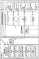
- Figure 3 shows a possible realization of a preferred simple protocol database (could also be regarded as a simple protocol management system 21) in form of a table with attribute columns.
- the table hosts information about the scan indication, the scanner type, the software (SW) version, the protocols in program connected with the scan indication, recommended protocol parameters and parameters that may be adjusted by a user.
- the shown table offers alternatives of adjustable parameters for different types of users ("tier 1 user" and "tier 2 user") having different access rights.
- the dots indicate that there may be more columns and/or lines.
- the column "scan indication” is drawn to certain diseases, e.g., unspecific head pain or suspected lesion as shown in the figure. It should be noted that certain diseases may be present multiple times, e.g., connected with different scanner types (possibly resulting in different protocols to be used).
- the column "scanner type”, e.g., "Prisma” scanner or “Aera” scanner offers the possibility to distinguish between different scanner types.
- the suggested protocol may depend on the used scanner type, even if examining a similar disease.
- SW version e.g., N4VE11B, N4VE11C, N4VE11E offers information about the software version. Depending on the SW version, possibly different protocols or parameter settings have to be used. In the shown case, this column only offers additional information.
- the column "protocols in program” comprises the actual protocols to be used for an examination connected with the scan indication of the certain row.
- 'T1' and 'T2' mean the contrasts to be measured, 'TOF' the time of flight with certain orientations ('tra' or 'sag').
- sequences /acquisition techniques such as 'dark fluid', 'turbo spin echo' (TSE) '2D Diffusion trace', 'FLASH (fl) 2D', 'MPRAGE', 'SWI' or 'SPACE'.
- the next column "recommended protocol parameters” comprises a list of parameters of the protocol of the certain row that may be adjusted or predefined by a user or an automatic routine (also a predefined list).
- the echo time TE and the repetition time TR are listed, but there may also be other parameters in this list.
- the columns “adjustable parameter” comprise a number of parameters, especially selected from the column “recommended protocol parameters”, that may be adjusted by a user depending on the access rights of the user.
- tier 1 users may amend or adjust certain parameters
- tier 2 users are not allowed to change any parameter.
- Figure 4 shows a preferred protocol management system 21 in form of a schematic exhibiting the configuration and customization options offered by the protocol management system 21.
- Example Scan Workflow shows a detailed view of the steps that would be performed in the scan queue 23 (see figure 5 ) for a specific disease.
- the upper part (“Program Composition in Program Editor”) was configured by a user, the lower part (Adaptions from Scan Work-flow Guidance System”) includes the planned execution of data analysis and was generated by the scan workflow guidance system 22.
- the left side of this figure shows toolboxes T available in the scan protocol editor 24 & scan program editor 25.
- One toolbox T comprises the protocols P used by the invention.
- it is a database D and may in a simple case solely form the protocol management system 21.
- the protocol management system 21 has a more complex structure.
- a user or the scan workflow guidance system 22 may find already configured models comprising a protocol P and also an auto planner and an automatic quality assurance module ("Auto QA").
- the additional modules, smart components C are fetched from the "Smart Component Toolbox” (lower left).
- the "Smart Detector Toolbox” comprises finding detectors F that are able to automatically fetch suitable configured models based on findings in images recorded by a protocol P of the scan workflow W. As it can be seen, a user has arranged such finding detectors F in the scan workflow W and they are processed by the scan workflow guidance system 22.
- the shown protocol management system 21 is associated with a suitable scan protocol editor 24 that allows the healthcare provider's central protocol configurators to conveniently edit, configure and manage the large set of scan protocols P.
- the latter could be setup (e.g., similarly to the Siemens Healthineers MR Dot Cockpit) to allow the protocol configurator to compose scan programs from preconfigured modules.
- the configurator may assemble the scan programs Pr choosing from a set of, e.g., suitably preconfigured scan protocols, automated planning features and scan workflow guidance features.
- One preferred variant of the scan protocol editor would allow the customer to compose their scan protocols P from a toolbox T of fundamental building blocks (e.g., sequences, reconstructions, and appropriate parameter sets).
- the healthcare provider may use the scan protocol editor 24 and the scan program editor 25 to preconfigure their own standardized smart modules S comprised of preconfigured scan protocols P, automated planning and quality assurance steps.
- the set of smart modules S as well as more fundamental elements inside the toolboxes T can be employed to assemble the scan program Pr in a subsequent step.
- a scan program standard can be configured from building blocks inside the various toolboxes T on the left side of Figure 4 .
- the healthcare provider can define up to n initial smart modules S (e.g., preconfigured protocols P with auxiliary smart components (e.g., automated slice planning, quality assurance steps) for each scan step) from the respective scan workflow toolboxes on the left side in Figure 4 . This allows the healthcare provider to setup their scan protocol P standards and derived scan programs Pr in a modular fashion for each clinical indication.
- a set of finding detectors F can be added after the n-th initial scan protocol P has finished.
- the finding detectors F will check the acquired image for the appearance of specific features (i.e., findings) and add, replace or remove preconfigured scan steps to/in/from the scan queue 23 based on the type of identified findings.
- the scan protocol editor 24 and the scan program editor 25 allows the healthcare provider to define which protocols P or smart modules S should be added, replaced, or removed to/in/from the scan queue manager 26 (see figure 5 ) in case the finding detectors F make a specific observation. Additional meta-information (e.g., priority of the added scan module S) may be configurable for each element for consideration by the scan queue manager 26.
- the finding detectors F may also take additional input into account (e.g., input from temperature, ECG, pulse, or motion, blood pressure or other vital sign sensors), or other patient information (e.g., lab data, prior images, clinical data) to improve decisions on the workflow W.
- patient information e.g., lab data, prior images, clinical data
- One preferred option of the Scan Protocol & Program manager would include the capability to transfer scan protocols P and scan programs Pr from one scanner configuration to another (e.g., from a 3 T scanner to a 1.5 T scanner).
- Figure 5 shows an example of a system 12 according to an embodiment of the invention, comprising a scan-user-interface 20 ("Scan UI input fields"), a scan queue 23, a protocol management system 21 (see also figure 4 ) and a scan workflow guidance system 22.
- Scan UI input fields a scan-user-interface 20
- scan queue 23 a scan queue 23
- protocol management system 21 a protocol management system 21 (see also figure 4 ) and a scan workflow guidance system 22.
- the scan workflow guidance system 22 uses the preconfigured scan protocols P and scan programs PR provided by the protocol management system 21 and is here designed to consider subsets of several input types, such as:
- the shown scan workflow guidance system 22 exhibits a modular architecture of mutually independent analysis branches 27.
- Each analysis branch 27 comprises an image analysis module 28 (each comprised of, e.g., one or a combination of several deep convolutional neural nets or machine learning based pattern detection algorithms) able to detect one or a specific combination of relevant features from the input MR image and can combine this with other features.
- the image analysis modules 28 are special finding detectors F, that find patient-specific circumstances that require a scan workflow adaptation. It should be noted that finding detectors F in general could operate on physiological data (like ECG data) or on images. If they operate on images, they would be realized by the image analysis modules 28 as shown here. Fully analogously, if the finding detector F is configured to operate on physiological data, it could be designated as a (single- or multi-channel) time-trace analyzer module. This, however, does not mean that all image analysis modules 28 always have to be finding detectors F. They could in general also perform other tasks.
- the set of relevant feature detectors may be suitably defined as a set of different detectors for different classes of, e.g., radiological findings, pathologies, image artifacts, or diseases.
- each analysis branch 27 may (either directly or following further dissemination/characterization of the detected feature) trigger the output, replacement or deletion of one or a set of preconfigured scan protocols P (incl. appropriate parameters) to/in/from the scan queue manager 26.
- P preconfigured scan protocols
- each feature detector is equipped with confidence metric to determine how certain the feature has been detected).
- auxiliary analysis modules might be helpful or required (e.g., image quality detection modules).
- all or only certain user groups may be allowed to manually trigger any or all of the finding detectors F by selecting the respective findings in the scan-user-interface 20 (see e.g., figure 3 ), such that the corresponding scan protocols P will be forwarded to the scan queue manager 26.
- the added scan protocols P may be tagged with priority labels according to the prior assessment of the healthcare provider as setup in the scan protocol management system 21.
- the scan queue manager 26 has the central task to curate the set of scan workflow W adaptation as output from the analysis branches 27. Among these tasks we see:
- a report is created by the scan workflow guidance system 22.
- These reports provide information regarding the initially selected workflow steps, provided suggestions incl. the basis for these suggestions and executed protocol steps incl. parameter settings as well as the executed protocol steps incl. parameter settings, entered comments, and scan times.
- These reports could also include DICOM objects which might need to be appropriately anonymized/de-identified according to the locally applicable data privacy law.
- the bottom left box in figure 2 shows the content of the scan queue 23.
- the dynamic change of the subsequent protocols happens in the box "Wait for SWF proposals" and can (for regulatory reasons) be confirmed by a user (following box).
- the subsequent protocols P provided by the scan workflow guidance system 22 could be executed in an automated fashion.
- a master user can configure the available indication-specific scan programs in the scan protocol editor and scan program editor at configuration time to be available in the protocol management system.
- the scanner user selects the patient's indication in the UI and thus selects the program to be scanned.
- This scan program will either be pulled from the protocol management system or exist on the scanner as a local copy until the next update from the master user. Additional, manual adding of protocols can be optionally available, but will not be essential for this invention.
Applications Claiming Priority (1)
| Application Number | Priority Date | Filing Date | Title |
|---|---|---|---|
| US202063071057P | 2020-08-27 | 2020-08-27 |
Publications (1)
| Publication Number | Publication Date |
|---|---|
| EP3961238A1 true EP3961238A1 (de) | 2022-03-02 |
Family
ID=76920680
Family Applications (1)
| Application Number | Title | Priority Date | Filing Date |
|---|---|---|---|
| EP21185537.4A Pending EP3961238A1 (de) | 2020-08-27 | 2021-07-14 | System und verfahren für standardisierte mrt-untersuchungen mit patientenzentrischen scan-arbeitsablaufanpassungen |
Country Status (3)
| Country | Link |
|---|---|
| US (1) | US20220068472A1 (de) |
| EP (1) | EP3961238A1 (de) |
| CN (1) | CN114121253A (de) |
Cited By (1)
| Publication number | Priority date | Publication date | Assignee | Title |
|---|---|---|---|---|
| CN117635559A (zh) * | 2023-11-28 | 2024-03-01 | 上海交通大学医学院附属瑞金医院 | 定位异常脑白质所连接灰质皮层的图像处理方法和系统 |
Citations (3)
| Publication number | Priority date | Publication date | Assignee | Title |
|---|---|---|---|---|
| EP3546973A1 (de) * | 2018-03-26 | 2019-10-02 | Koninklijke Philips N.V. | Prädiktive modifikation von magnetresonanzbildgebungsprotokollen |
| US20190320934A1 (en) * | 2018-04-18 | 2019-10-24 | Siemens Healthcare Gmbh | Medical image acquisition with sequence prediction using deep learning |
| US20190370958A1 (en) * | 2018-05-31 | 2019-12-05 | General Electric Company | Automated scanning workflow |
Family Cites Families (3)
| Publication number | Priority date | Publication date | Assignee | Title |
|---|---|---|---|---|
| US20120271840A1 (en) * | 2011-04-25 | 2012-10-25 | General Electric Company | Systems and methods for storing and providing scan protocol information |
| US20170156630A1 (en) * | 2015-12-08 | 2017-06-08 | Board Of Regents Of The University Of Texas System | System and method for adaptive and patient-specific magnetic resonance imaging |
| US10074038B2 (en) * | 2016-11-23 | 2018-09-11 | General Electric Company | Deep learning medical systems and methods for image reconstruction and quality evaluation |
-
2021
- 2021-07-14 EP EP21185537.4A patent/EP3961238A1/de active Pending
- 2021-08-27 US US17/458,753 patent/US20220068472A1/en active Pending
- 2021-08-27 CN CN202110996426.7A patent/CN114121253A/zh active Pending
Patent Citations (3)
| Publication number | Priority date | Publication date | Assignee | Title |
|---|---|---|---|---|
| EP3546973A1 (de) * | 2018-03-26 | 2019-10-02 | Koninklijke Philips N.V. | Prädiktive modifikation von magnetresonanzbildgebungsprotokollen |
| US20190320934A1 (en) * | 2018-04-18 | 2019-10-24 | Siemens Healthcare Gmbh | Medical image acquisition with sequence prediction using deep learning |
| US20190370958A1 (en) * | 2018-05-31 | 2019-12-05 | General Electric Company | Automated scanning workflow |
Non-Patent Citations (5)
| Title |
|---|
| ANDREW D BROWN ET AL: "Using machine learning for sequence-level automated MRI protocol selection in neuroradiology", JOURNAL OF THE AMERICAN MEDICAL INFORMATICS ASSOCIATION, vol. 25, no. 5, 27 October 2017 (2017-10-27), AMSTERDAM, NL, pages 568 - 571, XP055600609, ISSN: 1067-5027, DOI: 10.1093/jamia/ocx125 * |
| CARLIJN TENBERGEN ET AL.: "Initial evaluation of an automated acquisition workflow for multiparametric MR (spectroscopic) imaging of the prostate", PROCEEDINGS OF THE INTERNATIONAL SOCIETY FOR MAGNETIC RESONANCE IN MEDICINE, ISMRM, 27TH ANNUAL MEETING AND EXHIBITION, MONTRÉAL, QUÉBEC, CANADA, 11 MAY - 16 MAY 2019, no. 1852, 26 April 2019 (2019-04-26), XP040709238 * |
| HORGER WILHELM ET AL: "The Prostate Dot Engine - a System-Guided and Assisted Workflow to Improve Consistency in Prostate MR Exams", MAGNETOM FLASH, vol. 76, no. 1/2020, 26 March 2020 (2020-03-26), pages 73 - 77, XP055869536 * |
| SCHRAA BART: "Dot Configuration with the Dot Cockpit", MAGNETOM FLASH, vol. 62, no. 2/2015, 28 April 2015 (2015-04-28), pages 38 - 43, XP055869526, Retrieved from the Internet <URL:https://www.magnetomworld.siemens-healthineers.com/publications/magnetom-flash> * |
| WINKEL DAVID J ET AL: "A Fully Automated, End-to-End Prostate MRI Workflow Solution Incorporating Dot, Ultrashort Biparametric Imaging and Deep- Learning-based Detection, Classification, and Reporting", MAGNETOM FLASH, vol. 76, no. 1/2020, 26 March 2020 (2020-03-26), pages 78 - 83, XP055869540 * |
Cited By (2)
| Publication number | Priority date | Publication date | Assignee | Title |
|---|---|---|---|---|
| CN117635559A (zh) * | 2023-11-28 | 2024-03-01 | 上海交通大学医学院附属瑞金医院 | 定位异常脑白质所连接灰质皮层的图像处理方法和系统 |
| CN117635559B (zh) * | 2023-11-28 | 2024-04-30 | 上海交通大学医学院附属瑞金医院 | 定位异常脑白质所连接灰质皮层的图像处理方法和系统 |
Also Published As
| Publication number | Publication date |
|---|---|
| CN114121253A (zh) | 2022-03-01 |
| US20220068472A1 (en) | 2022-03-03 |
Similar Documents
| Publication | Publication Date | Title |
|---|---|---|
| US6674449B1 (en) | Multiple modality interface for imaging systems | |
| US8862201B2 (en) | Multi-modal imaging system and workstation with support for structured hypothesis testing | |
| US8418001B2 (en) | Context-related troubleshooting | |
| US8401259B2 (en) | Image diagnosis support system | |
| CN101271494B (zh) | 利用医学模态的图像数据记录的方法以及有关医学模态 | |
| JP5049283B2 (ja) | 診断のワークフローを管理するシステム、方法、該方法を実行するプログラムを記録した記録媒体 | |
| CN109924978B (zh) | 借助医学图像数据组表征检查对象内的干扰物体 | |
| US7996240B2 (en) | Knowledge-based system for supporting radiological assessment and diagnostics | |
| JP2009011829A (ja) | 電子医療記録影響データ取得、処理および表示システムおよび方法 | |
| US8712714B2 (en) | Measurement protocol for a medical technology apparatus | |
| DE112019003151T5 (de) | Verfahren zum ermöglichen einer magnetresonanzbildgebung einer person | |
| US7365538B2 (en) | Method and apparatus for MR image acquisition wherein operating parameter sets are displayed with an image element indicative of an acquisition result | |
| US20220068472A1 (en) | System and method for standardized mri examinations with patient-centric scan workflow adaptations | |
| US20150086092A1 (en) | Medical diagnostic imaging apparatus, medical image display apparatus, and medical image display method | |
| CN108573487B (zh) | 用于找出医学图像数据中的异常的方法及医学成像装置 | |
| US20230067146A1 (en) | Automated scout scan examination | |
| EP3287915A1 (de) | Erzeugung einer favoritenmenge umfassend mehrere protokolle zur ansteuerung eines medizinischen bildgebungsgerätes | |
| US20070185395A1 (en) | Method for implementation of a medical examination on a patient using a configurable medical examination apparatus | |
| US11830594B2 (en) | Method and apparatus for template based treatment outcome generation | |
| EP4145457A1 (de) | Verfahren und system zur bildbasierten operativen entscheidungsunterstützung | |
| EP4145464A1 (de) | Entscheidungsmodul und verfahren für bildbasierte betriebliche entscheidungsunterstützung | |
| US20220122256A1 (en) | Technique for performing a quality assessment for a medical image | |
| US20220115117A1 (en) | Technique for configuring a medical imaging device | |
| US20170315194A1 (en) | Operating method for a medical imaging apparatus | |
| US10275120B2 (en) | Medical imaging apparatus and method for the operation thereof |
Legal Events
| Date | Code | Title | Description |
|---|---|---|---|
| PUAI | Public reference made under article 153(3) epc to a published international application that has entered the european phase |
Free format text: ORIGINAL CODE: 0009012 |
|
| STAA | Information on the status of an ep patent application or granted ep patent |
Free format text: STATUS: THE APPLICATION HAS BEEN PUBLISHED |
|
| AK | Designated contracting states |
Kind code of ref document: A1 Designated state(s): AL AT BE BG CH CY CZ DE DK EE ES FI FR GB GR HR HU IE IS IT LI LT LU LV MC MK MT NL NO PL PT RO RS SE SI SK SM TR |
|
| STAA | Information on the status of an ep patent application or granted ep patent |
Free format text: STATUS: REQUEST FOR EXAMINATION WAS MADE |
|
| 17P | Request for examination filed |
Effective date: 20220902 |
|
| RBV | Designated contracting states (corrected) |
Designated state(s): AL AT BE BG CH CY CZ DE DK EE ES FI FR GB GR HR HU IE IS IT LI LT LU LV MC MK MT NL NO PL PT RO RS SE SI SK SM TR |
|
| STAA | Information on the status of an ep patent application or granted ep patent |
Free format text: STATUS: EXAMINATION IS IN PROGRESS |
|
| RAP1 | Party data changed (applicant data changed or rights of an application transferred) |
Owner name: SIEMENS HEALTHINEERS AG |
|
| 17Q | First examination report despatched |
Effective date: 20240117 |