EP3961218A1 - Composition and kit for detecting testican-1 for identifying exposure to particulate matter in lung and method using the same - Google Patents
Composition and kit for detecting testican-1 for identifying exposure to particulate matter in lung and method using the same Download PDFInfo
- Publication number
- EP3961218A1 EP3961218A1 EP21163037.1A EP21163037A EP3961218A1 EP 3961218 A1 EP3961218 A1 EP 3961218A1 EP 21163037 A EP21163037 A EP 21163037A EP 3961218 A1 EP3961218 A1 EP 3961218A1
- Authority
- EP
- European Patent Office
- Prior art keywords
- testican
- protein
- particulate matter
- lung
- expression level
- Prior art date
- Legal status (The legal status is an assumption and is not a legal conclusion. Google has not performed a legal analysis and makes no representation as to the accuracy of the status listed.)
- Granted
Links
Images

Classifications
-
- G—PHYSICS
- G01—MEASURING; TESTING
- G01N—INVESTIGATING OR ANALYSING MATERIALS BY DETERMINING THEIR CHEMICAL OR PHYSICAL PROPERTIES
- G01N33/00—Investigating or analysing materials by specific methods not covered by groups G01N1/00 - G01N31/00
- G01N33/48—Biological material, e.g. blood, urine; Haemocytometers
- G01N33/50—Chemical analysis of biological material, e.g. blood, urine; Testing involving biospecific ligand binding methods; Immunological testing
- G01N33/68—Chemical analysis of biological material, e.g. blood, urine; Testing involving biospecific ligand binding methods; Immunological testing involving proteins, peptides or amino acids
- G01N33/6884—Chemical analysis of biological material, e.g. blood, urine; Testing involving biospecific ligand binding methods; Immunological testing involving proteins, peptides or amino acids from lung
-
- C—CHEMISTRY; METALLURGY
- C12—BIOCHEMISTRY; BEER; SPIRITS; WINE; VINEGAR; MICROBIOLOGY; ENZYMOLOGY; MUTATION OR GENETIC ENGINEERING
- C12Q—MEASURING OR TESTING PROCESSES INVOLVING ENZYMES, NUCLEIC ACIDS OR MICROORGANISMS; COMPOSITIONS OR TEST PAPERS THEREFOR; PROCESSES OF PREPARING SUCH COMPOSITIONS; CONDITION-RESPONSIVE CONTROL IN MICROBIOLOGICAL OR ENZYMOLOGICAL PROCESSES
- C12Q1/00—Measuring or testing processes involving enzymes, nucleic acids or microorganisms; Compositions therefor; Processes of preparing such compositions
- C12Q1/68—Measuring or testing processes involving enzymes, nucleic acids or microorganisms; Compositions therefor; Processes of preparing such compositions involving nucleic acids
- C12Q1/6876—Nucleic acid products used in the analysis of nucleic acids, e.g. primers or probes
- C12Q1/6883—Nucleic acid products used in the analysis of nucleic acids, e.g. primers or probes for diseases caused by alterations of genetic material
-
- G—PHYSICS
- G01—MEASURING; TESTING
- G01N—INVESTIGATING OR ANALYSING MATERIALS BY DETERMINING THEIR CHEMICAL OR PHYSICAL PROPERTIES
- G01N33/00—Investigating or analysing materials by specific methods not covered by groups G01N1/00 - G01N31/00
- G01N33/48—Biological material, e.g. blood, urine; Haemocytometers
- G01N33/50—Chemical analysis of biological material, e.g. blood, urine; Testing involving biospecific ligand binding methods; Immunological testing
- G01N33/68—Chemical analysis of biological material, e.g. blood, urine; Testing involving biospecific ligand binding methods; Immunological testing involving proteins, peptides or amino acids
- G01N33/6893—Chemical analysis of biological material, e.g. blood, urine; Testing involving biospecific ligand binding methods; Immunological testing involving proteins, peptides or amino acids related to diseases not provided for elsewhere
-
- C—CHEMISTRY; METALLURGY
- C12—BIOCHEMISTRY; BEER; SPIRITS; WINE; VINEGAR; MICROBIOLOGY; ENZYMOLOGY; MUTATION OR GENETIC ENGINEERING
- C12Q—MEASURING OR TESTING PROCESSES INVOLVING ENZYMES, NUCLEIC ACIDS OR MICROORGANISMS; COMPOSITIONS OR TEST PAPERS THEREFOR; PROCESSES OF PREPARING SUCH COMPOSITIONS; CONDITION-RESPONSIVE CONTROL IN MICROBIOLOGICAL OR ENZYMOLOGICAL PROCESSES
- C12Q2600/00—Oligonucleotides characterized by their use
- C12Q2600/158—Expression markers
-
- G—PHYSICS
- G01—MEASURING; TESTING
- G01N—INVESTIGATING OR ANALYSING MATERIALS BY DETERMINING THEIR CHEMICAL OR PHYSICAL PROPERTIES
- G01N2800/00—Detection or diagnosis of diseases
- G01N2800/12—Pulmonary diseases
-
- G—PHYSICS
- G01—MEASURING; TESTING
- G01N—INVESTIGATING OR ANALYSING MATERIALS BY DETERMINING THEIR CHEMICAL OR PHYSICAL PROPERTIES
- G01N2800/00—Detection or diagnosis of diseases
- G01N2800/40—Disorders due to exposure to physical agents, e.g. heat disorders, motion sickness, radiation injuries, altitude sickness, decompression illness
-
- G—PHYSICS
- G01—MEASURING; TESTING
- G01N—INVESTIGATING OR ANALYSING MATERIALS BY DETERMINING THEIR CHEMICAL OR PHYSICAL PROPERTIES
- G01N2800/00—Detection or diagnosis of diseases
- G01N2800/70—Mechanisms involved in disease identification
Definitions
- the present disclosure relates to a composition or kit for detecting Testican-1 for identifying exposure to particulate matter in lungs and a method using the same.
- Fine dust also called particle matter, is a particulate material that floats in the air and is known to be typically generated from exhaust gases of automobiles, combustion of fossil fuels, road dusts, etc.
- Particle matter is a Group 1 carcinogen that can cause cancer, designated by the International Agency for Research on Cancer (IARC), and fine dust particles having a diameter of less than 10 ⁇ m may be induced into the lung and blood, consequently posing a major threat.
- IARC International Agency for Research on Cancer
- particulate matter increases the incidence of lung cancer by causing coughing or phlegm due to accumulation of dust in the bronchus, and inducing an inflammation reaction due to the dust accumulating in the pulmonary alveolus, thereby generating active oxygen, resulting in necrosis of tissues and degrading lung functions.
- compositions for identifying exposure to particulate matter in a lung are provided.
- kits for identifying exposure to particulate matter in a lung are provided.
- a method for detecting a biomarker to provide information required to identify exposure to particulate matter in a lung.
- An aspect is to provide a composition for identifying exposure to particulate matter in a lung, comprising an agent for measuring an expression level of a Testican-1 gene or an expression level of a Testican-1 protein or a fragment thereof.
- the term "particulate matter” refers to fine dust particulate matter (PM) floating in the air.
- the particulate matter may include ionic components, such as nitrates (NO 3- ), ammonium (NH 4+ ), or sulfate (SO 4 2- ), carbon compounds, metal compounds, polycyclic aromatic hydrocarbons (PAHs), nitro-PAHs, or a combination thereof.
- the particulate matter may be categorized according to its particle size: particulate matter (PM 10 ) with a diameter of not greater than 10 ⁇ m and ultrafine particulate matter (PM 2.5 ) with a diameter of not greater than 2.5 ⁇ m.
- PM 10 may include PM 2.5 .
- the particulate matter may have an average diameter of about 50 ⁇ m or less, about 40 ⁇ m or less, about 30 ⁇ m or less, about 20 ⁇ m or less, about 10 ⁇ m or less, about 9 ⁇ m or less, about 8 ⁇ m or less, about 7 ⁇ m or less, about 6 ⁇ m or less, about 5 ⁇ m or less, about 4 ⁇ m or less, about 3 ⁇ m or less, about 2.5 ⁇ m or less, about 2 ⁇ m or less, about 1 ⁇ m or less, or a combination thereof.
- the "lung” may be a lung tissue or a lung cell.
- the lung cell may be a lung fibroblast.
- the "exposure to particulate matter in a lung” may refer to a case in which particulate matter passes through the respiratory organ of a subject to come into contact with lung tissues or lung cells of the subject.
- Testican-1 may also be called SPOCK or SPOCK1.
- Testican-1 may be a protein encoded by SPOCK1 gene in humans.
- Testican-1 may be proteoglycan including chondroitin sulfate and heparan sulfate.
- Testican-1 may include an amino acid sequence of Uniprot ID NO: Q08629 in humans.
- Testican-1 may include an amino acid sequence of Uniprot ID NO: Q62288 in mice.
- the fragment may refer to a part of Testican-1 protein, and may be immunogenic polypeptide.
- the Testican-1 gene refers to a nucleic acid encoding Testican-1 protein.
- the Testican-1 gene may be a SPOCK1 gene.
- the expression level of Testican-1 gene may be an expression level of mRNA encoding Testican-1 protein.
- the expression level of mRNA may be a relative or absolute amount of mRNA.
- the measuring of the expression level of Testican-1 gene may be measuring an amount of mRNA.
- the expression level of Testican-1 protein or a fragment thereof may be a relative or absolute amount of Testican-1 protein or the fragment thereof.
- the measuring of the expression level of the Testican-1 protein or the fragment thereof may be measuring an amount of Testican-1 protein or the fragment thereof.
- the agent may be an antibody or an antigen-binding fragment, or aptamer, specifically binding to Testican-1 protein or the fragment thereof.
- the antibody may be a polyclonal antibody or a monoclonal antibody.
- the term "antibody” may be used interchangeably with the term "immunoglobulin".
- the antibody may be a polyclonal antibody or a monoclonal antibody.
- the antibody may be a full-length antibody.
- the antigen-binding fragment refers to a polypeptide including an antigen-binding site.
- the antigen-binding fragment may be a single-domain antibody, Fab, Fab', or scFv.
- the antibody or the antigen-binding fragment may be adhered to a solid support material.
- the solid support material may be, for example, a surface of a metal chip, plate, or well.
- the antibody or the antigen-binding fragment may be an anti-Testican-1 antibody or an antigen-binding fragment.
- the aptamer may be a single stranded nucleic acid (DNA, RNA or modified nucleic acid) or peptide specifically binding to Testican-1 protein or a fragment thereof.
- the agent may be a nucleic acid including a polynucleotide identical to or complementary to a polynucleotide encoding Testican-1 protein or a fragment thereof.
- the nucleic acid may be a primer, a probe, or an anti-sense oligonucleotide.
- the primer, the probe, or the anti-sense oligonucleotide may be labeled with a fluorescent substance, a chemiluminescent substance, or a radioactive isotope, at an end or inside thereof.
- the composition may be a composition for in vitro measuring an expression level of Testican-1 gene or an expression level of Testican-1 protein or a fragment thereof in a lung tissue or a lung cell.
- Another aspect is to provide a kit for identifying exposure to particulate matter in a lung, comprising an agent for measuring an expression level of a Testican-1 gene or an expression level of a Testican-1 protein or a fragment thereof.
- Testican-1 Testican-1 gene, fragment, expression level, lung, and exposure to particulate matter, are the same as described above.
- the kit may further include a sample necessary for identifying exposure to particulate matter.
- the kit may include a substrate, a suitable buffer solution, a chromogenic enzyme, a secondary antibody labeled with a fluorescent substance, or a chromogenic substrate.
- the kit may include a polymerase, a buffering agent, a nucleic acid, a coenzyme, a fluorescent substance, or a combination thereof.
- the polymerase may be, for example, a Taq polymerase.
- the kit may be a kit for in vitro measuring an expression level of Testican-1 gene or an expression level of Testican-1 protein or a fragment thereof in a lung tissue or a lung cell.
- Another aspect is to provide a method for detecting Testican-1 for providing information required in identifying exposure to particulate matter in a lung, the method comprising: measuring an expression level of Testican-1 gene or an expression level of Testican-1 protein or a fragment thereof in a biological sample isolated from a subject with suspected exposure to particulate matter; comparing the measured expression level with that in a normal control group; and identifying whether the Testican-1 expression level has increased as compared with that in the normal control group.
- Testican-1 Testican-1 gene, fragment, expression level, lung, and exposure to particulate matter, are the same as described above.
- the method comprises measuring an expression level of Testican-1 gene or an expression level of Testican-1 protein or a fragment thereof in a biological sample isolated from a subject with suspected exposure to particulate matter.
- the method may be performed in vitro.
- the subject may be a mammal, for example, such as a human being, a cow, a horse, a pig, a dog, a sheep, a goat, or a cat.
- the subject may be a subject suspected to be exposed or have been exposed to particulate matter.
- the biological sample refers to a sample obtained from the subject.
- the biological sample may include a lung tissue, a lung cell, blood, plasma, serum, bone marrow fluid, lymph fluid, saliva, tear fluid, mucosal fluid, amniotic fluid, or a combination thereof.
- the lung cell may be a lung fibroblast.
- the biological sample include a protein sample or a nucleic acid obtained from the sample.
- the measuring may include incubating the biological sample with an antibody or an antigen-binding fragment thereof, or an aptamer that specifically binds to Testican-1 protein or a fragment thereof.
- the measuring may be performed by electrophoresis, immunoblotting, enzyme-linked immunosorbent assay (ELISA), protein chip, immunoprecipitation, microarray, electron microscopy, or a combination thereof.
- the electrophoresis may be SDS-PAGE, isoelectric point electrophoresis, 2D electrophoresis, or a combination thereof.
- the measuring may include incubating the biological sample with a polynucleotide identical to or complementary to a polynucleotide encoding Testican-1 protein or a fragment thereof.
- the measuring may be performed by northern blotting or polymerase chain reaction (PCR).
- the PCR may be real time PCR or reverse transcription PCR.
- the method may include comparing the measured expression level with that in a normal control group.
- the method may include identifying whether the Testican-1 expression level has increased as compared with that in the normal control group.
- the normal control group refers to a group without exposure to particulate matter, and is referred to as a negative control group.
- the amount of the measured expression level of Testican-1 from sample exposed to particulate matter may be increased compared to the expression level of Testican-1 derived from the sample of the normal control group. For example, when the amount of detected Testican-1 protein is greater than that in the normal control group, the subject may be diagnosed as having a high probability of being exposed or being exposed to particulate matter.
- the subject identified or diagnosed with exposure to particulate matter may be determined to suffer from or have a high probability of a respiratory disease, a vascular disease, a brain disease, a cancer, or a combination thereof.
- the respiratory disease may be cold, nasal inflammation, asthma, sinusitis, or pneumonia.
- the vascular disease may be arteriosclerosis, arrhythmia, angina, myocardial infarction, heart failure, heart attack, or a combination thereof.
- the brain disease may be stroke, ischemic stroke, cognitive disorder, dementia, depression, or a combination thereof.
- the cancer may be a lung cancer or a bladder cancer.
- Example 1 Identification of differentially expressed proteins depending on ceoncentrations of particulate matter exposed to lung cells
- CCD-8Lu (ATCC) as a lung normal fibroblast cell line of a human lung was culturedin the presence of a medium with 10% (v/v) fetal bovine serum (Gibco ® , Thermo Fisher Scientific Inc.) and 1% (w/v) antibiotic-antimycotic (Gibco ® , Thermo Fisher Scientific Inc.) added to an Eagle's Minimum Essential Medium (EMEM, BD Bioscience) in a 37°C, 5% CO 2 incubator.
- EMEM Eagle's Minimum Essential Medium
- a particulate matter powder (SRM2975, National Institute of Standards & Technology) was dissolved in a phosphate buffered saline (PBS) to prepare 8 mg/mL of a particulate matter solution.
- PBS phosphate buffered saline
- the prepared particulate matter solution was diluted with an EMEM medium to prepare concentrations of 0 ⁇ g/mL, 100 ⁇ g/mL, 200 ⁇ g/mL, and 400 ⁇ g/mL.
- the prepared particulate matter solutions were added to CCD-8Lu cell line and incubated at 37 °C for 24 hours or 48 hours.
- Proteins that are differentially expressed in CCD-8Lu cells treated with particulate matter were analyzed.
- lysis buffer 50 mM Tris-HCI (pH 7.5), 7 M urea, 2 M thiourea, 1 mM EDTA, 150 mM NaCl, a protease inhibitor cocktail, and phosphatase inhibitor cocktail (Roche) were added to CCD-8Lu cells treated with particulate matter in the same manner as in Example 1.2, and then the CCD-8Lu cells harvested using a scraper.
- the harvested cells were transferred to a 1.5 mL Lo-bind tube and lysed using ultrasonic processor (Sonics & Materials, Inc.) to extract proteins from the CCD-8Lu cells.
- the extracted proteins were quantified using a Bradford assay kit.
- Urea was added to the extracted proteins so as to have a final urea concentration of 8 M, and 0.5 ⁇ mol of TCEP (tris(2-carboxyethyl)phosphine) was added thereto and incubated for 1 hour at room temperature to reduce disulfide bonds. Afterward, 1 ⁇ mol of IAA (2-lodoacetamide) was added and then incubated at room temperature under a dark condition for about 1 hour for protein alkylation. 25 mM of an ammonium bicarbonate solution was added to the protein reaction product to reduce the concentration of urea in the protein samples to 2 M or less.
- TCEP tris(2-carboxyethyl)phosphine
- the proteins were incubated at 30 °C for 2 hours in the presence of 3 mAU of endoproteinase LysC and then incubated at 37 °C overnight in the presence of trypsin.
- the obtained peptide sample was purified using a SepPak ® tC18 cartridge, followed by vacuum drying. The dried peptide sample was suspended in 100 mM of tetraethylammonium bromide (TEAB), and then the peptide was quantified.
- TEAB tetraethylammonium bromide
- TMT labeling reagents (Thermo Fisher Scientific) were dissolved in 41 ⁇ l of acetonitrile and then added to 100 ⁇ g of a peptide sample prepared in the same manner as in Example 1.3.
- a negative control group a peptide sample derived from CCD-8Lu cells incubated for 24 hours or 48 hours without treatment with particulate matter was used.
- 8 ⁇ l of 5% (w/v) hydroxylamine was added and incubated at 20 °C for 1 hour to stop the labeling process.
- the TMT-labeled peptide samples were combined using a single tube, and then purified using a SepPak ® tC18 cartridge, followed by vacuum drying.
- the dried peptide sample was suspended in 10 mM of ammonium formate (pH 10), injected into a C18 column (250 mm x 4.6 mm, 3.5 ⁇ m, 130 ⁇ ; Waters) and fractionated on an Agilent 1290 ultrahigh performance liquid chromatography (UHPLC) system at a flow rate of 0.5 mL/min.. 96 fractions were combined to give 24 fractions, and the fractions were then dried ( FIG. 1 ).
- UHPLC ultrahigh performance liquid chromatography
- Example 1.4 Each 5 ⁇ l of the samples obtained in the same manner as Example 1.4 was injected into a reverse-phase PepMap RSLC C18 column (50 cm X 75 ⁇ l) on Ultimate 3000 system.
- the column was equilibrated with 100% buffer A (100% water containing 0.1% (v/v) formic acid). 80% acetonitrile containing 0.1% (v/v) formic acid was used as a buffer B.
- the flow rate was 300 nL/min under the following gradient conditions:
- a nano high-performance liquid chromatography (HPLC) system was connected to Orbitrap Fusion Lumos Tribrid mass spectrometer.
- Survey full-scan MS spectra (400 to 2,000 m/z) were obtained with 1 microscan and 120,000 resolution, precursors were screened, and a preview mode for measuring a charge state was set.
- Preview survey scanning MS/MS spectra for 20 most intense ions were obtained from full-scanning ion traps with the following options: isolation width of 0.7 m/z; normalized collision energy of 35%; and a dynamic exclusion duration of 50 sec.
- the respective LC-MS/MS files were analyzed using the SEQUEST algorithm within Proteome Discoverer 2.4.
- MS and MS/MS data were searched by comparison with SwissProt human database. The searches were limited such that two missed cleavages were allowed for tryptic peptides. The searches were performed with fixed modification on carboamidomethylation of cysteines (+57.021 Da) and TMT labeling of lysine and peptide N-terminal (+229.163 Da), and variable modification on oxidation of methionine (+15.995 Da).
- Mass tolerances for MS/MS data and MS data were set to 0.6 Da and 10 ppm, respectively. For decoy database searches, a total of two target values, that is, 0.01 as a strict false discovery rate (FDR) and 0.05 as an eased FDR, were employed.
- FDR strict false discovery rate
- Relative amounts of proteins existing in cells treated with particulate matter in concentrations of 0, 100, 200, or 400 ⁇ g/mL for 24 hours or 48 hours were calculated based on the signal to noise values of reporter ions from the labeled peptides.
- the signal to noise values of reporter ions of the respective peptides were obtained using Proteome Discoverer 2.4.
- Perseus software was used. Even when no protein exists or proteins detected, if any, fall short of a detection limit, for compensating for null values and calculating a difference in the fold change among the respective samples, missing values were complemented by downshifting 0.3 and 1.8 in width from a normal distribution.
- Testican-1 As shown in Table 1, it was confirmed that the expression levels of Testican-1 in statistically significant proteins were increased as the particulate matter concentration increased.
- a graph showing signal to noise values of Testican-1 according to particulate matter concentrations is shown in FIG. 2 .
- Testican-1 was identified as a biomarker for identifying exposure to particulate matter in a lung.
- composition or kit for detecting Testican-1 for identifying exposure to particulate matter in a lung and the method for detecting Testican-1 using the same may be used in identifying the exposure to particulate matter in a lung in an objective and easy manner with high accuracy and high specificity.
Landscapes
- Life Sciences & Earth Sciences (AREA)
- Health & Medical Sciences (AREA)
- Chemical & Material Sciences (AREA)
- Engineering & Computer Science (AREA)
- Immunology (AREA)
- Molecular Biology (AREA)
- Proteomics, Peptides & Aminoacids (AREA)
- Biomedical Technology (AREA)
- Hematology (AREA)
- Urology & Nephrology (AREA)
- Analytical Chemistry (AREA)
- Biotechnology (AREA)
- Microbiology (AREA)
- Biochemistry (AREA)
- Physics & Mathematics (AREA)
- Pathology (AREA)
- General Health & Medical Sciences (AREA)
- Organic Chemistry (AREA)
- Cell Biology (AREA)
- Food Science & Technology (AREA)
- Medicinal Chemistry (AREA)
- General Physics & Mathematics (AREA)
- Zoology (AREA)
- Wood Science & Technology (AREA)
- Genetics & Genomics (AREA)
- General Engineering & Computer Science (AREA)
- Bioinformatics & Cheminformatics (AREA)
- Biophysics (AREA)
- Measuring Or Testing Involving Enzymes Or Micro-Organisms (AREA)
- Peptides Or Proteins (AREA)
- Investigating Or Analysing Biological Materials (AREA)
- Medicines That Contain Protein Lipid Enzymes And Other Medicines (AREA)
Abstract
Description
- This application is based on and claims priority under 35 U.S.C. §119 to Korean Patent Application No.
10-2020-0108049, filed on August 26, 2020 - The present disclosure relates to a composition or kit for detecting Testican-1 for identifying exposure to particulate matter in lungs and a method using the same.
- Fine dust, also called particle matter, is a particulate material that floats in the air and is known to be typically generated from exhaust gases of automobiles, combustion of fossil fuels, road dusts, etc. Particle matter is a
Group 1 carcinogen that can cause cancer, designated by the International Agency for Research on Cancer (IARC), and fine dust particles having a diameter of less than 10 µm may be induced into the lung and blood, consequently posing a major threat. It is known that the particulate matter increases the incidence of lung cancer by causing coughing or phlegm due to accumulation of dust in the bronchus, and inducing an inflammation reaction due to the dust accumulating in the pulmonary alveolus, thereby generating active oxygen, resulting in necrosis of tissues and degrading lung functions. - Therefore, since the particulate matter causes an inflammatory disease or leads to a potential risk of lung cancer in a serious case, there are constant demands for discovery of a biomarker capable of predicting a change in the lung due to the particulate matter and quickly responding to a change in risk factors.
-
- (Patent Document 1)
KR10-2018-0002324 A - (Patent Document 2)
KR10-2019-0127350 A - Provided is a composition for identifying exposure to particulate matter in a lung.
- Provided is a kit for identifying exposure to particulate matter in a lung.
- Provided is a method for detecting a biomarker to provide information required to identify exposure to particulate matter in a lung.
- Additional aspects will be set forth in part in the description which follows and, in part, will be apparent from the description, or may be learned by practice of the presented embodiments of the disclosure.
- An aspect is to provide a composition for identifying exposure to particulate matter in a lung, comprising an agent for measuring an expression level of a Testican-1 gene or an expression level of a Testican-1 protein or a fragment thereof.
- The term "particulate matter" refers to fine dust particulate matter (PM) floating in the air. The particulate matter may include ionic components, such as nitrates (NO3-), ammonium (NH4+), or sulfate (SO4 2-), carbon compounds, metal compounds, polycyclic aromatic hydrocarbons (PAHs), nitro-PAHs, or a combination thereof. The particulate matter may be categorized according to its particle size: particulate matter (PM10) with a diameter of not greater than 10 µm and ultrafine particulate matter (PM2.5) with a diameter of not greater than 2.5 µm. PM10 may include PM2.5. The particulate matter may have an average diameter of about 50 µm or less, about 40 µm or less, about 30 µm or less, about 20 µm or less, about 10 µm or less, about 9 µm or less, about 8 µm or less, about 7 µm or less, about 6 µm or less, about 5 µm or less, about 4 µm or less, about 3 µm or less, about 2.5 µm or less, about 2 µm or less, about 1 µm or less, or a combination thereof.
- The "lung" may be a lung tissue or a lung cell. The lung cell may be a lung fibroblast.
- The "exposure to particulate matter in a lung" may refer to a case in which particulate matter passes through the respiratory organ of a subject to come into contact with lung tissues or lung cells of the subject.
- Testican-1 may also be called SPOCK or SPOCK1. Testican-1 may be a protein encoded by SPOCK1 gene in humans. Testican-1 may be proteoglycan including chondroitin sulfate and heparan sulfate. Testican-1 may include an amino acid sequence of Uniprot ID NO: Q08629 in humans. Testican-1 may include an amino acid sequence of Uniprot ID NO: Q62288 in mice.
- The fragment may refer to a part of Testican-1 protein, and may be immunogenic polypeptide.
- The Testican-1 gene refers to a nucleic acid encoding Testican-1 protein. The Testican-1 gene may be a SPOCK1 gene.
- The expression level of Testican-1 gene may be an expression level of mRNA encoding Testican-1 protein. The expression level of mRNA may be a relative or absolute amount of mRNA. The measuring of the expression level of Testican-1 gene may be measuring an amount of mRNA.
- The expression level of Testican-1 protein or a fragment thereof may be a relative or absolute amount of Testican-1 protein or the fragment thereof. The measuring of the expression level of the Testican-1 protein or the fragment thereof may be measuring an amount of Testican-1 protein or the fragment thereof.
- The agent may be an antibody or an antigen-binding fragment, or aptamer, specifically binding to Testican-1 protein or the fragment thereof. The antibody may be a polyclonal antibody or a monoclonal antibody. The term "antibody" may be used interchangeably with the term "immunoglobulin". The antibody may be a polyclonal antibody or a monoclonal antibody. The antibody may be a full-length antibody. The antigen-binding fragment refers to a polypeptide including an antigen-binding site. The antigen-binding fragment may be a single-domain antibody, Fab, Fab', or scFv. The antibody or the antigen-binding fragment may be adhered to a solid support material. The solid support material may be, for example, a surface of a metal chip, plate, or well. The antibody or the antigen-binding fragment may be an anti-Testican-1 antibody or an antigen-binding fragment. The aptamer may be a single stranded nucleic acid (DNA, RNA or modified nucleic acid) or peptide specifically binding to Testican-1 protein or a fragment thereof.
- The agent may be a nucleic acid including a polynucleotide identical to or complementary to a polynucleotide encoding Testican-1 protein or a fragment thereof. The nucleic acid may be a primer, a probe, or an anti-sense oligonucleotide. The primer, the probe, or the anti-sense oligonucleotide may be labeled with a fluorescent substance, a chemiluminescent substance, or a radioactive isotope, at an end or inside thereof.
- The composition may be a composition for in vitro measuring an expression level of Testican-1 gene or an expression level of Testican-1 protein or a fragment thereof in a lung tissue or a lung cell.
- Another aspect is to provide a kit for identifying exposure to particulate matter in a lung, comprising an agent for measuring an expression level of a Testican-1 gene or an expression level of a Testican-1 protein or a fragment thereof.
- The Testican-1, Testican-1 gene, fragment, expression level, lung, and exposure to particulate matter, are the same as described above.
- The kit may further include a sample necessary for identifying exposure to particulate matter. For immunological detection of the antibody or the antigen-binding fragment thereof, the kit may include a substrate, a suitable buffer solution, a chromogenic enzyme, a secondary antibody labeled with a fluorescent substance, or a chromogenic substrate. For detection of nucleic acids, the kit may include a polymerase, a buffering agent, a nucleic acid, a coenzyme, a fluorescent substance, or a combination thereof. The polymerase may be, for example, a Taq polymerase.
- The kit may be a kit for in vitro measuring an expression level of Testican-1 gene or an expression level of Testican-1 protein or a fragment thereof in a lung tissue or a lung cell.
- Another aspect is to provide a method for detecting Testican-1 for providing information required in identifying exposure to particulate matter in a lung, the method comprising: measuring an expression level of Testican-1 gene or an expression level of Testican-1 protein or a fragment thereof in a biological sample isolated from a subject with suspected exposure to particulate matter; comparing the measured expression level with that in a normal control group; and identifying whether the Testican-1 expression level has increased as compared with that in the normal control group.
- The Testican-1, Testican-1 gene, fragment, expression level, lung, and exposure to particulate matter, are the same as described above.
- The method comprises measuring an expression level of Testican-1 gene or an expression level of Testican-1 protein or a fragment thereof in a biological sample isolated from a subject with suspected exposure to particulate matter.
- The method may be performed in vitro.
- The subject may be a mammal, for example, such as a human being, a cow, a horse, a pig, a dog, a sheep, a goat, or a cat. The subject may be a subject suspected to be exposed or have been exposed to particulate matter.
- The biological sample refers to a sample obtained from the subject. The biological sample may include a lung tissue, a lung cell, blood, plasma, serum, bone marrow fluid, lymph fluid, saliva, tear fluid, mucosal fluid, amniotic fluid, or a combination thereof. The lung cell may be a lung fibroblast. The biological sample include a protein sample or a nucleic acid obtained from the sample.
- The measuring may include incubating the biological sample with an antibody or an antigen-binding fragment thereof, or an aptamer that specifically binds to Testican-1 protein or a fragment thereof. The measuring may be performed by electrophoresis, immunoblotting, enzyme-linked immunosorbent assay (ELISA), protein chip, immunoprecipitation, microarray, electron microscopy, or a combination thereof. The electrophoresis may be SDS-PAGE, isoelectric point electrophoresis, 2D electrophoresis, or a combination thereof.
- The measuring may include incubating the biological sample with a polynucleotide identical to or complementary to a polynucleotide encoding Testican-1 protein or a fragment thereof. The measuring may be performed by northern blotting or polymerase chain reaction (PCR). The PCR may be real time PCR or reverse transcription PCR.
- The method may include comparing the measured expression level with that in a normal control group. The method may include identifying whether the Testican-1 expression level has increased as compared with that in the normal control group. The normal control group refers to a group without exposure to particulate matter, and is referred to as a negative control group. In the method, the amount of the measured expression level of Testican-1 from sample exposed to particulate matter may be increased compared to the expression level of Testican-1 derived from the sample of the normal control group. For example, when the amount of detected Testican-1 protein is greater than that in the normal control group, the subject may be diagnosed as having a high probability of being exposed or being exposed to particulate matter. The subject identified or diagnosed with exposure to particulate matter may be determined to suffer from or have a high probability of a respiratory disease, a vascular disease, a brain disease, a cancer, or a combination thereof. The respiratory disease may be cold, nasal inflammation, asthma, sinusitis, or pneumonia. The vascular disease may be arteriosclerosis, arrhythmia, angina, myocardial infarction, heart failure, heart attack, or a combination thereof. The brain disease may be stroke, ischemic stroke, cognitive disorder, dementia, depression, or a combination thereof. The cancer may be a lung cancer or a bladder cancer.
- The above and other aspects, features, and advantages of certain embodiments of the disclosure will be more apparent from the following description taken in conjunction with the accompanying drawings, in which:
-
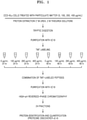
FIG. 1 is a workflow of an experimental method for analyzing a protein biomarker; and -
FIG. 2 is a graph showing intensities of Testican-1 depending on concentrations of particulate matter exposed to lung cells. - Reference will now be made in detail to embodiments, examples of which are illustrated in the accompanying drawings, wherein like reference numerals refer to like elements throughout. In this regard, the present embodiments may have different forms and should not be construed as being limited to the descriptions set forth herein. Accordingly, the embodiments are merely described below, by referring to the figures, to explain aspects of the present description. As used herein, the term "and/or" includes any and all combinations of one or more of the associated listed items. Expressions such as "at least one of," when preceding a list of elements, modify the entire list of elements and do not modify the individual elements of the list.
- Hereinafter, the present disclosure will be described in greater detail through examples. However, the following examples are provided for illustrating one or more specific examples, and the present disclosure is not limited thereto.
- Example 1. Identification of differentially expressed proteins depending on ceoncentrations of particulate matter exposed to lung cells
- CCD-8Lu (ATCC) as a lung normal fibroblast cell line of a human lung was culturedin the presence of a medium with 10% (v/v) fetal bovine serum (Gibco®, Thermo Fisher Scientific Inc.) and 1% (w/v) antibiotic-antimycotic (Gibco®, Thermo Fisher Scientific Inc.) added to an Eagle's Minimum Essential Medium (EMEM, BD Bioscience) in a 37°C, 5% CO2 incubator.
- A particulate matter powder (SRM2975, National Institute of Standards & Technology) was dissolved in a phosphate buffered saline (PBS) to prepare 8 mg/mL of a particulate matter solution. The prepared particulate matter solution was diluted with an EMEM medium to prepare concentrations of 0 µg/mL, 100 µg/mL, 200 µg/mL, and 400 µg/mL. The prepared particulate matter solutions were added to CCD-8Lu cell line and incubated at 37 °C for 24 hours or 48 hours.
- Proteins that are differentially expressed in CCD-8Lu cells treated with particulate matter were analyzed.
- Specifically, lysis buffer (50 mM Tris-HCI (pH 7.5), 7 M urea, 2 M thiourea, 1 mM EDTA, 150 mM NaCl, a protease inhibitor cocktail, and phosphatase inhibitor cocktail (Roche) were added to CCD-8Lu cells treated with particulate matter in the same manner as in Example 1.2, and then the CCD-8Lu cells harvested using a scraper. The harvested cells were transferred to a 1.5 mL Lo-bind tube and lysed using ultrasonic processor (Sonics & Materials, Inc.) to extract proteins from the CCD-8Lu cells. The extracted proteins were quantified using a Bradford assay kit.
- Urea was added to the extracted proteins so as to have a final urea concentration of 8 M, and 0.5 µmol of TCEP (tris(2-carboxyethyl)phosphine) was added thereto and incubated for 1 hour at room temperature to reduce disulfide bonds. Afterward, 1 µmol of IAA (2-lodoacetamide) was added and then incubated at room temperature under a dark condition for about 1 hour for protein alkylation. 25 mM of an ammonium bicarbonate solution was added to the protein reaction product to reduce the concentration of urea in the protein samples to 2 M or less. The proteins were incubated at 30 °C for 2 hours in the presence of 3 mAU of endoproteinase LysC and then incubated at 37 °C overnight in the presence of trypsin. The obtained peptide sample was purified using a SepPak® tC18 cartridge, followed by vacuum drying. The dried peptide sample was suspended in 100 mM of tetraethylammonium bromide (TEAB), and then the peptide was quantified.
- TMT labeling reagents (Thermo Fisher Scientific) were dissolved in 41 µl of acetonitrile and then added to 100 µg of a peptide sample prepared in the same manner as in Example 1.3. As a negative control group, a peptide sample derived from CCD-8Lu cells incubated for 24 hours or 48 hours without treatment with particulate matter was used. After the labeling reaction was made by incubation at 20 °C for 1 hour, 8 µl of 5% (w/v) hydroxylamine was added and incubated at 20 °C for 1 hour to stop the labeling process. The TMT-labeled peptide samples were combined using a single tube, and then purified using a SepPak® tC18 cartridge, followed by vacuum drying.
- The dried peptide sample was suspended in 10 mM of ammonium formate (pH 10), injected into a C18 column (250 mm x 4.6 mm, 3.5 µm, 130 Å; Waters) and fractionated on an Agilent 1290 ultrahigh performance liquid chromatography (UHPLC) system at a flow rate of 0.5 mL/min.. 96 fractions were combined to give 24 fractions, and the fractions were then dried (
FIG. 1 ). - Each 5 µl of the samples obtained in the same manner as Example 1.4 was injected into a reverse-phase PepMap RSLC C18 column (50 cm X 75 µl) on Ultimate 3000 system. The column was equilibrated with 100% buffer A (100% water containing 0.1% (v/v) formic acid). 80% acetonitrile containing 0.1% (v/v) formic acid was used as a buffer B. The flow rate was 300 nL/min under the following gradient conditions:
- 0 minute: 93% buffer A and 7% buffer B,
- 0 minute to 15 minutes: 7% buffer B,
- 15 minutes to 95 minutes: 7% to 40% buffer B,
- 95 minutes to 95.1 minutes: 40% to 95% buffer B,
- 95.1 minutes to 105 minutes: 95% buffer B, and
- 105 minutes to 105.1 minutes: 7% buffer B.
- A nano high-performance liquid chromatography (HPLC) system was connected to Orbitrap Fusion Lumos Tribrid mass spectrometer. Survey full-scan MS spectra (400 to 2,000 m/z) were obtained with 1 microscan and 120,000 resolution, precursors were screened, and a preview mode for measuring a charge state was set. Through preview survey scanning, MS/MS spectra for 20 most intense ions were obtained from full-scanning ion traps with the following options: isolation width of 0.7 m/z; normalized collision energy of 35%; and a dynamic exclusion duration of 50 sec.
- The respective LC-MS/MS files were analyzed using the SEQUEST algorithm within Proteome Discoverer 2.4. MS and MS/MS data were searched by comparison with SwissProt human database. The searches were limited such that two missed cleavages were allowed for tryptic peptides. The searches were performed with fixed modification on carboamidomethylation of cysteines (+57.021 Da) and TMT labeling of lysine and peptide N-terminal (+229.163 Da), and variable modification on oxidation of methionine (+15.995 Da). Mass tolerances for MS/MS data and MS data were set to 0.6 Da and 10 ppm, respectively. For decoy database searches, a total of two target values, that is, 0.01 as a strict false discovery rate (FDR) and 0.05 as an eased FDR, were employed.
- Relative amounts of proteins existing in cells treated with particulate matter in concentrations of 0, 100, 200, or 400 µg/mL for 24 hours or 48 hours were calculated based on the signal to noise values of reporter ions from the labeled peptides. The signal to noise values of reporter ions of the respective peptides were obtained using Proteome Discoverer 2.4. To obtain statistically significant proteins based on the signal to noise values for the respective proteins, Perseus software was used. Even when no protein exists or proteins detected, if any, fall short of a detection limit, for compensating for null values and calculating a difference in the fold change among the respective samples, missing values were complemented by downshifting 0.3 and 1.8 in width from a normal distribution.
- With regard to statistical significance, ANOVA test was performed on signal to noise values obtained three technical replicates of LC-MS/MS runs, FDR-adjusted p-values were obtained using a Benjamini-Hochberg correction, and, when the FDR-adjusted p-values were less than 0.05, the protein samples were considered to be statistically significant.
- From the results of protein identification performed in the same manner as described in Example 1.5, it was confirmed that a total of 7710 proteins were identified in all of the cell samples exposed to particulate matter according to particulate matter concentrations. Out of the identified proteins, 7368 proteins had signal to noise values suitable for quantitative analysis. When ANOVA statistical analyses were performed on the signal-to-noise based abundance values in the 7368 proteins, 2244 proteins having an FDR-adjusted p-value of less than 0.05 were obtained.,Some of the statistically significant proteins are listed in Table 1.
[Table 1] Un iProt Acce ssion No. Protein Name Exposure to particulate matter FDR p - val ue 24 hr 48 hr 0 µg/mL 1000 µg/mL 2000 µg/mL 4000 µg/mL 0 µg/mL 1000 µg/mL 2000 µg/mL 4000 µg/mL Q8WW Q8 Stabilin-2 7 21 46 109 7 23 83 213 5.22E-05 Q08629 Testican-1 112 116 198 340 103 229 341 826 4.03E-07 Q09666 Neuroblast differentiation-associated protein AHNAK 11208 9 115672 122006 119705 133488 144858 150985 157927 1.70E-15 Q15149 Plectin 63476 64854 65196 64610 66936 69208 67137 63865 0.0340 59 P35579 Myosin-9 61945 63975 65138 62931 58779 66231 61989 58232 1.32E-06 P08670 Vimentin 56391 62550 57955 55690 61048 82766 73385 74149 8.85E-08 P60709 Actin, cytoplasmic 1 39766 47691 41121 44084 41142 50932 49527 48686 1.79E-08 Q14315 Filamin-C 28862 29194 30716 31459 29113 33707 29644 30791 9.25E-07 Q14204 Cytoplasmic dynein 1 heavy chain 1 26535 26590 27018 27372 25166 22854 27094 27062 0.0019 77 P21333 Filamin-A 25427 27758 29355 30611 24413 30102 25990 27106 2.48E-07 P04406 Glyceraldehyd e-3-phosphate dehydrogenas e 24353 26823 26113 27199 24615 32183 31998 34510 2.16E-10 Q9Y490 Talin-1 23530 24268 24043 24079 23192 27793 21877 21322 4.92E-06 P13639 Elongation factor 2 25148 24349 23505 24076 21920 19094 18753 17693 0.0001 41 P06733 Alpha-enolase 22380 23647 23305 23541 24024 37593 29857 30035 1.51 E-06 P26038 Moesin 21886 20957 19682 20197 18435 15620 16390 15171 3.12E-10 P02545 Prelamin-A/C 22557 24106 24575 24226 23734 26410 20369 21045 0.0012 04 P08238 Heat shock protein HSP 90-beta 19977 19718 19906 20660 19108 14906 17150 15910 3.65E-07 P07437 Tubulin beta chain 20018 21430 20363 21264 22690 29412 27800 28033 2.77E-11 P11142 Heat shock cognate 71 kDa protein 20948 21495 21202 21118 20477 31265 21539 22983 1.06E-05 Q13813 Spectrin alpha chain, non-erythrocytic 1 20061 21026 23270 23891 21845 25026 27840 30462 1.11E-09 P07237 Protein disulfide-isomerase 19343 18991 20769 21139 19598 27933 22612 21707 0.0343 21 P11021 Endoplasmic reticulum chaperone BiP 19662 19988 20707 20768 19174 28544 22890 23872 3.80E-06 P14618 Pyruvate kinase PKM 18739 19027 18155 18288 18577 21206 18964 17754 0.0160 37 P08133 Annexin A6 18733 19005 19483 20361 16942 17602 17409 16944 0.0023 95 Q9UPN 3 Microtubule-actin crosslinking factor 1, isoforms 1/2/3/5 17511 19881 20242 20472 17834 19485 21080 21811 8.18E-05 P68104 Elongation factor 1 -alpha 1 17811 17694 17564 17569 15569 19470 17087 16436 0.0002 33 Q9NZM 1 Myoferlin 16600 17921 18506 18892 16181 17466 22456 21854 2.17E-05 O43707 Alpha-actinin-4 16344 17511 17911 18814 17547 19990 18774 19342 0.0008 84 Q00610 Clathrin heavy chain 1 15554 15540 15259 15567 14207 13961 14063 13222 0.0162 79 P00558 Phosphoglyce rate kinase 1 15897 16711 16417 15977 15957 19829 16402 15887 0.0145 54 P46940 Ras GTPase-activating-like protein IQGAP1 16078 15871 14984 10822 14991 12082 14328 11213 1.79E-08 O75369 Filamin-B 15162 15289 15610 15045 15465 16332 14091 13993 0.1830 9 Q01995 Transgelin 15295 15372 19699 18863 17674 25034 22972 24202 4.38E-09 P08758 Annexin A5 13916 14712 14581 15133 14592 18021 16600 16611 6.47E-05 Q9BQE 3 Tubulin alpha-1C chain 13634 14918 14460 15518 14751 15754 18410 17711 5.26E-07 P07814 Bifunctional glutamate/proli ne--tRNA ligase 13217 13117 13316 13898 12395 9987 10118 9523 7.96E-11 P27816 Microtubule-associated protein 4 13559 13363 13870 13736 13838 13134 11696 12020 1.37E-05 - As shown in Table 1, it was confirmed that the expression levels of Testican-1 in statistically significant proteins were increased as the particulate matter concentration increased. A graph showing signal to noise values of Testican-1 according to particulate matter concentrations is shown in
FIG. 2 . - Accordingly, the Testican-1 was identified as a biomarker for identifying exposure to particulate matter in a lung.
- According to an aspect, the composition or kit for detecting Testican-1 for identifying exposure to particulate matter in a lung and the method for detecting Testican-1 using the same may be used in identifying the exposure to particulate matter in a lung in an objective and easy manner with high accuracy and high specificity.
- It should be understood that embodiments described herein should be considered in a descriptive sense only and not for purposes of limitation. Descriptions of features or aspects within each embodiment should typically be considered as available for other similar features or aspects in other embodiments. While one or more embodiments have been described with reference to the figures, it will be understood by those of ordinary skills in the art that various changes in form and details may be made therein without departing from the spirit and scope of the disclosure as defined by the following claims.
Claims (10)
- A composition for identifying exposure to particulate matter in a lung comprising an agent for measuring an expression level of Testican-1 gene or an expression level of Testican-1 protein or a fragment thereof.
- The composition of claim 1, wherein the agent comprises:an antibody or an antigen-binding fragment, or an aptamer, which specifically binds to Testican-1 protein or a fragment thereof,; ora nucleic acid comprising a polynucleotide identical to or complementary to a polynucleotide encoding Testican-1 protein or a fragment thereof.
- The composition of claim 2, wherein the antibody is a polyclonal antibody or a monoclonal antibody.
- The composition of claim 2, wherein the nucleic acid is a primer, a probe, or an anti-sense oligonucleotide.
- A kit for identifying exposure to particulate matter in a lung comprising an agent for measuring an expression level of Testican-1 gene or an expression level of Testican-1 protein or a fragment thereof.
- A method for detecting Testican-1 for providing information required in identifying exposure to particulate matter in a lung, the method comprising:measuring an expression level of Testican-1 gene or an expression level of Testican-1 protein or a fragment thereof in a biological sample isolated from a subject with suspected exposure to particulate matter;comparing the measured expression level with that in a normal control group; andidentifying whether the Testican-1 expression level has increased as compared with that in the normal control group.
- The method of claim 6, wherein the biological sample is a lung tissue, a lung cell, blood, plasma, serum, bone marrow fluid, lymph fluid, saliva, tear fluid, mucosal fluid, amniotic fluid, or a combination thereof.
- The method of claim 6, wherein the measuring comprises incubating the biological sample with an antibody or an antigen-binding fragment thereof, or an aptamer, which specifically binds to Testican-1 protein or a fragment thereof.
- The method of claim 6, wherein the measuring comprises incubating the biological sample with a polynucleotide identical to or complementary to a polynucleotide encoding Testican-1 protein or a fragment thereof.
- The method of claim 6, wherein the measuring is performed by electrophoresis, immunoblotting, enzyme-linked immunosorbent assay (ELISA), protein chip, immunoprecipitation, microarray, electron microscopy, or a combination thereof.
Applications Claiming Priority (1)
| Application Number | Priority Date | Filing Date | Title |
|---|---|---|---|
| KR1020200108049A KR102380270B1 (en) | 2020-08-26 | 2020-08-26 | Composition or kit for detecting Testican-1 for identifying exposure to particulate matter in lung and method using the same |
Publications (2)
| Publication Number | Publication Date |
|---|---|
| EP3961218A1 true EP3961218A1 (en) | 2022-03-02 |
| EP3961218B1 EP3961218B1 (en) | 2024-11-13 |
Family
ID=74947090
Family Applications (1)
| Application Number | Title | Priority Date | Filing Date |
|---|---|---|---|
| EP21163037.1A Active EP3961218B1 (en) | 2020-08-26 | 2021-03-17 | Composition and kit for detecting testican-1 for identifying exposure to particulate matter in lung and method using the same |
Country Status (2)
| Country | Link |
|---|---|
| EP (1) | EP3961218B1 (en) |
| KR (1) | KR102380270B1 (en) |
Citations (4)
| Publication number | Priority date | Publication date | Assignee | Title |
|---|---|---|---|---|
| KR20180002324A (en) | 2016-06-29 | 2018-01-08 | (주)아모레퍼시픽 | Biomarker for identifying exposure to particulate matter and identification method using the same |
| KR101869509B1 (en) * | 2017-02-20 | 2018-07-20 | 경북대학교 산학협력단 | Composition and method for detecting a diagnostic marker for sepsis using Testican-1 |
| KR20190127350A (en) | 2018-05-04 | 2019-11-13 | 한국과학기술연구원 | Biomarkers for identification of inflammatory response-related genes under exposure to particulate matter(PM) 2.5 using human vascular endothelial cell line and the method using the same |
| KR20200108049A (en) | 2018-01-12 | 2020-09-16 | 차이나 아카데미 오브 텔레커뮤니케이션즈 테크놀로지 | Default quality of service (QoS) control method and apparatus |
Family Cites Families (1)
| Publication number | Priority date | Publication date | Assignee | Title |
|---|---|---|---|---|
| KR101673197B1 (en) * | 2014-11-12 | 2016-11-08 | 한국과학기술연구원 | microRNAs for identification of exposure to particulate matter 2.5(PM2.5) and the method of identification using thereof |
-
2020
- 2020-08-26 KR KR1020200108049A patent/KR102380270B1/en active Active
-
2021
- 2021-03-17 EP EP21163037.1A patent/EP3961218B1/en active Active
Patent Citations (4)
| Publication number | Priority date | Publication date | Assignee | Title |
|---|---|---|---|---|
| KR20180002324A (en) | 2016-06-29 | 2018-01-08 | (주)아모레퍼시픽 | Biomarker for identifying exposure to particulate matter and identification method using the same |
| KR101869509B1 (en) * | 2017-02-20 | 2018-07-20 | 경북대학교 산학협력단 | Composition and method for detecting a diagnostic marker for sepsis using Testican-1 |
| KR20200108049A (en) | 2018-01-12 | 2020-09-16 | 차이나 아카데미 오브 텔레커뮤니케이션즈 테크놀로지 | Default quality of service (QoS) control method and apparatus |
| KR20190127350A (en) | 2018-05-04 | 2019-11-13 | 한국과학기술연구원 | Biomarkers for identification of inflammatory response-related genes under exposure to particulate matter(PM) 2.5 using human vascular endothelial cell line and the method using the same |
Non-Patent Citations (2)
| Title |
|---|
| LEE YURI ET AL: "Testican-1, as a novel diagnosis of sepsis", JOURNAL OF CELLULAR BIOCHEMISTRY, vol. 119, no. 5, 25 January 2018 (2018-01-25), pages 4216 - 4223, XP055835761, ISSN: 0730-2312, Retrieved from the Internet <URL:https://onlinelibrary.wiley.com/doi/full-xml/10.1002/jcb.26661> DOI: 10.1002/jcb.26661 * |
| YU XIAO-FAN ET AL: "The role of miR-130a-3p and SPOCK1 in tobacco exposed bronchial epithelial BEAS-2B transformed cells: Comparison to A549 and H1299 lung cancer cell lines", JOURNAL OF TOXICOLOGY AND ENVIRONMENTAL HEALTH. PART A, vol. 82, no. 15, 3 August 2019 (2019-08-03), US, pages 862 - 869, XP055835825, ISSN: 1528-7394, DOI: 10.1080/15287394.2019.1664479 * |
Also Published As
| Publication number | Publication date |
|---|---|
| KR102380270B1 (en) | 2022-03-30 |
| KR20220026916A (en) | 2022-03-07 |
| EP3961218B1 (en) | 2024-11-13 |
Similar Documents
| Publication | Publication Date | Title |
|---|---|---|
| Merkel et al. | Proteomic study of human bronchoalveolar lavage fluids from smokers with chronic obstructive pulmonary disease by combining surface‐enhanced laser desorption/ionization‐mass spectrometry profiling with mass spectrometric protein identification | |
| US10534002B2 (en) | Selected reaction monitoring assays | |
| EP3271728B1 (en) | Materials and methods for diagnosis and treatment of alzheimer's disease | |
| US20220397576A1 (en) | Apparatuses and methods for detection of pancreatic cancer | |
| US9095549B2 (en) | Diagnosis and prognosis of colorectal cancer | |
| Bujold et al. | Proteomic profiling of amniotic fluid in preterm labor using two-dimensional liquid separation and mass spectrometry | |
| EP3304088B1 (en) | Biomolecules involved in alzheimer's disease | |
| KR20160142390A (en) | Protein biomarkers for immune assessment and prediction of transplant rejection | |
| Bergman et al. | Recent developments in proteomic methods and disease biomarkers | |
| JP2023520813A (en) | Biomarkers for the diagnosis of respiratory tract infections | |
| Stubendorff et al. | Urine protein profiling identified alpha-1-microglobulin and haptoglobin as biomarkers for early diagnosis of acute allograft rejection following kidney transplantation | |
| JP4515099B2 (en) | Methods for diagnosing inflammatory diseases and infections by determining LASP-1 immunoreactivity | |
| EP3961218B1 (en) | Composition and kit for detecting testican-1 for identifying exposure to particulate matter in lung and method using the same | |
| She et al. | Quantification of protein isoforms in mesenchymal stem cells by reductive dimethylation of lysines in intact proteins | |
| US9921225B2 (en) | Phenyl glyoxal probes | |
| Inserra et al. | Proteomic study of pilocytic astrocytoma pediatric brain tumor intracystic fluid | |
| KR102319445B1 (en) | Composition or kit for detecting Stabilin-2 for identifying exposure to particulate matter in lung and method using the same | |
| Weissinger et al. | Value of proteomics applied to the follow-up in stem cell transplantation | |
| WO2009138466A2 (en) | Novel polypeptides related to b-type natriuretic peptides and methods of their identification and use | |
| JP7029745B2 (en) | Glioblastoma marker and its use | |
| US11448648B2 (en) | Compositions and methods for diagnosing and differentiating systemic juvenile idiopathic arthritis and Kawasaki disease | |
| JP7211604B2 (en) | Glioblastoma markers and uses thereof | |
| CN108948174B (en) | A kind of citrulline modified peptide and its application | |
| US20130236917A1 (en) | Albumin-bound protein/peptide complex as a biomarker for disease | |
| KR102523964B1 (en) | Biomarker for screening cardiovascular comorbidities in a psoriasis patient and use thereof |
Legal Events
| Date | Code | Title | Description |
|---|---|---|---|
| PUAI | Public reference made under article 153(3) epc to a published international application that has entered the european phase |
Free format text: ORIGINAL CODE: 0009012 |
|
| STAA | Information on the status of an ep patent application or granted ep patent |
Free format text: STATUS: REQUEST FOR EXAMINATION WAS MADE |
|
| 17P | Request for examination filed |
Effective date: 20210317 |
|
| AK | Designated contracting states |
Kind code of ref document: A1 Designated state(s): AL AT BE BG CH CY CZ DE DK EE ES FI FR GB GR HR HU IE IS IT LI LT LU LV MC MK MT NL NO PL PT RO RS SE SI SK SM TR |
|
| STAA | Information on the status of an ep patent application or granted ep patent |
Free format text: STATUS: EXAMINATION IS IN PROGRESS |
|
| 17Q | First examination report despatched |
Effective date: 20230102 |
|
| GRAP | Despatch of communication of intention to grant a patent |
Free format text: ORIGINAL CODE: EPIDOSNIGR1 |
|
| STAA | Information on the status of an ep patent application or granted ep patent |
Free format text: STATUS: GRANT OF PATENT IS INTENDED |
|
| GRAS | Grant fee paid |
Free format text: ORIGINAL CODE: EPIDOSNIGR3 |
|
| INTG | Intention to grant announced |
Effective date: 20240909 |
|
| GRAA | (expected) grant |
Free format text: ORIGINAL CODE: 0009210 |
|
| STAA | Information on the status of an ep patent application or granted ep patent |
Free format text: STATUS: THE PATENT HAS BEEN GRANTED |
|
| AK | Designated contracting states |
Kind code of ref document: B1 Designated state(s): AL AT BE BG CH CY CZ DE DK EE ES FI FR GB GR HR HU IE IS IT LI LT LU LV MC MK MT NL NO PL PT RO RS SE SI SK SM TR |
|
| REG | Reference to a national code |
Ref country code: GB Ref legal event code: FG4D |
|
| REG | Reference to a national code |
Ref country code: CH Ref legal event code: EP |
|
| REG | Reference to a national code |
Ref country code: DE Ref legal event code: R096 Ref document number: 602021021661 Country of ref document: DE |
|
| REG | Reference to a national code |
Ref country code: IE Ref legal event code: FG4D |
|
| REG | Reference to a national code |
Ref country code: LT Ref legal event code: MG9D |
|
| REG | Reference to a national code |
Ref country code: NL Ref legal event code: MP Effective date: 20241113 |
|
| PG25 | Lapsed in a contracting state [announced via postgrant information from national office to epo] |
Ref country code: PT Free format text: LAPSE BECAUSE OF FAILURE TO SUBMIT A TRANSLATION OF THE DESCRIPTION OR TO PAY THE FEE WITHIN THE PRESCRIBED TIME-LIMIT Effective date: 20250313 Ref country code: IS Free format text: LAPSE BECAUSE OF FAILURE TO SUBMIT A TRANSLATION OF THE DESCRIPTION OR TO PAY THE FEE WITHIN THE PRESCRIBED TIME-LIMIT Effective date: 20250313 Ref country code: HR Free format text: LAPSE BECAUSE OF FAILURE TO SUBMIT A TRANSLATION OF THE DESCRIPTION OR TO PAY THE FEE WITHIN THE PRESCRIBED TIME-LIMIT Effective date: 20241113 |
|
| PG25 | Lapsed in a contracting state [announced via postgrant information from national office to epo] |
Ref country code: FI Free format text: LAPSE BECAUSE OF FAILURE TO SUBMIT A TRANSLATION OF THE DESCRIPTION OR TO PAY THE FEE WITHIN THE PRESCRIBED TIME-LIMIT Effective date: 20241113 Ref country code: NL Free format text: LAPSE BECAUSE OF FAILURE TO SUBMIT A TRANSLATION OF THE DESCRIPTION OR TO PAY THE FEE WITHIN THE PRESCRIBED TIME-LIMIT Effective date: 20241113 |
|
| REG | Reference to a national code |
Ref country code: AT Ref legal event code: MK05 Ref document number: 1742058 Country of ref document: AT Kind code of ref document: T Effective date: 20241113 |
|
| PG25 | Lapsed in a contracting state [announced via postgrant information from national office to epo] |
Ref country code: BG Free format text: LAPSE BECAUSE OF FAILURE TO SUBMIT A TRANSLATION OF THE DESCRIPTION OR TO PAY THE FEE WITHIN THE PRESCRIBED TIME-LIMIT Effective date: 20241113 |
|
| PG25 | Lapsed in a contracting state [announced via postgrant information from national office to epo] |
Ref country code: ES Free format text: LAPSE BECAUSE OF FAILURE TO SUBMIT A TRANSLATION OF THE DESCRIPTION OR TO PAY THE FEE WITHIN THE PRESCRIBED TIME-LIMIT Effective date: 20241113 |
|
| PG25 | Lapsed in a contracting state [announced via postgrant information from national office to epo] |
Ref country code: NO Free format text: LAPSE BECAUSE OF FAILURE TO SUBMIT A TRANSLATION OF THE DESCRIPTION OR TO PAY THE FEE WITHIN THE PRESCRIBED TIME-LIMIT Effective date: 20250213 |
|
| PG25 | Lapsed in a contracting state [announced via postgrant information from national office to epo] |
Ref country code: AT Free format text: LAPSE BECAUSE OF FAILURE TO SUBMIT A TRANSLATION OF THE DESCRIPTION OR TO PAY THE FEE WITHIN THE PRESCRIBED TIME-LIMIT Effective date: 20241113 Ref country code: LV Free format text: LAPSE BECAUSE OF FAILURE TO SUBMIT A TRANSLATION OF THE DESCRIPTION OR TO PAY THE FEE WITHIN THE PRESCRIBED TIME-LIMIT Effective date: 20241113 Ref country code: GR Free format text: LAPSE BECAUSE OF FAILURE TO SUBMIT A TRANSLATION OF THE DESCRIPTION OR TO PAY THE FEE WITHIN THE PRESCRIBED TIME-LIMIT Effective date: 20250214 |
|
| PG25 | Lapsed in a contracting state [announced via postgrant information from national office to epo] |
Ref country code: PL Free format text: LAPSE BECAUSE OF FAILURE TO SUBMIT A TRANSLATION OF THE DESCRIPTION OR TO PAY THE FEE WITHIN THE PRESCRIBED TIME-LIMIT Effective date: 20241113 |
|
| PG25 | Lapsed in a contracting state [announced via postgrant information from national office to epo] |
Ref country code: RS Free format text: LAPSE BECAUSE OF FAILURE TO SUBMIT A TRANSLATION OF THE DESCRIPTION OR TO PAY THE FEE WITHIN THE PRESCRIBED TIME-LIMIT Effective date: 20250213 |
|
| PG25 | Lapsed in a contracting state [announced via postgrant information from national office to epo] |
Ref country code: SM Free format text: LAPSE BECAUSE OF FAILURE TO SUBMIT A TRANSLATION OF THE DESCRIPTION OR TO PAY THE FEE WITHIN THE PRESCRIBED TIME-LIMIT Effective date: 20241113 |
|
| PG25 | Lapsed in a contracting state [announced via postgrant information from national office to epo] |
Ref country code: DK Free format text: LAPSE BECAUSE OF FAILURE TO SUBMIT A TRANSLATION OF THE DESCRIPTION OR TO PAY THE FEE WITHIN THE PRESCRIBED TIME-LIMIT Effective date: 20241113 |
|
| PG25 | Lapsed in a contracting state [announced via postgrant information from national office to epo] |
Ref country code: EE Free format text: LAPSE BECAUSE OF FAILURE TO SUBMIT A TRANSLATION OF THE DESCRIPTION OR TO PAY THE FEE WITHIN THE PRESCRIBED TIME-LIMIT Effective date: 20241113 |
|
| PG25 | Lapsed in a contracting state [announced via postgrant information from national office to epo] |
Ref country code: RO Free format text: LAPSE BECAUSE OF FAILURE TO SUBMIT A TRANSLATION OF THE DESCRIPTION OR TO PAY THE FEE WITHIN THE PRESCRIBED TIME-LIMIT Effective date: 20241113 |
|
| PG25 | Lapsed in a contracting state [announced via postgrant information from national office to epo] |
Ref country code: SK Free format text: LAPSE BECAUSE OF FAILURE TO SUBMIT A TRANSLATION OF THE DESCRIPTION OR TO PAY THE FEE WITHIN THE PRESCRIBED TIME-LIMIT Effective date: 20241113 |
|
| PG25 | Lapsed in a contracting state [announced via postgrant information from national office to epo] |
Ref country code: CZ Free format text: LAPSE BECAUSE OF FAILURE TO SUBMIT A TRANSLATION OF THE DESCRIPTION OR TO PAY THE FEE WITHIN THE PRESCRIBED TIME-LIMIT Effective date: 20241113 |
|
| PG25 | Lapsed in a contracting state [announced via postgrant information from national office to epo] |
Ref country code: IT Free format text: LAPSE BECAUSE OF FAILURE TO SUBMIT A TRANSLATION OF THE DESCRIPTION OR TO PAY THE FEE WITHIN THE PRESCRIBED TIME-LIMIT Effective date: 20241113 |
|
| REG | Reference to a national code |
Ref country code: DE Ref legal event code: R097 Ref document number: 602021021661 Country of ref document: DE |
|
| PG25 | Lapsed in a contracting state [announced via postgrant information from national office to epo] |
Ref country code: SE Free format text: LAPSE BECAUSE OF FAILURE TO SUBMIT A TRANSLATION OF THE DESCRIPTION OR TO PAY THE FEE WITHIN THE PRESCRIBED TIME-LIMIT Effective date: 20241113 |
|
| PLBE | No opposition filed within time limit |
Free format text: ORIGINAL CODE: 0009261 |
|
| STAA | Information on the status of an ep patent application or granted ep patent |
Free format text: STATUS: NO OPPOSITION FILED WITHIN TIME LIMIT |
|
| PG25 | Lapsed in a contracting state [announced via postgrant information from national office to epo] |
Ref country code: MC Free format text: LAPSE BECAUSE OF FAILURE TO SUBMIT A TRANSLATION OF THE DESCRIPTION OR TO PAY THE FEE WITHIN THE PRESCRIBED TIME-LIMIT Effective date: 20241113 |
|
| 26N | No opposition filed |
Effective date: 20250814 |
|
| REG | Reference to a national code |
Ref country code: CH Ref legal event code: H13 Free format text: ST27 STATUS EVENT CODE: U-0-0-H10-H13 (AS PROVIDED BY THE NATIONAL OFFICE) Effective date: 20251023 |
|
| PG25 | Lapsed in a contracting state [announced via postgrant information from national office to epo] |
Ref country code: LU Free format text: LAPSE BECAUSE OF NON-PAYMENT OF DUE FEES Effective date: 20250317 |
|
| REG | Reference to a national code |
Ref country code: BE Ref legal event code: MM Effective date: 20250331 |
|
| PG25 | Lapsed in a contracting state [announced via postgrant information from national office to epo] |
Ref country code: BE Free format text: LAPSE BECAUSE OF NON-PAYMENT OF DUE FEES Effective date: 20250331 |
|
| PG25 | Lapsed in a contracting state [announced via postgrant information from national office to epo] |
Ref country code: CH Free format text: LAPSE BECAUSE OF NON-PAYMENT OF DUE FEES Effective date: 20250331 |
|
| PG25 | Lapsed in a contracting state [announced via postgrant information from national office to epo] |
Ref country code: IE Free format text: LAPSE BECAUSE OF NON-PAYMENT OF DUE FEES Effective date: 20250317 |
|
| PGFP | Annual fee paid to national office [announced via postgrant information from national office to epo] |
Ref country code: GB Payment date: 20260224 Year of fee payment: 6 |
|
| PGFP | Annual fee paid to national office [announced via postgrant information from national office to epo] |
Ref country code: DE Payment date: 20260220 Year of fee payment: 6 |
|
| PGFP | Annual fee paid to national office [announced via postgrant information from national office to epo] |
Ref country code: FR Payment date: 20260224 Year of fee payment: 6 |