EP3946573B1 - System for determining light attenuation at optical members inserted in tissue - Google Patents
System for determining light attenuation at optical members inserted in tissue Download PDFInfo
- Publication number
- EP3946573B1 EP3946573B1 EP20716798.2A EP20716798A EP3946573B1 EP 3946573 B1 EP3946573 B1 EP 3946573B1 EP 20716798 A EP20716798 A EP 20716798A EP 3946573 B1 EP3946573 B1 EP 3946573B1
- Authority
- EP
- European Patent Office
- Prior art keywords
- light
- tissue
- optical
- equations
- optical members
- Prior art date
- Legal status (The legal status is an assumption and is not a legal conclusion. Google has not performed a legal analysis and makes no representation as to the accuracy of the status listed.)
- Active
Links
Images


Classifications
-
- A—HUMAN NECESSITIES
- A61—MEDICAL OR VETERINARY SCIENCE; HYGIENE
- A61B—DIAGNOSIS; SURGERY; IDENTIFICATION
- A61B18/00—Surgical instruments, devices or methods for transferring non-mechanical forms of energy to or from the body
- A61B18/18—Surgical instruments, devices or methods for transferring non-mechanical forms of energy to or from the body by applying electromagnetic radiation, e.g. microwaves
- A61B18/20—Surgical instruments, devices or methods for transferring non-mechanical forms of energy to or from the body by applying electromagnetic radiation, e.g. microwaves using laser
- A61B18/22—Surgical instruments, devices or methods for transferring non-mechanical forms of energy to or from the body by applying electromagnetic radiation, e.g. microwaves using laser the beam being directed along or through a flexible conduit, e.g. an optical fibre; Couplings or hand-pieces therefor
-
- A—HUMAN NECESSITIES
- A61—MEDICAL OR VETERINARY SCIENCE; HYGIENE
- A61N—ELECTROTHERAPY; MAGNETOTHERAPY; RADIATION THERAPY; ULTRASOUND THERAPY
- A61N5/00—Radiation therapy
- A61N5/06—Radiation therapy using light
- A61N5/0601—Apparatus for use inside the body
-
- A—HUMAN NECESSITIES
- A61—MEDICAL OR VETERINARY SCIENCE; HYGIENE
- A61N—ELECTROTHERAPY; MAGNETOTHERAPY; RADIATION THERAPY; ULTRASOUND THERAPY
- A61N5/00—Radiation therapy
- A61N5/06—Radiation therapy using light
- A61N5/0613—Apparatus adapted for a specific treatment
- A61N5/062—Photodynamic therapy, i.e. excitation of an agent
-
- A—HUMAN NECESSITIES
- A61—MEDICAL OR VETERINARY SCIENCE; HYGIENE
- A61N—ELECTROTHERAPY; MAGNETOTHERAPY; RADIATION THERAPY; ULTRASOUND THERAPY
- A61N5/00—Radiation therapy
- A61N5/06—Radiation therapy using light
- A61N5/0613—Apparatus adapted for a specific treatment
- A61N5/0625—Warming the body, e.g. hyperthermia treatment
-
- A—HUMAN NECESSITIES
- A61—MEDICAL OR VETERINARY SCIENCE; HYGIENE
- A61N—ELECTROTHERAPY; MAGNETOTHERAPY; RADIATION THERAPY; ULTRASOUND THERAPY
- A61N5/00—Radiation therapy
- A61N5/06—Radiation therapy using light
- A61N5/067—Radiation therapy using light using laser light
-
- G—PHYSICS
- G16—INFORMATION AND COMMUNICATION TECHNOLOGY [ICT] SPECIALLY ADAPTED FOR SPECIFIC APPLICATION FIELDS
- G16H—HEALTHCARE INFORMATICS, i.e. INFORMATION AND COMMUNICATION TECHNOLOGY [ICT] SPECIALLY ADAPTED FOR THE HANDLING OR PROCESSING OF MEDICAL OR HEALTHCARE DATA
- G16H20/00—ICT specially adapted for therapies or health-improving plans, e.g. for handling prescriptions, for steering therapy or for monitoring patient compliance
- G16H20/40—ICT specially adapted for therapies or health-improving plans, e.g. for handling prescriptions, for steering therapy or for monitoring patient compliance relating to mechanical, radiation or invasive therapies, e.g. surgery, laser therapy, dialysis or acupuncture
-
- A—HUMAN NECESSITIES
- A61—MEDICAL OR VETERINARY SCIENCE; HYGIENE
- A61B—DIAGNOSIS; SURGERY; IDENTIFICATION
- A61B17/00—Surgical instruments, devices or methods
- A61B2017/00017—Electrical control of surgical instruments
- A61B2017/00022—Sensing or detecting at the treatment site
- A61B2017/00057—Light
-
- A—HUMAN NECESSITIES
- A61—MEDICAL OR VETERINARY SCIENCE; HYGIENE
- A61B—DIAGNOSIS; SURGERY; IDENTIFICATION
- A61B18/00—Surgical instruments, devices or methods for transferring non-mechanical forms of energy to or from the body
- A61B18/18—Surgical instruments, devices or methods for transferring non-mechanical forms of energy to or from the body by applying electromagnetic radiation, e.g. microwaves
- A61B18/20—Surgical instruments, devices or methods for transferring non-mechanical forms of energy to or from the body by applying electromagnetic radiation, e.g. microwaves using laser
- A61B18/22—Surgical instruments, devices or methods for transferring non-mechanical forms of energy to or from the body by applying electromagnetic radiation, e.g. microwaves using laser the beam being directed along or through a flexible conduit, e.g. an optical fibre; Couplings or hand-pieces therefor
- A61B2018/2205—Characteristics of fibres
- A61B2018/2211—Plurality of fibres
-
- A—HUMAN NECESSITIES
- A61—MEDICAL OR VETERINARY SCIENCE; HYGIENE
- A61N—ELECTROTHERAPY; MAGNETOTHERAPY; RADIATION THERAPY; ULTRASOUND THERAPY
- A61N5/00—Radiation therapy
- A61N5/06—Radiation therapy using light
- A61N5/0601—Apparatus for use inside the body
- A61N2005/0612—Apparatus for use inside the body using probes penetrating tissue; interstitial probes
-
- A—HUMAN NECESSITIES
- A61—MEDICAL OR VETERINARY SCIENCE; HYGIENE
- A61N—ELECTROTHERAPY; MAGNETOTHERAPY; RADIATION THERAPY; ULTRASOUND THERAPY
- A61N5/00—Radiation therapy
- A61N5/06—Radiation therapy using light
- A61N2005/0626—Monitoring, verifying, controlling systems and methods
- A61N2005/0627—Dose monitoring systems and methods
- A61N2005/0628—Dose monitoring systems and methods including a radiation sensor
-
- A—HUMAN NECESSITIES
- A61—MEDICAL OR VETERINARY SCIENCE; HYGIENE
- A61N—ELECTROTHERAPY; MAGNETOTHERAPY; RADIATION THERAPY; ULTRASOUND THERAPY
- A61N5/00—Radiation therapy
- A61N5/06—Radiation therapy using light
- A61N2005/063—Radiation therapy using light comprising light transmitting means, e.g. optical fibres
Definitions
- This disclosure pertains in general to determine the presence of light-attenuating inhomogeneities in tissue when performing medical treatment or diagnostics using optical fibres in tissue. More particularly the disclosure relates to determining aggregation of blood at optical fibres which might affect the light propagation from or to said optical fibres. Especially the disclosure relates to determining the light attenuation due to blood aggregation to improve light dose control when performing photodynamic therapy or laser hyperthermia using optical fibres or other optical probes in tissue.
- the disclosure relates to a system to improve light dose control when performing light-based therapy using optical fibres or other optical probes in tissue.
- tumour diseases Numerous treatment modalities have been developed for treatment of malignant tumour diseases.
- tumour diseases continue to account for much human suffering, and are responsible for a high percentage of deaths in Western countries.
- a treatment modality, photodynamic therapy (PDT) provides a complement or alternative in the treatment field.
- a light-activated agent normally referred to as a sensitizer, is administered to the body percutaneously, orally or topically. It may accumulate in malignant tumours to a higher extent than in the surrounding healthy tissue.
- tumour area is then irradiated with non-thermal red light, normally from a laser, leading to excitation of the sensitizer to a more energetic state.
- non-thermal red light normally from a laser
- excitation of the sensitizer to a more energetic state Through energy transfer from the activated sensitizer to the oxygen molecules of the tissue, singlet state oxygen and other oxidative species are formed.
- Singlet oxygen is known to be particularly toxic to tissue; cells are eradicated and the tissue goes in necrosis. Because of the localization of the sensitizer to tumour cells a unique selectivity is obtained, where surrounding healthy tissue is spared.
- Laser hyperthermia is a related treatment modality that, instead of utilising a photosensitizer, heats the target tissue by higher laser power and causes tissue death by thermal effect.
- the limited penetration in tissue of the activating red light is a drawback of PDT, and similar limitations are present for laser hyperthermia.
- the result is that only surface tumours can be treated by surface irradiation.
- interstitial light delivery can be utilized.
- light-conducting optical fibres are brought into the tumour using, e.g. a syringe needle, in the lumen of which a fibre has been placed.
- a problem when performing interstitial illumination as described in the previous is that blood or other matter may accumulate at the distal ends of the optical fibres, which can attenuate the light exiting the fibres.
- the consequence of this is twofold: firstly, the attenuation may reduce the intended effect of the treatment since the light dose further away from the fibres is reduced, and secondly, any optical measurements performed through the fibres may be affected by the attenuation of the light.
- Publication WO 2012/076631 A1 describes determination of light attenuation values based on approximation that tissue is a homogenous medium.
- examples of the present disclosure preferably seek to mitigate, alleviate or eliminate one or more deficiencies, disadvantages or issues in the art, such as the above-identified, singly or in any combination by providing a system or a computer program according to the appended patent claims for determining attenuation of light in tissue.
- the attenuation may be used for determining the impact of the presence of blood aggregation at medical optical members, such as optical fibres, waveguides, light sources, or photodiodes, placed inside tissue.
- medical optical members such as optical fibres, waveguides, light sources, or photodiodes
- the trauma to the tissue may cause bleeding that accumulates at the tip of the member, or along the length of the member.
- the light absorption of blood is typically much higher than the average light absorption of tissue, due to the high concentration of haemoglobin in the blood.
- the accumulated blood thus acts as a light absorber that attenuates the emission of light from the optical members.
- One purpose of the present disclosure is to determine or estimate attenuation of the light in the tissue.
- Another purpose of the present disclosure is to determine or estimate attenuation of light that is caused by inhomogeneities at medical optical members placed inside tissue, such as accumulation of blood where the light is emitted from the members. Such inhomogeneities may cause local attenuation of the light and which may need to be accounted for.
- the disclosure in general consists of two steps, where the first step is to perform optical measurements to provide data that may be used to deduce the light attenuation. When a plurality of optical members are used, this step consists if emitting light by one member at a time, or by a certain collection of members, and detecting the light that is collected by other members at some distance away from the emitting members.
- the attenuation is determined by using a model-based algorithm to estimate the attenuation.
- This attenuation may be used for determining or estimating the attenuation caused by an inhomogeneity, such as the blood accumulation.
- the determination or estimation is in general performed by solving an inverse problem where the measurement data set from the first step is used as input and a set of attenuation values is the output.
- the disclosure is not limited to use when performing treatment of malignant tumours, but may be used in any situation where a treatment of tissue using optical members is performed, or any situation where measurements in tissue using optical members is performed.
- An advantage of using the described method of determining or estimating the light-attenuation is improved accuracy of the determined or estimated effective attenuation coefficients and attenuation values.
- the accuracy of the determined or estimated effective attenuation coefficients and attenuation values means that the dosimetry may be improved.
- the following disclosure focuses on examples of the present disclosure applicable to determining or estimating light attenuation in tissue.
- the disclosure may be applicable for determining the impact of the presence of light-attenuating inhomogeneities at medical optical members, such as optical fibres, waveguides, light sources, and/or photodiodes placed inside tissue. For example, this is advantageous for determining the impact of the presence of blood aggregation at medical optical members placed inside tissue.
- medical optical members such as optical fibres, waveguides, light sources, and/or photodiodes placed inside tissue.
- this is advantageous for determining the impact of the presence of blood aggregation at medical optical members placed inside tissue.
- the description is not limited to this application but may be applied to many other systems where determining the impact of the presence of light-attenuating inhomogeneities at medical optical members placed inside a turbid medium.
- FIG. 1 A tissue that has light-scattering and light absorbing properties as illustrated in Figure 1 .
- the light-scattering properties of the tissue may be denoted ⁇ s ( r ) in the geometrical coordinate r.
- the light-absorbing properties are denoted ⁇ a ( r ).
- the light fluence in any point r in the tissue due to one or multiple light sources may in general for example be described by Boltzmann transport theory for radiative transfer.
- the transport equation in general requires numerical solution.
- ⁇ eff [3 ⁇ a ( ⁇ a + ⁇ s )] 1/2
- D [3( ⁇ a + ⁇ s )] -1
- ⁇ eff is the effective attenuation coefficient
- D is the diffusion coefficient
- the absorption may be increase or decrease compared to tissue in general.
- the accumulated blood will absorb light to a higher degree than the tissue in general, since blood has an inherent high light absorption due to the high concentration of haemoglobin.
- the first step may be to perform a set of measurements where one member at a time emits light, and the other members collect light which is measured. It should be noted that the method is not limited to the case of one single member emitting at a time, and all other members collecting, but other measurement regimens are also possible, such as using a sub-set of all members that emit, or a sub-set of all members that collect, or combinations of these.
- K i+1 is a conversion factor for the conversion of the fluence rate to power collected by member i +1
- r i,i +1
- the factor K can be assumed to be the same for all members, or, alternatively, known for each member, or relatively known between all members.
- the measurement step provides a set of measurements that in this example can be expressed as: P 1,2 P 1,3 ... P 1 , N P 2,1 P 2,3 ... P N , N ⁇ 1
- this dataset may be used to determine the attenuation values A i .
- this dataset may be used to determine the attenuation values A i .
- P i , j P i K j A i A j 4 ⁇ D i r i , j ⁇ 1 exp ⁇ ⁇ eff i r i , j
- This system of equations can be solved using non-linear solution methods, such as, including but not limited to, Newton-type methods, the Levenberg-Marquardt method, or trust-region methods. Note that in general the system of equations may be overdetermined or underdetermined.
- the optical properties D and ⁇ eff can either be treated as unknowns, or can be assumed known from prior knowledge.
- Equation 7 represents a system of linear equations with the unknowns A i and ⁇ eff(i) . This system can be solved by normal methods for solving systems of linear equations, for example the least squares method in cases where the system is under- or overdetermined.
- the measurement system In order to obtain the attenuation values A i , the measurement system must be calibrated to measure the fluence rate in absolute terms. In practise, this condition may be unpractical to achieve. In the case where only relative measurements of fluence rate are available, the values A i can still be evaluated, but only in relative terms. Relative attenuation values may be used after normalisation. For example, the values A i may be normalised so that the value of A with the least attenuation is set to 1. This is a conservative approach, since the attenuation will generally be underestimated, so the resulting light doses will also be underestimated. Other normalisation approaches are also possible, for example based on prior knowledge of the normal range of attenuation expected for a particular case.
- the disclosure may not be limited to the conditions and approximations which are applied in the previous description. In the following, some other examples are described.
- the optical members may not be point sources but distributed sources such as diffusers, for example optical fibres with diffusers.
- diffusers When using diffusers, it may be required to use convolution. The same method could then be used as described above to calculate the effective attenuation coefficient and attenuation values, but it could become more complex.
- FEM Finite element method
- diffusers may be approximated as a plurality of point sources distributed over the dimensions of the diffuser.
- optical members may not be used for light delivery, but instead light sources are positioned directly inside the tissue.
- the light sources may be photodiodes, e.g., light emitting diodes (LED) or laser diodes.
- optical members may not be used for light collection and detection, but instead light detectors may be positioned directly inside the tissue.
- the light detectors may be, e.g., photodiodes, photomultiplier tubes, avalanche photodiodes, charge-coupled devices, or CMOS light sensitive devices.
- the medium may not be assumed to have homogeneous optical properties but may instead modelled based on prior knowledge.
- the medium may be assumed to have regions with different optical properties based on the anatomy of the tissue.
- the anatomy of the tissue may be known based on imaging data, e.g., x-ray, ultrasound or magnetic resonance imaging.
- the optical properties may be known for each region in advance based on prior data, or the optical properties may be solved for in the non-linear solver together with the attenuation values.
- the model of the light fluence rate may not be based on the analytical equation but instead numerical methods may be used to solve the transport equation. Examples of such methods are the finite element method, the discrete ordinates method, or Monte Carlo simulations.
- spectral information obtained by spectroscopic measurements may be used to improve the model to further improve the result.
- tissue chromophores e.g., haemoglobin, oxy-haemoglobin, water, and lipids
- measurements taken at multiple wavelengths may be used to constrain the model to get accurate results for both the attenuation values and the optical properties.
- the purpose of the method may not only be to determine the attenuation values A i , but also the general optical properties of the tissue, light scattering and light absorption.
- the purpose of the method may not only be to determine the attenuation due to inhomogeneities in the tissue, but also to determine losses in the optical system that performs the measurement.
- the system can be calibrated for light losses in a quick and efficient manner.
- the light may then be increased to compensate for attenuation of the light, either in the system or the fibres used for transferring light to and from the tissue.
- Any method or determinations or calculations described herein may be implemented on a control unit, a data processing device or a computing device.
- the computer implementation may be done as a computer program or a software to be executed on a control unit, a data processing device or a computing device.
- control unit a data processing device or a computing device.
- the control unit, data processing device or computing device may be connected to the optical detection system or be a standalone unit.
- control unit, data processing device or a computing device may be implemented by special-purpose software (or firmware) run on one or more general-purpose or special-purpose computing devices.
- each "element” or “means” of such a computing device refers to a conceptual equivalent of a method step; there is not always a one-to-one correspondence between elements/means and particular pieces of hardware or software routines.
- One piece of hardware sometimes comprises different means/elements.
- a processing unit serves as one element/means when executing one instruction, but serves as another element/means when executing another instruction.
- one element/means may be implemented by one instruction in some cases, but by a plurality of instructions in some other cases.
- Such a software controlled computing device may include one or more processing units, e.g. a CPU ("Central Processing Unit"), a DSP ("Digital Signal Processor"), an ASIC ("Application-Specific Integrated Circuit”), discrete analog and/or digital components, or some other programmable logical device, such as an FPGA ("Field Programmable Gate Array”).
- the control unit, data processing device or computing device may further include a system memory and a system bus that couples various system components including the system memory to the processing unit.
- the system bus may be any of several types of bus structures including a memory bus or memory controller, a peripheral bus, and a local bus using any of a variety of bus architectures.
- the system memory may include computer storage media in the form of volatile and/or non-volatile memory such as read only memory (ROM), random access memory (RAM) and flash memory.
- the special-purpose software may be stored in the system memory, or on other removable/non-removable volatile/non-volatile computer storage media which is included in or accessible to the computing device, such as magnetic media, optical media, flash memory cards, digital tape, solid state RAM, solid state ROM, etc.
- the data processing device may include one or more communication interfaces, such as a serial interface, a parallel interface, a USB interface, a wireless interface, a network adapter, etc., as well as one or more data acquisition devices, such as an A/D converter.
- the special-purpose software may be provided to the control unit or data processing device on any suitable computer-readable medium, including a record medium and a read-only memory.
- the system may include at least two optical members configured to be inserted into tissue, such as interstitially inserted.
- the at least two optical members may be configured for emitting and/or collecting light.
- the system may further include a control unit configured for controlling the at least two optical members so that light is transmitted to the tissue from at least one optical member and light is detected from the tissue by at least one optical member collecting light. This may yield a data set of measured values for pairs of emitting and collecting optical members.
- the control unit may be further configured for determining the attenuation values at the optical members and/or the optical properties of the tissue by using the data set of measured values to define a system of equations using a mathematical model of light propagation in tissue.
- the system of equations may be solved by the control unit by using a numerical solving method.
- the system may utilize any of the herein disclosed methods for determining or estimating the attenuation values at the optical members and/or the optical properties of the tissue.
- the system include a plurality of optical members.
- one single member at a time may emit light, and all other members collecting.
- other measurement regimens may be possible, such as using a sub-set of all members that emit, or a sub-set of all members that collect, or combinations of these.
- the determined attenuation values at the optical members and/or optical properties of the tissue be used for calculating a light dose for photodynamic therapy or laser hyperthermia.
- control unit be configured for using the determined attenuation values at the optical members and/or optical properties of the tissue for light dose delivery for photodynamic therapy or laser hyperthermia.
- the optical members be optical fibres, or optical fibres with diffusers.
- the optical members may transfer emitted light from a light source to the tissue and transfer collected light to a detector.
- the light source and detector may be any type of light source and detector herein disclosed.
- the optical members be photodiodes configured to be arranged in tissue, such as interstitially arranged in tissue.
- the optical members be configured to emit light at multiple wavelengths.
- the control unit may be configured for using spectral detection and known absorption spectra of tissue chromophores to constrain a solution when solving the systems of equations.
- the mathematical model of light propagation in tissue be based on Bolzmann transport equation.
- the data set of measured values be measured light powers and the mathematical model of light propagation in tissue may be used for expressing measured light power for setting up the system of equations to be solved based on the data set of measured light powers for pairs of emitting and collecting optical members.
- the attenuation values at the optical members and the optical properties of the tissue be unknown in the mathematical model of light propagation in tissue.
- a Newton-type method may be used as non-linear numerical solving methods for the system of equations.
- a Levenberg-Marquardt method may be used as non-linear numerical solving methods for the system of equations.
- a least-squares method be used as linear numerical solving method for the linearized system of equations.
- the method may include controlling at least two optical members so that light may be transmitted to the tissue from at least one optical member and light may be detected from the tissue by at least one optical member collecting light. This may be used for yielding a data set of measured values for pairs of emitting and collecting optical members.
- the method may further include determining the attenuation values at the optical members and/or the optical properties of the tissue by using the data set of measured values to define a system of equations using a mathematical model of light propagation in tissue and solving the system of equations by using a numerical solving method.
- the mathematical model of light propagation in tissue and a numerical solving method may be any of models or methods herein described.
- the method include calculating a light dose for photodynamic therapy or laser hyperthermia based on the determined attenuation values at the optical members and/or optical properties of the tissue.
- the method include controlling a light dose delivery to the photodynamic therapy or laser hyperthermia.
- the mathematical model of light propagation in tissue be based on Bolzmann transport equation.
- the data set of measured values be measured light powers and the mathematical model of light propagation in tissue may be used for expressing measured light power for setting up the system of equations to be solved based on the data set of measured light power for pairs of emitting and collecting optical members.
- the method may a Newton-type methods, a Levenberg-Marquardt method, or trust-region methods be used as non-linear numerical solving methods for the system of equations.
- a least-squares method be used as linear numerical solving method for the linearized system of equations.
- Any method herein described may be a computer implemented method.
- the computer program may include instructions which, when the computer program is executed by a computer, performs the steps of any of the herein described methods.
- the method in the disclosure was applied to measurement data obtained from a system for PDT of prostate tissue, the SpectraCure P18 system with IDOSE, J. Swartling, et al "System for interstitial photodynamic therapy with online dosimetry - First clinical experiences of prostate cancer", J Biomed Opt. 15, 058003 (2010 ).
- the system uses 18 individual optical members that can be used with for therapeutic PDT light delivery, or for optical spectroscopic measurements of the tissue for the purpose of dose planning.
- the system was calibrated so that the conversion factor K was known for each optical member. This was done by subjecting each member in light-collecting mode to a known light fluence (W/cm2) in a reference tissue phantom, and registering the corresponding reading on the internal spectrometer.
- W/cm2 known light fluence
- Measurement data was acquired by inserting the optical members into a tissue phantom (mix of water, intralipid and ink) with scattering and absorption properties similar to tissue at the measurement wavelength, 690 nm. Measurements were performed by the system by cycling through all combinations of emitting and collecting member, yielding a set a of measurements P 1,2 P 1,3 ... P 1,18 P 2,1 P 2,3 ... P 18,17
- ⁇ eff was independently measured using time-of-flight spectroscopy for reference.
- Figure 2 shows ⁇ eff values for each emitter member, using the old method and the new method. The value obtained by the reference measurement is also shown. Since the tissue phantom is homogeneous, the results for each member should be the same. Clearly, the results are much improved by the new method compared with the old.
- Figure 3 shows the attenuation values for each emitter member, using the old method and the new method.
- there are no reference values for the actual attenuation values which are caused by losses in the optical member line from the phantom to the detection system.
- part of these attenuation losses will translate to errors in the evaluation of ⁇ eff , so it cannot be expected that the evaluated attenuation values are correct with the old method.
- the algorithm was applied to actual measurements taken of prostate tissue in vivo in a clinical situation.
- the measurements were taken in the same manner as described in J. Swartling, et al "System for interstitial photodynamic therapy with online dosimetry - First clinical experiences of prostate cancer", J Biomed Opt. 15, 058003 (2010 ).
- the data set was again based on 18 optical members.
- Figure 4 shows ⁇ eff values for prostate tissue for each emitter member, using the old method and the new method.
- the old algorithm shows much more variation in the obtained values than the new algorithm.
- the values obtained by the old algorithm are also generally higher than those obtained with the new algorithm.
- Figure 5 shows the attenuation values in the prostate for each emitter member, using the old method and the new method.
- the attenuation values are generally lower than in the homogeneous phantom example, which is expected since there will be bleeding in the prostate tissue that causes increased attenuation.
Landscapes
- Health & Medical Sciences (AREA)
- Engineering & Computer Science (AREA)
- Biomedical Technology (AREA)
- Life Sciences & Earth Sciences (AREA)
- General Health & Medical Sciences (AREA)
- Nuclear Medicine, Radiotherapy & Molecular Imaging (AREA)
- Public Health (AREA)
- Veterinary Medicine (AREA)
- Animal Behavior & Ethology (AREA)
- Pathology (AREA)
- Radiology & Medical Imaging (AREA)
- Physics & Mathematics (AREA)
- Surgery (AREA)
- Medical Informatics (AREA)
- Optics & Photonics (AREA)
- Biophysics (AREA)
- Primary Health Care (AREA)
- Urology & Nephrology (AREA)
- Epidemiology (AREA)
- Electromagnetism (AREA)
- Otolaryngology (AREA)
- Heart & Thoracic Surgery (AREA)
- Molecular Biology (AREA)
- Measurement Of The Respiration, Hearing Ability, Form, And Blood Characteristics Of Living Organisms (AREA)
- Radiation-Therapy Devices (AREA)
Description
- This disclosure pertains in general to determine the presence of light-attenuating inhomogeneities in tissue when performing medical treatment or diagnostics using optical fibres in tissue. More particularly the disclosure relates to determining aggregation of blood at optical fibres which might affect the light propagation from or to said optical fibres. Especially the disclosure relates to determining the light attenuation due to blood aggregation to improve light dose control when performing photodynamic therapy or laser hyperthermia using optical fibres or other optical probes in tissue.
- The disclosure relates to a system to improve light dose control when performing light-based therapy using optical fibres or other optical probes in tissue.
- Within the field of medical therapy of tumour diseases, several treatment modalities have been developed for treatment of malignant tumour diseases. Surgery, cytostatic treatment, treatment with ionising radiation (gamma or particle radiation), isotope therapy, and brachytherapy employing radioactive needles are examples of common treatment modalities. In spite of great progress within therapy, tumour diseases continue to account for much human suffering, and are responsible for a high percentage of deaths in Western countries. A treatment modality, photodynamic therapy (PDT), provides a complement or alternative in the treatment field. A light-activated agent, normally referred to as a sensitizer, is administered to the body percutaneously, orally or topically. It may accumulate in malignant tumours to a higher extent than in the surrounding healthy tissue. The tumour area is then irradiated with non-thermal red light, normally from a laser, leading to excitation of the sensitizer to a more energetic state. Through energy transfer from the activated sensitizer to the oxygen molecules of the tissue, singlet state oxygen and other oxidative species are formed. Singlet oxygen is known to be particularly toxic to tissue; cells are eradicated and the tissue goes in necrosis. Because of the localization of the sensitizer to tumour cells a unique selectivity is obtained, where surrounding healthy tissue is spared.
- Laser hyperthermia is a related treatment modality that, instead of utilising a photosensitizer, heats the target tissue by higher laser power and causes tissue death by thermal effect.
- The limited penetration in tissue of the activating red light is a drawback of PDT, and similar limitations are present for laser hyperthermia. The result is that only surface tumours can be treated by surface irradiation. In order to treat thicker and deep-lying tumours, interstitial light delivery can be utilized. Here, light-conducting optical fibres are brought into the tumour using, e.g. a syringe needle, in the lumen of which a fibre has been placed.
- In order to achieve an efficient treatment, several fibres may be used to ascertain that all tumour cells are subjected to a sufficient dose of light. It has been shown to be achievable to perform dose calculations of the absorptive and scattering properties of the tissue. E.g., in the
patent EP 1 443 855 A1 a system is described, where multiple fibres are used for treatment as well as for measurement of the light flux which reaches a given fibre in the penetration through the tissue from the other fibres. In this way an improved calculation of the correct light dose can be achieved for all parts of the tumour. - A problem when performing interstitial illumination as described in the previous is that blood or other matter may accumulate at the distal ends of the optical fibres, which can attenuate the light exiting the fibres. The consequence of this is twofold: firstly, the attenuation may reduce the intended effect of the treatment since the light dose further away from the fibres is reduced, and secondly, any optical measurements performed through the fibres may be affected by the attenuation of the light.
- Publication
WO 2012/076631 A1 describes determination of light attenuation values based on approximation that tissue is a homogenous medium. - Hence, new improved apparatus for determining the impact of the presence of blood aggregation at medical optical fibres placed inside tissue would be advantageous.
- Accordingly, examples of the present disclosure preferably seek to mitigate, alleviate or eliminate one or more deficiencies, disadvantages or issues in the art, such as the above-identified, singly or in any combination by providing a system or a computer program according to the appended patent claims for determining attenuation of light in tissue. The attenuation may be used for determining the impact of the presence of blood aggregation at medical optical members, such as optical fibres, waveguides, light sources, or photodiodes, placed inside tissue. There is also described a method for determining attenuation of light in tissue. The method is however not a part of the invention.
- When optical members are inserted into tissue, the trauma to the tissue may cause bleeding that accumulates at the tip of the member, or along the length of the member. At visible and infrared wavelengths, the light absorption of blood is typically much higher than the average light absorption of tissue, due to the high concentration of haemoglobin in the blood. The accumulated blood thus acts as a light absorber that attenuates the emission of light from the optical members.
- One purpose of the present disclosure is to determine or estimate attenuation of the light in the tissue. Another purpose of the present disclosure is to determine or estimate attenuation of light that is caused by inhomogeneities at medical optical members placed inside tissue, such as accumulation of blood where the light is emitted from the members. Such inhomogeneities may cause local attenuation of the light and which may need to be accounted for. For determining or estimating attenuation of light, the disclosure in general consists of two steps, where the first step is to perform optical measurements to provide data that may be used to deduce the light attenuation. When a plurality of optical members are used, this step consists if emitting light by one member at a time, or by a certain collection of members, and detecting the light that is collected by other members at some distance away from the emitting members.
- In the second step, the attenuation is determined by using a model-based algorithm to estimate the attenuation. This attenuation may be used for determining or estimating the attenuation caused by an inhomogeneity, such as the blood accumulation. The determination or estimation is in general performed by solving an inverse problem where the measurement data set from the first step is used as input and a set of attenuation values is the output.
- It should be emphasized that the disclosure is not limited to use when performing treatment of malignant tumours, but may be used in any situation where a treatment of tissue using optical members is performed, or any situation where measurements in tissue using optical members is performed.
- An advantage of using the described method of determining or estimating the light-attenuation is improved accuracy of the determined or estimated effective attenuation coefficients and attenuation values. The accuracy of the determined or estimated effective attenuation coefficients and attenuation values means that the dosimetry may be improved.
- It should also be emphasized that the term "comprises/comprising" when used in this specification is taken to specify the presence of stated features, integers, steps or components but does not preclude the presence or addition of one or more other features, integers, steps, components or groups thereof.
- These and other aspects, features and advantages of which examples of the disclosure are capable of will be apparent and elucidated from the following description of examples of the present disclosure, reference being made to the accompanying drawings, in which
-
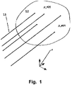
Fig. 1 is illustrating a plurality of optical members (11) that are inserted into a tissue (12), said tissue characterised by having light-scattering properties denoted µ s (r) in the geometrical coordinate r, and light-absorbing properties denoted µ a (r); -
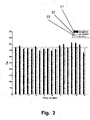
Fig. 2 is illustrating evaluated optical properties (µ eff ) using the old evaluation method (21) and the new, disclosed method (22), in a homogeneous tissue phantom. The value of µ eff as measured by an independent reference measurement method is also shown (23). -
Fig. 3 is illustrating evaluated attenuation values using the old evaluation method (31) and the new, disclosed method (32), in the same homogeneous tissue phantom as used inFig. 2 . -
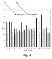
Fig. 4 is illustrating evaluated optical properties (µ eff ) using the old evaluation method (41) and the new, disclosed method (42) for prostate tissue. -
Fig. 5 is illustrating evaluated attenuation values using the old evaluation method (51) and the new, disclosed method (52), in the same prostate tissue as used inFig. 4 . - The following disclosure focuses on examples of the present disclosure applicable to determining or estimating light attenuation in tissue. The disclosure may be applicable for determining the impact of the presence of light-attenuating inhomogeneities at medical optical members, such as optical fibres, waveguides, light sources, and/or photodiodes placed inside tissue. For example, this is advantageous for determining the impact of the presence of blood aggregation at medical optical members placed inside tissue. However, it will be appreciated that the description is not limited to this application but may be applied to many other systems where determining the impact of the presence of light-attenuating inhomogeneities at medical optical members placed inside a turbid medium.
- Consider a tissue that has light-scattering and light absorbing properties as illustrated in
Figure 1 . A plurality of optical members (11), such as optical fibres, waveguides, light sources, and/or photodiodes is placed inside the tissue (12) for the main purpose of delivering a light dose into the tissue, or to perform measurements of the optical characteristics of the tissue. The light-scattering properties of the tissue may be denoted µ s (r) in the geometrical coordinate r. Likewise, the light-absorbing properties are denoted µ a (r). The light fluence in any point r in the tissue due to one or multiple light sources may in general for example be described by Boltzmann transport theory for radiative transfer. The transport equation in general requires numerical solution. For the purpose of example, analytical solutions to the transport equation exist in some specific cases. For example, the light fluence rate at distance r from a point source in a homogeneous medium, provided that µ s (r)>>µ a (r), may be expressed as
- where µ eff =[3µ a (µ a +µ s )]1/2, and D=[3(µ a +µ s )]-1. µ eff is the effective attenuation coefficient and D is the diffusion coefficient. Hence, the fluence rate as a function of the distance from the light source typically follows some type of exponential, determined by the effective attenuation coefficient µ eff .
- If light-attenuating inhomogeneities are present, the absorption may be increase or decrease compared to tissue in general. In case bleeding occurs at the optical members, the accumulated blood will absorb light to a higher degree than the tissue in general, since blood has an inherent high light absorption due to the high concentration of haemoglobin. Assuming that the blood only accumulates locally around the member, it will act to attenuate the light that is emitted from the member (and attenuate the light that is collected by the member in case it is used for measurements). An attenuation value A may be incorporated to the expression for the light fluence rate:

- where 0≤A≤1. Assuming that the entire volume of tissue may be approximated as homogeneous except for the presence of inhomogeneities, such as local bleeding at each member, and that each member is considered a point source, this gives a set of attenuation values Ai for each member, where the index i=1..N for N members.
- To determine the attenuation values Ai , the first step may be to perform a set of measurements where one member at a time emits light, and the other members collect light which is measured. It should be noted that the method is not limited to the case of one single member emitting at a time, and all other members collecting, but other measurement regimens are also possible, such as using a sub-set of all members that emit, or a sub-set of all members that collect, or combinations of these.
- In the case of member i emitting light, and member i+1 measuring the resulting light fluence, the measured light power may be expressed as

- where Pi is the light power emitted by member i, and Ki+1 is a conversion factor for the conversion of the fluence rate to power collected by member i+1, and r i,i+1=|r i+1-r i | is the distance between member i and i+1. The factor K can be assumed to be the same for all members, or, alternatively, known for each member, or relatively known between all members.
- The measurement step provides a set of measurements that in this example can be expressed as:
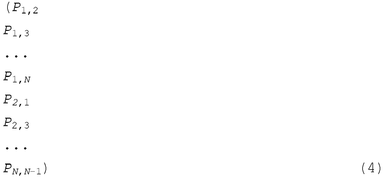
- In the next step, this dataset may be used to determine the attenuation values Ai . When using this dataset with Eq. 3, one obtains a non-linear system of equations with the attenuation values Ai , and the optical properties D and µ eff, as unknowns:

- This system of equations can be solved using non-linear solution methods, such as, including but not limited to, Newton-type methods, the Levenberg-Marquardt method, or trust-region methods. Note that in general the system of equations may be overdetermined or underdetermined. The optical properties D and µ eff can either be treated as unknowns, or can be assumed known from prior knowledge.
-
Equation 5 can be linearised, for example, by taking the logarithm of both sides:
- By making the assumptions that µ a <<µ s and that µ s is known and constant, we get

-
Equation 7 represents a system of linear equations with the unknowns Ai and µ eff(i) . This system can be solved by normal methods for solving systems of linear equations, for example the least squares method in cases where the system is under- or overdetermined. - In order to obtain the attenuation values Ai , the measurement system must be calibrated to measure the fluence rate in absolute terms. In practise, this condition may be unpractical to achieve. In the case where only relative measurements of fluence rate are available, the values Ai can still be evaluated, but only in relative terms. Relative attenuation values may be used after normalisation. For example, the values Ai may be normalised so that the value of A with the least attenuation is set to 1. This is a conservative approach, since the attenuation will generally be underestimated, so the resulting light doses will also be underestimated. Other normalisation approaches are also possible, for example based on prior knowledge of the normal range of attenuation expected for a particular case.
- In other examples, the disclosure may not be limited to the conditions and approximations which are applied in the previous description. In the following, some other examples are described.
- In an example, the optical members may not be point sources but distributed sources such as diffusers, for example optical fibres with diffusers. When using diffusers, it may be required to use convolution. The same method could then be used as described above to calculate the effective attenuation coefficient and attenuation values, but it could become more complex. A Finite element method (FEM) may then be applied to solve the complexity. Alternatively, diffusers may be approximated as a plurality of point sources distributed over the dimensions of the diffuser.
- In an example, optical members may not be used for light delivery, but instead light sources are positioned directly inside the tissue. The light sources may be photodiodes, e.g., light emitting diodes (LED) or laser diodes.
- In an example, optical members may not be used for light collection and detection, but instead light detectors may be positioned directly inside the tissue. The light detectors may be, e.g., photodiodes, photomultiplier tubes, avalanche photodiodes, charge-coupled devices, or CMOS light sensitive devices.
- In an example, the medium may not be assumed to have homogeneous optical properties but may instead modelled based on prior knowledge. For example, the medium may be assumed to have regions with different optical properties based on the anatomy of the tissue. The anatomy of the tissue may be known based on imaging data, e.g., x-ray, ultrasound or magnetic resonance imaging. The optical properties may be known for each region in advance based on prior data, or the optical properties may be solved for in the non-linear solver together with the attenuation values.
- In an example, the model of the light fluence rate may not be based on the analytical equation but instead numerical methods may be used to solve the transport equation. Examples of such methods are the finite element method, the discrete ordinates method, or Monte Carlo simulations.
- In an example, spectral information obtained by spectroscopic measurements, such as multiple wavelengths or white light, may be used to improve the model to further improve the result. By using known absorption spectra of tissue chromophores, e.g., haemoglobin, oxy-haemoglobin, water, and lipids, measurements taken at multiple wavelengths may be used to constrain the model to get accurate results for both the attenuation values and the optical properties.
- In an example, the purpose of the method may not only be to determine the attenuation values Ai , but also the general optical properties of the tissue, light scattering and light absorption.
- In an example, the purpose of the method may not only be to determine the attenuation due to inhomogeneities in the tissue, but also to determine losses in the optical system that performs the measurement. With this approach, the system can be calibrated for light losses in a quick and efficient manner. The light may then be increased to compensate for attenuation of the light, either in the system or the fibres used for transferring light to and from the tissue.
- Any method or determinations or calculations described herein may be implemented on a control unit, a data processing device or a computing device. The computer implementation may be done as a computer program or a software to be executed on a control unit, a data processing device or a computing device.
- Any methods or determinations or calculations described herein may be performed by a control unit, a data processing device or a computing device. The control unit, data processing device or computing device may be connected to the optical detection system or be a standalone unit.
- The control unit, data processing device or a computing device may be implemented by special-purpose software (or firmware) run on one or more general-purpose or special-purpose computing devices. In this context, it is to be understood that each "element" or "means" of such a computing device refers to a conceptual equivalent of a method step; there is not always a one-to-one correspondence between elements/means and particular pieces of hardware or software routines. One piece of hardware sometimes comprises different means/elements. For example, a processing unit serves as one element/means when executing one instruction, but serves as another element/means when executing another instruction. In addition, one element/means may be implemented by one instruction in some cases, but by a plurality of instructions in some other cases. Such a software controlled computing device may include one or more processing units, e.g. a CPU ("Central Processing Unit"), a DSP ("Digital Signal Processor"), an ASIC ("Application-Specific Integrated Circuit"), discrete analog and/or digital components, or some other programmable logical device, such as an FPGA ("Field Programmable Gate Array"). The control unit, data processing device or computing device may further include a system memory and a system bus that couples various system components including the system memory to the processing unit. The system bus may be any of several types of bus structures including a memory bus or memory controller, a peripheral bus, and a local bus using any of a variety of bus architectures. The system memory may include computer storage media in the form of volatile and/or non-volatile memory such as read only memory (ROM), random access memory (RAM) and flash memory. The special-purpose software may be stored in the system memory, or on other removable/non-removable volatile/non-volatile computer storage media which is included in or accessible to the computing device, such as magnetic media, optical media, flash memory cards, digital tape, solid state RAM, solid state ROM, etc. The data processing device may include one or more communication interfaces, such as a serial interface, a parallel interface, a USB interface, a wireless interface, a network adapter, etc., as well as one or more data acquisition devices, such as an A/D converter. The special-purpose software may be provided to the control unit or data processing device on any suitable computer-readable medium, including a record medium and a read-only memory.
- In aspect of the disclosure, but not limiting, is a system for determining light attenuation at an optical member inserted in tissue, and/or optical properties, such as light scattering properties and/or light-absorbing properties, of tissue is described. The system may include at least two optical members configured to be inserted into tissue, such as interstitially inserted. The at least two optical members may be configured for emitting and/or collecting light. The system may further include a control unit configured for controlling the at least two optical members so that light is transmitted to the tissue from at least one optical member and light is detected from the tissue by at least one optical member collecting light. This may yield a data set of measured values for pairs of emitting and collecting optical members.
- The control unit may be further configured for determining the attenuation values at the optical members and/or the optical properties of the tissue by using the data set of measured values to define a system of equations using a mathematical model of light propagation in tissue. The system of equations may be solved by the control unit by using a numerical solving method.
- The system may utilize any of the herein disclosed methods for determining or estimating the attenuation values at the optical members and/or the optical properties of the tissue.
- In some examples may the system include a plurality of optical members. In some examples one single member at a time may emit light, and all other members collecting. Alternatively, in some example other measurement regimens may be possible, such as using a sub-set of all members that emit, or a sub-set of all members that collect, or combinations of these.
- In some examples of the system, may the determined attenuation values at the optical members and/or optical properties of the tissue be used for calculating a light dose for photodynamic therapy or laser hyperthermia.
- In some examples of the system, may the control unit be configured for using the determined attenuation values at the optical members and/or optical properties of the tissue for light dose delivery for photodynamic therapy or laser hyperthermia.
- In some examples of the system, may the optical members be optical fibres, or optical fibres with diffusers. The optical members may transfer emitted light from a light source to the tissue and transfer collected light to a detector. The light source and detector may be any type of light source and detector herein disclosed.
- In some examples of the system, may the optical members be photodiodes configured to be arranged in tissue, such as interstitially arranged in tissue.
- In some examples of the system, may the optical members be configured to emit light at multiple wavelengths. Further, the control unit may be configured for using spectral detection and known absorption spectra of tissue chromophores to constrain a solution when solving the systems of equations.
- In some examples of the system, may the mathematical model of light propagation in tissue be based on Bolzmann transport equation.
- In some examples of the system, may the data set of measured values be measured light powers and the mathematical model of light propagation in tissue may be used for expressing measured light power for setting up the system of equations to be solved based on the data set of measured light powers for pairs of emitting and collecting optical members.
- In some examples of the system, may the attenuation values at the optical members and the optical properties of the tissue be unknown in the mathematical model of light propagation in tissue.
- In some examples of the system, may a Newton-type method, a Levenberg-Marquardt method, or trust-region methods be used as non-linear numerical solving methods for the system of equations.
- In some examples of the system, may a least-squares method be used as linear numerical solving method for the linearized system of equations.
- In aspect of the disclosure, but not limiting, is a method disclosed for determining light attenuation at an optical member inserted in tissue, and/or optical properties, such as light scattering properties and/or light-absorbing properties, of tissue. The method may include controlling at least two optical members so that light may be transmitted to the tissue from at least one optical member and light may be detected from the tissue by at least one optical member collecting light. This may be used for yielding a data set of measured values for pairs of emitting and collecting optical members. The method may further include determining the attenuation values at the optical members and/or the optical properties of the tissue by using the data set of measured values to define a system of equations using a mathematical model of light propagation in tissue and solving the system of equations by using a numerical solving method.
- The mathematical model of light propagation in tissue and a numerical solving method may be any of models or methods herein described.
- In some examples may the method include calculating a light dose for photodynamic therapy or laser hyperthermia based on the determined attenuation values at the optical members and/or optical properties of the tissue.
- In some examples may the method include controlling a light dose delivery to the photodynamic therapy or laser hyperthermia.
- In some examples of the method, may the mathematical model of light propagation in tissue be based on Bolzmann transport equation.
- In some examples of the method, may the data set of measured values be measured light powers and the mathematical model of light propagation in tissue may be used for expressing measured light power for setting up the system of equations to be solved based on the data set of measured light power for pairs of emitting and collecting optical members.
- In some exampless of the method, may a Newton-type methods, a Levenberg-Marquardt method, or trust-region methods be used as non-linear numerical solving methods for the system of equations.
- In some examples of the method, may a least-squares method be used as linear numerical solving method for the linearized system of equations.
- Any method herein described may be a computer implemented method.
- In some aspects of the disclosure is a computer program for determining light attenuation at an optical member inserted in tissue, and/or light scattering properties and light-absorbing properties of tissue described. The computer program may include instructions which, when the computer program is executed by a computer, performs the steps of any of the herein described methods.
- The method in the disclosure was applied to measurement data obtained from a system for PDT of prostate tissue, the SpectraCure P18 system with IDOSE, J. Swartling, et al "System for interstitial photodynamic therapy with online dosimetry - First clinical experiences of prostate cancer", J Biomed Opt. 15, 058003 (2010). The system uses 18 individual optical members that can be used with for therapeutic PDT light delivery, or for optical spectroscopic measurements of the tissue for the purpose of dose planning.
- In order to obtain the attenuation values, the system was calibrated so that the conversion factor K was known for each optical member. This was done by subjecting each member in light-collecting mode to a known light fluence (W/cm2) in a reference tissue phantom, and registering the corresponding reading on the internal spectrometer.
- Measurement data was acquired by inserting the optical members into a tissue phantom (mix of water, intralipid and ink) with scattering and absorption properties similar to tissue at the measurement wavelength, 690 nm. Measurements were performed by the system by cycling through all combinations of emitting and collecting member, yielding a set a of
measurements 
- This data set was then subjected to the evaluation of attenuation values and optical properties. To demonstrate the improvement given by the new evaluation method, the corresponding results using the old model were calculated for comparison. In the old model, the optical property µ eff was simply determined as the slope of log(P) around each member as a function of r (the logarithm of eq. 3). The attenuation values were then estimated based on the deviation from the least-squares solution to the µ eff value for the measurement data.
- The actual value of µ eff was independently measured using time-of-flight spectroscopy for reference.
-
Figure 2 shows µ eff values for each emitter member, using the old method and the new method. The value obtained by the reference measurement is also shown. Since the tissue phantom is homogeneous, the results for each member should be the same. Clearly, the results are much improved by the new method compared with the old. -
Figure 3 shows the attenuation values for each emitter member, using the old method and the new method. In this case, there are no reference values for the actual attenuation values, which are caused by losses in the optical member line from the phantom to the detection system. However, it is clear that with the old method, part of these attenuation losses will translate to errors in the evaluation of µ eff , so it cannot be expected that the evaluated attenuation values are correct with the old method. - In another example, the algorithm was applied to actual measurements taken of prostate tissue in vivo in a clinical situation. The measurements were taken in the same manner as described in J. Swartling, et al "System for interstitial photodynamic therapy with online dosimetry - First clinical experiences of prostate cancer", J Biomed Opt. 15, 058003 (2010). The data set was again based on 18 optical members.
-
Figure 4 shows µ eff values for prostate tissue for each emitter member, using the old method and the new method. The old algorithm shows much more variation in the obtained values than the new algorithm. The values obtained by the old algorithm are also generally higher than those obtained with the new algorithm. -
Figure 5 shows the attenuation values in the prostate for each emitter member, using the old method and the new method. In this example, the attenuation values are generally lower than in the homogeneous phantom example, which is expected since there will be bleeding in the prostate tissue that causes increased attenuation. - The indefinite articles "a" and "an," as used herein in the specification and in the claims, unless clearly indicated to the contrary, should be understood to mean "at least one." The phrase "and/or," as used herein in the specification and in the claims, should be understood to mean "either or both" of the elements so conjoined, i.e., elements that are conjunctively present in some cases and 30 disjunctively present in other cases. rhe invention is defined by the appended claims.
Claims (16)
- A system for determining light attenuation values at an optical member inserted in tissue and/or losses in an optical measuring system, the system comprises:at least two optical members configured to be inserted into tissue, said at least two optical members are configured for emitting and/or collecting light;a control unit configured for controlling said at least two optical members so that light is transmitted to said tissue from at least one optical member and light is detected from said tissue by at least one optical member collecting light, thereby yielding a data set of measured values for pairs of emitting and collecting optical members;wherein said control unit is further configured for determining said light attenuation values and for determining light-attenuating inhomogeneities at said optical member and/or losses in said optical measuring system using said light attenuation values, and wherein said light attenuation values are determined using said data set of measured values to define a system of equations using a mathematical model of light propagation in tissue and solving said system of equations by using a numerical solving method.
- The system of claim 1, wherein said light attenuation values are in the tissue close to said optical member or in the optical member.
- The system of any of claims 1 and 2, wherein said determined attenuation values at said optical members is used for calculating a light dose for photodynamic therapy or laser hyperthermia.
- The system of any of claims 1 to 3, wherein the control unit is configured for using said determined attenuation values at said optical members when determining light dose delivery for photodynamic therapy or laser hyperthermia.
- The system of any of claims 1 to 4, wherein said optical members are optical fibres, or optical fibres with diffusers to transfer emitted light from a light source to said tissue and for transferring collected light to a detector; or wherein said optical members are photodiodes configured to be arranged in tissue.
- The system of any of claims 1 to 5, wherein said optical members are configured to emit light at multiple wavelengths and wherein said control unit is configured for using spectral detection and known absorption spectra of tissue chromophores to constrain a solution when solving said systems of equations.
- The system of any of claims 1 to 6, wherein said mathematical model of light propagation in tissue is based on Boltzmann transport equation.
- The system of any of claims 1 to 7, wherein said data set of measured values are measured light powers and said mathematical model of light propagation in tissue is used for expressing measured light power for setting up said system of equations to be solved based on said data set of measured light powers for pairs of emitting and collecting optical members.
- The system of any of claims 1 to 8, wherein saidattenuation values at said optical members andoptical properties of said tissue are unknown in said mathematical model of light propagation in tissue.
- The system of any of claims 1 to 9, wherein said system of equations is non-linear solved by a non-linear numerical solving method, such as a Newton-type method, a Levenberg-Marquardt method, or a trust-region method.
- The system of any of claims 1 to 10, wherein said system of equations is a linearised system of equations, solved by a method for solving linear equations, such as a least-squares method.
- A computer program for determining light attenuation values caused by light-attenuating inhomogeneities at an optical member inserted in tissue and/or losses in an optical measuring system, comprising instructions which, when the computer program is executed by a computer of a control unit of a system according to claim 1, performs the following steps:controlling at least two optical members so that light is transmitted to said tissue from at least one optical member and light is detected from said tissue by at least one optical member collecting light, thereby yielding a data set of measured values for pairs of emitting and collecting optical members;determining said light attenuation values and determining light-attenuating inhomogeneities at said optical member and/or losses in an optical measuring system using said light attenuation values, and wherein said light attenuation values are determined using using said data set of measured values to define a system of equations using a mathematical model of light propagation in tissue and solving said system of equations by using a numerical solving method.
- The computer program of claim 12, calculating a light dose for photodynamic therapy or laser hyperthermia by including said determined attenuation values at said optical members, and controlling a light dose delivery to said photodynamic therapy or laser hyperthermia.
- The computer program of any of claims 12 to 13, wherein said mathematical model of light propagation in tissue is based on Boltzmann transport equation.
- The computer program of any of claims 12 to 14 wherein said data set of measured values are measured light powers and said mathematical model of light propagation in tissue is used for expressing measured light power for setting up said system of equations to be solved based on said data set of measured light powers for pairs of emitting and collecting optical members.
- The computer program of any of claims 12 to 15,wherein said system of equations is non-linear; solving said system of equations by a non-linear numerical solving method, such as a Newton-type method, a Levenberg-Marquardt method, or a trust-region method; orwherein said system of equations is a linearised system of equations; solving said system by a method for solving linear equations, such as a least-squares method.
Applications Claiming Priority (2)
| Application Number | Priority Date | Filing Date | Title |
|---|---|---|---|
| EP19167324.3A EP3718600B1 (en) | 2019-04-04 | 2019-04-04 | System for determining light attenuation at optical members inserted in tissue |
| PCT/EP2020/059790 WO2020201580A1 (en) | 2019-04-04 | 2020-04-06 | System and method for determining light attenuation at optical members inserted in tissue |
Publications (2)
| Publication Number | Publication Date |
|---|---|
| EP3946573A1 EP3946573A1 (en) | 2022-02-09 |
| EP3946573B1 true EP3946573B1 (en) | 2022-08-03 |
Family
ID=66092194
Family Applications (2)
| Application Number | Title | Priority Date | Filing Date |
|---|---|---|---|
| EP19167324.3A Active EP3718600B1 (en) | 2019-04-04 | 2019-04-04 | System for determining light attenuation at optical members inserted in tissue |
| EP20716798.2A Active EP3946573B1 (en) | 2019-04-04 | 2020-04-06 | System for determining light attenuation at optical members inserted in tissue |
Family Applications Before (1)
| Application Number | Title | Priority Date | Filing Date |
|---|---|---|---|
| EP19167324.3A Active EP3718600B1 (en) | 2019-04-04 | 2019-04-04 | System for determining light attenuation at optical members inserted in tissue |
Country Status (6)
| Country | Link |
|---|---|
| US (1) | US11331512B2 (en) |
| EP (2) | EP3718600B1 (en) |
| JP (1) | JP7640089B2 (en) |
| CN (1) | CN113646037B (en) |
| CA (1) | CA3135863A1 (en) |
| WO (1) | WO2020201580A1 (en) |
Family Cites Families (12)
| Publication number | Priority date | Publication date | Assignee | Title |
|---|---|---|---|---|
| US6138045A (en) * | 1998-08-07 | 2000-10-24 | Arch Development Corporation | Method and system for the segmentation and classification of lesions |
| US6138046A (en) * | 1999-04-20 | 2000-10-24 | Miravant Medical Technologies, Inc. | Dosimetry probe |
| SE522697C2 (en) | 2001-11-14 | 2004-03-02 | Spectracure Ab | Therapy and diagnostic systems with distributors for distribution of radiation |
| JP4084307B2 (en) * | 2001-12-26 | 2008-04-30 | 浜松ホトニクス株式会社 | Optical analysis method for inhomogeneous media |
| US20040267335A1 (en) * | 2003-04-23 | 2004-12-30 | John Tulip | Switched photodynamic therapy apparatus and method |
| SE527164C2 (en) * | 2003-05-14 | 2006-01-10 | Spectracure Ab | Interactive therapy/diagnosis system for tumor, has operation mode selector to optically direct non-ionizing electromagnetic therapeutic and/or diagnostic radiation to tumor site, through radiation conductor |
| CA2566570C (en) * | 2003-05-14 | 2012-04-10 | Spectracure Ab | System and method for therapy and diagnosis comprising translatory distributor for distribution of radiation |
| US20060095100A1 (en) * | 2004-10-29 | 2006-05-04 | Kian Shin Lee | Method and apparatus for regulating light administered at a patient treatment site |
| EP2066403B1 (en) * | 2006-08-15 | 2010-05-12 | Spectracure AB | System and method for controlling and adjusting interstitial photodynamic light therapy parameters |
| US8489164B2 (en) * | 2010-10-27 | 2013-07-16 | Medtronic, Inc. | Monitoring of tissue hemoglobin concentration |
| WO2012076631A1 (en) * | 2010-12-07 | 2012-06-14 | Spectracure Ab | System and method for interstitial photodynamic light therapy in combination with photosensitizers |
| CA2996503A1 (en) * | 2015-07-23 | 2017-01-26 | Health Research, Inc. | System and method for delivering light dose to tissue |
-
2019
- 2019-04-04 EP EP19167324.3A patent/EP3718600B1/en active Active
-
2020
- 2020-04-06 JP JP2021558950A patent/JP7640089B2/en active Active
- 2020-04-06 EP EP20716798.2A patent/EP3946573B1/en active Active
- 2020-04-06 US US17/600,302 patent/US11331512B2/en active Active
- 2020-04-06 WO PCT/EP2020/059790 patent/WO2020201580A1/en not_active Ceased
- 2020-04-06 CA CA3135863A patent/CA3135863A1/en active Pending
- 2020-04-06 CN CN202080027505.0A patent/CN113646037B/en active Active
Also Published As
| Publication number | Publication date |
|---|---|
| US20220088405A1 (en) | 2022-03-24 |
| US11331512B2 (en) | 2022-05-17 |
| WO2020201580A1 (en) | 2020-10-08 |
| CA3135863A1 (en) | 2020-10-08 |
| CN113646037A (en) | 2021-11-12 |
| EP3946573A1 (en) | 2022-02-09 |
| EP3718600A1 (en) | 2020-10-07 |
| CN113646037B (en) | 2022-10-18 |
| JP7640089B2 (en) | 2025-03-05 |
| JP2022529114A (en) | 2022-06-17 |
| EP3718600B1 (en) | 2025-11-05 |
Similar Documents
| Publication | Publication Date | Title |
|---|---|---|
| Profio et al. | Transport of light in tissue in photodynamic therapy | |
| Johansson et al. | Realtime light dosimetry software tools for interstitial photodynamic therapy of the human prostate | |
| CN101522261B (en) | System and method for controlling and adjusting interstitial photodynamic light therapy parameters | |
| US10463880B2 (en) | Medical instrument for high dose rate brachytherapy | |
| US20220001193A1 (en) | Methods and systems for photodynamic therapy calculations | |
| Belley et al. | Real-time dose-rate monitoring with gynecologic brachytherapy: Results of an initial clinical trial | |
| EP3325096B1 (en) | System for delivering dosed light to tissue | |
| US20200107756A1 (en) | Silicon photomultiplier array-based multispectral optical probes for image-guided radiotherapy | |
| van Doeveren et al. | On the development of a light dosimetry planning tool for photodynamic therapy in arbitrary shaped cavities: initial results | |
| EP3718600B1 (en) | System for determining light attenuation at optical members inserted in tissue | |
| Dimofte et al. | Determination of tissue optical properties in PDT treated Head & Neck patients | |
| Bentefour et al. | Validation of an in‐vivo proton beam range check method in an anthropomorphic pelvic phantom using dose measurements | |
| Tarasov et al. | Is the minimal erythema dose achievable at optical spectroscopy of skin in Vivo? | |
| Ong et al. | Monte Carlo modelling of fluorescence in semi-infinite turbid media | |
| Ong et al. | Estimation of fluorescence probing depth dependence on the distance between source and detector using Monte Carlo modeling | |
| JP6224464B2 (en) | Method and apparatus for measuring brown adipose tissue | |
| Lambson et al. | A theoretical and experimental examination of fluorescence in enclosed cavities | |
| Ismael et al. | A DIFFUSION EQUATION BASED ALGORITHM FOR DETERMINATION OF THE OPTIMAL NUMBER OF FIBERS USED FOR BREAST CANCER TREATMENT PLAN-NING IN PHOTODYNAMIC THERAPY | |
| Wiles | Simulating Cherenkov Radiation Generated by Radiotherapy Beams | |
| WO2023198724A1 (en) | System and method for combined thermal and photodynamic therapy of malignant tumors | |
| Li et al. | A comparison of two probes to determine rectum optical properties | |
| Wang et al. | Reconstruction of optical properties using a diffusion model for interstitial diffuse optical tomography | |
| Fu et al. | Study on the impact of different wound dressings on surface dose and deep dose analysis in head and neck radiation therapy | |
| Amasha et al. | A diffusion equation based algorithm for determination of the optimal number of fibers used for breast cancer treatment planning in photodynamic therapy | |
| Belley | Monte Carlo Analysis and Physics Characterization of a Novel Nanoparticle Detector for Medical and Micro-dosimetry Applications |
Legal Events
| Date | Code | Title | Description |
|---|---|---|---|
| STAA | Information on the status of an ep patent application or granted ep patent |
Free format text: STATUS: UNKNOWN |
|
| STAA | Information on the status of an ep patent application or granted ep patent |
Free format text: STATUS: THE INTERNATIONAL PUBLICATION HAS BEEN MADE |
|
| PUAI | Public reference made under article 153(3) epc to a published international application that has entered the european phase |
Free format text: ORIGINAL CODE: 0009012 |
|
| STAA | Information on the status of an ep patent application or granted ep patent |
Free format text: STATUS: REQUEST FOR EXAMINATION WAS MADE |
|
| 17P | Request for examination filed |
Effective date: 20211103 |
|
| AK | Designated contracting states |
Kind code of ref document: A1 Designated state(s): AL AT BE BG CH CY CZ DE DK EE ES FI FR GB GR HR HU IE IS IT LI LT LU LV MC MK MT NL NO PL PT RO RS SE SI SK SM TR |
|
| GRAP | Despatch of communication of intention to grant a patent |
Free format text: ORIGINAL CODE: EPIDOSNIGR1 |
|
| STAA | Information on the status of an ep patent application or granted ep patent |
Free format text: STATUS: GRANT OF PATENT IS INTENDED |
|
| DAV | Request for validation of the european patent (deleted) | ||
| DAX | Request for extension of the european patent (deleted) | ||
| INTG | Intention to grant announced |
Effective date: 20220513 |
|
| GRAS | Grant fee paid |
Free format text: ORIGINAL CODE: EPIDOSNIGR3 |
|
| GRAA | (expected) grant |
Free format text: ORIGINAL CODE: 0009210 |
|
| STAA | Information on the status of an ep patent application or granted ep patent |
Free format text: STATUS: THE PATENT HAS BEEN GRANTED |
|
| AK | Designated contracting states |
Kind code of ref document: B1 Designated state(s): AL AT BE BG CH CY CZ DE DK EE ES FI FR GB GR HR HU IE IS IT LI LT LU LV MC MK MT NL NO PL PT RO RS SE SI SK SM TR |
|
| REG | Reference to a national code |
Ref country code: AT Ref legal event code: REF Ref document number: 1508266 Country of ref document: AT Kind code of ref document: T Effective date: 20220815 Ref country code: CH Ref legal event code: EP |
|
| REG | Reference to a national code |
Ref country code: DE Ref legal event code: R096 Ref document number: 602020004357 Country of ref document: DE |
|
| REG | Reference to a national code |
Ref country code: IE Ref legal event code: FG4D |
|
| REG | Reference to a national code |
Ref country code: LT Ref legal event code: MG9D |
|
| REG | Reference to a national code |
Ref country code: NL Ref legal event code: MP Effective date: 20220803 |
|
| PG25 | Lapsed in a contracting state [announced via postgrant information from national office to epo] |
Ref country code: SE Free format text: LAPSE BECAUSE OF FAILURE TO SUBMIT A TRANSLATION OF THE DESCRIPTION OR TO PAY THE FEE WITHIN THE PRESCRIBED TIME-LIMIT Effective date: 20220803 Ref country code: RS Free format text: LAPSE BECAUSE OF FAILURE TO SUBMIT A TRANSLATION OF THE DESCRIPTION OR TO PAY THE FEE WITHIN THE PRESCRIBED TIME-LIMIT Effective date: 20220803 Ref country code: PT Free format text: LAPSE BECAUSE OF FAILURE TO SUBMIT A TRANSLATION OF THE DESCRIPTION OR TO PAY THE FEE WITHIN THE PRESCRIBED TIME-LIMIT Effective date: 20221205 Ref country code: NO Free format text: LAPSE BECAUSE OF FAILURE TO SUBMIT A TRANSLATION OF THE DESCRIPTION OR TO PAY THE FEE WITHIN THE PRESCRIBED TIME-LIMIT Effective date: 20221103 Ref country code: NL Free format text: LAPSE BECAUSE OF FAILURE TO SUBMIT A TRANSLATION OF THE DESCRIPTION OR TO PAY THE FEE WITHIN THE PRESCRIBED TIME-LIMIT Effective date: 20220803 Ref country code: LV Free format text: LAPSE BECAUSE OF FAILURE TO SUBMIT A TRANSLATION OF THE DESCRIPTION OR TO PAY THE FEE WITHIN THE PRESCRIBED TIME-LIMIT Effective date: 20220803 Ref country code: LT Free format text: LAPSE BECAUSE OF FAILURE TO SUBMIT A TRANSLATION OF THE DESCRIPTION OR TO PAY THE FEE WITHIN THE PRESCRIBED TIME-LIMIT Effective date: 20220803 Ref country code: FI Free format text: LAPSE BECAUSE OF FAILURE TO SUBMIT A TRANSLATION OF THE DESCRIPTION OR TO PAY THE FEE WITHIN THE PRESCRIBED TIME-LIMIT Effective date: 20220803 Ref country code: ES Free format text: LAPSE BECAUSE OF FAILURE TO SUBMIT A TRANSLATION OF THE DESCRIPTION OR TO PAY THE FEE WITHIN THE PRESCRIBED TIME-LIMIT Effective date: 20220803 |
|
| REG | Reference to a national code |
Ref country code: AT Ref legal event code: MK05 Ref document number: 1508266 Country of ref document: AT Kind code of ref document: T Effective date: 20220803 |
|
| PG25 | Lapsed in a contracting state [announced via postgrant information from national office to epo] |
Ref country code: PL Free format text: LAPSE BECAUSE OF FAILURE TO SUBMIT A TRANSLATION OF THE DESCRIPTION OR TO PAY THE FEE WITHIN THE PRESCRIBED TIME-LIMIT Effective date: 20220803 Ref country code: IS Free format text: LAPSE BECAUSE OF FAILURE TO SUBMIT A TRANSLATION OF THE DESCRIPTION OR TO PAY THE FEE WITHIN THE PRESCRIBED TIME-LIMIT Effective date: 20221203 Ref country code: HR Free format text: LAPSE BECAUSE OF FAILURE TO SUBMIT A TRANSLATION OF THE DESCRIPTION OR TO PAY THE FEE WITHIN THE PRESCRIBED TIME-LIMIT Effective date: 20220803 Ref country code: GR Free format text: LAPSE BECAUSE OF FAILURE TO SUBMIT A TRANSLATION OF THE DESCRIPTION OR TO PAY THE FEE WITHIN THE PRESCRIBED TIME-LIMIT Effective date: 20221104 |
|
| PG25 | Lapsed in a contracting state [announced via postgrant information from national office to epo] |
Ref country code: SM Free format text: LAPSE BECAUSE OF FAILURE TO SUBMIT A TRANSLATION OF THE DESCRIPTION OR TO PAY THE FEE WITHIN THE PRESCRIBED TIME-LIMIT Effective date: 20220803 Ref country code: RO Free format text: LAPSE BECAUSE OF FAILURE TO SUBMIT A TRANSLATION OF THE DESCRIPTION OR TO PAY THE FEE WITHIN THE PRESCRIBED TIME-LIMIT Effective date: 20220803 Ref country code: DK Free format text: LAPSE BECAUSE OF FAILURE TO SUBMIT A TRANSLATION OF THE DESCRIPTION OR TO PAY THE FEE WITHIN THE PRESCRIBED TIME-LIMIT Effective date: 20220803 Ref country code: CZ Free format text: LAPSE BECAUSE OF FAILURE TO SUBMIT A TRANSLATION OF THE DESCRIPTION OR TO PAY THE FEE WITHIN THE PRESCRIBED TIME-LIMIT Effective date: 20220803 Ref country code: AT Free format text: LAPSE BECAUSE OF FAILURE TO SUBMIT A TRANSLATION OF THE DESCRIPTION OR TO PAY THE FEE WITHIN THE PRESCRIBED TIME-LIMIT Effective date: 20220803 |
|
| REG | Reference to a national code |
Ref country code: DE Ref legal event code: R097 Ref document number: 602020004357 Country of ref document: DE |
|
| PG25 | Lapsed in a contracting state [announced via postgrant information from national office to epo] |
Ref country code: SK Free format text: LAPSE BECAUSE OF FAILURE TO SUBMIT A TRANSLATION OF THE DESCRIPTION OR TO PAY THE FEE WITHIN THE PRESCRIBED TIME-LIMIT Effective date: 20220803 Ref country code: EE Free format text: LAPSE BECAUSE OF FAILURE TO SUBMIT A TRANSLATION OF THE DESCRIPTION OR TO PAY THE FEE WITHIN THE PRESCRIBED TIME-LIMIT Effective date: 20220803 |
|
| PLBE | No opposition filed within time limit |
Free format text: ORIGINAL CODE: 0009261 |
|
| STAA | Information on the status of an ep patent application or granted ep patent |
Free format text: STATUS: NO OPPOSITION FILED WITHIN TIME LIMIT |
|
| PG25 | Lapsed in a contracting state [announced via postgrant information from national office to epo] |
Ref country code: AL Free format text: LAPSE BECAUSE OF FAILURE TO SUBMIT A TRANSLATION OF THE DESCRIPTION OR TO PAY THE FEE WITHIN THE PRESCRIBED TIME-LIMIT Effective date: 20220803 |
|
| 26N | No opposition filed |
Effective date: 20230504 |
|
| REG | Reference to a national code |
Ref country code: CH Ref legal event code: PL |
|
| PG25 | Lapsed in a contracting state [announced via postgrant information from national office to epo] |
Ref country code: LU Free format text: LAPSE BECAUSE OF NON-PAYMENT OF DUE FEES Effective date: 20230406 |
|
| REG | Reference to a national code |
Ref country code: BE Ref legal event code: MM Effective date: 20230430 |
|
| PG25 | Lapsed in a contracting state [announced via postgrant information from national office to epo] |
Ref country code: MC Free format text: LAPSE BECAUSE OF FAILURE TO SUBMIT A TRANSLATION OF THE DESCRIPTION OR TO PAY THE FEE WITHIN THE PRESCRIBED TIME-LIMIT Effective date: 20220803 |
|
| PG25 | Lapsed in a contracting state [announced via postgrant information from national office to epo] |
Ref country code: MC Free format text: LAPSE BECAUSE OF FAILURE TO SUBMIT A TRANSLATION OF THE DESCRIPTION OR TO PAY THE FEE WITHIN THE PRESCRIBED TIME-LIMIT Effective date: 20220803 Ref country code: LI Free format text: LAPSE BECAUSE OF NON-PAYMENT OF DUE FEES Effective date: 20230430 Ref country code: CH Free format text: LAPSE BECAUSE OF NON-PAYMENT OF DUE FEES Effective date: 20230430 |
|
| REG | Reference to a national code |
Ref country code: IE Ref legal event code: MM4A |
|
| PG25 | Lapsed in a contracting state [announced via postgrant information from national office to epo] |
Ref country code: BE Free format text: LAPSE BECAUSE OF NON-PAYMENT OF DUE FEES Effective date: 20230430 |
|
| PG25 | Lapsed in a contracting state [announced via postgrant information from national office to epo] |
Ref country code: IE Free format text: LAPSE BECAUSE OF NON-PAYMENT OF DUE FEES Effective date: 20230406 |
|
| PG25 | Lapsed in a contracting state [announced via postgrant information from national office to epo] |
Ref country code: IE Free format text: LAPSE BECAUSE OF NON-PAYMENT OF DUE FEES Effective date: 20230406 |
|
| PG25 | Lapsed in a contracting state [announced via postgrant information from national office to epo] |
Ref country code: IT Free format text: LAPSE BECAUSE OF FAILURE TO SUBMIT A TRANSLATION OF THE DESCRIPTION OR TO PAY THE FEE WITHIN THE PRESCRIBED TIME-LIMIT Effective date: 20220803 |
|
| PG25 | Lapsed in a contracting state [announced via postgrant information from national office to epo] |
Ref country code: BG Free format text: LAPSE BECAUSE OF FAILURE TO SUBMIT A TRANSLATION OF THE DESCRIPTION OR TO PAY THE FEE WITHIN THE PRESCRIBED TIME-LIMIT Effective date: 20220803 |
|
| PG25 | Lapsed in a contracting state [announced via postgrant information from national office to epo] |
Ref country code: BG Free format text: LAPSE BECAUSE OF FAILURE TO SUBMIT A TRANSLATION OF THE DESCRIPTION OR TO PAY THE FEE WITHIN THE PRESCRIBED TIME-LIMIT Effective date: 20220803 |
|
| PGFP | Annual fee paid to national office [announced via postgrant information from national office to epo] |
Ref country code: GB Payment date: 20250313 Year of fee payment: 6 |
|
| PGFP | Annual fee paid to national office [announced via postgrant information from national office to epo] |
Ref country code: DE Payment date: 20250424 Year of fee payment: 6 |
|
| PGFP | Annual fee paid to national office [announced via postgrant information from national office to epo] |
Ref country code: FR Payment date: 20250404 Year of fee payment: 6 |
|
| PG25 | Lapsed in a contracting state [announced via postgrant information from national office to epo] |
Ref country code: CY Free format text: LAPSE BECAUSE OF FAILURE TO SUBMIT A TRANSLATION OF THE DESCRIPTION OR TO PAY THE FEE WITHIN THE PRESCRIBED TIME-LIMIT; INVALID AB INITIO Effective date: 20200406 |
|
| PG25 | Lapsed in a contracting state [announced via postgrant information from national office to epo] |
Ref country code: HU Free format text: LAPSE BECAUSE OF FAILURE TO SUBMIT A TRANSLATION OF THE DESCRIPTION OR TO PAY THE FEE WITHIN THE PRESCRIBED TIME-LIMIT; INVALID AB INITIO Effective date: 20200406 |
|
| PG25 | Lapsed in a contracting state [announced via postgrant information from national office to epo] |
Ref country code: TR Free format text: LAPSE BECAUSE OF FAILURE TO SUBMIT A TRANSLATION OF THE DESCRIPTION OR TO PAY THE FEE WITHIN THE PRESCRIBED TIME-LIMIT Effective date: 20220803 |
|
| PG25 | Lapsed in a contracting state [announced via postgrant information from national office to epo] |
Ref country code: SI Free format text: LAPSE BECAUSE OF FAILURE TO SUBMIT A TRANSLATION OF THE DESCRIPTION OR TO PAY THE FEE WITHIN THE PRESCRIBED TIME-LIMIT Effective date: 20220803 |