EP3939612A1 - Optically heat-generating composite material, nanocluster, substance delivery carrier and pharmaceutical composition - Google Patents
Optically heat-generating composite material, nanocluster, substance delivery carrier and pharmaceutical composition Download PDFInfo
- Publication number
- EP3939612A1 EP3939612A1 EP20769042.1A EP20769042A EP3939612A1 EP 3939612 A1 EP3939612 A1 EP 3939612A1 EP 20769042 A EP20769042 A EP 20769042A EP 3939612 A1 EP3939612 A1 EP 3939612A1
- Authority
- EP
- European Patent Office
- Prior art keywords
- icg
- composite material
- nanocluster
- photo
- cancer
- Prior art date
- Legal status (The legal status is an assumption and is not a legal conclusion. Google has not performed a legal analysis and makes no representation as to the accuracy of the status listed.)
- Pending
Links
- 239000000126 substance Substances 0.000 title claims abstract description 30
- 239000002131 composite material Substances 0.000 title claims abstract description 27
- 239000008194 pharmaceutical composition Substances 0.000 title claims description 7
- -1 nanocluster Substances 0.000 title description 29
- 239000000049 pigment Substances 0.000 claims abstract description 33
- OKTJSMMVPCPJKN-UHFFFAOYSA-N Carbon Chemical compound [C] OKTJSMMVPCPJKN-UHFFFAOYSA-N 0.000 claims abstract description 29
- 229910052799 carbon Inorganic materials 0.000 claims abstract description 29
- 239000002086 nanomaterial Substances 0.000 claims abstract description 18
- 125000005647 linker group Chemical group 0.000 claims abstract description 7
- 239000004480 active ingredient Substances 0.000 claims description 21
- 239000003814 drug Substances 0.000 claims description 12
- 239000002113 nanodiamond Substances 0.000 claims description 11
- 229940079593 drug Drugs 0.000 claims description 9
- 125000000896 monocarboxylic acid group Chemical group 0.000 claims description 8
- 239000013543 active substance Substances 0.000 claims description 6
- 210000004027 cell Anatomy 0.000 description 35
- 206010028980 Neoplasm Diseases 0.000 description 29
- 201000011510 cancer Diseases 0.000 description 23
- 108020004707 nucleic acids Proteins 0.000 description 20
- 102000039446 nucleic acids Human genes 0.000 description 20
- 150000007523 nucleic acids Chemical class 0.000 description 20
- XLYOFNOQVPJJNP-UHFFFAOYSA-N water Chemical compound O XLYOFNOQVPJJNP-UHFFFAOYSA-N 0.000 description 17
- 239000007787 solid Substances 0.000 description 16
- 238000000034 method Methods 0.000 description 15
- 239000007853 buffer solution Substances 0.000 description 14
- 239000000243 solution Substances 0.000 description 14
- KDJUNNVUQBKNAY-UHFFFAOYSA-N 4-[(2E)-2-[(2E,4E,6E)-7-[3-[6-(2,5-dioxopyrrolidin-1-yl)oxy-6-oxohexyl]-1,1-dimethylbenzo[e]indol-3-ium-2-yl]hepta-2,4,6-trienylidene]-1,1-dimethylbenzo[e]indol-3-yl]butane-1-sulfonate Chemical compound CC1(C)\C(=C/C=C/C=C/C=C/C2=[N+](CCCCS([O-])(=O)=O)C3=C(C4=C(C=CC=C4)C=C3)C2(C)C)N(CCCCCC(=O)ON2C(=O)CCC2=O)C2=C1C1=C(C=CC=C1)C=C2 KDJUNNVUQBKNAY-UHFFFAOYSA-N 0.000 description 13
- DDRJAANPRJIHGJ-UHFFFAOYSA-N creatinine Chemical compound CN1CC(=O)NC1=N DDRJAANPRJIHGJ-UHFFFAOYSA-N 0.000 description 12
- 241000699670 Mus sp. Species 0.000 description 11
- 238000002474 experimental method Methods 0.000 description 11
- 108020004459 Small interfering RNA Proteins 0.000 description 10
- 239000003795 chemical substances by application Substances 0.000 description 10
- 239000006185 dispersion Substances 0.000 description 10
- 239000002773 nucleotide Substances 0.000 description 10
- 125000003729 nucleotide group Chemical group 0.000 description 10
- 108090000623 proteins and genes Proteins 0.000 description 10
- MZMNEDXVUJLQAF-UHFFFAOYSA-N 1-o-tert-butyl 2-o-methyl 4-hydroxypyrrolidine-1,2-dicarboxylate Chemical compound COC(=O)C1CC(O)CN1C(=O)OC(C)(C)C MZMNEDXVUJLQAF-UHFFFAOYSA-N 0.000 description 9
- 238000006243 chemical reaction Methods 0.000 description 8
- 238000003384 imaging method Methods 0.000 description 8
- 238000001727 in vivo Methods 0.000 description 8
- 108091032973 (ribonucleotides)n+m Proteins 0.000 description 7
- 241000699666 Mus <mouse, genus> Species 0.000 description 7
- 230000001093 anti-cancer Effects 0.000 description 7
- 239000002246 antineoplastic agent Substances 0.000 description 7
- 239000007864 aqueous solution Substances 0.000 description 7
- 230000037396 body weight Effects 0.000 description 7
- 238000002296 dynamic light scattering Methods 0.000 description 7
- 238000002073 fluorescence micrograph Methods 0.000 description 7
- 239000008223 sterile water Substances 0.000 description 7
- 239000004382 Amylase Substances 0.000 description 6
- 102000013142 Amylases Human genes 0.000 description 6
- 108010065511 Amylases Proteins 0.000 description 6
- 102000004625 Aspartate Aminotransferases Human genes 0.000 description 6
- 108010003415 Aspartate Aminotransferases Proteins 0.000 description 6
- 108010074051 C-Reactive Protein Proteins 0.000 description 6
- 102100032752 C-reactive protein Human genes 0.000 description 6
- 102000004420 Creatine Kinase Human genes 0.000 description 6
- 108010042126 Creatine kinase Proteins 0.000 description 6
- 108020004414 DNA Proteins 0.000 description 6
- 102000003855 L-lactate dehydrogenase Human genes 0.000 description 6
- 108700023483 L-lactate dehydrogenases Proteins 0.000 description 6
- DZBUGLKDJFMEHC-UHFFFAOYSA-N acridine Chemical compound C1=CC=CC2=CC3=CC=CC=C3N=C21 DZBUGLKDJFMEHC-UHFFFAOYSA-N 0.000 description 6
- 235000019418 amylase Nutrition 0.000 description 6
- 230000003833 cell viability Effects 0.000 description 6
- 229940109239 creatinine Drugs 0.000 description 6
- 230000000694 effects Effects 0.000 description 6
- 210000003743 erythrocyte Anatomy 0.000 description 6
- 210000000265 leukocyte Anatomy 0.000 description 6
- 241000282414 Homo sapiens Species 0.000 description 5
- 239000012901 Milli-Q water Substances 0.000 description 5
- 238000004458 analytical method Methods 0.000 description 5
- 239000001963 growth medium Substances 0.000 description 5
- 239000002245 particle Substances 0.000 description 5
- 102000004169 proteins and genes Human genes 0.000 description 5
- 238000011282 treatment Methods 0.000 description 5
- 238000001392 ultraviolet--visible--near infrared spectroscopy Methods 0.000 description 5
- 239000006144 Dulbecco’s modified Eagle's medium Substances 0.000 description 4
- 229930040373 Paraformaldehyde Natural products 0.000 description 4
- 230000009471 action Effects 0.000 description 4
- 239000012298 atmosphere Substances 0.000 description 4
- 230000015572 biosynthetic process Effects 0.000 description 4
- 239000003153 chemical reaction reagent Substances 0.000 description 4
- 150000001875 compounds Chemical class 0.000 description 4
- 238000009826 distribution Methods 0.000 description 4
- 238000005259 measurement Methods 0.000 description 4
- 239000000203 mixture Substances 0.000 description 4
- 229920002866 paraformaldehyde Polymers 0.000 description 4
- PYWVYCXTNDRMGF-UHFFFAOYSA-N rhodamine B Chemical compound [Cl-].C=12C=CC(=[N+](CC)CC)C=C2OC2=CC(N(CC)CC)=CC=C2C=1C1=CC=CC=C1C(O)=O PYWVYCXTNDRMGF-UHFFFAOYSA-N 0.000 description 4
- 238000000527 sonication Methods 0.000 description 4
- 238000003786 synthesis reaction Methods 0.000 description 4
- RCINICONZNJXQF-MZXODVADSA-N taxol Chemical compound O([C@@H]1[C@@]2(C[C@@H](C(C)=C(C2(C)C)[C@H](C([C@]2(C)[C@@H](O)C[C@H]3OC[C@]3([C@H]21)OC(C)=O)=O)OC(=O)C)OC(=O)[C@H](O)[C@@H](NC(=O)C=1C=CC=CC=1)C=1C=CC=CC=1)O)C(=O)C1=CC=CC=C1 RCINICONZNJXQF-MZXODVADSA-N 0.000 description 4
- NHBKXEKEPDILRR-UHFFFAOYSA-N 2,3-bis(butanoylsulfanyl)propyl butanoate Chemical compound CCCC(=O)OCC(SC(=O)CCC)CSC(=O)CCC NHBKXEKEPDILRR-UHFFFAOYSA-N 0.000 description 3
- FPQQSJJWHUJYPU-UHFFFAOYSA-N 3-(dimethylamino)propyliminomethylidene-ethylazanium;chloride Chemical compound Cl.CCN=C=NCCCN(C)C FPQQSJJWHUJYPU-UHFFFAOYSA-N 0.000 description 3
- 239000012109 Alexa Fluor 568 Substances 0.000 description 3
- 206010004593 Bile duct cancer Diseases 0.000 description 3
- YMWUJEATGCHHMB-UHFFFAOYSA-N Dichloromethane Chemical compound ClCCl YMWUJEATGCHHMB-UHFFFAOYSA-N 0.000 description 3
- QOSSAOTZNIDXMA-UHFFFAOYSA-N Dicylcohexylcarbodiimide Chemical compound C1CCCCC1N=C=NC1CCCCC1 QOSSAOTZNIDXMA-UHFFFAOYSA-N 0.000 description 3
- RTZKZFJDLAIYFH-UHFFFAOYSA-N Diethyl ether Chemical compound CCOCC RTZKZFJDLAIYFH-UHFFFAOYSA-N 0.000 description 3
- 239000007987 MES buffer Substances 0.000 description 3
- 241001465754 Metazoa Species 0.000 description 3
- OKKJLVBELUTLKV-UHFFFAOYSA-N Methanol Chemical compound OC OKKJLVBELUTLKV-UHFFFAOYSA-N 0.000 description 3
- YXFVVABEGXRONW-UHFFFAOYSA-N Toluene Chemical compound CC1=CC=CC=C1 YXFVVABEGXRONW-UHFFFAOYSA-N 0.000 description 3
- 210000004369 blood Anatomy 0.000 description 3
- 239000008280 blood Substances 0.000 description 3
- 238000004820 blood count Methods 0.000 description 3
- 238000005119 centrifugation Methods 0.000 description 3
- 238000012512 characterization method Methods 0.000 description 3
- 230000003013 cytotoxicity Effects 0.000 description 3
- 231100000135 cytotoxicity Toxicity 0.000 description 3
- 150000002148 esters Chemical class 0.000 description 3
- 238000011156 evaluation Methods 0.000 description 3
- 125000005843 halogen group Chemical group 0.000 description 3
- 238000011534 incubation Methods 0.000 description 3
- 239000000463 material Substances 0.000 description 3
- 229910052751 metal Inorganic materials 0.000 description 3
- 239000002184 metal Substances 0.000 description 3
- 150000002739 metals Chemical class 0.000 description 3
- 238000010172 mouse model Methods 0.000 description 3
- 239000008188 pellet Substances 0.000 description 3
- 238000002360 preparation method Methods 0.000 description 3
- 239000002994 raw material Substances 0.000 description 3
- 239000002904 solvent Substances 0.000 description 3
- KBPHJBAIARWVSC-XQIHNALSSA-N trans-lutein Natural products CC(=C/C=C/C=C(C)/C=C/C=C(C)/C=C/C1=C(C)CC(O)CC1(C)C)C=CC=C(/C)C=CC2C(=CC(O)CC2(C)C)C KBPHJBAIARWVSC-XQIHNALSSA-N 0.000 description 3
- 210000003462 vein Anatomy 0.000 description 3
- WTSKMKRYHATLLL-UHFFFAOYSA-N (6-benzoyloxy-3-cyanopyridin-2-yl) 3-[3-(ethoxymethyl)-5-fluoro-2,6-dioxopyrimidine-1-carbonyl]benzoate Chemical compound O=C1N(COCC)C=C(F)C(=O)N1C(=O)C1=CC=CC(C(=O)OC=2C(=CC=C(OC(=O)C=3C=CC=CC=3)N=2)C#N)=C1 WTSKMKRYHATLLL-UHFFFAOYSA-N 0.000 description 2
- PRDFBSVERLRRMY-UHFFFAOYSA-N 2'-(4-ethoxyphenyl)-5-(4-methylpiperazin-1-yl)-2,5'-bibenzimidazole Chemical compound C1=CC(OCC)=CC=C1C1=NC2=CC=C(C=3NC4=CC(=CC=C4N=3)N3CCN(C)CC3)C=C2N1 PRDFBSVERLRRMY-UHFFFAOYSA-N 0.000 description 2
- PXBFMLJZNCDSMP-UHFFFAOYSA-N 2-Aminobenzamide Chemical compound NC(=O)C1=CC=CC=C1N PXBFMLJZNCDSMP-UHFFFAOYSA-N 0.000 description 2
- OBYNJKLOYWCXEP-UHFFFAOYSA-N 2-[3-(dimethylamino)-6-dimethylazaniumylidenexanthen-9-yl]-4-isothiocyanatobenzoate Chemical compound C=12C=CC(=[N+](C)C)C=C2OC2=CC(N(C)C)=CC=C2C=1C1=CC(N=C=S)=CC=C1C([O-])=O OBYNJKLOYWCXEP-UHFFFAOYSA-N 0.000 description 2
- QCPFFGGFHNZBEP-UHFFFAOYSA-N 4,5,6,7-tetrachloro-3',6'-dihydroxyspiro[2-benzofuran-3,9'-xanthene]-1-one Chemical compound O1C(=O)C(C(=C(Cl)C(Cl)=C2Cl)Cl)=C2C21C1=CC=C(O)C=C1OC1=CC(O)=CC=C21 QCPFFGGFHNZBEP-UHFFFAOYSA-N 0.000 description 2
- WCKQPPQRFNHPRJ-UHFFFAOYSA-N 4-[[4-(dimethylamino)phenyl]diazenyl]benzoic acid Chemical compound C1=CC(N(C)C)=CC=C1N=NC1=CC=C(C(O)=O)C=C1 WCKQPPQRFNHPRJ-UHFFFAOYSA-N 0.000 description 2
- HSHNITRMYYLLCV-UHFFFAOYSA-N 4-methylumbelliferone Chemical compound C1=C(O)C=CC2=C1OC(=O)C=C2C HSHNITRMYYLLCV-UHFFFAOYSA-N 0.000 description 2
- OBKXEAXTFZPCHS-UHFFFAOYSA-N 4-phenylbutyric acid Chemical compound OC(=O)CCCC1=CC=CC=C1 OBKXEAXTFZPCHS-UHFFFAOYSA-N 0.000 description 2
- JBNOVHJXQSHGRL-UHFFFAOYSA-N 7-amino-4-(trifluoromethyl)coumarin Chemical compound FC(F)(F)C1=CC(=O)OC2=CC(N)=CC=C21 JBNOVHJXQSHGRL-UHFFFAOYSA-N 0.000 description 2
- CSCPPACGZOOCGX-UHFFFAOYSA-N Acetone Chemical compound CC(C)=O CSCPPACGZOOCGX-UHFFFAOYSA-N 0.000 description 2
- 102000007469 Actins Human genes 0.000 description 2
- 108010085238 Actins Proteins 0.000 description 2
- 108010012934 Albumin-Bound Paclitaxel Proteins 0.000 description 2
- IKYJCHYORFJFRR-UHFFFAOYSA-N Alexa Fluor 350 Chemical compound O=C1OC=2C=C(N)C(S(O)(=O)=O)=CC=2C(C)=C1CC(=O)ON1C(=O)CCC1=O IKYJCHYORFJFRR-UHFFFAOYSA-N 0.000 description 2
- WEJVZSAYICGDCK-UHFFFAOYSA-N Alexa Fluor 430 Chemical compound CC[NH+](CC)CC.CC1(C)C=C(CS([O-])(=O)=O)C2=CC=3C(C(F)(F)F)=CC(=O)OC=3C=C2N1CCCCCC(=O)ON1C(=O)CCC1=O WEJVZSAYICGDCK-UHFFFAOYSA-N 0.000 description 2
- WHVNXSBKJGAXKU-UHFFFAOYSA-N Alexa Fluor 532 Chemical compound [H+].[H+].CC1(C)C(C)NC(C(=C2OC3=C(C=4C(C(C(C)N=4)(C)C)=CC3=3)S([O-])(=O)=O)S([O-])(=O)=O)=C1C=C2C=3C(C=C1)=CC=C1C(=O)ON1C(=O)CCC1=O WHVNXSBKJGAXKU-UHFFFAOYSA-N 0.000 description 2
- ZAINTDRBUHCDPZ-UHFFFAOYSA-M Alexa Fluor 546 Chemical compound [H+].[Na+].CC1CC(C)(C)NC(C(=C2OC3=C(C4=NC(C)(C)CC(C)C4=CC3=3)S([O-])(=O)=O)S([O-])(=O)=O)=C1C=C2C=3C(C(=C(Cl)C=1Cl)C(O)=O)=C(Cl)C=1SCC(=O)NCCCCCC(=O)ON1C(=O)CCC1=O ZAINTDRBUHCDPZ-UHFFFAOYSA-M 0.000 description 2
- IGAZHQIYONOHQN-UHFFFAOYSA-N Alexa Fluor 555 Chemical compound C=12C=CC(=N)C(S(O)(=O)=O)=C2OC2=C(S(O)(=O)=O)C(N)=CC=C2C=1C1=CC=C(C(O)=O)C=C1C(O)=O IGAZHQIYONOHQN-UHFFFAOYSA-N 0.000 description 2
- IJGRMHOSHXDMSA-UHFFFAOYSA-N Atomic nitrogen Chemical compound N#N IJGRMHOSHXDMSA-UHFFFAOYSA-N 0.000 description 2
- BPYKTIZUTYGOLE-IFADSCNNSA-N Bilirubin Chemical compound N1C(=O)C(C)=C(C=C)\C1=C\C1=C(C)C(CCC(O)=O)=C(CC2=C(C(C)=C(\C=C/3C(=C(C=C)C(=O)N\3)C)N2)CCC(O)=O)N1 BPYKTIZUTYGOLE-IFADSCNNSA-N 0.000 description 2
- 206010006187 Breast cancer Diseases 0.000 description 2
- 208000026310 Breast neoplasm Diseases 0.000 description 2
- 108010037003 Buserelin Proteins 0.000 description 2
- SHHKQEUPHAENFK-UHFFFAOYSA-N Carboquone Chemical compound O=C1C(C)=C(N2CC2)C(=O)C(C(COC(N)=O)OC)=C1N1CC1 SHHKQEUPHAENFK-UHFFFAOYSA-N 0.000 description 2
- HEDRZPFGACZZDS-UHFFFAOYSA-N Chloroform Chemical compound ClC(Cl)Cl HEDRZPFGACZZDS-UHFFFAOYSA-N 0.000 description 2
- 101000904177 Clupea pallasii Gonadoliberin-1 Proteins 0.000 description 2
- 206010009944 Colon cancer Diseases 0.000 description 2
- 208000001333 Colorectal Neoplasms Diseases 0.000 description 2
- MYMOFIZGZYHOMD-UHFFFAOYSA-N Dioxygen Chemical compound O=O MYMOFIZGZYHOMD-UHFFFAOYSA-N 0.000 description 2
- GHASVSINZRGABV-UHFFFAOYSA-N Fluorouracil Chemical compound FC1=CNC(=O)NC1=O GHASVSINZRGABV-UHFFFAOYSA-N 0.000 description 2
- 239000000579 Gonadotropin-Releasing Hormone Substances 0.000 description 2
- 108010069236 Goserelin Proteins 0.000 description 2
- VEXZGXHMUGYJMC-UHFFFAOYSA-N Hydrochloric acid Chemical compound Cl VEXZGXHMUGYJMC-UHFFFAOYSA-N 0.000 description 2
- 102000014150 Interferons Human genes 0.000 description 2
- 108010050904 Interferons Proteins 0.000 description 2
- 102000015696 Interleukins Human genes 0.000 description 2
- 108010063738 Interleukins Proteins 0.000 description 2
- 108010000817 Leuprolide Proteins 0.000 description 2
- 206010058467 Lung neoplasm malignant Diseases 0.000 description 2
- UPYKUZBSLRQECL-UKMVMLAPSA-N Lycopene Natural products CC(=C/C=C/C=C(C)/C=C/C=C(C)/C=C/C1C(=C)CCCC1(C)C)C=CC=C(/C)C=CC2C(=C)CCCC2(C)C UPYKUZBSLRQECL-UKMVMLAPSA-N 0.000 description 2
- PEEHTFAAVSWFBL-UHFFFAOYSA-N Maleimide Chemical compound O=C1NC(=O)C=C1 PEEHTFAAVSWFBL-UHFFFAOYSA-N 0.000 description 2
- XUMBMVFBXHLACL-UHFFFAOYSA-N Melanin Chemical compound O=C1C(=O)C(C2=CNC3=C(C(C(=O)C4=C32)=O)C)=C2C4=CNC2=C1C XUMBMVFBXHLACL-UHFFFAOYSA-N 0.000 description 2
- NWIBSHFKIJFRCO-WUDYKRTCSA-N Mytomycin Chemical compound C1N2C(C(C(C)=C(N)C3=O)=O)=C3[C@@H](COC(N)=O)[C@@]2(OC)[C@@H]2[C@H]1N2 NWIBSHFKIJFRCO-WUDYKRTCSA-N 0.000 description 2
- 206010033128 Ovarian cancer Diseases 0.000 description 2
- 206010061535 Ovarian neoplasm Diseases 0.000 description 2
- 229930012538 Paclitaxel Natural products 0.000 description 2
- 206010061902 Pancreatic neoplasm Diseases 0.000 description 2
- 108091093037 Peptide nucleic acid Proteins 0.000 description 2
- 238000012228 RNA interference-mediated gene silencing Methods 0.000 description 2
- AUNGANRZJHBGPY-SCRDCRAPSA-N Riboflavin Chemical compound OC[C@@H](O)[C@@H](O)[C@@H](O)CN1C=2C=C(C)C(C)=CC=2N=C2C1=NC(=O)NC2=O AUNGANRZJHBGPY-SCRDCRAPSA-N 0.000 description 2
- 108010087230 Sincalide Proteins 0.000 description 2
- 101000857870 Squalus acanthias Gonadoliberin Proteins 0.000 description 2
- 208000005718 Stomach Neoplasms Diseases 0.000 description 2
- 238000003917 TEM image Methods 0.000 description 2
- WYURNTSHIVDZCO-UHFFFAOYSA-N Tetrahydrofuran Chemical compound C1CCOC1 WYURNTSHIVDZCO-UHFFFAOYSA-N 0.000 description 2
- PNNCWTXUWKENPE-UHFFFAOYSA-N [N].NC(N)=O Chemical compound [N].NC(N)=O PNNCWTXUWKENPE-UHFFFAOYSA-N 0.000 description 2
- 210000000683 abdominal cavity Anatomy 0.000 description 2
- 229940028652 abraxane Drugs 0.000 description 2
- 239000002253 acid Substances 0.000 description 2
- RJURFGZVJUQBHK-UHFFFAOYSA-N actinomycin D Natural products CC1OC(=O)C(C(C)C)N(C)C(=O)CN(C)C(=O)C2CCCN2C(=O)C(C(C)C)NC(=O)C1NC(=O)C1=C(N)C(=O)C(C)=C2OC(C(C)=CC=C3C(=O)NC4C(=O)NC(C(N5CCCC5C(=O)N(C)CC(=O)N(C)C(C(C)C)C(=O)OC4C)=O)C(C)C)=C3N=C21 RJURFGZVJUQBHK-UHFFFAOYSA-N 0.000 description 2
- 239000000556 agonist Substances 0.000 description 2
- RGCKGOZRHPZPFP-UHFFFAOYSA-N alizarin Chemical compound C1=CC=C2C(=O)C3=C(O)C(O)=CC=C3C(=O)C2=C1 RGCKGOZRHPZPFP-UHFFFAOYSA-N 0.000 description 2
- 229940100198 alkylating agent Drugs 0.000 description 2
- 239000002168 alkylating agent Substances 0.000 description 2
- 229950010949 ambamustine Drugs 0.000 description 2
- 239000003242 anti bacterial agent Substances 0.000 description 2
- 229940088710 antibiotic agent Drugs 0.000 description 2
- 238000012460 anticancer assay Methods 0.000 description 2
- 230000005540 biological transmission Effects 0.000 description 2
- 125000006367 bivalent amino carbonyl group Chemical group [H]N([*:1])C([*:2])=O 0.000 description 2
- 238000009529 body temperature measurement Methods 0.000 description 2
- 229910052794 bromium Inorganic materials 0.000 description 2
- CUWODFFVMXJOKD-UVLQAERKSA-N buserelin Chemical compound CCNC(=O)[C@@H]1CCCN1C(=O)[C@H](CCCN=C(N)N)NC(=O)[C@H](CC(C)C)NC(=O)[C@@H](COC(C)(C)C)NC(=O)[C@@H](NC(=O)[C@H](CO)NC(=O)[C@H](CC=1C2=CC=CC=C2NC=1)NC(=O)[C@H](CC=1NC=NC=1)NC(=O)[C@H]1NC(=O)CC1)CC1=CC=C(O)C=C1 CUWODFFVMXJOKD-UVLQAERKSA-N 0.000 description 2
- 229960002719 buserelin Drugs 0.000 description 2
- KVUAALJSMIVURS-ZEDZUCNESA-L calcium folinate Chemical compound [Ca+2].C1NC=2NC(N)=NC(=O)C=2N(C=O)C1CNC1=CC=C(C(=O)N[C@@H](CCC([O-])=O)C([O-])=O)C=C1 KVUAALJSMIVURS-ZEDZUCNESA-L 0.000 description 2
- 235000008207 calcium folinate Nutrition 0.000 description 2
- 239000011687 calcium folinate Substances 0.000 description 2
- 229960002115 carboquone Drugs 0.000 description 2
- 239000000969 carrier Substances 0.000 description 2
- 238000010609 cell counting kit-8 assay Methods 0.000 description 2
- 230000008859 change Effects 0.000 description 2
- 229910052801 chlorine Inorganic materials 0.000 description 2
- GLNDAGDHSLMOKX-UHFFFAOYSA-N coumarin 120 Chemical compound C1=C(N)C=CC2=C1OC(=O)C=C2C GLNDAGDHSLMOKX-UHFFFAOYSA-N 0.000 description 2
- VFLDPWHFBUODDF-FCXRPNKRSA-N curcumin Chemical compound C1=C(O)C(OC)=CC(\C=C\C(=O)CC(=O)\C=C\C=2C=C(OC)C(O)=CC=2)=C1 VFLDPWHFBUODDF-FCXRPNKRSA-N 0.000 description 2
- 210000004292 cytoskeleton Anatomy 0.000 description 2
- 238000001514 detection method Methods 0.000 description 2
- 239000010432 diamond Substances 0.000 description 2
- 239000002934 diuretic Substances 0.000 description 2
- 229940030606 diuretics Drugs 0.000 description 2
- 239000003937 drug carrier Substances 0.000 description 2
- 229950005450 emitefur Drugs 0.000 description 2
- 238000005516 engineering process Methods 0.000 description 2
- XHXYXYGSUXANME-UHFFFAOYSA-N eosin 5-isothiocyanate Chemical compound O1C(=O)C2=CC(N=C=S)=CC=C2C21C1=CC(Br)=C(O)C(Br)=C1OC1=C(Br)C(O)=C(Br)C=C21 XHXYXYGSUXANME-UHFFFAOYSA-N 0.000 description 2
- IINNWAYUJNWZRM-UHFFFAOYSA-L erythrosin B Chemical compound [Na+].[Na+].[O-]C(=O)C1=CC=CC=C1C1=C2C=C(I)C(=O)C(I)=C2OC2=C(I)C([O-])=C(I)C=C21 IINNWAYUJNWZRM-UHFFFAOYSA-L 0.000 description 2
- XPGDODOEEWLHOI-GSDHBNRESA-N ethyl (2s)-2-[[(2s)-2-[[(2s)-2-amino-3-(4-fluorophenyl)propanoyl]amino]-3-[3-[bis(2-chloroethyl)amino]phenyl]propanoyl]amino]-4-methylsulfanylbutanoate Chemical compound C([C@@H](C(=O)N[C@@H](CCSC)C(=O)OCC)NC(=O)[C@@H](N)CC=1C=CC(F)=CC=1)C1=CC=CC(N(CCCl)CCCl)=C1 XPGDODOEEWLHOI-GSDHBNRESA-N 0.000 description 2
- VYXSBFYARXAAKO-UHFFFAOYSA-N ethyl 2-[3-(ethylamino)-6-ethylimino-2,7-dimethylxanthen-9-yl]benzoate;hydron;chloride Chemical compound [Cl-].C1=2C=C(C)C(NCC)=CC=2OC2=CC(=[NH+]CC)C(C)=CC2=C1C1=CC=CC=C1C(=O)OCC VYXSBFYARXAAKO-UHFFFAOYSA-N 0.000 description 2
- 230000005284 excitation Effects 0.000 description 2
- 239000003172 expectorant agent Substances 0.000 description 2
- 238000004880 explosion Methods 0.000 description 2
- MHMNJMPURVTYEJ-UHFFFAOYSA-N fluorescein-5-isothiocyanate Chemical compound O1C(=O)C2=CC(N=C=S)=CC=C2C21C1=CC=C(O)C=C1OC1=CC(O)=CC=C21 MHMNJMPURVTYEJ-UHFFFAOYSA-N 0.000 description 2
- 229960002949 fluorouracil Drugs 0.000 description 2
- 206010017758 gastric cancer Diseases 0.000 description 2
- XGALLCVXEZPNRQ-UHFFFAOYSA-N gefitinib Chemical compound C=12C=C(OCCCN3CCOCC3)C(OC)=CC2=NC=NC=1NC1=CC=C(F)C(Cl)=C1 XGALLCVXEZPNRQ-UHFFFAOYSA-N 0.000 description 2
- 230000009368 gene silencing by RNA Effects 0.000 description 2
- XLXSAKCOAKORKW-AQJXLSMYSA-N gonadorelin Chemical compound C([C@@H](C(=O)NCC(=O)N[C@@H](CC(C)C)C(=O)N[C@@H](CCCNC(N)=N)C(=O)N1[C@@H](CCC1)C(=O)NCC(N)=O)NC(=O)[C@H](CO)NC(=O)[C@H](CC=1C2=CC=CC=C2NC=1)NC(=O)[C@H](CC=1N=CNC=1)NC(=O)[C@H]1NC(=O)CC1)C1=CC=C(O)C=C1 XLXSAKCOAKORKW-AQJXLSMYSA-N 0.000 description 2
- 229960003690 goserelin acetate Drugs 0.000 description 2
- 201000010536 head and neck cancer Diseases 0.000 description 2
- 208000014829 head and neck neoplasm Diseases 0.000 description 2
- 230000003862 health status Effects 0.000 description 2
- 239000000367 immunologic factor Substances 0.000 description 2
- 238000000338 in vitro Methods 0.000 description 2
- 229960004657 indocyanine green Drugs 0.000 description 2
- MOFVSTNWEDAEEK-UHFFFAOYSA-M indocyanine green Chemical compound [Na+].[O-]S(=O)(=O)CCCCN1C2=CC=C3C=CC=CC3=C2C(C)(C)C1=CC=CC=CC=CC1=[N+](CCCCS([O-])(=O)=O)C2=CC=C(C=CC=C3)C3=C2C1(C)C MOFVSTNWEDAEEK-UHFFFAOYSA-M 0.000 description 2
- 238000002347 injection Methods 0.000 description 2
- 239000007924 injection Substances 0.000 description 2
- 230000003834 intracellular effect Effects 0.000 description 2
- 229910052740 iodine Inorganic materials 0.000 description 2
- 230000001678 irradiating effect Effects 0.000 description 2
- GFIJNRVAKGFPGQ-LIJARHBVSA-N leuprolide Chemical compound CCNC(=O)[C@@H]1CCCN1C(=O)[C@H](CCCNC(N)=N)NC(=O)[C@H](CC(C)C)NC(=O)[C@@H](CC(C)C)NC(=O)[C@@H](NC(=O)[C@H](CO)NC(=O)[C@H](CC=1C2=CC=CC=C2NC=1)NC(=O)[C@H](CC=1N=CNC=1)NC(=O)[C@H]1NC(=O)CC1)CC1=CC=C(O)C=C1 GFIJNRVAKGFPGQ-LIJARHBVSA-N 0.000 description 2
- 229960004338 leuprorelin Drugs 0.000 description 2
- XZEUAXYWNKYKPL-WDYNHAJCSA-N levormeloxifene Chemical compound C1([C@H]2[C@@H](C3=CC=C(C=C3OC2(C)C)OC)C=2C=CC(OCCN3CCCC3)=CC=2)=CC=CC=C1 XZEUAXYWNKYKPL-WDYNHAJCSA-N 0.000 description 2
- 238000010859 live-cell imaging Methods 0.000 description 2
- 201000007270 liver cancer Diseases 0.000 description 2
- 208000014018 liver neoplasm Diseases 0.000 description 2
- 231100000053 low toxicity Toxicity 0.000 description 2
- 201000005202 lung cancer Diseases 0.000 description 2
- 208000020816 lung neoplasm Diseases 0.000 description 2
- 229960005375 lutein Drugs 0.000 description 2
- KBPHJBAIARWVSC-RGZFRNHPSA-N lutein Chemical compound C([C@H](O)CC=1C)C(C)(C)C=1\C=C\C(\C)=C\C=C\C(\C)=C\C=C\C=C(/C)\C=C\C=C(/C)\C=C\[C@H]1C(C)=C[C@H](O)CC1(C)C KBPHJBAIARWVSC-RGZFRNHPSA-N 0.000 description 2
- 208000015486 malignant pancreatic neoplasm Diseases 0.000 description 2
- 238000004519 manufacturing process Methods 0.000 description 2
- 108010082117 matrigel Proteins 0.000 description 2
- 239000011259 mixed solution Substances 0.000 description 2
- 230000004048 modification Effects 0.000 description 2
- 238000012986 modification Methods 0.000 description 2
- BLCLNMBMMGCOAS-UHFFFAOYSA-N n-[1-[[1-[[1-[[1-[[1-[[1-[[1-[2-[(carbamoylamino)carbamoyl]pyrrolidin-1-yl]-5-(diaminomethylideneamino)-1-oxopentan-2-yl]amino]-4-methyl-1-oxopentan-2-yl]amino]-3-[(2-methylpropan-2-yl)oxy]-1-oxopropan-2-yl]amino]-3-(4-hydroxyphenyl)-1-oxopropan-2-yl]amin Chemical compound C1CCC(C(=O)NNC(N)=O)N1C(=O)C(CCCN=C(N)N)NC(=O)C(CC(C)C)NC(=O)C(COC(C)(C)C)NC(=O)C(NC(=O)C(CO)NC(=O)C(CC=1C2=CC=CC=C2NC=1)NC(=O)C(CC=1NC=NC=1)NC(=O)C1NC(=O)CC1)CC1=CC=C(O)C=C1 BLCLNMBMMGCOAS-UHFFFAOYSA-N 0.000 description 2
- 210000000056 organ Anatomy 0.000 description 2
- 201000008968 osteosarcoma Diseases 0.000 description 2
- 229960001592 paclitaxel Drugs 0.000 description 2
- 201000002528 pancreatic cancer Diseases 0.000 description 2
- 208000008443 pancreatic carcinoma Diseases 0.000 description 2
- 244000052769 pathogen Species 0.000 description 2
- 239000000825 pharmaceutical preparation Substances 0.000 description 2
- 229940127557 pharmaceutical product Drugs 0.000 description 2
- KJFMBFZCATUALV-UHFFFAOYSA-N phenolphthalein Chemical compound C1=CC(O)=CC=C1C1(C=2C=CC(O)=CC=2)C2=CC=CC=C2C(=O)O1 KJFMBFZCATUALV-UHFFFAOYSA-N 0.000 description 2
- 239000006187 pill Substances 0.000 description 2
- 108010001062 polysaccharide-K Proteins 0.000 description 2
- ZCCUUQDIBDJBTK-UHFFFAOYSA-N psoralen Chemical compound C1=C2OC(=O)C=CC2=CC2=C1OC=C2 ZCCUUQDIBDJBTK-UHFFFAOYSA-N 0.000 description 2
- BBEAQIROQSPTKN-UHFFFAOYSA-N pyrene Chemical compound C1=CC=C2C=CC3=CC=CC4=CC=C1C2=C43 BBEAQIROQSPTKN-UHFFFAOYSA-N 0.000 description 2
- 230000005855 radiation Effects 0.000 description 2
- 102000005962 receptors Human genes 0.000 description 2
- 108020003175 receptors Proteins 0.000 description 2
- 238000011160 research Methods 0.000 description 2
- 229940043267 rhodamine b Drugs 0.000 description 2
- IZTQOLKUZKXIRV-YRVFCXMDSA-N sincalide Chemical compound C([C@@H](C(=O)N[C@@H](CCSC)C(=O)NCC(=O)N[C@@H](CC=1C2=CC=CC=C2NC=1)C(=O)N[C@@H](CCSC)C(=O)N[C@@H](CC(O)=O)C(=O)N[C@@H](CC=1C=CC=CC=1)C(N)=O)NC(=O)[C@@H](N)CC(O)=O)C1=CC=C(OS(O)(=O)=O)C=C1 IZTQOLKUZKXIRV-YRVFCXMDSA-N 0.000 description 2
- DAEPDZWVDSPTHF-UHFFFAOYSA-M sodium pyruvate Chemical compound [Na+].CC(=O)C([O-])=O DAEPDZWVDSPTHF-UHFFFAOYSA-M 0.000 description 2
- 238000007619 statistical method Methods 0.000 description 2
- 201000011549 stomach cancer Diseases 0.000 description 2
- KZNICNPSHKQLFF-UHFFFAOYSA-N succinimide Chemical compound O=C1CCC(=O)N1 KZNICNPSHKQLFF-UHFFFAOYSA-N 0.000 description 2
- 230000002194 synthesizing effect Effects 0.000 description 2
- ABZLKHKQJHEPAX-UHFFFAOYSA-N tetramethylrhodamine Chemical compound C=12C=CC(N(C)C)=CC2=[O+]C2=CC(N(C)C)=CC=C2C=1C1=CC=CC=C1C([O-])=O ABZLKHKQJHEPAX-UHFFFAOYSA-N 0.000 description 2
- 229940124597 therapeutic agent Drugs 0.000 description 2
- 238000000584 ultraviolet--visible--near infrared spectrum Methods 0.000 description 2
- FJHBOVDFOQMZRV-XQIHNALSSA-N xanthophyll Natural products CC(=C/C=C/C=C(C)/C=C/C=C(C)/C=C/C1=C(C)CC(O)CC1(C)C)C=CC=C(/C)C=CC2C=C(C)C(O)CC2(C)C FJHBOVDFOQMZRV-XQIHNALSSA-N 0.000 description 2
- ILFPCMXTASDZKM-YFKPBYRVSA-N (1s)-2-methylidene-3-oxocyclopentane-1-carboxylic acid Chemical compound OC(=O)[C@H]1CCC(=O)C1=C ILFPCMXTASDZKM-YFKPBYRVSA-N 0.000 description 1
- YBNMDCCMCLUHBL-UHFFFAOYSA-N (2,5-dioxopyrrolidin-1-yl) 4-pyren-1-ylbutanoate Chemical compound C=1C=C(C2=C34)C=CC3=CC=CC4=CC=C2C=1CCCC(=O)ON1C(=O)CCC1=O YBNMDCCMCLUHBL-UHFFFAOYSA-N 0.000 description 1
- WDQLRUYAYXDIFW-RWKIJVEZSA-N (2r,3r,4s,5r,6r)-4-[(2s,3r,4s,5r,6r)-3,5-dihydroxy-4-[(2r,3r,4s,5s,6r)-3,4,5-trihydroxy-6-(hydroxymethyl)oxan-2-yl]oxy-6-[[(2r,3r,4s,5s,6r)-3,4,5-trihydroxy-6-(hydroxymethyl)oxan-2-yl]oxymethyl]oxan-2-yl]oxy-6-(hydroxymethyl)oxane-2,3,5-triol Chemical compound O[C@@H]1[C@@H](CO)O[C@@H](O)[C@H](O)[C@H]1O[C@H]1[C@H](O)[C@@H](O[C@H]2[C@@H]([C@@H](O)[C@H](O)[C@@H](CO)O2)O)[C@H](O)[C@@H](CO[C@H]2[C@@H]([C@@H](O)[C@H](O)[C@@H](CO)O2)O)O1 WDQLRUYAYXDIFW-RWKIJVEZSA-N 0.000 description 1
- GIANIJCPTPUNBA-QMMMGPOBSA-N (2s)-3-(4-hydroxyphenyl)-2-nitramidopropanoic acid Chemical compound [O-][N+](=O)N[C@H](C(=O)O)CC1=CC=C(O)C=C1 GIANIJCPTPUNBA-QMMMGPOBSA-N 0.000 description 1
- NAALWFYYHHJEFQ-ZASNTINBSA-N (2s,5r,6r)-6-[[(2r)-2-[[6-[4-[bis(2-hydroxyethyl)sulfamoyl]phenyl]-2-oxo-1h-pyridine-3-carbonyl]amino]-2-(4-hydroxyphenyl)acetyl]amino]-3,3-dimethyl-7-oxo-4-thia-1-azabicyclo[3.2.0]heptane-2-carboxylic acid Chemical compound N([C@@H](C(=O)N[C@H]1[C@H]2SC([C@@H](N2C1=O)C(O)=O)(C)C)C=1C=CC(O)=CC=1)C(=O)C(C(N1)=O)=CC=C1C1=CC=C(S(=O)(=O)N(CCO)CCO)C=C1 NAALWFYYHHJEFQ-ZASNTINBSA-N 0.000 description 1
- DMASLKHVQRHNES-UPOGUZCLSA-N (3R)-beta,beta-caroten-3-ol Chemical compound C([C@H](O)CC=1C)C(C)(C)C=1/C=C/C(/C)=C/C=C/C(/C)=C/C=C/C=C(C)C=CC=C(C)C=CC1=C(C)CCCC1(C)C DMASLKHVQRHNES-UPOGUZCLSA-N 0.000 description 1
- JKQXZKUSFCKOGQ-JLGXGRJMSA-N (3R,3'R)-beta,beta-carotene-3,3'-diol Chemical compound C([C@H](O)CC=1C)C(C)(C)C=1/C=C/C(/C)=C/C=C/C(/C)=C/C=C/C=C(C)C=CC=C(C)C=CC1=C(C)C[C@@H](O)CC1(C)C JKQXZKUSFCKOGQ-JLGXGRJMSA-N 0.000 description 1
- MWWSFMDVAYGXBV-FGBSZODSSA-N (7s,9s)-7-[(2r,4s,5r,6s)-4-amino-5-hydroxy-6-methyloxan-2-yl]oxy-6,9,11-trihydroxy-9-(2-hydroxyacetyl)-4-methoxy-8,10-dihydro-7h-tetracene-5,12-dione;hydron;chloride Chemical compound Cl.O([C@H]1C[C@@](O)(CC=2C(O)=C3C(=O)C=4C=CC=C(C=4C(=O)C3=C(O)C=21)OC)C(=O)CO)[C@H]1C[C@H](N)[C@@H](O)[C@H](C)O1 MWWSFMDVAYGXBV-FGBSZODSSA-N 0.000 description 1
- ZPHYPKKFSHAVOE-YZIXBPQXSA-N (7s,9s)-7-[(2r,4s,5s,6s)-4-amino-6-methyl-5-[(2r)-oxan-2-yl]oxyoxan-2-yl]oxy-6,9,11-trihydroxy-9-(2-hydroxyacetyl)-4-methoxy-8,10-dihydro-7h-tetracene-5,12-dione;hydrochloride Chemical compound Cl.O([C@H]1[C@@H](N)C[C@@H](O[C@H]1C)O[C@H]1C[C@@](O)(CC=2C(O)=C3C(=O)C=4C=CC=C(C=4C(=O)C3=C(O)C=21)OC)C(=O)CO)[C@@H]1CCCCO1 ZPHYPKKFSHAVOE-YZIXBPQXSA-N 0.000 description 1
- FPVKHBSQESCIEP-UHFFFAOYSA-N (8S)-3-(2-deoxy-beta-D-erythro-pentofuranosyl)-3,6,7,8-tetrahydroimidazo[4,5-d][1,3]diazepin-8-ol Natural products C1C(O)C(CO)OC1N1C(NC=NCC2O)=C2N=C1 FPVKHBSQESCIEP-UHFFFAOYSA-N 0.000 description 1
- FDKXTQMXEQVLRF-ZHACJKMWSA-N (E)-dacarbazine Chemical compound CN(C)\N=N\c1[nH]cnc1C(N)=O FDKXTQMXEQVLRF-ZHACJKMWSA-N 0.000 description 1
- LKJPYSCBVHEWIU-KRWDZBQOSA-N (R)-bicalutamide Chemical compound C([C@@](O)(C)C(=O)NC=1C=C(C(C#N)=CC=1)C(F)(F)F)S(=O)(=O)C1=CC=C(F)C=C1 LKJPYSCBVHEWIU-KRWDZBQOSA-N 0.000 description 1
- WSLDOOZREJYCGB-UHFFFAOYSA-N 1,2-Dichloroethane Chemical compound ClCCCl WSLDOOZREJYCGB-UHFFFAOYSA-N 0.000 description 1
- FONKWHRXTPJODV-DNQXCXABSA-N 1,3-bis[2-[(8s)-8-(chloromethyl)-4-hydroxy-1-methyl-7,8-dihydro-3h-pyrrolo[3,2-e]indole-6-carbonyl]-1h-indol-5-yl]urea Chemical compound C1([C@H](CCl)CN2C(=O)C=3NC4=CC=C(C=C4C=3)NC(=O)NC=3C=C4C=C(NC4=CC=3)C(=O)N3C4=CC(O)=C5NC=C(C5=C4[C@H](CCl)C3)C)=C2C=C(O)C2=C1C(C)=CN2 FONKWHRXTPJODV-DNQXCXABSA-N 0.000 description 1
- IUOVOJHLRFQQNS-UHFFFAOYSA-N 1-(2-chloroethyl)-3-(2-methylsulfonylethyl)-1-nitrosourea Chemical compound CS(=O)(=O)CCNC(=O)N(N=O)CCCl IUOVOJHLRFQQNS-UHFFFAOYSA-N 0.000 description 1
- DUFUXAHBRPMOFG-UHFFFAOYSA-N 1-(4-anilinonaphthalen-1-yl)pyrrole-2,5-dione Chemical compound O=C1C=CC(=O)N1C(C1=CC=CC=C11)=CC=C1NC1=CC=CC=C1 DUFUXAHBRPMOFG-UHFFFAOYSA-N 0.000 description 1
- ZTTARJIAPRWUHH-UHFFFAOYSA-N 1-isothiocyanatoacridine Chemical compound C1=CC=C2C=C3C(N=C=S)=CC=CC3=NC2=C1 ZTTARJIAPRWUHH-UHFFFAOYSA-N 0.000 description 1
- NCYCYZXNIZJOKI-IOUUIBBYSA-N 11-cis-retinal Chemical compound O=C/C=C(\C)/C=C\C=C(/C)\C=C\C1=C(C)CCCC1(C)C NCYCYZXNIZJOKI-IOUUIBBYSA-N 0.000 description 1
- VGIRNWJSIRVFRT-UHFFFAOYSA-N 2',7'-difluorofluorescein Chemical compound OC(=O)C1=CC=CC=C1C1=C2C=C(F)C(=O)C=C2OC2=CC(O)=C(F)C=C21 VGIRNWJSIRVFRT-UHFFFAOYSA-N 0.000 description 1
- RUDINRUXCKIXAJ-UHFFFAOYSA-N 2,2,3,3,4,4,5,5,6,6,7,7,8,8,9,9,10,10,11,11,12,12,13,13,14,14,14-heptacosafluorotetradecanoic acid Chemical compound OC(=O)C(F)(F)C(F)(F)C(F)(F)C(F)(F)C(F)(F)C(F)(F)C(F)(F)C(F)(F)C(F)(F)C(F)(F)C(F)(F)C(F)(F)C(F)(F)F RUDINRUXCKIXAJ-UHFFFAOYSA-N 0.000 description 1
- SXGZJKUKBWWHRA-UHFFFAOYSA-N 2-(N-morpholiniumyl)ethanesulfonate Chemical compound [O-]S(=O)(=O)CC[NH+]1CCOCC1 SXGZJKUKBWWHRA-UHFFFAOYSA-N 0.000 description 1
- SXGZJKUKBWWHRA-UHFFFAOYSA-M 2-(N-morpholino)ethanesulfonate Chemical compound [O-]S(=O)(=O)CCN1CCOCC1 SXGZJKUKBWWHRA-UHFFFAOYSA-M 0.000 description 1
- FWLHAQYOFMQTHQ-UHFFFAOYSA-N 2-N-[8-[[8-(4-aminoanilino)-10-phenylphenazin-10-ium-2-yl]amino]-10-phenylphenazin-10-ium-2-yl]-8-N,10-diphenylphenazin-10-ium-2,8-diamine hydroxy-oxido-dioxochromium Chemical compound O[Cr]([O-])(=O)=O.O[Cr]([O-])(=O)=O.O[Cr]([O-])(=O)=O.Nc1ccc(Nc2ccc3nc4ccc(Nc5ccc6nc7ccc(Nc8ccc9nc%10ccc(Nc%11ccccc%11)cc%10[n+](-c%10ccccc%10)c9c8)cc7[n+](-c7ccccc7)c6c5)cc4[n+](-c4ccccc4)c3c2)cc1 FWLHAQYOFMQTHQ-UHFFFAOYSA-N 0.000 description 1
- AGMNQPKGRCRYQP-UHFFFAOYSA-N 2-[2-[2-[bis(carboxymethyl)amino]ethylamino]ethyl-(carboxymethyl)amino]acetic acid Chemical compound OC(=O)CN(CC(O)=O)CCNCCN(CC(O)=O)CC(O)=O AGMNQPKGRCRYQP-UHFFFAOYSA-N 0.000 description 1
- IOOMXAQUNPWDLL-UHFFFAOYSA-N 2-[6-(diethylamino)-3-(diethyliminiumyl)-3h-xanthen-9-yl]-5-sulfobenzene-1-sulfonate Chemical compound C=12C=CC(=[N+](CC)CC)C=C2OC2=CC(N(CC)CC)=CC=C2C=1C1=CC=C(S(O)(=O)=O)C=C1S([O-])(=O)=O IOOMXAQUNPWDLL-UHFFFAOYSA-N 0.000 description 1
- QCXJFISCRQIYID-IAEPZHFASA-N 2-amino-1-n-[(3s,6s,7r,10s,16s)-3-[(2s)-butan-2-yl]-7,11,14-trimethyl-2,5,9,12,15-pentaoxo-10-propan-2-yl-8-oxa-1,4,11,14-tetrazabicyclo[14.3.0]nonadecan-6-yl]-4,6-dimethyl-3-oxo-9-n-[(3s,6s,7r,10s,16s)-7,11,14-trimethyl-2,5,9,12,15-pentaoxo-3,10-di(propa Chemical compound C[C@H]1OC(=O)[C@H](C(C)C)N(C)C(=O)CN(C)C(=O)[C@@H]2CCCN2C(=O)[C@H](C(C)C)NC(=O)[C@H]1NC(=O)C1=C(N=C2C(C(=O)N[C@@H]3C(=O)N[C@H](C(N4CCC[C@H]4C(=O)N(C)CC(=O)N(C)[C@@H](C(C)C)C(=O)O[C@@H]3C)=O)[C@@H](C)CC)=C(N)C(=O)C(C)=C2O2)C2=C(C)C=C1 QCXJFISCRQIYID-IAEPZHFASA-N 0.000 description 1
- LAXVMANLDGWYJP-UHFFFAOYSA-N 2-amino-5-(2-aminoethyl)naphthalene-1-sulfonic acid Chemical compound NC1=CC=C2C(CCN)=CC=CC2=C1S(O)(=O)=O LAXVMANLDGWYJP-UHFFFAOYSA-N 0.000 description 1
- CPBJMKMKNCRKQB-UHFFFAOYSA-N 3,3-bis(4-hydroxy-3-methylphenyl)-2-benzofuran-1-one Chemical compound C1=C(O)C(C)=CC(C2(C3=CC=CC=C3C(=O)O2)C=2C=C(C)C(O)=CC=2)=C1 CPBJMKMKNCRKQB-UHFFFAOYSA-N 0.000 description 1
- RBTBFTRPCNLSDE-UHFFFAOYSA-N 3,7-bis(dimethylamino)phenothiazin-5-ium Chemical compound C1=CC(N(C)C)=CC2=[S+]C3=CC(N(C)C)=CC=C3N=C21 RBTBFTRPCNLSDE-UHFFFAOYSA-N 0.000 description 1
- GOLORTLGFDVFDW-UHFFFAOYSA-N 3-(1h-benzimidazol-2-yl)-7-(diethylamino)chromen-2-one Chemical compound C1=CC=C2NC(C3=CC4=CC=C(C=C4OC3=O)N(CC)CC)=NC2=C1 GOLORTLGFDVFDW-UHFFFAOYSA-N 0.000 description 1
- WUIABRMSWOKTOF-OYALTWQYSA-O 3-[[2-[2-[2-[[(2s,3r)-2-[[(2s,3s,4r)-4-[[(2s,3r)-2-[[6-amino-2-[(1s)-3-amino-1-[[(2s)-2,3-diamino-3-oxopropyl]amino]-3-oxopropyl]-5-methylpyrimidine-4-carbonyl]amino]-3-[(2r,3s,4s,5s,6s)-3-[(2r,3s,4s,5r,6r)-4-carbamoyloxy-3,5-dihydroxy-6-(hydroxymethyl)ox Chemical compound OS(O)(=O)=O.N([C@H](C(=O)N[C@H](C)[C@@H](O)[C@H](C)C(=O)N[C@@H]([C@H](O)C)C(=O)NCCC=1SC=C(N=1)C=1SC=C(N=1)C(=O)NCCC[S+](C)C)[C@@H](O[C@H]1[C@H]([C@@H](O)[C@H](O)[C@H](CO)O1)O[C@@H]1[C@H]([C@@H](OC(N)=O)[C@H](O)[C@@H](CO)O1)O)C=1N=CNC=1)C(=O)C1=NC([C@H](CC(N)=O)NC[C@H](N)C(N)=O)=NC(N)=C1C WUIABRMSWOKTOF-OYALTWQYSA-O 0.000 description 1
- JXEICPOBKSQAIU-UHFFFAOYSA-N 3-bromo-1-[12-(3-bromopropanoyl)-3,12-diaza-6,9-diazoniadispiro[5.2.5^{9}.2^{6}]hexadecan-3-yl]propan-1-one Chemical compound C1CN(C(=O)CCBr)CC[N+]21CC[N+]1(CCN(CC1)C(=O)CCBr)CC2 JXEICPOBKSQAIU-UHFFFAOYSA-N 0.000 description 1
- VXGRJERITKFWPL-UHFFFAOYSA-N 4',5'-Dihydropsoralen Natural products C1=C2OC(=O)C=CC2=CC2=C1OCC2 VXGRJERITKFWPL-UHFFFAOYSA-N 0.000 description 1
- FWBHETKCLVMNFS-UHFFFAOYSA-N 4',6-Diamino-2-phenylindol Chemical compound C1=CC(C(=N)N)=CC=C1C1=CC2=CC=C(C(N)=N)C=C2N1 FWBHETKCLVMNFS-UHFFFAOYSA-N 0.000 description 1
- YSCNMFDFYJUPEF-OWOJBTEDSA-N 4,4'-diisothiocyano-trans-stilbene-2,2'-disulfonic acid Chemical compound OS(=O)(=O)C1=CC(N=C=S)=CC=C1\C=C\C1=CC=C(N=C=S)C=C1S(O)(=O)=O YSCNMFDFYJUPEF-OWOJBTEDSA-N 0.000 description 1
- CLPFFLWZZBQMAO-UHFFFAOYSA-N 4-(5,6,7,8-tetrahydroimidazo[1,5-a]pyridin-5-yl)benzonitrile Chemical compound C1=CC(C#N)=CC=C1C1N2C=NC=C2CCC1 CLPFFLWZZBQMAO-UHFFFAOYSA-N 0.000 description 1
- AXDJCCTWPBKUKL-UHFFFAOYSA-N 4-[(4-aminophenyl)-(4-imino-3-methylcyclohexa-2,5-dien-1-ylidene)methyl]aniline;hydron;chloride Chemical compound Cl.C1=CC(=N)C(C)=CC1=C(C=1C=CC(N)=CC=1)C1=CC=C(N)C=C1 AXDJCCTWPBKUKL-UHFFFAOYSA-N 0.000 description 1
- OSWZKAVBSQAVFI-UHFFFAOYSA-N 4-[(4-isothiocyanatophenyl)diazenyl]-n,n-dimethylaniline Chemical compound C1=CC(N(C)C)=CC=C1N=NC1=CC=C(N=C=S)C=C1 OSWZKAVBSQAVFI-UHFFFAOYSA-N 0.000 description 1
- ZHSKUOZOLHMKEA-UHFFFAOYSA-N 4-[5-[bis(2-chloroethyl)amino]-1-methylbenzimidazol-2-yl]butanoic acid;hydron;chloride Chemical compound Cl.ClCCN(CCCl)C1=CC=C2N(C)C(CCCC(O)=O)=NC2=C1 ZHSKUOZOLHMKEA-UHFFFAOYSA-N 0.000 description 1
- TVZGACDUOSZQKY-LBPRGKRZSA-N 4-aminofolic acid Chemical compound C1=NC2=NC(N)=NC(N)=C2N=C1CNC1=CC=C(C(=O)N[C@@H](CCC(O)=O)C(O)=O)C=C1 TVZGACDUOSZQKY-LBPRGKRZSA-N 0.000 description 1
- SJQRQOKXQKVJGJ-UHFFFAOYSA-N 5-(2-aminoethylamino)naphthalene-1-sulfonic acid Chemical compound C1=CC=C2C(NCCN)=CC=CC2=C1S(O)(=O)=O SJQRQOKXQKVJGJ-UHFFFAOYSA-N 0.000 description 1
- 239000002677 5-alpha reductase inhibitor Substances 0.000 description 1
- ZWONWYNZSWOYQC-UHFFFAOYSA-N 5-benzamido-3-[[5-[[4-chloro-6-(4-sulfoanilino)-1,3,5-triazin-2-yl]amino]-2-sulfophenyl]diazenyl]-4-hydroxynaphthalene-2,7-disulfonic acid Chemical compound OC1=C(N=NC2=CC(NC3=NC(NC4=CC=C(C=C4)S(O)(=O)=O)=NC(Cl)=N3)=CC=C2S(O)(=O)=O)C(=CC2=C1C(NC(=O)C1=CC=CC=C1)=CC(=C2)S(O)(=O)=O)S(O)(=O)=O ZWONWYNZSWOYQC-UHFFFAOYSA-N 0.000 description 1
- NJYVEMPWNAYQQN-UHFFFAOYSA-N 5-carboxyfluorescein Chemical compound C12=CC=C(O)C=C2OC2=CC(O)=CC=C2C21OC(=O)C1=CC(C(=O)O)=CC=C21 NJYVEMPWNAYQQN-UHFFFAOYSA-N 0.000 description 1
- YERWMQJEYUIJBO-UHFFFAOYSA-N 5-chlorosulfonyl-2-[3-(diethylamino)-6-diethylazaniumylidenexanthen-9-yl]benzenesulfonate Chemical compound C=12C=CC(=[N+](CC)CC)C=C2OC2=CC(N(CC)CC)=CC=C2C=1C1=CC=C(S(Cl)(=O)=O)C=C1S([O-])(=O)=O YERWMQJEYUIJBO-UHFFFAOYSA-N 0.000 description 1
- AXGKYURDYTXCAG-UHFFFAOYSA-N 5-isothiocyanato-2-[2-(4-isothiocyanato-2-sulfophenyl)ethyl]benzenesulfonic acid Chemical compound OS(=O)(=O)C1=CC(N=C=S)=CC=C1CCC1=CC=C(N=C=S)C=C1S(O)(=O)=O AXGKYURDYTXCAG-UHFFFAOYSA-N 0.000 description 1
- VJXSSYDSOJBUAV-UHFFFAOYSA-N 6-(2,5-dimethoxy-benzyl)-5-methyl-pyrido[2,3-d]pyrimidine-2,4-diamine Chemical compound COC1=CC=C(OC)C(CC=2C(=C3C(N)=NC(N)=NC3=NC=2)C)=C1 VJXSSYDSOJBUAV-UHFFFAOYSA-N 0.000 description 1
- WYWHKKSPHMUBEB-UHFFFAOYSA-N 6-Mercaptoguanine Natural products N1C(N)=NC(=S)C2=C1N=CN2 WYWHKKSPHMUBEB-UHFFFAOYSA-N 0.000 description 1
- HWQQCFPHXPNXHC-UHFFFAOYSA-N 6-[(4,6-dichloro-1,3,5-triazin-2-yl)amino]-3',6'-dihydroxyspiro[2-benzofuran-3,9'-xanthene]-1-one Chemical compound C=1C(O)=CC=C2C=1OC1=CC(O)=CC=C1C2(C1=CC=2)OC(=O)C1=CC=2NC1=NC(Cl)=NC(Cl)=N1 HWQQCFPHXPNXHC-UHFFFAOYSA-N 0.000 description 1
- TXSWURLNYUQATR-UHFFFAOYSA-N 6-amino-2-(3-ethenylsulfonylphenyl)-1,3-dioxobenzo[de]isoquinoline-5,8-disulfonic acid Chemical compound O=C1C(C2=3)=CC(S(O)(=O)=O)=CC=3C(N)=C(S(O)(=O)=O)C=C2C(=O)N1C1=CC=CC(S(=O)(=O)C=C)=C1 TXSWURLNYUQATR-UHFFFAOYSA-N 0.000 description 1
- WQZIDRAQTRIQDX-UHFFFAOYSA-N 6-carboxy-x-rhodamine Chemical compound OC(=O)C1=CC=C(C([O-])=O)C=C1C(C1=CC=2CCCN3CCCC(C=23)=C1O1)=C2C1=C(CCC1)C3=[N+]1CCCC3=C2 WQZIDRAQTRIQDX-UHFFFAOYSA-N 0.000 description 1
- KAEVHZSIYLATMK-UHFFFAOYSA-N 6-n-[bis(aziridin-1-yl)phosphoryl]-2-n,2-n,7-trimethylpurine-2,6-diamine Chemical compound C=12N(C)C=NC2=NC(N(C)C)=NC=1NP(=O)(N1CC1)N1CC1 KAEVHZSIYLATMK-UHFFFAOYSA-N 0.000 description 1
- NKGPJODWTZCHGF-KQYNXXCUSA-N 6-thioinosinic acid Chemical compound O[C@@H]1[C@H](O)[C@@H](CO)O[C@H]1N1C2=NC=NC(S)=C2N=C1 NKGPJODWTZCHGF-KQYNXXCUSA-N 0.000 description 1
- KABRXLINDSPGDF-UHFFFAOYSA-N 7-bromoisoquinoline Chemical compound C1=CN=CC2=CC(Br)=CC=C21 KABRXLINDSPGDF-UHFFFAOYSA-N 0.000 description 1
- NKGPJODWTZCHGF-UHFFFAOYSA-N 9-[3,4-dihydroxy-5-(hydroxymethyl)oxolan-2-yl]-3h-purine-6-thione Chemical compound OC1C(O)C(CO)OC1N1C(NC=NC2=S)=C2N=C1 NKGPJODWTZCHGF-UHFFFAOYSA-N 0.000 description 1
- 239000005541 ACE inhibitor Substances 0.000 description 1
- 239000012103 Alexa Fluor 488 Substances 0.000 description 1
- 239000012110 Alexa Fluor 594 Substances 0.000 description 1
- 239000012112 Alexa Fluor 633 Substances 0.000 description 1
- 239000012114 Alexa Fluor 647 Substances 0.000 description 1
- 239000012115 Alexa Fluor 660 Substances 0.000 description 1
- 239000012116 Alexa Fluor 680 Substances 0.000 description 1
- 239000012118 Alexa Fluor 750 Substances 0.000 description 1
- 239000012099 Alexa Fluor family Substances 0.000 description 1
- NEZONWMXZKDMKF-JTQLQIEISA-N Alkannin Chemical compound C1=CC(O)=C2C(=O)C([C@@H](O)CC=C(C)C)=CC(=O)C2=C1O NEZONWMXZKDMKF-JTQLQIEISA-N 0.000 description 1
- ATXHVCQZZJYMCF-XUDSTZEESA-N Allylestrenol Chemical compound C1CC[C@@H]2[C@H]3CC[C@](C)([C@](CC4)(O)CC=C)[C@@H]4[C@@H]3CCC2=C1 ATXHVCQZZJYMCF-XUDSTZEESA-N 0.000 description 1
- 229940127512 Androgen Synthesis Inhibitors Drugs 0.000 description 1
- 206010002383 Angina Pectoris Diseases 0.000 description 1
- 102100024044 Aprataxin Human genes 0.000 description 1
- BFYIZQONLCFLEV-DAELLWKTSA-N Aromasine Chemical compound O=C1C=C[C@]2(C)[C@H]3CC[C@](C)(C(CC4)=O)[C@@H]4[C@@H]3CC(=C)C2=C1 BFYIZQONLCFLEV-DAELLWKTSA-N 0.000 description 1
- FYEHYMARPSSOBO-UHFFFAOYSA-N Aurin Chemical compound C1=CC(O)=CC=C1C(C=1C=CC(O)=CC=1)=C1C=CC(=O)C=C1 FYEHYMARPSSOBO-UHFFFAOYSA-N 0.000 description 1
- VGGGPCQERPFHOB-MCIONIFRSA-N Bestatin Chemical compound CC(C)C[C@H](C(O)=O)NC(=O)[C@@H](O)[C@H](N)CC1=CC=CC=C1 VGGGPCQERPFHOB-MCIONIFRSA-N 0.000 description 1
- GWZYPXHJIZCRAJ-UHFFFAOYSA-N Biliverdin Natural products CC1=C(C=C)C(=C/C2=NC(=Cc3[nH]c(C=C/4NC(=O)C(=C4C)C=C)c(C)c3CCC(=O)O)C(=C2C)CCC(=O)O)NC1=O GWZYPXHJIZCRAJ-UHFFFAOYSA-N 0.000 description 1
- RCNSAJSGRJSBKK-NSQVQWHSSA-N Biliverdin IX Chemical compound N1C(=O)C(C)=C(C=C)\C1=C\C1=C(C)C(CCC(O)=O)=C(\C=C/2C(=C(C)C(=C/C=3C(=C(C=C)C(=O)N=3)C)/N\2)CCC(O)=O)N1 RCNSAJSGRJSBKK-NSQVQWHSSA-N 0.000 description 1
- 206010005003 Bladder cancer Diseases 0.000 description 1
- 108010006654 Bleomycin Proteins 0.000 description 1
- ZOXJGFHDIHLPTG-UHFFFAOYSA-N Boron Chemical compound [B] ZOXJGFHDIHLPTG-UHFFFAOYSA-N 0.000 description 1
- 241000283690 Bos taurus Species 0.000 description 1
- 108091003079 Bovine Serum Albumin Proteins 0.000 description 1
- 208000003174 Brain Neoplasms Diseases 0.000 description 1
- COVZYZSDYWQREU-UHFFFAOYSA-N Busulfan Chemical compound CS(=O)(=O)OCCCCOS(C)(=O)=O COVZYZSDYWQREU-UHFFFAOYSA-N 0.000 description 1
- KLWPJMFMVPTNCC-UHFFFAOYSA-N Camptothecin Natural products CCC1(O)C(=O)OCC2=C1C=C3C4Nc5ccccc5C=C4CN3C2=O KLWPJMFMVPTNCC-UHFFFAOYSA-N 0.000 description 1
- 241000283707 Capra Species 0.000 description 1
- AOCCBINRVIKJHY-UHFFFAOYSA-N Carmofur Chemical compound CCCCCCNC(=O)N1C=C(F)C(=O)NC1=O AOCCBINRVIKJHY-UHFFFAOYSA-N 0.000 description 1
- DLGOEMSEDOSKAD-UHFFFAOYSA-N Carmustine Chemical compound ClCCNC(=O)N(N=O)CCCl DLGOEMSEDOSKAD-UHFFFAOYSA-N 0.000 description 1
- 206010008342 Cervix carcinoma Diseases 0.000 description 1
- JWBOIMRXGHLCPP-UHFFFAOYSA-N Chloditan Chemical compound C=1C=CC=C(Cl)C=1C(C(Cl)Cl)C1=CC=C(Cl)C=C1 JWBOIMRXGHLCPP-UHFFFAOYSA-N 0.000 description 1
- QMBJSIBWORFWQT-DFXBJWIESA-N Chlormadinone acetate Chemical compound C1=C(Cl)C2=CC(=O)CC[C@]2(C)[C@@H]2[C@@H]1[C@@H]1CC[C@@](C(C)=O)(OC(=O)C)[C@@]1(C)CC2 QMBJSIBWORFWQT-DFXBJWIESA-N 0.000 description 1
- RURLVUZRUFHCJO-UHFFFAOYSA-N Chromomycin A3 Natural products COC(C1Cc2cc3cc(OC4CC(OC(=O)C)C(OC5CC(O)C(OC)C(C)O5)C(C)O4)c(C)c(O)c3c(O)c2C(=O)C1OC6CC(OC7CC(C)(O)C(OC(=O)C)C(C)O7)C(O)C(C)O6)C(=O)C(O)C(C)O RURLVUZRUFHCJO-UHFFFAOYSA-N 0.000 description 1
- PTOAARAWEBMLNO-KVQBGUIXSA-N Cladribine Chemical compound C1=NC=2C(N)=NC(Cl)=NC=2N1[C@H]1C[C@H](O)[C@@H](CO)O1 PTOAARAWEBMLNO-KVQBGUIXSA-N 0.000 description 1
- 108091026890 Coding region Proteins 0.000 description 1
- 241000186216 Corynebacterium Species 0.000 description 1
- 241000699800 Cricetinae Species 0.000 description 1
- 239000004212 Cryptoxanthin Substances 0.000 description 1
- CMSMOCZEIVJLDB-UHFFFAOYSA-N Cyclophosphamide Chemical compound ClCCN(CCCl)P1(=O)NCCCO1 CMSMOCZEIVJLDB-UHFFFAOYSA-N 0.000 description 1
- UHDGCWIWMRVCDJ-CCXZUQQUSA-N Cytarabine Chemical compound O=C1N=C(N)C=CN1[C@H]1[C@@H](O)[C@H](O)[C@@H](CO)O1 UHDGCWIWMRVCDJ-CCXZUQQUSA-N 0.000 description 1
- 102000018832 Cytochromes Human genes 0.000 description 1
- 108010052832 Cytochromes Proteins 0.000 description 1
- 102000004127 Cytokines Human genes 0.000 description 1
- 108090000695 Cytokines Proteins 0.000 description 1
- AUNGANRZJHBGPY-UHFFFAOYSA-N D-Lyxoflavin Natural products OCC(O)C(O)C(O)CN1C=2C=C(C)C(C)=CC=2N=C2C1=NC(=O)NC2=O AUNGANRZJHBGPY-UHFFFAOYSA-N 0.000 description 1
- 108010092160 Dactinomycin Proteins 0.000 description 1
- XPDXVDYUQZHFPV-UHFFFAOYSA-N Dansyl Chloride Chemical compound C1=CC=C2C(N(C)C)=CC=CC2=C1S(Cl)(=O)=O XPDXVDYUQZHFPV-UHFFFAOYSA-N 0.000 description 1
- ZAFNJMIOTHYJRJ-UHFFFAOYSA-N Diisopropyl ether Chemical compound CC(C)OC(C)C ZAFNJMIOTHYJRJ-UHFFFAOYSA-N 0.000 description 1
- MWWSFMDVAYGXBV-RUELKSSGSA-N Doxorubicin hydrochloride Chemical compound Cl.O([C@H]1C[C@@](O)(CC=2C(O)=C3C(=O)C=4C=CC=C(C=4C(=O)C3=C(O)C=21)OC)C(=O)CO)[C@H]1C[C@H](N)[C@H](O)[C@H](C)O1 MWWSFMDVAYGXBV-RUELKSSGSA-N 0.000 description 1
- ZQZFYGIXNQKOAV-OCEACIFDSA-N Droloxifene Chemical compound C=1C=CC=CC=1C(/CC)=C(C=1C=C(O)C=CC=1)\C1=CC=C(OCCN(C)C)C=C1 ZQZFYGIXNQKOAV-OCEACIFDSA-N 0.000 description 1
- 241000196324 Embryophyta Species 0.000 description 1
- SAMRUMKYXPVKPA-VFKOLLTISA-N Enocitabine Chemical compound O=C1N=C(NC(=O)CCCCCCCCCCCCCCCCCCCCC)C=CN1[C@H]1[C@@H](O)[C@H](O)[C@@H](CO)O1 SAMRUMKYXPVKPA-VFKOLLTISA-N 0.000 description 1
- 102000004190 Enzymes Human genes 0.000 description 1
- 108090000790 Enzymes Proteins 0.000 description 1
- OBMLHUPNRURLOK-XGRAFVIBSA-N Epitiostanol Chemical compound C1[C@@H]2S[C@@H]2C[C@]2(C)[C@H]3CC[C@](C)([C@H](CC4)O)[C@@H]4[C@@H]3CC[C@H]21 OBMLHUPNRURLOK-XGRAFVIBSA-N 0.000 description 1
- VAPSMQAHNAZRKC-PQWRYPMOSA-N Epristeride Chemical compound C1C=C2C=C(C(O)=O)CC[C@]2(C)[C@@H]2[C@@H]1[C@@H]1CC[C@H](C(=O)NC(C)(C)C)[C@@]1(C)CC2 VAPSMQAHNAZRKC-PQWRYPMOSA-N 0.000 description 1
- 241000283073 Equus caballus Species 0.000 description 1
- 102000003951 Erythropoietin Human genes 0.000 description 1
- 108090000394 Erythropoietin Proteins 0.000 description 1
- 208000000461 Esophageal Neoplasms Diseases 0.000 description 1
- QTANTQQOYSUMLC-UHFFFAOYSA-O Ethidium cation Chemical compound C12=CC(N)=CC=C2C2=CC=C(N)C=C2[N+](CC)=C1C1=CC=CC=C1 QTANTQQOYSUMLC-UHFFFAOYSA-O 0.000 description 1
- BFPYWIDHMRZLRN-SLHNCBLASA-N Ethinyl estradiol Chemical compound OC1=CC=C2[C@H]3CC[C@](C)([C@](CC4)(O)C#C)[C@@H]4[C@@H]3CCC2=C1 BFPYWIDHMRZLRN-SLHNCBLASA-N 0.000 description 1
- 241000282326 Felis catus Species 0.000 description 1
- 208000022072 Gallbladder Neoplasms Diseases 0.000 description 1
- CEAZRRDELHUEMR-URQXQFDESA-N Gentamicin Chemical compound O1[C@H](C(C)NC)CC[C@@H](N)[C@H]1O[C@H]1[C@H](O)[C@@H](O[C@@H]2[C@@H]([C@@H](NC)[C@@](C)(O)CO2)O)[C@H](N)C[C@@H]1N CEAZRRDELHUEMR-URQXQFDESA-N 0.000 description 1
- 229930182566 Gentamicin Natural products 0.000 description 1
- BJJXHLWLUDYTGC-ANULTFPQSA-N Gestrinone Chemical compound C1CC(=O)C=C2CC[C@@H]([C@H]3[C@@](CC)([C@](CC3)(O)C#C)C=C3)C3=C21 BJJXHLWLUDYTGC-ANULTFPQSA-N 0.000 description 1
- 102000004269 Granulocyte Colony-Stimulating Factor Human genes 0.000 description 1
- 108010017080 Granulocyte Colony-Stimulating Factor Proteins 0.000 description 1
- 239000012981 Hank's balanced salt solution Substances 0.000 description 1
- 102000001554 Hemoglobins Human genes 0.000 description 1
- 108010054147 Hemoglobins Proteins 0.000 description 1
- 101000757586 Homo sapiens Aprataxin Proteins 0.000 description 1
- 101001018064 Homo sapiens Lysosomal-trafficking regulator Proteins 0.000 description 1
- 101000578940 Homo sapiens PDZ domain-containing protein MAGIX Proteins 0.000 description 1
- UFHFLCQGNIYNRP-UHFFFAOYSA-N Hydrogen Chemical compound [H][H] UFHFLCQGNIYNRP-UHFFFAOYSA-N 0.000 description 1
- VSNHCAURESNICA-UHFFFAOYSA-N Hydroxyurea Chemical compound NC(=O)NO VSNHCAURESNICA-UHFFFAOYSA-N 0.000 description 1
- 208000031226 Hyperlipidaemia Diseases 0.000 description 1
- XQFRJNBWHJMXHO-RRKCRQDMSA-N IDUR Chemical compound C1[C@H](O)[C@@H](CO)O[C@H]1N1C(=O)NC(=O)C(I)=C1 XQFRJNBWHJMXHO-RRKCRQDMSA-N 0.000 description 1
- XDXDZDZNSLXDNA-TZNDIEGXSA-N Idarubicin Chemical compound C1[C@H](N)[C@H](O)[C@H](C)O[C@H]1O[C@@H]1C2=C(O)C(C(=O)C3=CC=CC=C3C3=O)=C3C(O)=C2C[C@@](O)(C(C)=O)C1 XDXDZDZNSLXDNA-TZNDIEGXSA-N 0.000 description 1
- 235000000177 Indigofera tinctoria Nutrition 0.000 description 1
- 208000008839 Kidney Neoplasms Diseases 0.000 description 1
- ZDXPYRJPNDTMRX-VKHMYHEASA-N L-glutamine Chemical compound OC(=O)[C@@H](N)CCC(N)=O ZDXPYRJPNDTMRX-VKHMYHEASA-N 0.000 description 1
- 229930182816 L-glutamine Natural products 0.000 description 1
- FBOZXECLQNJBKD-ZDUSSCGKSA-N L-methotrexate Chemical compound C=1N=C2N=C(N)N=C(N)C2=NC=1CN(C)C1=CC=C(C(=O)N[C@@H](CCC(O)=O)C(O)=O)C=C1 FBOZXECLQNJBKD-ZDUSSCGKSA-N 0.000 description 1
- 239000005517 L01XE01 - Imatinib Substances 0.000 description 1
- 239000005411 L01XE02 - Gefitinib Substances 0.000 description 1
- 229920001491 Lentinan Polymers 0.000 description 1
- HLFSDGLLUJUHTE-SNVBAGLBSA-N Levamisole Chemical compound C1([C@H]2CN3CCSC3=N2)=CC=CC=C1 HLFSDGLLUJUHTE-SNVBAGLBSA-N 0.000 description 1
- 241001071917 Lithospermum Species 0.000 description 1
- GQYIWUVLTXOXAJ-UHFFFAOYSA-N Lomustine Chemical compound ClCCN(N=O)C(=O)NC1CCCCC1 GQYIWUVLTXOXAJ-UHFFFAOYSA-N 0.000 description 1
- JEVVKJMRZMXFBT-XWDZUXABSA-N Lycophyll Natural products OC/C(=C/CC/C(=C\C=C\C(=C/C=C/C(=C\C=C\C=C(/C=C/C=C(\C=C\C=C(/CC/C=C(/CO)\C)\C)/C)\C)/C)\C)/C)/C JEVVKJMRZMXFBT-XWDZUXABSA-N 0.000 description 1
- 206010025323 Lymphomas Diseases 0.000 description 1
- 108090000542 Lymphotoxin-alpha Proteins 0.000 description 1
- 102000004083 Lymphotoxin-alpha Human genes 0.000 description 1
- 102100033472 Lysosomal-trafficking regulator Human genes 0.000 description 1
- 102000007651 Macrophage Colony-Stimulating Factor Human genes 0.000 description 1
- 108010046938 Macrophage Colony-Stimulating Factor Proteins 0.000 description 1
- 241000124008 Mammalia Species 0.000 description 1
- IVDYZAAPOLNZKG-KWHRADDSSA-N Mepitiostane Chemical compound O([C@@H]1[C@]2(CC[C@@H]3[C@@]4(C)C[C@H]5S[C@H]5C[C@@H]4CC[C@H]3[C@@H]2CC1)C)C1(OC)CCCC1 IVDYZAAPOLNZKG-KWHRADDSSA-N 0.000 description 1
- VFKZTMPDYBFSTM-KVTDHHQDSA-N Mitobronitol Chemical compound BrC[C@@H](O)[C@@H](O)[C@H](O)[C@H](O)CBr VFKZTMPDYBFSTM-KVTDHHQDSA-N 0.000 description 1
- 235000010703 Modiola caroliniana Nutrition 0.000 description 1
- 244000038561 Modiola caroliniana Species 0.000 description 1
- 208000034578 Multiple myelomas Diseases 0.000 description 1
- 241000699660 Mus musculus Species 0.000 description 1
- 102100030856 Myoglobin Human genes 0.000 description 1
- 108010062374 Myoglobin Proteins 0.000 description 1
- KWYHDKDOAIKMQN-UHFFFAOYSA-N N,N,N',N'-tetramethylethylenediamine Chemical compound CN(C)CCN(C)C KWYHDKDOAIKMQN-UHFFFAOYSA-N 0.000 description 1
- ZDZOTLJHXYCWBA-VCVYQWHSSA-N N-debenzoyl-N-(tert-butoxycarbonyl)-10-deacetyltaxol Chemical compound O([C@H]1[C@H]2[C@@](C([C@H](O)C3=C(C)[C@@H](OC(=O)[C@H](O)[C@@H](NC(=O)OC(C)(C)C)C=4C=CC=CC=4)C[C@]1(O)C3(C)C)=O)(C)[C@@H](O)C[C@H]1OC[C@]12OC(=O)C)C(=O)C1=CC=CC=C1 ZDZOTLJHXYCWBA-VCVYQWHSSA-N 0.000 description 1
- 229930182474 N-glycoside Natural products 0.000 description 1
- 101710204212 Neocarzinostatin Proteins 0.000 description 1
- GRYLNZFGIOXLOG-UHFFFAOYSA-N Nitric acid Chemical compound O[N+]([O-])=O GRYLNZFGIOXLOG-UHFFFAOYSA-N 0.000 description 1
- 101710163270 Nuclease Proteins 0.000 description 1
- 229940122426 Nuclease inhibitor Drugs 0.000 description 1
- 108091005461 Nucleic proteins Proteins 0.000 description 1
- 206010030155 Oesophageal carcinoma Diseases 0.000 description 1
- 229940127450 Opioid Agonists Drugs 0.000 description 1
- 241000283973 Oryctolagus cuniculus Species 0.000 description 1
- 206010048991 Ovarian adenoma Diseases 0.000 description 1
- 102100028326 PDZ domain-containing protein MAGIX Human genes 0.000 description 1
- 208000018737 Parkinson disease Diseases 0.000 description 1
- 241001494479 Pecora Species 0.000 description 1
- 241000009328 Perro Species 0.000 description 1
- ISWSIDIOOBJBQZ-UHFFFAOYSA-N Phenol Chemical compound OC1=CC=CC=C1 ISWSIDIOOBJBQZ-UHFFFAOYSA-N 0.000 description 1
- BELBBZDIHDAJOR-UHFFFAOYSA-N Phenolsulfonephthalein Chemical compound C1=CC(O)=CC=C1C1(C=2C=CC(O)=CC=2)C2=CC=CC=C2S(=O)(=O)O1 BELBBZDIHDAJOR-UHFFFAOYSA-N 0.000 description 1
- 108010053210 Phycocyanin Proteins 0.000 description 1
- 206010035226 Plasma cell myeloma Diseases 0.000 description 1
- 229920002685 Polyoxyl 35CastorOil Polymers 0.000 description 1
- HFVNWDWLWUCIHC-GUPDPFMOSA-N Prednimustine Chemical compound O=C([C@@]1(O)CC[C@H]2[C@H]3[C@@H]([C@]4(C=CC(=O)C=C4CC3)C)[C@@H](O)C[C@@]21C)COC(=O)CCCC1=CC=C(N(CCCl)CCCl)C=C1 HFVNWDWLWUCIHC-GUPDPFMOSA-N 0.000 description 1
- 206010060862 Prostate cancer Diseases 0.000 description 1
- 208000000236 Prostatic Neoplasms Diseases 0.000 description 1
- 108010076504 Protein Sorting Signals Proteins 0.000 description 1
- KDCGOANMDULRCW-UHFFFAOYSA-N Purine Natural products N1=CNC2=NC=NC2=C1 KDCGOANMDULRCW-UHFFFAOYSA-N 0.000 description 1
- CZPWVGJYEJSRLH-UHFFFAOYSA-N Pyrimidine Chemical compound C1=CN=CN=C1 CZPWVGJYEJSRLH-UHFFFAOYSA-N 0.000 description 1
- 239000012980 RPMI-1640 medium Substances 0.000 description 1
- AHHFEZNOXOZZQA-ZEBDFXRSSA-N Ranimustine Chemical compound CO[C@H]1O[C@H](CNC(=O)N(CCCl)N=O)[C@@H](O)[C@H](O)[C@H]1O AHHFEZNOXOZZQA-ZEBDFXRSSA-N 0.000 description 1
- 241000700159 Rattus Species 0.000 description 1
- 206010038389 Renal cancer Diseases 0.000 description 1
- 102100040756 Rhodopsin Human genes 0.000 description 1
- 108090000820 Rhodopsin Proteins 0.000 description 1
- ZONYXWQDUYMKFB-UHFFFAOYSA-N SJ000286395 Natural products O1C2=CC=CC=C2C(=O)CC1C1=CC=CC=C1 ZONYXWQDUYMKFB-UHFFFAOYSA-N 0.000 description 1
- 206010039491 Sarcoma Diseases 0.000 description 1
- 229920002305 Schizophyllan Polymers 0.000 description 1
- 208000000453 Skin Neoplasms Diseases 0.000 description 1
- 238000000692 Student's t-test Methods 0.000 description 1
- QAOWNCQODCNURD-UHFFFAOYSA-L Sulfate Chemical compound [O-]S([O-])(=O)=O QAOWNCQODCNURD-UHFFFAOYSA-L 0.000 description 1
- NINIDFKCEFEMDL-UHFFFAOYSA-N Sulfur Chemical compound [S] NINIDFKCEFEMDL-UHFFFAOYSA-N 0.000 description 1
- 241000282898 Sus scrofa Species 0.000 description 1
- BPEGJWRSRHCHSN-UHFFFAOYSA-N Temozolomide Chemical compound O=C1N(C)N=NC2=C(C(N)=O)N=CN21 BPEGJWRSRHCHSN-UHFFFAOYSA-N 0.000 description 1
- 208000024313 Testicular Neoplasms Diseases 0.000 description 1
- FOCVUCIESVLUNU-UHFFFAOYSA-N Thiotepa Chemical compound C1CN1P(N1CC1)(=S)N1CC1 FOCVUCIESVLUNU-UHFFFAOYSA-N 0.000 description 1
- IWEQQRMGNVVKQW-OQKDUQJOSA-N Toremifene citrate Chemical compound OC(=O)CC(O)(C(O)=O)CC(O)=O.C1=CC(OCCN(C)C)=CC=C1C(\C=1C=CC=CC=1)=C(\CCCl)C1=CC=CC=C1 IWEQQRMGNVVKQW-OQKDUQJOSA-N 0.000 description 1
- YCPOZVAOBBQLRI-WDSKDSINSA-N Treosulfan Chemical compound CS(=O)(=O)OC[C@H](O)[C@@H](O)COS(C)(=O)=O YCPOZVAOBBQLRI-WDSKDSINSA-N 0.000 description 1
- UMILHIMHKXVDGH-UHFFFAOYSA-N Triethylene glycol diglycidyl ether Chemical compound C1OC1COCCOCCOCCOCC1CO1 UMILHIMHKXVDGH-UHFFFAOYSA-N 0.000 description 1
- 208000007097 Urinary Bladder Neoplasms Diseases 0.000 description 1
- 208000006105 Uterine Cervical Neoplasms Diseases 0.000 description 1
- 208000002495 Uterine Neoplasms Diseases 0.000 description 1
- JXLYSJRDGCGARV-WWYNWVTFSA-N Vinblastine Natural products O=C(O[C@H]1[C@](O)(C(=O)OC)[C@@H]2N(C)c3c(cc(c(OC)c3)[C@]3(C(=O)OC)c4[nH]c5c(c4CCN4C[C@](O)(CC)C[C@H](C3)C4)cccc5)[C@@]32[C@H]2[C@@]1(CC)C=CCN2CC3)C JXLYSJRDGCGARV-WWYNWVTFSA-N 0.000 description 1
- JKQXZKUSFCKOGQ-LQFQNGICSA-N Z-zeaxanthin Natural products C([C@H](O)CC=1C)C(C)(C)C=1C=CC(C)=CC=CC(C)=CC=CC=C(C)C=CC=C(C)C=CC1=C(C)C[C@@H](O)CC1(C)C JKQXZKUSFCKOGQ-LQFQNGICSA-N 0.000 description 1
- QOPRSMDTRDMBNK-RNUUUQFGSA-N Zeaxanthin Natural products CC(=C/C=C/C=C(C)/C=C/C=C(C)/C=C/C1=C(C)CCC(O)C1(C)C)C=CC=C(/C)C=CC2=C(C)CC(O)CC2(C)C QOPRSMDTRDMBNK-RNUUUQFGSA-N 0.000 description 1
- GZOSMCIZMLWJML-VJLLXTKPSA-N abiraterone Chemical compound C([C@H]1[C@H]2[C@@H]([C@]3(CC[C@H](O)CC3=CC2)C)CC[C@@]11C)C=C1C1=CC=CN=C1 GZOSMCIZMLWJML-VJLLXTKPSA-N 0.000 description 1
- 229960000853 abiraterone Drugs 0.000 description 1
- 238000002835 absorbance Methods 0.000 description 1
- 238000010521 absorption reaction Methods 0.000 description 1
- 150000008065 acid anhydrides Chemical class 0.000 description 1
- USZYSDMBJDPRIF-SVEJIMAYSA-N aclacinomycin A Chemical compound O([C@H]1[C@@H](O)C[C@@H](O[C@H]1C)O[C@H]1[C@H](C[C@@H](O[C@H]1C)O[C@H]1C[C@]([C@@H](C2=CC=3C(=O)C4=CC=CC(O)=C4C(=O)C=3C(O)=C21)C(=O)OC)(O)CC)N(C)C)[C@H]1CCC(=O)[C@H](C)O1 USZYSDMBJDPRIF-SVEJIMAYSA-N 0.000 description 1
- 229960004176 aclarubicin Drugs 0.000 description 1
- RJURFGZVJUQBHK-IIXSONLDSA-N actinomycin D Chemical compound C[C@H]1OC(=O)[C@H](C(C)C)N(C)C(=O)CN(C)C(=O)[C@@H]2CCCN2C(=O)[C@@H](C(C)C)NC(=O)[C@H]1NC(=O)C1=C(N)C(=O)C(C)=C2OC(C(C)=CC=C3C(=O)N[C@@H]4C(=O)N[C@@H](C(N5CCC[C@H]5C(=O)N(C)CC(=O)N(C)[C@@H](C(C)C)C(=O)O[C@@H]4C)=O)C(C)C)=C3N=C21 RJURFGZVJUQBHK-IIXSONLDSA-N 0.000 description 1
- 229950004955 adozelesin Drugs 0.000 description 1
- BYRVKDUQDLJUBX-JJCDCTGGSA-N adozelesin Chemical compound C1=CC=C2OC(C(=O)NC=3C=C4C=C(NC4=CC=3)C(=O)N3C[C@H]4C[C@]44C5=C(C(C=C43)=O)NC=C5C)=CC2=C1 BYRVKDUQDLJUBX-JJCDCTGGSA-N 0.000 description 1
- UNNKKUDWEASWDN-UHFFFAOYSA-N alkannin Natural products CC(=CCC(O)c1cc(O)c2C(=O)C=CC(=O)c2c1O)C UNNKKUDWEASWDN-UHFFFAOYSA-N 0.000 description 1
- JKQXZKUSFCKOGQ-LOFNIBRQSA-N all-trans-Zeaxanthin Natural products CC(=C/C=C/C=C(C)/C=C/C=C(C)/C=C/C1=C(C)CC(O)CC1(C)C)C=CC=C(/C)C=CC2=C(C)CC(O)CC2(C)C JKQXZKUSFCKOGQ-LOFNIBRQSA-N 0.000 description 1
- 229960002692 allylestrenol Drugs 0.000 description 1
- 229960000473 altretamine Drugs 0.000 description 1
- 125000003277 amino group Chemical group 0.000 description 1
- 229960003437 aminoglutethimide Drugs 0.000 description 1
- ROBVIMPUHSLWNV-UHFFFAOYSA-N aminoglutethimide Chemical compound C=1C=C(N)C=CC=1C1(CC)CCC(=O)NC1=O ROBVIMPUHSLWNV-UHFFFAOYSA-N 0.000 description 1
- 229960003896 aminopterin Drugs 0.000 description 1
- 229940035676 analgesics Drugs 0.000 description 1
- 229960002932 anastrozole Drugs 0.000 description 1
- YBBLVLTVTVSKRW-UHFFFAOYSA-N anastrozole Chemical compound N#CC(C)(C)C1=CC(C(C)(C#N)C)=CC(CN2N=CN=C2)=C1 YBBLVLTVTVSKRW-UHFFFAOYSA-N 0.000 description 1
- KZOWNALBTMILAP-JBMRGDGGSA-N ancitabine hydrochloride Chemical compound Cl.N=C1C=CN2[C@@H]3O[C@H](CO)[C@@H](O)[C@@H]3OC2=N1 KZOWNALBTMILAP-JBMRGDGGSA-N 0.000 description 1
- 229940035674 anesthetics Drugs 0.000 description 1
- 229940044094 angiotensin-converting-enzyme inhibitor Drugs 0.000 description 1
- 230000000954 anitussive effect Effects 0.000 description 1
- 239000000730 antalgic agent Substances 0.000 description 1
- 235000010208 anthocyanin Nutrition 0.000 description 1
- 239000004410 anthocyanin Substances 0.000 description 1
- 229930002877 anthocyanin Natural products 0.000 description 1
- 150000004636 anthocyanins Chemical class 0.000 description 1
- PYKYMHQGRFAEBM-UHFFFAOYSA-N anthraquinone Natural products CCC(=O)c1c(O)c2C(=O)C3C(C=CC=C3O)C(=O)c2cc1CC(=O)OC PYKYMHQGRFAEBM-UHFFFAOYSA-N 0.000 description 1
- 150000004056 anthraquinones Chemical class 0.000 description 1
- 230000002280 anti-androgenic effect Effects 0.000 description 1
- 230000002686 anti-diuretic effect Effects 0.000 description 1
- 230000003474 anti-emetic effect Effects 0.000 description 1
- 229940046836 anti-estrogen Drugs 0.000 description 1
- 230000001833 anti-estrogenic effect Effects 0.000 description 1
- 229940121363 anti-inflammatory agent Drugs 0.000 description 1
- 239000002260 anti-inflammatory agent Substances 0.000 description 1
- 230000000340 anti-metabolite Effects 0.000 description 1
- 239000000883 anti-obesity agent Substances 0.000 description 1
- 230000000648 anti-parkinson Effects 0.000 description 1
- 230000001754 anti-pyretic effect Effects 0.000 description 1
- 230000000692 anti-sense effect Effects 0.000 description 1
- 230000000259 anti-tumor effect Effects 0.000 description 1
- 239000000051 antiandrogen Substances 0.000 description 1
- 229940030495 antiandrogen sex hormone and modulator of the genital system Drugs 0.000 description 1
- 239000003416 antiarrhythmic agent Substances 0.000 description 1
- 229940125644 antibody drug Drugs 0.000 description 1
- 239000003529 anticholesteremic agent Substances 0.000 description 1
- 229940127226 anticholesterol agent Drugs 0.000 description 1
- 239000001961 anticonvulsive agent Substances 0.000 description 1
- 239000000935 antidepressant agent Substances 0.000 description 1
- 229940005513 antidepressants Drugs 0.000 description 1
- 239000003472 antidiabetic agent Substances 0.000 description 1
- 229940125708 antidiabetic agent Drugs 0.000 description 1
- 229940124538 antidiuretic agent Drugs 0.000 description 1
- 239000002111 antiemetic agent Substances 0.000 description 1
- 229940125683 antiemetic agent Drugs 0.000 description 1
- 239000003429 antifungal agent Substances 0.000 description 1
- 229940121375 antifungal agent Drugs 0.000 description 1
- 239000000427 antigen Substances 0.000 description 1
- 102000036639 antigens Human genes 0.000 description 1
- 108091007433 antigens Proteins 0.000 description 1
- 239000002255 antigout agent Substances 0.000 description 1
- 229940125715 antihistaminic agent Drugs 0.000 description 1
- 239000000739 antihistaminic agent Substances 0.000 description 1
- 239000002220 antihypertensive agent Substances 0.000 description 1
- 229940124572 antihypotensive agent Drugs 0.000 description 1
- 229960005475 antiinfective agent Drugs 0.000 description 1
- 239000003430 antimalarial agent Substances 0.000 description 1
- 229940033495 antimalarials Drugs 0.000 description 1
- 239000000228 antimanic agent Substances 0.000 description 1
- 229940100197 antimetabolite Drugs 0.000 description 1
- 239000002256 antimetabolite Substances 0.000 description 1
- 239000004599 antimicrobial Substances 0.000 description 1
- 229940125684 antimigraine agent Drugs 0.000 description 1
- 239000002282 antimigraine agent Substances 0.000 description 1
- 229940034982 antineoplastic agent Drugs 0.000 description 1
- 229940125710 antiobesity agent Drugs 0.000 description 1
- 239000000939 antiparkinson agent Substances 0.000 description 1
- 239000003908 antipruritic agent Substances 0.000 description 1
- 239000000164 antipsychotic agent Substances 0.000 description 1
- 239000002221 antipyretic Substances 0.000 description 1
- 229940125716 antipyretic agent Drugs 0.000 description 1
- 229940124575 antispasmodic agent Drugs 0.000 description 1
- 239000003200 antithyroid agent Substances 0.000 description 1
- 229940043671 antithyroid preparations Drugs 0.000 description 1
- 239000003434 antitussive agent Substances 0.000 description 1
- 229940124584 antitussives Drugs 0.000 description 1
- 239000003443 antiviral agent Substances 0.000 description 1
- 239000002249 anxiolytic agent Substances 0.000 description 1
- 230000036528 appetite Effects 0.000 description 1
- 235000019789 appetite Nutrition 0.000 description 1
- 239000012736 aqueous medium Substances 0.000 description 1
- 239000003886 aromatase inhibitor Substances 0.000 description 1
- 229940046844 aromatase inhibitors Drugs 0.000 description 1
- 150000004945 aromatic hydrocarbons Chemical class 0.000 description 1
- 206010003246 arthritis Diseases 0.000 description 1
- 208000006673 asthma Diseases 0.000 description 1
- KLNFSAOEKUDMFA-UHFFFAOYSA-N azanide;2-hydroxyacetic acid;platinum(2+) Chemical compound [NH2-].[NH2-].[Pt+2].OCC(O)=O KLNFSAOEKUDMFA-UHFFFAOYSA-N 0.000 description 1
- 229960000190 bacillus calmette–guérin vaccine Drugs 0.000 description 1
- 239000011324 bead Substances 0.000 description 1
- YBHILYKTIRIUTE-UHFFFAOYSA-N berberine Chemical compound C1=C2CC[N+]3=CC4=C(OC)C(OC)=CC=C4C=C3C2=CC2=C1OCO2 YBHILYKTIRIUTE-UHFFFAOYSA-N 0.000 description 1
- 229940093265 berberine Drugs 0.000 description 1
- QISXPYZVZJBNDM-UHFFFAOYSA-N berberine Natural products COc1ccc2C=C3N(Cc2c1OC)C=Cc4cc5OCOc5cc34 QISXPYZVZJBNDM-UHFFFAOYSA-N 0.000 description 1
- DMASLKHVQRHNES-ITUXNECMSA-N beta-cryptoxanthin Natural products CC(=C/C=C/C=C(C)/C=C/C=C(C)/C=C/C1=C(C)CC(O)CC1(C)C)C=CC=C(/C)C=CC2=C(C)CCCC2(C)C DMASLKHVQRHNES-ITUXNECMSA-N 0.000 description 1
- 235000002360 beta-cryptoxanthin Nutrition 0.000 description 1
- 229960002537 betamethasone Drugs 0.000 description 1
- UREBDLICKHMUKA-DVTGEIKXSA-N betamethasone Chemical compound C1CC2=CC(=O)C=C[C@]2(C)[C@]2(F)[C@@H]1[C@@H]1C[C@H](C)[C@@](C(=O)CO)(O)[C@@]1(C)C[C@@H]2O UREBDLICKHMUKA-DVTGEIKXSA-N 0.000 description 1
- 229960000997 bicalutamide Drugs 0.000 description 1
- 208000026900 bile duct neoplasm Diseases 0.000 description 1
- QBUVFDKTZJNUPP-UHFFFAOYSA-N biliverdin-IXalpha Natural products N1C(=O)C(C)=C(C=C)C1=CC1=C(C)C(CCC(O)=O)=C(C=C2C(=C(C)C(C=C3C(=C(C=C)C(=O)N3)C)=N2)CCC(O)=O)N1 QBUVFDKTZJNUPP-UHFFFAOYSA-N 0.000 description 1
- 229950006844 bizelesin Drugs 0.000 description 1
- OYVAGSVQBOHSSS-UAPAGMARSA-O bleomycin A2 Chemical compound N([C@H](C(=O)N[C@H](C)[C@@H](O)[C@H](C)C(=O)N[C@@H]([C@H](O)C)C(=O)NCCC=1SC=C(N=1)C=1SC=C(N=1)C(=O)NCCC[S+](C)C)[C@@H](O[C@H]1[C@H]([C@@H](O)[C@H](O)[C@H](CO)O1)O[C@@H]1[C@H]([C@@H](OC(N)=O)[C@H](O)[C@@H](CO)O1)O)C=1N=CNC=1)C(=O)C1=NC([C@H](CC(N)=O)NC[C@H](N)C(N)=O)=NC(N)=C1C OYVAGSVQBOHSSS-UAPAGMARSA-O 0.000 description 1
- 229960004395 bleomycin sulfate Drugs 0.000 description 1
- 238000009534 blood test Methods 0.000 description 1
- 238000009835 boiling Methods 0.000 description 1
- 210000000988 bone and bone Anatomy 0.000 description 1
- 206010006007 bone sarcoma Diseases 0.000 description 1
- BGECDVWSWDRFSP-UHFFFAOYSA-N borazine Chemical compound B1NBNBN1 BGECDVWSWDRFSP-UHFFFAOYSA-N 0.000 description 1
- 229910052796 boron Inorganic materials 0.000 description 1
- 229940124630 bronchodilator Drugs 0.000 description 1
- 239000000168 bronchodilator agent Substances 0.000 description 1
- 229960002092 busulfan Drugs 0.000 description 1
- 108700002839 cactinomycin Proteins 0.000 description 1
- 229960001921 calcium levofolinate Drugs 0.000 description 1
- KVUAALJSMIVURS-QNTKWALQSA-L calcium;(2s)-2-[[4-[[(6s)-2-amino-5-formyl-4-oxo-1,6,7,8-tetrahydropteridin-6-yl]methylamino]benzoyl]amino]pentanedioate Chemical compound [Ca+2].C([C@@H]1N(C=O)C=2C(=O)N=C(NC=2NC1)N)NC1=CC=C(C(=O)N[C@@H](CCC([O-])=O)C([O-])=O)C=C1 KVUAALJSMIVURS-QNTKWALQSA-L 0.000 description 1
- VSJKWCGYPAHWDS-FQEVSTJZSA-N camptothecin Chemical compound C1=CC=C2C=C(CN3C4=CC5=C(C3=O)COC(=O)[C@]5(O)CC)C4=NC2=C1 VSJKWCGYPAHWDS-FQEVSTJZSA-N 0.000 description 1
- 229940127093 camptothecin Drugs 0.000 description 1
- 239000002775 capsule Substances 0.000 description 1
- 150000004657 carbamic acid derivatives Chemical class 0.000 description 1
- 125000006297 carbonyl amino group Chemical group [H]N([*:2])C([*:1])=O 0.000 description 1
- PFKFTWBEEFSNDU-UHFFFAOYSA-N carbonyldiimidazole Chemical compound C1=CN=CN1C(=O)N1C=CN=C1 PFKFTWBEEFSNDU-UHFFFAOYSA-N 0.000 description 1
- 229960004562 carboplatin Drugs 0.000 description 1
- 190000008236 carboplatin Chemical compound 0.000 description 1
- 125000003178 carboxy group Chemical group [H]OC(*)=O 0.000 description 1
- 239000002327 cardiovascular agent Substances 0.000 description 1
- 229940125692 cardiovascular agent Drugs 0.000 description 1
- 235000012730 carminic acid Nutrition 0.000 description 1
- 239000004106 carminic acid Substances 0.000 description 1
- 229960003261 carmofur Drugs 0.000 description 1
- 229960005243 carmustine Drugs 0.000 description 1
- 235000005473 carotenes Nutrition 0.000 description 1
- 150000001746 carotenes Chemical class 0.000 description 1
- 108010047060 carzinophilin Proteins 0.000 description 1
- 235000005487 catechin Nutrition 0.000 description 1
- 150000001765 catechin Chemical class 0.000 description 1
- ADRVNXBAWSRFAJ-UHFFFAOYSA-N catechin Natural products OC1Cc2cc(O)cc(O)c2OC1c3ccc(O)c(O)c3 ADRVNXBAWSRFAJ-UHFFFAOYSA-N 0.000 description 1
- 238000004113 cell culture Methods 0.000 description 1
- 230000010261 cell growth Effects 0.000 description 1
- 201000010881 cervical cancer Diseases 0.000 description 1
- 239000013522 chelant Substances 0.000 description 1
- 238000007385 chemical modification Methods 0.000 description 1
- 239000007910 chewable tablet Substances 0.000 description 1
- 229960001616 chlormadinone acetate Drugs 0.000 description 1
- 229930002875 chlorophyll Natural products 0.000 description 1
- 235000019804 chlorophyll Nutrition 0.000 description 1
- ATNHDLDRLWWWCB-AENOIHSZSA-M chlorophyll a Chemical compound C1([C@@H](C(=O)OC)C(=O)C2=C3C)=C2N2C3=CC(C(CC)=C3C)=[N+]4C3=CC3=C(C=C)C(C)=C5N3[Mg-2]42[N+]2=C1[C@@H](CCC(=O)OC\C=C(/C)CCC[C@H](C)CCC[C@H](C)CCCC(C)C)[C@H](C)C2=C5 ATNHDLDRLWWWCB-AENOIHSZSA-M 0.000 description 1
- 208000006990 cholangiocarcinoma Diseases 0.000 description 1
- 239000000812 cholinergic antagonist Substances 0.000 description 1
- ZYVSOIYQKUDENJ-WKSBCEQHSA-N chromomycin A3 Chemical compound O([C@@H]1C[C@@H](O[C@H](C)[C@@H]1OC(C)=O)OC=1C=C2C=C3C[C@H]([C@@H](C(=O)C3=C(O)C2=C(O)C=1C)O[C@@H]1O[C@H](C)[C@@H](O)[C@H](O[C@@H]2O[C@H](C)[C@@H](O)[C@H](O[C@@H]3O[C@@H](C)[C@H](OC(C)=O)[C@@](C)(O)C3)C2)C1)[C@H](OC)C(=O)[C@@H](O)[C@@H](C)O)[C@@H]1C[C@@H](O)[C@@H](OC)[C@@H](C)O1 ZYVSOIYQKUDENJ-WKSBCEQHSA-N 0.000 description 1
- DQLATGHUWYMOKM-UHFFFAOYSA-L cisplatin Chemical compound N[Pt](N)(Cl)Cl DQLATGHUWYMOKM-UHFFFAOYSA-L 0.000 description 1
- 229960004316 cisplatin Drugs 0.000 description 1
- 229960002436 cladribine Drugs 0.000 description 1
- 229940080423 cochineal Drugs 0.000 description 1
- 230000000295 complement effect Effects 0.000 description 1
- 238000009833 condensation Methods 0.000 description 1
- 230000005494 condensation Effects 0.000 description 1
- 238000006482 condensation reaction Methods 0.000 description 1
- IQFVPQOLBLOTPF-HKXUKFGYSA-L congo red Chemical compound [Na+].[Na+].C1=CC=CC2=C(N)C(/N=N/C3=CC=C(C=C3)C3=CC=C(C=C3)/N=N/C3=C(C4=CC=CC=C4C(=C3)S([O-])(=O)=O)N)=CC(S([O-])(=O)=O)=C21 IQFVPQOLBLOTPF-HKXUKFGYSA-L 0.000 description 1
- 239000003246 corticosteroid Substances 0.000 description 1
- 229960001334 corticosteroids Drugs 0.000 description 1
- 235000019244 cryptoxanthin Nutrition 0.000 description 1
- 235000012754 curcumin Nutrition 0.000 description 1
- 239000004148 curcumin Substances 0.000 description 1
- 229940109262 curcumin Drugs 0.000 description 1
- 229960004397 cyclophosphamide Drugs 0.000 description 1
- UWFYSQMTEOIJJG-FDTZYFLXSA-N cyproterone acetate Chemical compound C1=C(Cl)C2=CC(=O)[C@@H]3C[C@@H]3[C@]2(C)[C@@H]2[C@@H]1[C@@H]1CC[C@@](C(C)=O)(OC(=O)C)[C@@]1(C)CC2 UWFYSQMTEOIJJG-FDTZYFLXSA-N 0.000 description 1
- 229960000978 cyproterone acetate Drugs 0.000 description 1
- 229960000684 cytarabine Drugs 0.000 description 1
- 229950006614 cytarabine ocfosfate Drugs 0.000 description 1
- YJTVZHOYBAOUTO-URBBEOKESA-N cytarabine ocfosfate Chemical compound O[C@H]1[C@H](O)[C@@H](COP(O)(=O)OCCCCCCCCCCCCCCCCCC)O[C@H]1N1C(=O)N=C(N)C=C1 YJTVZHOYBAOUTO-URBBEOKESA-N 0.000 description 1
- 229960003901 dacarbazine Drugs 0.000 description 1
- 229960000640 dactinomycin Drugs 0.000 description 1
- 229960000766 danazol Drugs 0.000 description 1
- POZRVZJJTULAOH-LHZXLZLDSA-N danazol Chemical compound C1[C@]2(C)[C@H]3CC[C@](C)([C@](CC4)(O)C#C)[C@@H]4[C@@H]3CCC2=CC2=C1C=NO2 POZRVZJJTULAOH-LHZXLZLDSA-N 0.000 description 1
- 229960003109 daunorubicin hydrochloride Drugs 0.000 description 1
- 239000000850 decongestant Substances 0.000 description 1
- 229940124581 decongestants Drugs 0.000 description 1
- 230000018044 dehydration Effects 0.000 description 1
- 238000006297 dehydration reaction Methods 0.000 description 1
- 229960003957 dexamethasone Drugs 0.000 description 1
- UREBDLICKHMUKA-CXSFZGCWSA-N dexamethasone Chemical compound C1CC2=CC(=O)C=C[C@]2(C)[C@]2(F)[C@@H]1[C@@H]1C[C@@H](C)[C@@](C(=O)CO)(O)[C@@]1(C)C[C@@H]2O UREBDLICKHMUKA-CXSFZGCWSA-N 0.000 description 1
- RGLYKWWBQGJZGM-ISLYRVAYSA-N diethylstilbestrol Chemical compound C=1C=C(O)C=CC=1C(/CC)=C(\CC)C1=CC=C(O)C=C1 RGLYKWWBQGJZGM-ISLYRVAYSA-N 0.000 description 1
- 229960000452 diethylstilbestrol Drugs 0.000 description 1
- NLORYLAYLIXTID-ISLYRVAYSA-N diethylstilbestrol diphosphate Chemical compound C=1C=C(OP(O)(O)=O)C=CC=1C(/CC)=C(\CC)C1=CC=C(OP(O)(O)=O)C=C1 NLORYLAYLIXTID-ISLYRVAYSA-N 0.000 description 1
- VFLDPWHFBUODDF-UHFFFAOYSA-N diferuloylmethane Natural products C1=C(O)C(OC)=CC(C=CC(=O)CC(=O)C=CC=2C=C(OC)C(O)=CC=2)=C1 VFLDPWHFBUODDF-UHFFFAOYSA-N 0.000 description 1
- 238000007865 diluting Methods 0.000 description 1
- NAGJZTKCGNOGPW-UHFFFAOYSA-K dioxido-sulfanylidene-sulfido-$l^{5}-phosphane Chemical compound [O-]P([O-])([S-])=S NAGJZTKCGNOGPW-UHFFFAOYSA-K 0.000 description 1
- 201000010099 disease Diseases 0.000 description 1
- 208000037265 diseases, disorders, signs and symptoms Diseases 0.000 description 1
- OOYIOIOOWUGAHD-UHFFFAOYSA-L disodium;2',4',5',7'-tetrabromo-4,5,6,7-tetrachloro-3-oxospiro[2-benzofuran-1,9'-xanthene]-3',6'-diolate Chemical compound [Na+].[Na+].O1C(=O)C(C(=C(Cl)C(Cl)=C2Cl)Cl)=C2C21C1=CC(Br)=C([O-])C(Br)=C1OC1=C(Br)C([O-])=C(Br)C=C21 OOYIOIOOWUGAHD-UHFFFAOYSA-L 0.000 description 1
- KPBGWWXVWRSIAY-UHFFFAOYSA-L disodium;2',4',5',7'-tetraiodo-6-isothiocyanato-3-oxospiro[2-benzofuran-1,9'-xanthene]-3',6'-diolate Chemical compound [Na+].[Na+].O1C(=O)C2=CC=C(N=C=S)C=C2C21C1=CC(I)=C([O-])C(I)=C1OC1=C(I)C([O-])=C(I)C=C21 KPBGWWXVWRSIAY-UHFFFAOYSA-L 0.000 description 1
- 239000012153 distilled water Substances 0.000 description 1
- VSJKWCGYPAHWDS-UHFFFAOYSA-N dl-camptothecin Natural products C1=CC=C2C=C(CN3C4=CC5=C(C3=O)COC(=O)C5(O)CC)C4=NC2=C1 VSJKWCGYPAHWDS-UHFFFAOYSA-N 0.000 description 1
- 229960003668 docetaxel Drugs 0.000 description 1
- 229940052760 dopamine agonists Drugs 0.000 description 1
- 239000003136 dopamine receptor stimulating agent Substances 0.000 description 1
- 239000002552 dosage form Substances 0.000 description 1
- 229950005454 doxifluridine Drugs 0.000 description 1
- ZWAOHEXOSAUJHY-ZIYNGMLESA-N doxifluridine Chemical compound O[C@@H]1[C@H](O)[C@@H](C)O[C@H]1N1C(=O)NC(=O)C(F)=C1 ZWAOHEXOSAUJHY-ZIYNGMLESA-N 0.000 description 1
- 229960002918 doxorubicin hydrochloride Drugs 0.000 description 1
- 229950004203 droloxifene Drugs 0.000 description 1
- 230000008030 elimination Effects 0.000 description 1
- 238000003379 elimination reaction Methods 0.000 description 1
- 229950011487 enocitabine Drugs 0.000 description 1
- YQGOJNYOYNNSMM-UHFFFAOYSA-N eosin Chemical compound [Na+].OC(=O)C1=CC=CC=C1C1=C2C=C(Br)C(=O)C(Br)=C2OC2=C(Br)C(O)=C(Br)C=C21 YQGOJNYOYNNSMM-UHFFFAOYSA-N 0.000 description 1
- 229960003265 epirubicin hydrochloride Drugs 0.000 description 1
- 229950002973 epitiostanol Drugs 0.000 description 1
- 229950009537 epristeride Drugs 0.000 description 1
- 229940105423 erythropoietin Drugs 0.000 description 1
- 201000004101 esophageal cancer Diseases 0.000 description 1
- IIUMCNJTGSMNRO-VVSKJQCTSA-L estramustine sodium phosphate Chemical compound [Na+].[Na+].ClCCN(CCCl)C(=O)OC1=CC=C2[C@H]3CC[C@](C)([C@H](CC4)OP([O-])([O-])=O)[C@@H]4[C@@H]3CCC2=C1 IIUMCNJTGSMNRO-VVSKJQCTSA-L 0.000 description 1
- 239000000328 estrogen antagonist Substances 0.000 description 1
- 229960002568 ethinylestradiol Drugs 0.000 description 1
- GOZRRIWDZQPGMN-UHFFFAOYSA-N ethyl 2-[5-(7h-purin-6-ylsulfanyl)pentanoylamino]acetate Chemical compound CCOC(=O)CNC(=O)CCCCSC1=NC=NC2=C1NC=N2 GOZRRIWDZQPGMN-UHFFFAOYSA-N 0.000 description 1
- 229960005237 etoglucid Drugs 0.000 description 1
- 229960005420 etoposide Drugs 0.000 description 1
- VJJPUSNTGOMMGY-MRVIYFEKSA-N etoposide Chemical compound COC1=C(O)C(OC)=CC([C@@H]2C3=CC=4OCOC=4C=C3[C@@H](O[C@H]3[C@@H]([C@@H](O)[C@@H]4O[C@H](C)OC[C@H]4O3)O)[C@@H]3[C@@H]2C(OC3)=O)=C1 VJJPUSNTGOMMGY-MRVIYFEKSA-N 0.000 description 1
- 229960000752 etoposide phosphate Drugs 0.000 description 1
- LIQODXNTTZAGID-OCBXBXKTSA-N etoposide phosphate Chemical compound COC1=C(OP(O)(O)=O)C(OC)=CC([C@@H]2C3=CC=4OCOC=4C=C3[C@@H](O[C@H]3[C@@H]([C@@H](O)[C@@H]4O[C@H](C)OC[C@H]4O3)O)[C@@H]3[C@@H]2C(OC3)=O)=C1 LIQODXNTTZAGID-OCBXBXKTSA-N 0.000 description 1
- 230000005713 exacerbation Effects 0.000 description 1
- 229960000255 exemestane Drugs 0.000 description 1
- 230000003419 expectorant effect Effects 0.000 description 1
- 229940066493 expectorants Drugs 0.000 description 1
- 238000013401 experimental design Methods 0.000 description 1
- 229950011548 fadrozole Drugs 0.000 description 1
- 239000012091 fetal bovine serum Substances 0.000 description 1
- 239000000835 fiber Substances 0.000 description 1
- DBEPLOCGEIEOCV-WSBQPABSSA-N finasteride Chemical compound N([C@@H]1CC2)C(=O)C=C[C@]1(C)[C@@H]1[C@@H]2[C@@H]2CC[C@H](C(=O)NC(C)(C)C)[C@@]2(C)CC1 DBEPLOCGEIEOCV-WSBQPABSSA-N 0.000 description 1
- 229960004039 finasteride Drugs 0.000 description 1
- 229930003949 flavanone Natural products 0.000 description 1
- 150000002207 flavanone derivatives Chemical class 0.000 description 1
- 235000011981 flavanones Nutrition 0.000 description 1
- 229960000390 fludarabine Drugs 0.000 description 1
- GIUYCYHIANZCFB-FJFJXFQQSA-N fludarabine phosphate Chemical compound C1=NC=2C(N)=NC(F)=NC=2N1[C@@H]1O[C@H](COP(O)(O)=O)[C@@H](O)[C@@H]1O GIUYCYHIANZCFB-FJFJXFQQSA-N 0.000 description 1
- GVEPBJHOBDJJJI-UHFFFAOYSA-N fluoranthrene Natural products C1=CC(C2=CC=CC=C22)=C3C2=CC=CC3=C1 GVEPBJHOBDJJJI-UHFFFAOYSA-N 0.000 description 1
- ZFKJVJIDPQDDFY-UHFFFAOYSA-N fluorescamine Chemical compound C12=CC=CC=C2C(=O)OC1(C1=O)OC=C1C1=CC=CC=C1 ZFKJVJIDPQDDFY-UHFFFAOYSA-N 0.000 description 1
- GNBHRKFJIUUOQI-UHFFFAOYSA-N fluorescein Chemical compound O1C(=O)C2=CC=CC=C2C21C1=CC=C(O)C=C1OC1=CC(O)=CC=C21 GNBHRKFJIUUOQI-UHFFFAOYSA-N 0.000 description 1
- 229960002074 flutamide Drugs 0.000 description 1
- MKXKFYHWDHIYRV-UHFFFAOYSA-N flutamide Chemical compound CC(C)C(=O)NC1=CC=C([N+]([O-])=O)C(C(F)(F)F)=C1 MKXKFYHWDHIYRV-UHFFFAOYSA-N 0.000 description 1
- 229960004421 formestane Drugs 0.000 description 1
- OSVMTWJCGUFAOD-KZQROQTASA-N formestane Chemical compound O=C1CC[C@]2(C)[C@H]3CC[C@](C)(C(CC4)=O)[C@@H]4[C@@H]3CCC2=C1O OSVMTWJCGUFAOD-KZQROQTASA-N 0.000 description 1
- 229960000297 fosfestrol Drugs 0.000 description 1
- YAKWPXVTIGTRJH-UHFFFAOYSA-N fotemustine Chemical compound CCOP(=O)(OCC)C(C)NC(=O)N(CCCl)N=O YAKWPXVTIGTRJH-UHFFFAOYSA-N 0.000 description 1
- 229960004783 fotemustine Drugs 0.000 description 1
- AQLRNQCFQNNMJA-UHFFFAOYSA-N fucoxanthin Natural products CC(=O)OC1CC(C)(C)C(=C=CC(=CC=CC(=CC=CC=C(/C)C=CC=C(/C)C(=O)CC23OC2(C)CC(O)CC3(C)C)C)CO)C(C)(O)C1 AQLRNQCFQNNMJA-UHFFFAOYSA-N 0.000 description 1
- SJWWTRQNNRNTPU-ABBNZJFMSA-N fucoxanthin Chemical compound C[C@@]1(O)C[C@@H](OC(=O)C)CC(C)(C)C1=C=C\C(C)=C\C=C\C(\C)=C\C=C\C=C(/C)\C=C\C=C(/C)C(=O)C[C@]1(C(C[C@H](O)C2)(C)C)[C@]2(C)O1 SJWWTRQNNRNTPU-ABBNZJFMSA-N 0.000 description 1
- 210000000232 gallbladder Anatomy 0.000 description 1
- 201000010175 gallbladder cancer Diseases 0.000 description 1
- 239000004083 gastrointestinal agent Substances 0.000 description 1
- 229940125695 gastrointestinal agent Drugs 0.000 description 1
- 229960005277 gemcitabine Drugs 0.000 description 1
- SDUQYLNIPVEERB-QPPQHZFASA-N gemcitabine Chemical compound O=C1N=C(N)C=CN1[C@H]1C(F)(F)[C@H](O)[C@@H](CO)O1 SDUQYLNIPVEERB-QPPQHZFASA-N 0.000 description 1
- 239000003193 general anesthetic agent Substances 0.000 description 1
- 229940045109 genistein Drugs 0.000 description 1
- 235000006539 genistein Nutrition 0.000 description 1
- TZBJGXHYKVUXJN-UHFFFAOYSA-N genistein Natural products C1=CC(O)=CC=C1C1=COC2=CC(O)=CC(O)=C2C1=O TZBJGXHYKVUXJN-UHFFFAOYSA-N 0.000 description 1
- ZCOLJUOHXJRHDI-CMWLGVBASA-N genistein 7-O-beta-D-glucoside Chemical compound O[C@@H]1[C@@H](O)[C@H](O)[C@@H](CO)O[C@H]1OC1=CC(O)=C2C(=O)C(C=3C=CC(O)=CC=3)=COC2=C1 ZCOLJUOHXJRHDI-CMWLGVBASA-N 0.000 description 1
- 229960004761 gestrinone Drugs 0.000 description 1
- 239000011521 glass Substances 0.000 description 1
- 229940080856 gleevec Drugs 0.000 description 1
- 239000003102 growth factor Substances 0.000 description 1
- 150000004820 halides Chemical class 0.000 description 1
- 150000008282 halocarbons Chemical class 0.000 description 1
- 108060003552 hemocyanin Proteins 0.000 description 1
- 229940022353 herceptin Drugs 0.000 description 1
- UUVWYPNAQBNQJQ-UHFFFAOYSA-N hexamethylmelamine Chemical compound CN(C)C1=NC(N(C)C)=NC(N(C)C)=N1 UUVWYPNAQBNQJQ-UHFFFAOYSA-N 0.000 description 1
- 230000003054 hormonal effect Effects 0.000 description 1
- 229940088597 hormone Drugs 0.000 description 1
- 239000005556 hormone Substances 0.000 description 1
- 229960001330 hydroxycarbamide Drugs 0.000 description 1
- 239000005555 hypertensive agent Substances 0.000 description 1
- 239000003326 hypnotic agent Substances 0.000 description 1
- 230000000147 hypnotic effect Effects 0.000 description 1
- 230000002631 hypothermal effect Effects 0.000 description 1
- 229960001176 idarubicin hydrochloride Drugs 0.000 description 1
- 229960004716 idoxuridine Drugs 0.000 description 1
- 229960001101 ifosfamide Drugs 0.000 description 1
- HOMGKSMUEGBAAB-UHFFFAOYSA-N ifosfamide Chemical compound ClCCNP1(=O)OCCCN1CCCl HOMGKSMUEGBAAB-UHFFFAOYSA-N 0.000 description 1
- KTUFNOKKBVMGRW-UHFFFAOYSA-N imatinib Chemical compound C1CN(C)CCN1CC1=CC=C(C(=O)NC=2C=C(NC=3N=C(C=CN=3)C=3C=NC=CC=3)C(C)=CC=2)C=C1 KTUFNOKKBVMGRW-UHFFFAOYSA-N 0.000 description 1
- 230000002519 immonomodulatory effect Effects 0.000 description 1
- 239000003018 immunosuppressive agent Substances 0.000 description 1
- 229940125721 immunosuppressive agent Drugs 0.000 description 1
- 238000002513 implantation Methods 0.000 description 1
- 229950008097 improsulfan Drugs 0.000 description 1
- DBIGHPPNXATHOF-UHFFFAOYSA-N improsulfan Chemical compound CS(=O)(=O)OCCCNCCCOS(C)(=O)=O DBIGHPPNXATHOF-UHFFFAOYSA-N 0.000 description 1
- 235000019239 indanthrene blue RS Nutrition 0.000 description 1
- UHOKSCJSTAHBSO-UHFFFAOYSA-N indanthrone blue Chemical compound C1=CC=C2C(=O)C3=CC=C4NC5=C6C(=O)C7=CC=CC=C7C(=O)C6=CC=C5NC4=C3C(=O)C2=C1 UHOKSCJSTAHBSO-UHFFFAOYSA-N 0.000 description 1
- 229940097275 indigo Drugs 0.000 description 1
- COHYTHOBJLSHDF-UHFFFAOYSA-N indigo powder Natural products N1C2=CC=CC=C2C(=O)C1=C1C(=O)C2=CC=CC=C2N1 COHYTHOBJLSHDF-UHFFFAOYSA-N 0.000 description 1
- 230000003993 interaction Effects 0.000 description 1
- 229940079322 interferon Drugs 0.000 description 1
- 229940047124 interferons Drugs 0.000 description 1
- 229940047122 interleukins Drugs 0.000 description 1
- 229940084651 iressa Drugs 0.000 description 1
- 229960000779 irinotecan hydrochloride Drugs 0.000 description 1
- GURKHSYORGJETM-WAQYZQTGSA-N irinotecan hydrochloride (anhydrous) Chemical compound Cl.C1=C2C(CC)=C3CN(C(C4=C([C@@](C(=O)OC4)(O)CC)C=4)=O)C=4C3=NC2=CC=C1OC(=O)N(CC1)CCC1N1CCCCC1 GURKHSYORGJETM-WAQYZQTGSA-N 0.000 description 1
- 150000002540 isothiocyanates Chemical class 0.000 description 1
- 201000010982 kidney cancer Diseases 0.000 description 1
- 229940115286 lentinan Drugs 0.000 description 1
- 229960003881 letrozole Drugs 0.000 description 1
- HPJKCIUCZWXJDR-UHFFFAOYSA-N letrozole Chemical compound C1=CC(C#N)=CC=C1C(N1N=CN=C1)C1=CC=C(C#N)C=C1 HPJKCIUCZWXJDR-UHFFFAOYSA-N 0.000 description 1
- 229960002293 leucovorin calcium Drugs 0.000 description 1
- 208000032839 leukemia Diseases 0.000 description 1
- 229960001614 levamisole Drugs 0.000 description 1
- 229950002728 levormeloxifene Drugs 0.000 description 1
- UGFHIPBXIWJXNA-UHFFFAOYSA-N liarozole Chemical compound ClC1=CC=CC(C(C=2C=C3NC=NC3=CC=2)N2C=NC=C2)=C1 UGFHIPBXIWJXNA-UHFFFAOYSA-N 0.000 description 1
- 229950007056 liarozole Drugs 0.000 description 1
- 230000031700 light absorption Effects 0.000 description 1
- 229960002247 lomustine Drugs 0.000 description 1
- 239000007937 lozenge Substances 0.000 description 1
- 235000012680 lutein Nutrition 0.000 description 1
- 239000001656 lutein Substances 0.000 description 1
- ORAKUVXRZWMARG-WZLJTJAWSA-N lutein Natural products CC(=C/C=C/C=C(C)/C=C/C=C(C)/C=C/C1=C(C)CCCC1(C)C)C=CC=C(/C)C=CC2C(=CC(O)CC2(C)C)C ORAKUVXRZWMARG-WZLJTJAWSA-N 0.000 description 1
- 235000012661 lycopene Nutrition 0.000 description 1
- 239000001751 lycopene Substances 0.000 description 1
- 229960004999 lycopene Drugs 0.000 description 1
- OAIJSZIZWZSQBC-GYZMGTAESA-N lycopene Chemical compound CC(C)=CCC\C(C)=C\C=C\C(\C)=C\C=C\C(\C)=C\C=C\C=C(/C)\C=C\C=C(/C)\C=C\C=C(/C)CCC=C(C)C OAIJSZIZWZSQBC-GYZMGTAESA-N 0.000 description 1
- 229940107698 malachite green Drugs 0.000 description 1
- QZIQJVCYUQZDIR-UHFFFAOYSA-N mechlorethamine hydrochloride Chemical compound Cl.ClCCN(C)CCCl QZIQJVCYUQZDIR-UHFFFAOYSA-N 0.000 description 1
- 239000002609 medium Substances 0.000 description 1
- PSGAAPLEWMOORI-PEINSRQWSA-N medroxyprogesterone acetate Chemical compound C([C@@]12C)CC(=O)C=C1[C@@H](C)C[C@@H]1[C@@H]2CC[C@]2(C)[C@@](OC(C)=O)(C(C)=O)CC[C@H]21 PSGAAPLEWMOORI-PEINSRQWSA-N 0.000 description 1
- 229960002985 medroxyprogesterone acetate Drugs 0.000 description 1
- RQZAXGRLVPAYTJ-GQFGMJRRSA-N megestrol acetate Chemical compound C1=C(C)C2=CC(=O)CC[C@]2(C)[C@@H]2[C@@H]1[C@@H]1CC[C@@](C(C)=O)(OC(=O)C)[C@@]1(C)CC2 RQZAXGRLVPAYTJ-GQFGMJRRSA-N 0.000 description 1
- 229960004296 megestrol acetate Drugs 0.000 description 1
- 229960001924 melphalan Drugs 0.000 description 1
- SGDBTWWWUNNDEQ-LBPRGKRZSA-N melphalan Chemical compound OC(=O)[C@@H](N)CC1=CC=C(N(CCCl)CCCl)C=C1 SGDBTWWWUNNDEQ-LBPRGKRZSA-N 0.000 description 1
- 229960000667 mepartricin Drugs 0.000 description 1
- 229950009246 mepitiostane Drugs 0.000 description 1
- GLVAUDGFNGKCSF-UHFFFAOYSA-N mercaptopurine Chemical compound S=C1NC=NC2=C1NC=N2 GLVAUDGFNGKCSF-UHFFFAOYSA-N 0.000 description 1
- 229960001428 mercaptopurine Drugs 0.000 description 1
- 108020004999 messenger RNA Proteins 0.000 description 1
- 230000004060 metabolic process Effects 0.000 description 1
- 229960000485 methotrexate Drugs 0.000 description 1
- ALPPGSBMHVCELA-WHUUVLPESA-N methyl (19E,21E,23E,25E,27E,29E,31E)-33-[(2R,3S,4S,5S,6R)-4-amino-3,5-dihydroxy-6-methyloxan-2-yl]oxy-17-[7-(4-aminophenyl)-5-hydroxy-7-oxoheptan-2-yl]-1,3,5,7,9,13,37-heptahydroxy-18-methyl-11,15-dioxo-16,39-dioxabicyclo[33.3.1]nonatriaconta-19,21,23,25,27,29,31-heptaene-36-carboxylate methyl (19E,21E,23E,25E,27E,29E,31E)-33-[(2R,3S,4S,5S,6R)-4-amino-3,5-dihydroxy-6-methyloxan-2-yl]oxy-1,3,5,7,9,13,37-heptahydroxy-17-[5-hydroxy-7-[4-(methylamino)phenyl]-7-oxoheptan-2-yl]-18-methyl-11,15-dioxo-16,39-dioxabicyclo[33.3.1]nonatriaconta-19,21,23,25,27,29,31-heptaene-36-carboxylate Chemical compound CC1\C=C\C=C\C=C\C=C\C=C\C=C\C=C\C(O[C@H]2[C@H]([C@@H](N)[C@H](O)[C@@H](C)O2)O)CC(O2)C(C(=O)OC)C(O)CC2(O)CC(O)CC(O)CC(O)CC(O)CC(=O)CC(O)CC(=O)OC1C(C)CCC(O)CC(=O)C1=CC=C(N)C=C1.C1=CC(NC)=CC=C1C(=O)CC(O)CCC(C)C1C(C)/C=C/C=C/C=C/C=C/C=C/C=C/C=C/C(O[C@H]2[C@H]([C@@H](N)[C@H](O)[C@@H](C)O2)O)CC(O2)C(C(=O)OC)C(O)CC2(O)CC(O)CC(O)CC(O)CC(O)CC(=O)CC(O)CC(=O)O1 ALPPGSBMHVCELA-WHUUVLPESA-N 0.000 description 1
- YACKEPLHDIMKIO-UHFFFAOYSA-N methylphosphonic acid Chemical class CP(O)(O)=O YACKEPLHDIMKIO-UHFFFAOYSA-N 0.000 description 1
- 229960000907 methylthioninium chloride Drugs 0.000 description 1
- 190000032366 miboplatin Chemical compound 0.000 description 1
- 229950002777 miboplatin Drugs 0.000 description 1
- 229960005485 mitobronitol Drugs 0.000 description 1
- 229960003539 mitoguazone Drugs 0.000 description 1
- MXWHMTNPTTVWDM-NXOFHUPFSA-N mitoguazone Chemical compound NC(N)=N\N=C(/C)\C=N\N=C(N)N MXWHMTNPTTVWDM-NXOFHUPFSA-N 0.000 description 1
- 229960004857 mitomycin Drugs 0.000 description 1
- 229960000350 mitotane Drugs 0.000 description 1
- ZAHQPTJLOCWVPG-UHFFFAOYSA-N mitoxantrone dihydrochloride Chemical compound Cl.Cl.O=C1C2=C(O)C=CC(O)=C2C(=O)C2=C1C(NCCNCCO)=CC=C2NCCNCCO ZAHQPTJLOCWVPG-UHFFFAOYSA-N 0.000 description 1
- 229960004169 mitoxantrone hydrochloride Drugs 0.000 description 1
- 150000002772 monosaccharides Chemical class 0.000 description 1
- 229940066491 mucolytics Drugs 0.000 description 1
- 229940035363 muscle relaxants Drugs 0.000 description 1
- 230000035772 mutation Effects 0.000 description 1
- 239000003158 myorelaxant agent Substances 0.000 description 1
- 239000002105 nanoparticle Substances 0.000 description 1
- 229950007221 nedaplatin Drugs 0.000 description 1
- QZGIWPZCWHMVQL-UIYAJPBUSA-N neocarzinostatin chromophore Chemical compound O1[C@H](C)[C@H](O)[C@H](O)[C@@H](NC)[C@H]1O[C@@H]1C/2=C/C#C[C@H]3O[C@@]3([C@@H]3OC(=O)OC3)C#CC\2=C[C@H]1OC(=O)C1=C(O)C=CC2=C(C)C=C(OC)C=C12 QZGIWPZCWHMVQL-UIYAJPBUSA-N 0.000 description 1
- 239000000842 neuromuscular blocking agent Substances 0.000 description 1
- PGSADBUBUOPOJS-UHFFFAOYSA-N neutral red Chemical compound Cl.C1=C(C)C(N)=CC2=NC3=CC(N(C)C)=CC=C3N=C21 PGSADBUBUOPOJS-UHFFFAOYSA-N 0.000 description 1
- 229960002653 nilutamide Drugs 0.000 description 1
- XWXYUMMDTVBTOU-UHFFFAOYSA-N nilutamide Chemical compound O=C1C(C)(C)NC(=O)N1C1=CC=C([N+]([O-])=O)C(C(F)(F)F)=C1 XWXYUMMDTVBTOU-UHFFFAOYSA-N 0.000 description 1
- 229960001420 nimustine Drugs 0.000 description 1
- VFEDRRNHLBGPNN-UHFFFAOYSA-N nimustine Chemical compound CC1=NC=C(CNC(=O)N(CCCl)N=O)C(N)=N1 VFEDRRNHLBGPNN-UHFFFAOYSA-N 0.000 description 1
- 229910017604 nitric acid Inorganic materials 0.000 description 1
- 229910052757 nitrogen Inorganic materials 0.000 description 1
- 238000011580 nude mouse model Methods 0.000 description 1
- 239000002674 ointment Substances 0.000 description 1
- 238000011275 oncology therapy Methods 0.000 description 1
- 230000003287 optical effect Effects 0.000 description 1
- 229960003327 ormeloxifene Drugs 0.000 description 1
- 230000010355 oscillation Effects 0.000 description 1
- DWAFYCQODLXJNR-BNTLRKBRSA-L oxaliplatin Chemical compound O1C(=O)C(=O)O[Pt]11N[C@@H]2CCCC[C@H]2N1 DWAFYCQODLXJNR-BNTLRKBRSA-L 0.000 description 1
- 229960001756 oxaliplatin Drugs 0.000 description 1
- 230000001590 oxidative effect Effects 0.000 description 1
- QUANRIQJNFHVEU-UHFFFAOYSA-N oxirane;propane-1,2,3-triol Chemical compound C1CO1.OCC(O)CO QUANRIQJNFHVEU-UHFFFAOYSA-N 0.000 description 1
- 229940094443 oxytocics prostaglandins Drugs 0.000 description 1
- 238000012856 packing Methods 0.000 description 1
- WOTPFVNWMLFMFW-ISLYRVAYSA-N para red Chemical compound OC1=CC=C2C=CC=CC2=C1\N=N\C1=CC=C(N(=O)=O)C=C1 WOTPFVNWMLFMFW-ISLYRVAYSA-N 0.000 description 1
- AFAIELJLZYUNPW-UHFFFAOYSA-N pararosaniline free base Chemical compound C1=CC(N)=CC=C1C(C=1C=CC(N)=CC=1)=C1C=CC(=N)C=C1 AFAIELJLZYUNPW-UHFFFAOYSA-N 0.000 description 1
- 239000000734 parasympathomimetic agent Substances 0.000 description 1
- 230000001717 pathogenic effect Effects 0.000 description 1
- 230000035515 penetration Effects 0.000 description 1
- 229960002340 pentostatin Drugs 0.000 description 1
- FPVKHBSQESCIEP-JQCXWYLXSA-N pentostatin Chemical compound C1[C@H](O)[C@@H](CO)O[C@H]1N1C(N=CNC[C@H]2O)=C2N=C1 FPVKHBSQESCIEP-JQCXWYLXSA-N 0.000 description 1
- 230000000079 pharmacotherapeutic effect Effects 0.000 description 1
- 229960003531 phenolsulfonphthalein Drugs 0.000 description 1
- 125000001997 phenyl group Chemical group [H]C1=C([H])C([H])=C(*)C([H])=C1[H] 0.000 description 1
- 229950009215 phenylbutanoic acid Drugs 0.000 description 1
- NMHMNPHRMNGLLB-UHFFFAOYSA-N phloretic acid Chemical compound OC(=O)CCC1=CC=C(O)C=C1 NMHMNPHRMNGLLB-UHFFFAOYSA-N 0.000 description 1
- 150000008298 phosphoramidates Chemical class 0.000 description 1
- 238000002428 photodynamic therapy Methods 0.000 description 1
- ZWLUXSQADUDCSB-UHFFFAOYSA-N phthalaldehyde Chemical compound O=CC1=CC=CC=C1C=O ZWLUXSQADUDCSB-UHFFFAOYSA-N 0.000 description 1
- NJBFOOCLYDNZJN-UHFFFAOYSA-N pipobroman Chemical compound BrCCC(=O)N1CCN(C(=O)CCBr)CC1 NJBFOOCLYDNZJN-UHFFFAOYSA-N 0.000 description 1
- 229960000952 pipobroman Drugs 0.000 description 1
- 229950001030 piritrexim Drugs 0.000 description 1
- 239000011505 plaster Substances 0.000 description 1
- 229920000767 polyaniline Polymers 0.000 description 1
- 239000008389 polyethoxylated castor oil Substances 0.000 description 1
- 229940034049 polysaccharide-k Drugs 0.000 description 1
- 150000004032 porphyrins Chemical class 0.000 description 1
- OXCMYAYHXIHQOA-UHFFFAOYSA-N potassium;[2-butyl-5-chloro-3-[[4-[2-(1,2,4-triaza-3-azanidacyclopenta-1,4-dien-5-yl)phenyl]phenyl]methyl]imidazol-4-yl]methanol Chemical compound [K+].CCCCC1=NC(Cl)=C(CO)N1CC1=CC=C(C=2C(=CC=CC=2)C2=N[N-]N=N2)C=C1 OXCMYAYHXIHQOA-UHFFFAOYSA-N 0.000 description 1
- 229960004694 prednimustine Drugs 0.000 description 1
- 229960005205 prednisolone Drugs 0.000 description 1
- OIGNJSKKLXVSLS-VWUMJDOOSA-N prednisolone Chemical compound O=C1C=C[C@]2(C)[C@H]3[C@@H](O)C[C@](C)([C@@](CC4)(O)C(=O)CO)[C@@H]4[C@@H]3CCC2=C1 OIGNJSKKLXVSLS-VWUMJDOOSA-N 0.000 description 1
- XYWJNTOURDMTPI-UHFFFAOYSA-N procodazole Chemical compound C1=CC=C2NC(CCC(=O)O)=NC2=C1 XYWJNTOURDMTPI-UHFFFAOYSA-N 0.000 description 1
- 229950000989 procodazole Drugs 0.000 description 1
- 239000000047 product Substances 0.000 description 1
- XJMOSONTPMZWPB-UHFFFAOYSA-M propidium iodide Chemical compound [I-].[I-].C12=CC(N)=CC=C2C2=CC=C(N)C=C2[N+](CCC[N+](C)(CC)CC)=C1C1=CC=CC=C1 XJMOSONTPMZWPB-UHFFFAOYSA-M 0.000 description 1
- 150000003180 prostaglandins Chemical class 0.000 description 1
- 229950007401 pumitepa Drugs 0.000 description 1
- 238000011403 purification operation Methods 0.000 description 1
- IGFXRKMLLMBKSA-UHFFFAOYSA-N purine Chemical compound N1=C[N]C2=NC=NC2=C1 IGFXRKMLLMBKSA-UHFFFAOYSA-N 0.000 description 1
- AJMSJNPWXJCWOK-UHFFFAOYSA-N pyren-1-yl butanoate Chemical compound C1=C2C(OC(=O)CCC)=CC=C(C=C3)C2=C2C3=CC=CC2=C1 AJMSJNPWXJCWOK-UHFFFAOYSA-N 0.000 description 1
- 239000002096 quantum dot Substances 0.000 description 1
- 239000010453 quartz Substances 0.000 description 1
- 230000002285 radioactive effect Effects 0.000 description 1
- 229960004622 raloxifene Drugs 0.000 description 1
- GZUITABIAKMVPG-UHFFFAOYSA-N raloxifene Chemical compound C1=CC(O)=CC=C1C1=C(C(=O)C=2C=CC(OCCN3CCCCC3)=CC=2)C2=CC=C(O)C=C2S1 GZUITABIAKMVPG-UHFFFAOYSA-N 0.000 description 1
- 229960002185 ranimustine Drugs 0.000 description 1
- 230000035484 reaction time Effects 0.000 description 1
- TUFFYSFVSYUHPA-UHFFFAOYSA-M rhodamine 123 Chemical compound [Cl-].COC(=O)C1=CC=CC=C1C1=C(C=CC(N)=C2)C2=[O+]C2=C1C=CC(N)=C2 TUFFYSFVSYUHPA-UHFFFAOYSA-M 0.000 description 1
- 239000002151 riboflavin Substances 0.000 description 1
- 235000019192 riboflavin Nutrition 0.000 description 1
- 229960002477 riboflavin Drugs 0.000 description 1
- ILFPCMXTASDZKM-UHFFFAOYSA-N sarkomycin Natural products OC(=O)C1CCC(=O)C1=C ILFPCMXTASDZKM-UHFFFAOYSA-N 0.000 description 1
- 229940125723 sedative agent Drugs 0.000 description 1
- 239000000932 sedative agent Substances 0.000 description 1
- 230000035939 shock Effects 0.000 description 1
- VYPSYNLAJGMNEJ-UHFFFAOYSA-N silicon dioxide Inorganic materials O=[Si]=O VYPSYNLAJGMNEJ-UHFFFAOYSA-N 0.000 description 1
- 201000000849 skin cancer Diseases 0.000 description 1
- 229940054269 sodium pyruvate Drugs 0.000 description 1
- 230000003595 spectral effect Effects 0.000 description 1
- 238000001228 spectrum Methods 0.000 description 1
- 238000010183 spectrum analysis Methods 0.000 description 1
- 239000000021 stimulant Substances 0.000 description 1
- 239000011550 stock solution Substances 0.000 description 1
- 229960005322 streptomycin Drugs 0.000 description 1
- 229960001052 streptozocin Drugs 0.000 description 1
- ZSJLQEPLLKMAKR-GKHCUFPYSA-N streptozocin Chemical compound O=NN(C)C(=O)N[C@H]1[C@@H](O)O[C@H](CO)[C@@H](O)[C@@H]1O ZSJLQEPLLKMAKR-GKHCUFPYSA-N 0.000 description 1
- 229960002317 succinimide Drugs 0.000 description 1
- 235000000346 sugar Nutrition 0.000 description 1
- 150000008163 sugars Chemical class 0.000 description 1
- COIVODZMVVUETJ-UHFFFAOYSA-N sulforhodamine 101 Chemical compound OS(=O)(=O)C1=CC(S([O-])(=O)=O)=CC=C1C1=C(C=C2C3=C4CCCN3CCC2)C4=[O+]C2=C1C=C1CCCN3CCCC2=C13 COIVODZMVVUETJ-UHFFFAOYSA-N 0.000 description 1
- 229910052717 sulfur Inorganic materials 0.000 description 1
- 239000011593 sulfur Substances 0.000 description 1
- 239000006228 supernatant Substances 0.000 description 1
- 239000000829 suppository Substances 0.000 description 1
- 239000006188 syrup Substances 0.000 description 1
- 235000020357 syrup Nutrition 0.000 description 1
- 239000003826 tablet Substances 0.000 description 1
- 229940095374 tabloid Drugs 0.000 description 1
- 229960003454 tamoxifen citrate Drugs 0.000 description 1
- FQZYTYWMLGAPFJ-OQKDUQJOSA-N tamoxifen citrate Chemical compound [H+].[H+].[H+].[O-]C(=O)CC(O)(CC([O-])=O)C([O-])=O.C=1C=CC=CC=1C(/CC)=C(C=1C=CC(OCCN(C)C)=CC=1)/C1=CC=CC=C1 FQZYTYWMLGAPFJ-OQKDUQJOSA-N 0.000 description 1
- 235000018553 tannin Nutrition 0.000 description 1
- 239000001648 tannin Substances 0.000 description 1
- 229920001864 tannin Polymers 0.000 description 1
- 229960001674 tegafur Drugs 0.000 description 1
- WFWLQNSHRPWKFK-ZCFIWIBFSA-N tegafur Chemical compound O=C1NC(=O)C(F)=CN1[C@@H]1OCCC1 WFWLQNSHRPWKFK-ZCFIWIBFSA-N 0.000 description 1
- 229960004964 temozolomide Drugs 0.000 description 1
- NRUKOCRGYNPUPR-QBPJDGROSA-N teniposide Chemical compound COC1=C(O)C(OC)=CC([C@@H]2C3=CC=4OCOC=4C=C3[C@@H](O[C@H]3[C@@H]([C@@H](O)[C@@H]4O[C@@H](OC[C@H]4O3)C=3SC=CC=3)O)[C@@H]3[C@@H]2C(OC3)=O)=C1 NRUKOCRGYNPUPR-QBPJDGROSA-N 0.000 description 1
- 229960001278 teniposide Drugs 0.000 description 1
- IMCGHZIGRANKHV-AJNGGQMLSA-N tert-butyl (3s,5s)-2-oxo-5-[(2s,4s)-5-oxo-4-propan-2-yloxolan-2-yl]-3-propan-2-ylpyrrolidine-1-carboxylate Chemical compound O1C(=O)[C@H](C(C)C)C[C@H]1[C@H]1N(C(=O)OC(C)(C)C)C(=O)[C@H](C(C)C)C1 IMCGHZIGRANKHV-AJNGGQMLSA-N 0.000 description 1
- 229960005353 testolactone Drugs 0.000 description 1
- BPEWUONYVDABNZ-DZBHQSCQSA-N testolactone Chemical compound O=C1C=C[C@]2(C)[C@H]3CC[C@](C)(OC(=O)CC4)[C@@H]4[C@@H]3CCC2=C1 BPEWUONYVDABNZ-DZBHQSCQSA-N 0.000 description 1
- YLQBMQCUIZJEEH-UHFFFAOYSA-N tetrahydrofuran Natural products C=1C=COC=1 YLQBMQCUIZJEEH-UHFFFAOYSA-N 0.000 description 1
- MPLHNVLQVRSVEE-UHFFFAOYSA-N texas red Chemical compound [O-]S(=O)(=O)C1=CC(S(Cl)(=O)=O)=CC=C1C(C1=CC=2CCCN3CCCC(C=23)=C1O1)=C2C1=C(CCC1)C3=[N+]1CCCC3=C2 MPLHNVLQVRSVEE-UHFFFAOYSA-N 0.000 description 1
- 238000002560 therapeutic procedure Methods 0.000 description 1
- 238000001931 thermography Methods 0.000 description 1
- 238000001757 thermogravimetry curve Methods 0.000 description 1
- RYYWUUFWQRZTIU-UHFFFAOYSA-K thiophosphate Chemical compound [O-]P([O-])([O-])=S RYYWUUFWQRZTIU-UHFFFAOYSA-K 0.000 description 1
- 229960001196 thiotepa Drugs 0.000 description 1
- 210000001685 thyroid gland Anatomy 0.000 description 1
- 229960003723 tiazofurine Drugs 0.000 description 1
- FVRDYQYEVDDKCR-DBRKOABJSA-N tiazofurine Chemical compound NC(=O)C1=CSC([C@H]2[C@@H]([C@H](O)[C@@H](CO)O2)O)=N1 FVRDYQYEVDDKCR-DBRKOABJSA-N 0.000 description 1
- 210000001519 tissue Anatomy 0.000 description 1
- 229960004167 toremifene citrate Drugs 0.000 description 1
- 239000003053 toxin Substances 0.000 description 1
- 231100000765 toxin Toxicity 0.000 description 1
- ZCIHMQAPACOQHT-ZGMPDRQDSA-N trans-isorenieratene Natural products CC(=C/C=C/C=C(C)/C=C/C=C(C)/C=C/c1c(C)ccc(C)c1C)C=CC=C(/C)C=Cc2c(C)ccc(C)c2C ZCIHMQAPACOQHT-ZGMPDRQDSA-N 0.000 description 1
- 230000007704 transition Effects 0.000 description 1
- 229960000575 trastuzumab Drugs 0.000 description 1
- 229960003181 treosulfan Drugs 0.000 description 1
- 229950001353 tretamine Drugs 0.000 description 1
- IUCJMVBFZDHPDX-UHFFFAOYSA-N tretamine Chemical compound C1CN1C1=NC(N2CC2)=NC(N2CC2)=N1 IUCJMVBFZDHPDX-UHFFFAOYSA-N 0.000 description 1
- 229960005294 triamcinolone Drugs 0.000 description 1
- GFNANZIMVAIWHM-OBYCQNJPSA-N triamcinolone Chemical compound O=C1C=C[C@]2(C)[C@@]3(F)[C@@H](O)C[C@](C)([C@@]([C@H](O)C4)(O)C(=O)CO)[C@@H]4[C@@H]3CCC2=C1 GFNANZIMVAIWHM-OBYCQNJPSA-N 0.000 description 1
- 229960000875 trofosfamide Drugs 0.000 description 1
- UMKFEPPTGMDVMI-UHFFFAOYSA-N trofosfamide Chemical compound ClCCN(CCCl)P1(=O)OCCCN1CCCl UMKFEPPTGMDVMI-UHFFFAOYSA-N 0.000 description 1
- 229950009811 ubenimex Drugs 0.000 description 1
- 210000000689 upper leg Anatomy 0.000 description 1
- 201000005112 urinary bladder cancer Diseases 0.000 description 1
- 206010046766 uterine cancer Diseases 0.000 description 1
- 238000012795 verification Methods 0.000 description 1
- 230000035899 viability Effects 0.000 description 1
- KDQAABAKXDWYSZ-PNYVAJAMSA-N vinblastine sulfate Chemical compound OS(O)(=O)=O.C([C@H](C[C@]1(C(=O)OC)C=2C(=CC3=C([C@]45[C@H]([C@@]([C@H](OC(C)=O)[C@]6(CC)C=CCN([C@H]56)CC4)(O)C(=O)OC)N3C)C=2)OC)C[C@@](C2)(O)CC)N2CCC2=C1NC1=CC=CC=C21 KDQAABAKXDWYSZ-PNYVAJAMSA-N 0.000 description 1
- 229960004982 vinblastine sulfate Drugs 0.000 description 1
- AQTQHPDCURKLKT-JKDPCDLQSA-N vincristine sulfate Chemical compound OS(O)(=O)=O.C([C@@H](C[C@]1(C(=O)OC)C=2C(=CC3=C([C@]45[C@H]([C@@]([C@H](OC(C)=O)[C@]6(CC)C=CCN([C@H]56)CC4)(O)C(=O)OC)N3C=O)C=2)OC)C[C@@](C2)(O)CC)N2CCC2=C1NC1=CC=CC=C21 AQTQHPDCURKLKT-JKDPCDLQSA-N 0.000 description 1
- 229960002110 vincristine sulfate Drugs 0.000 description 1
- 229960005212 vindesine sulfate Drugs 0.000 description 1
- 229960002066 vinorelbine Drugs 0.000 description 1
- GBABOYUKABKIAF-GHYRFKGUSA-N vinorelbine Chemical compound C1N(CC=2C3=CC=CC=C3NC=22)CC(CC)=C[C@H]1C[C@]2(C(=O)OC)C1=CC([C@]23[C@H]([C@]([C@H](OC(C)=O)[C@]4(CC)C=CCN([C@H]34)CC2)(O)C(=O)OC)N2C)=C2C=C1OC GBABOYUKABKIAF-GHYRFKGUSA-N 0.000 description 1
- NCYCYZXNIZJOKI-UHFFFAOYSA-N vitamin A aldehyde Natural products O=CC=C(C)C=CC=C(C)C=CC1=C(C)CCCC1(C)C NCYCYZXNIZJOKI-UHFFFAOYSA-N 0.000 description 1
- 235000008210 xanthophylls Nutrition 0.000 description 1
- 235000010930 zeaxanthin Nutrition 0.000 description 1
- 239000001775 zeaxanthin Substances 0.000 description 1
- 229940043269 zeaxanthin Drugs 0.000 description 1
- 229950009268 zinostatin Drugs 0.000 description 1
- FYQZGCBXYVWXSP-STTFAQHVSA-N zinostatin stimalamer Chemical compound O1[C@H](C)[C@H](O)[C@H](O)[C@@H](NC)[C@H]1OC1C/2=C/C#C[C@H]3O[C@@]3([C@H]3OC(=O)OC3)C#CC\2=C[C@H]1OC(=O)C1=C(C)C=CC2=C(C)C=C(OC)C=C12 FYQZGCBXYVWXSP-STTFAQHVSA-N 0.000 description 1
- 229950009233 zinostatin stimalamer Drugs 0.000 description 1
Images


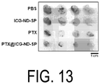
Classifications
-
- C—CHEMISTRY; METALLURGY
- C01—INORGANIC CHEMISTRY
- C01B—NON-METALLIC ELEMENTS; COMPOUNDS THEREOF; METALLOIDS OR COMPOUNDS THEREOF NOT COVERED BY SUBCLASS C01C
- C01B32/00—Carbon; Compounds thereof
- C01B32/25—Diamond
- C01B32/26—Preparation
-
- A—HUMAN NECESSITIES
- A61—MEDICAL OR VETERINARY SCIENCE; HYGIENE
- A61K—PREPARATIONS FOR MEDICAL, DENTAL OR TOILETRY PURPOSES
- A61K49/00—Preparations for testing in vivo
- A61K49/001—Preparation for luminescence or biological staining
- A61K49/0063—Preparation for luminescence or biological staining characterised by a special physical or galenical form, e.g. emulsions, microspheres
- A61K49/0069—Preparation for luminescence or biological staining characterised by a special physical or galenical form, e.g. emulsions, microspheres the agent being in a particular physical galenical form
- A61K49/0089—Particulate, powder, adsorbate, bead, sphere
- A61K49/0091—Microparticle, microcapsule, microbubble, microsphere, microbead, i.e. having a size or diameter higher or equal to 1 micrometer
- A61K49/0093—Nanoparticle, nanocapsule, nanobubble, nanosphere, nanobead, i.e. having a size or diameter smaller than 1 micrometer, e.g. polymeric nanoparticle
-
- A—HUMAN NECESSITIES
- A61—MEDICAL OR VETERINARY SCIENCE; HYGIENE
- A61K—PREPARATIONS FOR MEDICAL, DENTAL OR TOILETRY PURPOSES
- A61K31/00—Medicinal preparations containing organic active ingredients
- A61K31/33—Heterocyclic compounds
- A61K31/335—Heterocyclic compounds having oxygen as the only ring hetero atom, e.g. fungichromin
- A61K31/337—Heterocyclic compounds having oxygen as the only ring hetero atom, e.g. fungichromin having four-membered rings, e.g. taxol
-
- A—HUMAN NECESSITIES
- A61—MEDICAL OR VETERINARY SCIENCE; HYGIENE
- A61K—PREPARATIONS FOR MEDICAL, DENTAL OR TOILETRY PURPOSES
- A61K33/00—Medicinal preparations containing inorganic active ingredients
- A61K33/44—Elemental carbon, e.g. charcoal, carbon black
-
- A—HUMAN NECESSITIES
- A61—MEDICAL OR VETERINARY SCIENCE; HYGIENE
- A61K—PREPARATIONS FOR MEDICAL, DENTAL OR TOILETRY PURPOSES
- A61K41/00—Medicinal preparations obtained by treating materials with wave energy or particle radiation ; Therapies using these preparations
-
- A—HUMAN NECESSITIES
- A61—MEDICAL OR VETERINARY SCIENCE; HYGIENE
- A61K—PREPARATIONS FOR MEDICAL, DENTAL OR TOILETRY PURPOSES
- A61K41/00—Medicinal preparations obtained by treating materials with wave energy or particle radiation ; Therapies using these preparations
- A61K41/0052—Thermotherapy; Hyperthermia; Magnetic induction; Induction heating therapy
-
- A—HUMAN NECESSITIES
- A61—MEDICAL OR VETERINARY SCIENCE; HYGIENE
- A61K—PREPARATIONS FOR MEDICAL, DENTAL OR TOILETRY PURPOSES
- A61K41/00—Medicinal preparations obtained by treating materials with wave energy or particle radiation ; Therapies using these preparations
- A61K41/0057—Photodynamic therapy with a photosensitizer, i.e. agent able to produce reactive oxygen species upon exposure to light or radiation, e.g. UV or visible light; photocleavage of nucleic acids with an agent
-
- A—HUMAN NECESSITIES
- A61—MEDICAL OR VETERINARY SCIENCE; HYGIENE
- A61K—PREPARATIONS FOR MEDICAL, DENTAL OR TOILETRY PURPOSES
- A61K45/00—Medicinal preparations containing active ingredients not provided for in groups A61K31/00 - A61K41/00
-
- A—HUMAN NECESSITIES
- A61—MEDICAL OR VETERINARY SCIENCE; HYGIENE
- A61K—PREPARATIONS FOR MEDICAL, DENTAL OR TOILETRY PURPOSES
- A61K47/00—Medicinal preparations characterised by the non-active ingredients used, e.g. carriers or inert additives; Targeting or modifying agents chemically bound to the active ingredient
- A61K47/46—Ingredients of undetermined constitution or reaction products thereof, e.g. skin, bone, milk, cotton fibre, eggshell, oxgall or plant extracts
-
- A—HUMAN NECESSITIES
- A61—MEDICAL OR VETERINARY SCIENCE; HYGIENE
- A61K—PREPARATIONS FOR MEDICAL, DENTAL OR TOILETRY PURPOSES
- A61K47/00—Medicinal preparations characterised by the non-active ingredients used, e.g. carriers or inert additives; Targeting or modifying agents chemically bound to the active ingredient
- A61K47/50—Medicinal preparations characterised by the non-active ingredients used, e.g. carriers or inert additives; Targeting or modifying agents chemically bound to the active ingredient the non-active ingredient being chemically bound to the active ingredient, e.g. polymer-drug conjugates
- A61K47/51—Medicinal preparations characterised by the non-active ingredients used, e.g. carriers or inert additives; Targeting or modifying agents chemically bound to the active ingredient the non-active ingredient being chemically bound to the active ingredient, e.g. polymer-drug conjugates the non-active ingredient being a modifying agent
- A61K47/52—Medicinal preparations characterised by the non-active ingredients used, e.g. carriers or inert additives; Targeting or modifying agents chemically bound to the active ingredient the non-active ingredient being chemically bound to the active ingredient, e.g. polymer-drug conjugates the non-active ingredient being a modifying agent the modifying agent being an inorganic compound, e.g. an inorganic ion that is complexed with the active ingredient
-
- A—HUMAN NECESSITIES
- A61—MEDICAL OR VETERINARY SCIENCE; HYGIENE
- A61K—PREPARATIONS FOR MEDICAL, DENTAL OR TOILETRY PURPOSES
- A61K47/00—Medicinal preparations characterised by the non-active ingredients used, e.g. carriers or inert additives; Targeting or modifying agents chemically bound to the active ingredient
- A61K47/50—Medicinal preparations characterised by the non-active ingredients used, e.g. carriers or inert additives; Targeting or modifying agents chemically bound to the active ingredient the non-active ingredient being chemically bound to the active ingredient, e.g. polymer-drug conjugates
- A61K47/69—Medicinal preparations characterised by the non-active ingredients used, e.g. carriers or inert additives; Targeting or modifying agents chemically bound to the active ingredient the non-active ingredient being chemically bound to the active ingredient, e.g. polymer-drug conjugates the conjugate being characterised by physical or galenical forms, e.g. emulsion, particle, inclusion complex, stent or kit
- A61K47/6921—Medicinal preparations characterised by the non-active ingredients used, e.g. carriers or inert additives; Targeting or modifying agents chemically bound to the active ingredient the non-active ingredient being chemically bound to the active ingredient, e.g. polymer-drug conjugates the conjugate being characterised by physical or galenical forms, e.g. emulsion, particle, inclusion complex, stent or kit the form being a particulate, a powder, an adsorbate, a bead or a sphere
- A61K47/6923—Medicinal preparations characterised by the non-active ingredients used, e.g. carriers or inert additives; Targeting or modifying agents chemically bound to the active ingredient the non-active ingredient being chemically bound to the active ingredient, e.g. polymer-drug conjugates the conjugate being characterised by physical or galenical forms, e.g. emulsion, particle, inclusion complex, stent or kit the form being a particulate, a powder, an adsorbate, a bead or a sphere the form being an inorganic particle, e.g. ceramic particles, silica particles, ferrite or synsorb
-
- A—HUMAN NECESSITIES
- A61—MEDICAL OR VETERINARY SCIENCE; HYGIENE
- A61K—PREPARATIONS FOR MEDICAL, DENTAL OR TOILETRY PURPOSES
- A61K47/00—Medicinal preparations characterised by the non-active ingredients used, e.g. carriers or inert additives; Targeting or modifying agents chemically bound to the active ingredient
- A61K47/50—Medicinal preparations characterised by the non-active ingredients used, e.g. carriers or inert additives; Targeting or modifying agents chemically bound to the active ingredient the non-active ingredient being chemically bound to the active ingredient, e.g. polymer-drug conjugates
- A61K47/69—Medicinal preparations characterised by the non-active ingredients used, e.g. carriers or inert additives; Targeting or modifying agents chemically bound to the active ingredient the non-active ingredient being chemically bound to the active ingredient, e.g. polymer-drug conjugates the conjugate being characterised by physical or galenical forms, e.g. emulsion, particle, inclusion complex, stent or kit
- A61K47/6921—Medicinal preparations characterised by the non-active ingredients used, e.g. carriers or inert additives; Targeting or modifying agents chemically bound to the active ingredient the non-active ingredient being chemically bound to the active ingredient, e.g. polymer-drug conjugates the conjugate being characterised by physical or galenical forms, e.g. emulsion, particle, inclusion complex, stent or kit the form being a particulate, a powder, an adsorbate, a bead or a sphere
- A61K47/6927—Medicinal preparations characterised by the non-active ingredients used, e.g. carriers or inert additives; Targeting or modifying agents chemically bound to the active ingredient the non-active ingredient being chemically bound to the active ingredient, e.g. polymer-drug conjugates the conjugate being characterised by physical or galenical forms, e.g. emulsion, particle, inclusion complex, stent or kit the form being a particulate, a powder, an adsorbate, a bead or a sphere the form being a solid microparticle having no hollow or gas-filled cores
- A61K47/6929—Medicinal preparations characterised by the non-active ingredients used, e.g. carriers or inert additives; Targeting or modifying agents chemically bound to the active ingredient the non-active ingredient being chemically bound to the active ingredient, e.g. polymer-drug conjugates the conjugate being characterised by physical or galenical forms, e.g. emulsion, particle, inclusion complex, stent or kit the form being a particulate, a powder, an adsorbate, a bead or a sphere the form being a solid microparticle having no hollow or gas-filled cores the form being a nanoparticle, e.g. an immuno-nanoparticle
-
- A—HUMAN NECESSITIES
- A61—MEDICAL OR VETERINARY SCIENCE; HYGIENE
- A61K—PREPARATIONS FOR MEDICAL, DENTAL OR TOILETRY PURPOSES
- A61K49/00—Preparations for testing in vivo
- A61K49/001—Preparation for luminescence or biological staining
- A61K49/0013—Luminescence
- A61K49/0017—Fluorescence in vivo
- A61K49/0019—Fluorescence in vivo characterised by the fluorescent group, e.g. oligomeric, polymeric or dendritic molecules
- A61K49/0021—Fluorescence in vivo characterised by the fluorescent group, e.g. oligomeric, polymeric or dendritic molecules the fluorescent group being a small organic molecule
- A61K49/0032—Methine dyes, e.g. cyanine dyes
- A61K49/0034—Indocyanine green, i.e. ICG, cardiogreen
-
- A—HUMAN NECESSITIES
- A61—MEDICAL OR VETERINARY SCIENCE; HYGIENE
- A61K—PREPARATIONS FOR MEDICAL, DENTAL OR TOILETRY PURPOSES
- A61K49/00—Preparations for testing in vivo
- A61K49/001—Preparation for luminescence or biological staining
- A61K49/006—Biological staining of tissues in vivo, e.g. methylene blue or toluidine blue O administered in the buccal area to detect epithelial cancer cells, dyes used for delineating tissues during surgery
-
- A—HUMAN NECESSITIES
- A61—MEDICAL OR VETERINARY SCIENCE; HYGIENE
- A61K—PREPARATIONS FOR MEDICAL, DENTAL OR TOILETRY PURPOSES
- A61K9/00—Medicinal preparations characterised by special physical form
- A61K9/0012—Galenical forms characterised by the site of application
- A61K9/0019—Injectable compositions; Intramuscular, intravenous, arterial, subcutaneous administration; Compositions to be administered through the skin in an invasive manner
-
- A—HUMAN NECESSITIES
- A61—MEDICAL OR VETERINARY SCIENCE; HYGIENE
- A61K—PREPARATIONS FOR MEDICAL, DENTAL OR TOILETRY PURPOSES
- A61K9/00—Medicinal preparations characterised by special physical form
- A61K9/48—Preparations in capsules, e.g. of gelatin, of chocolate
- A61K9/50—Microcapsules having a gas, liquid or semi-solid filling; Solid microparticles or pellets surrounded by a distinct coating layer, e.g. coated microspheres, coated drug crystals
- A61K9/51—Nanocapsules; Nanoparticles
- A61K9/5107—Excipients; Inactive ingredients
- A61K9/5115—Inorganic compounds
-
- A—HUMAN NECESSITIES
- A61—MEDICAL OR VETERINARY SCIENCE; HYGIENE
- A61P—SPECIFIC THERAPEUTIC ACTIVITY OF CHEMICAL COMPOUNDS OR MEDICINAL PREPARATIONS
- A61P35/00—Antineoplastic agents
-
- A—HUMAN NECESSITIES
- A61—MEDICAL OR VETERINARY SCIENCE; HYGIENE
- A61P—SPECIFIC THERAPEUTIC ACTIVITY OF CHEMICAL COMPOUNDS OR MEDICINAL PREPARATIONS
- A61P43/00—Drugs for specific purposes, not provided for in groups A61P1/00-A61P41/00
-
- C—CHEMISTRY; METALLURGY
- C01—INORGANIC CHEMISTRY
- C01B—NON-METALLIC ELEMENTS; COMPOUNDS THEREOF; METALLOIDS OR COMPOUNDS THEREOF NOT COVERED BY SUBCLASS C01C
- C01B32/00—Carbon; Compounds thereof
- C01B32/15—Nano-sized carbon materials
-
- C—CHEMISTRY; METALLURGY
- C01—INORGANIC CHEMISTRY
- C01B—NON-METALLIC ELEMENTS; COMPOUNDS THEREOF; METALLOIDS OR COMPOUNDS THEREOF NOT COVERED BY SUBCLASS C01C
- C01B32/00—Carbon; Compounds thereof
- C01B32/25—Diamond
- C01B32/28—After-treatment, e.g. purification, irradiation, separation or recovery
-
- B—PERFORMING OPERATIONS; TRANSPORTING
- B82—NANOTECHNOLOGY
- B82Y—SPECIFIC USES OR APPLICATIONS OF NANOSTRUCTURES; MEASUREMENT OR ANALYSIS OF NANOSTRUCTURES; MANUFACTURE OR TREATMENT OF NANOSTRUCTURES
- B82Y5/00—Nanobiotechnology or nanomedicine, e.g. protein engineering or drug delivery
Definitions
- the present invention relates to a photo-exothermic composite material, a nanocluster, a substance delivery carrier and a pharmaceutical composition.
- ND may be used synonymously with CND
- nanodiamonds In addition to the inherent properties of diamonds, nanodiamonds (referred to hereinafter as "ND") also exhibit characteristics including a small average particle diameter and a large specific surface area, and provide merits such as being relatively inexpensive and easy to obtain. ND can be manufactured through an explosion method or a high-temperature / high-pressure method. ND also exhibit low toxicity, superior biocompatibility and stable fluorescence properties, and therefore applications in the biomedical field are being widely studied.
- CND carbon nanodiamonds
- Patent Document 1 a high-temperature/high-pressure method
- Patent Document 2 and 3 disclose modified CND and methods for manufacturing the same.
- Non-Patent Document 1 Although research pertaining to cancer hypothermia treatments using the photo-exothermic effect of CND has already been reported (Non-Patent Document 1), the photo-exothermic effect of the CND itself is very weak.
- Non-Patent Document 1 T. -K. Ryu, S. W. Baek, R. H. Kang, S. -W. Choi, Adv. Funct. Mater. 2016, 26, 6428 .
- a main object of the present invention is to provide a material that has a strong photo-exothermic effect and can be used in applications such as the treatment of tumors.
- the present invention provides the following photo-exothermic composite material, nanocluster, substance delivery carrier and pharmaceutical composition.
- the composite material of the present invention exhibits an excellent photo-exothermic property, and therefore when the composite material is distributed to a tumor site and irradiated with light, the composite material can exhibit an anti-tumor effect.
- nanoclusters further containing an anti-tumor agent are useful as pharmaceutical compositions.
- the photo-exothermic property can be further enhanced by including a fluorescent substance and pigment in the nanoclusters.
- the carbon nanomaterials (CNM) used as raw materials for the production of photo-exothermic composite materials include carbon nanodiamonds and carbon nanodots.
- a photo-exothermic composite material according to an embodiment of the present invention is represented by Formula (I) below: CNM-(Y 1 R) n1 (I) (wherein, CNM denotes a carbon nanomaterial; Y 1 denotes a divalent linking group; R denotes a group derived from a fluorescent substance or pigment; and n1 is an integer of 1 or greater).
- n1 denotes an integer of 1 or greater, and for example, denotes an integer from 1 to 30, from 1 to 20, from 1 to 15, from 1 to 10, from 1 to 8, or from 1 to 6.
- Examples of the divalent linking group denoted by Y 1 include -NH-, -O-, -COO-, -O-CO-, -CO-NH-, -NH-CO-, -NH-CO-O-, -O-CO-NH-, -O-CO-O-, and -NH-CO-NH-.
- R is a group derived from a fluorescent substance or pigment.
- examples of the fluorescent substance or pigment include quantum dots, Alexa Fluor-350, Alexa Fluor-430, Alexa Fluor-488, Alexa Fluor-532, Alexa Fluor-546, Alexa Fluor-555; Alexa Fluor-568, Alexa Fluor-594, Alexa Fluor-633, Alexa Fluor-647, Alexa Fluor-660, Alexa Fluor-680, Alexa Fluor-750, Cy2, Cy3, Cy3.5, Cy5, Cy5.5, Cy7, BODIPY CR-6G, BOPIPY 530/550, BODIPY FL, BODIPY 505/515, fluorescein isothiocyanate (FITC), eosin isothiocyanate, PE, Rhodamine B, BODIPY 580/605, Texas Red, APC, indocyanine green, 4-acetamido-4'-isothiocyanotostilbene-2,2'-disulfonic acid, acridine, acridine isothiocyanate
- the "group derived from a fluorescent substance or pigment” and denoted by R includes all moieties other than Y 1 derived from the raw materials used when binding the fluorescent substance or pigment. For example:
- R denotes a group derived from the ICG
- n1 is an integer of 1 or greater
- Y 1 is as described below.
- R is not the ICG itself, but includes a structure that emits the fluorescence of ICG, and is a group derived from ICG-NHS, which is a reagent for introducing ICG.
- R corresponds to the "group derived from a fluorescent substance or pigment”. The same applies to reagents for introducing fluorescent substances or pigments other than ICG-NHS.
- Y 1 is a compound in which an NH derived from an NH 2 group on the CNM surface is bonded with a CO derived from CO-O- (succinimide), which is a reactive group of the ICG-NHS, and Y 1 can be formed by bonding NH, CO, COO, O, or the like derived from a surface group (NH 2 , COOH, OH, or the like) of the CNM with an NH, CO, COO, O, CONH or the like derived from a reactive group (NH 2 , COOH, OH, NCO, acid halide, acid anhydride, active ester, halogen atom, or the like) of the reagent for introducing the fluorescent substance or pigment.
- a photo-exothermic composite material can be obtained by using, as necessary, a condensation agent (dicyclohexylcarbodiimide (DCC), water soluble DCC, carbonyldiimidazole, or the like) to react the CNM with the fluorescent substance or pigment.
- a condensation agent dicyclohexylcarbodiimide (DCC), water soluble DCC, carbonyldiimidazole, or the like
- the R group derived from a fluorescent substance or pigment
- examples of such a case are presented below.
- X denotes OH, a halogen atom (Cl, Br, I), or a group derived from an ester such as O-succinimidyl, maleimide, O-Me, OEt, and O-Bu
- R is a moiety excluding a reactive group (NH 2 , COOH, OH, COX) from the fluorescent substance or pigment, and corresponds to the group derived from a fluorescent substance or pigment
- n1 is an integer of 1 or greater.
- Carbon nanomaterials (CNM) having a large number of groups such as OH, COOH, and NH 2 on the surface are known, and a photo-exothermic composite material according to an embodiment of the present invention can be obtained in accordance with Schemes (1) to (10) below using such carbon nanomaterials.
- X denotes OH, a halogen atom (Cl, Br, I), or a group derived from an ester such as O-succinimidyl, maleimide, O-Me, OEt, or O-Bu;
- CNM denotes a carbon nanomaterial; n1 is an integer of 1 or greater; n2 is an integer of 0 or greater; and R is a group derived from a fluorescent substance or pigment.
- solvent examples include halogenated hydrocarbons such as chloroform, methylene chloride, and 1,2-dichloroethane; aromatic hydrocarbons such as toluene; tetrahydrofuran, diethyl ether, and diisopropyl ether.
- halogenated hydrocarbons such as chloroform, methylene chloride, and 1,2-dichloroethane
- aromatic hydrocarbons such as toluene
- tetrahydrofuran diethyl ether
- diisopropyl ether diisopropyl ether.
- the nanoclusters according to an embodiment of the present invention can be produced by suspending a photo-exothermic composite material in a suitable aqueous medium such as water or a buffer solution, and self-assembling through sonication. After sonication, the self-assembled nanoclusters can be separated by removing, through a purification operation such as centrifugation, any photo-exothermic composite material that did not form nanoclusters.
- a suitable aqueous medium such as water or a buffer solution
- the nanoclusters according to an embodiment of the present invention can be complexed with an active ingredient by being suspended in a suitable solvent containing the active ingredient.
- the active ingredient is present within or on the surface of the nanoclusters.
- the active ingredient include physiologically active substances as well as the fluorescent substances, and pigments.
- a nanocluster according to an embodiment of the present invention complexed with an active ingredient gradually releases the active ingredient when administered within or applied to the living body or skin of a mammal (such as a human, mouse, rat, hamster, horse, cow, pig, goat, sheep, rabbit, dog, or cat).
- a mammal such as a human, mouse, rat, hamster, horse, cow, pig, goat, sheep, rabbit, dog, or cat.
- the active ingredient is a physiologically active substance
- the nanoclusters function as delivery carriers for the physiologically active substance (drugs in particular).
- the active ingredient is a fluorescent substance or pigment
- the active ingredient can enhance photo-exothermic properties, and is useful for thermal therapy and photodynamic therapy of cancer. Such an active ingredient is also useful for bioimaging.
- nanoclusters according to an embodiment of the present invention complexed with an active ingredient are useful as pharmaceutical products or pharmaceutical compositions.
- dosage forms of pharmaceutical products include tablets, capsules, lozenges, pills, chewable tablets, injectable agents, suppositories, syrups, ointments, and plaster agents.
- the nanoclusters according to an embodiment of the present invention gradually release the active ingredient, and thus are also useful as delivery carriers of active ingredients.
- Physiologically active substances include drugs, nucleic acids, and proteins.
- drugs include, but are not limited to, anti-tumor agents, anti-hypertension agents, anti-hypotensive agents, anti-psychotic agents, analgesics, antidepressants, antimanic agents, anti-anxiety agents, sedatives, hypnotics, anti-epileptic agents, opioid agonists, asthma therapeutics, anesthetics, anti-arrhythmia agents, arthritis therapeutic agents, anti-spasmodic agents, ACE inhibitors, decongestants, antibiotics, anti-angina agents, diuretics, anti-Parkinson's disease agents, bronchodilators, antidiuretics, diuretics, anti-hyperlipidemia agents, immunosuppressive agents, immunomodulatory agents, anti-emetics, anti-infective agents, anti-neoplastic agents, antifungal agents, antiviral agents, anti-diabetic agents, anti-allergenic agents, antipyretics
- a preferred drug is an anti-tumor agent.
- anti-tumor agents include hormonal therapeutic agents (e.g., fosfestrol, diethylstilbestrol, chlorotrianiserin, medroxyprogesterone acetate, megestrol acetate, chlormadinone acetate, cyproterone acetate, danazol, allylestrenol, gestrinone, mepartricin, raloxifene, ormeloxifene, levormeloxifene, anti-estrogens (e.g., tamoxifen citrate, toremifene citrate), pill preparations, mepitiostane, testolactone, aminoglutethimide, LH-RH agonists (e.g., goserelin acetate, buserelin, leuprorelin), droloxifene, epitiostanol, ethynyl estradiol sulfonate,
- cancers targeted by anti-tumor agents include colorectal cancer, liver cancer, kidney cancer, head and neck cancer, esophageal cancer, stomach cancer, biliary cancer, gallbladder/bile duct cancer, pancreatic cancer, lung cancer, breast cancer, ovarian cancer, cervical cancer, uterine cancer, bladder cancer, prostate cancer, testicular tumors, bone and soft tissue sarcomas, leukemia, malignant lymphoma, multiple myeloma, skin cancer, and brain tumors.
- Preferable examples includes colorectal cancer, stomach cancer, head and neck cancer, lung cancer, breast cancer, pancreatic cancer, biliary cancer, and liver cancer.
- the nucleic acids are not particularly limited and may be DNA, RNA, chimeric nucleic acids of DNA and RNA, or hybrids of DNA/RNA. Further, the nucleic acid can be any of 1 to 3 strands, but is preferably single-stranded or double-stranded.
- the nucleic acid may be another nucleotide that is an N-glycoside of a purine or pyrimidine base, or another oligomer having a non-nucleotide skeleton (e.g., a commercially available peptide nucleic acid (PNA), etc.) or other oligomer with a special bond (provided that the oligomer contains a nucleotide having an arrangement that allows pairing of bases or attachment of bases as found in DNA or RNA).
- PNA peptide nucleic acid
- the nucleic acid may also be one with a known modification.
- nucleic acids having one or more natural nucleotides substituted with an analog examples include labeled nucleic acids known in the relevant technical field, capped nucleic acids, methylated nucleic acids, nucleic acids having one or more natural nucleotides substituted with an analog, nucleic acids subjected to intramolecular nucleotide modifications, for example, those with uncharged bonds (e.g., methylphosphonates, phosphotriesters, phosphoramidates, and carbamates, etc.), nucleic acids having a charged bond or a sulfur-containing bond (e.g., phosphorothioate, and phosphorodithioate, etc.), for example, nucleic acids having a side chain group such as a protein (nuclease, nuclease inhibitor, toxin, antibody, and signal peptide, etc.) or sugars (e.g., monosaccharides, etc.), nucleic acids having an intercalated compound (e.g.,
- siRNA is a double-stranded oligo RNA consisting of a nucleotide sequence of the mRNA or initial transcript of a target gene or a nucleotide sequence homologous to a partial sequence (preferably within the coding region) of the nucleotide sequence thereof (including the intron portion in the case of an initial transcript), and the complementary strand thereof.
- the length of the portion that is homologous to the target nucleotide and is contained in the siRNA is usually greater than or equal to approximately 18 bases, and for example, the length may be approximately 20 bases (typically, an approximate length from 21 to 23 bases). However, the length thereof is not particularly limited as long as RNA interference can be caused.
- the total length of the siRNA is approximately greater than or equal to 18 bases, and for example, the total length may be approximately 20 bases (typically, an approximate total length from 21 to 23 bases). However, the total length is not particularly limited as long as RNA interference can be caused.
- the relationship between the target nucleotide sequence and the sequence homologous thereto and contained in the siRNA may be a 100% match, or base mutations may be present (within a range of sameness of at least 70%, preferably 80% or higher, more preferably 90% or higher, and most preferably 95% or higher).
- the siRNA may have, at the 5' or 3' end, additional bases that consist of no more than 5 bases, and preferably 2 bases, and which do not form a base pair.
- the additional base may be DNA or RNA, but when DNA is used, the stability of the siRNA can be improved. Examples of sequences of such additional bases include, but are not limited to, ug-3', uu-3', tg-3', tt-3', ggg-3', guuu-3', gttt-3', ttttt-3', and uuuuu-3'.
- the siRNA may be for any target gene.
- the siRNA is preferably one that targets genes for which an enhanced expression is involved in the onset and/or exacerbation of a disease of interest, and more specifically, is one that targets genes for which an antisense nucleic acid to the gene thereof has advanced to a clinical or preclinical stage, or newly known genes.
- the siRNA may be used singly, or in a combination of two or more .
- proteins include enzymes, receptors, antibodies, antigens, and cytokines such as interferons and interleukins.
- the approximate average particle diameter of the nanocluster according to an embodiment of the present invention is from 1 to 1000 nm, preferably from 3 to 800 nm, more preferably from 5 to 500 nm, even more preferably from 10 to 300 nm, and particularly from 30 to 250 nm.
- the approximate zeta potential of the nanocluster is preferably from 5 to 30 mV, and more preferably from 10 to 25 mV.
- the approximate content of the active ingredient is preferably from 5 to 50 parts by mass, and more preferably from 10 to 20 parts by mass, per 100 parts by mass of the nanoclusters.
- ND (diameter: from 4 to 5 nm) were synthesized according to a previously reported preparation method ( V V. Danilenko, Combust., Explos. Shock Waves 2005, 41, 577 ; V. Y. Dolmatov, J. Superhard Mater. 2008, 30, 233 ; V. Y, Dolmatov, V. Myllymaki and A. Vehanen, J. Superhard Mater. 2013, 35, 143 .), The synthesized ND were purified by nitric acid and fired in a hydrogen gas atmosphere.
- the elements of the ND [contents of C (92.20%), H (0.74%), and N (2.30%)] were analyzed using an organic element analyzer (Micro Corder JM10; J-Science Lab Co., Ltd, Kyoto, Japan).
- the purified ND were dispersed in distilled water by a bead mill (Sand Grinder LSG-4U; Aimex Co., Ltd, Tokyo, Japan).
- the ND dispersion was centrifuged to remove the ND insoluble in water.
- the resulting supernatant dispersion (ND-ori) was used in an additional experiment.
- ND-ori dispersion 30 mg ml -1
- ICG-NHS ICG-NHS
- MES 2-(N-morpholino)ethane sulfonic acid
- Pellets obtained by centrifugation were re-dispersed in 1 ml of Mill-Q water by irradiating with ultrasonic waves for 10 minutes using a pulsed sonicator (VCX-600; Sonics, Danbury, CT, USA).
- VCX-600 Pulsonicator
- the ICG-ND-SP dispersion was used in a subsequent experiment.
- the ICG concentration (up to 0.75 mg ml -1 ) and the ND concentration (up to 3 mg ml -1 ) in the ICG-ND-SP dispersion were estimated using an UV-Vis-NIR spectrophotometer (V-730 BIO; Jasco, Tokyo, Japan) and thermogravimetric (TGA) measurements (using the Q500 thermogravimetric analyzer; TA Instruments, New Castle, DE, USA).
- BODIPY-ND-SP and Alexa568-ND-SP were synthesized by the same method used to synthesize the ICG-ND-SP. Specifically, BODIPY FL NHS Ester (Succinimidyl Ester) (1 mg; Invitrogen, Carlsbad, CA, USA) or Alexa Fluor 568 NHS Ester (Succinimidyl Ester) (1 mg; Invitrogen) was added to the reaction solution instead of ICG-NHS. After preparation, it was confirmed through measurements with a UV-Vis-NIR spectrophotometer that BODIPY (ca. 0.04 mg ml -1 ) and Alexa Fluor 568 molecules (ca. 0.04 mg ml -1 ) covalently bonded to the ND surface (ca. 3 mg ml -1 ).
- MES buffer solution pH 6.0, 100 mM
- the reaction solution was then vigorously stirred using a stirrer at room temperature for 1.5 hours, after which the solution was centrifuged and washed three times with Milli-Q water, and unreacted material was thus removed.
- the obtained pellets were re-dispersed in 10 ml of Mill-Q water through sonication for 10 minutes using a pulsed sonicator (VCX-600: Sonics, Danbury, CT, USA).
- the obtained PBA-ND-SP dispersion (PBA concentration: up to 0.15 mg ml -1 , ND concentration: up to 3 mg ml -1 ) was used for further experiments.
- APTX@ICG-ND-SP complex was prepared by the following method. 10 mg of PTX (Wako) and 10 ml of Milli-Q were added to the washed ICG-ND-SP pellets, after which the mixture was sonicated for 10 minutes using a pulsed sonicator. The resulting mixture was washed with Milli-Q water using a centrifuge and then stored at 4°C until used.
- PTX@ND-ori was prepared by sonicating 10 mg of PTX, 1 ml of ND-ori, and 9 ml of Milli-Q water using a pulsed sonicator for 10 minutes.
- Abraxane was purchased from Taiho Pharmaceutical Co., Ltd. and used as is in experiments without any treatment such as chemical modification.
- the hydrodynamic diameter of the ND-SP was measured by dynamic light scattering (DLS) (using the Photal FPAR-1000; Otsuka Electronics, Osaka, Japan).
- the spectral profile and concentration of a complex of ND-SP, fluorescent pigment, and PTX were measured using a UV-Vis-NIR spectrophotometer.
- the fluorescence properties of the ND-SP, fluorescent pigment, and ND-ori were analyzed using a fluorometer (FP-6500 or FP-8500; Jasco, Tokyo) and a microplate reader (Infinite M200 PRO; Tecan, Mannedorf, Switzerland).
- Human osteosarcoma cells (U2OS) and human ovarian adenoma cells (SKOV3) were obtained from the Japanese Collection of Research Bioresources Cell Bank (Tokyo, Japan), and were cultured with Dulbecco's Modified Eagle's Medium (DMEM) (Gibco, Grand Island, NY, USA) containing 10% fetal bovine serum, 2 mM L-glutamine, 1 mM sodium pyruvate, gentamycin, penicillin-streptomycin (100 IU ml -1 ), and Hank's balanced salt solution (Life Technologies, Carlsbad, CA, USA). The cells were cultured in a humidified chamber at 37°C with a 5% CO 2 atmosphere.
- DMEM Dulbecco's Modified Eagle's Medium
- Cell viability was evaluated using the Cell Counting Kit (CCK)-8 (Dojindo Laboratories, Kumamoto, Japan) according to the manual thereof. The cells were seeded onto a 96-well plate ( ⁇ 10 3 cells well -1 ) and incubated overnight. Next, the cells were exposed to a drug or nanocomplex dispersion solution and washed with a fresh culture solution, and then incubated in a CCK-8 solution. Finally, cell viability was calculated by measuring the absorbance at 450 nm with a microplate reader.
- CCK Cell Counting Kit
- the actin cytoskeleton
- Phallodin Thermo Fisher Scientific
- the cells were stained for 10 minutes at room temperature using a 4% paraformaldehyde solution or a pre-chilled methanol/acetone mixture (v:v of 1:1), after which the cells were washed with a PBS buffer solution and then observed.
- NIR fluorescence images of the ICG-ND-SP were observed using a fluorescence microscope equipped with a mirror unit (IRDYE800-33LP-A-U01; Semrock) and an electronically-magnifying CCD camera (DP80; Olympus).
- a confocal laser microscope (LSM 5 PASCAL, Carl Zeiss Inc., Tokyo, Japan) was used for intra-cellular imaging of the PBA-ND-SP, BODIPY-ND-SP, and Alexa568-ND-SP complexes.
- the temperature change of the laser irradiated ICG-ND-SP dispersion was examined in the following manner. Milli-Q water (200 ⁇ l, ICG and ND concentrations of 0.75 mg ml -1 and 3 mg ml -1 , respectively) in which ICG-ND-SP was dispersed was added to a quartz cell (GL Science, Tokyo, Japan). The sample was irradiated for 5 minutes with a fiber-coupled continuous wave laser (laser spot diameter: up to 4 mm; maximum power: 1 W, up to 80 mW mm -2 ; BRM-785-1.0-100-0.22-SMA; B&W Tek, Newark, DE, USA) at a wavelength of 785 nm.
- a fiber-coupled continuous wave laser laser spot diameter: up to 4 mm; maximum power: 1 W, up to 80 mW mm -2 ; BRM-785-1.0-100-0.22-SMA; B&W Tek, Newark, DE, USA
- a PBS buffer solution not containing ICG-ND-SP was also laser irradiated as a control.
- the temperature change of the solution upon laser irradiation was measured using a temperature sensor (AD-5601A; A&D, Tokyo, Japan).
- the ICG and ND concentrations in the ICG-NHS and ND-ori solutions were adjusted in advance to be the same as the ICG and ND concentrations in the ICG-ND-SP complex measured using a UV-Vis-NIR spectrophotometer.
- the effect of the concentrations of the ICG and ND on the photo-exothermic behavior of the ICG-ND-SP complex was examined by diluting the ICG-ND-SP dispersion (stock solution) with a DMEM culture medium.
- ROS analysis was implemented using a 96-well plate (Thermo Fisher Scientific) with a transparent bottom and a black main body, and Singlet Oxygen Sensor Green (SOSG) (Invitrogen) as a singlet oxygen detection reagent.
- Milli-Q water (100 ⁇ L) in which ICG-ND-SP was dispersed was diluted with a PBS buffer solution containing the SOSG.
- the final concentrations in the system of ICG, ND, and SOSG were 75 ⁇ g ml -1 , 300 ⁇ g ml -1 , and 1 ⁇ M, respectively.
- the sample was irradiated for 5 minutes with a near-infrared laser having a wavelength of 785 nm at an output of 1 W (ca. 80 mW mm -2 ).
- a PBS buffer solution not containing a nanocomplex was used as a control.
- Green fluorescence associated with ROS generation was measured using a microplate reader (excitation wavelength: 485 nm, fluorescence wavelength: 535 nm).
- SKOV3 cells (5 ⁇ 10 3 cells/well) were seeded onto a 96-well plate and incubated overnight to evaluate cell viability.
- the cells were treated with 100 ⁇ l of a DMEM culture medium containing various samples (ND-ori, ICG, ICG-ND-SP, PTX@ICG-ND-SP) or a PBS buffer solution not containing a nanocomplex, and then irradiated with a laser (wavelength of 785 nm, output of 1 W, up to 80 mW mm -2 ). After laser irradiation, the cells were washed and incubated in a fresh culture medium.
- SPF Specific Pathogen Free
- Cancer model mice bearing SKOV3 cells were created by injecting, into both thighs of the mice, 100 ⁇ l of a mixed solution (v/v of 1:1) of culture medium/Matrigel (Dow Corning, Corning, NY, USA) containing a quantity of 1 ⁇ 10 6 cells.
- mice were divided into four groups of three mice each, and 50 ⁇ l of various samples [sterile water containing PBS and ICG-ND-SP (ICG, 1.88 mg kg -1 ; ND-ori, 7.5 mg kg -1 ), sterile water containing ND-ori (7.5 mg kg -1 ), and sterile water containing PTX@ICG-ND-SP (PTX, 1.25 mg kg -1 ; ICG, 1.88 mg kg -1 ; ND-ori, 7.5 mg kg -1 ) was administered by syringe into the solid cancers.
- ICG 1.88 mg kg -1 ; ND-ori, 7.5 mg kg -1
- ⁇ indicates the standard deviation
- n denotes the number of samples that were used.
- the Student's t-test method was used for statistical analysis of data.
- the symbols ⁇ , ⁇ , and ⁇ indicate p values of ⁇ 0.05, ⁇ 0.005, and ⁇ 0.001, respectively.
Abstract
Description
- The present invention relates to a photo-exothermic composite material, a nanocluster, a substance delivery carrier and a pharmaceutical composition.
- The following abbreviations are used in the present specification and drawings.
- ND: nanodiamond
- CND: carbon nanodiamond
- CNDp: carbon nanodiamond particle
- SP: super nanoparticle (or cluster)
- ICG: Indocyanine Green
- PTX: Paclitaxel
- MES: 2-(N-morpholino)ethanesulfonate
- ND-ori: A raw material (original nanodiamond)
- PTX@ICG-ND-SP: A complex of PTX and a cluster (SP) configured from ICG-modified ND
- ICG-ND-SP: Nanoclusters in which a CND composite material to which ICG is bonded is self-assembled
- In the present specification, ND may be used synonymously with CND
- In addition to the inherent properties of diamonds, nanodiamonds (referred to hereinafter as "ND") also exhibit characteristics including a small average particle diameter and a large specific surface area, and provide merits such as being relatively inexpensive and easy to obtain. ND can be manufactured through an explosion method or a high-temperature / high-pressure method. ND also exhibit low toxicity, superior biocompatibility and stable fluorescence properties, and therefore applications in the biomedical field are being widely studied.
- In addition to the inherent properties of diamonds, carbon nanodiamonds (referred to hereinafter as "CND") also exhibit characteristics including a small average particle diameter and a large specific surface area, and provide merits such as being relatively inexpensive and easy to obtain. CND can be manufactured by methods such as an explosion method or a high-temperature/high-pressure method (Patent Document 1). CND also exhibit low toxicity, superior biocompatibility, and stable fluorescence properties, and therefore applications in the biomedical field are being widely studied. Furthermore,
Patent Documents - Although research pertaining to cancer hypothermia treatments using the photo-exothermic effect of CND has already been reported (Non-Patent Document 1), the photo-exothermic effect of the CND itself is very weak.
-
- Patent Document 1:
JP 2010-202458 A - Patent Document 2:
JP 2017-186234 A - Patent Document 3:
JP 2017-186235 A - Non-Patent Document 1: T. -K. Ryu, S. W. Baek, R. H. Kang, S. -W. Choi, Adv. Funct. Mater. 2016, 26, 6428.
- A main object of the present invention is to provide a material that has a strong photo-exothermic effect and can be used in applications such as the treatment of tumors.
- The present invention provides the following photo-exothermic composite material, nanocluster, substance delivery carrier and pharmaceutical composition.
- 1. A photo-exothermic composite material represented by Formula (I) below: CNM-(Y1-R)n1 (I)
(wherein, CNM denotes a carbon nanomaterial; Y1 denotes a divalent linking group; R denotes a group derived from a fluorescent substance or pigment; and n1 is an integer of 1 or greater). - 2. The photo-exothermic composite material according to 1, wherein the divalent linking group denoted by Y1 is selected from the group consisting of -NH-, - O-, -CO-O-, -O-CO-, -CO-NH-, -NH-CO-, -NH-CO-O-, -O-CO-NH-,-O-CO-O-, and -NH-CO-NH-.
- 3. The photo-exothermic composite material according to 1 or 2, wherein the carbon nanomaterial is a carbon nanodiamond or carbon nanodots.
- 4. The photo-exothermic composite material according to 3, wherein the carbon nanomaterial is a carbon nanodiamond.
- 5. The photo-exothermic composite material according to any one of 1 to 4,
wherein the carbon nanomaterial has at least one surface group selected from the group consisting of OH, COOH, and NH2, and the group derived from a fluorescent substance or pigment and denoted by R is bonded to the carbon nanomaterial through the surface group. - 6. A nanocluster in which the photo-exothermic composite material described in any of 1 to 5 is self-assembled.
- 7. The nanocluster according to 6, further complexed with an active ingredient.
- 8. The nanocluster according to 7, wherein the active ingredient is a physiologically active substance, a fluorescent substance, or a pigment.
- 9. A delivery carrier for an active ingredient, the delivery carrier containing the nanocluster described in 7 or 8.
- 10. A pharmaceutical composition containing the nanocluster described in 7 or 8, the nanocluster being complexed with a drug.
- The composite material of the present invention exhibits an excellent photo-exothermic property, and therefore when the composite material is distributed to a tumor site and irradiated with light, the composite material can exhibit an anti-tumor effect. Also, nanoclusters further containing an anti-tumor agent are useful as pharmaceutical compositions. The photo-exothermic property can be further enhanced by including a fluorescent substance and pigment in the nanoclusters.
-
-
FIG. 1 illustrates the synthesis and characterization of ICG-ND-SP.FIG. 1(a) outlines a scheme for synthesizing ICG-ND-SP. An amino group of ND-ori and a succinimidyl group of ICG-NHS are covalently bonded through a dehydration condensation reaction, after which a self-assembled ICG-ND-P nanocluster (ICG-ND-SP fluorescent nanocluster) is prepared by sonication.FIG. 1(b) is a graph showing a size distribution analysis of the ICG-ND-SP through dynamic light scattering (DLS).FIG. 1(c) is a transmission electron microscope (TEM) image of the ICG-ND-SP. The top right photograph is a highly magnified TEM image of the ICG-ND-SP.FIG. 1(d) is a graph showing a UV-Vis-NIR absorption spectral analysis of an ICG-ND-SP dispersed aqueous solution (ICG concentration = 18.75 µg ml-1, ND concentration = 75 µg ml-1) and of ICG-NHS (ICG concentration = 18.75 µg ml-1).FIG. 1(e) is a graph showing the fluorescence (FL) intensity of the ICG-ND-SP dispersed aqueous solution and an ICG-NHS (ICG concentration = 2 µg ml-1) aqueous solution.FIG. 1(f) is a graph showing the FL intensity analysis of an ICG-NHS aqueous solution and an ICG-ND-SP dispersed aqueous solution in which the ICG concentration was adjusted in advance to 0.75 µg ml-1. The ND concentration was 3 µg ml-1. -
FIG. 2 illustrates the intracellular penetration and distribution behavior of ICG-ND-SP.FIG. 2(a) is a graph showing the cytotoxicity of a variety of fluorescent molecule-modified ND-SP. Cytotoxicity was evaluated after incubation of various ND-SP in human osteosarcoma cells (U2OS) for 24 hours.FIG. 2(b) presents fluorescence microscope images of U2OS cells when ICG (ICG concentration = 1.5 µg ml-1) or ICG-ND-SP (ICG and ND concentrations adjusted to 1.5 and 6 µg ml-1, respectively) was incubated for 4 hours in a 37°C, 5% CO2 environment. The cells were washed with PBS and then fixed with 4% paraformaldehyde (PFA). -
FIG. 3 illustrates the in vitro anticancer effect of PTX@ICG-ND-SP as a drug carrier.FIG. 3(a) outlines a method for synthesizing PTX@ICG-ND-SP using noncovalent interaction between PTX and ICG-ND-SP.FIG. 3(b) is a graph showing the UV-Vis-NIR absorption spectra of ICG-ND-SP before being complexed with PTC (ICG-ND-SP) and after being complexed with PTX (PTX@ICG-ND-SP).FIG. 3(c) is a graph showing DLS measurement results of ICG-ND-SP and PTX@ICG-ND-SP.FIG. 3(d) is a graph showing the anticancer activity of Abraxane (an FDA approved and commercially available PTX-based nanomedicine), PTX@ND-ori, and PTX@ICG-ND-SP against human ovarian cancer cells (SKOV3) after 24 hours of incubation. -
FIG. 4 presents graphs of evaluations of the in vitro anticancer activity of photo-introduced ICG-ND-SP.FIG. 4(a) is a graph of temperature measurements of a PBS buffer solution, ND-ori, ICG, and ICG-ND-SP, which were irradiated with a laser, the temperature being measured at each laser irradiation time. The ICG and ND concentrations were set to 0.75 and 3 mg ml-1, respectively.FIG. 4(b) is a graph showing the impact of the ICG-ND-SP concentration on the increase in temperature at each laser irradiation time.FIG. 4(c) is a graph showing the ROS generation behavior from the PBS buffer solution, ND-ori, ICG, and ICG-ND-SP when irradiated and not irradiated with a laser. The ICG and ND concentrations were set to 75 and 300 µg ml-1, respectively.FIG. 4(d) is a graph showing the cell viability percentage of SKOV3 cells at each laser irradiation time of the PBS buffer solution, ND-ori, ICG, and ICG-ND-SP, which were irradiated with a laser.FIG. 4(e) is a graph showing the cell viability percentage after 24 hours following laser irradiation. The ICG and ND concentrations were set to 25 and 100 µg ml-1, respectively.FIG. 4(f) is a graph showing the viability percentage of SKOV3 cells exposed to ICG-ND-SP and PTX@ICG-ND-SP incubated for 24 hours after laser irradiation for 3 minutes. The PTX, ICG, and ND concentrations were set to 10, 75, and 300 µg ml-1, respectively. -
FIG. 5 illustrates the in vivo anticancer action of ICG-ND-SP as a drug carrier.FIG. 5(a) outlines a method for an in vivo anticancer experiment using ICG-ND-SP.FIG. 5(b) is a graph of the body weight of mice subjected to different treatments, andFIG. 5(c) is a graph of the solid cancer volume with the different treatments. The PBS buffer solution and sterile water in which was dispersed ND-ori, ICG-ND-SP, or PTX@ICG-ND-SP was administered into the abdominal cavity. The solutions were administered with the dosage concentrations set to 5 mg kg-1 for the PTX, 7.5 mg kg-1 for the ICG, and 30 mg kg-1 for the ND -
FIG. 6(a) is thermal images of the mouse body surface when irradiated with a 785 nm laser (laser output = 1 W (up to 80 mW/mm2), laser irradiation time = 180 sec) and administered PBS, ND-ori, ICG-ND-SP or PTX@ICG-ND-SP.FIG. 6(b) is a graph of the temperature of the solid cancer. The dosage concentrations were set to 1.25 mg kg-1 for the PTX, 1.88 mg kg-1 for the ICG, and 7.5 mg kg-1 for the ND. The mouse body surface at the time of thermal measurements was approximately 33°C. -
FIG. 7 illustrates an in vivo verification of the photo-exothermic action, photodynamic action, and pharmacotherapeutic action.FIG. 7(a) outlines an experimental design for verifying multidimensional anticancer effects using ICG-ND-SP.FIG. 7(b) presents photographs after 7 days and after 14 days of mice in which PBS, ND-ori, ICG-ND-SP, or PTX@ICG-ND-SP was administered directly to the solid cancer (SKOV3), and the solid cancer on the right side was irradiated with a laser (laser intensity = 1 W (up to 80 mW/mm2), laser irradiation time = 180 sec).FIG. 7(c) is a graph showing the size of the solid cancer on the right side, which was irradiated with a 785 nm laser.FIG. 7(d) is a graph of the size of the solid cancer on the left side, which was not irradiated with a laser.FIG. 7(e) is a graph of the transition in body weight after laser irradiation. The dosage concentrations were set to 1.25 mg kg-1 for the PTX, 1.88 mg kg-1 for the ICG, and 7.5 mg kg-1 for the ND -
FIG. 8 illustrates the in vivo distribution behavior of PTX@ICG-ND-SP obtained through near-infrared fluorescence bio-imaging.FIG. 8(a) presents fluorescence images of a mouse having solid cancer of SKOV3 cells and for which PTX@ICG-ND-SP (PTX concentration = 5 mg kg-1, ICG concentration = 7.5 mg kg-1, ND concentration = 30 mg kg-1) was administered through the caudal vein. The dashed circle indicates the position of the solid cancer.FIG. 8(b) is a graph of the radiation efficiency corresponding to the solid cancer at different times along the time axis.FIG. 8(c) is an ex vivo fluorescence image of isolated solid cancer and organs.FIG. 8(d) is a graph showing the quantitative radiation efficiency with respect to the solid cancer and organs at 24 hours after the administration of PTX@ICG-ND-SP. -
FIG. 9 illustrates the synthesis and characterization of ND-SP with various chemically modified fluorescent pigments.FIG. 9(a) is TEM images of ND-ori and ND-SP with chemically modified fluorescent pigments.FIG. 9(b) is a graph showing the DLS analysis of ND-ori and ND-SP with chemically modified fluorescent pigments.FIG. 9(c) is a graph of TGA curves for ND-ori and ICG-ND-SP. -
FIG. 10 presents graphs of evaluations of the optical characteristics of ND-SP with various chemically modified fluorescent pigments.FIGS. 10(a) to (c) are graphs of UV-Vis-NIR absorption spectra of various ND-SP. InFIG. 10(a) , the PBA (1-pyrenebutyric acid) concentration was 14.4 µg ml-1. The concentrations of PBA and ND in the PBA-ND-SP were set to 1.5 and 30 µg ml-1, respectively. InFIG. 10(b) , the BODIPY-NHS concentration was 1.34 µg ml-1. The concentrations of BODIPY and ND in BODIPY-ND-SP were set to 1 and 75 µg ml-1, respectively. InFIG. 10(c) , the concentration of Alexa568-NHS was 6.9 µg ml-1. The concentrations of Alexa568 and ND in the Alexa568-ND-SP were set to 4 and 30 µg ml-1, respectively.FIGS. 10(d) to (f) are graphs of the fluorescence (FL) intensity of various ND-SP. InFIG. 10(d) , the PBA concentration was 0.14 ng ml-1. The concentrations of PBA and ND in the PBA-ND-SP were set to 3 and 60 ng ml-1, respectively. InFIG. 10(e) , the BODIPY-NHS concentration was 5 ng ml-1. The concentrations of BODIPY and ND in the BODIPY-ND-SP were set to 40 and 3000 ng ml-1, respectively. InFIG. 10(f) , the Alexa568-NHS concentration was 5 ng ml-1. The concentrations of Alexa568 and ND in the Alexa568-ND-SP were set to 20 and 1500 ng ml-1, respectively. -
FIG. 11 presents images of fluorescence microscope observations of various ND-SP.FIG. 11(a) presents near infrared fluorescent live imaging of ICG-ND-SP (ICG concentration = 1.5 µg ml-1, ND concentration = 6 µg ml-1), ICG (1.5 µg ml-1), and ND-ori (ND concentration = 6 µg ml-1) after incubation with U2OS cells in a 5% CO2 atmosphere for 4 hours at 37°C. A control experiment in which a fluorescent pigment was not used was also conducted under the same conditions.FIG. 11(b) shows fluorescence microscope images of U2OS cells when PBA-ND-SP (PBA concentration = 0.3 µg ml-1, ND concentration = 6 µg ml-1) was incubated for 24 hours.FIG. 11(c) presents fluorescence microscope images of U2OS cells when Alexa568-ND-SP (Alexa568 concentration = 0.8 µg ml-1, ND concentration = 60 µg ml-1) or BODIPY-ND-SP (BODIPY concentration = 0.8 µg ml-1, ND concentration = 60 µg ml-1) was incubated for 6 hours. The nuclei and cytoskeletons (actin) were stained using Hoechst 33342 and Phallodin, respectively. The cells shown inFIGS. 11(b) and (c) were fixed with 4% PFA. -
FIG. 12 illustrates the dispersion stability of PTX@ICG-ND-SP in water.FIG. 12(a) is photographs when PTX@ICG-ND-SP (PTX, ICG, and ND-ori concentrations of 0.2, 0.06, and 0.6 mg ml-1, respectively) was incubated for 7 days in MilliQ water.FIG. 12(b) is a graph of DLS analysis results at various points in time (0, 1, 2, 3 and 7 days) of the PTX@ICG-ND-SP aqueous solution. -
FIG. 13 is a photograph of solid cancers isolated after the end of the experiment. - In the present specification, the carbon nanomaterials (CNM) used as raw materials for the production of photo-exothermic composite materials include carbon nanodiamonds and carbon nanodots.
- A photo-exothermic composite material according to an embodiment of the present invention is represented by Formula (I) below:
CNM-(Y1R)n1 (I)
(wherein, CNM denotes a carbon nanomaterial; Y1 denotes a divalent linking group; R denotes a group derived from a fluorescent substance or pigment; and n1 is an integer of 1 or greater). - Here, n1 denotes an integer of 1 or greater, and for example, denotes an integer from 1 to 30, from 1 to 20, from 1 to 15, from 1 to 10, from 1 to 8, or from 1 to 6.
- Examples of the divalent linking group denoted by Y1 include -NH-, -O-, -COO-, -O-CO-, -CO-NH-, -NH-CO-, -NH-CO-O-, -O-CO-NH-, -O-CO-O-, and -NH-CO-NH-. R is a group derived from a fluorescent substance or pigment. Here, examples of the fluorescent substance or pigment include quantum dots, Alexa Fluor-350, Alexa Fluor-430, Alexa Fluor-488, Alexa Fluor-532, Alexa Fluor-546, Alexa Fluor-555; Alexa Fluor-568, Alexa Fluor-594, Alexa Fluor-633, Alexa Fluor-647, Alexa Fluor-660, Alexa Fluor-680, Alexa Fluor-750, Cy2, Cy3, Cy3.5, Cy5, Cy5.5, Cy7, BODIPY CR-6G, BOPIPY 530/550, BODIPY FL, BODIPY 505/515, fluorescein isothiocyanate (FITC), eosin isothiocyanate, PE, Rhodamine B, BODIPY 580/605, Texas Red, APC, indocyanine green, 4-acetamido-4'-isothiocyanotostilbene-2,2'-disulfonic acid, acridine, acridine isothiocyanate, 5-(2'-aminoethyl) amino naphthalene-1-sulfonic acid (EDANS), 4-amino-N-[3-vinylsulfonyl) phenyl] naphthalimide-3,5 disulfonate (Lucifer Yellow VS), N- (4-anilino-1-naphthyl) maleimide, anthranilamide, brilliant yellow, coumarin, 7-amino-4-methyl coumarin (AMC), 7-amino-4-trifluoromethyl coumarin (coumarin 151), cyanosine, 4',6-diaminidino-2-phenylindole (DAPI), 5',5"-dibromopyrogallol-sulfonephthalein (Bromopyrogallol Red), 7-diethylamino-3-(4'-isocyanatophenyl)-4-methyl coumarin, diethylene triamine tetraacetic acid, 4,4'-diisothiocyanatodihydro-stilbene-2,2'-disulfonic acid, 4,4'-diisothiocyanatostilbene-2,2'-disulfonic acid, 5- [dimethylamino] naphthalene-1-sulfonyl chloride (DNS), 4-(4'-dimethylaminophenylazo) benzoic acid (DABCYL), 4-dimethylaminophenylazophenyl-4'-isothiocyanate (DABITC), eosin, eosin isothiocyanate, erythrosin, erythrosin B, erythrosin isothiocyanate, ethidium, 5-carboxyfluorescein (FAM), 5-(4,6-dichlorotriazin-2-yl) aminofluorescein (DTAF), 2',7'-dimethoxy-4',5'-dichloro-6-carboxyfluorescein (JOE), fluorescein, hexachloro-6-carboxyfluorescein (HEX), tetrachlorofluorescein (TET), fluorescamine, IR144, IR1446, malachite green isothiocyanate, 4-methylumbelliferone, ortho-cresolphthalein, nitrotyrosine, pararosaniline, phenol red, B-phycoerythrin, R-phycoerythrin, o-phthaldialdehyde, Oregon Green, propidium iodide, pyrene, pyrene butyrate, succinimidyl 1-pyrenebutyrate, Reactive Red 4, 6-carboxy-X-rhodamine (ROX), 6-carboxyrhodamine (R6G), lissamine rhodamine B sulfonyl chloride, rhodamine B, rhodamine 123, rhodamine green, rhodamine X isothiocyanate, sulforhodamine B, sulforhodamine 101, N,N,N',N'-tetramethyl-6-carboxyrhodamine (TAMRA), tetramethylrhodamine, tetramethylrhodamine isothiocyanate (TRITC), riboflavin, rosolic acid, acridine, aniline black, indanthrone, Congo red, methylene blue, neutral red, phenolphthalein, fuchsin, para red, mauve, carotene, xanthophyll, cryptoxanthin, zeaxanthin, fucoxanthin, lycopene, lutein, flavanone, anthochlor, anthocyanin, catechins, melanin, chlorophyll, cytochrome, hemoglobin, hemovanadin, hemocyanin, porphyrins, porphines, myoglobin, phycocyanin, biliverdin, bilirubin, alizarin, anthraquinone, indigo, curcumin, genistein, cochineal, shikonin, tannin, berberine, litmus, and rhodopsin. In the present specification, Alexa Fluor may be abbreviated as "Alexa".
- The "group derived from a fluorescent substance or pigment" and denoted by R includes all moieties other than Y1 derived from the raw materials used when binding the fluorescent substance or pigment. For example:
- (i) a carbon nanomaterial having a quantity of n1 NH2 groups, namely CNM(NH2)n1, and
- (ii) an N-hydroxysuccinimide ester of an ICG derivative (ICG-NHS) are reacted, and
- (iii) a photo-exothermic composite material CNM-(Y1-R)n1 is produced according to the following reaction scheme.
- (R denotes a group derived from the ICG, n1 is an integer of 1 or greater, and Y1 is as described below.)
- In the case of this reaction, the structures of ICG-NHS, ICG, R, and Y1 are as illustrated below.


- Y1= NHCO
- The above "R" is not the ICG itself, but includes a structure that emits the fluorescence of ICG, and is a group derived from ICG-NHS, which is a reagent for introducing ICG. Thus, "R" corresponds to the "group derived from a fluorescent substance or pigment". The same applies to reagents for introducing fluorescent substances or pigments other than ICG-NHS.
- Note that, Y1(NHCO) is a compound in which an NH derived from an NH2 group on the CNM surface is bonded with a CO derived from CO-O- (succinimide), which is a reactive group of the ICG-NHS, and Y1 can be formed by bonding NH, CO, COO, O, or the like derived from a surface group (NH2, COOH, OH, or the like) of the CNM with an NH, CO, COO, O, CONH or the like derived from a reactive group (NH2, COOH, OH, NCO, acid halide, acid anhydride, active ester, halogen atom, or the like) of the reagent for introducing the fluorescent substance or pigment.
- Also, when the fluorescent substance or pigment itself has a reactive group (NH2, COOH, OH, or the like) that does not substantially affect the light absorption or fluorescence properties, a photo-exothermic composite material can be obtained by using, as necessary, a condensation agent (dicyclohexylcarbodiimide (DCC), water soluble DCC, carbonyldiimidazole, or the like) to react the CNM with the fluorescent substance or pigment. In this case, the R (group derived from a fluorescent substance or pigment) is a moiety excluding the reactive group participating in the reaction from the fluorescent substance or pigment. Examples of such a case are presented below.
CNM(NH2)n1 + R-COOH → CNM(NHCOR)n1
CNM(NH2)n1 + R-NCO → CNM(NHCONHR)n1
CNM(COOH)n1 + R-NH2 → CNM(CONHR)n1
CNM(COOH)n1 + R-OH → CNM(CO-OR)n1
CNM(OH)n1 + R-COX → CNM(O-COR)n1
CNM(OH)n1 + R-NCO → CNM(O-CONHR)n1
- In the schemes above, X denotes OH, a halogen atom (Cl, Br, I), or a group derived from an ester such as O-succinimidyl, maleimide, O-Me, OEt, and O-Bu, R is a moiety excluding a reactive group (NH2, COOH, OH, COX) from the fluorescent substance or pigment, and corresponds to the group derived from a fluorescent substance or pigment, and n1 is an integer of 1 or greater.
- Carbon nanomaterials (CNM) having a large number of groups such as OH, COOH, and NH2 on the surface are known, and a photo-exothermic composite material according to an embodiment of the present invention can be obtained in accordance with Schemes (1) to (10) below using such carbon nanomaterials.
-


- (Wherein, X denotes OH, a halogen atom (Cl, Br, I), or a group derived from an ester such as O-succinimidyl, maleimide, O-Me, OEt, or O-Bu; CNM denotes a carbon nanomaterial; n1 is an integer of 1 or greater; n2 is an integer of 0 or greater; and R is a group derived from a fluorescent substance or pigment.)
- The reactions of Schemes (1) to (10) above can be carried out according to a known method, and a targeted product can be obtained by using from 1 mg to an excess amount of any compound selected from R-COX, R-NH2, R-OH, R-X, R-NCO, and R-O-CO-X per 1 g of a carbon nanomaterial having a surface group of OH, NH2, or COOH, and carrying out a reaction for 1 to 24 hours at a temperature of from 0°C to the boiling temperature of the solvent. Examples of the solvent include halogenated hydrocarbons such as chloroform, methylene chloride, and 1,2-dichloroethane; aromatic hydrocarbons such as toluene; tetrahydrofuran, diethyl ether, and diisopropyl ether.
- The nanoclusters according to an embodiment of the present invention can be produced by suspending a photo-exothermic composite material in a suitable aqueous medium such as water or a buffer solution, and self-assembling through sonication. After sonication, the self-assembled nanoclusters can be separated by removing, through a purification operation such as centrifugation, any photo-exothermic composite material that did not form nanoclusters.
- The nanoclusters according to an embodiment of the present invention can be complexed with an active ingredient by being suspended in a suitable solvent containing the active ingredient. The active ingredient is present within or on the surface of the nanoclusters. Examples of the active ingredient include physiologically active substances as well as the fluorescent substances, and pigments.
- A nanocluster according to an embodiment of the present invention complexed with an active ingredient gradually releases the active ingredient when administered within or applied to the living body or skin of a mammal (such as a human, mouse, rat, hamster, horse, cow, pig, goat, sheep, rabbit, dog, or cat). When the active ingredient is a physiologically active substance, the nanoclusters function as delivery carriers for the physiologically active substance (drugs in particular). When the active ingredient is a fluorescent substance or pigment, the active ingredient can enhance photo-exothermic properties, and is useful for thermal therapy and photodynamic therapy of cancer. Such an active ingredient is also useful for bioimaging.
- Thus, nanoclusters according to an embodiment of the present invention complexed with an active ingredient are useful as pharmaceutical products or pharmaceutical compositions. Examples of dosage forms of pharmaceutical products include tablets, capsules, lozenges, pills, chewable tablets, injectable agents, suppositories, syrups, ointments, and plaster agents. The nanoclusters according to an embodiment of the present invention gradually release the active ingredient, and thus are also useful as delivery carriers of active ingredients.
- Physiologically active substances include drugs, nucleic acids, and proteins. Examples of drugs include, but are not limited to, anti-tumor agents, anti-hypertension agents, anti-hypotensive agents, anti-psychotic agents, analgesics, antidepressants, antimanic agents, anti-anxiety agents, sedatives, hypnotics, anti-epileptic agents, opioid agonists, asthma therapeutics, anesthetics, anti-arrhythmia agents, arthritis therapeutic agents, anti-spasmodic agents, ACE inhibitors, decongestants, antibiotics, anti-angina agents, diuretics, anti-Parkinson's disease agents, bronchodilators, antidiuretics, diuretics, anti-hyperlipidemia agents, immunosuppressive agents, immunomodulatory agents, anti-emetics, anti-infective agents, anti-neoplastic agents, antifungal agents, antiviral agents, anti-diabetic agents, anti-allergenic agents, antipyretics, gout suppressants, antihistamines, anti-pruritic agents, bone regulators, cardiovascular agents, cholesterol lowering agents, antimalarials, antitussives, expectorants, mucolytic agents, nasal packing agents, dopamine agonists, gastrointestinal agents, muscle relaxants, neuromuscular blockers, parasympathomimetic agents, prostaglandins, stimulants, appetite suppressors, thyroid or anti-thyroid agents, hormones, anti-migraine agents, anti-obesity agents, and antiinflammatory agents. A preferred drug is an anti-tumor agent. Examples of anti-tumor agents include hormonal therapeutic agents (e.g., fosfestrol, diethylstilbestrol, chlorotrianiserin, medroxyprogesterone acetate, megestrol acetate, chlormadinone acetate, cyproterone acetate, danazol, allylestrenol, gestrinone, mepartricin, raloxifene, ormeloxifene, levormeloxifene, anti-estrogens (e.g., tamoxifen citrate, toremifene citrate), pill preparations, mepitiostane, testolactone, aminoglutethimide, LH-RH agonists (e.g., goserelin acetate, buserelin, leuprorelin), droloxifene, epitiostanol, ethynyl estradiol sulfonate, aromatase inhibitors (e.g., fadrozole hydrochloride, anastrozole, letrozole, exemestane, borazole, formestane), antiandrogens (e.g., flutamide, bicalutamide, nilutamide), 5α-reductase inhibitors (e.g., finasteride, epristeride), corticosteroids (e.g., dexamethasone, prednisolone, betamethasone, triamcinolone), androgen synthesis inhibitors (e.g., abiraterone), and retinoids and agents that slow the metabolism of retinoids (e.g., liarozole), and among these, LH-RH agonists (e.g., goserelin acetate, buserelin, leuprorelin)), alkylating agents (e.g., nitrogen mustards, nitrogen mustard-N-oxide hydrochloride, chlorambutyl, cyclophosphamide, ifosfamide, thiotepa, carboquone, improsulfan tosilate, busulfan, nimustine hydrochloride, mitobronitol, melphalan, dacarbazine, ranimustine, estramustine sodium phosphate, triethylene melamine, carmustine, lomustine, streptozocin, pipobroman, etoglucid, carboplatin, cisplatin, miboplatin, nedaplatin, oxaliplatin, altretamine, ambamustine, dibrospidium hydrochloride, fotemustine, prednimustine, pumitepa, ribomustin, temozolomide, treosulfan, trofosfamide, zinostatin stimalamer, carboquone, adozelesin, cystemustine, bizelesin), anti-metabolites (e.g., mercaptopurine, 6-mercaptopurine riboside, thioinosine, methotrexate, enocitabine, cytarabine, cytarabine ocfosfate, ancitabine hydrochloride, 5-FU-based drugs (e.g., fluorouracil, tegafur, UFT, doxifluridine, carmofur, gallocitabine, and emitefur), aminopterin, leucovorin calcium, tabloid, butocine, calcium folinate, calcium levofolinate, cladribine, emitefur, fludarabine, gemcitabine, hydroxycarbamide, pentostatin, piritrexim, idoxuridine, mitoguazone, tiazofurin, and ambamustine), anti-cancer antibiotics (e.g., actinomycin D, actinomycin C, mitomycin C, chromomycin A3, bleomycin hydrochloride, bleomycin sulfate, pepromycin sulfate, daunorubicin hydrochloride, doxorubicin hydrochloride, aclarubicin hydrochloride, pirarubicin hydrochloride, epirubicin hydrochloride, neocarzinostatin, misramycin, sarkomycin, carzinophilin, mitotane, xorubicin hydrochloride, mitoxantrone hydrochloride, and idarubicin hydrochloride), plant-derived anti-cancer agents (e.g., etoposide, etoposide phosphate, vinblastine sulfate, vincristine sulfate, vindesine sulfate, teniposide, paclitaxel, docetaxel, vinorelbine, camptothecin, irinotecan hydrochloride), biological response modifiers (BRM) (e.g., picibanil, Krestin, schizophyllan, lentinan, ubenimex, interferon, interleukin, macrophage colony stimulating factor, granulocyte colony stimulating factor, erythropoietin, lymphotoxin, BCG vaccine, corynebacterium parvum, levamisole, polysaccharide-K, and procodazol), and drugs that inhibit cell growth factors and the action of receptors thereof (e.g., antibody drugs such as trastuzumab (Herceptin (trade name); anti-HER2 antibody), ZD1839 (Iressa), and Gleevec). Examples of the cancers targeted by anti-tumor agents include colorectal cancer, liver cancer, kidney cancer, head and neck cancer, esophageal cancer, stomach cancer, biliary cancer, gallbladder/bile duct cancer, pancreatic cancer, lung cancer, breast cancer, ovarian cancer, cervical cancer, uterine cancer, bladder cancer, prostate cancer, testicular tumors, bone and soft tissue sarcomas, leukemia, malignant lymphoma, multiple myeloma, skin cancer, and brain tumors. Preferable examples includes colorectal cancer, stomach cancer, head and neck cancer, lung cancer, breast cancer, pancreatic cancer, biliary cancer, and liver cancer.
- The nucleic acids are not particularly limited and may be DNA, RNA, chimeric nucleic acids of DNA and RNA, or hybrids of DNA/RNA. Further, the nucleic acid can be any of 1 to 3 strands, but is preferably single-stranded or double-stranded. The nucleic acid may be another nucleotide that is an N-glycoside of a purine or pyrimidine base, or another oligomer having a non-nucleotide skeleton (e.g., a commercially available peptide nucleic acid (PNA), etc.) or other oligomer with a special bond (provided that the oligomer contains a nucleotide having an arrangement that allows pairing of bases or attachment of bases as found in DNA or RNA). The nucleic acid may also be one with a known modification. Examples include labeled nucleic acids known in the relevant technical field, capped nucleic acids, methylated nucleic acids, nucleic acids having one or more natural nucleotides substituted with an analog, nucleic acids subjected to intramolecular nucleotide modifications, for example, those with uncharged bonds (e.g., methylphosphonates, phosphotriesters, phosphoramidates, and carbamates, etc.), nucleic acids having a charged bond or a sulfur-containing bond (e.g., phosphorothioate, and phosphorodithioate, etc.), for example, nucleic acids having a side chain group such as a protein (nuclease, nuclease inhibitor, toxin, antibody, and signal peptide, etc.) or sugars (e.g., monosaccharides, etc.), nucleic acids having an intercalated compound (e.g., acridine and psoralen, etc.), nucleic acids having a chelate compound (e.g., metals, radioactive metals, boron, oxidative metals, etc.), nucleic acids containing an alkylating agent, and nucleic acids with modified bonds (e.g., α-anomeric nucleic acids, etc.). Preferred nucleic acids include RNA such as siRNA.
- An siRNA is a double-stranded oligo RNA consisting of a nucleotide sequence of the mRNA or initial transcript of a target gene or a nucleotide sequence homologous to a partial sequence (preferably within the coding region) of the nucleotide sequence thereof (including the intron portion in the case of an initial transcript), and the complementary strand thereof. The length of the portion that is homologous to the target nucleotide and is contained in the siRNA is usually greater than or equal to approximately 18 bases, and for example, the length may be approximately 20 bases (typically, an approximate length from 21 to 23 bases). However, the length thereof is not particularly limited as long as RNA interference can be caused. Ordinarily, the total length of the siRNA is approximately greater than or equal to 18 bases, and for example, the total length may be approximately 20 bases (typically, an approximate total length from 21 to 23 bases). However, the total length is not particularly limited as long as RNA interference can be caused.
- The relationship between the target nucleotide sequence and the sequence homologous thereto and contained in the siRNA may be a 100% match, or base mutations may be present (within a range of sameness of at least 70%, preferably 80% or higher, more preferably 90% or higher, and most preferably 95% or higher).
- The siRNA may have, at the 5' or 3' end, additional bases that consist of no more than 5 bases, and preferably 2 bases, and which do not form a base pair. The additional base may be DNA or RNA, but when DNA is used, the stability of the siRNA can be improved. Examples of sequences of such additional bases include, but are not limited to, ug-3', uu-3', tg-3', tt-3', ggg-3', guuu-3', gttt-3', ttttt-3', and uuuuu-3'.
- The siRNA may be for any target gene. The siRNA is preferably one that targets genes for which an enhanced expression is involved in the onset and/or exacerbation of a disease of interest, and more specifically, is one that targets genes for which an antisense nucleic acid to the gene thereof has advanced to a clinical or preclinical stage, or newly known genes.
- The siRNA may be used singly, or in a combination of two or more .
- Examples of proteins include enzymes, receptors, antibodies, antigens, and cytokines such as interferons and interleukins.
- The approximate average particle diameter of the nanocluster according to an embodiment of the present invention is from 1 to 1000 nm, preferably from 3 to 800 nm, more preferably from 5 to 500 nm, even more preferably from 10 to 300 nm, and particularly from 30 to 250 nm.
- The approximate zeta potential of the nanocluster is preferably from 5 to 30 mV, and more preferably from 10 to 25 mV.
- When the nanoclusters according to one preferable embodiment of the present invention are complexed with an active ingredient, the approximate content of the active ingredient is preferably from 5 to 50 parts by mass, and more preferably from 10 to 20 parts by mass, per 100 parts by mass of the nanoclusters.
- The present invention will be described in detail below based on examples, but the present invention is not limited to these examples.
- (1) Synthesis of ND Super Particle Complex: ND (diameter: from 4 to 5 nm) were synthesized according to a previously reported preparation method (V V. Danilenko, Combust., Explos. Shock Waves 2005, 41, 577; V. Y. Dolmatov, J. Superhard Mater. 2008, 30, 233; V. Y, Dolmatov, V. Myllymaki and A. Vehanen, J. Superhard Mater. 2013, 35, 143.), The synthesized ND were purified by nitric acid and fired in a hydrogen gas atmosphere. The elements of the ND [contents of C (92.20%), H (0.74%), and N (2.30%)] were analyzed using an organic element analyzer (Micro Corder JM10; J-Science Lab Co., Ltd, Kyoto, Japan). The purified ND were dispersed in distilled water by a bead mill (Sand Grinder LSG-4U; Aimex Co., Ltd, Tokyo, Japan). Next, the ND dispersion was centrifuged to remove the ND insoluble in water. The resulting supernatant dispersion (ND-ori) was used in an additional experiment. 0.1 ml of the ND-ori dispersion (ND concentration = 30 mg ml-1) and 1 mg of ICG-NHS (Goryo Chemical, Hokkaido, Japan) were dissolved in 5 ml of a 2-(N-morpholino)ethane sulfonic acid (MES) buffer solution (pH 6.0, 100 mM) by sonicating for 5 minutes using a bus-type sonicator (output: 80 W, oscillation frequency: 40 kHz) (USD-2R; AS ONE, Osaka, Japan). The mixture was additionally stirred vigorously with a stirrer for 1.5 hours at room temperature, and then washed three times with Mill-Q water using centrifugation, and an unreacted material was thus removed. Pellets obtained by centrifugation were re-dispersed in 1 ml of Mill-Q water by irradiating with ultrasonic waves for 10 minutes using a pulsed sonicator (VCX-600; Sonics, Danbury, CT, USA). The ICG-ND-SP dispersion was used in a subsequent experiment. In addition, the ICG concentration (up to 0.75 mg ml-1) and the ND concentration (up to 3 mg ml-1) in the ICG-ND-SP dispersion were estimated using an UV-Vis-NIR spectrophotometer (V-730 BIO; Jasco, Tokyo, Japan) and thermogravimetric (TGA) measurements (using the Q500 thermogravimetric analyzer; TA Instruments, New Castle, DE, USA).
- BODIPY-ND-SP and Alexa568-ND-SP were synthesized by the same method used to synthesize the ICG-ND-SP. Specifically, BODIPY FL NHS Ester (Succinimidyl Ester) (1 mg; Invitrogen, Carlsbad, CA, USA) or
Alexa Fluor 568 NHS Ester (Succinimidyl Ester) (1 mg; Invitrogen) was added to the reaction solution instead of ICG-NHS. After preparation, it was confirmed through measurements with a UV-Vis-NIR spectrophotometer that BODIPY (ca. 0.04 mg ml-1) andAlexa Fluor 568 molecules (ca. 0.04 mg ml-1) covalently bonded to the ND surface (ca. 3 mg ml-1). - PBA-ND-SP was basically synthesized by the same method as the other fluorescent molecule-modified ND-SP with the exception that the weight of the fluorescent molecules used for synthesis, the addition of water soluble carbodiimide (WSC), the volume of the reaction solution, and the reaction time were different. Namely, 1 ml of a ND-ori solution (ND concentration = 30 mg ml-1), 10 mg of 4-phenylbutyric acid (PBA) (Tokyo Chemical Industry, Tokyo, Japan), and 10 mg of WSC were added to 9 ml of an MES buffer solution (pH 6.0, 100 mM) and irradiated with ultrasonic waves for 20 minutes using a bus-type sonicator until the PBA was fully dissolved. The reaction solution was then vigorously stirred using a stirrer at room temperature for 1.5 hours, after which the solution was centrifuged and washed three times with Milli-Q water, and unreacted material was thus removed. The obtained pellets were re-dispersed in 10 ml of Mill-Q water through sonication for 10 minutes using a pulsed sonicator (VCX-600: Sonics, Danbury, CT, USA). The obtained PBA-ND-SP dispersion (PBA concentration: up to 0.15 mg ml-1, ND concentration: up to 3 mg ml-1) was used for further experiments.
- APTX@ICG-ND-SP complex was prepared by the following method. 10 mg of PTX (Wako) and 10 ml of Milli-Q were added to the washed ICG-ND-SP pellets, after which the mixture was sonicated for 10 minutes using a pulsed sonicator. The resulting mixture was washed with Milli-Q water using a centrifuge and then stored at 4°C until used. PTX@ND-ori was prepared by sonicating 10 mg of PTX, 1 ml of ND-ori, and 9 ml of Milli-Q water using a pulsed sonicator for 10 minutes. Abraxane was purchased from Taiho Pharmaceutical Co., Ltd. and used as is in experiments without any treatment such as chemical modification.
- (2) Characterization of ND-SP: The structure and morphology of the prepared ND-SP were measured using a high resolution transmission electron microscope (TEM) (EM-002B; Topcon, Tokyo, Japan) at an accelerating voltage of 120 kV
- The hydrodynamic diameter of the ND-SP was measured by dynamic light scattering (DLS) (using the Photal FPAR-1000; Otsuka Electronics, Osaka, Japan).
- The spectral profile and concentration of a complex of ND-SP, fluorescent pigment, and PTX were measured using a UV-Vis-NIR spectrophotometer.
- The fluorescence properties of the ND-SP, fluorescent pigment, and ND-ori were analyzed using a fluorometer (FP-6500 or FP-8500; Jasco, Tokyo) and a microplate reader (Infinite M200 PRO; Tecan, Mannedorf, Switzerland).
- (3) Cell Culture and Cytotoxicity Evaluation: Human osteosarcoma cells (U2OS) and human ovarian adenoma cells (SKOV3) were obtained from the Japanese Collection of Research Bioresources Cell Bank (Tokyo, Japan), and were cultured with Dulbecco's Modified Eagle's Medium (DMEM) (Gibco, Grand Island, NY, USA) containing 10% fetal bovine serum, 2 mM L-glutamine, 1 mM sodium pyruvate, gentamycin, penicillin-streptomycin (100 IU ml-1), and Hank's balanced salt solution (Life Technologies, Carlsbad, CA, USA). The cells were cultured in a humidified chamber at 37°C with a 5% CO2 atmosphere.
- Cell viability was evaluated using the Cell Counting Kit (CCK)-8 (Dojindo Laboratories, Kumamoto, Japan) according to the manual thereof. The cells were seeded onto a 96-well plate (× 103 cells well-1) and incubated overnight. Next, the cells were exposed to a drug or nanocomplex dispersion solution and washed with a fresh culture solution, and then incubated in a CCK-8 solution. Finally, cell viability was calculated by measuring the absorbance at 450 nm with a microplate reader.
- (4) Fluorescence Microscope Imaging: U2OS cells were seeded onto a glass bottom dish for imaging and incubated overnight. An ND-SP dispersed aqueous solution with various chemically-modified fluorescent pigments was added to cells adhered to the bottom of the dish, and the cells were incubated for 6 hours or 24 hours at 37°C in a 5% CO2atmosphere. The nucleus of each cell was stained for 10 minutes using Hoechst 33342 (1 µg ml-1; Thermo Fisher Scientific, Waltham, MA, USA). After three washes using a PBS buffer solution, the cells were maintained in an RPMI 1640 Phenol Red-free medium (Thermo Fisher Scientific) for live imaging. When imaging using PBA-ND-SP, the actin (cytoskeleton) was stained using Phallodin (Thermo Fisher Scientific). In immobilized imaging, the cells were stained for 10 minutes at room temperature using a 4% paraformaldehyde solution or a pre-chilled methanol/acetone mixture (v:v of 1:1), after which the cells were washed with a PBS buffer solution and then observed. Near infrared (NIR) fluorescence images of the ICG-ND-SP were observed using a fluorescence microscope equipped with a mirror unit (IRDYE800-33LP-A-U01; Semrock) and an electronically-magnifying CCD camera (DP80; Olympus). A confocal laser microscope (
LSM 5 PASCAL, Carl Zeiss Inc., Tokyo, Japan) was used for intra-cellular imaging of the PBA-ND-SP, BODIPY-ND-SP, and Alexa568-ND-SP complexes. - (5) Temperature Measurements: The temperature change of the laser irradiated ICG-ND-SP dispersion was examined in the following manner. Milli-Q water (200 µl, ICG and ND concentrations of 0.75 mg ml-1 and 3 mg ml-1, respectively) in which ICG-ND-SP was dispersed was added to a quartz cell (GL Science, Tokyo, Japan). The sample was irradiated for 5 minutes with a fiber-coupled continuous wave laser (laser spot diameter: up to 4 mm; maximum power: 1 W, up to 80 mW mm-2; BRM-785-1.0-100-0.22-SMA; B&W Tek, Newark, DE, USA) at a wavelength of 785 nm. Similarly, a PBS buffer solution not containing ICG-ND-SP was also laser irradiated as a control. The temperature change of the solution upon laser irradiation was measured using a temperature sensor (AD-5601A; A&D, Tokyo, Japan). The ICG and ND concentrations in the ICG-NHS and ND-ori solutions were adjusted in advance to be the same as the ICG and ND concentrations in the ICG-ND-SP complex measured using a UV-Vis-NIR spectrophotometer.
- The effect of the concentrations of the ICG and ND on the photo-exothermic behavior of the ICG-ND-SP complex was examined by diluting the ICG-ND-SP dispersion (stock solution) with a DMEM culture medium.
- (6) ROS Detection: ROS analysis was implemented using a 96-well plate (Thermo Fisher Scientific) with a transparent bottom and a black main body, and Singlet Oxygen Sensor Green (SOSG) (Invitrogen) as a singlet oxygen detection reagent. Milli-Q water (100 µL) in which ICG-ND-SP was dispersed was diluted with a PBS buffer solution containing the SOSG. The final concentrations in the system of ICG, ND, and SOSG were 75 µg ml-1, 300 µg ml-1, and 1 µM, respectively. Next, the sample was irradiated for 5 minutes with a near-infrared laser having a wavelength of 785 nm at an output of 1 W (ca. 80 mW mm-2). A PBS buffer solution not containing a nanocomplex was used as a control. Green fluorescence associated with ROS generation was measured using a microplate reader (excitation wavelength: 485 nm, fluorescence wavelength: 535 nm).
- (7) Cancer Cell Elimination Effect through Laser-Irradiated ICG-ND-SP: SKOV3 cells (5 × 10³ cells/well) were seeded onto a 96-well plate and incubated overnight to evaluate cell viability. The cells were treated with 100 µl of a DMEM culture medium containing various samples (ND-ori, ICG, ICG-ND-SP, PTX@ICG-ND-SP) or a PBS buffer solution not containing a nanocomplex, and then irradiated with a laser (wavelength of 785 nm, output of 1 W, up to 80 mW mm-2). After laser irradiation, the cells were washed and incubated in a fresh culture medium. The cell viability of samples immediately after laser irradiation and of samples incubated for 24 hours after laser irradiation was examined using a CCK-8 kit. Note that the concentrations of ICG and ND in the system were carefully adjusted to 25 and 100 µg ml-1.
- (8) In vivo Anticancer Assays: Female BALB/cAJc-nu/nu mice (n = 4; 8 weeks old, average body weight = 18 g) were obtained from Japan SLC (Hamamatsu, Japan) and were raised in a Specific Pathogen Free (SPF) environment in which specific pathogens were removed. Animal experiments were conducted according to an animal experiment plan that was reviewed by the Animal Experiment Ethics Committee of the National Institute of Advanced Industrial Science and Technology (AIST) and approved by the chairman of AIST. Cancer model mice bearing SKOV3 cells were created by injecting, into both thighs of the mice, 100 µl of a mixed solution (v/v of 1:1) of culture medium/Matrigel (Dow Corning, Corning, NY, USA) containing a quantity of 1 × 10⁶ cells. After 2 weeks, 200 µl of various samples [sterile water containing ICG-ND-SP (ICG, 7.5 mg kg-1; ND-ori, 30 mg kg-1), 10% Cremophor EL (Sigma-Aldrich) containing PTX (5 mg kg-1), sterile water containing PTX@ICG-ND-SP (PTX, 5 mg kg-1; ICG, 7.5 mg kg-1; ND-ori, 30 mg kg-1), and a PBS buffer solution (control)] were administered once every two days into the abdominal cavity of the mice for which the cancer volume had reached 50 mm3. The size of the cancer and the health status (mouse body weight) were monitored once every two days.
- (9) In vivo Multiple Cancer Therapy: A mixed solution (v/v of 1:1) of culture medium/Matrigel (Dow Corning, Corning, NY, USA) containing SKOV3 cells (1 × 10⁶) was subcutaneously administered into both sides of the backs of 8-week old female nude mice (average body weight = 18 g, N = 3; BALB/cSlc-nu/nu, Japan SLC). Three weeks after implantation, the mice were divided into four groups of three mice each, and 50 µl of various samples [sterile water containing PBS and ICG-ND-SP (ICG, 1.88 mg kg-1; ND-ori, 7.5 mg kg-1), sterile water containing ND-ori (7.5 mg kg-1), and sterile water containing PTX@ICG-ND-SP (PTX, 1.25 mg kg-1; ICG, 1.88 mg kg-1; ND-ori, 7.5 mg kg-1) was administered by syringe into the solid cancers. Only the solid cancer on the right side was irradiated for 3 minutes once every three days using a near infrared laser [
wavelength 785 nm, output (1 W, up to 80 mW/mm2)]. Note that when the size of the solid cancer was greater than the laser spot (diameter of up to 4 mm), two locations on the surface of the solid cancer were irradiated by laser for 3 minutes each. The body surface of each mouse when irradiating by laser was measured using IR thermography (i7; FLIR, Nashua, NH, USA). In the same manner as with the in vivo anticancer assays, the size of the cancer and the health status (mouse body weight) were monitored once every two days. - (10) In Vivo Distribution of ND-SP in Cancer Model Mice: 200 µl of sterile water containing PTX@ICG-ND-SP (PTX, 5 mg kg-1; ICG, 7.5 mg kg-1; ND-ori, 30 mg kg-1) was injected into the caudal vein of cancer model mice implanted with SKOV3. Near infrared fluorescence images of the mice and tumor tissues were measured using the IVIS Imaging Spectrum System (PerkinElmer, MA, USA). The excitation wavelength and the fluorescence wavelength were λex = 740 nm and λem = 800 nm, respectively. The images were analyzed using IVIS Living Imaging 3.0 software (PerkinElmer).
- (11) Blood Tests: A complete blood count (CBC) and biochemical parameters were measured by Japan SLC and Oriental Yeast Co. (Tokyo, Japan). Specifically, 200 µl of various samples [sterilized water containing ICG-ND-SP (ICG, 7.5 mg kg-1; ND-ori, 30 mg kg-1) or a PBS buffer solution] were administered through the caudal vein to 10-week old female BALB/cSlc mice (n = 5, average body weight = 21 g; Japan SLC). Blood samples were drawn at 7 days and 28 days after administration of the nanocomplex. The results are shown in Tables 1 and 2.
- [Table 1]
Complete Blood Count (CBC) and Biochemical Parameters of Mice One Week After Injection of PBS or ICG-ND-SP Method Item Units PBS (n=5) IGG-ND-SP (n=5) p-value CBC WBC ×102 µl-1 39+9 42±12 >0.05 RBC ×104 µl-1 917+52 895+32 >0.05 PLT ×104 µl -160+3 65+2 >0.05 Biochemical Parameters CRP µg ml-1 1.1±0.1 1.0±0.1 >0.05 TP g dl-1 4.0±0.1 4.1+0.3 >0.05 ALB g dl-1 2.7±0.1 2.7+0.3 >0.05 BUN mg dl -1 23+2 21±1 >0.05 CRE mg dl-1 0.11+0.02 0.09+0.02 >0.05 AST IU l -1 60±5 67±17 >0.05 LDH IU l-1 325+39 349+95 >0.05 AMY IU l-1 1837+173 1772±136 >0.05 CK IU l-1 359±165 266±187 >0.05 Abbreviations: ALB, albunim; AMY, amylase; AST, aspartate aminotransferase; BUN, blood urea nitrogen; CK, creatine kinase; CRE, creatinine; CRP, C-reactive protein; LDH, lactate dehydrogenase; PLT, platelet; RBC, red blood cell; TP, total protein; WBC, white blood cell. - [Table 2]
Complete Blood Count (CBC) and Biochemical Parameters of Mice Four Weeks After Injection of PBS or ICG-ND-SP Method Item Units PBS (n=5) IGG-ND-SP (n=5) p-value CBC WBC ×102 µl-1 46+8 42+8 >0.05 RBC ×104 µl-1 897+37 906+40 >0.05 PLT ×104 µl-1 65+6 61+5 >0.05 Biochemical Parameters CRP µg ml-1 1.0+0.2 1.0+0.2 >0.05 TP g dl-1 3.8+0.2 3.8+0.2 >0.05 ALB g dl-1 2.6±0.1 2.6±0.1 >0.05 BUN mg dl -1 17+4 17+4 >0.05 CRE mg dl-1 0.11±0.01 0.12+0.02 >0.05 AST IU l-1 59+15 59±13 >0.05 LDH IU l-1 261±118 250+77 >0.05 AMY IU l-1 1575+74 1686±135 >0.05 CK IU l-1 228+137 216+149 >0.05 Abbreviations: ALB, albunim; AMY, amylase; AST, aspartate aminotransferase; BUN, blood urea nitrogen; CK, creatine kinase; CRE, creatinine; CRP, C-reactive protein; LDH, lactate dehydrogenase; PLT, platelet; RBC, red blood cell; TP, total protein; WBC, white blood cell. - In the data, ± indicates the standard deviation, and n denotes the number of samples that were used. The Student's t-test method was used for statistical analysis of data. The symbols ∗, ∗∗, and ∗∗∗ indicate p values of <0.05, <0.005, and <0.001, respectively.
[Chem. 1] CNM-(NH2)n1 + n1(ICG-NHS) → CXM-(Y1-R)n1
Claims (10)
- A photo-exothermic composite material represented by Formula (I):
CNM-(Y1-R)n1 (I)
wherein, CNM denotes a carbon nanomaterial; Y1 denotes a divalent linking group; R denotes a group derived from a fluorescent substance or pigment; and n1 is an integer of 1 or greater. - The photo-exothermic composite material according to claim 1, wherein the divalent linking group represented by Y1 is selected from the group consisting of - NH-, -O-, -CO-O-, -O-CO-, -CO-NH-, -NH-CO-, -NH-CO-O-, -O-CO-NH-,-O-COO-, and -NH-CO-NH-.
- The photo-exothermic composite material according to claim 1, wherein the carbon nanomaterial is a carbon nanodiamond or carbon nanodots.
- The photo-exothermic composite material according to claim 3, wherein the carbon nanomaterial is a carbon nanodiamond.
- The photo-exothermic composite material according to any one of claims 1 to 4, wherein the carbon nanomaterial has at least one surface group selected from the group consisting of OH, COOH, and NH2, and the group derived from a fluorescent substance or pigment and denoted by R is bonded to the carbon nanomaterial through the surface group.
- A nanocluster in which the photo-exothermanyic composite material described in any of claims 1 to 5 is self-assembled.
- The nanocluster according to claim 6, further complexed with an active ingredient.
- The nanocluster according to claim 7, wherein the active ingredient is a physiologically active substance, a fluorescent substance, or a pigment.
- A delivery carrier for an active ingredient, the delivery carrier comprising the nanocluster described in claim 7 or 8.
- A pharmaceutical composition comprising the nanocluster described in claim 7 or 8, the nanocluster being complexed with a drug.
Applications Claiming Priority (2)
| Application Number | Priority Date | Filing Date | Title |
|---|---|---|---|
| JP2019045505 | 2019-03-13 | ||
| PCT/JP2020/008812 WO2020184271A1 (en) | 2019-03-13 | 2020-03-03 | Optically heat-generating composite material, nanocluster, substance delivery carrier and pharmaceutical composition |
Publications (2)
| Publication Number | Publication Date |
|---|---|
| EP3939612A1 true EP3939612A1 (en) | 2022-01-19 |
| EP3939612A4 EP3939612A4 (en) | 2023-03-29 |
Family
ID=72426021
Family Applications (1)
| Application Number | Title | Priority Date | Filing Date |
|---|---|---|---|
| EP20769042.1A Pending EP3939612A4 (en) | 2019-03-13 | 2020-03-03 | Optically heat-generating composite material, nanocluster, substance delivery carrier and pharmaceutical composition |
Country Status (11)
| Country | Link |
|---|---|
| US (1) | US20220133914A1 (en) |
| EP (1) | EP3939612A4 (en) |
| JP (1) | JP7436993B2 (en) |
| KR (1) | KR20210137106A (en) |
| CN (1) | CN113573735A (en) |
| AU (1) | AU2020239426A1 (en) |
| CA (1) | CA3133192A1 (en) |
| IL (1) | IL285545A (en) |
| SG (1) | SG11202107504XA (en) |
| TW (1) | TW202100167A (en) |
| WO (1) | WO2020184271A1 (en) |
Families Citing this family (2)
| Publication number | Priority date | Publication date | Assignee | Title |
|---|---|---|---|---|
| CN114181286A (en) * | 2021-11-01 | 2022-03-15 | 清华大学 | Nano protein compound, drug delivery system and application |
| CN116239101A (en) * | 2021-12-08 | 2023-06-09 | 深圳先进技术研究院 | Nano enzyme with near infrared fluorescence and superoxide dismutase active carbon point, preparation method and application thereof |
Family Cites Families (8)
| Publication number | Priority date | Publication date | Assignee | Title |
|---|---|---|---|---|
| JP4874751B2 (en) * | 2006-09-27 | 2012-02-15 | 株式会社イオンテクノセンター | Nanodiamonds for biomarkers used in MR imaging |
| JP5275088B2 (en) * | 2009-03-03 | 2013-08-28 | 株式会社ダイセル | Surface-modified nanodiamond and method for producing the same |
| WO2012128326A1 (en) * | 2011-03-23 | 2012-09-27 | 国立大学法人筑波大学 | Nanoparticles for photo dynamic therapy |
| WO2016198121A1 (en) * | 2015-06-12 | 2016-12-15 | Vib Vzw | Means and methods for treating lung hypoplasia |
| JP6824086B2 (en) | 2016-04-01 | 2021-02-03 | 株式会社ダイセル | Manufacturing method of surface-modified nanodiamond |
| JP6770469B2 (en) | 2016-04-01 | 2020-10-14 | 株式会社ダイセル | Surface-modified nanodiamond and its organic solvent dispersion |
| CN107812008B (en) * | 2017-11-15 | 2020-06-12 | 福州大学 | Preparation method of near-infrared fluorescence imaging small-molecule anticancer nano-drug |
| CN109276558A (en) * | 2018-09-19 | 2019-01-29 | 北京工业大学 | Functionalized nano diamond drug-loading system and preparation method with targeting |
-
2020
- 2020-03-03 KR KR1020217031877A patent/KR20210137106A/en not_active Application Discontinuation
- 2020-03-03 SG SG11202107504XA patent/SG11202107504XA/en unknown
- 2020-03-03 CA CA3133192A patent/CA3133192A1/en active Pending
- 2020-03-03 AU AU2020239426A patent/AU2020239426A1/en not_active Abandoned
- 2020-03-03 JP JP2021504943A patent/JP7436993B2/en active Active
- 2020-03-03 US US17/433,267 patent/US20220133914A1/en active Pending
- 2020-03-03 CN CN202080020343.8A patent/CN113573735A/en active Pending
- 2020-03-03 EP EP20769042.1A patent/EP3939612A4/en active Pending
- 2020-03-03 WO PCT/JP2020/008812 patent/WO2020184271A1/en unknown
- 2020-03-10 TW TW109107843A patent/TW202100167A/en unknown
-
2021
- 2021-08-11 IL IL285545A patent/IL285545A/en unknown
Also Published As
| Publication number | Publication date |
|---|---|
| JP7436993B2 (en) | 2024-02-22 |
| WO2020184271A1 (en) | 2020-09-17 |
| US20220133914A1 (en) | 2022-05-05 |
| SG11202107504XA (en) | 2021-08-30 |
| KR20210137106A (en) | 2021-11-17 |
| CA3133192A1 (en) | 2020-09-17 |
| AU2020239426A1 (en) | 2021-08-19 |
| TW202100167A (en) | 2021-01-01 |
| IL285545A (en) | 2021-09-30 |
| EP3939612A4 (en) | 2023-03-29 |
| JPWO2020184271A1 (en) | 2020-09-17 |
| CN113573735A (en) | 2021-10-29 |
Similar Documents
| Publication | Publication Date | Title |
|---|---|---|
| Rong et al. | Protein-based photothermal theranostics for imaging-guided cancer therapy | |
| Guan et al. | Fullerene/photosensitizer nanovesicles as highly efficient and clearable phototheranostics with enhanced tumor accumulation for cancer therapy | |
| EP3939612A1 (en) | Optically heat-generating composite material, nanocluster, substance delivery carrier and pharmaceutical composition | |
| JP6669655B2 (en) | Multifunctional RNA nanoparticles and methods of use | |
| Wang et al. | In vitro photothermal destruction of neuroblastoma cells using carbon nanotubes conjugated with GD2 monoclonal antibody | |
| US20100016409A1 (en) | Rna Nanoparticles and Nanotubes | |
| Sims et al. | A corrole nanobiologic elicits tissue-activated MRI contrast enhancement and tumor-targeted toxicity | |
| Lopes-Nunes et al. | Biological studies of an ICG-tagged aptamer as drug delivery system for malignant melanoma | |
| Chen et al. | Self-assembled DNA nanowires as quantitative dual-drug nanocarriers for antitumor chemophotodynamic combination therapy | |
| Li et al. | Targeted methotrexate prodrug conjugated with heptamethine cyanine dye improving chemotherapy and monitoring itself activating by dual-modal imaging | |
| Yan et al. | Effects of hydroxyl radicals produced by a zinc phthalocyanine photosensitizer on tumor DNA | |
| JP7202574B2 (en) | Modified carbon nanomaterials, nanoclusters, substance delivery carriers and pharmaceutical compositions | |
| Liu et al. | Photodynamic therapy mediated by upconversion nanoparticles to reduce glial scar formation and promote hindlimb functional recovery after spinal cord injury in rats | |
| Orefice et al. | Purposing plant-derived exosomes-like nanovesicles for drug delivery: Patents and literature review | |
| CN113502159B (en) | Preparation method of pH activated near-infrared I region fluorescence-emitting carbon quantum dot, product and application thereof | |
| Alyasiri et al. | Preparation and Characterization of allicin-loaded solid lipid nanoparticles surface-functionalized with folic acid-bonded chitosan: in vitro anticancer and antioxidant activities | |
| EP3939936A1 (en) | Modified carbon nanomaterial, nanocluster, substance delivery carrier and pharmaceutical composition | |
| Pan et al. | Self-assembled quercetin-Fe3+ nanoparticles for synergetic near-infrared light-triggered low-temperature photothermal/glutathione-activated chemodynamic therapy | |
| CN107096040B (en) | Gene-photothermal combined nanoparticle and preparation method and application thereof | |
| KR101730036B1 (en) | Method for Preparing Nanoparticle Cluster Using DNA Binding Protein | |
| CN106563126A (en) | Tarantula anticancer active peptide-modified gold nanoparticle tumor-targeting system and application thereof | |
| Li et al. | AIE nanoparticles camouflaged with tumor cell-derived exosomes for NIR-II imaging-guided photothermal therapy | |
| Gao et al. | Step-by-step Guided Nanoplatforms for Efficiently Sensitizing Phototherapy via Mitochondrial Metabolic Reprogramming | |
| Zheng et al. | Multifunctional DNA Nanoflower Applied for High Specific Photodynamic Cancer Therapy in vivo | |
| Aswathi et al. | Modified Fluorescent PEG-Based Carrier System for In-vitro Imaging |
Legal Events
| Date | Code | Title | Description |
|---|---|---|---|
| STAA | Information on the status of an ep patent application or granted ep patent |
Free format text: STATUS: THE INTERNATIONAL PUBLICATION HAS BEEN MADE |
|
| PUAI | Public reference made under article 153(3) epc to a published international application that has entered the european phase |
Free format text: ORIGINAL CODE: 0009012 |
|
| STAA | Information on the status of an ep patent application or granted ep patent |
Free format text: STATUS: REQUEST FOR EXAMINATION WAS MADE |
|
| 17P | Request for examination filed |
Effective date: 20211005 |
|
| AK | Designated contracting states |
Kind code of ref document: A1 Designated state(s): AL AT BE BG CH CY CZ DE DK EE ES FI FR GB GR HR HU IE IS IT LI LT LU LV MC MK MT NL NO PL PT RO RS SE SI SK SM TR |
|
| DAV | Request for validation of the european patent (deleted) | ||
| DAX | Request for extension of the european patent (deleted) | ||
| A4 | Supplementary search report drawn up and despatched |
Effective date: 20230301 |
|
| RIC1 | Information provided on ipc code assigned before grant |
Ipc: A61K 47/69 20170101ALI20230223BHEP Ipc: A61K 41/00 20060101ALI20230223BHEP Ipc: A61K 33/44 20060101ALI20230223BHEP Ipc: C01B 32/28 20170101ALI20230223BHEP Ipc: C01B 32/15 20170101ALI20230223BHEP Ipc: A61K 47/52 20170101ALI20230223BHEP Ipc: A61P 43/00 20060101ALI20230223BHEP Ipc: A61P 35/00 20060101ALI20230223BHEP Ipc: A61K 45/00 20060101AFI20230223BHEP |