EP3934728B1 - Femoral and venous arterial cannulas for mitigating the risk of limb ischemia - Google Patents
Femoral and venous arterial cannulas for mitigating the risk of limb ischemia Download PDFInfo
- Publication number
- EP3934728B1 EP3934728B1 EP20766367.5A EP20766367A EP3934728B1 EP 3934728 B1 EP3934728 B1 EP 3934728B1 EP 20766367 A EP20766367 A EP 20766367A EP 3934728 B1 EP3934728 B1 EP 3934728B1
- Authority
- EP
- European Patent Office
- Prior art keywords
- cannula
- balloon
- distal
- femoral
- proximal
- Prior art date
- Legal status (The legal status is an assumption and is not a legal conclusion. Google has not performed a legal analysis and makes no representation as to the accuracy of the status listed.)
- Active
Links
Images



Classifications
-
- A—HUMAN NECESSITIES
- A61—MEDICAL OR VETERINARY SCIENCE; HYGIENE
- A61M—DEVICES FOR INTRODUCING MEDIA INTO, OR ONTO, THE BODY; DEVICES FOR TRANSDUCING BODY MEDIA OR FOR TAKING MEDIA FROM THE BODY; DEVICES FOR PRODUCING OR ENDING SLEEP OR STUPOR
- A61M60/00—Blood pumps; Devices for mechanical circulatory actuation; Balloon pumps for circulatory assistance
- A61M60/30—Medical purposes thereof other than the enhancement of the cardiac output
- A61M60/34—Medical purposes thereof other than the enhancement of the cardiac output for enhancement of circulation to the extremities, e.g. the feet
-
- A—HUMAN NECESSITIES
- A61—MEDICAL OR VETERINARY SCIENCE; HYGIENE
- A61M—DEVICES FOR INTRODUCING MEDIA INTO, OR ONTO, THE BODY; DEVICES FOR TRANSDUCING BODY MEDIA OR FOR TAKING MEDIA FROM THE BODY; DEVICES FOR PRODUCING OR ENDING SLEEP OR STUPOR
- A61M1/00—Suction or pumping devices for medical purposes; Devices for carrying-off, for treatment of, or for carrying-over, body-liquids; Drainage systems
- A61M1/36—Other treatment of blood in a by-pass of the natural circulatory system, e.g. temperature adaptation, irradiation ; Extra-corporeal blood circuits
- A61M1/3613—Reperfusion, e.g. of the coronary vessels, e.g. retroperfusion
-
- A—HUMAN NECESSITIES
- A61—MEDICAL OR VETERINARY SCIENCE; HYGIENE
- A61M—DEVICES FOR INTRODUCING MEDIA INTO, OR ONTO, THE BODY; DEVICES FOR TRANSDUCING BODY MEDIA OR FOR TAKING MEDIA FROM THE BODY; DEVICES FOR PRODUCING OR ENDING SLEEP OR STUPOR
- A61M1/00—Suction or pumping devices for medical purposes; Devices for carrying-off, for treatment of, or for carrying-over, body-liquids; Drainage systems
- A61M1/36—Other treatment of blood in a by-pass of the natural circulatory system, e.g. temperature adaptation, irradiation ; Extra-corporeal blood circuits
- A61M1/3621—Extra-corporeal blood circuits
- A61M1/3653—Interfaces between patient blood circulation and extra-corporal blood circuit
- A61M1/3659—Cannulae pertaining to extracorporeal circulation
-
- A—HUMAN NECESSITIES
- A61—MEDICAL OR VETERINARY SCIENCE; HYGIENE
- A61M—DEVICES FOR INTRODUCING MEDIA INTO, OR ONTO, THE BODY; DEVICES FOR TRANSDUCING BODY MEDIA OR FOR TAKING MEDIA FROM THE BODY; DEVICES FOR PRODUCING OR ENDING SLEEP OR STUPOR
- A61M25/00—Catheters; Hollow probes
- A61M25/01—Introducing, guiding, advancing, emplacing or holding catheters
- A61M25/02—Holding devices, e.g. on the body
- A61M25/04—Holding devices, e.g. on the body in the body, e.g. expansible
-
- A—HUMAN NECESSITIES
- A61—MEDICAL OR VETERINARY SCIENCE; HYGIENE
- A61M—DEVICES FOR INTRODUCING MEDIA INTO, OR ONTO, THE BODY; DEVICES FOR TRANSDUCING BODY MEDIA OR FOR TAKING MEDIA FROM THE BODY; DEVICES FOR PRODUCING OR ENDING SLEEP OR STUPOR
- A61M25/00—Catheters; Hollow probes
- A61M25/10—Balloon catheters
- A61M25/1018—Balloon inflating or inflation-control devices
- A61M25/10181—Means for forcing inflation fluid into the balloon
- A61M25/10183—Compressible bulbs
-
- A—HUMAN NECESSITIES
- A61—MEDICAL OR VETERINARY SCIENCE; HYGIENE
- A61M—DEVICES FOR INTRODUCING MEDIA INTO, OR ONTO, THE BODY; DEVICES FOR TRANSDUCING BODY MEDIA OR FOR TAKING MEDIA FROM THE BODY; DEVICES FOR PRODUCING OR ENDING SLEEP OR STUPOR
- A61M60/00—Blood pumps; Devices for mechanical circulatory actuation; Balloon pumps for circulatory assistance
- A61M60/10—Location thereof with respect to the patient's body
- A61M60/122—Implantable pumps or pumping devices, i.e. the blood being pumped inside the patient's body
- A61M60/126—Implantable pumps or pumping devices, i.e. the blood being pumped inside the patient's body implantable via, into, inside, in line, branching on, or around a blood vessel
- A61M60/135—Implantable pumps or pumping devices, i.e. the blood being pumped inside the patient's body implantable via, into, inside, in line, branching on, or around a blood vessel inside a blood vessel, e.g. using grafting
-
- A—HUMAN NECESSITIES
- A61—MEDICAL OR VETERINARY SCIENCE; HYGIENE
- A61M—DEVICES FOR INTRODUCING MEDIA INTO, OR ONTO, THE BODY; DEVICES FOR TRANSDUCING BODY MEDIA OR FOR TAKING MEDIA FROM THE BODY; DEVICES FOR PRODUCING OR ENDING SLEEP OR STUPOR
- A61M60/00—Blood pumps; Devices for mechanical circulatory actuation; Balloon pumps for circulatory assistance
- A61M60/20—Type thereof
- A61M60/295—Balloon pumps for circulatory assistance
-
- A—HUMAN NECESSITIES
- A61—MEDICAL OR VETERINARY SCIENCE; HYGIENE
- A61M—DEVICES FOR INTRODUCING MEDIA INTO, OR ONTO, THE BODY; DEVICES FOR TRANSDUCING BODY MEDIA OR FOR TAKING MEDIA FROM THE BODY; DEVICES FOR PRODUCING OR ENDING SLEEP OR STUPOR
- A61M60/00—Blood pumps; Devices for mechanical circulatory actuation; Balloon pumps for circulatory assistance
- A61M60/80—Constructional details other than related to driving
- A61M60/841—Constructional details other than related to driving of balloon pumps for circulatory assistance
- A61M60/843—Balloon aspects, e.g. shapes or materials
-
- A—HUMAN NECESSITIES
- A61—MEDICAL OR VETERINARY SCIENCE; HYGIENE
- A61M—DEVICES FOR INTRODUCING MEDIA INTO, OR ONTO, THE BODY; DEVICES FOR TRANSDUCING BODY MEDIA OR FOR TAKING MEDIA FROM THE BODY; DEVICES FOR PRODUCING OR ENDING SLEEP OR STUPOR
- A61M60/00—Blood pumps; Devices for mechanical circulatory actuation; Balloon pumps for circulatory assistance
- A61M60/80—Constructional details other than related to driving
- A61M60/855—Constructional details other than related to driving of implantable pumps or pumping devices
- A61M60/857—Implantable blood tubes
-
- A—HUMAN NECESSITIES
- A61—MEDICAL OR VETERINARY SCIENCE; HYGIENE
- A61M—DEVICES FOR INTRODUCING MEDIA INTO, OR ONTO, THE BODY; DEVICES FOR TRANSDUCING BODY MEDIA OR FOR TAKING MEDIA FROM THE BODY; DEVICES FOR PRODUCING OR ENDING SLEEP OR STUPOR
- A61M60/00—Blood pumps; Devices for mechanical circulatory actuation; Balloon pumps for circulatory assistance
- A61M60/80—Constructional details other than related to driving
- A61M60/855—Constructional details other than related to driving of implantable pumps or pumping devices
- A61M60/865—Devices for guiding or inserting pumps or pumping devices into the patient's body
- A61M60/867—Devices for guiding or inserting pumps or pumping devices into the patient's body using position detection during deployment, e.g. for blood pumps mounted on and driven through a catheter
-
- A—HUMAN NECESSITIES
- A61—MEDICAL OR VETERINARY SCIENCE; HYGIENE
- A61M—DEVICES FOR INTRODUCING MEDIA INTO, OR ONTO, THE BODY; DEVICES FOR TRANSDUCING BODY MEDIA OR FOR TAKING MEDIA FROM THE BODY; DEVICES FOR PRODUCING OR ENDING SLEEP OR STUPOR
- A61M1/00—Suction or pumping devices for medical purposes; Devices for carrying-off, for treatment of, or for carrying-over, body-liquids; Drainage systems
- A61M1/36—Other treatment of blood in a by-pass of the natural circulatory system, e.g. temperature adaptation, irradiation ; Extra-corporeal blood circuits
- A61M1/3621—Extra-corporeal blood circuits
- A61M1/3666—Cardiac or cardiopulmonary bypass, e.g. heart-lung machines
-
- A—HUMAN NECESSITIES
- A61—MEDICAL OR VETERINARY SCIENCE; HYGIENE
- A61M—DEVICES FOR INTRODUCING MEDIA INTO, OR ONTO, THE BODY; DEVICES FOR TRANSDUCING BODY MEDIA OR FOR TAKING MEDIA FROM THE BODY; DEVICES FOR PRODUCING OR ENDING SLEEP OR STUPOR
- A61M25/00—Catheters; Hollow probes
- A61M2025/0008—Catheters; Hollow probes having visible markings on its surface, i.e. visible to the naked eye, for any purpose, e.g. insertion depth markers, rotational markers or identification of type
-
- A—HUMAN NECESSITIES
- A61—MEDICAL OR VETERINARY SCIENCE; HYGIENE
- A61M—DEVICES FOR INTRODUCING MEDIA INTO, OR ONTO, THE BODY; DEVICES FOR TRANSDUCING BODY MEDIA OR FOR TAKING MEDIA FROM THE BODY; DEVICES FOR PRODUCING OR ENDING SLEEP OR STUPOR
- A61M25/00—Catheters; Hollow probes
- A61M25/0021—Catheters; Hollow probes characterised by the form of the tubing
- A61M25/0023—Catheters; Hollow probes characterised by the form of the tubing by the form of the lumen, e.g. cross-section, variable diameter
- A61M25/0026—Multi-lumen catheters with stationary elements
- A61M25/003—Multi-lumen catheters with stationary elements characterized by features relating to least one lumen located at the distal part of the catheter, e.g. filters, plugs or valves
- A61M2025/0031—Multi-lumen catheters with stationary elements characterized by features relating to least one lumen located at the distal part of the catheter, e.g. filters, plugs or valves characterized by lumina for withdrawing or delivering, i.e. used for extracorporeal circuit treatment
-
- A—HUMAN NECESSITIES
- A61—MEDICAL OR VETERINARY SCIENCE; HYGIENE
- A61M—DEVICES FOR INTRODUCING MEDIA INTO, OR ONTO, THE BODY; DEVICES FOR TRANSDUCING BODY MEDIA OR FOR TAKING MEDIA FROM THE BODY; DEVICES FOR PRODUCING OR ENDING SLEEP OR STUPOR
- A61M25/00—Catheters; Hollow probes
- A61M25/10—Balloon catheters
- A61M2025/1043—Balloon catheters with special features or adapted for special applications
- A61M2025/105—Balloon catheters with special features or adapted for special applications having a balloon suitable for drug delivery, e.g. by using holes for delivery, drug coating or membranes
-
- A—HUMAN NECESSITIES
- A61—MEDICAL OR VETERINARY SCIENCE; HYGIENE
- A61M—DEVICES FOR INTRODUCING MEDIA INTO, OR ONTO, THE BODY; DEVICES FOR TRANSDUCING BODY MEDIA OR FOR TAKING MEDIA FROM THE BODY; DEVICES FOR PRODUCING OR ENDING SLEEP OR STUPOR
- A61M25/00—Catheters; Hollow probes
- A61M25/10—Balloon catheters
- A61M2025/1043—Balloon catheters with special features or adapted for special applications
- A61M2025/1052—Balloon catheters with special features or adapted for special applications for temporarily occluding a vessel for isolating a sector
-
- A—HUMAN NECESSITIES
- A61—MEDICAL OR VETERINARY SCIENCE; HYGIENE
- A61M—DEVICES FOR INTRODUCING MEDIA INTO, OR ONTO, THE BODY; DEVICES FOR TRANSDUCING BODY MEDIA OR FOR TAKING MEDIA FROM THE BODY; DEVICES FOR PRODUCING OR ENDING SLEEP OR STUPOR
- A61M25/00—Catheters; Hollow probes
- A61M25/10—Balloon catheters
- A61M2025/1043—Balloon catheters with special features or adapted for special applications
- A61M2025/1095—Balloon catheters with special features or adapted for special applications with perfusion means for enabling blood circulation while the balloon is in an inflated state or in a deflated state, e.g. permanent by-pass within catheter shaft
-
- A—HUMAN NECESSITIES
- A61—MEDICAL OR VETERINARY SCIENCE; HYGIENE
- A61M—DEVICES FOR INTRODUCING MEDIA INTO, OR ONTO, THE BODY; DEVICES FOR TRANSDUCING BODY MEDIA OR FOR TAKING MEDIA FROM THE BODY; DEVICES FOR PRODUCING OR ENDING SLEEP OR STUPOR
- A61M25/00—Catheters; Hollow probes
- A61M25/0067—Catheters; Hollow probes characterised by the distal end, e.g. tips
- A61M25/0068—Static characteristics of the catheter tip, e.g. shape, atraumatic tip, curved tip or tip structure
- A61M25/007—Side holes, e.g. their profiles or arrangements; Provisions to keep side holes unblocked
-
- A—HUMAN NECESSITIES
- A61—MEDICAL OR VETERINARY SCIENCE; HYGIENE
- A61M—DEVICES FOR INTRODUCING MEDIA INTO, OR ONTO, THE BODY; DEVICES FOR TRANSDUCING BODY MEDIA OR FOR TAKING MEDIA FROM THE BODY; DEVICES FOR PRODUCING OR ENDING SLEEP OR STUPOR
- A61M25/00—Catheters; Hollow probes
- A61M25/01—Introducing, guiding, advancing, emplacing or holding catheters
- A61M25/0105—Steering means as part of the catheter or advancing means; Markers for positioning
- A61M25/0108—Steering means as part of the catheter or advancing means; Markers for positioning using radio-opaque or ultrasound markers
Definitions
- the invention relates to the field of cannulas and more specifically to femoral and venous arterial cannulas and a method for mitigating the risk of limb ischemia.
- the invention provides femoral arterial and venous cannulas for cardiopulmonary support. Both cannulas utilize a balloon to isolate femoral artery and vein circulations from their respective systemic arterial and venous flow; subsequently these cannulas provide for dedicated femoral artery perfusion along with dedicated femoral vein drainage.
- the cannulas avoid the need to insert a distal limb perfusion cannula to facilitate arterial limb perfusion and therefore mitigate the complications associated therewith.
- ECMO ECMO is often fraught with a range of complications ranging in severity from bleeding to sepsis to death (Juo, et al. 2017).
- a major contributor to ECMO related morbidity and mortality is the development of the ischemic limb.
- the presence of a femoral arterial and venous cannula to achieve adequate cardiac output can significantly impair blood flow and venous drainage from the leg. This presents numerous challenges to the clinician. If not identified early, the consequences of limb ischemia are numerous, often requiring surgical intervention and challenges with blood pressure and hemodynamic patient management.
- a mechanical pump As well as gas and temperature exchange (mechanical oxygenator with an incorporated heat exchanger).
- gas and temperature exchange mechanical oxygenator with an incorporated heat exchanger.
- reservoirs and filters There may or may not be various types of reservoirs and filters required, this is dependent upon the type of support necessary for the patient's clinical status.
- These devices are connected to form a cardiopulmonary circuit with various lengths of tubing to join these multiple components.
- the cause of these events is often mechanical in nature; the presence of the cannula within the lumen of the blood vessel can obliterate the blood pathway thus inhibiting the ability to either deliver blood downstream (retrograde) from the point of cannula insertion in the artery or to drain blood from the vein distal to the point of cannula entry.
- limb ischemia is a common morbidity in the presence of femoral cannulation and is multi-factorial in nature. It is recognized that the diminished blood flow to the limb is significant in nature but is not the sole factor in precipitating limb ischemia. This feature when combined with reduced or negligible venous drainage from the limb renders it highly susceptible to the consequence of mal-perfusion.
- Compromised venous drainage of the limb results in limb engorgement with an overall increase in vascular pressure. This increase subsequently impacts upon the ability to deliver arterial flow while also inhibiting lymphatic drainage.
- limb ischemia The consequence of limb ischemia is significant. Venous thrombosis is a common morbidity, while tissue necrosis would be the ultimate sequelae. Further interventions are routinely required in the presence of limb ischemia such as embolectomies, fasciotomies and amputations.
- WO 2018/169503 describes a femoral arterial ECMO cannula with a balloon that has dual flow and that provides arterial body blood flow during femoral Extra Corporeal Membrane Oxygenation.
- a femoral artery cannula that allows distal blood flow by Magovern et al., THE JOURNAL OF THORACIC AND CARDIOVASCULAR SURGERY, vol. 130, no. 3, 1 September 2005, pages 684 - 686 , describes that a femoral artery cannula is used for certain types of circulatory support but can cause ischemia, especially during prolonged perfusion. The study tests the function of a femoral cannula designed to allow proximal and distal blood flow.
- WO 2016/022797 describes multi-lumen cannulae that can be used in various different medical procedures.
- the multi-lumen cannulae can comprise an elongated body comprising multiple different ports that connect to the various different sidewall lumens contained within the elongated body.
- the multi-lumen cannulae can also comprise a central lumen that extends through the entire elongated body and can be fluidly connected to the various different sidewall lumens.
- the multi-lumen cannulae can further comprise two balloons on the exterior of the elongated body, which can be used to isolate the right atrium of a patient's heart.
- a femoral arterial cannula may include an elongated hollow cylindrical cannula body having distal and proximal open ends.
- a low-profile balloon may be situated on the body adjacent the distal end and an inflation device may be connected to the balloon for inflating the balloon.
- four blood flow exit ports may be situated in the cannula body between the balloon and the distal end with two of the distal exit ports in-line with one-another along the cannula body and two other distal exit ports being 180° circumferentially offset from the first two distal exit ports.
- Two blood flow exit ports may be situated in the cannula body between the balloon and the proximal end with the proximal exit ports being 180° circumferentially offset and longitudinally offset from one-another.
- the cannula body may include a taper from the distal to the proximal end.
- the cannula body may also include a distal single radio-opaque marker and a proximal double radio-opaque marker.
- the inflation device may include a small-bore tubing connected to the balloon at a first end, a balloon cuff connected to a second end of said small bore tubing, a two-way valve connected to the balloon cuff, and a female luer port connected to the two-way valve.
- femoral venous cannula may include an elongated hollow cylindrical cannula body of varying lengths having distal and proximal open ends.
- a broad-profile balloon may be situated on the body adjacent the proximal end and an inflation device may be connected to the balloon for inflating said balloon.
- two blood flow entry ports may be situated in the cannula body between the balloon and the distal end with the distal entry ports being 180° circumferentially offset and longitudinally offset from one-another along the cannula body.
- Two blood flow entry ports may be situated in the cannula body between the balloon and the proximal end with the proximal entry ports being 180° circumferentially offset and longitudinally offset from one-another and 90° offset from the distal entry ports.
- the cannula body may include a taper from said distal to said proximal end.
- the cannula body may also include a distal single radio-opaque marker and a proximal double radio-opaque marker.
- the inflation device may include a small-bore tubing connected to said balloon at a first end, a balloon cuff connected to a second end of the small bore tubing, a two-way valve connected to the balloon cuff, and a female luer port connected to the two-way valve.
- the method may include inserting a femoral arterial cannula into a femoral artery of a limb with the femoral arterial cannula having a balloon to seal the femoral artery. Once in situ in the femoral artery, the method may include inflating the femoral arterial cannula balloon to seal the femoral artery to deliver blood flow in a retrograde direction to the limb while simultaneously delivering systemic arterial blood flow.
- the method may include inserting a femoral venous cannula into a femoral vein of the limb with the femoral venous cannula having a balloon to seal the femoral vein.
- the method may include inflating the femoral venous cannula balloon to seal the femoral vein to drain venous blood flow from the limb while simultaneously draining systemic venous blood flow.
- Ischemia is defined as, "an inadequate blood supply to an organ or part of the body". Tissue injury and/or death occur as a result of the initial ischemic insult.
- the extent of injury is determined primarily by the magnitude and duration of the interruption in the blood supply. If unaddressed, cell death by necrotic, necroptotic, apoptotic, and autophagic mechanisms occurs (Kalogeris, et al. 2017).
- More advanced stages of ischemia include mottled skin, increased limb circumference and compartment pressures above 30mmHg.
- Accumulation of inflammatory mediators in a hypoxic environment in conjunction with the inability to wash out these mediators via the congested lymphatic system results in both apoptotic and necrotic cell death. This can cause an increase in systemic potassium.
- Both inflammatory mediators and high potassium can result in significant vasoplegia similar to that of sepsis. This often requires high dose vasopressors to maintain adequate systemic perfusion pressures. If distal limb flow is re-established late on in the ischemic insult the subsequent wash out of inflammatory mediators and potassium accumulation can further potentiate the vasoplegia (Kalogeris, et al. 2017).
- BMI body mass index
- PVD peripheral vascular disease
- APACHE II Acute Physiology and Chronic Health Evaluation II score
- SOFA Sequential Organ Failure Assessment score: ECMO, extracorporeal membrane oxygenation. Data are presented as numbers (with percentages) or as means ⁇ standard deviation.
- Figure 1 Patient demographics taken from Yuo et al .2016. This table demonstrates that expected co-morbidities that are expected to increase likelihood of limb ischemia are non-correlating.
- limb ischemia could be more complex than solely reduced arterial flow to the limb. This is noted by the prevalence of reported limb ischemia in Veno-Venous ECMO via ELSO.org. This suggests that the generation of limb ischemia could also be caused by venous and lymphatic congestion due to the presence of the femoral venous cannula.
- Figure 2 Yuo et al .2016 demonstrating the difference between a rescue and preemptive strategy to address ischemic limb as a consequence to peripherally cannulated ECMO.
- femoral vessel complications associated with cannulation include arterial dissection, pseudo-aneurysms, thromboembolic complications and infectious arterial complications. It could be surmised that these complications are inclusive of placement of a distal limb cannula.
- the limb perfusion cannula provides flow via extra 1/4 inch tubing either via a Y connector off the main 3/8 inch tubing or an external port on the ECMO oxygenator, if the flow is not sufficient it can often result in clot formation and potentially embolic delivery to the limb.
- This can often compound limb ischemia and requires complex intervention by a vascular surgeon to perform embolectomies and or femoral bypass grafts. This can further expose the patient to excessive bleeding, potential for infection as well as increased blood transfusion rates.
- lymphatic congestion The etiology of lymphatic congestion is a complex process, which is not fully understood.
- lymph transport not venous capillary reabsorption
- lymph transport is the main process responsible for interstitial fluid drainage (Mortimer and Levick 2004). Therefore all edema is due to an imbalance between capillary filtration and lymph drainage. It is theorized therefore that in the presence of a femoral venous cannula, reduction in venous drainage from the limb results in subsequent venous engorgement. This then results in increased capillary perfusion pressures with subsequent fluid movement from the venous compartment to the lymphatic vessels.
- lymphatic compartment it is thought that the ability of the lymphatic compartment to compensate for this increased interstitial blood flow is low and limb edema ensues. If this is paired with low oncotic pressures (fluid overload is common in patients requiring ECMO as well as Cardiopulmonary bypass for OHS/MICS) increased capillary filtration then overwhelms increased lymph drainage, which subsequently under-compensates.
- Interstitial edema in this overwhelmed vascular compartment then increases the oxygen diffusion barrier resulting in cellular ischemia.
- the inflammatory process then further exacerbates the interstitial edema due to the development of "leaky” capillary beds.
- An object of the inventions is to mitigate the potential for limb ischemia occurring as a consequence to the presence of arterial and venous femoral cannulation.
- the cannulas are designed to require no additional components other than that of the arterial and venous cannula themselves.
- the cannulas are designed to be suitable for all femoral cannulation requirements, be it for ECMO, OHS, MICS or other cardiopulmonary systems.
- the function of the femoral arterial cannula is to provide retrograde arterial limb flow in the presence of systemic arterial flow via a single cannula.
- the function of the femoral venous cannula is to provide retrograde venous limb drainage in the presence of systemic venous flow via a single cannula.
- a femoral arterial cannula 10 features a cylindrical plastic body of tubing 12 which preferably has a biocompatible surface coating.
- the initial length 11 (3/8" OD) remains clear and a 3/8x3/8" luer lock connector is used to connect the initial length with a cardiopulmonary circuit; arterial blood flow enters the cannula 10 through this connector.
- This connector is a standard perfusion component and is not shown in the figures..
- the length of body tubing 12 tapers to one of the varied sizes; e.g. a 15 Fr (5.0mm) body of wire wound tubing 16 or a 17 Fr (5.6mm) body of wire wound tubing or a 19 Fr (6.3mm) body of wire wound tubing 16.
- a contiguous length of clear tubing 18 Distal to the length of wire wound tubing 16 is a contiguous length of clear tubing 18, the tip 20 of which is open ended and is the exit port for systemic arterial flow.
- Circumferentially attached to body tubing 12 is a low-profile compliant balloon 22.
- Balloon 22 remains parallel to the length of clear tubing 12 when non-inflated.
- the balloon 22 length is approximately 20mm.
- the external balloon 22 surface has a drug eluting coating.
- a channel 27, as best illustrated in Fig. 2 runs the length of body tubing 12 to accommodate and allow a small-bore length of tubing 28 to sit longitudinally flush along body tubing 12.
- Small-bore length tubing 28 is attached to balloon 22 at one end and at its other end, to a balloon cuff 30 that includes a two-way valve and female luer port 34.
- Exit ports 36 are contiguous with the blood pathway for systemic arterial flow.
- exit ports 38 Proximal to the proximal end of the balloon 22 are two exit ports 38 within the contiguous length of clear tubing 18; exit ports 38 are at 180 degrees circumferentially, and as best illustrated in Fig. 5 , are offset longitudinally from each other by 5mm (one exit port is 5mm from the proximal balloon end while the second exit port is 10mm from the proximal balloon end).
- a circular radio-opaque marker 40 Distal to the distal end of the balloon 22 is a circular radio-opaque marker 40 within the body tubing 12.
- Proximal to the proximal end of the cannula 10 are two parallel circular radio-opaque markers 42 within the body tubing 12.
- Positional markers 44 are identified on the length of the wire wound tubing 16 body of the body tubing 12 to identify cannula positional depth.
- the femoral arterial cannula 10 is designed to be inserted either by a surgical cut down or by a percutaneous Seldinger technique.
- the cannula 10 delivers systemic arterial blood flow 46 along the length of the cannula 10 body through to the cannula tip 20 and the additional blood pathway exit ports 38.
- the balloon 22 is designed to seal the femoral artery 100 once inflated.
- the balloon 22 has a drug eluting coating.
- the cannula 10 has a biocompatible surface coating.
- the balloon 22 is de-aired prior to insertion by means of injection and aspiration of saline/contrast solution.
- the balloon 22 will effectively isolate the blood flow of the proximal exit ports 38 such that they are only available to deliver flow in a retrograde direction down 48 to the ipsilateral limb. With the balloon 22 inflated there is dedicated limb blood flow delivery 48.
- the femoral venous cannula 50 features a cylindrical plastic length of tubing 52 which preferably has a biocompatible surface coating.
- the initial length 51 (3/8"OD) remains clear with a 3/8 ⁇ 3/8 connector in situ (non luer lock); venous blood flow exits the cannula 50 through this connector which is a standard perfusion item and not shown in the figures.
- This length of tubing 52 tapers to one of varied sizes: a 23Fr (7.6mm) body of wire wound tubing 54 or a 25Fr (8.3mm) body of wire wound tubing or a 27Fr (9.7mm) body of wire wound tubing..
- a contiguous length of clear tubing 52 Distal to the length of wire wound tubing 54 is a contiguous length of clear tubing 52. Attached to clear length of tubing 52 is a broad-profile compliant balloon 56 which remains parallel to the length of clear tubing 52 when non-inflated. The external balloon 56 surface has a drug eluting coating. The balloon 56 length is approximately 12mm. When inflated the balloon 56 remains cylindrical along its length and is parallel to the body tubing 52. A channel 57, as best illustrated in Figs. 7 and 11 , runs the length of body tubing 52 to accommodate and allow a small-bore length of tubing 58 to sit longitudinally flush along body tubing 52. Small-bore length tubing 58 is attached to balloon 56 at one end and at its other end, to a balloon cuff 62 that includes a two-way valve and female luer port 66.
- Distal to the distal end of the balloon 56 are two circular blood pathway entry ports 62 within the clear tubing 52; the two blood entry ports 62 are at 180 degrees circumferentially. These blood entry ports 62 are offset from each other by 5mm (one entry port is 5mm distal from the balloon 56 while the second entry port is 10mm from the balloon 56 end). These entry ports 62 are contiguous with the blood pathway 65 for systemic venous blood flow.
- Proximal to the proximal end of the balloon 56 are two blood pathway entry ports 64 within the clear tubing 52, these ports are at 180 degrees circumferentially but are off-set from each other by 5mm (one entry port is 5mm proximal from the balloon 56 end while the second entry port is 10mm from the balloon end). These ports 64 are circumferentially offset by 90 degrees from the two distal blood entry ports 62.
- a single circular radio-opaque marker 66 Distal to the distal end of the balloon 56 is a single circular radio-opaque marker 66 within the body of the cannula 50. Proximal to the proximal end of the cannula 10 are two parallel circular radio-opaque markers 68 within the body tubing 52.
- a contiguous length of wire wound tubing 70 Distal to the clear tubing 52 is a contiguous length of wire wound tubing 70. Distal to the length of wire wound tubing 70 is a contiguous length of clear tubing 72, the tip 74 of which is open ended and is the entry port for systemic venous flow 65. Within this length of clear tubing 72 are six circular blood pathway entry ports 76 within the clear tubing 72, three entry ports longitudinally with identical exit ports at 180 degrees circumferentially.
- Positional markers 68 are identified on the length of the wire wound tubing body 54 of the body tubing 52 to identify cannula positional depth.
- the femoral venous cannula 50 is designed to be in varying lengths depending upon its clinical application as well the physical size of the patient.
- the femoral venous cannula 50 that has application for V-A ECMO, OHS and MICS procedures has three lengths while the model for V-V ECMO has three lengths.
- the femoral venous model designed for V-V ECMO is a shorter length than that of the femoral venous cannula designed for V-A ECMO, OHS and MICS.
- the included schematic has individual design components identical to those of the longer cannula.
- the femoral venous cannula 50 is designed to be inserted either by a surgical cut down or by a percutaneous Seldinger technique.
- the cannula 50 drains systemic venous blood flow 65 along the length of the body tubing 52 from the cannula tip 74 and the additional blood pathway entry ports 62.
- the balloon 56 is designed to seal the femoral vein 200 once inflated.
- the balloon 56 has a drug eluting coating.
- the cannula 50 has a biocompatible surface coating. The balloon 56 is de-aired prior to insertion by means of injection and aspiration of saline/contrast solution.
- the balloon 56 will effectively isolate the blood flow of the proximal entry ports 64 such that they are only available to drain venous blood flow 67 from the ipsilateral limb. With the balloon 56 inflated there is dedicated limb flow drainage while the distal entry ports 62 directly above the balloon will drain venous blood from the lower length of the inferior vena cava.
- the femoral arterial 10 and venous 50 cannulas have been designed to facilitate the delivery of dedicated arterial limb flow in the presence of dedicated venous limb drainage.
- a non-perfused limb will suffer the inevitable clinical consequences of ischemia, however a limb that is adequately perfused but is insulted by having inadequate or absent venous drainage will similarly be subjected to a physiological insult that may ultimately be irrecoverable.
- By facilitating both arterial perfusion along with venous drainage will protect the limb from the adverse consequences of limb ischemia. This identifies and recognizes the multifactorial nature of ischemic limb associated with peripheral cannulation and therefore offers a clinically superior solution opposed to using one cannula in isolation.
Landscapes
- Health & Medical Sciences (AREA)
- Heart & Thoracic Surgery (AREA)
- Engineering & Computer Science (AREA)
- Life Sciences & Earth Sciences (AREA)
- Cardiology (AREA)
- General Health & Medical Sciences (AREA)
- Veterinary Medicine (AREA)
- Hematology (AREA)
- Anesthesiology (AREA)
- Animal Behavior & Ethology (AREA)
- Biomedical Technology (AREA)
- Public Health (AREA)
- Vascular Medicine (AREA)
- Mechanical Engineering (AREA)
- Biophysics (AREA)
- Pulmonology (AREA)
- Physics & Mathematics (AREA)
- Geometry (AREA)
- Transplantation (AREA)
- Child & Adolescent Psychology (AREA)
- External Artificial Organs (AREA)
Description
- This patent document contains material which is subject to copyright protection. The copyright owner has no objection to the facsimile reproduction by anyone of this patent document as it appears in the Patent and Trademark Office patent file or records, but otherwise reserves all copyright rights whatsoever.
- The invention relates to the field of cannulas and more specifically to femoral and venous arterial cannulas and a method for mitigating the risk of limb ischemia. The invention provides femoral arterial and venous cannulas for cardiopulmonary support. Both cannulas utilize a balloon to isolate femoral artery and vein circulations from their respective systemic arterial and venous flow; subsequently these cannulas provide for dedicated femoral artery perfusion along with dedicated femoral vein drainage. The cannulas avoid the need to insert a distal limb perfusion cannula to facilitate arterial limb perfusion and therefore mitigate the complications associated therewith.
- The use of extracorporeal membrane oxygenation (ECMO) in both acute and chronic based cardiac and respiratory failure is a world wide established and proven life saving measure. As reported by the Extracorporeal Life Support Organization, both the number of registered ECMO centers as well as the number of ECMO runs has dramatically increased since 1990. Consequently, the expected frequency of complications associated with such treatment has concomitantly and predictably increased. (www.ELSO.org)
- ECMO is often fraught with a range of complications ranging in severity from bleeding to sepsis to death (Juo, et al. 2017). A major contributor to ECMO related morbidity and mortality is the development of the ischemic limb. The presence of a femoral arterial and venous cannula to achieve adequate cardiac output can significantly impair blood flow and venous drainage from the leg. This presents numerous challenges to the clinician. If not identified early, the consequences of limb ischemia are numerous, often requiring surgical intervention and challenges with blood pressure and hemodynamic patient management.
- To facilitate access to the vascular system for various types of cardiopulmonary support it is necessary to gain access to both the arterial and venous system of a patient. It is a common procedure to position cannula in arteries and veins. These cannulas are designed to drain from and deliver blood to the circulation. There are alternative techniques beyond the use of a single arterial cannula, e.g. arterial graft anastomosed in a sideway manner to the femoral artery, dual arterial cannula, however the clinical status of the patient will often preclude the additional time required to facilitate such practice. In addition to this each and every additional intervention is associated with additional morbidities. There are on occasions clinical reasons to use more than one venous cannula however this is a practice more commonly associated with specific open-heart surgery procedures.
- Positioned between these cannulas are various devices that facilitate and control blood flow (a mechanical pump) as well as gas and temperature exchange (mechanical oxygenator with an incorporated heat exchanger). There may or may not be various types of reservoirs and filters required, this is dependent upon the type of support necessary for the patient's clinical status. These devices are connected to form a cardiopulmonary circuit with various lengths of tubing to join these multiple components.
- Depending upon the reason for initiating cardiopulmonary support it is often necessary to introduce these cannulas into the femoral artery and femoral vein. This can be to expedite the institution of the necessary cardiopulmonary support (ECMO - Extracorporeal Membrane Oxygenation) or may be for an elective procedure due to the nature of the cardiac surgery that is to be performed, i.e. redo OHS (open heart surgery), MICS (minimally invasive cardiac surgery), porcelain aorta, etc.
- For multiple and complex clinical reasons the drainage from and delivery of blood to the ipsilateral limb into which these cannulas have been placed can be seen to be clinically inferior to the patient's needs, i.e. inadequate arterial flow into the limb along with inadequate venous drainage from the limb.
- The cause of these events is often mechanical in nature; the presence of the cannula within the lumen of the blood vessel can obliterate the blood pathway thus inhibiting the ability to either deliver blood downstream (retrograde) from the point of cannula insertion in the artery or to drain blood from the vein distal to the point of cannula entry.
- Emergent cannulation of these vessels can often be traumatic resulting in damage to the vessel integrity along with vascular spasm. Limited evidence exists to suggest Body type and BMI are recognized as precipitators of limb mal-perfusion. The body of evidence strongly implies, however, that the ability to predict those susceptible to limb ischemia is highly ambiguous. This evidence highlights the need to institute both arterial and venous cannula designs shown below in all patients requiring ECMO or CPB. The use of dual arterial cannula (distal perfusion cannula) is complex and requires additional interventions; this is often precluded by the compromised nature of the patient's cardiac status.
- The incidence of limb ischemia is a common morbidity in the presence of femoral cannulation and is multi-factorial in nature. It is recognized that the diminished blood flow to the limb is significant in nature but is not the sole factor in precipitating limb ischemia. This feature when combined with reduced or negligible venous drainage from the limb renders it highly susceptible to the consequence of mal-perfusion.
- Compromised venous drainage of the limb results in limb engorgement with an overall increase in vascular pressure. This increase subsequently impacts upon the ability to deliver arterial flow while also inhibiting lymphatic drainage.
- The consequence of limb ischemia is significant. Venous thrombosis is a common morbidity, while tissue necrosis would be the ultimate sequelae. Further interventions are routinely required in the presence of limb ischemia such as embolectomies, fasciotomies and amputations.
- Accordingly, a need exists for a novel cannulation design and a method of using the same to ensure adequate arterial flow and venous drainage is maintained to the limbs during peripherally cannulated ECMO, open heart surgery (OHS) and minimally invasive heart surgery (MICS). Other objects of the invention will be apparent from the description that follows.
-
WO 2018/169503 describes a femoral arterial ECMO cannula with a balloon that has dual flow and that provides arterial body blood flow during femoral Extra Corporeal Membrane Oxygenation. - " A femoral artery cannula that allows distal blood flow" by Magovern et al., THE JOURNAL OF THORACIC AND CARDIOVASCULAR SURGERY, vol. 130, no. 3, 1 September 2005, pages 684 - 686, describes that a femoral artery cannula is used for certain types of circulatory support but can cause ischemia, especially during prolonged perfusion. The study tests the function of a femoral cannula designed to allow proximal and distal blood flow.
-
DE 10 2016 103560 describes a system for use in organ replacement or support procedures. The system comprises an extracorporeal organ function replacement or support device and a withdrawal catheter which has at least two catheter lumens surrounded by a catheter wall and a withdrawal opening having a first section for insertion into the vessel of the patient, the first section having at least one side opening in addition to the removal opening. The catheter wall, via which blood can be drained from the vessel into a first lumen. Furthermore, a first expandable element is provided in section, which can be expanded by supplying a fluid via a second lumen. -
WO 2016/022797 describes multi-lumen cannulae that can be used in various different medical procedures. The multi-lumen cannulae can comprise an elongated body comprising multiple different ports that connect to the various different sidewall lumens contained within the elongated body. The multi-lumen cannulae can also comprise a central lumen that extends through the entire elongated body and can be fluidly connected to the various different sidewall lumens. The multi-lumen cannulae can further comprise two balloons on the exterior of the elongated body, which can be used to isolate the right atrium of a patient's heart. - According to the present invention there is provided a femoral arterial cannula. The femoral arterial cannula may include an elongated hollow cylindrical cannula body having distal and proximal open ends. A low-profile balloon may be situated on the body adjacent the distal end and an inflation device may be connected to the balloon for inflating the balloon. Additionally, four blood flow exit ports may be situated in the cannula body between the balloon and the distal end with two of the distal exit ports in-line with one-another along the cannula body and two other distal exit ports being 180° circumferentially offset from the first two distal exit ports. Two blood flow exit ports may be situated in the cannula body between the balloon and the proximal end with the proximal exit ports being 180° circumferentially offset and longitudinally offset from one-another.
- The cannula body may include a taper from the distal to the proximal end. The cannula body may also include a distal single radio-opaque marker and a proximal double radio-opaque marker.
- The inflation device may include a small-bore tubing connected to the balloon at a first end, a balloon cuff connected to a second end of said small bore tubing, a two-way valve connected to the balloon cuff, and a female luer port connected to the two-way valve.
- In accordance with another embodiment of invention there is provided femoral venous cannula. The femoral venous cannula may include an elongated hollow cylindrical cannula body of varying lengths having distal and proximal open ends. A broad-profile balloon may be situated on the body adjacent the proximal end and an inflation device may be connected to the balloon for inflating said balloon. Additionally, two blood flow entry ports may be situated in the cannula body between the balloon and the distal end with the distal entry ports being 180° circumferentially offset and longitudinally offset from one-another along the cannula body. Two blood flow entry ports may be situated in the cannula body between the balloon and the proximal end with the proximal entry ports being 180° circumferentially offset and longitudinally offset from one-another and 90° offset from the distal entry ports.
- The cannula body may include a taper from said distal to said proximal end. The cannula body may also include a distal single radio-opaque marker and a proximal double radio-opaque marker.
- The inflation device may include a small-bore tubing connected to said balloon at a first end, a balloon cuff connected to a second end of the small bore tubing, a two-way valve connected to the balloon cuff, and a female luer port connected to the two-way valve.
- A method for mitigating the risk of limb ischemia is also described, said method not forming part of the claimed invention. The method may include inserting a femoral arterial cannula into a femoral artery of a limb with the femoral arterial cannula having a balloon to seal the femoral artery. Once in situ in the femoral artery, the method may include inflating the femoral arterial cannula balloon to seal the femoral artery to deliver blood flow in a retrograde direction to the limb while simultaneously delivering systemic arterial blood flow. Additionally, the method may include inserting a femoral venous cannula into a femoral vein of the limb with the femoral venous cannula having a balloon to seal the femoral vein. Once in situ in the femoral vein, the method may include inflating the femoral venous cannula balloon to seal the femoral vein to drain venous blood flow from the limb while simultaneously draining systemic venous blood flow.
- Other aspects of the invention will be appreciated by reference to the detailed description of the preferred embodiment and to the claims that follow.
- The preferred embodiment of the invention will be described by reference to the drawings thereof in which:
-
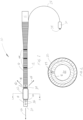
Fig. 1 is a femoral arterial cannula of the present invention; -
Fig. 2 is a cross-sectional view along line 2-2 ofFig. 1 ; -
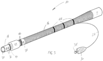
Fig. 3 is a perspective view of the femoral arterial cannula ofFig. 1 with an inflated low-profile balloon; -
Fig. 4 is the femoral arterial cannula ofFig. 1 inserted and in situ in a femoral artery; -
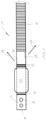
Fig. 5 is an isolation of the tip of the femoral arterial cannula ofFig, 1 rotated 90 degrees; -
Fig. 6 is a femoral venous cannula of the present invention; -
Fig. 7 is a cross-sectional view along line 7-7 ofFig. 6 ; -
Fig. 8 is a perspective view of the femoral venous cannula ofFig. 6 with an inflated broad-profile balloon; -
Fig. 9 is the femoral venous cannula ofFig. 6 inserted and in situ in a femoral vein; -
Fig. 10 is another embodiment the femoral venous cannula of the present invention; and -
Fig. 11 is a cross-sectional view along line 10-10 ofFig. 10 . - Ischemia is defined as, "an inadequate blood supply to an organ or part of the body". Tissue injury and/or death occur as a result of the initial ischemic insult.
- The extent of injury is determined primarily by the magnitude and duration of the interruption in the blood supply. If unaddressed, cell death by necrotic, necroptotic, apoptotic, and autophagic mechanisms occurs (Kalogeris, et al. 2017).
- Identification of limb ischemia caused by the presence of a femoral arterial cannula is of upmost importance with a universal recognition of the severity of consequences if not addressed early on. Given that this demographic of patients are most likely under general anesthesia the early signs of ischemia such as pain and paresthesia are often not possible to discern. This therefore relies on due diligence and frequent inspection of the patient's limbs. Regular limb inspection is essential, assessing for pallor, reduced temperature, slow capillary refill and assessment of pulses via palpation or doppler.
- More advanced stages of ischemia include mottled skin, increased limb circumference and compartment pressures above 30mmHg. Accumulation of inflammatory mediators in a hypoxic environment in conjunction with the inability to wash out these mediators via the congested lymphatic system results in both apoptotic and necrotic cell death. This can cause an increase in systemic potassium. Both inflammatory mediators and high potassium can result in significant vasoplegia similar to that of sepsis. This often requires high dose vasopressors to maintain adequate systemic perfusion pressures. If distal limb flow is re-established late on in the ischemic insult the subsequent wash out of inflammatory mediators and potassium accumulation can further potentiate the vasoplegia (Kalogeris, et al. 2017).
- Dependent on the extent at which the femoral artery and veins are occluded will determine the rate at which clinical signs for ischemia present themselves (Haley, et al.). This may be why ability to identify limb ischemia early on is fraught with difficulties. For example, a limb may present with reduced temperature and slow capillary refill, but integrity of pulses/flow are present via doppler and compartment pressures may be low. This may prompt a clinician to delay intervention given the potential for complications associated with the required intervention. The point at which intervention occurs therefore can vary significantly. Given the difficulty to reliably assess limb ischemia and varying degrees at which identifiable signs present themselves explains in part why some institutions consistently place a distal limb catheter as a prophylactic measure to ensure adequate limb flow is maintained.
- The incidence of reported limb ischemia in ECMO patients seems to vary significantly across ECMO centres.
- The North American 2017 ELSO report on Adult V-A ECMO complications shows a 5.5% incidence of limb ischemia with a 21% survival rate.
- This incidence seems low compared to publications on limb ischemia. A review of reported limb ischemia incidence by Lamb et al.2016 et al. reported a range of limb ischemia events from 10-70% of patients that receive peripherally cannulated ECMO. In a meta analysis of limb ischemia incidence (Juo et a1.2017) reported limb ischemia in 17% of patients out of a total 1886 patients. Possible reasons for discrepancies in reporting maybe due to compounding complications and primary causes of mortality rates in ECMO patients. The potential for limb ischemia being unidentified or shadowed by other causes of mortality may result in under reporting. Furthermore, there may also be significant differences in cannulation technique, size of cannula, point at which the cannula is inserted, native anatomy nuances as well as the timing with which distal limb catheters are inserted.
- It is however noted that the presence of limb ischemia in the absence of other ECMO related complications is devastating and considered an independent risk factor for mortality (Juo et al.)
Table 1. Patient Demographics Variable Total (N= 151) Rescue strategy (N = 107) Preemptive strategy (N=44) P Age (years) 54.6 ± 16.3 57.2 ± 15.1 48.1 ± 17.4 0.003 Male 90 (59.6) 64 (59.8) 26 (59.1) 0.935 Height (m) 1.6 ± 0.1 1.6 ± 0.1 1.6 ± 0.1 0.985 BSA 1.7 ± 0.2 1.7 ± 0.2 1.6 ± 0.1 0.291 BMI 23.2 ± 4.0 23.6 ± 4.1 22.4 ± 3.6 0.102 APACHE II 13.0 ± 6.3 12.2 ± 6.0 14.8 ± 6.9 0.026 SOFA 11.6 t 3.5 11.7 ± 3.5 11.3 ± 3.3 0.585 Ischemia risk factor Diabetes 25 (16.4) 21 (19.6) 4 (9.1) 0.113 Hypertension 39 (25.7) 32 (29.9) 7 (15.9) 0.074 Chronic renal insufficiency 6 (3.9) 5 (4.7) 1 (2.3) 0.493 Smoker 27 (17.8) 20 (18.7) 7 (15.9) 0.685 PVD 11 (7.2) 9 (8.4) 2 (4.5) 0.406 Cerebral infarction 5 (3.4) 5 (4.8) 0 0.139 Catheter size (Fr.) 17.4 ± 2.0 17.2 ± 2.1 17.9 ± 1.8 0.059 Diameter of catheter (mm) 5.8 ± 0.7 5.7 ± 0.7 6.0 ± 0.6 0.052 Cannula size (Fr.)-to-BSA ratio 10.6 ± 1.5 10.4 ± 1.6 11.0 ± 1.2 0.056 Diameter of cannula (mm)-to-BSA ratio 3.5 ± 0.5 3.5 ± 0.5 3.7 ± 0.4 0.050 ECMO days 5.2 ± 5.0 4.9 ± 4.9 6.0 ± 5.4 0.212 Vasopressor use 44 (29.1) 29 (27.1) 15 (34.1) 0.390 Dialysis 46 (30.3) 33 (30.8) 13 (29.5) 0.875 Aspirin use 29 (19.1) 19 (17.8) 10 (22.7) 0.481 Dual anti-platelet drug use 23 (15.1) 17 (15.9) 6 (13.6) 0.726 Warfarin use 2 (1.3) 1 (0.9) 1 (2.3) 0.513 Successful weaning 98 (64.9) 61 (57.0) 37 (84.1) 0.002 Overall mortality 83 (55.0) 66 (61.7) 17 (38.6) 0.010 BSA. body surface area; BMI. body mass index; PVD, peripheral vascular disease; APACHE II, Acute Physiology and Chronic Health Evaluation II score; SOFA. Sequential Organ Failure Assessment score: ECMO, extracorporeal membrane oxygenation. Data are presented as numbers (with percentages) or as means ± standard deviation. -
Figure 1 : Patient demographics taken from Yuo et al.2016. This table demonstrates that expected co-morbidities that are expected to increase likelihood of limb ischemia are non-correlating. - The identification of risk factors for the generation of limb ischemia is highly ambiguous. Presumed risk factors such as presence of obesity, diabetes, peripheral vascular disease and age does not seem to correlate with a higher incidence of limb ischemia. This is noted by Gander et al (2010) who experienced 52% incidence of limb ischemia and noted that, "No variable was predictive of the development of significant limb ischemia". It could be stipulated that the inherent risk could be solely due to the diameter of the femoral artery in relation to the size of the femoral arterial cannula as well as the point of insertion.
- Conversely, the development of limb ischemia could be more complex than solely reduced arterial flow to the limb. This is noted by the prevalence of reported limb ischemia in Veno-Venous ECMO via ELSO.org. This suggests that the generation of limb ischemia could also be caused by venous and lymphatic congestion due to the presence of the femoral venous cannula.
- Arguably it appears that there are a myriad of factors that result in limb ischemia which will likely explain why there are varying degrees of reported ischemic insults and degrees of success when surgical intervention and institution of a distal limb perfusion catheter is inserted.
- This is supported by Yuo et al.2016 who examined the difference between ECMO patients that received immediate placement of a distal limb perfusion catheter and those who had one inserted as a rescue strategy at the first signs of limb ischemia.
- The table (
Figure 2 ) from Yuo et al.2016 demonstrates that a rescue strategy was not as successful as a pre-emptive strategy. The patients that received a distal limb catheter at the time of ECMO initiation had zero incidence of limb ischemia and therefore required no further surgical intervention such as fasciotomy or amputation. It is also notable that the difference in mortality was significantly lower in the preemptive strategy.Table 2. The Cannula-Related Complications Developing During ECMO Support Total (N - 151) Rescue strategy (N - 107) Preemptive strategy (N - 44) P Ischemia 10 (6.6) 10 (9.3) 0 0.036 Surgical intervention 3 (2.0) 3 (2.8) 0 0.262 Fascioloniy 2 (1.3) 2 (1.9) 0 0.359 Amputation 1 (0.7) 1 (0.9) 0 0.248 Cannulation site bleeding 14 (9.3) 9 (8.4) 5 (11.4) 0.570 Data are presented as numbers (with percentages). -
Figure 2 : Yuo et al.2016 demonstrating the difference between a rescue and preemptive strategy to address ischemic limb as a consequence to peripherally cannulated ECMO. - This finding was also found by Lamb et al.2017 who through examination of their ECMO patients demonstrated that immediate institution of a distal limb cannula prevents the development of limb ischemia in 100% of their patient's versus those who did not receive a distal limb cannula where 50% developed ischemic limb complications that required intervention including fasciotomies. (See
Figure 3 ) - The insertion of a distal limb perfusion cannula is not without complication. Lamb et al. 2017 note that femoral vessel complications associated with cannulation include arterial dissection, pseudo-aneurysms, thromboembolic complications and infectious arterial complications. It could be surmised that these complications are inclusive of placement of a distal limb cannula.
- Furthermore the manner with which flow to the limb is incorporated into the ECMO circuit can expose the patient to potential hazards and requires due careand attention by the Perfusionist to ensure the integrity of the ECMO circuit is maintained.

- Given that the limb perfusion cannula provides flow via extra 1/4 inch tubing either via a Y connector off the main 3/8 inch tubing or an external port on the ECMO oxygenator, if the flow is not sufficient it can often result in clot formation and potentially embolic delivery to the limb. This can often compound limb ischemia and requires complex intervention by a vascular surgeon to perform embolectomies and or femoral bypass grafts. This can further expose the patient to excessive bleeding, potential for infection as well as increased blood transfusion rates. It is not uncommon for ECMO patients to receive multiple allogeneic blood transfusions. Given this patient demographic can be often worked up for heart transplant candidacy, increased bloodtransfusions can result in sensitizing the humoral immune system making it increasingly difficult to find a suitable, compatible donor. Therefore limiting allogeneic blood transfusions is essential.

- The etiology of lymphatic congestion is a complex process, which is not fully understood.
- The relationship between arterial, venous, interstitial and lymphatic blood flow is a dynamic complex balance which can be easily influenced by osmotic and oncotic changes as well as blood flow rates. It is important to note that lymph transport, not venous capillary reabsorption, is the main process responsible for interstitial fluid drainage (Mortimer and Levick 2004). Therefore all edema is due to an imbalance between capillary filtration and lymph drainage. It is theorized therefore that in the presence of a femoral venous cannula, reduction in venous drainage from the limb results in subsequent venous engorgement. This then results in increased capillary perfusion pressures with subsequent fluid movement from the venous compartment to the lymphatic vessels. It is thought that the ability of the lymphatic compartment to compensate for this increased interstitial blood flow is low and limb edema ensues. If this is paired with low oncotic pressures (fluid overload is common in patients requiring ECMO as well as Cardiopulmonary bypass for OHS/MICS) increased capillary filtration then overwhelms increased lymph drainage, which subsequently under-compensates.
- Interstitial edema in this overwhelmed vascular compartment then increases the oxygen diffusion barrier resulting in cellular ischemia. The inflammatory process then further exacerbates the interstitial edema due to the development of "leaky" capillary beds.
- It is believed that this is an under appreciated cause of limb ischemia in peripherally cannulated ECMO and CPB (OHS/MICS) patients and highlights the need to not only maintain arterial blood flow but also venous drainage and therefore prevent overwhelming the lymphatic compartments during the period of cardiopulmonary support. This may also explain why ischemic limb has been seen as a complication in Veno-venous ECMO patients where no arterial cannula is placed.
- An object of the inventions is to mitigate the potential for limb ischemia occurring as a consequence to the presence of arterial and venous femoral cannulation. The cannulas are designed to require no additional components other than that of the arterial and venous cannula themselves. The cannulas are designed to be suitable for all femoral cannulation requirements, be it for ECMO, OHS, MICS or other cardiopulmonary systems.
- The function of the femoral arterial cannula is to provide retrograde arterial limb flow in the presence of systemic arterial flow via a single cannula.
- The function of the femoral venous cannula is to provide retrograde venous limb drainage in the presence of systemic venous flow via a single cannula.
- Referring to
Figs. 1 to 5 a femoralarterial cannula 10 features a cylindrical plastic body oftubing 12 which preferably has a biocompatible surface coating. The initial length 11 (3/8" OD) remains clear and a 3/8x3/8" luer lock connector is used to connect the initial length with a cardiopulmonary circuit; arterial blood flow enters thecannula 10 through this connector. This connector is a standard perfusion component and is not shown in the figures.. - The length of
body tubing 12 tapers to one of the varied sizes; e.g. a 15 Fr (5.0mm) body of wire woundtubing 16 or a 17 Fr (5.6mm) body of wire wound tubing or a 19 Fr (6.3mm) body of wire woundtubing 16.. - Distal to the length of wire wound
tubing 16 is a contiguous length ofclear tubing 18, thetip 20 of which is open ended and is the exit port for systemic arterial flow. - Circumferentially attached to
body tubing 12 is a low-profilecompliant balloon 22.Balloon 22 remains parallel to the length ofclear tubing 12 when non-inflated. Theballoon 22 length is approximately 20mm. Theexternal balloon 22 surface has a drug eluting coating. When inflated theballoon 22 remains cylindrical along its length and is parallel to thebody 12 of thecannula 10. Achannel 27, as best illustrated inFig. 2 , runs the length ofbody tubing 12 to accommodate and allow a small-bore length oftubing 28 to sit longitudinally flush alongbody tubing 12. Small-bore length tubing 28 is attached to balloon 22 at one end and at its other end, to aballoon cuff 30 that includes a two-way valve andfemale luer port 34. - Distal to the distal end of the
balloon 22 are four circular bloodpathway exit ports 36 within contiguous length ofclear tubing 18; as illustrated, two exit ports longitudinally with identical blood flow exit ports at 180 degrees circumferentially.Exit ports 36 are contiguous with the blood pathway for systemic arterial flow. - Proximal to the proximal end of the
balloon 22 are twoexit ports 38 within the contiguous length ofclear tubing 18;exit ports 38 are at 180 degrees circumferentially, and as best illustrated inFig. 5 , are offset longitudinally from each other by 5mm (one exit port is 5mm from the proximal balloon end while the second exit port is 10mm from the proximal balloon end). - Distal to the distal end of the
balloon 22 is a circular radio-opaque marker 40 within thebody tubing 12. Proximal to the proximal end of thecannula 10 are two parallel circular radio-opaque markers 42 within thebody tubing 12.Positional markers 44 are identified on the length of the wire woundtubing 16 body of thebody tubing 12 to identify cannula positional depth. - The femoral
arterial cannula 10 is designed to be inserted either by a surgical cut down or by a percutaneous Seldinger technique. Thecannula 10 delivers systemicarterial blood flow 46 along the length of thecannula 10 body through to thecannula tip 20 and the additional bloodpathway exit ports 38. Theballoon 22 is designed to seal thefemoral artery 100 once inflated. Theballoon 22 has a drug eluting coating. Thecannula 10 has a biocompatible surface coating. Theballoon 22 is de-aired prior to insertion by means of injection and aspiration of saline/contrast solution. Once in situ and inflated theballoon 22 will effectively isolate the blood flow of theproximal exit ports 38 such that they are only available to deliver flow in a retrograde direction down 48 to the ipsilateral limb. With theballoon 22 inflated there is dedicated limbblood flow delivery 48. - Referring to
Figs. 6 to 11 , the femoralvenous cannula 50 features a cylindrical plastic length of tubing 52 which preferably has a biocompatible surface coating. The initial length 51 (3/8"OD) remains clear with a 3/8×3/8 connector in situ (non luer lock); venous blood flow exits thecannula 50 through this connector which is a standard perfusion item and not shown in the figures. This length of tubing 52 tapers to one of varied sizes: a 23Fr (7.6mm) body of wire woundtubing 54 or a 25Fr (8.3mm) body of wire wound tubing or a 27Fr (9.7mm) body of wire wound tubing.. - Distal to the length of wire wound
tubing 54 is a contiguous length of clear tubing 52. Attached to clear length of tubing 52 is a broad-profilecompliant balloon 56 which remains parallel to the length of clear tubing 52 when non-inflated. Theexternal balloon 56 surface has a drug eluting coating. Theballoon 56 length is approximately 12mm. When inflated theballoon 56 remains cylindrical along its length and is parallel to the body tubing 52. Achannel 57, as best illustrated inFigs. 7 and11 , runs the length of body tubing 52 to accommodate and allow a small-bore length oftubing 58 to sit longitudinally flush along body tubing 52. Small-bore length tubing 58 is attached to balloon 56 at one end and at its other end, to aballoon cuff 62 that includes a two-way valve andfemale luer port 66. - Distal to the distal end of the
balloon 56 are two circular bloodpathway entry ports 62 within the clear tubing 52; the twoblood entry ports 62 are at 180 degrees circumferentially. Theseblood entry ports 62 are offset from each other by 5mm (one entry port is 5mm distal from theballoon 56 while the second entry port is 10mm from theballoon 56 end). Theseentry ports 62 are contiguous with theblood pathway 65 for systemic venous blood flow. - Proximal to the proximal end of the
balloon 56 are two bloodpathway entry ports 64 within the clear tubing 52, these ports are at 180 degrees circumferentially but are off-set from each other by 5mm (one entry port is 5mm proximal from theballoon 56 end while the second entry port is 10mm from the balloon end). Theseports 64 are circumferentially offset by 90 degrees from the two distalblood entry ports 62. - Distal to the distal end of the
balloon 56 is a single circular radio-opaque marker 66 within the body of thecannula 50. Proximal to the proximal end of thecannula 10 are two parallel circular radio-opaque markers 68 within the body tubing 52. - Distal to the clear tubing 52 is a contiguous length of wire wound
tubing 70. Distal to the length of wire woundtubing 70 is a contiguous length ofclear tubing 72, thetip 74 of which is open ended and is the entry port for systemicvenous flow 65. Within this length ofclear tubing 72 are six circular bloodpathway entry ports 76 within theclear tubing 72, three entry ports longitudinally with identical exit ports at 180 degrees circumferentially. -
Positional markers 68 are identified on the length of the wire woundtubing body 54 of the body tubing 52 to identify cannula positional depth. - The femoral
venous cannula 50 is designed to be in varying lengths depending upon its clinical application as well the physical size of the patient. The femoralvenous cannula 50 that has application for V-A ECMO, OHS and MICS procedures has three lengths while the model for V-V ECMO has three lengths. - The femoral venous model designed for V-V ECMO is a shorter length than that of the femoral venous cannula designed for V-A ECMO, OHS and MICS. The included schematic has individual design components identical to those of the longer cannula.
- The femoral
venous cannula 50 is designed to be inserted either by a surgical cut down or by a percutaneous Seldinger technique. Thecannula 50 drains systemicvenous blood flow 65 along the length of the body tubing 52 from thecannula tip 74 and the additional bloodpathway entry ports 62. Theballoon 56 is designed to seal thefemoral vein 200 once inflated. Theballoon 56 has a drug eluting coating. Thecannula 50 has a biocompatible surface coating. Theballoon 56 is de-aired prior to insertion by means of injection and aspiration of saline/contrast solution. Once in situ and inflated theballoon 56 will effectively isolate the blood flow of theproximal entry ports 64 such that they are only available to drainvenous blood flow 67 from the ipsilateral limb. With theballoon 56 inflated there is dedicated limb flow drainage while thedistal entry ports 62 directly above the balloon will drain venous blood from the lower length of the inferior vena cava. - While it is recognized that either cannula can be used individually, the central concept to completely address and prevent limb ischemia is to use both arterial 10 and venous 50 cannula to work synergistically.
- This method does not form part of the claimed invention.
- The femoral arterial 10 and venous 50 cannulas have been designed to facilitate the delivery of dedicated arterial limb flow in the presence of dedicated venous limb drainage. A non-perfused limb will suffer the inevitable clinical consequences of ischemia, however a limb that is adequately perfused but is insulted by having inadequate or absent venous drainage will similarly be subjected to a physiological insult that may ultimately be irrecoverable. By facilitating both arterial perfusion along with venous drainage will protect the limb from the adverse consequences of limb ischemia. This identifies and recognizes the multifactorial nature of ischemic limb associated with peripheral cannulation and therefore offers a clinically superior solution opposed to using one cannula in isolation.
- While embodiments of the invention have been described and illustrated, such embodiments should be considered illustrative of the invention only. The invention may include variants not described or illustrated herein in detail. Thus, the embodiments described and illustrated herein should not be considered to limit the invention as construed in accordance with the accompanying claims.
-
- Theodosios Bisdas, Gernot Beutel, Gregor Warnecke, Marius M. Hoeper, Christian Kuehn, Axel Haverich, Omke E. Teebken. 2011. Vascular Complications in Patients Undergoing Femoral Cannulation for Extracorporeal Membrane Oxygenation Support. 92:626-31. Ann Thorac Surg.
- Jeffrey W. Gander , Jason C. Fisher, Ari R. Reichstein, Erica R. Gross, Gudrun Aspelund, William Middlesworth, Charles J. Stolar. 2010. Limb ischemia after common femoral artery cannulation for venoarterial extracorporeal membrane oxygenation: an unresolved problem. 45, 2136-2140. Journal of Pediatric Surgery.
- Yi Juo , Matthew Skancke, Yas Sanaiha, Aditya Mantha, Juan C. Jimenez, Peyman Benharash. 2017. Efficacy of Distal Perfusion Cannulae in Preventing Limb Ischemia During Extracorporeal Membrane Oxygenation: A Systematic Review and Meta-Analysis. 41 (11): E263-E273 Artificial Organs.
- Mary Jo Haley, Jason C. Fisher, Alejandro R. Ruiz-Elizalde, Charles J.H. Stolar, Nicholas J. Morrissey, William Middlesworth. 2008. Percutaneous distal perfusion of the lower extremity after femoral cannulation for venoarterial extracorporeal membrane oxygenation in a small child. 44, 437-440. Journal of Pediatric Surgery
- Theodore Kalogeris, Christopher P. Baines, Maike Krenz, and Ronald J. Korthuis. 2012. Cell Biology of IschemialReperfusion Injury. 298: 229-317. Int Rev Cell Mol Biol.
- Kathleen M. Lamb, Hitoshi Hirose. 2017. Vascular Complications in Extracoporeal Membrane Oxygenation. 33, 813-824. Crit Care Clin.
- Kathleen M. Lamb, Paul J. DiMuzio, Adam Johnson, Philip Batista, Neil Moudgill, Megan McCullough, Joshua A. Eisenberg, Hitoshi Hirose, MD, Nicholas C. Cavarocchi. 2016. Arterial protocol including prophylactic distal perfusion catheter decreases limb ischemia complications in patients undergoing extracorporeal membrane oxygenation.65:1074-9. Society for Vascular Surgery. Lukasz A. Adamczyk' Kristiana Gordon' Ivana Kholová' Lorine B. Meijer-Jorna'
- Niklas Telinius' Patrick J. Gallagher' Allard C. van der Wal' Ulrik Baandrup. 2016. Lymph vessels: the forgotten second circulation in health and disease. 469:3-17. Virchows Arch.
- Hye Ju Yeo, Seong Hoon Yoon, Doosoo Jeon, Yun Seong Kim, Woo Hyun Cho, Dohyung Kim, Seung Eun Lee. 2016. The Utility of Preemptive Distal Perfusion Cannulation During Peripheral Venoarterial Extracorporeal Membrane Oxygenation Support. Vol. 29, No. 4. Journal of interventional cardiology.
Claims (13)
- A femoral arterial cannula (10) comprising:an elongated hollow cannula body (12) having distal (20) and proximal (11) open ends;a balloon (22) situated on said body adjacent to said distal end;inflation means connected to said balloon for inflating said balloon;four blood flow exit ports (36) situated in said cannula body between said balloon and said distal end, two of said distal exit ports in-line with one-another along said cannula body and two other distal exit ports being 180° circumferentially offset from the first two distal exit ports; andtwo blood flow exit ports (38) situated in said cannula body between said balloon and said proximal end, said proximal exit ports being 180° circumferentially offset and longitudinally offset from one-another, wherein:the distal end is a tip of the elongated hollow cannula body, andthe proximal end is at the opposite end of the elongated hollow cannula body from the distal end.
- The femoral arterial cannula of claim 1 wherein said cannula body is an elongated hollow cylindrical body.
- The femoral arterial cannula of claim 1 wherein said balloon comprises a low-profile balloon.
- The femoral arterial cannula of claim 1 wherein said cannula body comprises a taper from said distal to said proximal end.
- The femoral arterial cannula of claim 1 wherein said cannula body comprises a distal single radio-opaque marker (40), or a proximal double radio-opaque marker (42).
- The femoral arterial cannula of claim 1 wherein said inflation means comprises:a small bore tubing (28) connected to said balloon at a first end;a balloon cuff (30) connected to a second end of said small bore tubing;a two-way valve connected to said balloon cuff; anda female luer port (34) connected to said two-way valve.
- A femoral venous cannula (50) comprising:an elongated hollow cannula body (52) having distal (74) and proximal (51) open ends;a balloon (56) situated on said body adjacent to said proximal end;inflation means connected to said balloon for inflating said balloon;two blood flow entry ports (62) situated in said cannula body between said balloon and said distal end, said distal entry ports being 180° circumferentially offset and longitudinally offset from one-another along said cannula body; andtwo blood flow entry ports (64) situated in said cannula body between said balloon and said proximal end, said proximal entry ports being 180° circumferentially offset and longitudinally offset from one-another and 90° offset from said distal entry ports, wherein:the distal end is a tip (74) of the elongated hollow cannula body, andthe proximal end is at the opposite end of the elongated hollow cannula body from the distal end.
- The femoral venous cannula of claim 7 wherein said elongated hollow cannula body is of various lengths.
- The femoral venous cannula of claim 7 wherein said cannula body is an elongated hollow cylindrical body.
- The femoral venous cannula of claim 7 wherein said balloon comprises a broad-profile balloon.
- The femoral venous cannula of claim 7 wherein said cannula body comprises a taper from said distal to said proximal end.
- The femoral venous cannula of claim 7 wherein said cannula body comprises a distal single radio-opaque marker (66), or a proximal double radio-opaque marker (68).
- The femoral venous cannula of claim 7 wherein said inflation means comprises:a small bore tubing (58) connected to said balloon at a first end;a balloon cuff (62) connected to a second end of said small bore tubing;a two-way valve connected to said balloon cuff; anda female luer port (66) connected to said two-way valve.
Applications Claiming Priority (2)
| Application Number | Priority Date | Filing Date | Title |
|---|---|---|---|
| CA3035749A CA3035749A1 (en) | 2019-03-05 | 2019-03-05 | Femoral and venous arterial cannulas and a method for mitigating the risk of limb ischemia |
| PCT/CA2020/050191 WO2020176968A1 (en) | 2019-03-05 | 2020-02-13 | Femoral and venous arterial cannulas and a method for mitigating the risk of limb ischemia |
Publications (4)
| Publication Number | Publication Date |
|---|---|
| EP3934728A1 EP3934728A1 (en) | 2022-01-12 |
| EP3934728A4 EP3934728A4 (en) | 2022-05-11 |
| EP3934728B1 true EP3934728B1 (en) | 2024-05-22 |
| EP3934728C0 EP3934728C0 (en) | 2024-05-22 |
Family
ID=72321875
Family Applications (1)
| Application Number | Title | Priority Date | Filing Date |
|---|---|---|---|
| EP20766367.5A Active EP3934728B1 (en) | 2019-03-05 | 2020-02-13 | Femoral and venous arterial cannulas for mitigating the risk of limb ischemia |
Country Status (5)
| Country | Link |
|---|---|
| US (1) | US20230166099A1 (en) |
| EP (1) | EP3934728B1 (en) |
| CA (1) | CA3035749A1 (en) |
| ES (1) | ES2981837T3 (en) |
| WO (1) | WO2020176968A1 (en) |
Families Citing this family (5)
| Publication number | Priority date | Publication date | Assignee | Title |
|---|---|---|---|---|
| WO2023148723A1 (en) * | 2022-02-03 | 2023-08-10 | Rambam Medtech Ltd. | Venous cannula insertion |
| EP4493250A4 (en) * | 2022-03-15 | 2026-03-18 | Total Flow Medical Ltd | Bidirectional flow cannulae, systems with bidirectional cannulae and methods for using them |
| CN219595415U (en) * | 2023-02-03 | 2023-08-29 | 南京鼓楼医院 | Femoral artery cannula for providing blood perfusion of lower limbs |
| WO2025057066A1 (en) * | 2023-09-11 | 2025-03-20 | Total Flow Medical Limited | Bidirectional flow cannulas and methods of using same |
| WO2026084924A1 (en) | 2024-10-14 | 2026-04-23 | Edwards Lifesciences Corporation | Bidirectional cannula |
Family Cites Families (14)
| Publication number | Priority date | Publication date | Assignee | Title |
|---|---|---|---|---|
| US3884242A (en) * | 1971-03-29 | 1975-05-20 | Mpc Kurgisil | Catheter assembly |
| US4211233A (en) * | 1978-01-05 | 1980-07-08 | Lin Edward D | Urethral catheter |
| US5584803A (en) * | 1991-07-16 | 1996-12-17 | Heartport, Inc. | System for cardiac procedures |
| US6821265B1 (en) * | 1996-04-10 | 2004-11-23 | Endoscopic Technologies, Inc. | Multichannel catheter |
| US5928181A (en) * | 1997-11-21 | 1999-07-27 | Advanced International Technologies, Inc. | Cardiac bypass catheter system and method of use |
| US20060247575A1 (en) * | 2001-12-21 | 2006-11-02 | Richard Cartledge | Balloon cannulae |
| US20110004057A1 (en) * | 2004-04-21 | 2011-01-06 | Acclarent, Inc. | Systems and methods for transnasal dilation of passageways in the ear, nose or throat |
| US20070016168A1 (en) * | 2005-06-29 | 2007-01-18 | Conway Anthony J | Method of making a catheter device |
| US8241248B2 (en) * | 2009-02-27 | 2012-08-14 | Cvdevices, Llc | Devices and systems for selective auto-retroperfusion of the cerebral venous system |
| US8992817B2 (en) * | 2012-12-10 | 2015-03-31 | Abbott Cardiovascular Systems, Inc. | Process of making a medical balloon |
| CN203447625U (en) * | 2013-09-03 | 2014-02-26 | 董爱强 | Double-pole double-air-bag femoral vein cannula |
| US20170232238A1 (en) | 2014-08-06 | 2017-08-17 | Edwars Litesciences Corporation | Multi-lumen cannulae |
| DE102016103560A1 (en) | 2016-02-29 | 2017-08-31 | Eberhard Karls Universität Tübingen Medizinische Fakultät | System for use in organ replacement or support procedures |
| TR201704099A2 (en) | 2017-03-17 | 2018-09-21 | T C Istanbul Medipol Ueniversitesi | A FEMORAL ARTERIAL ECMO (EXTRACORPOREAL MEMBRANE OXYGENATION EXTRACORPOREAL MEMBRANE OXYGENATION) CANNULA |
-
2019
- 2019-03-05 CA CA3035749A patent/CA3035749A1/en active Pending
-
2020
- 2020-02-13 ES ES20766367T patent/ES2981837T3/en active Active
- 2020-02-13 US US17/436,574 patent/US20230166099A1/en active Pending
- 2020-02-13 WO PCT/CA2020/050191 patent/WO2020176968A1/en not_active Ceased
- 2020-02-13 EP EP20766367.5A patent/EP3934728B1/en active Active
Also Published As
| Publication number | Publication date |
|---|---|
| US20230166099A1 (en) | 2023-06-01 |
| CA3035749A1 (en) | 2020-09-05 |
| WO2020176968A1 (en) | 2020-09-10 |
| EP3934728C0 (en) | 2024-05-22 |
| EP3934728A1 (en) | 2022-01-12 |
| EP3934728A4 (en) | 2022-05-11 |
| ES2981837T3 (en) | 2024-10-10 |
Similar Documents
| Publication | Publication Date | Title |
|---|---|---|
| US20230166099A1 (en) | Femoral venous and femoral arterial cannulas and a method for mitigating the risk of limb ischemia | |
| US6056717A (en) | Implantable vascular device | |
| US7341570B2 (en) | Apparatus and methods for treating congestive heart disease | |
| US6086553A (en) | Arteriovenous shunt | |
| US6053901A (en) | Subcutaneously implanted cannula and method for arterial access | |
| JPH04507050A (en) | Vascular access system for extracorporeal processing of blood | |
| JP2013078575A (en) | Catheter with external flow channel | |
| US6544206B1 (en) | Dialysis access system and method | |
| US20240009438A1 (en) | Devices and methods for fistula-free hemodialysis | |
| Raina et al. | Challenges of vascular access in the pediatric population | |
| Kasirajan et al. | The total artificial heart for biventricular heart failure and beyond | |
| Zhang et al. | Extracorporeal membrane oxygenation (ECMO) assisted mediastinal tumor resection and superior vena cava replacement are safe and feasible | |
| US20220273857A1 (en) | Novel type superior vena cava cannula for use in extrathoracic cannulation in open heart surgery | |
| US20230310187A1 (en) | Methods and systems for facilitating laminar flow between conduits | |
| US11077239B2 (en) | Tributary access device and methods of use | |
| Ng et al. | Lethal pulmonary embolism following left upper extremity angiogram/angioplasty with thrombectomy at malfunctioning arteriovenous fistula | |
| Christianson | Caring for a patient who has an implanted venous port | |
| Dosluoglu et al. | External femorofemoral bypass to relieve acute leg ischemia during circulatory assist | |
| Soundaravalli et al. | Open surgical repair of abdominal aortic aneurysm: Proximal aortic control by endoaortic balloon-A novel approach | |
| Granata et al. | Vascular access for acute extracorporeal renal replacement therapies | |
| Goranovska et al. | Extra-anatomic aorto-right subclavian bypass after complicated insertion of permanent dialysis catheter | |
| D'Intini et al. | Management of vascular catheters for acute renal replacement therapy | |
| Khanin | hemodialysis Access | |
| Conrad | Vascular Access | |
| Sato et al. | Two cases of chronic pulmonary thromboembolism saved by postoperative use of a percutaneous cardiopulmonary support device |
Legal Events
| Date | Code | Title | Description |
|---|---|---|---|
| STAA | Information on the status of an ep patent application or granted ep patent |
Free format text: STATUS: THE INTERNATIONAL PUBLICATION HAS BEEN MADE |
|
| PUAI | Public reference made under article 153(3) epc to a published international application that has entered the european phase |
Free format text: ORIGINAL CODE: 0009012 |
|
| STAA | Information on the status of an ep patent application or granted ep patent |
Free format text: STATUS: REQUEST FOR EXAMINATION WAS MADE |
|
| 17P | Request for examination filed |
Effective date: 20211005 |
|
| AK | Designated contracting states |
Kind code of ref document: A1 Designated state(s): AL AT BE BG CH CY CZ DE DK EE ES FI FR GB GR HR HU IE IS IT LI LT LU LV MC MK MT NL NO PL PT RO RS SE SI SK SM TR |
|
| A4 | Supplementary search report drawn up and despatched |
Effective date: 20220408 |
|
| RIC1 | Information provided on ipc code assigned before grant |
Ipc: A61M 25/04 20060101ALI20220404BHEP Ipc: A61M 25/00 20060101ALI20220404BHEP Ipc: A61M 1/36 20060101ALI20220404BHEP Ipc: A61M 25/10 20130101AFI20220404BHEP |
|
| DAV | Request for validation of the european patent (deleted) | ||
| DAX | Request for extension of the european patent (deleted) | ||
| GRAP | Despatch of communication of intention to grant a patent |
Free format text: ORIGINAL CODE: EPIDOSNIGR1 |
|
| STAA | Information on the status of an ep patent application or granted ep patent |
Free format text: STATUS: GRANT OF PATENT IS INTENDED |
|
| INTG | Intention to grant announced |
Effective date: 20231002 |
|
| GRAS | Grant fee paid |
Free format text: ORIGINAL CODE: EPIDOSNIGR3 |
|
| GRAA | (expected) grant |
Free format text: ORIGINAL CODE: 0009210 |
|
| STAA | Information on the status of an ep patent application or granted ep patent |
Free format text: STATUS: THE PATENT HAS BEEN GRANTED |
|
| RAP3 | Party data changed (applicant data changed or rights of an application transferred) |
Owner name: TOTAL FLOW MEDICAL LIMITED |
|
| AK | Designated contracting states |
Kind code of ref document: B1 Designated state(s): AL AT BE BG CH CY CZ DE DK EE ES FI FR GB GR HR HU IE IS IT LI LT LU LV MC MK MT NL NO PL PT RO RS SE SI SK SM TR |
|
| REG | Reference to a national code |
Ref country code: GB Ref legal event code: FG4D |
|
| REG | Reference to a national code |
Ref country code: CH Ref legal event code: EP |
|
| REG | Reference to a national code |
Ref country code: DE Ref legal event code: R096 Ref document number: 602020031322 Country of ref document: DE |
|
| REG | Reference to a national code |
Ref country code: IE Ref legal event code: FG4D |
|
| U01 | Request for unitary effect filed |
Effective date: 20240522 |
|
| U07 | Unitary effect registered |
Designated state(s): AT BE BG DE DK EE FI FR IT LT LU LV MT NL PT SE SI Effective date: 20240603 |
|
| PG25 | Lapsed in a contracting state [announced via postgrant information from national office to epo] |
Ref country code: IS Free format text: LAPSE BECAUSE OF FAILURE TO SUBMIT A TRANSLATION OF THE DESCRIPTION OR TO PAY THE FEE WITHIN THE PRESCRIBED TIME-LIMIT Effective date: 20240922 |
|
| PG25 | Lapsed in a contracting state [announced via postgrant information from national office to epo] |
Ref country code: HR Free format text: LAPSE BECAUSE OF FAILURE TO SUBMIT A TRANSLATION OF THE DESCRIPTION OR TO PAY THE FEE WITHIN THE PRESCRIBED TIME-LIMIT Effective date: 20240522 |
|
| REG | Reference to a national code |
Ref country code: ES Ref legal event code: FG2A Ref document number: 2981837 Country of ref document: ES Kind code of ref document: T3 Effective date: 20241010 |
|
| PG25 | Lapsed in a contracting state [announced via postgrant information from national office to epo] |
Ref country code: GR Free format text: LAPSE BECAUSE OF FAILURE TO SUBMIT A TRANSLATION OF THE DESCRIPTION OR TO PAY THE FEE WITHIN THE PRESCRIBED TIME-LIMIT Effective date: 20240823 |
|
| PG25 | Lapsed in a contracting state [announced via postgrant information from national office to epo] |
Ref country code: PL Free format text: LAPSE BECAUSE OF FAILURE TO SUBMIT A TRANSLATION OF THE DESCRIPTION OR TO PAY THE FEE WITHIN THE PRESCRIBED TIME-LIMIT Effective date: 20240522 |
|
| PG25 | Lapsed in a contracting state [announced via postgrant information from national office to epo] |
Ref country code: PL Free format text: LAPSE BECAUSE OF FAILURE TO SUBMIT A TRANSLATION OF THE DESCRIPTION OR TO PAY THE FEE WITHIN THE PRESCRIBED TIME-LIMIT Effective date: 20240522 Ref country code: NO Free format text: LAPSE BECAUSE OF FAILURE TO SUBMIT A TRANSLATION OF THE DESCRIPTION OR TO PAY THE FEE WITHIN THE PRESCRIBED TIME-LIMIT Effective date: 20240822 Ref country code: IS Free format text: LAPSE BECAUSE OF FAILURE TO SUBMIT A TRANSLATION OF THE DESCRIPTION OR TO PAY THE FEE WITHIN THE PRESCRIBED TIME-LIMIT Effective date: 20240922 Ref country code: HR Free format text: LAPSE BECAUSE OF FAILURE TO SUBMIT A TRANSLATION OF THE DESCRIPTION OR TO PAY THE FEE WITHIN THE PRESCRIBED TIME-LIMIT Effective date: 20240522 Ref country code: GR Free format text: LAPSE BECAUSE OF FAILURE TO SUBMIT A TRANSLATION OF THE DESCRIPTION OR TO PAY THE FEE WITHIN THE PRESCRIBED TIME-LIMIT Effective date: 20240823 Ref country code: RS Free format text: LAPSE BECAUSE OF FAILURE TO SUBMIT A TRANSLATION OF THE DESCRIPTION OR TO PAY THE FEE WITHIN THE PRESCRIBED TIME-LIMIT Effective date: 20240822 |
|
| PG25 | Lapsed in a contracting state [announced via postgrant information from national office to epo] |
Ref country code: CZ Free format text: LAPSE BECAUSE OF FAILURE TO SUBMIT A TRANSLATION OF THE DESCRIPTION OR TO PAY THE FEE WITHIN THE PRESCRIBED TIME-LIMIT Effective date: 20240522 |
|
| PG25 | Lapsed in a contracting state [announced via postgrant information from national office to epo] |
Ref country code: SK Free format text: LAPSE BECAUSE OF FAILURE TO SUBMIT A TRANSLATION OF THE DESCRIPTION OR TO PAY THE FEE WITHIN THE PRESCRIBED TIME-LIMIT Effective date: 20240522 Ref country code: RO Free format text: LAPSE BECAUSE OF FAILURE TO SUBMIT A TRANSLATION OF THE DESCRIPTION OR TO PAY THE FEE WITHIN THE PRESCRIBED TIME-LIMIT Effective date: 20240522 |
|
| PG25 | Lapsed in a contracting state [announced via postgrant information from national office to epo] |
Ref country code: SM Free format text: LAPSE BECAUSE OF FAILURE TO SUBMIT A TRANSLATION OF THE DESCRIPTION OR TO PAY THE FEE WITHIN THE PRESCRIBED TIME-LIMIT Effective date: 20240522 |
|
| PG25 | Lapsed in a contracting state [announced via postgrant information from national office to epo] |
Ref country code: SM Free format text: LAPSE BECAUSE OF FAILURE TO SUBMIT A TRANSLATION OF THE DESCRIPTION OR TO PAY THE FEE WITHIN THE PRESCRIBED TIME-LIMIT Effective date: 20240522 Ref country code: SK Free format text: LAPSE BECAUSE OF FAILURE TO SUBMIT A TRANSLATION OF THE DESCRIPTION OR TO PAY THE FEE WITHIN THE PRESCRIBED TIME-LIMIT Effective date: 20240522 Ref country code: RO Free format text: LAPSE BECAUSE OF FAILURE TO SUBMIT A TRANSLATION OF THE DESCRIPTION OR TO PAY THE FEE WITHIN THE PRESCRIBED TIME-LIMIT Effective date: 20240522 Ref country code: CZ Free format text: LAPSE BECAUSE OF FAILURE TO SUBMIT A TRANSLATION OF THE DESCRIPTION OR TO PAY THE FEE WITHIN THE PRESCRIBED TIME-LIMIT Effective date: 20240522 |
|
| REG | Reference to a national code |
Ref country code: DE Ref legal event code: R097 Ref document number: 602020031322 Country of ref document: DE |
|
| U20 | Renewal fee for the european patent with unitary effect paid |
Year of fee payment: 6 Effective date: 20250217 |
|
| PLBE | No opposition filed within time limit |
Free format text: ORIGINAL CODE: 0009261 |
|
| STAA | Information on the status of an ep patent application or granted ep patent |
Free format text: STATUS: NO OPPOSITION FILED WITHIN TIME LIMIT |
|
| 26N | No opposition filed |
Effective date: 20250225 |
|
| PG25 | Lapsed in a contracting state [announced via postgrant information from national office to epo] |
Ref country code: MC Free format text: LAPSE BECAUSE OF FAILURE TO SUBMIT A TRANSLATION OF THE DESCRIPTION OR TO PAY THE FEE WITHIN THE PRESCRIBED TIME-LIMIT Effective date: 20240522 |
|
| REG | Reference to a national code |
Ref country code: CH Ref legal event code: U11 Free format text: ST27 STATUS EVENT CODE: U-0-0-U10-U11 (AS PROVIDED BY THE NATIONAL OFFICE) Effective date: 20260301 |
|
| U20 | Renewal fee for the european patent with unitary effect paid |
Year of fee payment: 7 Effective date: 20260216 |
|
| PGFP | Annual fee paid to national office [announced via postgrant information from national office to epo] |
Ref country code: GB Payment date: 20260216 Year of fee payment: 7 |
|
| PGFP | Annual fee paid to national office [announced via postgrant information from national office to epo] |
Ref country code: ES Payment date: 20260303 Year of fee payment: 7 |
|
| PGFP | Annual fee paid to national office [announced via postgrant information from national office to epo] |
Ref country code: IE Payment date: 20260216 Year of fee payment: 7 |
|
| PGFP | Annual fee paid to national office [announced via postgrant information from national office to epo] |
Ref country code: CH Payment date: 20260301 Year of fee payment: 7 |