EP3930610B1 - Ki-systeme zur erkennung und dimensionierung von läsionen - Google Patents
Ki-systeme zur erkennung und dimensionierung von läsionen Download PDFInfo
- Publication number
- EP3930610B1 EP3930610B1 EP19917118.2A EP19917118A EP3930610B1 EP 3930610 B1 EP3930610 B1 EP 3930610B1 EP 19917118 A EP19917118 A EP 19917118A EP 3930610 B1 EP3930610 B1 EP 3930610B1
- Authority
- EP
- European Patent Office
- Prior art keywords
- lesion
- reference object
- cut out
- image
- polyps
- Prior art date
- Legal status (The legal status is an assumption and is not a legal conclusion. Google has not performed a legal analysis and makes no representation as to the accuracy of the status listed.)
- Active
Links
Images


Classifications
-
- G—PHYSICS
- G06—COMPUTING OR CALCULATING; COUNTING
- G06T—IMAGE DATA PROCESSING OR GENERATION, IN GENERAL
- G06T7/00—Image analysis
- G06T7/0002—Inspection of images, e.g. flaw detection
- G06T7/0012—Biomedical image inspection
-
- A—HUMAN NECESSITIES
- A61—MEDICAL OR VETERINARY SCIENCE; HYGIENE
- A61B—DIAGNOSIS; SURGERY; IDENTIFICATION
- A61B1/00—Instruments for performing medical examinations of the interior of cavities or tubes of the body by visual or photographical inspection, e.g. endoscopes; Illuminating arrangements therefor
- A61B1/00002—Operational features of endoscopes
- A61B1/00004—Operational features of endoscopes characterised by electronic signal processing
- A61B1/00009—Operational features of endoscopes characterised by electronic signal processing of image signals during a use of endoscope
- A61B1/000095—Operational features of endoscopes characterised by electronic signal processing of image signals during a use of endoscope for image enhancement
-
- A—HUMAN NECESSITIES
- A61—MEDICAL OR VETERINARY SCIENCE; HYGIENE
- A61B—DIAGNOSIS; SURGERY; IDENTIFICATION
- A61B1/00—Instruments for performing medical examinations of the interior of cavities or tubes of the body by visual or photographical inspection, e.g. endoscopes; Illuminating arrangements therefor
- A61B1/00002—Operational features of endoscopes
- A61B1/00004—Operational features of endoscopes characterised by electronic signal processing
- A61B1/00009—Operational features of endoscopes characterised by electronic signal processing of image signals during a use of endoscope
- A61B1/000096—Operational features of endoscopes characterised by electronic signal processing of image signals during a use of endoscope using artificial intelligence
-
- A—HUMAN NECESSITIES
- A61—MEDICAL OR VETERINARY SCIENCE; HYGIENE
- A61B—DIAGNOSIS; SURGERY; IDENTIFICATION
- A61B5/00—Measuring for diagnostic purposes; Identification of persons
- A61B5/42—Detecting, measuring or recording for evaluating the gastrointestinal, the endocrine or the exocrine systems
- A61B5/4222—Evaluating particular parts, e.g. particular organs
- A61B5/4255—Intestines, colon or appendix
-
- A—HUMAN NECESSITIES
- A61—MEDICAL OR VETERINARY SCIENCE; HYGIENE
- A61B—DIAGNOSIS; SURGERY; IDENTIFICATION
- A61B5/00—Measuring for diagnostic purposes; Identification of persons
- A61B5/72—Signal processing specially adapted for physiological signals or for diagnostic purposes
- A61B5/7235—Details of waveform analysis
- A61B5/7264—Classification of physiological signals or data, e.g. using neural networks, statistical classifiers, expert systems or fuzzy systems
- A61B5/7267—Classification of physiological signals or data, e.g. using neural networks, statistical classifiers, expert systems or fuzzy systems involving training the classification device
-
- A—HUMAN NECESSITIES
- A61—MEDICAL OR VETERINARY SCIENCE; HYGIENE
- A61B—DIAGNOSIS; SURGERY; IDENTIFICATION
- A61B90/00—Instruments, implements or accessories specially adapted for surgery or diagnosis and not covered by any of the groups A61B1/00 - A61B50/00, e.g. for luxation treatment or for protecting wound edges
- A61B90/36—Image-producing devices or illumination devices not otherwise provided for
- A61B90/37—Surgical systems with images on a monitor during operation
-
- G—PHYSICS
- G06—COMPUTING OR CALCULATING; COUNTING
- G06T—IMAGE DATA PROCESSING OR GENERATION, IN GENERAL
- G06T7/00—Image analysis
- G06T7/0002—Inspection of images, e.g. flaw detection
- G06T7/0012—Biomedical image inspection
- G06T7/0014—Biomedical image inspection using an image reference approach
-
- G—PHYSICS
- G06—COMPUTING OR CALCULATING; COUNTING
- G06T—IMAGE DATA PROCESSING OR GENERATION, IN GENERAL
- G06T7/00—Image analysis
- G06T7/10—Segmentation; Edge detection
- G06T7/13—Edge detection
-
- G—PHYSICS
- G06—COMPUTING OR CALCULATING; COUNTING
- G06T—IMAGE DATA PROCESSING OR GENERATION, IN GENERAL
- G06T7/00—Image analysis
- G06T7/60—Analysis of geometric attributes
- G06T7/62—Analysis of geometric attributes of area, perimeter, diameter or volume
-
- G—PHYSICS
- G16—INFORMATION AND COMMUNICATION TECHNOLOGY [ICT] SPECIALLY ADAPTED FOR SPECIFIC APPLICATION FIELDS
- G16H—HEALTHCARE INFORMATICS, i.e. INFORMATION AND COMMUNICATION TECHNOLOGY [ICT] SPECIALLY ADAPTED FOR THE HANDLING OR PROCESSING OF MEDICAL OR HEALTHCARE DATA
- G16H50/00—ICT specially adapted for medical diagnosis, medical simulation or medical data mining; ICT specially adapted for detecting, monitoring or modelling epidemics or pandemics
- G16H50/20—ICT specially adapted for medical diagnosis, medical simulation or medical data mining; ICT specially adapted for detecting, monitoring or modelling epidemics or pandemics for computer-aided diagnosis, e.g. based on medical expert systems
-
- G—PHYSICS
- G16—INFORMATION AND COMMUNICATION TECHNOLOGY [ICT] SPECIALLY ADAPTED FOR SPECIFIC APPLICATION FIELDS
- G16H—HEALTHCARE INFORMATICS, i.e. INFORMATION AND COMMUNICATION TECHNOLOGY [ICT] SPECIALLY ADAPTED FOR THE HANDLING OR PROCESSING OF MEDICAL OR HEALTHCARE DATA
- G16H50/00—ICT specially adapted for medical diagnosis, medical simulation or medical data mining; ICT specially adapted for detecting, monitoring or modelling epidemics or pandemics
- G16H50/70—ICT specially adapted for medical diagnosis, medical simulation or medical data mining; ICT specially adapted for detecting, monitoring or modelling epidemics or pandemics for mining of medical data, e.g. analysing previous cases of other patients
-
- A—HUMAN NECESSITIES
- A61—MEDICAL OR VETERINARY SCIENCE; HYGIENE
- A61B—DIAGNOSIS; SURGERY; IDENTIFICATION
- A61B90/00—Instruments, implements or accessories specially adapted for surgery or diagnosis and not covered by any of the groups A61B1/00 - A61B50/00, e.g. for luxation treatment or for protecting wound edges
- A61B90/36—Image-producing devices or illumination devices not otherwise provided for
- A61B90/37—Surgical systems with images on a monitor during operation
- A61B2090/373—Surgical systems with images on a monitor during operation using light, e.g. by using optical scanners
-
- G—PHYSICS
- G06—COMPUTING OR CALCULATING; COUNTING
- G06T—IMAGE DATA PROCESSING OR GENERATION, IN GENERAL
- G06T2207/00—Indexing scheme for image analysis or image enhancement
- G06T2207/10—Image acquisition modality
- G06T2207/10016—Video; Image sequence
-
- G—PHYSICS
- G06—COMPUTING OR CALCULATING; COUNTING
- G06T—IMAGE DATA PROCESSING OR GENERATION, IN GENERAL
- G06T2207/00—Indexing scheme for image analysis or image enhancement
- G06T2207/10—Image acquisition modality
- G06T2207/10068—Endoscopic image
-
- G—PHYSICS
- G06—COMPUTING OR CALCULATING; COUNTING
- G06T—IMAGE DATA PROCESSING OR GENERATION, IN GENERAL
- G06T2207/00—Indexing scheme for image analysis or image enhancement
- G06T2207/20—Special algorithmic details
- G06T2207/20081—Training; Learning
-
- G—PHYSICS
- G06—COMPUTING OR CALCULATING; COUNTING
- G06T—IMAGE DATA PROCESSING OR GENERATION, IN GENERAL
- G06T2207/00—Indexing scheme for image analysis or image enhancement
- G06T2207/20—Special algorithmic details
- G06T2207/20084—Artificial neural networks [ANN]
-
- G—PHYSICS
- G06—COMPUTING OR CALCULATING; COUNTING
- G06T—IMAGE DATA PROCESSING OR GENERATION, IN GENERAL
- G06T2207/00—Indexing scheme for image analysis or image enhancement
- G06T2207/30—Subject of image; Context of image processing
- G06T2207/30004—Biomedical image processing
- G06T2207/30028—Colon; Small intestine
- G06T2207/30032—Colon polyp
-
- G—PHYSICS
- G06—COMPUTING OR CALCULATING; COUNTING
- G06T—IMAGE DATA PROCESSING OR GENERATION, IN GENERAL
- G06T2207/00—Indexing scheme for image analysis or image enhancement
- G06T2207/30—Subject of image; Context of image processing
- G06T2207/30004—Biomedical image processing
- G06T2207/30096—Tumor; Lesion
Definitions
- the subject matter of this invention relates to detecting and sizing lesions and more particularly to an artificial intelligence platform for detecting and sizing lesions in real time.
- Colon polyps are growths on the inner lining of the colon and are very common. Colon polyps are significant because they may be or may become malignant (cancerous). They also are important because based on their size, number, and microscopic anatomy (histology), a clinician can predict which patients are more likely to develop more polyps and colon cancer.
- Polyps may take on various shapes. For example, pedunculated polyps look like a mushroom, are attached to the lining of the colon by a thin stalk and flop around inside the intestine. Sessile polyps do not have a stalk and are attached to the lining by a broad base. Flat colon polyps are flat or even slightly depressed. These may be difficult to identify because they are not as prominent as polypoid or sessile polyps with the commonly-available methods of diagnosing polyps.
- polyp The most common type of polyp is the adenoma or adenomatous polyp. It is an important type of polyp to identify not only because it is the most common, but because it is the most common risk factor for colon cancer.
- the likelihood that an adenoma will develop into (or has already developed into) cancer is partially dependent on its type, shape and size; the larger the polyp, the more likely it is that the polyp is or will become malignant (e.g., concern about the potential malignancy increases with a polyp size greater than one centimeter in size).
- Patent document US 10, 055,843 describes a system and methods for detecting polyps using optical images acquired during colonoscopy.
- Patent document US 2018/0279943 describes analysis of data, images and video characterizing mammalian skin damage conditions using a mobile device as a data collection engine.
- Michael F. Byrne et. al "Real-time differentiation of adenomatous and hyperplastic diminutive colorectal polyps during analysis of unaltered videos of standard colonoscopy using a deep learning model” describes an AI model trained on endoscopic video that can differentiate diminutive adenomas from hyperplastic polyps with high accuracy.
- Patent document US 2013/0041219 A1 describes a medical manipulator and a surgical support apparatus. Andrew M. Kaz, et.
- AI artificial intelligence
- AI platform 10 for detecting and sizing lesions during procedures in real-time. Note that while the embodiments are generally described with reference to detecting and sizing polyps, it is understood that the approach may be applied to any diseased tissue (i.e., lesions).
- AI platform 10 generally include: (1) a training system 12 that trains a detection classifier 20 based on a set of training images 16 and labels 18; and (2) a real time video analysis system 14 that utilizes the detection classifier 20 to analyze a video feed 22 in real-time to provide polyp detection 24 and polyp sizing 28.
- AI platform 10 may for example employ a neural network or other machine learning system.
- detection classifier 20 is trained with a deep learning system to detect both a lesion and a reference object, such as forceps, in the same image.
- a reference object such as forceps
- detection classifier 20 is trained with a deep learning system to detect both a lesion and a reference object, such as forceps, in the same image.
- two detection classifiers 20 could be trained; one that detects lesions and one that detects reference objects.
- the reference object is utilized to facilitate sizing of the lesion.
- Training images 16 may for example be obtained from frames in captured videos. Images used for training include random objects along with the desired objects, in this case polyps and have a variety of backgrounds and lighting conditions. Additionally, in some training images 16 the desired object is partially obscured, overlapped with something else, only halfway in the picture, etc.
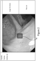
- FIG. 2 depicts an illustrative labeling tool 40 in which a user places a bounding box 42 around a lesion in a displayed training image 16 and selects a label for the type of lesion shown, e.g., a Pedunculated polyp, a Sessile polyp, a Flat polyp, etc.
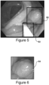
- Figure 3 depicts a further training image 16 in which a first bounding box 44 is placed around the lesion and a second bounding box 46 is placed around a reference object.
- approximately 3000 training images 16 are used, in which each has a resolution of 1280x1024 pixels.
- Detection classifier 20 may for example comprise a neural network model into which the pixel data from each training image is processed.
- an application programming interface (API) by TensorFlow TM can be used to construct the neural network model representative of a graph that includes nodes and edges.
- the model is mapped to underlying machine hardware.
- Nodes in the graph represent operations (e.g., machine learning functions, mathematical operations, etc.), and the edges represent the multidimensional data arrays also known as tensors communicated between the nodes.
- control dependencies can exist in the graph and denote that the source node must finish executing before the destination node starts executing.
- TiTensorFlow provides a platform in which the designer's design algorithm flow and computation architecture is automatically optimized.
- Nodes are assigned to computational devices and execute asynchronously, and in parallel once all the tensors on their incoming edges become available.
- the video processing required in the AI platform 10 can be very expensive in terms of CPU power. Accordingly, certain computer-vision sub-tasks are allocated to special-purpose hardware architectures, such as a GPU 23 (graphics processing unit), while others are allocated to the CPU 21.
- the GPU 23, for example, is an accelerator that is available not only on desktop computers but also on mobile devices such as smartphones and tablets. Accordingly, the model used herein has the built-in ability to configure GPU usage along with CPU usage to utilize machine resources most efficiently.
- the AI platform 10 may for example utilize a NVIDIA CUDA ® Deep Neural Network library (cuDNN), which is a GPU-accelerated library of primitives for deep neural networks.
- cuDNN provides a highly tuned implementation of standard routines such as forward and backward convolution, pooling normalization, and activation layers.
- cuDNN provides high-performance GPU acceleration that automates low-level GPU performance tuning.
- AI platform 10 may also for example utilize a NVIDIA Quadro P5000 16GB - 2560 CUDA CORES graphics card for development and testing. Anything above the NVIDIA GEFORCE GTX 1080 - 2560 CUDA CORES could likewise be utilized.
- the model is trained until the computed "loss" falls consistently below a threshold (e.g., 0.005 in the case of TensorFlow).
- a threshold e.g., 0.005 in the case of TensorFlow.
- images from a video feed 22 can be analyzed by the real-time video analysis system 14 to provide polyp detection 24 and reference object detection 26 (to determine polyp sizing 28).
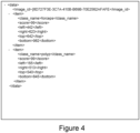
- Figure 4 depicts an illustrative .xml file for an image in the video feed 22 that includes an identified forceps and polyp.
- a lesion is first detected in real time while the clinician is doing the procedure.
- the size of the lesion is calculated using the reference object, and then an indication of the lesion and size are output with a video feed.
- forceps are used during a clinical procedure for intervention. Forceps comprise a slender flexible tool with movable cup-shaped jaws used to obtain biopsy specimens.
- Actual polyp size is determined based (1) on the measurement of the polyp size in pixels, (2) measurement of the size of the forceps in pixels, and (3) recalculation of the actual polyp size into a unit of length (e.g., millimeters) based on the known size of the forceps.
- Figure 7 describes an illustrative method for processing the polyp image data.
- a rectangular cut out image 54 ( Figure 6 ) containing the polyp is obtained (e.g., as an RGB image).
- the RGB image gets split into three monochrome channels: Red, Green, and Blue and is converted into a grayscale image.
- the image gets smoothed by applying a Gaussian function to reduce image details and variation of brightness thus enhancing image structure.
- the polyp edges or contours are identified, e.g., using a Canny multi-stage edge detection algorithm, and at S5, multiple polyp contours are identified using an OpenCV function.
- the contours that fall below a threshold e.g., contain less than 50 pixels, get filtered out.
- An example of this process is shown in Figure 8 in which the image on right depicts a filtered contour image and the image on the left depicts an unfiltered contour image.
- the minimum and maximum x and y-coordinates are determined (i.e., Xmin; Xmax; Ymin; Ymax).
- rectangle dimensions 50 for the polyp are recalculated.
- Dpx pixels
- Figure 9 depicts an illustrative process for calculating the pixel size of the forceps.
- the area defined by a circumscribed rectangle around the forceps gets cut out of the frame into a separate image, and at S11, the color image is converted into a gray monochrome image.
- the image gets smoothed by applying, e.g., a Gaussian function to reduce image details and variation of brightness thus enhancing image structure, and at S13, the forceps edges are identified using, e.g., Canny multi-stage edge detection algorithm.
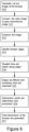
- straight lines are drawn along the forceps edges using a Hough transform technique, e.g., illustrative result shown in Figure 10 .
- edges are filtered and processed as follows.
- the number of lines gets reduced by filtering out short lines (e.g., less than 10 pixels).
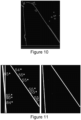
- the coefficients m and b and their angles of inclination to the x-axis are calculated.
- the lines with angles of inclination e.g., less than 21° and greater than 150° are removed.
- the lines remaining on the image get extended to the image boundaries.
- the coordinates of intersection points (midpoints) of the lines with the horizontal boundaries of the image are calculated.
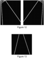
- a size of the forceps in pixels (Fpx) is the mean of the length of the perpendicular between the left and right lines and a distance between x-left and x-right points.
- a size of the forceps in pixels is considered equal to the distance between x-left and x-right points ( Figure 13 ).
- the actual size of the polyp e.g., in millimeters (mm) is determined as follows.
- a size of the forceps in mm (Fmm) is available, e.g, based on the device model or actual measurement.
- a polyp size in mm is outputted, e.g., on a computer screen in real time with the video feed.
- the AI platform 10 is utilized for detecting and sizing lesions. Additionally, the described platform 10 can also be very useful in early detection other diseases as well.
- the classifier can be trained to detect diseases such as Inflammatory Bowel Disease (IBD).
- IBD Inflammatory Bowel Disease
- IBD ulcerative colitis, which causes long-lasting inflammation and sores (ulcers) in the innermost lining of the large intestine (colon) and rectum.
- Another type of IBD is Crohn's disease, which is characterized by inflammation of the lining of the digestive tract, which often spreads deep into affected tissues. IBD can be debilitating and sometimes leads to life-threatening complications, and increases the risk of colon cancer.
- the AI platform 10 can further be used in comparing the size (area) before and after the treatment of the inflammation of the lining of the digestive tract which may appear in patches in Crohn's disease or a continuous pattern in Ulcerative colitis as well as early detection of precancerous lesions in patients with Crohn's and Ulcerative colitis.
- a machine-learned model to automatically detect the location of the diseases is another way in which this AI platform 10 can be useful. Accurate detection of the location of the disorders can be beneficial for the surgeons who perform a surgical treatment of the diseases after once a clinical procedure has been performed.
- determining Gastroenterology (GI) quality metrics is another AI platform 10 application. This would allow automated measurement of several metrics that likely reflect the quality of a GI clinical procedure.
- the process can be used to analyze digitized image frames captured during colonoscopy procedure.
- Information like insertion time, withdrawal time, images at the time of maximal intubation, Cecal intubation time, and landmark identified, quality of bowel prep can be automatically measured with the described method. As these metrics can be obtained automatically, it will help to quantify health-care processes and can aid in providing high-quality health care.
- the primary colonoscopy quality indicator is the adenoma detection rate (ADR), which is defined as the proportion of an endoscopist's screening colonoscopies in which one or more adenomas have been detected.
- ADR adenoma detection rate
- Studies of tandem colonoscopies have revealed that physicians may miss adenomas larger than 1 cm. to ensure adequate adenoma detection, studies have suggested that a physician's withdrawal time, not including polyp resection, should be on average at least 6-9 min.
- a paper published in 2006 by the ASGE/ACG Taskforce on Quality in Endoscopy recommended that the withdrawal time should be measured as an aggregate of a physician's practice rather than based on an individual patient given the variation in anatomy such the prominence of colonic folds.
- Another quality measure that affects adenoma detection is the quality of the bowel preparation.
- An adequate colon preparation is vital to ensure complete mucosal inspection. It has been reported that only three quarter of colonoscopies have an adequate colon preparation. High rates of missed adenomas and advanced neoplasia have been observed in patients with suboptimal colon preparations.
- the ADR should be calculated for each endoscopist based on data from screening examinations. If a physician's ADR is lower than the benchmark of 20%, quality improvement efforts are needed to increase this rate. Physicians should aim to achieve cecal intubation rates of 95% or greater in screening colonoscopies. Techniques for mucosal examination with a focus on mean withdrawal time should be assessed. The quality of bowel preparation in the endoscopy practice should also be determined and optimized. Finally, in adequately prepped and carefully examined colons, compliance with screening and surveillance guidelines for future exams is strongly recommended. These steps will ensure that colonoscopy is maximally effective in preventing CRC.
- Figure 14 depicts an overview of an illustrative process of implementing a client-service model using the real time video analysis system 14 of Figure 1 .
- a video frame image
- the service 62 receives the request, and searches for objects using a trained classifier.
- An XML result string is generated and is sent back to the client 60 in real time.
- the client 60 receives the response and analyzes the XML string to determine if a polyp was found. If no, a next image is sent to the service 62. If yes, the polyp is indicated as detected with the video feed (i.e., on a computer screen). Next a determination is made whether forceps were also found in the image. If no, a next image is sent to the service 62. If yes, the size of the polyp is calculated and output with the video feed.
- the AI platform may be employed in Video Capsule Endoscopy (VCE), which is a procedure that uses a tiny wireless camera to take pictures of the digestive tract.
- VCE Video Capsule Endoscopy
- a capsule endoscopy camera sits inside a capsule which a patient swallows. As the capsule travels through the digestive tract, the camera takes thousands of pictures that get transmitted to a recorder that the patient wears, e.g., on a belt around their waist.
- VCE helps doctors see inside the small intestine, an area that is hard to reach with more traditional endoscopy procedures.
- Traditional endoscopy involves passing a long, flexible tube equipped with a video camera down the throat or through the rectum.
- the camera used in capsule endoscopy takes thousands of color photos as it passes through the digestive tract.
- the images saved on the recorder then gets transferred to a computer with special software that strings the images together to create a video.
- the doctor watches the video to look for abnormalities within the digestive tract.
- the processes of watching the entire VCE of the small intestine takes about 45 minutes to 1 hour.
- the artificial intelligence (AI) platform can also be used to analyze these videos in order to detect lesions, which can then be evaluated by a clinician.
- a method for detecting the lesion in a saved video, e.g., from a VCE procedure, using artificial intelligence (AI) may for example include the steps of: providing a trained classifier that includes a deep learning model trained to detect lesions; processing a video after it has been created using the transferred images from the recorder; and using the trained classifier to determine if a video frame from the video has a lesion.
- AI artificial intelligence
- AI platform 10, client 60, and service 62 described herein may be implemented as a computer program product stored on a computer readable storage medium.
- the computer readable storage medium can be a tangible device that can retain and store instructions for use by an instruction execution device.
- the computer readable storage medium may be, for example, but is not limited to, an electronic storage device, a magnetic storage device, an optical storage device, an electromagnetic storage device, a semiconductor storage device, or any suitable combination of the foregoing.
- a non-exhaustive list of more specific examples of the computer readable storage medium includes the following: a portable computer diskette, a hard disk, a random access memory (RAM), a read-only memory (ROM), an erasable programmable read-only memory (EPROM or Flash memory), a static random access memory (SRAM), a portable compact disc read-only memory (CD-ROM), a digital versatile disk (DVD), a memory stick, and any suitable combination of the foregoing.
- RAM random access memory
- ROM read-only memory
- EPROM or Flash memory erasable programmable read-only memory
- SRAM static random access memory
- CD-ROM compact disc read-only memory
- DVD digital versatile disk
- a memory stick any suitable combination of the foregoing.
- a computer readable storage medium is not to be construed as being transitory signals per se, such as radio waves or other freely propagating electromagnetic waves, electromagnetic waves propagating through a waveguide or other transmission media (e.g., light pulses passing through a fiber-optic cable), or electrical signals transmitted through a wire.
- Computer readable program instructions described herein can be downloaded to respective computing/processing devices from a computer readable storage medium or to an external computer or external storage device via a network, for example, the Internet, a local area network, a wide area network and/or a wireless network.
- the network may comprise copper transmission cables, optical transmission fibers, wireless transmission, routers, firewalls, switches, gateway computers and/or edge servers.
- a network adapter card or network interface in each computing/processing device receives computer readable program instructions from the network and forwards the computer readable program instructions for storage in a computer readable storage medium within the respective computing/processing device.
- Computer readable program instructions for carrying out operations of the present invention may be assembler instructions, instruction-set-architecture (ISA) instructions, machine instructions, machine dependent instructions, microcode, firmware instructions, state-setting data, or either source code or object code written in any combination of one or more programming languages, including an object oriented programming language such as Java, Python, Smalltalk, C++ or the like, and conventional procedural programming languages, such as the "C" programming language or similar programming languages.
- ISA instruction-set-architecture
- machine instructions machine dependent instructions
- microcode firmware instructions
- state-setting data or either source code or object code written in any combination of one or more programming languages, including an object oriented programming language such as Java, Python, Smalltalk, C++ or the like, and conventional procedural programming languages, such as the "C" programming language or similar programming languages.
- the computer readable program instructions may execute entirely on the user's computer, partly on the user's computer, as a stand-alone software package, partly on the user's computer and partly on a remote computer or entirely on the remote computer or server.
- the remote computer may be connected to the user's computer through any type of network, including a local area network (LAN) or a wide area network (WAN), or the connection may be made to an external computer (for example, through the Internet using an Internet Service Provider).
- LAN local area network
- WAN wide area network
- Internet Service Provider for example, AT&T, MCI, Sprint, EarthLink, MSN, GTE, etc.
- electronic circuitry including, for example, programmable logic circuitry, field-programmable gate arrays (FPGA), or programmable logic arrays (PLA) may execute the computer readable program instructions by utilizing state information of the computer readable program instructions to personalize the electronic circuitry, in order to perform aspects of the present invention.
- FPGA field-programmable gate arrays
- PLA programmable logic arrays
- These computer readable program instructions may be provided to a processor of a general purpose computer, special purpose computer, or other programmable data processing apparatus to produce a machine, such that the instructions, which execute via the processor of the computer or other programmable data processing apparatus, create means for implementing the functions/acts specified in the flowchart and/or block diagram block or blocks.
- These computer readable program instructions may also be stored in a computer readable storage medium that can direct a computer, a programmable data processing apparatus, and/or other devices to function in a particular manner, such that the computer readable storage medium having instructions stored therein comprises an article of manufacture including instructions which implement aspects of the function/act specified in the flowchart and/or block diagram block or blocks.
- the computer readable program instructions may also be loaded onto a computer, other programmable data processing apparatus, or other device to cause a series of operational steps to be performed on the computer, other programmable apparatus or other device to produce a computer implemented process, such that the instructions which execute on the computer, other programmable apparatus, or other device implement the functions/acts specified in the flowchart and/or block diagram block or blocks.
- each block in the flowchart or block diagrams may represent a module, segment, or portion of instructions, which comprises one or more executable instructions for implementing the specified logical function(s).
- the functions noted in the block may occur out of the order noted in the figures.
- two blocks shown in succession may, in fact, be executed substantially concurrently, or the blocks may sometimes be executed in the reverse order, depending upon the functionality involved.
- a computing system may comprise any type of computing device and for example includes at least one processor, memory, an input/output (I/O) (e.g., one or more I/O interfaces and/or devices), and a communications pathway.
- processor(s) execute program code which is at least partially fixed in memory. While executing program code, processor(s) can process data, which can result in reading and/or writing transformed data from/to memory and/or I/O for further processing.
- the pathway provides a communications link between each of the components in computing system.
- I/O can comprise one or more human I/O devices, which enable a user to interact with computing system.
- Computing system may also be implemented in a distributed manner such that different components reside in different physical locations.
- the AI platform 10 or relevant components thereof may also be automatically or semi-automatically deployed into a computer system by sending the components to a central server or a group of central servers.
- the components are then downloaded into a target computer that will execute the components.
- the components are then either detached to a directory or loaded into a directory that executes a program that detaches the components into a directory.
- Another alternative is to send the components directly to a directory on a client computer hard drive.
- the process will select the proxy server code, determine on which computers to place the proxy servers' code, transmit the proxy server code, then install the proxy server code on the proxy computer.
- the components will be transmitted to the proxy server and then it will be stored on the proxy server.
Landscapes
- Engineering & Computer Science (AREA)
- Health & Medical Sciences (AREA)
- Life Sciences & Earth Sciences (AREA)
- Medical Informatics (AREA)
- Physics & Mathematics (AREA)
- Surgery (AREA)
- General Health & Medical Sciences (AREA)
- Public Health (AREA)
- Biomedical Technology (AREA)
- Pathology (AREA)
- Nuclear Medicine, Radiotherapy & Molecular Imaging (AREA)
- Radiology & Medical Imaging (AREA)
- Computer Vision & Pattern Recognition (AREA)
- Veterinary Medicine (AREA)
- Animal Behavior & Ethology (AREA)
- Molecular Biology (AREA)
- Heart & Thoracic Surgery (AREA)
- General Physics & Mathematics (AREA)
- Theoretical Computer Science (AREA)
- Biophysics (AREA)
- Artificial Intelligence (AREA)
- Signal Processing (AREA)
- Optics & Photonics (AREA)
- Quality & Reliability (AREA)
- Evolutionary Computation (AREA)
- Data Mining & Analysis (AREA)
- Physiology (AREA)
- Geometry (AREA)
- Primary Health Care (AREA)
- Epidemiology (AREA)
- Databases & Information Systems (AREA)
- Endocrinology (AREA)
- Gynecology & Obstetrics (AREA)
- Oral & Maxillofacial Surgery (AREA)
- Psychiatry (AREA)
- Mathematical Physics (AREA)
- Fuzzy Systems (AREA)
- Gastroenterology & Hepatology (AREA)
- Image Analysis (AREA)
- Image Processing (AREA)
Claims (9)
- Plattform für künstliche Intelligenz, KI (10) zur Erkennung und Größenbestimmung einer Läsion in Echtzeit während eines klinischen Verfahrens, umfassend:einen trainierten Klassierer (20), der ein Deep-Learning-Modell enthält, das trainiert ist, Läsionen und Referenzobjekte in Bilddaten zu erkennen; undein Echtzeit-Videoanalysesystem (14), das einen Video-Feed (22) während eines klinischen Verfahrens empfängt, den trainierten Klassierer (20) verwendet, um zu bestimmen, ob ein Videobild des Video-Feeds (22) sowohl eine Läsion als auch ein Referenzobjekt enthält, eine tatsächliche Größe der Läsion auf der Grundlage einer Pixelgröße für sowohl die Läsion als auch das Referenzobjekt berechnet und eine Mitteilung, dass die Läsion erkannt wurde, und die tatsächliche Größe der Läsion ausgibt,wobei die Pixelgröße für die Läsion bestimmt wird durch: Aufnehmen eines rechteckigen Bildausschnitts, der die Läsion enthält;Verbessern des Bildausschnitts durch Bildglättung;Verwenden eines Kantenerkennungsalgorithmus zum Erkennen von Konturen der Läsion; undBerechnen neuer rechteckiger Koordinaten der Läsion auf der Grundlage von bestimmten Koordinaten der Konturen, um eine Pixelgröße der Läsion, Dpx, bereitzustellen,wobei die Pixelgröße für das Referenzobjekt bestimmt wird durch:Aufnehmen eines rechteckigen Bildausschnitts, der das Referenzobjekt enthält;Verbessern des Bildausschnitts durch Bildglättung;Identifizieren von Kanten des Referenzobjekts und Einführen von geraden Linien entlang der Kanten;Filtern der geraden Linien auf der Grundlage jeweiliger Linienlängen, Linienneigungen und Schnittkoordinaten der geraden Linien mit Grenzen des Bildausschnitts, um ein Paar von sich schneidenden Linien zu identifizieren; undBestimmen einer Pixelgröße für das Referenzobjekt, Fpx, auf der Grundlage des Paars von sich schneidenden Linien, wobei die tatsächliche Größe der Läsion D berechnet wird als:
D = (Dpx * F) / Fpx, wobei F eine tatsächliche Größe des Referenzobjekts ist. - KI-Plattform (10) nach Anspruch 1, wobei die Läsion einen Polypen umfasst.
- KI-Plattform (10) nach Anspruch 1, wobei das Referenzobjekt wenigstens eines von einer Schlinge und einer Zange (52) enthält.
- KI-Plattform (10) nach Anspruch 1, wobei der trainierte Klassierer (20) ein neuronales Netz enthält, das mit einem Satz von Trainingsbildern (16) trainiert wurde, wobei eine Teilmenge der Trainingsbilder (16) Polypen verschiedener Typen, Polypen, die auf verschiedenem Hintergrund erscheinen, und Polypen, die unter verschiedenen Beleuchtungsbedingungen erscheinen, enthält.
- KI-Plattform (10) nach Anspruch 1, wobei die Mitteilung, dass die Läsion erkannt wurde, und die tatsächliche Größe der Läsion der Video-Feed während des klinischen Verfahrens ausgegeben werden.
- Computerimplementiertes Verfahren zur Erkennung und Größenbestimmung einer Läsion in Echtzeit während eines klinischen Verfahrens unter Verwendung von künstlicher Intelligenz, KI, umfassend:Bereitstellen eines trainierten Klassierers (20), der ein Deep-Learning-Modell enthält, das trainiert ist, Läsionen und Referenzobjekte in Bilddaten zu erkennen;Empfangen eines Video-Feeds (22) während eines klinischen Verfahrens;Verwenden des trainierten Klassierers (20), um zu bestimmen, ob ein Videobild des Video-Feeds (22) sowohl eine Läsion als auch ein Referenzobjekt enthält;Berechnen einer tatsächlichen Größe der Läsion auf der Grundlage einer Pixelgröße für sowohl die Läsion als auch das Referenzobjekt; undAusgeben einer Mitteilung, dass die Läsion erkannt wurde, und der tatsächlichen Größe der Läsion,wobei die Pixelgröße für die Läsion bestimmt wird durch:Aufnehmen eines rechteckigen Bildausschnitts, der die Läsion enthält;Verbessern des Bildausschnitts durch Bildglättung;Verwenden eines Kantenerkennungsalgorithmus zum Erkennen von Konturen der Läsion; undBerechnen neuer rechteckiger Koordinaten der Läsion auf der Grundlage von bestimmten Koordinaten der Konturen, um eine Pixelgröße der Läsion, Dpx, bereitzustellen,wobei die Pixelgröße für das Referenzobjekt bestimmt wird durch:Aufnehmen eines rechteckigen Bildausschnitt, der das Referenzobjekt enthält;Verbessern des Bildausschnitts durch Bildglättung;Identifizieren von Kanten des Referenzobjekts und Einführen von geraden Linien entlang der Kanten;Filtern der geraden Linien auf der Grundlage jeweiliger Linienlängen, Linienneigungen und Schnittkoordinaten der geraden Linien mit Grenzen des Bildausschnitts, um ein erhaltenes Paar von sich schneidenden Linien zu identifizieren; undBestimmen einer Pixelgröße für das Referenzobjekt, Fpx, auf der Grundlage des erhaltenen Paars von sich schneidenden Linien,wobei die tatsächliche Größe der Läsion D berechnet wird als:
D = (Dpx * F) / Fpx, wobei F eine tatsächliche Größe des Referenzobjekts ist. - Verfahren nach Anspruch 6, wobei das Referenzobjekt wenigstens eines von einer Schlinge und einer Zange (52) umfasst.
- Verfahren nach Anspruch 6, wobei der trainierte Klassierer (20) ein neuronales Netz enthält, das mit einem Satz von Trainingsbildern (16) trainiert wurde, wobei eine Teilmenge der Trainingsbilder (16) Polypen verschiedener Typen, Polypen, die auf verschiedenen Hintergrunden erscheinen, und Polypen, die unter verschiedenen Beleuchtungsbedingungen erscheinen, enthält.
- Computerlesbares Speichermedium, das Anweisungen umfasst, die bei Ausführung durch einen Computer bewirken, dass der Computer das Verfahren nach einem der Ansprüche 6 bis 8 ausführt.
Applications Claiming Priority (2)
| Application Number | Priority Date | Filing Date | Title |
|---|---|---|---|
| US16/288,843 US10957043B2 (en) | 2019-02-28 | 2019-02-28 | AI systems for detecting and sizing lesions |
| PCT/US2019/038431 WO2020176124A1 (en) | 2019-02-28 | 2019-06-21 | Ai systems for detecting and sizing lesions |
Publications (4)
| Publication Number | Publication Date |
|---|---|
| EP3930610A1 EP3930610A1 (de) | 2022-01-05 |
| EP3930610A4 EP3930610A4 (de) | 2022-11-30 |
| EP3930610C0 EP3930610C0 (de) | 2025-05-14 |
| EP3930610B1 true EP3930610B1 (de) | 2025-05-14 |
Family
ID=72236232
Family Applications (1)
| Application Number | Title | Priority Date | Filing Date |
|---|---|---|---|
| EP19917118.2A Active EP3930610B1 (de) | 2019-02-28 | 2019-06-21 | Ki-systeme zur erkennung und dimensionierung von läsionen |
Country Status (5)
| Country | Link |
|---|---|
| US (1) | US10957043B2 (de) |
| EP (1) | EP3930610B1 (de) |
| CN (1) | CN113573654B (de) |
| AU (1) | AU2019431299B2 (de) |
| WO (1) | WO2020176124A1 (de) |
Families Citing this family (27)
| Publication number | Priority date | Publication date | Assignee | Title |
|---|---|---|---|---|
| WO2020194568A1 (ja) * | 2019-03-27 | 2020-10-01 | Hoya株式会社 | 内視鏡用プロセッサ、情報処理装置、内視鏡システム、プログラム及び情報処理方法 |
| WO2020194663A1 (ja) * | 2019-03-28 | 2020-10-01 | オリンパス株式会社 | トラッキング装置、学習済モデル、内視鏡システム及びトラッキング方法 |
| WO2020194664A1 (ja) | 2019-03-28 | 2020-10-01 | オリンパス株式会社 | トラッキング装置、学習済モデル、内視鏡システム及びトラッキング方法 |
| EP3745298B1 (de) * | 2019-05-28 | 2022-05-25 | SCHOTT Schweiz AG | Klassifizierungsverfahren und -system für transparente artikel mit hohem durchsatz |
| JP7335157B2 (ja) * | 2019-12-25 | 2023-08-29 | 富士フイルム株式会社 | 学習データ作成装置、学習データ作成装置の作動方法及び学習データ作成プログラム並びに医療画像認識装置 |
| EP4211652A1 (de) * | 2020-09-08 | 2023-07-19 | Given Imaging Ltd. | Systeme und verfahren zur polyp-grössenschätzung |
| JP7374224B2 (ja) * | 2021-01-14 | 2023-11-06 | コ,ジファン | 内視鏡を用いた大腸検査ガイド装置 |
| US20220334851A1 (en) * | 2021-04-15 | 2022-10-20 | Nvidia Corporation | Application programming interface to generate a representation of graph code |
| TR2021010390A2 (tr) * | 2021-06-24 | 2023-01-23 | Erci̇yes Üni̇versi̇tesi̇ Strateji̇ Geli̇şti̇rme Dai̇re Başkanliği | Gerçek zamanli otomati̇k poli̇p tespi̇ti̇ i̇çi̇n deri̇n öğrenme tabanli karar destek si̇stemi̇ |
| WO2023003856A1 (en) | 2021-07-21 | 2023-01-26 | Utech Products, Inc. | Ai platform for processing speech and video information collected during a medical procedure |
| DE112022003919T5 (de) | 2021-08-09 | 2024-06-27 | Digestaid - Artificial Intelligence Development, Lda. | Automatische erfassung und differenzierung von zystischen pankreasläsionen mit endoskopischer ultrasonographie |
| JP7642498B2 (ja) * | 2021-09-08 | 2025-03-10 | 富士フイルム株式会社 | 画像処理装置、画像処理方法、及びプログラム |
| US12525145B2 (en) | 2021-12-03 | 2026-01-13 | Ambu A/S | Endoscopic training system |
| US12605039B2 (en) | 2021-12-03 | 2026-04-21 | Ambu A/S | Endoscope image processing device |
| EP4190271A1 (de) * | 2021-12-03 | 2023-06-07 | Ambu A/S | Verarbeitungsvorrichtung für endoskopbilder |
| WO2023102880A1 (zh) * | 2021-12-10 | 2023-06-15 | 曾稼志 | 气管插管影像的处理方法与系统以及气管插管的成效评量方法 |
| US20250061569A1 (en) * | 2021-12-27 | 2025-02-20 | Nec Corporation | Image processing device, image processing method, and recording medium |
| CN114305690B (zh) * | 2021-12-31 | 2023-12-26 | 杭州三坛医疗科技有限公司 | 一种手术导航定位方法及装置 |
| CN114066781B (zh) * | 2022-01-18 | 2022-05-10 | 浙江鸿禾医疗科技有限责任公司 | 胶囊内窥镜肠道图像的识别定位方法、存储介质和设备 |
| CN114680809A (zh) * | 2022-02-17 | 2022-07-01 | 河北大学 | 一种利用光斑测量肠镜下息肉大小的测量系统 |
| WO2023200491A1 (en) | 2022-04-12 | 2023-10-19 | Utech Products, Inc. | Multithreading video processing with cloud services to detect and analyze objects |
| US12211229B2 (en) | 2022-06-07 | 2025-01-28 | Hong Kong Applied Science and Technology Research Institute Company Limited | Method, device, and system for detecting and tracking objects in captured video using convolutional neural network |
| US12599284B2 (en) * | 2023-01-11 | 2026-04-14 | Nec Corporation | Endoscopic examination support apparatus, endoscopic examination support method, and recording medium |
| WO2024197056A1 (en) * | 2023-03-20 | 2024-09-26 | Ohio State Innovation Foundation | Convolutional neural network classification of presence or absence of disease with endoscopic or laryngoscopic video |
| JP2024150245A (ja) * | 2023-04-10 | 2024-10-23 | 富士フイルム株式会社 | 医療支援装置、内視鏡システム、医療支援方法、及びプログラム |
| JP2024164943A (ja) * | 2023-05-16 | 2024-11-28 | 富士フイルム株式会社 | 医療画像処理装置及び内視鏡システム |
| WO2025081350A1 (en) * | 2023-10-17 | 2025-04-24 | Abb Schweiz Ag | Method for generating a safe zone of a robot, electronic device and computer readable medium |
Family Cites Families (58)
| Publication number | Priority date | Publication date | Assignee | Title |
|---|---|---|---|---|
| JPH09508994A (ja) * | 1994-01-28 | 1997-09-09 | シュナイダー メディカル テクノロジーズ インコーポレイテッド | 像形成装置及び方法 |
| US5984870A (en) * | 1997-07-25 | 1999-11-16 | Arch Development Corporation | Method and system for the automated analysis of lesions in ultrasound images |
| US6801645B1 (en) * | 1999-06-23 | 2004-10-05 | Icad, Inc. | Computer aided detection of masses and clustered microcalcifications with single and multiple input image context classification strategies |
| US8543519B2 (en) * | 2000-08-07 | 2013-09-24 | Health Discovery Corporation | System and method for remote melanoma screening |
| US7499578B2 (en) * | 2002-10-18 | 2009-03-03 | Cornell Research Foundation, Inc. | System, method and apparatus for small pulmonary nodule computer aided diagnosis from computed tomography scans |
| US8634607B2 (en) * | 2003-09-23 | 2014-01-21 | Cambridge Research & Instrumentation, Inc. | Spectral imaging of biological samples |
| US7454045B2 (en) * | 2003-10-10 | 2008-11-18 | The United States Of America As Represented By The Department Of Health And Human Services | Determination of feature boundaries in a digital representation of an anatomical structure |
| US8871224B2 (en) * | 2003-12-09 | 2014-10-28 | Allergan, Inc. | Botulinum toxin therapy for skin disorders |
| WO2006078902A2 (en) * | 2005-01-19 | 2006-07-27 | Dermaspect, Llc | Devices and methods for identifying and monitoring changes of a suspect area on a patient |
| US7646902B2 (en) * | 2005-02-08 | 2010-01-12 | Regents Of The University Of Michigan | Computerized detection of breast cancer on digital tomosynthesis mammograms |
| US7474775B2 (en) * | 2005-03-31 | 2009-01-06 | University Of Iowa Research Foundation | Automatic detection of red lesions in digital color fundus photographs |
| US7758594B2 (en) * | 2005-05-20 | 2010-07-20 | Neotract, Inc. | Devices, systems and methods for treating benign prostatic hyperplasia and other conditions |
| ITRM20060213A1 (it) * | 2006-04-13 | 2007-10-14 | Univ Palermo | Metodo di elaborazione di immagini biomediche |
| JP2010504129A (ja) * | 2006-09-22 | 2010-02-12 | コーニンクレッカ フィリップス エレクトロニクス エヌ ヴィ | 肺結節の高度コンピュータ支援診断 |
| CN101143894A (zh) * | 2007-06-22 | 2008-03-19 | 中国药科大学 | 高效抑制血管生成多肽及其物理化学修饰方法和应用 |
| DE102007044548A1 (de) * | 2007-09-18 | 2009-04-09 | Siemens Ag | Computergestütztes Werkzeug und Verfahren zur multiplanaren 3D-Vermessung von Längen und Winkeln in der morphometrischen Computer- und Magnetresonanztomografie |
| US8126244B2 (en) * | 2007-09-21 | 2012-02-28 | Siemens Medical Solutions Usa, Inc. | User interface for polyp annotation, segmentation, and measurement in 3D computed tomography colonography |
| US20090118600A1 (en) * | 2007-11-02 | 2009-05-07 | Ortiz Joseph L | Method and apparatus for skin documentation and analysis |
| US9117133B2 (en) * | 2008-06-18 | 2015-08-25 | Spectral Image, Inc. | Systems and methods for hyperspectral imaging |
| KR100961661B1 (ko) * | 2009-02-12 | 2010-06-09 | 주식회사 래보 | 수술용 항법 장치 및 그 방법 |
| GB2468164B (en) * | 2009-02-27 | 2014-08-13 | Samsung Electronics Co Ltd | Computer-aided detection of lesions |
| US9125570B2 (en) * | 2010-07-16 | 2015-09-08 | The Board Of Trustees Of The Leland Stanford Junior University | Real-time tomosynthesis guidance for radiation therapy |
| US9208556B2 (en) * | 2010-11-26 | 2015-12-08 | Quantitative Insights, Inc. | Method, system, software and medium for advanced intelligent image analysis and display of medical images and information |
| US8900113B2 (en) * | 2011-01-21 | 2014-12-02 | Headwater Partners Ii Llc | Tracking of tumor location for targeted radiation treatment |
| WO2013018908A1 (ja) | 2011-08-04 | 2013-02-07 | オリンパス株式会社 | 医療用マニピュレータおよび手術支援装置 |
| WO2013049153A2 (en) * | 2011-09-27 | 2013-04-04 | Board Of Regents, University Of Texas System | Systems and methods for automated screening and prognosis of cancer from whole-slide biopsy images |
| WO2014105439A1 (en) * | 2012-12-24 | 2014-07-03 | Sanovas, Inc. | Anchored working channel |
| EP2772882A1 (de) * | 2013-03-01 | 2014-09-03 | Universite D'angers | Automatische Messung von Läsionen auf medizinischen Bildern |
| WO2014160510A2 (en) | 2013-03-13 | 2014-10-02 | Massachusetts Institute Of Technology | Photometric stereo endoscopy |
| WO2014139021A1 (en) * | 2013-03-15 | 2014-09-18 | Synaptive Medical (Barbados) Inc. | Intramodal synchronization of surgical data |
| US9741116B2 (en) | 2013-08-29 | 2017-08-22 | Mayo Foundation For Medical Education And Research | System and method for boundary classification and automatic polyp detection |
| US9445713B2 (en) * | 2013-09-05 | 2016-09-20 | Cellscope, Inc. | Apparatuses and methods for mobile imaging and analysis |
| US20150080652A1 (en) | 2013-09-18 | 2015-03-19 | Cerner Innovation, Inc. | Lesion detection and image stabilization using portion of field of view |
| WO2015084462A1 (en) * | 2013-12-03 | 2015-06-11 | Children's National Medical Center | Method and system for wound assessment and management |
| WO2015164768A1 (en) | 2014-04-24 | 2015-10-29 | Arizona Board Of Regents On Behalf Of Arizona State University | System and method for detecting polyps from learned boundaries |
| AU2015253295B2 (en) * | 2014-04-28 | 2018-03-29 | Northwestern University | Devices, methods, and systems of functional optical coherence tomography |
| EP3195191B1 (de) * | 2014-09-03 | 2020-10-21 | Ventana Medical Systems, Inc. | Systeme und verfahren zur berechnung von immunscores |
| US9700213B2 (en) | 2014-09-12 | 2017-07-11 | Mayo Foundation For Medical Education And Research | System and method for automatic polyp detection using global geometric constraints and local intensity variation patterns |
| US20160100789A1 (en) * | 2014-10-13 | 2016-04-14 | National Central University | Computer-aided diagnosis system and computer-aided diagnosis method |
| CN107106020A (zh) * | 2014-10-29 | 2017-08-29 | 组织分析股份有限公司 | 用于分析并且传输与哺乳动物皮损病有关的数据、图像和视频的系统与方法 |
| US9990472B2 (en) * | 2015-03-23 | 2018-06-05 | Ohio State Innovation Foundation | System and method for segmentation and automated measurement of chronic wound images |
| US20180096191A1 (en) | 2015-03-27 | 2018-04-05 | Siemens Aktiengesellschaft | Method and system for automated brain tumor diagnosis using image classification |
| US10055843B2 (en) * | 2015-03-31 | 2018-08-21 | Mayo Foundation For Medical Education And Research | System and methods for automatic polyp detection using convulutional neural networks |
| CN204520699U (zh) * | 2015-04-08 | 2015-08-05 | 李彬 | 一种实时读数宫腔测量装置 |
| CN107427227A (zh) * | 2015-06-15 | 2017-12-01 | 哈伊姆·埃米尔 | 调适皮肤治疗的系统及方法 |
| US10510144B2 (en) * | 2015-09-10 | 2019-12-17 | Magentiq Eye Ltd. | System and method for detection of suspicious tissue regions in an endoscopic procedure |
| US9760983B2 (en) * | 2015-10-19 | 2017-09-12 | Shanghai United Imaging Healthcare Co., Ltd. | System and method for image registration in medical imaging system |
| US20170164924A1 (en) * | 2015-12-15 | 2017-06-15 | Konica Minolta, Inc. | Ultrasound image diagnostic apparatus |
| EP3267394A1 (de) * | 2016-07-04 | 2018-01-10 | Centre National De La Recherche Scientifique | Verfahren und vorrichtung zur echtzeitdetektion von polypen in optischer kolonoskopie |
| US9589374B1 (en) | 2016-08-01 | 2017-03-07 | 12 Sigma Technologies | Computer-aided diagnosis system for medical images using deep convolutional neural networks |
| US10531825B2 (en) * | 2016-10-14 | 2020-01-14 | Stoecker & Associates, LLC | Thresholding methods for lesion segmentation in dermoscopy images |
| US20180263568A1 (en) | 2017-03-09 | 2018-09-20 | The Board Of Trustees Of The Leland Stanford Junior University | Systems and Methods for Clinical Image Classification |
| EP3603481B1 (de) | 2017-03-30 | 2023-05-03 | FUJIFILM Corporation | Medizinische bildverarbeitungsvorrichtung, endoskopsystem und verfahren zum betrieb einer medizinischen bildverarbeitungsvorrichtung |
| CN107730489A (zh) * | 2017-10-09 | 2018-02-23 | 杭州电子科技大学 | 无线胶囊内窥镜小肠病变计算机辅助检测系统及检测方法 |
| US10559080B2 (en) * | 2017-12-27 | 2020-02-11 | International Business Machines Corporation | Adaptive segmentation of lesions in medical images |
| US20190220738A1 (en) * | 2018-01-18 | 2019-07-18 | Amit Flank | Skin analysis system and method |
| TW202001804A (zh) * | 2018-04-20 | 2020-01-01 | 成真股份有限公司 | 用精細解析進行資料管理和機器學習的方法 |
| US11011257B2 (en) * | 2018-11-21 | 2021-05-18 | Enlitic, Inc. | Multi-label heat map display system |
-
2019
- 2019-02-28 US US16/288,843 patent/US10957043B2/en active Active
- 2019-06-21 WO PCT/US2019/038431 patent/WO2020176124A1/en not_active Ceased
- 2019-06-21 AU AU2019431299A patent/AU2019431299B2/en active Active
- 2019-06-21 EP EP19917118.2A patent/EP3930610B1/de active Active
- 2019-06-21 CN CN201980093189.4A patent/CN113573654B/zh active Active
Also Published As
| Publication number | Publication date |
|---|---|
| EP3930610C0 (de) | 2025-05-14 |
| AU2019431299A1 (en) | 2021-09-16 |
| US20200279373A1 (en) | 2020-09-03 |
| WO2020176124A1 (en) | 2020-09-03 |
| CN113573654A (zh) | 2021-10-29 |
| EP3930610A1 (de) | 2022-01-05 |
| CN113573654B (zh) | 2024-09-03 |
| US10957043B2 (en) | 2021-03-23 |
| AU2019431299B2 (en) | 2021-09-23 |
| EP3930610A4 (de) | 2022-11-30 |
| AU2019431299A8 (en) | 2021-09-30 |
Similar Documents
| Publication | Publication Date | Title |
|---|---|---|
| EP3930610B1 (de) | Ki-systeme zur erkennung und dimensionierung von läsionen | |
| Wang et al. | Development and validation of a deep-learning algorithm for the detection of polyps during colonoscopy | |
| US9445713B2 (en) | Apparatuses and methods for mobile imaging and analysis | |
| US12217449B2 (en) | Systems and methods for video-based positioning and navigation in gastroenterological procedures | |
| US20180263568A1 (en) | Systems and Methods for Clinical Image Classification | |
| KR20200116107A (ko) | 의료적 이미지화 절차의 자동화된 모니터링 | |
| RU2633320C2 (ru) | Отбор изображений для оптического исследования шейки матки | |
| CN116546916A (zh) | 用于虚拟胰腺造影管线的系统和方法 | |
| Gong et al. | Using deep learning to identify the recurrent laryngeal nerve during thyroidectomy | |
| Taunk et al. | Computer-assisted assessment of colonic polyp histopathology using probe-based confocal laser endomicroscopy | |
| Jütte et al. | Integrating generative AI with ABCDE rule analysis for enhanced skin cancer diagnosis, dermatologist training and patient education | |
| KR20230099995A (ko) | 자궁 경부암의 진단에 대한 정보 제공 방법 및 이를 이용한 자궁 경부암의 진단에 대한 정보 제공용 디바이스 | |
| Luca et al. | Artificial intelligence and deep learning, important tools in assisting gastroenterologists | |
| EP1782384B1 (de) | System und verfahren zur darmwandextraktion bei anwesenheit von markierten fäkalien oder kollabierten darmregionen | |
| WO2024202789A1 (ja) | 医療支援装置、内視鏡システム、医療支援方法、及びプログラム | |
| Garbaz et al. | Bleeding Segmentation Based on a U-Formed Network with Separable Contextual Feature-Guided in Wireless Capsule Endoscopy Images | |
| Luca et al. | An overview on computer processing for endoscopy and colonoscopy videos | |
| CN116542883A (zh) | 一种磁控胶囊胃镜影像病灶粘膜增强系统 | |
| WO2013150419A1 (en) | Quality-check during medical imaging procedure | |
| Thekkek et al. | Quantitative evaluation of in vivo vital-dye fluorescence endoscopic imaging for the detection of Barrett’s-associated neoplasia | |
| Hill | Segmentation of oral optical coherence tomography with deep learning | |
| Sushma et al. | WCE-bleed-net: a convolutional neural network with adaptive attention module for bleeding frame and region detection in wireless capsule endoscopy video | |
| RU2839240C2 (ru) | Способ и система измерения размеров новообразований внутри организма при проведении эндоскопических процедур на основе обработки серии эндоскопических изображений в автоматическом режиме | |
| Brenes | Integration of Multimodal, Multiscale Imaging and Biomarker Data for Squamous Precancer Detection and Diagnosis | |
| Narain et al. | A computer vision based colonoscopy support system for real-time monitoring of bowel preparation and colonic anatomical localization |
Legal Events
| Date | Code | Title | Description |
|---|---|---|---|
| STAA | Information on the status of an ep patent application or granted ep patent |
Free format text: STATUS: THE INTERNATIONAL PUBLICATION HAS BEEN MADE |
|
| PUAI | Public reference made under article 153(3) epc to a published international application that has entered the european phase |
Free format text: ORIGINAL CODE: 0009012 |
|
| STAA | Information on the status of an ep patent application or granted ep patent |
Free format text: STATUS: REQUEST FOR EXAMINATION WAS MADE |
|
| 17P | Request for examination filed |
Effective date: 20210903 |
|
| AK | Designated contracting states |
Kind code of ref document: A1 Designated state(s): AL AT BE BG CH CY CZ DE DK EE ES FI FR GB GR HR HU IE IS IT LI LT LU LV MC MK MT NL NO PL PT RO RS SE SI SK SM TR |
|
| DAV | Request for validation of the european patent (deleted) | ||
| DAX | Request for extension of the european patent (deleted) | ||
| A4 | Supplementary search report drawn up and despatched |
Effective date: 20221102 |
|
| RIC1 | Information provided on ipc code assigned before grant |
Ipc: A61B 17/94 20060101ALI20221026BHEP Ipc: G06T 7/13 20170101ALI20221026BHEP Ipc: G06T 7/00 20170101ALI20221026BHEP Ipc: G06T 7/62 20170101AFI20221026BHEP |
|
| REG | Reference to a national code |
Ref country code: DE Ref legal event code: R079 Ipc: G06T0007620000 Ref country code: DE Ref legal event code: R079 Ref document number: 602019070110 Country of ref document: DE Free format text: PREVIOUS MAIN CLASS: A61B0017940000 Ipc: G06T0007620000 |
|
| GRAP | Despatch of communication of intention to grant a patent |
Free format text: ORIGINAL CODE: EPIDOSNIGR1 |
|
| STAA | Information on the status of an ep patent application or granted ep patent |
Free format text: STATUS: GRANT OF PATENT IS INTENDED |
|
| RIC1 | Information provided on ipc code assigned before grant |
Ipc: G16H 50/70 20180101ALI20241211BHEP Ipc: G16H 50/20 20180101ALI20241211BHEP Ipc: A61B 1/00 20060101ALI20241211BHEP Ipc: G06T 7/00 20170101ALI20241211BHEP Ipc: G06T 7/62 20170101AFI20241211BHEP |
|
| INTG | Intention to grant announced |
Effective date: 20241223 |
|
| GRAS | Grant fee paid |
Free format text: ORIGINAL CODE: EPIDOSNIGR3 |
|
| GRAA | (expected) grant |
Free format text: ORIGINAL CODE: 0009210 |
|
| STAA | Information on the status of an ep patent application or granted ep patent |
Free format text: STATUS: THE PATENT HAS BEEN GRANTED |
|
| AK | Designated contracting states |
Kind code of ref document: B1 Designated state(s): AL AT BE BG CH CY CZ DE DK EE ES FI FR GB GR HR HU IE IS IT LI LT LU LV MC MK MT NL NO PL PT RO RS SE SI SK SM TR |
|
| REG | Reference to a national code |
Ref country code: GB Ref legal event code: FG4D |
|
| REG | Reference to a national code |
Ref country code: CH Ref legal event code: EP |
|
| REG | Reference to a national code |
Ref country code: IE Ref legal event code: FG4D |
|
| U01 | Request for unitary effect filed |
Effective date: 20250514 |
|
| U07 | Unitary effect registered |
Designated state(s): AT BE BG DE DK EE FI FR IT LT LU LV MT NL PT RO SE SI Effective date: 20250520 |
|
| PGFP | Annual fee paid to national office [announced via postgrant information from national office to epo] |
Ref country code: GB Payment date: 20250627 Year of fee payment: 7 |
|
| U20 | Renewal fee for the european patent with unitary effect paid |
Year of fee payment: 7 Effective date: 20250628 |
|
| PG25 | Lapsed in a contracting state [announced via postgrant information from national office to epo] |
Ref country code: ES Free format text: LAPSE BECAUSE OF FAILURE TO SUBMIT A TRANSLATION OF THE DESCRIPTION OR TO PAY THE FEE WITHIN THE PRESCRIBED TIME-LIMIT Effective date: 20250514 |
|
| PG25 | Lapsed in a contracting state [announced via postgrant information from national office to epo] |
Ref country code: GR Free format text: LAPSE BECAUSE OF FAILURE TO SUBMIT A TRANSLATION OF THE DESCRIPTION OR TO PAY THE FEE WITHIN THE PRESCRIBED TIME-LIMIT Effective date: 20250815 Ref country code: NO Free format text: LAPSE BECAUSE OF FAILURE TO SUBMIT A TRANSLATION OF THE DESCRIPTION OR TO PAY THE FEE WITHIN THE PRESCRIBED TIME-LIMIT Effective date: 20250814 |
|
| PG25 | Lapsed in a contracting state [announced via postgrant information from national office to epo] |
Ref country code: PL Free format text: LAPSE BECAUSE OF FAILURE TO SUBMIT A TRANSLATION OF THE DESCRIPTION OR TO PAY THE FEE WITHIN THE PRESCRIBED TIME-LIMIT Effective date: 20250514 |
|
| PG25 | Lapsed in a contracting state [announced via postgrant information from national office to epo] |
Ref country code: HR Free format text: LAPSE BECAUSE OF FAILURE TO SUBMIT A TRANSLATION OF THE DESCRIPTION OR TO PAY THE FEE WITHIN THE PRESCRIBED TIME-LIMIT Effective date: 20250514 |
|
| PG25 | Lapsed in a contracting state [announced via postgrant information from national office to epo] |
Ref country code: RS Free format text: LAPSE BECAUSE OF FAILURE TO SUBMIT A TRANSLATION OF THE DESCRIPTION OR TO PAY THE FEE WITHIN THE PRESCRIBED TIME-LIMIT Effective date: 20250814 |
|
| PG25 | Lapsed in a contracting state [announced via postgrant information from national office to epo] |
Ref country code: IS Free format text: LAPSE BECAUSE OF FAILURE TO SUBMIT A TRANSLATION OF THE DESCRIPTION OR TO PAY THE FEE WITHIN THE PRESCRIBED TIME-LIMIT Effective date: 20250914 |
|
| PG25 | Lapsed in a contracting state [announced via postgrant information from national office to epo] |
Ref country code: SM Free format text: LAPSE BECAUSE OF FAILURE TO SUBMIT A TRANSLATION OF THE DESCRIPTION OR TO PAY THE FEE WITHIN THE PRESCRIBED TIME-LIMIT Effective date: 20250514 |
|
| PG25 | Lapsed in a contracting state [announced via postgrant information from national office to epo] |
Ref country code: CZ Free format text: LAPSE BECAUSE OF FAILURE TO SUBMIT A TRANSLATION OF THE DESCRIPTION OR TO PAY THE FEE WITHIN THE PRESCRIBED TIME-LIMIT Effective date: 20250514 |
|
| PG25 | Lapsed in a contracting state [announced via postgrant information from national office to epo] |
Ref country code: SK Free format text: LAPSE BECAUSE OF FAILURE TO SUBMIT A TRANSLATION OF THE DESCRIPTION OR TO PAY THE FEE WITHIN THE PRESCRIBED TIME-LIMIT Effective date: 20250514 |
|
| REG | Reference to a national code |
Ref country code: CH Ref legal event code: H13 Free format text: ST27 STATUS EVENT CODE: U-0-0-H10-H13 (AS PROVIDED BY THE NATIONAL OFFICE) Effective date: 20260127 |
|
| PG25 | Lapsed in a contracting state [announced via postgrant information from national office to epo] |
Ref country code: MC Free format text: LAPSE BECAUSE OF FAILURE TO SUBMIT A TRANSLATION OF THE DESCRIPTION OR TO PAY THE FEE WITHIN THE PRESCRIBED TIME-LIMIT Effective date: 20250514 |
|
| PLBE | No opposition filed within time limit |
Free format text: ORIGINAL CODE: 0009261 |
|
| STAA | Information on the status of an ep patent application or granted ep patent |
Free format text: STATUS: NO OPPOSITION FILED WITHIN TIME LIMIT |
|
| REG | Reference to a national code |
Ref country code: CH Ref legal event code: L10 Free format text: ST27 STATUS EVENT CODE: U-0-0-L10-L00 (AS PROVIDED BY THE NATIONAL OFFICE) Effective date: 20260325 |
|
| PG25 | Lapsed in a contracting state [announced via postgrant information from national office to epo] |
Ref country code: IE Free format text: LAPSE BECAUSE OF NON-PAYMENT OF DUE FEES Effective date: 20250621 |
|
| 26N | No opposition filed |
Effective date: 20260217 |