EP3929304B1 - Molecular detection using ligation amplification - Google Patents
Molecular detection using ligation amplification Download PDFInfo
- Publication number
- EP3929304B1 EP3929304B1 EP21177377.5A EP21177377A EP3929304B1 EP 3929304 B1 EP3929304 B1 EP 3929304B1 EP 21177377 A EP21177377 A EP 21177377A EP 3929304 B1 EP3929304 B1 EP 3929304B1
- Authority
- EP
- European Patent Office
- Prior art keywords
- nucleic acid
- cases
- detection
- label
- overhang
- Prior art date
- Legal status (The legal status is an assumption and is not a legal conclusion. Google has not performed a legal analysis and makes no representation as to the accuracy of the status listed.)
- Active
Links
- 238000001514 detection method Methods 0.000 title claims description 410
- 230000003321 amplification Effects 0.000 title description 37
- 238000003199 nucleic acid amplification method Methods 0.000 title description 37
- 150000007523 nucleic acids Chemical class 0.000 claims description 374
- 102000039446 nucleic acids Human genes 0.000 claims description 344
- 108020004707 nucleic acids Proteins 0.000 claims description 344
- 238000000034 method Methods 0.000 claims description 152
- 125000003729 nucleotide group Chemical group 0.000 claims description 111
- 239000002773 nucleotide Substances 0.000 claims description 109
- 108020004414 DNA Proteins 0.000 claims description 95
- 108090000623 proteins and genes Proteins 0.000 claims description 79
- 102000004169 proteins and genes Human genes 0.000 claims description 75
- 238000009396 hybridization Methods 0.000 claims description 56
- 239000000203 mixture Substances 0.000 claims description 51
- 230000000692 anti-sense effect Effects 0.000 claims description 43
- 102000053602 DNA Human genes 0.000 claims description 26
- 239000003446 ligand Substances 0.000 claims description 24
- 108020004999 messenger RNA Proteins 0.000 claims description 14
- 108020004682 Single-Stranded DNA Proteins 0.000 claims description 12
- 239000000126 substance Substances 0.000 claims description 12
- 102000004190 Enzymes Human genes 0.000 claims description 10
- 108090000790 Enzymes Proteins 0.000 claims description 10
- 230000002255 enzymatic effect Effects 0.000 claims description 8
- GNBHRKFJIUUOQI-UHFFFAOYSA-N fluorescein Chemical compound O1C(=O)C2=CC=CC=C2C21C1=CC=C(O)C=C1OC1=CC(O)=CC=C21 GNBHRKFJIUUOQI-UHFFFAOYSA-N 0.000 claims description 7
- 238000006352 cycloaddition reaction Methods 0.000 claims description 5
- 238000005304 joining Methods 0.000 claims description 5
- YBJHBAHKTGYVGT-ZKWXMUAHSA-N (+)-Biotin Chemical compound N1C(=O)N[C@@H]2[C@H](CCCCC(=O)O)SC[C@@H]21 YBJHBAHKTGYVGT-ZKWXMUAHSA-N 0.000 claims description 4
- PAYRUJLWNCNPSJ-UHFFFAOYSA-N Aniline Chemical compound NC1=CC=CC=C1 PAYRUJLWNCNPSJ-UHFFFAOYSA-N 0.000 claims description 4
- SHIBSTMRCDJXLN-UHFFFAOYSA-N Digoxigenin Natural products C1CC(C2C(C3(C)CCC(O)CC3CC2)CC2O)(O)C2(C)C1C1=CC(=O)OC1 SHIBSTMRCDJXLN-UHFFFAOYSA-N 0.000 claims description 4
- RPTUSVTUFVMDQK-UHFFFAOYSA-N Hidralazin Chemical compound C1=CC=C2C(NN)=NN=CC2=C1 RPTUSVTUFVMDQK-UHFFFAOYSA-N 0.000 claims description 4
- QONQRTHLHBTMGP-UHFFFAOYSA-N digitoxigenin Natural products CC12CCC(C3(CCC(O)CC3CC3)C)C3C11OC1CC2C1=CC(=O)OC1 QONQRTHLHBTMGP-UHFFFAOYSA-N 0.000 claims description 4
- SHIBSTMRCDJXLN-KCZCNTNESA-N digoxigenin Chemical compound C1([C@@H]2[C@@]3([C@@](CC2)(O)[C@H]2[C@@H]([C@@]4(C)CC[C@H](O)C[C@H]4CC2)C[C@H]3O)C)=CC(=O)OC1 SHIBSTMRCDJXLN-KCZCNTNESA-N 0.000 claims description 4
- RMTXUPIIESNLPW-UHFFFAOYSA-N 1,2-dihydroxy-3-(pentadeca-8,11-dienyl)benzene Natural products CCCC=CCC=CCCCCCCCC1=CC=CC(O)=C1O RMTXUPIIESNLPW-UHFFFAOYSA-N 0.000 claims description 2
- UFBJCMHMOXMLKC-UHFFFAOYSA-N 2,4-dinitrophenol Chemical compound OC1=CC=C([N+]([O-])=O)C=C1[N+]([O-])=O UFBJCMHMOXMLKC-UHFFFAOYSA-N 0.000 claims description 2
- QARRXYBJLBIVAK-UEMSJJPVSA-N 3-[(8e,11e)-pentadeca-8,11-dienyl]benzene-1,2-diol;3-[(8e,11e)-pentadeca-8,11,14-trienyl]benzene-1,2-diol;3-[(8e,11e,13e)-pentadeca-8,11,13-trienyl]benzene-1,2-diol;3-[(e)-pentadec-8-enyl]benzene-1,2-diol;3-pentadecylbenzene-1,2-diol Chemical compound CCCCCCCCCCCCCCCC1=CC=CC(O)=C1O.CCCCCC\C=C\CCCCCCCC1=CC=CC(O)=C1O.CCC\C=C\C\C=C\CCCCCCCC1=CC=CC(O)=C1O.C\C=C\C=C\C\C=C\CCCCCCCC1=CC=CC(O)=C1O.OC1=CC=CC(CCCCCCC\C=C\C\C=C\CC=C)=C1O QARRXYBJLBIVAK-UEMSJJPVSA-N 0.000 claims description 2
- IYROWZYPEIMDDN-UHFFFAOYSA-N 3-n-pentadec-8,11,13-trienyl catechol Natural products CC=CC=CCC=CCCCCCCCC1=CC=CC(O)=C1O IYROWZYPEIMDDN-UHFFFAOYSA-N 0.000 claims description 2
- 102000002260 Alkaline Phosphatase Human genes 0.000 claims description 2
- 108020004774 Alkaline Phosphatase Proteins 0.000 claims description 2
- 150000001345 alkine derivatives Chemical class 0.000 claims description 2
- 150000001448 anilines Chemical class 0.000 claims description 2
- 150000001540 azides Chemical class 0.000 claims description 2
- 229960002685 biotin Drugs 0.000 claims description 2
- 235000020958 biotin Nutrition 0.000 claims description 2
- 239000011616 biotin Substances 0.000 claims description 2
- 229960002474 hydralazine Drugs 0.000 claims description 2
- DQTMTQZSOJMZSF-UHFFFAOYSA-N urushiol Natural products CCCCCCCCCCCCCCCC1=CC=CC(O)=C1O DQTMTQZSOJMZSF-UHFFFAOYSA-N 0.000 claims description 2
- 150000003852 triazoles Chemical class 0.000 claims 1
- 210000001519 tissue Anatomy 0.000 description 130
- 239000000523 sample Substances 0.000 description 117
- 230000000295 complement effect Effects 0.000 description 79
- 235000018102 proteins Nutrition 0.000 description 73
- 238000003556 assay Methods 0.000 description 69
- 238000006384 oligomerization reaction Methods 0.000 description 48
- 108091032973 (ribonucleotides)n+m Proteins 0.000 description 31
- 102000012410 DNA Ligases Human genes 0.000 description 30
- 108010061982 DNA Ligases Proteins 0.000 description 30
- 210000004027 cell Anatomy 0.000 description 30
- 206010028980 Neoplasm Diseases 0.000 description 26
- 238000012163 sequencing technique Methods 0.000 description 26
- 238000003860 storage Methods 0.000 description 24
- 102000004243 Tubulin Human genes 0.000 description 22
- 108090000704 Tubulin Proteins 0.000 description 22
- 208000037265 diseases, disorders, signs and symptoms Diseases 0.000 description 21
- 238000003384 imaging method Methods 0.000 description 21
- 101710163270 Nuclease Proteins 0.000 description 20
- 201000010099 disease Diseases 0.000 description 20
- 238000010362 genome editing Methods 0.000 description 20
- 230000015654 memory Effects 0.000 description 20
- 239000003153 chemical reaction reagent Substances 0.000 description 19
- 239000000427 antigen Substances 0.000 description 17
- 108091007433 antigens Proteins 0.000 description 17
- 102000036639 antigens Human genes 0.000 description 17
- 102000001435 Synapsin Human genes 0.000 description 14
- 108050009621 Synapsin Proteins 0.000 description 14
- 201000011510 cancer Diseases 0.000 description 14
- 230000001351 cycling effect Effects 0.000 description 12
- 238000002372 labelling Methods 0.000 description 12
- 239000002096 quantum dot Substances 0.000 description 12
- 238000006243 chemical reaction Methods 0.000 description 10
- 238000003776 cleavage reaction Methods 0.000 description 10
- 230000005684 electric field Effects 0.000 description 10
- 230000007017 scission Effects 0.000 description 10
- 239000000243 solution Substances 0.000 description 10
- XLYOFNOQVPJJNP-UHFFFAOYSA-N water Substances O XLYOFNOQVPJJNP-UHFFFAOYSA-N 0.000 description 10
- 239000004971 Cross linker Substances 0.000 description 9
- 238000004458 analytical method Methods 0.000 description 9
- 238000004891 communication Methods 0.000 description 9
- 238000010186 staining Methods 0.000 description 9
- 102000014914 Carrier Proteins Human genes 0.000 description 8
- 108010078791 Carrier Proteins Proteins 0.000 description 8
- 108091006146 Channels Proteins 0.000 description 8
- 108091028113 Trans-activating crRNA Proteins 0.000 description 8
- -1 antibody) Substances 0.000 description 8
- 210000004556 brain Anatomy 0.000 description 8
- 230000004048 modification Effects 0.000 description 8
- 238000012986 modification Methods 0.000 description 8
- 244000052769 pathogen Species 0.000 description 8
- 230000008569 process Effects 0.000 description 8
- 108010042407 Endonucleases Proteins 0.000 description 7
- 102000004533 Endonucleases Human genes 0.000 description 7
- 102000003960 Ligases Human genes 0.000 description 7
- 108090000364 Ligases Proteins 0.000 description 7
- 102000004160 Phosphoric Monoester Hydrolases Human genes 0.000 description 7
- 108090000608 Phosphoric Monoester Hydrolases Proteins 0.000 description 7
- 239000007983 Tris buffer Substances 0.000 description 7
- 150000001720 carbohydrates Chemical class 0.000 description 7
- 235000014633 carbohydrates Nutrition 0.000 description 7
- 230000001413 cellular effect Effects 0.000 description 7
- 230000018044 dehydration Effects 0.000 description 7
- 238000006297 dehydration reaction Methods 0.000 description 7
- 229940088598 enzyme Drugs 0.000 description 7
- 150000002632 lipids Chemical class 0.000 description 7
- 210000004072 lung Anatomy 0.000 description 7
- 210000000056 organ Anatomy 0.000 description 7
- LENZDBCJOHFCAS-UHFFFAOYSA-N tris Chemical compound OCC(N)(CO)CO LENZDBCJOHFCAS-UHFFFAOYSA-N 0.000 description 7
- 108091033409 CRISPR Proteins 0.000 description 6
- 206010018338 Glioma Diseases 0.000 description 6
- 206010025323 Lymphomas Diseases 0.000 description 6
- 230000027455 binding Effects 0.000 description 6
- 230000000903 blocking effect Effects 0.000 description 6
- 210000005056 cell body Anatomy 0.000 description 6
- 210000004940 nucleus Anatomy 0.000 description 6
- 239000011347 resin Substances 0.000 description 6
- 229920005989 resin Polymers 0.000 description 6
- 108091008146 restriction endonucleases Proteins 0.000 description 6
- 150000003839 salts Chemical class 0.000 description 6
- 238000003325 tomography Methods 0.000 description 6
- QGKMIGUHVLGJBR-UHFFFAOYSA-M (4z)-1-(3-methylbutyl)-4-[[1-(3-methylbutyl)quinolin-1-ium-4-yl]methylidene]quinoline;iodide Chemical compound [I-].C12=CC=CC=C2N(CCC(C)C)C=CC1=CC1=CC=[N+](CCC(C)C)C2=CC=CC=C12 QGKMIGUHVLGJBR-UHFFFAOYSA-M 0.000 description 5
- GOLORTLGFDVFDW-UHFFFAOYSA-N 3-(1h-benzimidazol-2-yl)-7-(diethylamino)chromen-2-one Chemical compound C1=CC=C2NC(C3=CC4=CC=C(C=C4OC3=O)N(CC)CC)=NC2=C1 GOLORTLGFDVFDW-UHFFFAOYSA-N 0.000 description 5
- GJCOSYZMQJWQCA-UHFFFAOYSA-N 9H-xanthene Chemical compound C1=CC=C2CC3=CC=CC=C3OC2=C1 GJCOSYZMQJWQCA-UHFFFAOYSA-N 0.000 description 5
- 206010003571 Astrocytoma Diseases 0.000 description 5
- 238000010453 CRISPR/Cas method Methods 0.000 description 5
- 102000052510 DNA-Binding Proteins Human genes 0.000 description 5
- 108091092584 GDNA Proteins 0.000 description 5
- SXRSQZLOMIGNAQ-UHFFFAOYSA-N Glutaraldehyde Chemical compound O=CCCCC=O SXRSQZLOMIGNAQ-UHFFFAOYSA-N 0.000 description 5
- 108090000862 Ion Channels Proteins 0.000 description 5
- 102000004310 Ion Channels Human genes 0.000 description 5
- 208000015914 Non-Hodgkin lymphomas Diseases 0.000 description 5
- 108091028043 Nucleic acid sequence Proteins 0.000 description 5
- 108091034117 Oligonucleotide Proteins 0.000 description 5
- 208000032225 Proximal spinal muscular atrophy type 1 Diseases 0.000 description 5
- 206010039491 Sarcoma Diseases 0.000 description 5
- 108020004688 Small Nuclear RNA Proteins 0.000 description 5
- 102000039471 Small Nuclear RNA Human genes 0.000 description 5
- 241000700605 Viruses Species 0.000 description 5
- 208000026481 Werdnig-Hoffmann disease Diseases 0.000 description 5
- 150000001412 amines Chemical class 0.000 description 5
- 230000015572 biosynthetic process Effects 0.000 description 5
- 239000000872 buffer Substances 0.000 description 5
- 229910003460 diamond Inorganic materials 0.000 description 5
- 239000010432 diamond Substances 0.000 description 5
- 230000014509 gene expression Effects 0.000 description 5
- 239000003102 growth factor Substances 0.000 description 5
- 229940088597 hormone Drugs 0.000 description 5
- 239000005556 hormone Substances 0.000 description 5
- 238000003364 immunohistochemistry Methods 0.000 description 5
- QDLAGTHXVHQKRE-UHFFFAOYSA-N lichenxanthone Natural products COC1=CC(O)=C2C(=O)C3=C(C)C=C(OC)C=C3OC2=C1 QDLAGTHXVHQKRE-UHFFFAOYSA-N 0.000 description 5
- 238000002493 microarray Methods 0.000 description 5
- 108091027963 non-coding RNA Proteins 0.000 description 5
- 102000042567 non-coding RNA Human genes 0.000 description 5
- 230000003287 optical effect Effects 0.000 description 5
- 239000012188 paraffin wax Substances 0.000 description 5
- 238000012545 processing Methods 0.000 description 5
- 102000005962 receptors Human genes 0.000 description 5
- 108020003175 receptors Proteins 0.000 description 5
- PYWVYCXTNDRMGF-UHFFFAOYSA-N rhodamine B Chemical compound [Cl-].C=12C=CC(=[N+](CC)CC)C=C2OC2=CC(N(CC)CC)=CC=C2C=1C1=CC=CC=C1C(O)=O PYWVYCXTNDRMGF-UHFFFAOYSA-N 0.000 description 5
- 241000894007 species Species 0.000 description 5
- 208000032471 type 1 spinal muscular atrophy Diseases 0.000 description 5
- 108010062271 Acute-Phase Proteins Proteins 0.000 description 4
- 102000011767 Acute-Phase Proteins Human genes 0.000 description 4
- 108010039209 Blood Coagulation Factors Proteins 0.000 description 4
- 102000015081 Blood Coagulation Factors Human genes 0.000 description 4
- 102000004506 Blood Proteins Human genes 0.000 description 4
- 108010017384 Blood Proteins Proteins 0.000 description 4
- 208000003174 Brain Neoplasms Diseases 0.000 description 4
- 101710096438 DNA-binding protein Proteins 0.000 description 4
- 102000010834 Extracellular Matrix Proteins Human genes 0.000 description 4
- 108010037362 Extracellular Matrix Proteins Proteins 0.000 description 4
- 208000007465 Giant cell arteritis Diseases 0.000 description 4
- DHMQDGOQFOQNFH-UHFFFAOYSA-N Glycine Chemical compound NCC(O)=O DHMQDGOQFOQNFH-UHFFFAOYSA-N 0.000 description 4
- 208000017604 Hodgkin disease Diseases 0.000 description 4
- 208000010747 Hodgkins lymphoma Diseases 0.000 description 4
- 101000617738 Homo sapiens Survival motor neuron protein Proteins 0.000 description 4
- 108010006519 Molecular Chaperones Proteins 0.000 description 4
- 102000005431 Molecular Chaperones Human genes 0.000 description 4
- 201000010133 Oligodendroglioma Diseases 0.000 description 4
- 206010034277 Pemphigoid Diseases 0.000 description 4
- 102000044126 RNA-Binding Proteins Human genes 0.000 description 4
- 241000194017 Streptococcus Species 0.000 description 4
- 102100021947 Survival motor neuron protein Human genes 0.000 description 4
- XSQUKJJJFZCRTK-UHFFFAOYSA-N Urea Chemical class NC(N)=O XSQUKJJJFZCRTK-UHFFFAOYSA-N 0.000 description 4
- 206010002026 amyotrophic lateral sclerosis Diseases 0.000 description 4
- 230000002337 anti-port Effects 0.000 description 4
- 230000008901 benefit Effects 0.000 description 4
- 239000003114 blood coagulation factor Substances 0.000 description 4
- 210000005013 brain tissue Anatomy 0.000 description 4
- 210000000481 breast Anatomy 0.000 description 4
- 230000021164 cell adhesion Effects 0.000 description 4
- 210000003169 central nervous system Anatomy 0.000 description 4
- 239000013604 expression vector Substances 0.000 description 4
- 239000000834 fixative Substances 0.000 description 4
- 108091006104 gene-regulatory proteins Proteins 0.000 description 4
- 102000034356 gene-regulatory proteins Human genes 0.000 description 4
- UYTPUPDQBNUYGX-UHFFFAOYSA-N guanine Chemical compound O=C1NC(N)=NC2=C1N=CN2 UYTPUPDQBNUYGX-UHFFFAOYSA-N 0.000 description 4
- 208000006454 hepatitis Diseases 0.000 description 4
- 231100000283 hepatitis Toxicity 0.000 description 4
- 210000000987 immune system Anatomy 0.000 description 4
- 208000020816 lung neoplasm Diseases 0.000 description 4
- 239000000463 material Substances 0.000 description 4
- 210000003205 muscle Anatomy 0.000 description 4
- 235000015097 nutrients Nutrition 0.000 description 4
- 230000001717 pathogenic effect Effects 0.000 description 4
- 239000008363 phosphate buffer Substances 0.000 description 4
- 108090000765 processed proteins & peptides Proteins 0.000 description 4
- 230000008439 repair process Effects 0.000 description 4
- 238000001338 self-assembly Methods 0.000 description 4
- 210000003491 skin Anatomy 0.000 description 4
- 210000000225 synapse Anatomy 0.000 description 4
- 230000008685 targeting Effects 0.000 description 4
- 238000013518 transcription Methods 0.000 description 4
- 230000035897 transcription Effects 0.000 description 4
- 230000032895 transmembrane transport Effects 0.000 description 4
- 101710159080 Aconitate hydratase A Proteins 0.000 description 3
- 101710159078 Aconitate hydratase B Proteins 0.000 description 3
- 201000003076 Angiosarcoma Diseases 0.000 description 3
- 208000031212 Autoimmune polyendocrinopathy Diseases 0.000 description 3
- 208000014644 Brain disease Diseases 0.000 description 3
- 201000009030 Carcinoma Diseases 0.000 description 3
- 208000030939 Chronic inflammatory demyelinating polyneuropathy Diseases 0.000 description 3
- 208000035473 Communicable disease Diseases 0.000 description 3
- 108010008532 Deoxyribonuclease I Proteins 0.000 description 3
- 102000007260 Deoxyribonuclease I Human genes 0.000 description 3
- 102100021238 Dynamin-2 Human genes 0.000 description 3
- WSFSSNUMVMOOMR-UHFFFAOYSA-N Formaldehyde Chemical compound O=C WSFSSNUMVMOOMR-UHFFFAOYSA-N 0.000 description 3
- 208000032612 Glial tumor Diseases 0.000 description 3
- 208000035895 Guillain-Barré syndrome Diseases 0.000 description 3
- 208000021519 Hodgkin lymphoma Diseases 0.000 description 3
- 101000817607 Homo sapiens Dynamin-2 Proteins 0.000 description 3
- 201000009794 Idiopathic Pulmonary Fibrosis Diseases 0.000 description 3
- 206010059176 Juvenile idiopathic arthritis Diseases 0.000 description 3
- 206010058467 Lung neoplasm malignant Diseases 0.000 description 3
- 201000003791 MALT lymphoma Diseases 0.000 description 3
- 241000124008 Mammalia Species 0.000 description 3
- 206010049567 Miller Fisher syndrome Diseases 0.000 description 3
- 108020004711 Nucleic Acid Probes Proteins 0.000 description 3
- 241000283973 Oryctolagus cuniculus Species 0.000 description 3
- 101710105008 RNA-binding protein Proteins 0.000 description 3
- 241000283984 Rodentia Species 0.000 description 3
- 208000002552 acute disseminated encephalomyelitis Diseases 0.000 description 3
- 230000005540 biological transmission Effects 0.000 description 3
- VYLDEYYOISNGST-UHFFFAOYSA-N bissulfosuccinimidyl suberate Chemical compound O=C1C(S(=O)(=O)O)CC(=O)N1OC(=O)CCCCCCC(=O)ON1C(=O)C(S(O)(=O)=O)CC1=O VYLDEYYOISNGST-UHFFFAOYSA-N 0.000 description 3
- 210000004369 blood Anatomy 0.000 description 3
- 239000008280 blood Substances 0.000 description 3
- 210000004204 blood vessel Anatomy 0.000 description 3
- 210000001185 bone marrow Anatomy 0.000 description 3
- 201000005795 chronic inflammatory demyelinating polyneuritis Diseases 0.000 description 3
- 238000013500 data storage Methods 0.000 description 3
- 238000004925 denaturation Methods 0.000 description 3
- 230000036425 denaturation Effects 0.000 description 3
- 230000000694 effects Effects 0.000 description 3
- 206010014599 encephalitis Diseases 0.000 description 3
- 210000001035 gastrointestinal tract Anatomy 0.000 description 3
- 238000011065 in-situ storage Methods 0.000 description 3
- 208000021005 inheritance pattern Diseases 0.000 description 3
- 208000017169 kidney disease Diseases 0.000 description 3
- 208000032839 leukemia Diseases 0.000 description 3
- 201000005202 lung cancer Diseases 0.000 description 3
- 206010025135 lupus erythematosus Diseases 0.000 description 3
- 208000030159 metabolic disease Diseases 0.000 description 3
- 238000003032 molecular docking Methods 0.000 description 3
- 210000000653 nervous system Anatomy 0.000 description 3
- 208000002154 non-small cell lung carcinoma Diseases 0.000 description 3
- 238000007899 nucleic acid hybridization Methods 0.000 description 3
- 239000002853 nucleic acid probe Substances 0.000 description 3
- 239000002777 nucleoside Substances 0.000 description 3
- 238000003752 polymerase chain reaction Methods 0.000 description 3
- 230000001242 postsynaptic effect Effects 0.000 description 3
- 230000003518 presynaptic effect Effects 0.000 description 3
- 210000002307 prostate Anatomy 0.000 description 3
- 206010039073 rheumatoid arthritis Diseases 0.000 description 3
- JQWHASGSAFIOCM-UHFFFAOYSA-M sodium periodate Chemical compound [Na+].[O-]I(=O)(=O)=O JQWHASGSAFIOCM-UHFFFAOYSA-M 0.000 description 3
- 239000007787 solid Substances 0.000 description 3
- 206010043207 temporal arteritis Diseases 0.000 description 3
- 238000011269 treatment regimen Methods 0.000 description 3
- 210000002229 urogenital system Anatomy 0.000 description 3
- 238000005406 washing Methods 0.000 description 3
- XUDGDVPXDYGCTG-UHFFFAOYSA-N (2,5-dioxopyrrolidin-1-yl) 2-[2-(2,5-dioxopyrrolidin-1-yl)oxycarbonyloxyethylsulfonyl]ethyl carbonate Chemical compound O=C1CCC(=O)N1OC(=O)OCCS(=O)(=O)CCOC(=O)ON1C(=O)CCC1=O XUDGDVPXDYGCTG-UHFFFAOYSA-N 0.000 description 2
- FXYPGCIGRDZWNR-UHFFFAOYSA-N (2,5-dioxopyrrolidin-1-yl) 3-[[3-(2,5-dioxopyrrolidin-1-yl)oxy-3-oxopropyl]disulfanyl]propanoate Chemical compound O=C1CCC(=O)N1OC(=O)CCSSCCC(=O)ON1C(=O)CCC1=O FXYPGCIGRDZWNR-UHFFFAOYSA-N 0.000 description 2
- VILFTWLXLYIEMV-UHFFFAOYSA-N 1,5-difluoro-2,4-dinitrobenzene Chemical group [O-][N+](=O)C1=CC([N+]([O-])=O)=C(F)C=C1F VILFTWLXLYIEMV-UHFFFAOYSA-N 0.000 description 2
- ALYNCZNDIQEVRV-UHFFFAOYSA-N 4-aminobenzoic acid Chemical compound NC1=CC=C(C(O)=O)C=C1 ALYNCZNDIQEVRV-UHFFFAOYSA-N 0.000 description 2
- QLHLYJHNOCILIT-UHFFFAOYSA-N 4-o-(2,5-dioxopyrrolidin-1-yl) 1-o-[2-[4-(2,5-dioxopyrrolidin-1-yl)oxy-4-oxobutanoyl]oxyethyl] butanedioate Chemical compound O=C1CCC(=O)N1OC(=O)CCC(=O)OCCOC(=O)CCC(=O)ON1C(=O)CCC1=O QLHLYJHNOCILIT-UHFFFAOYSA-N 0.000 description 2
- KDCGOANMDULRCW-UHFFFAOYSA-N 7H-purine Chemical compound N1=CNC2=NC=NC2=C1 KDCGOANMDULRCW-UHFFFAOYSA-N 0.000 description 2
- 208000008190 Agammaglobulinemia Diseases 0.000 description 2
- 206010002556 Ankylosing Spondylitis Diseases 0.000 description 2
- 102000008682 Argonaute Proteins Human genes 0.000 description 2
- 108010088141 Argonaute Proteins Proteins 0.000 description 2
- 201000001320 Atherosclerosis Diseases 0.000 description 2
- 241000972773 Aulopiformes Species 0.000 description 2
- 208000023275 Autoimmune disease Diseases 0.000 description 2
- 206010003827 Autoimmune hepatitis Diseases 0.000 description 2
- 208000009137 Behcet syndrome Diseases 0.000 description 2
- 208000009299 Benign Mucous Membrane Pemphigoid Diseases 0.000 description 2
- 241000589876 Campylobacter Species 0.000 description 2
- 241000700199 Cavia porcellus Species 0.000 description 2
- 206010007953 Central nervous system lymphoma Diseases 0.000 description 2
- 241000606161 Chlamydia Species 0.000 description 2
- 208000015943 Coeliac disease Diseases 0.000 description 2
- 206010009900 Colitis ulcerative Diseases 0.000 description 2
- 206010009944 Colon cancer Diseases 0.000 description 2
- 108010071942 Colony-Stimulating Factors Proteins 0.000 description 2
- 102000007644 Colony-Stimulating Factors Human genes 0.000 description 2
- 208000011231 Crohn disease Diseases 0.000 description 2
- 108010060248 DNA Ligase ATP Proteins 0.000 description 2
- 102100029995 DNA ligase 1 Human genes 0.000 description 2
- 201000004624 Dermatitis Diseases 0.000 description 2
- 238000002965 ELISA Methods 0.000 description 2
- 241000588724 Escherichia coli Species 0.000 description 2
- 102000018233 Fibroblast Growth Factor Human genes 0.000 description 2
- 108050007372 Fibroblast Growth Factor Proteins 0.000 description 2
- 201000008808 Fibrosarcoma Diseases 0.000 description 2
- ZHNUHDYFZUAESO-UHFFFAOYSA-N Formamide Chemical compound NC=O ZHNUHDYFZUAESO-UHFFFAOYSA-N 0.000 description 2
- 241000233866 Fungi Species 0.000 description 2
- 206010018364 Glomerulonephritis Diseases 0.000 description 2
- 239000004471 Glycine Substances 0.000 description 2
- 208000001258 Hemangiosarcoma Diseases 0.000 description 2
- 208000035186 Hemolytic Autoimmune Anemia Diseases 0.000 description 2
- OAKJQQAXSVQMHS-UHFFFAOYSA-N Hydrazine Chemical compound NN OAKJQQAXSVQMHS-UHFFFAOYSA-N 0.000 description 2
- 206010020983 Hypogammaglobulinaemia Diseases 0.000 description 2
- 208000010159 IgA glomerulonephritis Diseases 0.000 description 2
- 206010021263 IgA nephropathy Diseases 0.000 description 2
- 108090000723 Insulin-Like Growth Factor I Proteins 0.000 description 2
- 208000003456 Juvenile Arthritis Diseases 0.000 description 2
- 208000032514 Leukocytoclastic vasculitis Diseases 0.000 description 2
- 208000012309 Linear IgA disease Diseases 0.000 description 2
- 201000000251 Locked-in syndrome Diseases 0.000 description 2
- 206010027406 Mesothelioma Diseases 0.000 description 2
- 208000003250 Mixed connective tissue disease Diseases 0.000 description 2
- 208000012192 Mucous membrane pemphigoid Diseases 0.000 description 2
- 208000001089 Multiple system atrophy Diseases 0.000 description 2
- 206010029461 Nodal marginal zone B-cell lymphomas Diseases 0.000 description 2
- 229910019142 PO4 Inorganic materials 0.000 description 2
- 208000018737 Parkinson disease Diseases 0.000 description 2
- 208000000733 Paroxysmal Hemoglobinuria Diseases 0.000 description 2
- 208000008223 Pemphigoid Gestationis Diseases 0.000 description 2
- 108091093037 Peptide nucleic acid Proteins 0.000 description 2
- 102100036050 Phosphatidylinositol N-acetylglucosaminyltransferase subunit A Human genes 0.000 description 2
- 208000007913 Pituitary Neoplasms Diseases 0.000 description 2
- 108010038512 Platelet-Derived Growth Factor Proteins 0.000 description 2
- 102000010780 Platelet-Derived Growth Factor Human genes 0.000 description 2
- 208000006994 Precancerous Conditions Diseases 0.000 description 2
- 241000589516 Pseudomonas Species 0.000 description 2
- 201000001263 Psoriatic Arthritis Diseases 0.000 description 2
- 208000036824 Psoriatic arthropathy Diseases 0.000 description 2
- 102000014450 RNA Polymerase III Human genes 0.000 description 2
- 108010078067 RNA Polymerase III Proteins 0.000 description 2
- 208000005793 Restless legs syndrome Diseases 0.000 description 2
- 241000607142 Salmonella Species 0.000 description 2
- 101710184528 Scaffolding protein Proteins 0.000 description 2
- 241000607768 Shigella Species 0.000 description 2
- 208000000453 Skin Neoplasms Diseases 0.000 description 2
- 102000013275 Somatomedins Human genes 0.000 description 2
- 208000006045 Spondylarthropathies Diseases 0.000 description 2
- 206010042276 Subacute endocarditis Diseases 0.000 description 2
- 108010009583 Transforming Growth Factors Proteins 0.000 description 2
- 102000009618 Transforming Growth Factors Human genes 0.000 description 2
- 201000006704 Ulcerative Colitis Diseases 0.000 description 2
- 108010073929 Vascular Endothelial Growth Factor A Proteins 0.000 description 2
- 102000005789 Vascular Endothelial Growth Factors Human genes 0.000 description 2
- 108010019530 Vascular Endothelial Growth Factors Proteins 0.000 description 2
- 206010047115 Vasculitis Diseases 0.000 description 2
- 102100038039 Vesicular glutamate transporter 1 Human genes 0.000 description 2
- 208000014070 Vestibular schwannoma Diseases 0.000 description 2
- 108010017070 Zinc Finger Nucleases Proteins 0.000 description 2
- 208000004064 acoustic neuroma Diseases 0.000 description 2
- 208000009956 adenocarcinoma Diseases 0.000 description 2
- 150000001299 aldehydes Chemical class 0.000 description 2
- 238000000137 annealing Methods 0.000 description 2
- 239000003242 anti bacterial agent Substances 0.000 description 2
- 210000000709 aorta Anatomy 0.000 description 2
- 238000003491 array Methods 0.000 description 2
- 230000001580 bacterial effect Effects 0.000 description 2
- 230000003115 biocidal effect Effects 0.000 description 2
- 238000001574 biopsy Methods 0.000 description 2
- NXVYSVARUKNFNF-UHFFFAOYSA-N bis(2,5-dioxopyrrolidin-1-yl) 2,3-dihydroxybutanedioate Chemical compound O=C1CCC(=O)N1OC(=O)C(O)C(O)C(=O)ON1C(=O)CCC1=O NXVYSVARUKNFNF-UHFFFAOYSA-N 0.000 description 2
- LNQHREYHFRFJAU-UHFFFAOYSA-N bis(2,5-dioxopyrrolidin-1-yl) pentanedioate Chemical group O=C1CCC(=O)N1OC(=O)CCCC(=O)ON1C(=O)CCC1=O LNQHREYHFRFJAU-UHFFFAOYSA-N 0.000 description 2
- 210000000988 bone and bone Anatomy 0.000 description 2
- 239000004202 carbamide Substances 0.000 description 2
- 210000000845 cartilage Anatomy 0.000 description 2
- 210000000170 cell membrane Anatomy 0.000 description 2
- 238000010382 chemical cross-linking Methods 0.000 description 2
- 201000010002 cicatricial pemphigoid Diseases 0.000 description 2
- 229940047120 colony stimulating factors Drugs 0.000 description 2
- 239000003086 colorant Substances 0.000 description 2
- 239000002131 composite material Substances 0.000 description 2
- 150000001875 compounds Chemical class 0.000 description 2
- 230000001054 cortical effect Effects 0.000 description 2
- 210000005257 cortical tissue Anatomy 0.000 description 2
- 238000004132 cross linking Methods 0.000 description 2
- 238000003745 diagnosis Methods 0.000 description 2
- 238000010586 diagram Methods 0.000 description 2
- 206010012818 diffuse large B-cell lymphoma Diseases 0.000 description 2
- ZLFRJHOBQVVTOJ-UHFFFAOYSA-N dimethyl hexanediimidate Chemical group COC(=N)CCCCC(=N)OC ZLFRJHOBQVVTOJ-UHFFFAOYSA-N 0.000 description 2
- FRTGEIHSCHXMTI-UHFFFAOYSA-N dimethyl octanediimidate Chemical compound COC(=N)CCCCCCC(=N)OC FRTGEIHSCHXMTI-UHFFFAOYSA-N 0.000 description 2
- LRPQMNYCTSPGCX-UHFFFAOYSA-N dimethyl pimelimidate Chemical compound COC(=N)CCCCCC(=N)OC LRPQMNYCTSPGCX-UHFFFAOYSA-N 0.000 description 2
- 208000016097 disease of metabolism Diseases 0.000 description 2
- ZWIBGKZDAWNIFC-UHFFFAOYSA-N disuccinimidyl suberate Chemical compound O=C1CCC(=O)N1OC(=O)CCCCCCC(=O)ON1C(=O)CCC1=O ZWIBGKZDAWNIFC-UHFFFAOYSA-N 0.000 description 2
- 150000002148 esters Chemical class 0.000 description 2
- 230000005284 excitation Effects 0.000 description 2
- 229940126864 fibroblast growth factor Drugs 0.000 description 2
- 238000001917 fluorescence detection Methods 0.000 description 2
- 102000034287 fluorescent proteins Human genes 0.000 description 2
- 108091006047 fluorescent proteins Proteins 0.000 description 2
- 208000005017 glioblastoma Diseases 0.000 description 2
- 210000002216 heart Anatomy 0.000 description 2
- 210000003709 heart valve Anatomy 0.000 description 2
- 208000029824 high grade glioma Diseases 0.000 description 2
- 150000002463 imidates Chemical class 0.000 description 2
- 238000007654 immersion Methods 0.000 description 2
- 239000007943 implant Substances 0.000 description 2
- 210000004263 induced pluripotent stem cell Anatomy 0.000 description 2
- 208000015181 infectious disease Diseases 0.000 description 2
- 208000027866 inflammatory disease Diseases 0.000 description 2
- 230000002757 inflammatory effect Effects 0.000 description 2
- NOESYZHRGYRDHS-UHFFFAOYSA-N insulin Chemical compound N1C(=O)C(NC(=O)C(CCC(N)=O)NC(=O)C(CCC(O)=O)NC(=O)C(C(C)C)NC(=O)C(NC(=O)CN)C(C)CC)CSSCC(C(NC(CO)C(=O)NC(CC(C)C)C(=O)NC(CC=2C=CC(O)=CC=2)C(=O)NC(CCC(N)=O)C(=O)NC(CC(C)C)C(=O)NC(CCC(O)=O)C(=O)NC(CC(N)=O)C(=O)NC(CC=2C=CC(O)=CC=2)C(=O)NC(CSSCC(NC(=O)C(C(C)C)NC(=O)C(CC(C)C)NC(=O)C(CC=2C=CC(O)=CC=2)NC(=O)C(CC(C)C)NC(=O)C(C)NC(=O)C(CCC(O)=O)NC(=O)C(C(C)C)NC(=O)C(CC(C)C)NC(=O)C(CC=2NC=NC=2)NC(=O)C(CO)NC(=O)CNC2=O)C(=O)NCC(=O)NC(CCC(O)=O)C(=O)NC(CCCNC(N)=N)C(=O)NCC(=O)NC(CC=3C=CC=CC=3)C(=O)NC(CC=3C=CC=CC=3)C(=O)NC(CC=3C=CC(O)=CC=3)C(=O)NC(C(C)O)C(=O)N3C(CCC3)C(=O)NC(CCCCN)C(=O)NC(C)C(O)=O)C(=O)NC(CC(N)=O)C(O)=O)=O)NC(=O)C(C(C)CC)NC(=O)C(CO)NC(=O)C(C(C)O)NC(=O)C1CSSCC2NC(=O)C(CC(C)C)NC(=O)C(NC(=O)C(CCC(N)=O)NC(=O)C(CC(N)=O)NC(=O)C(NC(=O)C(N)CC=1C=CC=CC=1)C(C)C)CC1=CN=CN1 NOESYZHRGYRDHS-UHFFFAOYSA-N 0.000 description 2
- 208000036971 interstitial lung disease 2 Diseases 0.000 description 2
- 208000002551 irritable bowel syndrome Diseases 0.000 description 2
- 201000002215 juvenile rheumatoid arthritis Diseases 0.000 description 2
- 210000004185 liver Anatomy 0.000 description 2
- 210000005228 liver tissue Anatomy 0.000 description 2
- 201000011614 malignant glioma Diseases 0.000 description 2
- 201000009020 malignant peripheral nerve sheath tumor Diseases 0.000 description 2
- 239000003550 marker Substances 0.000 description 2
- 206010027191 meningioma Diseases 0.000 description 2
- PQIOSYKVBBWRRI-UHFFFAOYSA-N methylphosphonyl difluoride Chemical group CP(F)(F)=O PQIOSYKVBBWRRI-UHFFFAOYSA-N 0.000 description 2
- 238000012544 monitoring process Methods 0.000 description 2
- 201000006417 multiple sclerosis Diseases 0.000 description 2
- 238000007838 multiplex ligation-dependent probe amplification Methods 0.000 description 2
- 206010028417 myasthenia gravis Diseases 0.000 description 2
- 208000008795 neuromyelitis optica Diseases 0.000 description 2
- 210000002569 neuron Anatomy 0.000 description 2
- 150000003833 nucleoside derivatives Chemical class 0.000 description 2
- 201000003045 paroxysmal nocturnal hemoglobinuria Diseases 0.000 description 2
- 230000010412 perfusion Effects 0.000 description 2
- 230000002093 peripheral effect Effects 0.000 description 2
- 208000033808 peripheral neuropathy Diseases 0.000 description 2
- 239000010452 phosphate Substances 0.000 description 2
- 125000002467 phosphate group Chemical group [H]OP(=O)(O[H])O[*] 0.000 description 2
- 230000004962 physiological condition Effects 0.000 description 2
- 108010025221 plasma protein Z Proteins 0.000 description 2
- 102000040430 polynucleotide Human genes 0.000 description 2
- 108091033319 polynucleotide Proteins 0.000 description 2
- 239000002157 polynucleotide Substances 0.000 description 2
- 229920000136 polysorbate Polymers 0.000 description 2
- 208000016800 primary central nervous system lymphoma Diseases 0.000 description 2
- 102000004196 processed proteins & peptides Human genes 0.000 description 2
- 235000019515 salmon Nutrition 0.000 description 2
- 201000000306 sarcoidosis Diseases 0.000 description 2
- 238000004626 scanning electron microscopy Methods 0.000 description 2
- 238000003196 serial analysis of gene expression Methods 0.000 description 2
- 230000011664 signaling Effects 0.000 description 2
- 201000000849 skin cancer Diseases 0.000 description 2
- 206010040872 skin infection Diseases 0.000 description 2
- 210000000813 small intestine Anatomy 0.000 description 2
- 239000012279 sodium borohydride Substances 0.000 description 2
- 229910000033 sodium borohydride Inorganic materials 0.000 description 2
- 206010062113 splenic marginal zone lymphoma Diseases 0.000 description 2
- 201000005671 spondyloarthropathy Diseases 0.000 description 2
- 208000008467 subacute bacterial endocarditis Diseases 0.000 description 2
- 235000000346 sugar Nutrition 0.000 description 2
- 238000003786 synthesis reaction Methods 0.000 description 2
- 125000003396 thiol group Chemical group [H]S* 0.000 description 2
- 208000009174 transverse myelitis Diseases 0.000 description 2
- 238000012800 visualization Methods 0.000 description 2
- 238000001262 western blot Methods 0.000 description 2
- VOTJUWBJENROFB-UHFFFAOYSA-N 1-[3-[[3-(2,5-dioxo-3-sulfopyrrolidin-1-yl)oxy-3-oxopropyl]disulfanyl]propanoyloxy]-2,5-dioxopyrrolidine-3-sulfonic acid Chemical compound O=C1C(S(=O)(=O)O)CC(=O)N1OC(=O)CCSSCCC(=O)ON1C(=O)C(S(O)(=O)=O)CC1=O VOTJUWBJENROFB-UHFFFAOYSA-N 0.000 description 1
- NCYCYZXNIZJOKI-IOUUIBBYSA-N 11-cis-retinal Chemical compound O=C/C=C(\C)/C=C\C=C(/C)\C=C\C1=C(C)CCCC1(C)C NCYCYZXNIZJOKI-IOUUIBBYSA-N 0.000 description 1
- QWENRTYMTSOGBR-UHFFFAOYSA-N 1H-1,2,3-Triazole Chemical compound C=1C=NNN=1 QWENRTYMTSOGBR-UHFFFAOYSA-N 0.000 description 1
- INGWEZCOABYORO-UHFFFAOYSA-N 2-(furan-2-yl)-7-methyl-1h-1,8-naphthyridin-4-one Chemical compound N=1C2=NC(C)=CC=C2C(O)=CC=1C1=CC=CO1 INGWEZCOABYORO-UHFFFAOYSA-N 0.000 description 1
- QQHITEBEBQNARV-UHFFFAOYSA-N 3-[[2-carboxy-2-(2,5-dioxopyrrolidin-1-yl)-2-sulfoethyl]disulfanyl]-2-(2,5-dioxopyrrolidin-1-yl)-2-sulfopropanoic acid Chemical compound O=C1CCC(=O)N1C(S(O)(=O)=O)(C(=O)O)CSSCC(S(O)(=O)=O)(C(O)=O)N1C(=O)CCC1=O QQHITEBEBQNARV-UHFFFAOYSA-N 0.000 description 1
- XFDUHJPVQKIXHO-UHFFFAOYSA-N 3-aminobenzoic acid Chemical compound NC1=CC=CC(C(O)=O)=C1 XFDUHJPVQKIXHO-UHFFFAOYSA-N 0.000 description 1
- FWBHETKCLVMNFS-UHFFFAOYSA-N 4',6-Diamino-2-phenylindol Chemical compound C1=CC(C(=N)N)=CC=C1C1=CC2=CC=C(C(N)=N)C=C2N1 FWBHETKCLVMNFS-UHFFFAOYSA-N 0.000 description 1
- 102100038222 60 kDa heat shock protein, mitochondrial Human genes 0.000 description 1
- 208000030507 AIDS Diseases 0.000 description 1
- 241000224422 Acanthamoeba Species 0.000 description 1
- 241000922028 Acanthamoeba astronyxis Species 0.000 description 1
- 241000224423 Acanthamoeba castellanii Species 0.000 description 1
- 241000167877 Acanthamoeba culbertsoni Species 0.000 description 1
- 241001165303 Acanthamoeba divionensis Species 0.000 description 1
- 241000921991 Acanthamoeba hatchetti Species 0.000 description 1
- 241000921990 Acanthamoeba healyi Species 0.000 description 1
- 241000224430 Acanthamoeba polyphaga Species 0.000 description 1
- 241001455958 Acanthamoeba rhysodes Species 0.000 description 1
- 208000002874 Acne Vulgaris Diseases 0.000 description 1
- 206010056508 Acquired epidermolysis bullosa Diseases 0.000 description 1
- 102000004373 Actin-related protein 2 Human genes 0.000 description 1
- 108090000963 Actin-related protein 2 Proteins 0.000 description 1
- 102000007469 Actins Human genes 0.000 description 1
- 108010085238 Actins Proteins 0.000 description 1
- 206010000748 Acute febrile neutrophilic dermatosis Diseases 0.000 description 1
- 208000024893 Acute lymphoblastic leukemia Diseases 0.000 description 1
- 208000014697 Acute lymphocytic leukaemia Diseases 0.000 description 1
- 208000031261 Acute myeloid leukaemia Diseases 0.000 description 1
- 208000026872 Addison Disease Diseases 0.000 description 1
- 208000002485 Adiposis dolorosa Diseases 0.000 description 1
- 208000006888 Agnosia Diseases 0.000 description 1
- 208000032671 Allergic granulomatous angiitis Diseases 0.000 description 1
- 208000037540 Alveolar soft tissue sarcoma Diseases 0.000 description 1
- 208000024827 Alzheimer disease Diseases 0.000 description 1
- 208000031277 Amaurotic familial idiocy Diseases 0.000 description 1
- 206010001935 American trypanosomiasis Diseases 0.000 description 1
- 206010002329 Aneurysm Diseases 0.000 description 1
- 206010002412 Angiocentric lymphomas Diseases 0.000 description 1
- 241000476964 Anncaliia algerae Species 0.000 description 1
- 208000003343 Antiphospholipid Syndrome Diseases 0.000 description 1
- 208000001839 Antisynthetase syndrome Diseases 0.000 description 1
- 206010003062 Apraxia Diseases 0.000 description 1
- 101100243447 Arabidopsis thaliana PER53 gene Proteins 0.000 description 1
- 206010003101 Arnold-Chiari Malformation Diseases 0.000 description 1
- 208000022211 Arteriovenous Malformations Diseases 0.000 description 1
- 241000228212 Aspergillus Species 0.000 description 1
- 206010065869 Astrocytoma, low grade Diseases 0.000 description 1
- 206010003645 Atopy Diseases 0.000 description 1
- 208000006096 Attention Deficit Disorder with Hyperactivity Diseases 0.000 description 1
- 206010003805 Autism Diseases 0.000 description 1
- 208000020706 Autistic disease Diseases 0.000 description 1
- 206010071576 Autoimmune aplastic anaemia Diseases 0.000 description 1
- 208000000659 Autoimmune lymphoproliferative syndrome Diseases 0.000 description 1
- 206010069002 Autoimmune pancreatitis Diseases 0.000 description 1
- 208000022106 Autoimmune polyendocrinopathy type 2 Diseases 0.000 description 1
- 206010003840 Autonomic nervous system imbalance Diseases 0.000 description 1
- 208000010839 B-cell chronic lymphocytic leukemia Diseases 0.000 description 1
- 206010003908 B-cell small lymphocytic lymphoma Diseases 0.000 description 1
- 208000032791 BCR-ABL1 positive chronic myelogenous leukemia Diseases 0.000 description 1
- 241000894006 Bacteria Species 0.000 description 1
- 201000001178 Bacterial Pneumonia Diseases 0.000 description 1
- 208000004926 Bacterial Vaginosis Diseases 0.000 description 1
- 208000035143 Bacterial infection Diseases 0.000 description 1
- 201000002827 Balo concentric sclerosis Diseases 0.000 description 1
- 206010004146 Basal cell carcinoma Diseases 0.000 description 1
- 208000023328 Basedow disease Diseases 0.000 description 1
- 102100022548 Beta-hexosaminidase subunit alpha Human genes 0.000 description 1
- 208000008439 Biliary Liver Cirrhosis Diseases 0.000 description 1
- 208000033222 Biliary cirrhosis primary Diseases 0.000 description 1
- 206010005003 Bladder cancer Diseases 0.000 description 1
- 208000009766 Blau syndrome Diseases 0.000 description 1
- 206010005949 Bone cancer Diseases 0.000 description 1
- 208000018084 Bone neoplasm Diseases 0.000 description 1
- 241000588807 Bordetella Species 0.000 description 1
- 241000589968 Borrelia Species 0.000 description 1
- 241000283690 Bos taurus Species 0.000 description 1
- 101000693922 Bos taurus Albumin Proteins 0.000 description 1
- 201000006390 Brachial Plexus Neuritis Diseases 0.000 description 1
- 241000614861 Brachiola Species 0.000 description 1
- 206010006143 Brain stem glioma Diseases 0.000 description 1
- 206010006187 Breast cancer Diseases 0.000 description 1
- 208000026310 Breast neoplasm Diseases 0.000 description 1
- 241000589562 Brucella Species 0.000 description 1
- 208000011691 Burkitt lymphomas Diseases 0.000 description 1
- 108010074051 C-Reactive Protein Proteins 0.000 description 1
- 102100032752 C-reactive protein Human genes 0.000 description 1
- 201000002829 CREST Syndrome Diseases 0.000 description 1
- 108010040467 CRISPR-Associated Proteins Proteins 0.000 description 1
- 101150018129 CSF2 gene Proteins 0.000 description 1
- 101150069031 CSN2 gene Proteins 0.000 description 1
- 102000000905 Cadherin Human genes 0.000 description 1
- 108050007957 Cadherin Proteins 0.000 description 1
- 108090000312 Calcium Channels Proteins 0.000 description 1
- 102000003922 Calcium Channels Human genes 0.000 description 1
- 241000222120 Candida <Saccharomycetales> Species 0.000 description 1
- 241000283707 Capra Species 0.000 description 1
- 208000005024 Castleman disease Diseases 0.000 description 1
- 206010007882 Cellulitis Diseases 0.000 description 1
- 241000282693 Cercopithecidae Species 0.000 description 1
- 208000024699 Chagas disease Diseases 0.000 description 1
- 108010058432 Chaperonin 60 Proteins 0.000 description 1
- 208000015321 Chiari malformation Diseases 0.000 description 1
- 201000006082 Chickenpox Diseases 0.000 description 1
- 206010008609 Cholangitis sclerosing Diseases 0.000 description 1
- 208000005243 Chondrosarcoma Diseases 0.000 description 1
- 206010008748 Chorea Diseases 0.000 description 1
- 108010077544 Chromatin Proteins 0.000 description 1
- 208000006545 Chronic Obstructive Pulmonary Disease Diseases 0.000 description 1
- 208000010833 Chronic myeloid leukaemia Diseases 0.000 description 1
- 201000000724 Chronic recurrent multifocal osteomyelitis Diseases 0.000 description 1
- 208000006344 Churg-Strauss Syndrome Diseases 0.000 description 1
- 241000193403 Clostridium Species 0.000 description 1
- 208000010007 Cogan syndrome Diseases 0.000 description 1
- 208000011038 Cold agglutinin disease Diseases 0.000 description 1
- 206010009868 Cold type haemolytic anaemia Diseases 0.000 description 1
- 102000008186 Collagen Human genes 0.000 description 1
- 108010035532 Collagen Proteins 0.000 description 1
- 208000001333 Colorectal Neoplasms Diseases 0.000 description 1
- 206010010071 Coma Diseases 0.000 description 1
- 108700040183 Complement C1 Inhibitor Proteins 0.000 description 1
- 102000055157 Complement C1 Inhibitor Human genes 0.000 description 1
- 102000016574 Complement C3-C5 Convertases Human genes 0.000 description 1
- 108010067641 Complement C3-C5 Convertases Proteins 0.000 description 1
- 108010069112 Complement System Proteins Proteins 0.000 description 1
- 102000000989 Complement System Proteins Human genes 0.000 description 1
- 206010010252 Concentric sclerosis Diseases 0.000 description 1
- 206010010254 Concussion Diseases 0.000 description 1
- RYGMFSIKBFXOCR-UHFFFAOYSA-N Copper Chemical compound [Cu] RYGMFSIKBFXOCR-UHFFFAOYSA-N 0.000 description 1
- 108050007222 Coronin Proteins 0.000 description 1
- 241000186216 Corynebacterium Species 0.000 description 1
- 241000709687 Coxsackievirus Species 0.000 description 1
- 208000020406 Creutzfeldt Jacob disease Diseases 0.000 description 1
- 208000003407 Creutzfeldt-Jakob Syndrome Diseases 0.000 description 1
- 208000010859 Creutzfeldt-Jakob disease Diseases 0.000 description 1
- 208000019707 Cryoglobulinemic vasculitis Diseases 0.000 description 1
- 241001337994 Cryptococcus <scale insect> Species 0.000 description 1
- 241000223935 Cryptosporidium Species 0.000 description 1
- 241000223936 Cryptosporidium parvum Species 0.000 description 1
- 101150074775 Csf1 gene Proteins 0.000 description 1
- VMQMZMRVKUZKQL-UHFFFAOYSA-N Cu+ Chemical compound [Cu+] VMQMZMRVKUZKQL-UHFFFAOYSA-N 0.000 description 1
- 208000014311 Cushing syndrome Diseases 0.000 description 1
- 241000179197 Cyclospora Species 0.000 description 1
- 241000016605 Cyclospora cayetanensis Species 0.000 description 1
- 241000205707 Cystoisospora belli Species 0.000 description 1
- 241000701022 Cytomegalovirus Species 0.000 description 1
- 102000010831 Cytoskeletal Proteins Human genes 0.000 description 1
- 108010037414 Cytoskeletal Proteins Proteins 0.000 description 1
- HMFHBZSHGGEWLO-SOOFDHNKSA-N D-ribofuranose Chemical compound OC[C@H]1OC(O)[C@H](O)[C@@H]1O HMFHBZSHGGEWLO-SOOFDHNKSA-N 0.000 description 1
- 238000000018 DNA microarray Methods 0.000 description 1
- 238000001712 DNA sequencing Methods 0.000 description 1
- 108700020911 DNA-Binding Proteins Proteins 0.000 description 1
- 206010012289 Dementia Diseases 0.000 description 1
- 208000016192 Demyelinating disease Diseases 0.000 description 1
- 206010012438 Dermatitis atopic Diseases 0.000 description 1
- 206010012442 Dermatitis contact Diseases 0.000 description 1
- 206010012468 Dermatitis herpetiformis Diseases 0.000 description 1
- 208000008334 Dermatofibrosarcoma Diseases 0.000 description 1
- 206010057070 Dermatofibrosarcoma protuberans Diseases 0.000 description 1
- 206010059352 Desmoid tumour Diseases 0.000 description 1
- 208000004986 Diffuse Cerebral Sclerosis of Schilder Diseases 0.000 description 1
- 201000003066 Diffuse Scleroderma Diseases 0.000 description 1
- 208000024134 Diffuse cutaneous systemic sclerosis Diseases 0.000 description 1
- BWGNESOTFCXPMA-UHFFFAOYSA-N Dihydrogen disulfide Chemical compound SS BWGNESOTFCXPMA-UHFFFAOYSA-N 0.000 description 1
- 208000006926 Discoid Lupus Erythematosus Diseases 0.000 description 1
- 108700019745 Disks Large Homolog 4 Proteins 0.000 description 1
- 102000047174 Disks Large Homolog 4 Human genes 0.000 description 1
- 201000010374 Down Syndrome Diseases 0.000 description 1
- 208000021866 Dressler syndrome Diseases 0.000 description 1
- 206010013976 Dyspraxia Diseases 0.000 description 1
- 208000014094 Dystonic disease Diseases 0.000 description 1
- 102000001039 Dystrophin Human genes 0.000 description 1
- 108010069091 Dystrophin Proteins 0.000 description 1
- 102000016942 Elastin Human genes 0.000 description 1
- 108010014258 Elastin Proteins 0.000 description 1
- 241000243234 Encephalitozoon Species 0.000 description 1
- 241001442404 Encephalitozoon bieneusi Species 0.000 description 1
- 241000243212 Encephalitozoon cuniculi Species 0.000 description 1
- 241001126846 Encephalitozoon hellem Species 0.000 description 1
- 241000596569 Encephalitozoon intestinalis Species 0.000 description 1
- 206010014759 Endometrial neoplasm Diseases 0.000 description 1
- 201000009273 Endometriosis Diseases 0.000 description 1
- 241000224431 Entamoeba Species 0.000 description 1
- 241000224432 Entamoeba histolytica Species 0.000 description 1
- 241000194033 Enterococcus Species 0.000 description 1
- 241001126836 Enterocytozoon Species 0.000 description 1
- 241000991587 Enterovirus C Species 0.000 description 1
- 206010014954 Eosinophilic fasciitis Diseases 0.000 description 1
- 208000018428 Eosinophilic granulomatosis with polyangiitis Diseases 0.000 description 1
- 206010014967 Ependymoma Diseases 0.000 description 1
- 241001492222 Epicoccum Species 0.000 description 1
- 201000005231 Epithelioid sarcoma Diseases 0.000 description 1
- 241000214054 Equine rhinitis A virus Species 0.000 description 1
- 241000283073 Equus caballus Species 0.000 description 1
- 201000000297 Erysipelas Diseases 0.000 description 1
- 206010015226 Erythema nodosum Diseases 0.000 description 1
- 206010015251 Erythroblastosis foetalis Diseases 0.000 description 1
- 241000588722 Escherichia Species 0.000 description 1
- 208000000461 Esophageal Neoplasms Diseases 0.000 description 1
- 208000004332 Evans syndrome Diseases 0.000 description 1
- 208000006168 Ewing Sarcoma Diseases 0.000 description 1
- 206010063599 Exposure to chemical pollution Diseases 0.000 description 1
- 206010015848 Extraskeletal osteosarcomas Diseases 0.000 description 1
- 108010054218 Factor VIII Proteins 0.000 description 1
- 102000001690 Factor VIII Human genes 0.000 description 1
- 108010071289 Factor XIII Proteins 0.000 description 1
- 208000013452 Fallopian tube neoplasm Diseases 0.000 description 1
- 241000282326 Felis catus Species 0.000 description 1
- 102000008857 Ferritin Human genes 0.000 description 1
- 108050000784 Ferritin Proteins 0.000 description 1
- 238000008416 Ferritin Methods 0.000 description 1
- 206010068715 Fibrodysplasia ossificans progressiva Diseases 0.000 description 1
- 208000001640 Fibromyalgia Diseases 0.000 description 1
- 102000016359 Fibronectins Human genes 0.000 description 1
- 108010067306 Fibronectins Proteins 0.000 description 1
- 208000019331 Foodborne disease Diseases 0.000 description 1
- 241000710198 Foot-and-mouth disease virus Species 0.000 description 1
- 102100028115 Forkhead box protein P2 Human genes 0.000 description 1
- 102100027581 Forkhead box protein P3 Human genes 0.000 description 1
- 241000589601 Francisella Species 0.000 description 1
- 208000024412 Friedreich ataxia Diseases 0.000 description 1
- 206010017533 Fungal infection Diseases 0.000 description 1
- 102000003688 G-Protein-Coupled Receptors Human genes 0.000 description 1
- 108090000045 G-Protein-Coupled Receptors Proteins 0.000 description 1
- 102000027484 GABAA receptors Human genes 0.000 description 1
- 108091008681 GABAA receptors Proteins 0.000 description 1
- 101150106478 GPS1 gene Proteins 0.000 description 1
- 241000287828 Gallus gallus Species 0.000 description 1
- 208000007882 Gastritis Diseases 0.000 description 1
- 206010061166 Gastroenteritis bacterial Diseases 0.000 description 1
- 208000031852 Gastrointestinal stromal cancer Diseases 0.000 description 1
- 208000015872 Gaucher disease Diseases 0.000 description 1
- 108700028146 Genetic Enhancer Elements Proteins 0.000 description 1
- 241000224466 Giardia Species 0.000 description 1
- 241000224467 Giardia intestinalis Species 0.000 description 1
- 201000004311 Gilles de la Tourette syndrome Diseases 0.000 description 1
- 201000010915 Glioblastoma multiforme Diseases 0.000 description 1
- 206010018378 Glomerulonephritis rapidly progressive Diseases 0.000 description 1
- 102000042092 Glucose transporter family Human genes 0.000 description 1
- 108091052347 Glucose transporter family Proteins 0.000 description 1
- 102100030652 Glutamate receptor 1 Human genes 0.000 description 1
- 101710087628 Glutamate receptor 1 Proteins 0.000 description 1
- 102000028180 Glycophorins Human genes 0.000 description 1
- 108091005250 Glycophorins Proteins 0.000 description 1
- 208000024869 Goodpasture syndrome Diseases 0.000 description 1
- 206010072579 Granulomatosis with polyangiitis Diseases 0.000 description 1
- 208000015023 Graves' disease Diseases 0.000 description 1
- 108020005004 Guide RNA Proteins 0.000 description 1
- 241000606790 Haemophilus Species 0.000 description 1
- 208000016905 Hashimoto encephalopathy Diseases 0.000 description 1
- 208000030836 Hashimoto thyroiditis Diseases 0.000 description 1
- 241000589989 Helicobacter Species 0.000 description 1
- 208000006050 Hemangiopericytoma Diseases 0.000 description 1
- 108010054147 Hemoglobins Proteins 0.000 description 1
- 102000001554 Hemoglobins Human genes 0.000 description 1
- 201000004331 Henoch-Schoenlein purpura Diseases 0.000 description 1
- 206010019617 Henoch-Schonlein purpura Diseases 0.000 description 1
- HTTJABKRGRZYRN-UHFFFAOYSA-N Heparin Chemical compound OC1C(NC(=O)C)C(O)OC(COS(O)(=O)=O)C1OC1C(OS(O)(=O)=O)C(O)C(OC2C(C(OS(O)(=O)=O)C(OC3C(C(O)C(O)C(O3)C(O)=O)OS(O)(=O)=O)C(CO)O2)NS(O)(=O)=O)C(C(O)=O)O1 HTTJABKRGRZYRN-UHFFFAOYSA-N 0.000 description 1
- 208000005176 Hepatitis C Diseases 0.000 description 1
- 206010019939 Herpes gestationis Diseases 0.000 description 1
- 108010033040 Histones Proteins 0.000 description 1
- 102000006947 Histones Human genes 0.000 description 1
- 241000228402 Histoplasma Species 0.000 description 1
- 102000010029 Homer Scaffolding Proteins Human genes 0.000 description 1
- 108010077223 Homer Scaffolding Proteins Proteins 0.000 description 1
- 241001272567 Hominoidea Species 0.000 description 1
- 101001059881 Homo sapiens Forkhead box protein P2 Proteins 0.000 description 1
- 101000861452 Homo sapiens Forkhead box protein P3 Proteins 0.000 description 1
- 101000643374 Homo sapiens Serrate RNA effector molecule homolog Proteins 0.000 description 1
- 208000029470 Hughes-Stovin syndrome Diseases 0.000 description 1
- 241000701085 Human alphaherpesvirus 3 Species 0.000 description 1
- 241000701044 Human gammaherpesvirus 4 Species 0.000 description 1
- 208000023105 Huntington disease Diseases 0.000 description 1
- 206010020751 Hypersensitivity Diseases 0.000 description 1
- 208000031814 IgA Vasculitis Diseases 0.000 description 1
- 208000014919 IgG4-related retroperitoneal fibrosis Diseases 0.000 description 1
- 206010021531 Impetigo Diseases 0.000 description 1
- 108090001061 Insulin Proteins 0.000 description 1
- 102000004877 Insulin Human genes 0.000 description 1
- 208000005615 Interstitial Cystitis Diseases 0.000 description 1
- 206010022773 Intracranial pressure increased Diseases 0.000 description 1
- 206010061252 Intraocular melanoma Diseases 0.000 description 1
- 208000000209 Isaacs syndrome Diseases 0.000 description 1
- 241000567229 Isospora Species 0.000 description 1
- 208000007766 Kaposi sarcoma Diseases 0.000 description 1
- 208000011200 Kawasaki disease Diseases 0.000 description 1
- 102000011782 Keratins Human genes 0.000 description 1
- 108010076876 Keratins Proteins 0.000 description 1
- 208000008839 Kidney Neoplasms Diseases 0.000 description 1
- 201000010743 Lambert-Eaton myasthenic syndrome Diseases 0.000 description 1
- 208000031671 Large B-Cell Diffuse Lymphoma Diseases 0.000 description 1
- 108090001090 Lectins Proteins 0.000 description 1
- 102000004856 Lectins Human genes 0.000 description 1
- 241000589248 Legionella Species 0.000 description 1
- 208000007764 Legionnaires' Disease Diseases 0.000 description 1
- 208000018142 Leiomyosarcoma Diseases 0.000 description 1
- 206010024229 Leprosy Diseases 0.000 description 1
- 241000589902 Leptospira Species 0.000 description 1
- 208000034624 Leukocytoclastic Cutaneous Vasculitis Diseases 0.000 description 1
- 206010024434 Lichen sclerosus Diseases 0.000 description 1
- 108090000543 Ligand-Gated Ion Channels Proteins 0.000 description 1
- 102000004086 Ligand-Gated Ion Channels Human genes 0.000 description 1
- 241000186781 Listeria Species 0.000 description 1
- 208000000185 Localized scleroderma Diseases 0.000 description 1
- 208000016604 Lyme disease Diseases 0.000 description 1
- 208000031422 Lymphocytic Chronic B-Cell Leukemia Diseases 0.000 description 1
- 208000009777 Majeed syndrome Diseases 0.000 description 1
- 208000006644 Malignant Fibrous Histiocytoma Diseases 0.000 description 1
- 206010064281 Malignant atrophic papulosis Diseases 0.000 description 1
- 208000032271 Malignant tumor of penis Diseases 0.000 description 1
- 208000025205 Mantle-Cell Lymphoma Diseases 0.000 description 1
- 201000005505 Measles Diseases 0.000 description 1
- 241000712079 Measles morbillivirus Species 0.000 description 1
- 208000000172 Medulloblastoma Diseases 0.000 description 1
- 208000027530 Meniere disease Diseases 0.000 description 1
- 201000009906 Meningitis Diseases 0.000 description 1
- 206010027202 Meningitis bacterial Diseases 0.000 description 1
- 241001295810 Microsporidium Species 0.000 description 1
- 208000019695 Migraine disease Diseases 0.000 description 1
- 206010027982 Morphoea Diseases 0.000 description 1
- 208000026072 Motor neurone disease Diseases 0.000 description 1
- 208000003445 Mouth Neoplasms Diseases 0.000 description 1
- 208000005647 Mumps Diseases 0.000 description 1
- 241000711386 Mumps virus Species 0.000 description 1
- 241000699666 Mus <mouse, genus> Species 0.000 description 1
- 101100219625 Mus musculus Casd1 gene Proteins 0.000 description 1
- 206010028424 Myasthenic syndrome Diseases 0.000 description 1
- 241000186359 Mycobacterium Species 0.000 description 1
- 241001467553 Mycobacterium africanum Species 0.000 description 1
- 241000187479 Mycobacterium tuberculosis Species 0.000 description 1
- 241000204031 Mycoplasma Species 0.000 description 1
- 208000033761 Myelogenous Chronic BCR-ABL Positive Leukemia Diseases 0.000 description 1
- 208000033776 Myeloid Acute Leukemia Diseases 0.000 description 1
- 208000033833 Myelomonocytic Chronic Leukemia Diseases 0.000 description 1
- 208000037538 Myelomonocytic Juvenile Leukemia Diseases 0.000 description 1
- 108060008487 Myosin Proteins 0.000 description 1
- 102000003505 Myosin Human genes 0.000 description 1
- 201000002481 Myositis Diseases 0.000 description 1
- 102000004868 N-Methyl-D-Aspartate Receptors Human genes 0.000 description 1
- 108090001041 N-Methyl-D-Aspartate Receptors Proteins 0.000 description 1
- 241000224436 Naegleria Species 0.000 description 1
- 241000224438 Naegleria fowleri Species 0.000 description 1
- 241000169176 Natronobacterium gregoryi Species 0.000 description 1
- 241000588653 Neisseria Species 0.000 description 1
- 208000009869 Neu-Laxova syndrome Diseases 0.000 description 1
- 108010069196 Neural Cell Adhesion Molecules Proteins 0.000 description 1
- 102100027347 Neural cell adhesion molecule 1 Human genes 0.000 description 1
- 206010029229 Neuralgic amyotrophy Diseases 0.000 description 1
- 206010072359 Neuromyotonia Diseases 0.000 description 1
- 208000002537 Neuronal Ceroid-Lipofuscinoses Diseases 0.000 description 1
- 101100385413 Neurospora crassa (strain ATCC 24698 / 74-OR23-1A / CBS 708.71 / DSM 1257 / FGSC 987) csm-3 gene Proteins 0.000 description 1
- 102000019315 Nicotinic acetylcholine receptors Human genes 0.000 description 1
- 108050006807 Nicotinic acetylcholine receptors Proteins 0.000 description 1
- 241001126829 Nosema Species 0.000 description 1
- 108010077850 Nuclear Localization Signals Proteins 0.000 description 1
- 108091005461 Nucleic proteins Proteins 0.000 description 1
- 206010030155 Oesophageal carcinoma Diseases 0.000 description 1
- 208000005225 Opsoclonus-Myoclonus Syndrome Diseases 0.000 description 1
- 206010033128 Ovarian cancer Diseases 0.000 description 1
- 206010061535 Ovarian neoplasm Diseases 0.000 description 1
- 108010064719 Oxyhemoglobins Proteins 0.000 description 1
- 102400000050 Oxytocin Human genes 0.000 description 1
- XNOPRXBHLZRZKH-UHFFFAOYSA-N Oxytocin Natural products N1C(=O)C(N)CSSCC(C(=O)N2C(CCC2)C(=O)NC(CC(C)C)C(=O)NCC(N)=O)NC(=O)C(CC(N)=O)NC(=O)C(CCC(N)=O)NC(=O)C(C(C)CC)NC(=O)C1CC1=CC=C(O)C=C1 XNOPRXBHLZRZKH-UHFFFAOYSA-N 0.000 description 1
- 101800000989 Oxytocin Proteins 0.000 description 1
- 206010053869 POEMS syndrome Diseases 0.000 description 1
- 241000282579 Pan Species 0.000 description 1
- 206010061902 Pancreatic neoplasm Diseases 0.000 description 1
- 241001631646 Papillomaviridae Species 0.000 description 1
- 229930040373 Paraformaldehyde Natural products 0.000 description 1
- 208000002606 Paramyxoviridae Infections Diseases 0.000 description 1
- 206010048705 Paraneoplastic cerebellar degeneration Diseases 0.000 description 1
- 208000000821 Parathyroid Neoplasms Diseases 0.000 description 1
- 208000004788 Pars Planitis Diseases 0.000 description 1
- 241001494479 Pecora Species 0.000 description 1
- 208000029082 Pelvic Inflammatory Disease Diseases 0.000 description 1
- 201000011152 Pemphigus Diseases 0.000 description 1
- 208000002471 Penile Neoplasms Diseases 0.000 description 1
- 206010034299 Penile cancer Diseases 0.000 description 1
- 208000031845 Pernicious anaemia Diseases 0.000 description 1
- 241000009328 Perro Species 0.000 description 1
- 208000002163 Phyllodes Tumor Diseases 0.000 description 1
- 206010071776 Phyllodes tumour Diseases 0.000 description 1
- 102100033226 Pikachurin Human genes 0.000 description 1
- 101710178799 Pikachurin Proteins 0.000 description 1
- 201000007286 Pilocytic astrocytoma Diseases 0.000 description 1
- 208000000766 Pityriasis Lichenoides Diseases 0.000 description 1
- 206010048895 Pityriasis lichenoides et varioliformis acuta Diseases 0.000 description 1
- 241001492488 Pleistophora Species 0.000 description 1
- 201000007288 Pleomorphic xanthoastrocytoma Diseases 0.000 description 1
- 241000233870 Pneumocystis Species 0.000 description 1
- 206010035664 Pneumonia Diseases 0.000 description 1
- 208000000474 Poliomyelitis Diseases 0.000 description 1
- 206010065159 Polychondritis Diseases 0.000 description 1
- 208000007048 Polymyalgia Rheumatica Diseases 0.000 description 1
- 241001672814 Porcine teschovirus 1 Species 0.000 description 1
- 102000004257 Potassium Channel Human genes 0.000 description 1
- 201000010769 Prader-Willi syndrome Diseases 0.000 description 1
- 208000006664 Precursor Cell Lymphoblastic Leukemia-Lymphoma Diseases 0.000 description 1
- 206010065857 Primary Effusion Lymphoma Diseases 0.000 description 1
- 208000012654 Primary biliary cholangitis Diseases 0.000 description 1
- 206010036711 Primary mediastinal large B-cell lymphomas Diseases 0.000 description 1
- 208000037534 Progressive hemifacial atrophy Diseases 0.000 description 1
- 206010060862 Prostate cancer Diseases 0.000 description 1
- 208000000236 Prostatic Neoplasms Diseases 0.000 description 1
- 108010007568 Protamines Proteins 0.000 description 1
- 102000007327 Protamines Human genes 0.000 description 1
- 229940124158 Protease/peptidase inhibitor Drugs 0.000 description 1
- 101800004937 Protein C Proteins 0.000 description 1
- 102000017975 Protein C Human genes 0.000 description 1
- 229940096437 Protein S Drugs 0.000 description 1
- 108010066124 Protein S Proteins 0.000 description 1
- 102000029301 Protein S Human genes 0.000 description 1
- 201000004681 Psoriasis Diseases 0.000 description 1
- 208000003670 Pure Red-Cell Aplasia Diseases 0.000 description 1
- CZPWVGJYEJSRLH-UHFFFAOYSA-N Pyrimidine Chemical compound C1=CN=CN=C1 CZPWVGJYEJSRLH-UHFFFAOYSA-N 0.000 description 1
- 108700020471 RNA-Binding Proteins Proteins 0.000 description 1
- 206010071141 Rasmussen encephalitis Diseases 0.000 description 1
- 208000004160 Rasmussen subacute encephalitis Diseases 0.000 description 1
- 241000700159 Rattus Species 0.000 description 1
- 101100047461 Rattus norvegicus Trpm8 gene Proteins 0.000 description 1
- 208000003782 Raynaud disease Diseases 0.000 description 1
- 208000012322 Raynaud phenomenon Diseases 0.000 description 1
- 208000015634 Rectal Neoplasms Diseases 0.000 description 1
- 206010038111 Recurrent cancer Diseases 0.000 description 1
- 206010070308 Refractory cancer Diseases 0.000 description 1
- 208000033464 Reiter syndrome Diseases 0.000 description 1
- 206010038389 Renal cancer Diseases 0.000 description 1
- 241000725643 Respiratory syncytial virus Species 0.000 description 1
- 206010038979 Retroperitoneal fibrosis Diseases 0.000 description 1
- 208000006289 Rett Syndrome Diseases 0.000 description 1
- 102100040756 Rhodopsin Human genes 0.000 description 1
- 108090000820 Rhodopsin Proteins 0.000 description 1
- 102000006382 Ribonucleases Human genes 0.000 description 1
- 108010083644 Ribonucleases Proteins 0.000 description 1
- 108091028664 Ribonucleotide Proteins 0.000 description 1
- PYMYPHUHKUWMLA-LMVFSUKVSA-N Ribose Natural products OC[C@@H](O)[C@@H](O)[C@@H](O)C=O PYMYPHUHKUWMLA-LMVFSUKVSA-N 0.000 description 1
- 241000606701 Rickettsia Species 0.000 description 1
- 241000710799 Rubella virus Species 0.000 description 1
- 238000011579 SCID mouse model Methods 0.000 description 1
- 108091006283 SLC17A7 Proteins 0.000 description 1
- 101800001700 Saposin-D Proteins 0.000 description 1
- 201000010848 Schnitzler Syndrome Diseases 0.000 description 1
- 206010039705 Scleritis Diseases 0.000 description 1
- 206010039710 Scleroderma Diseases 0.000 description 1
- 102000003800 Selectins Human genes 0.000 description 1
- 108090000184 Selectins Proteins 0.000 description 1
- 102100035712 Serrate RNA effector molecule homolog Human genes 0.000 description 1
- 108010071390 Serum Albumin Proteins 0.000 description 1
- 102000007562 Serum Albumin Human genes 0.000 description 1
- 108010045517 Serum Amyloid P-Component Proteins 0.000 description 1
- 102100036202 Serum amyloid P-component Human genes 0.000 description 1
- 208000032384 Severe immune-mediated enteropathy Diseases 0.000 description 1
- 208000009106 Shy-Drager Syndrome Diseases 0.000 description 1
- 241000580858 Simian-Human immunodeficiency virus Species 0.000 description 1
- 241000700584 Simplexvirus Species 0.000 description 1
- 208000021386 Sjogren Syndrome Diseases 0.000 description 1
- 108010052164 Sodium Channels Proteins 0.000 description 1
- 102000018674 Sodium Channels Human genes 0.000 description 1
- FAPWRFPIFSIZLT-UHFFFAOYSA-M Sodium chloride Chemical compound [Na+].[Cl-] FAPWRFPIFSIZLT-UHFFFAOYSA-M 0.000 description 1
- 206010067672 Spasmodic dysphonia Diseases 0.000 description 1
- 102100036428 Spondin-1 Human genes 0.000 description 1
- 101710092167 Spondin-1 Proteins 0.000 description 1
- 241001279361 Stachybotrys Species 0.000 description 1
- 241000191940 Staphylococcus Species 0.000 description 1
- 241000191967 Staphylococcus aureus Species 0.000 description 1
- 206010072148 Stiff-Person syndrome Diseases 0.000 description 1
- 208000005718 Stomach Neoplasms Diseases 0.000 description 1
- 241000193996 Streptococcus pyogenes Species 0.000 description 1
- 208000006011 Stroke Diseases 0.000 description 1
- 208000032851 Subarachnoid Hemorrhage Diseases 0.000 description 1
- 108091027544 Subgenomic mRNA Proteins 0.000 description 1
- 241000282898 Sus scrofa Species 0.000 description 1
- 208000002286 Susac Syndrome Diseases 0.000 description 1
- 208000010265 Sweet syndrome Diseases 0.000 description 1
- 208000027522 Sydenham chorea Diseases 0.000 description 1
- 206010042742 Sympathetic ophthalmia Diseases 0.000 description 1
- 101710084141 Synaptic vesicle glycoprotein 2A Proteins 0.000 description 1
- 102000004874 Synaptophysin Human genes 0.000 description 1
- 108090001076 Synaptophysin Proteins 0.000 description 1
- 108091008874 T cell receptors Proteins 0.000 description 1
- 102000016266 T-Cell Antigen Receptors Human genes 0.000 description 1
- 210000001744 T-lymphocyte Anatomy 0.000 description 1
- 101150080074 TP53 gene Proteins 0.000 description 1
- 208000001106 Takayasu Arteritis Diseases 0.000 description 1
- 208000022292 Tay-Sachs disease Diseases 0.000 description 1
- 208000024313 Testicular Neoplasms Diseases 0.000 description 1
- 206010057644 Testis cancer Diseases 0.000 description 1
- 206010043376 Tetanus Diseases 0.000 description 1
- 241001648840 Thosea asigna virus Species 0.000 description 1
- 108090000190 Thrombin Proteins 0.000 description 1
- 208000031981 Thrombocytopenic Idiopathic Purpura Diseases 0.000 description 1
- 208000024770 Thyroid neoplasm Diseases 0.000 description 1
- 208000002474 Tinea Diseases 0.000 description 1
- 201000010618 Tinea cruris Diseases 0.000 description 1
- 206010051526 Tolosa-Hunt syndrome Diseases 0.000 description 1
- 208000010641 Tooth disease Diseases 0.000 description 1
- 206010044074 Torticollis Diseases 0.000 description 1
- 208000000323 Tourette Syndrome Diseases 0.000 description 1
- 208000016620 Tourette disease Diseases 0.000 description 1
- 101001023030 Toxoplasma gondii Myosin-D Proteins 0.000 description 1
- 241001249162 Trachipleistophora Species 0.000 description 1
- 241001249135 Trachipleistophora hominis Species 0.000 description 1
- 108010073062 Transcription Activator-Like Effectors Proteins 0.000 description 1
- 208000032109 Transient ischaemic attack Diseases 0.000 description 1
- 208000030886 Traumatic Brain injury Diseases 0.000 description 1
- 241000589886 Treponema Species 0.000 description 1
- 241000893966 Trichophyton verrucosum Species 0.000 description 1
- 241000223109 Trypanosoma cruzi Species 0.000 description 1
- 208000026911 Tuberous sclerosis complex Diseases 0.000 description 1
- 208000037386 Typhoid Diseases 0.000 description 1
- 208000025851 Undifferentiated connective tissue disease Diseases 0.000 description 1
- 208000017379 Undifferentiated connective tissue syndrome Diseases 0.000 description 1
- 208000015778 Undifferentiated pleomorphic sarcoma Diseases 0.000 description 1
- 208000023915 Ureteral Neoplasms Diseases 0.000 description 1
- 206010046458 Urethral neoplasms Diseases 0.000 description 1
- 208000007097 Urinary Bladder Neoplasms Diseases 0.000 description 1
- 208000006105 Uterine Cervical Neoplasms Diseases 0.000 description 1
- 208000002495 Uterine Neoplasms Diseases 0.000 description 1
- 201000005969 Uveal melanoma Diseases 0.000 description 1
- 206010046851 Uveitis Diseases 0.000 description 1
- 208000037009 Vaginitis bacterial Diseases 0.000 description 1
- 206010046980 Varicella Diseases 0.000 description 1
- 101710107964 Vesicular glutamate transporter 1 Proteins 0.000 description 1
- 241000607598 Vibrio Species 0.000 description 1
- 206010047642 Vitiligo Diseases 0.000 description 1
- 241000144556 Vittaforma Species 0.000 description 1
- 241000144554 Vittaforma corneae Species 0.000 description 1
- 206010047741 Vulval cancer Diseases 0.000 description 1
- 208000004354 Vulvar Neoplasms Diseases 0.000 description 1
- 208000033559 Waldenström macroglobulinemia Diseases 0.000 description 1
- 208000000260 Warts Diseases 0.000 description 1
- 241000607734 Yersinia <bacteria> Species 0.000 description 1
- 210000001015 abdomen Anatomy 0.000 description 1
- 239000002253 acid Substances 0.000 description 1
- 206010000496 acne Diseases 0.000 description 1
- 208000009621 actinic keratosis Diseases 0.000 description 1
- 208000024447 adrenal gland neoplasm Diseases 0.000 description 1
- 210000004504 adult stem cell Anatomy 0.000 description 1
- 210000004712 air sac Anatomy 0.000 description 1
- 208000026935 allergic disease Diseases 0.000 description 1
- 230000007815 allergy Effects 0.000 description 1
- 208000004631 alopecia areata Diseases 0.000 description 1
- HMFHBZSHGGEWLO-UHFFFAOYSA-N alpha-D-Furanose-Ribose Natural products OCC1OC(O)C(O)C1O HMFHBZSHGGEWLO-UHFFFAOYSA-N 0.000 description 1
- 208000008524 alveolar soft part sarcoma Diseases 0.000 description 1
- 125000003277 amino group Chemical group 0.000 description 1
- 229960004050 aminobenzoic acid Drugs 0.000 description 1
- 210000001691 amnion Anatomy 0.000 description 1
- 206010002022 amyloidosis Diseases 0.000 description 1
- 238000010171 animal model Methods 0.000 description 1
- 201000007201 aphasia Diseases 0.000 description 1
- 238000013459 approach Methods 0.000 description 1
- 230000005744 arteriovenous malformation Effects 0.000 description 1
- 206010003246 arthritis Diseases 0.000 description 1
- 208000006673 asthma Diseases 0.000 description 1
- 201000008937 atopic dermatitis Diseases 0.000 description 1
- 208000010668 atopic eczema Diseases 0.000 description 1
- 230000001363 autoimmune Effects 0.000 description 1
- 201000004984 autoimmune cardiomyopathy Diseases 0.000 description 1
- 208000001974 autoimmune enteropathy Diseases 0.000 description 1
- 201000000448 autoimmune hemolytic anemia Diseases 0.000 description 1
- 208000027625 autoimmune inner ear disease Diseases 0.000 description 1
- 201000004339 autoimmune neuropathy Diseases 0.000 description 1
- 201000005011 autoimmune peripheral neuropathy Diseases 0.000 description 1
- 201000011385 autoimmune polyendocrine syndrome Diseases 0.000 description 1
- 201000009780 autoimmune polyendocrine syndrome type 2 Diseases 0.000 description 1
- 206010071572 autoimmune progesterone dermatitis Diseases 0.000 description 1
- 201000003710 autoimmune thrombocytopenic purpura Diseases 0.000 description 1
- 208000029407 autoimmune urticaria Diseases 0.000 description 1
- 201000004982 autoimmune uveitis Diseases 0.000 description 1
- 210000003050 axon Anatomy 0.000 description 1
- 210000003719 b-lymphocyte Anatomy 0.000 description 1
- 208000022362 bacterial infectious disease Diseases 0.000 description 1
- 201000009904 bacterial meningitis Diseases 0.000 description 1
- 239000011324 bead Substances 0.000 description 1
- 229910001423 beryllium ion Inorganic materials 0.000 description 1
- 230000002146 bilateral effect Effects 0.000 description 1
- 239000012472 biological sample Substances 0.000 description 1
- 206010005159 blepharospasm Diseases 0.000 description 1
- 230000000744 blepharospasm Effects 0.000 description 1
- 230000004397 blinking Effects 0.000 description 1
- 210000001772 blood platelet Anatomy 0.000 description 1
- 208000029028 brain injury Diseases 0.000 description 1
- 210000000621 bronchi Anatomy 0.000 description 1
- 208000000594 bullous pemphigoid Diseases 0.000 description 1
- 229940009550 c1 esterase inhibitor Drugs 0.000 description 1
- 239000005018 casein Substances 0.000 description 1
- BECPQYXYKAMYBN-UHFFFAOYSA-N casein, tech. Chemical compound NCCCCC(C(O)=O)N=C(O)C(CC(O)=O)N=C(O)C(CCC(O)=N)N=C(O)C(CC(C)C)N=C(O)C(CCC(O)=O)N=C(O)C(CC(O)=O)N=C(O)C(CCC(O)=O)N=C(O)C(C(C)O)N=C(O)C(CCC(O)=N)N=C(O)C(CCC(O)=N)N=C(O)C(CCC(O)=N)N=C(O)C(CCC(O)=O)N=C(O)C(CCC(O)=O)N=C(O)C(COP(O)(O)=O)N=C(O)C(CCC(O)=N)N=C(O)C(N)CC1=CC=CC=C1 BECPQYXYKAMYBN-UHFFFAOYSA-N 0.000 description 1
- 235000021240 caseins Nutrition 0.000 description 1
- 101150055766 cat gene Proteins 0.000 description 1
- 108020001778 catalytic domains Proteins 0.000 description 1
- 230000036755 cellular response Effects 0.000 description 1
- 239000004568 cement Substances 0.000 description 1
- 208000025997 central nervous system neoplasm Diseases 0.000 description 1
- 230000002490 cerebral effect Effects 0.000 description 1
- 206010008129 cerebral palsy Diseases 0.000 description 1
- 201000002866 cervical dystonia Diseases 0.000 description 1
- 230000003196 chaotropic effect Effects 0.000 description 1
- 238000012512 characterization method Methods 0.000 description 1
- 210000000038 chest Anatomy 0.000 description 1
- 210000003483 chromatin Anatomy 0.000 description 1
- 208000019069 chronic childhood arthritis Diseases 0.000 description 1
- 208000032852 chronic lymphocytic leukemia Diseases 0.000 description 1
- 201000010902 chronic myelomonocytic leukemia Diseases 0.000 description 1
- 208000025302 chronic primary adrenal insufficiency Diseases 0.000 description 1
- 208000013507 chronic prostatitis Diseases 0.000 description 1
- 208000024376 chronic urticaria Diseases 0.000 description 1
- 239000011248 coating agent Substances 0.000 description 1
- 238000000576 coating method Methods 0.000 description 1
- 206010009887 colitis Diseases 0.000 description 1
- 229920001436 collagen Polymers 0.000 description 1
- 208000029742 colonic neoplasm Diseases 0.000 description 1
- 230000000052 comparative effect Effects 0.000 description 1
- 201000003056 complement component 2 deficiency Diseases 0.000 description 1
- 230000009514 concussion Effects 0.000 description 1
- 230000021615 conjugation Effects 0.000 description 1
- 208000010247 contact dermatitis Diseases 0.000 description 1
- 101150055601 cops2 gene Proteins 0.000 description 1
- 210000004087 cornea Anatomy 0.000 description 1
- 102000018123 coronin Human genes 0.000 description 1
- KRQUGYHEWIVMJV-UHFFFAOYSA-N coronin Natural products CCCCCCCCCCCCC=C/CCC1OC1CCC2OC2CCCCCCCCCCC3=CC(C)OC3=O KRQUGYHEWIVMJV-UHFFFAOYSA-N 0.000 description 1
- 210000000877 corpus callosum Anatomy 0.000 description 1
- 201000005637 crescentic glomerulonephritis Diseases 0.000 description 1
- 201000003278 cryoglobulinemia Diseases 0.000 description 1
- 208000018261 cutaneous leukocytoclastic angiitis Diseases 0.000 description 1
- 208000004921 cutaneous lupus erythematosus Diseases 0.000 description 1
- 208000030381 cutaneous melanoma Diseases 0.000 description 1
- 210000000172 cytosol Anatomy 0.000 description 1
- 210000001787 dendrite Anatomy 0.000 description 1
- 108010002255 deoxyhemoglobin Proteins 0.000 description 1
- 239000005547 deoxyribonucleotide Substances 0.000 description 1
- 125000002637 deoxyribonucleotide group Chemical group 0.000 description 1
- 230000001419 dependent effect Effects 0.000 description 1
- 201000001981 dermatomyositis Diseases 0.000 description 1
- 201000006827 desmoid tumor Diseases 0.000 description 1
- 238000011161 development Methods 0.000 description 1
- 230000018109 developmental process Effects 0.000 description 1
- LSXWFXONGKSEMY-UHFFFAOYSA-N di-tert-butyl peroxide Chemical compound CC(C)(C)OOC(C)(C)C LSXWFXONGKSEMY-UHFFFAOYSA-N 0.000 description 1
- 239000012969 di-tertiary-butyl peroxide Substances 0.000 description 1
- 206010012601 diabetes mellitus Diseases 0.000 description 1
- 125000000664 diazo group Chemical group [N-]=[N+]=[*] 0.000 description 1
- 208000028919 diffuse intrinsic pontine glioma Diseases 0.000 description 1
- 208000026144 diffuse midline glioma, H3 K27M-mutant Diseases 0.000 description 1
- 230000029087 digestion Effects 0.000 description 1
- 150000002009 diols Chemical class 0.000 description 1
- 206010013023 diphtheria Diseases 0.000 description 1
- 208000035475 disorder Diseases 0.000 description 1
- 238000002224 dissection Methods 0.000 description 1
- 238000010494 dissociation reaction Methods 0.000 description 1
- 230000005593 dissociations Effects 0.000 description 1
- 208000007784 diverticulitis Diseases 0.000 description 1
- 201000004997 drug-induced lupus erythematosus Diseases 0.000 description 1
- 208000019479 dysautonomia Diseases 0.000 description 1
- 206010013932 dyslexia Diseases 0.000 description 1
- 208000010118 dystonia Diseases 0.000 description 1
- 210000000959 ear middle Anatomy 0.000 description 1
- 229920002549 elastin Polymers 0.000 description 1
- 238000001493 electron microscopy Methods 0.000 description 1
- 210000000750 endocrine system Anatomy 0.000 description 1
- 238000005516 engineering process Methods 0.000 description 1
- 230000007613 environmental effect Effects 0.000 description 1
- 230000006862 enzymatic digestion Effects 0.000 description 1
- 201000001564 eosinophilic gastroenteritis Diseases 0.000 description 1
- 108060002564 ependymin Proteins 0.000 description 1
- 102000012805 ependymin Human genes 0.000 description 1
- 210000002615 epidermis Anatomy 0.000 description 1
- 201000011114 epidermolysis bullosa acquisita Diseases 0.000 description 1
- 206010015037 epilepsy Diseases 0.000 description 1
- 210000002919 epithelial cell Anatomy 0.000 description 1
- 201000004101 esophageal cancer Diseases 0.000 description 1
- 201000006517 essential tremor Diseases 0.000 description 1
- 102000015694 estrogen receptors Human genes 0.000 description 1
- 108010038795 estrogen receptors Proteins 0.000 description 1
- IYBKWXQWKPSYDT-UHFFFAOYSA-L ethylene glycol disuccinate bis(sulfo-N-succinimidyl) ester sodium salt Chemical compound [Na+].[Na+].O=C1C(S(=O)(=O)[O-])CC(=O)N1OC(=O)CCC(=O)OCCOC(=O)CCC(=O)ON1C(=O)C(S([O-])(=O)=O)CC1=O IYBKWXQWKPSYDT-UHFFFAOYSA-L 0.000 description 1
- 201000008815 extraosseous osteosarcoma Diseases 0.000 description 1
- 208000002980 facial hemiatrophy Diseases 0.000 description 1
- 229960000301 factor viii Drugs 0.000 description 1
- 229940012444 factor xiii Drugs 0.000 description 1
- 201000001343 fallopian tube carcinoma Diseases 0.000 description 1
- 230000035558 fertility Effects 0.000 description 1
- 210000004700 fetal blood Anatomy 0.000 description 1
- 208000001031 fetal erythroblastosis Diseases 0.000 description 1
- 210000003754 fetus Anatomy 0.000 description 1
- 239000000835 fiber Substances 0.000 description 1
- 238000000445 field-emission scanning electron microscopy Methods 0.000 description 1
- 238000000684 flow cytometry Methods 0.000 description 1
- 229960002143 fluorescein Drugs 0.000 description 1
- 238000000799 fluorescence microscopy Methods 0.000 description 1
- 238000007840 fluorescent microbead array Methods 0.000 description 1
- 201000003444 follicular lymphoma Diseases 0.000 description 1
- 238000009472 formulation Methods 0.000 description 1
- 239000012634 fragment Substances 0.000 description 1
- 210000000232 gallbladder Anatomy 0.000 description 1
- 206010017758 gastric cancer Diseases 0.000 description 1
- 230000002496 gastric effect Effects 0.000 description 1
- 238000007429 general method Methods 0.000 description 1
- 210000004907 gland Anatomy 0.000 description 1
- 150000004676 glycans Chemical class 0.000 description 1
- 230000005484 gravity Effects 0.000 description 1
- 201000009277 hairy cell leukemia Diseases 0.000 description 1
- 201000010536 head and neck cancer Diseases 0.000 description 1
- 208000014829 head and neck neoplasm Diseases 0.000 description 1
- 230000036541 health Effects 0.000 description 1
- 229910001385 heavy metal Inorganic materials 0.000 description 1
- 210000003958 hematopoietic stem cell Anatomy 0.000 description 1
- 229920000669 heparin Polymers 0.000 description 1
- 229960002897 heparin Drugs 0.000 description 1
- 208000005252 hepatitis A Diseases 0.000 description 1
- 208000002672 hepatitis B Diseases 0.000 description 1
- VKYKSIONXSXAKP-UHFFFAOYSA-N hexamethylenetetramine Chemical compound C1N(C2)CN3CN1CN2C3 VKYKSIONXSXAKP-UHFFFAOYSA-N 0.000 description 1
- 208000002557 hidradenitis Diseases 0.000 description 1
- 201000007162 hidradenitis suppurativa Diseases 0.000 description 1
- 238000012165 high-throughput sequencing Methods 0.000 description 1
- 238000011540 hip replacement Methods 0.000 description 1
- 208000003906 hydrocephalus Diseases 0.000 description 1
- 201000006362 hypersensitivity vasculitis Diseases 0.000 description 1
- 238000010166 immunofluorescence Methods 0.000 description 1
- 208000015446 immunoglobulin a vasculitis Diseases 0.000 description 1
- 239000003547 immunosorbent Substances 0.000 description 1
- 238000002650 immunosuppressive therapy Methods 0.000 description 1
- 201000008319 inclusion body myositis Diseases 0.000 description 1
- 206010022000 influenza Diseases 0.000 description 1
- 230000000977 initiatory effect Effects 0.000 description 1
- 229940125396 insulin Drugs 0.000 description 1
- 102000006495 integrins Human genes 0.000 description 1
- 108010044426 integrins Proteins 0.000 description 1
- 102000027411 intracellular receptors Human genes 0.000 description 1
- 108091008582 intracellular receptors Proteins 0.000 description 1
- 201000009941 intracranial hypertension Diseases 0.000 description 1
- 208000026876 intravascular large B-cell lymphoma Diseases 0.000 description 1
- 150000002500 ions Chemical class 0.000 description 1
- 201000005992 juvenile myelomonocytic leukemia Diseases 0.000 description 1
- 208000017476 juvenile neuronal ceroid lipofuscinosis Diseases 0.000 description 1
- HSMPDPBYAYSOBC-UHFFFAOYSA-N khellin Chemical compound O1C(C)=CC(=O)C2=C1C(OC)=C1OC=CC1=C2OC HSMPDPBYAYSOBC-UHFFFAOYSA-N 0.000 description 1
- 210000003734 kidney Anatomy 0.000 description 1
- 201000010982 kidney cancer Diseases 0.000 description 1
- 210000000244 kidney pelvis Anatomy 0.000 description 1
- 210000003127 knee Anatomy 0.000 description 1
- 208000003849 large cell carcinoma Diseases 0.000 description 1
- 210000002429 large intestine Anatomy 0.000 description 1
- 239000002523 lectin Substances 0.000 description 1
- 210000005240 left ventricle Anatomy 0.000 description 1
- 208000036546 leukodystrophy Diseases 0.000 description 1
- 201000011486 lichen planus Diseases 0.000 description 1
- 210000003041 ligament Anatomy 0.000 description 1
- 208000012987 lip and oral cavity carcinoma Diseases 0.000 description 1
- 206010024627 liposarcoma Diseases 0.000 description 1
- 201000007270 liver cancer Diseases 0.000 description 1
- 208000014018 liver neoplasm Diseases 0.000 description 1
- 238000011068 loading method Methods 0.000 description 1
- 208000022080 low-grade astrocytoma Diseases 0.000 description 1
- 210000001165 lymph node Anatomy 0.000 description 1
- 208000012804 lymphangiosarcoma Diseases 0.000 description 1
- 230000001926 lymphatic effect Effects 0.000 description 1
- 210000004698 lymphocyte Anatomy 0.000 description 1
- 208000006116 lymphomatoid granulomatosis Diseases 0.000 description 1
- 208000025036 lymphosarcoma Diseases 0.000 description 1
- 238000007403 mPCR Methods 0.000 description 1
- 208000015486 malignant pancreatic neoplasm Diseases 0.000 description 1
- 238000007726 management method Methods 0.000 description 1
- 238000004519 manufacturing process Methods 0.000 description 1
- 201000001441 melanoma Diseases 0.000 description 1
- 238000002844 melting Methods 0.000 description 1
- 230000008018 melting Effects 0.000 description 1
- 208000037941 meningococcal disease Diseases 0.000 description 1
- 206010063344 microscopic polyangiitis Diseases 0.000 description 1
- 206010027599 migraine Diseases 0.000 description 1
- 230000009149 molecular binding Effects 0.000 description 1
- 238000010369 molecular cloning Methods 0.000 description 1
- 208000001725 mucocutaneous lymph node syndrome Diseases 0.000 description 1
- 238000007837 multiplex assay Methods 0.000 description 1
- 238000007839 multiplex biomarker analysis Methods 0.000 description 1
- 208000010805 mumps infectious disease Diseases 0.000 description 1
- 201000006938 muscular dystrophy Diseases 0.000 description 1
- 201000003631 narcolepsy Diseases 0.000 description 1
- 201000008383 nephritis Diseases 0.000 description 1
- 210000000944 nerve tissue Anatomy 0.000 description 1
- 230000001537 neural effect Effects 0.000 description 1
- 208000029974 neurofibrosarcoma Diseases 0.000 description 1
- 201000007607 neuronal ceroid lipofuscinosis 3 Diseases 0.000 description 1
- 201000001119 neuropathy Diseases 0.000 description 1
- 230000007823 neuropathy Effects 0.000 description 1
- 230000037311 normal skin Effects 0.000 description 1
- 125000003835 nucleoside group Chemical group 0.000 description 1
- 201000002575 ocular melanoma Diseases 0.000 description 1
- 206010073131 oligoastrocytoma Diseases 0.000 description 1
- 201000008482 osteoarthritis Diseases 0.000 description 1
- 201000008968 osteosarcoma Diseases 0.000 description 1
- 230000003647 oxidation Effects 0.000 description 1
- 238000007254 oxidation reaction Methods 0.000 description 1
- XNOPRXBHLZRZKH-DSZYJQQASA-N oxytocin Chemical compound C([C@H]1C(=O)N[C@H](C(N[C@@H](CCC(N)=O)C(=O)N[C@@H](CC(N)=O)C(=O)N[C@@H](CSSC[C@H](N)C(=O)N1)C(=O)N1[C@@H](CCC1)C(=O)N[C@@H](CC(C)C)C(=O)NCC(N)=O)=O)[C@@H](C)CC)C1=CC=C(O)C=C1 XNOPRXBHLZRZKH-DSZYJQQASA-N 0.000 description 1
- 229960001723 oxytocin Drugs 0.000 description 1
- 201000005580 palindromic rheumatism Diseases 0.000 description 1
- 210000000496 pancreas Anatomy 0.000 description 1
- 201000002528 pancreatic cancer Diseases 0.000 description 1
- 208000008443 pancreatic carcinoma Diseases 0.000 description 1
- 229920002866 paraformaldehyde Polymers 0.000 description 1
- 210000002990 parathyroid gland Anatomy 0.000 description 1
- 201000001976 pemphigus vulgaris Diseases 0.000 description 1
- 239000000813 peptide hormone Substances 0.000 description 1
- 239000000137 peptide hydrolase inhibitor Substances 0.000 description 1
- VLTRZXGMWDSKGL-UHFFFAOYSA-M perchlorate Inorganic materials [O-]Cl(=O)(=O)=O VLTRZXGMWDSKGL-UHFFFAOYSA-M 0.000 description 1
- VLTRZXGMWDSKGL-UHFFFAOYSA-N perchloric acid Chemical class OCl(=O)(=O)=O VLTRZXGMWDSKGL-UHFFFAOYSA-N 0.000 description 1
- 210000004303 peritoneum Anatomy 0.000 description 1
- 208000005026 persistent vegetative state Diseases 0.000 description 1
- 238000002823 phage display Methods 0.000 description 1
- NBIIXXVUZAFLBC-UHFFFAOYSA-K phosphate Chemical compound [O-]P([O-])([O-])=O NBIIXXVUZAFLBC-UHFFFAOYSA-K 0.000 description 1
- 150000004713 phosphodiesters Chemical group 0.000 description 1
- 210000003635 pituitary gland Anatomy 0.000 description 1
- 210000004224 pleura Anatomy 0.000 description 1
- 201000000317 pneumocystosis Diseases 0.000 description 1
- 201000006292 polyarteritis nodosa Diseases 0.000 description 1
- 229920000642 polymer Polymers 0.000 description 1
- 208000005987 polymyositis Diseases 0.000 description 1
- 229920001282 polysaccharide Polymers 0.000 description 1
- 239000005017 polysaccharide Substances 0.000 description 1
- 108020001213 potassium channel Proteins 0.000 description 1
- 238000002360 preparation method Methods 0.000 description 1
- 208000018290 primary dysautonomia Diseases 0.000 description 1
- 201000009395 primary hyperaldosteronism Diseases 0.000 description 1
- 201000000742 primary sclerosing cholangitis Diseases 0.000 description 1
- 230000000750 progressive effect Effects 0.000 description 1
- 201000002212 progressive supranuclear palsy Diseases 0.000 description 1
- AAEVYOVXGOFMJO-UHFFFAOYSA-N prometryn Chemical compound CSC1=NC(NC(C)C)=NC(NC(C)C)=N1 AAEVYOVXGOFMJO-UHFFFAOYSA-N 0.000 description 1
- 201000007094 prostatitis Diseases 0.000 description 1
- 229940070353 protamines Drugs 0.000 description 1
- 238000003498 protein array Methods 0.000 description 1
- 229960000856 protein c Drugs 0.000 description 1
- 238000002331 protein detection Methods 0.000 description 1
- 230000004850 protein–protein interaction Effects 0.000 description 1
- 208000005069 pulmonary fibrosis Diseases 0.000 description 1
- 208000009954 pyoderma gangrenosum Diseases 0.000 description 1
- 238000011002 quantification Methods 0.000 description 1
- 238000010791 quenching Methods 0.000 description 1
- 230000000171 quenching effect Effects 0.000 description 1
- 208000002574 reactive arthritis Diseases 0.000 description 1
- 230000009257 reactivity Effects 0.000 description 1
- 206010038038 rectal cancer Diseases 0.000 description 1
- 201000001275 rectum cancer Diseases 0.000 description 1
- 230000000306 recurrent effect Effects 0.000 description 1
- 210000001350 reed-sternberg cell Anatomy 0.000 description 1
- 208000016691 refractory malignant neoplasm Diseases 0.000 description 1
- 230000022532 regulation of transcription, DNA-dependent Effects 0.000 description 1
- 208000009169 relapsing polychondritis Diseases 0.000 description 1
- 230000001850 reproductive effect Effects 0.000 description 1
- 238000011160 research Methods 0.000 description 1
- 230000004044 response Effects 0.000 description 1
- 201000009410 rhabdomyosarcoma Diseases 0.000 description 1
- 201000003068 rheumatic fever Diseases 0.000 description 1
- 208000004124 rheumatic heart disease Diseases 0.000 description 1
- 239000002336 ribonucleotide Substances 0.000 description 1
- 125000002652 ribonucleotide group Chemical group 0.000 description 1
- 210000005245 right atrium Anatomy 0.000 description 1
- 201000005404 rubella Diseases 0.000 description 1
- 101150116497 sacm1l gene Proteins 0.000 description 1
- 208000010157 sclerosing cholangitis Diseases 0.000 description 1
- 238000012216 screening Methods 0.000 description 1
- 238000005204 segregation Methods 0.000 description 1
- 239000004065 semiconductor Substances 0.000 description 1
- 238000007841 sequencing by ligation Methods 0.000 description 1
- 206010040400 serum sickness Diseases 0.000 description 1
- 201000003708 skin melanoma Diseases 0.000 description 1
- 244000005714 skin microbiome Species 0.000 description 1
- 201000010153 skin papilloma Diseases 0.000 description 1
- 239000010454 slate Substances 0.000 description 1
- 208000019116 sleep disease Diseases 0.000 description 1
- 208000000587 small cell lung carcinoma Diseases 0.000 description 1
- 150000003384 small molecules Chemical class 0.000 description 1
- 239000011780 sodium chloride Substances 0.000 description 1
- JVBXVOWTABLYPX-UHFFFAOYSA-L sodium dithionite Chemical compound [Na+].[Na+].[O-]S(=O)S([O-])=O JVBXVOWTABLYPX-UHFFFAOYSA-L 0.000 description 1
- 201000002849 spasmodic dystonia Diseases 0.000 description 1
- 206010041823 squamous cell carcinoma Diseases 0.000 description 1
- 210000000130 stem cell Anatomy 0.000 description 1
- 201000011549 stomach cancer Diseases 0.000 description 1
- 201000004059 subependymal giant cell astrocytoma Diseases 0.000 description 1
- 150000008163 sugars Chemical class 0.000 description 1
- 230000003319 supportive effect Effects 0.000 description 1
- 206010042863 synovial sarcoma Diseases 0.000 description 1
- 230000002194 synthesizing effect Effects 0.000 description 1
- 208000006379 syphilis Diseases 0.000 description 1
- 230000009885 systemic effect Effects 0.000 description 1
- 239000003826 tablet Substances 0.000 description 1
- 210000002435 tendon Anatomy 0.000 description 1
- BWMISRWJRUSYEX-SZKNIZGXSA-N terbinafine hydrochloride Chemical compound Cl.C1=CC=C2C(CN(C\C=C\C#CC(C)(C)C)C)=CC=CC2=C1 BWMISRWJRUSYEX-SZKNIZGXSA-N 0.000 description 1
- 230000002381 testicular Effects 0.000 description 1
- 201000003120 testicular cancer Diseases 0.000 description 1
- 238000012360 testing method Methods 0.000 description 1
- 229960004072 thrombin Drugs 0.000 description 1
- 206010043554 thrombocytopenia Diseases 0.000 description 1
- 210000001685 thyroid gland Anatomy 0.000 description 1
- 206010043778 thyroiditis Diseases 0.000 description 1
- 201000004647 tinea pedis Diseases 0.000 description 1
- 210000003437 trachea Anatomy 0.000 description 1
- 238000012546 transfer Methods 0.000 description 1
- 102000027257 transmembrane receptors Human genes 0.000 description 1
- 108091008578 transmembrane receptors Proteins 0.000 description 1
- 230000009529 traumatic brain injury Effects 0.000 description 1
- 206010044652 trigeminal neuralgia Diseases 0.000 description 1
- 201000008827 tuberculosis Diseases 0.000 description 1
- 208000009999 tuberous sclerosis Diseases 0.000 description 1
- 201000008297 typhoid fever Diseases 0.000 description 1
- 241000701161 unidentified adenovirus Species 0.000 description 1
- 241001529453 unidentified herpesvirus Species 0.000 description 1
- 241000712461 unidentified influenza virus Species 0.000 description 1
- 201000005112 urinary bladder cancer Diseases 0.000 description 1
- 208000019206 urinary tract infection Diseases 0.000 description 1
- 210000002700 urine Anatomy 0.000 description 1
- VBEQCZHXXJYVRD-GACYYNSASA-N uroanthelone Chemical compound C([C@@H](C(=O)N[C@H](C(=O)N[C@@H](CS)C(=O)N[C@@H](CC(N)=O)C(=O)N[C@@H](CS)C(=O)N[C@H](C(=O)N[C@@H]([C@@H](C)CC)C(=O)NCC(=O)N[C@@H](CC=1C=CC(O)=CC=1)C(=O)N[C@@H](CO)C(=O)NCC(=O)N[C@@H](CC(O)=O)C(=O)N[C@@H](CCCNC(N)=N)C(=O)N[C@@H](CS)C(=O)N[C@@H](CCC(N)=O)C(=O)N[C@@H]([C@@H](C)O)C(=O)N[C@@H](CCCNC(N)=N)C(=O)N[C@@H](CC(O)=O)C(=O)N[C@@H](CC(C)C)C(=O)N[C@@H](CCCNC(N)=N)C(=O)N[C@@H](CC=1C2=CC=CC=C2NC=1)C(=O)N[C@@H](CC=1C2=CC=CC=C2NC=1)C(=O)N[C@@H](CCC(O)=O)C(=O)N[C@@H](CC(C)C)C(=O)N[C@@H](CCCNC(N)=N)C(O)=O)C(C)C)[C@@H](C)O)NC(=O)[C@H](CO)NC(=O)[C@H](CC(O)=O)NC(=O)[C@H](CC(C)C)NC(=O)[C@H](CO)NC(=O)[C@H](CCC(O)=O)NC(=O)[C@@H](NC(=O)[C@H](CC=1NC=NC=1)NC(=O)[C@H](CCSC)NC(=O)[C@H](CS)NC(=O)[C@@H](NC(=O)CNC(=O)CNC(=O)[C@H](CC(N)=O)NC(=O)[C@H](CC(C)C)NC(=O)[C@H](CS)NC(=O)[C@H](CC=1C=CC(O)=CC=1)NC(=O)CNC(=O)[C@H](CC(O)=O)NC(=O)[C@H](CC=1C=CC(O)=CC=1)NC(=O)[C@H](CO)NC(=O)[C@H](CO)NC(=O)[C@H]1N(CCC1)C(=O)[C@H](CS)NC(=O)CNC(=O)[C@H]1N(CCC1)C(=O)[C@H](CC=1C=CC(O)=CC=1)NC(=O)[C@H](CO)NC(=O)[C@@H](N)CC(N)=O)C(C)C)[C@@H](C)CC)C1=CC=C(O)C=C1 VBEQCZHXXJYVRD-GACYYNSASA-N 0.000 description 1
- 206010046766 uterine cancer Diseases 0.000 description 1
- 210000004291 uterus Anatomy 0.000 description 1
- 206010046885 vaginal cancer Diseases 0.000 description 1
- 208000013139 vaginal neoplasm Diseases 0.000 description 1
- 210000003462 vein Anatomy 0.000 description 1
- 230000003612 virological effect Effects 0.000 description 1
- 208000006542 von Hippel-Lindau disease Diseases 0.000 description 1
- 108010047303 von Willebrand Factor Proteins 0.000 description 1
- 102100036537 von Willebrand factor Human genes 0.000 description 1
- 229960001134 von willebrand factor Drugs 0.000 description 1
- 201000005102 vulva cancer Diseases 0.000 description 1
Images
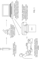


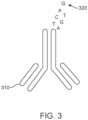
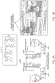

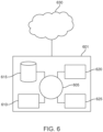


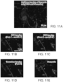


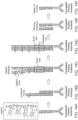

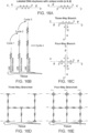

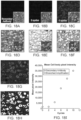

Classifications
-
- G—PHYSICS
- G01—MEASURING; TESTING
- G01N—INVESTIGATING OR ANALYSING MATERIALS BY DETERMINING THEIR CHEMICAL OR PHYSICAL PROPERTIES
- G01N33/00—Investigating or analysing materials by specific methods not covered by groups G01N1/00 - G01N31/00
- G01N33/48—Biological material, e.g. blood, urine; Haemocytometers
- G01N33/50—Chemical analysis of biological material, e.g. blood, urine; Testing involving biospecific ligand binding methods; Immunological testing
- G01N33/53—Immunoassay; Biospecific binding assay; Materials therefor
- G01N33/531—Production of immunochemical test materials
- G01N33/532—Production of labelled immunochemicals
-
- C—CHEMISTRY; METALLURGY
- C12—BIOCHEMISTRY; BEER; SPIRITS; WINE; VINEGAR; MICROBIOLOGY; ENZYMOLOGY; MUTATION OR GENETIC ENGINEERING
- C12Q—MEASURING OR TESTING PROCESSES INVOLVING ENZYMES, NUCLEIC ACIDS OR MICROORGANISMS; COMPOSITIONS OR TEST PAPERS THEREFOR; PROCESSES OF PREPARING SUCH COMPOSITIONS; CONDITION-RESPONSIVE CONTROL IN MICROBIOLOGICAL OR ENZYMOLOGICAL PROCESSES
- C12Q1/00—Measuring or testing processes involving enzymes, nucleic acids or microorganisms; Compositions therefor; Processes of preparing such compositions
- C12Q1/68—Measuring or testing processes involving enzymes, nucleic acids or microorganisms; Compositions therefor; Processes of preparing such compositions involving nucleic acids
- C12Q1/6813—Hybridisation assays
- C12Q1/6827—Hybridisation assays for detection of mutation or polymorphism
-
- C—CHEMISTRY; METALLURGY
- C07—ORGANIC CHEMISTRY
- C07K—PEPTIDES
- C07K16/00—Immunoglobulins [IGs], e.g. monoclonal or polyclonal antibodies
- C07K16/18—Immunoglobulins [IGs], e.g. monoclonal or polyclonal antibodies against material from animals or humans
- C07K16/20—Immunoglobulins [IGs], e.g. monoclonal or polyclonal antibodies against material from animals or humans from protozoa
-
- C—CHEMISTRY; METALLURGY
- C12—BIOCHEMISTRY; BEER; SPIRITS; WINE; VINEGAR; MICROBIOLOGY; ENZYMOLOGY; MUTATION OR GENETIC ENGINEERING
- C12Q—MEASURING OR TESTING PROCESSES INVOLVING ENZYMES, NUCLEIC ACIDS OR MICROORGANISMS; COMPOSITIONS OR TEST PAPERS THEREFOR; PROCESSES OF PREPARING SUCH COMPOSITIONS; CONDITION-RESPONSIVE CONTROL IN MICROBIOLOGICAL OR ENZYMOLOGICAL PROCESSES
- C12Q1/00—Measuring or testing processes involving enzymes, nucleic acids or microorganisms; Compositions therefor; Processes of preparing such compositions
- C12Q1/34—Measuring or testing processes involving enzymes, nucleic acids or microorganisms; Compositions therefor; Processes of preparing such compositions involving hydrolase
- C12Q1/44—Measuring or testing processes involving enzymes, nucleic acids or microorganisms; Compositions therefor; Processes of preparing such compositions involving hydrolase involving esterase
-
- C—CHEMISTRY; METALLURGY
- C12—BIOCHEMISTRY; BEER; SPIRITS; WINE; VINEGAR; MICROBIOLOGY; ENZYMOLOGY; MUTATION OR GENETIC ENGINEERING
- C12Q—MEASURING OR TESTING PROCESSES INVOLVING ENZYMES, NUCLEIC ACIDS OR MICROORGANISMS; COMPOSITIONS OR TEST PAPERS THEREFOR; PROCESSES OF PREPARING SUCH COMPOSITIONS; CONDITION-RESPONSIVE CONTROL IN MICROBIOLOGICAL OR ENZYMOLOGICAL PROCESSES
- C12Q1/00—Measuring or testing processes involving enzymes, nucleic acids or microorganisms; Compositions therefor; Processes of preparing such compositions
- C12Q1/68—Measuring or testing processes involving enzymes, nucleic acids or microorganisms; Compositions therefor; Processes of preparing such compositions involving nucleic acids
- C12Q1/6804—Nucleic acid analysis using immunogens
-
- C—CHEMISTRY; METALLURGY
- C12—BIOCHEMISTRY; BEER; SPIRITS; WINE; VINEGAR; MICROBIOLOGY; ENZYMOLOGY; MUTATION OR GENETIC ENGINEERING
- C12Q—MEASURING OR TESTING PROCESSES INVOLVING ENZYMES, NUCLEIC ACIDS OR MICROORGANISMS; COMPOSITIONS OR TEST PAPERS THEREFOR; PROCESSES OF PREPARING SUCH COMPOSITIONS; CONDITION-RESPONSIVE CONTROL IN MICROBIOLOGICAL OR ENZYMOLOGICAL PROCESSES
- C12Q1/00—Measuring or testing processes involving enzymes, nucleic acids or microorganisms; Compositions therefor; Processes of preparing such compositions
- C12Q1/68—Measuring or testing processes involving enzymes, nucleic acids or microorganisms; Compositions therefor; Processes of preparing such compositions involving nucleic acids
- C12Q1/6806—Preparing nucleic acids for analysis, e.g. for polymerase chain reaction [PCR] assay
-
- C—CHEMISTRY; METALLURGY
- C12—BIOCHEMISTRY; BEER; SPIRITS; WINE; VINEGAR; MICROBIOLOGY; ENZYMOLOGY; MUTATION OR GENETIC ENGINEERING
- C12Q—MEASURING OR TESTING PROCESSES INVOLVING ENZYMES, NUCLEIC ACIDS OR MICROORGANISMS; COMPOSITIONS OR TEST PAPERS THEREFOR; PROCESSES OF PREPARING SUCH COMPOSITIONS; CONDITION-RESPONSIVE CONTROL IN MICROBIOLOGICAL OR ENZYMOLOGICAL PROCESSES
- C12Q1/00—Measuring or testing processes involving enzymes, nucleic acids or microorganisms; Compositions therefor; Processes of preparing such compositions
- C12Q1/68—Measuring or testing processes involving enzymes, nucleic acids or microorganisms; Compositions therefor; Processes of preparing such compositions involving nucleic acids
- C12Q1/6813—Hybridisation assays
- C12Q1/6816—Hybridisation assays characterised by the detection means
- C12Q1/6818—Hybridisation assays characterised by the detection means involving interaction of two or more labels, e.g. resonant energy transfer
-
- C—CHEMISTRY; METALLURGY
- C12—BIOCHEMISTRY; BEER; SPIRITS; WINE; VINEGAR; MICROBIOLOGY; ENZYMOLOGY; MUTATION OR GENETIC ENGINEERING
- C12Q—MEASURING OR TESTING PROCESSES INVOLVING ENZYMES, NUCLEIC ACIDS OR MICROORGANISMS; COMPOSITIONS OR TEST PAPERS THEREFOR; PROCESSES OF PREPARING SUCH COMPOSITIONS; CONDITION-RESPONSIVE CONTROL IN MICROBIOLOGICAL OR ENZYMOLOGICAL PROCESSES
- C12Q1/00—Measuring or testing processes involving enzymes, nucleic acids or microorganisms; Compositions therefor; Processes of preparing such compositions
- C12Q1/68—Measuring or testing processes involving enzymes, nucleic acids or microorganisms; Compositions therefor; Processes of preparing such compositions involving nucleic acids
- C12Q1/6813—Hybridisation assays
- C12Q1/6816—Hybridisation assays characterised by the detection means
- C12Q1/682—Signal amplification
-
- C—CHEMISTRY; METALLURGY
- C12—BIOCHEMISTRY; BEER; SPIRITS; WINE; VINEGAR; MICROBIOLOGY; ENZYMOLOGY; MUTATION OR GENETIC ENGINEERING
- C12Q—MEASURING OR TESTING PROCESSES INVOLVING ENZYMES, NUCLEIC ACIDS OR MICROORGANISMS; COMPOSITIONS OR TEST PAPERS THEREFOR; PROCESSES OF PREPARING SUCH COMPOSITIONS; CONDITION-RESPONSIVE CONTROL IN MICROBIOLOGICAL OR ENZYMOLOGICAL PROCESSES
- C12Q1/00—Measuring or testing processes involving enzymes, nucleic acids or microorganisms; Compositions therefor; Processes of preparing such compositions
- C12Q1/68—Measuring or testing processes involving enzymes, nucleic acids or microorganisms; Compositions therefor; Processes of preparing such compositions involving nucleic acids
- C12Q1/6813—Hybridisation assays
- C12Q1/6816—Hybridisation assays characterised by the detection means
- C12Q1/6823—Release of bound markers
-
- C—CHEMISTRY; METALLURGY
- C12—BIOCHEMISTRY; BEER; SPIRITS; WINE; VINEGAR; MICROBIOLOGY; ENZYMOLOGY; MUTATION OR GENETIC ENGINEERING
- C12Q—MEASURING OR TESTING PROCESSES INVOLVING ENZYMES, NUCLEIC ACIDS OR MICROORGANISMS; COMPOSITIONS OR TEST PAPERS THEREFOR; PROCESSES OF PREPARING SUCH COMPOSITIONS; CONDITION-RESPONSIVE CONTROL IN MICROBIOLOGICAL OR ENZYMOLOGICAL PROCESSES
- C12Q1/00—Measuring or testing processes involving enzymes, nucleic acids or microorganisms; Compositions therefor; Processes of preparing such compositions
- C12Q1/68—Measuring or testing processes involving enzymes, nucleic acids or microorganisms; Compositions therefor; Processes of preparing such compositions involving nucleic acids
- C12Q1/6876—Nucleic acid products used in the analysis of nucleic acids, e.g. primers or probes
- C12Q1/6883—Nucleic acid products used in the analysis of nucleic acids, e.g. primers or probes for diseases caused by alterations of genetic material
- C12Q1/6886—Nucleic acid products used in the analysis of nucleic acids, e.g. primers or probes for diseases caused by alterations of genetic material for cancer
-
- C—CHEMISTRY; METALLURGY
- C12—BIOCHEMISTRY; BEER; SPIRITS; WINE; VINEGAR; MICROBIOLOGY; ENZYMOLOGY; MUTATION OR GENETIC ENGINEERING
- C12Q—MEASURING OR TESTING PROCESSES INVOLVING ENZYMES, NUCLEIC ACIDS OR MICROORGANISMS; COMPOSITIONS OR TEST PAPERS THEREFOR; PROCESSES OF PREPARING SUCH COMPOSITIONS; CONDITION-RESPONSIVE CONTROL IN MICROBIOLOGICAL OR ENZYMOLOGICAL PROCESSES
- C12Q1/00—Measuring or testing processes involving enzymes, nucleic acids or microorganisms; Compositions therefor; Processes of preparing such compositions
- C12Q1/68—Measuring or testing processes involving enzymes, nucleic acids or microorganisms; Compositions therefor; Processes of preparing such compositions involving nucleic acids
- C12Q1/6876—Nucleic acid products used in the analysis of nucleic acids, e.g. primers or probes
- C12Q1/6888—Nucleic acid products used in the analysis of nucleic acids, e.g. primers or probes for detection or identification of organisms
-
- G—PHYSICS
- G01—MEASURING; TESTING
- G01N—INVESTIGATING OR ANALYSING MATERIALS BY DETERMINING THEIR CHEMICAL OR PHYSICAL PROPERTIES
- G01N1/00—Sampling; Preparing specimens for investigation
- G01N1/28—Preparing specimens for investigation including physical details of (bio-)chemical methods covered elsewhere, e.g. G01N33/50, C12Q
- G01N1/36—Embedding or analogous mounting of samples
-
- G—PHYSICS
- G01—MEASURING; TESTING
- G01N—INVESTIGATING OR ANALYSING MATERIALS BY DETERMINING THEIR CHEMICAL OR PHYSICAL PROPERTIES
- G01N33/00—Investigating or analysing materials by specific methods not covered by groups G01N1/00 - G01N31/00
- G01N33/48—Biological material, e.g. blood, urine; Haemocytometers
- G01N33/50—Chemical analysis of biological material, e.g. blood, urine; Testing involving biospecific ligand binding methods; Immunological testing
- G01N33/53—Immunoassay; Biospecific binding assay; Materials therefor
- G01N33/531—Production of immunochemical test materials
- G01N33/532—Production of labelled immunochemicals
- G01N33/533—Production of labelled immunochemicals with fluorescent label
-
- G—PHYSICS
- G01—MEASURING; TESTING
- G01N—INVESTIGATING OR ANALYSING MATERIALS BY DETERMINING THEIR CHEMICAL OR PHYSICAL PROPERTIES
- G01N33/00—Investigating or analysing materials by specific methods not covered by groups G01N1/00 - G01N31/00
- G01N33/48—Biological material, e.g. blood, urine; Haemocytometers
- G01N33/50—Chemical analysis of biological material, e.g. blood, urine; Testing involving biospecific ligand binding methods; Immunological testing
- G01N33/58—Chemical analysis of biological material, e.g. blood, urine; Testing involving biospecific ligand binding methods; Immunological testing involving labelled substances
- G01N33/582—Chemical analysis of biological material, e.g. blood, urine; Testing involving biospecific ligand binding methods; Immunological testing involving labelled substances with fluorescent label
-
- G—PHYSICS
- G01—MEASURING; TESTING
- G01N—INVESTIGATING OR ANALYSING MATERIALS BY DETERMINING THEIR CHEMICAL OR PHYSICAL PROPERTIES
- G01N33/00—Investigating or analysing materials by specific methods not covered by groups G01N1/00 - G01N31/00
- G01N33/48—Biological material, e.g. blood, urine; Haemocytometers
- G01N33/50—Chemical analysis of biological material, e.g. blood, urine; Testing involving biospecific ligand binding methods; Immunological testing
- G01N33/58—Chemical analysis of biological material, e.g. blood, urine; Testing involving biospecific ligand binding methods; Immunological testing involving labelled substances
- G01N33/585—Chemical analysis of biological material, e.g. blood, urine; Testing involving biospecific ligand binding methods; Immunological testing involving labelled substances with a particulate label, e.g. coloured latex
-
- C—CHEMISTRY; METALLURGY
- C12—BIOCHEMISTRY; BEER; SPIRITS; WINE; VINEGAR; MICROBIOLOGY; ENZYMOLOGY; MUTATION OR GENETIC ENGINEERING
- C12Q—MEASURING OR TESTING PROCESSES INVOLVING ENZYMES, NUCLEIC ACIDS OR MICROORGANISMS; COMPOSITIONS OR TEST PAPERS THEREFOR; PROCESSES OF PREPARING SUCH COMPOSITIONS; CONDITION-RESPONSIVE CONTROL IN MICROBIOLOGICAL OR ENZYMOLOGICAL PROCESSES
- C12Q1/00—Measuring or testing processes involving enzymes, nucleic acids or microorganisms; Compositions therefor; Processes of preparing such compositions
- C12Q1/68—Measuring or testing processes involving enzymes, nucleic acids or microorganisms; Compositions therefor; Processes of preparing such compositions involving nucleic acids
- C12Q1/6809—Methods for determination or identification of nucleic acids involving differential detection
-
- C—CHEMISTRY; METALLURGY
- C12—BIOCHEMISTRY; BEER; SPIRITS; WINE; VINEGAR; MICROBIOLOGY; ENZYMOLOGY; MUTATION OR GENETIC ENGINEERING
- C12Q—MEASURING OR TESTING PROCESSES INVOLVING ENZYMES, NUCLEIC ACIDS OR MICROORGANISMS; COMPOSITIONS OR TEST PAPERS THEREFOR; PROCESSES OF PREPARING SUCH COMPOSITIONS; CONDITION-RESPONSIVE CONTROL IN MICROBIOLOGICAL OR ENZYMOLOGICAL PROCESSES
- C12Q2563/00—Nucleic acid detection characterized by the use of physical, structural and functional properties
- C12Q2563/107—Nucleic acid detection characterized by the use of physical, structural and functional properties fluorescence
-
- C—CHEMISTRY; METALLURGY
- C12—BIOCHEMISTRY; BEER; SPIRITS; WINE; VINEGAR; MICROBIOLOGY; ENZYMOLOGY; MUTATION OR GENETIC ENGINEERING
- C12Y—ENZYMES
- C12Y605/00—Ligases forming phosphoric ester bonds (6.5)
- C12Y605/01—Ligases forming phosphoric ester bonds (6.5) forming phosphoric ester bonds (6.5.1)
- C12Y605/01001—DNA ligase (ATP) (6.5.1.1)
Definitions
- Multiply labeled antisense oligomers can provide signals on the order of a few fluorophores per primary antibody, but dense labeling of the antisense oligomer may result in quenching among the fluorophores.
- a ligation amplification method is disclosed herein to provide a higher signal intensity that is comparable to secondary amplification.
- WO 2007/076128 discloses compositions and methods for detection and quantification of individual target molecules in biomolecular samples.
- WO 2015/017586 discloses methods and compositions for imaging targets of interest.
- Ki-Cheol Han et al. discloses an immunosorbent assay using antibody-oligonucleotide conjugates.
- compositions, kits, methods, and systems for detecting a target molecule in a sample comprising contacting a target molecule in a sample with a detection couplet, wherein the detection couplet comprises a first nucleic acid and a second nucleic acid, wherein each nucleic acid has a target recognition region and a self-hybridization region, wherein the target recognition region of the first nucleic acid binds a first region of the target molecule, wherein the target recognition region of the second nucleic acid binds a second region of the target molecule, and wherein the self-hybridization region of the first nucleic acid and the self-hybridization region of the second nucleic acid are hybridized to form a double-stranded nucleic acid label.
- the present invention provides a composition that comprises:
- the invention further provides a kit that comprises the detection molecule, the antisense oligomer, and the detection labels of the invention, wherein at least one of the detection labels comprises a multi-way branch with overhangs configured for hybridization with detection labels.
- the invention provides a method, comprising contacting the target molecule with the detection molecule, the antisense oligomer, and the detection label of the present invention, wherein the detection label comprises a multi-way branch with overhangs configured for hybridization with detection labels
- the double-stranded nucleic acid label has at least two consecutive base pairs, for example, at least 2, at least 3, at least 4, at least 5, at least 6, at least 7, at least 8, at least 9, at least 10, at least 11, at least 12, at least 13, at least 14, at least 15, at least 20, at least 25, at least 30, at least 35, at least 40, at least 45, at least 50, at least 100, at least 150, at least 200, or at least 250 consecutive base pairs.
- the double-stranded nucleic acid label has an overhang.
- the overhang has at least 1 unpaired nucleotide, for example, at least 2, at least 3, at least 4, at least 5, at least 6, at least 7, at least 8, at least 9, at least 10, at least 11, at least 12, at least 13, at least 14, at least 15, at least 20, at least 25, at least 30, at least 35, at least 40, at least 45, at least 50, at least 100, at least 150, at least 200, or at least 250 unpaired nucleotides.
- the target molecule is a RNA molecule. In some cases, the target molecule is an mRNA molecule. In some cases, the first nucleic acid and the second nucleic acid are single-stranded DNA. In some cases, the first region and the second region of the target molecule are separated by 2 to 15 nucleotides. In some cases, the first region and the second region of the target molecule are separated by 2 to 5 nucleotides. In some cases, the first region and the second region of the target molecule are separated by 5 to 10 nucleotides. In some cases, the first region and the second region of the target molecule are separated by 10 to 15 nucleotides. In some cases, the first region and the second region of the target molecule are separated by 15 to 20 nucleotides.
- the method further comprises fixing the detection couplet to the sample using a crosslinker.
- the first nucleic acid or the second nucleic acid has a free amine (-NH2) modification.
- the fixing the detection couplet comprises contacting the detection couplet with an amine-specific crosslinker.
- the overhang comprises one nucleotide. In some cases, the overhang comprises a plurality of nucleotides. In some cases, the overhang comprises at least about 2, 3, 4, 5, 10, 15, 20, 25, 30, 40, 50, 60, 70, 80, 90, 100, 150, 200, 250, 300, 350, 400, 450, or 500 of nucleotides. In some cases, the method further comprises contacting the double-stranded nucleic acid label with a DNA ligase. In some cases, the method further comprises contacting the double-stranded nucleic acid label with at least one detection label.
- the at least one detection label is a double-stranded nucleic acid with an overhang that is complementary to the overhang of the double-stranded nucleic acid label. In some cases, the at least one detection label is a single-stranded nucleic acid that is complementary to the overhang of the double-stranded nucleic acid label. In some cases, the at least one detection label comprises a plurality of detection labels. In some cases, the double-stranded nucleic acid label and the at least one detection label are ligated using the DNA ligase. In some cases, the at least one detection label comprises at least 5, 10, 15, 20, 25, or 30 detection labels. In some cases, the at least one detection label comprises a cleavable linker. In some cases, the at least one detection label does not comprise a cleavable linker.
- the at least one detection label comprises a detection tag. In some cases, the at least one detection label comprises a plurality of detection tags. In some cases, the detection tag comprises a quantum dot. In some cases, the detection tag comprises a fluorophore. In some cases, the fluorophore comprises coumarin, rhodamine, xanthene, fluorescein, or cyanine. In some cases, the method comprises detecting the detection tag, thereby detecting the presence of the target molecule in the sample. In some cases, the method comprises contacting the sample with a plurality of detection couplets. In some cases, each of the plurality of detection couplets binds a different target molecule.
- the sample is an intact tissue sample.
- the method comprises embedding the intact tissue sample in a resin such that the intact tissue sample can be sliced into sections of thickness between 20 and 1000 nm.
- the intact tissue sample is a bone marrow tissue sample, a gastrointestinal tract tissue sample, a lung tissue sample, a liver tissue sample, a prostate tissue sample, a nervous system tissue sample, a urogenital system tissue sample, a brain tissue sample, a breast tissue sample, a muscle tissue sample, or a skin tissue sample.
- the method comprises dehydration of the intact tissue sample. In some cases, the method does not comprise dehydration of the intact tissue sample.
- the method of comprises diagnosing a condition or disease associated with the presence of the target molecule in the sample.
- the condition or disease is a kidney disease, an infectious disease, a metabolic disease, a pre-cancerous condition, a cancerous condition, or a brain disorder.
- the sample is a paraffin-embedded tissue sample.
- the sample is a formalin-fixed paraffin-embedded (FFPE) tissue sample.
- the method further comprises contacting the double-stranded nucleic acid label with a third nucleic acid, wherein the third nucleic acid comprises a sequence that is complementary to a sequence of the first nucleic acid, a sequence of the second nucleic acid, or both.
- the third nucleic acid comprises a sequence that is complementary to a sequence of the first nucleic acid.
- the third nucleic acid comprises a sequence that is complementary to a sequence of the second nucleic acid.
- the third nucleic acid comprises a sequence that is complementary to a sequence of the first nucleic acid and a sequence of the second nucleic acid.
- the third nucleic acid binds to the sequence of the first nucleic acid, the sequence of the second nucleic acid, or both. In one example, the third nucleic acid binds to the sequence of the first nucleic acid. In one example, the third nucleic acid binds to the sequence of the second nucleic acid. In one example, the third nucleic acid binds to the sequence of the first nucleic acid and the sequence of the second nucleic acid.
- the first nucleic acid and the second nucleic acid in the double-stranded nucleic acid label each has at least one unpaired nucleotide. In some cases, the first nucleic acid and the second nucleic acid in the double-stranded nucleic acid label each has at least 1 unpaired nucleotide, for example, at least 2, at least 3, at least 4, at least 5, at least 6, at least 7, at least 8, at least 9, at least 10, at least 11, at least 12, at least 13, at least 14, at least 15, at least 20, at least 25, at least 30, at least 35, at least 40, at least 45, at least 50, at least 100, at least 150, at least 200, or at least 250 unpaired nucleotides.
- the third nucleic acid comprises a sequence that is complementary to an unpaired sequence of the first nucleic acid, an unpaired sequence of the second nucleic acid, or both. In some cases, the third nucleic acid comprises a sequence that is complementary to the entire unpaired sequence of the first nucleic acid. In some cases, the third nucleic acid comprises a sequence that is complementary to the entire unpaired sequence of the second nucleic acid. In some cases, the third nucleic acid comprises a first sequence that is complementary to the entire unpaired sequence of the first nucleic acid and a second sequence that is complementary to the entire unpaired sequence of the second nucleic acid.
- the third nucleic acid binds to the sequence of the first nucleic acid and the sequence of the second nucleic acid, thereby creating a multi-way branch.
- the multi-way branch is a n-way branch, wherein the n-way branch comprises n single stranded nucleic acid that are linked together to form a nucleic acid structure.
- the nucleic acid structure can have n terminals and/or n overhangs.
- the multi-way branch can be a three-way branch, wherein the three-way branch comprises three single stranded nucleic acid that are linked together to form a nucleic acid structure that have three terminals and/or three overhangs (see e.g., Figure 17A-C and Figure 16C ).
- the three-way branch comprises at least two overhangs.
- at least two of said at least two overhangs comprise same sequence or unique sequences.
- at least two of said at least two overhangs comprise complementary sequences.
- the method further comprises contacting the third nucleic acid with a fourth nucleic acid, wherein the fourth nucleic acid comprises a sequence that is complementary to a sequence of the first nucleic acid, a sequence of the second nucleic acid, a sequence of the third nucleic acid, or any combination thereof.
- the fourth nucleic acid binds to the sequence of the third nucleic acid and the sequence of the first or second nucleic acid, thereby creating a four-way branch (see e.g., Figure 16C ).
- the four-way branch comprises four single stranded nucleic acid that are linked together to form a nucleic acid structure that have four terminals and/or four overhangs.
- the four-way branch comprises at least three overhangs.
- at least two of the at least three overhangs comprise same sequence or unique sequences.
- at least two of the at least three overhangs comprise complementary sequences.
- the method further comprises contacting at least one of the overhangs with a detection label. In some cases, the method further comprises contacting the third nucleic acid with a detection label. In some cases, the method further comprises contacting the fourth nucleic acid with a detection label. In some cases, the detection label comprises a multi-way branch. In some cases, the multi-way branch is a three-way branch or a four-way branch. In some cases, the detection label comprises an overhang that is complementary to the at least one of the overhangs. In some cases, the detection label is linked to the at least one of the overhangs by direct hybridization, enzymatic ligation, or chemical ligation.
- a method comprising contacting a target molecule in a sample with a detection molecule and an antisense oligomer, wherein the detection molecule comprises at least one ligand that binds the target molecule, wherein the at least one ligand is linked to a single-stranded nucleic acid, and wherein the single-stranded nucleic acid is hybridized with the antisense oligomer to form a double-stranded nucleic acid label with an overhang.
- the detection molecule or antisense comprises at least 3 nucleotides, for example at least 5, at least 10, at least 15, at least 20, at least 25, at least 30, at least 35, at least 40, at least 45, at least 50, at least 60, at least 70, at least 80, at least 90, at least 100, at least 150, at least 200, at least 250, at least 300, at least 350, at least 400, at least 450, or at least 500 nucleotides.
- the detection molecule or the antisense comprises no more than 500 nucleotides, for example no more than 5, no more than 10, no more than 15, no more than 20, no more than 25, no more than 30, no more than 35, no more than 40, no more than 45, no more than 50, no more than 60, no more than 70, no more than 80, no more than 90, no more than 100, no more than 150, no more than 200, no more than 250, no more than 300, no more than 350, no more than 400, no more than 450, or no more than 500 nucleotides.
- the detection molecule or the antisense comprises about 3-500 nucleotides, for example, from about 3-500, about 3-400, about 3-300, about 3-200, about 3-100, about 3-50, about 3-20, about 3-10, about 10-500, about 10-400, about 10-300, about 10-200, about 10-100, about 10-50, about 10-20, about 20-500, about 20-400, about 20-300, about 20-200, about 20-100, about 20-50, about 50-500, about 50-400, about 50-300, about 50-200, about 50-100, about 100-500, about 100-400, about 100-300, about 100-200, about 200-500, about 200-400, about 200-300, about 300-500, about 300-400, or about 400-500 nucleotides.
- the detection molecule or antisense can comprise about 3-150 nucleotides.
- the ligand is an antibody.
- the target molecule is a protein.
- the overhang comprises one nucleotide.
- the overhang comprises a plurality of nucleotides.
- the method further comprises contacting the double-stranded nucleic acid label with a DNA ligase.
- the method further comprises contacting the double-stranded nucleic acid label with at least one detection label.
- the at least one detection label is a double-stranded nucleic acid with an overhang that is complementary to the overhang of the double-stranded nucleic acid label.
- the at least one detection label is a plurality of detection labels.
- the at least one detection label is a single-stranded nucleic acid that is complementary to the overhang of the double-stranded nucleic acid label. In some cases, the at least one detection label comprises a plurality of detection labels. In some cases, the at least one detection label comprises at least 5, 10, 15, 20, 25, or 30 detection labels. In some cases, the at least one detection label comprises a cleavable linker. In some cases, the at least one detection label does not comprise a cleavable linker.
- the at least one detection label comprises a detection tag. In some cases, the at least one detection label comprises a plurality of detection tags. In some cases, the detection tag comprises a quantum dot. In some cases, the detection tag comprises a fluorophore. In some cases, the fluorophore comprises coumarin, rhodamine, xanthene, fluorescein, or cyanine. In some cases, the method comprises detecting the detection tag, thereby detecting the presence of the target molecule in the sample. In some cases, the method comprises contacting the sample with a plurality of detection molecules. In some cases, each of the plurality of detection molecules binds a different target molecule.
- the detection label comprises at least 3 nucleotides, for example at least 5, at least 10, at least 15, at least 20, at least 25, at least 30, at least 35, at least 40, at least 45, at least 50, at least 60, at least 70, at least 80, at least 90, at least 100, at least 150, at least 200, at least 250, at least 300, at least 350, at least 400, at least 450, or at least 500 nucleotides.
- the detection label comprises no more than 500 nucleotides, for example no more than 5, no more than 10, no more than 15, no more than 20, no more than 25, no more than 30, no more than 35, no more than 40, no more than 45, no more than 50, no more than 60, no more than 70, no more than 80, no more than 90, no more than 100, no more than 150, no more than 200, no more than 250, no more than 300, no more than 350, no more than 400, no more than 450, or no more than 500 nucleotides.
- the detection label comprises about 3-500 nucleotides, for example, from about 3-500, about 3-400, about 3-300, about 3-200, about 3-100, about 3-50, about 3-20, about 3-10, about 10-500, about 10-400, about 10-300, about 10-200, about 10-100, about 10-50, about 10-20, about 20-500, about 20-400, about 20-300, about 20-200, about 20-100, about 20-50, about 50-500, about 50-400, about 50-300, about 50-200, about 50-100, about 100-500, about 100-400, about 100-300, about 100-200, about 200-500, about 200-400, about 200-300, about 300-500, about 300-400, or about 400-500 nucleotides.
- the detection label can comprise about 3-150 nucleotides.
- the sample is an intact tissue sample.
- the method comprises embedding the intact tissue sample in a resin such that the intact tissue sample can be sliced into sections of thickness between 20 and 1000 nm.
- the intact tissue sample is a bone marrow tissue sample, a gastrointestinal tract tissue sample, a lung tissue sample, a liver tissue sample, a prostate tissue sample, a nervous system tissue sample, a urogenital system tissue sample, a brain tissue sample, a breast tissue sample, a muscle tissue sample, or a skin tissue sample.
- the method comprises dehydration of the intact tissue sample. In some cases, the method does not comprise dehydration of the intact tissue sample.
- the method of comprises diagnosing a condition or disease associated with the presence of the target molecule in the sample.
- the condition or disease is a kidney disease, an infectious disease, a metabolic disease, a pre-cancerous condition, a cancerous condition, or a brain disorder.
- the target molecule comprises a component in a gene-editing assay.
- the gene-editing assay is a CRISPR assay.
- the gene-editing assay is a CRISPR/Cas assay.
- the gene-editing assay is an NgAgo assay.
- the component comprises a Cas nuclease, a target DNA, a DNA-targeting RNA, a trans-activating crRNA (tracrRNA), a donor repair template, or any combination thereof.
- the component comprises a Cas nuclease.
- the component comprises a Cas9 nuclease.
- the target molecule comprises a cellular molecule.
- the target molecule comprises a cell surface molecule.
- the target molecule comprises a carbohydrate, a lipid, a protein, or a nucleic acid.
- the target molecule comprises a protein.
- the protein comprises a cytoskeletal protein, an extracellular matrix protein, a plasma protein, a coagulation factor, an acute phase protein, a hemoprotein, a cell adhesion, a transmembrane transport protein, an ion channel, a synport/antiport protein, a hormone, a growth factor, a receptor, a DNA-binding protein, a RNA-binding protein, a transcription regulatory protein, an immune system protein, a nutrient storage or transport protein, a chaperone protein, an enzyme, or any combination thereof.
- the target molecule comprises a nucleic acid.
- the nucleic acid comprises an mRNA, a tRNA, a rRNA, a snRNA, an non-coding RNA molecule, or any combination thereof.
- the method further comprises contacting the double-stranded nucleic acid label with a detection label.
- the detection label comprises a multi-way branch.
- the multi-way branch is a three-way branch or a four-way branch.
- the detection label comprises an overhang that is complementary to the overhang of the double-stranded nucleic acid label.
- the detection label is linked to the overhang of the double-stranded nucleic acid label by direct hybridization, enzymatic ligation, or chemical ligation.
- the method further comprises contacting the detection label with a second detection label.
- the second detection label comprises a multi-way branch.
- the multi-way branch is a three-way branch or a four-way branch.
- the second detection label comprises an overhang that is complementary to the overhang of the detection label.
- compositions comprising: a detection couplet, wherein the detection couplet comprises a first nucleic acid and a second nucleic acid, wherein each nucleic acid has a target recognition region and a self-hybridization region, wherein the target recognition region of the first nucleic acid binds a first region of a target molecule, wherein the target recognition region of the second nucleic acid binds a second region of the target molecule, and wherein the self-hybridization region of the first nucleic acid and the self-hybridization region of the second nucleic acid are hybridized to form a double-stranded nucleic acid label.
- the double-stranded nucleic acid label has at least two consecutive base pairs, for example, at least 2, at least 3, at least 4, at least 5, at least 6, at least 7, at least 8, at least 9, at least 10, at least 11, at least 12, at least 13, at least 14, or at least 15 consecutive base pairs.
- the double-stranded nucleic acid label has an overhang.
- the overhang comprises one nucleotide.
- the overhang comprises a plurality of nucleotides.
- the overhang comprises at least 5, 10, 15, 20, 25, or 30 nucleotides.
- the overhang has at least 1 unpaired nucleotide, for example, at least 2, at least 3, at least 4, at least 5, at least 6, at least 7, at least 8, at least 9, at least 10, at least 11, at least 12, at least 13, at least 14, or at least 15 unpaired nucleotides.
- the target molecule is an mRNA molecule.
- the first nucleic acid and the second nucleic acid are single-stranded DNA.
- each of the first nucleic acid and the second nucleic acid has a free amine (-NH2) modification.
- the composition further comprises a DNA ligase.
- the composition further comprises at least one detection label.
- the at least one detection label is a double-stranded nucleic acid with an overhang that is complementary to the overhang of the double-stranded nucleic acid label. In some cases, the at least one detection label is a single-stranded nucleic acid that is complementary to the overhang of the double-stranded nucleic acid label. In some cases, the at least one detection label comprises a plurality of detection labels. In some cases, the at least one detection label comprises at least 5, 10, 15, 20, 25, or 30 detection labels. In some cases, the at least one detection label does not comprise a cleavable linker. In some cases, the at least one detection label comprises a cleavable linker. In some cases, the at least one detection label comprises a detection tag.
- the at least one detection label comprises a plurality of detection tags.
- the detection tag comprises a quantum dot.
- the detection tag comprises a fluorophore.
- the fluorophore comprises coumarin, rhodamine, xanthene, fluorescein, or cyanine.
- the composition comprises a plurality of detection couplets. In some cases, each of the plurality of detection couplets binds a different target molecule.
- compositions further comprising a third nucleic acid, wherein the third nucleic acid comprises a sequence that is complementary to a sequence of the first nucleic acid, a sequence of the second nucleic acid, or both.
- the third nucleic acid comprises a sequence that is complementary to a sequence of the first nucleic acid.
- the third nucleic acid comprises a sequence that is complementary to a sequence of the second nucleic acid.
- the third nucleic acid comprises a sequence that is complementary to a sequence of the first nucleic acid and a sequence of the second nucleic acid.
- the third nucleic acid binds to the sequence of the first nucleic acid and the sequence of the second nucleic acid, thereby creating a multi-way branch.
- the multi-way branch is a three-way branch. In some cases, the three-way branch comprises at least two overhangs.
- the composition further comprises a fourth nucleic acid, wherein the fourth nucleic acid comprises a sequence that is complementary to a sequence of the first nucleic acid, a sequence of the second nucleic acid, a sequence of the third nucleic acid, or any combination thereof.
- the fourth nucleic acid binds to the sequence of the third nucleic acid and the sequence of the first or second nucleic acid, thereby creating a four-way branch.
- the four-way branch comprises at least three overhangs.
- at least two of the overhangs comprise same sequence or unique sequences.
- at least two of said overhangs comprise complementary sequences.
- the composition further comprises a detection label.
- the detection label comprises a multi-way branch.
- the multi-way branch is a three-way branch or a four-way branch.
- the detection label comprises an overhang that is complementary to the at least one of the overhangs.
- compositions comprising: a detection molecule and an antisense oligomer, wherein the detection molecule comprises at least one ligand that binds a target molecule, wherein the at least one ligand is linked to a single-stranded nucleic acid, and wherein the single-stranded nucleic acid is hybridized to the antisense oligomer to form a double-stranded nucleic acid label with an overhang.
- the ligand is an antibody.
- the target molecule is a protein.
- the overhang comprises one nucleotide.
- the overhang comprises a plurality of nucleotides.
- the overhang comprises at least 5, 10, 15, 20, 25, or 30 nucleotides.
- the composition further comprises a DNA ligase.
- the composition further comprises at least one detection label.
- the at least one detection label is a double-stranded nucleic acid with an overhang that is complementary to the overhang of the double-stranded nucleic acid label.
- the at least one detection label is a single-stranded nucleic acid that is complementary to the overhang of the double stranded nucleic acid label.
- the at least one detection label comprises at least 5, 10, 15, 20, 25, or 30 detection labels.
- the at least one detection label comprises a cleavable linker.
- the at least one detection label does not comprise a cleavable linker. In some cases, the at least one detection label comprises a detection tag. In some cases, the at least one detection label comprises a plurality of detection tags. In some cases, the detection tag comprises a quantum dot. In some cases, the detection tag comprises a fluorophore. In some cases, the fluorophore comprises coumarin, rhodamine, xanthene, fluorescein, or cyanine. In some cases, the composition comprises a plurality of detection molecules. In some cases, each of the plurality of detection molecules binds a different target molecule.
- the target molecule comprises a component in a gene-editing assay.
- the gene-editing assay is a CRISPR assay.
- the gene-editing assay is a CRISPR/Cas assay.
- the gene-editing assay is an NgAgo assay.
- the component comprises a Cas nuclease, a target DNA, a DNA-targeting RNA, a trans-activating crRNA (tracrRNA), a donor repair template, or any combination thereof.
- the component comprises a Cas nuclease.
- the component comprises a Cas9 nuclease.
- the target molecule comprises a cellular molecule.
- the target molecule comprises a cell surface molecule.
- the target molecule comprises a carbohydrate, a lipid, a protein, or a nucleic acid.
- the target molecule comprises a protein.
- the protein comprises a cytoskeletal protein, an extracellular matrix protein, a plasma protein, a coagulation factor, an acute phase protein, a hemoprotein, a cell adhesion, a transmembrane transport protein, an ion channel, a synport/antiport protein, a hormone, a growth factor, a receptor, a DNA-binding protein, a RNA-binding protein, a transcription regulatory protein, an immune system protein, a nutrient storage or transport protein, a chaperone protein, an enzyme, or any combination thereof.
- the target molecule comprises a nucleic acid.
- the nucleic acid comprises an mRNA, a tRNA, a rRNA, a snRNA, an non-coding RNA molecule, or any combination thereof.
- the at least one detection label comprises an overhang that is complementary to the overhang of the double-stranded nucleic acid label. In some cases, the at least one detection label comprises a multi-way branch. In some cases, the multi-way branch is a three-way branch or a four-way branch. In some cases, the at least one detection label is linked to the overhang of the double-stranded nucleic acid label by direct hybridization, enzymatic ligation, or chemical ligation.
- the composition further comprises a second detection label.
- the second detection label comprises an overhang that is complementary to said overhang of said at least one detection label.
- the second detection label comprises a multi-way branch.
- the multi-way branch is a three-way branch or a four-way branch.
- a kit which can be used in methods described above, comprising: a detection couplet, wherein the detection couplet comprises a first nucleic acid and a second nucleic acid, wherein each nucleic acid has a target recognition region and a self-hybridization region, wherein the target recognition region of the first nucleic acid binds a first region of a target molecule, wherein the target recognition region of the second nucleic acid binds a second region of the target molecule, and wherein the self-hybridization region of the first nucleic acid and the self-hybridization region of the second nucleic acid are hybridized to form a double-stranded nucleic acid label; and a first reagent for use when contacting the detection couplet with the target molecule.
- the double-stranded nucleic acid label has an overhang.
- the kit comprises one or more compositions described above.
- kits which can be used in methods described above, comprising: a detection molecule and an antisense oligomer, wherein the detection molecule comprises at least one ligand that binds a target molecule, wherein the at least one ligand is linked to a single-stranded nucleic acid, and wherein the single-stranded nucleic acid is hybridized to the antisense oligomer to form a double-stranded nucleic acid label with an overhang; and a first reagent for use when contacting the detection couplet with the target molecule.
- the kit further comprises a second reagent for use in detection of the target molecule.
- the target molecule comprises a component in a gene-editing assay.
- the gene-editing assay is a CRISPR assay.
- the gene-editing assay is a CRISPR/Cas assay.
- the gene-editing assay is an NgAgo assay.
- the component comprises a Cas nuclease, a target DNA, a DNA-targeting RNA, a trans-activating crRNA (tracrRNA), a donor repair template, or any combination thereof.
- the component comprises a Cas nuclease.
- the component comprises a Cas9 nuclease.
- the target molecule comprises a cellular molecule.
- the target molecule comprises a cell surface molecule.
- the target molecule comprises a carbohydrate, a lipid, a protein, or a nucleic acid.
- the target molecule comprises a protein.
- the protein comprises a cytoskeletal protein, an extracellular matrix protein, a plasma protein, a coagulation factor, an acute phase protein, a hemoprotein, a cell adhesion, a transmembrane transport protein, an ion channel, a synport/antiport protein, a hormone, a growth factor, a receptor, a DNA-binding protein, a RNA-binding protein, a transcription regulatory protein, an immune system protein, a nutrient storage or transport protein, a chaperone protein, an enzyme, or any combination thereof.
- the target molecule comprises a nucleic acid.
- the nucleic acid comprises an mRNA, a tRNA, a rRNA, a snRNA, an non-coding RNA molecule, or any combination thereof.
- the kit comprises any of the compositions disclosed herein.
- ranges include the range endpoints. Additionally, every subrange and value within the range is present as if explicitly written out.
- the term "about” in relation to a reference numerical value can include a range of values plus or minus 10% from that value.
- the amount “about 10” includes amounts from 9 to 11, including the reference numbers of 9, 10, and 11.
- the term “about” in relation to a reference numerical value can also include a range of values plus or minus 10%, 9%, 8%, 7%, 6%, 5%, 4%, 3%, 2%, or 1% from that value.
- nucleic acid generally refers to a polymeric form of nucleotides of any length.
- Nucleic acids can include ribonucleotides, deoxyribonucleotides or peptide nucleic acids (PNAs), that comprise purine and pyrimidine bases, or other natural, chemically or biochemically modified, non-natural, or derivatized nucleotide bases.
- PNAs peptide nucleic acids
- a nucleic acid can be single or double stranded.
- the backbone of the polynucleotide can comprise sugars and phosphate groups, as may typically be found in RNA or DNA, or modified or substituted sugar or phosphate groups.
- a polynucleotide may comprise modified nucleotides, such as methylated nucleotides and nucleotide analogs.
- the sequence of nucleotides may be interrupted by non-nucleotide components.
- nucleoside, nucleotide, deoxynucleoside and deoxynucleotide generally include analogs such as those described herein. These analogs are those molecules having some structural features in common with a naturally occurring nucleoside or nucleotide such that when incorporated into a nucleic acid or oligonucleotide sequence, they allow hybridization with a naturally occurring nucleic acid sequence in solution.
- nucleic acid molecule may be a DNA molecule.
- the nucleic acid molecule may be an RNA molecule.
- the nucleic acid molecule may be a synthetic molecule.
- the nucleic acid molecule may be a synthetic molecule that pair to a DNA or RNA molecule.
- the term "IHC” or “immunohistochemistry” refers to the process of selectively imaging antigens (e.g . proteins or nucleic acids) in cells of a tissue section. This method can employ antibodies that bind to specific antigens in biological tissues.
- the detection signal e.g . fluorescence
- the detection signal can be amplified by ligation amplification methods as provided herein. In some embodiments, the detection signal is amplified via ligation oligomerization. In some embodiments, the detection signal is amplified via templated oligomerization.
- FIG. 1 illustrates an exemplary process of using the compositions, kits, methods, and systems described herein.
- a detection molecule can be used to contact a sample (e.g ., whole tissue sample).
- the presence of the detection molecule can be detected by an imaging instruction (e.g ., a microscope).
- an imaging instruction e.g ., a microscope
- a computer system can be used to identify one or more tissue types, one or more target molecules in the sample, and/or one or more conditions.
- the results can be communicated to doctor or physicians for identification or diagnosis of a disease or a condition.
- a ligation amplification method may comprise: contacting a target molecule in a sample with a detection molecule (e.g ., detection couplet) with an overhang; contacting the detection molecule with one or more double-stranded nucleic acids with complimentary overhangs; and ligating the one or more double-stranded nucleic acids to the detection molecule ( e.g. , to amplify the detection signal) using a DNA ligase.
- This method may also be referred to herein as ligation oligomerization.
- a ligation amplification method may comprise: contacting a target molecule in a sample with a detection molecule (e.g ., detection couplet) with an overhang; contacting the detection molecule with one or more single-stranded nucleic acids that is complimentary to the overhang sequence of the detection molecule; and ligating the one or more single-stranded nucleic acids to the detection molecule ( e.g. , to amplify the detection signal) using a DNA ligase.
- This method may also be referred to herein as templated oligomerization.
- templated oligomerization and “controlled oligomerization” can be used interchangeably herein to refer to the methods of ligation amplification comprising contacting at least one single-stranded detection label complementary to a detection couplet overhang.
- Exemplary methods comprising ligation oligomerization or templated oligomerization for detecting a target molecule in a tissue sample 201 are illustrated in Figures 2A , 2B and 13 .
- Figure 2A illustrates an exemplary ligation oligomerization method for detecting a target molecule in a tissue sample 201.
- the target molecule can be an mRNA molecule 202.
- the method can comprise contacting the target molecule with a detection couplet, wherein the detection couplet comprises a first nucleic acid 203 and a second nucleic acid 204.
- the first nucleic acid 203 and second nucleic acid 204 each can have a target recognition region 205A & 205B, as well as a self-hybridization region 206A & 206B.
- the target recognition region of the first nucleic acid 205A can bind a first region of the target molecule, and the target recognition region of the second nucleic acid 205B can bind a second region of the target molecule.
- the self-hybridization region of the first nucleic acid 206A and the self-hybridization region of the second nucleic acid 206B can be hybridized to form a double-stranded nucleic acid label 206A & 206B.
- the double-stranded nucleic acid label 206A & 206B can have at least three consecutive base pairs.
- the double-stranded nucleic acid label 206A & 206B can have an overhang 207.
- the method can further comprise contacting the double-stranded nucleic acid label 206A & 206B with a detection label 208, wherein the detection label 208 is a double-stranded nucleic acid with an overhang 209 that is complementary to the overhang 207 of the double-stranded nucleic acid label 206.
- the detection label 208 can comprise a detection tag ( e.g ., fluorophore).
- the method can further comprise contacting the detection label with a DNA ligase 210, wherein the DNA ligase 210 can ligate the detection label 208 and the double-stranded nucleic acid label 206A & 206B.
- the ligation oligomerization method provided herein can further comprise the DNA ligase 210 repeating the ligation process multiple times and add a plurality of double-stranded detection label 208 to the double-stranded nucleic acid label 206A & 206B, amplifying the detection signal for imaging applications.
- Figure 2B illustrates an exemplary templated oligomerization method for detecting a target molecule in a tissue sample 201.
- the templated oligomerization method provided herein can further comprise a detection couplet, wherein the detection couplet comprises a first nucleic acid 203 and a second nucleic acid 204.
- the first nucleic acid 203 and second nucleic acid 204 each can have a target recognition region 205A & 205B and a self-hybridization region 206A & 205B.
- the method can further comprise contacting the double-stranded nucleic acid label 206A & 206B with a detection label 208, wherein the detection label 208 is a double-stranded nucleic acid with an overhang 209 that is complementary to the overhang 207 of the double-stranded nucleic acid label 206.
- the detection label 208 can comprise a detection tag ( e.g ., fluorophore).
- the method can further comprise contacting the detection label with a DNA ligase 210, wherein the DNA ligase 210 can ligate the detection label 208 and the double-stranded nucleic acid label 206A & 206B.
- the method can further comprise synthesizing an overhang 211 onto the strand 213.
- the detection couplet can further comprise an overhang 211 comprising a plurality of nucleotides, for example, 1, 2, 3, 4, 5, 6, 7, 8, 9, 10, 11, 12, 13, 14, 15, 16, 17, 18, 19, or 20 nucleotides, wherein the overhang is complementary to a plurality of single-stranded nucleic acid detection labels 212.
- the detection label 212 can comprise a detection tag (e.g ., fluorophore).
- the templated oligomerization method can further comprise contacting the plurality of the detection labels 212 with a DNA ligase 210, wherein the DNA ligase 210 can ligate the plurality of the detection labels 212 and the overhang 211.
- the sequences of at least two detection labels are identical, for example, at least 2, at least 3, at least 4, at least 5, at least 6, at least 7, at least 8, at least 9, at least 10, at least 11, at least 12, at least 13, at least 14, or at least 15, or at least 16, or at least 17, or at least 18, or at least 19, or at least 20 detection labels.
- the sequences of at least a first detection label are different from the sequences of at least a second detection label.
- the plurality of the detection label may comprise at least 2, 3, 4, 5, 6, 7, 8, 9, 10, 11, 12, 13, 14, 15, 16, 17, 18, 19 or 20 detection labels 212, wherein the sequence of a first detection label is complementary to at least a first two consecutive nucleotides of the overhang 211, the sequence of a second detection label is complementary to at least a second two consecutive nucleotides of the overhang 211, the sequence of a third detection label is complementary to at least a third two consecutive nucleotides of the overhang 211, and so forth.
- the methods provided herein can amplify the detection signal for imaging applications.
- fluorescence detection signal is amplified through ligation oligomerization, as shown in Figure 13A .
- fluorescence detection signal is amplified through template oligomerization, in which fluorescence amplification occurs through the sequential hybridization and ligation of single stranded small DNA with fluorophores, as shown in Figure 13B .
- the methods disclosed herein can comprise contacting the sample or the detection molecule with at least one detection label (e.g. , double-stranded DNA), wherein the at least one detection label has an overhang that is complementary to the overhang of the double-stranded nucleic acid label.
- the methods disclosed herein can comprise contacting the sample or the detection molecule with at least one detection label (e.g ., single-stranded DNA), wherein the at least one detection label is complementary to the overhang of either the first or the second strand of the detection couplet.
- the at least one detection label can be a nucleic acid molecule, such as a single-stranded DNA, a single-stranded RNA, or double-stranded DNA.
- the detection label can have a complementary overhang sequence of: 3'-ATC-5'.
- the overhang sequence of the detection label can be uniquely complementary to the overhang of a particular double-stranded nucleic acid label.
- the overhang sequence of the detection label can be complementary to the overhangs of a plurality of double-stranded nucleic acid labels, for example, between 1, 2, 3, 4, 5, 6, 7, 8, 9, 10, 11, 12, 13, 14, 15, 16, 17, 18, 19 and 20 double-stranded nucleic acid labels.
- the overhang sequence of the double-stranded nucleic acid label can be uniquely complementary to the overhangs of a particular detection label.
- the overhang sequence of the double-stranded nucleic acid label can be complementary to the overhangs of a plurality of detection labels, for example, between 1, 2, 3, 4, 5, 6, 7, 8, 9, 10, 11, 12, 13, 14, 15, 16, 17, 18, 19 and 20 detection labels.
- the methods disclosed herein can comprise contacting the sample or the detection molecule with at least one DNA ligase.
- the DNA ligase can facilitate the joining of DNA strands together by catalyzing the formation of a phosphodiester bond.
- the DNA ligase can comprise an E. coli DNA ligase, a T4 DNA ligase, or a mammalian DNA ligase I, II III, or IV.
- the double-stranded nucleic acid label can be ligated to a first detection label to form an amplified double-stranded nucleic acid label, for example, using a DNA ligase.
- the amplified double-stranded nucleic acid label can be ligated to a second detection label to form another amplified double-stranded nucleic acid label. This process can be repeated multiple times to form an amplified double-stranded nucleic acid label comprising a plurality of detection labels.
- the first detection label can have the same sequence as the second detection label.
- the double-stranded nucleic acid label 510 of the detection molecule 520 can be amplified with four double-stranded detection labels 530 with the same sequence through a ligation reaction.
- the first detection label can have a different sequence as the second detection label.
- the detection molecule 520 can be amplified with four single-stranded detection labels with the same sequence through a templated oligomerization ligation reaction as illustrated in Figure 2B .
- the detection molecule 520 can be amplified with one double stranded nucleic acid molecule 510 and three single-stranded detection labels with the same sequence through a templated oligomerization ligation reaction as illustrated in Figure 2B .
- the target molecule can be any molecule of interest in the sample.
- the target molecule can be a carbohydrate, a lipid, a protein, or a nucleic acid (e.g ., DNA or RNA).
- the target molecule can be an RNA molecule, for example, an mRNA, a tRNA, a rRNA, a snRNA, or an non-coding RNA molecule.
- the present methods can comprise contacting the sample with a detection molecule that binds the target molecule.
- the detection molecule can comprise at least one ligand (e.g ., antibody), bead, or nucleic acid.
- the ligand can be an antibody linked to a single-stranded nucleic acid.
- the single-stranded nucleic acid can be hybridized to an antisense oligomer, which has a sequence complementary to the single-stranded nucleic acid, to form a double-stranded nucleic acid label, wherein the double-stranded nucleic acid label has an overhang.
- the hybridization of the single-stranded nucleic acid and the antisense oligomer can be performed after the binding of the ligand to the target molecule.
- the hybridization of the single-stranded nucleic acid and the antisense oligomer can be performed before the binding of the ligand to the target molecule.
- the ligand can be an antibody linked to a double-stranded nucleic acid label, wherein the double-stranded nucleic acid label has an overhang.
- overhang refers to a stretch of unpaired nucleotides in the end of a nucleic acid (e.g ., DNA) molecule. These unpaired nucleotides can be in either strand, creating either 3' or 5' overhangs.
- An overhang can comprise a single nucleotide.
- the detection molecule is an antibody 310 linked to a double-stranded nucleic acid (e.g., DNA) label with a single Guanine (G) overhang 320 .
- G Guanine
- a double-stranded DNA molecule with the following sequences can form a single nucleotide overhang:
- An overhang can comprise a single nucleotide or a plurality of nucleotides, for example, 1, 2, 3, 4, 5, 6, 7, 8, 9, 10, 11, 12, 13, 14, 15, 16, 17, 18, 19, or 20 nucleotides.
- the overhang can comprise at least about 1, at least about 2, at least about 3, at least about 4, at least about 5, at least about 6, at least about 7, at least about 8, at least about 9, at least about 10, at least about 11, at least about 12, at least about 13, at least about 14, at least about 15, at least about 16, at least about 17, at least about 18, at least about 19, or at least about 20 nucleotides.
- the overhang can comprise from about 1 to about 20 nucleotides, for example, from about 1 to about 2, from about 1 to about 3, from about 1 to about 4, from about 1 to about 5, from about 1 to about 10, from about 1 to about 15, from about 1 to about 20, from about 2 to about 3, from about 2 to about 4, from about 2 to about 5, from about 2 to about 10, from about 2 to about 15, from about 2 to about 20, from about 3 to about 4, from about 3 to about 5, from about 3 to about 10, from about 3 to about 15, from about 3 to about 20, from about 4 to about 5, from about 4 to about 10, from about 4 to about 15, from about 4 to about 20, from about 5 to about 10, from about 5 to about 15, from about 5 to about 20, from about 10 to about 15, from about 10 to about 20, or from about 15 to about 20 nucleotides.
- the overhang sequence can be TAG, CAT, ACA, CAT, or AAT.
- a double-stranded DNA molecule with the following sequences can form a three-nucleotide
- the detection molecule can comprise a plurality of nucleotides that binds the target molecule.
- the detection molecule can be a detection couplet, which comprises a first nucleic acid and a second nucleic acid.
- the first and second nucleic acids can be single-stranded DNA.
- the first and/or second nucleic acid can comprise a plurality of nucleotides in length, for example, at least about 10, at least about 15, at least about 20, at least about 25, at least about 30, at least about 35, at least about 40, at least about 45, at least about 50, at least about 55, at least about 60, at least about 65, at least about 70, at least about 75, at least about 80, at least about 85, at least about 90, at least about 95, or at least about 100 nucleotides.
- the first and/or second nucleic acid can comprise about 5-150 nucleotides in length, for example, about 5-150, about 5-130, about 5-110, about 5-90, about 5-70, about 5-50, about 5-30, about 5-10, about 10-150, about 10-130, about 10-110, about 10-90, about 10-70, about 10-50, about 10-30, about 30-150, about 30-130, about 30-110, about 30-90, about 30-70, about 30-50, about 50-150, about 50-130, about 50-110, about 50-90, about 50-70, about 70-150, about 70-130, about 70-110, about 70-90, about 90-150, about 90-130, about 90-110, about 110-150, about 110-130, or about 130-150 nucleotides.
- the first nucleic acid has a first recognition region 205A (e.g. , RNA recognition region) that binds a first target molecule (e.g ., mRNA).
- the second nucleic acid has a second recognition region 205A (e.g ., RNA recognition region) that binds a second target molecule ( e.g ., mRNA).
- the first and second target molecule can be the same molecule.
- the first target molecule and second target molecule can be different molecules.
- the recognition region of the first and/or second nucleic acid binds to the target molecule by hybridizing to a sequence of the target molecule that is complementary to the sequence of the recognition region.
- the recognition region can comprise a single nucleotide or a plurality of nucleotides, for example, 1, 2, 3, 4, 5, 6, 7, 8, 9, 10, 11, 12, 13, 14, 15, 16, 17, 18, 19, or 20 nucleotides.
- the recognition region can comprise at least about 1, at least about 2, at least about 3, at least about 4, at least about 5, at least about 6, at least about 7, at least about 8, at least about 9, at least about 10, at least about 11, at least about 12, at least about 13, at least about 14, at least about 15, at least about 16, at least about 17, at least about 18, at least about 19, or at least about 20 nucleotides.
- the recognition region can comprise from about 1 to about 20 nucleotides, for example, from about 1 to about 2, from about 1 to about 3, from about 1 to about 4, from about 1 to about 5, from about 1 to about 10, from about 1 to about 15, from about 1 to about 20, from about 2 to about 3, from about 2 to about 4, from about 2 to about 5, from about 2 to about 10, from about 2 to about 15, from about 2 to about 20, from about 3 to about 4, from about 3 to about 5, from about 3 to about 10, from about 3 to about 15, from about 3 to about 20, from about 4 to about 5, from about 4 to about 10, from about 4 to about 15, from about 4 to about 20, from about 5 to about 10, from about 5 to about 15, from about 5 to about 20, from about 10 to about 15, from about 10 to about 20, or from about 15 to about 20 nucleotides.
- the first recognition region and the second recognition region can recognize two sequences of the target molecule that are separated by at least about 1, at least about 2, at least about 3, at least about 4, at least about 5, at least about 6, at least about 7, at least about 8, at least about 9, at least about 10, at least about 11, at least about 12, at least about 13, at least about 14, at least about 15, at least about 16, at least about 17, at least about 18, at least about 19, or at least about 20 nucleotides.
- the first recognition region and the second recognition region can recognize two sequences of the target molecule that are separated by from about 1 to about 20 nucleotides, for example, from about 1 to about 2, from about 1 to about 3, from about 1 to about 4, from about 1 to about 5, from about 1 to about 10, from about 1 to about 15, from about 1 to about 20, from about 2 to about 3, from about 2 to about 4, from about 2 to about 5, from about 2 to about 10, from about 2 to about 15, from about 2 to about 20, from about 3 to about 4, from about 3 to about 5, from about 3 to about 10, from about 3 to about 15, from about 3 to about 20, from about 4 to about 5, from about 4 to about 10, from about 4 to about 15, from about 4 to about 20, from about 5 to about 10, from about 5 to about 15, from about 5 to about 20, from about 10 to about 15, from about 10 to about 20, or from about 15 to about 20 nucleotides.
- the first recognition region and the second recognition region can recognize two sequences of the target mRNA molecule that are separated by 3 nucleotides
- the first nucleic acid can comprise a first self-hybridization region 206A ( Figure 4A ).
- the second nucleic acid can comprise a second self-hybridization region 206B.
- the sequences of the first and second self-hybridization regions can be complementary.
- the first and second self-hybridization regions can hybridize through sense-antisense hybridization to form a double-stranded nucleic acid label.
- the double-stranded nucleic acid label can be partially double-stranded and partially single-stranded.
- the self-hybridization region can comprise a single nucleotide or a plurality of nucleotides, for example, 1, 2, 3, 4, 5, 6, 7, 8, 9, 10, 11, 12, 13, 14, 15, 16, 17, 18, 19, or 20 nucleotides.
- the self-hybridization region can comprise at least about 1, at least about 2, at least about 3, at least about 4, at least about 5, at least about 6, at least about 7, at least about 8, at least about 9, at least about 10, at least about 11, at least about 12, at least about 13, at least about 14, at least about 15, at least about 16, at least about 17, at least about 18, at least about 19, or at least about 20 nucleotides.
- the self-hybridization region can comprise from about 1 to about 20 nucleotides, for example, from about 1 to about 2, from about 1 to about 3, from about 1 to about 4, from about 1 to about 5, from about 1 to about 10, from about 1 to about 15, from about 1 to about 20, from about 2 to about 3, from about 2 to about 4, from about 2 to about 5, from about 2 to about 10, from about 2 to about 15, from about 2 to about 20, from about 3 to about 4, from about 3 to about 5, from about 3 to about 10, from about 3 to about 15, from about 3 to about 20, from about 4 to about 5, from about 4 to about 10, from about 4 to about 15, from about 4 to about 20, from about 5 to about 10, from about 5 to about 15, from about 5 to about 20, from about 10 to about 15, from about 10 to about 20, or from about 15 to about 20 nucleotides.
- the methods herein can comprise hybridizing the self-hybridization regions of the first nucleic acid and the second nucleic acid to form a double-stranded nucleic acid label with an overhang 207 described herein (examples are shown in Figures 4A and 4B ).
- the detection couplet can comprise 3' and/or 5' modification that can be used for fixation of the detection couplet to the sample (e.g ., tissue). Fixing detection couplet to the tissue can allow various processing without negatively affecting the detectability of the target molecule (e.g ., mRNA).
- the target molecule e.g ., mRNA
- the detection couplet including the first and second nucleic acids, can have a 3' or 5' modification, such as a free amine (-NH2) modification (shown in Figure 4A ).
- the methods disclosed herein can comprise contacting an amine reactive crosslinker, such as a NHS ester crosslinker, a imidoester crosslinker, or a difluoro crosslinker.
- the NHS ester crosslinker can be disuccinimidyl glutarate (DSG), disuccinimidyl suberate (DSS), bis(sulfosuccinimidyl)suberate (BS3), tris-(succinimidyl)aminotriacetate (TSAT), PEGylated bis(sulfosuccinimidyl)suberate (BS(PEG)5, BS(PEG)9), dithiobis(succinimidyl propionate) (DSP), 3,3'-dithiobis(sulfosuccinimidyl propionate) (DTSSP), disuccinimidyl tartrate (DST), bis(2-(succinimidooxycarbonyloxy)ethyl)sulfone (BSOCOES), ethylene glycol bis(succinimidyl succinate) (EGS), or Sulfo-EGS.
- DSG disuccinimidyl glutarate
- the imidoester crosslinker can be dimethyl adipimidate (DMA), dimethyl pimelimidate (DMP), dimethyl suberimidate (DMS), or Wang and Richard's Reagent (DTBP).
- the difluoro crosslinker can be 1,5-difluoro-2,4-dinitrobenzene (DFDNB).
- the methods disclosed herein can comprise contacting the sample or the detection molecule with at least one detection label (e.g. , double-stranded DNA), wherein the at least one detection label has an overhang that is complementary to the overhang of the double-stranded nucleic acid label.
- the at least one detection label can be a nucleic acid molecule, such as a single-stranded DNA, a single-stranded RNA, or double-stranded DNA. For example, if the double-stranded nucleic acid label has an overhang sequence of: 5'-TAG-3', then the detection label can have a complementary overhang sequence of: 3'-ATC-5'.
- the overhang sequence of the detection label can be uniquely complementary to the overhang of a particular double-stranded nucleic acid label.
- the overhang sequence of the detection label can be complementary to the overhangs of a plurality of double-stranded nucleic acid labels, for example, between 1, 2, 3, 4, 5, 6, 7, 8, 9, 10, 11, 12, 13, 14, 15, 16, 17, 18, 19 and 20 double-stranded nucleic acid labels.
- the overhang sequence of the double-stranded nucleic acid label can be uniquely complementary to the overhangs of a particular detection label.
- the overhang sequence of the double-stranded nucleic acid label can be complementary to the overhangs of a plurality of detection labels, for example, between 1, 2, 3, 4, 5, 6, 7, 8, 9, 10, 11, 12, 13, 14, 15, 16, 17, 18, 19 and 20 detection labels.
- the detection labels can comprise one or more detection tags.
- the detection label can have at least one overhang that can be adapted for manual assembly and/or self-assembly.
- the detection labels can be adapted for manual assembly and can have the serial application of individual units to build a larger complex (e.g., signaling complex).
- the detection labels can be adapted for self-assembly and can have the simultaneous application of all components to build a larger complex (e.g., signaling complex).
- the methods disclosed herein can comprise contacting the sample or the detection molecule with at least one DNA ligase.
- the DNA ligase can facilitate the joining of DNA strands together by catalyzing the formation of a phosphodiester bond.
- the DNA ligase can comprise an E. coli DNA ligase, a T4 DNA ligase, or a mammalian DNA ligase I, II III, or IV.
- the double-stranded nucleic acid label can be ligated to a first detection label to form an amplified double-stranded nucleic acid label, for example, using a DNA ligase.
- the amplified double-stranded nucleic acid label can be ligated to a second detection label to form another amplified double-stranded nucleic acid label. This process can be repeated multiple times to form an amplified double-stranded nucleic acid label comprising a plurality of detection labels.
- the first detection label can have the same sequence as the second detection label.
- the double-stranded nucleic acid label 510 of the detection molecule 520 can be amplified with four double-stranded detection labels 530 with the same sequence through a ligation reaction.
- the first detection label can have a different sequence as the second detection label.
- the detection molecule 520 can be amplified with four single-stranded detection labels with the same sequence through a templated oligomerization ligation reaction as illustrated in Figure 2B .
- the detection molecule 520 can be amplified with one double stranded acid molecule 510 and three single-stranded detection labels with the same sequence through a templated oligomerization ligation reaction as illustrated in
- Natural and synthetic nucleic acids can be joined by direct hybridization, enzymatic ligation or chemical ligation.
- direct hybridization comprises the joining of nucleic acids by base pairing only.
- Direct hybridization can be non-covalent and the stability of the joined structure can be sensitive to environmental factors such as salt concentration and temperature.
- Enzymatic ligation can provide a direct covalent linkage of nucleic acids, but may require a protein ligase.
- Chemical ligation can provide a convenient method of covalently joining two nucleic acids.
- Chemical ligation forms can comprise: crosslinking of nucleic acids with 3' and 5' amino groups using an aldehyde to directly join the strand; oxidation of 3' and 5' sulfhydryl groups to generate a disulfide linked nucleic acid strands; and bio-orthogonal "click" chemistries that can covalently join the 3' and 5' ends of adjacent nucleic acid strands, for example, a 3' labeled azide and a 5' labeled alkyne can be joined via Cu(I) catalyzed cycloaddition to form a triazol covalent linkage instead of the normal phosphate bond ( El-Sagheer, A. H. & Brown, T.
- the ligation can be by enzymatic ligation, chemical cross-linking, for example aldehyde or sulfhydryl crosslinking, or bio-orthogonal "click" chemistries such as alkyne-azide cycloadditions.
- the detection labels used to generate a larger complex can be either linear or branched.
- a structure made of only linear detection labels may amplify signals linearly to the number of linear modules.
- a structure made of branched, or a combination of linear and branched, detection labels may provide a signal that can be amplified exponentially to the number of branching detection labels.
- the structures and effects of branched oligomerization are shown in Examples 11 to 14.
- the multi-way branch is a n-way branch, wherein the n-way branch comprises n single stranded nucleic acid that are linked together to form a nucleic acid structure.
- the nucleic acid structure can have n terminals and/or n overhangs.
- the multi-way branch can be a three-way branch, wherein the three-way branch comprises three single stranded nucleic acid that are linked together to form a nucleic acid structure that have three terminals and/or three overhangs (see e.g., Figure 17A-C and Figure 16C ).
- the multi-way branch can be a four-way branch.
- the four-way branch can comprise four single stranded nucleic acid that are linked together to form a nucleic acid structure that have four terminals and/or four overhangs.
- the detection label can comprise at least one detection tag.
- the detection label may also comprise a plurality of detection tags.
- the detection label may comprise between 1, 2, 3, 4, 5, 6, 7, 8, 9, 10, 11, 12, 13, 14, 15, 16, 17, 18, 19 and 20 detection tags.
- the plurality of detection tags can be the same type of detection tag.
- the plurality of detection tags can comprise more than one types of detection tags.
- the plurality of detection tags can comprise more than one types of detection tags with the same color.
- the plurality of detection tags can comprise more than one types of detection tags with different colors.
- the plurality of detection tags can comprise more than one colors.
- the at least one detection tag can be attached to the detection label by a linker, for example, a cleavable or a non-cleavable linker.
- a linker for example, a cleavable or a non-cleavable linker.
- a plurality of detection tags are attached to the detection label, each spaced between seven and ten bases apart from each other.
- the plurality of detection tags are attached such that each tag is spaced between 2, 3, 4, 5, 6, 7, 8, 9, 10, 11, 12, 13, 14, and 15 bases apart.
- the plurality of detection tags may comprise between two and ten detection tags.
- the plurality of detection tags may comprise between 1, 2, 3, 4, 5, 6, 7, 8, 9, 10, 11, 12, 13, 14, 15, 16, 17, 18, 19 and 20 detection tags.
- the detection tag can be a quantum dot (QD).
- the detection tag can be a fluorophore. Any fluorophore and/or QD known to the skilled artisan may be employed in the methods and systems described herein.
- the fluorophore may comprise a coumarin, rhodamine, xanthene, fluorescein, or cyanine.
- the placement and number of detection tags may be optimized to enhance spatial resolution of the detection.
- the detection tag can be a hapten.
- the hapten can comprise aniline, an aniline derivative (o-, m-, or p-aminobenzoic acid), urushiol, hydralazine, fluorescein, biotin, digoxigenin, or dinitrophenol.
- the hapten can be digoxigenin.
- the hapten e.g ., digoxigenin
- an enzyme labeled antibody e.g ., HRP, AP
- the hybridization of the detection tag with the nucleic acid label may comprise application of an electric field.
- the electric field may be applied for about 1, 2, 3, 4, 5, 6, 7, 8, 9, 10, 12, 15, 20, 25, 30, 35, 40, 45, 50, 55, or 60 seconds.
- the electric field may be applied for between 0.5, 1, 1.5, 2, 2.5, 3, 3.5, 4, 4.5, and 5 minutes.
- the electric field may be applied for up to 5, 10, 15, 20, 25, 30, 35, 40, 45, 50, 55, 60, 65, 70, 75, 80, 85, or 90 minutes.
- the electric field may be applied between 1 and 60 minutes.
- the methods disclosed herein can comprise contacting the sample and/or the detection molecule with phosphatase and/or another double-stranded DNA, such as sonicated salmon sperm.
- phosphatase and/or another double-stranded DNA such as sonicated salmon sperm
- the phosphatase and/or another double-stranded DNA e.g ., sonicated salmon sperm
- the methods described herein may comprise a detection step that comprises determining the sequence of each nucleic acid label.
- any sequencing method that can be performed in-situ can be utilized for sequencing the nucleic acid labels herein. These include for instance sequencing by synthesis, sequencing by ligation, sequencing by hybridization among other methods known to the skilled artisan.
- Commercially available nucleic acid sequencing kits may be optimized for use with the methods and systems described herein.
- sequence of each nucleic acid label may be determined by sequencing by synthesis. In some instances, the sequence of each nucleic acid label may be determined by sequencing by hybridization. Sequencing by hybridization may involve use of the tag hybridization method described in the examples below.
- Tag sequencing is a variant of direct sequencing and uses tags that are about 60 base pairs (bp) consisting of four 15mer units as described in the examples below. In some cases each oligomer is 9, 10, 11, 12, 13, 14, 15, 16, 17, 18, 19, or 20 nucleic acids in length. In some cases, tag 'sequencing' by hybridization is used with QD-labeled oligomers. Using QDs enables reasonably high-speed STORM-like imaging. Further, the quantum dots do not need to be photo-activated, are resistant to photobleaching, and require a single color for excitation. In some cases, tag sequencing is used with cleavable fluorescent labels.
- compositions, kits, methods, and systems for performing array tomography (AT) on an intact tissue to facilitate spatially resolved identification of a plurality of proteins in the tissue can image neural circuit architectures, such as brain tissues.
- the one version of the AT process as currently practiced comprises tissue processing similar to that used for electron microscopy, including chemical fixation, dehydration, and embedding in resin.
- Tissue blocks are cut on an ultramicrotome using a diamond knife.
- Contact cement applied to the block sides, ensures that serial sections stick together to form long ribbons.
- coated coverslips the coating having been engineered to tightly adhere to embedded-tissue sections, holding them flat for reliable autofocus and retaining them through multiple staining cycles.
- Arrays are stained using antibodies, lectins, or other reagents and detected by automated fluorescence microscopy, often at the diffraction limit.
- Antibodies can be stripped, and staining and imaging repeated multiple times to build up a high-dimensional data set from a given tissue volume.
- Arrays can also be stained with heavy metals and imaged by field-emission scanning electron microscopy (SEM). Images are stitched, aligned, and each light (and SEM) cycle merged into a 3D volume comprising all channels. Volumes can be analyzed, for example, to assess the spatial relationships among various markers, providing identification and characterization of synapses, cell types, and other features of interest.
- SEM field-emission scanning electron microscopy
- the methods and systems described herein may be used to perform array tomography on an intact tissue sample.
- An intact tissue as described herein includes tissues that are sectioned on one dimension and contiguous in the other two dimensions. These tissues are characterized by minimal dissociation.
- An intact tissue sample is one wherein after sectioning, the sample retains tissue architecture and other cells normally found in the whole tissue. Exemplary methods of fixing intact tissue for the methods and systems described herein are provided in Example 1 below. Additionally methods of isolating and fixing intact tissue samples known to the skilled artisan can be employed for the methods and systems described herein.
- the intact tissue may be embedded in a resin such that the tissue can be sliced into sections of thickness between 20 and 1000 nm.
- the sample is a paraffin-embedded tissue sample.
- the sample is a formalin-fixed paraffin-embedded (FFPE) tissue sample.
- the thickness of the section may be 25, 30, 35, 40, 45, 50, 55, 60, 70 80, 90 100, 150, 200, 250, 300, 350, 400, 450, 500, 550, 600, 650, 700, 750, 800, 850, 900, 950 or 1000 nm. In some methods, the thickness of the section may be about 100, 150, 200, 250, 300, 350, 400, 450, 500, 600, 700, 800, 900 or 1000 nm. In some cases, the method may not comprise dehydration of the intact tissue.
- the tissue is not dehydrated and resin embedded.
- a section collector is utilized to automatically collect ribbons produced on an ultramicrotome and place them on pre-defined regions of coated, precision coverslips, of sizes ranging from a microscope slide to a microtiter plate.
- Intact tissue samples that can be studied by this method may include for instance biopsied tissues for detection of one or more conditions.
- Tissue heterogeneity can present a complex chemical environment that can cause nonspecific background signal that obscures the specific biological signal.
- blocking solutions comprising mixtures of unlabeled single- or double-stranded nucleic acids, enzymes such as alkaline phosphatase, polymerases, ligases and nucleases, charged polymers such as PEG, charged polysaccharides such as heparin, and proteins such as BSA and casein, can be used.
- Blocking solutions can be added to any of the solutions used to label, stain, detect or wash the tissue during the procedure. Salt and pH levels can also be used to reduce or remove undesired, off-target background.
- Salt and pH levels in solutions can be used to improve specificity and/or signal strength. Salt and pH levels can also be used to remove unwanted background; for example, the use of chaotropic salts, such as salts of perchlorate, guanidium, urea or ions high on the Hofmeister series for the disruption of protein stability. Chemicals that can destabilize nucleic acid hybridization, such as formamide, urea or formaldehyde can also be used during labeling, staining, detection or washing to prevent or remove off target signals.
- chaotropic salts such as salts of perchlorate, guanidium, urea or ions high on the Hofmeister series for the disruption of protein stability.
- Chemicals that can destabilize nucleic acid hybridization such as formamide, urea or formaldehyde can also be used during labeling, staining, detection or washing to prevent or remove off target signals.
- the methods may comprise contacting the intact tissue with a plurality of antibodies, wherein each antibody that binds a specific protein is linked to a unique nucleic acid.
- each antibody in the plurality of antibodies may bind a different protein.
- the antibodies may be of the same or different isotypes, and the nucleic acids may comprise DNA and/or RNA.
- antibodies that bind different proteins may be of the same or different isotypes.
- the at least one antibody may be cross-linked to the tissue.
- a method described herein may comprise contacting the intact tissue with a plurality of antibodies, wherein each antibody that binds a specific protein is linked to a unique nucleic acid.
- Also disclosed herein are methods comprising contacting a target molecule in a sample with a detection couplet, wherein the detection couplet comprises a first nucleic acid and a second nucleic acid, wherein each nucleic acid has a target recognition region and a self-hybridization region, wherein the target recognition region of the first nucleic acid binds a first region of the target molecule, wherein the target recognition region of the second nucleic acid binds a second region of the target molecule, and wherein the self-hybridization region of the first nucleic acid and the self-hybridization region of the second nucleic acid are hybridized to form a double-stranded nucleic acid label.
- the double-stranded nucleic acid label has an overhang.
- the method further comprises contacting the double-stranded nucleic acid label with a DNA ligase.
- the method further comprises contacting the double-stranded nucleic acid label with at least one detection label.
- the at least one detection label is a double-stranded nucleic acid with an overhang that is complementary to the overhang of the double-stranded nucleic acid label.
- the double-stranded nucleic acid label and the at least one detection label are ligated using the DNA ligase.
- the detection of the plurality of antibodies may be spatially resolved.
- Some of the methods and systems described herein comprise use of a microfluidic chamber. Some methods may be fully automated.
- Some methods described herein may be used to identify the protein composition of the tissue sample, and/or diagnose a physiological condition or disease as described above. Some methods may be used to identify the tissue class of a particular intact tissue.
- contacting the tissue with an antibody may comprise application of an electric field.
- the electric field may be applied for about 1, 2, 3, 4, 5, 6, 7, 8, 9, 10, 12, 15, 20, 25, 30, 35, 40, 45, 50, 55, or 60 seconds.
- the electric field may be applied for between 0.5, 1, 1.5, 2, 2.5, 3, 3.5, 4, 4.5 or 5 minutes.
- the electric field may be applied for up to 5, 10, 15, 20, 25, 30, 35, 40, 45, 50, 55, 60, 65, 70, 75, 80, 85, or 90 minutes.
- the electric field may be applied between 1 and 60 minutes.
- compositions, kits, methods, and systems disclosed herein can also be used in a detection assay (e.g ., multiplex detection assay).
- the detection assay can be used in a DNA microarray (e.g ., for gene expression or SNP detection).
- the detection assay can be used in serial analysis of gene expression (SAGE) (e.g ., for gene expression).
- SAGE serial analysis of gene expression
- the detection assay can be used in high-throughput sequencing (e.g ., produce millions of short DNA sequences in parallel).
- the detection assay can be used in multiplex polymerase chain reaction (PCR) (e.g ., for applications requiring the amplification or sequencing of DNA or RNA).
- PCR polymerase chain reaction
- the detection assay can be used in multiplex ligation-dependent probe amplification (MLPA). In some cases, the detection assay can be used in DNA sequencing by ligation. In some cases, the detection assay can be used in fluorescent microbead array.
- MLPA multiplex ligation-dependent probe amplification
- the detection assay can be used in protein microarray (e.g ., for measuring protein-protein interactions or small molecule binding).
- the detection assay can be used in antibody microarray (e.g ., a type of protein array in which antibodies are arrayed).
- the detection assay can be used in phage display ( e.g ., for screening large protein libraries for interacting proteins or other molecules).
- the detection assay can be used in antibody profiling (e.g ., multiple HLA antibody identification or reactivity prediction against a panel of organ donor population).
- the detection assay can be used in Luminex/XMAP principle based multiplexing.
- the detection assay can be used in binding antibody multiplex assay (BAMA) (e.g ., for profiling multiple antibody isotypes and/or subclasses).
- BAMA binding antibody multiplex assay
- the detection assay can be used in tissue microarray (e.g ., for analyzing multiple tissue samples). In some cases, the detection assay can be used in cellular microarray ( e.g ., for observing cellular responses against a panel of materials). In some cases, the detection assay can be used in chemical compound microarray ( e.g ., for assaying multiple chemical compounds for specific activities). In some cases, the detection assay can be used in multiplex detection of western blot ( e.g ., for simultaneous detection of two or more targets on a western blot). In some cases, the detection assay can be used in multiplex biomarker analysis ( e.g ., for analyzing urine). In some cases, the detection assay can be used in enzyme-linked immunosorbent assay (ELISA) ( e.g ., for parallelized processing using microtiter plates). In some cases, the detection assay can be used in flow cytometry.
- ELISA enzyme-linked immunosorbent assay
- the detection assay can simultaneously detect a plurality of targets in a sample using a detection assay.
- the detection assay can simultaneously detect at least two targets, for example, at least 2, at least 3, at least 4, at least 5, at least 6, at least 7, at least 8, at least 9, at least 10, at least 15, at least 20, at least 25, at least 30, at least 35, at least 40, at least 45, at least 50, at least 55, at least 60, at least 65, at least 70, at least 75, at least 80, at least 85, at least 90, at least 95, at least 100, at least 150, at least 200, at least 250, at least 300, at least 350, at least 400, at least 450, or at least 500 targets in a sample.
- the detection assay can simultaneously detect from 2 to 500 targets, for example, from 2 to 5, from 2 to 10, from 2 to 100, from 2 to 500, from 5 to 10, from 5 to 100, from 5 to 500, from 10 to 100, from 10 to 500, or from 100 to 50 targets in a sample.
- the detection assay can detect a cell.
- the cell can be a specific type of cell.
- the cell can have a specific origin ( e.g ., from a specific organ).
- the cell can have a specific status ( e.g ., healthy cell, cancer cell).
- the detection assay can detect a plurality of targets in a sample.
- the plurality of targets are a plurality of cells.
- the plurality of cells are different types of cells.
- the plurality of cells have different origins ( e.g ., from different organs).
- the plurality of cells have different statuses ( e.g ., healthy cells, cancer cells).
- the detection assay can detect a target molecule.
- the target molecule can be any molecule of interest in the sample.
- the target molecule can be a carbohydrate, a lipid, a protein, or a nucleic acid ( e.g ., DNA or RNA).
- the target molecule can be an RNA molecule, for example, an mRNA, a tRNA, a rRNA, a snRNA, or an non-coding RNA molecule.
- the target molecule can be a cellular molecule.
- the cellular molecule can be a molecule inside a cell membrane that separates the interior of all cells from the outside environment.
- the cellular molecule can be a carbohydrate, a lipid, a protein, or a nucleic acid (e.g ., DNA or RNA).
- the target molecule can be a cell surface molecule.
- the cell surface molecule can be a molecule on a cell membrane that separates the interior of all cells from the outside environment.
- the cell surface molecule can be a carbohydrate, a lipid, a protein, or a nucleic acid (e.g ., DNA or RNA).
- the detection assay can detect a protein. In some cases, the detection assay can detect a plurality of proteins. In some cases, the proteins can be cytoskeletal proteins, such as Actin, Arp2/3, Formin, Coronin, Dystrophin, FtsZ, Keratin, Myosin, and Tubulin. In some cases, the proteins can be extracellular matrix proteins, such as Collagen, Elastin, F-spondin, Pikachurin, and Fibronectin. In some cases, the proteins can be plasma proteins, such as Serum Amyloid P Component and Serum albumin.
- cytoskeletal proteins such as Actin, Arp2/3, Formin, Coronin, Dystrophin, FtsZ, Keratin, Myosin, and Tubulin.
- the proteins can be extracellular matrix proteins, such as Collagen, Elastin, F-spondin, Pikachurin, and Fibronectin. In some cases, the proteins can be plasma proteins, such as Serum Amyloid P Component and Serum album
- the proteins can be coagulation factors, such as Complement proteins (e.g., C1-inhibitor, C3-convertase), Factor VIII, Factor XIII, Protein C, Protein S, Protein Z, Protein Z-related protease inhibitor, Thrombin, and Von Willebrand Factor.
- the proteins can be Acute phase proteins (e.g., C-reactive protein).
- the proteins can be Hemoproteins, such as Hemoglobin (e.g., oxyhemoglobin, deoxyhemoglobin).
- the proteins can be Cell adhesion, such as Cadherin, Ependymin, Integrin, NCAM, and Selectin.
- the proteins can be transmembrane transport proteins, such as CFTR, Glycophorin D, and Scramblase.
- the proteins can be ion channels.
- the proteins can be ligand-gated ion channels, such as Nicotinic acetylcholine receptor and GABAa receptors.
- the proteins can be voltage-gated ion channels, such as Potassium channels, Calcium channels, and Sodium channels.
- the proteins can be Synport/Antiport proteins, such as glucose transporter.
- the proteins can be hormones or growth factors.
- the proteins can be growth factors, such as Colony-stimulating factors (CSFs), Epidermal growth factor (EGF), Fibroblast growth factor (FGF), Platelet-derived growth factor (PDGF), Transforming growth factors (TGFs), and Vascular endothelial growth factor (VEGF).
- CSFs Colony-stimulating factors
- EGF Epidermal growth factor
- FGF Fibroblast growth factor
- PDGF Platelet-derived growth factor
- TGFs Transforming growth factors
- VEGF Vascular endothelial growth factor
- the proteins can be peptide hormones, such as Insulin, Insulin-like growth factor (IGF), and Oxytocin.
- the proteins can be receptors.
- the proteins can be transmembrane receptors, such as G-protein-coupled receptor (e.g., rhodopsin).
- the proteins can be intracellular receptors, such as Estrogen receptor.
- the proteins can be DNA-binding protein, such as Histones and Protamines.
- the proteins can be transcription regulatory proteins, such as C-myc, FOXP2, FOXP3, MyoD, and P53.
- the proteins can be RNA-binding proteins, such as SRRT.
- the proteins can be immune system proteins, such as Immunoglobins, Major histocompatibility antigens, and T cell receptors.
- the proteins can be nutrient storage or transport proteins, such as Ferritin.
- the proteins can be chaperone proteins, such as GroEL.
- the proteins can be enzymes.
- the compositions, kits, methods, and systems disclosed herein can also be used in a genome editing assay.
- the genome editing assay can be a CRISPR (Clustered Regularly Interspaced Short Palindromic Repeats) assay.
- the genome editing assay can be a CRISPR/Cas (CRISPR-associated protein) nuclease assay.
- the genome editing assay can be a Zinc finger nucleases (ZFN) assay.
- the genome editing assay can be a TAL-effector nuclease (TALEN) assay.
- the genome editing assay can be a meganuclease assay.
- the genome editing assay can be an NgAgo ( Natronobacterium gregoryi Argonaute) assay.
- compositions, kits, methods, and systems disclosed herein can detect a component in the CRISPR (e.g., CRISPR/Cas) assay.
- the component can be a Cas nuclease or a variant thereof.
- the Cas nuclease can direct cleavage of one or both strands at a location in a target DNA sequence.
- the Cas nuclease can be a nickase having one or more inactivated catalytic domains that cleaves a single strand of a target DNA sequence.
- Cas nucleases include Cas1, Cas1B, Cas2, Cas3, Cas4, Cas5, Cas6, Cas7, Cas8, Cas9 (also known as Csn1 and Csx12), Cas10, Csy1, Csy2, Csy3, Cse1, Cse2, Csc1, Csc2, Csa5, Csn2, Csm2, Csm3, Csm4, Csm5, Csm6, Cmr1, Cmr3, Cmr4, Cmr5, Cmr6, Cpf1, Csb1, Csb2, Csb3, Csx17, Csx14, Csx10, Csx16, CsaX, Csx3, Csx1, Csx15, Csf1, Csf2, Csf3, Csf4, homologs thereof, variants thereof, mutants thereof, and derivatives thereof.
- the component can be a target DNA.
- the component can be a DNA-targeting RNA (e.g ., a single guide RNA or sgRNA) containing a guide sequence that targets Cas9 to the target genomic DNA.
- the component can be a scaffold sequence that interacts with Cas9, such as a trans-activating crRNA (tracrRNA).
- the component can be a donor repair template.
- the nucleotide sequence encoding the DNA-targeting RNA can be cloned into an expression cassette or an expression vector. In some disclosures, the nucleotide sequence is produced by PCR and contained in an expression cassette.
- the nucleotide sequence encoding the DNA-targeting RNA can be PCR amplified and appended to a promoter sequence, e.g ., a U6 RNA polymerase III promoter sequence.
- a promoter sequence e.g ., a U6 RNA polymerase III promoter sequence.
- the nucleotide sequence encoding the DNA-targeting RNA is cloned into an expression vector that contains a promoter, e.g ., a U6 RNA polymerase III promoter, and a transcriptional control element, enhancer, U6 termination sequence, one or more nuclear localization signals, etc.
- the expression vector is multicistronic or bicistronic and can also include a nucleotide sequence encoding a fluorescent protein, an epitope tag and/or an antibiotic resistance marker.
- the first nucleotide sequence encoding for example, a fluorescent protein
- a second nucleotide sequence encoding for example, an antibiotic resistance marker using the sequence encoding a self-cleaving peptide, such as a viral 2A peptide.
- 2A peptides including foot-and-mouth disease virus 2A (F2A); equine rhinitis A virus 2A (E2A); porcine teschovirus-1 2A (P2A) and Thoseaasigna virus 2A (T2A) have high cleavage efficiency such that two proteins can be expressed simultaneously yet separately from the same RNA transcript.
- F2A foot-and-mouth disease virus 2A
- E2A equine rhinitis A virus 2A
- P2A porcine teschovirus-1 2A
- T2A Thoseaasigna virus 2A
- compositions, kits, methods, and systems disclosed herein can detect a component in the NgAgo assay.
- the component can be an endonuclease.
- the component can be an Argonaute endonuclease.
- the component can be a single-stranded DNA (ssDNA).
- the component can be a guide ssDNA.
- the component can be a 5' phosphorylated ssDNA (gDNA).
- the gDNA can be from about 10 to about 100 nucleotides, for example, from about 10 to about 20 nucleotides, from about 10 to about 40 nucleotides, from about 10 to about 60 nucleotides, from about 10 to about 80 nucleotides, from about 10 to about 100 nucleotides, from about 20 to about 40 nucleotides, from about 20 to about 60 nucleotides, from about 20 to about 80 nucleotides, from about 20 to about 100 nucleotides, from about 40 to about 60 nucleotides, from about 40 to about 80 nucleotides, from about 40 to about 100 nucleotides, from about 60 to about 80 nucleotides, from about 60 to about 100 nucleotides, or from about 80 to about 100 nucleotides.
- the gDNA can be about 20 nucleotides.
- the gDNA can be about 24 nucleotides.
- the gDNA can be about 30 nucleotides
- Physiological conditions or diseases can be diagnosed by the methods provided herein by the identification of target molecules (e.g ., protein, mRNA) associated with such conditions or diseases.
- the target molecules can be found in the intact tissue sample.
- the conditions or diseases can include, for instance, kidney diseases such as crescentic glomerulonephritis, infectious diseases that can be diagnosed by studying biopsied lymph node tissue, metabolic diseases including amyloidosis, and fertility levels as may be detected from testicular biopsies.
- Pre-cancerous and cancerous conditions can be identified by applying the methods described herein to biopsied intact tumor tissues.
- Other tissues that are generally studied by biopsies can be analyzed by the methods and systems described herein, for instance, bone marrow, gastrointestinal tract, lung, liver, prostate, nervous system, urogenital system, brain, breast, muscle and skin.
- the conditions or diseases can also include brain disorders, for instance, Acoustic Neuroma, Acquired Brain Injury, Agenesis Corpus Callosum, Alzheimer's Disease, Amyotrophic Lateral Sclerosis, Aneurysm, Aphasia, Arteriovenous Malformation, Attention Deficit Hyperactivity Disorder (ADHD), Autism, Batten Disease, Behcet's Disease, Blepharospasm, Brain Tumour and/or Cancer, Cerebral Lupus, Cerebral Palsy, Cervical Dystonia, Charcot-Marie-Tooth Disorder, Chiari Malformation, Chronic Inflammatory Demyelinating Polyneuropathy, Coma, Concussion, Creutzfeldt-Jakob Disease, Dementia (Non-Alzheimer Type), Down Syndrome, Dysautonomia, Dyslexia, Dyspraxia, Dystonia, Encephalitis, Epilepsy, Essential Tremor, Friedreich's Ataxia, Gaucher Disease, Guillain-Barre Syndrome, Huntington's Disease,
- the conditions or diseases can also include cancer.
- the cancer may be a recurrent and/or refractory cancer.
- Examples of cancers include, but are not limited to, sarcomas, carcinomas, lymphomas or leukemias.
- sarcomas are cancers of the bone, cartilage, fat, muscle, blood vessels, or other connective or supportive tissue.
- Sarcomas can include, but not limited to, bone cancer, fibrosarcoma, chondrosarcoma, Ewing's sarcoma, malignant hemangioendothelioma, malignant schwannoma, bilateral vestibular schwannoma, osteosarcoma, soft tissue sarcomas (e.g., alveolar soft part sarcoma, angiosarcoma, cystosarcoma phylloides, dermatofibrosarcoma, desmoid tumor, epithelioid sarcoma, extraskeletal osteosarcoma, fibrosarcoma, hemangiopericytoma, hemangiosarcoma, Kaposi's sarcoma, leiomyosarcoma, liposarcoma, lymphangiosarcoma, lymphosarcoma
- carcinomas are cancers that begin in the epithelial cells, which are cells that cover the surface of the body, produce hormones, and make up glands.
- Carcinomas can include, but not limited to, breast cancer, pancreatic cancer, lung cancer, colon cancer, colorectal cancer, rectal cancer, kidney cancer, bladder cancer, stomach cancer, prostate cancer, liver cancer, ovarian cancer, brain cancer, vaginal cancer, vulvar cancer, uterine cancer, oral cancer, penile cancer, testicular cancer, esophageal cancer, skin cancer, cancer of the fallopian tubes, head and neck cancer, gastrointestinal stromal cancer, adenocarcinoma, cutaneous or intraocular melanoma, cancer of the anal region, cancer of the small intestine, cancer of the endocrine system, cancer of the thyroid gland, cancer of the parathyroid gland, cancer of the adrenal gland, cancer of the urethra, cancer of the renal pelvis, cancer of the ureter, cancer of the endometrium, cancer of the
- the cancer may be a lung cancer.
- Lung cancer can start in the airways that branch off the trachea to supply the lungs (bronchi) or the small air sacs of the lung (the alveoli).
- Lung cancers can include non-small cell lung carcinoma (NSCLC), small cell lung carcinoma, and mesotheliomia.
- NSCLC non-small cell lung carcinoma
- Examples of NSCLC can include squamous cell carcinoma, adenocarcinoma, and large cell carcinoma.
- the mesothelioma may be a cancerous tumor of the lining of the lung and chest cavitity (pleura) or lining of the abdomen (peritoneum).
- the mesothelioma may be due to asbestos exposure.
- the cancer may be a brain cancer, such as a glioblastoma.
- the cancer may be a central nervous system (CNS) tumor.
- CNS tumors may be classified as gliomas or nongliomas.
- the glioma may be malignant glioma, high grade glioma, diffuse intrinsic pontine glioma. Examples of gliomas can include astrocytomas, oligodendrogliomas (or mixtures of oligodendroglioma and astocytoma elements), and ependymomas.
- Astrocytomas can include, but not limited to, low-grade astrocytomas, anaplastic astrocytomas, glioblastoma multiforme, pilocytic astrocytoma, pleomorphic xanthoastrocytoma, and subependymal giant cell astrocytoma.
- Oligodendrogliomas can include low-grade oligodendrogliomas (or oligoastrocytomas) and anaplastic oligodendriogliomas.
- Nongliomas can include meningiomas, pituitary adenomas, primary CNS lymphomas, and medulloblastomas.
- the cancer may be a meningioma.
- Lymphomas can be cancers of the lymphocytes and may develop from either B or T lymphocytes.
- the two major types of lymphoma can be Hodgkin's lymphoma, previously known as Hodgkin's disease, and non-Hodgkin's lymphoma.
- Hodgkin's lymphoma can be marked by the presence of the Reed-Sternberg cell.
- Non-Hodgkin's lymphomas can be all lymphomas which are not Hodgkin's lymphoma.
- Non-Hodgkin lymphomas may be indolent lymphomas and aggressive lymphomas.
- Non-Hodgkin's lymphomas can include, but not limited to, diffuse large B cell lymphoma, follicular lymphoma, mucosa-associated lymphatic tissue lymphoma (MALT), small cell lymphocytic lymphoma, mantle cell lymphoma, Burkitt's lymphoma, mediastinal large B cell lymphoma, Waldenström macroglobulinemia, nodal marginal zone B cell lymphoma (NMZL), splenic marginal zone lymphoma (SMZL), extranodal marginal zone B cell lymphoma, intravascular large B cell lymphoma, primary effusion lymphoma, and lymphomatoid granulomatosis.
- MALT mucosa-associated lymphatic tissue lymphoma
- MALT mucosa-associated lymphatic tissue lymphoma
- small cell lymphocytic lymphoma mantle cell lymphoma
- Burkitt's lymphoma mediastinal large B cell
- the leukemia may be an acute lymphocytic leukemia, acute myelocytic leukemia, chronic lymphocytic leukemia, or chronic myelocytic leukemia. Additional types of leukemias can include hairy cell leukemia, chronic myelomonocytic leukemia, and juvenile myelomonocytic leukemia.
- the diseases and/or conditions can include, but not limited to, atherosclerosis, inflammatory diseases, autoimmune diseases, rheumatic heart disease.
- inflammatory diseases include, but are not limited to, acne vulgaris, Alzheimer's, ankylosing spondylitis, arthritis (osteoarthritis, rheumatoid arthritis (RA), psoriatic arthritis), asthma, atherosclerosis, celiac disease, chronic prostatitis, Crohn's disease, colitis, dermatitis, diverticulitis, fibromyalgia, glomerulonephritis, hepatitis, irritable bowel syndrome (IBS), systemic lupus erythematous (SLE), nephritis, Parkinson's disease, pelvic inflammatory disease, sarcoidosis, ulcerative colitis, and vasculitis.
- IBS irritable bowel syndrome
- SLE systemic lupus erythematous
- nephritis Parkinson's
- the conditions or diseases can be autoimmune diseases including, but not limited to, acute disseminated encephalomyelitis (ADEM), Addison's disease, agammaglobulinemia, alopecia areata, amyotrophic Lateral Sclerosis, ankylosing spondylitis, antiphospholipid syndrome, antisynthetase syndrome, atopic allergy, atopic dermatitis, autoimmune aplastic anemia, autoimmune cardiomyopathy, autoimmune enteropathy, autoimmune hemolytic anemia, autoimmune hepatitis, autoimmune inner ear disease, autoimmune lymphoproliferative syndrome, autoimmune peripheral neuropathy, autoimmune pancreatitis, autoimmune polyendocrine syndrome, autoimmune progesterone dermatitis, autoimmune thrombocytopenic purpura, autoimmune urticaria, autoimmune uveitis, Balo disease/Balo concentric sclerosis, Behcet's disease, Berger's disease, Bickerstaffs encephalitis
- compositions, kits, methods, and systems disclosed herein may also be useful for detecting, monitoring, diagnosing and/or predicting a subject's response to an implanted device.
- exemplary medical devices can include but not limited to stents, replacement heart valves, implanted cerebella stimulators, hip replacement joints, breast implants, and knee implants.
- compositions, kits, methods, and systems disclosed herein may also be useful for detecting, monitoring, quantitating, or evaluating one or more pathogen-derived nucleic acid molecules or one or more diseases or conditions caused by one or more pathogens.
- pathogens can include, but not limited to, Bordetella, Borrelia, Brucella, Campylobacter, Chlamydia, Chlamydophila, Clostridium, Corynebacterium, Enterococcus, Escherichia, Francisella, Haemophilus, Helicobacter, Legionella, Leptospira, Listeria, Mycobacterium, Mycoplasma, Neisseria, Pseudomonas, Rickettsia, Salmonella, Shigella, Staphylococcus, Streptococcus, Treponema, Vibrio, or Yersinia.
- Additional pathogens can include, but not limited to, Mycobacterium tuberculosis, Streptococc
- the disease or conditions caused by one or more pathogens may comprise tuberculosis, pneumonia, foodborne illnesses, tetanus, typhoid fever, diphtheria, syphilis, leprosy, bacterial vaginosis, bacterial meningitis, bacterial pneumonia, a urinary tract infection, bacterial gastroenteritis, and bacterial skin infection.
- bacterial skin infections can include, but not limited to, impetigo which may be caused by Staphylococcus aureus or Streptococcus pyogenes ; erysipelas which may be caused by a streptococcus bacterial infection of the deep epidermis with lymphatic spread; and cellulitis which may be caused by normal skin flora or by exogenous bacteria.
- the pathogen may be a fungus, such as, Candida, Aspergillus, Cryptococcus, Histoplasma, Pneumocystis, and Stachybotrys.
- diseases or conditions caused by a fungus can include, but not limited to, jock itch, yeast infection, ringworm, and athlete's foot.
- the pathogen may be a virus.
- viruses can include, but not limited to, adenovirus, coxsackievirus, Epstein-Barr virus, Hepatitis virus (e.g., Hepatitis A, B, and C), herpes simplex virus (type 1 and 2), cytomegalovirus, herpes virus, HIV, influenza virus, measles virus, mumps virus, papillomavirus, parainfluenza virus, poliovirus, respiratory syncytial virus, rubella virus, and varicella-zoster virus.
- diseases or conditions caused by viruses can include, but not limited to, cold, flu, hepatitis, AIDS, chicken pox, rubella, mumps, measles, warts, and poliomyelitis.
- the pathogen may be a protozoan, such as Acanthamoeba (e.g., A. astronyxis, A. castellanii, A. culbertsoni, A. hatchetti, A. polyphaga, A. rhysodes, A. healyi, A. divionensis), Brachiola (e.g., B connori, B. vesicularum), Cryptosporidium (e.g., C. parvum), Cyclospora (e.g., C. cayetanensis), Encephalitozoon (e.g., E. cuniculi, E. hellem, E. intestinalis), Entamoeba (e.g., E., E.
- Acanthamoeba e.g., A. astronyxis, A. castellanii, A. culbertsoni, A. hatchetti, A. polyphaga, A. rhysodes, A. healyi
- Enterocytozoon e.g., E. bieneusi
- Giardia e.g., G. lamblia
- Isospora e.g, I. belli
- Microsporidium e.g., M. africanum, M. ceylonensis
- Naegleria e.g., N. fowleri
- Nosema e.g., N. algerae, N. ocularum
- Pleistophora e.g., Trachipleistophora (e.g., T. anthropophthera, T. hominis)
- Vittaforma e.g., V. corneae
- Figure 6 shows a computer system 601 that is programmed or otherwise configured to analyze genotype data according to methods of the disclosure.
- the computer system 601 can regulate various aspects of genotype analysis of the present disclosure, such as, for example, analysis by inheritance pattern scores, and/or analysis by association pattern scores.
- the computer system 601 includes a central processing unit (CPU, also "processor” and “computer processor” herein) 605, which can be a single core or multi core processor, or a plurality of processors for parallel processing.
- the computer system 601 also includes memory or memory location 610 (e.g ., random-access memory, read-only memory, flash memory), electronic storage unit 615 (e.g. , hard disk), communication interface 620 (e.g ., network adapter) for communicating with one or more other systems, and peripheral devices 625, such as cache, other memory, data storage and/or electronic display adapters.
- the memory 610, storage unit 615, interface 620 and peripheral devices 625 are in communication with the CPU 605 through a communication bus (solid lines), such as a motherboard.
- the storage unit 615 can be a data storage unit (or data repository) for storing data.
- the computer system 601 can be operatively coupled to a computer network ("network") 630 with the aid of the communication interface 620.
- the network 630 can be the Internet, an internet and/or extranet, or an intranet and/or extranet that is in communication with the Internet.
- the network 630 in some cases is a telecommunication and/or data network.
- the network 630 can include one or more computer servers, which can enable distributed computing, such as cloud computing.
- the network 630 in some cases with the aid of the computer system 601, can implement a peer-to-peer network, which may enable devices coupled to the computer system 601 to behave as a client or a server.
- the CPU 605 can execute a sequence of machine-readable instructions, which can be embodied in a program or software.
- the instructions may be stored in a memory location, such as the memory 610. Examples of operations performed by the CPU 605 can include fetch, decode, execute, and writeback.
- the CPU 605 can be processor that is programmed for performing the methods of feature-based ranking of genes (FROG), variant inheritance pattern ranking (VIPR), determining segregation patterns, determining inheritance patterns, determining association scores, and ranking phenotypes, genotypes and any data associated with the methods of the disclosure.
- FROG feature-based ranking of genes
- VIPPR variant inheritance pattern ranking
- determining segregation patterns determining inheritance patterns
- determining association scores determining association scores
- ranking phenotypes genotypes and any data associated with the methods of the disclosure.
- the storage unit 615 can store files, such as drivers, libraries and saved programs.
- the storage unit 615 can store programs generated by users and recorded sessions, as well as output(s) associated with the programs.
- the storage unit 615 can store user data, e.g ., user preferences and user programs.
- the computer system 601 in some cases can include one or more additional data storage units that are external to the computer system 601, such as located on a remote server that is in communication with the computer system 601 through an intranet or the Internet.
- the computer system 601 can communicate with one or more remote computer systems through the network 630.
- the computer system 601 can communicate with a remote computer system of a user (e.g ., operator).
- remote computer systems include personal computers (e.g ., portable PC), slate or tablet PC's ( e.g. , Apple ® iPad, Samsung ® Galaxy Tab), telephones, Smart phones ( e.g ., Apple ® iPhone, Android-enabled device, Blackberry ® ), or personal digital assistants.
- the user can access the computer system 601 via the network 630.
- Methods as described herein can be implemented by way of machine (e.g ., computer processor) executable code stored on an electronic storage location of the computer system 401, such as, for example, on the memory 610 or electronic storage unit 615.
- the machine executable or machine readable code can be provided in the form of software.
- the code can be executed by the processor 605.
- the code can be retrieved from the storage unit 615 and stored on the memory 610 for ready access by the processor 605.
- the electronic storage unit 615 can be precluded, and machine-executable instructions are stored on memory 610.
- the code can be pre-compiled and configured for use with a machine have a processer adapted to execute the code, or can be compiled during runtime.
- the code can be supplied in a programming language that can be selected to enable the code to execute in a pre-compiled or as-compiled fashion.
- aspects of the systems and methods provided herein can be embodied in programming.
- Various aspects of the technology may be thought of as “products” or “articles of manufacture” typically in the form of machine (or processor) executable code and/or associated data that is carried on or embodied in a type of machine readable medium.
- Machine-executable code can be stored on an electronic storage unit, such memory (e.g ., read-only memory, random-access memory, flash memory) or a hard disk.
- “Storage” type media can include any or all of the tangible memory of the computers, processors or the like, or associated modules thereof, such as various semiconductor memories, tape drives, disk drives and the like, which may provide non-transitory storage at any time for the software programming.
- All or portions of the software may at times be communicated through the Internet or various other telecommunication networks. Such communications, for example, may enable loading of the software from one computer or processor into another, for example, from a management server or host computer into the computer platform of an application server.
- another type of media that may bear the software elements includes optical, electrical and electromagnetic waves, such as used across physical interfaces between local devices, through wired and optical landline networks and over various air-links.
- the physical elements that carry such waves, such as wired or wireless links, optical links or the like, also may be considered as media bearing the software.
- terms such as computer or machine "readable medium” refer to any medium that participates in providing instructions to a processor for execution.
- a machine readable medium such as computer-executable code
- a tangible storage medium such as computer-executable code
- Non-volatile storage media include, for example, optical or magnetic disks, such as any of the storage devices in any computer(s) or the like, such as may be used to implement the databases, etc. shown in the drawings.
- Volatile storage media include dynamic memory, such as main memory of such a computer platform.
- Tangible transmission media include coaxial cables; copper wire and fiber optics, including the wires that comprise a bus within a computer system.
- Carrier-wave transmission media may take the form of electric or electromagnetic signals, or acoustic or light waves such as those generated during radio frequency (RF) and infrared (IR) data communications.
- RF radio frequency
- IR infrared
- Common forms of computer-readable media therefore include for example: a floppy disk, a flexible disk, hard disk, magnetic tape, any other magnetic medium, a CD-ROM, DVD or DVD-ROM, any other optical medium, punch cards paper tape, any other physical storage medium with patterns of holes, a RAM, a ROM, a PROM and EPROM, a FLASH-EPROM, any other memory chip or cartridge, a carrier wave transporting data or instructions, cables or links transporting such a carrier wave, or any other medium from which a computer may read programming code and/or data.
- Many of these forms of computer readable media may be involved in carrying one or more sequences of one or more instructions to a processor for execution.
- the computer system 601 can include or be in communication with an electronic display that comprises a user interface (UI) for providing, for example, a display, graph, chart and/or list in graphical and/or numerical form of the genotype analysis according to the methods of the disclosure, which may include inheritance analysis, causative variant discovery analysis, and diagnosis.
- UI user interface
- Examples of UI's include, without limitation, a graphical user interface (GUI) and web-based user interface.
- GUI graphical user interface
- the data generated by the ranking can be displayed ( e.g ., on a computer).
- the data can be displayed in a numerical and/or graphical form.
- data can be displayed as a list, as statistics (e.g ., p-values, standard deviations), as a chart ( e.g. , pie chart), as a graph ( e.g.
- line graph bar graph
- a histogram as a map, as a heat map, as a timeline, as a tree chart, as a flowchart, as a cartogram, as a bubble chart, a polar area diagram, as a diagram, as a stream graph, as a Gantt chart, as a Nolan chart, as a smith chart, as a chevron plot, as a plot, as a box plot, as a dot plot, as a probability plot, as a scatter plot, and as a biplot, or any combination thereof.
- compositions of the present invention may be useful in methods disclosed herein and used on a subject, preferably human.
- the subject may be a male or a female.
- the subject may be a fetus, infant, child, adolescent, teenager or adult.
- the subject may be patients of any age.
- the subject may be a patient of less than about 10 years old.
- the subject may be a patient of at least about 0, 5, 10, 20, 30, 40, 50, 60, 70, 80, 90, or 100 years old.
- the subject may be in utero.
- the subject is a patient or other individual undergoing a treatment regimen, or being evaluated for a treatment regimen (e.g ., immunosuppressive therapy).
- the subject is not undergoing a treatment regimen.
- the subject may be a healthy subject.
- the subjects may be mammals or non-mammals.
- the subjects are a mammal, such as, a human, non-human primate (e.g ., apes, monkeys, chimpanzees), cat, dog, rabbit, goat, horse, cow, pig, rodent, mouse, SCID mouse, rat, guinea pig, or sheep.
- a human, non-human primate e.g ., apes, monkeys, chimpanzees
- cat dog, rabbit, goat, horse, cow, pig, rodent, mouse, SCID mouse, rat, guinea pig, or sheep.
- species variants or homologs of these genes can be used in a non-human animal model.
- Species variants may be the genes in different species having greatest sequence identity and similarity in functional properties to one another. Many of such species variants human genes may be listed in the Swiss-Prot database.
- compositions of the present invention may be useful in methods disclosed herein and used on a transplant recipient who is a recipient of a solid organ or a fragment of a solid organ.
- the solid organ may be a lung, kidney, heart, liver, pancreas, large intestine, small intestine, gall bladder, reproductive organ or a combination thereof.
- the transplant recipient may be a recipient of a tissue or cell.
- the tissue or cell may be amnion, skin, bone, blood, marrow, blood stem cells, platelets, umbilical cord blood, cornea, middle ear, heart valve, vein, cartilage, tendon, ligament, nerve tissue, embryonic stem (ES) cells, induced pluripotent stem cells (IPSCs), stem cells, adult stem cells, hematopoietic stem cells, or a combination thereof.
- ES embryonic stem
- IPCs induced pluripotent stem cells
- stem cells adult stem cells, hematopoietic stem cells, or a combination thereof.
- Dissection tools were set up and PBS and filtered fixative were prepared, ready to flow, without air bubbles.
- the rodent was anesthetized without killing, the heart was exposed and the right atrium was cut and a cannula inserted into the left ventricle. A blunted ⁇ 20 G needle, shortened to about 1 cm was used. In some cases, for organisms where the aorta is not fragile or easily destroyed, it is optimal to cannulate the aorta.
- the fixative was then allowed to flow for about 10 minutes by use of gravity flow; or in some instances a perfusion pump. This was then perfused with up to 5 ml of PBS.
- thick tissue sections are analyzed, approximately 200 nm-1 ⁇ m for resin-embedded tissue. It is noted that thick sections allow imaging larger volumes per unit time. Imaging may be performed at high magnification or with relatively low magnification objectives, 10-20x. For instance in the analysis of biopsied tissues such as tumors, it may be preferred to perform analysis of thick sections at lower magnifications. The lower magnification allows analysis of large fields of tissue with subcellular resolution.
- the tissue is not dehydrated and resin embedded, rather the labeling methods described below are applied to antibodies that have been validated for staining of fixed, hydrated tissue.
- a section collector is utilized to automatically collect ribbons produced on an ultramicrotome and place them on pre-defined regions of coated, precision coverslips, of sizes ranging from a microscope slide to a microtiter plate.
- Figures 7A to 7J ligation amplification with phosphatase and sonicated DNA block improved signal to noise ratio.
- Figure 7A show the max projection of DNA-tagged acetylated tubulin from 10 (70nm) sections.
- Figure 7A shows DNA-labeled primary visualized via ligation oligomerization.
- Figure 7B shows the same DNA-primary visualized by traditional fluorescent secondary in the same tissue sections.
- Figure 7C shows DNA-labeled primary visualized with fluorescently labeled antisense oligomers in the same tissue as Figures 7A and 7B.
- Figures 7D-7F show the close-up views of Figures 7A-7C (as highlighted by yellow box).
- White arrowheads in Figure 7D shows point to off-target ligation visualization of nucleus, which are absent in Figure 7E (white arrowheads).
- Figure 7F shows antisense detection also incurred off-target signal, although the nucleus has lesser labeling.
- Figures 7G-7I show in different tissue, DNA-labeled synapsin antibody is visualized post phosphatase and sonicated DNA block.
- Figure 7G shows significant decrease in nuclear labeling, bringing off-target labeling (white arrowheads) on par with secondary detection Figure 7H (white arrowheads).
- Figure 7I shows the sonicated DNA block also improved antisense detection noise.
- Figure 7J shows the superimposed intensity histograms of each method on logarithmic scale.
- Figure 8 shows the panels that contain projection of 27 sections in cortex. All proteins marked (DNA) were detected using fluorescently labeled DNA oligomers. GluR1 (rabbit) and NR1 (mouse) were detected first using secondary antibodies, the primaries having been applied to the tissue and fixed with glutaraldehyde. All other antibodies were simultaneously incubated then fixed to tissue using glutaraldehyde. VGluT1 (guinea pig) and MBP (chicken) were detected using the appropriate species-specific antibody. Each antibody presented similar patterns individually and under the dense-labeling condition. Final application of mouse and rabbit antibodies shows the dense labeling of tissue by all antibodies in this panel.
- the 9 antibodies are: 2 presynaptic scaffolding proteins (Bassoon, Synapsin), 3 presynaptic vesicular proteins (Synaptic Vesicle Protein 2, Synaptophysin, Vesicular Glutamate Transporter 1), 2 post-synaptic scaffolding proteins (Homer, Post-synaptic Density Protein 95), and 2 post-synaptic receptor proteins (Glutamate Receptor 1, NMDA Receptor 1).
- tag sequencing by hybridization is employed.
- each of the 100 antibodies has a tag consisting of 4 unique 15mers (corresponding to A,T,C or G) at each of the positions, requiring 16 unique oligomers, in total. The 'sequencing' could be from either end. All that is required is that when sequencing position is m, the 4 oligomers that are complementary to the tag oligomers in position m are introduced.
- oligomers each labeled with a distinguishable fluorophore, complementary to the 4 unique sequences on the distal end of the tags, are introduced and read out; the fluorophores are then removed, either by cleaving the linker, or by enzymatically cleaving the dsDNA to release the fluorophore.
- FIGs 10A-D an example of fast detection of multiplexed antigens using tag sequencing is shown.
- Figures 10A-C show sequential detection of oligomer modules followed by removal of the fluorescently labeled oligomers by restriction endonuclease cleavage.
- Figure 10D shows using the color combinations generated across imaging cycles, final images for each antigen are reconstructed.
- p number of fluorescent channels, e.g ., unique detection oligomers
- n number of modules
- there are 3 modules and 3 fluorescent channels, thus 3 3 27 potential antigens.
- Antibodies with long DNA tags were generated using the method in Example 6, imaged through sequential reads, and obtained good read-to-read consistency between individual tags. Merging the images allowed us to uniquely identify single antigens (shown in Figures 11A-E ).
- Figure 11A shows the synapsin labels surrounding GFP labeled neurons.
- the synapsin labels around the GFP dendrite and cell body were computationally highlighted using a dilated version of the GFP label as a mask to extract the synapsin puncta from the total volume.
- Figure 11B shows the pass one detection of synapsin using fluorescent DNA oligomers. Using secondary detection of the same synapsin stain as fiducial, it was determined that 103761 out of 111970 secondary labeled synapsin puncta were detected (or a 7.3% false negative rate) using DNA. There was also a 3.9% false positive rate where DNA labeled puncta did not correspond to a secondary labeled puncta (shown in Figure 11D ) .
- Figure 11C shows the pass two detection of synapsin using fluorescent DNA oligomers. Compared to secondary (shown in Figure 11D ), pass two had a 9.5% false negative rate and a 3.5% false positive rate. Compared to first pass, pass two had a 5% false negative rate and a 2.4% false positive rate.
- Figure 11E shows the composite of all three passes.
- Figure 12A shows the acetylated tubulin visualized using DNA conjugated anti-acetylated Tubulin antibody. The detection was performed using ligation amplification following phosphatase and sonicated DNA block.
- Figure 12B shows that the same section after 30min in endonuclease solution removed acetylated tubulin signal. Residual fluorescence due to autofluorescence of blood vessels and longer exposure times.
- Figure 12C shows that the same tissue was restained using secondary antibodies.
- Figure 12D shows the close-up view of the acetylated tubulin structures in the yellow boxes from Figures 12A and 12C.
- Figure 12E shows the acetylated tubulin staining in cortical tissue with glioma. The strong staining of axon traced where stable acetylated tubulin was densest, while the undifferentiated tumor did not express this stable form of tubulin.
- DNA conjugated antibodies an ideal archival reagent for immunohistochemistry (IHC) detection.
- the DNA conjugated antibodies are designed with a terminal restriction site that upon digestion and removal of ligated fluorescent detection oligos returns to its original sequence, thus preparing it for future archival detection.
- a terminal (...GGG [hemi-SMA1 site]) on an antibody conjugated DNA may generate a complete SMA1 site (...GGGccc%) after ligation detection ( Figure. 14A ).
- the DNA attached to the antibody may be double stranded.
- the DNA conjugated antibody may be archived in double stranded form.
- the DNA conjugated antibody may be archived in single stranded form. Double stranded DNA may be more stable than single stranded DNA.
- the anti-sense strand may be removed upon archival retrieval before ligation detection commences for imaging. Removal of the antisense strand may occur before storage.
- Removal of the antisense-strand may be accomplished via denaturation (chemical or heat), nickase facilitated denaturation (in which a nickase makes the antisense strand into shorter segments thus reducing the melting temperature), or RNA antisense (in which an RNase can selectively remove the RNA portion of the duplex ( Figure 14E ).
- the stained tissue may be returned to the original antibody stained state, thus ready to fresh fluorescent detection ( Figure 14F ).
- Figure 15A-F provided examples of improved detection of nucleic acid and protein-binding reagents in biological samples by signal amplification using branched labeled nucleic acids.
- FIG. 15A shows that a target molecule (e.g., target) in a sample is detected by the method described herein.
- the target molecule can be a protein, RNA, DNA, or other molecule or structure detected by the amplification process.
- the target is contacted by a detection molecule comprising a ligand that is capable of binding the target molecule.
- the ligand is linked to one or more single-stranded nucleic acid (e.g., tag).
- the tag can be DNA, RNA, unnatural derivatives of DNA or RNA, or synthetic bases that can base pair like DNA or RNA.
- the tag is recognized by an antisense oligomer (e.g., probe) that comprises a complementary region and overhang region.
- the complementary region of the tag can be hybridized to a complementary region on the tag.
- the probe can form a double-stranded nucleic acid with an overhang.
- the overhang region can allows the docking, via base paring, of a detection label (e.g., labeled detector in Figure 15A ) .
- a detection label e.g., labeled detector in Figure 15A
- One or more detection label can be added via a ligation oligomerization or templated (hybridization) oligomerization.
- the probe can be of any length, for example, shorter than 250 bases.
- Figure 15B shows that a long probe can dock n detection label (e.g. detector...n detector).
- Detector strands can be from 3 to 150 bases.
- the docking of the detectors may create a nucleic acid without a terminal overhang.
- the docking of the detectors may create a terminal overhang.
- the terminal overhang can allow for linear or branched extension or the addition of a hairpin terminal (See Figure 15D ), which may be labeled or not labeled.
- Figure 15C shows the probe can be relatively short, and a long primary detector label (e.g., primary detector) may be hybridized to the probe. And then the primary detector may be the base-paring target of multiple, labeled secondary detector strands. The primary detector, probe and tag may be labeled or not labeled.
- primary detector label e.g., primary detector
- Figure 15D shows that instead of the addition of single-stranded oligos, a singly or multiply labeled duplex detector can be used.
- This duplex can be extended n times either via manual cycling or self-assembly.
- the terminal end of this structure can be an overhang, blunt end or hairpin.
- Figure 15E shows that instead of linear duplex detectors, n-branched detectors can be used to build an extended labeled structure.
- a part of the sequence of a first detection label (D1) is complementary to the overhang of the probe.
- D1 is hybridized to the probe.
- a second detection label (D2) hybridizes to another part of the sequence of D1.
- a third detection label (D3) is complementary and hybridized to a part of D1 and D2, and thus creates a branched detector.
- the branched detector can further be extended by binding to a fourth detection label (D4), which can be further hybridized to a fifth detection label (D5) and a sixth detection label (D6).
- the terminal end of this structure can be an overhang, blunt end or hairpin.
- Figure 15F shows that linear and branched detectors can be mixed in the extension of an n-branched structure.
- the linear detectors can be either duplexes or short or long single strands with secondary detectors as noted in Figure 15C .
- This structure can be built either through cycling of units or self-assembly.
- the terminal end of this structure can be an overhang, blunt end or hairpin.
- Figure 16A -F provided examples of linear and branched duplex ligation oligomerization.
- a labeled linear nucleic acid module e.g., detection label
- the labeled linear nucleic acid module can ligate on the other end to a branched duplexed nucleic acid module.
- the ligation can be effected by enzymatic ligation or chemical cross-linking.
- the linear nucleic acid module can be either duplex or single-strand nucleic acid.
- the linear nucleic acid module can be tagged with one or more detection tag (e.g., fluorophores). If linear nucleic acid module is single-stranded, it can be labeled by annealing one or more complementary single-stranded nucleic acid oligos that have been labeled with one or more detection tags.
- the linear module can be of any length and designed with either unique or complementary overhangs on both the 5' and 3' ends. If the overhangs are unique (e.g., ⁇ and ⁇ are unique ends), the linear module may not self-oligomerize (see Figure 16A ).
- Figure 16A shows labeled duplexed nucleic acid units with unique complementary ends [ ⁇ and ⁇ '] and [ ⁇ and ⁇ '].
- Round dots represent labels that can be detected by a variety of modalities. Labels can include fluorescent molecules and other fluorescent entities, as well as enzymes, peptides and radioisotopes. In some cases, the number of labels per nucleic acid strand is not restricted to the number of round dots shown, as density will depend on label type and nucleic acid strand length. To achieve amplification, cycling of units can be used. If the overhangs are complementary (not shown), for example, the linear nucleic acid module has complementary ends ⁇ and ⁇ ', the detection units can self-assemble.
- a nucleic acid-tagged structure such as an antibody or another nucleic acid
- a complementary single-stranded probe can be detected by annealing a complementary single-stranded probe, thus forming an overhang compatible with the linear and branched labeled units.
- Shown in Figure 16B is linear amplification using alternating ⁇ / ⁇ and ⁇ '/ ⁇ ' labeled duplex units.
- Figure 16C shows that branched nucleic acid structures can be generated with the same unique complementary ends. Exemplary 3-way and 4-way branch structures are shown in
- the shown structure can be generated by cycling alternating ⁇ / ⁇ and ⁇ '/ ⁇ ' labeled three-way branch units ( Figure 16C ).
- the shown structure can be generated by cycling alternating ⁇ / ⁇ and ⁇ '/ ⁇ ' labeled four-way branch units ( Figure 16C ) .
- the ends of branched units described in the figure can be designed with complementary ends, thus allowing the self-assembling of the structures.
- Controlled sequential application of two complementary modules through serial cycling can promote oligomerization and signal amplification ( Figure 16B ).
- 3-way, 4-way or n-way branches can be introduced within the cycling of modules to increase signal amplification ( Figure 16C-E ) .
- 3-way branches can be generated by 3 single stranded nucleic acids creating a T-structure with 3 overhangs.
- 4-way branches can be generated by 4 single stranded nucleic acids creating a cross structure with 4 overhangs and so on.
- the branched nucleic acid structures can be designed with endonuclease sites or labels with cleavable linkers, either of which can be used for the subsequent removal of the detection tag (e.g., fluorescent moieties).
- the detection tag e.g., fluorescent moieties
- This can allow serial application, detection and removal of labeled nucleic acid structures for highly multiplexed identification of proteins and/or nucleic acids in tissue samples.
- the branched and linear detector units, as described here can have complementary ends, such that it can allow the self-assembling of described structures.
- Example 12 Branched amplification of in situ nucleic acids detection
- the branched detection method can be used to detect nucleic acids in tissue ( Figure 17A - 17C ).
- two specific recognition probes can target adjacent sequences in a target nucleic acid ( Figure 17A ).
- the probes can have a complementary region whereby nucleic acid hybridization can bring the two molecules together if they are in close proximity ( Figure 17A ).
- two adjacent nucleic acid probes generate a stem hairpin structure upon which a secondary probe can hybridize.
- Figure 17B-17C show that the two probes can have two unique sequences that can be recognized by a secondary single stranded nucleic acid probe where upon hybridization to the adjoining probes can generate a 3-way branched structure that can be the initiation point of branched oligomerization as described above ( Figure 16C-D ) .
- Figure 18A-18F shows exemplary images of 5 ⁇ m-thick sections of FFPE mouse brain cortex labelled with a DNA-tagged anti-acetylated tubulin primary antibody.
- the primary antibody was detected with an alexa-594 labelled secondary antibody ( Figure 18A ) or branched oligomerization ( Figure 18B-18F ).
- Tissues were incubated with a detector probe complementary to the tag, and then with cycles of branched "T" and alexa-594-labelled linear detectors through two ( Figure 18B ), four ( Figure 18C ), six ( Figure 18D ), eight ( Figure 18E ) or ten ( Figure 18F ) cycles, where a cycle comprises one "T" and one linear detector ligation reaction.
- Main images are shown at identical exposure and contrast; insets show a sub-region of each at optimum contrast settings..
- Figure 18G shows example of 6-cycle amplification with an overlay of a cell body mask (white regions in Figure 18H ).
- Figure 18I shows the mean pixel intensity for pixels within the cell body mask for secondary antibody (diamond) and 2 - 10 cycles of branched oligomerization (circles). Masks were created using the staining pattern of a fluorescent secondary antibody in a different channel (Alexa 647). Overall, the branched oligomerization example demonstrated the signal amplification as the number of cycle increases.
- Example 14 Branched oligomerization greatly amplifies the signal in resin-embedded tissue
- Figure 19A-19F shows exemplary images of 70nm-thick sections of mouse brain cortex labelled with a DNA-tagged anti-acetylated tubulin primary antibody, imaged at 63x/1.4 NA under oil immersion.
- the primary antibody was detected with an alexa-594 labelled secondary antibody ( Figure 19A ) or branched oligomerization ( Figure 19B -19E ).
- Tissues were incubated with a detector probe complementary to the tag, and then with cycles of branched "T" and alexa-594-labelled linear detectors through two ( Figure 19B ), four ( Figure 19C ), six ( Figure 19D ), or eight ( Figure 19E ) cycles, where a cycle comprises one "T" and one linear detector ligation reaction.
- FIG. 19F shows the mean image intensity (grey value from the 16-bit image) for secondary antibody (diamond) and 2 - 8 cycles of branched oligomerization (circles).
Description
- This invention was made with government support under NINDS Grant SBIR 1R43NS092180-01 awarded by the National Institutes of Health. The government has certain rights in the invention.
- It can be challenging to obtain signal levels comparable to traditional secondary antibody staining, which results on average in a signal on the order of about 15 fluorophores per primary antibody. Multiply labeled antisense oligomers can provide signals on the order of a few fluorophores per primary antibody, but dense labeling of the antisense oligomer may result in quenching among the fluorophores. A ligation amplification method is disclosed herein to provide a higher signal intensity that is comparable to secondary amplification.
-
WO 2007/076128 discloses compositions and methods for detection and quantification of individual target molecules in biomolecular samples. -
WO 2015/017586 discloses methods and compositions for imaging targets of interest. - Ki-Cheol Han et al. (Bioconjug Chem (2010); 21(12):2190-6) discloses an immunosorbent assay using antibody-oligonucleotide conjugates.
- Disclosed herein are compositions, kits, methods, and systems for detecting a target molecule in a sample. Disclosed herein is a method comprising contacting a target molecule in a sample with a detection couplet, wherein the detection couplet comprises a first nucleic acid and a second nucleic acid, wherein each nucleic acid has a target recognition region and a self-hybridization region, wherein the target recognition region of the first nucleic acid binds a first region of the target molecule, wherein the target recognition region of the second nucleic acid binds a second region of the target molecule, and wherein the self-hybridization region of the first nucleic acid and the self-hybridization region of the second nucleic acid are hybridized to form a double-stranded nucleic acid label.
- The invention is defined by the claims. Further aspects and preferred embodiments are defined in the dependent claims. Any aspects, embodiments and examples of the present disclosure which do not fall under the scope of the appended claims do not form part of the invention and are merely provided for illustrative purposes.
- Accordingly, in a first aspect the present invention provides a composition that comprises:
- (a) a detection molecule;
- (b) an antisense oligomer, wherein the detection molecule comprises: i) a ligand that binds a target molecule in a sample; and ii) a single-stranded nucleic acid linked to the ligand, and wherein the single-stranded nucleic acid is capable of hybridizing to the antisense oligomer to form a double-stranded nucleic acid that has an overhang comprising one or more unpaired nucleotides in the single-stranded nucleic acid or the antisense oligomer;
- (c) two or more linear detectors; and
- (d) a branched detector that is a double-stranded DNA and comprises at least three overhangs: one overhang for ligation with the double-stranded nucleic acid; and two or more overhangs for ligation with the two or more linear detectors, wherein at least one of the two or more linear detectors comprises a detection tag.
- In a second aspect, the invention further provides a kit that comprises the detection molecule, the antisense oligomer, and the detection labels of the invention, wherein at least one of the detection labels comprises a multi-way branch with overhangs configured for hybridization with detection labels.
- In a third aspect, the invention provides a method, comprising contacting the target molecule with the detection molecule, the antisense oligomer, and the detection label of the present invention, wherein the detection label comprises a multi-way branch with overhangs configured for hybridization with detection labels
- In some cases, the double-stranded nucleic acid label has at least two consecutive base pairs, for example, at least 2, at least 3, at least 4, at least 5, at least 6, at least 7, at least 8, at least 9, at least 10, at least 11, at least 12, at least 13, at least 14, at least 15, at least 20, at least 25, at least 30, at least 35, at least 40, at least 45, at least 50, at least 100, at least 150, at least 200, or at least 250 consecutive base pairs.
- In some cases, the double-stranded nucleic acid label has an overhang. In some cases, the overhang has at least 1 unpaired nucleotide, for example, at least 2, at least 3, at least 4, at least 5, at least 6, at least 7, at least 8, at least 9, at least 10, at least 11, at least 12, at least 13, at least 14, at least 15, at least 20, at least 25, at least 30, at least 35, at least 40, at least 45, at least 50, at least 100, at least 150, at least 200, or at least 250 unpaired nucleotides.
- In some cases, the target molecule is a RNA molecule. In some cases, the target molecule is an mRNA molecule. In some cases, the first nucleic acid and the second nucleic acid are single-stranded DNA. In some cases, the first region and the second region of the target molecule are separated by 2 to 15 nucleotides. In some cases, the first region and the second region of the target molecule are separated by 2 to 5 nucleotides. In some cases, the first region and the second region of the target molecule are separated by 5 to 10 nucleotides. In some cases, the first region and the second region of the target molecule are separated by 10 to 15 nucleotides. In some cases, the first region and the second region of the target molecule are separated by 15 to 20 nucleotides.
- In some cases, the method further comprises fixing the detection couplet to the sample using a crosslinker. In some cases, the first nucleic acid or the second nucleic acid has a free amine (-NH2) modification. In some cases, the fixing the detection couplet comprises contacting the detection couplet with an amine-specific crosslinker.
- In some cases, the overhang comprises one nucleotide. In some cases, the overhang comprises a plurality of nucleotides. In some cases, the overhang comprises at least about 2, 3, 4, 5, 10, 15, 20, 25, 30, 40, 50, 60, 70, 80, 90, 100, 150, 200, 250, 300, 350, 400, 450, or 500 of nucleotides. In some cases, the method further comprises contacting the double-stranded nucleic acid label with a DNA ligase. In some cases, the method further comprises contacting the double-stranded nucleic acid label with at least one detection label. In some cases, the at least one detection label is a double-stranded nucleic acid with an overhang that is complementary to the overhang of the double-stranded nucleic acid label. In some cases, the at least one detection label is a single-stranded nucleic acid that is complementary to the overhang of the double-stranded nucleic acid label. In some cases, the at least one detection label comprises a plurality of detection labels. In some cases, the double-stranded nucleic acid label and the at least one detection label are ligated using the DNA ligase. In some cases, the at least one detection label comprises at least 5, 10, 15, 20, 25, or 30 detection labels. In some cases, the at least one detection label comprises a cleavable linker. In some cases, the at least one detection label does not comprise a cleavable linker.
- In some cases, the at least one detection label comprises a detection tag. In some cases, the at least one detection label comprises a plurality of detection tags. In some cases, the detection tag comprises a quantum dot. In some cases, the detection tag comprises a fluorophore. In some cases, the fluorophore comprises coumarin, rhodamine, xanthene, fluorescein, or cyanine. In some cases, the method comprises detecting the detection tag, thereby detecting the presence of the target molecule in the sample. In some cases, the method comprises contacting the sample with a plurality of detection couplets. In some cases, each of the plurality of detection couplets binds a different target molecule.
- In some cases, the sample is an intact tissue sample. In some cases, the method comprises embedding the intact tissue sample in a resin such that the intact tissue sample can be sliced into sections of thickness between 20 and 1000 nm. In some cases, the intact tissue sample is a bone marrow tissue sample, a gastrointestinal tract tissue sample, a lung tissue sample, a liver tissue sample, a prostate tissue sample, a nervous system tissue sample, a urogenital system tissue sample, a brain tissue sample, a breast tissue sample, a muscle tissue sample, or a skin tissue sample. In some cases, the method comprises dehydration of the intact tissue sample. In some cases, the method does not comprise dehydration of the intact tissue sample. In some cases, the method of comprises diagnosing a condition or disease associated with the presence of the target molecule in the sample. In some cases, the condition or disease is a kidney disease, an infectious disease, a metabolic disease, a pre-cancerous condition, a cancerous condition, or a brain disorder. In some cases, the sample is a paraffin-embedded tissue sample. In some cases, the sample is a formalin-fixed paraffin-embedded (FFPE) tissue sample.
- In some cases, the method further comprises contacting the double-stranded nucleic acid label with a third nucleic acid, wherein the third nucleic acid comprises a sequence that is complementary to a sequence of the first nucleic acid, a sequence of the second nucleic acid, or both. In one example, the third nucleic acid comprises a sequence that is complementary to a sequence of the first nucleic acid. In one example, the third nucleic acid comprises a sequence that is complementary to a sequence of the second nucleic acid. In one example, the third nucleic acid comprises a sequence that is complementary to a sequence of the first nucleic acid and a sequence of the second nucleic acid. In some cases, the third nucleic acid binds to the sequence of the first nucleic acid, the sequence of the second nucleic acid, or both. In one example, the third nucleic acid binds to the sequence of the first nucleic acid. In one example, the third nucleic acid binds to the sequence of the second nucleic acid. In one example, the third nucleic acid binds to the sequence of the first nucleic acid and the sequence of the second nucleic acid.
- In some cases, the first nucleic acid and the second nucleic acid in the double-stranded nucleic acid label each has at least one unpaired nucleotide. In some cases, the first nucleic acid and the second nucleic acid in the double-stranded nucleic acid label each has at least 1 unpaired nucleotide, for example, at least 2, at least 3, at least 4, at least 5, at least 6, at least 7, at least 8, at least 9, at least 10, at least 11, at least 12, at least 13, at least 14, at least 15, at least 20, at least 25, at least 30, at least 35, at least 40, at least 45, at least 50, at least 100, at least 150, at least 200, or at least 250 unpaired nucleotides. In some cases, the third nucleic acid comprises a sequence that is complementary to an unpaired sequence of the first nucleic acid, an unpaired sequence of the second nucleic acid, or both. In some cases, the third nucleic acid comprises a sequence that is complementary to the entire unpaired sequence of the first nucleic acid. In some cases, the third nucleic acid comprises a sequence that is complementary to the entire unpaired sequence of the second nucleic acid. In some cases, the third nucleic acid comprises a first sequence that is complementary to the entire unpaired sequence of the first nucleic acid and a second sequence that is complementary to the entire unpaired sequence of the second nucleic acid.
- In some cases, the third nucleic acid binds to the sequence of the first nucleic acid and the sequence of the second nucleic acid, thereby creating a multi-way branch. In some cases, the multi-way branch is a n-way branch, wherein the n-way branch comprises n single stranded nucleic acid that are linked together to form a nucleic acid structure. The nucleic acid structure can have n terminals and/or n overhangs. For example, the multi-way branch can be a three-way branch, wherein the three-way branch comprises three single stranded nucleic acid that are linked together to form a nucleic acid structure that have three terminals and/or three overhangs (see e.g.,
Figure 17A-C andFigure 16C ). In some cases, the three-way branch comprises at least two overhangs. In some cases, at least two of said at least two overhangs comprise same sequence or unique sequences. In some cases, at least two of said at least two overhangs comprise complementary sequences. - In some cases, the method further comprises contacting the third nucleic acid with a fourth nucleic acid, wherein the fourth nucleic acid comprises a sequence that is complementary to a sequence of the first nucleic acid, a sequence of the second nucleic acid, a sequence of the third nucleic acid, or any combination thereof. In some cases, the fourth nucleic acid binds to the sequence of the third nucleic acid and the sequence of the first or second nucleic acid, thereby creating a four-way branch (see e.g.,
Figure 16C ). In some cases, the four-way branch comprises four single stranded nucleic acid that are linked together to form a nucleic acid structure that have four terminals and/or four overhangs. In some cases, the four-way branch comprises at least three overhangs. In some cases, at least two of the at least three overhangs comprise same sequence or unique sequences. In some cases, at least two of the at least three overhangs comprise complementary sequences. - In some cases, the method further comprises contacting at least one of the overhangs with a detection label. In some cases, the method further comprises contacting the third nucleic acid with a detection label. In some cases, the method further comprises contacting the fourth nucleic acid with a detection label. In some cases, the detection label comprises a multi-way branch. In some cases, the multi-way branch is a three-way branch or a four-way branch. In some cases, the detection label comprises an overhang that is complementary to the at least one of the overhangs. In some cases, the detection label is linked to the at least one of the overhangs by direct hybridization, enzymatic ligation, or chemical ligation.
- In another aspect, disclosed herein is a method comprising contacting a target molecule in a sample with a detection molecule and an antisense oligomer, wherein the detection molecule comprises at least one ligand that binds the target molecule, wherein the at least one ligand is linked to a single-stranded nucleic acid, and wherein the single-stranded nucleic acid is hybridized with the antisense oligomer to form a double-stranded nucleic acid label with an overhang. In some cases, the detection molecule or antisense comprises at least 3 nucleotides, for example at least 5, at least 10, at least 15, at least 20, at least 25, at least 30, at least 35, at least 40, at least 45, at least 50, at least 60, at least 70, at least 80, at least 90, at least 100, at least 150, at least 200, at least 250, at least 300, at least 350, at least 400, at least 450, or at least 500 nucleotides. In some cases, the detection molecule or the antisense comprises no more than 500 nucleotides, for example no more than 5, no more than 10, no more than 15, no more than 20, no more than 25, no more than 30, no more than 35, no more than 40, no more than 45, no more than 50, no more than 60, no more than 70, no more than 80, no more than 90, no more than 100, no more than 150, no more than 200, no more than 250, no more than 300, no more than 350, no more than 400, no more than 450, or no more than 500 nucleotides. In some cases, the detection molecule or the antisense comprises about 3-500 nucleotides, for example, from about 3-500, about 3-400, about 3-300, about 3-200, about 3-100, about 3-50, about 3-20, about 3-10, about 10-500, about 10-400, about 10-300, about 10-200, about 10-100, about 10-50, about 10-20, about 20-500, about 20-400, about 20-300, about 20-200, about 20-100, about 20-50, about 50-500, about 50-400, about 50-300, about 50-200, about 50-100, about 100-500, about 100-400, about 100-300, about 100-200, about 200-500, about 200-400, about 200-300, about 300-500, about 300-400, or about 400-500 nucleotides. For instance, the detection molecule or antisense can comprise about 3-150 nucleotides.
- In some cases, the ligand is an antibody. In some cases, the target molecule is a protein. In some cases, the overhang comprises one nucleotide. In some cases, the overhang comprises a plurality of nucleotides. In some cases, the method further comprises contacting the double-stranded nucleic acid label with a DNA ligase. In some cases, the method further comprises contacting the double-stranded nucleic acid label with at least one detection label. In some cases, the at least one detection label is a double-stranded nucleic acid with an overhang that is complementary to the overhang of the double-stranded nucleic acid label. In some cases, the at least one detection label is a plurality of detection labels. In some cases, the at least one detection label is a single-stranded nucleic acid that is complementary to the overhang of the double-stranded nucleic acid label. In some cases, the at least one detection label comprises a plurality of detection labels. In some cases, the at least one detection label comprises at least 5, 10, 15, 20, 25, or 30 detection labels. In some cases, the at least one detection label comprises a cleavable linker. In some cases, the at least one detection label does not comprise a cleavable linker.
- In some cases, the at least one detection label comprises a detection tag. In some cases, the at least one detection label comprises a plurality of detection tags. In some cases, the detection tag comprises a quantum dot. In some cases, the detection tag comprises a fluorophore. In some cases, the fluorophore comprises coumarin, rhodamine, xanthene, fluorescein, or cyanine. In some cases, the method comprises detecting the detection tag, thereby detecting the presence of the target molecule in the sample. In some cases, the method comprises contacting the sample with a plurality of detection molecules. In some cases, each of the plurality of detection molecules binds a different target molecule.
- In some cases, the detection label comprises at least 3 nucleotides, for example at least 5, at least 10, at least 15, at least 20, at least 25, at least 30, at least 35, at least 40, at least 45, at least 50, at least 60, at least 70, at least 80, at least 90, at least 100, at least 150, at least 200, at least 250, at least 300, at least 350, at least 400, at least 450, or at least 500 nucleotides. In some cases, the detection label comprises no more than 500 nucleotides, for example no more than 5, no more than 10, no more than 15, no more than 20, no more than 25, no more than 30, no more than 35, no more than 40, no more than 45, no more than 50, no more than 60, no more than 70, no more than 80, no more than 90, no more than 100, no more than 150, no more than 200, no more than 250, no more than 300, no more than 350, no more than 400, no more than 450, or no more than 500 nucleotides. In some cases, the detection label comprises about 3-500 nucleotides, for example, from about 3-500, about 3-400, about 3-300, about 3-200, about 3-100, about 3-50, about 3-20, about 3-10, about 10-500, about 10-400, about 10-300, about 10-200, about 10-100, about 10-50, about 10-20, about 20-500, about 20-400, about 20-300, about 20-200, about 20-100, about 20-50, about 50-500, about 50-400, about 50-300, about 50-200, about 50-100, about 100-500, about 100-400, about 100-300, about 100-200, about 200-500, about 200-400, about 200-300, about 300-500, about 300-400, or about 400-500 nucleotides. For instance, the detection label can comprise about 3-150 nucleotides.
- In some cases, the sample is an intact tissue sample. In some cases, the method comprises embedding the intact tissue sample in a resin such that the intact tissue sample can be sliced into sections of thickness between 20 and 1000 nm. In some cases, the intact tissue sample is a bone marrow tissue sample, a gastrointestinal tract tissue sample, a lung tissue sample, a liver tissue sample, a prostate tissue sample, a nervous system tissue sample, a urogenital system tissue sample, a brain tissue sample, a breast tissue sample, a muscle tissue sample, or a skin tissue sample. In some cases, the method comprises dehydration of the intact tissue sample. In some cases, the method does not comprise dehydration of the intact tissue sample. In some cases, the method of comprises diagnosing a condition or disease associated with the presence of the target molecule in the sample. In some cases, the condition or disease is a kidney disease, an infectious disease, a metabolic disease, a pre-cancerous condition, a cancerous condition, or a brain disorder.
- In some cases, the target molecule comprises a component in a gene-editing assay. In some cases, the gene-editing assay is a CRISPR assay. In some cases, the gene-editing assay is a CRISPR/Cas assay. In some cases, the gene-editing assay is an NgAgo assay. In some cases, the component comprises a Cas nuclease, a target DNA, a DNA-targeting RNA, a trans-activating crRNA (tracrRNA), a donor repair template, or any combination thereof. In some cases, the component comprises a Cas nuclease. In some cases, the component comprises a Cas9 nuclease. In some cases, the target molecule comprises a cellular molecule. In some cases, the target molecule comprises a cell surface molecule. In some cases, the target molecule comprises a carbohydrate, a lipid, a protein, or a nucleic acid. In some cases, the target molecule comprises a protein. In some cases, the protein comprises a cytoskeletal protein, an extracellular matrix protein, a plasma protein, a coagulation factor, an acute phase protein, a hemoprotein, a cell adhesion, a transmembrane transport protein, an ion channel, a synport/antiport protein, a hormone, a growth factor, a receptor, a DNA-binding protein, a RNA-binding protein, a transcription regulatory protein, an immune system protein, a nutrient storage or transport protein, a chaperone protein, an enzyme, or any combination thereof. In some cases, the target molecule comprises a nucleic acid. In some cases, the nucleic acid comprises an mRNA, a tRNA, a rRNA, a snRNA, an non-coding RNA molecule, or any combination thereof.
- In some cases, the method further comprises contacting the double-stranded nucleic acid label with a detection label. In some cases, the detection label comprises a multi-way branch. In some cases, the multi-way branch is a three-way branch or a four-way branch. In some cases, the detection label comprises an overhang that is complementary to the overhang of the double-stranded nucleic acid label. In some cases, the detection label is linked to the overhang of the double-stranded nucleic acid label by direct hybridization, enzymatic ligation, or chemical ligation.
- In some cases, the method further comprises contacting the detection label with a second detection label. In some cases, the second detection label comprises a multi-way branch. In some cases, the multi-way branch is a three-way branch or a four-way branch. In some cases, the second detection label comprises an overhang that is complementary to the overhang of the detection label.
- Disclosed herein is a composition, comprising: a detection couplet, wherein the detection couplet comprises a first nucleic acid and a second nucleic acid, wherein each nucleic acid has a target recognition region and a self-hybridization region, wherein the target recognition region of the first nucleic acid binds a first region of a target molecule, wherein the target recognition region of the second nucleic acid binds a second region of the target molecule, and wherein the self-hybridization region of the first nucleic acid and the self-hybridization region of the second nucleic acid are hybridized to form a double-stranded nucleic acid label. In some cases, the double-stranded nucleic acid label has at least two consecutive base pairs, for example, at least 2, at least 3, at least 4, at least 5, at least 6, at least 7, at least 8, at least 9, at least 10, at least 11, at least 12, at least 13, at least 14, or at least 15 consecutive base pairs. In some cases, the double-stranded nucleic acid label has an overhang. In some cases, the overhang comprises one nucleotide. In some cases, the overhang comprises a plurality of nucleotides. In some cases, the overhang comprises at least 5, 10, 15, 20, 25, or 30 nucleotides. In some cases, the overhang has at least 1 unpaired nucleotide, for example, at least 2, at least 3, at least 4, at least 5, at least 6, at least 7, at least 8, at least 9, at least 10, at least 11, at least 12, at least 13, at least 14, or at least 15 unpaired nucleotides. In some cases, the target molecule is an mRNA molecule. In some cases, the first nucleic acid and the second nucleic acid are single-stranded DNA. In some cases, each of the first nucleic acid and the second nucleic acid has a free amine (-NH2) modification. In some cases, the composition further comprises a DNA ligase. In some cases, the composition further comprises at least one detection label. In some cases, the at least one detection label is a double-stranded nucleic acid with an overhang that is complementary to the overhang of the double-stranded nucleic acid label. In some cases, the at least one detection label is a single-stranded nucleic acid that is complementary to the overhang of the double-stranded nucleic acid label. In some cases, the at least one detection label comprises a plurality of detection labels. In some cases, the at least one detection label comprises at least 5, 10, 15, 20, 25, or 30 detection labels. In some cases, the at least one detection label does not comprise a cleavable linker. In some cases, the at least one detection label comprises a cleavable linker. In some cases, the at least one detection label comprises a detection tag. In some cases, the at least one detection label comprises a plurality of detection tags. In some cases, the detection tag comprises a quantum dot. In some cases, the detection tag comprises a fluorophore. In some cases, the fluorophore comprises coumarin, rhodamine, xanthene, fluorescein, or cyanine. In some cases, the composition comprises a plurality of detection couplets. In some cases, each of the plurality of detection couplets binds a different target molecule.
- Disclosed herein is a composition further comprising a third nucleic acid, wherein the third nucleic acid comprises a sequence that is complementary to a sequence of the first nucleic acid, a sequence of the second nucleic acid, or both. In some cases, the third nucleic acid comprises a sequence that is complementary to a sequence of the first nucleic acid. In some cases, the third nucleic acid comprises a sequence that is complementary to a sequence of the second nucleic acid. In some cases, the third nucleic acid comprises a sequence that is complementary to a sequence of the first nucleic acid and a sequence of the second nucleic acid. In some cases, the third nucleic acid binds to the sequence of the first nucleic acid and the sequence of the second nucleic acid, thereby creating a multi-way branch. In some cases, the multi-way branch is a three-way branch. In some cases, the three-way branch comprises at least two overhangs.
- In some cases, the composition further comprises a fourth nucleic acid, wherein the fourth nucleic acid comprises a sequence that is complementary to a sequence of the first nucleic acid, a sequence of the second nucleic acid, a sequence of the third nucleic acid, or any combination thereof. In some cases, the fourth nucleic acid binds to the sequence of the third nucleic acid and the sequence of the first or second nucleic acid, thereby creating a four-way branch. In some cases, the four-way branch comprises at least three overhangs. In some cases, at least two of the overhangs comprise same sequence or unique sequences. In some cases, at least two of said overhangs comprise complementary sequences.
- In some cases, the composition further comprises a detection label. In some cases, the detection label comprises a multi-way branch. In some cases, the multi-way branch is a three-way branch or a four-way branch. In some cases, the detection label comprises an overhang that is complementary to the at least one of the overhangs.
- Disclosed herein is a composition, comprising: a detection molecule and an antisense oligomer, wherein the detection molecule comprises at least one ligand that binds a target molecule, wherein the at least one ligand is linked to a single-stranded nucleic acid, and wherein the single-stranded nucleic acid is hybridized to the antisense oligomer to form a double-stranded nucleic acid label with an overhang. In some cases, the ligand is an antibody. In some cases, the target molecule is a protein. In some cases, the overhang comprises one nucleotide. In some cases, the overhang comprises a plurality of nucleotides. In some cases, the overhang comprises at least 5, 10, 15, 20, 25, or 30 nucleotides. In some cases, the composition further comprises a DNA ligase. In some cases, the composition further comprises at least one detection label. In some cases, the at least one detection label is a double-stranded nucleic acid with an overhang that is complementary to the overhang of the double-stranded nucleic acid label. In some cases, the at least one detection label is a single-stranded nucleic acid that is complementary to the overhang of the double stranded nucleic acid label. In some cases, the at least one detection label comprises at least 5, 10, 15, 20, 25, or 30 detection labels. In some cases, the at least one detection label comprises a cleavable linker. In some cases, the at least one detection label does not comprise a cleavable linker. In some cases, the at least one detection label comprises a detection tag. In some cases, the at least one detection label comprises a plurality of detection tags. In some cases, the detection tag comprises a quantum dot. In some cases, the detection tag comprises a fluorophore. In some cases, the fluorophore comprises coumarin, rhodamine, xanthene, fluorescein, or cyanine. In some cases, the composition comprises a plurality of detection molecules. In some cases, each of the plurality of detection molecules binds a different target molecule.
- In some cases, the target molecule comprises a component in a gene-editing assay. In some cases, the gene-editing assay is a CRISPR assay. In some cases, the gene-editing assay is a CRISPR/Cas assay. In some cases, the gene-editing assay is an NgAgo assay. In some cases, the component comprises a Cas nuclease, a target DNA, a DNA-targeting RNA, a trans-activating crRNA (tracrRNA), a donor repair template, or any combination thereof. In some cases, the component comprises a Cas nuclease. In some cases, the component comprises a Cas9 nuclease. In some cases, the target molecule comprises a cellular molecule. In some cases, the target molecule comprises a cell surface molecule. In some cases, the target molecule comprises a carbohydrate, a lipid, a protein, or a nucleic acid. In some cases, the target molecule comprises a protein. In some cases, the protein comprises a cytoskeletal protein, an extracellular matrix protein, a plasma protein, a coagulation factor, an acute phase protein, a hemoprotein, a cell adhesion, a transmembrane transport protein, an ion channel, a synport/antiport protein, a hormone, a growth factor, a receptor, a DNA-binding protein, a RNA-binding protein, a transcription regulatory protein, an immune system protein, a nutrient storage or transport protein, a chaperone protein, an enzyme, or any combination thereof. In some cases, the target molecule comprises a nucleic acid. In some cases, the nucleic acid comprises an mRNA, a tRNA, a rRNA, a snRNA, an non-coding RNA molecule, or any combination thereof.
- In some cases, the at least one detection label comprises an overhang that is complementary to the overhang of the double-stranded nucleic acid label. In some cases, the at least one detection label comprises a multi-way branch. In some cases, the multi-way branch is a three-way branch or a four-way branch. In some cases, the at least one detection label is linked to the overhang of the double-stranded nucleic acid label by direct hybridization, enzymatic ligation, or chemical ligation.
- In some cases, the composition further comprises a second detection label. In some cases, the second detection label comprises an overhang that is complementary to said overhang of said at least one detection label. In some cases, the second detection label comprises a multi-way branch. In some cases, the multi-way branch is a three-way branch or a four-way branch.
- Disclosed herein is a kit, which can be used in methods described above, comprising: a detection couplet, wherein the detection couplet comprises a first nucleic acid and a second nucleic acid, wherein each nucleic acid has a target recognition region and a self-hybridization region, wherein the target recognition region of the first nucleic acid binds a first region of a target molecule, wherein the target recognition region of the second nucleic acid binds a second region of the target molecule, and wherein the self-hybridization region of the first nucleic acid and the self-hybridization region of the second nucleic acid are hybridized to form a double-stranded nucleic acid label; and a first reagent for use when contacting the detection couplet with the target molecule. In some cases, the double-stranded nucleic acid label has an overhang. In some cases, the kit comprises one or more compositions described above.
- Disclosed herein is a kit, which can be used in methods described above, comprising: a detection molecule and an antisense oligomer, wherein the detection molecule comprises at least one ligand that binds a target molecule, wherein the at least one ligand is linked to a single-stranded nucleic acid, and wherein the single-stranded nucleic acid is hybridized to the antisense oligomer to form a double-stranded nucleic acid label with an overhang; and a first reagent for use when contacting the detection couplet with the target molecule.
- In some cases, the kit further comprises a second reagent for use in detection of the target molecule.
- In some cases, the target molecule comprises a component in a gene-editing assay. In some cases, the gene-editing assay is a CRISPR assay. In some cases, the gene-editing assay is a CRISPR/Cas assay. In some cases, the gene-editing assay is an NgAgo assay. In some cases, the component comprises a Cas nuclease, a target DNA, a DNA-targeting RNA, a trans-activating crRNA (tracrRNA), a donor repair template, or any combination thereof. In some cases, the component comprises a Cas nuclease. In some cases, the component comprises a Cas9 nuclease. In some cases, the target molecule comprises a cellular molecule. In some cases, the target molecule comprises a cell surface molecule. In some cases, the target molecule comprises a carbohydrate, a lipid, a protein, or a nucleic acid. In some cases, the target molecule comprises a protein. In some cases, the protein comprises a cytoskeletal protein, an extracellular matrix protein, a plasma protein, a coagulation factor, an acute phase protein, a hemoprotein, a cell adhesion, a transmembrane transport protein, an ion channel, a synport/antiport protein, a hormone, a growth factor, a receptor, a DNA-binding protein, a RNA-binding protein, a transcription regulatory protein, an immune system protein, a nutrient storage or transport protein, a chaperone protein, an enzyme, or any combination thereof. In some cases, the target molecule comprises a nucleic acid. In some cases, the nucleic acid comprises an mRNA, a tRNA, a rRNA, a snRNA, an non-coding RNA molecule, or any combination thereof.
- In some cases, the kit comprises any of the compositions disclosed herein.
- Additional aspects and advantages of the present disclosure will become readily apparent to those skilled in this art from the following detailed description, wherein only illustrative embodiments of the present disclosure are shown and described. As will be realized, the present disclosure is capable of other and different embodiments, and its several details are capable of modifications in various obvious respects, all without departing from the disclosure. Accordingly, the drawings and description are to be regarded as illustrative in nature, and not as restrictive.
- The technical information set out below may in some respects go beyond the disclosure of the invention, which is defined exclusively by the appended claims. The additional technical information is provided to place the actual invention in a broader technical context and to illustrate possible related technical developments. Such additional technical information which does not fall within the scope of the appended claims, is not part of the invention.
- The novel features of the invention are set forth with particularity in the appended claims. A better understanding of the features and advantages of the present invention will be obtained by reference to the following detailed description that sets forth illustrative embodiments, in which the principles of the invention are utilized, and the accompanying drawings or figures (also "FIG." and "FIGS." herein), of which:
-
Figure 1 illustrates an exemplary process of using the compositions, kits, methods, and systems described herein. -
Figure 2A illustrates an exemplary ligation oligomerization method for detecting a target molecule in a tissue sample. -
Figure 2B illustrates an exemplary templated oligomerization method for detecting a target molecule in a tissue sample. -
Figure 3 illustrates an antibody linked to a double-stranded nucleic acid (e.g., DNA) label with a single Guanine (G) overhang. -
Figure 4A illustrates a detection couplet, including a first nucleic acid and a second nucleic acid, wherein each nucleic acid can have a recognition region (e.g., RNA recognition region), a self-hybridization region, and a fixable end.Figure 4B illustrates that the self-hybridization regions of the first nucleic acid and the second nucleic acid can be hybridized to form a double-stranded nucleic acid label with an overhang. -
Figure 5 illustrates that a double-stranded nucleic acid label of the detection molecule can be amplified with four double-stranded detection label with the same sequence through a ligation reaction. -
Figure 6 schematically illustrates an example control system implementing methods of the present disclosure. -
Figures 7A-7J illustrate the max projection of DNA-tagged acetylated tubulin from 10 (70nm) sections.Figure 7A illustrates DNA-labeled primary visualized via ligation oligomerization.Figure 7B illustrates the same DNA-labeled primary visualized by traditional fluorescent secondary in the same tissue sections.Figure 7C illustrates DNA-labeled primary visualized with fluorescently labeled antisense oligomers in the same tissue asFigure 4A and Figure 4B .Figures7 D-7F illustrate the close-up views ofFigures 7A-7C (as highlighted by yellow box).Figure 7D illustrates point to off-target ligation visualization of nucleus, which are absent inFigure 7E (white arrowheads).Figure 7F illustrates antisense detection also incurred off-target signal, although the nucleus has lesser labeling.Figures 7G-7I illustrate in different tissue, DNA-labeled synapsin antibody can be visualized post phosphatase and sonicated DNA block.Figure 7G illustrates significant decrease in nuclear labeling, bringing off-target labeling (white arrowheads) on par with secondary detectionFigure 7H (white arrowheads).Figure 7I illustrates the sonicated DNA block also improved antisense detection noise.Figure 7J illustrates the superimposed intensity histograms of each method on logarithmic scale. -
Figure 8 illustrates the panels that contain projection of 27 sections in cortex. -
Figure 9 illustrates the simultaneous targeting the same structure (cortical synapse) using nine antibodies. -
Figures 10A-D illustrate an example of fast detection of multiplexed antigens using tag sequencing.Figure 10A illustratescycle 1 of the sequential detection of oligomer modules followed by removal of the fluorescently labeled oligomers by restriction endonuclease cleavage.Figure 10B illustratescycle 2 of the sequential detection of oligomer modules followed by removal of the fluorescently labeled oligomers by restriction endonuclease cleavage.Figure 10C illustratescycle 3 of the sequential detection of oligomer modules followed by removal of the fluorescently labeled oligomers by restriction endonuclease cleavage.Figure 10D illustrates using the color combinations generated across imaging cycles, final images for each antigen can be reconstructed. -
Figure 11A illustrates the synapsin labels surrounding GFP labeled neurons.Figure 11B illustrates the pass one detection of synapsin using fluorescent DNA oligomers.Figure 11C illustrates the pass two detection of synapsin using fluorescent DNA oligomers.Figure 11D illustrates the pass three detection of synapsin using secondary antibody.Figure 11E illustrates the composite of all three passes. -
Figure 12A illustrates the acetylated tubulin visualized using DNA conjugated anti-acetylated Tubulin antibody obtained on 10um paraffin sections (e.g., formalin-fixed paraffin-embedded (FFPE) tissue).Figure 12B illustrates that the same section after 30min in endonuclease solution removed acetylated tubulin signal.Figure 12C illustrates that the same tissue restained using secondary antibodies.Figure 12D illustrates the close-up view of the acetylated tubulin structures in the yellow boxes fromFigures 12A and 12C. Figure 12E illustrates the acetylated tubulin staining in cortical tissue with glioma. -
Figures 13A-13B compare two ligation implication methods.Figure 13A illustrates fluorescence amplification through ligation oligomerization method. Small DNA duplexes with fluorophores are sequentially ligated to a growing strand through specific recognition of sticky ends, leading to amplification of florescence signal.Figure 13B illustrates fluorescence amplification through template oligomerization. Fluorescence amplification occurs through the sequential hybridization and ligation of single stranded small DNA with fluorophores, leading to amplification of fluorescence signal. -
Figures 14A-14F illustrate DNA-antibody archival immunohistochemistry for archival imaging.Figure 14A illustrates DNA conjugated primary on antigen.Figure 14B illustrates antisense detection of conjugate.Figure 14C illustrates templated ligation detection and imaging of tissue.Figure 14D illustrates removal all detection DNA using endonuclease.Figures 14E-14F illustrate that tissue can be dehydrated and stored in either double stranded (Figure 14E ) or single stranded form (Figure 14F). Figure 14E illustrates that tissue can be dehydrated and stored in either double stranded or single stranded form. Antisense can be removed through denaturation or enzymatic digestion.Figure 14F illustrates that upon removal of the antisense, the stained tissue with DNA conjugate primary is returned to the original antibody stained state, thus ready to fresh fluorescent detection. -
Figures 15A-15F illustrate examples of signal amplification and detection.Figure 15A shows that a tagged target can be recognized by a single probe that base-pairs to a complementary region on the tag.Figure 15B shows that a long probe can dock n detection label (e.g. detector...n detector).Figure 15C shows that a long primary detector label (e.g., primary detector) can be hybridized to a short probe.Figure 15D shows that instead of the addition of single-stranded oligos, a singly or multiply labeled duplex detector can be used.Figure 15E shows that instead of linear duplex detectors, n-branched detectors can be used to build an extended labeled structure.Figure 15F shows that linear and branched detectors can be mixed in the extension of an n-branched structure. -
Figures 16A-16E illustrate examples of linear and branched duplex ligation oligomerization.Figure 16A shows labeled duplexed nucleic acid units with unique complementary ends [α and α'] and [β and β'].Figure 16B shows the linear amplification using alternating α/β and α'/β' labeled duplex units.Figure 16C shows exemplary 3-way and 4-way branch structures.Figure 16D shows the structure generated by cycling of 3-way branches.Figure 16E shows the structure generated by cycling of 4-way branches. -
Figures 17A-17C illustrate examples of branched amplification of in situ nucleic acids detection.Figure 17A shows that two adjacent nucleic acid probes can generate a stem hairpin structure upon which a secondary probe hybridizes.Figure 17B shows the two nucleic acid adjoining probes can generate a 3-way branched structure.Figure 17C shows the hybridization of the probe generates a three-way branch structure that can directly be used in branched amplification. -
Figures 18A-18I illustrate that branched oligomerization greatly amplifies the signal in formalin-fixed paraffin embedded (FFPE) tissue.Figure 18A shows the image with an alexa-594 labelled secondary antibody.Figure 18B shows the image with branched "T" and alexa-594-labelled linear detectors through two cycles.Figure 18C shows the image with branched "T" and alexa-594-labelled linear detectors through four cycles.Figure 18D shows the image with branched "T" and alexa-594-labelled linear detectors through six cycles.Figure 18E shows the image with branched "T" and alexa-594-labelled linear detectors through eight cycles.Figure 18F shows the image with branched "T" and alexa-594-labelled linear detectors through ten cycles.Figure 18G shows example of 6-cycle amplification with an overlay of a cell body mask.Figure 18H shows the cell body mask used inFigure 18G. Figure 18I shows the mean pixel intensity for pixels within the cell body mask for secondary antibody (diamond) and 2 - 10 cycles of branched oligomerization (circles). -
Figures 19A-19F illustrate exemplary images of 70nm-thick sections of mouse brain cortex labelled with a DNA-tagged anti-acetylated tubulin primary antibody, imaged at 63x/1.4 NA under oil immersion.Figure 19A shows the image with an alexa-594 labelled secondary antibody.Figure 19B shows the image with branched "T" and alexa-594-labelled linear detectors through two cycles.Figure 19C shows the image with branched "T" and alexa-594-labelled linear detectors through four cycles.Figure 19D shows the image with branched "T" and alexa-594-labelled linear detectors through six cycles.Figure 19E shows the image with branched "T" and alexa-594-labelled linear detectors through eight cycles.Figure 19F shows the mean image intensity (grey value from the 16-bit image) for secondary antibody (diamond) and 2 - 8 cycles of branched oligomerization (circles). - Unless defined otherwise, all technical and scientific terms used herein have the same meaning as commonly understood by one of the ordinary skill in the art to which this disclosure belongs. Although any methods and materials similar or equivalent to those described herein can be used in the practice or testing of the formulations or unit doses herein, some methods and materials are now described. Unless mentioned otherwise, the techniques employed or contemplated herein are standard methodologies. The materials, methods and examples are illustrative only and not limiting.
- The details of one or more inventive embodiments are set forth in the accompanying drawings, the claims, and the description herein. Other features, objects, and advantages of the inventive embodiments disclosed and contemplated herein can be combined with any other embodiment unless explicitly excluded.
- As used herein, unless otherwise indicated, terms such as "contain," "containing," "include," "including," and the like mean "comprising."
- As used herein, unless otherwise indicated, any embodiment can be combined with any other embodiment.
- As used herein, unless otherwise indicated, some inventive embodiments herein contemplate numerical ranges. When ranges are present, the ranges include the range endpoints. Additionally, every subrange and value within the range is present as if explicitly written out.
- The term "about" in relation to a reference numerical value can include a range of values plus or minus 10% from that value. For example, the amount "about 10" includes amounts from 9 to 11, including the reference numbers of 9, 10, and 11. The term "about" in relation to a reference numerical value can also include a range of values plus or minus 10%, 9%, 8%, 7%, 6%, 5%, 4%, 3%, 2%, or 1% from that value.
- The singular forms "a", "an", and "the" are used herein to include plural references unless the context clearly dictates otherwise.
- The use of the alternative (e.g., "or") should be understood to mean either one, both, or any combination thereof of the alternatives.
- The term "nucleic acid" as used herein generally refers to a polymeric form of nucleotides of any length. Nucleic acids can include ribonucleotides, deoxyribonucleotides or peptide nucleic acids (PNAs), that comprise purine and pyrimidine bases, or other natural, chemically or biochemically modified, non-natural, or derivatized nucleotide bases. A nucleic acid can be single or double stranded. The backbone of the polynucleotide can comprise sugars and phosphate groups, as may typically be found in RNA or DNA, or modified or substituted sugar or phosphate groups. A polynucleotide may comprise modified nucleotides, such as methylated nucleotides and nucleotide analogs. The sequence of nucleotides may be interrupted by non-nucleotide components. Thus the terms nucleoside, nucleotide, deoxynucleoside and deoxynucleotide generally include analogs such as those described herein. These analogs are those molecules having some structural features in common with a naturally occurring nucleoside or nucleotide such that when incorporated into a nucleic acid or oligonucleotide sequence, they allow hybridization with a naturally occurring nucleic acid sequence in solution. Typically, these analogs are derived from naturally occurring nucleosides and nucleotides by replacing and/or modifying the base, the ribose, or the phosphodiester moiety. The changes can be tailor made to stabilize or destabilize hybrid formation or enhance the specificity of hybridization with a complementary nucleic acid sequence as desired. The nucleic acid molecule may be a DNA molecule. The nucleic acid molecule may be an RNA molecule. The nucleic acid molecule may be a synthetic molecule. The nucleic acid molecule may be a synthetic molecule that pair to a DNA or RNA molecule.
- As used herein, the term "IHC" or "immunohistochemistry" refers to the process of selectively imaging antigens (e.g. proteins or nucleic acids) in cells of a tissue section. This method can employ antibodies that bind to specific antigens in biological tissues. The detection signal (e.g. fluorescence) can be amplified by ligation amplification methods as provided herein. In some embodiments, the detection signal is amplified via ligation oligomerization. In some embodiments, the detection signal is amplified via templated oligomerization.
- Practicing this invention utilizes routine techniques in the field of molecular biology. Basic texts disclosing the general methods of use in this invention include Sambrook and Russell, Molecular Cloning, A Laboratory Manual (3rd ed. 2001); Kriegler, Gene Transfer and Expression: A Laboratory Manual (1990); and Current Protocols in Molecular Biology (Ausubel et al., eds., 1994)).
- Disclosed herein are compositions, kits, methods, and systems for detecting a target molecule in a sample.
Figure 1 illustrates an exemplary process of using the compositions, kits, methods, and systems described herein. First, a detection molecule can be used to contact a sample (e.g., whole tissue sample). The presence of the detection molecule can be detected by an imaging instruction (e.g., a microscope). Then a computer system can be used to identify one or more tissue types, one or more target molecules in the sample, and/or one or more conditions. The results can be communicated to doctor or physicians for identification or diagnosis of a disease or a condition. - In some disclosures, a ligation amplification method provided herein may comprise: contacting a target molecule in a sample with a detection molecule (e.g., detection couplet) with an overhang; contacting the detection molecule with one or more double-stranded nucleic acids with complimentary overhangs; and ligating the one or more double-stranded nucleic acids to the detection molecule (e.g., to amplify the detection signal) using a DNA ligase. This method may also be referred to herein as ligation oligomerization.
- In another disclosure, a ligation amplification method provided herein may comprise: contacting a target molecule in a sample with a detection molecule (e.g., detection couplet) with an overhang; contacting the detection molecule with one or more single-stranded nucleic acids that is complimentary to the overhang sequence of the detection molecule; and ligating the one or more single-stranded nucleic acids to the detection molecule (e.g., to amplify the detection signal) using a DNA ligase. This method may also be referred to herein as templated oligomerization. The terms "templated oligomerization" and "controlled oligomerization" can be used interchangeably herein to refer to the methods of ligation amplification comprising contacting at least one single-stranded detection label complementary to a detection couplet overhang. Exemplary methods comprising ligation oligomerization or templated oligomerization for detecting a target molecule in a
tissue sample 201 are illustrated inFigures 2A ,2B and13 .Figure 2A illustrates an exemplary ligation oligomerization method for detecting a target molecule in atissue sample 201. In some cases, the target molecule can be anmRNA molecule 202. The method can comprise contacting the target molecule with a detection couplet, wherein the detection couplet comprises a firstnucleic acid 203 and a secondnucleic acid 204. The firstnucleic acid 203 and secondnucleic acid 204 each can have atarget recognition region 205A & 205B, as well as a self-hybridization region 206A & 206B. The target recognition region of the firstnucleic acid 205A can bind a first region of the target molecule, and the target recognition region of the secondnucleic acid 205B can bind a second region of the target molecule. The self-hybridization region of the firstnucleic acid 206A and the self-hybridization region of the secondnucleic acid 206B can be hybridized to form a double-strandednucleic acid label 206A & 206B. The double-strandednucleic acid label 206A & 206B can have at least three consecutive base pairs. The double-strandednucleic acid label 206A & 206B can have anoverhang 207. The method can further comprise contacting the double-strandednucleic acid label 206A & 206B with adetection label 208, wherein thedetection label 208 is a double-stranded nucleic acid with anoverhang 209 that is complementary to theoverhang 207 of the double-stranded nucleic acid label 206. Thedetection label 208 can comprise a detection tag (e.g., fluorophore). The method can further comprise contacting the detection label with aDNA ligase 210, wherein theDNA ligase 210 can ligate thedetection label 208 and the double-strandednucleic acid label 206A & 206B. - The ligation oligomerization method provided herein can further comprise the
DNA ligase 210 repeating the ligation process multiple times and add a plurality of double-strandeddetection label 208 to the double-strandednucleic acid label 206A & 206B, amplifying the detection signal for imaging applications. -
Figure 2B illustrates an exemplary templated oligomerization method for detecting a target molecule in atissue sample 201. For example, the templated oligomerization method provided herein can further comprise a detection couplet, wherein the detection couplet comprises a firstnucleic acid 203 and a secondnucleic acid 204. The firstnucleic acid 203 and secondnucleic acid 204 each can have atarget recognition region 205A & 205B and a self-hybridization region 206A & 205B. In some embodiments of templated oligomerization, the method can further comprise contacting the double-strandednucleic acid label 206A & 206B with adetection label 208, wherein thedetection label 208 is a double-stranded nucleic acid with anoverhang 209 that is complementary to theoverhang 207 of the double-stranded nucleic acid label 206. Thedetection label 208 can comprise a detection tag (e.g., fluorophore). The method can further comprise contacting the detection label with aDNA ligase 210, wherein theDNA ligase 210 can ligate thedetection label 208 and the double-strandednucleic acid label 206A & 206B. The method can further comprise synthesizing anoverhang 211 onto thestrand 213. With respect to templated oligomerization, the detection couplet can further comprise anoverhang 211 comprising a plurality of nucleotides, for example, 1, 2, 3, 4, 5, 6, 7, 8, 9, 10, 11, 12, 13, 14, 15, 16, 17, 18, 19, or 20 nucleotides, wherein the overhang is complementary to a plurality of single-stranded nucleic acid detection labels 212. Thedetection label 212 can comprise a detection tag (e.g., fluorophore). The templated oligomerization method can further comprise contacting the plurality of the detection labels 212 with aDNA ligase 210, wherein theDNA ligase 210 can ligate the plurality of the detection labels 212 and theoverhang 211. In some disclosures the sequences of at least two detection labels are identical, for example, at least 2, at least 3, at least 4, at least 5, at least 6, at least 7, at least 8, at least 9, at least 10, at least 11, at least 12, at least 13, at least 14, or at least 15, or at least 16, or at least 17, or at least 18, or at least 19, or at least 20 detection labels. In some disclosures , the sequences of at least a first detection label are different from the sequences of at least a second detection label. In some disclosures, the plurality of the detection label may comprise at least 2, 3, 4, 5, 6, 7, 8, 9, 10, 11, 12, 13, 14, 15, 16, 17, 18, 19 or 20detection labels 212, wherein the sequence of a first detection label is complementary to at least a first two consecutive nucleotides of theoverhang 211, the sequence of a second detection label is complementary to at least a second two consecutive nucleotides of theoverhang 211, the sequence of a third detection label is complementary to at least a third two consecutive nucleotides of theoverhang 211, and so forth. The methods provided herein can amplify the detection signal for imaging applications. - In some embodiments, fluorescence detection signal is amplified through ligation oligomerization, as shown in
Figure 13A . In some embodiments, fluorescence detection signal is amplified through template oligomerization, in which fluorescence amplification occurs through the sequential hybridization and ligation of single stranded small DNA with fluorophores, as shown inFigure 13B . - The methods disclosed herein can comprise contacting the sample or the detection molecule with at least one detection label (e.g., double-stranded DNA), wherein the at least one detection label has an overhang that is complementary to the overhang of the double-stranded nucleic acid label. The methods disclosed herein can comprise contacting the sample or the detection molecule with at least one detection label (e.g., single-stranded DNA), wherein the at least one detection label is complementary to the overhang of either the first or the second strand of the detection couplet. The at least one detection label can be a nucleic acid molecule, such as a single-stranded DNA, a single-stranded RNA, or double-stranded DNA. For example, if the double-stranded nucleic acid label has an overhang sequence of: 5'-TAG-3', then the detection label can have a complementary overhang sequence of: 3'-ATC-5'. For example, if a sequence of a single-stranded nucleic acid label is 5'-GGTA-3', then the overhang sequence comprises 3'-CCAT-5'. The overhang sequence of the detection label can be uniquely complementary to the overhang of a particular double-stranded nucleic acid label. The overhang sequence of the detection label can be complementary to the overhangs of a plurality of double-stranded nucleic acid labels, for example, between 1, 2, 3, 4, 5, 6, 7, 8, 9, 10, 11, 12, 13, 14, 15, 16, 17, 18, 19 and 20 double-stranded nucleic acid labels. Similarly, the overhang sequence of the double-stranded nucleic acid label can be uniquely complementary to the overhangs of a particular detection label. The overhang sequence of the double-stranded nucleic acid label can be complementary to the overhangs of a plurality of detection labels, for example, between 1, 2, 3, 4, 5, 6, 7, 8, 9, 10, 11, 12, 13, 14, 15, 16, 17, 18, 19 and 20 detection labels.
- The methods disclosed herein can comprise contacting the sample or the detection molecule with at least one DNA ligase. The DNA ligase can facilitate the joining of DNA strands together by catalyzing the formation of a phosphodiester bond. For example, the DNA ligase can comprise an E. coli DNA ligase, a T4 DNA ligase, or a mammalian DNA ligase I, II III, or IV.
- The double-stranded nucleic acid label can be ligated to a first detection label to form an amplified double-stranded nucleic acid label, for example, using a DNA ligase. The amplified double-stranded nucleic acid label can be ligated to a second detection label to form another amplified double-stranded nucleic acid label. This process can be repeated multiple times to form an amplified double-stranded nucleic acid label comprising a plurality of detection labels. In some cases, the first detection label can have the same sequence as the second detection label. As shown in
Figure 5 , the double-strandednucleic acid label 510 of thedetection molecule 520 can be amplified with four double-strandeddetection labels 530 with the same sequence through a ligation reaction. In some cases, the first detection label can have a different sequence as the second detection label. In some cases, thedetection molecule 520 can be amplified with four single-stranded detection labels with the same sequence through a templated oligomerization ligation reaction as illustrated inFigure 2B . In some cases, thedetection molecule 520 can be amplified with one double strandednucleic acid molecule 510 and three single-stranded detection labels with the same sequence through a templated oligomerization ligation reaction as illustrated inFigure 2B . - Disclosed herein are compositions, kits, methods, and systems for detecting a target molecule in a sample. The target molecule can be any molecule of interest in the sample. The target molecule can be a carbohydrate, a lipid, a protein, or a nucleic acid (e.g., DNA or RNA). The target molecule can be an RNA molecule, for example, an mRNA, a tRNA, a rRNA, a snRNA, or an non-coding RNA molecule.
- The present methods can comprise contacting the sample with a detection molecule that binds the target molecule. The detection molecule can comprise at least one ligand (e.g., antibody), bead, or nucleic acid. The ligand can be an antibody linked to a single-stranded nucleic acid. The single-stranded nucleic acid can be hybridized to an antisense oligomer, which has a sequence complementary to the single-stranded nucleic acid, to form a double-stranded nucleic acid label, wherein the double-stranded nucleic acid label has an overhang. The hybridization of the single-stranded nucleic acid and the antisense oligomer can be performed after the binding of the ligand to the target molecule. The hybridization of the single-stranded nucleic acid and the antisense oligomer can be performed before the binding of the ligand to the target molecule. In some cases, the ligand can be an antibody linked to a double-stranded nucleic acid label, wherein the double-stranded nucleic acid label has an overhang.
- The term "overhang" as used herein refers to a stretch of unpaired nucleotides in the end of a nucleic acid (e.g., DNA) molecule. These unpaired nucleotides can be in either strand, creating either 3' or 5' overhangs. An overhang can comprise a single nucleotide. For example, in
Figure 3 , the detection molecule is anantibody 310 linked to a double-stranded nucleic acid (e.g., DNA) label with a single Guanine (G)overhang 320. In another example, a double-stranded DNA molecule with the following sequences can form a single nucleotide overhang: - 5'-ATCTGACTA-3'
- 3'-TAGACTGA-5'
- An overhang can comprise a single nucleotide or a plurality of nucleotides, for example, 1, 2, 3, 4, 5, 6, 7, 8, 9, 10, 11, 12, 13, 14, 15, 16, 17, 18, 19, or 20 nucleotides. In some cases, the overhang can comprise at least about 1, at least about 2, at least about 3, at least about 4, at least about 5, at least about 6, at least about 7, at least about 8, at least about 9, at least about 10, at least about 11, at least about 12, at least about 13, at least about 14, at least about 15, at least about 16, at least about 17, at least about 18, at least about 19, or at least about 20 nucleotides. In some cases, the overhang can comprise from about 1 to about 20 nucleotides, for example, from about 1 to about 2, from about 1 to about 3, from about 1 to about 4, from about 1 to about 5, from about 1 to about 10, from about 1 to about 15, from about 1 to about 20, from about 2 to about 3, from about 2 to about 4, from about 2 to about 5, from about 2 to about 10, from about 2 to about 15, from about 2 to about 20, from about 3 to about 4, from about 3 to about 5, from about 3 to about 10, from about 3 to about 15, from about 3 to about 20, from about 4 to about 5, from about 4 to about 10, from about 4 to about 15, from about 4 to about 20, from about 5 to about 10, from about 5 to about 15, from about 5 to about 20, from about 10 to about 15, from about 10 to about 20, or from about 15 to about 20 nucleotides. For instance, the overhang sequence can be TAG, CAT, ACA, CAT, or AAT. In another example, a double-stranded DNA molecule with the following sequences can form a three-nucleotide overhang:
- 5'-ATCTGACTACA-3'
- 3'-TAGACTGA-5'
- The detection molecule can comprise a plurality of nucleotides that binds the target molecule. For example, the detection molecule can be a detection couplet, which comprises a first nucleic acid and a second nucleic acid. The first and second nucleic acids can be single-stranded DNA.
- The first and/or second nucleic acid can comprise a plurality of nucleotides in length, for example, at least about 10, at least about 15, at least about 20, at least about 25, at least about 30, at least about 35, at least about 40, at least about 45, at least about 50, at least about 55, at least about 60, at least about 65, at least about 70, at least about 75, at least about 80, at least about 85, at least about 90, at least about 95, or at least about 100 nucleotides. The first and/or second nucleic acid can comprise about 5-150 nucleotides in length, for example, about 5-150, about 5-130, about 5-110, about 5-90, about 5-70, about 5-50, about 5-30, about 5-10, about 10-150, about 10-130, about 10-110, about 10-90, about 10-70, about 10-50, about 10-30, about 30-150, about 30-130, about 30-110, about 30-90, about 30-70, about 30-50, about 50-150, about 50-130, about 50-110, about 50-90, about 50-70, about 70-150, about 70-130, about 70-110, about 70-90, about 90-150, about 90-130, about 90-110, about 110-150, about 110-130, or about 130-150 nucleotides.
- As illustrated in the example of
Figure 4A , the first nucleic acid has afirst recognition region 205A (e.g., RNA recognition region) that binds a first target molecule (e.g., mRNA). Similarly, the second nucleic acid has asecond recognition region 205A (e.g., RNA recognition region) that binds a second target molecule (e.g., mRNA). In some cases, the first and second target molecule can be the same molecule. In some cases, the first target molecule and second target molecule can be different molecules. The recognition region of the first and/or second nucleic acid binds to the target molecule by hybridizing to a sequence of the target molecule that is complementary to the sequence of the recognition region. - The recognition region can comprise a single nucleotide or a plurality of nucleotides, for example, 1, 2, 3, 4, 5, 6, 7, 8, 9, 10, 11, 12, 13, 14, 15, 16, 17, 18, 19, or 20 nucleotides. In some cases, the recognition region can comprise at least about 1, at least about 2, at least about 3, at least about 4, at least about 5, at least about 6, at least about 7, at least about 8, at least about 9, at least about 10, at least about 11, at least about 12, at least about 13, at least about 14, at least about 15, at least about 16, at least about 17, at least about 18, at least about 19, or at least about 20 nucleotides. In some cases, the recognition region can comprise from about 1 to about 20 nucleotides, for example, from about 1 to about 2, from about 1 to about 3, from about 1 to about 4, from about 1 to about 5, from about 1 to about 10, from about 1 to about 15, from about 1 to about 20, from about 2 to about 3, from about 2 to about 4, from about 2 to about 5, from about 2 to about 10, from about 2 to about 15, from about 2 to about 20, from about 3 to about 4, from about 3 to about 5, from about 3 to about 10, from about 3 to about 15, from about 3 to about 20, from about 4 to about 5, from about 4 to about 10, from about 4 to about 15, from about 4 to about 20, from about 5 to about 10, from about 5 to about 15, from about 5 to about 20, from about 10 to about 15, from about 10 to about 20, or from about 15 to about 20 nucleotides.
- The first recognition region and the second recognition region can recognize two sequences of the target molecule that are separated by at least about 1, at least about 2, at least about 3, at least about 4, at least about 5, at least about 6, at least about 7, at least about 8, at least about 9, at least about 10, at least about 11, at least about 12, at least about 13, at least about 14, at least about 15, at least about 16, at least about 17, at least about 18, at least about 19, or at least about 20 nucleotides. The first recognition region and the second recognition region can recognize two sequences of the target molecule that are separated by from about 1 to about 20 nucleotides, for example, from about 1 to about 2, from about 1 to about 3, from about 1 to about 4, from about 1 to about 5, from about 1 to about 10, from about 1 to about 15, from about 1 to about 20, from about 2 to about 3, from about 2 to about 4, from about 2 to about 5, from about 2 to about 10, from about 2 to about 15, from about 2 to about 20, from about 3 to about 4, from about 3 to about 5, from about 3 to about 10, from about 3 to about 15, from about 3 to about 20, from about 4 to about 5, from about 4 to about 10, from about 4 to about 15, from about 4 to about 20, from about 5 to about 10, from about 5 to about 15, from about 5 to about 20, from about 10 to about 15, from about 10 to about 20, or from about 15 to about 20 nucleotides. For example, in
Figure 4B , the first recognition region and the second recognition region can recognize two sequences of the target mRNA molecule that are separated by 3 nucleotides ("UCA" and "CCU" onFigure 4B ). - The first nucleic acid can comprise a first self-
hybridization region 206A (Figure 4A ). Similarly, the second nucleic acid can comprise a second self-hybridization region 206B. The sequences of the first and second self-hybridization regions can be complementary. The first and second self-hybridization regions can hybridize through sense-antisense hybridization to form a double-stranded nucleic acid label. The double-stranded nucleic acid label can be partially double-stranded and partially single-stranded. - The self-hybridization region can comprise a single nucleotide or a plurality of nucleotides, for example, 1, 2, 3, 4, 5, 6, 7, 8, 9, 10, 11, 12, 13, 14, 15, 16, 17, 18, 19, or 20 nucleotides. In some cases, the self-hybridization region can comprise at least about 1, at least about 2, at least about 3, at least about 4, at least about 5, at least about 6, at least about 7, at least about 8, at least about 9, at least about 10, at least about 11, at least about 12, at least about 13, at least about 14, at least about 15, at least about 16, at least about 17, at least about 18, at least about 19, or at least about 20 nucleotides. In some cases, the self-hybridization region can comprise from about 1 to about 20 nucleotides, for example, from about 1 to about 2, from about 1 to about 3, from about 1 to about 4, from about 1 to about 5, from about 1 to about 10, from about 1 to about 15, from about 1 to about 20, from about 2 to about 3, from about 2 to about 4, from about 2 to about 5, from about 2 to about 10, from about 2 to about 15, from about 2 to about 20, from about 3 to about 4, from about 3 to about 5, from about 3 to about 10, from about 3 to about 15, from about 3 to about 20, from about 4 to about 5, from about 4 to about 10, from about 4 to about 15, from about 4 to about 20, from about 5 to about 10, from about 5 to about 15, from about 5 to about 20, from about 10 to about 15, from about 10 to about 20, or from about 15 to about 20 nucleotides.
- The methods herein can comprise hybridizing the self-hybridization regions of the first nucleic acid and the second nucleic acid to form a double-stranded nucleic acid label with an
overhang 207 described herein (examples are shown inFigures 4A and 4B ). - The detection couplet can comprise 3' and/or 5' modification that can be used for fixation of the detection couplet to the sample (e.g., tissue). Fixing detection couplet to the tissue can allow various processing without negatively affecting the detectability of the target molecule (e.g., mRNA).
- The detection couplet, including the first and second nucleic acids, can have a 3' or 5' modification, such as a free amine (-NH2) modification (shown in
Figure 4A ). The methods disclosed herein can comprise contacting an amine reactive crosslinker, such as a NHS ester crosslinker, a imidoester crosslinker, or a difluoro crosslinker. The NHS ester crosslinker can be disuccinimidyl glutarate (DSG), disuccinimidyl suberate (DSS), bis(sulfosuccinimidyl)suberate (BS3), tris-(succinimidyl)aminotriacetate (TSAT), PEGylated bis(sulfosuccinimidyl)suberate (BS(PEG)5, BS(PEG)9), dithiobis(succinimidyl propionate) (DSP), 3,3'-dithiobis(sulfosuccinimidyl propionate) (DTSSP), disuccinimidyl tartrate (DST), bis(2-(succinimidooxycarbonyloxy)ethyl)sulfone (BSOCOES), ethylene glycol bis(succinimidyl succinate) (EGS), or Sulfo-EGS. The imidoester crosslinker can be dimethyl adipimidate (DMA), dimethyl pimelimidate (DMP), dimethyl suberimidate (DMS), or Wang and Richard's Reagent (DTBP). The difluoro crosslinker can be 1,5-difluoro-2,4-dinitrobenzene (DFDNB). - The methods disclosed herein can comprise contacting the sample or the detection molecule with at least one detection label (e.g., double-stranded DNA), wherein the at least one detection label has an overhang that is complementary to the overhang of the double-stranded nucleic acid label. The at least one detection label can be a nucleic acid molecule, such as a single-stranded DNA, a single-stranded RNA, or double-stranded DNA. For example, if the double-stranded nucleic acid label has an overhang sequence of: 5'-TAG-3', then the detection label can have a complementary overhang sequence of: 3'-ATC-5'. The overhang sequence of the detection label can be uniquely complementary to the overhang of a particular double-stranded nucleic acid label. The overhang sequence of the detection label can be complementary to the overhangs of a plurality of double-stranded nucleic acid labels, for example, between 1, 2, 3, 4, 5, 6, 7, 8, 9, 10, 11, 12, 13, 14, 15, 16, 17, 18, 19 and 20 double-stranded nucleic acid labels. Similarly, the overhang sequence of the double-stranded nucleic acid label can be uniquely complementary to the overhangs of a particular detection label. The overhang sequence of the double-stranded nucleic acid label can be complementary to the overhangs of a plurality of detection labels, for example, between 1, 2, 3, 4, 5, 6, 7, 8, 9, 10, 11, 12, 13, 14, 15, 16, 17, 18, 19 and 20 detection labels. The detection labels can comprise one or more detection tags. The detection label can have at least one overhang that can be adapted for manual assembly and/or self-assembly. In some cases, the detection labels can be adapted for manual assembly and can have the serial application of individual units to build a larger complex (e.g., signaling complex). In some cases, the detection labels can be adapted for self-assembly and can have the simultaneous application of all components to build a larger complex (e.g., signaling complex).
- The methods disclosed herein can comprise contacting the sample or the detection molecule with at least one DNA ligase. The DNA ligase can facilitate the joining of DNA strands together by catalyzing the formation of a phosphodiester bond. For example, the DNA ligase can comprise an E. coli DNA ligase, a T4 DNA ligase, or a mammalian DNA ligase I, II III, or IV.
- The double-stranded nucleic acid label can be ligated to a first detection label to form an amplified double-stranded nucleic acid label, for example, using a DNA ligase. The amplified double-stranded nucleic acid label can be ligated to a second detection label to form another amplified double-stranded nucleic acid label. This process can be repeated multiple times to form an amplified double-stranded nucleic acid label comprising a plurality of detection labels. In some cases, the first detection label can have the same sequence as the second detection label. As shown in
Figure 5 , the double-strandednucleic acid label 510 of thedetection molecule 520 can be amplified with four double-strandeddetection labels 530 with the same sequence through a ligation reaction. In some cases, the first detection label can have a different sequence as the second detection label. In some cases, thedetection molecule 520 can be amplified with four single-stranded detection labels with the same sequence through a templated oligomerization ligation reaction as illustrated inFigure 2B . In some cases, thedetection molecule 520 can be amplified with one double strandedacid molecule 510 and three single-stranded detection labels with the same sequence through a templated oligomerization ligation reaction as illustrated in - Natural and synthetic nucleic acids can be joined by direct hybridization, enzymatic ligation or chemical ligation. In some cases, direct hybridization comprises the joining of nucleic acids by base pairing only. Direct hybridization can be non-covalent and the stability of the joined structure can be sensitive to environmental factors such as salt concentration and temperature. Enzymatic ligation can provide a direct covalent linkage of nucleic acids, but may require a protein ligase. Chemical ligation can provide a convenient method of covalently joining two nucleic acids. Chemical ligation forms can comprise: crosslinking of nucleic acids with 3' and 5' amino groups using an aldehyde to directly join the strand; oxidation of 3' and 5' sulfhydryl groups to generate a disulfide linked nucleic acid strands; and bio-orthogonal "click" chemistries that can covalently join the 3' and 5' ends of adjacent nucleic acid strands, for example, a 3' labeled azide and a 5' labeled alkyne can be joined via Cu(I) catalyzed cycloaddition to form a triazol covalent linkage instead of the normal phosphate bond (El-Sagheer, A. H. & Brown, T. Click Nucleic Acid Ligation: Applications in Biology and Nanotechnology. Acc. Chem. Res. 45, 1258-1267 (2012). The ligation can be by enzymatic ligation, chemical cross-linking, for example aldehyde or sulfhydryl crosslinking, or bio-orthogonal "click" chemistries such as alkyne-azide cycloadditions.
- The detection labels used to generate a larger complex can be either linear or branched. A structure made of only linear detection labels may amplify signals linearly to the number of linear modules. A structure made of branched, or a combination of linear and branched, detection labels may provide a signal that can be amplified exponentially to the number of branching detection labels. The structures and effects of branched oligomerization are shown in Examples 11 to 14.
- In some cases, the multi-way branch is a n-way branch, wherein the n-way branch comprises n single stranded nucleic acid that are linked together to form a nucleic acid structure. The nucleic acid structure can have n terminals and/or n overhangs. For example, the multi-way branch can be a three-way branch, wherein the three-way branch comprises three single stranded nucleic acid that are linked together to form a nucleic acid structure that have three terminals and/or three overhangs (see e.g.,
Figure 17A-C andFigure 16C ). In another example, the multi-way branch can be a four-way branch. The four-way branch can comprise four single stranded nucleic acid that are linked together to form a nucleic acid structure that have four terminals and/or four overhangs. - The detection label can comprise at least one detection tag. In some cases, the detection label may also comprise a plurality of detection tags. In some cases, the detection label may comprise between 1, 2, 3, 4, 5, 6, 7, 8, 9, 10, 11, 12, 13, 14, 15, 16, 17, 18, 19 and 20 detection tags. In some cases, the plurality of detection tags can be the same type of detection tag. In some cases, the plurality of detection tags can comprise more than one types of detection tags. In some cases, the plurality of detection tags can comprise more than one types of detection tags with the same color. In some cases, the plurality of detection tags can comprise more than one types of detection tags with different colors. In some cases, the plurality of detection tags can comprise more than one colors.
- The at least one detection tag can be attached to the detection label by a linker, for example, a cleavable or a non-cleavable linker. In some cases, a plurality of detection tags are attached to the detection label, each spaced between seven and ten bases apart from each other. In some cases, the plurality of detection tags are attached such that each tag is spaced between 2, 3, 4, 5, 6, 7, 8, 9, 10, 11, 12, 13, 14, and 15 bases apart. In some instances, the plurality of detection tags may comprise between two and ten detection tags. In some cases, the plurality of detection tags may comprise between 1, 2, 3, 4, 5, 6, 7, 8, 9, 10, 11, 12, 13, 14, 15, 16, 17, 18, 19 and 20 detection tags.
- In some cases, the detection tag can be a quantum dot (QD). In some cases, the detection tag can be a fluorophore. Any fluorophore and/or QD known to the skilled artisan may be employed in the methods and systems described herein. In some exemplary instances, the fluorophore may comprise a coumarin, rhodamine, xanthene, fluorescein, or cyanine. In some cases, the placement and number of detection tags may be optimized to enhance spatial resolution of the detection.
- In some cases, the detection tag can be a hapten. The hapten can comprise aniline, an aniline derivative (o-, m-, or p-aminobenzoic acid), urushiol, hydralazine, fluorescein, biotin, digoxigenin, or dinitrophenol. For example, the hapten can be digoxigenin. The hapten (e.g., digoxigenin) can be recognized by an enzyme labeled antibody (e.g., HRP, AP) that can catalyze the generation of absorptive or fluorescent molecules.
- The hybridization of the detection tag with the nucleic acid label may comprise application of an electric field. In some cases, the electric field may be applied for about 1, 2, 3, 4, 5, 6, 7, 8, 9, 10, 12, 15, 20, 25, 30, 35, 40, 45, 50, 55, or 60 seconds. In some cases, the electric field may be applied for between 0.5, 1, 1.5, 2, 2.5, 3, 3.5, 4, 4.5, and 5 minutes. In some cases, the electric field may be applied for up to 5, 10, 15, 20, 25, 30, 35, 40, 45, 50, 55, 60, 65, 70, 75, 80, 85, or 90 minutes. In some cases, the electric field may be applied between 1 and 60 minutes.
- The methods disclosed herein can comprise contacting the sample and/or the detection molecule with phosphatase and/or another double-stranded DNA, such as sonicated salmon sperm. The phosphatase and/or another double-stranded DNA (e.g., sonicated salmon sperm) can be used to reduce undesired signals in the nucleus and/or cytosol due to the presence of DNA (chromatin) and DNA binding proteins.
- Also provided are systems suitable for carrying out the methods described herein, and kits for use with such systems.
- The methods described herein may comprise a detection step that comprises determining the sequence of each nucleic acid label. In general any sequencing method that can be performed in-situ can be utilized for sequencing the nucleic acid labels herein. These include for instance sequencing by synthesis, sequencing by ligation, sequencing by hybridization among other methods known to the skilled artisan. Commercially available nucleic acid sequencing kits may be optimized for use with the methods and systems described herein.
- In some cases the sequence of each nucleic acid label may be determined by sequencing by synthesis. In some instances, the sequence of each nucleic acid label may be determined by sequencing by hybridization. Sequencing by hybridization may involve use of the tag hybridization method described in the examples below.
- Tag sequencing is a variant of direct sequencing and uses tags that are about 60 base pairs (bp) consisting of four 15mer units as described in the examples below. In some cases each oligomer is 9, 10, 11, 12, 13, 14, 15, 16, 17, 18, 19, or 20 nucleic acids in length. In some cases, tag 'sequencing' by hybridization is used with QD-labeled oligomers. Using QDs enables reasonably high-speed STORM-like imaging. Further, the quantum dots do not need to be photo-activated, are resistant to photobleaching, and require a single color for excitation. In some cases, tag sequencing is used with cleavable fluorescent labels.
- Also provided are systems suitable for carrying out the methods described herein, and kits for use with such systems.
- Also disclosed herein are compositions, kits, methods, and systems for performing array tomography (AT) on an intact tissue to facilitate spatially resolved identification of a plurality of proteins in the tissue. In some cases, the methods can image neural circuit architectures, such as brain tissues.
- It may be noted that the one version of the AT process as currently practiced comprises tissue processing similar to that used for electron microscopy, including chemical fixation, dehydration, and embedding in resin. Tissue blocks are cut on an ultramicrotome using a diamond knife. Contact cement, applied to the block sides, ensures that serial sections stick together to form long ribbons. These are collected on coated coverslips, the coating having been engineered to tightly adhere to embedded-tissue sections, holding them flat for reliable autofocus and retaining them through multiple staining cycles. Arrays are stained using antibodies, lectins, or other reagents and detected by automated fluorescence microscopy, often at the diffraction limit. Antibodies can be stripped, and staining and imaging repeated multiple times to build up a high-dimensional data set from a given tissue volume. Arrays can also be stained with heavy metals and imaged by field-emission scanning electron microscopy (SEM). Images are stitched, aligned, and each light (and SEM) cycle merged into a 3D volume comprising all channels. Volumes can be analyzed, for example, to assess the spatial relationships among various markers, providing identification and characterization of synapses, cell types, and other features of interest.
- The methods and systems described herein may be used to perform array tomography on an intact tissue sample. An intact tissue as described herein includes tissues that are sectioned on one dimension and contiguous in the other two dimensions. These tissues are characterized by minimal dissociation. An intact tissue sample is one wherein after sectioning, the sample retains tissue architecture and other cells normally found in the whole tissue. Exemplary methods of fixing intact tissue for the methods and systems described herein are provided in Example 1 below. Additionally methods of isolating and fixing intact tissue samples known to the skilled artisan can be employed for the methods and systems described herein. In some of the methods described herein, the intact tissue may be embedded in a resin such that the tissue can be sliced into sections of thickness between 20 and 1000 nm. In some cases, the sample is a paraffin-embedded tissue sample. In some cases, the sample is a formalin-fixed paraffin-embedded (FFPE) tissue sample. In some methods, the thickness of the section may be 25, 30, 35, 40, 45, 50, 55, 60, 70 80, 90 100, 150, 200, 250, 300, 350, 400, 450, 500, 550, 600, 650, 700, 750, 800, 850, 900, 950 or 1000 nm. In some methods, the thickness of the section may be about 100, 150, 200, 250, 300, 350, 400, 450, 500, 600, 700, 800, 900 or 1000 nm. In some cases, the method may not comprise dehydration of the intact tissue. In some cases, the tissue is not dehydrated and resin embedded. In some cases, a section collector is utilized to automatically collect ribbons produced on an ultramicrotome and place them on pre-defined regions of coated, precision coverslips, of sizes ranging from a microscope slide to a microtiter plate. Intact tissue samples that can be studied by this method may include for instance biopsied tissues for detection of one or more conditions.
- Tissue heterogeneity can present a complex chemical environment that can cause nonspecific background signal that obscures the specific biological signal. To reduce the undesired off-target signal, blocking solutions comprising mixtures of unlabeled single- or double-stranded nucleic acids, enzymes such as alkaline phosphatase, polymerases, ligases and nucleases, charged polymers such as PEG, charged polysaccharides such as heparin, and proteins such as BSA and casein, can be used. Blocking solutions can be added to any of the solutions used to label, stain, detect or wash the tissue during the procedure. Salt and pH levels can also be used to reduce or remove undesired, off-target background. Salt and pH levels in solutions can be used to improve specificity and/or signal strength. Salt and pH levels can also be used to remove unwanted background; for example, the use of chaotropic salts, such as salts of perchlorate, guanidium, urea or ions high on the Hofmeister series for the disruption of protein stability. Chemicals that can destabilize nucleic acid hybridization, such as formamide, urea or formaldehyde can also be used during labeling, staining, detection or washing to prevent or remove off target signals.
- Disclosed herein are methods comprising: contacting an intact tissue sample with at least one antibody that binds a particular protein, wherein said antibody is linked to a nucleic acid; and detecting said nucleic acid, thereby detecting the presence of said protein in the tissue sample. In an aspect, the methods may comprise contacting the intact tissue with a plurality of antibodies, wherein each antibody that binds a specific protein is linked to a unique nucleic acid. In some cases each antibody in the plurality of antibodies may bind a different protein. The antibodies may be of the same or different isotypes, and the nucleic acids may comprise DNA and/or RNA. In some cases antibodies that bind different proteins may be of the same or different isotypes. In some methods, the at least one antibody may be cross-linked to the tissue. In some cases, a method described herein may comprise contacting the intact tissue with a plurality of antibodies, wherein each antibody that binds a specific protein is linked to a unique nucleic acid.
- Also disclosed herein are methods comprising contacting a target molecule in a sample with a detection couplet, wherein the detection couplet comprises a first nucleic acid and a second nucleic acid, wherein each nucleic acid has a target recognition region and a self-hybridization region, wherein the target recognition region of the first nucleic acid binds a first region of the target molecule, wherein the target recognition region of the second nucleic acid binds a second region of the target molecule, and wherein the self-hybridization region of the first nucleic acid and the self-hybridization region of the second nucleic acid are hybridized to form a double-stranded nucleic acid label. In some cases, the double-stranded nucleic acid label has an overhang. In some cases, the method further comprises contacting the double-stranded nucleic acid label with a DNA ligase. In some cases, the method further comprises contacting the double-stranded nucleic acid label with at least one detection label. In some cases, the at least one detection label is a double-stranded nucleic acid with an overhang that is complementary to the overhang of the double-stranded nucleic acid label. In some cases, the double-stranded nucleic acid label and the at least one detection label are ligated using the DNA ligase.
- In some of the methods described herein, the detection of the plurality of antibodies may be spatially resolved. Some of the methods and systems described herein comprise use of a microfluidic chamber. Some methods may be fully automated.
- Some methods described herein may be used to identify the protein composition of the tissue sample, and/or diagnose a physiological condition or disease as described above. Some methods may be used to identify the tissue class of a particular intact tissue.
- In some of the methods described herein, contacting the tissue with an antibody may comprise application of an electric field. In some cases, the electric field may be applied for about 1, 2, 3, 4, 5, 6, 7, 8, 9, 10, 12, 15, 20, 25, 30, 35, 40, 45, 50, 55, or 60 seconds. In some cases, the electric field may be applied for between 0.5, 1, 1.5, 2, 2.5, 3, 3.5, 4, 4.5 or 5 minutes. In some cases, the electric field may be applied for up to 5, 10, 15, 20, 25, 30, 35, 40, 45, 50, 55, 60, 65, 70, 75, 80, 85, or 90 minutes. In some cases, the electric field may be applied between 1 and 60 minutes.
- Also disclosed are systems suitable for carrying out the methods described herein, and kits for use with such systems.
- The compositions, kits, methods, and systems disclosed herein can also be used in a detection assay (e.g., multiplex detection assay). In some cases, the detection assay can be used in a DNA microarray (e.g., for gene expression or SNP detection). In some cases, the detection assay can be used in serial analysis of gene expression (SAGE) (e.g., for gene expression). In some cases, the detection assay can be used in high-throughput sequencing (e.g., produce millions of short DNA sequences in parallel). In some cases, the detection assay can be used in multiplex polymerase chain reaction (PCR) (e.g., for applications requiring the amplification or sequencing of DNA or RNA). In some cases, the detection assay can be used in multiplex ligation-dependent probe amplification (MLPA). In some cases, the detection assay can be used in DNA sequencing by ligation. In some cases, the detection assay can be used in fluorescent microbead array.
- In some cases, the detection assay can be used in protein microarray (e.g., for measuring protein-protein interactions or small molecule binding). In some cases, the detection assay can be used in antibody microarray (e.g., a type of protein array in which antibodies are arrayed). In some cases, the detection assay can be used in phage display (e.g., for screening large protein libraries for interacting proteins or other molecules). In some cases, the detection assay can be used in antibody profiling (e.g., multiple HLA antibody identification or reactivity prediction against a panel of organ donor population). In some cases, the detection assay can be used in Luminex/XMAP principle based multiplexing. In some cases, the detection assay can be used in binding antibody multiplex assay (BAMA) (e.g., for profiling multiple antibody isotypes and/or subclasses).
- In some cases, the detection assay can be used in tissue microarray (e.g., for analyzing multiple tissue samples). In some cases, the detection assay can be used in cellular microarray (e.g., for observing cellular responses against a panel of materials). In some cases, the detection assay can be used in chemical compound microarray (e.g., for assaying multiple chemical compounds for specific activities). In some cases, the detection assay can be used in multiplex detection of western blot (e.g., for simultaneous detection of two or more targets on a western blot). In some cases, the detection assay can be used in multiplex biomarker analysis (e.g., for analyzing urine). In some cases, the detection assay can be used in enzyme-linked immunosorbent assay (ELISA) (e.g., for parallelized processing using microtiter plates). In some cases, the detection assay can be used in flow cytometry.
- In some cases, the detection assay can simultaneously detect a plurality of targets in a sample using a detection assay. In some cases, the detection assay can simultaneously detect at least two targets, for example, at least 2, at least 3, at least 4, at least 5, at least 6, at least 7, at least 8, at least 9, at least 10, at least 15, at least 20, at least 25, at least 30, at least 35, at least 40, at least 45, at least 50, at least 55, at least 60, at least 65, at least 70, at least 75, at least 80, at least 85, at least 90, at least 95, at least 100, at least 150, at least 200, at least 250, at least 300, at least 350, at least 400, at least 450, or at least 500 targets in a sample. In some cases, the detection assay can simultaneously detect from 2 to 500 targets, for example, from 2 to 5, from 2 to 10, from 2 to 100, from 2 to 500, from 5 to 10, from 5 to 100, from 5 to 500, from 10 to 100, from 10 to 500, or from 100 to 50 targets in a sample.
- In some cases, the detection assay can detect a cell. In some cases, the cell can be a specific type of cell. In some cases, the cell can have a specific origin (e.g., from a specific organ). In some cases, the cell can have a specific status (e.g., healthy cell, cancer cell). In some cases, the detection assay can detect a plurality of targets in a sample. In some cases, the plurality of targets are a plurality of cells. In some cases, the plurality of cells are different types of cells. In some cases, the plurality of cells have different origins (e.g., from different organs). In some cases, the plurality of cells have different statuses (e.g., healthy cells, cancer cells).
- In some cases, the detection assay can detect a target molecule. The target molecule can be any molecule of interest in the sample. The target molecule can be a carbohydrate, a lipid, a protein, or a nucleic acid (e.g., DNA or RNA). The target molecule can be an RNA molecule, for example, an mRNA, a tRNA, a rRNA, a snRNA, or an non-coding RNA molecule.
- In some cases, the target molecule can be a cellular molecule. In some cases, the cellular molecule can be a molecule inside a cell membrane that separates the interior of all cells from the outside environment. In some cases, the cellular molecule can be a carbohydrate, a lipid, a protein, or a nucleic acid (e.g., DNA or RNA).
- In some cases, the target molecule can be a cell surface molecule. In some cases, the cell surface molecule can be a molecule on a cell membrane that separates the interior of all cells from the outside environment. In some cases, the cell surface molecule can be a carbohydrate, a lipid, a protein, or a nucleic acid (e.g., DNA or RNA).
- In some cases, the detection assay can detect a protein. In some cases, the detection assay can detect a plurality of proteins. In some cases, the proteins can be cytoskeletal proteins, such as Actin, Arp2/3, Formin, Coronin, Dystrophin, FtsZ, Keratin, Myosin, and Tubulin. In some cases, the proteins can be extracellular matrix proteins, such as Collagen, Elastin, F-spondin, Pikachurin, and Fibronectin. In some cases, the proteins can be plasma proteins, such as Serum Amyloid P Component and Serum albumin. In some cases, the proteins can be coagulation factors, such as Complement proteins (e.g., C1-inhibitor, C3-convertase), Factor VIII, Factor XIII, Protein C, Protein S, Protein Z, Protein Z-related protease inhibitor, Thrombin, and Von Willebrand Factor. In some cases, the proteins can be Acute phase proteins (e.g., C-reactive protein). In some cases, the proteins can be Hemoproteins, such as Hemoglobin (e.g., oxyhemoglobin, deoxyhemoglobin). In some cases, the proteins can be Cell adhesion, such as Cadherin, Ependymin, Integrin, NCAM, and Selectin. In some cases, the proteins can be transmembrane transport proteins, such as CFTR, Glycophorin D, and Scramblase. In some cases, the proteins can be ion channels. In some cases, the proteins can be ligand-gated ion channels, such as Nicotinic acetylcholine receptor and GABAa receptors. In some cases, the proteins can be voltage-gated ion channels, such as Potassium channels, Calcium channels, and Sodium channels. In some cases, the proteins can be Synport/Antiport proteins, such as glucose transporter. In some cases, the proteins can be hormones or growth factors. In some cases, the proteins can be growth factors, such as Colony-stimulating factors (CSFs), Epidermal growth factor (EGF), Fibroblast growth factor (FGF), Platelet-derived growth factor (PDGF), Transforming growth factors (TGFs), and Vascular endothelial growth factor (VEGF). In some cases, the proteins can be peptide hormones, such as Insulin, Insulin-like growth factor (IGF), and Oxytocin. In some cases, the proteins can be receptors. In some cases, the proteins can be transmembrane receptors, such as G-protein-coupled receptor (e.g., rhodopsin). In some cases, the proteins can be intracellular receptors, such as Estrogen receptor. In some cases, the proteins can be DNA-binding protein, such as Histones and Protamines. In some cases, the proteins can be transcription regulatory proteins, such as C-myc, FOXP2, FOXP3, MyoD, and P53. In some cases, the proteins can be RNA-binding proteins, such as SRRT. In some cases, the proteins can be immune system proteins, such as Immunoglobins, Major histocompatibility antigens, and T cell receptors. In some cases, the proteins can be nutrient storage or transport proteins, such as Ferritin. In some cases, the proteins can be chaperone proteins, such as GroEL. In some cases, the proteins can be enzymes.
- The compositions, kits, methods, and systems disclosed herein can also be used in a genome editing assay. The genome editing assay can be a CRISPR (Clustered Regularly Interspaced Short Palindromic Repeats) assay. The genome editing assay can be a CRISPR/Cas (CRISPR-associated protein) nuclease assay. The genome editing assay can be a Zinc finger nucleases (ZFN) assay. The genome editing assay can be a TAL-effector nuclease (TALEN) assay. The genome editing assay can be a meganuclease assay. The genome editing assay can be an NgAgo (Natronobacterium gregoryi Argonaute) assay.
- The compositions, kits, methods, and systems disclosed herein can detect a component in the CRISPR (e.g., CRISPR/Cas) assay. In some cases, the component can be a Cas nuclease or a variant thereof. The Cas nuclease can direct cleavage of one or both strands at a location in a target DNA sequence. For example, the Cas nuclease can be a nickase having one or more inactivated catalytic domains that cleaves a single strand of a target DNA sequence. Non-limiting examples of Cas nucleases include Cas1, Cas1B, Cas2, Cas3, Cas4, Cas5, Cas6, Cas7, Cas8, Cas9 (also known as Csn1 and Csx12), Cas10, Csy1, Csy2, Csy3, Cse1, Cse2, Csc1, Csc2, Csa5, Csn2, Csm2, Csm3, Csm4, Csm5, Csm6, Cmr1, Cmr3, Cmr4, Cmr5, Cmr6, Cpf1, Csb1, Csb2, Csb3, Csx17, Csx14, Csx10, Csx16, CsaX, Csx3, Csx1, Csx15, Csf1, Csf2, Csf3, Csf4, homologs thereof, variants thereof, mutants thereof, and derivatives thereof.
- In some cases, the component can be a target DNA. In some cases, the component can be a DNA-targeting RNA (e.g., a single guide RNA or sgRNA) containing a guide sequence that targets Cas9 to the target genomic DNA. In some cases, the component can be a scaffold sequence that interacts with Cas9, such as a trans-activating crRNA (tracrRNA). In some cases, the component can be a donor repair template. The nucleotide sequence encoding the DNA-targeting RNA can be cloned into an expression cassette or an expression vector. In some disclosures, the nucleotide sequence is produced by PCR and contained in an expression cassette. For instances, the nucleotide sequence encoding the DNA-targeting RNA can be PCR amplified and appended to a promoter sequence, e.g., a U6 RNA polymerase III promoter sequence. In other disclosures, the nucleotide sequence encoding the DNA-targeting RNA is cloned into an expression vector that contains a promoter, e.g., a U6 RNA polymerase III promoter, and a transcriptional control element, enhancer, U6 termination sequence, one or more nuclear localization signals, etc. In some disclosures, the expression vector is multicistronic or bicistronic and can also include a nucleotide sequence encoding a fluorescent protein, an epitope tag and/or an antibiotic resistance marker. In certain instances of the bicistronic expression vector, the first nucleotide sequence encoding, for example, a fluorescent protein, is linked to a second nucleotide sequence encoding, for example, an antibiotic resistance marker using the sequence encoding a self-cleaving peptide, such as a viral 2A peptide. 2A peptides including foot-and-mouth disease virus 2A (F2A); equine rhinitis A virus 2A (E2A); porcine teschovirus-1 2A (P2A) and Thoseaasigna virus 2A (T2A) have high cleavage efficiency such that two proteins can be expressed simultaneously yet separately from the same RNA transcript.
- The compositions, kits, methods, and systems disclosed herein can detect a component in the NgAgo assay. In some cases, the component can be an endonuclease. In some cases, the component can be an Argonaute endonuclease. In some cases, the component can be a single-stranded DNA (ssDNA). In some cases, the component can be a guide ssDNA. In some cases, the component can be a 5' phosphorylated ssDNA (gDNA). In some cases, the gDNA can be from about 10 to about 100 nucleotides, for example, from about 10 to about 20 nucleotides, from about 10 to about 40 nucleotides, from about 10 to about 60 nucleotides, from about 10 to about 80 nucleotides, from about 10 to about 100 nucleotides, from about 20 to about 40 nucleotides, from about 20 to about 60 nucleotides, from about 20 to about 80 nucleotides, from about 20 to about 100 nucleotides, from about 40 to about 60 nucleotides, from about 40 to about 80 nucleotides, from about 40 to about 100 nucleotides, from about 60 to about 80 nucleotides, from about 60 to about 100 nucleotides, or from about 80 to about 100 nucleotides. In some cases, the gDNA can be about 20 nucleotides. In some cases, the gDNA can be about 24 nucleotides. In some cases, the gDNA can be about 30 nucleotides.
- Physiological conditions or diseases can be diagnosed by the methods provided herein by the identification of target molecules (e.g., protein, mRNA) associated with such conditions or diseases. In some cases, the target molecules can be found in the intact tissue sample. The conditions or diseases can include, for instance, kidney diseases such as crescentic glomerulonephritis, infectious diseases that can be diagnosed by studying biopsied lymph node tissue, metabolic diseases including amyloidosis, and fertility levels as may be detected from testicular biopsies. Pre-cancerous and cancerous conditions can be identified by applying the methods described herein to biopsied intact tumor tissues. Other tissues that are generally studied by biopsies can be analyzed by the methods and systems described herein, for instance, bone marrow, gastrointestinal tract, lung, liver, prostate, nervous system, urogenital system, brain, breast, muscle and skin.
- In some cases, the conditions or diseases can also include brain disorders, for instance, Acoustic Neuroma, Acquired Brain Injury, Agenesis Corpus Callosum, Alzheimer's Disease, Amyotrophic Lateral Sclerosis, Aneurysm, Aphasia, Arteriovenous Malformation, Attention Deficit Hyperactivity Disorder (ADHD), Autism, Batten Disease, Behcet's Disease, Blepharospasm, Brain Tumour and/or Cancer, Cerebral Lupus, Cerebral Palsy, Cervical Dystonia, Charcot-Marie-Tooth Disorder, Chiari Malformation, Chronic Inflammatory Demyelinating Polyneuropathy, Coma, Concussion, Creutzfeldt-Jakob Disease, Dementia (Non-Alzheimer Type), Down Syndrome, Dysautonomia, Dyslexia, Dyspraxia, Dystonia, Encephalitis, Epilepsy, Essential Tremor, Friedreich's Ataxia, Gaucher Disease, Guillain-Barre Syndrome, Huntington's Disease, Hydrocephalus, Intracranial Hypertension, Leukodystrophy, Locked-In Syndrome (LiS), Meniere's Disease, Meningitis, Meningococcal Disease, Migraine, Minimally Conscious State, Motor Neurone Disease, Multiple Sclerosis, Multiple System Atrophy, Muscular Dystrophy, Myasthenia Gravis, Narcolepsy, Parkinson's Disease, Peripheral Neuropathy, Prader-Willi Syndrome, Progressive Supranuclear Palsy, Restless Legs Syndrome, Rett Syndrome, Shy Drager Syndrome, Sleep Disorders, Spasmodic Dysphonia, Stroke, Subarachnoid Haemorrhage, Sydenham's Chorea, Tay-Sachs Disease, Tourette Syndrome, Transient Ischaemic Attack, Transverse Myelitis, Traumatic Brain Injury, Trigeminal Neuralgia, Tuberous Sclerosis, Vegetative State, and Von Hippel-Lindau Syndrome.
- In some cases, the conditions or diseases can also include cancer. The cancer may be a recurrent and/or refractory cancer. Examples of cancers include, but are not limited to, sarcomas, carcinomas, lymphomas or leukemias.
- In some cases, sarcomas are cancers of the bone, cartilage, fat, muscle, blood vessels, or other connective or supportive tissue. Sarcomas can include, but not limited to, bone cancer, fibrosarcoma, chondrosarcoma, Ewing's sarcoma, malignant hemangioendothelioma, malignant schwannoma, bilateral vestibular schwannoma, osteosarcoma, soft tissue sarcomas (e.g., alveolar soft part sarcoma, angiosarcoma, cystosarcoma phylloides, dermatofibrosarcoma, desmoid tumor, epithelioid sarcoma, extraskeletal osteosarcoma, fibrosarcoma, hemangiopericytoma, hemangiosarcoma, Kaposi's sarcoma, leiomyosarcoma, liposarcoma, lymphangiosarcoma, lymphosarcoma, malignant fibrous histiocytoma, neurofibrosarcoma, rhabdomyosarcoma, and synovial sarcoma).
- In some cases, carcinomas are cancers that begin in the epithelial cells, which are cells that cover the surface of the body, produce hormones, and make up glands. Carcinomas can include, but not limited to, breast cancer, pancreatic cancer, lung cancer, colon cancer, colorectal cancer, rectal cancer, kidney cancer, bladder cancer, stomach cancer, prostate cancer, liver cancer, ovarian cancer, brain cancer, vaginal cancer, vulvar cancer, uterine cancer, oral cancer, penile cancer, testicular cancer, esophageal cancer, skin cancer, cancer of the fallopian tubes, head and neck cancer, gastrointestinal stromal cancer, adenocarcinoma, cutaneous or intraocular melanoma, cancer of the anal region, cancer of the small intestine, cancer of the endocrine system, cancer of the thyroid gland, cancer of the parathyroid gland, cancer of the adrenal gland, cancer of the urethra, cancer of the renal pelvis, cancer of the ureter, cancer of the endometrium, cancer of the cervix, cancer of the pituitary gland, neoplasms of the central nervous system (CNS), primary CNS lymphoma, brain stem glioma, and spinal axis tumors. The cancer may be a skin cancer, such as a basal cell carcinoma, squamous, melanoma, nonmelanoma, or actinic (solar) keratosis.
- The cancer may be a lung cancer. Lung cancer can start in the airways that branch off the trachea to supply the lungs (bronchi) or the small air sacs of the lung (the alveoli). Lung cancers can include non-small cell lung carcinoma (NSCLC), small cell lung carcinoma, and mesotheliomia. Examples of NSCLC can include squamous cell carcinoma, adenocarcinoma, and large cell carcinoma. The mesothelioma may be a cancerous tumor of the lining of the lung and chest cavitity (pleura) or lining of the abdomen (peritoneum). The mesothelioma may be due to asbestos exposure. The cancer may be a brain cancer, such as a glioblastoma.
- The cancer may be a central nervous system (CNS) tumor. CNS tumors may be classified as gliomas or nongliomas. The glioma may be malignant glioma, high grade glioma, diffuse intrinsic pontine glioma. Examples of gliomas can include astrocytomas, oligodendrogliomas (or mixtures of oligodendroglioma and astocytoma elements), and ependymomas. Astrocytomas can include, but not limited to, low-grade astrocytomas, anaplastic astrocytomas, glioblastoma multiforme, pilocytic astrocytoma, pleomorphic xanthoastrocytoma, and subependymal giant cell astrocytoma. Oligodendrogliomas can include low-grade oligodendrogliomas (or oligoastrocytomas) and anaplastic oligodendriogliomas. Nongliomas can include meningiomas, pituitary adenomas, primary CNS lymphomas, and medulloblastomas. The cancer may be a meningioma.
- Lymphomas can be cancers of the lymphocytes and may develop from either B or T lymphocytes. The two major types of lymphoma can be Hodgkin's lymphoma, previously known as Hodgkin's disease, and non-Hodgkin's lymphoma. Hodgkin's lymphoma can be marked by the presence of the Reed-Sternberg cell. Non-Hodgkin's lymphomas can be all lymphomas which are not Hodgkin's lymphoma. Non-Hodgkin lymphomas may be indolent lymphomas and aggressive lymphomas. Non-Hodgkin's lymphomas can include, but not limited to, diffuse large B cell lymphoma, follicular lymphoma, mucosa-associated lymphatic tissue lymphoma (MALT), small cell lymphocytic lymphoma, mantle cell lymphoma, Burkitt's lymphoma, mediastinal large B cell lymphoma, Waldenström macroglobulinemia, nodal marginal zone B cell lymphoma (NMZL), splenic marginal zone lymphoma (SMZL), extranodal marginal zone B cell lymphoma, intravascular large B cell lymphoma, primary effusion lymphoma, and lymphomatoid granulomatosis.
- The leukemia may be an acute lymphocytic leukemia, acute myelocytic leukemia, chronic lymphocytic leukemia, or chronic myelocytic leukemia. Additional types of leukemias can include hairy cell leukemia, chronic myelomonocytic leukemia, and juvenile myelomonocytic leukemia.
- The diseases and/or conditions can include, but not limited to, atherosclerosis, inflammatory diseases, autoimmune diseases, rheumatic heart disease. Examples of inflammatory diseases include, but are not limited to, acne vulgaris, Alzheimer's, ankylosing spondylitis, arthritis (osteoarthritis, rheumatoid arthritis (RA), psoriatic arthritis), asthma, atherosclerosis, celiac disease, chronic prostatitis, Crohn's disease, colitis, dermatitis, diverticulitis, fibromyalgia, glomerulonephritis, hepatitis, irritable bowel syndrome (IBS), systemic lupus erythematous (SLE), nephritis, Parkinson's disease, pelvic inflammatory disease, sarcoidosis, ulcerative colitis, and vasculitis.
- In some cases, the conditions or diseases can be autoimmune diseases including, but not limited to, acute disseminated encephalomyelitis (ADEM), Addison's disease, agammaglobulinemia, alopecia areata, amyotrophic Lateral Sclerosis, ankylosing spondylitis, antiphospholipid syndrome, antisynthetase syndrome, atopic allergy, atopic dermatitis, autoimmune aplastic anemia, autoimmune cardiomyopathy, autoimmune enteropathy, autoimmune hemolytic anemia, autoimmune hepatitis, autoimmune inner ear disease, autoimmune lymphoproliferative syndrome, autoimmune peripheral neuropathy, autoimmune pancreatitis, autoimmune polyendocrine syndrome, autoimmune progesterone dermatitis, autoimmune thrombocytopenic purpura, autoimmune urticaria, autoimmune uveitis, Balo disease/Balo concentric sclerosis, Behcet's disease, Berger's disease, Bickerstaffs encephalitis, Blau syndrome, bullous pemphigoid, Castleman's disease, celiac disease, Chagas disease, chronic inflammatory demyelinating polyneuropathy, chronic recurrent multifocal osteomyelitis, chronic obstructive pulmonary disease, Churg-Strauss syndrome, cicatricial pemphigoid, Cogan syndrome, cold agglutinin disease, complement component 2 deficiency, contact dermatitis, cranial arteritis, CREST syndrome, Crohn's disease, Cushing's syndrome, cutaneous leukocytoclastic angiitis, Dego's disease, Dercum's disease, dermatitis herpetiformis, dermatomyositis, diabetes mellitus type 1, diffuse cutaneous systemic sclerosis, Dressler's syndrome, drug-induced lupus, discoid lupus erythematosus, eczema, endometriosis, enthesitis-related arthritis, eosinophilic fasciitis, eosinophilic gastroenteritis, epidermolysis bullosa acquisita, erythema nodosum, erythroblastosis fetalis, essential mixed cryoglobulinemia, Evan's syndrome, fibrodysplasia ossificans progressiva, fibrosing alveolitis (or idiopathic pulmonary fibrosis), gastritis, gastrointestinal pemphigoid, giant cell arteritis, glomerulonephritis, Goodpasture's syndrome, Graves' disease, Guillain-Barré syndrome (GBS), Hashimoto's encephalopathy, Hashimoto's thyroiditis, Henoch-Schonlein purpura, herpes gestationis aka gestational pemphigoid, hidradenitis suppurativa, Hughes-Stovin syndrome, hypogammaglobulinemia, idiopathic inflammatory demyelinating diseases, idiopathic pulmonary fibrosis, IgA nephropathy, inclusion body myositis, chronic inflammatory demyelinating polyneuropathy, interstitial cystitis, juvenile idiopathic arthritis aka juvenile rheumatoid arthritis, Kawasaki's disease, Lambert-Eaton myasthenic syndrome, leukocytoclastic vasculitis, Lichen planus, Lichen sclerosus, linear IgA disease (LAD), Lou Gehrig's disease (Also Amyotrophic lateral sclerosis), lupoid hepatitis aka autoimmune hepatitis, lupus erythematosus, Majeed syndrome, Ménière's disease, microscopic polyangiitis, mixed connective tissue disease, morphea, Mucha-Habermann disease, multiple sclerosis, myasthenia gravis, myositis, neuromyelitis optica (also Devic's disease), neuromyotonia, occular cicatricial pemphigoid, opsoclonus myoclonus syndrome, Ord's thyroiditis, palindromic rheumatism, PANDAS (pediatric autoimmune neuropsychiatric disorders associated with streptococcus), paraneoplastic cerebellar degeneration, paroxysmal nocturnal hemoglobinuria (PNH), Parry Romberg syndrome, Parsonage-Turner syndrome, Pars planitis, pemphigus vulgaris, pernicious anaemia, perivenous encephalomyelitis, POEMS syndrome, polyarteritis nodosa, polymyalgia rheumatica, polymyositis, primary biliary cirrhosis, primary sclerosing cholangitis, progressive inflammatory neuropathy, psoriasis, psoriatic arthritis, pyoderma gangrenosum, pure red cell aplasia, Rasmussen's encephalitis, Raynaud phenomenon, relapsing polychondritis, Reiter's syndrome, restless leg syndrome, retroperitoneal fibrosis, rheumatoid arthritis, rheumatic fever, sarcoidosis, Schmidt syndrome another form of APS, Schnitzler syndrome, scleritis, scleroderma, serum sickness, Sjögren's syndrome, spondyloarthropathy, Stiff person syndrome, subacute bacterial endocarditis (SBE), Susac's syndrome, Sweet's syndrome, sympathetic ophthalmia, Takayasu's arteritis, temporal arteritis (also known as "giant cell arteritis"), thrombocytopenia, Tolosa-Hunt syndrome, transverse myelitis, ulcerative colitis, undifferentiated connective tissue disease different from mixed connective tissue disease, undifferentiated spondyloarthropathy, urticarial vasculitis, vasculitis, vitiligo, and Wegener's granulomatosis.
- The compositions, kits, methods, and systems disclosed herein may also be useful for detecting, monitoring, diagnosing and/or predicting a subject's response to an implanted device. Exemplary medical devices can include but not limited to stents, replacement heart valves, implanted cerebella stimulators, hip replacement joints, breast implants, and knee implants.
- The compositions, kits, methods, and systems disclosed herein may also be useful for detecting, monitoring, quantitating, or evaluating one or more pathogen-derived nucleic acid molecules or one or more diseases or conditions caused by one or more pathogens. Exemplary pathogens can include, but not limited to, Bordetella, Borrelia, Brucella, Campylobacter, Chlamydia, Chlamydophila, Clostridium, Corynebacterium, Enterococcus, Escherichia, Francisella, Haemophilus, Helicobacter, Legionella, Leptospira, Listeria, Mycobacterium, Mycoplasma, Neisseria, Pseudomonas, Rickettsia, Salmonella, Shigella, Staphylococcus, Streptococcus, Treponema, Vibrio, or Yersinia. Additional pathogens can include, but not limited to, Mycobacterium tuberculosis, Streptococcus, Pseudomonas, Shigella, Campylobacter, and Salmonella.
- The disease or conditions caused by one or more pathogens may comprise tuberculosis, pneumonia, foodborne illnesses, tetanus, typhoid fever, diphtheria, syphilis, leprosy, bacterial vaginosis, bacterial meningitis, bacterial pneumonia, a urinary tract infection, bacterial gastroenteritis, and bacterial skin infection. Examples of bacterial skin infections can include, but not limited to, impetigo which may be caused by Staphylococcus aureus or Streptococcus pyogenes; erysipelas which may be caused by a streptococcus bacterial infection of the deep epidermis with lymphatic spread; and cellulitis which may be caused by normal skin flora or by exogenous bacteria.
- The pathogen may be a fungus, such as, Candida, Aspergillus, Cryptococcus, Histoplasma, Pneumocystis, and Stachybotrys. Examples of diseases or conditions caused by a fungus can include, but not limited to, jock itch, yeast infection, ringworm, and athlete's foot.
- The pathogen may be a virus. Examples of viruses can include, but not limited to, adenovirus, coxsackievirus, Epstein-Barr virus, Hepatitis virus (e.g., Hepatitis A, B, and C), herpes simplex virus (
type 1 and 2), cytomegalovirus, herpes virus, HIV, influenza virus, measles virus, mumps virus, papillomavirus, parainfluenza virus, poliovirus, respiratory syncytial virus, rubella virus, and varicella-zoster virus. Examples of diseases or conditions caused by viruses can include, but not limited to, cold, flu, hepatitis, AIDS, chicken pox, rubella, mumps, measles, warts, and poliomyelitis. - The pathogen may be a protozoan, such as Acanthamoeba (e.g., A. astronyxis, A. castellanii, A. culbertsoni, A. hatchetti, A. polyphaga, A. rhysodes, A. healyi, A. divionensis), Brachiola (e.g., B connori, B. vesicularum), Cryptosporidium (e.g., C. parvum), Cyclospora (e.g., C. cayetanensis), Encephalitozoon (e.g., E. cuniculi, E. hellem, E. intestinalis), Entamoeba (e.g., E. histolytica), Enterocytozoon (e.g., E. bieneusi), Giardia (e.g., G. lamblia), Isospora (e.g, I. belli), Microsporidium (e.g., M. africanum, M. ceylonensis), Naegleria (e.g., N. fowleri), Nosema (e.g., N. algerae, N. ocularum), Pleistophora, Trachipleistophora (e.g., T. anthropophthera, T. hominis), and Vittaforma (e.g., V. corneae).
- The present disclosure provides computer control systems that are programmed to implement methods of the disclosure.
Figure 6 shows acomputer system 601 that is programmed or otherwise configured to analyze genotype data according to methods of the disclosure. Thecomputer system 601 can regulate various aspects of genotype analysis of the present disclosure, such as, for example, analysis by inheritance pattern scores, and/or analysis by association pattern scores. - The
computer system 601 includes a central processing unit (CPU, also "processor" and "computer processor" herein) 605, which can be a single core or multi core processor, or a plurality of processors for parallel processing. Thecomputer system 601 also includes memory or memory location 610 (e.g., random-access memory, read-only memory, flash memory), electronic storage unit 615 (e.g., hard disk), communication interface 620 (e.g., network adapter) for communicating with one or more other systems, andperipheral devices 625, such as cache, other memory, data storage and/or electronic display adapters. Thememory 610,storage unit 615,interface 620 andperipheral devices 625 are in communication with the CPU 605 through a communication bus (solid lines), such as a motherboard. Thestorage unit 615 can be a data storage unit (or data repository) for storing data. Thecomputer system 601 can be operatively coupled to a computer network ("network") 630 with the aid of thecommunication interface 620. Thenetwork 630 can be the Internet, an internet and/or extranet, or an intranet and/or extranet that is in communication with the Internet. Thenetwork 630 in some cases is a telecommunication and/or data network. Thenetwork 630 can include one or more computer servers, which can enable distributed computing, such as cloud computing. Thenetwork 630, in some cases with the aid of thecomputer system 601, can implement a peer-to-peer network, which may enable devices coupled to thecomputer system 601 to behave as a client or a server. - The CPU 605 can execute a sequence of machine-readable instructions, which can be embodied in a program or software. The instructions may be stored in a memory location, such as the
memory 610. Examples of operations performed by the CPU 605 can include fetch, decode, execute, and writeback. The CPU 605 can be processor that is programmed for performing the methods of feature-based ranking of genes (FROG), variant inheritance pattern ranking (VIPR), determining segregation patterns, determining inheritance patterns, determining association scores, and ranking phenotypes, genotypes and any data associated with the methods of the disclosure. - The
storage unit 615 can store files, such as drivers, libraries and saved programs. Thestorage unit 615 can store programs generated by users and recorded sessions, as well as output(s) associated with the programs. Thestorage unit 615 can store user data, e.g., user preferences and user programs. Thecomputer system 601 in some cases can include one or more additional data storage units that are external to thecomputer system 601, such as located on a remote server that is in communication with thecomputer system 601 through an intranet or the Internet. - The
computer system 601 can communicate with one or more remote computer systems through thenetwork 630. For instance, thecomputer system 601 can communicate with a remote computer system of a user (e.g., operator). Examples of remote computer systems include personal computers (e.g., portable PC), slate or tablet PC's (e.g., Apple® iPad, Samsung® Galaxy Tab), telephones, Smart phones (e.g., Apple® iPhone, Android-enabled device, Blackberry®), or personal digital assistants. The user can access thecomputer system 601 via thenetwork 630. - Methods as described herein can be implemented by way of machine (e.g., computer processor) executable code stored on an electronic storage location of the computer system 401, such as, for example, on the
memory 610 orelectronic storage unit 615. The machine executable or machine readable code can be provided in the form of software. During use, the code can be executed by the processor 605. In some cases, the code can be retrieved from thestorage unit 615 and stored on thememory 610 for ready access by the processor 605. In some situations, theelectronic storage unit 615 can be precluded, and machine-executable instructions are stored onmemory 610. - The code can be pre-compiled and configured for use with a machine have a processer adapted to execute the code, or can be compiled during runtime. The code can be supplied in a programming language that can be selected to enable the code to execute in a pre-compiled or as-compiled fashion.
- Aspects of the systems and methods provided herein, such as the
computer system 601, can be embodied in programming. Various aspects of the technology may be thought of as "products" or "articles of manufacture" typically in the form of machine (or processor) executable code and/or associated data that is carried on or embodied in a type of machine readable medium. Machine-executable code can be stored on an electronic storage unit, such memory (e.g., read-only memory, random-access memory, flash memory) or a hard disk. "Storage" type media can include any or all of the tangible memory of the computers, processors or the like, or associated modules thereof, such as various semiconductor memories, tape drives, disk drives and the like, which may provide non-transitory storage at any time for the software programming. All or portions of the software may at times be communicated through the Internet or various other telecommunication networks. Such communications, for example, may enable loading of the software from one computer or processor into another, for example, from a management server or host computer into the computer platform of an application server. Thus, another type of media that may bear the software elements includes optical, electrical and electromagnetic waves, such as used across physical interfaces between local devices, through wired and optical landline networks and over various air-links. The physical elements that carry such waves, such as wired or wireless links, optical links or the like, also may be considered as media bearing the software. As used herein, unless restricted to non-transitory, tangible "storage" media, terms such as computer or machine "readable medium" refer to any medium that participates in providing instructions to a processor for execution. - Hence, a machine readable medium, such as computer-executable code, may take many forms, including but not limited to, a tangible storage medium, a carrier wave medium or physical transmission medium. Non-volatile storage media include, for example, optical or magnetic disks, such as any of the storage devices in any computer(s) or the like, such as may be used to implement the databases, etc. shown in the drawings. Volatile storage media include dynamic memory, such as main memory of such a computer platform. Tangible transmission media include coaxial cables; copper wire and fiber optics, including the wires that comprise a bus within a computer system. Carrier-wave transmission media may take the form of electric or electromagnetic signals, or acoustic or light waves such as those generated during radio frequency (RF) and infrared (IR) data communications. Common forms of computer-readable media therefore include for example: a floppy disk, a flexible disk, hard disk, magnetic tape, any other magnetic medium, a CD-ROM, DVD or DVD-ROM, any other optical medium, punch cards paper tape, any other physical storage medium with patterns of holes, a RAM, a ROM, a PROM and EPROM, a FLASH-EPROM, any other memory chip or cartridge, a carrier wave transporting data or instructions, cables or links transporting such a carrier wave, or any other medium from which a computer may read programming code and/or data. Many of these forms of computer readable media may be involved in carrying one or more sequences of one or more instructions to a processor for execution.
- The
computer system 601 can include or be in communication with an electronic display that comprises a user interface (UI) for providing, for example, a display, graph, chart and/or list in graphical and/or numerical form of the genotype analysis according to the methods of the disclosure, which may include inheritance analysis, causative variant discovery analysis, and diagnosis. Examples of UI's include, without limitation, a graphical user interface (GUI) and web-based user interface. - The data generated by the ranking can be displayed (e.g., on a computer). The data can be displayed in a numerical and/or graphical form. For example, data can be displayed as a list, as statistics (e.g., p-values, standard deviations), as a chart (e.g., pie chart), as a graph (e.g., line graph, bar graph), as a histogram, as a map, as a heat map, as a timeline, as a tree chart, as a flowchart, as a cartogram, as a bubble chart, a polar area diagram, as a diagram, as a stream graph, as a Gantt chart, as a Nolan chart, as a smith chart, as a chevron plot, as a plot, as a box plot, as a dot plot, as a probability plot, as a scatter plot, and as a biplot, or any combination thereof.
- Often, the compositions of the present invention may be useful in methods disclosed herein and used on a subject, preferably human. The subject may be a male or a female. The subject may be a fetus, infant, child, adolescent, teenager or adult. The subject may be patients of any age. For example, the subject may be a patient of less than about 10 years old. For example, the subject may be a patient of at least about 0, 5, 10, 20, 30, 40, 50, 60, 70, 80, 90, or 100 years old. The subject may be in utero. Often, the subject is a patient or other individual undergoing a treatment regimen, or being evaluated for a treatment regimen (e.g., immunosuppressive therapy). However, in some instances, the subject is not undergoing a treatment regimen. For example, the subject may be a healthy subject.
- In some cases, the subjects may be mammals or non-mammals. Preferably, the subjects are a mammal, such as, a human, non-human primate (e.g., apes, monkeys, chimpanzees), cat, dog, rabbit, goat, horse, cow, pig, rodent, mouse, SCID mouse, rat, guinea pig, or sheep. In some methods, species variants or homologs of these genes can be used in a non-human animal model. Species variants may be the genes in different species having greatest sequence identity and similarity in functional properties to one another. Many of such species variants human genes may be listed in the Swiss-Prot database.
- The compositions of the present invention may be useful in methods disclosed herein and used on a transplant recipient who is a recipient of a solid organ or a fragment of a solid organ. The solid organ may be a lung, kidney, heart, liver, pancreas, large intestine, small intestine, gall bladder, reproductive organ or a combination thereof. In some instances, the transplant recipient may be a recipient of a tissue or cell. The tissue or cell may be amnion, skin, bone, blood, marrow, blood stem cells, platelets, umbilical cord blood, cornea, middle ear, heart valve, vein, cartilage, tendon, ligament, nerve tissue, embryonic stem (ES) cells, induced pluripotent stem cells (IPSCs), stem cells, adult stem cells, hematopoietic stem cells, or a combination thereof.
- It is understood that the examples and embodiments described herein are for illustrative purposes only and that various modifications or changes in light thereof will be suggested to persons skilled in the art and are to be included within this application and scope of the appended claims. The scope of the invention is defined by the claims. Any Examples which fall outside the scope of the claims do not form part of the invention and are described for comparative and/or illustrative purposes.
- The reagents employed in the examples are commercially available or can be prepared using commercially available instrumentation, methods, or reagents known in the art. The foregoing examples illustrate various aspects of the invention and practice of the methods of the invention. The examples are not intended to provide an exhaustive description of the many different embodiments of the invention. Thus, although the forgoing invention has been described in some detail by way of illustration and example for purposes of clarity of understanding, those of ordinary skill in the art will realize readily that many changes and modifications can be made thereto.
- Generally tissue preparation methods outlined in Micheva, K. D., O'Rourke, N., Busse, B., & Smith, S. J. (2010). Array Tomography: Rodent Brain Fixation and Embedding. Cold Spring Harbor Protocols, 2010(11), were used (by adapting and optimizing for the difference in the tissue type and the organism from which the tissue is obtained).
- Mouse brain was dissected and fixed as follows:
- Dissection tools were set up and PBS and filtered fixative were prepared, ready to flow, without air bubbles. 2% glutaraldehyde and 2% depolymerized paraformaldehyde, were dissolved in 0.1 M phosphate buffer, with pH between 6.8 and 7.2.
- The rodent was anesthetized without killing, the heart was exposed and the right atrium was cut and a cannula inserted into the left ventricle. A blunted ~20 G needle, shortened to about 1 cm was used. In some cases, for organisms where the aorta is not fragile or easily destroyed, it is optimal to cannulate the aorta. The fixative was then allowed to flow for about 10 minutes by use of gravity flow; or in some instances a perfusion pump. This was then perfused with up to 5 ml of PBS.
- About 5 cc of heparinized saline was then put in the tube, to help flush blood. Perfusion with fixative was performed for 10 minutes. The brain was removed within 20 minutes of fixation. The whole brain was postfixed in the same fixative overnight in the refrigerator. After rinsing 2X in 0.1M Phosphate buffer, the tissue was stored up to one week at 4°C.
- In certain cases, thick tissue sections are analyzed, approximately 200 nm-1µm for resin-embedded tissue. It is noted that thick sections allow imaging larger volumes per unit time. Imaging may be performed at high magnification or with relatively low magnification objectives, 10-20x. For instance in the analysis of biopsied tissues such as tumors, it may be preferred to perform analysis of thick sections at lower magnifications. The lower magnification allows analysis of large fields of tissue with subcellular resolution.
- In some cases, the tissue is not dehydrated and resin embedded, rather the labeling methods described below are applied to antibodies that have been validated for staining of fixed, hydrated tissue.
- In some cases, a section collector is utilized to automatically collect ribbons produced on an ultramicrotome and place them on pre-defined regions of coated, precision coverslips, of sizes ranging from a microscope slide to a microtiter plate.
- Reagents:
- 1. DNA conjugated Antibodies
- 2. Antisense detection oligomers with sticky overhang (e.g., GGG)
- 3. Double Stranded oligomers (DSO) with 5' phosphate, internal fluorophore (e.g., Alexa 594) and terminal sticky ends (e.g., 5' CCC & 3' GGG)
- 4. T4 DNA ligase - Ligase Solution with PEG
- 5. Restriction endonucleases - Used to remove the fluorophores. The DSO's are designed so that a unique endonuclease can remove a specific color (e.g., CCCGGG = SMA1). Endonucleases with 100% activity at room temperature is use (e.g., SMA1, BamH1 & Sac1)
- 6. Tris buffer - 0.05M Tris with 0.1% Tween and 50mM Glycine - made in nuclease free water, then autoclaved
- 7. Roche Blocking Reagent for nucleic acid hybridization and detection (Sigma 11096176001) - 10% (w/v) stock [10x]
- 8. Glutaraldehyde
- 9. Sodium Borohydride
- 10. Nuclease-free water
- Procedure:
- 1. Pre-Block - place tissue in Tris buffer in 1% (w/v) [1x] Blocking Reagent for 1min.
- 2. Antibody Stain - Dilute DNA-Antibody in Tris buffer in 1% (w/v) [1×] Blocking Reagent Overnight. Concentration of antibody is determined on an antibody to antibody basis.
- 3. Primary Wash - Wash tissue in Tris buffer, 1 min each 3 times. Finish by washing with
Nuclease Free Water 1 time (do not need to incubate). If >3 DNA-antibodies are used, i.e., multi-detection rounds are expected, fix with 1% gluteraldehyde in water for 1 min. Wash withWater 1 min, then treat the tissue with 1% Sodium Borohydride in Tris (Without Tween and Glycine) for 1 min (to remove autofluoresence). Wash withwater 1 min. - 4. DNA Hybridization (All steps at room temperature) - Dilute antisense detection oligomers in Ligase buffer to a final concentration of 100nM. Place ligase buffer on tissue. Meanwhile, prepare a 2x concentration 200nM DSO in ligase buffer at the same volume as the detection oligo solution on tissue.
- 5. Ligation Reaction -
Put 1 µl T4 DNA ligase per 20µl solution with tissue oligo mixture, then add the DSO ligase buffer mixture and mix. Let stand for 5min. - 6. Secondary Wash - Wash tissue with Nuclease Free Water, followed by 1 min each 3 times wash of Tris buffer.
- 7. Mount and Image - Wash again with Water, then mount sample and image.
- 8. Wash and destain - Wash with Nuclease Free Water, place restriction endonucleases in digest buffer on tissue and incubate for 5min. Follow with a Water wash.
- 9. Restain - If there are other DNA-Antibodies to be imaged, restart DNA Hybridization.
- Cleavable linkers
- 1. Cleavage in 2% hydrazine in phosphate buffer; 30-120min, RT: Dde
- 2. Cleavage in 10mM sodium periodate (NaIO4) phosphate buffer; 20min, RT, dark; diol
- 3. Cleavage in 50mM sodium dithionite (Na2S204); diazo
- Similar reagents and protocols as described in Examples 1 and 2 were used in this example.
- As shown in
Figures 7A to 7J , ligation amplification with phosphatase and sonicated DNA block improved signal to noise ratio.Figure 7A show the max projection of DNA-tagged acetylated tubulin from 10 (70nm) sections.Figure 7A shows DNA-labeled primary visualized via ligation oligomerization.Figure 7B shows the same DNA-primary visualized by traditional fluorescent secondary in the same tissue sections.Figure 7C shows DNA-labeled primary visualized with fluorescently labeled antisense oligomers in the same tissue asFigures 7A and 7B. Figures 7D-7F show the close-up views ofFigures 7A-7C (as highlighted by yellow box). White arrowheads inFigure 7D shows point to off-target ligation visualization of nucleus, which are absent inFigure 7E (white arrowheads).Figure 7F shows antisense detection also incurred off-target signal, although the nucleus has lesser labeling.Figures 7G-7I show in different tissue, DNA-labeled synapsin antibody is visualized post phosphatase and sonicated DNA block.Figure 7G shows significant decrease in nuclear labeling, bringing off-target labeling (white arrowheads) on par with secondary detectionFigure 7H (white arrowheads).Figure 7I shows the sonicated DNA block also improved antisense detection noise.Figure 7J shows the superimposed intensity histograms of each method on logarithmic scale. - Similar reagents and protocols as described in Examples 1 and 2 were used in this example.
-
Figure 8 shows the panels that contain projection of 27 sections in cortex. All proteins marked (DNA) were detected using fluorescently labeled DNA oligomers. GluR1 (rabbit) and NR1 (mouse) were detected first using secondary antibodies, the primaries having been applied to the tissue and fixed with glutaraldehyde. All other antibodies were simultaneously incubated then fixed to tissue using glutaraldehyde. VGluT1 (guinea pig) and MBP (chicken) were detected using the appropriate species-specific antibody. Each antibody presented similar patterns individually and under the dense-labeling condition. Final application of mouse and rabbit antibodies shows the dense labeling of tissue by all antibodies in this panel. - Similar reagents and protocols as described in Examples 1 and 2 were used in this example.
- This example (shown in
Figure 9 ) demonstrated that simultaneous targeting the same structure (cortical synapse) using 9 antibodies did not affect antibody labeling and did not disrupt tight protein detection around synapse. The 9 antibodies are: 2 presynaptic scaffolding proteins (Bassoon, Synapsin), 3 presynaptic vesicular proteins (Synaptic Vesicle Protein 2, Synaptophysin, Vesicular Glutamate Transporter 1), 2 post-synaptic scaffolding proteins (Homer, Post-synaptic Density Protein 95), and 2 post-synaptic receptor proteins (Glutamate Receptor 1, NMDA Receptor 1). - The protein markers that define pre- and post-synaptic structures labeled under dense condition (9 simultaneous targeting the same structure) as it did on as a single label. Furthermore, the acquisition of this 18 protein channel data using our current method required only a single day with multiple imaging sessions, whereas using tradition array tomography methods (Micheva, et.al.) this would have taken a week.
- Similar reagents and protocols as described in Examples 1 and 2 were used in this example.
- In some cases, tag sequencing by hybridization is employed. This is a variant of direct sequencing uses tags that are about 60 base pairs (bp) consisting of four 15mer units. With this approach, the number of unique combinations is 4n, or 256 for n =4. For the detection of for
instance 100 proteins in a tissue sample, each of the 100 antibodies has a tag consisting of 4 unique 15mers (corresponding to A,T,C or G) at each of the positions, requiring 16 unique oligomers, in total. The 'sequencing' could be from either end. All that is required is that when sequencing position is m, the 4 oligomers that are complementary to the tag oligomers in position m are introduced. For example, oligomers, each labeled with a distinguishable fluorophore, complementary to the 4 unique sequences on the distal end of the tags, are introduced and read out; the fluorophores are then removed, either by cleaving the linker, or by enzymatically cleaving the dsDNA to release the fluorophore. The latter method requires sequencing from the distal end. Because this tag-sequencing method allows using each fluorophore in each round, the formula for the number of unique tags is pn (p = number of fluorescent channels, n = number of reads) (above we assumed p=4 as shown inFigure 10 ). - For example in
Figures 10A-D , an example of fast detection of multiplexed antigens using tag sequencing is shown.Figures 10A-C show sequential detection of oligomer modules followed by removal of the fluorescently labeled oligomers by restriction endonuclease cleavage.Figure 10D shows using the color combinations generated across imaging cycles, final images for each antigen are reconstructed. The potential number of unique antigens is pn (p = number of fluorescent channels, e.g., unique detection oligomers; n = number of modules). InFigure 10 , there are 3 modules and 3 fluorescent channels, thus 33 = 27 potential antigens. By increasing either the module number of fluorescent channel number, we could easily image hundreds of antigens in a short amount of time, e.g., 4 module groups and 4 fluorescent channels = 256 potential antigens. - Tag 'sequencing' by hybridization works well with QD-labeled oligomers. Using QDs allows increasing p, from 4 to 6, or more. Assuming p=6, and n=3, 216 antibodies could be uniquely labeled, using only 6x3=18, unique oligomers and 3 reads. Using QDs enables reasonably high-speed STORM-like imaging. It has been demonstrated that one can take advantage of quantum dot blinking to obtain three-dimensional super-resolution imaging with ~15 nm in the plane. Further, the quantum dots do not need to be photo-activated, are resistant to photobleaching, and require a single color for excitation.
- Similar reagents and protocols as described in Examples 1 and 2 were used in this example.
- Antibodies with long DNA tags were generated using the method in Example 6, imaged through sequential reads, and obtained good read-to-read consistency between individual tags. Merging the images allowed us to uniquely identify single antigens (shown in
Figures 11A-E ). -
Figure 11A shows the synapsin labels surrounding GFP labeled neurons. The synapsin labels around the GFP dendrite and cell body were computationally highlighted using a dilated version of the GFP label as a mask to extract the synapsin puncta from the total volume.Figure 11B shows the pass one detection of synapsin using fluorescent DNA oligomers. Using secondary detection of the same synapsin stain as fiducial, it was determined that 103761 out of 111970 secondary labeled synapsin puncta were detected (or a 7.3% false negative rate) using DNA. There was also a 3.9% false positive rate where DNA labeled puncta did not correspond to a secondary labeled puncta (shown inFigure 11D). Figure 11C shows the pass two detection of synapsin using fluorescent DNA oligomers. Compared to secondary (shown inFigure 11D ), pass two had a 9.5% false negative rate and a 3.5% false positive rate. Compared to first pass, pass two had a 5% false negative rate and a 2.4% false positive rate.Figure 11E shows the composite of all three passes. - This example demonstrated that "tag sequencing" was indeed possible even without one-to-one DNA antibody conjugation and super-resolution imaging and the method can be used to image highly multiplexed antigens without the use of difficult and inefficient chemistry or complicated imaging methods.
- Similar reagents and protocols as described in Examples 1 and 2 were used in this example.
- This example demonstrated that the method disclosed herein is useful beyond thin (50-100nm) resin-embedded tissue. Samples in this example were obtained on thicker paraffin-embedded tissue sections (5-20um) (e.g., formalin-fixed paraffin-embedded (FFPE) tissue), which are commonly used in cancer research and diagnostic applications. Using an antibody against acetylated tubulin, it was demonstrated that the detection methods revealed a density of acetylated tubulin in the cellularly differentiated normal brain tissue while the less differentiated cancer cells did not express this stable form of tubulin.
- As shown in
Figures 12A-E , all the samples were obtained on 10um paraffin sections.Figure 12A shows the acetylated tubulin visualized using DNA conjugated anti-acetylated Tubulin antibody. The detection was performed using ligation amplification following phosphatase and sonicated DNA block.Figure 12B shows that the same section after 30min in endonuclease solution removed acetylated tubulin signal. Residual fluorescence due to autofluorescence of blood vessels and longer exposure times.Figure 12C shows that the same tissue was restained using secondary antibodies.Figure 12D shows the close-up view of the acetylated tubulin structures in the yellow boxes fromFigures 12A and 12C. Figure 12E shows the acetylated tubulin staining in cortical tissue with glioma. The strong staining of axon traced where stable acetylated tubulin was densest, while the undifferentiated tumor did not express this stable form of tubulin. - The stability of DNA (e.g., half-life of 500 years) and the ability of DNA to retain its functional characteristics even after long periods of dehydration makes DNA conjugated antibodies an ideal archival reagent for immunohistochemistry (IHC) detection. In this example, the DNA conjugated antibodies are designed with a terminal restriction site that upon digestion and removal of ligated fluorescent detection oligos returns to its original sequence, thus preparing it for future archival detection. A terminal (...GGG [hemi-SMA1 site]) on an antibody conjugated DNA may generate a complete SMA1 site (...GGGccc...) after ligation detection (
Figure. 14A ). After imaging and cleavage by SMA1 (...GGG x ccc...), the antibody conjugated DNA strand returns to the original sequence with the terminal (...GGG) (Figures. 14B-14D ). The DNA attached to the antibody may be double stranded. The DNA conjugated antibody may be archived in double stranded form. In another example, the DNA conjugated antibody may be archived in single stranded form. Double stranded DNA may be more stable than single stranded DNA. When the archived DNA conjugated antibody is in double stranded form, the anti-sense strand may be removed upon archival retrieval before ligation detection commences for imaging. Removal of the antisense strand may occur before storage. Removal of the antisense-strand may be accomplished via denaturation (chemical or heat), nickase facilitated denaturation (in which a nickase makes the antisense strand into shorter segments thus reducing the melting temperature), or RNA antisense (in which an RNase can selectively remove the RNA portion of the duplex (Figure 14E ). Upon removal of the antisense strand, the stained tissue may be returned to the original antibody stained state, thus ready to fresh fluorescent detection (Figure 14F ). -
Figure 15A-F provided examples of improved detection of nucleic acid and protein-binding reagents in biological samples by signal amplification using branched labeled nucleic acids. -
Figure 15A shows that a target molecule (e.g., target) in a sample is detected by the method described herein. The target molecule can be a protein, RNA, DNA, or other molecule or structure detected by the amplification process. The target is contacted by a detection molecule comprising a ligand that is capable of binding the target molecule. The ligand is linked to one or more single-stranded nucleic acid (e.g., tag). The tag can be DNA, RNA, unnatural derivatives of DNA or RNA, or synthetic bases that can base pair like DNA or RNA. The tag is recognized by an antisense oligomer (e.g., probe) that comprises a complementary region and overhang region. The complementary region of the tag can be hybridized to a complementary region on the tag. The probe can form a double-stranded nucleic acid with an overhang. The overhang region can allows the docking, via base paring, of a detection label (e.g., labeled detector inFigure 15A ). One or more detection label can be added via a ligation oligomerization or templated (hybridization) oligomerization. The probe can be of any length, for example, shorter than 250 bases. -
Figure 15B shows that a long probe can dock n detection label (e.g. detector...n detector). Detector strands can be from 3 to 150 bases. In some cases, the docking of the detectors may create a nucleic acid without a terminal overhang. In some cases, the docking of the detectors may create a terminal overhang. When present, the terminal overhang can allow for linear or branched extension or the addition of a hairpin terminal (SeeFigure 15D ), which may be labeled or not labeled. -
Figure 15C shows the probe can be relatively short, and a long primary detector label (e.g., primary detector) may be hybridized to the probe. And then the primary detector may be the base-paring target of multiple, labeled secondary detector strands. The primary detector, probe and tag may be labeled or not labeled. -
Figure 15D shows that instead of the addition of single-stranded oligos, a singly or multiply labeled duplex detector can be used. This duplex can be extended n times either via manual cycling or self-assembly. The terminal end of this structure can be an overhang, blunt end or hairpin. -
Figure 15E shows that instead of linear duplex detectors, n-branched detectors can be used to build an extended labeled structure. For example, a part of the sequence of a first detection label (D1) is complementary to the overhang of the probe. D1 is hybridized to the probe. A second detection label (D2) hybridizes to another part of the sequence of D1. A third detection label (D3) is complementary and hybridized to a part of D1 and D2, and thus creates a branched detector. The branched detector can further be extended by binding to a fourth detection label (D4), which can be further hybridized to a fifth detection label (D5) and a sixth detection label (D6). The terminal end of this structure can be an overhang, blunt end or hairpin. -
Figure 15F shows that linear and branched detectors can be mixed in the extension of an n-branched structure. The linear detectors can be either duplexes or short or long single strands with secondary detectors as noted inFigure 15C . This structure can be built either through cycling of units or self-assembly. The terminal end of this structure can be an overhang, blunt end or hairpin. -
Figure 16A -F provided examples of linear and branched duplex ligation oligomerization. A labeled linear nucleic acid module (e.g., detection label) can be ligated on one end to a double-stranded nucleic acid (e.g., formed by a single-stranded nucleic acid linked to a ligand and an antisense oligomer) that can target a molecule of interest in a sample. The labeled linear nucleic acid module can ligate on the other end to a branched duplexed nucleic acid module. The ligation can be effected by enzymatic ligation or chemical cross-linking. The linear nucleic acid module can be either duplex or single-strand nucleic acid. The linear nucleic acid module can be tagged with one or more detection tag (e.g., fluorophores). If linear nucleic acid module is single-stranded, it can be labeled by annealing one or more complementary single-stranded nucleic acid oligos that have been labeled with one or more detection tags. The linear module can be of any length and designed with either unique or complementary overhangs on both the 5' and 3' ends. If the overhangs are unique (e.g., α and β are unique ends), the linear module may not self-oligomerize (seeFigure 16A). Figure 16A shows labeled duplexed nucleic acid units with unique complementary ends [α and α'] and [β and β']. Round dots represent labels that can be detected by a variety of modalities. Labels can include fluorescent molecules and other fluorescent entities, as well as enzymes, peptides and radioisotopes. In some cases, the number of labels per nucleic acid strand is not restricted to the number of round dots shown, as density will depend on label type and nucleic acid strand length. To achieve amplification, cycling of units can be used. If the overhangs are complementary (not shown), for example, the linear nucleic acid module has complementary ends α and α', the detection units can self-assemble. - As shown in
Figure 16B , a nucleic acid-tagged structure, such as an antibody or another nucleic acid, can be detected by annealing a complementary single-stranded probe, thus forming an overhang compatible with the linear and branched labeled units. Shown inFigure 16B is linear amplification using alternating α/β and α'/β' labeled duplex units. -
Figure 16C shows that branched nucleic acid structures can be generated with the same unique complementary ends. Exemplary 3-way and 4-way branch structures are shown in -
Figure 16D shows the cycling of 3-way branches that can result in 2y amplification of signal, where y = number of cycles. The shown structure can be generated by cycling alternating α/β and α'/β' labeled three-way branch units (Figure 16C ). -
Figure 16E shows the cycling of 4-way branches that can result in 3y amplification of signal, where y = number of cycles. The shown structure can be generated by cycling alternating α/β and α'/β' labeled four-way branch units (Figure 16C ). Cycling of n branched units can provide amplification of (n-1)y, where: n = the number of branches of the branch unit, and y = the number of cycles performed. The ends of branched units described in the figure can be designed with complementary ends, thus allowing the self-assembling of the structures. - Controlled sequential application of two complementary modules through serial cycling can promote oligomerization and signal amplification (
Figure 16B ). 3-way, 4-way or n-way branches can be introduced within the cycling of modules to increase signal amplification (Figure 16C-E ). 3-way branches can be generated by 3 single stranded nucleic acids creating a T-structure with 3 overhangs. 4-way branches can be generated by 4 single stranded nucleic acids creating a cross structure with 4 overhangs and so on. The sequential application of branched and/or unbranched structures can allow exponential amplification of signal dictated by the number of oligomerization cycles that can be represented by the formula: (n-1)y, where: n = the number of branches of the branch unit, and y = the number of cycles performed. - Moreover, the branched nucleic acid structures can be designed with endonuclease sites or labels with cleavable linkers, either of which can be used for the subsequent removal of the detection tag (e.g., fluorescent moieties). This can allow serial application, detection and removal of labeled nucleic acid structures for highly multiplexed identification of proteins and/or nucleic acids in tissue samples. Furthermore, the branched and linear detector units, as described here, can have complementary ends, such that it can allow the self-assembling of described structures.
- The branched detection method can be used to detect nucleic acids in tissue (
Figure 17A-17C ). In this application, two specific recognition probes can target adjacent sequences in a target nucleic acid (Figure 17A ). The probes can have a complementary region whereby nucleic acid hybridization can bring the two molecules together if they are in close proximity (Figure 17A ). In this example, two adjacent nucleic acid probes generate a stem hairpin structure upon which a secondary probe can hybridize. Furthermore,Figure 17B-17C show that the two probes can have two unique sequences that can be recognized by a secondary single stranded nucleic acid probe where upon hybridization to the adjoining probes can generate a 3-way branched structure that can be the initiation point of branched oligomerization as described above (Figure 16C-D ). -
Figure 18A-18F shows exemplary images of 5 µm-thick sections of FFPE mouse brain cortex labelled with a DNA-tagged anti-acetylated tubulin primary antibody. The primary antibody was detected with an alexa-594 labelled secondary antibody (Figure 18A ) or branched oligomerization (Figure 18B-18F ). Tissues were incubated with a detector probe complementary to the tag, and then with cycles of branched "T" and alexa-594-labelled linear detectors through two (Figure 18B ), four (Figure 18C ), six (Figure 18D ), eight (Figure 18E ) or ten (Figure 18F ) cycles, where a cycle comprises one "T" and one linear detector ligation reaction. Main images are shown at identical exposure and contrast; insets show a sub-region of each at optimum contrast settings.. -
Figure 18G shows example of 6-cycle amplification with an overlay of a cell body mask (white regions inFigure 18H). Figure 18I shows the mean pixel intensity for pixels within the cell body mask for secondary antibody (diamond) and 2 - 10 cycles of branched oligomerization (circles). Masks were created using the staining pattern of a fluorescent secondary antibody in a different channel (Alexa 647). Overall, the branched oligomerization example demonstrated the signal amplification as the number of cycle increases. -
Figure 19A-19F shows exemplary images of 70nm-thick sections of mouse brain cortex labelled with a DNA-tagged anti-acetylated tubulin primary antibody, imaged at 63x/1.4 NA under oil immersion. The primary antibody was detected with an alexa-594 labelled secondary antibody (Figure 19A ) or branched oligomerization (Figure 19B -19E ). Tissues were incubated with a detector probe complementary to the tag, and then with cycles of branched "T" and alexa-594-labelled linear detectors through two (Figure 19B ), four (Figure 19C ), six (Figure 19D ), or eight (Figure 19E ) cycles, where a cycle comprises one "T" and one linear detector ligation reaction. Main images are shown at identical exposure and contrast. Insets show a sub-region of each at optimum contrast settings, centered on the same blood vessel, with DAPI stained nuclei in blue, and tubulin immunofluorescence in green.Figure 19F shows the mean image intensity (grey value from the 16-bit image) for secondary antibody (diamond) and 2 - 8 cycles of branched oligomerization (circles).
Claims (15)
- A composition that comprises:(a) a detection molecule;(b) an antisense oligomer, wherein the detection molecule comprises: i) a ligand that binds a target molecule in a sample; and ii) a single-stranded nucleic acid linked to the ligand, and wherein the single-stranded nucleic acid is capable of hybridizing to the antisense oligomer to form a double-stranded nucleic acid that has an overhang comprising one or more unpaired nucleotides in the single-stranded nucleic acid or the antisense oligomer;(c) two or more linear detectors; and(d) a branched detector that is a double-stranded DNA and comprises at least three overhangs: one overhang for ligation with the double-stranded nucleic acid; and two or more overhangs for ligation with the two or more linear detectors, wherein at least one of the two or more linear detectors comprises a detection tag.
- The composition of claim 1, wherein the ligand is an antibody, or wherein the target molecule is a protein.
- The composition of claim 1, wherein the detection tag comprises a hapten.
- The composition of claim 3, wherein the hapten is recognized by an enzyme-labeled antibody that catalyzes the generation of absorptive or fluorescent molecules.
- The composition of claim 3, wherein the hapten comprises biotin, digoxigenin, fluorescein, aniline or an aniline derivative, urushiol, hydralazine, or dinitrophenol.
- The composition of claim 5, further comprising a fluorophore-labelled protein.
- The composition of claim 1, wherein the detection tag comprises an enzyme.
- The composition of claim 7, wherein the wherein the enzyme comprises an alkaline phosphatase or HRP.
- The composition of claim 1, wherein the one overhang of the branched detector is linked to the overhang of the double-stranded nucleic acid by a direct hybridization, an enzymatic ligation, or a chemical ligation to form a structure.
- The composition of claim 1, wherein the target molecule comprises an mRNA and the ligand that binds to the target molecule comprises a nucleic acid.
- The composition of claim 10, wherein the ligand and the single-stranded nucleic acid linked to the ligand both comprise single-stranded DNA
- The composition of claim 9, wherein the chemical ligation comprises covalently joining a 3', 5' or internally labeled azide and a 5', 3' or internally labeled alkyne via cycloaddition to form a triazole covalent linkage.
- The composition of claim 12, wherein the cycloaddition comprises a Cu(I)-catalyzed cycloaddition.
- A kit that comprises the detection molecule, the antisense oligomer, the two or more linear detectors, and the branched detector of any one of claims 1-13.
- A method, comprising contacting the target molecule with the detection molecule, the antisense oligomer, the two or more linear detectors, and the branched detector of any one of claims 1-13.
Priority Applications (1)
| Application Number | Priority Date | Filing Date | Title |
|---|---|---|---|
| EP23190368.3A EP4328323A2 (en) | 2016-12-16 | 2017-12-15 | Molecular detection using ligation amplification |
Applications Claiming Priority (5)
| Application Number | Priority Date | Filing Date | Title |
|---|---|---|---|
| US201662435424P | 2016-12-16 | 2016-12-16 | |
| US201762480107P | 2017-03-31 | 2017-03-31 | |
| US201762509995P | 2017-05-23 | 2017-05-23 | |
| EP17881278.0A EP3555313B1 (en) | 2016-12-16 | 2017-12-15 | Molecular detection using ligation amplification |
| PCT/US2017/066841 WO2018112422A1 (en) | 2016-12-16 | 2017-12-15 | Molecular detection using ligation amplification |
Related Parent Applications (2)
| Application Number | Title | Priority Date | Filing Date |
|---|---|---|---|
| EP17881278.0A Division EP3555313B1 (en) | 2016-12-16 | 2017-12-15 | Molecular detection using ligation amplification |
| EP17881278.0A Division-Into EP3555313B1 (en) | 2016-12-16 | 2017-12-15 | Molecular detection using ligation amplification |
Related Child Applications (2)
| Application Number | Title | Priority Date | Filing Date |
|---|---|---|---|
| EP23190368.3A Division EP4328323A2 (en) | 2016-12-16 | 2017-12-15 | Molecular detection using ligation amplification |
| EP23190368.3A Division-Into EP4328323A2 (en) | 2016-12-16 | 2017-12-15 | Molecular detection using ligation amplification |
Publications (2)
| Publication Number | Publication Date |
|---|---|
| EP3929304A1 EP3929304A1 (en) | 2021-12-29 |
| EP3929304B1 true EP3929304B1 (en) | 2024-02-21 |
Family
ID=62559496
Family Applications (3)
| Application Number | Title | Priority Date | Filing Date |
|---|---|---|---|
| EP21177377.5A Active EP3929304B1 (en) | 2016-12-16 | 2017-12-15 | Molecular detection using ligation amplification |
| EP23190368.3A Pending EP4328323A2 (en) | 2016-12-16 | 2017-12-15 | Molecular detection using ligation amplification |
| EP17881278.0A Active EP3555313B1 (en) | 2016-12-16 | 2017-12-15 | Molecular detection using ligation amplification |
Family Applications After (2)
| Application Number | Title | Priority Date | Filing Date |
|---|---|---|---|
| EP23190368.3A Pending EP4328323A2 (en) | 2016-12-16 | 2017-12-15 | Molecular detection using ligation amplification |
| EP17881278.0A Active EP3555313B1 (en) | 2016-12-16 | 2017-12-15 | Molecular detection using ligation amplification |
Country Status (8)
| Country | Link |
|---|---|
| US (2) | US10781477B2 (en) |
| EP (3) | EP3929304B1 (en) |
| JP (2) | JP7093355B2 (en) |
| CN (1) | CN110300806B (en) |
| CA (1) | CA3047127A1 (en) |
| DK (2) | DK3929304T3 (en) |
| FI (1) | FI3929304T3 (en) |
| WO (1) | WO2018112422A1 (en) |
Families Citing this family (2)
| Publication number | Priority date | Publication date | Assignee | Title |
|---|---|---|---|---|
| DK3929304T3 (en) * | 2016-12-16 | 2024-04-08 | Aratome Llc | MOLECULAR DETECTION BY LIGATION AMPLIFICATION |
| US11076219B2 (en) * | 2019-04-12 | 2021-07-27 | Bose Corporation | Automated control of noise reduction or noise masking |
Family Cites Families (44)
| Publication number | Priority date | Publication date | Assignee | Title |
|---|---|---|---|---|
| CA2039517C (en) * | 1990-04-03 | 2006-11-07 | David Segev | Dna probe signal amplification |
| US5386020A (en) * | 1991-01-10 | 1995-01-31 | New York University | Multiply connected, three-dimensional nucleic acid structures |
| US5681697A (en) * | 1993-12-08 | 1997-10-28 | Chiron Corporation | Solution phase nucleic acid sandwich assays having reduced background noise and kits therefor |
| US5854033A (en) * | 1995-11-21 | 1998-12-29 | Yale University | Rolling circle replication reporter systems |
| US6072044A (en) * | 1996-04-26 | 2000-06-06 | New York University | Nanoconstructions of geometrical objects and lattices from antiparallel nucleic acid double crossover molecules |
| US6261771B1 (en) * | 1998-02-18 | 2001-07-17 | The United States Of America As Represented By The Secretary Of The Army | Method and apparatus for detection of multiple nucleic acid sequences and multiple antigens |
| US6403309B1 (en) * | 1999-03-19 | 2002-06-11 | Valigen (Us), Inc. | Methods for detection of nucleic acid polymorphisms using peptide-labeled oligonucleotides and antibody arrays |
| JP2001013139A (en) | 1999-06-28 | 2001-01-19 | Toyobo Co Ltd | Immunological measuring method |
| US6686461B1 (en) * | 2000-03-22 | 2004-02-03 | Solulink Bioscience, Inc. | Triphosphate oligonucleotide modification reagents and uses thereof |
| US7033758B2 (en) * | 2000-06-02 | 2006-04-25 | Bayer Corporation | Highly sensitive gene detection and localization using in situ branched-DNA hybridization |
| US6531283B1 (en) | 2000-06-20 | 2003-03-11 | Molecular Staging, Inc. | Protein expression profiling |
| DE10201886B4 (en) * | 2001-01-19 | 2014-01-23 | Honda Giken Kogyo K.K. | A polymer electrolyte membrane and a process for producing the same, and solid polymer electrolyte fuel cell using the same |
| US20020132260A1 (en) | 2001-02-22 | 2002-09-19 | Erlander Mark G. | Quantitative immunohistochemistry (QIHC) |
| US20030082584A1 (en) * | 2001-06-29 | 2003-05-01 | Liang Shi | Enzymatic ligation-based identification of transcript expression |
| US20030148335A1 (en) | 2001-10-10 | 2003-08-07 | Li Shen | Detecting targets by unique identifier nucleotide tags |
| EP1660858A4 (en) | 2003-07-21 | 2007-10-24 | Amplified Proteomics Inc | Multiplexed analyte detection |
| US7727721B2 (en) | 2005-03-08 | 2010-06-01 | California Institute Of Technology | Hybridization chain reaction amplification for in situ imaging |
| US8241874B2 (en) * | 2005-05-04 | 2012-08-14 | Wayne State University | Rolling circle amplification |
| US20070048759A1 (en) * | 2005-06-10 | 2007-03-01 | Dan Luo | Detection of target molecules with labeled nucleic acid detection molecules |
| ES2565670T3 (en) | 2005-06-20 | 2016-04-06 | Advanced Cell Diagnostics, Inc. | Nucleic acid detection methods in individual cells and identification of rare cells in large populations of heterogeneous cells |
| DE602006020577D1 (en) | 2005-07-01 | 2011-04-21 | Dako Denmark As | MONOMERS AND POLYMERS LEFT FOR CONJUGATING BIOLOGICAL MOLECULES AND OTHER SUBSTANCES |
| US20100015607A1 (en) * | 2005-12-23 | 2010-01-21 | Nanostring Technologies, Inc. | Nanoreporters and methods of manufacturing and use thereof |
| US9315536B2 (en) * | 2006-08-29 | 2016-04-19 | Creatrone, Inc. | Self-assembling oligonucleotides and methods |
| DK2442107T3 (en) * | 2006-11-01 | 2014-05-19 | Ventana Med Syst Inc | Haptenes, hapten conjugates, compositions thereof and processes for their preparation and use |
| US8309306B2 (en) * | 2008-11-12 | 2012-11-13 | Nodality, Inc. | Detection composition |
| ATE523603T1 (en) * | 2008-11-21 | 2011-09-15 | Chimera Biotec Gmbh | CONJUGATE COMPLEXES FOR ANALYTE DETECTION |
| DK2539355T3 (en) * | 2010-02-26 | 2017-01-02 | Ventana Med Syst Inc | IN-SITU hybridization with polytag probes |
| US9512471B2 (en) * | 2010-06-30 | 2016-12-06 | Diacarta Inc | Methods and kits for detecting human papillomavirus |
| EP2630260B1 (en) | 2010-10-21 | 2015-11-18 | Advanced Cell Diagnostics, Inc. | An ultra sensitive method for in situ detection of nucleic acids |
| US9556473B2 (en) * | 2011-02-15 | 2017-01-31 | Leica Biosystems Newcastle Ltd | Methods for identifying nucleic acid sequences |
| US8946389B2 (en) | 2011-04-25 | 2015-02-03 | University Of Washington | Compositions and methods for multiplex biomarker profiling |
| GB201108678D0 (en) * | 2011-05-24 | 2011-07-06 | Olink Ab | Multiplexed proximity ligation assay |
| US9651549B2 (en) * | 2012-07-13 | 2017-05-16 | Genisphere, Llc | Lateral flow assays using DNA dendrimers |
| GB201218909D0 (en) | 2012-10-22 | 2012-12-05 | Univ Singapore | Assay for the parallel detection of biological material based on PCR |
| WO2014145620A2 (en) * | 2013-03-15 | 2014-09-18 | The Broad Institute, Inc. | Novel hybridization probes and uses thereof |
| WO2015017586A1 (en) | 2013-07-30 | 2015-02-05 | President And Fellows Of Harvard College | Quantitative dna-based imaging and super-resolution imaging |
| DE102014001685A1 (en) * | 2014-02-07 | 2015-08-13 | Kiekert Aktiengesellschaft | Motor vehicle lock with integrated closing aid |
| US9909167B2 (en) | 2014-06-23 | 2018-03-06 | The Board Of Trustees Of The Leland Stanford Junior University | On-slide staining by primer extension |
| EP3204768A4 (en) | 2014-10-08 | 2018-03-21 | Aratome LLC | High resolution imaging of tissue proteins |
| ES2893100T3 (en) | 2015-10-12 | 2022-02-08 | Advanced Cell Diagnostics Inc | In situ detection of nucleotide variants in samples with a high level of noise, and related compositions and methods |
| CN105353131B (en) | 2015-10-23 | 2017-04-19 | 山东大学 | Cytokine multiple detection method based on dual coding and monomolecular counting |
| KR20230047506A (en) * | 2016-05-02 | 2023-04-07 | 엔코디아, 인코포레이티드 | Macromolecule analysis employing nucleic acid encoding |
| ES2961743T3 (en) * | 2016-09-26 | 2024-03-13 | Becton Dickinson Co | Measurement of protein expression using reagents with barcoded oligonucleotide sequences |
| DK3929304T3 (en) * | 2016-12-16 | 2024-04-08 | Aratome Llc | MOLECULAR DETECTION BY LIGATION AMPLIFICATION |
-
2017
- 2017-12-15 DK DK21177377.5T patent/DK3929304T3/en active
- 2017-12-15 EP EP21177377.5A patent/EP3929304B1/en active Active
- 2017-12-15 EP EP23190368.3A patent/EP4328323A2/en active Pending
- 2017-12-15 FI FIEP21177377.5T patent/FI3929304T3/en active
- 2017-12-15 CN CN201780086781.2A patent/CN110300806B/en active Active
- 2017-12-15 CA CA3047127A patent/CA3047127A1/en active Pending
- 2017-12-15 DK DK17881278.0T patent/DK3555313T3/en active
- 2017-12-15 JP JP2019531917A patent/JP7093355B2/en active Active
- 2017-12-15 EP EP17881278.0A patent/EP3555313B1/en active Active
- 2017-12-15 WO PCT/US2017/066841 patent/WO2018112422A1/en unknown
-
2019
- 2019-01-23 US US16/254,926 patent/US10781477B2/en active Active
-
2020
- 2020-07-27 US US16/939,921 patent/US20200377933A1/en active Pending
-
2022
- 2022-02-14 JP JP2022020366A patent/JP2022058977A/en active Pending
Also Published As
| Publication number | Publication date |
|---|---|
| US20190153522A1 (en) | 2019-05-23 |
| EP4328323A2 (en) | 2024-02-28 |
| CN110300806A (en) | 2019-10-01 |
| WO2018112422A1 (en) | 2018-06-21 |
| CN110300806B (en) | 2024-03-19 |
| FI3929304T3 (en) | 2024-03-27 |
| EP3929304A1 (en) | 2021-12-29 |
| JP7093355B2 (en) | 2022-06-29 |
| EP3555313A4 (en) | 2020-08-12 |
| DK3929304T3 (en) | 2024-04-08 |
| JP2022058977A (en) | 2022-04-12 |
| US10781477B2 (en) | 2020-09-22 |
| CA3047127A1 (en) | 2018-06-21 |
| US20200377933A1 (en) | 2020-12-03 |
| EP3555313A1 (en) | 2019-10-23 |
| JP2020516232A (en) | 2020-06-11 |
| EP3555313B1 (en) | 2021-07-21 |
| DK3555313T3 (en) | 2021-08-09 |
Similar Documents
| Publication | Publication Date | Title |
|---|---|---|
| US11180804B2 (en) | In situ ATAC sequencing | |
| Nagendran et al. | Automated cell-type classification in intact tissues by single-cell molecular profiling | |
| JP6977014B2 (en) | Transfer to Natural Chromatin for Individual Epigenomics | |
| Bao et al. | Capturing the interactome of newly transcribed RNA | |
| Jathar et al. | Technological developments in lncRNA biology | |
| US20190177776A1 (en) | Method for generating double stranded dna libraries and sequencing methods for the identification of methylated cytosines | |
| US11753686B2 (en) | Methods for using mosaicism in nucleic acids sampled distal to their origin | |
| Zhang et al. | Identification and characterization of a class of MALAT1-like genomic loci | |
| CN109983125A (en) | The method for generating the nucleic acid sequence library for detecting by fluorescent in situ sequencing | |
| CA2765857A1 (en) | Method for generating aptamers with improved off-rates | |
| CN105899672A (en) | Compositions for rna-chromatin interaction analysis and uses thereof | |
| CN107109698B (en) | RNA STITCH sequencing: assay for direct mapping RNA-RNA interaction in cells | |
| US20200377933A1 (en) | Molecular detection using ligation amplification | |
| JP2023547394A (en) | Nucleic acid detection method by oligohybridization and PCR-based amplification | |
| Sheehan et al. | In situ visualization of m6A sites in cellular mRNAs | |
| Zhao et al. | Enhancing Specific Fluorescence In Situ Hybridization with Quantum Dots for Single-Molecule RNA Imaging in Formalin-Fixed Paraffin-Embedded Tumor Tissues | |
| Tingey et al. | Technologies Enabling Single-Molecule Super-Resolution Imaging of mRNA. Cells 2022, 11, 3079 | |
| WO2021108370A1 (en) | Compositions, sets, and methods related to target analysis | |
| AU2013203588C1 (en) | Method for generating aptamers with improved off-rates | |
| Chen | Doctoral Committee: Professor Andrew S. Belmont, Chair Professor Lisa J. Stubbs Professor Brian C. Freeman Associate Professor William M. Brieher | |
| Hauling et al. | In Situ Detection of Individual mRNA Molecules with Padlock Probes and Target-Primed Rolling-Circle Amplification in Fixed Mouse Brain Tissues |
Legal Events
| Date | Code | Title | Description |
|---|---|---|---|
| PUAI | Public reference made under article 153(3) epc to a published international application that has entered the european phase |
Free format text: ORIGINAL CODE: 0009012 |
|
| STAA | Information on the status of an ep patent application or granted ep patent |
Free format text: STATUS: THE APPLICATION HAS BEEN PUBLISHED |
|
| AC | Divisional application: reference to earlier application |
Ref document number: 3555313 Country of ref document: EP Kind code of ref document: P |
|
| AK | Designated contracting states |
Kind code of ref document: A1 Designated state(s): AL AT BE BG CH CY CZ DE DK EE ES FI FR GB GR HR HU IE IS IT LI LT LU LV MC MK MT NL NO PL PT RO RS SE SI SK SM TR |
|
| B565 | Issuance of search results under rule 164(2) epc |
Effective date: 20211008 |
|
| STAA | Information on the status of an ep patent application or granted ep patent |
Free format text: STATUS: REQUEST FOR EXAMINATION WAS MADE |
|
| 17P | Request for examination filed |
Effective date: 20220628 |
|
| RBV | Designated contracting states (corrected) |
Designated state(s): AL AT BE BG CH CY CZ DE DK EE ES FI FR GB GR HR HU IE IS IT LI LT LU LV MC MK MT NL NO PL PT RO RS SE SI SK SM TR |
|
| GRAP | Despatch of communication of intention to grant a patent |
Free format text: ORIGINAL CODE: EPIDOSNIGR1 |
|
| STAA | Information on the status of an ep patent application or granted ep patent |
Free format text: STATUS: GRANT OF PATENT IS INTENDED |
|
| INTG | Intention to grant announced |
Effective date: 20221021 |
|
| GRAJ | Information related to disapproval of communication of intention to grant by the applicant or resumption of examination proceedings by the epo deleted |
Free format text: ORIGINAL CODE: EPIDOSDIGR1 |
|
| STAA | Information on the status of an ep patent application or granted ep patent |
Free format text: STATUS: REQUEST FOR EXAMINATION WAS MADE |
|
| GRAP | Despatch of communication of intention to grant a patent |
Free format text: ORIGINAL CODE: EPIDOSNIGR1 |
|
| STAA | Information on the status of an ep patent application or granted ep patent |
Free format text: STATUS: GRANT OF PATENT IS INTENDED |
|
| INTC | Intention to grant announced (deleted) | ||
| RIN1 | Information on inventor provided before grant (corrected) |
Inventor name: LENZI, DAVID Inventor name: WANG, GORDON Inventor name: TRAUTMAN, JAY |
|
| INTG | Intention to grant announced |
Effective date: 20230323 |
|
| P01 | Opt-out of the competence of the unified patent court (upc) registered |
Effective date: 20230516 |
|
| GRAJ | Information related to disapproval of communication of intention to grant by the applicant or resumption of examination proceedings by the epo deleted |
Free format text: ORIGINAL CODE: EPIDOSDIGR1 |
|
| STAA | Information on the status of an ep patent application or granted ep patent |
Free format text: STATUS: REQUEST FOR EXAMINATION WAS MADE |
|
| INTC | Intention to grant announced (deleted) | ||
| RAP3 | Party data changed (applicant data changed or rights of an application transferred) |
Owner name: ARATOME LLC |
|
| GRAP | Despatch of communication of intention to grant a patent |
Free format text: ORIGINAL CODE: EPIDOSNIGR1 |
|
| STAA | Information on the status of an ep patent application or granted ep patent |
Free format text: STATUS: GRANT OF PATENT IS INTENDED |
|
| INTG | Intention to grant announced |
Effective date: 20230919 |
|
| RIN1 | Information on inventor provided before grant (corrected) |
Inventor name: LENZI, DAVID Inventor name: WANG, GORDON Inventor name: TRAUTMAN, JAY |
|
| GRAS | Grant fee paid |
Free format text: ORIGINAL CODE: EPIDOSNIGR3 |
|
| GRAA | (expected) grant |
Free format text: ORIGINAL CODE: 0009210 |
|
| STAA | Information on the status of an ep patent application or granted ep patent |
Free format text: STATUS: THE PATENT HAS BEEN GRANTED |
|
| AC | Divisional application: reference to earlier application |
Ref document number: 3555313 Country of ref document: EP Kind code of ref document: P |
|
| AK | Designated contracting states |
Kind code of ref document: B1 Designated state(s): AL AT BE BG CH CY CZ DE DK EE ES FI FR GB GR HR HU IE IS IT LI LT LU LV MC MK MT NL NO PL PT RO RS SE SI SK SM TR |
|
| REG | Reference to a national code |
Ref country code: GB Ref legal event code: FG4D |
|
| REG | Reference to a national code |
Ref country code: CH Ref legal event code: EP |
|
| REG | Reference to a national code |
Ref country code: IE Ref legal event code: FG4D |
|
| REG | Reference to a national code |
Ref country code: DE Ref legal event code: R096 Ref document number: 602017079473 Country of ref document: DE |
|
| REG | Reference to a national code |
Ref country code: DK Ref legal event code: T3 Effective date: 20240405 |