EP3926540B1 - Verfahren zur filtrierung eines normalem medizinischen bildes, verfahren zur interpretation eines medizinischen bildes und rechenvorrichtung zur implementierung der verfahren - Google Patents
Verfahren zur filtrierung eines normalem medizinischen bildes, verfahren zur interpretation eines medizinischen bildes und rechenvorrichtung zur implementierung der verfahren Download PDFInfo
- Publication number
- EP3926540B1 EP3926540B1 EP21178486.3A EP21178486A EP3926540B1 EP 3926540 B1 EP3926540 B1 EP 3926540B1 EP 21178486 A EP21178486 A EP 21178486A EP 3926540 B1 EP3926540 B1 EP 3926540B1
- Authority
- EP
- European Patent Office
- Prior art keywords
- score
- input image
- abnormality
- disease
- cut
- Prior art date
- Legal status (The legal status is an assumption and is not a legal conclusion. Google has not performed a legal analysis and makes no representation as to the accuracy of the status listed.)
- Active
Links
Images


Classifications
-
- A—HUMAN NECESSITIES
- A61—MEDICAL OR VETERINARY SCIENCE; HYGIENE
- A61B—DIAGNOSIS; SURGERY; IDENTIFICATION
- A61B6/00—Apparatus or devices for radiation diagnosis; Apparatus or devices for radiation diagnosis combined with radiation therapy equipment
- A61B6/52—Devices using data or image processing specially adapted for radiation diagnosis
- A61B6/5211—Devices using data or image processing specially adapted for radiation diagnosis involving processing of medical diagnostic data
- A61B6/5217—Devices using data or image processing specially adapted for radiation diagnosis involving processing of medical diagnostic data extracting a diagnostic or physiological parameter from medical diagnostic data
-
- A—HUMAN NECESSITIES
- A61—MEDICAL OR VETERINARY SCIENCE; HYGIENE
- A61B—DIAGNOSIS; SURGERY; IDENTIFICATION
- A61B6/00—Apparatus or devices for radiation diagnosis; Apparatus or devices for radiation diagnosis combined with radiation therapy equipment
- A61B6/50—Apparatus or devices for radiation diagnosis; Apparatus or devices for radiation diagnosis combined with radiation therapy equipment specially adapted for specific body parts; specially adapted for specific clinical applications
-
- G—PHYSICS
- G06—COMPUTING OR CALCULATING; COUNTING
- G06F—ELECTRIC DIGITAL DATA PROCESSING
- G06F18/00—Pattern recognition
- G06F18/20—Analysing
- G06F18/24—Classification techniques
- G06F18/245—Classification techniques relating to the decision surface
-
- G—PHYSICS
- G06—COMPUTING OR CALCULATING; COUNTING
- G06T—IMAGE DATA PROCESSING OR GENERATION, IN GENERAL
- G06T7/00—Image analysis
- G06T7/0002—Inspection of images, e.g. flaw detection
- G06T7/0012—Biomedical image inspection
-
- G—PHYSICS
- G16—INFORMATION AND COMMUNICATION TECHNOLOGY [ICT] SPECIALLY ADAPTED FOR SPECIFIC APPLICATION FIELDS
- G16H—HEALTHCARE INFORMATICS, i.e. INFORMATION AND COMMUNICATION TECHNOLOGY [ICT] SPECIALLY ADAPTED FOR THE HANDLING OR PROCESSING OF MEDICAL OR HEALTHCARE DATA
- G16H30/00—ICT specially adapted for the handling or processing of medical images
- G16H30/40—ICT specially adapted for the handling or processing of medical images for processing medical images, e.g. editing
-
- G—PHYSICS
- G16—INFORMATION AND COMMUNICATION TECHNOLOGY [ICT] SPECIALLY ADAPTED FOR SPECIFIC APPLICATION FIELDS
- G16H—HEALTHCARE INFORMATICS, i.e. INFORMATION AND COMMUNICATION TECHNOLOGY [ICT] SPECIALLY ADAPTED FOR THE HANDLING OR PROCESSING OF MEDICAL OR HEALTHCARE DATA
- G16H50/00—ICT specially adapted for medical diagnosis, medical simulation or medical data mining; ICT specially adapted for detecting, monitoring or modelling epidemics or pandemics
- G16H50/20—ICT specially adapted for medical diagnosis, medical simulation or medical data mining; ICT specially adapted for detecting, monitoring or modelling epidemics or pandemics for computer-aided diagnosis, e.g. based on medical expert systems
-
- G—PHYSICS
- G16—INFORMATION AND COMMUNICATION TECHNOLOGY [ICT] SPECIALLY ADAPTED FOR SPECIFIC APPLICATION FIELDS
- G16H—HEALTHCARE INFORMATICS, i.e. INFORMATION AND COMMUNICATION TECHNOLOGY [ICT] SPECIALLY ADAPTED FOR THE HANDLING OR PROCESSING OF MEDICAL OR HEALTHCARE DATA
- G16H50/00—ICT specially adapted for medical diagnosis, medical simulation or medical data mining; ICT specially adapted for detecting, monitoring or modelling epidemics or pandemics
- G16H50/30—ICT specially adapted for medical diagnosis, medical simulation or medical data mining; ICT specially adapted for detecting, monitoring or modelling epidemics or pandemics for calculating health indices; for individual health risk assessment
-
- G—PHYSICS
- G06—COMPUTING OR CALCULATING; COUNTING
- G06T—IMAGE DATA PROCESSING OR GENERATION, IN GENERAL
- G06T2207/00—Indexing scheme for image analysis or image enhancement
- G06T2207/10—Image acquisition modality
- G06T2207/10072—Tomographic images
-
- G—PHYSICS
- G06—COMPUTING OR CALCULATING; COUNTING
- G06T—IMAGE DATA PROCESSING OR GENERATION, IN GENERAL
- G06T2207/00—Indexing scheme for image analysis or image enhancement
- G06T2207/10—Image acquisition modality
- G06T2207/10116—X-ray image
-
- G—PHYSICS
- G06—COMPUTING OR CALCULATING; COUNTING
- G06T—IMAGE DATA PROCESSING OR GENERATION, IN GENERAL
- G06T2207/00—Indexing scheme for image analysis or image enhancement
- G06T2207/20—Special algorithmic details
- G06T2207/20016—Hierarchical, coarse-to-fine, multiscale or multiresolution image processing; Pyramid transform
-
- G—PHYSICS
- G06—COMPUTING OR CALCULATING; COUNTING
- G06T—IMAGE DATA PROCESSING OR GENERATION, IN GENERAL
- G06T2207/00—Indexing scheme for image analysis or image enhancement
- G06T2207/20—Special algorithmic details
- G06T2207/20024—Filtering details
-
- G—PHYSICS
- G06—COMPUTING OR CALCULATING; COUNTING
- G06T—IMAGE DATA PROCESSING OR GENERATION, IN GENERAL
- G06T2207/00—Indexing scheme for image analysis or image enhancement
- G06T2207/20—Special algorithmic details
- G06T2207/20081—Training; Learning
-
- G—PHYSICS
- G06—COMPUTING OR CALCULATING; COUNTING
- G06T—IMAGE DATA PROCESSING OR GENERATION, IN GENERAL
- G06T2207/00—Indexing scheme for image analysis or image enhancement
- G06T2207/20—Special algorithmic details
- G06T2207/20084—Artificial neural networks [ANN]
-
- G—PHYSICS
- G06—COMPUTING OR CALCULATING; COUNTING
- G06T—IMAGE DATA PROCESSING OR GENERATION, IN GENERAL
- G06T2207/00—Indexing scheme for image analysis or image enhancement
- G06T2207/30—Subject of image; Context of image processing
- G06T2207/30004—Biomedical image processing
- G06T2207/30061—Lung
- G06T2207/30064—Lung nodule
-
- G—PHYSICS
- G06—COMPUTING OR CALCULATING; COUNTING
- G06T—IMAGE DATA PROCESSING OR GENERATION, IN GENERAL
- G06T2207/00—Indexing scheme for image analysis or image enhancement
- G06T2207/30—Subject of image; Context of image processing
- G06T2207/30004—Biomedical image processing
- G06T2207/30096—Tumor; Lesion
Definitions
- the present disclosure relates to methods of classifying a medical and a computing device implementing the methods.
- AI-based medical image reading technology can analyze the entire medical image with an AI algorithm and provide an abnormal lesion visually.
- a specialized doctor for image reading hereinafter, referred to as a "reader" can be provided with an analysis result of the medical image from the diagnosis assistance system and read the medical image with reference thereto.
- the reader can check a reading result provided by the diagnosis assistance system and medical records of a patient in a worklist, and can change the image reading order by way of worklist sorting based on specific criteria (e.g., emergency, abnormality, etc.).
- specific criteria e.g., emergency, abnormality, etc.
- the reader can preferentially read an image required to be read urgently or an image where an abnormality is detected, rather than an image analyzed as normal.
- the function worklist sorting is only to change the reading order of the images already included in the worklist, reading a normal image should be done in the end. Therefore, workload of the reader does not change.
- the US 2020/160983 A1 discloses a medical scan triaging system operable to generate a global abnormality probability for each of a plurality of medical scans by utilizing a computer vision model trained on a training set of medical scans.
- a triage probability threshold is determined based on user input to a client device.
- a first subset of the plurality of medical scans, designated for human review, is determined by identifying medical scans with a corresponding global abnormality probability that compares favorably to the triage probability threshold.
- a second subset of the plurality of medical scans, designated as normal is determined by identifying ones of the plurality of medical scans with a corresponding global abnormality probability that compares unfavorably to the triage probability threshold.
- the problem of the present invention is solved by a method of classifying a medical image by a computing device according to the independent claims 1 and 6, as well as by a computing device according to the independent claim 11.
- the dependent claims refer to further advantageous developments of the present invention.
- the method may further include adding the analysis result to a reading worklist, and the input image whose abnormality score is less than or equal to the cut-off score may be not added to the reading worklist.
- a filtering result may be added to a separate report from the reading worklist.
- Obtaining the abnormality score may include, when obtaining disease prediction scores for different diseases from the abnormality prediction model, aggregating the disease prediction scores to determine the abnormality score.
- Obtaining the abnormality score may include, obtaining calibrated disease prediction scores based on calibration that converts a cut-off score for each disease, which makes the specific reading sensitivity, into the cut-off score, and determining a maximum value among the calibrated disease prediction scores as the abnormality score.
- filtering the input image may include calculating a cut-off score for each disease which makes the specific reading sensitivity, and filtering the input image when the abnormality score for each disease is less than or equal to the cut-off score for a corresponding disease, for all of the different diseases.
- the abnormality prediction model may include a feature extraction model trained to output a feature of the input image, and at least one disease prediction head model trained to predict at least one disease based on features output from the feature extraction model.
- the abnormality prediction model may have a sensitivity between 90 % and 100 %.
- the method may further include obtaining an analysis result of the input image using a classification model that distinguishes between weak normal and abnormal when the abnormality score of the input image is greater than the cut-off score.
- the method may further include adding the analysis result to a reading worklist, and the input image classified into strong normal may be not added to the reading worklist.
- Obtaining the abnormality score may include, when obtaining disease prediction scores for different diseases from the abnormality prediction model, calibrating the disease prediction scores so as to provide the specific reading sensitivity for the different diseases at the cut-off score, and determining a maximum value among the calibrated disease prediction scores as the abnormality score of the input image.
- filtering the input image may include calculating a cut-off score for each disease which makes the specific reading sensitivity, and filtering the input image when the abnormality score for each disease is less than or equal to the cut-off score for a corresponding disease, for all of the different diseases.
- the abnormality prediction model may include a feature extraction model trained to output a feature of the input image, and a plurality of disease prediction head models that are trained to predict the plurality of diseases based on features output from the feature extraction model.
- the input image may include a chest x-ray image
- the plurality of diseases may include at least two of consolidation, nodule, and pneumothorax.
- the reading sensitivity may have a value between 90 and 100 %.
- the method may further include obtaining an analysis result of the input image using a classification model that distinguishes between weak normal and abnormal when the abnormality score of the input image is greater than the cut-off score, and adding the analysis result to a reading worklist.
- the input image classified into strong normal may be not added to the reading worklist.
- the abnormality prediction model may include a feature extraction model trained to output a feature of the input image, and at least one disease prediction head model trained to predict at least one disease based on features output from the feature extraction model.
- a computing device does not perform a subsequent analysis on images classified into strong normal and intensively analyzes only images other than the strong normal images, thereby improving computing efficiency.
- the input image is analyzed using an artificial intelligence model that has learned a difficult task of distinguishing between weak normal and abnormal, it is possible to improve the performance of distinguishing between weak normal and abnormal.
- the word “comprise”, “include” or “have”, and variations such as “comprises”, “comprising”, “includes”, “including”, “has” or “having” will be understood to imply the inclusion of stated elements but not the exclusion of any other elements.
- the term “unit”, “-er”, “-or” or “module” described in the specification mean a unit for processing at least one function and operation, and may be implemented by hardware components or software components, and combinations thereof.
- a task refers to an assignment to be solved through machine learning or a work to be done through machine learning.
- each of the recognition, classification, and prediction may correspond to an individual task.
- An artificial intelligence model of the present disclosure is a model for learning at least one task, and may be implemented as software or a program to be executed on a computing device.
- the program is stored in a storage medium (nontransitory storage media) and includes instructions for executing operations of the present disclosure by a processor.
- the program may be downloaded via a network, or sold as a product.
- the present disclosure may be applied to medical images of various areas photographed with various modalities.
- the modalities of medical images may be X-ray, magnetic resonance imaging (MRI), ultrasound, computed tomography (CT), mammography (MMG), or digital breast tomosynthesis (DBT).
- MRI magnetic resonance imaging
- CT computed tomography
- MMG mammography
- DBT digital breast tomosynthesis
- a chest X-ray image may be described as an example.
- a general diagnosis assistance system analyzes an input image as normal or abnormal based on a cut-off score.
- the cut-off score is determined by adjusting the trade-off between sensitivity and specificity. If the sensitivity is set very high, false positives increase and the specificity decreases, resulting in increased user fatigue. Therefore, the existing diagnosis assistance systems set the reading sensitivity not very high so that the images analyzed as normal require a doctor's reading. However, since most cases are normal cases having no abnormality, it is required to improve the efficiency of the reading work.
- the present disclosure filters the images determined that is definitely normal so as not to require the doctor's reading.
- Such non-suspicious and definite normal is referred to as "strong normal”.
- strong normal a case that is not strong normal and thus requires the doctor's reading.
- FIG. 1 is a diagram for explaining learning of an abnormality prediction model and a classification model according to an embodiment
- FIG. 2 is a diagram schematically showing a two-stage analysis including strong normal filtering according to an embodiment.
- a computing device 10 operated by at least one processor may train an abnormality prediction model 100, which is an artificial intelligence model, using at least some of training data 20.
- the abnormality prediction model 100 may learn about a task of predicting an abnormality for features of a medical image and outputting the predicted result as an abnormality score.
- the abnormality prediction model 100 is used to filter out a definite normal image (hereinafter referred to as a "strong normal" image) among input images, and has a high reading sensitivity.
- the abnormality prediction model 100 may have a very high sensitivity between 90 % and 100 %.
- the abnormality prediction model 100 has an ultrahigh sensitivity of 99 %.
- the computing device 10 may train the abnormality prediction model 100 by assigning a weight to abnormal images among the training data 20.
- the computing device 10 may train the abnormality prediction model 100 by assigning the weight to the abnormal images that are difficult to be classified, and iteratively train the abnormality prediction model 100 on the abnormal images that are difficult to be classified.
- the computing device 10 may train the abnormality prediction model 100 by adjusting the abnormal images to maintain a specific ratio in an objective function.
- the computing device 10 may train a classification model 200, which is an artificial intelligence model, using at least some of the training data 20.
- the classification model 200 may learn a classification task using the training data annotated with weak normal and abnormal other than strong normal. That is, the classification model 200 may intensively learn a difficult task of distinguishing the input image into weak normal or abnormal.
- the classification model 200 may be implemented with various neural networks that can classify input features into classification classes.
- weak normal means a case that can be suspicious as malignant but is normal, and is used to distinguish normal into strong normal and normal that is not strong normal.
- a computing device 10 performs a two-stage analysis including strong normal filtering using the learned abnormality prediction model 100 and the learned classification model 200.
- the computing device 10 may calculate an abnormality score of an input image using the learned abnormality prediction model 100, and may classify and filter the input image whose abnormality score is less than or equal to a cut-off score (e.g., 0.05) into strong normal, based on the cut-off score which makes (or sets) a specific reading sensitivity (e.g., 99 %).
- a cut-off score e.g., 0.05
- the computing device 10 may classify input images into strong normal images and the remaining images, and filter out the strong normal images so that no subsequent classification analysis is performed on the strong normal images.
- the computing device 10 may perform the subsequent analysis for classifying or differentiating the remaining images into classes other than the strong normal class.
- the classes other than strong normal class may include, for example, weak normal and abnormal, and may be further subdivided.
- the computing device 10 classifies the remaining unfiltered images into the classes such as weak normal or abnormal, using the learned classification model 200.
- the images with the classification result other than strong normal, such as weak normal or abnormal are added to a reading worklist (hereinafter, referred to as a "worklist").
- the analysis result (disease position or disease prediction score, etc.) obtained by using the classification model 200 may be visually displayed on the input image.
- the visual display method may be selected from among various methods including secondary capture.
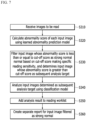
- the computing device 10 filters and classifies the input images according to the abnormality score, as in Table 1.
- the image classified into strong normal is not further analyzed and is excluded from the worklist.
- the image that is not filtered out and is analyzed as normal (weak normal) is not excluded from the worklist and is required to be checked by the reader.
- a heatmap visually indicating a position or predicted value of an abnormal lesion may be displayed.
- Table 1 Abnormality Score Case Action [0, 0.05) Strong normal Excluded from worklist [0.05, 0.15) Normal/weak normal Not excluded from worklist, and Required to be checked by reader [0.15, 1.0] Abnormal Abnormal heatmap shown
- the computing device 10 may exclude the strong normal images from the worklist by filtering them through the two-stage analysis including strong normal filtering based on the abnormality score and the subsequent analysis, and add only the remaining images including the results of the subsequent analysis to the worklist. Since the images filtered as strong normal have a very low probability of abnormal lesions, there is no need to perform the subsequent analysis for them and to add them to the worklist that is required to be read by the reader unlike the images that are not strong normal. On the other hand, the analysis result of the images filtered as strong normal may be prepared as a report in a different form than the worklist.
- the worklist is a list of images that are required to be read by a reader in a reading procedure, and, in a broad sense, may mean a list of various tasks including medical actions related to patients in a medical institution. Because the worklist merely includes images classified into non-strong normal cases (e.g., weak normal and abnormal cases), unnecessary reading work for strong normal cases that are clearly normal may be reduced. Since a ratio of the strong normal cases varies depending on an image modality and a type of disease, the reduction ratio of the reading workload may be different. In the normal cases that occupy the majority, the workload of the reader can be significantly reduced because the strong normal images are excluded from the worklist.
- non-strong normal cases e.g., weak normal and abnormal cases
- unnecessary reading work for strong normal cases that are clearly normal may be reduced. Since a ratio of the strong normal cases varies depending on an image modality and a type of disease, the reduction ratio of the reading workload may be different. In the normal cases that occupy the majority, the workload of the reader can be significantly reduced because the strong normal
- a task of training the abnormal prediction model 100 may be implemented over a plurality of computing devices in a distributed manner, it is assumed that the computing device 10 performs operations of the present disclosure for convenience of description.
- the image reading based on the two-stage analysis may be performed.
- the computing devices positioned in the hospital may transmit an image to the server device, receive the analysis result of the image from the server device, and then add the analysis result to the worklist.
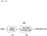
- FIG. 3A and FIG. 3B show an example of an abnormality prediction model according to an embodiment.
- an abnormality prediction model 100a or 100b may be designed as a single-headed network or a multi-headed network.
- the abnormality prediction model 100a or 100b may include a feature extractor for extracting a feature of an input image, and one abnormality/disease prediction head model.
- the abnormality prediction model 100a designed as the single-head network may include a feature extraction model 110 and an abnormality prediction head model 130.
- the feature extraction model 110 is a neural network model trained to extract a feature for detecting a lesion from an input image, and outputs the feature of the input image.
- the abnormality prediction head model 130 is a neural network model trained to predict an abnormality probability for the features output from the feature extraction model 110, and outputs a prediction result as an abnormality score.
- a computing device 10 may classify and filter the input image whose abnormality score is less than or equal to a cut-off score into strong normal, based on the cut-off score for making a desired reading sensitivity (e.g., 99 %).
- the abnormality prediction model 100b designed as the multi-head network may include a feature extraction model 110 and a plurality of disease prediction head models 140-1, 140-2, ..., 140-n.
- Each disease prediction head model is a neural network model trained to predict a corresponding disease, and outputs the prediction result as a corresponding disease prediction score.
- the disease prediction score may correspond to the abnormality score
- a score output from the disease prediction head model may be called the disease prediction score in order to distinguish it from the abnormality score calculated by combining the disease prediction scores.
- the plurality of disease prediction head models 140-1 to 140-n) may be configured in parallel according to types of diseases (lesions or findings) that can be analyzed in an image photographed with a specific modality.
- the abnormality prediction model 100 may include the disease prediction head models that independently predict consolidation, nodule, and pneumothorax, respectively.
- each of the plurality of disease prediction head models 140-1 to 140-n) is independently trained based on learning data related to a corresponding disease.
- a prediction difficulty of each disease may be different, and an amount of training data related to each disease may be different. Therefore, since the distribution of disease prediction scores output from each disease prediction head model is different, the disease prediction score that becomes the desired reading sensitivity (e.g., 99 %) may be different for each disease. If the strong normal images and the remaining images are classified based on the same cut-off score (e.g., 0.05), the reading sensitivity may vary for each disease.
- a lesion that is easy to detect may be sensitively detected, and a lesion that is difficult to detect may be detected less sensitively.
- a method of filtering images so as to allow the computing device 10 to provide the same reading sensitivity (e.g., 99 %) regardless of a disease type is described below.
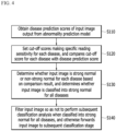
- FIG. 4 and FIG. 5 each are a flowchart of a strong normal filtering method according to an embodiment.
- a computing device 10 obtains disease prediction scores (e.g., a consolidation score, a nodule score, and a pneumothorax score) of an input image that are output from disease prediction head models of an abnormality prediction model 100b, respectively (S110).
- disease prediction scores e.g., a consolidation score, a nodule score, and a pneumothorax score
- the computing device 10 sets cut-off scores C1, C2, and C3 which make a specific reading sensitivity (e.g., 99 %) for each disease, and compares the cut-off score for each disease with the disease prediction score (S120).
- the cut-off score for each disease may not be the same. If the disease prediction score is less than the cut-off score, the input image is filtered as strong normal.
- the computing device 10 determines whether the input image is strong normal or non-strong normal for each disease based on the comparison result, and determines whether the input image is classified into strong normal for all of the plurality of diseases (S130).
- the input image with the disease prediction score less than or equal to the cut-off score is classified into strong normal, and the input image with the disease prediction score greater than the cut-off score is classified into non-strong normal.
- the computing device 10 filters the input image so as not to perform a subsequent classification analysis when the input image is classified into strong normal for all of the plurality of diseases, and forwards the input image to a subsequent classification stage when the input image is not classified into strong normal for all of the plurality of diseases (S140).
- the computing device 10 finally classifies the input image as strong normal when the input image is classified into strong normal for all of the plurality of diseases, and determines that the input image is not strong normal when the input image is not classified into strong normal for all of the plurality of diseases. For example, when all of the consolidation score, nodule score and pneumothorax score predicted for the input image are less than or equal to the cut-off score of the corresponding disease, the input image is classified into strong normal.
- the input image classified into strong normal is not added to a worklist, unlike an image on which the subsequent classification analysis is to be performed. Instead, the computing device 10 may create a separate report, which is distinguished from the worklist, for the input image classified into strong normal.
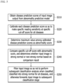
- a computing device 10 obtains disease prediction scores (e.g., a consolidation score, a nodule score, and a pneumothorax score) of an input image that are output from disease prediction head models of an abnormality prediction model 100b, respectively (S210).
- disease prediction scores e.g., a consolidation score, a nodule score, and a pneumothorax score
- the computing device 10 calibrates each disease prediction score so as to make a specific reading sensitivity (e.g., 99 %) at a specific cut-off score (e.g., 0.05) for all diseases (S220).
- a specific reading sensitivity e.g., 99 %
- a specific cut-off score e.g., 0.05
- the computing device 10 determines a maximum value among the calibrated disease prediction scores as an abnormality score (S230).
- the computing device 10 compares a specific cut-off score of 0.05 with the abnormality score, and determines whether the input image is strong normal or non-strong normal based on the comparison result (S240).
- the computing device 10 filters the input image so as not to perform a subsequent classification analysis when the input image is classified into strong normal, and forwards the input image to a subsequent classification stage when the input image is not classified into strong normal (S250).
- the computing device 10 may create a separate report for the input image classified into strong normal without adding the analysis result to the worklist.
- the computing device 10 may calibrate each disease prediction score as follows.
- the calibrated disease prediction scores may be calculated based on calibration that converts the cutoff score for each disease, which makes the specific reading sensitivity, into the same cut-off score. As a result, even if the input image is classified into strong normal at a single cut-off score, the same reading sensitivity for all diseases may be provided.
- a pneumothorax score y1 may be converted into a pneumothorax score y1' through Equation 1, in order to shift the cut-off score c1 to a specific score (e.g., 0.05).
- a consolidation score y2 may be converted into a calibrated consolidation score y2' as in Equation 1.
- a nodule score y3 may be converted into a calibrated nodule score y3' as in Equation 1.
- the computing device 10 determines a maximum value among the calibrated disease prediction scores y1', y2', and y3' as an abnormality score, and compares the cut-off score of 0.05 with the abnormality score. If the abnormality score is less than or equal to the cut-off score of 0.05, the input image is classified into strong normal.
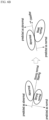
- FIG. 6A and FIG. 6B are drawings schematically illustrating a merit of a two-stage analysis including strong normal filtering according to an embodiment.
- a general classification model is trained to distinguish normal including strong normal and weak normal from abnormal. Since distinguishing input features into weak normal and abnormal is difficult compared to distinguishing the input features into strong normal and abnormal, the general classification model tends to perform learning more easily and does not properly learn a task for distinguishing between weak normal and abnormal.
- a computing device 10 can filter images predicted as strong normal through strong normal filtering to exclude them from subsequent classification, and classify input features into weak normal and abnormal through a classification model 200 for distinguishing between weak normal and abnormal.
- the classification model 200 may intensively learn a difficult task for distinguishing between weak normal and abnormal, and thus can have high classification performance.
- the computing device 10 may add the analysis result on the input image of the classification model 200 to the worklist.
- the analysis result may include a detection result for at least one disease (e.g., consolidation, nodule, pneumothorax, etc.) that can be analyzed in modality.
- FIG. 7 is a flowchart of a medical image reading method according to an embodiment.
- a computing device 10 receives images to be read (S310).
- the computing device 10 calculates an abnormality score of each input image using the learned abnormality prediction model 100 (S320).
- the abnormality prediction model 100 may be an abnormality prediction model 100a configured with a single-head network or an abnormality prediction model 100b configured with a multi-head network.
- the computing device 10 may calibrate a plurality of disease prediction scores output from abnormality prediction models 100b so as to make the same reading sensitivity at the same cut-off score for all diseases. Further, the computing device 10 may determine a maximum value among the calibrated disease prediction scores as the abnormality score.
- the computing device 10 filters an input image whose abnormality score is less than or equal to the cut-off score as strong normal, based on a cut-off score that makes a specific reading sensitivity, and determines an input image whose abnormality score is greater than the cut-off score as a subsequent analysis target (S330).
- the computing device 10 performs an analysis of distinguishing the input images determined as the subsequent analysis target into classification classes (e.g., weak normal and abnormal) using a classification model 200 (S340).
- classification classes e.g., weak normal and abnormal
- the computing device 10 adds the analysis result to a worklist that is required to be read by a reader (S350).
- the computing device 10 may create a separate report for the input image filtered as strong normal (S360).
- FIG. 8 is a configuration diagram of a computing device according to an embodiment.
- a computing device 10 may include one or more processors 11, a memory 13 on which a computer program to be executed by the processor 11 is loaded, a storage 15 which stores the program and various data, a communication interface 17, and a bus 19 for connecting them.
- the computing device 10 may further include various elements.
- the program may include instructions which make the processor 11 to perform methods or operations according to various embodiments of the present disclosure when loaded on the memory 13.
- the processor 11 may perform methods or operations according to various embodiments of the present disclosure by executing the instructions.
- the program includes a series of computer-readable instructions that are grouped by function and is executed by the processor.
- the processor 11 controls overall operation of each element of the computing device 10.
- the processor 11 may be configured to include at least one of a central processing unit (CPU), a microprocessor unit (MPU), a microcontroller unit (MCU), a graphics processing unit (GPU), and any form of processor well known in the technical field of the present disclosure. Further, the processor 11 may perform computation for at least one application or program to execute methods or operations according to embodiments of the present disclosure.
- CPU central processing unit
- MPU microprocessor unit
- MCU microcontroller unit
- GPU graphics processing unit
- the memory 13 stores various kinds of data, commands, and/or information. To execute methods or operations according to various embodiments of the present disclosure, the memory 13 may load one or more programs from the storage 15.
- the memory 13 may be implemented as a volatile memory such as a random access memory (RAM), but the technical scope of the present disclosure is not limited thereto.
- the storage 15 may non-temporarily store the program.
- the storage 15 may include a nonvolatile memory, such as a read only memory (ROM), an erasable programmable ROM (EPROM), an electrically erasable programmable ROM (EEPROM) or a flash memory, a hard disk, a removable disk, or any form of computer-readable recording medium well known in the art to which the present disclosure pertains.
- ROM read only memory
- EPROM erasable programmable ROM
- EEPROM electrically erasable programmable ROM
- flash memory a hard disk, a removable disk, or any form of computer-readable recording medium well known in the art to which the present disclosure pertains.
- the communication interface 17 supports wired or wireless Internet communication of the computing device 10. Further, the communication interface 17 may support various communication methods as well as Internet communication. To this end, the communication interface 17 may include a communication module well known in the technical field of the present disclosure.
- the bus 19 provides a communication function between elements of the computing device 10.
- the bus 19 may be implemented as various forms of buses, such as an address bus, a data bus, and a control bus.
- the embodiments of the present invention described above are not implemented through only the apparatus and the method, but may also be implemented through a program that realizes functions corresponding to the configuration of the embodiments of the present invention or a recording medium on which the program is recorded.
Landscapes
- Engineering & Computer Science (AREA)
- Health & Medical Sciences (AREA)
- Medical Informatics (AREA)
- Public Health (AREA)
- General Health & Medical Sciences (AREA)
- Biomedical Technology (AREA)
- Nuclear Medicine, Radiotherapy & Molecular Imaging (AREA)
- Radiology & Medical Imaging (AREA)
- Life Sciences & Earth Sciences (AREA)
- Physics & Mathematics (AREA)
- Epidemiology (AREA)
- Primary Health Care (AREA)
- Pathology (AREA)
- Computer Vision & Pattern Recognition (AREA)
- Data Mining & Analysis (AREA)
- Theoretical Computer Science (AREA)
- General Physics & Mathematics (AREA)
- Databases & Information Systems (AREA)
- Quality & Reliability (AREA)
- Surgery (AREA)
- Heart & Thoracic Surgery (AREA)
- Veterinary Medicine (AREA)
- Animal Behavior & Ethology (AREA)
- Biophysics (AREA)
- High Energy & Nuclear Physics (AREA)
- Optics & Photonics (AREA)
- Molecular Biology (AREA)
- Oral & Maxillofacial Surgery (AREA)
- Physiology (AREA)
- Dentistry (AREA)
- Bioinformatics & Computational Biology (AREA)
- Evolutionary Biology (AREA)
- Bioinformatics & Cheminformatics (AREA)
- General Engineering & Computer Science (AREA)
- Artificial Intelligence (AREA)
- Evolutionary Computation (AREA)
- Apparatus For Radiation Diagnosis (AREA)
- Measuring And Recording Apparatus For Diagnosis (AREA)
- Image Analysis (AREA)
- Image Processing (AREA)
Claims (11)
- Verfahren zum Klassifizieren eines medizinischen Bildes durch eine Rechenvorrichtung (10), die durch mindestens einen Prozessor (11) betrieben wird, wobei das Verfahren umfasst:Erhalten eines Abnormalitätswerts eines Eingangsbildes unter Verwendung eines Abnormalitätsvorhersagemodells (100);Ausschließen des Eingangsbildes aus der anschließenden Analyse und Klassifizieren des Eingangsbildes als stark normal, wenn der Abnormalitätswert kleiner oder gleich einem Grenzwert ist, auf Basis des Grenzwerts, der eine spezifische Leseempfindlichkeit ausmacht; undErhalten eines Analyseergebnisses des Eingangsbildes unter Verwendung eines Klassifikationsmodells (200), das das Eingangsbild in schwach normal oder abnormal unterscheidet, wenn der Abnormalitätswert größer als der Grenzwert ist,wobei das Klassifikationsmodell (200) ein Künstliche-Intelligenz-Modell beinhaltet, das eine Aufgabe des Unterscheidens des Eingangsbildes in schwach normal oder abnormal erlernt hat, unddadurch gekennzeichnet, dass, wobei das Erhalten des Abnormalitätswerts das Erhalten von kalibrierten Krankheitsvorhersagewerten durch Kalibrierung, die einen Grenzwert für jede Krankheit, der die spezifische Leseempfindlichkeit ausmacht, in den Grenzwert umwandelt, und das Bestimmen eines Maximalwerts unter den kalibrierten Krankheitsvorhersagewerten als Abnormalitätswert umfasst.
- Verfahren nach Anspruch 1, das weiter das Hinzufügen des Analyseergebnisses zu einer Lese-Arbeitsliste umfasst,
wobei das Eingangsbild, dessen Abnormalitätswert kleiner oder gleich dem Grenzwert ist, nicht zur Lese-Arbeitsliste hinzugefügt wird. - Verfahren nach Anspruch 1 oder 2, wobei das Erhalten des Abnormalitätswerts, beim Erhalten von Krankheitsvorhersagewerten für unterschiedliche Krankheiten aus dem Abnormalitätsvorhersagemodell (100), das Aggregieren der Krankheitsvorhersagewerte umfasst, um den Abnormalitätswert zu bestimmen.
- Verfahren nach einem der Ansprüche 1 bis 3, wobei das Ausschließen des Eingangsbildes in einem Fall, in dem jeder von Krankheitsvorhersagewerten für unterschiedliche Krankheiten als Abnormalitätswert für jede Krankheit aus dem Abnormalitätsvorhersagemodell (100) erhalten wird, das Berechnen eines Grenzwerts für jede Krankheit, der die spezifische Leseempfindlichkeit ausmacht, und das Ausschließen des Eingangsbildes für alle der unterschiedlichen Krankheiten umfasst, wenn der Abnormalitätswert für jede Krankheit kleiner oder gleich dem Grenzwert für eine entsprechende Krankheit ist.
- Verfahren nach einem der Ansprüche 1 bis 4, wobei das Abnormalitätsvorhersagemodell umfasst:ein Merkmalsextraktionsmodell (110), das darauf trainiert ist, ein Merkmal des Eingangsbildes auszugeben; undmindestens ein Krankheitsvorhersage-Kopfmodell (130), das darauf trainiert ist, mindestens eine Krankheit auf Basis von Merkmalen, die von dem Merkmalsextraktionsmodell (110) ausgegeben werden, vorherzusagen, und/oderwobei das Abnormalitätsvorhersagemodell (100) eine Empfindlichkeit zwischen 90 % und 100 % aufweist.
- Verfahren zum Klassifizieren eines medizinischen Bildes durch eine Rechenvorrichtung (10), die durch mindestens einen Prozessor (11) betrieben wird, wobei das Verfahren umfasst:Festlegen eines Grenzwerts, der eine spezifische Leseempfindlichkeit für ein Abnormalitätsvorhersagemodell (100) ausmacht, das einen Abnormalitätswert eines medizinischen Bildes ausgibt;Erhalten eines Abnormalitätswerts eines Eingangsbildes unter Verwendung des Abnormalitätsvorhersagemodells (100);wenn der Abnormalitätswert des Eingangsbildes kleiner oder gleich dem Grenzwert ist, Klassifizieren des Eingangsbildes als stark normal und Ausschließen des Eingangsbildes aus der anschließenden Analyse; undErhalten eines Analyseergebnisses des Eingangsbildes unter Verwendung eines Klassifikationsmodells (200), das zwischen schwach normal und abnormal unterscheidet, wenn der Abnormalitätswert des Eingangsbildes größer ist als der Grenzwert,dadurch gekennzeichnet, dass, wobei das Erhalten des Abnormalitätswerts beim Erhalten von Krankheitsvorhersagewerten für unterschiedliche Krankheiten aus dem Abnormalitätsvorhersagemodell (100) das Kalibrieren der Krankheitsvorhersagewerte, um die spezifische Leseempfindlichkeit für die unterschiedlichen Krankheiten am Grenzwert bereitzustellen, und das Bestimmen eines Maximalwerts unter den kalibrierten Krankheitsvorhersagewerten als Abnormalitätswert des Eingangsbildes umfasst.
- Verfahren nach Anspruch 6, weiter umfassend:
Hinzufügen des Analyseergebnisses zu einer Lese-Arbeitsliste, wobei das als stark normal klassifizierte Eingangsbild nicht zur Lese-Arbeitsliste hinzugefügt wird. - Verfahren nach Anspruch 6 oder 7, wobei das Ausschließen des Eingangsbildes in einem Fall, in dem jeder von Krankheitsvorhersagewerten für unterschiedliche Krankheiten aus dem Abnormalitätsvorhersagemodell (100) als Abnormalitätswert für jede Krankheit erhalten wird, das Berechnen eines Grenzwerts für jede Krankheit, der die spezifische Leseempfindlichkeit ausmacht, und das Ausschließen des Eingangsbildes für alle der unterschiedlichen Krankheiten umfasst, wenn der Abnormalitätswert für jede Krankheit kleiner oder gleich dem Grenzwert für eine entsprechende Krankheit ist.
- Verfahren nach einem der vorstehenden Ansprüche,wobei das Eingangsbild ein Röntgenbild des Brustkorbs beinhaltet, undwobei die Vielzahl von Krankheiten mindestens zwei von Konsolidierung, Knoten und Pneumothorax beinhaltet.
- Verfahren nach den Ansprüchen 1 oder 6,
wobei die Leseempfindlichkeit einen Wert zwischen 90 % und 100 % aufweist. - Rechenvorrichtung (10), umfassend:einen Speicher, der ein Abnormalitätsvorhersagemodell (100), das darauf trainiert ist, einen Abnormalitätswert eines Eingangsbildes auszugeben, und ein Klassifikationsmodell (200) speichert, das darauf trainiert ist, das Eingangsbild in schwach normal und abnormal zu unterscheiden; undeinen Prozessor (11), der einen Abnormalitätswert des Eingangsbildes unter Verwendung des Abnormalitätsvorhersagemodells (100) erhält, das Eingangsbild in stark normal klassifiziert und das Eingangsbild aus der anschließenden Analyse ausschließt, wenn der Abnormalitätswert des Eingangsbildes kleiner oder gleich einem Grenzwert ist, und ein Analyseergebnis des Eingangsbildes unter Verwendung des Klassifikationsmodells (200) erhält, das das Eingangsbild in schwach normal oder abnormal unterscheidet, wenn der Abnormalitätswert des Eingangsbildes größer ist als der Grenzwert,wobei das Klassifikationsmodell (200) ein Künstliche-Intelligenz-Modell beinhaltet, das eine Aufgabe des Unterscheidens des Eingangsbildes in schwach normal oder abnormal erlernt hat, undwobei das Erhalten des Abnormalitätswerts das Erhalten von kalibrierten Krankheitsvorhersagewerten durch Kalibrierung, die einen Grenzwert für jede Krankheit, der die spezifische Leseempfindlichkeit ausmacht, in den Grenzwert umwandelt, und das Bestimmen eines Maximalwerts unter den kalibrierten Krankheitsvorhersagewerten als Abnormalitätswert umfasst, und bevorzugt wobei das Abnormalitätsvorhersagemodell (100) umfasst:ein Merkmalsextraktionsmodell (110), das darauf trainiert ist, ein Merkmal des Eingangsbildes auszugeben; undmindestens ein Krankheitsvorhersage-Kopfmodell (130), das darauf trainiert ist, mindestens eine Krankheit auf Basis von Merkmalen, die von dem Merkmalsextraktionsmodell (110) ausgegeben werden, vorherzusagen.
Priority Applications (1)
| Application Number | Priority Date | Filing Date | Title |
|---|---|---|---|
| EP24223267.6A EP4502830A3 (de) | 2020-06-15 | 2021-06-09 | Verfahren zur filterung eines normalen medizinischen bildes, verfahren zur interpretation eines medizinischen bildes und rechenvorrichtung zur implementierung der verfahren |
Applications Claiming Priority (1)
| Application Number | Priority Date | Filing Date | Title |
|---|---|---|---|
| KR1020200072257A KR102231698B1 (ko) | 2020-06-15 | 2020-06-15 | 정상 의료 영상을 필터링하는 방법, 이를 이용한 의료 영상 판독 방법 및 컴퓨팅 장치 |
Related Child Applications (2)
| Application Number | Title | Priority Date | Filing Date |
|---|---|---|---|
| EP24223267.6A Division EP4502830A3 (de) | 2020-06-15 | 2021-06-09 | Verfahren zur filterung eines normalen medizinischen bildes, verfahren zur interpretation eines medizinischen bildes und rechenvorrichtung zur implementierung der verfahren |
| EP24223267.6A Division-Into EP4502830A3 (de) | 2020-06-15 | 2021-06-09 | Verfahren zur filterung eines normalen medizinischen bildes, verfahren zur interpretation eines medizinischen bildes und rechenvorrichtung zur implementierung der verfahren |
Publications (3)
| Publication Number | Publication Date |
|---|---|
| EP3926540A1 EP3926540A1 (de) | 2021-12-22 |
| EP3926540C0 EP3926540C0 (de) | 2025-02-05 |
| EP3926540B1 true EP3926540B1 (de) | 2025-02-05 |
Family
ID=75257037
Family Applications (2)
| Application Number | Title | Priority Date | Filing Date |
|---|---|---|---|
| EP21178486.3A Active EP3926540B1 (de) | 2020-06-15 | 2021-06-09 | Verfahren zur filtrierung eines normalem medizinischen bildes, verfahren zur interpretation eines medizinischen bildes und rechenvorrichtung zur implementierung der verfahren |
| EP24223267.6A Pending EP4502830A3 (de) | 2020-06-15 | 2021-06-09 | Verfahren zur filterung eines normalen medizinischen bildes, verfahren zur interpretation eines medizinischen bildes und rechenvorrichtung zur implementierung der verfahren |
Family Applications After (1)
| Application Number | Title | Priority Date | Filing Date |
|---|---|---|---|
| EP24223267.6A Pending EP4502830A3 (de) | 2020-06-15 | 2021-06-09 | Verfahren zur filterung eines normalen medizinischen bildes, verfahren zur interpretation eines medizinischen bildes und rechenvorrichtung zur implementierung der verfahren |
Country Status (3)
| Country | Link |
|---|---|
| US (2) | US11574727B2 (de) |
| EP (2) | EP3926540B1 (de) |
| KR (2) | KR102231698B1 (de) |
Families Citing this family (3)
| Publication number | Priority date | Publication date | Assignee | Title |
|---|---|---|---|---|
| US12555231B2 (en) * | 2022-04-22 | 2026-02-17 | The General Hospital Corporation | Detecting ischemic stroke mimic using deep learning-based analysis of medical images |
| KR102798774B1 (ko) * | 2022-11-29 | 2025-04-23 | 주식회사 카이미 | 민감도 설정이 가능한 인공지능 기반의 병변 검출 방법 및 장치 |
| US20250080751A1 (en) * | 2023-08-30 | 2025-03-06 | Nec Laboratories America, Inc. | Machine learning model for video with real-time rate control |
Family Cites Families (18)
| Publication number | Priority date | Publication date | Assignee | Title |
|---|---|---|---|---|
| US6310967B1 (en) * | 1998-04-29 | 2001-10-30 | University Of South Florida | Normal and abnormal tissue identification system and method for medical images such as digital mammograms |
| US7623694B2 (en) * | 2006-01-31 | 2009-11-24 | Mevis Medical Solutions, Inc. | Method and apparatus for classifying detection inputs in medical images |
| CA2779301C (en) | 2009-11-27 | 2017-08-15 | Dog Microsystems Inc. | Method and system for filtering image data and use thereof in virtual endoscopy |
| JP5670695B2 (ja) * | 2010-10-18 | 2015-02-18 | ソニー株式会社 | 情報処理装置及び方法、並びにプログラム |
| KR101144964B1 (ko) * | 2010-10-21 | 2012-05-11 | 전남대학교산학협력단 | 간질성 폐질환 검출 시스템 및 그 방법 |
| EP2570970A1 (de) * | 2011-09-16 | 2013-03-20 | Technische Universität Berlin | Verfahren und System zur automatischen Analyse eines Bildes einer biologischen Probe |
| US20140058748A1 (en) * | 2012-08-23 | 2014-02-27 | Cerner Innovation, Inc. | Populating custom patient worklists using demographic and clinical criteria |
| KR20150077184A (ko) | 2013-12-27 | 2015-07-07 | 삼성전자주식회사 | 의료 영상의 병변 유사도 판단 장치 및 방법 |
| US9811631B2 (en) * | 2015-09-30 | 2017-11-07 | General Electric Company | Automated cloud image processing and routing |
| KR101880678B1 (ko) | 2016-10-12 | 2018-07-20 | (주)헬스허브 | 기계학습을 통한 의료영상 판독 및 진단 통합 시스템 |
| US10499857B1 (en) * | 2017-09-19 | 2019-12-10 | Deepradiology Inc. | Medical protocol change in real-time imaging |
| US11024415B2 (en) * | 2017-12-15 | 2021-06-01 | International Business Machines Corporation | Automated worklist prioritization of patient care based on cognitive classification of medical images |
| KR101943011B1 (ko) * | 2018-01-22 | 2019-01-28 | 주식회사 뷰노 | 피검체의 의료 영상 판독을 지원하는 방법 및 이를 이용한 장치 |
| KR20190105460A (ko) * | 2018-03-05 | 2019-09-17 | 주식회사 인공지능연구원 | 의료 진단 리포트 생성 장치 및 방법 |
| CN112292691B (zh) * | 2018-06-18 | 2024-06-04 | 谷歌有限责任公司 | 用于使用深度学习提高癌症检测的方法与系统 |
| US10733727B2 (en) * | 2018-11-14 | 2020-08-04 | Qure.Ai Technologies Private Limited | Application of deep learning for medical imaging evaluation |
| US10943681B2 (en) * | 2018-11-21 | 2021-03-09 | Enlitic, Inc. | Global multi-label generating system |
| KR102063492B1 (ko) | 2018-11-30 | 2020-01-08 | 아주대학교산학협력단 | 기계학습용 의료 영상 데이터의 학습 노이즈 필터링 방법 및 그 시스템 |
-
2020
- 2020-06-15 KR KR1020200072257A patent/KR102231698B1/ko active Active
- 2020-10-22 US US17/077,142 patent/US11574727B2/en active Active
-
2021
- 2021-03-16 KR KR1020210034111A patent/KR20210155339A/ko active Pending
- 2021-06-09 EP EP21178486.3A patent/EP3926540B1/de active Active
- 2021-06-09 EP EP24223267.6A patent/EP4502830A3/de active Pending
-
2022
- 2022-12-22 US US18/086,962 patent/US11928817B2/en active Active
Also Published As
| Publication number | Publication date |
|---|---|
| EP4502830A3 (de) | 2025-04-23 |
| KR102231698B1 (ko) | 2021-03-24 |
| US20230128769A1 (en) | 2023-04-27 |
| EP3926540A1 (de) | 2021-12-22 |
| US20210391059A1 (en) | 2021-12-16 |
| US11574727B2 (en) | 2023-02-07 |
| EP3926540C0 (de) | 2025-02-05 |
| KR20210155339A (ko) | 2021-12-22 |
| EP4502830A2 (de) | 2025-02-05 |
| US11928817B2 (en) | 2024-03-12 |
Similar Documents
| Publication | Publication Date | Title |
|---|---|---|
| AU2018376561B2 (en) | Three-dimensional medical image analysis method and system for identification of vertebral fractures | |
| EP3719807B1 (de) | Voraussagen eines pathologischen zustandes aus einem medizinischen bild | |
| Niemeijer et al. | Information fusion for diabetic retinopathy CAD in digital color fundus photographs | |
| US11928817B2 (en) | Method for filtering normal medical image, method for interpreting medical image, and computing device implementing the methods | |
| US7236619B2 (en) | System and method for computer-aided detection and characterization of diffuse lung disease | |
| JP6837376B2 (ja) | 画像処理装置および方法並びにプログラム | |
| CN101084501A (zh) | 数字医学图像分析 | |
| KR102600401B1 (ko) | 분류 및 분할을 이용한 의료 영상 분석 방법, 장치 및 컴퓨터 프로그램 | |
| CN111226287A (zh) | 用于分析医学成像数据集的方法、用于分析医学成像数据集的系统、计算机程序产品以及计算机可读介质 | |
| KR100998630B1 (ko) | 폐질환 자동 분류 방법 | |
| CN114926396A (zh) | 一种精神障碍类磁共振图像初步筛查模型构建方法 | |
| EP4471797A1 (de) | Auf künstlicher intelligenz basierende biomarkerauswahlvorrichtung und verfahren | |
| Ennaji et al. | Ensemble learning with weighted voting classifier for melanoma diagnosis | |
| US11508065B2 (en) | Methods and systems for detecting acquisition errors in medical images | |
| CN119418899A (zh) | 病理诊断质控的智能筛查方法和系统 | |
| KR20250015795A (ko) | 영상 및 비영상 데이터를 이용한 관절염 예후 통합 예측 방법, 장치 및 컴퓨터 프로그램 | |
| CN114127859B (zh) | 用于在非对比头部ct中快速检测和索引危急区域的自动化系统 | |
| JP2025536473A (ja) | 標本画像の異常の検出 | |
| KR20250093123A (ko) | 척추관 및 신경근관에서의 협착증 평가 방법 및 장치 | |
| Saygılı | Rapid and Precise Identification of COVID-19 through Segmentation and Classification of CT and X-ray Images | |
| Dash et al. | Deep Neural Networks for Accurate Detection of Nail Disorders | |
| Shah et al. | Predictive Analytics for Diabetes and Diabetic Retinopathy Using Deep Learning on Fundus Images | |
| Perera | Decision Support System to Diagnose COVID-19 through Chest X-Ray Analysis | |
| Sanjayprabu et al. | Detection of Covid-19 using Chest X-ray Images and Texture Feature Extraction | |
| WO2024229150A2 (en) | Systems and methods for extracting vertebral bodies from two-dimensional image data and classifying fracture |
Legal Events
| Date | Code | Title | Description |
|---|---|---|---|
| PUAI | Public reference made under article 153(3) epc to a published international application that has entered the european phase |
Free format text: ORIGINAL CODE: 0009012 |
|
| STAA | Information on the status of an ep patent application or granted ep patent |
Free format text: STATUS: REQUEST FOR EXAMINATION WAS MADE |
|
| 17P | Request for examination filed |
Effective date: 20210609 |
|
| AK | Designated contracting states |
Kind code of ref document: A1 Designated state(s): AL AT BE BG CH CY CZ DE DK EE ES FI FR GB GR HR HU IE IS IT LI LT LU LV MC MK MT NL NO PL PT RO RS SE SI SK SM TR |
|
| B565 | Issuance of search results under rule 164(2) epc |
Effective date: 20211109 |
|
| RBV | Designated contracting states (corrected) |
Designated state(s): AL AT BE BG CH CY CZ DE DK EE ES FI FR GB GR HR HU IE IS IT LI LT LU LV MC MK MT NL NO PL PT RO RS SE SI SK SM TR |
|
| STAA | Information on the status of an ep patent application or granted ep patent |
Free format text: STATUS: EXAMINATION IS IN PROGRESS |
|
| 17Q | First examination report despatched |
Effective date: 20231220 |
|
| REG | Reference to a national code |
Ref legal event code: R079 Ref country code: DE Ref legal event code: R079 Ref document number: 602021025738 Country of ref document: DE Free format text: PREVIOUS MAIN CLASS: G06K0009620000 Ipc: G06T0007000000 |
|
| RIC1 | Information provided on ipc code assigned before grant |
Ipc: G06F 18/245 20230101ALI20240723BHEP Ipc: G06T 7/00 20170101AFI20240723BHEP |
|
| GRAP | Despatch of communication of intention to grant a patent |
Free format text: ORIGINAL CODE: EPIDOSNIGR1 |
|
| STAA | Information on the status of an ep patent application or granted ep patent |
Free format text: STATUS: GRANT OF PATENT IS INTENDED |
|
| INTG | Intention to grant announced |
Effective date: 20240904 |
|
| GRAS | Grant fee paid |
Free format text: ORIGINAL CODE: EPIDOSNIGR3 |
|
| GRAA | (expected) grant |
Free format text: ORIGINAL CODE: 0009210 |
|
| STAA | Information on the status of an ep patent application or granted ep patent |
Free format text: STATUS: THE PATENT HAS BEEN GRANTED |
|
| AK | Designated contracting states |
Kind code of ref document: B1 Designated state(s): AL AT BE BG CH CY CZ DE DK EE ES FI FR GB GR HR HU IE IS IT LI LT LU LV MC MK MT NL NO PL PT RO RS SE SI SK SM TR |
|
| REG | Reference to a national code |
Ref country code: GB Ref legal event code: FG4D |
|
| REG | Reference to a national code |
Ref country code: CH Ref legal event code: EP |
|
| REG | Reference to a national code |
Ref country code: IE Ref legal event code: FG4D |
|
| REG | Reference to a national code |
Ref country code: DE Ref legal event code: R096 Ref document number: 602021025738 Country of ref document: DE |
|
| U01 | Request for unitary effect filed |
Effective date: 20250220 |
|
| U07 | Unitary effect registered |
Designated state(s): AT BE BG DE DK EE FI FR IT LT LU LV MT NL PT RO SE SI Effective date: 20250228 |
|
| U20 | Renewal fee for the european patent with unitary effect paid |
Year of fee payment: 5 Effective date: 20250521 |
|
| PG25 | Lapsed in a contracting state [announced via postgrant information from national office to epo] |
Ref country code: RS Free format text: LAPSE BECAUSE OF FAILURE TO SUBMIT A TRANSLATION OF THE DESCRIPTION OR TO PAY THE FEE WITHIN THE PRESCRIBED TIME-LIMIT Effective date: 20250505 |
|
| PG25 | Lapsed in a contracting state [announced via postgrant information from national office to epo] |
Ref country code: PL Free format text: LAPSE BECAUSE OF FAILURE TO SUBMIT A TRANSLATION OF THE DESCRIPTION OR TO PAY THE FEE WITHIN THE PRESCRIBED TIME-LIMIT Effective date: 20250205 |
|
| PG25 | Lapsed in a contracting state [announced via postgrant information from national office to epo] |
Ref country code: ES Free format text: LAPSE BECAUSE OF FAILURE TO SUBMIT A TRANSLATION OF THE DESCRIPTION OR TO PAY THE FEE WITHIN THE PRESCRIBED TIME-LIMIT Effective date: 20250205 |
|
| PGFP | Annual fee paid to national office [announced via postgrant information from national office to epo] |
Ref country code: GB Payment date: 20250521 Year of fee payment: 5 |
|
| PG25 | Lapsed in a contracting state [announced via postgrant information from national office to epo] |
Ref country code: IS Free format text: LAPSE BECAUSE OF FAILURE TO SUBMIT A TRANSLATION OF THE DESCRIPTION OR TO PAY THE FEE WITHIN THE PRESCRIBED TIME-LIMIT Effective date: 20250605 Ref country code: NO Free format text: LAPSE BECAUSE OF FAILURE TO SUBMIT A TRANSLATION OF THE DESCRIPTION OR TO PAY THE FEE WITHIN THE PRESCRIBED TIME-LIMIT Effective date: 20250505 |
|
| PG25 | Lapsed in a contracting state [announced via postgrant information from national office to epo] |
Ref country code: HR Free format text: LAPSE BECAUSE OF FAILURE TO SUBMIT A TRANSLATION OF THE DESCRIPTION OR TO PAY THE FEE WITHIN THE PRESCRIBED TIME-LIMIT Effective date: 20250205 |
|
| PG25 | Lapsed in a contracting state [announced via postgrant information from national office to epo] |
Ref country code: GR Free format text: LAPSE BECAUSE OF FAILURE TO SUBMIT A TRANSLATION OF THE DESCRIPTION OR TO PAY THE FEE WITHIN THE PRESCRIBED TIME-LIMIT Effective date: 20250506 |
|
| PG25 | Lapsed in a contracting state [announced via postgrant information from national office to epo] |
Ref country code: SM Free format text: LAPSE BECAUSE OF FAILURE TO SUBMIT A TRANSLATION OF THE DESCRIPTION OR TO PAY THE FEE WITHIN THE PRESCRIBED TIME-LIMIT Effective date: 20250205 |
|
| PG25 | Lapsed in a contracting state [announced via postgrant information from national office to epo] |
Ref country code: CZ Free format text: LAPSE BECAUSE OF FAILURE TO SUBMIT A TRANSLATION OF THE DESCRIPTION OR TO PAY THE FEE WITHIN THE PRESCRIBED TIME-LIMIT Effective date: 20250205 |
|
| PG25 | Lapsed in a contracting state [announced via postgrant information from national office to epo] |
Ref country code: SK Free format text: LAPSE BECAUSE OF FAILURE TO SUBMIT A TRANSLATION OF THE DESCRIPTION OR TO PAY THE FEE WITHIN THE PRESCRIBED TIME-LIMIT Effective date: 20250205 |
|
| PLBE | No opposition filed within time limit |
Free format text: ORIGINAL CODE: 0009261 |
|
| STAA | Information on the status of an ep patent application or granted ep patent |
Free format text: STATUS: NO OPPOSITION FILED WITHIN TIME LIMIT |
|
| REG | Reference to a national code |
Ref country code: CH Ref legal event code: L10 Free format text: ST27 STATUS EVENT CODE: U-0-0-L10-L00 (AS PROVIDED BY THE NATIONAL OFFICE) Effective date: 20251217 |
|
| 26N | No opposition filed |
Effective date: 20251106 |
|
| REG | Reference to a national code |
Ref country code: CH Ref legal event code: H13 Free format text: ST27 STATUS EVENT CODE: U-0-0-H10-H13 (AS PROVIDED BY THE NATIONAL OFFICE) Effective date: 20260127 |
|
| PG25 | Lapsed in a contracting state [announced via postgrant information from national office to epo] |
Ref country code: MC Free format text: LAPSE BECAUSE OF FAILURE TO SUBMIT A TRANSLATION OF THE DESCRIPTION OR TO PAY THE FEE WITHIN THE PRESCRIBED TIME-LIMIT Effective date: 20250205 |