EP3926537A1 - Verfahren zur segmentierung eines medizinischen bildes, verfahren zur segmentierung eines bildes, zugehörige vorrichtung und system - Google Patents
Verfahren zur segmentierung eines medizinischen bildes, verfahren zur segmentierung eines bildes, zugehörige vorrichtung und system Download PDFInfo
- Publication number
- EP3926537A1 EP3926537A1 EP20755319.9A EP20755319A EP3926537A1 EP 3926537 A1 EP3926537 A1 EP 3926537A1 EP 20755319 A EP20755319 A EP 20755319A EP 3926537 A1 EP3926537 A1 EP 3926537A1
- Authority
- EP
- European Patent Office
- Prior art keywords
- medical image
- processed
- time
- cross
- coordinate
- Prior art date
- Legal status (The legal status is an assumption and is not a legal conclusion. Google has not performed a legal analysis and makes no representation as to the accuracy of the status listed.)
- Granted
Links
Images


Classifications
-
- G—PHYSICS
- G06—COMPUTING OR CALCULATING; COUNTING
- G06T—IMAGE DATA PROCESSING OR GENERATION, IN GENERAL
- G06T7/00—Image analysis
- G06T7/10—Segmentation; Edge detection
- G06T7/11—Region-based segmentation
-
- G—PHYSICS
- G06—COMPUTING OR CALCULATING; COUNTING
- G06T—IMAGE DATA PROCESSING OR GENERATION, IN GENERAL
- G06T7/00—Image analysis
- G06T7/10—Segmentation; Edge detection
- G06T7/149—Segmentation; Edge detection involving deformable models, e.g. active contour models
-
- G—PHYSICS
- G06—COMPUTING OR CALCULATING; COUNTING
- G06F—ELECTRIC DIGITAL DATA PROCESSING
- G06F18/00—Pattern recognition
- G06F18/20—Analysing
- G06F18/21—Design or setup of recognition systems or techniques; Extraction of features in feature space; Blind source separation
- G06F18/214—Generating training patterns; Bootstrap methods, e.g. bagging or boosting
-
- G—PHYSICS
- G06—COMPUTING OR CALCULATING; COUNTING
- G06T—IMAGE DATA PROCESSING OR GENERATION, IN GENERAL
- G06T7/00—Image analysis
- G06T7/0002—Inspection of images, e.g. flaw detection
- G06T7/0012—Biomedical image inspection
-
- G—PHYSICS
- G06—COMPUTING OR CALCULATING; COUNTING
- G06V—IMAGE OR VIDEO RECOGNITION OR UNDERSTANDING
- G06V10/00—Arrangements for image or video recognition or understanding
- G06V10/20—Image preprocessing
- G06V10/26—Segmentation of patterns in the image field; Cutting or merging of image elements to establish the pattern region, e.g. clustering-based techniques; Detection of occlusion
-
- G—PHYSICS
- G06—COMPUTING OR CALCULATING; COUNTING
- G06V—IMAGE OR VIDEO RECOGNITION OR UNDERSTANDING
- G06V10/00—Arrangements for image or video recognition or understanding
- G06V10/70—Arrangements for image or video recognition or understanding using pattern recognition or machine learning
- G06V10/77—Processing image or video features in feature spaces; using data integration or data reduction, e.g. principal component analysis [PCA] or independent component analysis [ICA] or self-organising maps [SOM]; Blind source separation
- G06V10/774—Generating sets of training patterns; Bootstrap methods, e.g. bagging or boosting
-
- G—PHYSICS
- G06—COMPUTING OR CALCULATING; COUNTING
- G06V—IMAGE OR VIDEO RECOGNITION OR UNDERSTANDING
- G06V20/00—Scenes; Scene-specific elements
- G06V20/60—Type of objects
- G06V20/64—Three-dimensional objects
-
- G—PHYSICS
- G06—COMPUTING OR CALCULATING; COUNTING
- G06T—IMAGE DATA PROCESSING OR GENERATION, IN GENERAL
- G06T2207/00—Indexing scheme for image analysis or image enhancement
- G06T2207/10—Image acquisition modality
- G06T2207/10028—Range image; Depth image; 3D point clouds
-
- G—PHYSICS
- G06—COMPUTING OR CALCULATING; COUNTING
- G06T—IMAGE DATA PROCESSING OR GENERATION, IN GENERAL
- G06T2207/00—Indexing scheme for image analysis or image enhancement
- G06T2207/10—Image acquisition modality
- G06T2207/10072—Tomographic images
- G06T2207/10088—Magnetic resonance imaging [MRI]
- G06T2207/10096—Dynamic contrast-enhanced magnetic resonance imaging [DCE-MRI]
-
- G—PHYSICS
- G06—COMPUTING OR CALCULATING; COUNTING
- G06T—IMAGE DATA PROCESSING OR GENERATION, IN GENERAL
- G06T2207/00—Indexing scheme for image analysis or image enhancement
- G06T2207/10—Image acquisition modality
- G06T2207/10116—X-ray image
-
- G—PHYSICS
- G06—COMPUTING OR CALCULATING; COUNTING
- G06T—IMAGE DATA PROCESSING OR GENERATION, IN GENERAL
- G06T2207/00—Indexing scheme for image analysis or image enhancement
- G06T2207/20—Special algorithmic details
- G06T2207/20081—Training; Learning
-
- G—PHYSICS
- G06—COMPUTING OR CALCULATING; COUNTING
- G06T—IMAGE DATA PROCESSING OR GENERATION, IN GENERAL
- G06T2207/00—Indexing scheme for image analysis or image enhancement
- G06T2207/30—Subject of image; Context of image processing
- G06T2207/30004—Biomedical image processing
-
- G—PHYSICS
- G06—COMPUTING OR CALCULATING; COUNTING
- G06T—IMAGE DATA PROCESSING OR GENERATION, IN GENERAL
- G06T2207/00—Indexing scheme for image analysis or image enhancement
- G06T2207/30—Subject of image; Context of image processing
- G06T2207/30004—Biomedical image processing
- G06T2207/30068—Mammography; Breast
-
- G—PHYSICS
- G06—COMPUTING OR CALCULATING; COUNTING
- G06T—IMAGE DATA PROCESSING OR GENERATION, IN GENERAL
- G06T2207/00—Indexing scheme for image analysis or image enhancement
- G06T2207/30—Subject of image; Context of image processing
- G06T2207/30004—Biomedical image processing
- G06T2207/30096—Tumor; Lesion
-
- G—PHYSICS
- G06—COMPUTING OR CALCULATING; COUNTING
- G06V—IMAGE OR VIDEO RECOGNITION OR UNDERSTANDING
- G06V20/00—Scenes; Scene-specific elements
- G06V20/40—Scenes; Scene-specific elements in video content
- G06V20/49—Segmenting video sequences, i.e. computational techniques such as parsing or cutting the sequence, low-level clustering or determining units such as shots or scenes
-
- G—PHYSICS
- G06—COMPUTING OR CALCULATING; COUNTING
- G06V—IMAGE OR VIDEO RECOGNITION OR UNDERSTANDING
- G06V20/00—Scenes; Scene-specific elements
- G06V20/60—Type of objects
- G06V20/69—Microscopic objects, e.g. biological cells or cellular parts
- G06V20/695—Preprocessing, e.g. image segmentation
-
- G—PHYSICS
- G06—COMPUTING OR CALCULATING; COUNTING
- G06V—IMAGE OR VIDEO RECOGNITION OR UNDERSTANDING
- G06V2201/00—Indexing scheme relating to image or video recognition or understanding
- G06V2201/03—Recognition of patterns in medical or anatomical images
Definitions
- the present disclosure relates to the field of computer technologies, and in particular, to an image segmentation technology.
- Image segmentation is a technology and process of segmenting an image into several particular regions having special properties, and obtaining a target of interest.
- medical image segmentation takes an important role in the medical analysis technologies and is the key to deciding whether a medical image can provide a reliable basis for clinical diagnosis and treatment.
- the development of the medical image segmentation technology not only affects the development of other related technologies in medical image processing, for example, visualization and three-dimensional reconstruction, but also occupies an extremely important position in biomedical image analysis.
- a deep learning network model is trained by using medical images at all time points, and then the lesion region on the medical image is segmented by using the deep learning network model.
- segmenting the lesion region by using the deep learning network model has high accuracy but involves heavy workload since the medical image at each time point is labeled when training the model, increasing the complexity of the model training.
- a medical image segmentation method, an image segmentation method, and a related apparatus are provided according to the embodiments of the present disclosure, to at least reduce the workload of labeling to some extent, so as to facilitate training of a medical image segmentation model and improve the diagnosis efficiency of doctors.
- a medical image segmentation method including: acquiring a to-be-processed medical image set, the to-be-processed medical image set including multiple to-be-processed medical images corresponding to different time points; processing the to-be-processed medical image set in a time dimension based on the to-be-processed medical images and the time points corresponding to the to-be-processed medical images to obtain a temporal dynamic image; and extracting a target region feature from the temporal dynamic image by using a medical image segmentation model, to acquire a target region.
- a medical image segmentation apparatus including: an acquisition module, configured to acquire a to-be-processed medical image set, the to-be-processed medical image set including multiple to-be-processed medical images corresponding to different time points; a processing module, configured to process the to-be-processed medical image set in a time dimension based on the to-be-processed medical images and the time points corresponding to the to-be-processed medical images to obtain a temporal dynamic image; and a segmentation module, configured to extract a target region feature from the temporal dynamic image by using a medical image segmentation model, to acquire a target region.
- the medical image segmentation apparatus includes: a cross section determining module, configured to determine a coordinate plane corresponding to each of the cross sections and a cross section coordinate axis according to three dimensions of the three-dimensional medical image, the cross section coordinate axis being perpendicular to the coordinate plane.
- the processing module includes: a four-dimensional data acquisition unit, configured to determine four-dimensional data corresponding to each of the cross sections based on to-be-processed medical image data corresponding to the cross section and the time points; a first time component acquisition unit, configured to analyze the four-dimensional data corresponding to each of the cross sections to obtain a time component corresponding to the cross section; a second time component acquisition unit, configured to determine a target time component corresponding to the multiple to-be-processed medical image sets based on the time component corresponding to each of the cross sections; and a post-processing unit, configured to perform post-processing on the target time component to obtain the temporal dynamic image.
- the four-dimensional data acquisition unit includes: a spatial coordinate determining unit, configured to determine a first coordinate, a second coordinate, and a cross section coordinate based on three-dimensional medical image data corresponding to the cross section, the first coordinate, the second coordinate, and the cross section coordinate being perpendicular to each other; a time coordinate determining unit, configured to determine a time coordinate based on the time points; and a 4D data determining unit, configured to construct four-dimensional coordinate axes based on the first coordinate, the second coordinate, the cross section coordinate, and the time coordinate, and determine the four-dimensional data based on the four-dimensional coordinate axes.
- the first time component acquisition unit includes: a first image data acquisition unit, configured to determine a target cross section based on the cross section coordinate, and acquire first image data corresponding to the target cross section, the first image data including the first coordinate, the second coordinate, and the time coordinate; and a multi-dimensional analyzing unit, configured to perform a multi-dimensional analysis on the first image data to acquire a time component corresponding to the target cross section. The foregoing operations are repeated until the time components respectively corresponding to the cross sections are acquired.
- the second time component acquisition unit is configured to: determine the target time component based on the time component corresponding to each of the cross sections, and the first coordinate, the second coordinate, and the cross section coordinate that correspond to each of the cross sections.
- the multi-dimensional analyzing unit is configured to: perform the multi-dimensional analysis on the first image data by using three-dimensional Clifford algebra to acquire the time component corresponding to the target cross section.
- the post-processing unit is configured to: for each of the cross sections, respectively determine sub-time components corresponding to the cross section at the time points based on the target time component, the number of the sub-time components being the same as the number of the time points; average the sub-time components corresponding to the cross section to acquire a target average value; and construct the temporal dynamic image based on the target average value.
- the post-processing unit is configured to: for each of the cross sections, respectively determine sub-time components corresponding to the cross section at the time points based on the target time component, the number of the sub-time components being the same as the number of the time points; acquire a maximum sub-time component among the sub-time components corresponding to the cross section; and construct the temporal dynamic image based on the maximum sub-time component.
- the post-processing unit is configured to: for each of the cross sections, respectively determine sub-time components corresponding to the cross section at the time points based on the target time component, the number of the sub-time components being the same as the number of the time points; acquire a maximum sub-time component and a minimum sub-time component among the sub-time components corresponding to the cross section, and subtract the minimum sub-time component from the maximum sub-time component to obtain a target difference; and construct the temporal dynamic image based on the target difference.
- the medical image segmentation apparatus further includes: a sample acquisition module, configured to acquire a temporal dynamic image sample and a labeled target region sample corresponding to the temporal dynamic image sample; and a training module, configured to train a to-be-trained medical image segmentation model by using the temporal dynamic image sample and the labeled target region sample, to obtain the medical image segmentation model.
- the three-dimensional medical image is a three-dimensional dynamic contrast enhanced magnetic resonance imaging image.
- a medical image segmentation system including: a detection device, configured to scan and detect a detection object to acquire a to-be-processed medical image set, the to-be-processed medical image set including multiple to-be-processed medical images corresponding to different time points; and an electronic device, connected to the detection device, and including a storage apparatus and a processor, the storage apparatus being configured to store one or more programs, and the one or more programs, when executed by the processor, causing the processor to implement the foregoing medical image segmentation method.
- an image segmentation method including: acquiring a to-be-processed image set, the to-be-processed image set including multiple to-be-processed images corresponding to different time points; processing the to-be-processed image set in a time dimension based on the to-be-processed images and the time points corresponding to the to-be-processed images to obtain a temporal dynamic image; and extracting a target region feature from the temporal dynamic image by using an image segmentation model to acquire a target region.
- to-be-processed images that correspond to a same time point and that are in the multiple to-be-processed image sets form a three-dimensional image
- multiple to-be-processed images corresponding to different time points form a three-dimensional image sequence
- a temporal dynamic image is obtained.
- the temporal dynamic image is obtained through extraction from the to-be-processed image set and may reflect changes of image data at different time points. Therefore, the temporal dynamic image may be processed by using a medical image segmentation model, to extract the target region from the temporal dynamic image.
- a temporal dynamic image is extracted from the to-be-processed image set.
- the quantity of images is reduced.
- the workload of labeling is reduced, so that the training of the medical image segmentation model is more convenient, improving diagnosis efficiency.
- the block diagrams shown in the accompanying drawings are merely functional entities and do not necessarily correspond to physically independent entities. That is, the functional entities may be implemented in a software form, or in one or more hardware modules or integrated circuits, or in different networks and/or processor apparatuses and/or microcontroller apparatuses.
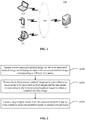
- FIG. 1 is a schematic diagram of an exemplary system architecture to which a technical solution according to an embodiment of the present disclosure may be applied.
- a system architecture 100 may include a terminal device (which may be one or more of a desktop computer 101, a tablet computer 102, and a portable computer 103 shown in FIG. 1 , or certainly may be another terminal device having a display screen, or the like), a network 104, and a server 105.
- the network 104 is configured to provide a medium of a communication link between the terminal device and the server 105.
- the network 104 may include various connection types, such as, a wired communication link, a wireless communication link, and the like.
- the quantities of the terminal device, the network, and the server in FIG. 1 are merely exemplary. There may be any quantities of terminal devices, networks, and servers according to an actual requirement.
- the server 105 may be a server cluster including multiple servers, or the like.
- a user may use a desktop computer 101 (or a tablet computer 102 or a portable computer 103) to upload a to-be-processed medical image set to a server 105
- to-be-processed medical images included in the to-be-processed medical image set may be any examination and detection images, such as computed tomography (CT) images, magnetic resonance imaging (MRI) images, or other examination and detection images of which image information changes over time.
- CT computed tomography
- MRI magnetic resonance imaging
- the server 105 After acquiring the to-be-processed medical image set, the server 105 processes the to-be-processed medical image set in a time dimension based on to the to-be-processed medical images and corresponding time points, so as to convert the original to-be-processed medical image set into a temporal dynamic image that effectively reflects the difference between a lesion region and a non-lesion region, and inputs the temporal dynamic image into a trained medical image segmentation model to extract a target region feature from the temporal dynamic image by using the medical image segmentation model, so as to acquire a target region.
- multiple to-be-processed medical image sets are processed in a time dimension, to acquire a temporal dynamic image that clearly presents a lesion region, such that doctors do not need to analyze multiple medical images acquired at different time points to determine the lesion region, thereby further improving the diagnosis efficiency of the doctors.
- the target region in the temporal dynamic image is segmented by using the medical image segmentation model, so that the segmentation accuracy of the lesion region can be improved, thereby providing support for the clinical diagnosis made by the doctors. Because the temporal dynamic image replaces the multiple medical images acquired at different time points, the workload of labeling is reduced, so that the training of the medical image segmentation model is more convenient.
- a medical image segmentation method provided in the embodiments of the present disclosure is generally performed by the server 105, and accordingly, a medical image segmentation apparatus is generally disposed in the server 105.
- the terminal device may also have functions similar to those of the server, so as to perform the medical image segmentation solution provided in the embodiments of the present disclosure.
- a computer deep learning segmentation algorithm if medical images at all time points are used for training a deep learning network model, due to patient movement at different time periods, it is difficult to accurately register lesion regions at other time points by labeling the medical images at one time point. Therefore, it is necessary to label the medical images at all time points or perform 3D registration on the medical images at all time points, which makes the training of the deep learning network model more difficult.
- a medical image segmentation method is provided according to an embodiment of the present disclosure.
- the implementation details of the technical solutions in the embodiments of the present disclosure are described in detail in the following.
- FIG. 2 is a flowchart of a medical image segmentation method according to an embodiment of the present disclosure.
- the medical image segmentation method may be performed by a server, and the server may be the server shown in FIG. 1 .
- the medical image segmentation method at least includes following steps S210 to S230, which are described in detailed as follows.
- step S210 the server acquires a to-be-processed medical image set, the to-be-processed medical image set including multiple to-be-processed medical images corresponding to different time points.
- the to-be-processed medical image set may be acquired by performing detections on samples obtained from patients or performing physical examinations on patients by hospital departments.
- the to-be-processed medical images in the to-be-processed medical image set may be CT images, MRI images, X-ray images, and other images that reflect properties of a lesion region changing over time.
- the type of the to-be-processed medical images is not limited in the present disclosure.
- the MRI images may be further magnetic resonance images obtained by performing magnetic resonance imaging on a lesion region such as a lung tumor, a stomach tumor, or a liver tumor.
- a contrast agent when the magnetic resonance imaging is performed on the breast tumor, a contrast agent is injected into an examinee, then the examinee lies prone on an examination table, with the breast placed in a special coil, and finally a nuclear magnetic resonance instrument performs an overall scan on the breast with the movement of the examination table.
- the contrast agent is a chemical injected (or taken) into human tissues or organs to enhance an image observation effect, which may be, for example, iron, manganese, and other magnetic substances. The densities of these chemicals are higher or lower than that of surrounding tissues.
- the contrast agent can change a relaxation rate of water protons in local tissues in the body, thereby improving an imaging contrast ratio and resolution ratio of a non-lesion region to a lesion region, so as to provide more information for location and diagnosis of the lesion region.
- the content of the contrast agent in the tissue changes with the flow of blood. For example, blood circulation in the non-lesion region is smooth, the content of the contrast agent decreases rapidly, and the brightness of a region corresponding to the non-lesion region in the magnetic resonance image gradually increases.
- the to-be-processed medical images at multiple time points may be acquired for the same tissue cross section based on the foregoing features of the contrast agent.
- the server may acquire one or more to-be-processed medical image sets.
- medical image segmentation may be performed on 2D images.
- each of the to-be-processed medical image sets corresponds to a cross section, and to-be-processed medical images corresponding to the same time point in the to-be-processed medical image sets form a 3D medical image, and in this case, the medical image segmentation may be performed on a 3D image.
- Medical image segmentation on the 3D image is mainly described in the embodiments of the present disclosure.
- the server acquires multiple to-be-processed medical image sets, and each of the to-be-processed medical image sets includes multiple to-be-processed medical images corresponding to different time points, that is, the to-be-processed medical image sets correspond to different cross sections, and the to-be-processed medical images in the same to-be-processed medical image set are obtained by acquiring information of the same cross section at different time points.
- the multiple to-be-processed medical image sets are obtained by scanning different cross sections of the breast by using a nuclear magnetic resonance imager during a scanning process, and the multiple to-be-processed medical images in the same to-be-processed medical image set are generated when one of the cross sections of the breast is scanned at multiple time points by using the nuclear magnetic resonance imager during the scanning process.
- time points corresponding to the multiple to-be-processed medical images may be consecutive time points, and a medical image sequence may be formed by using the multiple to-be-processed medical image sets.
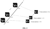
- FIG. 3 is a schematic diagram of to-be-processed medical images corresponding to multiple different time points in a to-be-processed medical image set.
- the to-be-processed medical images corresponding to the same time point in the to-be-processed medical image sets may form a three-dimensional medical image.
- to-be-processed medical images corresponding to the same time point in the to-be-processed medical image sets form a 3D MRI image.
- the multiple to-be-processed medical image sets may form a 3D MRI image sequence.
- dynamic contrast enhanced magnetic resonance imaging may be performed on the breast tumor, to obtain a 3D DCE-MRI image.
- a medical image sequence formed by the multiple to-be-processed medical image sets is a three dimensional dynamic contrast enhanced magnetic resonance imaging (3D DCE-MRI) image sequence.
- the medical image sequence may alternatively be a two-dimensional image sequence.
- Most of medical images in clinical diagnosis are 3D medical images. Therefore, description is made in the following by using a 3D DCE-MRI image sequence as an example.
- step S220 the to-be-processed medical image set is processed in a time dimension based on the to-be-processed medical images and the time points corresponding to the to-be-processed medical images, to obtain a temporal dynamic image.
- the to-be-processed medical image set may be processed in a time dimension based on the to-be-processed medical images and the time points corresponding to the to-be-processed medical images to acquire the temporal dynamic image.
- a coordinate plane corresponding to a cross section and a cross section coordinate axis may be determined according to three dimensions of the 3D medical image.
- a coordinate system corresponding to the 3D medical image may be a 3D Cartesian coordinate system.
- any coordinate axis may be selected as the cross section coordinate axis, and a coordinate plane perpendicular to the cross section coordinate axis is the coordinate plane corresponding to the cross section.
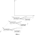
- FIG. 4 is a schematic diagram showing distribution of cross sections.
- the z-axis may be defined as the cross section coordinate axis, that is, the to-be-processed medical image sets are distributed along the z-axis
- the x-y coordinate plane is the coordinate plane corresponding to the cross section, that is, any to-be-processed medical image is an image on the x-y coordinate system.
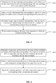
- FIG. 5 is a flowchart of acquiring a temporal dynamic image.
- the process of processing the to-be-processed medical image set in a time dimension based on the to-be-processed medical images and the time points corresponding to the to-be-processed medical images to obtain the temporal dynamic image includes the following steps S510 to S540, which are described in detail as follows.
- step S510 four-dimensional data corresponding to each of the cross sections is determined based on to-be-processed medical image data corresponding to the cross section and the time points.
- each cross section corresponds to the to-be-processed medical images at the multiple time points, and coordinates corresponding to the cross sections are different. Therefore, a first coordinate, a second coordinate, and a cross section coordinate may be first determined based on the to-be-processed medical image data corresponding to the cross section, where the first coordinate, the second coordinate, and the cross section coordinate are perpendicular to each other, and the first coordinate and the second coordinate form the coordinate plane corresponding to the cross section. Then, a time coordinate is determined based on the time points.
- the first coordinate may be specifically the x-axis
- the second coordinate may be specifically the y-axis
- the cross section coordinate may be specifically the z-axis.
- the time coordinate t may be determined according to the time points corresponding to the to-be-processed medical images.
- a four-dimensional coordinate system (x, y, z, t) may be determined based on the first coordinate, the second coordinate, the cross section coordinate, and the time coordinate.
- Four-dimensional data I(x, y, z, t) corresponding to the multiple to-be-processed medical image sets may be determined based on the four-dimensional coordinate system.
- step S520 four-dimensional data corresponding to each of the cross sections is analyzed to obtain a time component corresponding to the cross section.
- FIG. 6 is a flowchart of acquiring the time component corresponding to each of the cross sections. As shown in FIG. 6 , the process of acquiring the time component corresponding to each of the cross sections includes following step S610 to S630, which are described in detail below.
- step S610 a target cross section is determined based on the cross section coordinate, and first image data corresponding to the target cross section is acquired, the first image data including the first coordinate, the second coordinate, and the time coordinate.
- a coordinate value i on the z-axis may be determined as the cross section coordinate, and the target cross section corresponding to the cross section coordinate is acquired based on the cross section coordinate.
- the target cross section includes multiple to-be-processed medical images corresponding to different time points.
- step S620 a multi-dimensional analysis is performed on the first image data to acquire a time component corresponding to the target cross section.
- the multi-dimensional analysis may be performed on the first image data to acquire the component of the first image data in the time dimension.
- three-dimensional Clifford algebra may be used for calculating a multi-dimensional analytic signal ⁇ (x, y, t) of the first image data f(x, y, t), so as to split an original signal into components in different dimensions.
- Clifford algebra also known as geometric algebra, combines two operations of inner product and outer product, is a generalization of complex algebra, quaternion algebra, and outer algebra, and has wide application in geometry and physics.
- the equation includes eight components.
- a direction of each of the components is determined by e 1 , e 2 and e 3 , and the eight components are mutually orthogonal in the Clifford algebraic space.
- e 1 corresponds to information in an x direction in the space of the first image data f(x, y, t)
- e 2 corresponds to information in a y direction in the space of the first image data f(x, y, t)
- e 3 corresponds to information in a t direction in the space of the first image data f(x, y, t).
- the information in the t direction is concerned.
- step S630 the foregoing steps are repeated until time components respectively corresponding to the cross sections are acquired.
- steps S610 and S620 may be repeated to obtain the time components corresponding to the cross sections.
- step S530 a target time component corresponding to the multiple to-be-processed medical image sets is determined based on the time component corresponding to each of the cross sections.
- step S540 post-processing is performed on the target time component to obtain the temporal dynamic image.
- post-processing may be performed on the target time component to acquire the 3D temporal dynamic image.
- post-processing methods there are many post-processing methods. For example, an average value of the target time component I'(x, y, z, t) along the t-axis may be calculated, a maximum value of the target time component I'(x, y, z, t) along the t-axis may be calculated, or a difference between the maximum value and a minimum value of the target time component I'(x, y, z, t) along the t-axis may be calculated.
- the post-processing may alternatively be performed on the target time component I'(x, y, z, t) in other manners. This is not described in detail in the present disclosure.
- calculating the average value of the target time component I'(x, y, z, t) along the t-axis includes: for each of the cross sections, respectively determining sub-time components corresponding to the cross section at the time points based on the target time component, the number of sub-time components being the same as the number of time points; averaging the sub-time components corresponding to the cross section to acquire a target average value; and constructing the temporal dynamic image based on the target average value.
- calculating the maximum value of the target time component I'(x, y, z, t) along the t-axis includes: for each of the cross sections, respectively determining sub-time components corresponding the cross section at the time points based on the target time component, the number of the sub-time components being the same as the number of the time points; acquiring a maximum sub-time component among the sub-time components corresponding to the cross section; and constructing the temporal dynamic image based on the maximum sub-time component.
- t t 1 , t 2 , ... , t n
- calculating the difference between the maximum value and a minimum value of the target time component I'(x, y, z, t) along the t-axis includes: for each of the cross sections, respectively determining sub-time components corresponding to the cross sections at the time points based on the target time component, the number of the sub-time components being the same as the number of the time points; acquiring a maximum sub-time components and a minimum sub-time components among the sub-time components corresponding to the cross section, and subtracting the minimum sub-time component from the maximum minimum sub-time component to obtain a target difference; and constructing the temporal dynamic image based on the target difference.
- t t 1 , t 2 , ... , t n ⁇ Min I ′ x y z t
- t t 1 , t 2 , ... , t n
- the three dimensional temporal dynamic image I t (x, y, z) may be obtained based on the calculation result. That is, a change of pixel brightness of multiple 3D DCE-MRI images at different times is obtained.
- the difference between the maximum value and the minimum value of the sub-time component reflects the difference between the maximum value and the minimum value obtained at different time points of a 3D DCE-MRI image sequence at the same spatial position, and the difference can reflect a brightness change of points in the space to the greatest extent. Therefore, in an embodiment of the present disclosure, by using the target difference as a reference, the temporal dynamic image may be constructed based on the target differences, so as to improve the efficiency of medical image segmentation.
- a target region feature is extracted from the temporal dynamic image by using a medical image segmentation model, to acquire a target region.
- the temporal dynamic image may be inputted into a medical image segmentation model, to extract the target region feature from the temporal dynamic image by using the medical image segmentation model, to acquire the target region.
- a medical image segmentation model may be a trained deep learning segmentation module.
- the medical image segmentation model may be a deep learning segmentation module for processing a 3D image, such as a 3D Unet model, a 3D vnet model, a fully convolutional neural network model, and the like.
- the type of the deep learning segmentation module is not limited in the embodiments of the present disclosure.
- the target region is a lesion region such as a tumor region, or a calcification region. Medical workers may take the target region as a region of interest, and further analyze the region of interest, so as to make the optimal regimen.
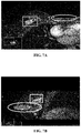
- FIG. 7A to 7C show DCE-MRI images at a certain time point after a contrast agent is injected in the related art.
- FIG. 7A is a cross section image of a breast, and the cross section is a longitudinal section that divides a body into upper and lower parts
- FIG. 7B is a sagittal plane image of the breast, and the sagittal plane is a longitudinal section that divides the body into left and right parts
- FIG. 7C is a coronal plane image of the breast, and the coronal plane is a longitudinal section that divides the body into front and rear parts.
- Rectangular frames in FIG. 7A to 7C are lesion regions, and elliptical frames are non-lesion regions. It can be seen from FIG. 7A to 7C that both the lesion regions and the non-lesion regions are presented as bright pixels, and a medical image segmentation model cannot distinguish the lesion regions from the non-lesion regions, and thus cannot accurately segment the lesion regions.
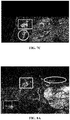
- FIG. 8A to 8C show 3D temporal dynamic images after a contrast agent is injected.
- FIG. 8A is a cross section image of a breast
- FIG. 8B is a sagittal plane image of the breast
- FIG. 8C is a coronal plane image of the breast.
- Rectangular frames in FIG. 8A to 8C are lesion regions, and elliptical frames are non-lesion regions.
- the pixel brightness of the lesion regions in FIG. 8A to 8C is higher, and the pixel brightness of the non-lesion regions is lower. Therefore, the contrast between the pixels of the lesion regions and the non-lesion regions is more obvious, and a medical image segmentation model can quickly distinguish the lesion regions from the non-lesion regions, and can further accurately segment the lesion regions.
- a to-be-trained medical image segmentation model may further be trained, so as to acquire a medical image segmentation model for subsequent image segmentation on the to-be-processed medical image.
- FIG. 9 is a flowchart of training a to-be-trained medical image segmentation model. As shown in FIG. 9 , a process of training the to-be-trained medical image segmentation model includes following steps S910 to S920, that are described in detail below.
- step S910 a temporal dynamic image sample and a labeled target region sample corresponding to the temporal dynamic image sample are obtained.
- a method of acquiring the temporal dynamic image sample is the same as that of acquiring the temporal dynamic image in the foregoing embodiments. Details are not repeated here.
- a target region (a lesion region) in the temporal dynamic image may be labeled manually, so as to obtain the labeled target region sample corresponding to the temporal dynamic image sample.
- multiple samples may be used for training the to-be-trained medical image segmentation model to acquire optimal parameters of the to-be-trained medical image segmentation model.
- 3D DCE-MRI image data of 244 malignant tumor patients may be selected.
- Data of 221 cases among the 244 cases is used as training data to train the to-be-trained medical image segmentation model, and data of 23 cases among the 244 cases is used as test data to test the trained medical image segmentation model to determine whether the model reaches a stable state.
- quantities of training data and test data in the present disclosure include but are not limited to the foregoing examples, and are not limited in the present disclosure.
- step S920 the to-be-trained medical image segmentation model are trained by using the temporal dynamic image sample and the labeled target region sample, to obtain the medical image segmentation model.
- the temporal dynamic image sample may be inputted into the to-be-trained medical image segmentation model to acquire the target region extracted by using the to-be-trained medical image segmentation model; and the extracted target region is compared with the labeled target region sample corresponding to the inputted dynamic image sample to determine the segmentation accuracy of the to-be-trained medical image segmentation model.
- the to-be-trained medical image segmentation model reaches a stable state and may be used as a medical image segmentation model for subsequent medical image segmentation; and if the segmentation accuracy does not reach the preset threshold, the parameters of the to-be-processed medical image segmentation model continues to be adjusted until the segmentation accuracy of the outputted segmented image reaches or exceeds the preset threshold.
- the preset threshold may be set according to actual needs, for example, may set to 95%. After the training is completed, the trained medical image segmentation model may be tested through test data to determine whether the model is widely applicable to any temporal dynamic image.
- the segmentation accuracy of the lesion region segmented according to the technical solutions of the embodiments of the present disclosure is greatly improved compared with that of a lesion region obtained by segmenting a to-be-processed medical image by using a medical image segmentation model that is trained by using original DCE-MRI data.
- Table 1 shows experimental results of using the original DCE-MRI data and the 3D temporal dynamic image as follows: Table 1 Experimental result of using the original DCE-MRI data Experimental result of using the 3D temporal dynamic image Segmentation precision: mean value+/-variance 68.8+/-19.8% 79.3%+/-8.5%
- the segmentation accuracy of using the 3D temporal dynamic image in the embodiments of the present disclosure is 10% on average higher than that of using the original DCE-MRI data. That is, the technical solutions in the embodiments of the present disclosure can effectively improve diagnosis efficiency of doctors and the segmentation accuracy of medical images.
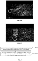
- FIG. 10A to 10C show schematic diagrams of interfaces of performing tumor segmentation on a background-enhancement-type medical image.
- FIG. 10A shows a labeling result of a background-enhancement-type medical image
- FIG. 10B shows a segmentation result of a background-enhancement-type medical image by using a 3D temporal dynamic image, and the segmentation accuracy of this method reaches 87%
- FIG. 10A shows a labeling result of a background-enhancement-type medical image
- FIG. 10B shows a segmentation result of a background-enhancement-type medical image by using a 3D temporal dynamic image, and the segmentation accuracy of this method reaches 87%
- 10C shows a segmentation result of a background-enhancement-type medical image by using original DCE-MRI data, and the segmentation accuracy of this method is 69%.
- the technical solutions in the embodiments of the present disclosure can improve the precision of medical image segmentation, can be used for segmenting various types of medical images, and have wide application.
- a group of 3D data (a temporal dynamic image) can be extracted from multiple groups of 3D DCE-MRI data by using the technical solutions in the foregoing embodiments of the present disclosure, to be directly used for labeling a target region and training a medical image segmentation model, so that the training of the medical image segmentation model is more convenient, and a case that doctors need to choose 3D DCE-MRI images at which time point to read first when performing MRI image diagnosis can be avoided. Instead, doctors may first view 3D images acquired by using the technical solutions in the embodiments of the present disclosure to observe a lesion region, and then select 3D DCE-MRI images at certain time points, thereby improving diagnosis efficiency.
- FIG. 11 shows a flowchart of an image segmentation method as follows.
- a to-be-processed image set is acquired, the to-be-processed image set including multiple to-be-processed images corresponding to different time points.
- the to-be-processed image set is processed in a time dimension based on the to-be-processed images and the time points corresponding to the to-be-processed images, to obtain a temporal dynamic image.
- a target region feature is extracted from the temporal dynamic image by using an image segmentation model, to acquire a target region.
- the image segmentation method is similar to the medical image segmentation method in the foregoing embodiments.
- the method can not only segment medical images, but also can be used for segmenting any other types of images, for example, segmenting a sample image in a biological experiment, segmenting an image in a metal processing process, and segmenting a damage location in a pipeline.
- the image segmentation method in the embodiments of the present disclosure can be used for segmentation.
- the image segmentation method may be implemented by the implementations of the medical image segmentation method in the embodiments of the present disclosure. Therefore, details are not repeated in the present disclosure.
- to-be-processed images corresponding to the same time point in the multiple to-be-processed image sets can form a three-dimensional image
- multiple to-be-processed images corresponding to different time points may form a three-dimensional image sequence.
- the three-dimensional image sequence may be identified and segmented to acquire a target region therein.
- apparatus embodiments of the present disclosure and the apparatus embodiments may be used for performing the medical image segmentation method in the foregoing embodiment of the present disclosure.
- the apparatus embodiments of the present disclosure For details not disclosed in the apparatus embodiments of the present disclosure, refer to the foregoing embodiment of the medical image segmentation method of the present disclosure.
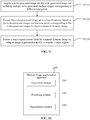
- FIG. 12 shows a schematic block diagram of a medical image segmentation apparatus according to the present disclosure.
- a medical image segmentation apparatus 1200 includes an acquisition module 1201, a processing module 1202, and a segmentation module 1203.
- the acquisition module 1201 is configured to acquire a to-be-processed medical image set, the to-be-processed medical image set including multiple to-be-processed medical images corresponding to different time points.
- the processing module 1202 is configured to process the to-be-processed medical image set in a time dimension based on the to-be-processed medical images and the time points corresponding to the to-be-processed medical images to obtain a temporal dynamic image.
- the segmentation module 1203 is configured to extract a target region feature from the temporal dynamic image by using a medical image segmentation model, to acquire a target region.
- the medical image segmentation apparatus 1200 includes: a cross section determining module 1204, configured to determine a coordinate plane corresponding to each of the cross sections and a cross section coordinate axis according to three dimensions of the 3D medical image, the cross section coordinate axis being perpendicular to the coordinate plane.
- the processing module 1202 includes: a four-dimensional data acquisition unit, configured to determine four-dimensional data corresponding to each of the cross sections based on to-be-processed medical image data corresponding to the cross section and the time points; a first time component acquisition unit, configured to analyze the four-dimensional data corresponding to each of the cross sections to obtain a time component corresponding to the cross section; a second time component acquisition unit, configured to determine a target time component corresponding to the multiple to-be-processed medical image sets based on the time component corresponding to each of the cross sections; and a post-processing unit, configured to perform post-processing on the target time component to obtain the temporal dynamic image.
- the four-dimensional data acquisition unit includes: a spatial coordinate determining unit, configured to determine a first coordinate, a second coordinate, and a cross section coordinate based on the to-be-processed medical image data corresponding to the cross section, the first coordinate, the second coordinate, and the cross section coordinate being perpendicular to each other; a time coordinate determining unit, configured to determine a time coordinate based on the time points; and a four-dimensional data determining unit, configured to construct four-dimensional coordinate axes based on the first coordinate, the second coordinate, the cross section coordinate, and the time coordinate, and determine the four-dimensional data based on the four-dimensional coordinate axes.
- the first time component acquisition unit includes: a first image data acquisition unit, configured to determine a target cross section based on the cross section coordinate, and acquire first image data corresponding to the target cross section, the first image data including the first coordinate, the second coordinate, and the time coordinate; and a multi-dimensional analyzing unit, configured to perform a multi-dimensional analysis on the first image data to acquire a time component corresponding to the target cross section; repeating the foregoing operations until time component respectively corresponding to the cross sections are acquired.
- a first image data acquisition unit configured to determine a target cross section based on the cross section coordinate, and acquire first image data corresponding to the target cross section, the first image data including the first coordinate, the second coordinate, and the time coordinate
- a multi-dimensional analyzing unit configured to perform a multi-dimensional analysis on the first image data to acquire a time component corresponding to the target cross section; repeating the foregoing operations until time component respectively corresponding to the cross sections are acquired.
- the second time component acquisition unit is configured to: determine the target time component based on the time component corresponding to each of the cross sections, and the first coordinate, the second coordinate, and the cross section coordinate that correspond to each of the cross sections.
- the multi-dimensional analyzing unit is configured to: perform the multi-dimensional analysis on the first image data by using three-dimensional Clifford algebra, to acquire the time component corresponding to the target cross section.
- the post-processing unit is configured to: for each of the cross sections, respectively determine sub-time components corresponding to the cross section at the time points based on the target time component, the number of the sub-time components being the same as the number of the time points; average the sub-time components corresponding to the cross section to acquire a target average value; and construct the temporal dynamic image based on the target average value.
- the post-processing unit is configured to: for each of the cross sections, respectively determine sub-time components corresponding to the cross section at the time points based on the target time component, the number of the sub-time components being the same as the number of the time points; and acquire a maximum sub-time component among the sub-time components corresponding the cross section; and construct the temporal dynamic image based on the maximum sub-time component.
- the post-processing unit is configured to: for each of the cross sections, respectively determine sub-time components corresponding to the cross section at the time points based on the target time component, the number of the sub-time components being the same as the number of the time points; and acquire a maximum sub-time component and a minimum sub-time component among the sub-time components corresponding to the cross section, and subtracting the minimum sub-time component from the maximum sub-time component to acquire a target difference; and construct the temporal dynamic image based on the target differences.
- the medical image segmentation apparatus 1200 further includes: a sample acquisition module 1205 and a training module 1206.
- the sample acquisition module 1205 is configured to acquire a temporal dynamic image sample and a labeled target region sample corresponding to the temporal dynamic sample; and the training module 1206 is configured to train a to-be-trained medical image segmentation model by using the temporal dynamic image sample and the labeled target region sample, to obtain the medical image segmentation model.
- the 3D medical image is a 3D DCE-MRI image.
- FIG. 13 shows a medical image segmentation system.
- the medical image segmentation system 1300 includes a detection device 1301 and an electronic device 1302.
- the detection device 1301 is configured to scan and detect a detection object to acquire a to-be-processed medical image set, the to-be-processed medical image set including multiple to-be-processed medical images corresponding to different time points; and the electronic device 1302 is connected to the detection device, and includes a storage apparatus and a processor, the storage apparatus being configured to store one or more programs, and the one or more programs, when executed by the processor, causing the processor to implement the foregoing medical image segmentation method.
- the detection device 1301 may be a scanning apparatus for acquiring scanned images in a CT device, and the scanned apparatus includes a ray emitting source, a detector, and a scanning frame, may be a scanning apparatus for acquiring scanned images in a nuclear magnetic resonance imaging device, and the scanning apparatus includes a magnet part, a magnetic resonance spectroscopy part, and a scanning table, or may be a scanning apparatus for acquiring scanned images in a fluoroscopy device, and the scanning device includes a ray emitting source and a detector.
- the scanning apparatus may alternatively be another detection device, as long as the device may be used for scanning the detection object to acquire a scanned image. This is not specifically limited in the present disclosure.
- the to-be-processed medical image sets may be transmitted to a storage apparatus 1302a and/or a processor 1302b in the electronic device 1302, and the storage apparatus 1302a further stores one or more programs for the processor 1302b to execute.
- the processor 1302b may execute one or more programs stored in the storage apparatus 1302a on the to-be-processed medical image set, that is, the processor 1302b can perform image segmentation on the to-be-processed medical image set according to the technical solutions in the embodiments of the present disclosure to acquire the target region.
- the processor 1302b may alternatively transmit an image including the target region to a display device (not shown) connected to the electronic device 1302 for display, so that doctors may observe and determine a focus and make a regimen.
- FIG. 14 is a schematic structural diagram of a computer system of an electronic device 1302 adapted to implement embodiments of the present disclosure.
- the computer system 1400 of the electronic device shown in FIG. 14 is merely an example, and does not constitute any limitation on functions and use ranges of the embodiments of the present disclosure.
- the computer system 1400 includes a central processing unit (CPU) 1401, which may perform various proper actions and processing based on a program stored in a read-only memory (ROM) 1402 or a program loaded from a storage part 1408 into a random access memory (RAM) 1403.
- the RAM 1403 further stores various programs and data required for system operations.
- the CPU 1401, the ROM 1402, and the RAM 1403 are connected to each other through a bus 1404.
- An input/output (I/O) interface 1405 is also connected to the bus 1404.
- the following components are connected to the I/O interface 1405: an input part 1406 including a keyboard, a mouse, or the like, an output part 1407 including a cathode ray tube (CRT), a liquid crystal display (LCD), a speaker, or the like, a storage part 1408 including a hard disk, or the like, and a communication part 1409 including a network interface card such as a local area network (LAN) card or a modem.
- the communication part 1409 performs communication processing through a network such as the Internet.
- a driver 1410 is also connected to the I/O interface 1405 as required.
- a removable medium 1411 such as a magnetic disk, an optical disc, a magneto-optical disk, or a semiconductor memory is installed on the drive 1410 as required, so that a computer program read from the removable medium 1411 is installed into the storage part 1408 as required.
- this embodiment of the present disclosure includes a computer program product, the computer program product includes a computer program carried on a computer-readable medium, and the computer program includes program code used for performing the methods shown in the flowcharts.
- the computer program may be downloaded and installed through the communication part 1409 from a network, and/or installed from the removable medium 1411.
- CPU central processing unit
- the computer-readable medium shown in the embodiments of the present disclosure may be a computer-readable signal medium or a computer-readable storage medium or any combination of the two.
- the computer-readable storage medium may be, for example, but is not limited to, an electrical, magnetic, optical, electromagnetic, infrared, or semiconductor system, apparatus, or device, or any combination thereof.
- a more specific example of the computer-readable storage medium may include but is not limited to: an electrical connection having one or more wires, a portable computer magnetic disk, a hard disk, a random access memory (RAM), a read-only memory (ROM), an erasable programmable read-only memory (EPROM), a flash memory, an optical fiber, a compact disk read-only memory (CD-ROM), an optical storage device, a magnetic storage device, or any appropriate combination thereof.
- the computer-readable storage medium may be any tangible medium containing or storing a program, and the program may be used by or used in combination with an instruction execution system, an apparatus, or a device.
- a computer-readable signal medium may include a data signal being in a baseband or propagated as a part of a carrier wave, the data signal carrying computer-readable program code. Such a propagated data signal may be in multiple forms, including but not limited to an electromagnetic signal, an optical signal, or any suitable combination thereof.
- the computer-readable signal medium may be further any computer-readable medium in addition to a computer-readable storage medium.
- the computer-readable medium may send, propagate, or transmit a program that is used by or used in conjunction with an instruction execution system, an apparatus, or a device.
- the program code included in the computer-readable medium may be transmitted by using any suitable medium, including but not limited to: a wireless medium, a wire, or the like, or any suitable combination thereof.
- each box in a flowchart or a block diagram may represent a module, a program segment, or a part of code.
- the module, the program segment, or the part of code includes one or more executable instructions used for implementing designated logic functions.
- functions annotated in boxes may alternatively occur in a sequence different from that annotated in an accompanying drawing. For example, actually two boxes shown in succession may be performed basically in parallel, and sometimes the two boxes may be performed in a reverse sequence. This is determined by a related function.
- Each box in a block diagram or a flowchart and a combination of boxes in the block diagram or the flowchart may be implemented by using a dedicated hardware-based system configured to perform a designated function or operation, or may be implemented by using a combination of dedicated hardware and a computer instruction.
- a related unit described in the embodiments of the present disclosure may be implemented in a software manner, or may be implemented in a hardware manner, and the unit described can also be set in a processor. Names of these units do not constitute a limitation on the units in a case.
- the present disclosure further provides a computer-readable medium.
- the computer-readable medium may be included in the electronic device described in the foregoing embodiments, or may exist alone and is not disposed in the electronic device.
- the computer-readable medium carries one or more programs, the one or more programs, when executed by the electronic device, causing the electronic device to implement the method described in the foregoing embodiments.
- the software product may be stored in a non-volatile storage medium (which may be a CD-ROM, a USB flash drive, a removable hard disk, or the like) or on the network, including several instructions for instructing a computing device (which may be a personal computer, a server, a touch terminal, a network device, or the like) to perform the methods according to the embodiments of the present disclosure.
- a non-volatile storage medium which may be a CD-ROM, a USB flash drive, a removable hard disk, or the like
- a computing device which may be a personal computer, a server, a touch terminal, a network device, or the like
Landscapes
- Engineering & Computer Science (AREA)
- Theoretical Computer Science (AREA)
- Physics & Mathematics (AREA)
- General Physics & Mathematics (AREA)
- Computer Vision & Pattern Recognition (AREA)
- Software Systems (AREA)
- Multimedia (AREA)
- Evolutionary Computation (AREA)
- Data Mining & Analysis (AREA)
- Artificial Intelligence (AREA)
- Medical Informatics (AREA)
- General Health & Medical Sciences (AREA)
- Health & Medical Sciences (AREA)
- General Engineering & Computer Science (AREA)
- Life Sciences & Earth Sciences (AREA)
- Evolutionary Biology (AREA)
- Bioinformatics & Computational Biology (AREA)
- Nuclear Medicine, Radiotherapy & Molecular Imaging (AREA)
- Radiology & Medical Imaging (AREA)
- Quality & Reliability (AREA)
- Bioinformatics & Cheminformatics (AREA)
- Computing Systems (AREA)
- Databases & Information Systems (AREA)
- Magnetic Resonance Imaging Apparatus (AREA)
- Apparatus For Radiation Diagnosis (AREA)
Applications Claiming Priority (2)
| Application Number | Priority Date | Filing Date | Title |
|---|---|---|---|
| CN201910116353.0A CN109872312B (zh) | 2019-02-15 | 2019-02-15 | 医学图像分割方法、装置、系统及图像分割方法 |
| PCT/CN2020/074712 WO2020164468A1 (zh) | 2019-02-15 | 2020-02-11 | 医学图像分割方法、图像分割方法及相关装置、系统 |
Publications (4)
| Publication Number | Publication Date |
|---|---|
| EP3926537A1 true EP3926537A1 (de) | 2021-12-22 |
| EP3926537A4 EP3926537A4 (de) | 2022-04-06 |
| EP3926537C0 EP3926537C0 (de) | 2025-01-01 |
| EP3926537B1 EP3926537B1 (de) | 2025-01-01 |
Family
ID=66918681
Family Applications (1)
| Application Number | Title | Priority Date | Filing Date |
|---|---|---|---|
| EP20755319.9A Active EP3926537B1 (de) | 2019-02-15 | 2020-02-11 | Verfahren zur segmentierung eines medizinischen bildes, verfahren zur segmentierung eines bildes, zugehörige vorrichtung und system |
Country Status (5)
| Country | Link |
|---|---|
| US (2) | US11954864B2 (de) |
| EP (1) | EP3926537B1 (de) |
| CN (2) | CN110490851B (de) |
| TW (1) | TWI750583B (de) |
| WO (1) | WO2020164468A1 (de) |
Families Citing this family (14)
| Publication number | Priority date | Publication date | Assignee | Title |
|---|---|---|---|---|
| CN110490851B (zh) | 2019-02-15 | 2021-05-11 | 腾讯科技(深圳)有限公司 | 基于人工智能的乳腺图像分割方法、装置及系统 |
| CN110445954B (zh) * | 2019-07-26 | 2022-04-26 | 腾讯医疗健康(深圳)有限公司 | 图像采集方法、装置及电子设备 |
| EP4107964A1 (de) * | 2020-02-20 | 2022-12-28 | Align Technology, Inc. | Medizinische bilddatenkompression und -extraktion auf client-seite |
| CN111429474B (zh) * | 2020-02-27 | 2023-04-07 | 西北大学 | 基于混合卷积的乳腺dce-mri图像病灶分割模型建立及分割方法 |
| CN112154483B (zh) * | 2020-07-15 | 2025-04-04 | 北京肿瘤医院(北京大学肿瘤医院) | 一种利用光学体表运动信号合成实时图像的方法及系统 |
| CN112365959B (zh) * | 2020-12-07 | 2024-05-28 | 推想医疗科技股份有限公司 | 修改三维图像的标注的方法及装置 |
| CN113160253B (zh) * | 2020-12-29 | 2024-01-30 | 南通大学 | 基于稀疏标记的三维医学图像分割方法及存储介质 |
| US11580646B2 (en) * | 2021-03-26 | 2023-02-14 | Nanjing University Of Posts And Telecommunications | Medical image segmentation method based on U-Net |
| CN115482180A (zh) * | 2021-05-31 | 2022-12-16 | 通用电气精准医疗有限责任公司 | 目标区域确定方法和医学成像系统 |
| CN113838020B (zh) * | 2021-09-17 | 2024-06-18 | 仰和华健数字医疗科技(上海)有限公司 | 一种基于钼靶影像的病变区域量化方法 |
| CN114332132A (zh) * | 2021-12-31 | 2022-04-12 | 联影智能医疗科技(成都)有限公司 | 图像分割方法、装置和计算机设备 |
| CN114937147B (zh) * | 2022-05-25 | 2025-08-29 | 浙江大学 | 针对肝脏ct影像的分段智能识别模型及识别方法 |
| CN116543001B (zh) * | 2023-05-26 | 2024-01-12 | 广州工程技术职业学院 | 彩色图像边缘检测方法及装置、设备、存储介质 |
| CN118039088B (zh) * | 2024-04-15 | 2024-06-07 | 达州爱迦飞诗特科技有限公司 | 一种人工智能辅助诊断数据处理系统 |
Family Cites Families (36)
| Publication number | Priority date | Publication date | Assignee | Title |
|---|---|---|---|---|
| US6961454B2 (en) * | 2001-10-04 | 2005-11-01 | Siemens Corporation Research, Inc. | System and method for segmenting the left ventricle in a cardiac MR image |
| US8131043B2 (en) * | 2005-09-16 | 2012-03-06 | The Ohio State University | Method and apparatus for detecting interventricular dyssynchrony |
| EP1780651A1 (de) * | 2005-10-25 | 2007-05-02 | Bracco Imaging, S.P.A. | Verfahren und System zur automatischen Verarbeitung und Bewertung von Bildern, im Besonderen diagnostischer Bilder |
| CN101334895B (zh) * | 2008-08-07 | 2011-09-14 | 清华大学 | 一种针对动态增强乳腺磁共振影像序列的影像分割方法 |
| US20100150418A1 (en) * | 2008-12-15 | 2010-06-17 | Fujifilm Corporation | Image processing method, image processing apparatus, and image processing program |
| EP2336979B1 (de) * | 2009-11-05 | 2014-03-12 | TomTec Imaging Systems GmbH | Verfahren und Vorrichtung zur Segmentierung von medizinischen Bilddaten |
| US20110200227A1 (en) * | 2010-02-17 | 2011-08-18 | Siemens Medical Solutions Usa, Inc. | Analysis of data from multiple time-points |
| WO2012052887A1 (en) * | 2010-10-19 | 2012-04-26 | Koninklijke Philips Electronics N.V. | Medical image system |
| CN103236058B (zh) * | 2013-04-25 | 2016-04-13 | 内蒙古科技大学 | 获取四维心脏图像感兴趣体积的方法 |
| CN104143035B (zh) * | 2013-05-10 | 2016-01-20 | 上海联影医疗科技有限公司 | 一种分割乳腺病灶的方法 |
| CN103426169B (zh) * | 2013-07-26 | 2016-12-28 | 西安华海盈泰医疗信息技术有限公司 | 一种医学图像的分割方法 |
| US9652871B2 (en) * | 2015-01-28 | 2017-05-16 | Impac Medical Systems, Inc. | Three dimensional localization of a moving target for adaptive radiation therapy |
| CN104809723B (zh) * | 2015-04-13 | 2018-01-19 | 北京工业大学 | 基于超体素和图割算法的三维肝脏ct图像自动分割方法 |
| WO2017039663A1 (en) * | 2015-09-03 | 2017-03-09 | Siemens Healthcare Gmbh | Multi-view, multi-source registration of moving anatomies and devices |
| KR101718868B1 (ko) * | 2015-09-21 | 2017-03-22 | 한국과학기술연구원 | 자동 의료영상 분할에 의한 3차원 악안면 모델 형성 방법, 이를 수행하는 자동 영상 분할과 모델 형성 서버 및 이를 저장하는 기록매체 |
| CN109069859B (zh) * | 2016-02-02 | 2021-04-27 | 医科达有限公司 | 放射治疗系统和确定解剖区域的精确运动的成像方法 |
| CN106056610A (zh) * | 2016-06-02 | 2016-10-26 | 南方医科大学 | 基于图割的肺4d‑ct多相位肿瘤联合分割方法 |
| CN106228601B (zh) * | 2016-07-21 | 2019-08-06 | 山东大学 | 基于小波变换的多尺度锥束ct图像快速三维重建方法 |
| US11039757B2 (en) * | 2016-11-22 | 2021-06-22 | Cedars-Sinai Medical Center | Method and system for cardiac motion corrected MR exam using deformable registration |
| CN106600621B (zh) * | 2016-12-08 | 2019-07-19 | 温州医科大学 | 基于婴幼儿脑瘤多模态mri图的时空协同分割方法 |
| TWI756365B (zh) | 2017-02-15 | 2022-03-01 | 美商脫其泰有限責任公司 | 圖像分析系統及相關方法 |
| CN108509830B (zh) * | 2017-02-28 | 2020-12-01 | 华为技术有限公司 | 一种视频数据处理方法及设备 |
| EP3410393A1 (de) * | 2017-06-01 | 2018-12-05 | Siemens Healthcare GmbH | Vergleich von medizinischen bildern |
| TW201903708A (zh) | 2017-06-06 | 2019-01-16 | 國立陽明大學 | 數位減影血管攝影圖像的分析方法與系統 |
| US10219768B2 (en) * | 2017-06-08 | 2019-03-05 | Emass Llc | Method for standardizing target lesion selection and tracking on medical images |
| WO2019023900A1 (zh) * | 2017-07-31 | 2019-02-07 | 深圳联影医疗科技有限公司 | 在体数据中提取感兴趣区域的方法及系统 |
| CN107808377B (zh) * | 2017-10-31 | 2019-02-12 | 北京青燕祥云科技有限公司 | 一种肺叶中病灶的定位装置 |
| EP3714467A4 (de) * | 2017-11-22 | 2021-09-15 | Arterys Inc. | Inhaltsbasiertes wiederauffinden von bildern für die läsionsanalyse |
| CN108038848B (zh) | 2017-12-07 | 2020-08-11 | 上海交通大学 | 基于医学影像序列斑块稳定性指标的快速计算方法及系统 |
| CN108109170B (zh) * | 2017-12-18 | 2022-11-08 | 上海联影医疗科技股份有限公司 | 医学图像扫描方法及医学影像设备 |
| CN108537803B (zh) * | 2018-03-30 | 2019-08-23 | 北京灵医灵科技有限公司 | 一种ct图像交互分割方法及装置 |
| WO2019238804A1 (en) * | 2018-06-13 | 2019-12-19 | Siemens Healthcare Gmbh | Localization and classification of abnormalities in medical images |
| US11446008B2 (en) * | 2018-08-17 | 2022-09-20 | Tokitae Llc | Automated ultrasound video interpretation of a body part with one or more convolutional neural networks |
| CN109242863B (zh) * | 2018-09-14 | 2021-10-26 | 北京市商汤科技开发有限公司 | 一种缺血性脑卒中图像区域分割方法及装置 |
| CN109215764B (zh) * | 2018-09-21 | 2021-05-04 | 苏州瑞派宁科技有限公司 | 一种医学图像四维可视化的方法及装置 |
| CN110490851B (zh) * | 2019-02-15 | 2021-05-11 | 腾讯科技(深圳)有限公司 | 基于人工智能的乳腺图像分割方法、装置及系统 |
-
2019
- 2019-02-15 CN CN201910741376.0A patent/CN110490851B/zh active Active
- 2019-02-15 CN CN201910116353.0A patent/CN109872312B/zh active Active
-
2020
- 2020-02-10 TW TW109104094A patent/TWI750583B/zh active
- 2020-02-11 WO PCT/CN2020/074712 patent/WO2020164468A1/zh not_active Ceased
- 2020-02-11 EP EP20755319.9A patent/EP3926537B1/de active Active
-
2021
- 2021-04-23 US US17/239,532 patent/US11954864B2/en active Active
-
2024
- 2024-02-09 US US18/437,357 patent/US12272072B2/en active Active
Also Published As
| Publication number | Publication date |
|---|---|
| EP3926537C0 (de) | 2025-01-01 |
| CN110490851B (zh) | 2021-05-11 |
| US20210264613A1 (en) | 2021-08-26 |
| CN109872312B (zh) | 2022-12-20 |
| CN109872312A (zh) | 2019-06-11 |
| US12272072B2 (en) | 2025-04-08 |
| CN110490851A (zh) | 2019-11-22 |
| US11954864B2 (en) | 2024-04-09 |
| TWI750583B (zh) | 2021-12-21 |
| WO2020164468A1 (zh) | 2020-08-20 |
| EP3926537A4 (de) | 2022-04-06 |
| US20240212165A1 (en) | 2024-06-27 |
| EP3926537B1 (de) | 2025-01-01 |
| TW202032577A (zh) | 2020-09-01 |
Similar Documents
| Publication | Publication Date | Title |
|---|---|---|
| EP3926537B1 (de) | Verfahren zur segmentierung eines medizinischen bildes, verfahren zur segmentierung eines bildes, zugehörige vorrichtung und system | |
| CN111080584B (zh) | 医学图像的质控方法、计算机设备和可读存储介质 | |
| US9471987B2 (en) | Automatic planning for medical imaging | |
| CN117291830A (zh) | 使用自关注深度学习进行图像增强的系统和方法 | |
| Heydarheydari et al. | Auto-segmentation of head and neck tumors in positron emission tomography images using non-local means and morphological frameworks | |
| CN106491151B (zh) | Pet图像获取方法及系统 | |
| US20080292164A1 (en) | System and method for coregistration and analysis of non-concurrent diffuse optical and magnetic resonance breast images | |
| US20140003690A1 (en) | Motion correction apparatus and method | |
| CN107798682A (zh) | 图像分割系统、方法、装置和计算机可读存储介质 | |
| US8655040B2 (en) | Integrated image registration and motion estimation for medical imaging applications | |
| CN111445550B (zh) | Pet图像的迭代重建方法、装置和计算机可读存储介质 | |
| US10460508B2 (en) | Visualization with anatomical intelligence | |
| US20100284598A1 (en) | Image registration alignment metric | |
| US20140225926A1 (en) | Method and system for generating an image as a combination of two existing images | |
| CN113989231A (zh) | 动力学参数的确定方法、装置、计算机设备和存储介质 | |
| WO2021225816A1 (en) | Apparatus for monitoring treatment side effects | |
| EP4231234A1 (de) | Tiefenlernen zur registrierung eines anatomischen zu funktionellen bildern | |
| Jaspers et al. | Deep learning synthesis of white-blood from dark-blood late gadolinium enhancement cardiac magnetic resonance | |
| CN118115409A (zh) | 基于深度学习模拟对比增强乳腺癌mri的方法 | |
| HK40009258A (en) | Method, apparatus, and system for segmenting medical image, and method for segmenting image | |
| CN114299189A (zh) | 一种图像处理方法和系统 | |
| HK40009258B (en) | Method, apparatus, and system for segmenting medical image, and method for segmenting image | |
| Wang | Artificial intelligence applications in oncology to augment data and support decisions | |
| CN111035403A (zh) | 一种扫描时机确定方法、装置、设备及存储介质 | |
| Li et al. | Computed tomography ring artifact correction method with super-pixel segmentation and adaptive relative total variation |
Legal Events
| Date | Code | Title | Description |
|---|---|---|---|
| STAA | Information on the status of an ep patent application or granted ep patent |
Free format text: STATUS: THE INTERNATIONAL PUBLICATION HAS BEEN MADE |
|
| PUAI | Public reference made under article 153(3) epc to a published international application that has entered the european phase |
Free format text: ORIGINAL CODE: 0009012 |
|
| STAA | Information on the status of an ep patent application or granted ep patent |
Free format text: STATUS: REQUEST FOR EXAMINATION WAS MADE |
|
| 17P | Request for examination filed |
Effective date: 20210416 |
|
| AK | Designated contracting states |
Kind code of ref document: A1 Designated state(s): AL AT BE BG CH CY CZ DE DK EE ES FI FR GB GR HR HU IE IS IT LI LT LU LV MC MK MT NL NO PL PT RO RS SE SI SK SM TR |
|
| A4 | Supplementary search report drawn up and despatched |
Effective date: 20220307 |
|
| RIC1 | Information provided on ipc code assigned before grant |
Ipc: G06K 9/62 20060101ALI20220301BHEP Ipc: G06T 7/149 20170101ALI20220301BHEP Ipc: G06T 7/11 20170101AFI20220301BHEP |
|
| DAV | Request for validation of the european patent (deleted) | ||
| DAX | Request for extension of the european patent (deleted) | ||
| RAP3 | Party data changed (applicant data changed or rights of an application transferred) |
Owner name: TENCENT TECHNOLOGY (SHENZHEN) COMPANY LIMITED |
|
| REG | Reference to a national code |
Ref country code: DE Free format text: PREVIOUS MAIN CLASS: G06K0009620000 Ref country code: DE Ref legal event code: R079 Ref document number: 602020044069 Country of ref document: DE Free format text: PREVIOUS MAIN CLASS: G06K0009620000 Ipc: G06T0007110000 |
|
| GRAP | Despatch of communication of intention to grant a patent |
Free format text: ORIGINAL CODE: EPIDOSNIGR1 |
|
| STAA | Information on the status of an ep patent application or granted ep patent |
Free format text: STATUS: GRANT OF PATENT IS INTENDED |
|
| RIC1 | Information provided on ipc code assigned before grant |
Ipc: G06V 20/64 20220101ALI20240918BHEP Ipc: G06V 10/774 20220101ALI20240918BHEP Ipc: G06V 10/26 20220101ALI20240918BHEP Ipc: G06T 7/149 20170101ALI20240918BHEP Ipc: G06T 7/11 20170101AFI20240918BHEP |
|
| INTG | Intention to grant announced |
Effective date: 20241010 |
|
| GRAS | Grant fee paid |
Free format text: ORIGINAL CODE: EPIDOSNIGR3 |
|
| GRAA | (expected) grant |
Free format text: ORIGINAL CODE: 0009210 |
|
| STAA | Information on the status of an ep patent application or granted ep patent |
Free format text: STATUS: THE PATENT HAS BEEN GRANTED |
|
| AK | Designated contracting states |
Kind code of ref document: B1 Designated state(s): AL AT BE BG CH CY CZ DE DK EE ES FI FR GB GR HR HU IE IS IT LI LT LU LV MC MK MT NL NO PL PT RO RS SE SI SK SM TR |
|
| REG | Reference to a national code |
Ref country code: GB Ref legal event code: FG4D |
|
| REG | Reference to a national code |
Ref country code: CH Ref legal event code: EP |
|
| REG | Reference to a national code |
Ref country code: DE Ref legal event code: R096 Ref document number: 602020044069 Country of ref document: DE |
|
| REG | Reference to a national code |
Ref country code: IE Ref legal event code: FG4D |
|
| U01 | Request for unitary effect filed |
Effective date: 20250123 |
|
| U07 | Unitary effect registered |
Designated state(s): AT BE BG DE DK EE FI FR IT LT LU LV MT NL PT RO SE SI Effective date: 20250130 |
|
| PGFP | Annual fee paid to national office [announced via postgrant information from national office to epo] |
Ref country code: GB Payment date: 20250227 Year of fee payment: 6 |
|
| U20 | Renewal fee for the european patent with unitary effect paid |
Year of fee payment: 6 Effective date: 20250424 |
|
| PG25 | Lapsed in a contracting state [announced via postgrant information from national office to epo] |
Ref country code: PL Free format text: LAPSE BECAUSE OF FAILURE TO SUBMIT A TRANSLATION OF THE DESCRIPTION OR TO PAY THE FEE WITHIN THE PRESCRIBED TIME-LIMIT Effective date: 20250101 |
|
| PG25 | Lapsed in a contracting state [announced via postgrant information from national office to epo] |
Ref country code: ES Free format text: LAPSE BECAUSE OF FAILURE TO SUBMIT A TRANSLATION OF THE DESCRIPTION OR TO PAY THE FEE WITHIN THE PRESCRIBED TIME-LIMIT Effective date: 20250101 |
|
| PG25 | Lapsed in a contracting state [announced via postgrant information from national office to epo] |
Ref country code: IS Free format text: LAPSE BECAUSE OF FAILURE TO SUBMIT A TRANSLATION OF THE DESCRIPTION OR TO PAY THE FEE WITHIN THE PRESCRIBED TIME-LIMIT Effective date: 20250501 Ref country code: NO Free format text: LAPSE BECAUSE OF FAILURE TO SUBMIT A TRANSLATION OF THE DESCRIPTION OR TO PAY THE FEE WITHIN THE PRESCRIBED TIME-LIMIT Effective date: 20250401 |
|
| PG25 | Lapsed in a contracting state [announced via postgrant information from national office to epo] |
Ref country code: HR Free format text: LAPSE BECAUSE OF FAILURE TO SUBMIT A TRANSLATION OF THE DESCRIPTION OR TO PAY THE FEE WITHIN THE PRESCRIBED TIME-LIMIT Effective date: 20250101 |
|
| PG25 | Lapsed in a contracting state [announced via postgrant information from national office to epo] |
Ref country code: GR Free format text: LAPSE BECAUSE OF FAILURE TO SUBMIT A TRANSLATION OF THE DESCRIPTION OR TO PAY THE FEE WITHIN THE PRESCRIBED TIME-LIMIT Effective date: 20250402 |
|
| PG25 | Lapsed in a contracting state [announced via postgrant information from national office to epo] |
Ref country code: CZ Free format text: LAPSE BECAUSE OF FAILURE TO SUBMIT A TRANSLATION OF THE DESCRIPTION OR TO PAY THE FEE WITHIN THE PRESCRIBED TIME-LIMIT Effective date: 20250101 |
|
| REG | Reference to a national code |
Ref country code: CH Ref legal event code: PL |
|
| PG25 | Lapsed in a contracting state [announced via postgrant information from national office to epo] |
Ref country code: SM Free format text: LAPSE BECAUSE OF FAILURE TO SUBMIT A TRANSLATION OF THE DESCRIPTION OR TO PAY THE FEE WITHIN THE PRESCRIBED TIME-LIMIT Effective date: 20250101 |
|
| PG25 | Lapsed in a contracting state [announced via postgrant information from national office to epo] |
Ref country code: MC Free format text: LAPSE BECAUSE OF FAILURE TO SUBMIT A TRANSLATION OF THE DESCRIPTION OR TO PAY THE FEE WITHIN THE PRESCRIBED TIME-LIMIT Effective date: 20250101 |
|
| PG25 | Lapsed in a contracting state [announced via postgrant information from national office to epo] |
Ref country code: CH Free format text: LAPSE BECAUSE OF NON-PAYMENT OF DUE FEES Effective date: 20250228 |
|
| PG25 | Lapsed in a contracting state [announced via postgrant information from national office to epo] |
Ref country code: SK Free format text: LAPSE BECAUSE OF FAILURE TO SUBMIT A TRANSLATION OF THE DESCRIPTION OR TO PAY THE FEE WITHIN THE PRESCRIBED TIME-LIMIT Effective date: 20250101 |
|
| PLBE | No opposition filed within time limit |
Free format text: ORIGINAL CODE: 0009261 |
|
| STAA | Information on the status of an ep patent application or granted ep patent |
Free format text: STATUS: NO OPPOSITION FILED WITHIN TIME LIMIT |